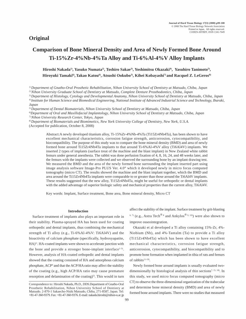
Comparison of Bone Mineral Density and Area of Newly Formed Bone Around Ti15%Zr4%Nb4%Ta Alloy and...
10
Comparison of Bone Mineral Density and Area of Newly Formed Bone Around Ti-15%Zr-4%Nb-4%Ta Alloy and Ti-6%Al-4%V Alloy Implants Hiroshi Nakada 1) , Yasuko Numata 2) , Toshiro Sakae 3) , Yoshimitsu Okazaki 4) , Yasuhiro Tanimoto 5) , Hiroyuki Tamaki 6) , Takao Katou 6) , Atsushi Ookubo 3) , Kihei Kobayashi 7) and Racquel Z. LeGeros 8) 1) Department of Gnatho-Oral Prosthetic Rehabilitation, Nihon University School of Dentistry at Matsudo, Chiba, Japan 2) Nihon University Graduate School of Dentistry at Matsudo, Complete Denture Prosthodontics, Chiba, Japan 3) Department of Histology, Cytology and Developmental Anatomy, Nihon University School of Dentistry at Matsudo, Chiba, Japan 4) Institute for Human Science and Biomedical Engineering, National Institute of Advanced Industrial Science and Technology, Ibaraki, Japan 5) Department of Dental Biomaterials, Nihon University School of Dentistry at Matsudo, Chiba, Japan 6) Department of Oral and Maxillofacial Implantology, Nihon University School of Dentistry at Matsudo, Chiba, Japan 7) Nihon University Research Center, Tokyo, Japan 8) Department of Biomaterials and Biomimetics, New York University College of Dentistry, New York, U.S.A. (Accepted for publication, October 8, 2008) Abstract:A newly developed titanium alloy, Ti-15%Zr-4%Nb-4%Ta (Ti15Zr4Nb4Ta), has been shown to have excellent mechanical characteristics, corrosion fatigue strength, anticorrosion, cytocompatibility, and biocompatibility. The purpose of this study was to compare the bone mineral density (BMD) and area of newly formed bone around Ti15Zr4Nb4Ta implants to that around Ti-6%Al-4%V alloy (Ti6Al4V) implants. We inserted 2 types of implants (surface treat of the machine and the blast implant) in New Zealand white rabbit femurs under general anesthesia. The rabbit was done perfusion fixation of 4, 8, 16, 24, and 48 weeks later, and the femurs with the implants were collected and we observed the surrounding bone by an implant drawing test. We measured the BMD and the area of the newly formed bone surrounding the implant inserted part using image analysis software Image-Pro PLUS Ver. 4.0 ® which it developed newly in micro focus computed tomography (micro CT). The results showed the machine and the blast implant together, which the BMD and area around the Ti15Zr4Nb4Ta implants were comparable to or greater than those around the Ti6Al4V implants. These results suggested that the new alloy, Ti15Zr4Nb4Ta, might be useful for orthopedic or dental implants with the added advantage of superior biologic safety and mechanical properties than the current alloy, Ti6Al4V. Key words: Implant, Surface treatment, Bone area, Bone mineral density, Micro CT Introduction Surface treatment of implants also plays an important role in their stability. Plasma-sprayed HA has been used for coating orthopedic and dental implants, thus combining the mechanical strength of Ti alloy (e.g., Ti-6%Al-4%V: Ti6Al4V) and the bioactivity of calcium phosphate (specifically, hydroxyapatite, HA) 1) . HA-coated implants were shown to accelerate junction with the bone and provide a stronger bone-implant interface 1-3) . However, analysis of HA-coated orthopedic and dental implants showed that the coating consisted of HA and amorphous calcium phosphate, ACP 4 and that the ACP/HA ratio may affect the stability of the coating (e.g., high ACP/HA ratio may cause premature resorption and delamination of the coating) 5) . This would in turn affect the stability of the implant. Surface treatment by grit-blasting 6, 7) (e.g., Astra Tech ® 8 and Ankylos ® 9, 10) ) were also shown to improve osseointegration. Okazaki et al developed a Ti alloy containing 15%-Zr, 4%- Niobium (Nb), and 4%-Tantalm (Ta) to provide a Ti alloy (Ti15Zr4Nb4Ta) which has been shown to have excellent mechanical characteristics, corrosion fatigue strength, anticorrosion, cytocompatibility, and biocompatibility and to promote bone formation when implanted in tibia of rats and femurs of rabbits 11-19) . Newly formed bone around implants is usually evaluated two- dimensionally by histological analysis of thin sections 7, 15, 18) . In this study, we used micro focus computed tomography (micro CT) to observe the three-dimensional organization of the trabecular and determine bone mineral density (BMD) and area of newly formed bone around implants. There were no studies that measured Corespondence to: Hiroshi Nakada, Ph.D., DDS.Department of Gnatho-Oral Prosthetic Rehabilitation, Nihon University School of Dentistry at Matsudo. 2-870-1 Sakaecho-Nishi Matsudo, Chiba, 271-8587, Japan. Tel: +81-47-360-9379. Fax: +81-47-360-9376. E-mail: [email protected] Original Journal of Hard Tissue Biology 17[3] (2008) p99-108 © 2008 The Hard Tissue Biology Network Association Printed in Japan, All rights reserved. CODEN-JHTBFF, ISSN 1341-7649 99
Transcript of Comparison of Bone Mineral Density and Area of Newly Formed Bone Around Ti15%Zr4%Nb4%Ta Alloy and...
Hiroshi Nakada et al. Comparison of BMD and Area of Newly Formed Bone Around Implants.
Comparison of Bone Mineral Density and Area of Newly Formed Bone AroundTi-15%Zr-4%Nb-4%Ta Alloy and Ti-6%Al-4%V Alloy Implants
Hiroshi Nakada1), Yasuko Numata2), Toshiro Sakae3), Yoshimitsu Okazaki4), Yasuhiro Tanimoto5),Hiroyuki Tamaki6), Takao Katou6), Atsushi Ookubo3), Kihei Kobayashi7) and Racquel Z. LeGeros8)
1) Department of Gnatho-Oral Prosthetic Rehabilitation, Nihon University School of Dentistry at Matsudo, Chiba, Japan2) Nihon University Graduate School of Dentistry at Matsudo, Complete Denture Prosthodontics, Chiba, Japan3) Department of Histology, Cytology and Developmental Anatomy, Nihon University School of Dentistry at Matsudo, Chiba, Japan4) Institute for Human Science and Biomedical Engineering, National Institute of Advanced Industrial Science and Technology, Ibaraki,Japan5) Department of Dental Biomaterials, Nihon University School of Dentistry at Matsudo, Chiba, Japan6)Department of Oral and Maxillofacial Implantology, Nihon University School of Dentistry at Matsudo, Chiba, Japan7) Nihon University Research Center, Tokyo, Japan8) Department of Biomaterials and Biomimetics, New York University College of Dentistry, New York, U.S.A.(Accepted for publication, October 8, 2008)
Abstract:A newly developed titanium alloy, Ti-15%Zr-4%Nb-4%Ta (Ti15Zr4Nb4Ta), has been shown to haveexcellent mechanical characteristics, corrosion fatigue strength, anticorrosion, cytocompatibility, andbiocompatibility. The purpose of this study was to compare the bone mineral density (BMD) and area of newlyformed bone around Ti15Zr4Nb4Ta implants to that around Ti-6%Al-4%V alloy (Ti6Al4V) implants. Weinserted 2 types of implants (surface treat of the machine and the blast implant) in New Zealand white rabbitfemurs under general anesthesia. The rabbit was done perfusion fixation of 4, 8, 16, 24, and 48 weeks later, andthe femurs with the implants were collected and we observed the surrounding bone by an implant drawing test.We measured the BMD and the area of the newly formed bone surrounding the implant inserted part usingimage analysis software Image-Pro PLUS Ver. 4.0® which it developed newly in micro focus computedtomography (micro CT). The results showed the machine and the blast implant together, which the BMD andarea around the Ti15Zr4Nb4Ta implants were comparable to or greater than those around the Ti6Al4V implants.These results suggested that the new alloy, Ti15Zr4Nb4Ta, might be useful for orthopedic or dental implantswith the added advantage of superior biologic safety and mechanical properties than the current alloy, Ti6Al4V.
Key words: Implant, Surface treatment, Bone area, Bone mineral density, Micro CT
IntroductionSurface treatment of implants also plays an important role in
their stability. Plasma-sprayed HA has been used for coatingorthopedic and dental implants, thus combining the mechanicalstrength of Ti alloy (e.g., Ti-6%Al-4%V: Ti6Al4V) and thebioactivity of calcium phosphate (specifically, hydroxyapatite,HA)1). HA-coated implants were shown to accelerate junction withthe bone and provide a stronger bone-implant interface1-3).However, analysis of HA-coated orthopedic and dental implantsshowed that the coating consisted of HA and amorphous calciumphosphate, ACP4 and that the ACP/HA ratio may affect the stabilityof the coating (e.g., high ACP/HA ratio may cause prematureresorption and delamination of the coating)5). This would in turn
affect the stability of the implant. Surface treatment by grit-blasting6, 7) (e.g., Astra Tech® 8 and Ankylos® 9, 10)) were also shown to
improve osseointegration.Okazaki et al developed a Ti alloy containing 15%-Zr, 4%-
Niobium (Nb), and 4%-Tantalm (Ta) to provide a Ti alloy(Ti15Zr4Nb4Ta) which has been shown to have excellentmechanical characteristics, corrosion fatigue strength,anticorrosion, cytocompatibility, and biocompatibility and topromote bone formation when implanted in tibia of rats and femursof rabbits11-19).
Newly formed bone around implants is usually evaluated two-dimensionally by histological analysis of thin sections7, 15, 18). Inthis study, we used micro focus computed tomography (microCT) to observe the three-dimensional organization of the trabecularand determine bone mineral density (BMD) and area of newlyformed bone around implants. There were no studies that measured
Corespondence to: Hiroshi Nakada, Ph.D., DDS.Department of Gnatho-OralProsthetic Rehabilitation, Nihon University School of Dentistry atMatsudo. 2-870-1 Sakaecho-Nishi Matsudo, Chiba, 271-8587, Japan. Tel:+81-47-360-9379. Fax: +81-47-360-9376. E-mail: [email protected]
Original
Journal of Hard Tissue Biology 17[3] (2008) p99-108 © 2008 The Hard Tissue Biology Network Association
Printed in Japan, All rights reserved.CODEN-JHTBFF, ISSN 1341-7649
99
J.Hard Tissue Biology Vol. 17(3):99-108. 2008
the BMD surrounding the Ti15Zr4Nb4Ta implants by the implantdrawing test and the distribution domain of newly formed bonefor three dimensions till now.
The purpose of this study was to compare the BMD and areaof newly formed bone around two types of Ti alloy implants(Ti6Al4V and Ti15Zr4Nb4Ta) that had received two kinds ofsurface treatments (machined and blasted) using micro CT.
Materials and MethodsImplant materials We showed the chemical composition of Ti15Zr4Nb4Ta andTi6Al4V used in this study to Table I. The implants used acylindrical (a diameter of 3.1mm and 30.0mm in length) type.The implants were surface treated by either mechanical polishing(machined) or grit-blasting with alumina abrasive (blasted). Both
Titanium alloy Zr Nb Ta Pd Fe O N H C Al V Ti
Ti-15Zr-4Nb-4Ta 15.24 3.90 3.92 0.22 0.022 0.162 0.048 0.011 0.002 - - Bal.**Ti-6Al-4V ELI* - - - - 0.198 0.101 0.003 0.005 0.011 6.24 4.19 Bal.**
* ELI. : Extra Low Interstitial ** Bal. : Balance
Table I Chemical composition (mass %) of the two types of Ti alloy used in the study.
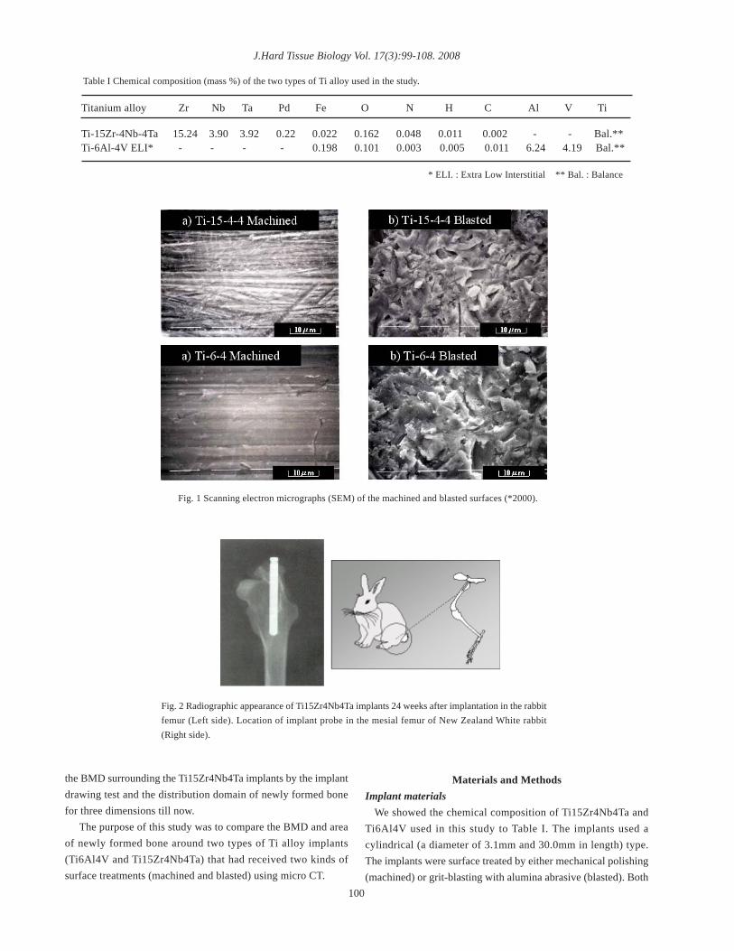
Fig. 1 Scanning electron micrographs (SEM) of the machined and blasted surfaces (*2000).
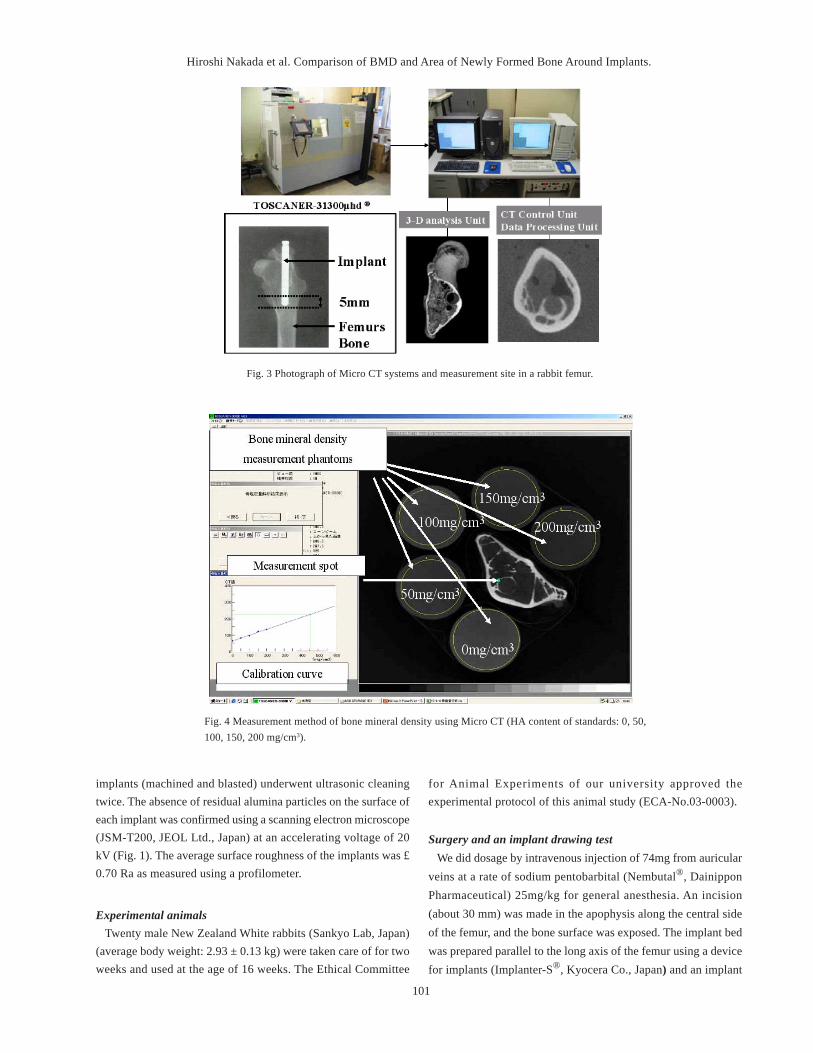
Fig. 2 Radiographic appearance of Ti15Zr4Nb4Ta implants 24 weeks after implantation in the rabbitfemur (Left side). Location of implant probe in the mesial femur of New Zealand White rabbit(Right side).
100
Hiroshi Nakada et al. Comparison of BMD and Area of Newly Formed Bone Around Implants.
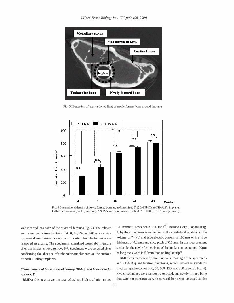
Fig. 3 Photograph of Micro CT systems and measurement site in a rabbit femur.
implants (machined and blasted) underwent ultrasonic cleaningtwice. The absence of residual alumina particles on the surface ofeach implant was confirmed using a scanning electron microscope(JSM-T200, JEOL Ltd., Japan) at an accelerating voltage of 20kV (Fig. 1). The average surface roughness of the implants was £0.70 Ra as measured using a profilometer.
Experimental animals Twenty male New Zealand White rabbits (Sankyo Lab, Japan)(average body weight: 2.93 ± 0.13 kg) were taken care of for twoweeks and used at the age of 16 weeks. The Ethical Committee
for Animal Experiments of our university approved theexperimental protocol of this animal study (ECA-No.03-0003).
Surgery and an implant drawing test We did dosage by intravenous injection of 74mg from auricularveins at a rate of sodium pentobarbital (Nembutal®, DainipponPharmaceutical) 25mg/kg for general anesthesia. An incision(about 30 mm) was made in the apophysis along the central sideof the femur, and the bone surface was exposed. The implant bedwas prepared parallel to the long axis of the femur using a devicefor implants (Implanter-S®, Kyocera Co., Japan) and an implant
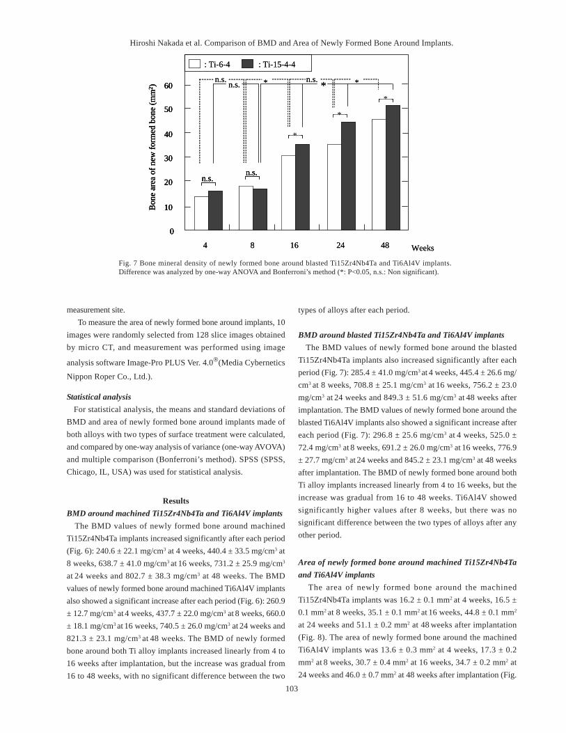
Fig. 4 Measurement method of bone mineral density using Micro CT (HA content of standards: 0, 50,100, 150, 200 mg/cm3).
101
J.Hard Tissue Biology Vol. 17(3):99-108. 2008
was inserted into each of the bilateral femurs (Fig. 2). The rabbitswere done perfusion fixation of 4, 8, 16, 24, and 48 weeks laterby general anesthesia since implants inserted. And the femurs wereremoved surgically. The specimens examined were rabbit femursafter the implants were removed18). Specimens were selected afterconfirming the absence of trabecular attachments on the surfaceof both Ti alloy implants.
Measurement of bone mineral density (BMD) and bone area bymicro CT BMD and bone area were measured using a high resolution micro
Fig. 5 Illustration of area (a dotted line) of newly formed bone around implants.
Bon
e m
iner
al d
ensi
ty m
g/cm
3
0
200
400
600
800
1000
4 Weeks4824168
n.s.n.s.
n.s.
n.s.
n.s.
: Ti-6-4 : Ti-15-4-4
* *
Bon
e m
iner
al d
ensi
ty m
g/cm
3
0
200
400
600
800
1000
4 Weeks4824168
n.s.n.s.n.s.n.s.
n.s.n.s.
n.s.n.s.
n.s.n.s.
: Ti-6-4 : Ti-15-4-4
* *
Fig. 6 Bone mineral density of newly formed bone around machined Ti15Zr4Nb4Ta and Ti6Al4V implants.Difference was analyzed by one-way ANOVA and Bonferroni’s method (*: P<0.05, n.s.: Non significant).
CT scanner (Toscaner-31300 mhd®, Toshiba Corp., Japan) (Fig.3) by the cone beam scan method in the non-helical mode at a tubevoltage of 74 kV, and tube electric current of 110 mA with a slicethickness of 0.2 mm and slice pitch of 0.1 mm. In the measurementsite, as for the newly formed bone of the implant surrounding, 100µmof long axes were in 5.0mm than an implant tip20).
BMD was measured by simultaneous imaging of the specimensand 5 BMD quantification phantoms, which served as standards(hydroxyapatite contents: 0, 50, 100, 150, and 200 mg/cm3: Fig. 4).Five slice images were randomly selected, and newly formed bonethat was not continuous with cortical bone was selected as the
102
Hiroshi Nakada et al. Comparison of BMD and Area of Newly Formed Bone Around Implants.
types of alloys after each period.
BMD around blasted Ti15Zr4Nb4Ta and Ti6Al4V implants The BMD values of newly formed bone around the blastedTi15Zr4Nb4Ta implants also increased significantly after eachperiod (Fig. 7): 285.4 ± 41.0 mg/cm3 at 4 weeks, 445.4 ± 26.6 mg/cm3 at 8 weeks, 708.8 ± 25.1 mg/cm3 at 16 weeks, 756.2 ± 23.0mg/cm3 at 24 weeks and 849.3 ± 51.6 mg/cm3 at 48 weeks afterimplantation. The BMD values of newly formed bone around theblasted Ti6Al4V implants also showed a significant increase aftereach period (Fig. 7): 296.8 ± 25.6 mg/cm3 at 4 weeks, 525.0 ±72.4 mg/cm3 at 8 weeks, 691.2 ± 26.0 mg/cm3 at 16 weeks, 776.9± 27.7 mg/cm3 at 24 weeks and 845.2 ± 23.1 mg/cm3 at 48 weeksafter implantation. The BMD of newly formed bone around bothTi alloy implants increased linearly from 4 to 16 weeks, but theincrease was gradual from 16 to 48 weeks. Ti6Al4V showedsignificantly higher values after 8 weeks, but there was nosignificant difference between the two types of alloys after anyother period.
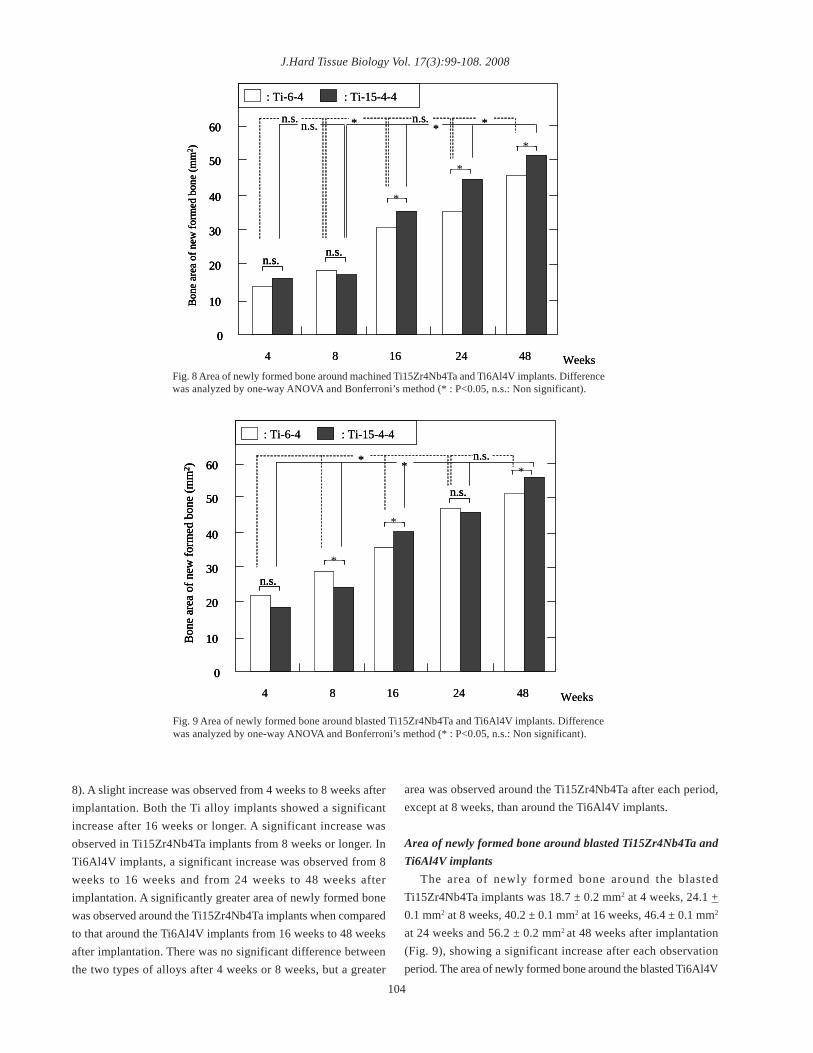
Area of newly formed bone around machined Ti15Zr4Nb4Taand Ti6Al4V implants The area of newly formed bone around the machinedTi15Zr4Nb4Ta implants was 16.2 ± 0.1 mm2 at 4 weeks, 16.5 ±0.1 mm2 at 8 weeks, 35.1 ± 0.1 mm2 at 16 weeks, 44.8 ± 0.1 mm2
at 24 weeks and 51.1 ± 0.2 mm2 at 48 weeks after implantation(Fig. 8). The area of newly formed bone around the machinedTi6Al4V implants was 13.6 ± 0.3 mm2 at 4 weeks, 17.3 ± 0.2mm2 at 8 weeks, 30.7 ± 0.4 mm2 at 16 weeks, 34.7 ± 0.2 mm2 at24 weeks and 46.0 ± 0.7 mm2 at 48 weeks after implantation (Fig.
4 Weeks4824168
60
0
10
20
30
40
50
Bon
e ar
ea o
f new
form
ed b
one
(mm
2 )
: Ti-6-4 : Ti-15-4-4
*
*
*
n.s.n.s.
*n.s. *n.s. *n.s.
4 Weeks4824168
60
0
10
20
30
40
50
Bon
e ar
ea o
f new
form
ed b
one
(mm
2 ) 60
0
10
20
30
40
50
Bon
e ar
ea o
f new
form
ed b
one
(mm
2 )
: Ti-6-4 : Ti-15-4-4
***
**
***
n.s.n.s.n.s.n.s.
*n.s. *n.s. *n.s.
Fig. 7 Bone mineral density of newly formed bone around blasted Ti15Zr4Nb4Ta and Ti6Al4V implants.Difference was analyzed by one-way ANOVA and Bonferroni’s method (*: P<0.05, n.s.: Non significant).
103
ResultsBMD around machined Ti15Zr4Nb4Ta and Ti6Al4V implants The BMD values of newly formed bone around machinedTi15Zr4Nb4Ta implants increased significantly after each period(Fig. 6): 240.6 ± 22.1 mg/cm3 at 4 weeks, 440.4 ± 33.5 mg/cm3 at8 weeks, 638.7 ± 41.0 mg/cm3 at 16 weeks, 731.2 ± 25.9 mg/cm3
at 24 weeks and 802.7 ± 38.3 mg/cm3 at 48 weeks. The BMDvalues of newly formed bone around machined Ti6Al4V implantsalso showed a significant increase after each period (Fig. 6): 260.9± 12.7 mg/cm3 at 4 weeks, 437.7 ± 22.0 mg/cm3 at 8 weeks, 660.0± 18.1 mg/cm3 at 16 weeks, 740.5 ± 26.0 mg/cm3 at 24 weeks and821.3 ± 23.1 mg/cm3 at 48 weeks. The BMD of newly formedbone around both Ti alloy implants increased linearly from 4 to16 weeks after implantation, but the increase was gradual from16 to 48 weeks, with no significant difference between the two
measurement site.To measure the area of newly formed bone around implants, 10
images were randomly selected from 128 slice images obtainedby micro CT, and measurement was performed using image
analysis software Image-Pro PLUS Ver. 4.0®(Media Cybernetics
Nippon Roper Co., Ltd.).
Statistical analysis For statistical analysis, the means and standard deviations ofBMD and area of newly formed bone around implants made ofboth alloys with two types of surface treatment were calculated,and compared by one-way analysis of variance (one-way AVOVA)and multiple comparison (Bonferroni’s method). SPSS (SPSS,Chicago, IL, USA) was used for statistical analysis.
J.Hard Tissue Biology Vol. 17(3):99-108. 2008
8). A slight increase was observed from 4 weeks to 8 weeks afterimplantation. Both the Ti alloy implants showed a significantincrease after 16 weeks or longer. A significant increase wasobserved in Ti15Zr4Nb4Ta implants from 8 weeks or longer. InTi6Al4V implants, a significant increase was observed from 8weeks to 16 weeks and from 24 weeks to 48 weeks afterimplantation. A significantly greater area of newly formed bonewas observed around the Ti15Zr4Nb4Ta implants when comparedto that around the Ti6Al4V implants from 16 weeks to 48 weeksafter implantation. There was no significant difference betweenthe two types of alloys after 4 weeks or 8 weeks, but a greater
4 Weeks4824168
60
0
10
20
30
40
50
Bon
e ar
ea o
f new
form
ed b
one
(mm
2 )
: Ti-6-4 : Ti-15-4-4
*
*
*
n.s.n.s.
*n.s. *n.s.*n.s.
4 Weeks4824168
60
0
10
20
30
40
50
Bon
e ar
ea o
f new
form
ed b
one
(mm
2 )
60
0
10
20
30
40
50
Bon
e ar
ea o
f new
form
ed b
one
(mm
2 )
: Ti-6-4 : Ti-15-4-4
***
**
***
n.s.n.s.n.s.n.s.
*n.s. *n.s.*n.s.
Fig. 8 Area of newly formed bone around machined Ti15Zr4Nb4Ta and Ti6Al4V implants. Differencewas analyzed by one-way ANOVA and Bonferroni’s method (* : P<0.05, n.s.: Non significant).
4 Weeks4824168
60
0
10
20
30
40
50
Bon
e ar
ea o
f new
form
ed b
one
(mm
2 )
*
*
n.s.
n.s.
*
: Ti-6-4 : Ti-15-4-4
* n.s.*
4 Weeks4824168
60
0
10
20
30
40
50
Bon
e ar
ea o
f new
form
ed b
one
(mm
2 ) 60
0
10
20
30
40
50
Bon
e ar
ea o
f new
form
ed b
one
(mm
2 )
***
**
n.s.n.s.
n.s.n.s.
**
: Ti-6-4 : Ti-15-4-4
* n.s.*
Fig. 9 Area of newly formed bone around blasted Ti15Zr4Nb4Ta and Ti6Al4V implants. Differencewas analyzed by one-way ANOVA and Bonferroni’s method (* : P<0.05, n.s.: Non significant).
104
area was observed around the Ti15Zr4Nb4Ta after each period,except at 8 weeks, than around the Ti6Al4V implants.
Area of newly formed bone around blasted Ti15Zr4Nb4Ta andTi6Al4V implants The area of newly formed bone around the blastedTi15Zr4Nb4Ta implants was 18.7 ± 0.2 mm2 at 4 weeks, 24.1 +0.1 mm2 at 8 weeks, 40.2 ± 0.1 mm2 at 16 weeks, 46.4 ± 0.1 mm2
at 24 weeks and 56.2 ± 0.2 mm2 at 48 weeks after implantation(Fig. 9), showing a significant increase after each observationperiod. The area of newly formed bone around the blasted Ti6Al4V
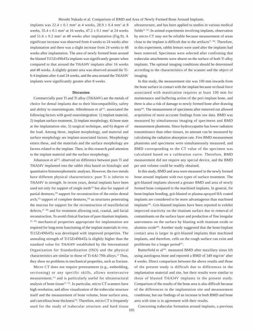
Hiroshi Nakada et al. Comparison of BMD and Area of Newly Formed Bone Around Implants.implants was 22.4 ± 0.1 mm2 at 4 weeks, 28.9 ± 0.4 mm2 at 8weeks, 35.4 ± 0.1 mm2 at 16 weeks, 47.2 ± 0.1 mm2 at 24 weeksand 51.6 ± 0.2 mm2 at 48 weeks after implantation (Fig.9). Asignificant increase was observed from 4 weeks to 24 weeks afterimplantation and there was a slight increase from 24 weeks to 48weeks after implantation. The area of newly formed bone aroundthe blasted Ti15Zr4Nb4Ta implants was significantly greater whencompared to that around the Ti6Al4V implants after 16 weeksand 48 weeks. A slightly greater area was observed around the Ti-6-4 implants after 4 and 24 weeks, and the area around the Ti6Al4Vimplants were significantly greater after 8 weeks.
DiscussionCommercially pure Ti and Ti alloy (Ti6Al4V) are the metals of
choice for dental implants due to their biocompatibility, safetyand ability to osseointegrate. Albrektsson et al21). associated thefollowing factors with good osseointegration: 1) implant material,2) implant surface treatment, 3) implant morphology, 4) bone stateat the implantation site, 5) surgical technique, and 6) degree ofthe load. Among these, implant morphology, and material andsurface morphology are implant-associated factors. Morphologyenters these, and the materials and the surface morphology arefactors related to the implant. Then, in this research paid attentionto the implant material and the surface morphology.
Johansson et al22). observed no difference between pure Ti andTi6Al4V implanted into the rabbit tibia based on histologic andquantitative histomorphometric analyses. However, the two metalshave different physical characteristics: pure Ti is inferior toTi6Al4V in strength. In recent years, dental implants have beenused not only for support of single teeth23) but also for support ofpartial dentures,24) support for reconstruction of the entire dentalarch,25) support of complete dentures,26) as structures penetratingthe mucosa for support for the reconstruction of maxillofacialdefects,27, 28) and for treatment allowing oral, cranial, and facialreconstruction. To avoid clinical fracture of pure titanium implants,29, 30) mechanical properties appropriate for implantation arerequired for long-term functioning of the implant materials in vivo.Ti15Zr4Nb4Ta was developed with improved properties. Theannealing strength of Ti15Zr4Nb4Ta is slightly higher than thestandard value for Ti6Al4V established by the InternationalOrganization for Standardization (ISO) and the physicalcharacteristics are similar to those of Ti-6Al-7Nb alloys.12 Thus,they show no problems in mechanical properties, such as fracture.
Micro CT does not require pretreatment (e.g., embedding,sectioning) or any specific skills, allows noninvasivemeasurement,31) and is particularly useful for ultrastructuralanalysis of bone tissue32, 33). In particular, micro CT scanners havehigh resolution, and allow visualization of the trabecular structureitself and the measurement of bone volume, bone surface area,and cancellous bone thickness34). Therefore, micro CT is frequentlyused for the study of trabecular structure and hard tissue
ultrastructure, and has been applied to studies in various medicalfields35, 36). In animal experiments involving implants, observationby micro CT may not be reliable because measurement of areasclose to the implant is difficult due to the artifacts37, 38). Therefore,in this experiment, rabbit femurs were used after the implants hadbeen removed. Specimens were selected after confirming thattrabecular attachments were absent on the surface of both Ti alloyimplants. The optimal imaging conditions should be determinedaccording to the characteristics of the scanner and the object ofimaging.
In this study, the measurement site was 100 mm inwards fromthe bone surface in contact with the implant because occlusal forceassociated with mastication requires at least 100 mm formaintenance and buffering action of the peri-implant bone, andthere is also a risk of damage to newly formed bone after drawingtests20). The measurement of specimens after removed out allowedacquisition of more accurate findings from raw data. BMD wasmeasured by simultaneous imaging of specimens and BMDmeasurement phantoms. Since hydroxyapatite has lower radiationtransmittance than other tissues, its amount can be measured bycalculating the radiation absorption rate. Five BMD measurementphantoms and specimens were simultaneously measured, andBMD corresponding to the CT value of the specimen wascalculated based on a calibration curve. Therefore, BMDmeasurement did not require any special device, and the BMDper unit volume could be readily obtained.
In this study, BMD and area were measured in the newly formedbone around implants with two types of surface treatment. Thegrit-blasted implants showed a greater BMD and area of newlyformed bone compared to the machined implants. In general, forbone/implant bonding, grit-blasted or plasma-sprayed HA coatedimplants are considered to be more advantageous than machinedimplants39). Grit-blasted implants have been reported to exhibitimproved reactivity on the titanium surface due to removal ofcontaminants on the surface layer and production of fine irregularunevenness on the surface by blasting with titanium oxide oralumina oxide40). Another study suggested that the bone/implantcontact area is larger in grit-blasted implants than machinedimplants, and therefore, cells on the rough surface can exist andproliferate for a longer period41).
Butterfield et al42). measured BMD after maxillary sinus liftusing autologous bone and reported a BMD of 349 mg/cm3 after4 weeks. Direct comparison between the above results and thoseof the present study is difficult due to differences in theimplantation material and site, but their results were similar tothose of blasted Ti6Al4V implants in the present study.Comparison of the results of the bone area is also difficult becauseof the differences in the implantation site and measurementconditions, but our findings of an increase in both BMD and bonearea with time is in agreement with their results.
Concerning trabecular formation around implants, a previous105
J.Hard Tissue Biology Vol. 17(3):99-108. 2008study showed active bone formation in the early stage from 2weeks to 6 weeks after implantation, but trabecular resorptionoccurred after 24 weeks or more.43 In the present study, a linearincrease in BMD was observed from 4 weeks to 16 weeks afterimplantation, and a gradual increase thereafter until 48 weeks afterimplantation for both types of surface treatment on both alloys.The area of newly formed bone also increased from 4 weeks to 48weeks after implantation.
Newly formed bone around the implant should be maintaineddespite loads such as occlusion force applied on bone. Newlyformed trabeculae are not mature enough, and the crystallinity oftrabeculae should be gradually improved by stimulating boneprecursor cells, increasing alkaline phosphatase activity, andenhancing the incorporation of newly formed collagen into bonematrix. In addition, to maintain BMD, modeling-remodeling isindispensable, and the bone area should be further increased forthe establishment of the bone basis for osseointegration.
These results suggest the usefulness of Ti15Zr4Nb4Ta as animplant material for biological applications. Though manytitanium alloys have been developed as implant materials, artificialmaterials that do not induce negative responses during theirconstant functioning in vivo should be selected. Hence, furtherresearch is necessary to analyze the crystallinity, i.e., quality ofbone formed around Ti15Zr4Nb4Ta implants.
ConclusionWe investigated the characteristics of bone on the machined or
blasted surface of Ti15Zr4Nb4Ta, a potential titanium alloy forbiological use, and Ti6Al4V, a control that was conventionallyused. The BMD and bone area were measured by micro CT innewly formed bone 100 mm from the bone surface around implantsof these alloys with two types of surface treatment.
The BMD and bone area around the Ti15Zr4Nb4Ta implantswere comparable to or greater than those around the Ti6Al4Vimplants. The Ti15Zr4Nb4Ta contains ions that are biologicallysafer. These results suggested that the new alloy, Ti15Zr4Nb4Tamight be useful for orthopedic or dental implants with the addedadvantage of superior biologic safety and mechanical propertiesthan the current alloy, Ti6Al4V. Further studies are warranted onthe new alloy to evaluate its potential as an implant material fororthopedic and dental applications.
AcknowledgmentsThe authors gratefully acknowledge Dr. Takeshi Machida and
Dr. Taketoshi Suwa in department of Gnatho-Oral ProsthticRehabilitation, Nihon University School of dentistry at Matsudoin Japan. This study was supported in part by Grant-in-Aid forScientific Research (C) (18592145) and Young Scientists(B)(19791462) from the Japan Society for the Promotion of Scienceand Nihon University Individual Research Grant for 2008.
106
References1. Gotfredsen K, Wennerberg A, Johansson C and Skovgaard L
T, Hjorting-Hansen E. Anchorage of TiO2-Blasted HA-Coatedand Machined Implants. An Experimental Study with Rabbits.J Biomed Mater Res 29: 1223-1231, 1995
2. Ong J L, Carnes D L and Bessho K. Evaluation of TitaniumPlasma-Sprayed and Plasma-Sprayed HydroxyapatiteImplants in vivo. Biomaterials 25: 4601-4606, 2004
3. Cook S D, Thomas K A, Dalton J E, Volkman R K, WhitecloudT S and Kay J E. Hydroxylapatite coating of porous implantsimproves bone ingrowth and interface attachment strength.J Biomed Mater Res 26: 989-1001, 1992
4. LeGeros R Z, LeGeros J P, Kim Y, Kijkowska R, Zheng R,Bautista C and Wong J L. Calcium phosphates in plasma-sprayed HA coatings. Ceramic Transactions 48: 173-189,1995
5. LeGeros R Z, Kim YE, Kijkowska R, Zurita V, Bleiwas C,Huang PY, Edwards B, Dimaano F and LeGeros J P. HA/ACP ratios in calcium phosphate coatings on dental andorthopedic implants. Effect on properties. Bioceramics 11:181-184,1998
6. Feighan J E, Goldberg V M, Davy D, Parr J A and StevensonS. The Influence of Surface-Blasting on the Incorporation ofTitanium-Alloy Implants in a Rabbit Intramedullary Model.J Bone Joint Surg Am 77: 1380-1395, 1995
7. Wennerberg A, Albrektsson T, Johansson C and AnderssonB. Experimental Study of Turned and Grit-Blasted Screw-Shaped Implants with Special Emphasis on Effects ofBlasting Material and Surface Topography. Biomaterials 17:15-22,1996
8. Norton M R. A Short-Term Clinical Evaluation of ImmediatelyRestored Maxillary TiO2 blast Single-Tooth Implants. Int JOral Maxillofac Implants 19: 274-281, 2004
9. Döring K, Eisenmann E. and Stiller M. Functional and EstheticConsiderations for Single-Tooth Ankylos Implant-Crowns:8 Years of Clinical Performance. J Oral Implantol 30: 198-209, 2004
10. Nentwig G H. The Ankylos Implant System. Concept andClinical Application. J Oral Implantol 30: 171-177, 2004
11. Okazaki Y. Effect of Friction on Anodic Polarization Propertiesof Metallic Biomaterials. Biomaterials 23: 2071-2077, 2002
12. Okazaki Y, Rao S, Ito Y and Tateishi T. CorrosionResistance, Mechanical Properties, Corrosion FatigueStrength and Cytocompatibility of New Ti Alloys withoutAl and V. Biomaterials 19: 1197-1215, 1998
13. I to A, Okazaki Y, Tateishi T and I to Y. In v i troBiocompatibility, Mechanical Properties, and CorrosionResistance of Ti-Zr-Nb-Ta-Pd and Ti-Sn-Nb-Ta-Pd Alloys.J Biomed Mater Res 29: 893-899, 1995
14. Nakada H. A Study on Corrosion Resistance andBiocompatibility by Rat Implantation of New Titanium Alloy
Hiroshi Nakada et al. Comparison of BMD and Area of Newly Formed Bone Around Implants.
107
for Medical Implants (in Japanese). Nihon Univ J Oral Sci25: 103-112, 1999
15. Okazaki Y, Nishimura E, Nakada H and Kobayashi K. SurfaceAnalysis of Ti-15Zr-4Nb-4Ta Alloy after Implantation in RatTibia. Biomaterials 22: 599-607, 2001
16. Manabe T. Histopathologycal Analysis on New Bone TissueFormed Around Metallic Implants in the Rat Tibia (inJapanese). Nihon Univ J Oral Sci 28: 31-47, 2002
17 Okazaki Y, Goto E, Manabe T and Kobayashi K.Comparison of Metal Concentrations in Rat Tibia Tissueswith Various Metallic Implants. Biomaterials 25: 5913-59202004
18. Machida T. A Study of Osseointegration of Titanium alloyImplants: The Investigation on Effect of Different SurfaceTreatment (in Japanese). Nihon Univ J Oral Sci 30: 245-257, 2004
19. Okazaki Y and Gotoh E. Comparison of Metal Release fromVarious Metallic Biomaterials in vitro. Biomaterials, 26: 11-21, 2005
20. Nakada H, Suwa T, Numata Y, Okazaki Y, Sakae T, HayakawaT, Kato M, Kaneda T, LeGeros R Z, Kato T, Gunji A,Kuwahara K and Kobayashi K. Analysis of bone mineraldensity and bone area of newly formed bone around implantsusing micro-CT after pull-out tests. Int J Oral-Med Sci 5:97-106, 2007
21. Albrektsson T, Brånemark PI, Hansson HA and Lindstrom J.Osseointegrated Titanium Implants. Requirements forEnsuring a Long-Lasting, Direct Bone-to-Implant Anchoragein Man. Acta Orthop Scand 52: 155-170, 1981
22. Johansson CB, Han CH. and Wennerberg A. A QuantitativeComparison of Machined Commercially Pure Titanium andTitanium-Aluminum-Vanadium Implants in Rabbit Bone. IntJ Oral Maxillofac Implants 13: 315-321, 1998
23. Sabri R. Four Single-Tooth Implants as SupernumeraryPremolars in the Treatment of Diastemas and Microdontia-Report of a Case. Int J Oral Maxillofac Implants 13: 706-709, 1998
24. Knabe C and Hoffmeister B. The Use of Implant-SupportedCeramometal Titanium Prostheses Following Sinus Lift andAugmentation Procedures. A Clinical Report. Int J OralMaxillofac Implants 13: 102-108, 1998
25. Balshi TJ, Wolfinger GJ, Balshi SF. Analysis of 356Pterygomaxillary Implants in Edentulous Arches For FixedProsthesis Anchorage. Int J Oral Maxillofac Implants 14: 398-406, 1999
26. Mericske-Stern R, Oetterli M, Kiener P and Mericske E AFollow-up Study of Maxillary Implants Supporting anOverdenture: Clinical and Radiographic Results. Int J OralMaxillofac Implants 17: 678-686, 2002
27. Nishimura R D, Roumanas E, Moy P K, Sugai T andFreymiller E G. Osseointegrated Implants and Orbital Defects.
U CLA. Experience. J Prosthet Dent 79: 304-309, 1998.28. Lemon J C, Chambers M S, Wesley P J, Reece G P and
Martin J W. Rehabilitation of a Midface Defect withReconstructive Surgery and Facial Prosthetics: A CaseReport. Int J Oral Maxillofac Implants 11: 101-105, 1996
29. Muroff F I. Removal and Replacement of a Fractured DentalImplant: Case Report. Implant Dent 12: 206-210, 2003
30. Rangert B, Krogh P H, Langer B and Van Roekel N. BendingOverload and Implant Fracture: A Retrospective ClinicalAnalysis. Int J Oral Maxillofac Implants 10: 326-334, 1995
31. Sennerby L, Thomsen P and Ericson L E. A morphometricand biomechanic comparison of titanium implants insertedin rabbit cortical and cancellous bone. Int J Oral MaxillofacImplants 7: 62-71, 1992
32. Ericsson I, Johansson C B, Bystedt H and Norton M R. Ahistomorphometric evaluation of bone-to-implant contact onmachine-prepared and roughened titanium dental implants:A pilot study in the dog. Clin Oral Implants Res 5: 202-206,1994
33. Evans GH., Mendez AJ. and Caudill RF. Loaded and nonloadedtitanium versus hydroxyapatite-coated threaded implants inthe canine mandible. Int J Oral Maxillofac Implants 11: 360-371, 1996
34. Beck T J, Looker A C, Ruff C B, Sievanen H and Wahner HW. Structural trends in the aging femoral neck and proximalshaft: analysis of the Third National Health and NutritionExamination Survey dual-energy X-ray absorptiometry data.J Bone Miner Res 15: 2297-2304, 2000
35. Ito M, Nishida A, Nakamura T, Uetani M and Hayashi K.Differences of three-dimensional trabecular microstructurein osteopenic rat models caused by ovariectomy andneurectomy. Bone 30: 594-598, 2002
36. Shiraishi A., Higashi S., Masaki T, Saito M, Ito M, Ikeda Sand Nakamura T. A comparison of alfacalcidol andmenatetrenone for the treatment of bone loss in anovariectomized rat model of osteoporosis. Calcif Tissue Int71: 69-79, 2002
37. Numata Y, Sakae T, Nakada H, Suwa T, LeGeros RZ, OkazakiY and Kobayashi K. Micro-CT analysis of rabbit cancellousbone around implants. J Hard Tissue Biology 16: 91-93, 2007
38. Van Oossterwyck H, Duyck J, Vander Sloten J, Van der PerreG, Jansen J, Wevers M and Naert I. Use of microfocuscomputerized tomography as a new technique forcharacterizing bone tissue around oral implants. J OralImplantol 26: 5-12, 2000
39. Gotfredsen K, Wennerberg A, Johansson C, Skovgaard LT,Hjorting Hansen E. Anchorage of TiO2-Blasted, HA-Coated,and Machined Implants. An Experimental Study with Rabbits.J Biomed Mater Res 29: 1223-1231, 1995
40. Wennerberg A, Albrektsson T, Johansson C and AnderssonB. Experimental Study of Turned and Grit-Blasted Screw-
J.Hard Tissue Biology Vol. 17(3):99-108. 2008Shaped Implants with Special Emphasis on Effects ofBlasting Material and Surface Topography. Biomaterials 17:15-22, 1996
41. Piattelli M, Scarano A, Paolantonio M, Iezzi G, Petrone Gand Piattelli A. Bone Response to Machined and ResorbableBlast Material Titanium Implants- An Experimental Studyin Rabbits. J Oral Implantol 28: 2-8, 2002
42. Butterfield K J, Bennett J, Gronowicz G and Adams D. Effect
of platelet-rich plasma with autogenous bone graft formaxillary sinus augmentation in a rabbit model. J OralMaxillofac Surg 63: 370-376, 2005
43. Kawamura H, Ito A, Muramatsu T, Miyakawa S, Ochiai Nand Tateishi T. Long-term implantation of zinc-releasingcalcium phosphate ceramics in rabbit femora. J Biomed MaterRes A 15: 468-474, 2003
108