Corrigendum: The deubiquitinase USP9X suppresses pancreatic ductal adenocarcinoma
Upload
putrabusinessschoolCategory
view
0download
0
Research ArticleComparison of Apoptotic Inducing Effect of Zerumbone andZerumbone-Loaded Nanostructured Lipid Carrier on HumanMammary Adenocarcinoma MDA-MB-231 Cell Line
Mahnaz Hosseinpour,1 Ahmad Bustamam Abdul,1,2 Heshu Sulaiman Rahman,1,3,4
Abdullah Rasedee,1,3 Swee Keong Yeap,1 Negin Ahmadi,1 Hemn Hassan Othman,3,4
and Max Stanley Chartrand5
1 Institute of Bioscience, Universiti Putra Malaysia (UPM), 43400 Serdang, Selangor, Malaysia2 Faculty of Medicine and Health Sciences, Universiti Putra Malaysia (UPM), 43400 Serdang, Selangor, Malaysia3 Faculty of Veterinary Medicine, Universiti Putra Malaysia (UPM), 43400 Serdang, Selangor, Malaysia4 Faculty of Veterinary Medicine, University of Sulaimani, Sulaimani Nwe, Street 27, Sulaimani City, Kurdistan Region, Iraq5 DigiCare Behavioral Research, Casa Grande, AZ, USA
Correspondence should be addressed to Mahnaz Hosseinpour; mahnaz [email protected] andAhmad Bustamam Abdul; [email protected]
Received 10 August 2014; Revised 30 October 2014; Accepted 30 October 2014; Published 18 November 2014
Academic Editor: Hongchen Chen Gu
Copyright © 2014 Mahnaz Hosseinpour et al. This is an open access article distributed under the Creative Commons AttributionLicense, which permits unrestricted use, distribution, and reproduction in any medium, provided the original work is properlycited.
This study investigated the anticancer effect of zerumbone (ZER) and zerumbone-loaded nanostructured lipid carrier (ZER-NLC)on the human mammary gland adenocarcinoma (MDA-MB-231) cell line. The effect of ZER and ZER-NLC on MDA-MB-231 cellswas determined via electron and fluorescent microscopy and flow cytometry using the Annexin V, cell cycle, and Tdt-mediateddUTP nick-end labeling assays. We demonstrated that ZER and ZER-NLC significantly suppressed the proliferation of MDA-MB-231 cells with an IC
50of 5.96 ± 0.13 and 6.01 ± 0.11 𝜇g/mL, respectively. ZER and ZER-NLC arrested MDA-MB-231 cell cycle at the
G2/M phase. The induction of apoptosis by ZER and ZER-NLC was via the intrinsic pathway through the release of cytochrome cand activation of caspase-3 and caspase-9. The treatments also caused the downregulation of antiapoptotic Bcl-2, Bcl-xL proteins,and proliferating cell nuclear protein and upregulation of proapoptotic Bax protein. Therefore, loading of ZER into NLC did notcompromise the anticancer effects of ZER on MDA-MB-231 cells. In conclusion, ZER-NLC, which increased the bioavailability ofZER, is an effective agent in the treatment of cancers.
1. Introduction
The main life-threatening and the most prevalent diseasesin women are breast cancers [1]. Development of breastcancers is associated with several risk factors such as age,genes, and family history. There are several treatment meth-ods currently being instituted for breast cancers to includechemotherapy, hormonal therapy using tamoxifen and dox-orubicin, radiotherapy, and surgery. Most of these treatmentmethods are not specific for cancer cells and more oftenalso damage normal cells. Current studies are increasinglyproviding evidences that therapeutic compounds from nat-ural sources are promising for effective treatment of chronic
human diseases including cancers, without significant side-effect [2]. Among these compounds is zerumbone (ZER),extracted from the essential oils of Zingiber zerumbet Smithrhizome.
Zerumbone has versatile pharmaceutical properties in-cluding anti-inflammatory, antitumor, antioxidant, antimi-crobial, and antinociceptive activities [3, 4]. This compoundalso significantly suppresses activation of nuclear factor 𝜅-light-chain-enhancer in activated B cells (NF-𝜅B) and NF-𝜅B-related gene expression generated by carcinogens invarious other cells [5]. Zerumbone has been reported toinhibit proliferation of cervical, ovarian, colon, breast, andliver cancers [6, 7].
Hindawi Publishing CorporationJournal of NanomaterialsVolume 2014, Article ID 742738, 10 pageshttp://dx.doi.org/10.1155/2014/742738

2 Journal of Nanomaterials
72h24h48h
0
20
40
60
80
100
120
0 50 100 150
Cel
l via
bilit
y (%
)
ZER concentration (𝜇g/mL)
MDA-MB-231
(a)
0
20
40
60
80
100
120
0 50 100 150
Cel
l via
bilit
y (%
)
ZER-NLC concentration (𝜇g/mL)
MDA-MB-231
72h24h48h
(b)
Figure 1: Viability of MDA-MB-231 cells treated with (a) ZER and (b) ZER-NLC after 24, 48, and 72 hours.
0
20
40
60
80
100
120
0 50 100 150
Cel
l via
bilit
y (%
)
ZER concentration (𝜇g/mL)
MCF-10A
72h24h48h
(a)
0
20
40
60
80
100
120
0 50 100 150
Cel
l via
bilit
y (%
)
ZER-NLC concentration (𝜇g/mL)
MCF-10A
72h24h48h
(b)
Figure 2: Viability of MCF-10A cells treated with (a) ZER and (b) ZER-NLC after 24, 48, and 72 hours.
Although zerumbone has pharmaceutical potentials, likemany other drugs and synthetic therapeutic compounds, itsapplication in therapy is hindered by poor water solubilitythat leads to poor bioavailability and delivery [8]. Thisproperty retards zerumbone absorption in the gastroin-testinal tract making oral applications relatively ineffective.The poor absorption in the gastrointestinal tract not onlyreduces bioavailability of zerumbone but also causes mucosaltoxicity [9, 10]. One approach to overcome this obstacle isto incorporate drugs into lipid nanocarriers, which wouldimprove solubility and delivery of zerumbone by facilitatingtransportation across several anatomical barriers. Solid lipidnanoparticles (SLN), nanostructured lipid carriers (NLC),and lipid-drug conjugates are examples of lipid-based car-rier systems that can be used to incorporate therapeuticcompounds. Compound-loaded lipids nanocarriers exhibitdesirable characteristics including low toxicity, controlledrelease, biodegradable, high drug content, low cost, and easyupscaling [11, 12].
Nanostructured lipid carrier, a second generation lipidnanocarrier produced by high-pressure homogenization, iscomposed of a solid and liquid lipid matrix with disruptedcrystalline nanostructure [13, 14]. This lipid nanocarrier,while overcoming some of potential therapeutic applicationproblems associated with SLN, also possesses high loadingcapacity, low water content of particle suspension, and avoid-ance of drug expulsion [15, 16]. The purpose of this studywas to determine the effect of ZER and zerumbone-loadednanostructured lipid carriers (ZER-NLC) on the proliferationof a breast cancer (MDA-MB-231) cell line.
2. Materials and Methods
2.1. Materials. The pure colorless ZER and ZER-NLC wereprepared according to themethod described by Rahman et al.[10]. MDA-MB-231 cells (human breast adenocarcinoma),MCF-10A (normal breast cell line), RPMI-1640, and Dul-becco’s modified Eagle’s medium (DMEM) were obtained

Journal of Nanomaterials 3
24 hours C
ontro
l
(a)
48 hours
(b)
72 hours
(c)
ZER AB
MB
(d)
CS
MB
MB
NF
(e)
MB
NF
(f)
ZER-
NLC
MB
(g)
MB
CS
(h)
NF
(i)
Figure 3: Morphology of MDA-MB-231 cells treated with ((d), (e), (f)) ZER and ((g), (h), (i)) ZER-NLC after 24, 48, and 72 hours. (a), (b),and (c) are untreated cells after 24, 48, and 72 hours, respectively. MB =membrane blebbing, NF = nuclear fragmentation, CS = cell shrinkage,and AB = apoptotic body (×400).
from the American Type Culture Collection (Baltimore,MD, USA). Dimethyl sulfoxide (DMSO), fetal bovine serum(FBS), penicillin-streptomycin, propidium iodide, acridineorange, trypsin-EDTA, and MTT powder were purchasedfrom Sigma Chemical Co. (St. Louis, MO, USA).
2.2. Cytotoxicity Assay. MDA-MB-231 and MCF-10A cellswere cultured according to the manufacture’s instruction(ATCC,USA) and each cell linewas seeded at a concentrationof 2 × 10⁵ cells/mL into a 96-well plate.The cytotoxic effect of100, 50, 25, 12.5, 6.25, 3.125, 1.562, and 0 𝜇g/mL of ZER andZER-NLC on these breast cells was determined using the 3-[4,5-dimethylthiazol-2-yl]-2,5 diphenyl tetrazolium bromide(MTT) dye after 24, 48, and 72 hours of incubation [17]. Allassays were done in triplicate and 0.1%DMSOwas used as thenegative control.
2.3. Morphological Characterization. MDA-MB-231 cells (2 ×10⁶ cells/mL) were then treated with IC
50concentrations of
ZER and ZER-NLC for 24, 48, and 72 hours. At end of eachtreatment period, the morphology of ZER- and ZER-NLC-treated cells was observed under an inverted microscope(Leica, Tokyo, Japan).
2.4. Quantification of Apoptosis. To determine the effect ofZER and ZER-NLC on apoptosis of breast cancer cells, theMDA-MB-231 cells (2 × 10⁶ cells/mL) treated for 24, 48, and72 hours were double-stained with propidium iodide (PI)and acridine-orange (AO) according to standard procedureand examined under fluorescence microscope (Leica, Tokyo,Japan) [18].
2.5. Annexin Assay. Induction of apoptosis in the treatedMDA-MB-231 cells was determined using the AnnexinV-fluorescein isothiocyanate (FITC) kit (Sigma-Aldrich)according to manufacturer’s instructions and analysed by BDflow cytometer equipped with an argon laser (Cyan ADP;Dako Denmark A/P, Glostrup, Denmark).
4 Journal of Nanomaterials
24 hr 48 hr 72 hr
Con
trol VC
ZER MB
CC
CF
CC
LA
LASN
LA
SN
CC
CF
ZER-
NLC
MB
CC
(a)
(b1) (c1) (d1)
24 hr 48 hr 72 hr
(b2) (c2) (d2)
50𝜇m
50𝜇m
50𝜇m 50𝜇m
50𝜇m50𝜇m50𝜇m
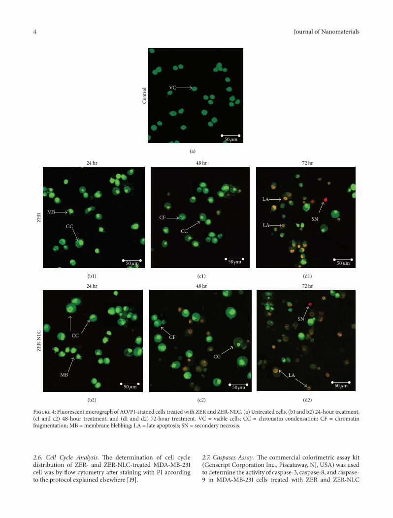
Figure 4: Fluorescent micrograph of AO/PI-stained cells treated with ZER and ZER-NLC. (a) Untreated cells, (b1 and b2) 24-hour treatment,(c1 and c2) 48-hour treatment, and (d1 and d2) 72-hour treatment. VC = viable cells; CC = chromatin condensation; CF = chromatinfragmentation; MB = membrane blebbing; LA = late apoptosis; SN = secondary necrosis.
2.6. Cell Cycle Analysis. The determination of cell cycledistribution of ZER- and ZER-NLC-treated MDA-MB-231cell was by flow cytometry after staining with PI accordingto the protocol explained elsewhere [19].
2.7. Caspases Assay. The commercial colorimetric assay kit(Genscript Corporation Inc., Piscataway, NJ, USA) was usedto determine the activity of caspase-3, caspase-8, and caspase-9 in MDA-MB-231 cells treated with ZER and ZER-NLC

Journal of Nanomaterials 5Pr
opid
ium
iodi
de
103
104
102
101
100
103102101100 104
Annexin V FITC
12 hours
Con
trol
(a)Pr
opid
ium
iodi
de
103
104
102
101
100
103102101100 104
Annexin V FITC
24 hours
(b)
Prop
idiu
m io
dide
103
104
102
101
100
103102101100 104
Annexin V FITC
48 hours
(c)
Prop
idiu
m io
dide
103
104
102
101
100
103102101100 104
Annexin V FITC
ZER
(d)
Prop
idiu
m io
dide
103
104
102
101
100
103102101100 104
Annexin V FITC
(e)Pr
opid
ium
iodi
de
103
104
102
101
100
103102101100 104
Annexin V FITC
(f)
Prop
idiu
m io
dide
103
104
102
101
100
103102101100 104
Annexin V FITC
ZER-
NLC
(g)
Prop
idiu
m io
dide
103
104
102
101
100
103102101100 104
Annexin V FITC
(h)
Prop
idiu
m io
dide
103
104
102
101
100
103102101100 104
Annexin V FITC
(i)
Figure 5: MDA-MB-231 cell treated with ZER and ZER-NLC and labeled with FITC-conjugated Annexin V and propidium iodide. ((a), (b),(c)) untreated cells, ((d), (e), (f)) cells treated with ZER, and ((g), (h), (i)) cells treated with ZER-NLC. Cells were treated for 12, 24, and 48hours.
for 12, 24, and 48 hours, according to the manufacturer’sinstructions.
2.8. TUNEL Assay. The Tdt-mediated dUTP nick-end label-ing (TUNEL) with the apoptotic detection kit (DeadEndfluorometric TUNEL system; Promega, Madison, WI, USA)was applied to determine the mode of cell death induced byZER and ZER-NLC.The assay quantifies fragmented DNA in
apoptotic cells using the FACSCalibur flow cytometer (BD)equipped with an argon laser (BD).
2.9. Western Blotting Analysis. MDA-MB-231 cells (1 × 10⁶)treated with ZER and ZER-NLC were lysed in the radioim-munoprecipitation assay (RIPA) buffer (Thermo Fisher Sci-entific, Waltham, MA, USA) and expressions of cytochromec, Bcl-2, Bcl-xL and Bax proteins, and proliferating cell

6 Journal of Nanomaterials
Con
trol
Cou
nts
0
60
120
180
240
300
0 200 400 600
DNA content
12 hours
G0/G1
Sub-G0/G1
S
G2 + M
(a)C
ount
s
0
60
120
180
240
300
0 200 400 600
DNA content
24 hours
G0/G1
Sub-G0/G1
S
G2 + M
(b)
Cou
nts
0
60
120
180
240
300
0 200 400 600
DNA content
48 hours
G0/G1
Sub-G0/G1
S
G2 + M
(c)
ZER
Cou
nts
0
60
120
180
240
300
0 200 400 600
DNA content
G0/G1
Sub-G0/G1
S
G2 + M
(d)
Cou
nts
0
60
120
180
240
300
0 200 400 600
DNA content
G0/G1
Sub-G0/G1
S
G2 + M
(e)C
ount
s
0
60
120
180
240
300
0 200 400 600
DNA content
G0/G1
Sub-G0/G1
S
G2 + M
(f)
ZER-
NLC
Cou
nts
0
60
120
180
240
300
0 200 400 600
DNA content
G0/G1
Sub-G0/G1
S
G2 + M
(g)
Cou
nts
0
60
120
180
240
300
0 200 400 600
DNA content
G0/G1
Sub-G0/G1
S
G2 + M
(h)
Cou
nts
0
60
120
180
240
300
0 200 400 600
DNA content
G0/G1
Sub-G0/G1
S
G2 + M
(i)
Figure 6: Cell cycle analysis of MDA-MB-231 cells treated with ((d), (e), (f)) ZER and ((g), (h), (i)) ZER-NLC after 24, 48, and 72 hours. (a),(b), and (c) are untreated cells after 24, 48, and 72 h, respectively.
nuclear antigen (PCNA) determined by western blotting.Briefly, protein (25𝜇g) from treated and untreated cells wasseparated on 10% on sodium dodecyl sulfate polyacrylamidegel and transferred to polyvinylidene difluoride membranes(Bio-Rad Laboratories, Hercules, CA, USA). The membranewas blocked with 5% nonfat dry milk in phosphate-bufferedsaline with 0.05% Tween-20 for 60 minutes at room tem-perature. The membrane was probed by incubation with theprimary rabbit anti-Bcl-2, -Bcl-xL, -Bax, -cytochrome c, and-PCNA antibodies (Abcam, USA) followed by horseradishperoxidase-conjugated secondary rabbit anti-goat antibody(Abcam, USA). The antibody binding was detected by
chemiluminescence system (ECL Western blot substrate;Abcam, Cambridge, UK). The membrane was also probedwith rabbit anti-𝛽-actin antibody (Abcam, USA) as a loadingcontrol. Finally Image J software was used to quantify thebands.
2.10. Statistical Analysis. All experiments were performed intriplicate and results were presented as mean ± standarddeviation. Statistical package for the social sciences (SPSS),version 19.0 (SPSS Inc., Chicago, USA), and analysis ofvariance (ANOVA) were utilized for statistical analysis.
Journal of Nanomaterials 7
104
103
102
101
100
TUN
EL F
ITC
0 200 400 600 800 1000
DNA-area
Con
trol
24 hours
(a)
104
103
102
101
100
TUN
EL F
ITC
0 200 400 600 800 1000
DNA-area
48 hours
(b)
104
103
102
101
100
TUN
EL F
ITC
0 200 400 600 800 1000
DNA-area
72 hours
(c)
104
103
102
101
100
TUN
EL F
ITC
0 200 400 600 800 1000
DNA-area
ZER
(d)
104
103
102
101
100
TUN
EL F
ITC
0 200 400 600 800 1000
DNA-area
(e)
104
103
102
101
100
TUN
EL F
ITC
0 200 400 600 800 1000
DNA-area
(f)
104
103
102
101
100
TUN
EL F
ITC
0 200 400 600 800 1000
DNA-area
ZER-
NLC
(g)
104
103
102
101
100
TUN
EL F
ITC
0 200 400 600 800 1000
DNA-area
(h)
104
103
102
101
100
TUN
EL F
ITC
0 200 400 600 800 1000
DNA-area
(i)
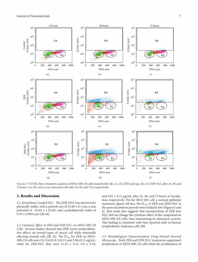
Figure 7: TUNEL flow cytometric analysis of MDA-MB-231 cells treated with ((d), (e), (f)) ZER and ((g), (h), (i)) ZER-NLC after 24, 48, and72 hours. (a), (b), and (c) are untreated cells after 24, 48, and 72 h, respectively.
3. Results and Discussion
3.1. Zerumbone-LoadedNLC. TheZER-NLCwas shown to bephysically stable, with a particle size of 52.68 ± 0.1 nm, a zetapotential of −25.03 ± 1.24mV, and a polydispersity index of0.29 ± 0.0041 𝜇m [10, 14].
3.2. Cytotoxic Effect of ZER and ZER-NLC on MDA-MB-231Cells. Several studies showed that ZER exerts antiprolifera-tive effects on several types of cancer cell while minimallyaffecting normal cells [20, 21]. The IC
50for ZER on MDA-
MB-231 cells were 10.15±0.9, 8.3±0.15, and 5.96±0.13 𝜇g/mL,while for ZER-NLC they were 11.45 ± 0.12, 9.4 ± 0.18,
and 6.01 ± 0.11 𝜇g/mL after 24, 48, and 72 hours of incuba-tion, respectively. On the MCF-10A cell, a normal epithelialmammary gland cell line, the IC
50of ZER and ZER-NLC at
the same incubation periods were similarly low (Figures 1 and2). This study also suggests that incorporation of ZER intoNLC did not change the cytotoxic effect of the compound onMDA-MB-231 cells, thus maintaining its anticancer activity.This finding is consistent with that reported early in humanlymphoblastic leukemia cells [10].
3.3. Morphological Characterization Using Normal InvertedMicroscope. Both ZER and ZER-NLC treatments suppressedproliferation of MDA-MB-231 cells while the proliferation of

8 Journal of Nanomaterials
0
0.1
0.2
0.3
0.4
0.5
0.6
0.7
Con
trol
12 h
ZER
24 h
ZER
48 h
ZER
Con
trol
12 h
ZER
-NLC
24 h
ZER
-NLC
48 h
ZER
-NLC
ZER ZER-NLC
Opt
ical
den
sity
(450
nm
)
Time (hours)
Caspase activity
Caspase-9Caspase-3
Caspase-8
Figure 8: Caspases activity in MDA-MB-231 cells treated with ZERand ZER-NLC for 12, 24, and 48 h.
untreated cells (control) increased and remained confluentduring all incubation periods. Morphologically, MDA-MB-231 cells treated with ZER and ZER-NLC showed irregularshape, membrane blebbing, cell shrinkage, chromatin con-densation, and presence of apoptotic bodies (Figure 3).
3.4. Morphological Characterisation Using Acridine Orange/Propidium Iodide (AO/PI). Using the AO and PI double-stainingmethod, the breast cancer cells treated with ZER andZER-NLC showed apoptotic features in a time-dependentmanner, while untreated cells showed green nuclear stainingsuggesting intact nuclear structure (Figure 4). Early apoptosisin treated cells indicated DNA fragmentation after 24 hours,as shown by the bright-green color. Chromatin condensationwas also observed by the appearance of orange color in thecells indicating formation of apoptotic bodies, characteristicof late apoptosis. On the other hand, necrotic cells, charac-terised by reddish-orange nuclei, were also observed after 72hours of treatments.
3.5. Apoptosis Detection by Annexin Assay. MDA-MB-231cells treatedwith ZER andZER-NLC showed apoptosis, whileviable cells number decreased with period of treatment (Fig-ure 5; see Supplementary Table S1 in Supplementary Mate-rial available online at http://dx.doi.org/10.1155/2014/742738).There was a significant shift (𝑃 < 0.05) from early tolate apoptosis in the distribution of treated MDA-MB-231cells after ZER and ZER-NLC treatments. This shift in celldistribution is accompanied by decrease in distribution ofviable cells, particularly after 48 hours of treatment. TheAnnexin V assay further confirms that both ZER and ZER-NLC induce apoptosis in MDA-MB-231 cells in a time-dependent manner.
3.6. Cell Cycle Analysis Using Flow Cytometry. The resultsdemonstrated that ZER and ZER-NLC induced depletion ofthe G1 phase and S phase of MDA-MB-231 cells with con-comitant accumulation of the sub-G0/G1 and G2/M phasesat all periods of treatment (Figure 6 and Table S2). The studyalso showed that both ZER and ZER-NLC caused markedarrest of (𝑃 < 0.05) MDA-MB-231 cells at G2/M phasethat increased with duration of treatment. Concurrently,the sub-G0/G1 peak became evident beginning as early as12 hours of treatment in the ZER-NLC-treated cells. Thispeak appeared later, at 24 hours with ZER treatment. Theresults show that both ZER and ZER-NLC are potent in theinduction of apoptosis of the MDA-MD-231 cells. Cell cyclearrest is a normal regulatory pathway that allows for tissuemaintenance, such as repair of cell damage. Cell cycle arrest isalso dependent on availability of essential hormones, growthfactors, and nutrients [22]. In disease, gene mutation andtumorigenesis can be induced by defects in these cell cyclecheckpoints leading to uncontrolled proliferation. With thetreatment of ZER and ZER-NLC, MDA-MB-231 cell cyclearrest at G2/M phase is proposed to occur through increasedexpression of cyclin B1, Cdk1, Cdc25C, and Cdc25B proteins[23].
3.7. Apoptosis Detection Using TUNEL Assay. Nuclear frag-mentation was observed in MDA-MB-231 cells treated witheither ZER or ZER-NLC. The DNA fragmentation showsthat the rate of apoptosis of MDA-MB-231 cells increasedgradually and significantly (𝑃 < 0.05) during the treatmentperiods (Figure 7). However, untreated cells proliferatedwithout showing apoptosis (Table S3).
3.8. Detection of Caspase Protease Activity. Caspases areamong essential enzymes of the apoptosis-signaling pathway.Caspase-3, initiated by caspase-8, plays a key role in thedeath receptor pathway, while caspase-9 is a major playerin the mitochondrial pathway [24, 25]. Thus these caspaseswere chosen to determine the mechanism of apoptosis inthe MDA-MB-231 cells treated with ZER and ZER-NLC(Figure 8). In this study, the activity of caspase-3 and caspase-9 increased significantly (𝑃 < 0.05) with time in ZER- andZER-NLC-treated cells. However, there was no increase incaspase-8 activity in the MDA-MB-cells during the sameperiod of treatment (Table S4). The results suggest that ZERand ZER-NLC induced apoptosis of MDA-MB-231 via theintrinsic pathway.
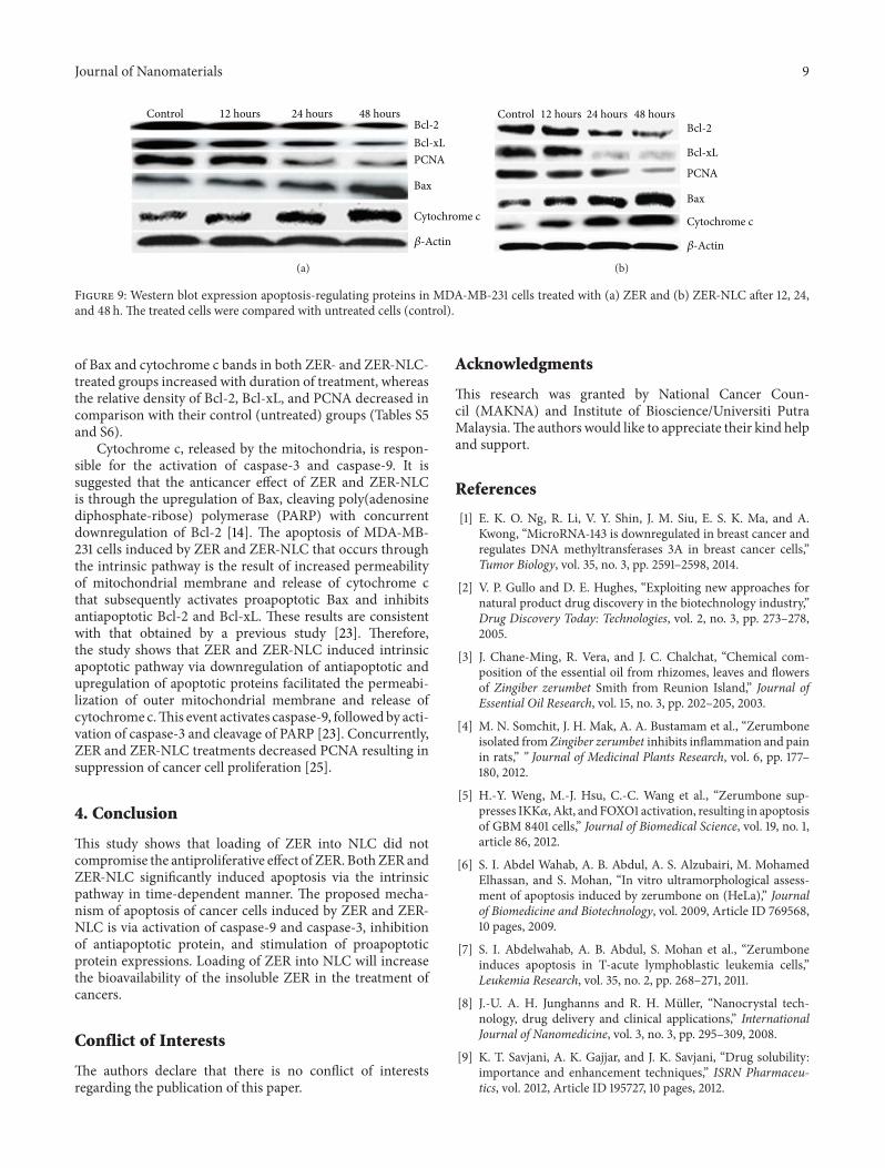
3.9. Western Blotting. Determination of pro- and antiapop-totic proteins is another method of ascertaining the modeof apoptosis in treated cells. In this study, Bax, Bcl-2, andBcl-xL protein expressions inMDA-MB-231 cells treatedwithZER and ZER-NLC were determined. Other proteins, PCNAand cytochrome c, that play roles in cell proliferation anddeath were also estimated. The treatment of MDA-MB-231cells with ZER and ZER-NLC caused significant (𝑃 < 0.05)downregulation of Bcl-2 and Bcl-xL proteins and PCNA,while the Bax protein was upregulated and cytochrome cprotein expression increased (Figure 9). The relative density
Journal of Nanomaterials 9
ControlBcl-2Bcl-xLPCNA
Bax
Cytochrome c
𝛽-Actin
48 hours24 hours12 hours
(a)
Bcl-2
Bcl-xL
PCNA
Bax
𝛽-Actin
Control 48 hours24 hours12 hours
Cytochrome c
(b)
Figure 9: Western blot expression apoptosis-regulating proteins in MDA-MB-231 cells treated with (a) ZER and (b) ZER-NLC after 12, 24,and 48 h. The treated cells were compared with untreated cells (control).
of Bax and cytochrome c bands in both ZER- and ZER-NLC-treated groups increased with duration of treatment, whereasthe relative density of Bcl-2, Bcl-xL, and PCNA decreased incomparison with their control (untreated) groups (Tables S5and S6).
Cytochrome c, released by the mitochondria, is respon-sible for the activation of caspase-3 and caspase-9. It issuggested that the anticancer effect of ZER and ZER-NLCis through the upregulation of Bax, cleaving poly(adenosinediphosphate-ribose) polymerase (PARP) with concurrentdownregulation of Bcl-2 [14]. The apoptosis of MDA-MB-231 cells induced by ZER and ZER-NLC that occurs throughthe intrinsic pathway is the result of increased permeabilityof mitochondrial membrane and release of cytochrome cthat subsequently activates proapoptotic Bax and inhibitsantiapoptotic Bcl-2 and Bcl-xL. These results are consistentwith that obtained by a previous study [23]. Therefore,the study shows that ZER and ZER-NLC induced intrinsicapoptotic pathway via downregulation of antiapoptotic andupregulation of apoptotic proteins facilitated the permeabi-lization of outer mitochondrial membrane and release ofcytochrome c.This event activates caspase-9, followed by acti-vation of caspase-3 and cleavage of PARP [23]. Concurrently,ZER and ZER-NLC treatments decreased PCNA resulting insuppression of cancer cell proliferation [25].
4. Conclusion
This study shows that loading of ZER into NLC did notcompromise the antiproliferative effect of ZER. BothZER andZER-NLC significantly induced apoptosis via the intrinsicpathway in time-dependent manner. The proposed mecha-nism of apoptosis of cancer cells induced by ZER and ZER-NLC is via activation of caspase-9 and caspase-3, inhibitionof antiapoptotic protein, and stimulation of proapoptoticprotein expressions. Loading of ZER into NLC will increasethe bioavailability of the insoluble ZER in the treatment ofcancers.
Conflict of Interests
The authors declare that there is no conflict of interestsregarding the publication of this paper.
Acknowledgments
This research was granted by National Cancer Coun-cil (MAKNA) and Institute of Bioscience/Universiti PutraMalaysia.The authors would like to appreciate their kind helpand support.
References
[1] E. K. O. Ng, R. Li, V. Y. Shin, J. M. Siu, E. S. K. Ma, and A.Kwong, “MicroRNA-143 is downregulated in breast cancer andregulates DNA methyltransferases 3A in breast cancer cells,”Tumor Biology, vol. 35, no. 3, pp. 2591–2598, 2014.
[2] V. P. Gullo and D. E. Hughes, “Exploiting new approaches fornatural product drug discovery in the biotechnology industry,”Drug Discovery Today: Technologies, vol. 2, no. 3, pp. 273–278,2005.
[3] J. Chane-Ming, R. Vera, and J. C. Chalchat, “Chemical com-position of the essential oil from rhizomes, leaves and flowersof Zingiber zerumbet Smith from Reunion Island,” Journal ofEssential Oil Research, vol. 15, no. 3, pp. 202–205, 2003.
[4] M. N. Somchit, J. H. Mak, A. A. Bustamam et al., “Zerumboneisolated fromZingiber zerumbet inhibits inflammation and painin rats,” ” Journal of Medicinal Plants Research, vol. 6, pp. 177–180, 2012.
[5] H.-Y. Weng, M.-J. Hsu, C.-C. Wang et al., “Zerumbone sup-presses IKK𝛼, Akt, and FOXO1 activation, resulting in apoptosisof GBM 8401 cells,” Journal of Biomedical Science, vol. 19, no. 1,article 86, 2012.
[6] S. I. Abdel Wahab, A. B. Abdul, A. S. Alzubairi, M. MohamedElhassan, and S. Mohan, “In vitro ultramorphological assess-ment of apoptosis induced by zerumbone on (HeLa),” Journalof Biomedicine and Biotechnology, vol. 2009, Article ID 769568,10 pages, 2009.
[7] S. I. Abdelwahab, A. B. Abdul, S. Mohan et al., “Zerumboneinduces apoptosis in T-acute lymphoblastic leukemia cells,”Leukemia Research, vol. 35, no. 2, pp. 268–271, 2011.
[8] J.-U. A. H. Junghanns and R. H. Muller, “Nanocrystal tech-nology, drug delivery and clinical applications,” InternationalJournal of Nanomedicine, vol. 3, no. 3, pp. 295–309, 2008.
[9] K. T. Savjani, A. K. Gajjar, and J. K. Savjani, “Drug solubility:importance and enhancement techniques,” ISRN Pharmaceu-tics, vol. 2012, Article ID 195727, 10 pages, 2012.
10 Journal of Nanomaterials
[10] H. S. Rahman, A. Rasedee, C. W. How et al., “Zerumbone-loaded nanostructured lipid carriers: preparation, charac-terization, and antileukemic effect,” International Journal ofNanomedicine, vol. 8, pp. 2769–2781, 2013.
[11] A. Patidar, D. S. Thakur, P. Kumar, and J. Verma, “A reviewon novel lipid based nanocarriers,” International Journal ofPharmacy and Pharmaceutical Sciences, vol. 2, no. 4, pp. 30–35,2010.
[12] E. B. Souto, S. A. Wissing, C. M. Barbosa, and R. H. Muller,“Development of a controlled release formulation based onSLN and NLC for topical clotrimazole delivery,” InternationalJournal of Pharmaceutics, vol. 278, no. 1, pp. 71–77, 2004.
[13] A. Chinsriwongkul, P. Chareanputtakhun, T. Ngawhirunpat etal., “Nanostructured lipid carriers (NLC) for parenteral deliveryof an anticancer drug,” AAPS PharmSciTech, vol. 13, no. 1, pp.150–158, 2012.
[14] H. S. Rahman, A. Rasedee, A. B. Abdul et al., “Zerumbone-loaded nanostructured lipid carrier induces G2/M cell cyclearrest and apoptosis via mitochondrial pathway in a humanlymphoblastic leukemia cell line,” International Journal ofNanomedicine, vol. 9, no. 1, pp. 527–538, 2014.
[15] J. Pardeike, A.Hommoss, andR.H.Muller, “Lipid nanoparticles(SLN, NLC) in cosmetic and pharmaceutical dermal products,”International Journal of Pharmaceutics, vol. 366, no. 1-2, pp. 170–184, 2009.
[16] S. Selvamuthukumar and R. Velmurugan, “Nanostructuredlipid carriers: a potential drug carrier for cancer chemotherapy,”Lipids in Health and Disease, vol. 11, article 159, 2012.
[17] S. A. S. Sakinah, S. Tri Handayani, and L. P. A. Hawariah,“Zerumbone induced apoptosis in liver cancer cells viamodula-tion of Bax/ Bcl-2 ratio,” Cancer Cell International, vol. 7, article4, 2007.
[18] S. I. Abdelwahab, A. B. Abdul, Z. N. M. Zain, and A. H. A.Hadi, “Zerumbone inhibits interleukin-6 and induces apoptosisand cell cycle arrest in ovarian and cervical cancer cells,”International Immunopharmacology, vol. 12, no. 4, pp. 594–602,2012.
[19] S. Syam, A. B. Abdul, M. A. Sukari, S. Mohan, S. I. Abdelwahab,and T. S. Wah, “The growth suppressing effects of girinimbineon hepg2 involve induction of apoptosis and cell cycle arrest,”Molecules, vol. 16, no. 8, pp. 7155–7170, 2011.
[20] A. B. H. Adbul, A. S. Al-Zubairi, N. D. Tailan et al., “Anti-cancer activity of natural compound (Zerumbone) extractedfrom Zingiber zerumbet in human HeLa cervical cancer cells,”International Journal of Pharmacology, vol. 4, no. 3, pp. 160–168,2008.
[21] A. Deorukhkar, N. Ahuja, A. Mercado et al., “Zerumbone, asesquiterpene from southeast Asian edible ginger sensitizes col-orectal cancer cells to radiation therapy,” International Journal ofRadiation Oncology, Biology, Physics, vol. 78, no. 3, supplement,p. S654, 2010.
[22] J. A. Pietenpol and Z. A. Stewart, “Cell cycle checkpointsignaling: cell cycle arrest versus apoptosis,” Toxicology, vol. 181-182, pp. 475–481, 2002.
[23] A. Sehrawat, J. A. Arlotti, A. Murakami, and S. V. Singh,“Zerumbone causes Bax- and Bak-mediated apoptosis inhuman breast cancer cells and inhibits orthotopic xenograftgrowth in vivo,” Breast Cancer Research and Treatment, vol. 136,no. 2, pp. 429–441, 2012.
[24] N. O’Donovan, J. Crown, H. Stunell et al., “Caspase 3 in breastcancer,”Clinical Cancer Research, vol. 9, no. 2, pp. 738–742, 2003.
[25] H. Zhao, Y.-H. Lo, L.Ma et al., “Targeting tyrosine phosphoryla-tion of pcna inhibits prostate cancer growth,”Molecular CancerTherapeutics, vol. 10, no. 1, pp. 29–36, 2011.

Submit your manuscripts athttp://www.hindawi.com
ScientificaHindawi Publishing Corporationhttp://www.hindawi.com Volume 2014
CorrosionInternational Journal of
Hindawi Publishing Corporationhttp://www.hindawi.com Volume 2014
Polymer ScienceInternational Journal of
Hindawi Publishing Corporationhttp://www.hindawi.com Volume 2014
Hindawi Publishing Corporationhttp://www.hindawi.com Volume 2014
CeramicsJournal of
Hindawi Publishing Corporationhttp://www.hindawi.com Volume 2014
CompositesJournal of
NanoparticlesJournal of
Hindawi Publishing Corporationhttp://www.hindawi.com Volume 2014
Hindawi Publishing Corporationhttp://www.hindawi.com Volume 2014
International Journal of
Biomaterials
Hindawi Publishing Corporationhttp://www.hindawi.com Volume 2014
NanoscienceJournal of
TextilesHindawi Publishing Corporation http://www.hindawi.com Volume 2014
Journal of
NanotechnologyHindawi Publishing Corporationhttp://www.hindawi.com Volume 2014
Journal of
CrystallographyJournal of
Hindawi Publishing Corporationhttp://www.hindawi.com Volume 2014
The Scientific World JournalHindawi Publishing Corporation http://www.hindawi.com Volume 2014
Hindawi Publishing Corporationhttp://www.hindawi.com Volume 2014
CoatingsJournal of
Advances in
Materials Science and EngineeringHindawi Publishing Corporationhttp://www.hindawi.com Volume 2014
Smart Materials Research
Hindawi Publishing Corporationhttp://www.hindawi.com Volume 2014
Hindawi Publishing Corporationhttp://www.hindawi.com Volume 2014
MetallurgyJournal of
Hindawi Publishing Corporationhttp://www.hindawi.com Volume 2014
BioMed Research International
MaterialsJournal of
Hindawi Publishing Corporationhttp://www.hindawi.com Volume 2014
Nano
materials
Hindawi Publishing Corporationhttp://www.hindawi.com Volume 2014
Journal ofNanomaterials
Copyright © 2022 FDOKUMEN