Comparison between apo and complexed structures of bothropstoxin-I reveals the role of Lys122 and...
14
This article appeared in a journal published by Elsevier. The attached copy is furnished to the author for internal non-commercial research and education use, including for instruction at the authors institution and sharing with colleagues. Other uses, including reproduction and distribution, or selling or licensing copies, or posting to personal, institutional or third party websites are prohibited. In most cases authors are permitted to post their version of the article (e.g. in Word or Tex form) to their personal website or institutional repository. Authors requiring further information regarding Elsevier’s archiving and manuscript policies are encouraged to visit: http://www.elsevier.com/copyright
-
Upload
independent -
Category
Documents
-
view
2 -
download
0
Transcript of Comparison between apo and complexed structures of bothropstoxin-I reveals the role of Lys122 and...

This article appeared in a journal published by Elsevier. The attachedcopy is furnished to the author for internal non-commercial researchand education use, including for instruction at the authors institution
and sharing with colleagues.
Other uses, including reproduction and distribution, or selling orlicensing copies, or posting to personal, institutional or third party
websites are prohibited.
In most cases authors are permitted to post their version of thearticle (e.g. in Word or Tex form) to their personal website orinstitutional repository. Authors requiring further information
regarding Elsevier’s archiving and manuscript policies areencouraged to visit:
http://www.elsevier.com/copyright

Author's personal copy
Comparison between apo and complexed structures of bothropstoxin-I revealsthe role of Lys122 and Ca2+-binding loop region for the catalytically inactiveLys49-PLA2s
Carlos A.H. Fernandes a,b,1, Daniela P. Marchi-Salvador a,c,1, Guilherme M. Salvador a,b, Mabel C.O. Silva a,Tássia R. Costa b,c, Andreimar M. Soares b,c, Marcos R.M. Fontes a,b,*
a Departamento de Física e Biofísica, Instituto de Biociências, UNESP – Univ Estadual Paulista, Botucatu, SP, Brazilb Instituto Nacional de Ciência e Tecnologia em Toxinas, CNPq, Brazilc Departamento de Análises Clínicas, Toxicológicas e Bromatológicas, FCFRP, USP, Av. do Café s/n, 14040-903, Ribeirão Preto, SP, Brazil
a r t i c l e i n f o
Article history:Received 4 December 2009Received in revised form 23 February 2010Accepted 31 March 2010Available online 4 April 2010
Keywords:Phospholipase A2
MyotoxicityHomologue phospholipase A2 (Lys49-PLA2s)Ca2+-binding loopX-ray crystallography
a b s t r a c t
Phospholipases A2 (Asp49-PLA2s) are enzymes responsible for cellular membrane disruption throughCa2+-dependent hydrolysis of phospholipids. A class of these proteins (Lys49-PLA2s) does not show cat-alytic activity but can exert a pronounced local myotoxic effect that is not neutralized by serum therapy.In this work, we present five structures of Lys49-PLA2s from snakes of the Bothrops genus in apo form,complexed with PEG molecules and chemically modified by p-bromofenacil bromide (BPB), a classicinhibitor of PLA2. We present herein an extensive structural analysis including: (i) the function of hydro-phobic long-chain molecules as Lys49-PLA2s inhibitors, (ii) the role of Lys122, previously indicated asbeing responsible for Lys49-PLA2s catalytic inactivity and, (iii) a structural comparison of the Ca2+-bind-ing loop region between Lys49 and Asp49-PLA2s. The Lys122 analysis of 30 different monomers for apoand complexed Lys49-PLA2s structures shows that this residue is very flexible and may bind to differentcarboxyl groups giving stability to the crystal structures. The structural comparisons of the Ca2+-bindingloop region between Lys49 and Asp49-PLA2s reveal the importance of the Tyr28 residue conservation inAsp49-PLA2s to the integrity of this loop. The Tyr28 residue stabilizes this region by an interaction withGly35 residue. In Lys49-PLA2s and low-catalytic Asp49-PLA2s this interaction does not occur, preventingthe binding of Ca2+.
� 2010 Elsevier Inc. All rights reserved.
1. Introduction
Snake venoms comprise a complex mixture of pharmacologicalcomponents able to affect several biological systems. One of themain components of snake venoms is the phospholipases A2 (PLA2s– EC 3.1.1.4), which are small and stable molecules (14–18 kDa)found in a great diversity of organisms and biological fluids (Schal-oske and Dennis, 2006). These enzymes hydrolyze fatty acids fromthe sn-2 position of glycerophospholipids, releasing lysophospho-lipids and free fatty acids, such as arachidonic acid, which areinvolved in many important biological processes (Tsuboi et al.,2002). Arachidonic acid molecules, for example, can be convertedto eicosanoids which serve as secondary messengers in a widerange of physiological and pathological processes including sleep
regulation, immune responses, inflammation, and pain perception(Moolenaar et al., 2004). Otherwise, the lysophospholipids canserve as precursors for lipid mediators involved in cell prolifera-tion, survival and migration and in many inflammatory processes(Moolenaar et al., 2004; Prescott et al., 2000).
The catalytic site of snake venom PLA2 (svPLA2s) is composed offour residues; His48, Asp49, Tyr52 and Asp99 (Magro et al., 2009).Two action mechanisms for svPLA2s were proposed (Rogers et al.,1996; Scott et al., 1990) and in both, His48 and Asp49 play essen-tial roles, in coordinating catalytic waters and mobilizing the Ca2+
ion, respectively. Furthermore, several biochemical studies demon-strated that the Ca2+ ion is an obligatory cofactor for sPLA2s catal-ysis. Studies of its replacement by other divalent ions (Cd2+, Sr2+,Ba2+, Mg2+) showed that these ions were unable to keep the sub-strate bound to the enzyme, with the exception of the Cd2+ whichallows the toxin to bind to the substrate although without catalyticactivity (Yu et al., 1993). This can be explained by different coordi-nation geometries assumed by the tetrahedral intermediate due tothe presence of the Cd2+ and Ca2+ ions, which determine the elec-
1047-8477/$ - see front matter � 2010 Elsevier Inc. All rights reserved.doi:10.1016/j.jsb.2010.03.019
* Corresponding author at: Departamento de Física e Biofísica, Instituto deBiociências, UNESP – Univ Estadual Paulista, Botucatu, SP, Brazil. Fax: +55 1438153744.
E-mail addresses: [email protected] (M.R.M. Fontes).1 These authors contributed equally to this work.
Journal of Structural Biology 171 (2010) 31–43
Contents lists available at ScienceDirect
Journal of Structural Biology
journal homepage: www.elsevier .com/locate /y jsbi

Author's personal copy
trophilic behavior of the catalytic site (Yu et al., 1993). However,later experiments demonstrated that Ni2+ and Co2+ ions supporta significant catalytic hydrolysis of phospholipids with determinedhead groups (Yu et al., 1998). These results can be also attributedto differences in coordination geometry and indicate a noteworthyplasticity of the active site environment (Yu et al., 1998).
A distinct group of svPLA2s, known as homologue PLA2s orLys49-PLA2s, exhibits a natural replacement of the key Asp49 res-idue by a Lys49. Lys49-PLA2s are catalytically inactive due to theirincapacity to bind the cofactor Ca2+ since the e-amino group ofLys49 occupies exactly the position of the coordinated Ca2+ ion inAsp49-PLA2 (Scott et al., 1992; Holland et al., 1990).
Additionally, site-directed mutagenesis with a Lys49-PLA2
showed that a Lys49Asp mutant remained catalytically inactive,demonstrating that not only the single Asp49Lys replacementbut also other possible structural changes are responsible forthe lack of enzymatic activity. A classic crystallographic study ofpiratoxin II (PrTX-II), a Lys49-PLA2 from Bothrops pirajai venomcomplexed with fatty acid, proposed that Lys122 (conserved inall Lys49-PLA2s but rare in Asp49-PLA2s) interacts with the car-bonyl of Cys29 hyperpolarizing the peptide bond betweenCys29 and Gly30. This strong interaction causes an increasedaffinity for the fatty acid head group, precluding the release offree fatty acid produced after an initial phospholipid hydrolysisand interruption of the catalytic cycle. Strengthening this hypoth-esis, a structural analysis of eight dimeric Lys49-PLA2s found thatLys122 interacts with Cys29 in both monomers of the structurespresenting ligands in their hydrophobic channels. Otherwise, inthe structures with no ligands the Lys122 presents an alternativeconformation in one or both monomers (Magro et al., 2003). Inspite of their catalytic inactivity, Lys49-PLA2s are responsible forinducing myonecrosis by a Ca2+-independent membrane-damag-ing mechanism which is not efficiently neutralized by serumtherapy and can lead to permanent tissue loss, disability oramputation (Gutierrez and Lomonte, 1995). Several studies haveshown that segment 115–129 of the C-terminal region, which in-cludes a variable combination of positively charged and hydro-phobic residues, is responsible for this myotoxic activity.Recently, the importance of this region was reinforced by the pro-posal of a myotoxic site of Lys49-PLA2s from snakes of the Bothr-ops genus that contains, besides Lys20, C-terminus residues(Lys115 and Arg118) (dos Santos et al., 2009).
Since the study with the structure of piratoxin-II (PrTX-II) com-plexed with fatty acid (Lee et al., 2001), a large number of struc-tures of Lys49-PLA2s, in native or complexed forms, have beensolved (dos Santos et al., 2009; Magro et al., 2003; Marchi-Salvadoret al., 2009; Murakami et al., 2005, 2007; Watanabe et al., 2005),thus necessitating a revision of the position of Lys122 in thesenew structures in an attempt to more accurately define the roleof Lys122 in the catalytic inactivity of Lys49-PLA2s. Furthermore,with this large number of structures, structural comparisons be-tween Ca2+ binding loop regions of Lys49 and Asp49-PLA2s canbe very accurate and give important insights into the inability ofLys49-PLA2s to bind Ca2+ cofactor. In order to carefully discussthese points, we present, in this study, five crystal structures ofbothropstoxin-I, a Lys49-PLA2 isolated from Bothrops jararacussuvenom: two of them in native form, each with a different numberof molecules in the asymmetric unit; two chemically modified byp-bromophenacyl bromide (BPB), a well-known inhibitor of snakevenom PLA2s, each with a different number of molecules in theasymmetric unit; and, finally, one structure co-crystallized withPEG 4000. Moreover, these complexed structures from the sameprotein enable a structural comparison between two differenttypes of Lys49-PLA2 ligands: (i) bound to the ‘‘active site” (e.g.BPB) and (ii) occupying the hydrophobic channel (e.g. PEG 4000;a-tocopherol).
2. Materials and methods
2.1. Purification and chemical modification
BthTX-I was isolated from Bothrops jararacussu snake venom byion-exchange chromatography on CM-Sepharose (Homsi-Brande-burgo et al., 1988). Modification of His48 with q-bromophenacylbromide (BPB) was carried out as previously described (Diaz-Ore-iro and Gutierrez, 1997). Briefly, approximately 3 mg of theBthTX-I was dissolved in 1 ml of 100 mM ammonium bicarbonate,pH 8.0; and 150 ll of BPB solution (0.8 mg.ml�1 in ethanol) wasadded. The mixture was incubated for 24 h and the excess reagentwas removed by ultrafiltration, followed by lyophilization.
2.2. Dynamic light scattering
The dynamic light scattering (DLS) measurements were per-formed with native BthTX-I at the crystallization temperature(291 K) at the concentration of 3.2 mg.ml�1 using a DynaPro TITANinstrument (Wyatt Technology). The results were analyzed withthe Dynamics v.6.10 software. The data were measured one hun-dred times in triplicate.
2.3. Crystallization
Crystallization experiments were performed with lyophilizedsamples of native BthTX-I and BthTX-I/BPB dissolved in ultra-purewater at the concentration of 12 mg.ml�1. Crystals of these com-plexes were obtained by the conventional hanging-drop vapor-dif-fusion method (McPherson, 2003) using the sparse-matrix method(Jancarik and Kim, 1991) with Crystal Screen I (HamptonResearch). Crystals of the apoBthTX-I were obtained with: one(apo-mBthTX-I) and two (apo-dBthTX-I) molecules in theasymmetric unit, BthTX-I chemically modified by p-bromophena-cyl bromide (BPB) also with one (mBthTX-I/BPB) and two(dBthTX-I/BPB) molecules in the asymmetric unit and BthTX-Icomplexed with PEG 4000 (BthTX-I/PEG4K). Crystallization condi-tions are shown in Table 1.
2.4. X-ray data collection and processing
X-ray diffraction data were collected using a synchrotron-radi-ation source (MX1 and MX2 stations – LNLS, Campinas, Brazil) witha MAR CCD imaging-plate detector (MAR Research). Crystals weremounted in a nylon loop and flash cooled in a stream of nitrogen at100 K using no cryoprotectant. Data were processed using the HKLprogram package (Otwinowski and Minor, 1997). Data-processingstatistics are presented in Table 1.
2.5. Structure determination and refinement
The structures were solved by the molecular replacement meth-od using the program AmoRe (Navaza, 1994) and coordinates ofLys49-PLA2 PrTX-I (PDB ID 2q2j) (dos Santos et al., 2009). The mod-el choice was based on the best results of correlation and R-factorfrom the AMoRe program. After a cycle of simulated annealingrefinement using the CNS program (Brunger et al., 1998), the elec-tron densities were inspected and the amino acid sequence was in-serted. The modeling process was always performed by manualrebuilding with the program Coot (Emsley and Cowtan, 2004).Electron density maps calculated with coefficients 2|Fobs| � 1|Fcalc|and simulated annealing omit maps calculated with analogouscoefficients were generally used. The models were improved, asjudged by the free R-factor (Brunger et al., 1998), through roundsof crystallographic refinement (positional and restrained isotropic
32 C.A.H. Fernandes et al. / Journal of Structural Biology 171 (2010) 31–43

Author's personal copy
individual B-factor refinement, with an overall anisotropic temper-ature factor and bulk solvent correction) using the REFMAC (Murs-hudov et al., 1997) and CNS (Brunger et al., 1998) programs. Thedictionaries of residue His48–BPB employed by CNS, REFMAC andCoot programs were obtained from the structure of BthA-I/BPB(PDB ID 1Z76) (Magro et al., 2005). Ions and solvent moleculeswere also added and refined with the program REFMAC. The refine-ment statistics for the final models are shown in Table 2. In thestructure of dBthTX-I/BPB, substitution of some residues were nec-essary due to the lack of electron density in side chains (monomerA: Lys36 ? Gly, Lys106 ? Ala, Lys122 ? Gly and Lys127 ? Gly;monomer B: Lys69 ? Ala and Lys122 ? Ala). The quality of themodels was checked with the RAMPAGE program (Lovell et al.,2003). The contacts between the proteins and the ligands wereanalyzed with the LIGPLOT program (Wallace et al., 1995). The
coordinates were deposited in the Protein Data Bank (PDB) underthe identification codes 3I3I (apo-mBthTX-I), 3HZD (apo-dBthTX-I), 3HZW (dBthTX-I/BPB), 3IO3 (mBthTX-I/BPB) and 3IQ3 (BthTX-I/PEG4K).
2.6. Comparative analysis
Molecular comparisons of the structures were performed usingthe ‘‘O” program (Jones et al., 1991) with only the Ca coordinates.The structures of BaspTX-II (myotoxin II from Bothrops asper) inits native form (PDB ID 1CLP) (Arni et al., 1995) and complexed withsuramin (PDB ID 1Y4L) (Murakami et al., 2005), BnSP-6 (myotoxin Ifrom Bothrops pauloensis; PDB ID 1PC9) (Magro et al., 2003), BnSP-7(myotoxin II from Bothrops pauloensis; PDB ID 1PA0) (Magro et al.,2003), BthTX-I (myotoxin I from Bothrops jararacussu) complexed
Table 1Crystallization and X-ray data collection parameters.
apo-mBthTX-I apo-dBthTX-I mBthTX-I/BPB dBthTX-I/BPB BthTX-I/PEG4K
Crystallizationconditions
20% (w/v) isopropanol;18% (w/v) PEG 4000;100 mM Sodium CitratepH 6.0
26% (w/v) PEG 4000;200 mM Lithium Sulfate;100 mM Tris HCl pH 8.5
20% (w/v) isopropanol;23% (w/v) PEG 4000;100 mM Sodium CitratepH 6.0
20% (w/v) isopropanol;23% (w/v) PEG 4000;100 mM Sodium CitratepH 6.1
29% (w/v) PEG 4000;200 mM LithiumSulfate; 100 mM TrisHCl pH 8.5
Crystallizationtemperature (K)
283 298 283 291 291
Unit cell (Å) a = 57.8; b = 80.1; c = 67.8 a = b = 55.671;c = 127.836; c = 120�
a = 49.2; b = 65.8; c = 85.4 a = 50.6; b = 62.4; c = 87.1 a = 38.5; b = 70.8;c = 43.8; b = 102.2�
Spacial group C2221 P3121 C2221 P212121 P21
Matthews coefficientVM (Å3Da�1)
2.87 2.10 2.74 2.57 2.14
Molecules in ASU 1 2 1 2 2Solvent content (%) 57.18 41.48 55.24 51.84 42.53Resolution (Å) 50–1.82 (1.90–1.82)a 40–1.91 (2.00–1.91)a 18.42–1.48 (1.53–1.48)a 29.40–2.28 (2.36–2.28)a 50–1.55 (1.62–1.55)a
Redundancy 3.1 (2.7)a 3.0 (2.4)a 6.1(6.1)a 4.2 (4.1)a 3.3 (2.2)a
Completeness (%) 97.3 (92.5)a 92.3 (91.6)a 88.7 (88.4)a 93.3 (94.0)a 98.2 (99.4)a
Rmergeb 6.4 (30.9)a 12.8 (47.3)a 9.1 (49.1)a 4.7 (23.5)a 6.7 (43.8)a
I/r (I) 15.77 (3.19)a 8.05 (1.89)a 10.45 (3.08)a 26.26 (4.81)a 12.73 (1.82)a
a Numbers in parenthesis are for the highest resolution shell.b Rmerge = Rhkl(Ri(|Ihkl,i � hIhkli|))/Rhkl,hIhkli, where Ihkl,i is the intensity of an individual measurement of the reflection with Miller indices h, k and l, and hIhklis the mean
intensity of that reflection. Calculated for I >�3r (I).
Table 2Structure refinement statistics.
apo-mBthTX-I apo-dBthTX-I mBthTX-I/BPB dBthTX-I/BPB BthTX-I/PEG4K
Resolution (Å) 50–1.82 (1.90–1.82)a 40–1.91 (2.00–1.91)a 18.42–1.48 (1.53–1.48)a 29.40–2.28 (2.36–2.28)a 50–1.55 (1.62–1.55)a
Unique reflections 14091(1647)a 17151(2081)a 22361 (2212)a 12419 (1202)a 32689 (4127)a
Completeness (%) 97.3 (92.5)a 92.3 (91.6)a 88.7 (88.4)a 93.3 (94.0)a 98.2 (99.4)a
Rcrystb (%) 15.8 22.3 20.6 23.5 20.0
Rfreec (%) 21.2 25.6 22.3 25.4 24.6
Number of non-hydrogen atomsProtein 953 1906 953 1873 1906p-Bromophenacyl bromide – – 10 20 –PEG molecule – – – – 70Isopropanol – – 4 4 –Sulfate – – – – 10Lithium – 1 – – –Waters 260 190 350 128 241Mean B-factor (Å2)d
Overall 30.8 36.527 27.88 48.73 29.1Mean G-factore 0.4 1.1 0.7 2.2 0.9Ramachandran plot (%)f
Residues in favored region 95.8 96.2 93.3 94.1 97.1Residues in allowed region 4.2 3.8 6.7 3.8 2.9Residues in outlier region – – – 2.1 –
a Numbers in parenthesis are for the highest resolution shell.b Rcryst = Rhkl(||Fobshkl| � |Fcalchkl||)/|Fobshkl|, where |Fobshkl| and |Fcalchkl| are the observed and calculated structure factor amplitudes.c Rfree is equivalent to Rcryst but calculated with reflections (5%) omitted from the refinement process.d Calculated with CNS program (Brunger et al., 1998).e Calculated with the PROCHECK program (Laskowski et al., 1993).f Calculated with RAMPAGE program (Lovell et al., 2003).
C.A.H. Fernandes et al. / Journal of Structural Biology 171 (2010) 31–43 33

Author's personal copy
with aT (PDB ID 3CXI) (dos Santos et al., 2009), MjTX-II (myotoxin IIfrom Bothrops moojeni) complexed with stearic acid (PDB ID 1XXS)(Watanabe et al., 2005), PrTX-I (myotoxin I from Bothrops pirajai) inits native form (PDB ID 2Q2J) and complexed with aT (PDB ID 3CYL)(dos Santos et al., 2009), PrTX-II (myotoxin II from Bothrops pirajai)with a natural fatty acid (PDB ID 1QLL) (Lee et al., 2001), BthA-I (acidphospholipase A2 from Bothrops jararacussu; PDB ID 1U73) (Magroet al., 2005), acid phospholipase A2 from Gloydius halys (PDB ID1PSJ) (Wang et al., 1996), DPLA2 (phospholipase A2 from Daboiarussellii pulchella; PDB ID 1FB2) (Chandra et al., 2001) and Beta2-Bungarotoxin (phospholipase A2 from Bungarus multicinctus; PDBID 1BUN) (Kwong et al., 1995) were used in the comparative analy-sis.. All the figures were generated by the Pymol (DeLano, 2002) and‘‘O” (Jones et al., 1991) programs. Analysis of the quaternary assem-blies and interfacial contacts of the crystallographic models wereperformed using the online interactive tool PISA (Krissinel and Hen-rick, 2007) available at the European Bioinformatics Institute server(http://www.ebi.ac.uk). Two different angles, hA (aperture angle)and hT (torsional angle) were used to quantify the oligomericchanges between the apo and complexed forms according to a mod-el previously proposed (dos Santos et al., 2009). In this model, thecoordinates (x,y,z) of Ca atoms from the a-helices h2 and h3 wereused to define two vectors (A and B) whose scalar product definedthe aperture angle (hA). The torsional angle (hT) is calculated byusing the normal plane formed by the vectors A and B from onemonomer and the vector B from the other monomer.
3. Results and discussion
3.1. Overall structures
The crystals diffracted in the range between 1.48 Å and 2.28 Å(Table 2). The two structures that present one molecule in theasymmetric unit belong to the C2221 space group. The structuresthat possess two molecules in the asymmetric unit belong to: (i)P3121 space group for native structures and (ii) P212121 or P21
space groups for complexed structures. This feature follows thepattern previously observed in dimeric bothropic Lys49-PLA2s(dos Santos et al., 2009). All the refinements converged to final Rvalues between 15.8% and 23.5% (Rfree values between 21.2% and25.6%) The final models present a better stereochemical qualitythan expected for structures with the same resolution as indicatedby the overall G-factor and by Ramachandran plot analysis (Table2). The five structures have seven disulfide bridges in each mono-mer with the following structural features: (i) an N-terminal a-he-lix; (ii) a ‘‘short” helix, (iii) a Ca2+ binding loop; (iv) two anti-parallel a-helices (2 and 3); (v) two short strands of anti-parallelb-sheet (b-wing); and (vi) a C-terminal loop (Fig. 1), similarly toall the other class II PLA2s.
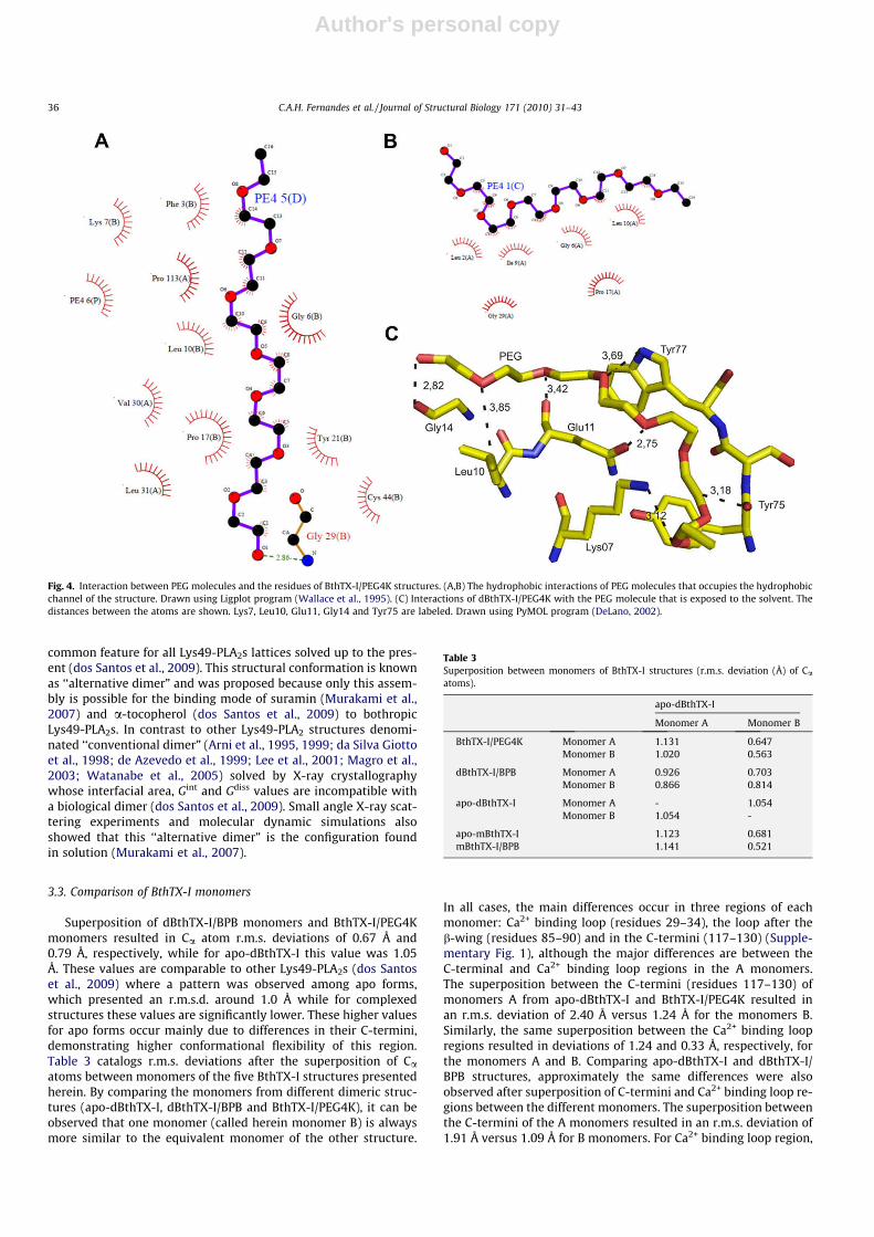
In both BthTX-I/BPB structures, BPB molecules are covalentlybound to the Nd1 atom of His48 residue from all monomers(Fig. 2A) and the phenacyl group of BPB molecule extends alongthe hydrophobic channel of the proteins, engaging hydrophobicinteractions with Leu5 and Gly30. Additionally, BPB molecules alsointeract (distance <4.0 Å) with Tyr22, Gly23, Cys45 and Lys49 fordBthTX-I/BPB and Gly23, Gly30, Lys49 and one isopropanol mole-cule for mBthTX-I/BPB (Fig. 3). The electron density map forBthTX-I/PEG4K structure showed clear positions for three PEGmolecules (Fig. 2B). Two of them are positioned similarly to PEGmolecules in the BthTX-I/suramin structure and to a-tocopherolin BthTX-I/aT and PrTX-I/aT structures, i.e., interacting with a largenumber of residues along the hydrophobic channel of the protein(Fig. 4). The third PEG molecule, in a position similar to the PEGmolecule in BthTX-I/aT structure, interacts (distance <4.0 Å) withLys7, Glu10, Gly13, Tyr76 and Trp77 of monomer B (Fig. 4).
3.2. Oligomeric assembly
The results of dynamic light scattering experiments at 291 K(crystallization temperature) with apo-BthTX-I indicated a meanhydrodynamic radius (RH) of 2.5 nm with a polydispersity of10.7% This RH value corresponds to a molecular weight of approx-imately 27 kDa and is, thus, equivalent to a dimer.
Fig. 1. Dimeric structures of (A) apo-dBthTX-I, (B) dBthTX-I/BPB and (C) BthTX-I/PEG4K show as a cartoon representation. The BPB and PEG molecules (yellow) are showed bysticks. Drawn using PyMOL program (DeLano, 2002). (For interpretation of the references to color in this figure legend, the reader is referred to the web version of this paper.)
34 C.A.H. Fernandes et al. / Journal of Structural Biology 171 (2010) 31–43

Author's personal copy
Although apo-mBthTX-I and mBthTX-I/BPB present a monomerin the asymmetric unit, the examination of their unit-cell packingusing the PISA software (Krissinel and Henrick, 2007) shows a di-meric conformation stable in solution for both structures. Theapo-mBthTX-I presents a complexation significance score (CSS) of1.0, an interfacial area of 528.3 Å2, Gint = �13.0 kcal/mol andGdiss = 2.0 kcal/mol while for mBthTX-I/BPB the values are CSS of
0.565, interface are of 683.2 Å2, Gint = �27.6 kcal/mol andGdiss = 5.7 kcal/mol. The apo-dBthTX-I, dBthTX-I/BPB and BthTX-I/PEG4K structures, which present a dimer in the asymmetric unit,also exhibit the same dimeric conformation that is stable in solu-tion according to the PISA software with CSS, interfacial area, Gint
and Gdiss values compatible with a biological dimer (data notshown). The assembly with the higher complexation score is a
Fig. 2. Electron density difference maps for the region corresponding to (A) BthTX-I/PEG4K hydrophobic channel (B) BthTX-I/PEG4K around Lys7 residue region and (C)mBthTX-I/BPB ‘‘active site” region. The maps were calculated with coefficients |Fobs| � |Fcalc| and contoured at 1.5 standard deviations. The ligands molecules were notconsidered electron density maps calculation and were drawn for clarity. Drawn using PyMOL program (DeLano, 2002).
Fig. 3. Interaction between BPB molecules and the residues of dBthTX-I/BPB and mBthTX-I/BPB structures. The distances between the atoms are shown. (A) Monomer A fromdBthTX-I/BPB interactions. Tyr22, Val31 and His48 residues are labeled. (B) Monomer B from dBthTX-I/BPB interactions. Gly23, Cys45, His48 and Lys49 residues are labeled.(C) Monomer from mBthTX-I/BPB interactions. Gly23, Gly30, His48, Lys49 residues and propanol molecule are labeled. Drawn using PyMOL program (DeLano, 2002).
C.A.H. Fernandes et al. / Journal of Structural Biology 171 (2010) 31–43 35
Author's personal copy
common feature for all Lys49-PLA2s lattices solved up to the pres-ent (dos Santos et al., 2009). This structural conformation is knownas ‘‘alternative dimer” and was proposed because only this assem-bly is possible for the binding mode of suramin (Murakami et al.,2007) and a-tocopherol (dos Santos et al., 2009) to bothropicLys49-PLA2s. In contrast to other Lys49-PLA2 structures denomi-nated ‘‘conventional dimer” (Arni et al., 1995, 1999; da Silva Giottoet al., 1998; de Azevedo et al., 1999; Lee et al., 2001; Magro et al.,2003; Watanabe et al., 2005) solved by X-ray crystallographywhose interfacial area, Gint and Gdiss values are incompatible witha biological dimer (dos Santos et al., 2009). Small angle X-ray scat-tering experiments and molecular dynamic simulations alsoshowed that this ‘‘alternative dimer” is the configuration foundin solution (Murakami et al., 2007).
3.3. Comparison of BthTX-I monomers
Superposition of dBthTX-I/BPB monomers and BthTX-I/PEG4Kmonomers resulted in Ca atom r.m.s. deviations of 0.67 Å and0.79 Å, respectively, while for apo-dBthTX-I this value was 1.05Å. These values are comparable to other Lys49-PLA2s (dos Santoset al., 2009) where a pattern was observed among apo forms,which presented an r.m.s.d. around 1.0 Å while for complexedstructures these values are significantly lower. These higher valuesfor apo forms occur mainly due to differences in their C-termini,demonstrating higher conformational flexibility of this region.Table 3 catalogs r.m.s. deviations after the superposition of Caatoms between monomers of the five BthTX-I structures presentedherein. By comparing the monomers from different dimeric struc-tures (apo-dBthTX-I, dBthTX-I/BPB and BthTX-I/PEG4K), it can beobserved that one monomer (called herein monomer B) is alwaysmore similar to the equivalent monomer of the other structure.
In all cases, the main differences occur in three regions of eachmonomer: Ca2+ binding loop (residues 29–34), the loop after theb-wing (residues 85–90) and in the C-termini (117–130) (Supple-mentary Fig. 1), although the major differences are between theC-terminal and Ca2+ binding loop regions in the A monomers.The superposition between the C-termini (residues 117–130) ofmonomers A from apo-dBthTX-I and BthTX-I/PEG4K resulted inan r.m.s. deviation of 2.40 Å versus 1.24 Å for the monomers B.Similarly, the same superposition between the Ca2+ binding loopregions resulted in deviations of 1.24 and 0.33 Å, respectively, forthe monomers A and B. Comparing apo-dBthTX-I and dBthTX-I/BPB structures, approximately the same differences were alsoobserved after superposition of C-termini and Ca2+ binding loop re-gions between the different monomers. The superposition betweenthe C-termini of the A monomers resulted in an r.m.s. deviation of1.91 Å versus 1.09 Å for B monomers. For Ca2+ binding loop region,
Fig. 4. Interaction between PEG molecules and the residues of BthTX-I/PEG4K structures. (A,B) The hydrophobic interactions of PEG molecules that occupies the hydrophobicchannel of the structure. Drawn using Ligplot program (Wallace et al., 1995). (C) Interactions of dBthTX-I/PEG4K with the PEG molecule that is exposed to the solvent. Thedistances between the atoms are shown. Lys7, Leu10, Glu11, Gly14 and Tyr75 are labeled. Drawn using PyMOL program (DeLano, 2002).
Table 3Superposition between monomers of BthTX-I structures (r.m.s. deviation (Å) of Ca
atoms).
apo-dBthTX-I
Monomer A Monomer B
BthTX-I/PEG4K Monomer A 1.131 0.647Monomer B 1.020 0.563
dBthTX-I/BPB Monomer A 0.926 0.703Monomer B 0.866 0.814
apo-dBthTX-I Monomer A - 1.054Monomer B 1.054 -
apo-mBthTX-I 1.123 0.681mBthTX-I/BPB 1.141 0.521
36 C.A.H. Fernandes et al. / Journal of Structural Biology 171 (2010) 31–43

Author's personal copy
the superposition resulted in respective deviations of 1.20 and 0.65Å, respectively, for the A and B monomers.
These structural changes were probably induced by the ligands(BPB or PEG molecules) leading to a new oligomeric conformationwhere some new interfacial interactions were established. For apo-dBthTX-I, one hydrogen bond was observed between its monomers(Val31:Lys69) whereas in the dBthTX-I/BPB and BthTX-I/PEG4Kstructures two new interchain hydrogen bond were established(Asn17:Tyr119 and Tyr119:Tyr119). These hydrogen bonds wereinduced by the rearrangement of the C-terminus of monomer Awhich increases the symmetry between monomers when the BPBand the PEG molecules are present at the ‘‘active site” of the pro-tein as observed for PrTX-I/aT and PrTX-I/BPB complexed struc-tures (dos Santos et al., 2009; Marchi-Salvador et al., 2009).
4. Discussion
4.1. The ‘‘two-angle model” applied to BthTX-I structures
Several experiments on bothropic Lys49-PLA2s, including onesutilizing electrophoresis, spectroscopy (Arni et al., 1999; da SilvaGiotto et al., 1998), small angle X-ray scattering (Murakami et al.,2007) and dynamic light scattering (the present article), demon-strated that bothropic Lys49-PLA2s are dimeric proteins in solu-tion. Furthermore, the dimeric state is also found in vivo, and isstable even upon heating in the presence of b-mercaptoethanol(da Silva Giotto et al., 1998; Soares et al., 2000). pH-induced disso-ciation studies also demonstrated a considerable decrease of mem-brane-damaging activity after the dimeric dissociation (Anguloet al., 2005).
Since the BthTX-I structure was solved for the first time in 1998(da Silva Giotto et al., 1998), the oligomeric assembly for bothropicLys49-PLA2s has remained the main issue in several other struc-tural articles involving this class of proteins (Magro et al., 2003;Watanabe et al., 2005). In this precursor study on BthTX-I, its pro-posed molecular hinge model (aperture angle) was based on onlytwo configurations of BthTX-I (‘‘open” and ‘‘closed”). This proposalwas subsequently revised (Magro et al., 2003) using seven differentstructures at higher resolution. More recently, a useful tridimen-sional configuration model between the monomers has been pro-posed based on two angles: hT (torsional angle) and hA (apertureangle) (dos Santos et al., 2009). This two-angle model was estab-lished for the ‘‘alternative dimer” by the observation that theLys49-PLA2s complexed to any ligand present a rearrangement ofthe C-terminus of one monomer thus increasing the symmetry be-tween the two monomers and aligning them in the same plane.This alignment results in lower torsional and higher aperture an-gles for complexed structures. Then, when these angles are calcu-lated for both complexed and apo Lys49-PLA2s, their oligomericconformation can be divided into two classes: apo and complexedstructures (dos Santos et al., 2009). In the case of dBthTX-I/BPB andBthTX-I/PEG4K structures the calculated angles are, respectively,hT = 53� and hA = 23�; hT = 40� and hA = 28�. These values are similarin the BthTX-I/aT and other complexed structures including PrTX-I/aT, MjTX-II/stearic acid, and BaspTX-II/suramin (dos Santos et al.,2009). By contrast, the torsional/aperture angles for apo-dBthTX-Iare, respectively, 60� and 7�, which differ greatly from those ofthe complexed structures but are very similar to the angles foundin other apo structures (PrTX-I, BnSP-7, and BnSP-6) (dos Santoset al., 2009).
The disorder in apo Lys49-PLA2 C-termini and the action of a li-gand to order this region by increasing the symmetry between themonomers have been observed in bothropic Lys49-PLA2s (dos San-tos et al., 2009; Magro et al., 2003). The reorganization of the C-ter-minal region of one monomer when a ligand is present at the
‘‘active site” (e.g. BPB molecule) or hydrophobic channel (e.g. PEG4000) induces a better order at its C-terminus leading to the estab-lishment of a new interchain hydrogen bond between Tyr119 res-idues. This phenomenon has been observed in both dBthTX-I/BPBand BthTX-I/PEG4K structures and is also a common feature amongthe complexed bothropic Lys49-PLA2s and probably is an impor-tant characteristic for the myotoxicity of these toxins (dos Santoset al., 2009; Magro et al., 2003). Site-directed mutagenesis experi-ments also support this hypothesis since a significant decrease inthe myotoxic activity was observed after Y119A mutant was testedin vivo (Chioato et al., 2007).
4.2. The role of hydrophobic long-chain molecules for Lys49-PLA2s
The structures of Piratoxin-II from B. pirajai (PrTX-II) and Num-TX-I from B. nummifer revealed electron densities that were inter-preted as fatty acids within the hydrophobic channel leading to theactive site (de Azevedo et al., 1999; Lee et al., 2001). More recently,the structure of the complex MjTX-II bound to a fatty acid (stearicacid) was solved (Watanabe et al., 2005). This molecule was foundoccupying the hydrophobic channel for PrTX-II, NumTX-I, andMjTX-II structures, despite the fact that MjTX-II was co-crystallizedwith the fatty acid, and NumTX-I and PrTX-II were interpreted as afatty acid since these molecules were not added to the crystalliza-tion solution (Fig. 5). However, the structures of the complexes be-tween the putative fatty acid bound to the PrTX-II and the stearicacid bound to MjTX-II present significant differences. In contrastto the PrTX-II structure, no water molecule was found bound tothe stearic acid head group in the MjTX-II structure. Instead, thecarboxyl oxygens of the stearic acid form hydrogen bonds withHis 48 and Gly 30 and occupy a position similar to that of a solventmolecule in the PrTX-II structure. Due to the inclusion of a water
Fig. 5. Superposition of PEG molecule (dark blue) from dBTHTX-I/PEG4K structure,stearic acid (yellow) from MjTX-II/stearic acid structure and putative fatty acid(green) from PrTX-II/fatty acid structure in the ‘‘active site” region. His48 and Lys49are showed by sticks. The water molecule in the PrTX-II/fatty acid structure (W39)is shown as a green sphere. Drawn using PyMOL program (DeLano, 2002). (Forinterpretation of the references to color in this figure legend, the reader is referredto the web version of this paper.)
C.A.H. Fernandes et al. / Journal of Structural Biology 171 (2010) 31–43 37

Author's personal copy
molecule, the supposed fatty acid in the PrTX-II is shifted out onecarbon position in relation to stearic acid of the MjTX-II (Fig. 5).
Both polyethylene glycol (PEG3350 or PEG4000) and fatty acidmolecules are, in general, structurally similar. The main differencebetween them is that the fatty acid polar head has two oxygenatoms while the PEG molecule has only one oxygen atom. Then,
given that the PrTX-II was crystallized in the presence ofPEG3350 and the interaction of ligand with the ‘‘active site” oc-curred by means of a water molecule (Fig. 5), we suggest the pres-ence of a PEG3350 instead of a fatty acid molecule in thehydrophobic channel where the water molecule would be the oxy-gen head of PEG molecule. In contrast, NumTX-I and MjTX-II struc-
Table 4The role of Lys122 residue for apo and complexed Lys49-PLA2s. B-factor side chains, electron density and polarization of Cys29–Gly30 analyses.
B-factor average of allprotein atoms (Å2)
B-factor average of Lys122 sidechain atoms (Å2)
Electron density for Lys122 sidechain (cut-off 1.2r)
Polarization of Cys29–Gly30peptide bond
Monomer A Monomer B Monomer A Monomer B Monomer A Monomer B
Apo dBthTX-I 41.1 62.9 31.7 Yes Yes No YesmBthTX-I 27.8 60.0 – Yes – No –PrTX-I 34.1 42.4 31.2 Yes Yes No YesBaspTX-II 12.1 27.1 23.0 a a Yes NoBnSP-7 30.5 51.7 55.4 Yes Yes No NoBnSP-6 38.3 47.5 68.9 Yes Yes Yes No
Complexed dBthTX-I/BPB 48.7 – – No No No NomBthTX-I/BPB 29.2 26.1 – Yes – Yes –BthTX-I/PEG4K 27.8 37.5 45.0 Yes Yes Yes NoBthTX-I/aT 30.4 – 47.2 No Yes No NoPrTX-I/BPB 48.9 66.9 65.1 Yes Yes No NoPrTX-I/aT 22.7 37.7 38.1 Yes Yes No NoPrTX-II/fatty acid 31.1 32.8 32.7 a a Yes YesMjTX-II/stearic acid 33.6 39.1 36.0 Yes Yes Yes YesBaspTX-II/suramin 24.0 32.5 48.1 Yes Yes Yes NoBthTX-I/PEG 400 41.1 60.4 51.2 Yes Yes No Yes
a Structure factors values not available in PDB Data Bank.
Fig. 6. Interactions of Lys122 with several residues in different Lys49-PLA2s structures. The distances between the atoms are shown. (A) apo-mBthTX-I. (B) mBthTX-I/BPB. (C)BaspTX-II/Suramin. (D) apo-BaspTX-II. (E) BnSP-7. (F) BnSP-6. Drawn using PyMOL program (DeLano, 2002).
38 C.A.H. Fernandes et al. / Journal of Structural Biology 171 (2010) 31–43

Author's personal copy
tures were not crystallized in the presence of polyethylene glycol,thus enhancing the chance of binding of fatty acid in the hydropho-bic channel. This doubt only can be resolved with new crystalliza-tion experiments that utilize PrTX-II in the absence of PEG3350 or
in the presence of a specific fatty acid molecule. To corroborate thisdata, the BthTX-I/PEG4K structure was solved to a high resolution(1.5 Å) and also presented electron densities for both monomerswithin the hydrophobic channel, which could be interpretedambiguously as polyethylene glycol 4000 molecules.
The structures of a-tocopherol bound to BthTX-I/aT and PrTX-I/aT also revealed the high affinity of Lys49-PLA2s for long-chainhydrophobic molecules by the hydrophobic channel (dos Santoset al., 2009). Recently, the presence of two different binding re-gions has been proposed for Lys49-PLA2s and membranes: the ‘‘ac-tive site/hydrophobic channel” and the ‘‘myotoxic site” (Marchi-Salvador et al., 2009). In this hypothesis the hydrophobic channelwould be the anchor by which the protein may bind to the mem-brane phospholipids while the myotoxic site would be responsiblefor the membrane destabilization. This suggestion may explainwhy the Lys49-PLA2s have high affinity for long-chain hydrophobicmolecules.
Table 5Superposition between Ca2+ binding loop regions (residues 26–34) between mono-mers of snake venom PLA2s (r.m.s. deviation (Å) of Ca atoms).
apo-dBthTX-I
Lys49 PrTX-I 0.37BnSP-7 0.49BnSP-6 0.49PrTX-II/fatty acid 0.61MjTX-II/fatty acid 0.61BthTX-I/aT 0.41PrTX-I/aT 0.41BthTX-I/PEG4K 0.32BaspTX-II 1.09godMT-II 1.52
Asp49 BthA-I-PLA2 1.89Acid-PLA2 1.75DPLA2 1.61b2-Bungarotoxin 1.71
Asp49a BthTX-II 2.15PrTX-III 3.18
a Asp49-PLA2s with reduced catalytic activity.
Fig. 7. Ca superposition of Ca2+-binding loop region of Lys49-PLA2s (red) andAsp49-PLA2s (green) and low-catalytic-activity Asp49-PLA2s (blue) structures. Thenumbers correspond to the residues that form the Ca2+-binding loop region. TheAsp49-PLA2s (green) are represented by BthA-I-PLA2 from Bothrops jararacussuvenom (PDB ID 1U73), Acid Phospholipase from Gloydius halys venom (PDB ID 1PSJ),DPLA2 from Daboia russelli pulchella venom (PDB ID 1FB2) and b-2 Bungarotoxinfrom Bungarus multicinctus venom (PDB ID 1BUN). The low-catalytic-activityAsp49-PLA2s (blue) are represented by BthTX-II from Bothrops jararacussu venom(PDB ID 2OQD) and PrTX-III from Bothrops pirajai venom (PDB ID 1GMZ). The Lys49-PLA2s (red) are represented by apo-dBthTX-I from Bothrops jararacussu venom (thiswork, PDB ID 3HZD), apo-PrTX-I from Bothrops pirajai venom (PDB ID 2Q2J), BnSP-7from Bothrops pauloensis venom (PDB ID 1PA0), apo-BaspTX-II from Bothrops aspervenom (PDB ID 1CLP), godMT-II from Cerrophidion goodmani venom (PDB ID 1GOD)and Anum-II from Atropoides nummifer venom (PDB ID 2AOZ). Drawn using ‘‘O”(Jones et al., 1991) and PyMOL (DeLano, 2002) programs. (For interpretation of thereferences to color in this figure legend, the reader is referred to the web version ofthis paper.)
Fig. 8. Amino acid sequence alignment of Ca2+-binding loop region of severalAsp49-PLA2s and Lys49-PLA2s (codes used in alignment and its NCBI entries areshown Supplementary Table 1). Residues of the beginning of Ca2+-binding loop(residue 26) and involved in stabilization of this region (residues 28 and 35) arehighlighted.
C.A.H. Fernandes et al. / Journal of Structural Biology 171 (2010) 31–43 39

Author's personal copy
4.3. What is the role of Lys122 in the Lys49-PLA2s?
In the crystal structure of Piratoxin-II from B. pirajai (PrTX-II),the presence of additional electron density in the active site regionled the authors to the hypothesis that a fatty acid molecule associ-ated with a conserved water molecule remains bound to the pro-tein following an interrupted catalytic cycle arrested at the finalproduct release step (Lee et al., 2001). In this model, the Ne atomof the Lys49 neutralizes the formal negative charge of the transi-tion state oxyanion, and the Ne of Lys122 serves as an auxiliaryelectrophile in substrate hydrolysis. Lys122 would hyperpolarizeCys29–Gly30 peptide by increasing its affinity for the fatty acid,thus trapping this enzymatic product at the active site (Lee et al.,2001). However, after the PrTX-II structure was solved, severalother Lys49-PLA2s structures, in apo and complexed forms, havebeen solved (dos Santos et al., 2009; Magro et al., 2003; Marchi-Salvador et al., 2009; Murakami et al., 2005, 2007; Watanabeet al., 2005), thus necessitating a review of the position of Lys122in these new structures.
Table 4 summarizes different structural data of Lys122 for 30different monomers for apo and complexed Lys49-PLA2 whosecoordinates are available in the PDB data bank. There are 12monomers in which Lys122 interacts with the Cys29 versus 18monomers where this interaction does not occur. However, thepolarization of both monomers in the same structure occurs onlyfor the complexes MjTX-II-stearic acid and PrTX-II-putative fattyacid. apo-Lys49-PLA2s – including BnSP-6, BnSP-7, mBthTX-I,dBthTX-I, PrTX-I and BaspTX-II – present one or no Lys122–Cys29 interaction. Other complexes – including mBthTX-I/BPB,dBtTX-I/BPB, BthTX-I/PEG4000, BthTX-I/aT, PrTX-I/aT, BaspTX-II/suramin and BthTX-I/PEG400 – also present one or no Lys122–Cys29 interaction. In some cases, Lys122 side chains lack elec-tron density above 1.2r, probably due to their high flexibility,not interacting with Cys29 or any other residue. Table 4 alsoshows our analysis of the B-factors for Lys122 side chains. It
can be observed that only seven Lys122 residues present B-factorvalues compatible with the average of their respective proteins,while all others present very high values (in most cases the val-ues are 50% higher). These results indicate the high flexibility ofthese residues in the majority of the structures which is notcompatible with the polarization process previously proposedby Lee et al. (2001).
The analysis of these structures also revealed another interest-ing structural feature of Lys122. Besides Cys29, this residue canalso interact with a carboxyl group from the other residues:Tyr25 in mBthTX-I/BPB; Gly26 in BnSP-6, apo-BaspTX-II andmBthTX-I/BPB; Cys27 in BaspTX-II/suramin, apo-mBthTX-I andapo-dBthTX-I; Leu32 and Gly33 in BnSP-7; and Arg34 in BaspTX-II/suramin. Therefore, Lys 122 is a very flexible residue that mayadopt random configurations, interacting with different sites ofnegative charges (Fig 6). Similarly, for MjTX-II/stearic acid andPrTX-II, there would be no functional reason for the Lys122/Cys29 interaction, like for the other interactions formed byLys122 in other Lys49-PLA2s.
Interestingly, a crystal structure of myotoxic and catalyticallyinactive Ser49-PLA2 from Echis carinatus sochureki presents anadditional electron density in hydrophobic channel that was mod-eled as lauric acid (Zhou et al., 2008). However, in this class ofPLA2s, the interaction with fatty acid is independent of Lys122,since this residue is not conserved for Ser49-PLA2 and there isnot any residue capable of hyperpolarizing the Cys29–Gly30 pep-tide bond.
Then, we can hypothesize that the affinity of the hydrophobicchannel by fatty acids are related to membrane anchorage ratherthan Lys49-PLA2s catalytic inactivity, which may, eventually, beaided by hyperpolarization caused by Lys122. As proposed bydos Santos et al. (2009), this interaction causes a side-by-sidealignment of the region responsible for myotoxic activity(C-terminus/myotoxic site), allowing the interaction with themembrane. In this hypothesis two functions may be associated
Fig. 9. Tyr28 (Asp49-PLA2s) and Asn28 (Lys49-PLA2s) side chain residues are shown in the Ca2+-binding loop region superposition of (A) Asp49-PLA2s (green) and Lys49-PLA2s (red) structures and (B) Asp49-PLA2s (green) and low-catalytic-activity Asp49-PLA2s (blue) structures. The Asp49-PLA2s (green) are represented by BthA-I-PLA2 fromBothrops jararacussu venom (PDB ID 1U73), Acid Phospholipase from Gloydius halys venom (PDB ID 1PSJ), DPLA2 from Daboia russelli pulchella venom (PDB ID 1FB2) and b-2Bungarotoxin from Bungarus multicinctus venom (PDB ID 1BUN). The low-catalytic-activity Asp49-PLA2s (blue) are represented by BthTX-II from Bothrops jararacussu venom(PDB ID 2OQD) and PrTX-III from Bothrops pirajai venom (PDB ID 1GMZ). The Lys49-PLA2s (red) are represented by apo-dBthTX-I from Bothrops jararacussu venom (this work,PDB ID 3HZD), apo-PrTX-I from Bothrops pirajai venom (PDB ID 2Q2J), BnSP-7 from Bothrops pauloensis venom (PDB ID 1PA0), apo-BaspTX-II from Bothrops asper venom (PDBID 1CLP), godMT-II from Cerrophidion goodmani venom (PDB ID 1GOD) and Anum-II from Atropoides nummifer venom (PDB ID 2AOZ). Drawn using ‘‘O” (Jones et al., 1991) andPyMOL (DeLano, 2002) programs.
40 C.A.H. Fernandes et al. / Journal of Structural Biology 171 (2010) 31–43
Author's personal copy
with Lys122 residue: (i) to aid fatty acid anchorage in thehydrophobic channel by hyperpolarization of Cys29, (ii) directaction on the muscle membrane given that this residue is inthe C-terminal region of Lys49-PLA2s which is related to theirmyotoxic activity.
4.4. Ca2+-binding loop and its role in the Lys49-PLA2s catalyticinactivity
Lys49-PLA2s are very intriguing proteins because they sharehigh sequential identity with Asp49-PLA2s but do not display cat-
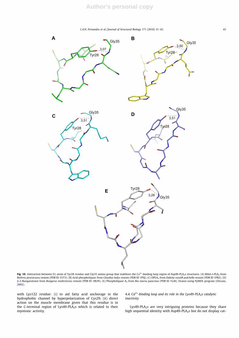
Fig. 10. Interaction between Oc atom of Tyr28 residue and Gly35 amino group that stabilizes the Ca2+-binding loop region of Asp49-PLA2s structures. (A) BthA-I-PLA2 fromBothros jararacussu venom (PDB ID 1U73). (B) Acid phospholipase from Gloydius halys venom (PDB ID 1PSJ). (C) DPLA2 from Daboia russelli pulchella venom (PDB ID 1FB2). (D)b-2-Bungarotoxin from Bungarus multicinctus venom (PDB ID 1BUN). (E) Phospholipase A2 from Bos taurus pancreas (PDB ID 1G4I). Drawn using PyMOL program (DeLano,2002).
C.A.H. Fernandes et al. / Journal of Structural Biology 171 (2010) 31–43 41

Author's personal copy
alytic activity. Furthermore, both Lys49-PLA2s and Asp49-PLA2shave a highly conserved catalytic network (His48, Tyr53 andAsp99) with the exception of Asp49 residue in Lys49-PLA2s. Site-directed mutagenesis experiments with a Lys49-PLA2 showed thatLys49Asp mutant remained catalytically inactive, demonstrating,in turn, that this single mutation is not crucial to the lack of enzy-matic activity in these toxins (Ward et al., 2002), and suggestingthat the catalytic inactivity of these homologue PLA2s may be re-lated to other structural features.
In order to understand the reasons for the catalytic inactivity ofLys49-PLA2s, we performed a structural comparison of Ca2+ bind-ing loop regions from Lys49-PLA2s, Asp49-PLA2s and low-cata-lytic-activity Asp49-PLA2s (BthTX-II and PrTX-III) which alsodisplay myotoxic activity as Lys49-PLA2s (Correa et al., 2008).
Table 5 and Fig. 7 show the Ca superposition between the Ca2+
binding loop regions (residues 27–35) from several snake venomPLA2s. This comparison reveals that this loop adopts a similar con-formation within Lys49-PLA2s and within Asp49-PLA2s, but whenthese two classes are compared the conformation becomes verydifferent. Additionally, the low-catalytic Asp49-PLA2s presentstrongly distorted Ca2+-binding loops when compared to Lys49-PLA2s and other Asp49-PLA2s, as previously described by Correaet al. (2008).
Amino acid sequence alignment of Ca2+-binding loop region ofLys49-PLA2s and Asp49-PLA2s from several snakes, showed thatTyr28 is conserved for all Asp49-PLA2s while Asn28 is conservedfor Lys49-PLA2s (Fig. 8). These are the only strictly conserved resi-dues in the Ca2+-binding loop regions in these proteins. Further-more, the Tyr28 side chain is also structurally conserved in Asp49-PLA2s, with the exception of low-catalytic Asp49-PLA2s (Fig. 9).
The structural analysis of Tyr28 of Asp49-PLA2s reveals animportant stability feature of the Ca2+-binding loop region. AllAsp49-PLA2s (except low-catalytic Asp49-PLA2s) present a con-served interaction between Oc atom of Tyr28 and Gly35 aminogroup in the range from 3.1 Å to 3.5 Å (Fig. 10) which is also pres-ent in other catalytic PLA2s, including pancreatic bovine PLA2 (Stei-ner et al., 2001). Recently, the lack of this interaction was pointedas responsible for Ca2+-binding loop disarrangement in Ser49-PLA2
Ecarpholin S from Echis carinatus sochureki venom (Zhou et al.,2008). This Tyr28–Gly35 interaction may provide more structuralstability for the Ca2+-binding loop which may be verified by theconservation of the distance d (Supplementary Fig. 2) that reflectsthe aperture of this loop. Table 6 shows that the distance d be-tween Ca atoms of Tyr28 and Gly33 in Asp49-PLA2s (except low-catalytic-activity Asp49-PLA2s) is approximately 4.3 Å. In contrast,for Lys49-PLA2s, due to the natural mutation Tyr28Asn, the dis-tance d becomes greater than 6.3 Å. This open configuration ofCa2+-binding loop may be one of the factors responsible for theinability of Ca2+ ion to bind to Lys49-PLA2, even when the Lys49Aspmutation is performed by site-directed mutagenesis (Ward et al.,2002).
Despite the presence of Tyr28 in low-catalytic Asp49-PLA2s,these toxins present a great distortion in the Ca2+-binding loop re-gion (Fig. 6 and Table 5). Fig. 9 shows that Tyr28 side chain in low-catalytic Asp49-PLA2s does not have the same orientation as theTyr28 side chains in other Asp49-PLA2s probably due to the interac-tion of this residue with the Asp49 side chain (Correa et al., 2008).Consequently, the Tyr28–Gly35 interaction does not form, causingthe disorder observed in the Ca2+- binding loop region of these en-zymes and, consequently, preventing the binding of Ca2+ ion.
5. Concluding remarks
Despite its catalytic inactivity, the hydrophobic channel ofLys49-PLA2 is a very intriguing region due to its conservationof most of residues involved in catalytic activity and high affin-ity for long-chain hydrophobic molecules. On account of the lat-ter characteristic, in this work, we reinforce the previousproposal of Marchi-Salvador et al. (2009) of the two differentbinding regions of Lys49-PLA2s and membranes, the hydropho-bic channel and the myotoxic site/C-terminal, where the hydro-phobic channel would be the anchor for the myotoxic action ofLys49-PLA2s.
Herein, the controversial role of Lys122 was analyzed in depth,in light of the 30 different known monomers for apo and com-plexed Lys49-PLA2s structures. The data show that the Ne ofLys122 probably binds to different carboxyl groups in order to sta-bilize the crystal structures and that its role may be associatedwith membrane binding rather than hyperpolarization of Cys29/Gly30. However, if this polarization really occurs, we believe thatthis interaction is related to membrane anchorage of Lys49-PLA2s,rather than a role in its catalytic inactivity.
Finally, a structural comparison of Ca2+ binding loop regionsfrom Lys49-PLA2s and Asp49-PLA2s reveals that the conservationof the residue Tyr28 in Asp49-PLA2s is vital to the structural integ-rity of this region in these enzymes. This residue stabilizes the Ca2+
binding loop region by an interaction between its Oc atom and theGly35 amino group. In Lys49-PLA2s that have an Asn residue in-stead of a Tyr residue in position 28, this interaction does not oc-cur, which contributes to destabilizing the Ca2+-binding loopregion and preventing the binding of Ca2+ cofactor. In low-catalyticAsp49-PLA2s, despite the presence of Tyr28, this interaction doesnot occur, due to its interaction with Asp49 residue which also pre-vents the binding of Ca2+ ion.
The hypotheses raised in this work may be useful for guidingnew biophysical and biochemical experiments that may defini-tively clarify the action mechanism of intriguing snake venomPLA2s and lead to the design of structure-based inhibitors to com-plement the serum therapy, thus preventing the permanent inju-ries still caused by these proteins in snakebite victims aroundthe world.
6. Atomic coordinates
The coordinates were deposited in the Protein Data Bank withidentification codes 3HZD, 3HZW, 3IO3, 3IQ3, 3I3I.
Acknowledgments
The authors gratefully acknowledge the financial support fromFundação de Amparo à Pesquisa do Estado de São Paulo (FAPESP),Conselho Nacional de Desenvolvimento Científico e Tecnológico(CNPq) and Laboratório Nacional de Luz Síncrontron (LNLS, Campi-nas-SP).
Table 6Values of d distance between Ca atoms of Tyr28 and Gly33 reflecting the aperture ofCa2+-binding loop in Asp49-PLA2s and Lys49-PLA2s structures.
Class Protein d
Lys49-PLA2s apo-dBthTX-I 6.05PrTX-I 6.22BnSP-7 6.46Anum-II 6.30godMT-II 5.06
Asp49-PLA2s BthA-I-PLA2s 4.20Acid-PLA2s 4.41DPLA2 4.58b2-Bungarotoxin 4.33PLA2s pancreatic bovine 3.91
42 C.A.H. Fernandes et al. / Journal of Structural Biology 171 (2010) 31–43

Author's personal copy
Appendix A. Supplementary data
Supplementary data associated with this article can be found, inthe online version, at doi:10.1016/j.jsb.2010.03.019.
References
Angulo, Y., Gutierrez, J.M., Soares, A.M., Cho, W., Lomonte, B., 2005. Myotoxic andcytolytic activities of dimeric Lys49 phospholipase A2 homologues are reduced,but not abolished, by a pH-induced dissociation. Toxicon 46, 291–296.
Arni, R.K., Ward, R.J., Gutierrez, J.M., Tulinsky, A., 1995. Structure of a calcium-independent phospholipase-like myotoxic protein from Bothrops asper venom.Acta Crystallogr. D Biol. Crystallogr. 51, 311–317.
Arni, R.K., Fontes, M.R.M., Barberato, C., Gutierrez, J.M., Diaz, C., Ward, R.J., 1999.Crystal structure of myotoxin II, a monomeric Lys49-phospholipase A2
homologue isolated from the venom of Cerrophidion (Bothrops) godmani. Arch.Biochem. Biophys. 366, 177–182.
de Azevedo, W.F., Ward, R.J., Gutierrez, J.M., Arni, R.K., 1999. Structure of a Lys49phospholipase A2 homologue isolated from the venom of Bothrops nummifer(jumping viper). Toxicon 37, 371–384.
Brunger, A.T., Adams, P.D., Clore, G.M., DeLano, W.L., Gros, P., Grosse-Kunstleve,R.W., Jiang, J.S., Kuszewski, J., Nilges, M., Pannu, N.S., Read, R.J., Rice, L.M.,Simonson, T., Warren, G.L., 1998. Crystallography & NMR system: a newsoftware suite for macromolecular structure determination. Acta Crystallogr. D– Biol. Crystallogr. 54, 905–921.
Chandra, V., Kaur, P., Jasti, J., Betzel, C., Singh, T.P., 2001. Regulation of catalyticfunction by molecular association: structure of phospholipase A2 from Daboiarusselli pulchella (DPLA2) at 1.9 Å resolution. Acta Crystallogr. D Biol. Crystallogr.57, 1793–1798.
Chioato, L., Aragao, E.A., Ferreira, T.L., de Medeiros, A.I., Faccioli, L.H., Ward, R.J.,2007. Mapping of the structural determinants of artificial and biologicalmembrane damaging activities of a Lys49 phospholipase A2 by scanningalanine mutagenesis. Biochim. Biophys. Acta – Biomembr. 1768, 1247–1257.
Correa, L.C., Marchi-Salvador, D.P., Cintra, A.C.O., Sampaio, S.V., Soares, A.A., Fontes,M.R.M., 2008. Crystal structure of a myotoxic Asp49-phospholipase A2 with lowcatalytic activity: insights into Ca2+-independent catalytic mechanism. Biochim.Biophys. Acta – Proteins Proteomics 1784, 591–599.
DeLano, W.L., 2002. The PyMOL Molecular Graphics System, DeLano Scientific LLC,San Carlos, CA.
Diaz-Oreiro, C., Gutierrez, J.M., 1997. Chemical modification of histidine and lysineresidues of myotoxic phospholipases A2 isolated from Bothrops asper andBothrops godmani snake venoms: effects on enzymatic and pharmacologicalproperties. Toxicon 35, 241–252.
Emsley, P., Cowtan, K., 2004. Coot: model-building tools for molecular graphics.Acta Crystallogr. D Biol. Crystallogr. 60, 2126–2132.
Gutierrez, J.M., Lomonte, B., 1995. Phospholipase A2 myotoxins from Bothrops snakevenoms. Toxicon 33, 1405–1424.
Holland, D.R., Clancy, L.L., Muchmore, S.W., Ryde, T.J., Einspahr, H.M., Finzel, B.C.,Heinrikson, R.L., Watenpaugh, K.D., 1990. The crystal structure of a lysine 49phospholipase A2 from the venom of the cottonmouth snake at 2.0 Å resolution.J Biol Chem 265, 17649–17656.
Homsi-Brandeburgo, M.I., Queiroz, L.S., Santo-Neto, H., Rodrigues-Simioni, L., Giglio,J.R., 1988. Fractionation of Bothrops jararacussu snake venom: partial chemicalcharacterization and biological activity of bothropstoxin. Toxicon 26, 615–627.
Jancarik, J., Kim, S.H., 1991. Sparse-matrix sampling – a screening method forcrystallization of proteins. J. Appl. Crystallogr. 24, 409–411.
Jones, T.A., Zou, J.Y., Cowan, S.W., Kjeldgaard, M., 1991. Improved methods forbuilding protein models in electron-density maps and the location of errors inthese models. Acta Crystallogr. A 47, 110–119.
Krissinel, E., Henrick, K., 2007. Inference of macromolecular assemblies fromcrystalline state. J. Mol. Biol. 372, 774–797.
Kwong, P.D., McDonald, N.Q., Sigler, P.B., Hendrickson, W.A., 1995. Structure of beta2-bungarotoxin: potassium channel binding by Kunitz modules and targetedphospholipase action. Structure 3, 1109–1119.
Laskowski, R.A., Macarthur, M.W., Moss, D.S., Thornton, J.M., 1993. Procheck - aprogram to check the stereochemical quality of protein structures. J. Appl.Crystallogr. 26, 283–291.
Lee, W.H., da Silva Giotto, M.T., Marangoni, S., Toyama, M.H., Polikarpov, I., Garratt,R.C., 2001. Structural basis for low catalytic activity in Lys49 phospholipases A2– a hypothesis: the crystal structure of piratoxin II complexed to fatty acid.Biochemistry 40, 28–36.
Lovell, S.C., Davis, I.W., Arendall 3rd, W.B., de Bakker, P.I., Word, J.M., Prisant, M.G.,Richardson, J.S., Richardson, D.C., 2003. Structure validation by Calphageometry: phi, psi and Cbeta deviation. Proteins 50, 437–450.
Magro, A.J., Soares, A.M., Giglio, J.R., Fontes, M.R.M., 2003. Crystal structures ofBnSP-7 and BnSP-6, two Lys49-phospholipases A2: quaternary structure andinhibition mechanism insights. Biochem. Biophys. Res. Commun. 311, 713–720.
Magro, A.J., Takeda, A.A., Soares, A.M., Fontes, M.R.M., 2005. Structure of BthA-Icomplexed with p-bromophenacyl bromide: possible correlations with lack ofpharmacological activity. Acta Crystallogr. D Biol. Crystallogr. 61, 1670–1677.
Magro, A.J., Fernandes, C.A., dos Santos, J.I., Fontes, M.R., 2009. Influence of quaternaryconformation on the biological activities of the Asp49-phospholipases A2s fromsnake venoms. Protein Pept. Lett. 16, 852–859.
Marchi-Salvador, D.P., Fernandes, C.A., Silveira, L.B., Soares, A.M., Fontes, M.R., 2009.Crystal structure of a phospholipase A2 homolog complexed with p-bromophenacyl bromide reveals important structural changes associated withthe inhibition of myotoxic activity. Biochim. Biophys. Acta 1794, 1583–1590.
McPherson, A., 2003. Introduction to Macromolecular Crystallography, Wiley-Liss,Hoboken.
Moolenaar, W.H., van Meeteren, L.A., Giepmans, B.N., 2004. The ins and outs oflysophosphatidic acid signaling. BioEssays 26, 870–881.
Murakami, M.T., Arruda, E.Z., Melo, P.A., Martinez, A.B., Calil-Elias, S., Tomaz, M.A.,Lomonte, B., Gutierrez, J.M., Arni, R.K., 2005. Inhibition of myotoxic activity ofBothrops asper myotoxin II by the anti-trypanosomal drug surarnin. J. Mol. Biol.350, 416–426.
Murakami, M.T., Vicoti, M.M., Abrego, J.R.B., Lourenzoni, M.R., Cintra, A.C.O., Arruda,E.Z., Tomaz, M.A., Melo, P.A., Arni, R.K., 2007. Interfacial surface charge and freeaccessibility to the PLA2-active site-like region are essential requirements forthe activity of Lys49 PLA2 homologues. Toxicon 49, 378–387.
Murshudov, G.N., Vagin, A.A., Dodson, E.J., 1997. Refinement of macromolecularstructures by the maximum-likelihood method. Acta Crystallogr. D Biol.Crystallogr. 53, 240–255.
Navaza, J., 1994. Amore – an automated package for molecular replacement. ActaCrystallogr. A 50, 157–163.
Otwinowski, Z., Minor, W., 1997. Processing of X-ray diffraction data collected inoscillation mode. Macromol. Crystallogr. Pt A 276, 307–326.
Prescott, S.M., Zimmerman, G.A., Stafforini, D.M., McIntyre, T.M., 2000. Platelet-activating factor and related lipid mediators. Annu. Rev. Biochem. 69, 419–445.
Rogers, J., Yu, B.Z., Serves, S.V., Tsivgoulis, G.M., Sotiropoulos, D.N., Ioannou, P.V.,Jain, M.K., 1996. Kinetic basis for the substrate specificity during hydrolysis ofphospholipids by secreted phospholipase A2. Biochemistry 35, 9375–9384.
dos Santos, J.I., Soares, A.M., Fontes, M.R.M., 2009. Comparative structural studies onLys49-phospholipases A2 from Bothrops genus reveal their myotoxic site. J.Struct. Biol. 167, 106–116.
Schaloske, R.H., Dennis, E.A., 2006. The phospholipase A2 superfamily and its groupnumbering system. Biochim. Biophys. Acta 1761, 1246–1259.
Scott, D.L., White, S.P., Otwinowski, Z., Yuan, W., Gelb, M.H., Sigler, P.B., 1990.Interfacial catalysis: the mechanism of phospholipase A2. Science 250, 1541–1546.
Scott, D.L., Achari, A., Vidal, J.C., Sigler, P.B., 1992. Crystallographic and biochemicalstudies of the (inactive) Lys49 phospholipase A2 from the venom of Agkistrodonpiscivorus piscivorus. J. Biol. Chem. 267, 22645–22657.
da Silva Giotto, M.T., Garratt, R.C., Oliva, G., Mascarenhas, Y.P., Giglio, J.R., Cintra,A.C.O., de Azevedo, W.F., Arni, R.K., Ward, R.J., 1998. Crystallographic andspectroscopic characterization of a molecular hinge: conformational changes inbothropstoxin I, a dimeric Lys49 phospholipase A2 homologue. Proteins–Struct.Funct. Bioinform. 30, 442–454.
Soares, A.M., Guerra-Sa, R., Borja-Oliveira, C.R., Rodrigues, V.M., Rodrigues-Simioni,L., Rodrigues, V., Fontes, M.R.M., Lomonte, B., Gutierrez, J.M., Giglio, J.R., 2000.Structural and functional characterization of BnSP-7, a Lys49 myotoxicphospholipase A(2) homologue from Bothrops neuwiedi pauloensis venom.Arch. Biochem. Biophys. 378, 201–209.
Steiner, R.A., Rozeboom, H.K., de Vrires, A., Kalk, K.H., Murshudov, G.N., Wilson, K.S.,Dijkstra, B.W., 2001. X-ray structure of bovine pancreatic phospholipase A2 atatomic resolution. Acta Crystallogr. D Biol. Crystallogr. 57, 516–526.
Tsuboi, K., Sugimoto, Y., Ichikawa, A., 2002. Prostanoid receptor subtypes.Prostaglandins Other Lipid Mediat. 68–69, 535–556.
Wallace, A.C., Laskowski, R.A., Thornton, J.M., 1995. LIGPLOT: a program to generateschematic diagrams of protein–ligand interactions. Protein Eng. 8, 127–134.
Wang, X.Q., Yang, J., Gui, L.L., Lin, Z.J., Chen, Y.C., Zhou, Y.C., 1996. Crystal structureof an acidic phospholipase A2 from the venom of Agkistrodon halys pallas at 2.0 Åresolution. J. Mol. Biol. 255, 669–676.
Ward, R.J., Chioato, L., de Oliveira, A.H.C., Ruller, R., Sa, J.M., 2002. Active-sitemutagenesis of a Lys(49)-phospholipase A2: biological and membrane-disrupting activities in the absence of catalysis. Biochem. J. 362, 89–96.
Watanabe, L., Soares, A.M., Ward, R.J., Fontes, M.R.M., Arni, R.K., 2005. Structuralinsights for fatty acid binding in a Lys49-phospholipase A2: crystal structure ofmyotoxin II from Bothrops moojeni complexed with stearic acid. Biochimie 87,161–167.
Yu, B.Z., Berg, O.G., Jain, M.K., 1993. The divalent cation is obligatory for the bindingof ligands to the catalytic site of secreted phospholipase A2. Biochemistry 32,6485–6492.
Yu, B.Z., Rogers, J., Nicol, G.R., Theopold, K.H., Seshadri, K., Vishweshwara, S., Jain,M.K., 1998. Catalytic significance of the specificity of divalent cations as KS
* andkcat
* cofactors for secreted phospholipase A2. Biochemistry 37, 12576–12587.Zhou, X., Tan, T.C., Valiyaveettil, S., Go, M.L., Kini, R.M., Velazquez-Campoy, A.,
Sivaraman, J., 2008. Structural characterization of myotoxic ecarpholin S fromEchis carinatus venom. Biophys. J. 95, 3366–3380.
C.A.H. Fernandes et al. / Journal of Structural Biology 171 (2010) 31–43 43