Uniform Model Completeness for the Real Field with the Weierstrass ℘ Function
Upload
independentCategory
view
3download
0
Cancer Epidemiology xxx (2015) xxx–xxx
G Model
CANEP-840; No. of Pages 9
Comparability, diagnostic validity and completeness of Nigeriancancer registries
B.J.S. al-Haddad a,*, Elima Jedy-Agba b, Emmanuel Oga b, E.R. Ezeome c,Christopher C. Obiorah d, Michael Okobia e, J. Olufemi Ogunbiyi f, Cornelius Ozobia Ukah g,Abidemi Omonisi h, A.M.E. Nwofor g, Festus Igbinoba i, Clement Adebamowo b
a Medical Scientist Training Program, University of Minnesota, 420 Delaware Street SE, B681 Mayo Building, MMC 293, Minneapolis, MN 55414, USAb Institute of Human Virology, Nigeria, Pent House, Maina Court, Plot 252, Herbert Macaulay Way, Central Business District, P.O. Box 9396, Garki, Abuja,
Nigeriac University of Nigeria Teaching Hospital, Ituku-Ozalla, Enugu, Enugu State, Nigeriad University of Port Harcourt Teaching Hospital, East-West Road, Port Harcourt, Rivers State, Nigeriae Professor Olikoye Ransome-Kuti (Midwestern Nigeria) Cancer Registry, Department of Surgery, University of Benin Teaching Hospital, P.M.B. 1111, Benin
City, Edo State, Nigeriaf Ibadan Cancer Registry, Department of Pathology, University College Hospital, P.M.B. 5116, Ibadan, Nigeriag Nnewi Cancer Registry, Nnamdi Azikiwe University Teaching Hospital, P.M.B. 5025, Nnewi, Anambra State, Nigeriah Ife Ijesha Cancer Registry, Obafemi Awolowo University Teaching Hospitals, PO Box 1923, Ile Ife, Osun State, Nigeriai Abuja Cancer Registry, National Hospital, Abuja, Plot 132, Central District (Phase 11), P.M.B. 425, Garki-Abuja, Federal Capital Territory, Nigeria
A R T I C L E I N F O
Article history:
Received 8 July 2014
Received in revised form 4 March 2015
Accepted 5 March 2015
Available online xxx
Keywords:
Cancer
Surveillance
Cancer registration
Nigeria
Africa
Validity
Morphological verification
Comparability
A B S T R A C T
Background: Like many countries in Africa, Nigeria is improving the quality and coverage of its cancer
surveillance. This work is essential to address this growing category of chronic diseases, but is made
difficult by economic, geographic and other challenges.
Purpose: To evaluate the completeness, comparability and diagnostic validity of Nigeria’s cancer
registries.
Methods: Completeness was measured using children’s age-specific incidence (ASI) and an established
metric based on a modified Poisson distribution with regional comparisons. We used a registry
questionnaire as well as percentages of death-certificate-only cases, morphologically verified cases, and
case registration errors to examine comparability and diagnostic validity.
Results: Among the children’s results, we found that over half of all cancers were non-Hodgkin
lymphoma. There was also evidence of incompleteness. Considering the regional completeness
comparisons, we found potential evidence of cancer-specific general incompleteness as well as what
appears to be incompleteness due to inability to diagnose specific cancers. We found that registration
was generally comparable, with some exceptions. Since autopsies are not common across Nigeria, coding
for both them and death-certificate-only cases was also rare. With one exception, registries in our study
had high rates of morphological verification of female breast, cervical and prostate cancers.
Conclusions: Nigeria’s registration procedures were generally comparable to each other and to
international standards, and we found high rates of morphological verification, suggesting high
diagnostic validity. There was, however, evidence of incompleteness.
� 2015 Elsevier Ltd. All rights reserved.
Contents lists available at ScienceDirect
Cancer EpidemiologyThe International Journal of Cancer Epidemiology, Detection, and Prevention
jou r nal h o mep age: w ww.c an cer ep idem io log y.n et
* Corresponding author. Tel.: +1 6123313928.
E-mail addresses: [email protected] (B.J.S. al-Haddad),
[email protected] (E. Jedy-Agba), [email protected] (E. Oga),
[email protected] (E.R. Ezeome), [email protected]
(C.C. Obiorah), [email protected] (M. Okobia), [email protected]
(J.O. Ogunbiyi), [email protected] (C.O. Ukah),
[email protected] (A. Omonisi), [email protected] (A.M.E. Nwofor),
[email protected] (F. Igbinoba), [email protected] (C. Adebamowo).
Please cite this article in press as: al-Haddad BJS, et al. Comparability, dCancer Epidemiology (2015), http://dx.doi.org/10.1016/j.canep.2015
http://dx.doi.org/10.1016/j.canep.2015.03.010
1877-7821/� 2015 Elsevier Ltd. All rights reserved.
1. Introduction
Nigeria is the most populous country in Africa, and is alsounique on the continent for having begun cancer registration in1960, soon after independence [1,2]. Today, there are at least 24cancer registries in Nigeria. Of these, one (Ibadan) has had datapublished in the International Agency for Research on Cancer(IARC) publication Cancer on Five Continents [3–6].
iagnostic validity and completeness of Nigerian cancer registries..03.010

Fig. 1. Participating cancer registry coverage in Nigeria. (1) * University of Abuja
Teaching Hospital Cancer Registry, (2) * The Abuja Cancer Registry, National
Hospital, Abuja, (3) University of Calabar Teaching Hospital Cancer Registry, (4) The
Ibadan Cancer Registry, (5) Professor Olikoye Ransome-Kuti (Midwestern Nigeria)
Cancer Registry, (6) Abeokuta Cancer Registry, (7) Cancer Registry, Federal Medical
Center, Ido Ekiti, (8) University of Nigeria Teaching Hospital Cancer Registry, (9) Ife
Ijesha Cancer Registry, (10) Ilorin Cancer Registry, (11) Nnewi Cancer Registry and
(12) University of Port Harcourt Teaching Hospital Cancer Registry. *For this work,
these catchment areas were considered together as merged.
B.J.S. al-Haddad et al. / Cancer Epidemiology xxx (2015) xxx–xxx2
G Model
CANEP-840; No. of Pages 9
As in other countries, many Nigerian cancer registries areattempting to become population-based or, if they alreadyconsider themselves population-based, they are working onimproving case ascertainment within catchment areas. This,however, is difficult because of weak health-sector infrastructureand access, challenging geographies, inadequate case ascertain-ment in older populations, insufficient diagnostic facilities, andpoor collaboration among reporting sources.
In this paper we study comparability, diagnostic validity andcompleteness of participating Nigerian cancer registries [7–10].These qualities are essential for characterizing cancer epidemiolo-gy in a given population.
2. Materials and methods
In collaboration with the Institute of Human Virology, Nigeria[11], and the Federal Ministry of Health [12], ethical approval wassought and granted from the University of Minnesota InstitutionalReview Board [13] and the National Health Research EthicsCommittee of Nigeria [14]. Twenty-four Nigerian cancer registrieswere invited to participate regardless of whether they consideredthemselves population- or hospital-based. Fourteen of thesereceived permission to participate from their local authorities.Two submitted data with 4.5 and 61.4 cases per year, with morethan 88% of the cancer classification codes missing; these wereexcluded from the study. The remaining 12 registries were: (1)University of Abuja Teaching Hospital Cancer Registry; (2) TheAbuja Cancer Registry, National Hospital, Abuja; (3) University ofCalabar Teaching Hospital Cancer Registry; (4) The Ibadan CancerRegistry; (5) Professor Olikoye Ransome-Kuti (MidwesternNigeria) Cancer Registry; (6) Abeokuta Cancer Registry; (7) CancerRegistry, Federal Medical Center, Ido Ekiti; (8) University of NigeriaTeaching Hospital Cancer Registry; (9) Ife Ijesha Cancer Registry;(10) Ilorin Cancer Registry; (11) Nnewi Cancer Registry; and (12)University of Port Harcourt Teaching Hospital Cancer Registry (seeFig. 1).
2.1. Data
Participating cancer registries submitted data from the years oftheir choice. Registries were also surveyed with a questionnaire toestablish working catchment areas and to gather other pertinentmetadata. Data were cleaned and standardized. Some registriesincluded standardized case addresses, which were used for caseinclusion in working catchment areas. If registries did not useCanReg4 (case registration software created and maintained by theInternational Agency for Research on Cancer, IARC) and did notcode cases using any internationally recognized pathologicalclassification system, a professional cancer registrar was hiredto code these using the International Classification of Diseases forOncology, third edition (ICD-O-3) [15]. Case data were thenharmonized for subsequent calculations of age-standardizedcancer rates (ASRs). The professional registrar also recoded apreviously ICD-O-3-coded registry (Ibadan) that had been strippedof coding to examine fidelity.
Population data from the 1991 and 2006 censuses came fromthe National Bureau of Statistics [16] and the National PopulationCommission of Nigeria [17]. Geographic data came from the GlobalAdministrative Areas project [18].
2.2. Population imputation, age-specific incidence and age-
standardized rates
Using the population data at the state and local governmentarea (LGA) level, a linear model was used to impute the populationgrowth from 1989 to 2011 in 5-year age groups [19]. Cancer
Please cite this article in press as: al-Haddad BJS, et al. Comparability, dCancer Epidemiology (2015), http://dx.doi.org/10.1016/j.canep.2015
age-specific incidences (ASIs) by gender were calculated and thenused for estimation of the ASRs using the World StandardPopulation [20].
2.3. Children’s ASIs
Completeness was first examined using childhood cancer rates.Although there are well-documented exceptions [21], these ratesare relatively homogeneous across populations compared to adultrates [7,21,22]. As such, this measure estimates absolute com-pleteness by examining individual registry rates for all cancers inboys and girls aged 0–4, 5–9 and 10–14 years. These werecompared to the 10th and 90th percentiles of the global rates fromVolume VIII of Cancer Incidence in Five Continents [7,22].
2.4. Regional completeness
Registry completeness was examined by comparing registryASRs for select cancers by measuring the individual registry rates’distances from the mean rate of the GLOBOCAN 2012 estimatedrates from The Gambia, Mali, Guinea Conakry and Niger in terms ofa Z2 score distributed in a x2 distribution using the methodestablished by Bray and Parkin [23–25].
Registries with scores >3.84 (corresponding to the probabilitydistribution < 0.05) were considered as exhibiting evidence ofincomplete recording [23,24].
2.5. Comparability
Comparability of registry data was evaluated through: (1) thesystem used for classification and coding of neoplasms; (2) thedefinitions of incidence dates; (3) how primary cancers weredifferentiated from recurrent or metastatic cancer; and (4) thepercentages of cancers detected through autopsy [10,15,26,27].
iagnostic validity and completeness of Nigerian cancer registries..03.010
B.J.S. al-Haddad et al. / Cancer Epidemiology xxx (2015) xxx–xxx 3
G Model
CANEP-840; No. of Pages 9
2.6. Diagnostic validity
We used three previously published measures to assess cancerregistry validity: (1) percentage of site- and sex-specific casesmorphologically verified; (2) percentage of cases registered on thebasis of death certificates alone; and (3) percentage of cases withIARC-CHECK errors [10].
2.6.1. Morphological verification
The percentages of morphologically verified lung, colorectum,liver and bile-duct cancers and non-Hodgkins lymphoma in bothsexes, as well as female breast, cervical and ovarian cancers andprostate cancer were found by querying cancer registry databases[28]. To examine the percentages of morphologically verifiedcancers relative to other registries, we used a test statistic based ona binomial model which produces another Z2 score which isinterpreted like those discussed above [24].
2.6.2. Death certificate only
Establishing the number of cases registered based on deathcertificate only (DCO) was done by querying registry CanReg4databases for basis of diagnosis ‘‘death certificate’’. The registrywith the lowest percentage of DCO cases was chosen as the countryreference. All registries were evaluated using ratios and associated95% confidence intervals (95% CIs), with the registry with thelowest percentage in the denominator.
2.6.3. IARC-CHECK
IARC-CHECK is a software tool developed by the IARC thatreviews registry data for internal consistency among variables. Forexample, it checks data to ensure that only females are recorded ashaving cervical cancer and that patient birth dates occur beforedate of incidence. Registry data were put into IARC-CHECK anderrors were examined in two ways. First, all registries werechecked for errors in all fields except grade (standard method).Second, registries were examined for errors only in fields that theyrecorded (particular method) in all years submitted. Using theregistry with the lowest percentage of errors as the benchmark,other registries were evaluated using ratios and associated 95% CIswith the lowest registry percentage in the denominator [29].
2.7. Software
STATA release 12 was used for data management, ratecalculations and statistics, population imputation and matrixand table management [30]. R and its associated lattice package
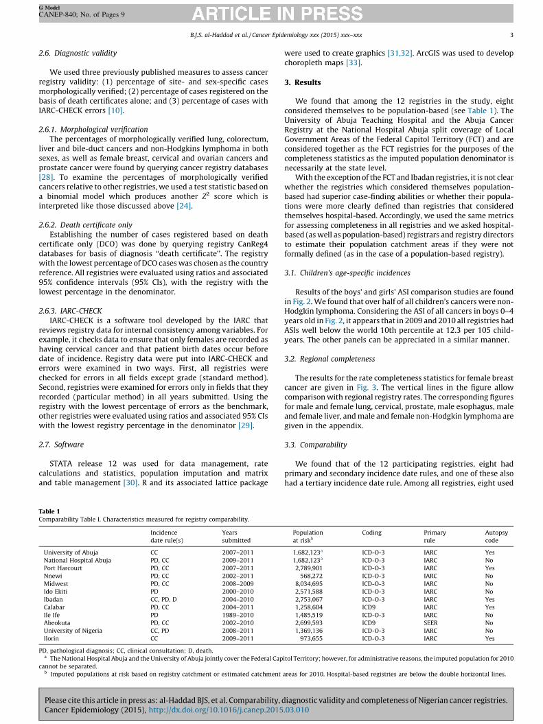
Table 1Comparability Table I. Characteristics measured for registry comparability.
Incidence
date rule(s)
Years
submitted
University of Abuja CC 2007–2011
National Hospital Abuja PD, CC 2009–2011
Port Harcourt PD, CC 2007–2011
Nnewi PD, CC 2002–2011
Midwest PD, CC 2008–2009
Ido Ekiti PD 2000–2010
Ibadan CC, PD, D 2004–2010
Calabar PD, CC 2004–2011
Ile Ife PD 1989–2010
Abeokuta PD, CC 2002–2010
University of Nigeria CC, PD 2008–2011
Ilorin CC 2009–2011
PD, pathological diagnosis; CC, clinical consultation; D, death.a The National Hospital Abuja and the University of Abuja jointly cover the Federal Cap
cannot be separated.b Imputed populations at risk based on registry catchment or estimated catchment
Please cite this article in press as: al-Haddad BJS, et al. Comparability, dCancer Epidemiology (2015), http://dx.doi.org/10.1016/j.canep.2015
were used to create graphics [31,32]. ArcGIS was used to developchoropleth maps [33].
3. Results
We found that among the 12 registries in the study, eightconsidered themselves to be population-based (see Table 1). TheUniversity of Abuja Teaching Hospital and the Abuja CancerRegistry at the National Hospital Abuja split coverage of LocalGovernment Areas of the Federal Capitol Territory (FCT) and areconsidered together as the FCT registries for the purposes of thecompleteness statistics as the imputed population denominator isnecessarily at the state level.
With the exception of the FCT and Ibadan registries, it is not clearwhether the registries which considered themselves population-based had superior case-finding abilities or whether their popula-tions were more clearly defined than registries that consideredthemselves hospital-based. Accordingly, we used the same metricsfor assessing completeness in all registries and we asked hospital-based (as well as population-based) registrars and registry directorsto estimate their population catchment areas if they were notformally defined (as in the case of a population-based registry).
3.1. Children’s age-specific incidences
Results of the boys’ and girls’ ASI comparison studies are foundin Fig. 2. We found that over half of all children’s cancers were non-Hodgkin lymphoma. Considering the ASI of all cancers in boys 0–4years old in Fig. 2, it appears that in 2009 and 2010 all registries hadASIs well below the world 10th percentile at 12.3 per 105 child-years. The other panels can be appreciated in a similar manner.
3.2. Regional completeness
The results for the rate completeness statistics for female breastcancer are given in Fig. 3. The vertical lines in the figure allowcomparison with regional registry rates. The corresponding figuresfor male and female lung, cervical, prostate, male esophagus, maleand female liver, and male and female non-Hodgkin lymphoma aregiven in the appendix.
3.3. Comparability
We found that of the 12 participating registries, eight hadprimary and secondary incidence date rules, and one of these alsohad a tertiary incidence date rule. Among all registries, eight used
Population
at riskb
Coding Primary
rule
Autopsy
code
1,682,123a ICD-O-3 IARC Yes
1,682,123a ICD-O-3 IARC No
2,789,901 ICD-O-3 IARC Yes
568,272 ICD-O-3 IARC No
8,034,695 ICD-O-3 IARC No
2,571,588 ICD-O-3 IARC No
2,753,067 ICD-O-3 IARC Yes
1,258,604 ICD9 IARC Yes
1,485,519 ICD-O-3 IARC No
2,699,593 ICD9 SEER No
1,369,136 ICD-O-3 IARC No
973,655 ICD-O-3 IARC Yes
itol Territory; however, for administrative reasons, the imputed population for 2010
areas for 2010. Hospital-based registries are below the double horizontal lines.
iagnostic validity and completeness of Nigerian cancer registries..03.010
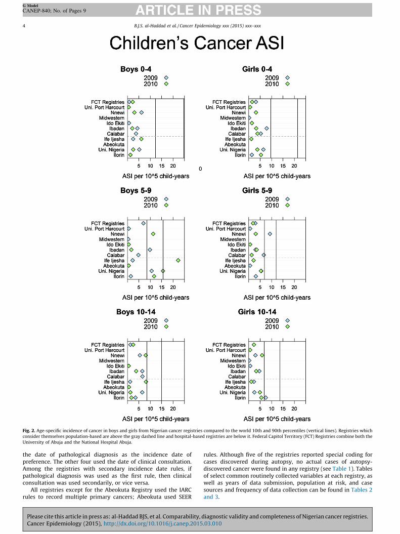
Fig. 2. Age-specific incidence of cancer in boys and girls from Nigerian cancer registries compared to the world 10th and 90th percentiles (vertical lines). Registries which
consider themselves population-based are above the gray dashed line and hospital-based registries are below it. Federal Capitol Territory (FCT) Registries combine both the
University of Abuja and the National Hospital Abuja.
B.J.S. al-Haddad et al. / Cancer Epidemiology xxx (2015) xxx–xxx4
G Model
CANEP-840; No. of Pages 9
the date of pathological diagnosis as the incidence date ofpreference. The other four used the date of clinical consultation.Among the registries with secondary incidence date rules, ifpathological diagnosis was used as the first rule, then clinicalconsultation was used secondarily, or vice versa.
All registries except for the Abeokuta Registry used the IARCrules to record multiple primary cancers; Abeokuta used SEER
Please cite this article in press as: al-Haddad BJS, et al. Comparability, dCancer Epidemiology (2015), http://dx.doi.org/10.1016/j.canep.2015
rules. Although five of the registries reported special coding forcases discovered during autopsy, no actual cases of autopsy-discovered cancer were found in any registry (see Table 1). Tablesof select common routinely collected variables at each registry, aswell as years of data submission, population at risk, and casesources and frequency of data collection can be found in Tables 2and 3.
iagnostic validity and completeness of Nigerian cancer registries..03.010
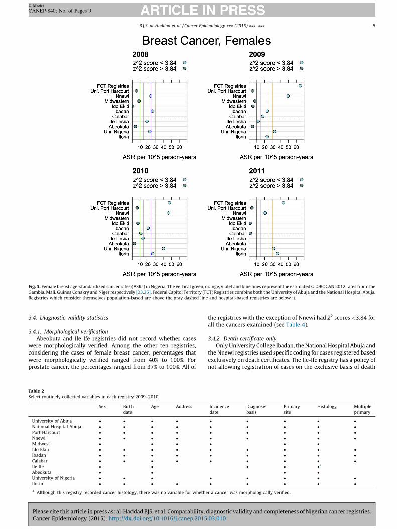
Fig. 3. Female breast age-standardized cancer rates (ASRs) in Nigeria. The vertical green, orange, violet and blue lines represent the estimated GLOBOCAN 2012 rates from The
Gambia, Mali, Guinea Conakry and Niger respectively [23,25]. Federal Capitol Territory (FCT) Registries combine both the University of Abuja and the National Hospital Abuja.
Registries which consider themselves population-based are above the gray dashed line and hospital-based registries are below it.
B.J.S. al-Haddad et al. / Cancer Epidemiology xxx (2015) xxx–xxx 5
G Model
CANEP-840; No. of Pages 9
3.4. Diagnostic validity statistics
3.4.1. Morphological verification
Abeokuta and Ile Ife registries did not record whether caseswere morphologically verified. Among the other ten registries,considering the cases of female breast cancer, percentages thatwere morphologically verified ranged from 40% to 100%. Forprostate cancer, the percentages ranged from 37% to 100%. All of
Table 2Select routinely collected variables in each registry 2009–2010.
Sex Birth
date
Age Address
University of Abuja � � � �
National Hospital Abuja � � � �
Port Harcourt � � � �
Nnewi � � � �
Midwest � � �
Ido Ekiti � � � �
Ibadan � � � �
Calabar � � � �
Ile Ife � �
Abeokuta � �
University of Nigeria � � �
Ilorin � � � �
a Although this registry recorded cancer histology, there was no variable for whethe
Please cite this article in press as: al-Haddad BJS, et al. Comparability, dCancer Epidemiology (2015), http://dx.doi.org/10.1016/j.canep.2015
the registries with the exception of Nnewi had Z2 scores <3.84 forall the cancers examined (see Table 4).
3.4.2. Death certificate only
Only University College Ibadan, the National Hospital Abuja andthe Nnewi registries used specific coding for cases registered basedexclusively on death certificates. The Ile-Ife registry has a policy ofnot allowing registration of cases on the exclusive basis of death
Incidence
date
Diagnosis
basis
Primary
site
Histology Multiple
primary
� � � � �� � � � �� � � � �� � � � �� � �� � � � �� � � � �� � � � �
� � �a
� �� � � � �� � � � �
r a cancer was morphologically verified.
iagnostic validity and completeness of Nigerian cancer registries..03.010
Table 3Comparability Table II.
Base Case finding Data sources Frequency
University of Abuja P Active University, Hospitals of catchment area LGAs 2� week
National Hospital Abuja P Active National Hospital, Asokoro district hospital, Garki Hospital, State House Clinic, Primus
Specialist Hospital
Weekly
Port Harcourt P Active University of Port Harcourt Teaching Hospital, private hospitals within Port Harcourt
and surrounding LGAs
Monthly
Nnewi P Active Nnamdi Azikiwe University Teaching Hospital, all hospitals and pathology diagnostic
centers in and around Nnewi
Daily
Midwest P Active University of Benin Teaching Hospital, Central Hospital – Benin City, Stella Obasanjo
Hospital, Ashama Foundation Pathology Laboratory, Central Hospital – Warri, FMC Asaba,
Central Hospital – Agbor, Central Hospital – Sapele, Central Hospital – Ughell
Weekly
Ido Ekiti P Active FMC Ido-Ekiti, Ekiti State University Teaching Hospital, almost all public and private
hospitals in Ekiti state
Weekly
Ibadan P Active 39 Government and Private Hospitals and Clinics in LGAs Monthly
Calabar P Active General Hospitals of LGAs, University of Calabar Weekly
Ile Ife H Active Obafemi Awolowo University Teaching Hospitals in Ife, Ilesha and Imesiile and the
Adventist Hospital Ile-Ife, some cases from private institutions
Weekly
Abeokuta H Active Pathology and Medical Records Departments of the FMC Abeokuta Monthly
University of Nigeria H Active Hospital pathology records, specialist clinics, wards and records department 4� week
Ilorin H Active Pathology laboratories, hospital wards, outpatient clinics and mortuary register Weekly
Notes: H: self considered hospital based; P: self considered population based; FMC: Federal Medical Center; LGA: local government area.
Table 4Percentage of cancers morphologically verified in participating Nigerian cancer registries.
National ^% UA %i NHA %i PH %i Nn %i MW %i IE %i Ib %i Cal %i UNTH %ib Il %i
b
Breast 78.9 100 93.3 100 40a 81.1 100 71.7 99.1 87.9 97.9
Cervix 79.5 100 91.3 94.5 29.5a 76.3 100 81.6 100 73.7 100
Ovary 74.8 100 87.5 100 26.5a 53.4 100 85.2 100 79.4 100
Prostate 76.8 100 94.3 100 36.6a 76.4 100 76.6 100 88 95.3
Liver F 55.6 83.3 100 100 40 26.1 100 25.3 72.7 90.9 88.9
Liver M 49.8 98.4 100 100 11.8 42.9 100 15.5 50 85 100
Lung F 73 100 71.4 – 0 50 100 62.5 100 100 100
Lung M 74.2 – 100 100 33.3 100 100 60 100 95.5 100
NHL F 82.3 – 100 100 – 78.3 100 73.8 75 90.9 100
NHL M 77.2 100 94.1 100 – 80 100 64 0 90.2 100
Colorectum F 79.7 100 100 100 33.3a 95.8 100 77.5 100 84.2 100
Colorectum M 73.4 100 92 100 21.5a 96.9 100 71.7 100 90.8 100
Cal: Calabar; Ib: Ibadan; IE: Ido Ekiti; Il: Ilorin; MW: Midwest; NHA: National Hospital Abuja; Nn: Nnewi PH: Port Harcourt; UA: University of Abuja; UNTH: University of
Nigeria Teaching Hospital.a Z2 scores >3.84.b Self-identified hospital-based registry.
Table 5Death certificate only cases in Nigerian cancer registries.
% Proportion ratio 95% CI
Ibadan 0.0307 1 –
National Hospital Abuja 0.229 7.455 1.67, 33.28
Nnewi 1.2 38.92 20.69, 73.23
Table 6Particular IARC check errors in Nigerian cancer registries.
% Proportion ratio 95% CI
University of Abuja 0.424 1.695 0.707, 4.062
National Hospital Abuja 3.28 13.14 6.638, 26.01
Port Harcourt 0.318 1.274 0.531, 3.054
Nnewi 0.63 2.519 1.227, 5.171
Midwest 0.685 2.738 1.352, 5.548
Ido Ekiti 0.25 1 .
Ibadan 0.423 1.691 0.856, 3.340
Ibadan Recoded 0.845 3.381 1.720, 6.649
Calabar 75.2 300.8 153.6, 589.0
Ile Ife 5.78 23.13 11.8, 45.32
Abeokuta 2.97 11.88 5.882, 24
University of Nigeria 0.43 1.721 0.842, 3.518
Ilorin 7.31 29.26 14.83, 57.71
B.J.S. al-Haddad et al. / Cancer Epidemiology xxx (2015) xxx–xxx6
G Model
CANEP-840; No. of Pages 9
Please cite this article in press as: al-Haddad BJS, et al. Comparability, dCancer Epidemiology (2015), http://dx.doi.org/10.1016/j.canep.2015
certificates without a post-mortem examination. This registry didnot have a variable for DCO cases. Ibadan had the lowestpercentage of these to total cases, and the National Hospital Abujaand Nnewi cancer registries had 7.455 (95% CI 1.670, 33.28) and38.92 (95% CI 20.69, 73.23) times higher DCO cases than Ibadanrespectively (see Table 5).
3.4.3. IARC-CHECK
The IARC-CHECK study was carried out in two ways. The first wasstandardized in that all registries were evaluated on the samecriteria regardless of whether or not a given registry collected everyitem used for evaluation. As such, registries not recording incidencedates for any cases would have errors with every case in this method.The results of the standard method are available on request.
The second method evaluated registries only on the data that theycollected. As such, the criteria used to measure the number of errorsvaries with each registry. These results are presented in Table 6.
4. Discussion
4.1. Children’s age-specific incidences
The comparisons of registry children’s ASIs to the world 10thand 90th percentiles can be thought as an estimate of the absolute
iagnostic validity and completeness of Nigerian cancer registries..03.010

B.J.S. al-Haddad et al. / Cancer Epidemiology xxx (2015) xxx–xxx 7
G Model
CANEP-840; No. of Pages 9
completeness of a registry [7,21,22]. Among the self-identifiedhospital-based registries, it is not surprising to see lower-than-expected children’s ASIs because these registries were not attemptingto capture all cases of cancer in their estimated catchment areas. Forthe population-based registries, there are many potential reasons forthe lower rates. It is possible, but unlikely, that rates in these registrieswere lower because there were fewer cases of childhood cancer inthese populations. It is more likely that the lower rates were due toinsufficient resources for case-seeking.
Many registries considered themselves population-based, buteither covered too large an area to catch all cancers within theirpopulation (Midwestern Registry) or had modest-sized catchmentareas but did not have the resources or infrastructure to seek out allcases within it (Calabar). These registries, with ASIs lower than theworld 10th percentile, likely did not capture all of the cases thatoccurred within their populations.
Only for boys 5–9 years old were several registries (Universityof Nigeria, Nnewi, Ilorin and Ibadan) between the global 10th and90th percentiles. It is possible that this is an effect of age-heapingwhere many parents of boys in this age range may state that theirboy is 5 years old while in fact he is younger. This would increasethe cases counted in the 5–9-year-old group. However, this is notseen among girls nor among boys 10–14 years old, making thisseem unlikely.
It is not clear why the 2010 rate for Ife Ijesha is so much higherthan the 90th global percentile.
We found that more than half of these children’s cancers werenon-Hodgkin’s lymphoma which were likely Burkitt’s lymphoma.Data from the Ibadan registry in 1999 showed very high age-standardized rates of Burkitt’s and other non-Hodgkin lymphomasat that time as well [21].
4.2. Regional completeness
Similarly to the findings for children’s cancers, the regionalcomparison completeness statistics showed evidence of generalincompleteness across multiple cancers (see supplementaryfigures in the appendix). Our average ASRs are, in general, lowerthan the estimated rates from GLOBOCAN as illustrated by thevertical lines in these figures and Z2 scores. The lower rates in ourstudy in comparison may suggest incompleteness. However, itshould be noted that some registries have rates that approach theGLOBOCAN rates and, more importantly, several registries haveconsistent rates over multiple years. In considering comparisonswith registries in the region, it appears that rates are comparablefor rarer cancers like female lung cancer and esophageal cancer inmales. It appears that rates are notably lower for liver cancer inboth males and females as well as for lung cancer in males. Thelower rates of liver cancer may reflect difficulty in diagnosing thesecancers, while the low rates of lung cancer could reflect genuinelylower rates due, in part, to the strong legislation in forceprohibiting smoking in public places as well as the ban on directand indirect tobacco marketing [34].
Registry completeness is a function of treatment-seeking on thepart of the patient- and case-seeking by the cancer registrypersonnel. While there may be very little that registry personnelcan do to increase treatment-seeking behavior and care access,completeness may be improved by changing how case addresses arerecorded. In particular, we found that recording case addresses usinglocal government areas (LGAs), Nigeria’s smallest census-basedgeographic unit, would facilitate more accurate rate calculations.
4.3. Comparability
The majority of Nigerian registries used the date of pathologicaldiagnosis as the preferred incidence date. The Ibadan cancer
Please cite this article in press as: al-Haddad BJS, et al. Comparability, dCancer Epidemiology (2015), http://dx.doi.org/10.1016/j.canep.2015
registry employed a third incidence rule where if neither the dateof clinical consultation nor the date of pathological diagnosis wereavailable, they then used the date of death. However, despite thehomogeneous training provided to almost all the registries, a thirdof registries preferred to use the date of clinical consultation. Theimplications of using one date over another are not clear; however,since for rate calculation purposes only the year is used, suchdifferences, even occurring on annual margins, would not beexpected to introduce an important magnitude of variation. Theclinical consultations will almost always precede the pathologicaldiagnosis except in cases where the cancer is discoveredposthumously [35–38].
All registries, with the exception of Abeokuta, used IARC rulesfor multiple primary cancers. Abeokuta used the SEER rules. Thereis evidence that the use of the SEER multiple primary rules resultsin higher ASRs than those registered using the IARC/IACR multipleprimary rule [39]. Our findings did not confirm this (resultsavailable on request).
Lastly, although some registries had specific coding for autopsy-discovered cancers, no actual cases were recorded. This may be duein large part to the relative rarity of autopsies in some parts ofNigeria because of taboo [40]. Despite these considerations, itappears that data across the country is relatively comparable forthe majority of studied comparability characteristics.
4.4. Diagnostic validity
4.4.1. Morphologically verified
Among the ten registries, only the Nnewi registry showedevidence of low morphological verification for multiple cancers(see Table 4). Excluding this registry, these results lend confidenceto an essential component of diagnostic validity in Nigerian cancerregistries. While high levels of morphological verification areimportant from the perspective of validity, very high levels mayindicate that a population-based registry is missing casesdiagnosed clinically and therefore is incomplete [10].
In general, neither cancer-specific nor all-cancer measures ofmorphological verification are only infrequently reported in theliterature. See references [41–44] for exceptions. This is becausethese measures require a regional or national context to beinterpretable. In West Africa, the registry in Abidjan between 1995and 1997 has been reported to have had an overall rate ofmorphological verification of around 80% for all sites combined inmales and females [45]. At the lower end of the spectrum, thenational registry in The Gambia between 1995 and 1999 had ratesof around 18% and 30% for males and females respectively [45].These numbers increased to around 39% for each sex after theexclusion of liver cancers.
In Nigeria, in our sample of registries, 79% of breast cancerswere morphologically verified, but only 50–56% of liver cancerswere morphologically verified. These latter statistics may be due tolate presentation of the malignancy resulting in poor surgicalcandidacy for biopsy and treatment.
4.4.2. Death certificate only
The DCO index has, in the past, been used as an indicator ofcompleteness in population-based registries. In Nigeria, because ofthe heterogeneity of autopsy practice, many undiagnosed cancersleading to death are likely never discovered. It is therefore notsurprising that only three registries had cases coded as registeredfrom DCO. In all three, the percentage of DCO cases ranged from0.031% to 1.2%. An unfavorable interpretation of this finding wouldbe that registrars are missing many cancer mortalities and theopportunity to record these important mortality statistics. On theother hand, this could be interpreted favorably in the sense that itdoes not appear that registries are heavily relying on cause of death
iagnostic validity and completeness of Nigerian cancer registries..03.010

B.J.S. al-Haddad et al. / Cancer Epidemiology xxx (2015) xxx–xxx8
G Model
CANEP-840; No. of Pages 9
from death certificates to populate their registries. Evaluation ofthis particular method of validity is contentious because of widevariation in the use of death certificates, vital statistics, quality ofcause of death statements, and other reasons [46]. The mostfavored approach seems to be comparison to other registries in theregion [24,41]. Although the DCO percentage is not commonlyreported in the literature, the national registry in The Gambiareports DCO percentages of 6.6% and 3.6% for males and femalesrespectively [45].
4.4.3. IARC-CHECK
The registry with the lowest number of IARC-CHECK errors wasIdo Ekiti with only 0.25% of cases with an error. Calabar’s data hadthe most cases with errors; however, this was likely due to havingsubmitted multiple years before adoption of CanReg4 for case datamanagement. Many of the errors would have presumably beendetected and rectified if CanReg4 had been used rather thanMicrosoft Excel. However, there are other registries (Abeokuta andMidwest) who also did not use CanReg4 and still had low errorpercentages.
Importantly, case data from Abeokuta, Ile Ife and Midwest wererecoded by a professional cancer registrar in the United States. Theregistrar also coded a version of the Ibadan data with the ICD-O-3codes removed. Since the percentage of errors was higher in therecoded Ibadan version than the original version, it appears thatsome error was likely introduced into the coding of Abeokuta, IleIfe and Midwest data.
4.5. Conclusions
We found consistently lower-than-expected children’s ASIs inalmost all registries, which likely reflects inability to capture all thecases in catchment areas. We found that over half of these caseswere non-Hodgkin lymphoma.
Considering the regional completeness comparisons, we foundseveral estimations that were consistent across years, but alsodiscovered evidence of cancer-specific general incompleteness aswell as what may be incompleteness due to inability to diagnosespecific cancers. Among the population-based registries withrigorous case-finding procedures and resources, consistent esti-mates across years for particular cancers suggested completenessfor specific cancers. Rates for male esophageal cancer and femalebreast cancer were comparable with other registries in the region,while rates for liver cancer in men and women were lower thanthose of other registries in the region.
Standardizing patient addresses to only the smallest census-based geographic unit of residence in the year before seekingtreatment is an essential next step toward improving cancerregistry completeness in Nigeria. Similarly, regular reviews ofregistry case-finding procedures would likely help to improvecompleteness. The comparability metrics examined demonstratedthat Nigerian registries are relatively comparable to each other andto international standards. Although one registry used SEER rulesfor multiple primaries, the majority used rules from the IARC.Differences in incidence date rules, while not desirable from auniformity standpoint, may be unlikely to lead to severeestimation differences. Few registries had special coding forautopsy-detected cases or DCO cases.
With the exception of the cancer registry at Nnewi, all thecancer registries had high rates of morphologic verification for themost common cancers, including breast, cervix and prostatecancers. The percentage of morphological verification was lowestfor male liver cancer. Although seven of the participating cancerregistries considered themselves to be population-based, only four(Ibadan, National Hospital Abuja, University of Abuja and Calabar)are considered to have the experience, personnel and resources
Please cite this article in press as: al-Haddad BJS, et al. Comparability, dCancer Epidemiology (2015), http://dx.doi.org/10.1016/j.canep.2015
necessary to carry out case-finding with the rigor expected of apopulation-based registry.
4.6. Necessary assumptions and limitations
This work and these conclusions are based on severalassumptions. First, there is the problem of defined populationand catchment area. Second, while some registries had standard-ized methods for recording the address of a given case, mostregistries did not, leading to the assumption that all casesregistered should fall into the associated catchment area. Evenamong Nigerian registries with defined populations, the regularand complete ascertainment of all cases is an ongoing challengemade more difficult by insufficient resources, personnel, geogra-phy and strife.
Authorship contribution
al-Haddad, Jedy-Agba, Oga, Adebamowo involved in studyconception and design.
al-Haddad, Jedy-Agba, Oga, Adebamowo, Ezeome, Obiorah,Okobia, Ogunbiyi, Ukah, Omonisi, Nwofor, Igbinoba involved inacquisition of data, analysis and interpretation of data, drafting ofmanuscript and critical revision.
Acknowledgments
This work would not have been possible without the assistance ofDr Patience Osinubi at the Federal Ministry of Health in securingpermissions. We would also like to thank Dr Titilola Akinremi,Professor Ima-Obong A. Ekanem, Dr Theresa Ize Otu, Dr OlagokeErinomo, Professor Babatunde J Olasode, Dr E.A.O Afolayan, Mrs AgnesO. Fabowale, CCR, Mr B.B. Etim-Okon, Mr Henry Kumai, Miss GloriaOsubor, Ms Ovo Agofure, Adedeji Adeyanju, Mr Ojo Adebayo, MrsElizabeth Udeh, Mr O. Olajide, Dr R. Saidu, Miss Gift Oviemuno, Mr AkinLadipo, Mr Joel Bassey, Mr Jerome Seyil, Mr Ubasi Agada Batholom, MrIlugbemi Segun, Mr Kehinde Oladele, Miss Ann Okorafor, Mr G.T.Ayilara, Ms G.C. Oyeka and Mr Dick Benson. We also benefited from theadvice and guidance of Drs. Logan Spector, J. Michael Oakes, AlvaroAlonso, Richard MacLehouse and Francis Harvey.
This work was supported by the University of MinnesotaMedical Scientist Training Program (NIH MSTP grant T32GM008244), an Interdisciplinary Doctoral Fellowship from theMinnesota Population Center and the Graduate School, the HawleyAward and the Wilfred Wetzel Fellowship. Drs Adebamowo, Jedy-Agba and Oga were supported by the IHV-UM Capacity Develop-ment for Research into AIDS Associated Malignancies (NIH/NCID43CA153792-01).
Appendix A. Supplementary data
Supplementary data associated with this article can be found, in
the online version, at http://dx.doi.org/10.1016/j.canep.2015.03.010.
References
[1] Falola T, Heaton MM. A history of Nigeria. New York: Cambridge UniversityPress, 2008.
[2] Parkin DM, Ferlay J, Hamdi-Cherif M, Sitas F, Thomas JO, Wabinga H, et al.Cancer in Africa: epidemiology and prevention; 2003.
[3] Doll R, Payne P, Waterhouse JAH. Cancer incidence in five continents; 1966,http://ci5.iarc.fr/CI5i-ix/ci5i-ix.htm.
[4] Doll R, Muir CS, Waterhouse JAH. Cancer incidence in five continents; 1970,http://ci5.iarc.fr/CI5i-ix/ci5i-ix.htm.
[5] Waterhouse J, Muir CS, Correa P, Powell J. Cancer incidence in five continents;1976, http://ci5.iarc.fr/CI5i-ix/ci5i-ix.htm.
[6] IACR. IACR members – Africa, Tech. rep.; 2011, http://www.iacr.com.fr/Africact oct09.pdf.
iagnostic validity and completeness of Nigerian cancer registries..03.010
B.J.S. al-Haddad et al. / Cancer Epidemiology xxx (2015) xxx–xxx 9
G Model
CANEP-840; No. of Pages 9
[7] Parkin DM, Bray F. Evaluation of data quality in the cancer registry: principlesand methods. Part II. Completeness. Eur J Cancer 2009;45(5):756–64, http://www.ncbi.nlm.nih.gov/pubmed/19128954.
[8] Curado MP, Voti L, Sortino-Rachou AM. Cancer registration data and qualityindicators in low and middle income countries: their interpretation and potentialuse for the improvement of cancer care. Cancer Causes Control 2009;20:751–6.
[9] Parkin DM, Wabinga H, Nambooze S. Completeness in an African cancerregistry. Cancer Causes Control (CCC) 2001;12(2):147–52, http://www.ncbi.nlm.nih.gov/pubmed/11246843.
[10] Bray F, Parkin DM. Evaluation of data quality in the cancer registry: principlesand methods. Part I. Comparability, validity and timeliness. Eur J Cancer2009;45(5):747–55. http://dx.doi.org/10.1016/j.ejca.2008.11.032, http://www.ncbi.nlm.nih.gov/pubmed/19117750.
[11] N. Institute of Human Virology. About Institute of Human Virology, Nigeria;2009, http://ihvnigeria.org/ihvnweb/webnew/index.php/about-ihvn/introduction.html.
[12] Federal Ministry of Health. Federal Ministry of Health; 2012, http://www.fmh.gov.ng/.
[13] Regents of the University of Minnesota. Institutional Review Board: HRPP:OVPR: University of Minnesota; 2012, http://www.irb.umn.edu/.
[14] Federal Ministry of Health: Department of Health Planning and Research.National Code of Health Research Ethics Committee of Nigeria (NHREC); 2007,http://www.nhrec.net.
[15] Fritz A, Percy C, Jack A, Shanmugaratnam K, Sobin L, Parkin DM, Whelan S, eds.International classification of diseases for oncology. 3rd ed., Lyon, France:International Agency for Research on Cancer, 2000.
[16] Nationa Bureau of Statistics. National Bureau of Statistics; 2012, http://www.nigerianstat.gov.ng/.
[17] National Population Commission of Nigeria. 2006 Census Priority Tables 1–15;2011, http://www.population.gov.ng/.
[18] Global Administrative Areas. http://www.gadm.org/about.[19] Siegel JS, Swanson DA. The methods and materials of demography. 2nd ed. San
Diego: Elsevier, 2004.[20] Bray F. Age standardization. In: Parkin DM, Whelan SL, Ferlay J, Teppo L,
Thomas DB, eds. Cancer incidence in five continents, vol. VIII. Lyon, France:IARC Scientific Publications, 2002: 87–9.
[21] Stiller CA, Parkin DM. Geographic and ethnic variations in the incidence ofchildhood cancer. Br Med Bull 1996;52(2):682–703.
[22] Parkin DM, Whelan SL, Ferlay J, Teppo L, Thomas DB. Cancer incidence in fivecontinents; 2002.
[23] Parkin DM, Plummer M. Comparability and quality of data. In: Parkin DM,Whelan SL, Ferlay J, Teppo L, Thomas DB, eds. Cancer incidence in fivecontinents, vol. VIII. Lyon: IARC, 2002: 57–73.
[24] Bray F, Parkin DM. Evaluation of data quality in cancer registry: principles andmethods.Part I.Comparability,validityandtimeliness.EurJCancer2009;45:747–55.
[25] Ferlay J, Soerjomataram I, Ervik M, Dikshit R, Eser S, Mathers C, et al. GLO-BOCAN 2012 v1.0. Cancer Incidence and Mortality Worldwide: IARC Cancer-Base No. 11 [Internet]; 2013, http://globocan.iarc.fr.
Please cite this article in press as: al-Haddad BJS, et al. Comparability, dCancer Epidemiology (2015), http://dx.doi.org/10.1016/j.canep.2015
[26] Schouten L, Jager J, van den Brandt P. Quality of cancer registry data: acomparison of data provided by clinicians with those of registration personnel.Br J Cancer 1993;68(5):974–7.
[27] Johnson C, Adamo M, eds. SEER Program Coding and Staging Manual 2007, no.07. National Institutes of Health; 2008.
[28] Ferlay J, Shin HR, Bray F, Forman D, Mathers C, Parkin DM. GLOBOCAN 2008:cancer incidence and mortality worldwide; 2010, http://globocan.iarc.fr.
[29] Ferlay J, Burkhard C, Whelan S, Parkin DM. Check, conversion programs forcancer registries (IARC/IACR tools for cancer registries). Lyon, France: Tech.Rep., 2005, http://www.iacr.com.fr/TechRep42-MPrules.pdf.
[30] StataCorp.. Stata statistical software: release 12; 2011.[31] R Core Team. R: a language and environment for statistical computing; 2012,
http://www.r-project.org/.[32] Sarkar D. Lattice: multivariate data visualization with R. New York: Springer,
2008, http://lmdvr.r-forge.r-project.org.[33] ESRI. ArcGIS desktop: release 10; 2011.[34] Federal Government of Nigeria. National Tobacco Control Bill; 2009.[35] Adamo M, Johnson C, Ruhl J, Dickie L, eds. SEER Program Coding and Staging
Manual. Bethesda, MD: National Cancer Institute, 2010 . seer.cancer.gov/manuals/2010/SPCSM2010maindoc.pdf.
[36] IACR. IARC cancer incidence in five continents volume X: call for data; 2011,www.iacr.com.fr/CI5/CI5X/call_for_data-EN.pdf.
[37] Jensen O, Parkin D, MacLennan R, Muir C, Skeet R, eds. Cancer registration:principles and methods. Lyon, France: IARC Scientific Publications, 1991 ,www.iarc.fr/en/publications/pdfs-online/epi/sp95/SP95.pdf.
[38] European Network of Cancer Registries. Recommendations for coding inci-dence date; 1999, www.encr.com.fr/incideng.pdf.
[39] Hotes JL, Ellison LF, Howe HL, Friesen I, Kohler B. Variation in breast cancercounts using SEER and IARC multiple primary coding rules. Cancer CausesControl 2004;15(2):185–91.
[40] Oluwasola O, Fawole O, Otegbayo A, Ogun G, Adebamowo C, Bamigboye A. Theautopsy: knowledge, attitude, and perceptions of doctors and relatives of thedeceased. Arch Pathol Lab Med 2009;133(1):78–82.
[41] Chen W, Zheng R, Zhang S, Li N, Zhao P, Li G, et al. Report of incidence andmortality in china (sic) cancer registries, 2008. Chin J Cancer Res2012;24(3):171–80.
[42] Tumino R, Ferretti S. Quality and completeness indices. Epidemiol Prev2004;28(2 (Suppl.)):17–21.
[43] Stang A, Parkin D, Ferlay J, Jockel K. International uveal melanoma incidencetrends in view of a decreasing proportion of morphological verification. Int JCancer 2005;114(1):114–23.
[44] Enerly E, Bray F, Mellem C, Hansen BT, Kj_ lberg G, Dahl1 T, et al. Qualityassessment of the registration of vulvar and vaginal premalignant lesions atthe Cancer Registry of Norway. Acta Oncol 2012;51(1):45–50.
[45] Shimakawa Y, Bah E, Wild CP, Hall AJ. Evaluation of data quality at the GambiaNational Cancer Registry. Int J Cancer 2012;132(3):658–65.
[46] Brenner H. Limitations of the death certificate only index as a measure ofincompleteness of cancer registration. Br J Cancer 1996;74(7):1155.
iagnostic validity and completeness of Nigerian cancer registries..03.010
Copyright © 2022 FDOKUMEN


![Successful weight loss maintenance in Portugal and in the USA: comparing results from two National Registries [portuguese]](https://static.fdokumen.com/doc/165x107/63311274f4e961c184054d3d/successful-weight-loss-maintenance-in-portugal-and-in-the-usa-comparing-results.jpg)