Combining a Solution-Phase Derived Library with In-Situ Cellular Bioassay: Prompt Screening of...
21
1 Combining a solution-phase derived library with in-situ cellular bioassay: Prompt screening of amide-forming minilibraries using MTT assay Li-Wu Chiang, 1 Kai Pei, 1 Shao-Wei Chen, 1 Ho-Lien Huang, 1 Kun-Ju Lin, 2 Tzu-Chen Yen, 2 and Chung-Shan Yu 1,3* 1 Department of Biomedical Engineering and Environmental Sciences, National Tsing-Hua University Hsinchu, Taiwan 2 Department of Nuclear Medicine, Chang-Gung Memorial Hospital, Taiwan 3 Institute of Nuclear Engineering and Science, National Tsing-Hua University Hsinchu 300, Taiwan Fax: (+886)3-5718649 Tel: (+886)3-5751922 E-mail: [email protected] Synthesis, bioactivity and molecular docking of old compounds
Transcript of Combining a Solution-Phase Derived Library with In-Situ Cellular Bioassay: Prompt Screening of...

1
Combining a solution-phase derived library with in-situ
cellular bioassay: Prompt screening of amide-forming
minilibraries using MTT assay
Li-Wu Chiang,1 Kai Pei,
1Shao-Wei Chen,
1 Ho-Lien Huang,
1 Kun-Ju Lin,
2 Tzu-Chen Yen,
2 and Chung-Shan
Yu1,3*
1Department of Biomedical Engineering and Environmental Sciences, National Tsing-Hua University
Hsinchu, Taiwan
2Department of Nuclear Medicine, Chang-Gung Memorial Hospital, Taiwan
3Institute of Nuclear Engineering and Science, National Tsing-Hua University Hsinchu
300, Taiwan
Fax: (+886)3-5718649
Tel: (+886)3-5751922
E-mail: [email protected]
Synthesis, bioactivity and molecular docking of old compounds
2
Abstract
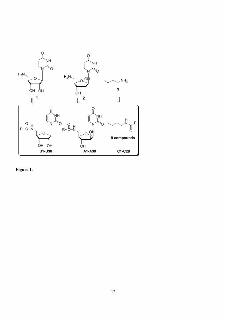
We constructed a mini library using a solution-phase synthesis through coupling of three core amino
compounds (5'-amino-5'-deoxy uridine, 5'-amino-2',5'-di-deoxy arabinosyl uridine, and butan-1-amine) with
30 carboxylic acids via amide bond formation. The simplified structural core compound butan-1-amine was
selectively coupled with 9 carboxylic acids as control. MTT assay of the crude mixtures showed that
analogues derived from fenbufen, butylfenbufen C15; ethacrynic acid, butyl ethacrynic amide C18; and
sphingosines, Sph-1, Sph-2 and U27 had an increased cytotoxicity against MCF-7 cells as well as A549
cells. Structural elucidation with molecular docking suggested that cytotoxicity of these compounds is
mainly due to the inhibition of enzymes regulating cellular apoptosis.
Keywords: amide, assay, A549, in-situ, library, MCF7, molecular docking, solution phase
3
There has been growing interest in a method combining a solution-phase derived library with an in situ
bioassay on microtiter plate.1 Construction of the library is initiated through a core compound, either as a
lead of natural product or a transition-state analog from mechanistic considerations, followed by coupling
with various carboxylic acids as building blocks. The libraries were constructed on a microtiter plate or a set
of centrifuge tubes. In each well or tube, the products obtained were screened for their binding affinity for
the enzyme of interest. The bioactivity derived from the mixture, in general, is consistent with that of the
purified product. Indeed, a number of potential substrates for numerous enzymes including sulfotransferase,2
fucosidase,3 fucosyltransferase,4 protease,5 and protease dimerization, have been discovered by this
approach.6 Since this method is mainly focused on enzymatic assays, a further application in cellular assays
may be of importance. In this article, we sought to couple the amide-forming libraries with a cell-line based
assay. In addition, we elucidated the mechanisms related to bioactivity of potential amides through molecular
docking.
As part of our ongoing research, we are focusing on the development of nucleoside analogs to be used
as prodrugs to target herpes simplex virus thymidine kinase gene.7-9 Thus, 5'-amino-5'-deoxy analogues of
pyrimidine nucleosides set as a core compound. To clarify whether the bioactivity is mainly associated with
the core 5'-amino nucleoside compounds, a structurally simplified butan-1-amine was used as control for
comparison purposes. In addition to the commercially available butan-1-amine, the preparation of these
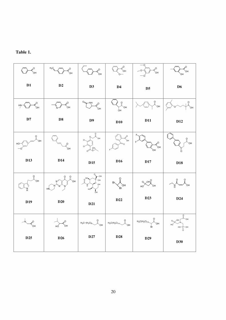
nucleoside analogs has been reported.10 In this study, carboxylic acid moieties were structurally classified in
four types as follows: mono-aromatic ring (1-15), di-aromatic rings (16-18), fused rings (19-20), and
aliphatic groups containing heteroatoms such as phosphor and aza acids 21-30 (Table 1).
The coupling product in each well was diluted before transfering to the plate. Thus the appropriate
working concentration could be determined since the cellular survival ratio of most compounds acting as
negative control is greater than 80%. The method is illustrated in Fig. 2.

4
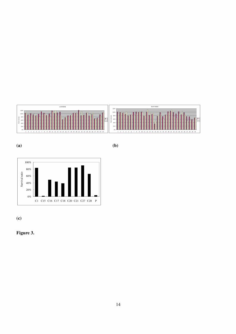
As shown by crude assay results (Fig. 3(a)-(c)), the cellular survival ratio for the majority of the
products was over 60%, suggesting the absence of cytotoxicity. On the other hand, the coupling products
U27, A15, C15, and C18 exhibited biological activity. These potential compounds were further prepared and
purified on flash chromatography to be submitted for a “clean” analysis. In contrast, we disregarded the
evaluation of U15 due to the higher activity of its 2′-epimer A15 against both A549 and MCF7 cell lines.
Unexpectedly, a subtle cytotoxicity of U27 was evident (Fig. 3(b) and supporting information: S16,
S17). As a control, a referential core compound: butan-1-amine was coupled with the corresponding acid
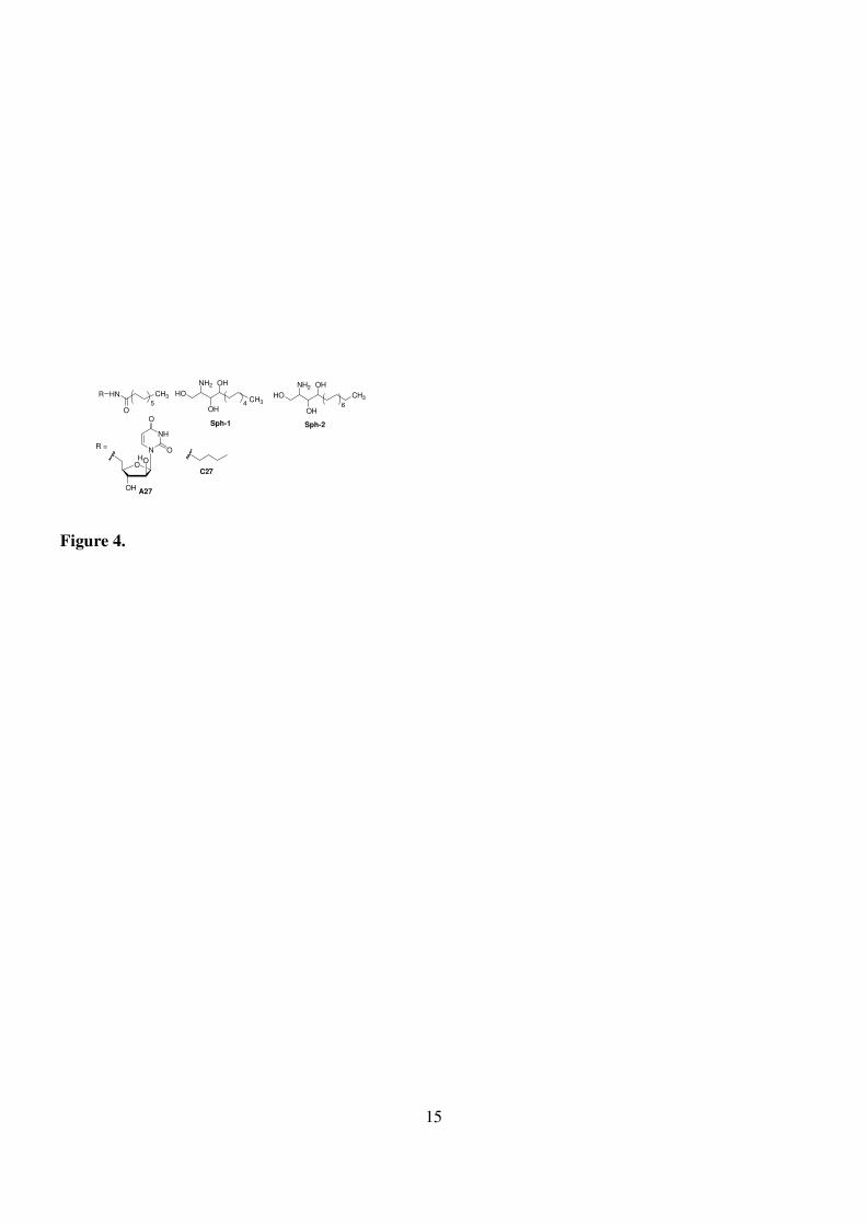
moieties to provide C27 (Fig. 3(b)). Since U27 shares structural similarities to sphingosine, the sphingosine
analogs Sph-1 and Sph-2 (carbon length of 13 and 18, respectively), were prepared for comparison purposes
(Fig. 4). While the bioactivity is mainly due to both the lipid part and the hydrophilic head, the hydrophilic
moieties of sphingosine are more effective in inducing cytotoxicity. The hydrophilic moieties of sphingosine
are capable to induce specific proapoptotic signals that may account for cytotoxicity.11-14
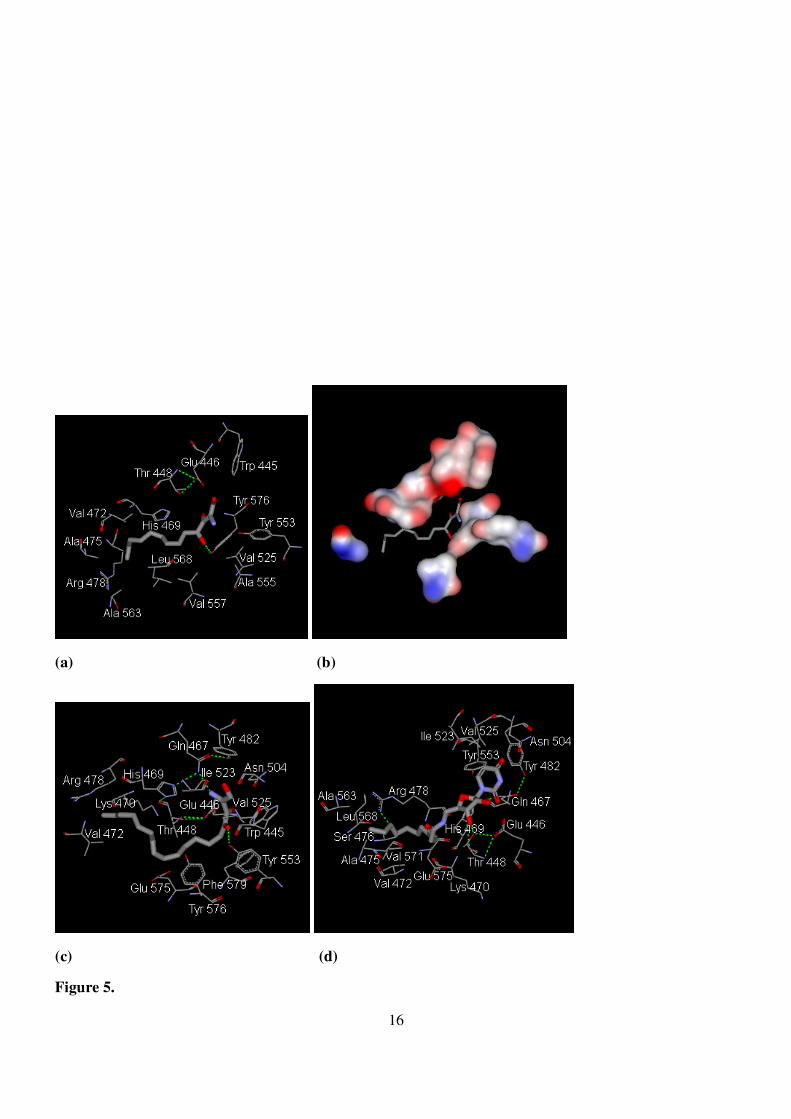
In spite of the few data available on the membrane receptor and X-ray diffraction data, an attempt to
clarify the probable binding mechanisms through molecular docking was performed. The START, known as
a domain of steroidogenic acute regulatory protein-related lipid transfer (StARD10), has been reported to be
overexpressed in breast cancer.15 In addition, the START domain of ceramide transport (CERT) protein is
known for its transferring ability of natural D-erythro ceramides, mainly used for transporting from ER to
Golgi apparatus.16 The two START domains have similar characteristics and consist of 210 and 250 amino
acids, respectively. The cavity of CERT START domain comprises a line of hydrophobic and polar charged
heads. According to the crystal structure of the complex formed by CERT domain of 2e3n with ceramides
having acyl groups of various lengths, the hydrogen bond formed by the hydrophilic head of the substrate
within the deep active site plays a critical role for the biological activity.16 Notably, the OH group at C-1
forming a hydrogen bond with the guanidine group of arginine-442 is crucial. Our docking results (Fig. 5(a)-
(c)) suggested a similar interaction formed among numerous amino acid residues including hydrophilic and
hydrophobic contacts. Interestingly, the 4-OH group forms a hydrogen bond to OH of tyrosine-576 closing

5
to the tyrosine-553, which is responsible for the hydrogen bond to the amide group of natural ceramide.
Impressively, in the case of Sph-2, the 2-NH2 and 4-OH groups form hydrogen bonds to glutamine-467 and
tyrosine-553, respectively (Fig. 5(c)). Similarly, although the 3′-OH group of U27 forms a hydrogen bond to
the OH of threonine-448, the cavity is still able to accommodate the pyrimidine base ring (Fig. 5(d)). Our
results suggest that the START domain is the site potentially responsible for mediating apoptosis through
these lipid analogs.
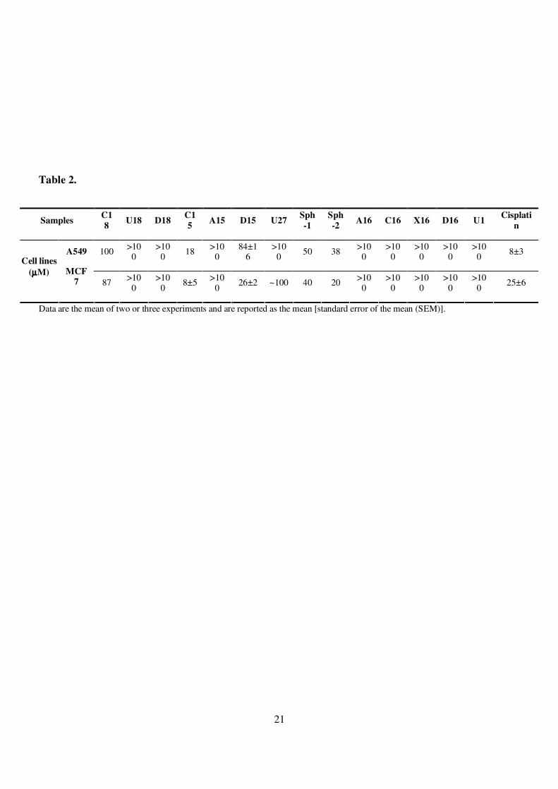
During the crude assay of C-series compounds, an unexpected bioactivity of C18 against both A549 and
MCF7 was evident (Fig. S16 and S17 in S.I., Table 2). Acid moieties D18 also known as fenbufen is a
member of the non-steroid anti-inflammatory drugs (NSAIDs). As an inhibitor targeting cyclooxygenase,
fenbufen is effective in rheumatoid arthritis and osteoarthritis.17 Although NSAIDs such as diclofenac may
inhibit tumor growth in vitro (IC50: 360 µM)18 and in vivo,19 there was no significant improvement in
cytotoxicity through modification of other NSAIDs, for instance fenoprofen.20 Therefore, fenbufen (D18) is
an interesting probe for studying the relationship between these two diseases through structural modification.
Furthermore, C18 is a potential hit and may act as a starter for further screening of a new hit compound or
even a lead compound. The potential enzyme targeted by C18 has been suggested to be cyclooxygenase.21
COX-2 is an inducible cyclooxygenase isoform that plays a major role in inflammation and has been found
to be hyperexpressed in several human tumors.22-24 COX-2 overexpression is involved in cancer growth and
invasion.25 Various NSAIDS have been studied as anticancer drugs. Since the bioactive flurbiprofen mimics
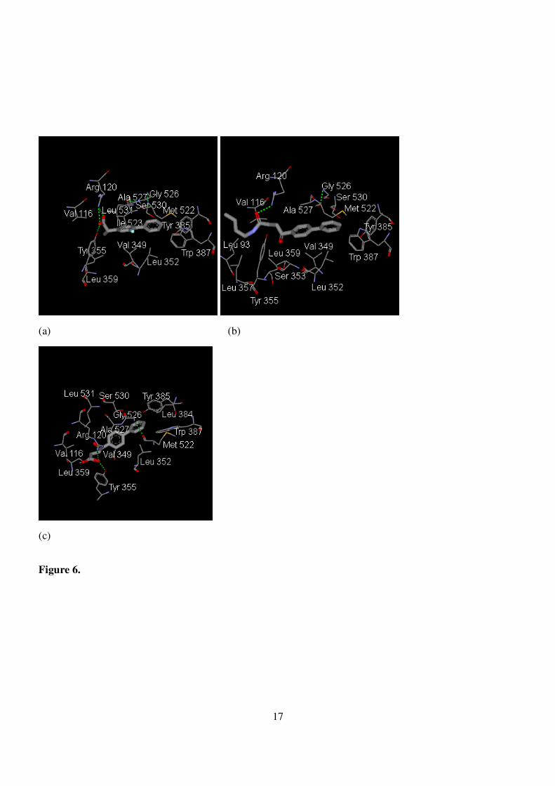
our butylfenbufen, the 3-D crystal structure of the complex formed by prostaglandin H2 synthase-1 from
goat with flurbiprofen was chosen as model (Fig. 6(a)). When examining the inner structure formed by
flurbiprofen within the active site, butyl fenbufen - rather than fenbufen - shared a similar environment (Fig.
6(b) and 6(c)). This is likely to be ascribed to the hydrogen bond formed between the oxo group and the
terminal amino group of arginine-120 and the van der Waals contact formed between butyl group and Val
116 as well as Leu93.
Instead of a hydrogen bond formed between the oxo group and arginine-120 as for butyl fenbufen,
6
fenbufen formed a hydrogen bond between the oxo group and tyrosine-355. The slightly incline-to-the-left
structure weakened the nonpolar contact of the biphenyl ring with the deep pocket of active site, thereby
diminishing its activity. Nevertheless, in spite of the success in the elucidation of the structure-activity
relationship through molecular-docking, the apoptotic mechanism in terms of the block of COX-2 might not
fully account for the subtle cytotoxicity of butylfenbufen. Accordingly, targeting cyclooxygenase may be not
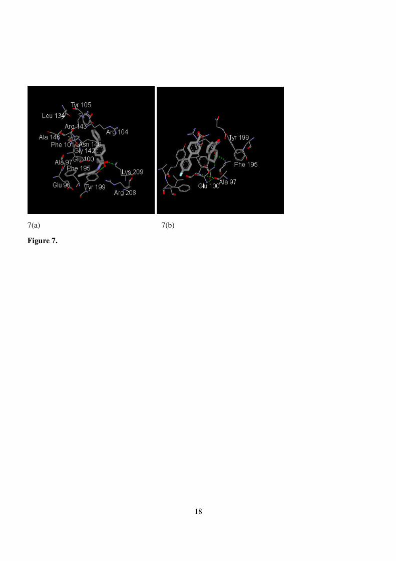
the sole antiproliferative mechanism of NSAIDs.22 We therefore examined the PDB bank to find the related
report regarding the probable human-derived enzyme responsible for the proapoptotic activity. The screening
results obtained by confining to the substructure of diphenyl rings indicated that proteins of the Bcl-x or Bcl-
2 families overexpressed in many cancers fit these criteria.26 However, a contradictory docking result for
butylfenbufen was obtained (Fig. 7(a)). In contrast to the report of the extension of the biphenyl ring of 4′-
fluoro-1,1′-biphenyl-4-carboxylic acid (4-FC) into the deep pocket of the active site (Fig. 7(b)), an inverted
binding of the butylfenbufen was evident. Furthermore, most of the binding energy of this model is resulting
from the coverage of the biphenyl group on the protein surface, a result that is not consistent with 4-FC′s.
The proapoptotic activity of butylfenbufen is thus unlikely to occur via inhibition of Bcl-x proteins.
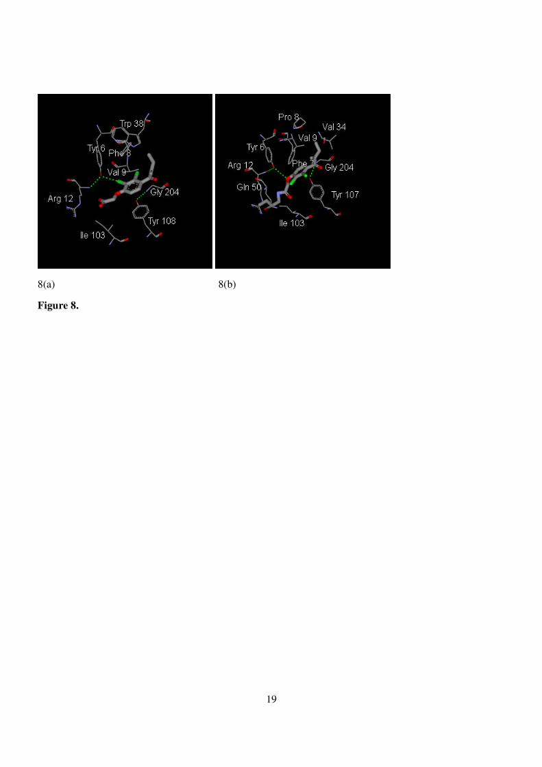
Acid moieties D15, known as ethacrynic acid, are classified as a group of diuretics that can block
Na+-K+-2Cl- symporter in the thick ascending limb of the loop of Henle. They are generally used in patients
with acute pulmonary edema.17 Furthermore, D15 is an inhibitor of betaglutathiontransferase (BGTT) whose
upregulation has been associated with drug resistance during chemotherapy of various cancers.27,28 Further
analysis of the binding pattern between C15 and BGTT was performed by using the DS program according
to 3D-data of 2gss from the PDB bank (Fig. 8(a)).29 Compared to ethacrynic acid, the extra butyl group
extends into the deep pocket, an unidentified extra site freely available for extra stabilization (Fig. 8(b)).
Furthermore, both residues of tyrosine-107 and phenyl alanine-7 near the entrance of the pocket provided a
primary stabilization for the benzene ring of C15 through a pi-pi stackering, a mimic of a sandwich complex.
The terminal amido group in the glutamine-51 residue may provide indirect polar contact with the amido
group of butyl ethacrynic amide. Interestingly, despite the presence of the butyl group, there is a loose space

7
surrounded by the residues of glutamine-64, serine-65 (not specified in Fig. 22) and the terminal butyl group
of butyl ethacrynic amide. This may provide a base for future modifications of the butyl group in order to
develop potential inhibitors.
Interestingly, a recent study has shown that a butyl ester derivative of ethacrynic acid may induce
apoptosis in leukemia cells.30 Our approach may thus provides a new avenue to explore new potential
compounds based on the concept of “old drug new use”.
In conclusion, we have described a simple method for screening an in vitro solution-derived library
using the MTT assay at the cellular level. The probe screening of this mini-library (comprising of 69
compounds derived from three core amines) was helpful in the quest of novel hit compounds such as C18. It
also shows enough sensitivity to detect bioactive compounds based on a relative survival ratio of > 60% for
controls. The hit compound C15 may be further modified as a potential hit compound through a series of
HTS. Investigation of the mechanism responsible for the inhibition of proliferation of cancer cells was
performed by molecular docking using the DS program. The cytotoxicity of U27, C18 and C15 discovered
through the combinatorial approach is likely to occur mainly via binding-mediated inhibition of apoptosis-
regulating enzymes.
Methods
The reagents used in the amide bond formation was core amine (1 mg, 4 µmol), carboxylic acid (1 eq),
DIEA (1.2 eq) and HBTU (1.1 eq). Both the starting core amine and the carboxylic acids were prepared as a
stock solution of DMSO at a concentration of 0.1 mmol/200 µL and 5 µmol/10 µL, respectively. HBTU and
DIEA were dissolved in DMSO as a concentration of 5 µmol/10 µL, respectively. Each of the acid portion
was firstly mixed with HBTU in an plastic tube for 30 sec, followed by the addition of a mixture of core
amine (10 µL) and DIEA (10 µL) in a total volume of 40 µL. All vials were shaked for 1 min. A portion of
the mixture (10 µL) was transferred to a novel tube followed by addition of 990 µL of water. A volume of
8
10 µL was pipetted out and added into the corresponding well of mictrotiterplate planted with 100 µL of
A549 or MCF7 cells in a concentration of 30000 cells/mL. After an incubation of 2 days, the supernatants
were removed through washing followed by MTT reagents and the absorbance at 580 nm was recorded
according to the usual protocol conducted routinely in the medicinal chemistry laboratory.
Acknowledgment We are grateful to the National Science Council of Taiwan and CGMH_NTHU Joint
Research for providing financial support (NSC- 95-2113-M-007-039 and CGTH96N2342E1).
Supporting Information Available. Details of the experimental procedures including bioassay,
characterization data and molecular docking for library members including 1H- and 13C-NMR and molecular
docking by DS program under ligandfit working model for library members.
References
(1) Brik A., Wu C.-Y., Wong C.-H., Org. Biomol. Chem., 4, 1446-1457 (2006).
(2) Best M., Brik A., Chapman E., Lee L., Cheng W.-C., Wong C.-H., ChemBioChem, 5, 811-819 (2004).
(3) Wu C.-Y., Chang, C.-F., Chen J. S. Y., Lee, S.-T., Wong C.-H., Lin, C.-H., Angew. Chem.-Int. Edit., 42,
4661-4664 (2003).
(4) Lee L. V., Mitchell M. L., Huang S. J., Fokin V. V., Sharpless K. B., Wong C. H., J. Am. Chem. Soc.
125, 9588-9589 (2003).
(5) Brik A., Lin Y.-C., Elder J. WongC.-H., Chem. Biol., 9, 891-896 (2002).
(6) Lee S. G., Chmielewski J., Chem. Biol., 13, 421-426 (2006).
(7) Yu C.-S., Chiang, L.-W., Wu, C.-H., Wang, R.-T., Chen, S.-W., Wang, H.-Y., Yeh, C.-H., Nucl. Med.
Biol., 33, 367-370 (2006).

9
(8) Yu C.-S., Wu C.-H., Chiang L.-W., Pei K., Hsu Z.-K., Synthesis, 3835-3840 (2006)
(9) Yu C.-S., Wang R.-T., Chiang L.-W., Lee M.-S., Tetrahedron Lett., 48, 2979-2982 (2007).
(10) Winans K. A., Bertozzi C. R., Chem. Biol. 9, 113-129 (2002).
(11) Teixeira-Clerc F., Julien B., Grenard P., Van Nhieu J. T., Deveaux V., Li L. Y., Serriere-Lanneau V.,
Ledent C., Mallat A., Lotersztajn S., Nat. Med., 12, 671-676 (2006).
(12) Ogretmen B., Hannun Y. A., Nat. Rev. Cancer, 4, 604-616 (2004).
(13) Coursol S., Fan L. M., Le Stunff H., Spiegel S., Gilroy S., Assmann S. M., Nature, 423, 651-654 (2003).
(14) Padrón J. M., Curr. Med. Chem. 13, 755-770 (2006).
(15) Olayioye M. A., Vehring S., Müller P., Hermann A., Schiller J., Thiele C., Lindeman G. J., Visvader J. E.,
Pomorski T., J. Biol. Chem. 280, 27436-27442 (2005).
(16) Kudo, N., Kumaga K., Tomishige N., Yamaji T., Wakatsuki S., Nishijima M., Hanada K., Kato R., Proc.
Natl. Acad. Sci. U. S. A., 105, 488-493 (2008).
(17) Hardman J. G., Goodman G. A., Limbird L. E., "Goodman and Gilman’s The pharmacological basis of
therapeutics," 9th ed., McGraw-Hill Companies, Southern California, 1996.
(18) Johnsen J. I., Lindskog M., Ponthan F., Pettersen I., Elfman L., Orrego A., Sveinbjornsson B., Kogner
P., Cancer Res. 64, 7210-7215 (2004).
(19) Takada Y., Bhardwaj A., Potdar P., Aggarwal B. B., Oncogene, 23, 9247-9258 (2004).
(20) Barbaric´ M., Kralj M., Marjanovic ́M., Husnjak I., Pavelic´ K., Filipovic´-Grcic´ J., Zorc D., Zorc B.,
Eur. J. Med. Chem., 42, 20-29 (2007).
(21) Thun M. J., Henley S. J., Patrono C., J. Natl. Cancer Inst. 94, 252-266 (2002).

10
(22) Bock J. M., Menon S. G., Goswami P. C., Sinclair L. L., Bedford N. S., Domann F. E., Trask D. K., Mol.
Carcinogen, 46, 857-864 (2007).
(23) Brown J. R., DuBois R. N., Clin. Cancer Res. 10, 4266s-4269s (2004).
(24) Lee D. W., Sung M. W., Park S. W., Seong W. J., Roh J. L., Park B., Heo D. S., Kim K. H., Anticancer
Res. 22, 2089-2096 (2002).
(25) Bottone F. G., Moon Y., Kim J. S., Alson-Mills B., Ishibashi M., Eling T. E., Mol. Cancer Ther., 4, 693-
703 (2005).
(26) Oltersdorf T., Elmore S. W., Shoemaker A. R., Armstrong R. C., Augeri D. J., Belli B. A., Bruncko M.,
Deckwerth T. L., Dinges J., Hajduk P. J., Joseph M. K., Kitada S., Korsmeyer S. J., Kunzer A. R., Letai
A., Li C., Mitten M. J., Nettesheim D. G., Ng S., Nimmer P. M., O'Connor J. M., Oleksijew A., Petros A.
M., Reed J.C., Shen W., Tahir S. K., Thompson C. B., Tomaselli K. J., Wang B. L., Wendt M. D.,
Zhang H. C., Fesik S. W., Rosenberg S. H., Nature, 435, 677-681 (2005).
(27) Townsend D. M., Tew K. D., Oncogene, 22, 7369-7375 (2003).
(28) Zhang K., Mack P., Wong K. P., Int. J. Oncol., 12, 871-882 (1998).
(29) Oakley A. J., Rossjohn J., Lo Bello M., Caccuri A. M., Federici G., Parker M.W., Biochemistry, 36,
576-585 (1997).
(30) Wang K., Li C., Song D., Zhao G., Zhao L., Jing Y., Cancer Res. 67, 7856-7864 (2007).

11
Titles of legends
Figure 1. Libraries constructed through a coupling of three core amines with carboxylic acids.
Figure 2. Flowchart of the dilution process.
Figure 3. (a) Assay of cytotoxicity against A549 cell line by the libraries prepared from uridine analogue 1
and arabinosyl uridine analogue 2. (b) Assay of cytotoxicity against MCF-7 cell line by the
libraries prepared from uridine analogue 1 and arabinosyl uridine analogue 2. (c) Assay of
cytotoxicity against MCF-7 cell lines by the libraries prepared from n-butanamine. P: Cisplatin.
Figure 4. Lipid-containing compounds used in the bioassay.
Figure 5. (a) Docking of SPh-1 to the active site defined by the ligand of ceramide. (b) Schematic
representation of the electronic clouds of amino acid residues participating in the binding to Sph-1.
(c) Amino acid residues participating in the binding to Sph-2. (d) Amino acid residues
participating in the binding to U27.
Figure 6. (a) The amino acid residues of 1 CQE participating in direct binding to flurbiprofen. (b) The
amino acid residues of 1 CQE participating in direct binding to butyl fenbufen. (c) The amino acid
residues of 1 CQE participating in direct binding to fenbufen.
Figure 7. (a) Residues of active site of Bcl-x responsible for the binding to butyl fenbufen. (b) Binding of
4FC and TN1 to 1YSG (Bcl-X). Biphenyl ring extending into the active site.
Figure 8. (a) 2gss docking with ethacrynic acid. (b) 2gss docking with butyl ethacrynic amide C15.
Table 1. Carboxylic acids as building blocks for amide bond formation.
Table 2. IC50 of the compounds purified from independent synthesis.
12
O
OHOH
H2N
N
NH
O
O
OOH
OH
H2N
N
NH
O
O
O
OHOH
HN
N
NH
O
O
C
O
R
OOH
OH
HN
N
NH
O
O
C
O
R
NH2
HN
O
R
1 2
3
U1-U30 A1-A30
9 compounds
C1-C28
Figure 1.

13
dilution factor: 100
dilution factor: 10
Cellular assay
10 µL 1000 µL
10 µL
100 µL
R1-NH2 + R2-COOH
Figure 2.
14
A549/RPMI
0%
20%
40%
60%
80%
100%
120%
1 2 3 4 5 6 7 8 9 10 11 12 13 14 15 16 17 18 19 20 21 22 23 24 25 26 27 28 29 30
Surv
ival
rat
io(
A
U
MCF7/MEM
0%
20%
40%
60%
80%
100%
120%
1 2 3 4 5 6 7 8 9 10 11 12 13 14 15 16 17 18 19 20 21 22 23 24 25 26 27 28 29 30
Surv
ival
ratio(
A
U
(a) (b)
0%
20%
40%
60%
80%
100%
C1 C15 C16 C17 C18 C20 C21 C27 C28 P
Surv
ival
rat
io(
(c)
Figure 3.
15
CH3
O
HN5
R HO
NH2
OH
OH
CH34HO
NH2
OH
OH
6
CH3
Sph-1 Sph-2
NH
O
ON
OO
OH
H
R =
A27
C27
Figure 4.
16
(a) (b)
(c) (d)
Figure 5.
17
(a) (b)
(c)
Figure 6.
18
7(a) 7(b)
Figure 7.
19
8(a) 8(b)
Figure 8.
20
Table 1.
OH
O
D1
OH
OH2C
D2
OH
O
D3
OH
O
O
D4
OH
O
O
O
O
D5
OH
O
OH
D6
OH
OHN
D7
OH
O
N
D8
O
OH
NHO
D9
O
OH
OH
D10
O
OH
D11
OHO
O
D12
OH
O
HO
O
D13
O
OH
D14
Cl
Cl
OOH
O
O
CH2
D15
O
O
F
OH
D16
OH
O
F
F
OH
D17
O
OH
O
D18
NH
OH
O
D19
N
N N
O
N
HN
OH
O
D20
OH
O
HO
OH
OHNH
OHO
OOH
D21
OH
O
Br
Br
D22
O
OHP
O
HO
OH
D23
OH
OO
NH
D24
OH
ON
D25
OH
O
HO
D26
(H2C)9H3CO
OH
D27
(H2C)14H3CO
OH
D28
(H2C)13H3CO
OH
Br
D29
NP
P
OH
O
O
O
HO OH
HO
OH
D30
21
Table 2.
Samples C1
8 U18 D18
C1
5 A15 D15 U27
Sph
-1
Sph
-2 A16 C16 X16 D16 U1
Cisplati
n
100 >10
0 >10
0 18
>100
84±16
>100
50 38 >10
0 >10
0 >10
0 >10
0 >10
0 8±3
Cell lines
(µµµµM)
A549
MCF
7 87 >10
0 >10
0 8±5
>100
26±2 ~100 40 20 >10
0 >10
0 >10
0 >10
0 >10
0 25±6
Data are the mean of two or three experiments and are reported as the mean [standard error of the mean (SEM)].