Bioassay for ichthyocidal activity of Pfiesteria piscicida : Characterization of a culture flask...
14
Bioassay for ichthyocidal activity of Pfiesteria piscicida: Characterization of a culture flask assay format M.S. Quesenberry, K. Saito, D.N. Krupatkina, J.A.F. Robledo, T. Drgon, W.T. Pecher, N. O’Leary, M. Alavi, T. Miller, R.E. Schneider, R. Belas, J.R. Deeds, A.R. Place, Y. Zohar and G.R. Vasta* Center of Marine Biotechnology, University of Maryland Biotechnology Institute, 701 East Pratt Street, Baltimore, MD 21202, USA; *Author for correspondence (e-mail: [email protected]; phone: +1–410-234–8826; fax: +1–410-234–8896) Received 24 August 2001; accepted in revised form 14 January 2002 Key words: Culture flask format, Dinoflagellates, Environmental isolates, Fish bioassay, Heterotrophic, Ichthyo- cidal, Microbial community, PCR detection, Pfiesteria piscicida, Water quality Abstract The description of the heterotrophic dinoflagellate Pfiesteria piscicida as the causative agent of fish lesions and deaths along the mid-Atlantic estuaries has revealed the need for bioassays to assess its potential toxigenicity. We designed a bioassay in which fish are exposed to clonal dinoflagellate strains or environmental consortia (e.g. environmental water or sediment samples) in 750-mL culture flasks, and examined the relationships among di- noflagellate proliferation profiles, the presence of P. piscicida and fish deaths. Assay development and charac- terization were accomplished with dinoflagellate clonal cultures (P. piscicida, Karlodinium micrum, Prorocentrum minimum, CCMP1828, CCMP1829, and CCMP1834) co-incubated with sets of two young adult sheepshead minnows (Cyprinodon variegatus). Variables characterized included water quality (pH, dissolved oxygen, am- monia, nitrate and nitrite concentrations), the effect of the presence of fish on the proliferation and compositions of protist (dinoflagellate, protozoa, diatom) and bacterial populations, and time of fish death. The presence of fish in experimental flasks induced proliferation of P. piscicida and Cryptoperidiniopsis sp. (but not K. micrum or P. minimum) with populations rising between days 6 and 10, and declining 4 to 5 days later. Some fish deaths occurred when or soon after P. piscicida cell numbers were maximal. We conclude that this assay enables the assessment of acute effects of ichthyocidal dinoflagellates on fish during the first 10 days (Stage A) of the ex- perimental course. Fish deaths during the subsequent 10 to 20 days (Stage B) may be attributed to the prolifera- tion of ichthyocidal dinoflagellates, pathogenic bacteria and/or deteriorating water quality, whereas those beyond a period of approximately three weeks (Stage C) can be most certainly attributed to deteriorated water quality. Application of the flask assay to environmental samples [n=53] yielded fish deaths in all three stages: A, 78%; B, 11%, and C, 2%, with fish still living in 9% of the sample waters tested at the conclusion of the experiment beyond three weeks. The majority of samples that resulted in fish death in stage A tested positive for P. piscicida by PCR. If implemented with cautious interpretation, this assay should prove useful in monitoring blooms for the presence of P. piscicida and other dinoflagellate species potentially harmful to fish. Abbreviations: ASW – artificial seawater, CCMP – Provasoli-Guillard National Center for Culture of Marine Phytoplankton, CFU – colony-forming unit, DO – dissolved oxygen, NTS – non-transcribed spacer, PCR – poly- merase chain reaction 241 Journal of Applied Phycology 14: 241–254, 2002. © 2002 Kluwer Academic Publishers. Printed in the Netherlands.
-
Upload
independent -
Category
Documents
-
view
0 -
download
0
Transcript of Bioassay for ichthyocidal activity of Pfiesteria piscicida : Characterization of a culture flask...
Bioassay for ichthyocidal activity of Pfiesteria piscicida: Characterizationof a culture flask assay format
M.S. Quesenberry, K. Saito, D.N. Krupatkina, J.A.F. Robledo, T. Drgon, W.T. Pecher, N.O’Leary, M. Alavi, T. Miller, R.E. Schneider, R. Belas, J.R. Deeds, A.R. Place, Y. Zoharand G.R. Vasta*Center of Marine Biotechnology, University of Maryland Biotechnology Institute, 701 East Pratt Street,Baltimore, MD 21202, USA; *Author for correspondence (e-mail: [email protected]; phone:+1–410-234–8826; fax: +1–410-234–8896)
Received 24 August 2001; accepted in revised form 14 January 2002
Key words: Culture flask format, Dinoflagellates, Environmental isolates, Fish bioassay, Heterotrophic, Ichthyo-cidal, Microbial community, PCR detection, Pfiesteria piscicida, Water quality
Abstract
The description of the heterotrophic dinoflagellate Pfiesteria piscicida as the causative agent of fish lesions anddeaths along the mid-Atlantic estuaries has revealed the need for bioassays to assess its potential toxigenicity.We designed a bioassay in which fish are exposed to clonal dinoflagellate strains or environmental consortia (e.g.environmental water or sediment samples) in 750-mL culture flasks, and examined the relationships among di-noflagellate proliferation profiles, the presence of P. piscicida and fish deaths. Assay development and charac-terization were accomplished with dinoflagellate clonal cultures (P. piscicida, Karlodinium micrum, Prorocentrumminimum, CCMP1828, CCMP1829, and CCMP1834) co-incubated with sets of two young adult sheepsheadminnows (Cyprinodon variegatus). Variables characterized included water quality (pH, dissolved oxygen, am-monia, nitrate and nitrite concentrations), the effect of the presence of fish on the proliferation and compositionsof protist (dinoflagellate, protozoa, diatom) and bacterial populations, and time of fish death. The presence of fishin experimental flasks induced proliferation of P. piscicida and Cryptoperidiniopsis sp. (but not K. micrum or P.minimum) with populations rising between days 6 and 10, and declining 4 to 5 days later. Some fish deathsoccurred when or soon after P. piscicida cell numbers were maximal. We conclude that this assay enables theassessment of acute effects of ichthyocidal dinoflagellates on fish during the first 10 days (Stage A) of the ex-perimental course. Fish deaths during the subsequent 10 to 20 days (Stage B) may be attributed to the prolifera-tion of ichthyocidal dinoflagellates, pathogenic bacteria and/or deteriorating water quality, whereas those beyonda period of approximately three weeks (Stage C) can be most certainly attributed to deteriorated water quality.Application of the flask assay to environmental samples [n=53] yielded fish deaths in all three stages: A, 78%; B,11%, and C, 2%, with fish still living in 9% of the sample waters tested at the conclusion of the experimentbeyond three weeks. The majority of samples that resulted in fish death in stage A tested positive for P. piscicidaby PCR. If implemented with cautious interpretation, this assay should prove useful in monitoring blooms for thepresence of P. piscicida and other dinoflagellate species potentially harmful to fish.
Abbreviations: ASW – artificial seawater, CCMP – Provasoli-Guillard National Center for Culture of MarinePhytoplankton, CFU – colony-forming unit, DO – dissolved oxygen, NTS – non-transcribed spacer, PCR – poly-merase chain reaction
241Journal of Applied Phycology 14: 241–254, 2002.© 2002 Kluwer Academic Publishers. Printed in the Netherlands.
Introduction
During summer 1997, mass fish mortalities(ca.15,000 fish) occurred in Chesapeake Bay and itstributaries, requiring public waterway closures thattouched off widespread media coverage and substan-tial economic losses in the local seafood industry, aschronicled by the Maryland Department of NaturalResources (http://www.dnr.state.md.us/pfiesteria/sop.html). State agencies responded to the crisisthrough a Technical Advisory Committee, which for-mulated a list of possible causes for further investi-gation: “physical irritation from microbial infectionsof stressed fish, harmful chemicals, secondary infec-tions by bacteria, viruses, and fungi, Pfiesteria pisci-cida; and other microorganisms” (http://www.d-nr.state.md.us/bay/pfiesteria/98_lesion.html).
An increase over the past decade or two in theepisodic frequency of ocean disease events, usuallyaffecting only marine life, has contributed to risingawareness of health issues in the marine environment(Harvell et al. 1999). The 1997 fish kills in the Ches-apeake Bay were unusual, because human health ef-fects among individuals exposed to the presumablycontaminated waters were reported, leading to de-scriptions of a new toxic exposure syndrome charac-terized by unique neurological effects and memoryloss (Grattan et al. 1998; Bever et al. 1998). The de-scription of the heterotrophic dinoflagellate P. pisci-cida or its toxin(s) as the causative agent of theabove-mentioned fish lesions and deaths, and delete-rious effects on human health along the mid-Atlanticestuaries (Steidinger et al. 1996; Grattan et al. 1998;Bever et al. 1998; Kane et al. 1998; Matuszak et al.1998) revealed the pressing need for bioassays thatwould enable the assessment of its potential toxige-nicity. To investigate the effects of P. piscicida on fishwe found it necessary to attempt to replicate its re-ported ichthyocidal activity under controlled labora-tory conditions. This would enable us to establishcausal links on which to base mechanistic investiga-tions and potential production of the proposed bioac-tive/toxic agent(s) necessary for its characterization.
Although others have attempted to establish fishbioassays in aquaria (Burkholder et al. 1992), no con-sensus methodology has been published. The urgentneed for such a consensus is documented in the pro-ceedings of a workshop organized by the Centers forDisease Control and Prevention (CDC), focused onthis need (“Inter-Laboratory Quality Assurance ofFish Bioassays for the Microorganism Pfiesteria pis-
cicida”, National Center of Environmental Health,Centers for Disease Control and Prevention, Atlanta,GA, 25-26, January, 2000). The need for replicates ofmultiple experimental conditions with parallel treat-ments required a small scale, higher throughput as-say. Thus, we designed a bioassay format in 750-mLvented culture flasks, in which complicated variablessuch as the mechanical action of air pumps and ef-fects of biofilters were avoided. The assay protocolwas optimized with clonal dinoflagellate cultures, in-cluding P. piscicida, and the indicator of the biologi-cal activity to be measured was fish mortality. In or-der to characterize the assay system we monitoredpopulation dynamics of selected bacteria and protistaby assessing cell types and numbers, and measuredmultiple water quality parameters over the course ofthe experiments. This information also led to the defi-nition of those conditions in which the possiblecauses of fish mortality may be unrelated to the bio-logical activity of interest. The assay was tested withenvironmental sediment and water samples from sitesin Maryland where fish-kill events had been reportedearlier, and the presence of P. piscicida had been con-firmed by PCR (Robledo et al. 2000b). The fish mor-tality bioassay described here may constitute a usefultool to investigate ichthyocidal activity of any sus-pected aquatic microorganism or consortium, andprovide opportunities for the isolation and identifica-tion of the causative agent.
Materials and methods
Dinoflagellate cultures: Clonal cultures of P. pisci-cida, K. micrum, and P. minimum were gifts of DrsK. A. Steidinger (Florida Department of Environmen-tal Protection, St. Petersburg, FL), D. K. Stoecker(Center for Environmental Studies, University ofMaryland, Cambridge, MD), and D. W. Coats (Smith-sonian Environmental Research Center, Edgewater,MD), respectively. Dinoflagellate cultures CC-MP1828, CCMP1829, and CCMP1934, were pur-chased from CCMP (West Boothbay Harbor, ME).Mixed dinoflagellate cultures were established fromenvironmental samples collected by the Core Facilityfor Culture of Toxic Dinoflagellates, Center of Ma-rine Biotechnology (University of Maryland Biotech-nology Institute, Baltimore, MD). Dinoflagellate cul-tures were maintained in f/2 medium (Guillard 1975),enriched with soil and chicken manure extract, underalternating periods of 14 h light and 10 h dark (white-
242
fluorescent; 150 �mol photon m−2s−1) at 23 °C, andfed with Rhodomonas sp. (CCMP768). Cell densitieswere assessed by fixing culture samples in an equalvolume of buffered 2.5% glutaraldehyde solution, andcounting dinoflagellate zoospores in a hemacytome-ter.
Experimental fish: Initial feasibility studies used alocal variety of bullhead minnows (Pimephales vigi-lax), but all subsequent experiments were carried outwith approximately 60-day old sheepshead minnows(Cyprinodon variegatus) purchased either from Ches-apeake Culture (Hayes, VA) or Aquatic BiosystemsInc. (Fort Collins, CO), and gradually acclimated to 7ppt salinity (pH 8.0) at 23 °C for at least two weeksprior to use.
Collection and processing of environmental sam-ples. Eight independent sites in Maryland, KingsCreek and St. Peter’s Creek (Manokin River), JenkinsCreek (Tangier Sound), Pocomoke River, NanticokeRiver, Chicamacomico River, and Middle River, weresampled for water and sediment from June to Octo-ber, 1999 (Table 1). Water samples were filteredthrough 20 �m nylon filters. Sediment samples wereresuspended into a slurry with artificial sea water(ASW; salinity 7 ppt, pH 8.0, aerated overnight andfilter-sterilized (0.2 �m); Instant Ocean, (AquariumSystems Inc., Mentor, OH) and sequentially sievedthrough a coarse metal mesh and a 60 �m nylon fil-ter.
Fish bioassays: All bioassays were carried out intriplicate in upright 750-mL culture flasks fitted withvented caps (Corning Inc., Acton, MA). For experi-ments with clonal dinoflagellate cultures, flasks wereinoculated with 50 mL of a 9000 ± 90 cells mL−1 di-noflagellate clonal cultures, and ASW added to a fi-nal volume of 500 mL, for a final cell density of 900± 9 cells mL−1. For experiments where environmen-tal samples were tested, either a sieved (20 �m) wa-ter sample in ASW (250 mL water sample: 250 mLASW) or a suspension of sieved (60 �m) sedimentslurry in ASW (50 mL slurry: 450 mL ASW) wereplaced in the culture flasks, and the pH adjusted to8.0 by addition of CaCO3. Control flasks containedeither 500 mL ASW, autoclave-sterilized sedimentslurry in ASW (50 mL: 450 mL), or f/2 medium inASW (50 mL: 450 mL). Two fish were placed in eachexperimental or control flask, and maintained at 23 °Cunder a 14 h light/10 h dark light cycle (white-fluo-rescent; 150 �mol photon m−2 s−1). Fish were fedwith Tetra Min (Tetra Sales, Blacksburg, VA), ap-proximately 1 g flake/20 g fish every other day. The
headspace in the assay flasks was purged daily withpure oxygen for approximately 30 sec. Fish weremonitored for deaths three times a day, and watersamples collected every other day and processed fordinoflagellate counts, and microbial and water qual-ity analysis. In the event both fish died, these werereplaced by another set of two, which were likewisemonitored.
PCR-based detection of P. piscicida: Environmen-tal water and sediment samples, and experimental andcontrol flasks were monitored for the presence of P.piscicida with a PCR-based detection assay (Robledoet al. 2000a). Briefly, DNA from cultured P. piscicidawas extracted using the DNeasy 96 Tissue kit(QIAGEN, Chatworth, California) following Robledoet al. (2000b). DNA concentration and quality wasestimated by optical density at 260 and 260/280 nm,respectively. Water samples from the environment (50mL) and fish bioassays (1–2 mL) were centrifuged(2,000 × g, 10 min), the pellet resuspended in 100 �Lof TE (Tris-EDTA buffer, pH 7.4), boiled for 10 min,clarified by centrifugation, and the supernatant (10�L aliquots) used for PCR detection of P. piscicida.
PCR primers which amplify a 429 bp target se-quence in the non-transcribed spacer (NTS) and smallsubunit (SSU) of the P. piscicida rRNA gene, wereused for the detection of the dinoflagellate as reportedelsewhere (Robledo et al. 2000a). Controls for the in-tegrity of the DNA templates consisted of reactionmixtures containing “universal” actin primers (pro-vided by Dr. G. W. Warr, Medical University of SouthCarolina, Charleston, SC).
Bacteriological analyses: Total culturable bacteriawere enumerated using half-strength Marine Agar(18.7 g Difco Marine broth 2216, 15 g Difco Bacto-agar, and 1000 mL dH2O) as described by Alavi etal. (2001). Four selective/differential nutrient mediawere prepared according to the instructions of themanufacturer (Oxoid Limited, Hampshire, England)to isolate and identify potential bacterial pathogens.Aeromonas Medium, Desoxycholate Citrate Agar forpathogenic enteric bacteria, Pseudomonas Agar Baseplus C-F-C selective supplement (cetrimide, fucidin,and cephaloridine), Cholera Medium TCBS forVibrio spp. were dispensed into sterile petri dishes forsubsequent inoculation.
Water samples (10 mL) for bacteriological analy-sis were collected aseptically from each flask that wassampled every 7 days during the course of the exper-iment. Each water sample was serially diluted with 15ppt sterile ASW and a 100 �L inoculum spread in
243
Table 1. Environmental samples tested.
Sample Sampling Location Fish Mor-talityA/B/C/nd
PCR forP.piscicida
pH Ammonia(mg L−1
N)
Water:Kings Creek, Manokin River A + 5.9 9Jenkins Creek, Tangier Sound A − 5.7 3
A − 9.2 14A − 6.7 14A − 8.6 20B − 7.4 5
Pocomoke River C − 6.3 2Pocomoke Sound A − 5.6 2
A − 8.8 8St. Peter’s Creek, ManokinRiver
B + 7.6 14
Nanticoke River nd − 7.9 3Chicamacomico River A − 7.9 15
A − 5.6 12A − 8.5 4A − 8.9 5
Middle River A + 8.7 17A + 8.9 10
Chesapeake Bay Area (MDDNR)
B − 6.2 4
A − 6.2 17A − 6.1 8A − 6.4 5B − 6.0 8A − 7.8 7A − 7.8 12A + 7.8 6B − 7.6 3A − 8.5 14A − 7.9 14A − 7.9 5A − 8.0 3A − 7.4 3A + 7.1 3A − 7.0 2A − 7.1 2no death + ND NDno death − ND ND
Sediment:Kings Creek, Manokin River B + 5.1 15
A + 6.2 10Jenkins Creek, Tangier Sound B ND 6.6 15
A ND 6.5 9Pocomoke River A ND 5.1 3
A ND 5.9 10A ND 5.5 9B ND 6.4 9
Pocomoke Sound A − 6.7 2St. Peter’s Creek, ManokinRiver
A ND 5.9 3
A ND 6.5 11nd ND 5.2 9
Nanticoke River A ND 7.9 20A + 7.2 14
Chicamacomico River B ND 7.8 6A ND 6.0 20
Fish Farm, Manokin River B + 8.0 13
ND: not determinedMD DNR: Maryland department of natural resources
244
duplicate on each of the five bacteriological media.The bacteria were incubated at 35 °C for 24 h. Fol-lowing incubation, colony forming units (CFUs)mL−1 of sample were determined. Bacteria identifi-cation was based on colony growth on selective me-dia as well as coloration following identification byestablished methods (http://www.cfsan.fda.gov/˜ebam/bam-mm.html).
Enumeration of protozoa and diatoms: Water sam-ples (1 mL) for enumeration of protozoa and diatomswere collected from each flask every other day, andcell densities were assessed by fixation with Lugol’ssolution (Lányi 1987), and counting under the lightmicroscope.
Water quality analyses: Concentrations of ammo-nia, nitrate, and nitrite in experimental flasks weremeasured by HACH colorimetric assay kits follow-ing the manufacturer’s instructions (HACH Co.,Loveland, CO). In a separate experiment, dissolvedoxygen levels were monitored for 28 days in a singletissue culture flask containing 500 mL sterile ASWand two sheepshead minnows where the headspacewas routinely purged with oxygen as described above.Oxygen concentrations were recorded continuouslyusing a Sable Systems ReadOx-4H oxygen analyzer(Sable Systems, Henderson NV) with an oxygen mi-croelectrode that did not require stirring (ModelE101, Cameron Instrument Co., Port Aransas TX).Oxygen data were acquired and analyzed using a Sa-ble Systems DATASCAN V software package (SableSystems, Henderson NV). All other experimentalconditions were the same as described for the previ-ous experiments.
Results
Development and characterization of the fishbioassay
Fish maintenance in flasksPrior to exposing fish to cultured dinoflagellates orenvironmental samples, we characterized the systemby maintaining fish in 750-mL flasks either in ASW(500 mL) or ASW (450 mL) with f/2 medium (50mL), and assessing changes in water quality [pH, to-tal ammonia, nitrite, and nitrate] throughout the ex-periment (Figure 1A). Ammonia concentrations in-creased gradually to peak in the fourth week,followed by a steady decline. Both nitrate and nitritereached detectable levels of 0.5–1 mg L−1 N in thethird week, rising to approximately 8 mg L−1 N byweek six. Fish deaths were observed at these highconcentrations of nitrogen species, although at muchhigher ammonia levels than those compatible withfish survival, as indicated by water quality guidelines(Figure 1B).
Proliferation of clonal dinoflagellates in presence orabsence of fishUpright flasks were inoculated with clonal cultures ofP. piscicida, P. minimum, and K. micrum, and incu-bated either with or without fish. Proliferation of di-noflagellates was assessed by triplicate counts everyother day. Proliferation of P. piscicida and CC-MP1834, a recent P. piscicida isolate, was reproduc-ibly enhanced in the presence of the fish (Figure 2A).Cell counts revealed a peak in cell density from day
Figure 1. Changes in water quality in culture flasks with unexposed fish during a 6-week period. Panel A: Ammonia (�), nitrate (�), nitrite(�), and pH (©) in 500 mL ASW with 2 fish in a 750-mL culture flask. Panel B: Acute toxicity of total ammonia concentration as a functionof pH [Adapted from “Acute Criteria for Total Ammonia: General Warm-Water Fishery”, from a publication (Clean Water Commission,Department of Natural Resources (2001); http://mosl.sos.state.mo.us/csr/10csr/10c20-7b.pdf). Acute toxicity is defined as “conditions pro-ducing adverse effects or lethality on aquatic life following short-term exposure”].
245
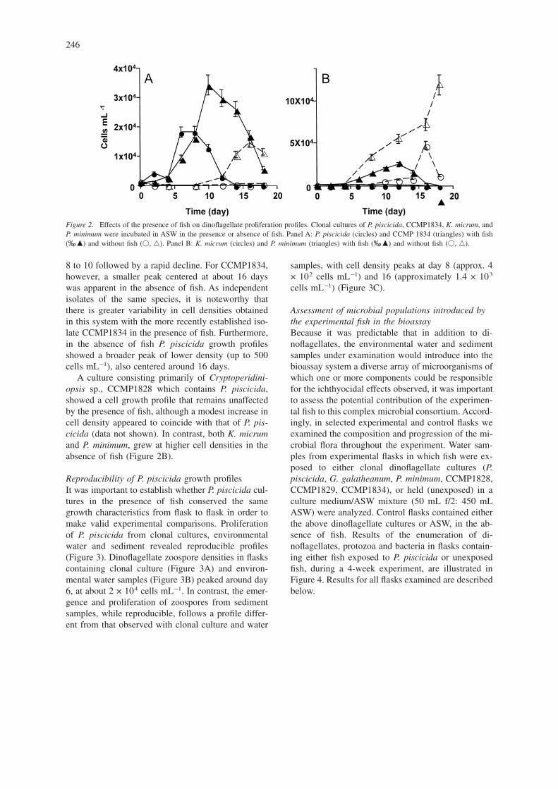
8 to 10 followed by a rapid decline. For CCMP1834,however, a smaller peak centered at about 16 dayswas apparent in the absence of fish. As independentisolates of the same species, it is noteworthy thatthere is greater variability in cell densities obtainedin this system with the more recently established iso-late CCMP1834 in the presence of fish. Furthermore,in the absence of fish P. piscicida growth profilesshowed a broader peak of lower density (up to 500cells mL−1), also centered around 16 days.
A culture consisting primarily of Cryptoperidini-opsis sp., CCMP1828 which contains P. piscicida,showed a cell growth profile that remains unaffectedby the presence of fish, although a modest increase incell density appeared to coincide with that of P. pis-cicida (data not shown). In contrast, both K. micrumand P. minimum, grew at higher cell densities in theabsence of fish (Figure 2B).
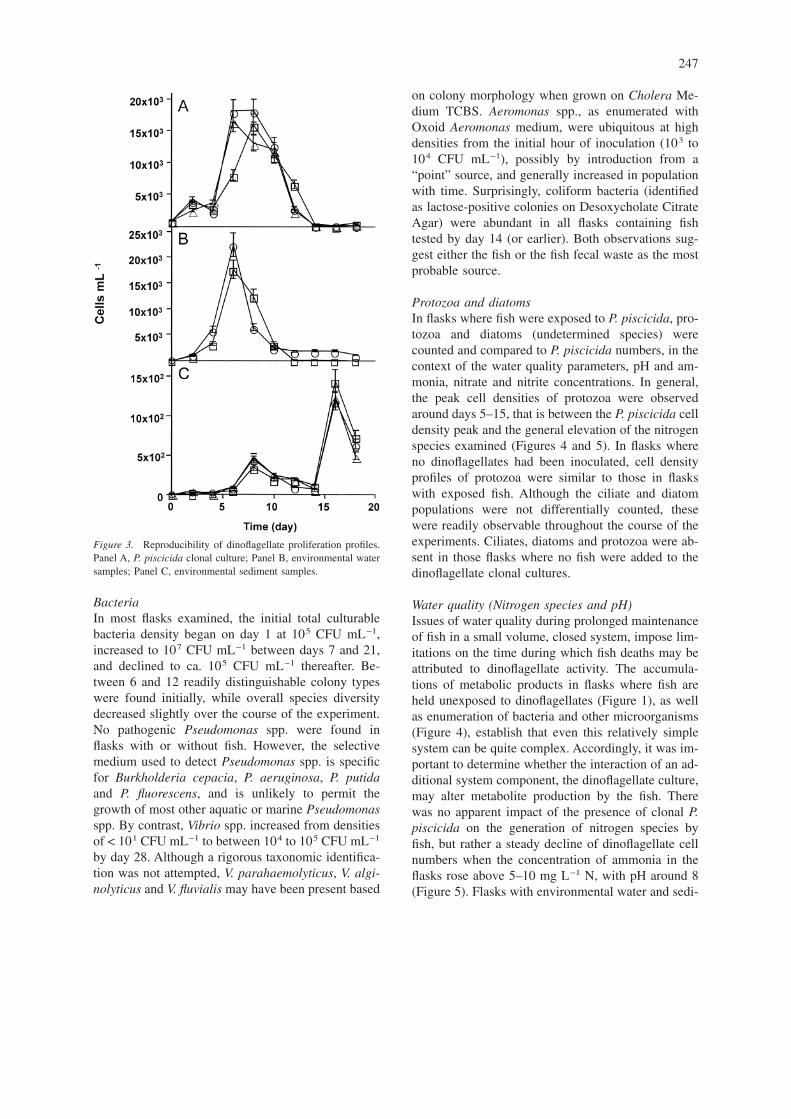
Reproducibility of P. piscicida growth profilesIt was important to establish whether P. piscicida cul-tures in the presence of fish conserved the samegrowth characteristics from flask to flask in order tomake valid experimental comparisons. Proliferationof P. piscicida from clonal cultures, environmentalwater and sediment revealed reproducible profiles(Figure 3). Dinoflagellate zoospore densities in flaskscontaining clonal culture (Figure 3A) and environ-mental water samples (Figure 3B) peaked around day6, at about 2 × 10⁴ cells mL−1. In contrast, the emer-gence and proliferation of zoospores from sedimentsamples, while reproducible, follows a profile differ-ent from that observed with clonal culture and water
samples, with cell density peaks at day 8 (approx. 4× 10² cells mL−1) and 16 (approximately 1.4 × 10³
cells mL−1) (Figure 3C).
Assessment of microbial populations introduced bythe experimental fish in the bioassayBecause it was predictable that in addition to di-noflagellates, the environmental water and sedimentsamples under examination would introduce into thebioassay system a diverse array of microorganisms ofwhich one or more components could be responsiblefor the ichthyocidal effects observed, it was importantto assess the potential contribution of the experimen-tal fish to this complex microbial consortium. Accord-ingly, in selected experimental and control flasks weexamined the composition and progression of the mi-crobial flora throughout the experiment. Water sam-ples from experimental flasks in which fish were ex-posed to either clonal dinoflagellate cultures (P.piscicida, G. galatheanum, P. minimum, CCMP1828,CCMP1829, CCMP1834), or held (unexposed) in aculture medium/ASW mixture (50 mL f/2: 450 mLASW) were analyzed. Control flasks contained eitherthe above dinoflagellate cultures or ASW, in the ab-sence of fish. Results of the enumeration of di-noflagellates, protozoa and bacteria in flasks contain-ing either fish exposed to P. piscicida or unexposedfish, during a 4-week experiment, are illustrated inFigure 4. Results for all flasks examined are describedbelow.
Figure 2. Effects of the presence of fish on dinoflagellate proliferation profiles. Clonal cultures of P. piscicida, CCMP1834, K. micrum, andP. minimum were incubated in ASW in the presence or absence of fish. Panel A: P. piscicida (circles) and CCMP 1834 (triangles) with fish(‰, �) and without fish (�, �). Panel B: K. micrum (circles) and P. minimum (triangles) with fish (‰, �) and without fish (�, �).
246
BacteriaIn most flasks examined, the initial total culturablebacteria density began on day 1 at 105 CFU mL−1,increased to 107 CFU mL−1 between days 7 and 21,and declined to ca. 105 CFU mL−1 thereafter. Be-tween 6 and 12 readily distinguishable colony typeswere found initially, while overall species diversitydecreased slightly over the course of the experiment.No pathogenic Pseudomonas spp. were found inflasks with or without fish. However, the selectivemedium used to detect Pseudomonas spp. is specificfor Burkholderia cepacia, P. aeruginosa, P. putidaand P. fluorescens, and is unlikely to permit thegrowth of most other aquatic or marine Pseudomonasspp. By contrast, Vibrio spp. increased from densitiesof < 101 CFU mL−1 to between 104 to 105 CFU mL−1
by day 28. Although a rigorous taxonomic identifica-tion was not attempted, V. parahaemolyticus, V. algi-nolyticus and V. fluvialis may have been present based
on colony morphology when grown on Cholera Me-dium TCBS. Aeromonas spp., as enumerated withOxoid Aeromonas medium, were ubiquitous at highdensities from the initial hour of inoculation (103 to104 CFU mL−1), possibly by introduction from a“point” source, and generally increased in populationwith time. Surprisingly, coliform bacteria (identifiedas lactose-positive colonies on Desoxycholate CitrateAgar) were abundant in all flasks containing fishtested by day 14 (or earlier). Both observations sug-gest either the fish or the fish fecal waste as the mostprobable source.
Protozoa and diatomsIn flasks where fish were exposed to P. piscicida, pro-tozoa and diatoms (undetermined species) werecounted and compared to P. piscicida numbers, in thecontext of the water quality parameters, pH and am-monia, nitrate and nitrite concentrations. In general,the peak cell densities of protozoa were observedaround days 5–15, that is between the P. piscicida celldensity peak and the general elevation of the nitrogenspecies examined (Figures 4 and 5). In flasks whereno dinoflagellates had been inoculated, cell densityprofiles of protozoa were similar to those in flaskswith exposed fish. Although the ciliate and diatompopulations were not differentially counted, thesewere readily observable throughout the course of theexperiments. Ciliates, diatoms and protozoa were ab-sent in those flasks where no fish were added to thedinoflagellate clonal cultures.
Water quality (Nitrogen species and pH)Issues of water quality during prolonged maintenanceof fish in a small volume, closed system, impose lim-itations on the time during which fish deaths may beattributed to dinoflagellate activity. The accumula-tions of metabolic products in flasks where fish areheld unexposed to dinoflagellates (Figure 1), as wellas enumeration of bacteria and other microorganisms(Figure 4), establish that even this relatively simplesystem can be quite complex. Accordingly, it was im-portant to determine whether the interaction of an ad-ditional system component, the dinoflagellate culture,may alter metabolite production by the fish. Therewas no apparent impact of the presence of clonal P.piscicida on the generation of nitrogen species byfish, but rather a steady decline of dinoflagellate cellnumbers when the concentration of ammonia in theflasks rose above 5–10 mg L−1 N, with pH around 8(Figure 5). Flasks with environmental water and sedi-
Figure 3. Reproducibility of dinoflagellate proliferation profiles.Panel A, P. piscicida clonal culture; Panel B, environmental watersamples; Panel C, environmental sediment samples.
247
ment samples, which contained complex bacterialcommunities, attained lower ammonia concentrationsrelative to those in the control and clonal dinoflagel-late culture flasks (data not shown). Differences inwater chemistry were observed in flasks within 12hours of a fish death, compared to parallel flasks inwhich no fish death had occurred (Figure 6). The ob-served trend in these flasks was for total ammonia(ionized and un-ionized) to increase, while both ni-trite and nitrate decreased, with a clear increase of thetoxic un-ionized ammonia.
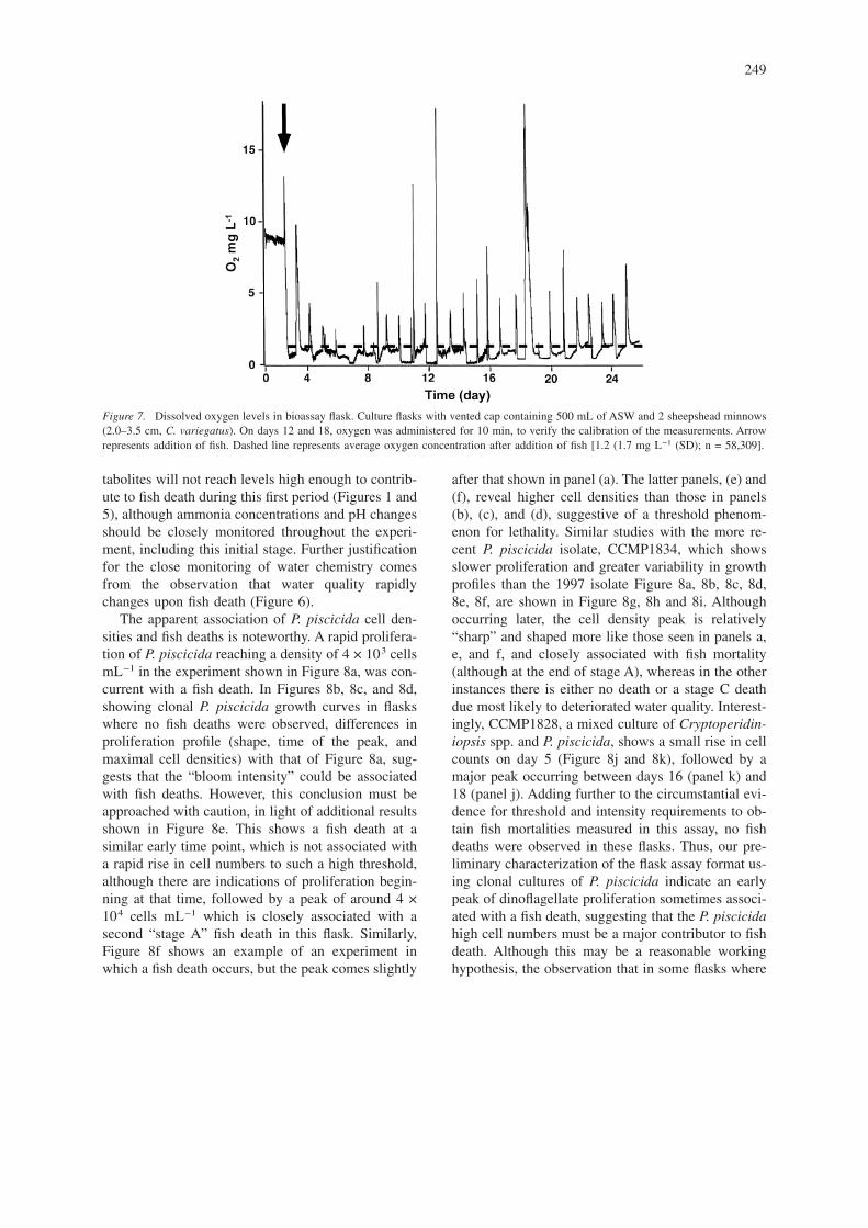
Dissolved oxygenExcept for brief periods immediately following thedaily purging of the flask headspace with pure oxy-gen, dissolved oxygen concentration remained at orbelow 1.5 mg L−1 for the entire 28 days of the ex-periment (Figure 7). During this time, fish showed no
obvious signs of disease or stress, but were occasion-ally found at the water surface. Fish continued to feedthroughout the experiment.
Summary of the fish bioassayProliferation of 11 clonal dinoflagellates cultures in-oculated to fish bioassay flask and observed fish deathwere shown in Figure 8. In order to facilitate the in-terpretation of bioassay results, we have divided thebioassay period into three stages: “stage A” (first 10days), “stage B” (10–20 days), and “stage C” (beyond20 days). The definition of the initial 10-day period(Stage A) is critical to the experiment, because it issufficiently brief that fish deaths which occur in thistime period may not be attributable to pathogenic bac-teria which colonize the flask (Figure 4). Likewise, inmost cases the accumulation of potentially toxic me-
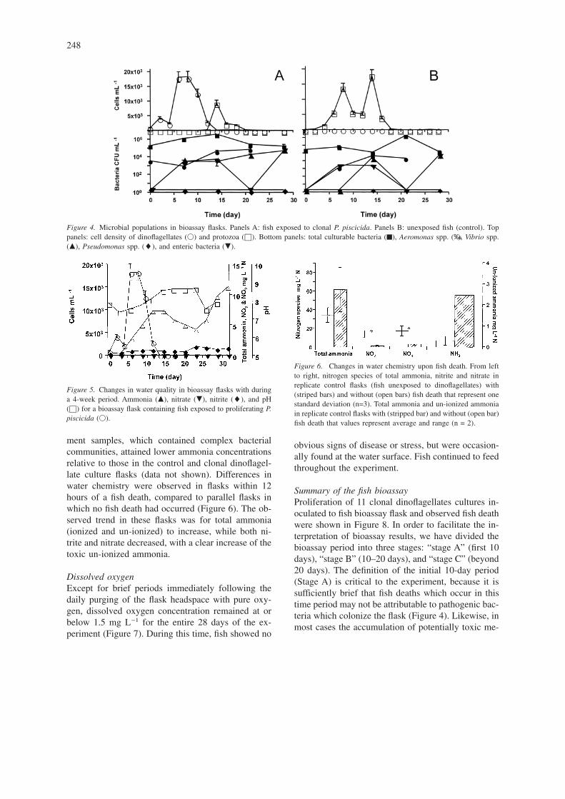
Figure 4. Microbial populations in bioassay flasks. Panels A: fish exposed to clonal P. piscicida. Panels B: unexposed fish (control). Toppanels: cell density of dinoflagellates (�) and protozoa (©). Bottom panels: total culturable bacteria (™), Aeromonas spp. (‰), Vibrio spp.(�), Pseudomonas spp. (�), and enteric bacteria (�).
Figure 5. Changes in water quality in bioassay flasks with duringa 4-week period. Ammonia (�), nitrate (�), nitrite (�), and pH(©) for a bioassay flask containing fish exposed to proliferating P.piscicida (�).
Figure 6. Changes in water chemistry upon fish death. From leftto right, nitrogen species of total ammonia, nitrite and nitrate inreplicate control flasks (fish unexposed to dinoflagellates) with(striped bars) and without (open bars) fish death that represent onestandard deviation (n=3). Total ammonia and un-ionized ammoniain replicate control flasks with (stripped bar) and without (open bar)fish death that values represent average and range (n = 2).
248
tabolites will not reach levels high enough to contrib-ute to fish death during this first period (Figures 1 and5), although ammonia concentrations and pH changesshould be closely monitored throughout the experi-ment, including this initial stage. Further justificationfor the close monitoring of water chemistry comesfrom the observation that water quality rapidlychanges upon fish death (Figure 6).
The apparent association of P. piscicida cell den-sities and fish deaths is noteworthy. A rapid prolifera-tion of P. piscicida reaching a density of 4 × 10³ cellsmL−1 in the experiment shown in Figure 8a, was con-current with a fish death. In Figures 8b, 8c, and 8d,showing clonal P. piscicida growth curves in flaskswhere no fish deaths were observed, differences inproliferation profile (shape, time of the peak, andmaximal cell densities) with that of Figure 8a, sug-gests that the “bloom intensity” could be associatedwith fish deaths. However, this conclusion must beapproached with caution, in light of additional resultsshown in Figure 8e. This shows a fish death at asimilar early time point, which is not associated witha rapid rise in cell numbers to such a high threshold,although there are indications of proliferation begin-ning at that time, followed by a peak of around 4 ×10⁴ cells mL−1 which is closely associated with asecond “stage A” fish death in this flask. Similarly,Figure 8f shows an example of an experiment inwhich a fish death occurs, but the peak comes slightly
after that shown in panel (a). The latter panels, (e) and(f), reveal higher cell densities than those in panels(b), (c), and (d), suggestive of a threshold phenom-enon for lethality. Similar studies with the more re-cent P. piscicida isolate, CCMP1834, which showsslower proliferation and greater variability in growthprofiles than the 1997 isolate Figure 8a, 8b, 8c, 8d,8e, 8f, are shown in Figure 8g, 8h and 8i. Althoughoccurring later, the cell density peak is relatively“sharp” and shaped more like those seen in panels a,e, and f, and closely associated with fish mortality(although at the end of stage A), whereas in the otherinstances there is either no death or a stage C deathdue most likely to deteriorated water quality. Interest-ingly, CCMP1828, a mixed culture of Cryptoperidin-iopsis spp. and P. piscicida, shows a small rise in cellcounts on day 5 (Figure 8j and 8k), followed by amajor peak occurring between days 16 (panel k) and18 (panel j). Adding further to the circumstantial evi-dence for threshold and intensity requirements to ob-tain fish mortalities measured in this assay, no fishdeaths were observed in these flasks. Thus, our pre-liminary characterization of the flask assay format us-ing clonal cultures of P. piscicida indicate an earlypeak of dinoflagellate proliferation sometimes associ-ated with a fish death, suggesting that the P. piscicidahigh cell numbers must be a major contributor to fishdeath. Although this may be a reasonable workinghypothesis, the observation that in some flasks where
Figure 7. Dissolved oxygen levels in bioassay flask. Culture flasks with vented cap containing 500 mL of ASW and 2 sheepshead minnows(2.0–3.5 cm, C. variegatus). On days 12 and 18, oxygen was administered for 10 min, to verify the calibration of the measurements. Arrowrepresents addition of fish. Dashed line represents average oxygen concentration after addition of fish [1.2 (1.7 mg L−1 (SD); n = 58,309].
249
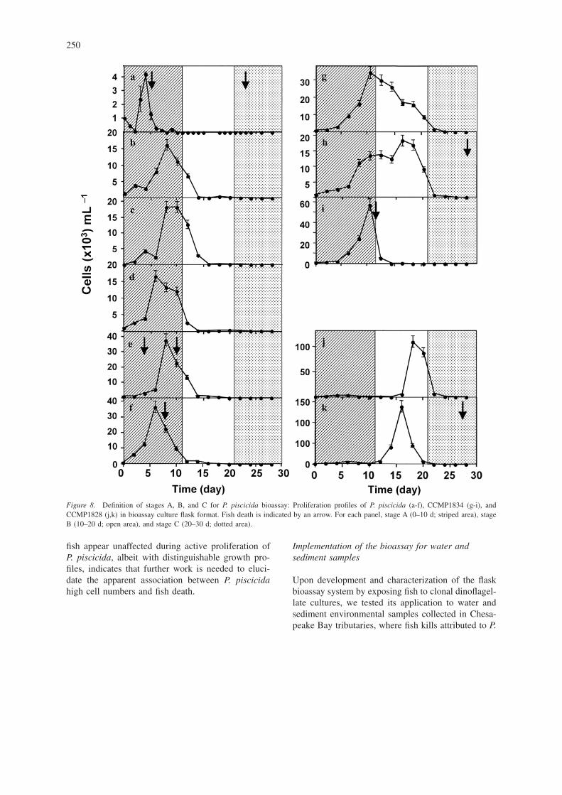
fish appear unaffected during active proliferation ofP. piscicida, albeit with distinguishable growth pro-files, indicates that further work is needed to eluci-date the apparent association between P. piscicidahigh cell numbers and fish death.
Implementation of the bioassay for water andsediment samples
Upon development and characterization of the flaskbioassay system by exposing fish to clonal dinoflagel-late cultures, we tested its application to water andsediment environmental samples collected in Chesa-peake Bay tributaries, where fish kills attributed to P.
Figure 8. Definition of stages A, B, and C for P. piscicida bioassay: Proliferation profiles of P. piscicida (a-f), CCMP1834 (g-i), andCCMP1828 (j,k) in bioassay culture flask format. Fish death is indicated by an arrow. For each panel, stage A (0–10 d; striped area), stageB (10–20 d; open area), and stage C (20–30 d; dotted area).
250
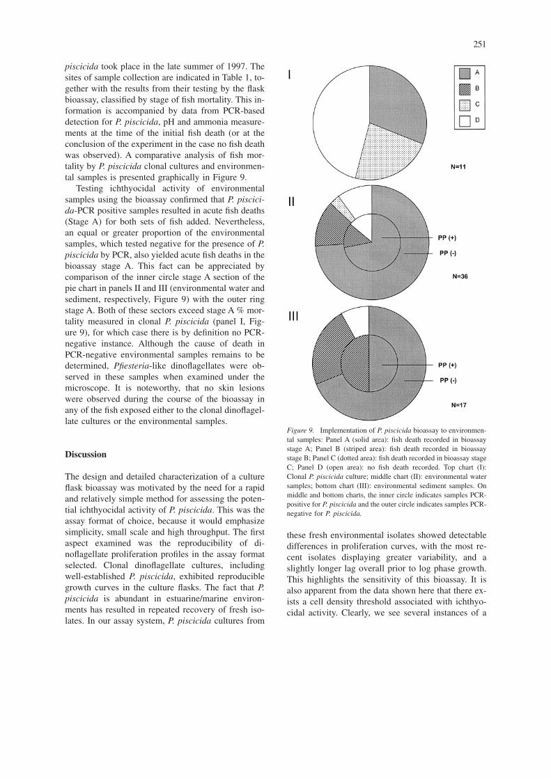
piscicida took place in the late summer of 1997. Thesites of sample collection are indicated in Table 1, to-gether with the results from their testing by the flaskbioassay, classified by stage of fish mortality. This in-formation is accompanied by data from PCR-baseddetection for P. piscicida, pH and ammonia measure-ments at the time of the initial fish death (or at theconclusion of the experiment in the case no fish deathwas observed). A comparative analysis of fish mor-tality by P. piscicida clonal cultures and environmen-tal samples is presented graphically in Figure 9.
Testing ichthyocidal activity of environmentalsamples using the bioassay confirmed that P. piscici-da-PCR positive samples resulted in acute fish deaths(Stage A) for both sets of fish added. Nevertheless,an equal or greater proportion of the environmentalsamples, which tested negative for the presence of P.piscicida by PCR, also yielded acute fish deaths in thebioassay stage A. This fact can be appreciated bycomparison of the inner circle stage A section of thepie chart in panels II and III (environmental water andsediment, respectively, Figure 9) with the outer ringstage A. Both of these sectors exceed stage A % mor-tality measured in clonal P. piscicida (panel I, Fig-ure 9), for which case there is by definition no PCR-negative instance. Although the cause of death inPCR-negative environmental samples remains to bedetermined, Pfiesteria-like dinoflagellates were ob-served in these samples when examined under themicroscope. It is noteworthy, that no skin lesionswere observed during the course of the bioassay inany of the fish exposed either to the clonal dinoflagel-late cultures or the environmental samples.
Discussion
The design and detailed characterization of a cultureflask bioassay was motivated by the need for a rapidand relatively simple method for assessing the poten-tial ichthyocidal activity of P. piscicida. This was theassay format of choice, because it would emphasizesimplicity, small scale and high throughput. The firstaspect examined was the reproducibility of di-noflagellate proliferation profiles in the assay formatselected. Clonal dinoflagellate cultures, includingwell-established P. piscicida, exhibited reproduciblegrowth curves in the culture flasks. The fact that P.piscicida is abundant in estuarine/marine environ-ments has resulted in repeated recovery of fresh iso-lates. In our assay system, P. piscicida cultures from
these fresh environmental isolates showed detectabledifferences in proliferation curves, with the most re-cent isolates displaying greater variability, and aslightly longer lag overall prior to log phase growth.This highlights the sensitivity of this bioassay. It isalso apparent from the data shown here that there ex-ists a cell density threshold associated with ichthyo-cidal activity. Clearly, we see several instances of a
Figure 9. Implementation of P. piscicida bioassay to environmen-tal samples: Panel A (solid area): fish death recorded in bioassaystage A; Panel B (striped area): fish death recorded in bioassaystage B; Panel C (dotted area): fish death recorded in bioassay stageC; Panel D (open area): no fish death recorded. Top chart (I):Clonal P. piscicida culture; middle chart (II): environmental watersamples; bottom chart (III): environmental sediment samples. Onmiddle and bottom charts, the inner circle indicates samples PCR-positive for P. piscicida and the outer circle indicates samples PCR-negative for P. piscicida.
251
rapid rise in P. piscicida to high cell densities associ-ated closely in time with fish death. It is these eventson which we currently focus mechanistic investiga-tions. The presence of fish selectively modulated theproliferation of some dinoflagellate species, with agrowth enhancing effect on P. piscicida and Crypto-peridiniopsis sp. The fish-derived factor(s) that stim-ulate or inhibit growth of dinoflagellates in flaskshave not been identified yet, but their effects on di-noflagellate proliferation were also highly reproduc-ible in this assay format.
Once the dinoflagellate growth profiles in the flaskformat had been characterized, it was critical to as-sess those water quality parameters, such as dissolvedoxygen, ammonia, nitrate and nitrite levels, and pH,that are known to impact fish health (Wedemeyer1996). Although dissolved oxygen was not measuredin all experimental flasks, results in a pilot experimentover eight weeks suggests that the daily purging withoxygen of the flask headspace (approximately 250mL) provides sufficient oxygen for fish survival.Thus, attention was focused on pH and concentrationsof ammonia, nitrate and nitrite, nitrogen species thatare expected to accumulate during normal fish meta-bolism in a closed assay system. Although the pHvaried from flask to flask during the experiments, atthe value ASW was initially buffered (pH 8.0), thelevel of ammonia required for acute toxicity would beapproximately 8 mg L−1 N. To our surprise, fish sur-vived in this system well past this limit, into the rangeof ammonia 60 mg L−1 N, perhaps due to a slow ac-climatization to the ammonia. Once the initial set offish perished, however, the death of the replacementswas very rapid, usually in a few hours, suggesting thatthe initial set of fish present from the outset had ac-climated during the course of the experiment to highammonia concentrations that are toxic to naïve fish.The toxic effects of nitrogen metabolites have beenreported for several fish species (Miller et al. 1990).Their effects vary with salinity and ionic state, withnitrite constituting the greatest health threat. Theequilibrium between ionized ammonia (non-toxic)and un-ionized ammonia (toxic) is governed by therelationship % un-ionized ammonia = 100/[1 + anti-log (pKa- pH)], where the pKa is temperature-depen-dent (Wedemeyer 1996). Interconversion by sequen-tial oxidation of un-ionized ammonia to nitratethrough the intermediate nitrite occurs in free equi-librium, leading to methemoglobinemia (brown blooddisease) when nitrite acts on hemoglobin (Wedemeyer1996). Nitrifying bacteria, such as those present in
aquarium biofilters, will metabolize some of the ni-trogen species produced, contributing to a healthy en-vironment in large-scale bioassays, such as the aquar-ium tank format. In order to simplify the design,however, we did not attempt biofiltration in the flaskformat.
One important observation to come out of thisstudy, which should be considered in any experimen-tal format aimed at examining bioactive effects ofphytoplankton on fish, is the fact that the in additionto normal metabolites, fish introduce their own bioticmicroenvironment in the flasks. The presence of cili-ates, diatoms, protozoa, and bacteria, such as Vibrio,Aeromonas, and coliforms, in flasks containing fish,which are not detectable in the dinoflagellate clonalcultures (Alavi et al. 2001), supports this conclusion.Although this may not be surprising or novel, it wascritical to document this phenomenon in detail in or-der to define the conditions necessary for a valid bio-assay, because in a closed system some of the micro-bial species identified may reach numbers potentiallyharmful to fish. The exogenously introduced micro-bial flora showed distinct profiles from flask to flask.We did not observe uniformity even while using fishfrom the same stock, as evidenced, for example, bysubstantial variability of the bacterial populations be-tween the flasks. Because these did not appear to con-stitute a significant threat to fish health until the sec-ond week, we set a conservative 10 day cutoff toconsider fish death as possibly due to P. piscicida orother dinoflagellates activity when associated with acell density peak occurring during that time period.Proliferation of diatoms or protozoa appeared to lagbehind that of P. piscicida or Pfiesteria-like di-noflagellates in the flask assay format. Ciliates havebeen reported as proliferating following a dinoflagel-late bloom and decline in a profile that suggests graz-ing on dinoflagellates (Stoecker et al. 2000). The siev-ing procedures for sample preparation weimplemented were designed to eliminate ciliates andother organisms larger than the dinoflagellates of in-terest, and avoid predation. Despite these measures, avariable number of ciliates were present in the flasksin which fish were added, but not in those flaskswhere no fish was added to the clonal dinoflagellatecultures. This observation further suggests that theexperimental fish may introduce an array of microbialspecies, including ciliates, in the flask environment.Formalin-treatment of the experimental fish (Speareand MacNair 1996), a possible alternative for circum-venting the problem, has several drawbacks, includ-
252
ing (1) additional stress to the fish which would con-stitute an additional burden to the immune system,and for which the overall impact on fish health wouldbe difficult to account; (2) the intrinsic variability ofthe treatment which would be difficult to measure re-liably; and (3) the likelihood that the treatment maybe effective for the external microbial community as-sociated with fish skin and mucous, but not for thegut microbial flora. Under the assumption that bacte-ria and other microflora will colonize the flasks re-gardless of the treatment, we chose to measure andclassify the microbial populations rather than attemptto eliminate them. Potentially pathogenic bacterialpopulations, for example, rose from very low but im-mediately detectable numbers present at the introduc-tion of all components into the flask, to significantlyhigh densities at about day 10, coincident with therise in nitrogen species to threshold toxic values.
In summary, our studies clearly indicate that al-though the flask bioassay may constitute the simplestformat to assess fish mortality related to harmful di-noflagellates, it is still a complex and dynamic sys-tem. Nevertheless, the development and characteriza-tion of the flask bioassay has permitted the followingobservations: (a) the presence of fish stimulates pro-liferation of P. piscicida but this is not a general phe-nomenon; (b) the levels of ammonia, nitrate and ni-trite fluctuate but gradually increase to concentrationsbeyond limits for fish survival at approximately 21days; (c) the experimental fish may carry a diversemicrobial flora, including some recognized fish patho-gens, protozoa, and dinoflagellates, which activelyproliferate in the flasks. From these observations wehave empirically determined the operational parame-ters under which a fish death may be attributable tofactors associated with a peak in dinoflagellate pro-liferation.
Based on these observations, we defined three as-say stages that are relevant to the interpretation of thedata. The critical threshold of the first 10 days con-stitutes “stage A”, during which dinoflagellate prolif-eration may be the cause of fish death. This is fol-lowed by the intermediate “stage B”, from 10 to 20days, during which the combined impact of rising ni-trogen species and proliferation of bacteria and pro-tozoa render a fish death uninterpretable for our pur-poses. Finally, a fish death past 20 days, “stage C”,would be most likely caused by accumulation of toxicnitrogen species (Figures 8 and 9).
Although the fish deaths were not reproducible forall P. piscicida isolates (Figure 8), when these take
place in less than 10 days and are synchronous withthe dinoflagellate proliferation peak, then a prelimi-nary causative connection may be established and fur-ther investigated. The advantages of the system arethat many samples may be screened simultaneouslyto maximize sample throughput. To our knowledge,we have described here a flask format bioassay for P.piscicida to one of the higher degrees of detail thanwe are able to find published elsewhere in the litera-ture, and should be widely applicable for the identi-fication and further investigation of aquatic microor-ganisms that exhibit ichthyocidal activity.
The implementation of the bioassay using environ-mental samples showed a higher propensity for acutefish deaths (Stage A) in P. piscicida-PCR positivesamples. However, interpretation of this observationmay be obscured by the fact that an equal or greaterproportion of the environmental samples which testednegative for the presence of P. piscicida by PCR alsoyielded acute fish deaths, for which no particularcause has been identified. However, these samplesmostly contain Pfiesteria-like organisms, and thehighly specific PCR assay we implemented will de-tect only P. piscicida (Robledo et al. 2000b). There-fore, we cannot rule out the possibility that the highratio of acute fish deaths observed in the environmen-tal samples may be due to other Pfiesteria specieswhich have been described as ichthyocidal, such asthe recently reported P. shumwayae (Glasgow et al.2000). Although this may limit the interpretation ofthe results for complex environmental samples, itwould broaden the applicability of the bioassay.
Acknowledgements
This study was supported by grants NIEHS 1PO1ES09563 and ECOHAB NA860P0492. We thank Dr.Sherwood Hall (Food and Drug Administration) foruseful discussions during the early stages of bioassaydevelopment.
References
Alavi M., Miller T., Erlandson K., Schneider R. and Belas R. 2001.Bacteria community associated with Pfiesteiria-like dinoflagel-late cultures. Environ. Microbiol. 3: 380–396.
253
Bever C.T., Grattan L. Jr and Morris J.G. 1998. Neurologic symp-toms following Pfiesteria exposure: case report and literaturereview. MD Med. J. 47: 120–123.
Burkholder J.M., Noga E.J., Hobbs C.H. and Glasgow H.B. Jr1992. New ’phantom’ dinoflagellate is the causative agent ofmajor estuarine fish kills. Nature 358: 407–410.
Clean Water Commission, Department of Natural Resources 2001.Water Quality, in Missouri State Code of Regulations. Docu-ment 10 CSR 20-7. Office of Secretary of State, Jefferson City,MO, USA, p. 135.
Glasgow W.B., Burkholder J.M. Jr, Morton S. and Springer J. 2000.A new species of toxic Pfiesteria. In: Conference Proceedings:International Conference on Harmful Algal Blooms, Tasmania,Australia. (Abstract, p. 19).
Grattan L.M., Oldach D., Perl T.M., Lowitt M.H., Matuszak D.L.,Dickson C. et al. 1998. Learning and memory difficulties afterenvironmental exposure to waterways. Lancet 352: 532–539.
Guillard R.R.L. 1975. Culture of phytoplankton for feeding marineinvertebrates. In: Smith W.L. and Chanley M.H. (eds), Cultureof Marine Invertebrate Animals. Plenum Press, New York, pp.26–60.
Harvell C.D., Kim K., Burkholder J.M., Colwell R.R., Epstein P.R.,Grimes D.J. et al. 1999. Emerging marine diseases–climatelinks and anthropogenic factors. Science 285: 1505–1510.
Kane A.S., Oldach D. and Reimschuessel R. 1998. Fish lesions inthe Chesapeake Bay: Pfiesteria-like dinoflagellates and otheretiologies. MD Med. J. 47: 106–112.
Lányi B. 1987. Classical and rapid identification methods for med-ically important bacteria. In: Colwell R.R. and Grigorova R.(eds), Methods in Microbiology. Vol. 19. Academic Press Ltd,Orlando, FL, USA.
Matuszak D.L., Taylor J.L., Dickson C. and Benjamin G.C. 1998.Toxic-Pfiesteria-surveillance for human disease in Maryland.MD Med. J. 47: 144–147.
Miller D.C., Poucher S., Cardin J.A. and Hansen D. 1990. Theacute and chronic toxicity of ammonia to marine fish and amysid. Arch. environ. Contam. Toxicol. 19: 40–48.
Robledo J.A.F., Pecher W.T. and Vasta G.R. 2000b. High-through-put isolation of oyster DNA facilitates diagnosis of “Dermo”disease. Qiagen News 5: 15–17.
Robledo J.A.F., Saito K., Coats D.W., Steidinger K.A. and VastaG.R. 2000a. The non-transcribed spacer of the rRNA locus as atarget for specific detection of the dinoflagellate Pfiesteria pis-cicida and protistan parasites (Perkinsus spp.). In: Ninth Inter-national Conference on Harmful Algal Blooms. Interbovern-mental Oceanographic commission of UNESCO, HobartAustralia, 7–11 February 2000., pp. 238–241.
Speare D.J. and MacNair N. 1996. Effects of intermittent exposureto therapeutic levels of formalin on growth characteristics andbody condition of juvenile rainbow trout. J. aquat. Anim.Health 8: 58–63.
Steidinger K.A., Burkholder J.M., Glasgow H.B., Hobbs C.W. Jr,Garrett J.K., Truby E.W. et al. 1996. Pfiesteria piscicida gen.et sp. nov. (Pfiesteriaceae fam. nov.), a new toxic dinoflagel-late with a complex life cycle and behavior. J. Phycol. 32:157–164.
Stoecker D.K., Stevens K. and Gustafson D.E. 2000. Potential pre-dation on Pfiesteria piscicida. In: Conference Proceedings: In-ternational Conference on Harmful Algal Blooms, Tasmania,Australia, (Abstract, p. 59).
Wedemeyer G.A. 1996. Physiology of Fish in Intensive CultureSystems. Chapman and Hall, New York, 232 p.
254