A Second Generation of Carbamate-Based Fatty Acid Amide Hydrolase Inhibitors with Improved Activity...
18
A second generation of carbamate-based fatty acid amide hydrolase inhibitors with improved activity in vivo Jason R. Clapper [a] , Dr. Federica Vacondio [b] , Alvin R. King [a] , Dr. Andrea Duranti [c] , Dr. Andrea Tontini [c] , Prof. Claudia Silva [b] , Dr. Silvano Sanchini [c] , Prof. Giorgio Tarzia [c] , Prof. Marco Mor [b] , and Prof. Daniele Piomelli [a],[d],* [a] Departments of Pharmacology and Biological Chemistry, 360 MSRII, University of California, Irvine, Irvine, CA, 92697 USA [b] Pharmaceutical Department, University of Parma, Viale G. P. Usberti 27/A, Campus Universitario, Parma, 43100, Italy [c] Institute of Medicinal Chemistry, University of Urbino “Carlo Bo”, Piazza del Rinascimento, 6, Urbino, 61029, Italy [d] Unit of Drug Discovery and Development, Italian Institute of Technology, via Morego 30, Genoa, 16163, Italy Abstract The fatty acid ethanolamides are a class of signaling lipids that include agonists at cannabinoid and type-α peroxisome proliferator-activated receptors. In the brain, these compounds are primarily hydrolyzed by the intracellular serine enzyme fatty acid amide hydrolase (FAAH). O- aryl carbamate FAAH inhibitors such as URB597 are being evaluated clinically for the treatment of pain and anxiety, but interactions with carboxylesterases in liver might limit their usefulness. Here we explore two strategies aimed at overcoming this limitation. Lipophilic N-terminal substitutions, which enhance FAAH recognition, yield potent inhibitors but render such compounds both susceptible to attack by broad-spectrum hydrolases and inactive in vivo. By contrast, polar electron-donating O-aryl substituents, which decrease carbamate reactivity, yield compounds, such as URB694, that are highly potent FAAH inhibitors in vivo and less reactive with off-target carboxylesterases. The results suggest that an approach balancing inhibitor reactivity with target recognition produces FAAH inhibitors that display significantly improved drug-likeness. Keywords Enzymes; hydrolases; carbamate; FAAH inhibitor; carboxylesterase Introduction Fatty acid amide hydrolase (FAAH)[1–4] is a membrane-bound serine enzyme that catalyzes the intracellular hydrolysis of the fatty acid ethanolamide family of signaling lipids, which includes endogenous agonists at cannabinoid CB 1 and CB 2 receptors[5,6], and type-α peroxisome proliferator-activated receptors[7–9]. Natural FAAH substrates such as the endocannabinoid anandamide[5,6] play important physiological roles, and deficits in the functions of these biomolecules have been implicated in a broad range of disease Phone: (949) 824-6180, Fax: (949) 824-6305, [email protected]. NIH Public Access Author Manuscript ChemMedChem. Author manuscript; available in PMC 2011 January 5. Published in final edited form as: ChemMedChem. 2009 September ; 4(9): 1505–1513. doi:10.1002/cmdc.200900210. NIH-PA Author Manuscript NIH-PA Author Manuscript NIH-PA Author Manuscript
Transcript of A Second Generation of Carbamate-Based Fatty Acid Amide Hydrolase Inhibitors with Improved Activity...

A second generation of carbamate-based fatty acid amidehydrolase inhibitors with improved activity in vivo
Jason R. Clapper[a], Dr. Federica Vacondio[b], Alvin R. King[a], Dr. Andrea Duranti[c], Dr.Andrea Tontini[c], Prof. Claudia Silva[b], Dr. Silvano Sanchini[c], Prof. Giorgio Tarzia[c], Prof.Marco Mor[b], and Prof. Daniele Piomelli[a],[d],*
[a] Departments of Pharmacology and Biological Chemistry, 360 MSRII, University of California,Irvine, Irvine, CA, 92697 USA[b] Pharmaceutical Department, University of Parma, Viale G. P. Usberti 27/A, CampusUniversitario, Parma, 43100, Italy[c] Institute of Medicinal Chemistry, University of Urbino “Carlo Bo”, Piazza del Rinascimento, 6,Urbino, 61029, Italy[d] Unit of Drug Discovery and Development, Italian Institute of Technology, via Morego 30,Genoa, 16163, Italy
AbstractThe fatty acid ethanolamides are a class of signaling lipids that include agonists at cannabinoidand type-α peroxisome proliferator-activated receptors. In the brain, these compounds areprimarily hydrolyzed by the intracellular serine enzyme fatty acid amide hydrolase (FAAH). O-aryl carbamate FAAH inhibitors such as URB597 are being evaluated clinically for the treatmentof pain and anxiety, but interactions with carboxylesterases in liver might limit their usefulness.Here we explore two strategies aimed at overcoming this limitation. Lipophilic N-terminalsubstitutions, which enhance FAAH recognition, yield potent inhibitors but render suchcompounds both susceptible to attack by broad-spectrum hydrolases and inactive in vivo. Bycontrast, polar electron-donating O-aryl substituents, which decrease carbamate reactivity, yieldcompounds, such as URB694, that are highly potent FAAH inhibitors in vivo and less reactivewith off-target carboxylesterases. The results suggest that an approach balancing inhibitorreactivity with target recognition produces FAAH inhibitors that display significantly improveddrug-likeness.
KeywordsEnzymes; hydrolases; carbamate; FAAH inhibitor; carboxylesterase
IntroductionFatty acid amide hydrolase (FAAH)[1–4] is a membrane-bound serine enzyme that catalyzesthe intracellular hydrolysis of the fatty acid ethanolamide family of signaling lipids, whichincludes endogenous agonists at cannabinoid CB1 and CB2 receptors[5,6], and type-αperoxisome proliferator-activated receptors[7–9]. Natural FAAH substrates such as theendocannabinoid anandamide[5,6] play important physiological roles, and deficits in thefunctions of these biomolecules have been implicated in a broad range of disease
Phone: (949) 824-6180, Fax: (949) 824-6305, [email protected].
NIH Public AccessAuthor ManuscriptChemMedChem. Author manuscript; available in PMC 2011 January 5.
Published in final edited form as:ChemMedChem. 2009 September ; 4(9): 1505–1513. doi:10.1002/cmdc.200900210.
NIH
-PA Author Manuscript
NIH
-PA Author Manuscript
NIH
-PA Author Manuscript

conditions[10]. Consequently, inhibition of FAAH activity, which is expected to reducesuch deficits, has become the focus of intense drug discovery efforts. Among the variousclasses of FAAH inhibitors identified so far[10–12], two have demonstrated favorableactivity and selectivity profiles in vivo: the O-aryl carbamates discovered in ourlaboratories[13–15] and the piperidine/piperazine ureas developed at Pfizer Inc.[16–17]. Theformer class comprises N-cyclohexylcarbamic acid O-aryl ester derivatives such as URB524and URB597 (Figure 1), which feature a unique combination of anxiolytic, antidepressantand analgesic properties[18–21], accompanied by a lack of addiction liability in rodent andprimate models[19,21].
The mechanism by which O-aryl carbamates inhibit FAAH has been investigated in detail.Quantitative structure-activity relationship (QSAR) and computational studies, aided by theresolution of FAAH’s crystal structure[23], have suggested that these molecules bind toFAAH with their N-terminal moiety positioned within the acyl chain binding (ACB) channelof the enzyme and their O-biphenyl group occupying a large fraction of its cytosolic access(CA) channel[15,24,25]. This orientation allows the catalytic nucleophile, serine 241(ser241), to attack the compound’s carbonyl group to yield a carbamoylated, inactiveenzyme (Figure 2). Consistent with this model, QSAR analyses have shown thataromatically restrained lipophilic substituents with a bent shape, such as a meta-oriented O-biphenyl group, optimally conform to the stereoelectronic requirements of the CAchannel[13], while hydrophilic substituents on the distal phenyl ring, such as the 3′-carbamoyl group of URB597, enhance enzyme-inhibitor interactions by forming hydrogenbonds with specific amino acid residues of the same domain[14] (Figure 2). It appearstherefore that a combination of covalent binding to catalytic ser241 and noncovalentinteractions with specific recognition elements in the active site of FAAH is necessary toachieve high inhibitory potency in this class of compounds. Surprisingly, however, no clearcorrelation has yet been found between the chemical reactivity of the carbamate group inthese molecules and their rank order potency for FAAH inhibition[26]. A plausibleexplanation for this finding is provided by the unique electronic properties of the serine-serine-lysine catalytic triad of FAAH, which allow ser241 to interact productively withmechanism-based inhibitors of relatively low chemical reactivity, such as the piperidine/piperazine ureas[16,17].
URB597, the index compound in the carbamic acid O-aryl ester class of FAAH inhibitors,displays remarkable target selectivity for FAAH both in vitro and in vivo[27–29].Nevertheless, at concentrations that exceed those needed to fully block FAAH activity, thecompound was shown to interact with several broad-spectrum esterases in liver[16,30]. Thisinhibition may affect the pharmacokinetic behavior of a variety of therapeutic agents thatcontain ester or amide bonds, and are substrates for liver carboxylesterases[31]. Are theseoff-target effects inherently linked to the presence of a carbamate moiety, or is it possible toreduce their occurrence without diminishing inhibitory potency? We reported previouslythat in a set of cyclohexylcarbamic acid aryl esters the electrophilicity of the carbamateinfluenced its chemical stability and that introduction of small electron donor substituents atconjugated positions of the O-aryl moiety increased overall hydrolytic stability of thecarbamate group towards in vitro chemical and enzymatic hydrolysis[32]. In the presentstudy, we have explored two strategies aimed at addressing the above question. The first wasto maximize the inhibitors’ complementarity for FAAH by enhancing interactions withunoccupied regions of its active site (Figure 2). The second, similar to what was previouslyreported,[32] consisted in modulating the reactivity of the carbamate group with theobjective of lowering its sensitivity to nucleophilic attack by hydrolases. We selected afocused set of O-aryl carbamates, most of which were previously published[13–15,26,32],that allowed us to test each of these strategies, and investigated the pharmacodynamic andpharmacokinetic properties of these compounds both in vitro and in vivo.
Clapper et al. Page 2
ChemMedChem. Author manuscript; available in PMC 2011 January 5.
NIH
-PA Author Manuscript
NIH
-PA Author Manuscript
NIH
-PA Author Manuscript
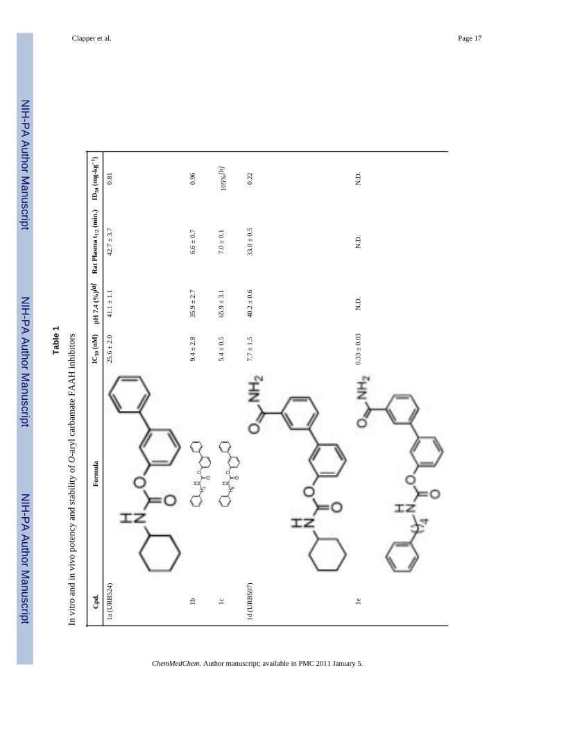
ResultsCompounds 1a (URB524)[13], 1b[15], 1c[15], 1d (URB597)[14], 1g (URB694)[26], 1h[26]and 1i[26] were previously published and were synthesized as reported. 4-Phenylbutylcarbamic acid 3′-carbamoylbiphenyl-3-yl ester (1e) and 8-phenyloctylcarbamicacid 3′-carbamoylbiphenyl-3-yl ester (1f) were obtained by addition of 3′-carbamoylbiphenyl-3-ol to 4-phenylbutylisocyanate (for 1e) or 8-phenyloctylisocyanate (for1e). 4-Phenylbutylisocyanate was commercially available while 8-phenyloctylisocyanatewas synthesized as reported[15].
Effect of lipophilic N-substitutions on inhibitory potencyPrevious studies have shown that lipophilic substitutions to the N-fragment of O-arylcarbamate FAAH inhibitors, which likely enhance the complementarity of these moleculesfor hydrophobic surfaces in the ACB channel, increase their inhibitory potency invitro[13,24,33]. Consistent with those findings, we found that introducing flexible andhydrophobic N-substituents on the scaffold of URB524 produced compounds that werehighly potent at inhibiting FAAH activity in rat brain membranes (Table 1). For example,the N-phenyloctyl derivative 1c inhibited FAAH activity with a median inhibitoryconcentration (IC50) that was approximately 5 times more potent than parent compoundURB524 (Table 1). A similar, albeit slightly less marked, potency shift was also observedwith the N-phenylbutyl derivative 1b. Moreover, lipophilic N-substituents produced furthereffects on potency when combined with small polar substitutions on the distal phenyl ring,as documented by the 3′-carbamoyl derivative 1f, which inhibited FAAH activity with an invitro potency that was approximately 30-fold higher than that of the index compound,URB597 (Table 1 Figure 3A). Compound 1e did not substantially differ from 1f (Table 1)and was not further examined.
Effect of N-substitutions on metabolic stabilityN-Substituted derivatives of URB524 and URB597, such as compounds 1b, 1c, 1e, and 1fare very potent FAAH inhibitors, but are much less stable than their parent molecules whenincubated in rat plasma in vitro (Table 1). For example, the N-phenylbutyl derivative 1bdisplayed a median life-time (t1/2) in plasma that was approximately 6-fold shorter than thatof URB524 (Table 1). A comparable loss in metabolic stability was observed withcompound 1c (Table 1). This instability likely resulted from increased sensitivity tohydrolysis by plasma esterases, because both 1b and 1c were as stable as parent URB524 inaqueous buffer at neutral pH (Table 1).
Effects of N-substituted inhibitors in vivoThe metabolic lability of O-aryl carbamates with hydrophobic N-substituents suggests thatthe effectiveness of these compounds in vivo might also be suboptimal. To test this idea, weexamined the pharmacokinetic and pharmacodynamic properties of the most potent inhibitorin the series, 1f, comparing them to those of URB597. Administration of a maximallyefficacious dose of URB597 to mice (1 mg-kg−1, intraperitoneal, i.p.)[34] was followed by arapid distribution of the drug in serum and brain tissue (Figure 3B). URB597 and 1f weremeasured by liquid chromatography/mass spectrometry (LC/MS), using the structuralanalog N-cyclohexyl biphenyl-3-ylacetamide as an internal standard. The limit ofquantification in the assays was 0.4 pmol/sample. Brain concentrations of URB597 reachedmaximal levels (Cmax) approximately 15 min following administration and rapidly declinedafterwards (Figure 3B). Consistent with this time-course of distribution, the drug produced acomplete blockade of brain FAAH activity, measured by an ex vivo assay, within 10–15 minof injection (Figure 3C). Finally, URB597 inhibited mouse brain FAAH with a medianeffective dose (ID50) (Table 1,Figure 3D) comparable to the ID50 value previously reported
Clapper et al. Page 3
ChemMedChem. Author manuscript; available in PMC 2011 January 5.
NIH
-PA Author Manuscript
NIH
-PA Author Manuscript
NIH
-PA Author Manuscript

in rats (0.15 mg-kg−1)[18]. In contrast with the results obtained with URB597,administration of compound 1f produced little or no increase in drug levels in serum andbrain (Figure 3B) or inhibition of brain FAAH activity (Figure 3C and 3D). A similardissociation between in vitro and in vivo inhibitory potencies was observed with compounds1b and 1c (Table 1). The latter, in particular, was approximately 5 times more potent thanparent URB524 in vitro (Table 1), yet was completely inactive in vivo when injected atdoses as high as 1 mg-kg−1 (Figure 3D).
The results outlined above confirm prior observations suggesting that lipophilic phenylbutylor phenyloctyl N-substituents, which enhance hydrophobic interactions with the ACBpocket of FAAH, improve the potency of O-aryl carbamate inhibitors in vitro[15]. Thefindings also indicate, however, that the same structural modifications markedly reduce thecompounds’ stability thus impairing their ability to reach effective concentrations in vivo.
Modulation of carbamate reactivity increases metabolic stability in vitroWe hypothesized that reducing the reactivity of the carbonyl carbon would yield compoundsthat might escape nucleophilic attack by broad-spectrum esterases in plasma and liver whileretaining the ability to interact with FAAH. Consistent with this prediction, insertion of anelectron-donating substituent, such as a hydroxyl or amino group, in the para position of theproximal phenyl ring of URB524 did not significantly affect inhibitory potency in vitro (1gand 1h, Table 1 and Figure 4A). The modifications caused, however, a marked increase inthe compounds’ stability in plasma, in comparison to other molecules in the series (Table 1).For example, compound 1g (URB694) displayed an average t1/2 in plasma that wasapproximately 29-fold greater than that of URB597 (Table 1). By contrast, para substitutionwith the strong electron-withdrawing nitro group (1i) negatively impacted potency, likelydue to the very low chemical stability of the compound in water (Table 1).
Modulation of carbamate reactivity improves activity in vivoThe enhanced stability of URB694 in vitro was accompanied by an improved distribution ofthe inhibitor in vivo. Thus, after a single systemic administration (1 mg-kg−1, i.p.), URB694reached a Cmax in brain tissue that was 6-fold and 2-fold higher than those achieved byURB524 and URB597, respectively (Cmax in pmol/g: URB524, 74 ± 3; URB597, 226 ± 29;URB694, 466 ± 96; n = 3 per timepoint) (Figure 4B). Additionally, time-course experimentsrevealed that the area under the curve (AUC) for URB694 in the brain, a measure of tissueexposure to the drug, was substantially greater in comparison with URB524 or URB597(AUC in arbitrary units: URB524, 2589 ± 423; URB597, 6036 ± 899; URB694, 30314 ±4374; n = 3) (Figure 4B). As expected, increased drug exposure resulted in a markedenhancement of inhibitory potency in vivo (Table 1, Figure 4D). Importantly, a similareffect on in vivo potency was observed with the p-amino derivative 1h (Table 1).
Effects on liver carboxylesterasesIt has been suggested that O-aryl carbamate FAAH inhibitors react with several broad-spectrum carboxylesterases in liver[16,30]. We reasoned that lowering the reactivity of thesecompounds might not only reduce their interaction with plasma hydrolases, but also limittheir interactions with liver esterases. Consistent with this prediction, URB694 andcompound 1h had reduced effects on liver carboxylesterase activities in rat microsomepreparations, compared to those exerted by URB597 (Figure 5). Together, the resultssuggest that appropriate modulation of reactivity in a series of O-aryl carbamate FAAHinhibitors yields new pharmacological agents endowed with markedly improved stabilityand selectivity for FAAH.
Clapper et al. Page 4
ChemMedChem. Author manuscript; available in PMC 2011 January 5.
NIH
-PA Author Manuscript
NIH
-PA Author Manuscript
NIH
-PA Author Manuscript

DiscussionClinical evidence indicates that Δ9-tetrahydrocannabinol (Δ9-THC), the active constituent ofcannabis, alleviates neuropathic pain[35,36], improves muscle spasticity in multiplesclerosis[37] and reduces chemotherapy-induced nausea[38]. Because the psychotropiceffects of this drug limit its therapeutic usefulness, an alternative approach might be todevelop agents that amplify endocannabinoid signaling in the brain and other tissues.Animal studies indicate indeed that inhibitors of FAAH-mediated anandamide degradationare potent at relieving symptoms of pain, anxiety, depression and nausea[18–21].Additionally, evidence suggests that FAAH inhibitors lack reinforcing properties in rodentand primate models[19,22], a therapeutic advantage that distinguishes this class of drugsfrom direct-acting cannabinoid agonists such as Δ9-THC.
Although current FAAH inhibitors incorporate chemical scaffolds of diverse structures,carbamate-based compounds remain important for three reasons. First, a number of potentand selective carbamate FAAH inhibitors have been reported in the scientific and patentliterature[10–12]. Second, such inhibitors – and particularly O-aryl carbamates such asURB597 – are widely used experimentally and cross-validated data on theirpharmacological actions are now becoming available. Finally, preclinical studies show thatURB597 has favorable target selectivity and safety profiles, providing an early indication forthe ‘druggability’ of this class of agents[27,29]. Despite these favorable properties, reportssuggest that URB597 may interact with carboxylesterases in liver and possibly othertissues[16,30]. In the present study, we evaluated two strategies to improve the drug-likenessof O-aryl carbamate inhibitors while reducing their untoward effects. The first strategy wasdirected at enhancing inhibitor recognition by FAAH. We reasoned that adding flexiblelipophilic groups onto the N-portion of the compounds might increase their interactions withFAAH enhancing target specificity and inhibitory potency in vivo. The second strategy wasaimed at modulating the reactivity of the carbamate group through substitution of smallpolar groups on the para position of the proximal phenyl ring. We hypothesized that thesechanges would increase the compounds’ stability without significantly affecting their abilityto inhibit FAAH.
The results reported in the present study show that an approach aimed at enhancing theinteractions of O-aryl carbamate FAAH inhibitors with FAAH may be less effective thanone directed at balancing inhibitor recognition with lower carbamate reactivity. We foundthat analogs of the compound URB524, which contained lipophilic phenylalkyl substituents(compounds 1b and 1c), were more potent in vitro, but considerably less stable in ratplasma. This lack of metabolic stability resulted in a loss of activity in vivo (Table 1). Forexample, compound 1f was 27 times more potent than URB597 at inhibiting FAAH in vitroyet it failed to reach the brain and inhibit FAAH in vivo. Based on these results, weconclude that addition of flexible lipophilic N-substituents to the O-aryl carbamate scaffoldenhances FAAH inhibitory potency in vitro, but creates molecules that are more susceptibleto metabolism in plasma and less pharmacologically active in vivo.
Our second approach was aimed at managing the high reactivity inherent to the carbamategroup, a property that likely underlies the off-target interactions of URB597. We havepreviously shown that small electron-donating substitutions at the para position of theproximal phenyl ring of URB524, which are expected to reduce the partial positive chargeon the carbonyl group, yield inhibitors of significant in vitro potency[26]. Two factors mightexplain this result. First, the strong nucleophilic nature of catalytic ser241 may allow FAAHto react with substrates and inhibitors of widely different electrophilicity[39]. Second,modeling studies suggest that hydrophilic substituents of appropriate size are sterically
Clapper et al. Page 5
ChemMedChem. Author manuscript; available in PMC 2011 January 5.
NIH
-PA Author Manuscript
NIH
-PA Author Manuscript
NIH
-PA Author Manuscript

tolerated within the CA channel, where they may engage in favorable electrostaticinteractions with neighboring amino-acid residues[26].
To modulate the reactivity of the carbamate group we introduced hydroxyl (URB694) oramino (compound 1h) groups onto the para position of the proximal phenyl ring ofURB524. We expected that these electron-donating substitutions would increase the electrondensity around the carbonyl carbon of the molecules thereby reducing their electrophilicityand increasing their chemical and metabolic stability. Indeed, URB694 and 1h had in vitropotencies that were similar to that of URB524, but were more stable than URB524 both inneutral buffer and rat plasma (Table 1). Accordingly, URB694 achieved superior braintissue penetration and was 5-fold more potent in vitro than parent compound URB524(Figure 4). Finally, URB694 and 1h were significantly less reactive than URB597 towardliver carboxylesterases (Figure 5).
In conclusion, the O-aryl carbamate URB694 demonstrated greater plasma stability,prolonged life-span in vivo, and decreased activity toward liver carboxylesterases incomparison to URB597. Thus, URB694 might complement URB597 as an experimental tooland offer a starting point for new FAAH inhibitors with improved drug-likeness. Moregenerally, our findings highlight the flexibility of carbamate-based agents as enzymeinhibitors, and underscore the value of integrating target recognition and reactivity toachieve optimal effectiveness with this class of agents.
Experimental SectionChemistry
Anandamide was prepared as previously described[40]. [3H]-Anandamide was purchasedfrom American Radiolabeled Chemicals (St. Louis, MO). Other compounds weresynthesized as outlined below. All reagents were purchased from Sigma-Aldrich orLancaster in the highest quality commercially available. Solvents were RP grade.Chromatographic separations were performed on silica gel columns by flashchromatography (Kieselgel 60, 0.040–0.063 mm, Merck). TLC analyses were performed onprecoated silica gel on aluminum sheets (Kieselgel 60 F254, Merck). Melting points weredetermined on a Büchi SMP-510 capillary melting point apparatus. EI-MS analyses (70 eV)were recorded with a Fisons Trio 1000 spectrometer; only molecular ions (M+) and basepeaks are given. 1H NMR and 13C NMR spectra were recorded on a Bruker AC 200 or 50,respectively, spectrometer and analyzed using the WIN-NMR software package; chemicalshifts were measured by using the central peak of the solvent. IR spectra were obtained on aNicolet Avatar 360 spectrometer. Elemental analyses were performed on a Carlo Erbaanalyzer.
Synthesis of alkylcarbamic acid biphenyl-3-yl esters 1e and 1f: The opportune isocyanate(2.2 mmol) and Et3N (12 mg, 0.017 mL, 0.12 mmol) were added to a stirred solution of 3′-carbamoylbiphenyl-3-ol (426 mg, 2 mmol) in toluene (10 mL). After the reactants wererefluxed [24 h in the case of 1f; 5 h, then 3 h after the addition of a further amount of 4-phenylbutylisocyanate (771 mg, 4.4 mmol) in the case of 1e], the mixture was cooled andconcentrated. Purification of the residue by column chromatography (cyclohexane:EtOAc,3:7) and recrystallization gave the desired compounds.
4-Phenylbutylcarbamic acid 3′-carbamoylbiphenyl-3-yl ester (1e)—white crystals(691 mg, 89%); mp: 168–170 °C (EtOH); 1H NMR (200 MHz, CDCl3): δ = 1.57–1.81 (m,4H), 2.64–2.71 (t, 2H), 3.27–3.36 (q, 2H), 5.09 (br t, 1H), 5.76 (s, 1H), 6.30 (s, 1H), 7.09–7.55 (m, 10H), 7.72–7.81 (m, 2H), 8.02 (m, 1H) ppm; 13C NMR (50 MHz, CDCl3): δ =28.5, 29.4, 35.5, 41.2, 120.5, 121.0, 124.1, 125.9, 126.2, 126.5, 128.4 (2xC), 129.1, 129.7,
Clapper et al. Page 6
ChemMedChem. Author manuscript; available in PMC 2011 January 5.
NIH
-PA Author Manuscript
NIH
-PA Author Manuscript
NIH
-PA Author Manuscript

130.6, 134.0, 140.7, 141.6, 142.0, 151.5, 154.7, 169.5 ppm; IR (Nujol): ν = 3349, 3156,1712 cm−1; MS (EI): m/z 388 (M+), 91 (100); Anal. calcd for C24H24N2O3: C 74.21, H6.23, N 7.21, found: C 74.42, H 6.19, N 7.09.
8-Phenyloctylcarbamic acid 3′-carbamoylbiphenyl-3-yl ester (1f)—white crystals(196 mg, 22%) (calculated from the corresponding carboxylic acid derivative); mp: 140–141°C (EtOH); 1H NMR (200 MHz, CDCl3): δ = 1.26–1.74 (m, 12H), 2.57–2.65 (t, 2H), 3.23–3.33 (q, 2H), 5.08 (br t, 1H), 5.76 (s, 1H), 6.30 (s, 1H), 7.12–7.55 (m, 10H), 7.73–7.80 (m,2H), 8.02 (m, 1H) ppm; 13C NMR (50 MHz, CDCl3): δ = 26.8, 29.2 (2xC), 29.4, 29.8, 31.5,36.0, 41.3, 120.6, 121.0, 124.0, 125.6, 126.2, 126.5, 128.2, 128.4, 129.1, 129.7, 130.6,134.0, 140.8, 141.5, 142.8, 151.5, 154.6, 169.4 ppm; IR (Nujol): ν = 3354, 3186, 1712cm−1; MS (EI): m/z 444 (M+), 91 (100); Anal. calcd for C28H32N2O3: C 75.65, H 7.26, N6.30, found: C 75.83, H 7.10, N 6.26.
Cyclohexylcarbamic acid 6-aminobiphenyl-3-yl ester (1h)—white-colouredcrystals; mp: 126 °C (EtOH); 13C NMR (50 MHz, CDCl3): δ = 24.7, 25.5, 33.3, 50.1, 108.7,111.8, 125.0, 127.2, 128.8, 129.1, 131.1, 138.9, 144.2, 151.3, 153.7 ppm; 1H NMR, IR andMS (EI) spectra are according to the literature[26].
Cyclohexylcarbamic acid 6-nitrobiphenyl-3-yl ester (1i)—off-white needles; mp:132–134 °C (EtOH). 13C NMR (50 MHz, CDCl3): δ = 24.7, 25.4, 33.2, 50.4, 117.6, 125.7,128.0, 128.2, 128.7, 132.5, 133.1, 137.0, 149.1, 150.3, 152.6 ppm; 1H NMR, IR and MS(EI) spectra are according to the literature[26].
AnimalsMale Swiss Webster mice (20–35 g) and male Wistar rats (250–300 g) were group-housedin standard cages at room temperature on a 12 h light/dark cycle. Water and standard chowpellets were available ad libitum. Experiments at the University of California, Irvine (UCI),were performed in an AAALAC-accredited facility, met the National Institutes of Healthguidelines for the care and use of laboratory animals, and were approved by the InstitutionalAnimal Care and Use Committee of UCI. Experiments performed at the University of Parmawere in compliance with the European Community Council Directive 86 (609) EEC, and theexperimental protocol was carried out in compliance with Italian regulations (DL 116/92)and with local ethical committee guidelines for animal research.
Brain Homogenate and Membrane PreparationAnimals were anesthetized with isoflurane and decapitated. The brains were removed andimmediately frozen in liquid N2. For ex vivo determination of FAAH activity, frozen brainswere thawed and homogenized in 10 vol of ice-cold Tris buffer (50 mM, pH 7.5) containing0.32 M sucrose. The homogenates were centrifuged at 1000 x g for 10 min at 4 °C, andFAAH activity was measured as described below. For in vitro FAAH activity assays, thesupernatants from the 1000 x g spin were further centrifuged at 27,000 x g for 30 min at 4°C. Pellets were suspended in 20% of the original homogenization volume in Tris buffer (50mM, pH 7.5). Homogenate and membrane protein concentrations were determined with theBCA protein assay kit (Thermo Scientific, Rockford, Il).
Stability Measurements in Buffer and PlasmaCompounds (final concentration: 1 μM; 1% dimethylsulfoxide, DMSO) were incubated at37 °C in 10 mM phosphate buffered saline (PBS) at pH 7.4. Samples were withdrawn aftervarious time intervals, and injected into a Shimadzu LC system (Shimadzu Europe,Duisburg, Germany) for quantification. Calibration curves were built for each compound by
Clapper et al. Page 7
ChemMedChem. Author manuscript; available in PMC 2011 January 5.
NIH
-PA Author Manuscript
NIH
-PA Author Manuscript
NIH
-PA Author Manuscript

spiking PBS with known amounts of compounds. For plasma stability experiments, wholeblood was drawn from rats via cardiac puncture and transferred to heparinized tubes. Theplasma was separated by centrifugation at 1,900 x g and diluted to 80% (vol/vol) with PBS(100 mM, pH 7.4) to buffer the pH. Test compounds were dissolved in DMSO at aconcentration of 100 μM. 5 μL of the stock solution were added to 0.5 mL of pooled ratplasma in PBS at pH 7.4. Samples were allowed to incubate at 37 °C in a shaking water bathand, at various time points, 50 μL samples were withdrawn, added to two volumes of ice-cold acetonitrile and centrifuged at 8000 x g for 5 min at 4 °C. The supernatants (20 μL)were fractionated on a reversed-phase Supelcosil LC-18-DB, 5 μm, 150 × 4.6 mm column(Supelco, Bellefonte, PA) with a linear gradient of acetonitrile/0.1% formic acid in water ata flow rate of 1 ml-min−1. Detection was accomplished at the absorbance maximum of eachcompound with a Shimadzu SPD-10A UV-VIS detector. Calibration curves were built foreach compound by spiking rat plasma with known amounts of compounds in the testedconcentration range. Apparent half-life times (t1/2) for the disappearance of test compoundswere calculated from the pseudo first-order rate constants obtained by linear regression ofthe log compound concentration versus time plots.
FAAH ActivityRat brain membranes and mouse brain homogenates were used in the in vitro and ex vivoexperiments, respectively. FAAH activity was measured at 37°C for 30 min in 0.5 mL Trisbuffer (50 mM, pH 7.5) containing fatty acid-free bovine serum albumin (BSA) (0.05%,weight/vol), 50 μg of protein from total brain homogenates or membranes, 10 μManandamide and [3H]-anandamide (10,000 cpm, specific activity 60 Ci/mmol). The reactionswere stopped with 1 mL chloroform/methanol (1:1) and centrifugation at 2,000 x g for 10min at 4 °C. Radioactivity in the aqueous layer was measured by liquid scintillationcounting. For in vitro experiments, the drugs were dissolved in DMSO and assayed in thereaction without pre- incubation. Final DMSO concentration was 1%.
Carboxylesterase ActivityRat liver microsomes (BD Biosciences, Bedford, MA) were used as a source of livercarboxylesterases. Inhibition assays were conducted as reported elsewhere[41] with minormodifications. Briefly, 2.5 μg of liver microsomes were incubated with differentconcentrations of test compounds (30, 100 and 300 nM) for 30 min at room temperature.Substrate p-nitrophenylacetate (Sigma, Gallarate, Italy) was added to a final concentration of1 mM. 20 min after substrate addition, UV absorbance was measured at 405 nm for theappearance of the p-nitrophenol product.
Sample preparation for LC/MSBrain—Mice were sacrificed with isoflurane and tissues were collected and immediatelyfrozen in liquid N2. Frozen tissues were weighed and homogenized in methanol (1 mL)containing N- cyclohexyl biphenyl-3-ylacetamide as internal standard. Drugs were extractedfrom tissue with chloroform (2 vol) and washed with water (1 vol). Organic phases werecollected and dried under N2. The pellet was resuspended in 50 μL methanol for LC/MSanalysis.
Serum—Trunk blood was collected from decapitated animals, allowed to clot and placedon ice. The clotted blood was immediately centrifuged at 18,000 x g for 10 min at 4 °C andthe serum was transferred to glass vials and brought up to 1 mL with water. Proteins wereprecipitated with ice-cold acetone (1 mL) containing N-cyclohexyl biphenyl-3-ylacetamideas an internal standard, and the precipitate was removed by centrifugation at 3000 x g for 10
Clapper et al. Page 8
ChemMedChem. Author manuscript; available in PMC 2011 January 5.
NIH
-PA Author Manuscript
NIH
-PA Author Manuscript
NIH
-PA Author Manuscript

min at 4 °C. The samples were dried under N2 to remove the acetone and extracted withchloroform/methanol as described above.
LC/MS analysisTissue levels of compounds 1a, 1d, 1f, and 1g were measured using an 1100-LC systemcoupled to a 1946A-MS detector (Agilent Technologies, Inc., Palo Alto, CA) equipped withan electrospray ionization interface. Drugs were separated using a XDB Eclipse C18 column(50 X 4.6-mm inner diameter, 1.8 μm, Zorbax). 1a, 1d, and 1g were eluted using a lineargradient of 60% to 100% of A in B over 3 min at a flow rate of 1.0 mL-min−1. Mobile phaseA consisted of methanol containing 0.25% acetic acid, 5 mM ammonium acetate; mobilephase B consisted of water containing 0.25% acetic acid, 5 mM ammonium acetate. 1f waseluted with a gradient of methanol in water (from 85% to 90% methanol in 2.5 min) at aflow rate of 0.5 mL-min−1. Column temperature was kept at 40 °C. MS detection was in thepositive ionization mode, capillary voltage was set at 3 kV, and fragmentor voltage wasvaried from 120 to 140 V. N2 was used as drying gas at a flow rate of 13 L-min−1 and atemperature of 350 °C. Nebulizer pressure was set at 60 psi. Quantitations were conductedusing N-cyclohexyl biphenyl-3-ylacetamide (m/z = 294) as an internal standard. Theprotonated ions ([M+H+]) for 1a, 1g, and the Na+ adduct ([M+Na+]) for 1d and 1f weremonitored in the selective ion-monitoring mode. Limit of quantification was 0.4 pmol.
Statistical AnalysesResults are expressed as the mean ± SEM. Statistical significance was evaluated using one-way ANOVA followed by Student-Newman-Keuls post hoc test. P ≤ 0.05 was consideredsignificant (SigmaStat v. 3.2).
AcknowledgmentsThis work was supported by the National Institute on Drug Abuse (DA012413) and by the Italian Ministry ofInstruction, University and Research (2005032713_002) and University of Urbino “Carlo Bo”. DP, AD, AT, GT,and MM are inventors in a patent disclosing FAAH inhibitors, owned by the University of California, Irvine, theUniversity of Urbino, ‘Carlo Bo’ Italy, and the University of Parma, Italy. The patent was licensed to OrganonBiosciences, now a unit of Schering-Plough.
References1. Désarnaud F, Cadas H, Piomelli D. J Biol Chem. 1995; 270:6030–6035. [PubMed: 7890734]2. Ueda N, Kurahashi Y, Yamamoto S, Tokunaga T. J Biol Chem. 1995; 270:23823–23827. [PubMed:
7559559]3. Hillard CJ, Wilkison DM, Edgemond WS, Campbell WB. Biochim Biophys Acta. 1995; 1257:249–
256. [PubMed: 7647100]4. Cravatt BF, Giang DK, Mayfield SP, Boger DL, Lerner RA, Gilula NB. Nature. 1996; 384:83–87.
[PubMed: 8900284]5. Devane WA, Hanuš L, Breuer A, Pertwee RG, Stevenson LA, Griffin G, Gibson D, Mandelbaum A,
Etinger A, Mechoulam R. Science. 1992; 258:1946–1949. [PubMed: 1470919]6. Di Marzo V, Fontana A, Cadas H, Schinelli S, Cimino G, Schwartz JC, Piomelli D. Nature. 1994;
372:686–691. [PubMed: 7990962]7. Fu J, Gaetani S, Oveisi F, Lo Verme J, Serrano A, Rodríguez de Fonseca F, Rosengarth A, Luecke
H, Di Giacomo B, Tarzia G, Piomelli D. Nature. 2003; 425:90–93. [PubMed: 12955147]8. Astarita G, Di Giacomo B, Gaetani S, Oveisi F, Compton TR, Rivara S, Tarzia G, Mor M, Piomelli
D. J Pharmacol Exp Ther. 2006; 318:563–570. [PubMed: 16702440]9. LoVerme J, Russo R, La Rana G, Fu J, Farthing J, Mattace-Raso G, Meli R, Hohmann A, Calignano
A, Piomelli D. J Pharmacol Exp Ther. 2006; 319:1051–1061. [PubMed: 16997973]10. Di Marzo V. Nat Rev Drug Discov. 2008; 7:438–455. [PubMed: 18446159]
Clapper et al. Page 9
ChemMedChem. Author manuscript; available in PMC 2011 January 5.
NIH
-PA Author Manuscript
NIH
-PA Author Manuscript
NIH
-PA Author Manuscript

11. Minkkilä A, Saario SM, Käsnänen H, Leppänen J, Poso A, Nevalainen T. J Med Chem. 2008;51:7057–7060. [PubMed: 18983140]
12. Seierstad M, Breitenbucher JG. J Med Chem. 2008; 51:7327–7343. [PubMed: 18983142]13. Tarzia G, Duranti A, Tontini A, Piersanti G, Mor M, Rivara S, Plazzi PV, Park C, Kathuria S,
Piomelli D. J Med Chem. 2003; 46:2352–2360. [PubMed: 12773040]14. Mor M, Rivara S, Lodola A, Plazzi PV, Tarzia G, Duranti A, Tontini A, Piersanti G, Kathuria S,
Piomelli D. J Med Chem. 2004; 47:4998–5008. [PubMed: 15456244]15. Mor M, Lodola A, Rivara S, Vacondio F, Duranti A, Tontini A, Sanchini S, Piersanti G, Clapper
JR, King AR, Tarzia G, Piomelli D. J Med Chem. 2008; 51:3487–3498. [PubMed: 18507372]16. Ahn K, Johnson DS, Fitzgerald LR, Liimatta M, Arendse A, Stevenson T, Lund ET, Nugent RA,
Nomanbhoy TK, Alexander JP, Cravatt BF. Biochemistry. 2007; 46:13019–13030. [PubMed:17949010]
17. Ahn K, Johnson DS, Mileni M, Beidler D, Long JZ, McKinney MK, Weerapana E, Sadagopan N,Liimatta M, Smith SE, Lazerwith S, Stiff C, Kamtekar S, Bhattacharya K, Zhang Y, Swaney S,Becelaere KV, Stevens RC, Cravatt BF. Chem Biol. 2009; 16:411–420. [PubMed: 19389627]
18. Kathuria S, Gaetani S, Fegley D, Valiño F, Duranti A, Tontini A, Mor M, Tarzia G, La Rana G,Calignano A, Giustino A, Tattoli M, Palmery M, Cuomo V, Piomelli D. Nat Med. 2003; 9:76–81.[PubMed: 12461523]
19. Gobbi G, Bambico FR, Mangieri R, Bortolato M, Campolongo P, Solinas M, Cassano T, MorgeseMG, Debonnel G, Duranti A, Tontini A, Tarzia G, Mor M, Trezza V, Goldberg SR, Cuomo V,Piomelli D. Proc Natl Acad Sci U S A. 2005; 102:18620–18625. [PubMed: 16352709] 2006;103:2465.
20. Bortolato M, Mangieri RA, Fu J, Kim JH, Arguello O, Duranti A, Tontini A, Mor M, Tarzia G,Piomelli D. Biol Psychiatry. 2007; 62:1103–1110. [PubMed: 17511970]
21. Russo R, LoVerme J, La Rana G, Compton TR, Parrott J, Duranti A, Tontini A, Mor M, Tarzia G,Calignano A, Piomelli D. J Pharmacol Exp Ther. 2007; 322:236–242. [PubMed: 17412883]
22. Justinova Z, Mangieri RA, Bortolato M, Chefer SI, Mukhin AG, Clapper JR, King AR, Redhi GH,Yasar S, Piomelli D, Goldberg SR. Biol Psychiatry. 2008; 64:930–937. [PubMed: 18814866]
23. Bracey MH, Hanson MA, Masuda KR, Stevens RC, Cravatt BF. Science. 2002; 298:1793–1796.[PubMed: 12459591]
24. Alexander JP, Cravatt BF. Chem Biol. 2005; 12:1179–1187. [PubMed: 16298297]25. Lodola A, Mor M, Rivara S, Christov C, Tarzia G, Piomelli D, Mulholland AJ. Chem Commun.
2008; 38:214–216.26. Tarzia G, Duranti A, Gatti G, Piersanti G, Tontini A, Rivara S, Lodola A, Plazzi PV, Mor M,
Kathuria S, Piomelli D. ChemMedChem. 2006; 1:130–139. [PubMed: 16892344]27. Piomelli D, Tarzia G, Duranti A, Tontini A, Mor M, Compton TR, Dasse O, Monaghan EP, Parrott
JA, Putman D. CNS Drug Rev. 2006; 12:21–38. [PubMed: 16834756]28. Lichtman AH, Leung D, Shelton C, Saghatelian A, Hardouin C, Boger D, Cravatt BF. J Pharmacol
Exp Ther. 2004; 311:441–448. [PubMed: 15229230]29. Clapper JR, Duranti A, Tontini A, Mor M, Tarzia G, Piomelli D. Pharmacol Res. 2006; 54:341–
344. [PubMed: 16935521]30. Zhang D, Saraf A, Kolasa T, Bhatia P, Zheng GZ, Patel M, Lannoye GS, Richardson P, Stewart A,
Rogers JC, Brioni JD, Surowy CS. Neuropharmacology. 2007; 52:1095–1105. [PubMed:17217969]
31. Satoh T, Hosokawa M. Annu Rev Pharmacol Toxicol. 1998; 38:257–288. [PubMed: 9597156]32. Vacondio F, Silva C, Lodola A, Fioni A, Rivara S, Duranti A, Tontini A, Sanchini S, Clapper JR,
Piomelli D, Mor M, Tarzia G. ChemMedChem. 2009 In press.33. Minkkilä A, Myllymäki MJ, Saario SM, Castillo-Melendez JA, Koskinen AM, Fowler CJ,
Leppänen J, Nevalainen T. Eur J Med Chem. 2009; 44:2994–3008. [PubMed: 19232787]34. Fegley D, Gaetani S, Duranti A, Tontini A, Mor M, Tarzia G, Piomelli D. J Pharmacol Exp Ther.
2005; 313:352–358. [PubMed: 15579492]35. Ellis RJ, Toperoff W, Vaida F, van den Brande G, Gonzales J, Gouaux B, Bentley H, Atkinson JH.
Neuropsychopharmacology. 2009; 34:672–680. [PubMed: 18688212]
Clapper et al. Page 10
ChemMedChem. Author manuscript; available in PMC 2011 January 5.
NIH
-PA Author Manuscript
NIH
-PA Author Manuscript
NIH
-PA Author Manuscript

36. Abrams DI, Jay CA, Shade SB, Vizoso H, Reda H, Press S, Kelly ME, Rowbotham MC, PetersenKL. Neurology. 2007; 68:515–521. [PubMed: 17296917]
37. Rog DJ, Nurmikko TJ, Friede T, Young CA. Neurology. 2005; 65:812–819. [PubMed: 16186518]38. Davis MP. Exp Opin Investig Drugs. 2008; 17:85–95.39. McKinney MK, Cravatt BF. J Biol Chem. 2003; 278:37393–37399. [PubMed: 12734197]40. Astarita G, Ahmed F, Piomelli D. J Lipid Res. 2008; 49:48–57. [PubMed: 17957091]41. Wheelock CE, Severson TF, Hammock BD. Chem Res Toxicol. 2001; 14:1563–1572. [PubMed:
11743738]
Clapper et al. Page 11
ChemMedChem. Author manuscript; available in PMC 2011 January 5.
NIH
-PA Author Manuscript
NIH
-PA Author Manuscript
NIH
-PA Author Manuscript

Figure 1.Chemical structures of URB524 (1a) and URB597 (1d).
Clapper et al. Page 12
ChemMedChem. Author manuscript; available in PMC 2011 January 5.
NIH
-PA Author Manuscript
NIH
-PA Author Manuscript
NIH
-PA Author Manuscript

Figure 2.Representation of URB597 docked into the active site of FAAH. URB597 is oriented withits O-biphenyl moiety positioned within the cytoplasmic access (CA) channel and its N-cyclohexyl group occupying the proximal portion of the acyl-chain binding (ACB) domainof the enzyme.
Clapper et al. Page 13
ChemMedChem. Author manuscript; available in PMC 2011 January 5.
NIH
-PA Author Manuscript
NIH
-PA Author Manuscript
NIH
-PA Author Manuscript

Figure 3.Pharmacodynamic and pharmacokinetic profiles of compounds URB597, 1f and 1c. (a)Effects of URB597 (squares) and 1f (circles) on rat brain FAAH activity in vitro. Results areexpressed as the mean ± SEM of 3–5 experiments, each performed in triplicate. (b) Braintissue and serum (inset) concentrations of URB597 and 1f over a 2 h time-course followinga single administration in mice (1 mg-kg−1 i.p.). (c) Time-course of brain FAAH inhibitionby URB597 and 1f measured ex vivo after a single drug injection in mice (1 mg-kg−1 i.p.).**P < 0.01 vs. 0 time, ANOVA, followed by Student-Newman-Keuls post-test. (d) Dose-dependent inhibition of brain FAAH activity by administration of URB597, 1f and 1c(triangles) in mice (i.p). FAAH activity was measured ex vivo as in panel (c).
Clapper et al. Page 14
ChemMedChem. Author manuscript; available in PMC 2011 January 5.
NIH
-PA Author Manuscript
NIH
-PA Author Manuscript
NIH
-PA Author Manuscript

Figure 4.Pharmacodynamic and pharmacokinetic profile of URB694. (a) Effects of URB524(squares) and URB694 (circles) on rat brain FAAH activity in vitro. Results are expressed asthe mean ± SEM of 3 experiments, each performed in triplicate. (b) Brain tissueconcentrations of URB524 and URB694 over a 2 h time-course following a single druginjection in mice (1 mg-kg−1 i.p.). (c) Time-course of brain FAAH inhibition by URB524and URB694 measured ex vivo 1 h after a single drug injection in mice (1 mg-kg −1 i.p.). *P< 0.05 vs. 0 time, **P < 0.01 vs. 0 time, ***P < 0.001 vs. 0 time, ANOVA, followed byStudent-Newman-Keuls post-test. (d) Dose-dependent inhibition of FAAH activity bysystemic administration of URB524 and URB694 in mice. FAAH activity was measured exvivo as in panel (c).
Clapper et al. Page 15
ChemMedChem. Author manuscript; available in PMC 2011 January 5.
NIH
-PA Author Manuscript
NIH
-PA Author Manuscript
NIH
-PA Author Manuscript

Figure 5.Effect of FAAH inhibitors on liver carboxylesterase activity. In vitro carboxylesteraseactivities were measured in rat liver microsomes in the presence of URB597 (squares),URB694 (circles) and 1h (triangles). Results, in percent of control activity measured in thepresence of vehicle, are expressed as the mean ± SEM of 6 experiments, **P < 0.01URB694 or 1h vs URB597, ***P < 0.001 URB694 or 1h vs URB597, ANOVA, followedby Student-Newman-Keuls post-test.
Clapper et al. Page 16
ChemMedChem. Author manuscript; available in PMC 2011 January 5.
NIH
-PA Author Manuscript
NIH
-PA Author Manuscript
NIH
-PA Author Manuscript
NIH
-PA Author Manuscript
NIH
-PA Author Manuscript
NIH
-PA Author Manuscript
Clapper et al. Page 17
Tabl
e 1
In v
itro
and
in v
ivo
pote
ncy
and
stab
ility
of O
-ary
l car
bam
ate
FAA
H in
hibi
tors
Cpd
.Fo
rmul
aIC
50 (n
M)
pH 7
.4 (%
)[a]
Rat
Pla
sma
t 1/2
(min
.)ID
50 (m
g-kg
−1 )
1a (U
RB
524)
25.6
± 2
.041
.1 ±
1.1
42.7
± 3
.70.
81
1b9.
4 ±
2.8
35.9
± 2
.76.
6 ±
0.7
0.96
1c5.
4 ±
0.5
65.9
± 3
.17.
0 ±
0.1
105%
[b]
1d (U
RB
597)
7.7
± 1.
540
.2 ±
0.6
33.0
± 0
.50.
22
1e0.
33 ±
0.0
3N
.D.
N.D
.N
.D.
ChemMedChem. Author manuscript; available in PMC 2011 January 5.

NIH
-PA Author Manuscript
NIH
-PA Author Manuscript
NIH
-PA Author Manuscript
Clapper et al. Page 18
Cpd
.Fo
rmul
aIC
50 (n
M)
pH 7
.4 (%
)[a]
Rat
Pla
sma
t 1/2
(min
.)ID
50 (m
g-kg
−1 )
1f0.
26 ±
0.0
242
.8 ±
1.4
4.0
± 0.
165
%b
1g (U
RB
694)
30.0
± 5
.878
.0 ±
2.5
950
± 27
0.16
1h27
.2 ±
4.8
89.0
± 0
.578
4 ±
280.
18
1i>
30,0
001.
7 ±
0.1[
c]N
.D.
N.D
.
[a] V
alue
s exp
ress
ed a
s per
cent
rem
aini
ng a
fter 2
4 h
incu
batio
n at
37°
Cel
sius
.
[b] Pe
rcen
t FA
AH
act
ivity
rem
aini
ng a
t 1 m
g-kg−
1 .
[c] V
alue
exp
ress
ed a
s t1/
2 (m
in).
ChemMedChem. Author manuscript; available in PMC 2011 January 5.