Synaptotagmin 7 functions as a Ca 2+ -sensor for synaptic vesicle replenishment
Cognitive impairment in Gdi1-deficient mice isassociated with altered synaptic vesicle poolsand short-term synaptic plasticity, and can becorrected by appropriate learning training
Veronica Bianchi1, Pasqualina Farisello2,3,5, Pietro Baldelli2,3,5, Virginia Meskenaite6,
Marco Milanese4, Matteo Vecellio1, Sven Muhlemann6, Hans Peter Lipp6,
Giambattista Bonanno4, Fabio Benfenati2,3,5, Daniela Toniolo7 and Patrizia D’Adamo1,�
1Dulbecco Telethon Institute at DIBIT-San Raffaele Scientific Institute, Milan, Italy, 2Department of Neuroscience and
Brain Technologies, The Italian Institute of Technology, 3Department of Experimental Medicine, Section of Physiology
and 4Department of Experimental Medicine, Section of Pharmacology and Toxicology, University of Genoa, Genoa,
Italy, 5Italian Institute of Neuroscience, Genoa, Italy, 6Institute of Anatomy, University of Zurich, Zurich, Switzerland
and 7Genetics of Common Disorders Unit, DIBIT-San Raffaele Scientific Institute, Milan, Italy
Received July 28, 2008; Revised and Accepted September 30, 2008
The GDI1 gene, responsible in human for X-linked non-specific mental retardation, encodes aGDI, aregulatory protein common to all GTPases of the Rab family. Its alteration, leading to membrane accumu-lation of different Rab GTPases, may affect multiple steps in neuronal intracellular traffic. Using electronmicroscopy and electrophysiology, we now report that lack of aGDI impairs several steps in synaptic vesicle(SV) biogenesis and recycling in the hippocampus. Alteration of the SV reserve pool (RP) and a 50%reduction in the total number of SV in adult synapses may be dependent on a defective endosomal-dependent recycling and may lead to the observed alterations in short-term plasticity. As predicted by thesynaptic characteristics of the mutant mice, the short-term memory deficit, observed when using fear-conditioning protocols with short intervals between trials, disappeared when the Gdi1 mutants were allowedto have longer intervals between sessions. Likewise, previously observed deficits in radial maze learningcould be corrected by providing less challenging pre-training. This implies that an intact RP of SVs isnecessary for memory processing under challenging conditions in mice. The possibility to correct the learn-ing deficit in mice may have clinical implication for future studies in human.
INTRODUCTION
Mental retardation (MR) is a common disorder that affects�2% of the human population. Symptoms appear early in post-natal life and are often associated to specific clinical features orbehavioural characteristics. The cognitive defect may be oftenthe only symptom and differential diagnosis is frequentlyimpossible. The recent advancement in genome analysis haslead to identification of many genome abnormalities andnew syndromes as well as novel single gene identification (1).
Particularly fruitful has been the search for X-linked genesthat have been mainly identified by systematic sequencing offamiliar cases mapped to the X chromosome (http://www.ggc.org/xlmr.htm) (2). More than 30 genes have been ident-ified in such a way and have helped to define neuronal com-ponents that seem to be more often associated to MR. Manysynaptic proteins and transcription factors have been foundamong the X-linked genes for MR, largely indicating someof the pathways that may be primarily affected in the alterationof the cognitive functions. Among the X-linked non-specific
�To whom correspondence should be addressed at: Dulbecco Telethon Institute, Molecular Genetic of Mental Retardation Unit, DIBIT-San RaffaeleScientific Institute, via Olgettina 58, 20132 Milan, Italy. Tel: þ39 0226434935; Fax: þ39 0226434767; Email: [email protected]
# The Author 2008. Published by Oxford University Press. All rights reserved.For Permissions, please email: [email protected]
Human Molecular Genetics, 2009, Vol. 18, No. 1 105–117doi:10.1093/hmg/ddn321Advance Access published on October 1, 2008
by guest on Novem
ber 8, 2014http://hm
g.oxfordjournals.org/D
ownloaded from

mental retardation (XLMR) genes, GDI1 that encodes aGDI,one of the proteins controlling the activity of the smallGTPases of the Rab family, pointed to an involvement ofintracellular traffic pathways in synaptogenesis and synapticfunction (3). Along the same line is the finding of mutationsin Rab3 p130 GAP gene in Warburg Micro syndrome (4)and Martsolf syndrome (5), severe autosomal recessive dis-orders characterized by developmental defects and MR, andthe Rab3a (6–8) and RIM1a-deficient mice (9) that presentsynaptic vesicle (SV) defects and cognitive deficits. Stillhowever little is known of the fine molecular pathwaysinvolved and studies of mouse models carrying mutations inknown genes for MR may help in this direction.
We have previously reported the construction of micecarrying a deletion of the Gdi1 gene (10). The mutants wereviable and fertile and did not present visible morphologicalor neuropathological alterations. However, the lack of Gdi1in mice impaired hippocampus-dependent short-term memoryformation as well as greatly reduced mouse male aggressionand modified their social interaction pattern. The finding ofshort-term memory defects, frequently associated to MR inhuman and the presence of behavioral alterations, also charac-teristic of MR, indicated that the Gdi1 knock-out (KO) rep-resents a good model to study human MR and particularly todefine the intracellular trafficking pathway(s) altered by lackof Gdi1.
The role of aGDI is to bind Rab GTPases in theirGDP-bound inactive form, to retrieve them from membranes,and to maintain a soluble pool of inactive proteins ready to bereused (3). More than 60 Rab proteins have been described inhumans, some are ubiquitous; others have a regulatedexpression, tissue or developmental specific. Each Rab has acharacteristics subcellular distribution and they appear toserve as determinant of organelle identity as well as torecruit specific effectors molecules on target membranes(11,12). Biochemical analysis of Rab GTPases in the brainof Gdi1 KO mice revealed the expected increase in themembrane-bound form of many of the Rab proteins thatcould be studied (10). However, the alteration in the balancebetween membrane-bound and soluble form was particularlyvisible only for Rab GTPases involved in endosomal traffic,Rab4 and Rab5 whose amount and intracellular distributionappeared highly altered in brain extracts of the KO mice.The most abundant Rab GTPase in brain, Rab3a, involved inSV exocytosis (13), was not greatly affected by lack ofaGDI, and the ubiquitous form of GDI, bGDI, was also notup-regulated (10). Altogether, the data suggested that lack ofaGDI could affect mainly some of the Rab proteins in brain,and that GDI-dependent membrane detachment did not seemto be an important step in many Rab proteins function.
Despite the pleiotropic effect that lack of Gdi1 could cause,and the changes in the relative distribution of many Rab pro-teins in the mutants, the preliminary electrophysiologicalanalysis suggested that synaptic transmission was not greatlyaffected. Here we show that Gdi1 KO hippocampal synapsespresent a large decrease in the reserve pool (RP) of SVs thatdid not affect long-term potentiation (LTP) but resulted in aslow SV recovery after SV depletion. The latter defect leadsto a slow recovery from synaptic depression, and may causethe short-term memory deficit in mice. In support of the
hypothesis we show that memory formation in Gdi1 KOmice depends from the training protocol used and mayjust be a question of the time allowed to process and storeinformation.
RESULTS
Gdi1 KO mice suffer from a massive reductionin SV density
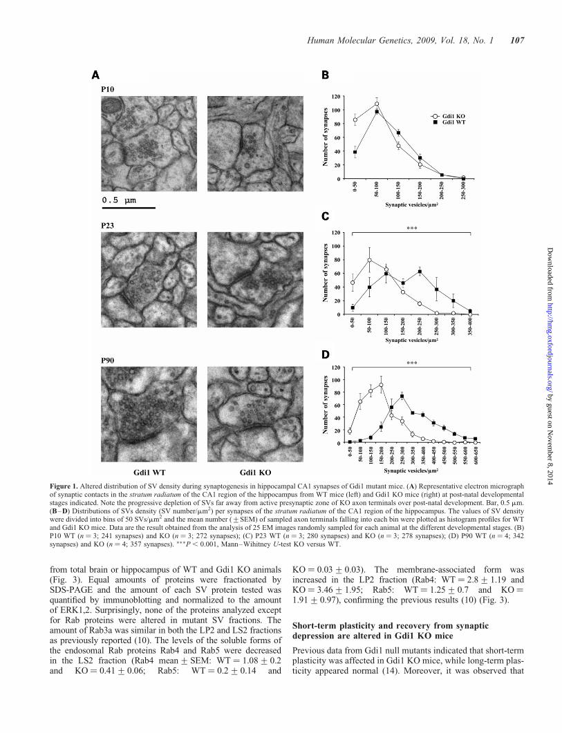
Synapses in stratum radiatum of CA1 were analyzed by elec-tron microscopy (EM) in Gdi1 KO and wild-type (WT) miceat post-natal day (P) 10, P23 and as adults at P90. Gdi1 KOmice showed a massive depletion of SVs in synaptic terminalsat P90 when compared with WT mice (Fig. 1A). All synapsesanalyzed were similarly affected and no difference betweenexcitatory (asymmetric) and inhibitory (symmetric) synapsescould be detected.
To quantify the changes in SV density during synapsedevelopment, 240– 350 synapses were counted at all develop-mental stages for each genotype. The results of the morpho-metric analysis are shown in Table 1 and Fig. 1B–D. Thereduction in SV density in Gdi1 KO terminals was significantat P23 and at the adult stage P90. The distributions of synapticdensity were significantly shifted towards synapses containingless SVs (Mann–Whitney U-test; P23 and P90, KO versusWT, P , 0.0001, both age levels). The average SV densitywas reduced to 42 and 50% of that of WT mice at P23 andP90, respectively. At P10 a small and not significant decreaseof 20% in SV density could be ascribed to a 50% increase ofthe number of synapses containing less than 50 SVs. No differ-ences could be detected in the average synaptic area or in theactive zone (AZ) length (Table 1). Similar reduction in SVdensity was found in the somatosensory cortex terminals(data not shown).
Since SV seemed to cluster near the AZ, we analyzed thenumber of SVs at progressively larger distance from the AZ,as described in Materials and Methods (Fig. 2A). The numberof docked SVs, 0–50 nm from the AZ (Fig. 2A) or manuallycounted (Fig. 2B), was unchanged. The same was found forSVs within 200–250 nm from the AZ (Fig. 2A). Highly statisti-cal significant differences between WT and Gdi1 KO synapseswere observed at distances �250 nm from the AZ (Mann–Whitney U-test KO versus WT, P ¼ 0.0003) (Fig. 2A). Theratio between the number of SVs in KO and WT synapsesdecreased sharply to about 5.5 times less SVs in KO mice(mean+SEM: 13.2+2.9) when compared with WT mice(mean+SEM: 73.5+22.2) synapses at 500 nm from theAZ (Fig. 2A). Thus, the morphometric analysis indicates thatthe lack of aGDI markedly impaired the RP of SVs, while itleft the readily releasable pool (RRP) relatively unaffected.
The total amount of SV proteins is not alteredin Gdi1 KO mice
To establish whether the SV proteome was altered by themutation, integral SV proteins, Rab3a interacting proteins,proteins involved in SV exocytosis and recycling and RabGTPases were analyzed by western blot in enriched SVmembrane fraction (LP2) and synaptosolic (LS2) fractions,
106 Human Molecular Genetics, 2009, Vol. 18, No. 1
by guest on Novem
ber 8, 2014http://hm
g.oxfordjournals.org/D
ownloaded from
from total brain or hippocampus of WT and Gdi1 KO animals(Fig. 3). Equal amounts of proteins were fractionated bySDS-PAGE and the amount of each SV protein tested wasquantified by immunoblotting and normalized to the amountof ERK1,2. Surprisingly, none of the proteins analyzed exceptfor Rab proteins were altered in mutant SV fractions. Theamount of Rab3a was similar in both the LP2 and LS2 fractionsas previously reported (10). The levels of the soluble forms ofthe endosomal Rab proteins Rab4 and Rab5 were decreasedin the LS2 fraction (Rab4 mean+SEM: WT ¼ 1.08+0.2and KO¼ 0.41+0.06; Rab5: WT ¼ 0.2+0.14 and
KO ¼ 0.03+0.03). The membrane-associated form wasincreased in the LP2 fraction (Rab4: WT ¼ 2.8+1.19 andKO ¼ 3.46+1.95; Rab5: WT ¼ 1.25+0.7 and KO¼1.91+0.97), confirming the previous results (10) (Fig. 3).
Short-term plasticity and recovery from synapticdepression are altered in Gdi1 KO mice
Previous data from Gdi1 null mutants indicated that short-termplasticity was affected in Gdi1 KO mice, while long-term plas-ticity appeared normal (14). Moreover, it was observed that
Figure 1. Altered distribution of SV density during synaptogenesis in hippocampal CA1 synapses of Gdi1 mutant mice. (A) Representative electron micrographof synaptic contacts in the stratum radiatum of the CA1 region of the hippocampus from WT mice (left) and Gdi1 KO mice (right) at post-natal developmentalstages indicated. Note the progressive depletion of SVs far away from active presynaptic zone of KO axon terminals over post-natal development. Bar, 0.5 mm.(B–D) Distributions of SVs density (SV number/mm2) per synapses of the stratum radiatum of the CA1 region of the hippocampus. The values of SV densitywere divided into bins of 50 SVs/mm2 and the mean number (+SEM) of sampled axon terminals falling into each bin were plotted as histogram profiles for WTand Gdi1 KO mice. Data are the result obtained from the analysis of 25 EM images randomly sampled for each animal at the different developmental stages. (B)P10 WT (n ¼ 3; 241 synapses) and KO (n ¼ 3; 272 synapses); (C) P23 WT (n ¼ 3; 280 synapses) and KO (n ¼ 3; 278 synapses); (D) P90 WT (n ¼ 4; 342synapses) and KO (n ¼ 4; 357 synapses). ���P , 0.001, Mann–Wihitney U-test KO versus WT.
Human Molecular Genetics, 2009, Vol. 18, No. 1 107
by guest on Novem
ber 8, 2014http://hm
g.oxfordjournals.org/D
ownloaded from
a 30 s train of pulses at 5 Hz was followed by a pronouncedpost-tetanic depression that suggested depletion of the SVpools in the mutant mice (10). To better define the character-istics of synaptic plasticity in Gdi1 KO mice, we analyzedpaired-pulse facilitation (PPF), post-tetanic potentiation(PTP) and synaptic depression at excitatory synapses of theCA1 region of the hippocampus.
Extracellular field excitatory postsynaptic potential (fEPSP)were recorded in the stratum radiatum of the CA1 region ofacute hippocampal slices collected from adult mice by stimulat-ing the Schaffer collateral pathway. The post-synaptic com-ponent of the field potential was totally inhibited by theaddition of 6-cyano-7-nitroquinoxaline-2,3-dione (CNQX)(10 mM), which isolated the fast fiber volley spike due to the acti-vation of voltage-gated Naþ channels, while the successiveapplication of tetrodotoxin (TTX) (1 mM) completely suppressedthe fiber volley wave (Supplementary Material, Fig. S1A).Fibers volley amplitudes were monitored in both experimentalgroups to ensure stable field potential recordings and showed asimilar long-lasting stability in the number of recruited afferentfibers by the extracellular stimulation in both WT and Gdi1 KOslices (Supplementary Material, Fig. S1B).
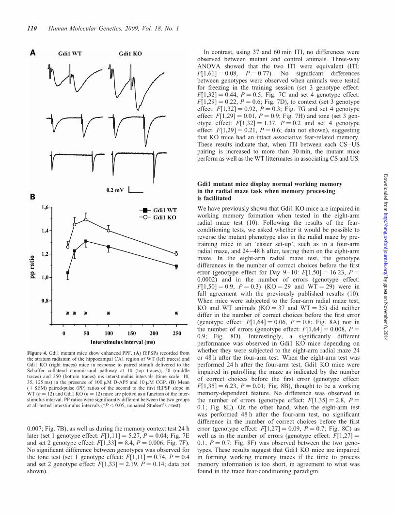
When Schaffer collaterals pathways were stimulated by twoconsecutive stimuli administered at interpulse intervalsranging between 10 and 250 ms (Fig. 4A), the magnitude ofPPF was significantly greater in Gdi1 KO mice than in WT lit-termates (Fig. 4B) at all tested time intervals. The ratio of thesecond to the first fEPSP slope at 50 ms interstimulus intervalwas 1.31+ 0.05 (WT ¼ 12) and 1.50+ 0.057 (KO ¼ 12) inWT and Gdi1 KO mice, respectively (P , 0.05; n ¼ 12;Fig. 4B). The strongly enhanced PPF observed in mutantmice is in partial agreement with previously reported results,which reported a PPF increase limited to interpulse intervalsshorter than 50 ms (14). This result is compatible with eitherlower release probability, larger build-up of Ca2þ ions, orhigher rate of fast SV recycling mechanisms.
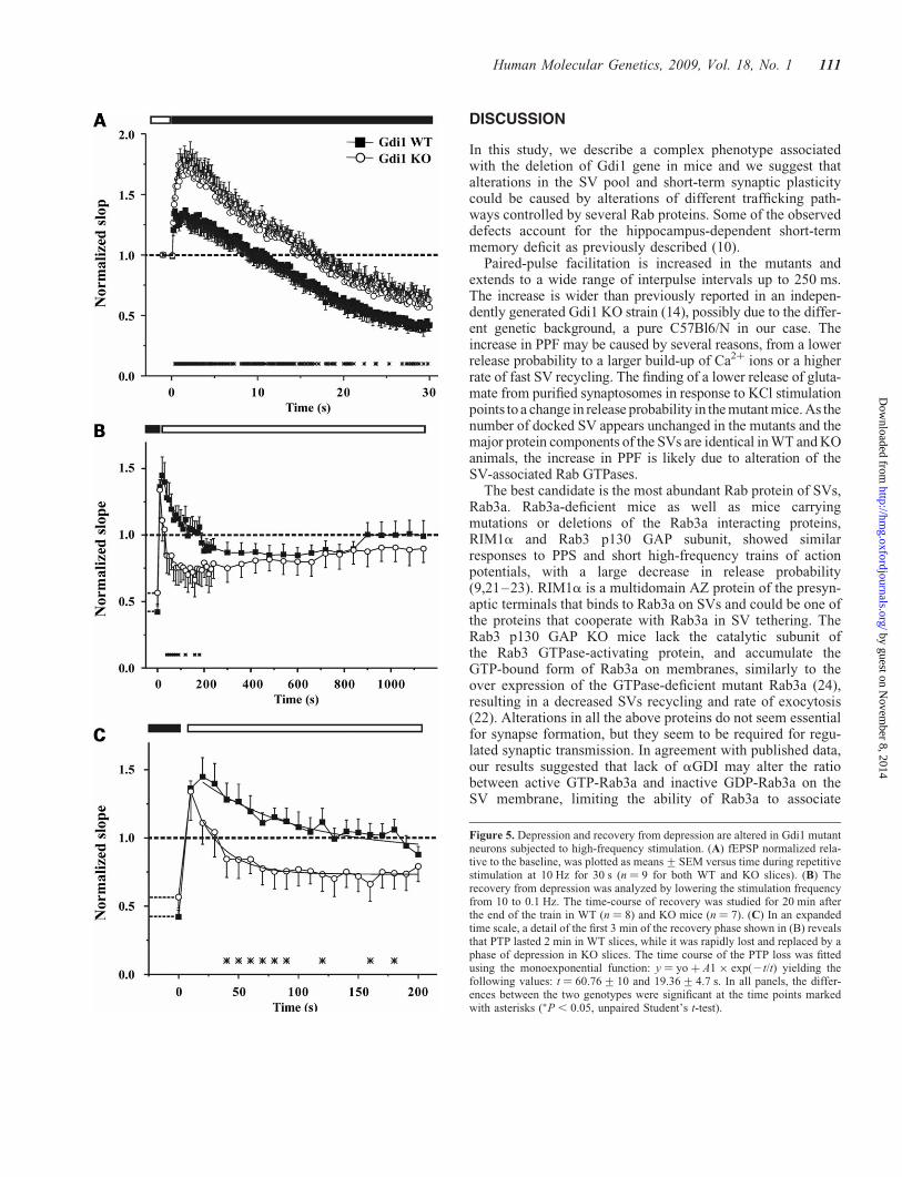
To evaluate synaptic depression, the Schaffer collateralpathway was stimulated with a train of 300 stimuli deliveredat a frequency of 10 Hz. The progressive change in fEPSPslope ratio during the train and the subsequent recoveryfrom depression, obtained by returning the stimulation fre-quency to 0.1 Hz, were compared in WT and mutant slices.In WT mice, the long-lasting repetitive stimulation elicitedan initial moderate facilitation of 130+ 9% with respect tobasal transmission before the train followed by a decrementedphase that reached a depression of 42.5+ 4.5% at the end ofthe stimulation train (Fig. 5A). In contrast, Gdi1 KO synapses
displayed a larger facilitation during the initial phase of tetanicstimulation (174+ 11%) and a subsequent depression charac-terized by a kinetic similar to that observed in WT synapses.At the end of the train, Gdi1 KO synapses reached a milderdepression plateau of 60.6+ 8.3% (P , 0.05 KO versusWT; n ¼ 9 for both WT and KO slices; Fig. 5A). The initiallarger facilitation observed in Gdi1 KO slices is similar towhat we observed in the PPF and the milder depression islikely due to the more intense facilitation observed at thebeginning of the train.
When the stimulation frequency was returned to 0.1 Hz bothWT and Gdi1 KO synapses revealed a similar extent of PTP(136+ 5.4 and 134+ 21% in WT and KO mice, respect-ively), a transient increase in synaptic strength caused byCa2þ accumulation within the presynaptic terminal (15). InWT mice, the PTP phase lasted for 2 min and was slowlyand progressively replaced by a moderate depression phaselasting 11 min and reaching a depression plateau of 85.6+6.7% (Fig. 5B). On the contrary, PTP lasted ,30 s in Gdi1KO mice, and was soon replaced by a phase of intensedepression that reached a plateau level of 71.3+ 12%, 3 minafter the end of the train (P , 0.05 KO versus WT; n ¼ 7and n ¼ 8 for WT and KO mice, respectively; Fig. 5B). Thefitting of the first 3 min of the recovery phase with a mono-exponential curve revealed a faster decay of the PTP phasein KO slices (t ¼ 60.76+ 10 and 19.36+ 4.7 s for WT andKO mice, respectively; Fig. 5C). This effect could be the con-sequence of the strong SV depletion observed in the KOsynapses by EM, which alter the SV recruitment from the RP.
Glutamate release in response to KCl is decreasedin Gdi1 KO synaptosomes
To better define the release properties of the Gdi1 KO mice,release experiments were conducted in hippocampalsynaptosomes purified from adult mice and exposed to KCl,known to evoke voltage-gated Ca2þ-channel-dependentrelease (16,17). Synaptosomes were purified from the hippo-campus of Gdi1 KO and WT mice and exposed in superfusionto KCl, to monitor the stimulus-evoked overflow of endogen-ous glutamate. During the first 36 min of superfusion withstandard medium the spontaneous outflow asymptoticallydiminished to a constant level; a 90 s exposure to the stimulusat t ¼ 39 min produced a transient increase of neurotransmitterrelease that soon returned to the basal levels (SupplementaryMaterial, Fig. S2A). The release induced by 50 mM KClappeared only partly dependent on external Ca2þ, as reported
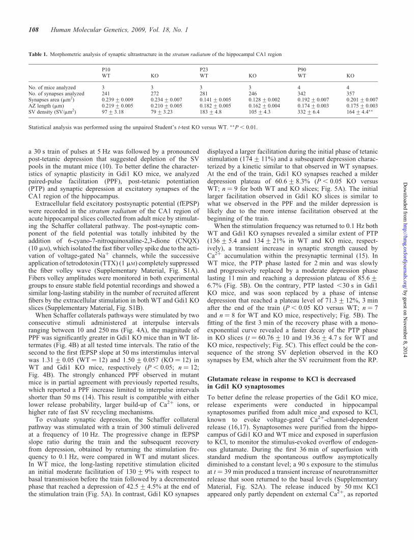
Table 1. Morphometric analysis of synaptic ultrastructure in the stratum radiatum of the hippocampal CA1 region
P10 P23 P90WT KO WT KO WT KO
No. of mice analyzed 3 3 3 3 4 4No. of synapses analyzed 241 272 281 246 342 357Synapses area (mm2) 0.239+0.009 0.234+0.007 0.141+0.005 0.128+0.002 0.192+0.007 0.201+0.007AZ length (mm) 0.219+0.005 0.210+0.005 0.182+0.005 0.162+0.004 0.174+0.003 0.175+0.003SV density (SV/mm2) 97+3.18 79+3.23 183+4.8 105+4.3 332+6.4 164+4.4��
Statistical analysis was performed using the unpaired Student’s t-test KO versus WT. ��P , 0.01.
108 Human Molecular Genetics, 2009, Vol. 18, No. 1
by guest on Novem
ber 8, 2014http://hm
g.oxfordjournals.org/D
ownloaded from

for rat brain synaptosomes (18,19) (Supplementary Material,Fig. S2B).
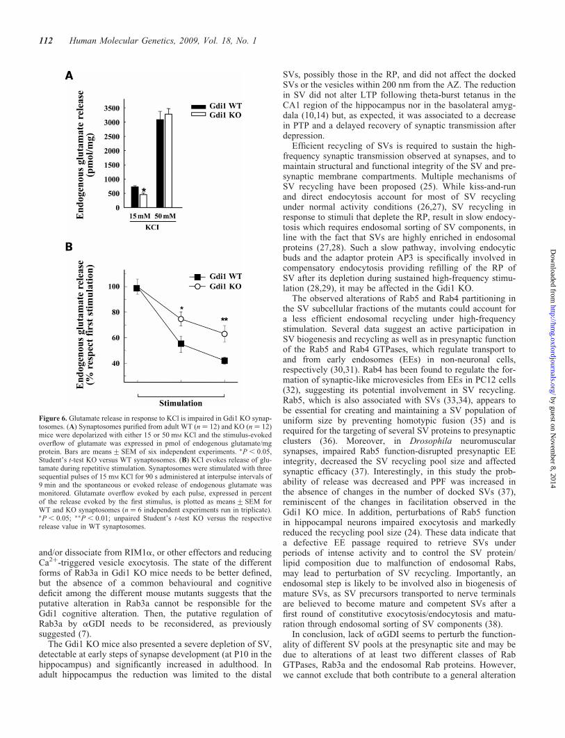
Evoked glutamate release from hippocampal synaptosomeswas directly related to the strength of the applied stimulus. Asshown in Fig. 6A, glutamate overflow elicited by 15 mM KClwas significantly reduced by �30–40% in mutant synapto-somes with respect to WT synaptosomes. However, whenhigher KCl concentrations (50 mM) were used, the glutamateoverflow was similar in WT and KO synaptosomes(Fig. 6A). We also measured the extent of glutamate releasein response to three sequential depolarizing pulses of 15 mM
KCl applied at 9 min interpulse intervals (Fig. 6B). Consistentwith previous reports (20), glutamate overflow from WTsynaptosomes progressively decreased in response to sequen-tial stimuli, as a result of the SV depletion from RRP. Interest-ingly, glutamate overflow from KO synaptosomes wassignificantly less depressed after repetitive pulses (Fig. 6B).The lower release of glutamate from purified KO synapto-somes in response to KCl stimulation suggest a change inrelease probability that does not depend on the number ofdocked SV in the RRP, which is in fact unchanged in themutants.
Deficits of Gdi1 mutant in trace fear conditioningis corrected by applying longer intertrialintervals during training session
We previously showed that Gdi1 KO mice were impaired informing association between the conditioning stimulus (CS)and an unconditioned stimulus (US) in the trace fear-conditioning test in which mutants were trained with fivetrials separated by short intertrial interval (ITI) of 148 s (10).To assess whether the ITI duration plays a key role in associ-ative memory formation in Gdi1 KO mice, we modified thetraining protocol. Four sets of Gdi1 KO and WT male mice lit-termates were subjected to trace fear conditioning using five
trials separated by 15, 26, 37 or 60 min ITI, as showed inFig. 7A–H.
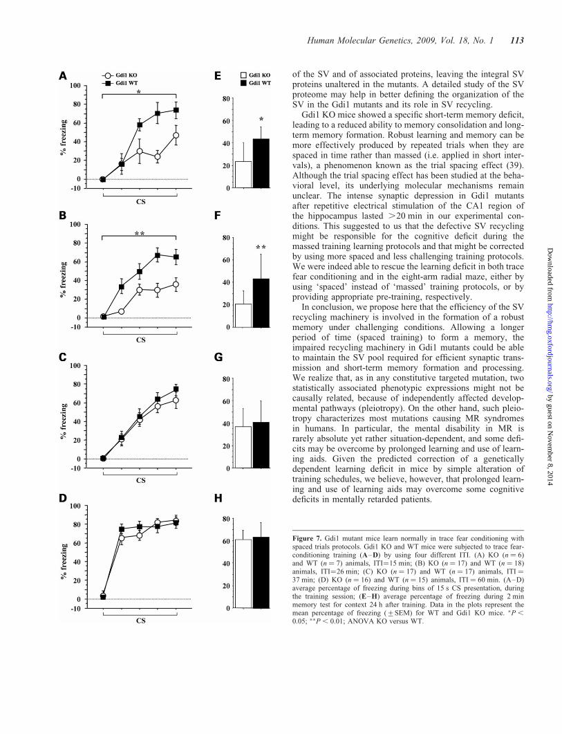
Using 15 and 26 min ITI, Gdi1 KO mice froze significantlyless than WT mice when the CS was presented during the con-ditioning sessions (Fig. 7A and B). Three-way ANOVAshowed that the two ITI were equivalent (ITI: F[1,44] ¼0.07, P ¼ 0.78). In both protocols, the percentage of freezingover the five CS tones presentation increased in both WT andGdi1 KO mice during the conditioning session (CS effect:F[4,184] ¼ 49.5, P , 0.0001). Significant differencesbetween genotypes were observed for freezing in the trainingsession (set 1 genotype effect: F[1,11] ¼ 13.9, P ¼ 0.02;Fig. 7A and set 2 genotype effect: F[1,33] ¼ 6.94, P ¼
Figure 3. Analysis of SV and Rab proteins in nerve terminal fractions of wild-type and Gdi1 mutant mice. Crude SV fractions (LP2) and synaptosol (LS2)prepared from total brain of WT (n ¼ 3) and Gdi1 KO (n ¼ 3) P90 micewere analyzed by SDS-PAGE and immunoblotting. A battery of antibodies(listed in Supplementary Material, Table S1) against SV proteins, Rab3a inter-acting proteins, exo- and endocytosis markers and Rab proteins, and ERK1,2to normalize the total amount of proteins loaded, were used. A differencebetween genotypes was observed for Rab4 and Rab5 (LP2 fraction: Rab4:WT ¼ 2.8+1.19 and KO ¼ 3.46+1.95; Rab5: WT ¼ 1.25+0.7 andKO ¼ 1.91+0.97; LS2 fraction: Rab4: WT ¼ 1.08+0.2 and KO ¼ 0.41+0.06; Rab5: WT ¼ 0.2+0.14 and KO ¼ 0.03+0.03). VGLUT, vesicularglutamate transporter; VGAT, vesicular GABA transporter; SV2, SVprotein-2.
Figure 2. Gdi1 mutant mice showed altered SV reserve pool. (A) Distributionof absolute number of SVs (means+SEM) located within successive 50 nmthick shells from the AZ was calculated, by using the Excel macro. The analy-sis was done from 342 synaptic terminals of P90 WT (n ¼ 4) and 357 synapticterminal Gdi1 KO (n ¼ 4) mice in CA1 region of the hippocampus. ���P ,
0.001, Mann–Whitney U-test KO versus WT, for the intervals between 250and 550 nm. (B) Manually counting of docked SVs from 342 synaptic terminalof P90 WT (n ¼ 4) and 357 synaptic terminal Gdi1 KO (n ¼ 4) mice hippo-campus and plotted as means (+SEM).
Human Molecular Genetics, 2009, Vol. 18, No. 1 109
by guest on Novem
ber 8, 2014http://hm
g.oxfordjournals.org/D
ownloaded from
0.007; Fig. 7B), as well as during the memory context test 24 hlater (set 1 genotype effect: F[1,11] ¼ 5.27, P ¼ 0.04; Fig. 7Eand set 2 genotype effect: F[1,33] ¼ 8.4, P ¼ 0.006; Fig. 7F).No significant difference between genotypes was observed forthe tone test (set 1 genotype effect: F[1,11] ¼ 0.74, P ¼ 0.4and set 2 genotype effect: F[1,33] ¼ 2.19, P ¼ 0.14; data notshown).
In contrast, using 37 and 60 min ITI, no differences wereobserved between mutant and control animals. Three-wayANOVA showed that the two ITI were equivalent (ITI:F[1,61] ¼ 0.08, P ¼ 0.77). No significant differencesbetween genotypes were observed when animals were testedfor freezing in the training session (set 3 genotype effect:F[1,32] ¼ 0.44, P ¼ 0.5; Fig. 7C and set 4 genotype effect:F[1,29] ¼ 0.22, P ¼ 0.6; Fig. 7D), to context (set 3 genotypeeffect: F[1,32] ¼ 0.92, P ¼ 0.3; Fig. 7G and set 4 genotypeeffect: F[1,29] ¼ 0.01, P ¼ 0.9; Fig. 7H) and tone (set 3 gen-otype effect: F[1,32] ¼ 1.37, P ¼ 0.2 and set 4 genotypeeffect: F[1,29] ¼ 0.21, P ¼ 0.6; data not shown), suggestingthat KO mice had an intact associative fear-related memory.These results indicate that, when ITI between each CS–USpairing is increased to more than 30 min, the mutant miceperform as well as the WT littermates in associating CS and US.
Gdi1 mutant mice display normal working memoryin the radial maze task when memory processingis facilitated
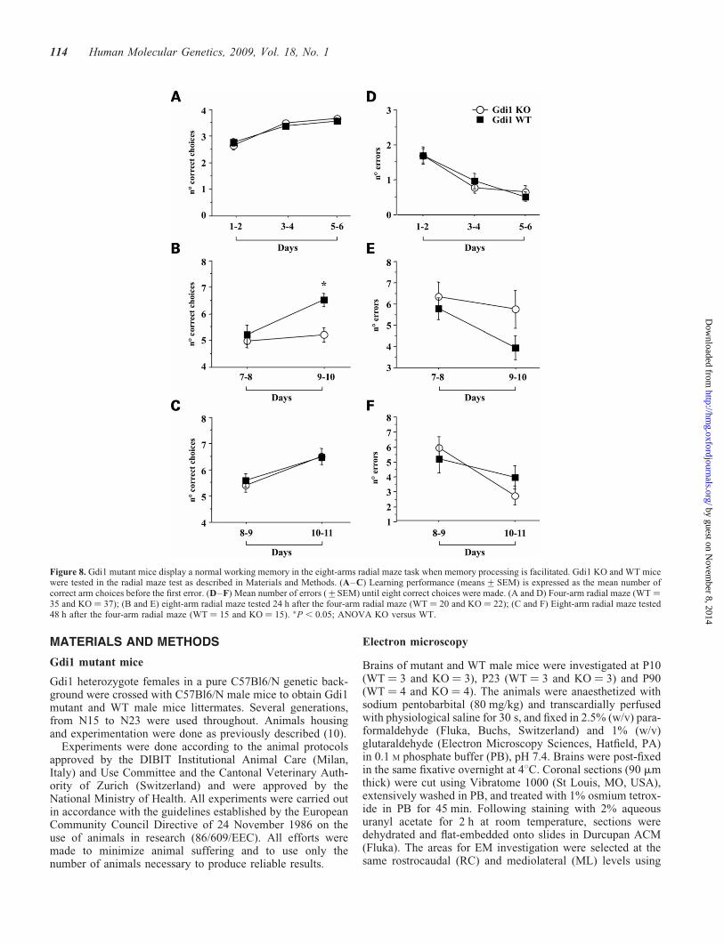
We have previously shown that Gdi1 KO mice are impaired inworking memory formation when tested in the eight-armradial maze test (10). Following the results of the fear-conditioning tests, we asked whether it would be possible toreverse the mutant phenotype also in the radial maze by pre-training mice in an ‘easier set-up’, such as in a four-armradial maze, and 24–48 h after, testing them on the eight-armmaze. In the eight-arm radial maze test, the genotypedifferences in the number of correct choices before the firsterror (genotype effect for Day 9–10: F[1,50] ¼ 16.23, P ¼0.0002) and in the number of errors (genotype effect:F[1,50] ¼ 0.9, P ¼ 0.3) (KO ¼ 29 and WT ¼ 29) were infull agreement with the previously published results (10).When mice were subjected to the four-arm radial maze test,KO and WT animals (KO ¼ 37 and WT ¼ 35) did neitherdiffer in the number of correct choices before the first error(genotype effect: F[1,64] ¼ 0.06, P ¼ 0.8; Fig. 8A) nor inthe number of errors (genotype effect: F[1,64] ¼ 0.008, P ¼0.9; Fig. 8D). Interestingly, a significantly differentperformance was observed in Gdi1 KO mice depending onwhether they were subjected to the eight-arm radial maze 24or 48 h after the four-arm test. When the eight-arm test wasperformed 24 h after the four-arm test, Gdi1 KO mice wereimpaired in patrolling the maze as indicated by the numberof correct choices before the first error (genotype effect:F[1,35] ¼ 6.23, P ¼ 0.01; Fig. 8B), thought to be a workingmemory-dependent feature. No difference was observed inthe number of errors (genotype effect: F[1,35] ¼ 2.8, P ¼0.1; Fig. 8E). On the other hand, when the eight-arm testwas performed 48 h after the four-arm test, no significantdifference in the number of correct choices before the firsterror (genotype effect: F[1,27] ¼ 0.09, P ¼ 0.7; Fig. 8C) aswell as in the number of errors (genotype effect: F[1,27] ¼0.1, P ¼ 0.7; Fig. 8F) was observed between the two geno-types. These results suggest that Gdi1 KO mice are impairedin forming working memory traces if the time to processmemory information is too short, in agreement to what wasfound in the trace fear-conditioning paradigm.
Figure 4. Gdi1 mutant mice show enhanced PPF. (A) fEPSPs recorded fromthe stratum radiatum of the hippocampal CA1 region of WT (left traces) andGdi1 KO (right traces) mice in response to paired stimuli delivered to theSchaffer collateral commissural pathway at 10 (top traces), 50 (middletraces) and 250 (bottom traces) ms interstimulus intervals (time scale: 10,35, 125 ms) in the presence of 100 mM D-AP5 and 10 mM CGP. (B) Mean(+SEM) paired-pulse (PP) ratios of the second to the first fEPSP slope inWT (n ¼ 12) and Gdi1 KO (n ¼ 12) mice are plotted as a function of the inter-stimulus interval. PP ratios were significantly different between the two groupsat all tested interstimulus intervals (�P , 0.05, unpaired Student’s t-test).
110 Human Molecular Genetics, 2009, Vol. 18, No. 1
by guest on Novem
ber 8, 2014http://hm
g.oxfordjournals.org/D
ownloaded from
DISCUSSION
In this study, we describe a complex phenotype associatedwith the deletion of Gdi1 gene in mice and we suggest thatalterations in the SV pool and short-term synaptic plasticitycould be caused by alterations of different trafficking path-ways controlled by several Rab proteins. Some of the observeddefects account for the hippocampus-dependent short-termmemory deficit as previously described (10).
Paired-pulse facilitation is increased in the mutants andextends to a wide range of interpulse intervals up to 250 ms.The increase is wider than previously reported in an indepen-dently generated Gdi1 KO strain (14), possibly due to the differ-ent genetic background, a pure C57Bl6/N in our case. Theincrease in PPF may be caused by several reasons, from a lowerrelease probability to a larger build-up of Ca2þ ions or a higherrate of fast SV recycling. The finding of a lower release of gluta-mate from purified synaptosomes in response to KCl stimulationpoints to a change in release probability in the mutant mice. As thenumber of docked SV appears unchanged in the mutants and themajor protein components of the SVs are identical in WT and KOanimals, the increase in PPF is likely due to alteration of theSV-associated Rab GTPases.
The best candidate is the most abundant Rab protein of SVs,Rab3a. Rab3a-deficient mice as well as mice carryingmutations or deletions of the Rab3a interacting proteins,RIM1a and Rab3 p130 GAP subunit, showed similarresponses to PPS and short high-frequency trains of actionpotentials, with a large decrease in release probability(9,21–23). RIM1a is a multidomain AZ protein of the presyn-aptic terminals that binds to Rab3a on SVs and could be one ofthe proteins that cooperate with Rab3a in SV tethering. TheRab3 p130 GAP KO mice lack the catalytic subunit ofthe Rab3 GTPase-activating protein, and accumulate theGTP-bound form of Rab3a on membranes, similarly to theover expression of the GTPase-deficient mutant Rab3a (24),resulting in a decreased SVs recycling and rate of exocytosis(22). Alterations in all the above proteins do not seem essentialfor synapse formation, but they seem to be required for regu-lated synaptic transmission. In agreement with published data,our results suggested that lack of aGDI may alter the ratiobetween active GTP-Rab3a and inactive GDP-Rab3a on theSV membrane, limiting the ability of Rab3a to associate
Figure 5. Depression and recovery from depression are altered in Gdi1 mutantneurons subjected to high-frequency stimulation. (A) fEPSP normalized rela-tive to the baseline, was plotted as means+SEM versus time during repetitivestimulation at 10 Hz for 30 s (n ¼ 9 for both WT and KO slices). (B) Therecovery from depression was analyzed by lowering the stimulation frequencyfrom 10 to 0.1 Hz. The time-course of recovery was studied for 20 min afterthe end of the train in WT (n ¼ 8) and KO mice (n ¼ 7). (C) In an expandedtime scale, a detail of the first 3 min of the recovery phase shown in (B) revealsthat PTP lasted 2 min in WT slices, while it was rapidly lost and replaced by aphase of depression in KO slices. The time course of the PTP loss was fittedusing the monoexponential function: y ¼ yo þ A1 � exp(2t/t) yielding thefollowing values: t ¼ 60.76+10 and 19.36+4.7 s. In all panels, the differ-ences between the two genotypes were significant at the time points markedwith asterisks (�P , 0.05, unpaired Student’s t-test).
Human Molecular Genetics, 2009, Vol. 18, No. 1 111
by guest on Novem
ber 8, 2014http://hm
g.oxfordjournals.org/D
ownloaded from
and/or dissociate from RIM1a, or other effectors and reducingCa2þ-triggered vesicle exocytosis. The state of the differentforms of Rab3a in Gdi1 KO mice needs to be better defined,but the absence of a common behavioural and cognitivedeficit among the different mouse mutants suggests that theputative alteration in Rab3a cannot be responsible for theGdi1 cognitive alteration. Then, the putative regulation ofRab3a by aGDI needs to be reconsidered, as previouslysuggested (7).
The Gdi1 KO mice also presented a severe depletion of SV,detectable at early steps of synapse development (at P10 in thehippocampus) and significantly increased in adulthood. Inadult hippocampus the reduction was limited to the distal
SVs, possibly those in the RP, and did not affect the dockedSVs or the vesicles within 200 nm from the AZ. The reductionin SV did not alter LTP following theta-burst tetanus in theCA1 region of the hippocampus nor in the basolateral amyg-dala (10,14) but, as expected, it was associated to a decreasein PTP and a delayed recovery of synaptic transmission afterdepression.
Efficient recycling of SVs is required to sustain the high-frequency synaptic transmission observed at synapses, and tomaintain structural and functional integrity of the SV and pre-synaptic membrane compartments. Multiple mechanisms ofSV recycling have been proposed (25). While kiss-and-runand direct endocytosis account for most of SV recyclingunder normal activity conditions (26,27), SV recycling inresponse to stimuli that deplete the RP, result in slow endocy-tosis which requires endosomal sorting of SV components, inline with the fact that SVs are highly enriched in endosomalproteins (27,28). Such a slow pathway, involving endocyticbuds and the adaptor protein AP3 is specifically involved incompensatory endocytosis providing refilling of the RP ofSV after its depletion during sustained high-frequency stimu-lation (28,29), it may be affected in the Gdi1 KO.
The observed alterations of Rab5 and Rab4 partitioning inthe SV subcellular fractions of the mutants could account fora less efficient endosomal recycling under high-frequencystimulation. Several data suggest an active participation inSV biogenesis and recycling as well as in presynaptic functionof the Rab5 and Rab4 GTPases, which regulate transport toand from early endosomes (EEs) in non-neuronal cells,respectively (30,31). Rab4 has been found to regulate the for-mation of synaptic-like microvesicles from EEs in PC12 cells(32), suggesting its potential involvement in SV recycling.Rab5, which is also associated with SVs (33,34), appears tobe essential for creating and maintaining a SV population ofuniform size by preventing homotypic fusion (35) and isrequired for the targeting of several SV proteins to presynapticclusters (36). Moreover, in Drosophila neuromuscularsynapses, impaired Rab5 function-disrupted presynaptic EEintegrity, decreased the SV recycling pool size and affectedsynaptic efficacy (37). Interestingly, in this study the prob-ability of release was decreased and PPF was increased inthe absence of changes in the number of docked SVs (37),reminiscent of the changes in facilitation observed in theGdi1 KO mice. In addition, perturbations of Rab5 functionin hippocampal neurons impaired exocytosis and markedlyreduced the recycling pool size (24). These data indicate thata defective EE passage required to retrieve SVs underperiods of intense activity and to control the SV protein/lipid composition due to malfunction of endosomal Rabs,may lead to perturbation of SV recycling. Importantly, anendosomal step is likely to be involved also in biogenesis ofmature SVs, as SV precursors transported to nerve terminalsare believed to become mature and competent SVs after afirst round of constitutive exocytosis/endocytosis and matu-ration through endosomal sorting of SV components (38).
In conclusion, lack of aGDI seems to perturb the function-ality of different SV pools at the presynaptic site and may bedue to alterations of at least two different classes of RabGTPases, Rab3a and the endosomal Rab proteins. However,we cannot exclude that both contribute to a general alteration
Figure 6. Glutamate release in response to KCl is impaired in Gdi1 KO synap-tosomes. (A) Synaptosomes purified from adult WT (n ¼ 12) and KO (n ¼ 12)mice were depolarized with either 15 or 50 mM KCl and the stimulus-evokedoverflow of glutamate was expressed in pmol of endogenous glutamate/mgprotein. Bars are means+SEM of six independent experiments. �P , 0.05,Student’s t-test KO versus WT synaptosomes. (B) KCl evokes release of glu-tamate during repetitive stimulation. Synaptosomes were stimulated with threesequential pulses of 15 mM KCl for 90 s administered at interpulse intervals of9 min and the spontaneous or evoked release of endogenous glutamate wasmonitored. Glutamate overflow evoked by each pulse, expressed in percentof the release evoked by the first stimulus, is plotted as means+SEM forWT and KO synaptosomes (n ¼ 6 independent experiments run in triplicate).�P , 0.05; ��P , 0.01; unpaired Student’s t-test KO versus the respectiverelease value in WT synaptosomes.
112 Human Molecular Genetics, 2009, Vol. 18, No. 1
by guest on Novem
ber 8, 2014http://hm
g.oxfordjournals.org/D
ownloaded from
of the SV and of associated proteins, leaving the integral SVproteins unaltered in the mutants. A detailed study of the SVproteome may help in better defining the organization of theSV in the Gdi1 mutants and its role in SV recycling.
Gdi1 KO mice showed a specific short-term memory deficit,leading to a reduced ability to memory consolidation and long-term memory formation. Robust learning and memory can bemore effectively produced by repeated trials when they arespaced in time rather than massed (i.e. applied in short inter-vals), a phenomenon known as the trial spacing effect (39).Although the trial spacing effect has been studied at the beha-vioral level, its underlying molecular mechanisms remainunclear. The intense synaptic depression in Gdi1 mutantsafter repetitive electrical stimulation of the CA1 region ofthe hippocampus lasted .20 min in our experimental con-ditions. This suggested to us that the defective SV recyclingmight be responsible for the cognitive deficit during themassed training learning protocols and that might be correctedby using more spaced and less challenging training protocols.We were indeed able to rescue the learning deficit in both tracefear conditioning and in the eight-arm radial maze, either byusing ‘spaced’ instead of ‘massed’ training protocols, or byproviding appropriate pre-training, respectively.
In conclusion, we propose here that the efficiency of the SVrecycling machinery is involved in the formation of a robustmemory under challenging conditions. Allowing a longerperiod of time (spaced training) to form a memory, theimpaired recycling machinery in Gdi1 mutants could be ableto maintain the SV pool required for efficient synaptic trans-mission and short-term memory formation and processing.We realize that, as in any constitutive targeted mutation, twostatistically associated phenotypic expressions might not becausally related, because of independently affected develop-mental pathways (pleiotropy). On the other hand, such pleio-tropy characterizes most mutations causing MR syndromesin humans. In particular, the mental disability in MR israrely absolute yet rather situation-dependent, and some defi-cits may be overcome by prolonged learning and use of learn-ing aids. Given the predicted correction of a geneticallydependent learning deficit in mice by simple alteration oftraining schedules, we believe, however, that prolonged learn-ing and use of learning aids may overcome some cognitivedeficits in mentally retarded patients.
Figure 7. Gdi1 mutant mice learn normally in trace fear conditioning withspaced trials protocols. Gdi1 KO and WT mice were subjected to trace fear-conditioning training (A–D) by using four different ITI. (A) KO (n ¼ 6)and WT (n ¼ 7) animals, ITI¼15 min; (B) KO (n ¼ 17) and WT (n ¼ 18)animals, ITI¼26 min; (C) KO (n ¼ 17) and WT (n ¼ 17) animals, ITI ¼37 min; (D) KO (n ¼ 16) and WT (n ¼ 15) animals, ITI ¼ 60 min. (A–D)average percentage of freezing during bins of 15 s CS presentation, duringthe training session; (E–H) average percentage of freezing during 2 minmemory test for context 24 h after training. Data in the plots represent themean percentage of freezing (+SEM) for WT and Gdi1 KO mice. �P ,
0.05; ��P , 0.01; ANOVA KO versus WT.
Human Molecular Genetics, 2009, Vol. 18, No. 1 113
by guest on Novem
ber 8, 2014http://hm
g.oxfordjournals.org/D
ownloaded from
MATERIALS AND METHODS
Gdi1 mutant mice
Gdi1 heterozygote females in a pure C57Bl6/N genetic back-ground were crossed with C57Bl6/N male mice to obtain Gdi1mutant and WT male mice littermates. Several generations,from N15 to N23 were used throughout. Animals housingand experimentation were done as previously described (10).
Experiments were done according to the animal protocolsapproved by the DIBIT Institutional Animal Care (Milan,Italy) and Use Committee and the Cantonal Veterinary Auth-ority of Zurich (Switzerland) and were approved by theNational Ministry of Health. All experiments were carried outin accordance with the guidelines established by the EuropeanCommunity Council Directive of 24 November 1986 on theuse of animals in research (86/609/EEC). All efforts weremade to minimize animal suffering and to use only thenumber of animals necessary to produce reliable results.
Electron microscopy
Brains of mutant and WT male mice were investigated at P10(WT ¼ 3 and KO ¼ 3), P23 (WT ¼ 3 and KO ¼ 3) and P90(WT ¼ 4 and KO ¼ 4). The animals were anaesthetized withsodium pentobarbital (80 mg/kg) and transcardially perfusedwith physiological saline for 30 s, and fixed in 2.5% (w/v) para-formaldehyde (Fluka, Buchs, Switzerland) and 1% (w/v)glutaraldehyde (Electron Microscopy Sciences, Hatfield, PA)in 0.1 M phosphate buffer (PB), pH 7.4. Brains were post-fixedin the same fixative overnight at 48C. Coronal sections (90 mmthick) were cut using Vibratome 1000 (St Louis, MO, USA),extensively washed in PB, and treated with 1% osmium tetrox-ide in PB for 45 min. Following staining with 2% aqueousuranyl acetate for 2 h at room temperature, sections weredehydrated and flat-embedded onto slides in Durcupan ACM(Fluka). The areas for EM investigation were selected at thesame rostrocaudal (RC) and mediolateral (ML) levels using
Figure 8. Gdi1 mutant mice display a normal working memory in the eight-arms radial maze task when memory processing is facilitated. Gdi1 KO and WT micewere tested in the radial maze test as described in Materials and Methods. (A–C) Learning performance (means+SEM) is expressed as the mean number ofcorrect arm choices before the first error. (D–F) Mean number of errors (+SEM) until eight correct choices were made. (A and D) Four-arm radial maze (WT ¼35 and KO ¼ 37); (B and E) eight-arm radial maze tested 24 h after the four-arm radial maze (WT ¼ 20 and KO ¼ 22); (C and F) Eight-arm radial maze tested48 h after the four-arm radial maze (WT ¼ 15 and KO ¼ 15). �P , 0.05; ANOVA KO versus WT.
114 Human Molecular Genetics, 2009, Vol. 18, No. 1
by guest on Novem
ber 8, 2014http://hm
g.oxfordjournals.org/D
ownloaded from
the mouse brain atlas (40). Hippocampal CA1 samples werecut out from the slide at Bregma 22.20 and 22.40 mm RC,and 1.05–1.45 and 2.70–3.10 mm ML, respectively. Serialultrathin sections were collected on pioloform-coated single-slot copper grids and examined with a CM100 electron micro-scope (Philips Electron Optics, Eindhoven, The Netherlands)at 80 kV. The section thickness was controlled using theSmall’s minimal fold method (41); 55+ 3 nm thick sectionswere singled out from series for image sampling.
Morphometric analysis
The selected section was oriented at �700 magnification toselect four random fields at least 80 mm distant, within themiddle portion of CA1 stratum radiatum. Four to eight non-overlapping images at 3–5 mm distance were captured perrandom field at an initial �27 500 magnification. In total, 25images or area of 380 mm2 were sampled for each animalwith Bioscan Camera 792 (Gatan, Pleasanton, CA, USA)using Digital Micrograph 2.5 software (NCMI, Houston, TX,USA). A synapse was identified by the clustering of SV andby the presence of a post-synaptic density (PSD). Synapticprofiles touching the exclusion lines were not counted. Thelengths of AZ, the numbers of SVs and the areas of theaxonal terminals were measured by using ImageJ software(NIH, Bethesda, MD, USA). The density of SV per axon term-inal area was obtained dividing the number of SVs by thecross-section areas of the axonal terminals; the area occupiedby mitochondria was subtracted from the total area of axonalterminal. The distance of each SV from the presynaptic mem-brane was determined by using an Excel Macro.
Western blot analysis
Six brains from P90 mice (KO ¼ 3; WT ¼ 3) were dissected,fractionated and analyzed as previously described (10). Anti-bodies are listed in Supplementary Material, Table S1.
Hippocampal slices and field recordings
Horizontal hippocampal slices were obtained from 14–16weeks old male mice. Mice were anaesthetized with halothane(Sigma-Aldrich, Milan, Italy) and decapitated; the brain wasquickly removed and immersed in an ice-cold oxygenated‘cutting’ solution composed of (mM): 125 NaCl, 25NaHCO3, 25 C6H12O6, 2.5 KCl, 1.25 NaH2PO4, 1 CaCl2, 2MgCl2, 0.4 ascorbic acid, 2 sodium pyruvate, 3 myo-Inositol.The solution was saturated with 95% O2 and 5% CO2 (42).The dissected hemispheres were cut into 0.3 mm thick slicesusing an HM 650 V vibratome (Microm InternationalGmbH, Walldorf, Germany). Slices were incubated in‘cutting’ solution at 358C for at least 1 h and then transferredinto a ‘submerged’ recording chamber which was continu-ously superfused at a rate of �1.5 ml/min with the standardrecording solution composed of (mM): 125 NaCl, 25NaHCO3, 25 C6H12O6, 2.5 KCl, 1.25 NaH2PO4, 2 CaCl2, 1MgCl2. D-2-amino-5-phosphonopentanoic acid (100 mM;Tocris Bioscience, Bristol, UK) and (3-aminopropyl) (diethox-ymethyl) phosphinic acid (CGP; 10 mM; Tocris) were added inall the experiments to block N-methyl-D-aspartate and GABAB
receptors, respectively. For the pharmacological characteriz-ation, CNQX (10 mM; Tocris) and TTX (1 mM; Sigma) wereperfused in the recording chamber. The bath temperaturewas monitored and maintained at 338C. Extracellular fieldpotentials were recorded in the stratum radiatum of the CA1region with a glass microelectrode (1–2 MV) filled with stan-dard recording solution. Electrical stimulation (test pulses at0.1 Hz, 0.25 ms duration, stimulus intensity fixed at 2/3 ofmaximal evoked EPSP amplitude) was delivered through aconcentric bipolar tungsten electrode inserted into thestratum radiatum of the CA1 region (see SupplementaryMaterial, Fig. S1). Field EPSPs were acquired by using theMultiClamp 700B (Axon Instruments, Molecular Devices,Sunnyvale, CA, USA) amplifier and the pClamp 9.2 software(Axon Instruments). The fEPSP slope was evaluated as alinear regression of the rise phase from 10 to 70% of thefEPSP amplitude (R2 . 0.9) using a proprietary programdeveloped in an R-CRAN environment (Thierry Nieus, unpub-lished data). For each experiment, the ‘baseline slope’ was cal-culated by averaging the slope values obtained after thestimulation of the Schaffer collaterals at 0.1 Hz for 5 min. Inthe high-frequency repetitive stimulation protocols, datawere expressed in percent of the baseline slope as means+SEM. In the paired-pulse experiments, the fEPSP slope ratiobetween the second and the first stimulus was calculated. Stat-istical analysis was performed by using Origin 7.0 software(OriginLab Corporation, Northampton, MA, USA) andfigures were edited with Corel Draw 12 (Corel Corporation,Ottawa, Canada).
Glutamate-release experiments
Mice were killed by decapitation and the hippocampi wererapidly dissected out. Percoll-purified synaptosomes were pre-pared as previously described (43). Synaptosomes werere-suspended in physiological medium with the followingcomposition (mM): NaCl 125, KCl 3, MgSO4 1.2, CaCl2 1.2,NaH2PO4 1, NaHCO3 22, glucose 10 (pH 7.4 when equili-brated with 95% O2/5% CO2); and incubated at 378C for15 min under gentle shaking in a 95% O2/5% CO2 atmosphere.At the end of the incubation, aliquots of the suspensions werestratified onto microporous filters at the bottom of parallelsuperfusion chambers maintained at 378C (Ugo Basile,Comerio, Italy) and superfused with standard medium at0.5 ml/min (18). After 36 min of superfusion needed to equili-brate the system, samples were collected according to the fol-lowing scheme: two 3-min samples (t ¼ 36–39 min and t ¼45–48 min; basal release) before and after one 6-minsample (t ¼ 39–45 min; stimulus-evoked release). Synapto-somes were exposed to a 90 s pulse of 15 or 50 mM KCl atthe end of the first fraction collected (t ¼ 39 min) (Supplemen-tary Material, Fig. S2). In some experiments, three successive50 mM KCl pulses were applied to the same synaptosomalpreparation separated by recovery intervals of 9 min (t ¼ 39,48 and 57 min). The collected fractions and superfused synap-tosomes were analyzed for the endogenous glutamate content.Endogenous amino acids were measured fluorometrically byhigh performance liquid chromatography following pre-column derivatization with O-phthalaldehyde and separationon a C18 reverse-phase chromatographic column (44).
Human Molecular Genetics, 2009, Vol. 18, No. 1 115
by guest on Novem
ber 8, 2014http://hm
g.oxfordjournals.org/D
ownloaded from
The endogenous amino acid release was expressed as nmol/mgof protein. The stimulus-evoked overflow was estimated bysubtracting the transmitter content of the two 3-min samples(basal release) from the release evoked in the 6-min samplecollected during and after the depolarization pulse(stimulus-evoked release).
Trace fear conditioning
Auditory trace fear conditioning was performed as previouslydescribed (10). Briefly, mice were placed in an opaque con-ditioning chamber (L �W � H: 25 � 17 � 23 cm) with agrid floor through which scrambled foot shocks could be deliv-ered as US (0.26 mA average intensity). The chamber wasplaced into a dimly lit (,5 lux) sound attenuating box (back-ground noise level 55 dB), and a speaker on top of thechamber allowed to deliver sounds as CS (2000 Hz). Allmice were pre-exposed to the test chamber for 10 min onthe 2 days preceding conditioning. A trial started with thepresentation of the CS (15 s), followed 15 s later by the pres-entation of the shock for 2 s. This was repeated five times withvariable ITI (see later). During each trial, the frequency offreezing (absence of movements except respiration) was con-tinuously recorded manually on a computer keyboard.Twenty-four hours after fear conditioning, mice were placedin the conditioning box again, measuring their freezing beha-vior without sound presentation for 2 min (‘contextual freez-ing’). The dependent variable was the percentage of freezingtime (means+SEM) during the exposure to the conditioningchamber. Four sets of animals were used, taken from N17 toN23 generation. The mice were tested by using training proto-cols with different ITI during the conditioning session: 15, 26,37 and 60 min, respectively.
Radial maze test
Radial maze learning was done using the same apparatus andprocedure as described (10). Briefly, food-deprived mice(maintained at 85% of their free-feeding weight) wereadapted to the maze for 2 days and then allowed to collectfood morsels from a cup at the end of each arm during tendaily trials. Three sets of animals were used, taken fromN15 to N21 generation. They underwent different training pro-tocols varying the number of open arms. Fifty-eight animalswere trained for 10 days in an eight-arm radial maze accordingto a standard protocol (KO ¼ 29, WT ¼ 29). Another 72 micewere trained for six consecutive days in a four-arm radialmaze (KO ¼ 37, WT ¼ 35). A subset of these mice (KO ¼22, WT ¼ 20) underwent, after 24 h on Day 7, furthertesting in an eight-arm radial maze for four days. The remain-ing mice underwent, after 48 h on Day 8, further testing in aneight-arm radial maze for 4 days (KO ¼ 15, WT ¼ 15).
For statistical analysis, the ANOVA with repeated measureswas used (SAS Institute, Cary, NC,, www.statview.com).Normal distribution and homogeneity of variances amongour data samples were tested with the K–S normality testand Bartlett’s test for homogeneity. Our data did not signifi-cantly differ from computed ideal variables. Main effectswere verified using non-parametric tests. To test the effectsof Gdi1 deficiency, we compared Gdi1 KO and WT mice
littermates. To check for generation dependence of mutationeffect, we include ‘generation’ as an additional between-subject ANOVA factor. No generation–genotype interactionswere found, indicating that none of the reported genotypeeffects were generation dependent. Repeated ANOVA wasused to compare genotype effect across different sessions.
SUPPLEMENTARY MATERIAL
Supplementary Material is available at HMG Online.
FUNDING
This work was supported by grants from Telethon, Italy(TCP04015 to P.D., GGP030192 to D.T., GCP05134 toF.B.); Compagnia San Paolo (to P.D., P.B. and P.F.), Ministryof the University and Research (F.B. and P.B., PRIN 2006),the Fondazione Pierfranco e Luisa Mariani, Italy (to F.B.),the Swiss National Science Foundation and the NCCR‘Neural Plasticity and Repair’ (to H.-P.L).
ACKNOWLEDGEMENTS
We thank Dr A. Menegon (DIBIT-San Raffaele ScientificInstitute, Milan, Italy) and D. Ghezzi (The Italian Instituteof Technology, Genoa, Italy) for the creation of the ExcelMacro and Thierry Nieus (The Italian Institute of Technology,Genoa, Italy) for his invaluable help and suggestions in dataanalysis. Urs Ziegler provided expert help for electronmicroscopy, and we appreciate the technical support of IngerDrescher-Lindh and Rosmarie Lang (Institute of Anatomy,University of Zurich, Zurich, Switzerland).
Conflict of Interest statement. None declared.
REFERENCES
1. Raymond, F.L. and Tarpey, P. (2006) The genetics of mental retardation.Hum. Mol. Genet., 15 (Spec. 2), R110–R116.
2. Chiurazzi, P., Schwartz, C.E., Gecz, J. and Neri, G. (2008) XLMR genes:update 2007. Eur. J. Hum. Genet., 16, 422–434.
3. Takai, Y., Sasaki, T. and Matozaki, T. (2001) Small GTP-bindingproteins. Physiol. Rev., 81, 153–208.
4. Aligianis, I.A., Johnson, C.A., Gissen, P., Chen, D., Hampshire, D.,Hoffmann, K., Maina, E.N., Morgan, N.V., Tee, L., Morton, J. et al.(2005) Mutations of the catalytic subunit of RAB3GAP cause WarburgMicro syndrome. Nat. Genet., 37, 221–223.
5. Aligianis, I.A., Morgan, N.V., Mione, M., Johnson, C.A., Rosser, E.,Hennekam, R.C., Adams, G., Trembath, R.C., Pilz, D.T., Stoodley, N.et al. (2006) Mutation in Rab3 GTPase-activating protein (RAB3GAP)noncatalytic subunit in a kindred with Martsolf syndrome. Am. J. Hum.Genet., 78, 702–707.
6. Hensbroek, R.A., Kamal, A., Baars, A.M., Verhage, M. and Spruijt, B.M.(2003) Spatial, contextual and working memory are not affected by theabsence of mossy fiber long-term potentiation and depression. Behav.Brain Res., 138, 215–223.
7. D’Adamo, P., Wolfer, D.P., Kopp, C., Tobler, I., Toniolo, D. and Lipp,H.P. (2004) Mice deficient for the synaptic vesicle protein Rab3a showimpaired spatial reversal learning and increased explorative activity butnone of the behavioral changes shown by mice deficient for the Rab3aregulator Gdi1. Eur. J. Neurosci., 19, 1895–1905.
8. Yang, S., Farias, M., Kapfhamer, D., Tobias, J., Grant, G., Abel, T. andBucan, M. (2007) Biochemical, molecular and behavioral phenotypes ofRab3A mutations in the mouse. Genes Brain Behav., 6, 77–96.
116 Human Molecular Genetics, 2009, Vol. 18, No. 1
by guest on Novem
ber 8, 2014http://hm
g.oxfordjournals.org/D
ownloaded from
9. Powell, C.M., Schoch, S., Monteggia, L., Barrot, M., Matos, M.F.,Feldmann, N., Sudhof, T.C. and Nestler, E.J. (2004) The presynapticactive zone protein RIM1alpha is critical for normal learning andmemory. Neuron, 42, 143–153.
10. D’Adamo, P., Welzl, H., Papadimitriou, S., Raffaele di Barletta, M.,Tiveron, C., Tatangelo, L., Pozzi, L., Chapman, P.F., Knevett, S.G.,Ramsay, M.F. et al. (2002) Deletion of the mental retardation gene Gdi1impairs associative memory and alters social behavior in mice. Hum. Mol.
Genet., 11, 2567–2580.11. Zerial, M. and McBride, H. (2001) Rab proteins as membrane organizers.
Nat. Rev. Mol. Cell Biol., 2, 107–117.12. Bock, J.B., Matern, H.T., Peden, A.A. and Scheller, R.H. (2001) A
genomic perspective on membrane compartment organization. Nature,409, 839–841.
13. Geppert, M., Goda, Y., Stevens, C.F. and Sudhof, T.C. (1997) The smallGTP-binding protein Rab3A regulates a late step in synaptic vesiclefusion. Nature, 387, 810–814.
14. Ishizaki, H., Miyoshi, J., Kamiya, H., Togawa, A., Tanaka, M., Sasaki, T.,Endo, K., Mizoguchi, A., Ozawa, S. and Takai, Y. (2000) Role of rabGDP dissociation inhibitor alpha in regulating plasticity of hippocampalneurotransmission. Proc. Natl Acad. Sci. USA, 97, 11587–11592.
15. Zucker, R.S. and Regehr, W.G. (2002) Short-term synaptic plasticity.Annu. Rev. Physiol., 64, 355–405.
16. Ashton, A.C. and Dolly, J.O. (2000) A late phase of exocytosis fromsynaptosomes induced by elevated [Ca2þ]i is not blocked by Clostridialneurotoxins. J. Neurochem., 74, 1979–1988.
17. Stigliani, S., Raiteri, L., Fassio, A. and Bonanno, G. (2003) The sensitivityof catecholamine release to botulinum toxin C1 and E suggests selectivetargeting of vesicles set into the readily releasable pool. J. Neurochem.,85, 409–421.
18. Raiteri, L., Stigliani, S., Zedda, L., Raiteri, M. and Bonanno, G. (2002)Multiple mechanisms of transmitter release evoked by ‘pathologically’elevated extracellular [Kþ]: involvement of transporter reversal andmitochondrial calcium. J. Neurochem., 80, 706–714.
19. Raiteri, L., Zappettini, S., Milanese, M., Fedele, E., Raiteri, M. andBonanno, G. (2007) Mechanisms of glutamate release elicited in ratcerebrocortical nerve endings by ‘pathologically’ elevated extraterminalKþ concentrations. J. Neurochem., 103, 952–961.
20. Fassio, A., Merlo, D., Mapelli, J., Menegon, A., Corradi, A., Mete, M.,Zappettini, S., Bonanno, G., Valtorta, F., D’Angelo, E. et al. (2006) Thesynapsin domain E accelerates the exoendocytotic cycle of synapticvesicles in cerebellar Purkinje cells. J. Cell Sci., 119, 4257–4268.
21. Schluter, O.M., Schmitz, F., Jahn, R., Rosenmund, C. and Sudhof, T.C.(2004) A complete genetic analysis of neuronal Rab3 function.J. Neurosci., 24, 6629–6637.
22. Sakane, A., Manabe, S., Ishizaki, H., Tanaka-Okamoto, M., Kiyokage, E.,Toida, K., Yoshida, T., Miyoshi, J., Kamiya, H., Takai, Y. et al. (2006)Rab3 GTPase-activating protein regulates synaptic transmission andplasticity through the inactivation of Rab3. Proc. Natl Acad. Sci. USA,103, 10029–10034.
23. Schluter, O.M., Basu, J., Sudhof, T.C. and Rosenmund, C. (2006) Rab3superprimes synaptic vesicles for release: implications for short-termsynaptic plasticity. J. Neurosci., 26, 1239–1246.
24. Star, E.N., Newton, A.J. and Murthy, V.N. (2005) Real-time imaging ofRab3a and Rab5a reveals differential roles in presynaptic function.J. Physiol., 569, 103–117.
25. Voglmaier, S.M. and Edwards, R.H. (2007) Do different endocyticpathways make different synaptic vesicles? Curr. Opin. Neurobiol., 17,374–380.
26. Rizzoli, S.O. and Betz, W.J. (2005) Synaptic vesicle pools. Nat. Rev.Neurosci., 6, 57–69.
27. Rizzoli, S.O. and Jahn, R. (2007) Kiss-and-run, collapse and ‘readilyretrievable’ vesicles. Traffic, 8, 1137–1144.
28. Voglmaier, S.M., Kam, K., Yang, H., Fortin, D.L., Hua, Z., Nicoll, R.A.and Edwards, R.H. (2006) Distinct endocytic pathways control the rateand extent of synaptic vesicle protein recycling. Neuron, 51, 71–84.
29. Faundez, V., Horng, J.T. and Kelly, R.B. (1998) A function for the AP3coat complex in synaptic vesicle formation from endosomes. Cell, 93,423–432.
30. Bucci, C., Parton, R.G., Mather, I.H., Stunnenberg, H., Simons, K.,Hoflack, B. and Zerial, M. (1992) The small GTPase rab5 functions as aregulatory factor in the early endocytic pathway. Cell, 70, 715–728.
31. Mohrmann, K., Leijendekker, R., Gerez, L. and van Der Sluijs, P. (2002)Rab4 regulates transport to the apical plasma membrane in Madin–Darbycanine kidney cells. J. Biol. Chem., 277, 10474–10481.
32. de Wit, H., Lichtenstein, Y., Kelly, R.B., Geuze, H.J., Klumperman, J. andvan der Sluijs, P. (2001) Rab4 regulates formation of synaptic-likemicrovesicles from early endosomes in PC12 cells. Mol. Biol. Cell, 12,3703–3715.
33. Fischer von Mollard, G., Stahl, B., Walch-Solimena, C., Takei, K.,Daniels, L., Khoklatchev, A., De Camilli, P., Sudhof, T.C. and Jahn, R.(1994) Localization of Rab5 to synaptic vesicles identifies endosomalintermediate in synaptic vesicle recycling pathway. Eur. J. Cell Biol., 65,319–326.
34. de Hoop, M.J., Huber, L.A., Stenmark, H., Williamson, E., Zerial, M.,Parton, R.G. and Dotti, C.G. (1994) The involvement of the smallGTP-binding protein Rab5a in neuronal endocytosis. Neuron, 13, 11–22.
35. Shimizu, H., Kawamura, S. and Ozaki, K. (2003) An essential role ofRab5 in uniformity of synaptic vesicle size. J. Cell Sci., 116, 3583–3590.
36. Kanaani, J., Diacovo, M.J., El-Husseini Ael, D., Bredt, D.S. andBaekkeskov, S. (2004) Palmitoylation controls trafficking of GAD65 fromGolgi membranes to axon-specific endosomes and a Rab5a-dependentpathway to presynaptic clusters. J. Cell Sci., 117, 2001–2013.
37. Wucherpfennig, T., Wilsch-Brauninger, M. and Gonzalez-Gaitan, M.(2003) Role of Drosophila Rab5 during endosomal trafficking at thesynapse and evoked neurotransmitter release. J. Cell Biol., 161, 609–624.
38. Bonanomi, D., Benfenati, F. and Valtorta, F. (2006) Protein sorting in thesynaptic vesicle life cycle. Prog. Neurobiol., 80, 177–217.
39. Lattal, K.M. (1999) Trial and intertrial durations in Pavlovianconditioning: issues of learning and performance. J. Exp. Psychol. Anim.Behav. Process, 25, 433–450.
40. Hof, P.R. and Schmitz, C. (2000) Current trends in neurostereology—introduction to the special issue ‘Recent advances in neurostereology’.J. Chem. Neuroanat., 20, 3–5.
41. Royet, J.P. (1991) Stereology: a method for analyzing images. Prog.Neurobiol., 37, 433–474.
42. Bischofberger, J., Engel, D., Li, L., Geiger, J.R. and Jonas, P. (2006)Patch-clamp recording from mossy fiber terminals in hippocampal slices.Nat. Protoc., 1, 2075–2081.
43. Stigliani, S., Zappettini, S., Raiteri, L., Passalacqua, M., Melloni, E.,Venturi, C., Tacchetti, C., Diaspro, A., Usai, C. and Bonanno, G. (2006)Glia re-sealed particles freshly prepared from adult rat brain arecompetent for exocytotic release of glutamate. J. Neurochem., 96,656–668.
44. Bonanno, G., Giambelli, R., Raiteri, L., Tiraboschi, E., Zappettini, S.,Musazzi, L., Raiteri, M., Racagni, G. and Popoli, M. (2005) Chronicantidepressants reduce depolarization-evoked glutamate release andprotein interactions favoring formation of SNARE complex inhippocampus. J. Neurosci., 25, 3270–3279.
Human Molecular Genetics, 2009, Vol. 18, No. 1 117
by guest on Novem
ber 8, 2014http://hm
g.oxfordjournals.org/D
ownloaded from
Copyright © 2022 FDOKUMEN


![PhD Thesis [Corrected Version] - Minjie Cai](https://static.fdokumen.com/doc/165x107/6327dcb3e491bcb36c0b7db0/phd-thesis-corrected-version-minjie-cai.jpg)