Physical and Functional Interaction between the Dopamine Transporter and the Synaptic Vesicle...
33
Physical and Functional Interaction between the Dopamine Transporter and the Synaptic Vesicle protein Synaptogyrin-3 Loreto A. Egaña 1 , Rolando A. Cuevas 1 , Tracy B. Baust 1 , Leonardo A. Parra 1 , Rehana K. Leak 2 , Sarah Hochendoner 1 , Karina Peña 1 , Marisol Quiroz 1 , Weimin C. Hong 1 , Mario M. Dorostkar 3 , Roger Janz 4 , Harald H. Sitte 3 , and Gonzalo E. Torres 1,5 1 Department of Neurobiology, University of Pittsburgh School of Medicine, Pittsburgh, PA 15261 2 Department of Neurology, University of Pittsburgh School of Medicine, Pittsburgh, PA 15261 3 Medical University of Vienna, Institute of Pharmacology, Center for Biomolecular Medicine and Pharmacology, A-1090 Vienna, Austria 4 Department of Neurobiology and Anatomy, University of Texas Medical School, Houston, TX 77030 5 Department of Pharmacology, University of Pittsburgh School of Medicine, Pittsburgh, PA 15261 Abstract Uptake through the dopamine transporter (DAT) represents the primary mechanism used to terminate dopaminergic transmission in brain. Although it is well known that dopamine (DA) taken up by the transporter is used to replenish synaptic vesicle stores for subsequent release, the molecular details of this mechanism are not completely understood. Here, we identified the synaptic vesicle protein synaptogyrin-3 as a DAT interacting protein using the split ubiquitin system. This interaction was confirmed through co-immunoprecipitation experiments using heterologous cell lines and mouse brain. DAT and synaptogyrin-3 co-localized at presynaptic terminals from mouse striatum. Using FRET microscopy, we show that both proteins interact in live neurons. Pull-down assays with GST proteins revealed that the cytoplasmic amino termini of both DAT and synaptogyrin-3 are sufficient for this interaction. Furthermore, the amino terminus of DAT is capable of binding purified synaptic vesicles from brain tissue. Functional assays revealed that synaptogyrin-3 expression correlated with DAT activity in PC12 and MN9D cells, but not in the non-neuronal HEK-293 cells. These changes were not attributed to changes in transporter cell surface levels or to direct effect of the protein- protein interaction. Instead, the synaptogyrin-3 effect on DAT activity was abolished in the presence of the vesicular monoamine transporter-2 (VMAT2) inhibitor reserpine, suggesting a dependence on the vesicular DA storage system. Finally, we provide evidence for a biochemical complex involving DAT, synaptogyrin-3, and VMAT2. Collectively, our data identifies a novel interaction between DAT and synaptogyrin-3 and suggests a physical and functional link between DAT and the vesicular DA system. Keywords dopamine; dopamine transporter; synaptogyrin-3; synaptic vesicles; uptake; protein-protein interactions Correspondence should be addressed to Dr. Gonzalo E. Torres, Department of Neurobiology, University of Pittsburgh School of Medicine, Room 6061, BST3, 3501 Fifth Avenue, Pittsburgh, PA 15261. Email: [email protected]. NIH Public Access Author Manuscript J Neurosci. Author manuscript; available in PMC 2010 April 8. Published in final edited form as: J Neurosci. 2009 April 8; 29(14): 4592–4604. doi:10.1523/JNEUROSCI.4559-08.2009. NIH-PA Author Manuscript NIH-PA Author Manuscript NIH-PA Author Manuscript
-
Upload
independent -
Category
Documents
-
view
2 -
download
0
Transcript of Physical and Functional Interaction between the Dopamine Transporter and the Synaptic Vesicle...

Physical and Functional Interaction between the DopamineTransporter and the Synaptic Vesicle protein Synaptogyrin-3
Loreto A. Egaña1, Rolando A. Cuevas1, Tracy B. Baust1, Leonardo A. Parra1, Rehana K.Leak2, Sarah Hochendoner1, Karina Peña1, Marisol Quiroz1, Weimin C. Hong1, Mario M.Dorostkar3, Roger Janz4, Harald H. Sitte3, and Gonzalo E. Torres1,51Department of Neurobiology, University of Pittsburgh School of Medicine, Pittsburgh, PA 152612Department of Neurology, University of Pittsburgh School of Medicine, Pittsburgh, PA 152613Medical University of Vienna, Institute of Pharmacology, Center for Biomolecular Medicine andPharmacology, A-1090 Vienna, Austria4Department of Neurobiology and Anatomy, University of Texas Medical School, Houston, TX 770305Department of Pharmacology, University of Pittsburgh School of Medicine, Pittsburgh, PA 15261
AbstractUptake through the dopamine transporter (DAT) represents the primary mechanism used to terminatedopaminergic transmission in brain. Although it is well known that dopamine (DA) taken up by thetransporter is used to replenish synaptic vesicle stores for subsequent release, the molecular detailsof this mechanism are not completely understood. Here, we identified the synaptic vesicle proteinsynaptogyrin-3 as a DAT interacting protein using the split ubiquitin system. This interaction wasconfirmed through co-immunoprecipitation experiments using heterologous cell lines and mousebrain. DAT and synaptogyrin-3 co-localized at presynaptic terminals from mouse striatum. UsingFRET microscopy, we show that both proteins interact in live neurons. Pull-down assays with GSTproteins revealed that the cytoplasmic amino termini of both DAT and synaptogyrin-3 are sufficientfor this interaction. Furthermore, the amino terminus of DAT is capable of binding purified synapticvesicles from brain tissue. Functional assays revealed that synaptogyrin-3 expression correlated withDAT activity in PC12 and MN9D cells, but not in the non-neuronal HEK-293 cells. These changeswere not attributed to changes in transporter cell surface levels or to direct effect of the protein-protein interaction. Instead, the synaptogyrin-3 effect on DAT activity was abolished in the presenceof the vesicular monoamine transporter-2 (VMAT2) inhibitor reserpine, suggesting a dependence onthe vesicular DA storage system. Finally, we provide evidence for a biochemical complex involvingDAT, synaptogyrin-3, and VMAT2. Collectively, our data identifies a novel interaction betweenDAT and synaptogyrin-3 and suggests a physical and functional link between DAT and the vesicularDA system.
Keywordsdopamine; dopamine transporter; synaptogyrin-3; synaptic vesicles; uptake; protein-proteininteractions
Correspondence should be addressed to Dr. Gonzalo E. Torres, Department of Neurobiology, University of Pittsburgh School of Medicine,Room 6061, BST3, 3501 Fifth Avenue, Pittsburgh, PA 15261. Email: [email protected].
NIH Public AccessAuthor ManuscriptJ Neurosci. Author manuscript; available in PMC 2010 April 8.
Published in final edited form as:J Neurosci. 2009 April 8; 29(14): 4592–4604. doi:10.1523/JNEUROSCI.4559-08.2009.
NIH
-PA Author Manuscript
NIH
-PA Author Manuscript
NIH
-PA Author Manuscript

INTRODUCTIONIn the central nervous system, the neurotransmitter dopamine (DA) mediates a wide array ofphysiological functions including regulation of locomotor activity, cognitive processes,neuroendocrine secretion, and the control of motivated behaviors including emotion, affect,and reward mechanisms (Carlsson, 1987; Greengard, 2001). Consequently, dysfunctions inthe DA system are believed to contribute to the development of several neurological andpsychiatric conditions such as Parkinson’s disease, dystonia, depression, schizophrenia,attention-deficit hyperactivity disorder (ADHD), Tourette syndrome, and drug addiction(Carlsson, 1987; Roth and Elsworth, 1995; Koob and Le Moal, 1997; Greengard, 2001;Adinoff, 2004).
DA, released into the synaptic cleft, is transported back to the presynaptic terminal via theplasma membrane DA transporter (DAT). Thus, re-uptake through DAT is the most effectiveway to limit the lifetime of DA signaling in the brain (Amara and Kuhar, 1993; Giros andCaron, 1993; Cragg and Rice, 2004). DAT is the molecular target for therapeutic agents usedin the treatment of mental disorders such as ADHD and depression as well as for cocaine andamphetamine, which are highly addictive and are major substances of abuse worldwide (Barkerand Blakely, 1995; Amara and Sonders, 1998; Robbins and Everitt, 1999). Deletion of theDAT gene in mice leads to significant neurochemical changes characterized by a dramaticreduction of intracellular DA stores (95% decrease of DA levels compared to wild-type mice,Giros et al., 1996; Jones et al., 1998). Indeed, depletion of DA stores in DAT-KO mice isobserved despite the fact that the DA synthesis rate was elevated by two fold. Reduction ofmonoamine levels in brain has also been observed after chronic treatment with monoaminetransporter inhibitors in mice (Avni et al., 1975; Rattray, 1991; Wilson et al., 1996; Wu et al.,1997; Baumann et al., 1998; Volkow et al., 1999; Wang et al., 1999; Mateo et al., 2005). Thus,a complex series of neuroadaptive changes occur to try to maintain DA homeostasis in theabsence of DAT. These findings illustrate the importance of DAT in the maintenance of normalDA levels and suggest that most of the DA taken up by the transporter is re-packaged in synapticvesicles for subsequent release; however, the molecular details for this mechanism are notcompletely understood. To begin to address these elusive mechanisms, we have embarked ona systematic search for proteins that interact with DAT and are examining their physiologicalsignificance.
Over recent years, an increasing number of DAT interacting proteins have been identified (forreview see Torres, 2006). These interactions suggest that the synaptic distribution, targeting,compartmentalization, trafficking, and functional properties of DAT can be regulated viaprotein-protein interactions. Here; using the mating-based split ubiquitin yeast two-hybridsystem, we described the synaptic vesicle protein synaptogyrin-3 as a DAT interacting protein.Our findings show that synaptogyrin-3 and DAT interact in yeast, heterologous cells, mousebrain tissue, and live neurons in culture. Our results also demonstrate that the cytoplasmicamino termini of both DAT and synaptogyrin-3 are sufficient for this interaction. Furthermore,the amino terminus of DAT binds synaptic vesicles from a purified brain preparation.Overexpression of synaptogyrin-3 in neuronal-like PC12 and MN9D cells, but not in non-neuronal HEK 293 cells, results in an increase in DAT activity the levels of the transporter atthe plasma membrane. Instead, the effect of synaptogyrin-3 was abolished in the presence ofthe vesicular monoamine transporter-2 (VMAT2) inhibitor reserpine, suggesting thatsynaptogyrin-3’s ability to regulate DAT activity depends on the vesicular DA storage system.Finally, we provide evidence for a biochemical complex involving DAT, synaptogyrin-3, andVMAT2. The physiological implications of this novel interaction are discussed.
Egaña et al. Page 2
J Neurosci. Author manuscript; available in PMC 2010 April 8.
NIH
-PA Author Manuscript
NIH
-PA Author Manuscript
NIH
-PA Author Manuscript

MATERIALS AND METHODSReagents
The antibodies against synaptogyrin-1 and synaptogyrin-3 have been described previously(Belizaire et al., 2004). Antibodies against synaptotagmin, synaptophysin, clathrin, and Rab5were obtained from BD Transduction Laboratories (San Jose, CA). The synaptobrevin, SV2,and PSD-93 antibodies were obtained from Synaptic Systems (Gottingen, Germany). DATH80, DAT C20, and VMAT2 C20 antibodies were from Santa Cruz Biotechnology (SantaCruz, CA). Millipore (Billerica, MA) supplied the DAT AB5802, DAT MAB369, Na+/K+
ATPase C464.6, and VMAT 2 AB1767 antibodies, as well as the non-specific IgGs from goatPP40 and rabbit PP64. The myc (9E10) and SNAP-25 antibodies were purchased from Sigma(St. Louis, MO). The monoclonal transferrin receptor antibody was supplied by ZymedLaboratories (San Francisco, CA). Secondary antibodies conjugated with HRP were fromJackson Immunoresearch (West Grove, PA). Sulfo-NHS-SS-biotin, ultralink avidin beads, andDithiobis(succininidyl propionate) (DSP) were from Pierce (Rockford, IL). All other reagentswere obtained from Sigma unless stated otherwise.
Mating-based split ubiquitin yeast two-hybrid systemThe mouse DAT cDNA was subcloned into the pCCW-Ste vector from Dualsystems BiotechAG (Zurich, Switzerland), verified by automated sequencing, and expressed in the reporteryeast strain DSY-5. Yeast cells expressing mDAT were co-transformed with a mouse cDNAbrain library expressed in the vector pNubG-x (Dualsystems Biotech). Selection of positiveinteractions was carried out in single dropout media lacking tryptophan, leucine, and histidine(BD Biosciences Clontech; Palo Alto, CA). Isolation of plasmid DNAs were performed asdescribed previously (Torres et al., 2001) and verified by automated sequencing.
Cell culture and transfectionsHEK 293 and PC12 cells were obtained from the American Type Culture Collection (ATCC;Manassas, VA.). HEK 293 cells were cultured in MEM supplemented with 10% fetal bovineserum (FBS), 1 mM glutamine, and 50 µg/ml of each penicillin and streptomycin at 37°C in ahumidified, 5% CO2 incubator and transfected using the CaPO4 precipitation method with 5µg of total DNA. PC12 cells were cultured in DMEM supplemented with 5% FBS, 5% horseserum, 1 mM glutamine, and 50 µg/ml of each penicillin and streptomycin at 37°C in ahumidified, 10% CO2 incubator and transfected using Lipofectamine 2000 (Invitrogen,Carlsbad, CA). MN9D cells were provided by Dr. Alfred Heller (University of Chicago) andmaintained in DMEM high glucose, supplemented with 10% FBS, at 37°C in a humidified,5% CO2 incubator. The human DAT cDNA was cloned into pcDNA3.1(+) using KpnI andXbaI sites and used to transfect MN9D cells with Lipofectamine 2000 combined with aCombiTag Magneto transfection reagent (OZ Biosciences, Marseille, France). DAT-expressing single clones (MN9D-DAT cells) were selected with G418, verified by DATimmunoblot and immunofluorescence, and maintained in DMEM containing pyridoxal and0.5 mg/ml G418. MN9D-DAT cells were transiently transfected using Lipofectamine 2000.For, FRET experiments, rat hippocampal neurons were prepared as described previously(Goetze et al., 2003). Briefly, hippocampi were dissected from neonatal Sprague-Dawley rats;which had been sacrificed by decapitation in accordance with the University’s InstitutionalAnimal Care and Use Regulations. The tissue was cut into small pieces, incubated in 1 mg/mlpapain for 30 min at 36°C, and dissociated by trituration in NMEM-B27 medium (MEMsupplemented with 1.1 g sodium pyruvate, 2 mM L-glutamine, 220 mg NaHCO3, 600 mg D-glucose, 2,500 IU penicillin, 2.5 mg streptomycin, and 2 ml of B27 supplement per 100 ml).Approximately 300,000 cells were seeded into 35-mm culture chambers containing poly-D-lysine-coated glass coverslips. The medium was exchanged the day after the preparation and
Egaña et al. Page 3
J Neurosci. Author manuscript; available in PMC 2010 April 8.
NIH
-PA Author Manuscript
NIH
-PA Author Manuscript
NIH
-PA Author Manuscript

then once per week. Transfections of hippocampal neurons were performed withLipofectamine 2000.
Generation of siRNA stable clonesThe pSilencer 4.1-CMV Neo vector was obtained from Ambion (Austin, TX) and used to insertsmall interfering RNAs (siRNA) against synaptogyrin-3. siRNAs were constructed by insertingpairs of annealed DNA oligonucleotides into pSilencer 4.1-CMV Neo between BamHI andHindIII restriction sites according to the manufacturer's instructions. The synaptogyrin-3siRNA target sequence was 5'-TCAGTGGCAACGTACAGCA-3' corresponding tonucleotides 570 to 588 of rat synaptogyrin-3. The sequence 5'-ACTACCGTTGTTATAGGTG-3' was used as a scramble control. To isolate siRNA-containing clones, PC12 cells were transfected with pSilencer 4.1-CMV-SYG3 siRNA alongwith pEF6 His A vector which contains a blasticidin-resistance gene. 24 h after transfections,5 µg/ml of blasticidin and 500 µg/ml of neomycin were added to the medium. After selection,stable clones were maintained in 500 µg/ml neomycin. Western blot analysis forsynaptogyrin-3 was performed as described.
Synaptosomal and synaptic vesicle preparationStriata from C57B1/6J mice were homogenized with 10 strokes in a glass Teflon homogenizerin 10 volumes of 0.32 M sucrose, 10 mM HEPES-NaOH, pH 7.4, supplemented with proteaseinhibitors (1 µg/ml each pepstatin, leupeptin, and aprotinin, 40 µg/ml bestatin, and 2 µg/mlE-64). The homogenate was centrifuged at 1,000 × g for 10 min at 4°C and the resultingsupernatant was centrifuged at 15,000 × g for 15 min at 4°C to yield a crude synaptosomalpellet. To isolate an enriched-synaptic vesicle preparation, the pellet containing the crudesynaptosomes was lysed hypo-osmotically through the addition of water containing proteaseinhibitors. The lysed synaptosomes were centrifuged at 23,000 × g for 20 min at 4°C, and theresulting pellet (synaptic plasma membrane) discarded. The final supernatant was centrifugedfor 2 h at 200,000 × g at 4°C to yield the synaptic vesicle-enriched pellet. This final pellet(termed P4) was resuspended in phosphate buffered saline (PBS) and used in subsequentexperiments. In order to obtain a pure synaptic vesicle pool, we isolated vesicles based on theprotocol described by Morciano et al. (2005) with some modifications. Whole brain fromC57B1/6J mice were homogenized with 20 strokes in a glass Teflon homogenizer in 10volumes of 0.32 M sucrose, 5 mM Tris/HCl, pH 7.4, supplemented with protease inhibitors.The homogenate was centrifuged at 1,000 × g for 10 min at 4°C. The resulting pellet wasdiscarded and the entire supernatant was layered on top of a discontinuous percoll gradient,prepared using three different layers of v/v percoll solutions (3%, 10%, 23%) diluted in theoriginal homogenization buffer. The percoll gradient was centrifuged at 31,400 × g for 20 minat 4°C and the turbid fraction was collected containing isolated synaptosomes. Four volumesof the original homogenization buffer were added to the synaptosomes and the sample wascentrifuged again at 20,000 × g for 1 h at 4°C. The resulting pellet was hypo-osmotically lysedin 6.5 ml of 5 mM Tris/HCl, pH 7.4) by pipetting the sample up-down-up 20 times and passingthe sample through both a 22 and 27½ gauge needle. This suspension was centrifuged at188,000 × g for 2 h at 4°C. The resulting pellet was resuspended in 0.5 ml sucrose buffer (200mM sucrose, 0.1 mM MgCl2, 0.5 mM EGTA, 10 mM HEPES, pH 7.4) and layered onto adiscontinuous sucrose gradient ranging from 0.3 M to 1.2 M sucrose (both prepared in 10 mMHEPES, 0.5 mM EGTA, pH 7.4). The sucrose gradient was centrifuged at 85,000 × g for 2 hat 4°C and 500 µl fractions were collected top to bottom and used in subsequent experiments.
Co-precipitation with GST fusion proteinscDNA fragments coding for intracellular domains of DAT, NET, or synaptogyrin-3 wereamplified by PCR and subcloned into the pGEX4T-1 vector. Constructs containing GST fusion
Egaña et al. Page 4
J Neurosci. Author manuscript; available in PMC 2010 April 8.
NIH
-PA Author Manuscript
NIH
-PA Author Manuscript
NIH
-PA Author Manuscript

proteins were sequenced, expressed in bacteria, and purified as described previously (Carneiroet al., 2002). Briefly, recombinant constructs were transformed in BL21 Escherichia coli andtreated with 0.1 mM isopropyl-β-D-thiogalactopyranoside (IPTG) for 4 h at 37°C. The cellswere harvested by centrifugation at 4,300 × g for 10 min and resuspended in buffer A (50 mMEDTA in PBS) containing protease inhibitors. The cells were then sonicated and lysed byadding 1% Triton X-100 to the mixture for 1 h at 4°C followed by centrifugation at 9,500 × gfor 10 min. The resulting supernatants were incubated with glutathione-Sepharose 4B beads(GE Healthcare; Piscataway, NJ), washed three times with buffer A, and maintained at 4°C.To pull-down proteins from brain lysates, fresh mouse brain, or striatal tissue was homogenizedin 10 volumes of buffer B (20 mM Hepes, pH 7.6, 125 mM NaCl, 10% glycerol, 1 mM EDTA,1 mM EGTA) containing protease inhibitors. After homogenization, 1% Triton X-100 or PBSwas added and the mixture was incubated for 1 h at 4°C. The homogenate was then centrifugedat 16,000 × g for 10 min at 4°C, and the supernatant was incubated with 50 µg of glutathionebeads containing GST fusion proteins. The mixtures were incubated overnight at 4°C, washedthree times with buffer B and once with PBS, resuspended in 25 µl of SDS-PAGE samplebuffer, and maintained at 4°C. For GST-pull-down experiments using the P4 fraction, sampleswere cross-linked using 1 mM DSP for 30 min at RT following overnight incubation with 50µg of the GST-fusion proteins. The cross-linking reaction was inactivated by incubation with50 mM Tris for 10 min at RT. The sample was lysed at 4°C with rotation using buffer B withor without 1% Triton X-100 and washed twice with buffer B, and once with PBS. Finally, GSTpull-down assays using the purified synaptic vesicle pool used buffer B containing 1.2 mMCaCl2 and 1.2 mM of MgSO4 and followed the same protocol used for pull-downs in the P4fraction. In all cases the final sample was resuspended in 25 µl of SDS-PAGE sample buffer,analyzed by SDS-PAGE and immunoblot with the indicated antibodies.
Immunoprecipitations and Western Blot analysisStriatal synaptosomes or transfected HEK 293 or MN9D cells were washed with ice-cold PBSand lysed in buffer B containing 1% Triton X-100 and protease inhibitors at 4°C for 1 h.Samples were centrifuged for 10 min at 14,000 × g to remove cellular debris. In some casesusing striatal synaptosome preparations, interacting proteins were cross-linked with DSP asdescribed previously. The protein concentration of the solubilized material was determinedusing the BCA reagent from Pierce. Immunoprecipitations were carried out using 2–4 mg oftotal protein. Samples were incubated overnight with the indicated antibody at 4°C, followedby the addition of 30 µl of a mixture of protein A and protein G sepharose beads (GEHealthcare). Immunoprecipitated proteins were recovered by centrifugation at 14,000 × g for2 min, washed three times with buffer B and resuspended in protein sample buffer. Sampleswere incubated at 37°C for 30 min, separated by SDS-PAGE on 10% Tris-HCl polyacrylamidegels, and transferred to nitrocellulose membranes using the BioRad system. Whole brain lysateswere used in all experiments as positive control loading 50 µg of protein per lane. Nitrocellulosemembranes were first blocked for 1 h in TBS buffer (50 mM Tris-HCl, 150 mM NaCl, 0.2%Tween 20) containing 5% dry milk, then incubated with the indicated primary antibody for 1h in blocking buffer, washed three times for 10 min each, and incubated with an HRP-conjugated secondary antibody. Following all antibody incubations, membranes were washedthree times with TBS buffer and protein bands were visualized using the West Pico system(Pierce).
Transport assaysThe conditions to examine DAT-mediated uptake in cultured cells have been describedpreviously (Torres et al., 2003a). Briefly, 72–96 h after transfections, medium was removed,and DAT-mediated uptake was measured following incubation of cells for 5 min with 250 µlof uptake buffer (5 mM Tris base, 7.5 mM HEPES, 120 mM NaCl, 5.4 mM KCl, 1.2 mMCaCl2, 1.2 mM MgSO4, 1 mM ascorbic acid, and 5 mM glucose, pH 7.4). In some cases,
Egaña et al. Page 5
J Neurosci. Author manuscript; available in PMC 2010 April 8.
NIH
-PA Author Manuscript
NIH
-PA Author Manuscript
NIH
-PA Author Manuscript

reserpine (1 µM for PC12, 100 µM for HEK 293 cells) was added to the buffer for 10 minbefore the uptake reaction. For PC12 cells, 20 nM of the substrate; [3H]-DA,(dihydroxyphenylethylamine,3,4-[7-3H], 34,8 Ci/mmol, PerkinElmer, Boston, MA) andincreasing concentrations of cold DA ranging from 100 nM to 100 µM were used. In MN9Dcells, 20 nM of the substrate [3H]-MPP+ (1-methyl-4-phenylpryridinium) (31.6 Ci/mmol,PerkinElmer, Boston, MA) and increasing concentrations of cold MPP+ ranging from 100 nMto 100 µM were used. After rinsing with 1 ml of NaCl-free uptake buffer, cells were solubilizedin 0.5 ml of 1% SDS and the radioactivity incorporated into the cells was measured by liquidscintillation counting. Nonspecific uptake was determined in the presence of 300 µM of DAor MPP+. The protein concentration was measured using the BCA protein assay kit (Pierce).Data are presented as the mean ± S.E.
Cell Surface BiotinylationTransfected PC12 and MN9D cells were washed three times with PBS and then incubated withgentle agitation for 30 min at 4°C with 1 ml of 1.5mg/ml sulfo-NHS-SS-biotin prepared in 150mM NaCl, 2 mM CaCl2, 10 mM triethanolamine, pH 7.8. This reaction was quenched byincubating the cells for an additional 10 min with 50mM glycine in PBS. Cells were thenwashed with PBS and incubated in radioimmune precipitation assay buffer (RIPA) (10 mMTris, 150 mM NaCl, 1 mM EDTA, 0.1% SDS, 1% Triton X-100, and 1% sodium deoxycholate,pH 7.4) at 4°C for 1 h. Each sample was divided into two aliquots. One aliquot was used forisolation of biotinylated proteins with ultralink-immobilized avidin beads (Pierce). The secondaliquot was used to determine total DAT levels. Samples were analyzed by SDS-PAGE andWestern blotting with the MAB369 rat anti-DAT antibody and an HRP-conjugated secondaryantibody.
Fluorescence Resonance Energy Transfer (FRET)FRET (Schmid and Sitte, 2003) was measured with an epi-fluorescence microscope (Carl ZeissTM210, Germany) using the “three-filter method” according to the method described in Xiaand Liu, 2001. Neonatal rat hippocampal neurons were prepared as described previously andseeded onto poly-D-lysine-coated glass coverslips (24 mm diameter). The next day, cells weretransiently transfected using Lipofectamine 2000. Media was replaced with Krebs-HBS buffer(in mM: 10 HEPES, 120 NaCl, 3 KCl, 2 CaCl2, 2 MgCl2) and images were taken using a 63Xoil objective and a LUDL filter wheel that allows for rapid exchange of filters (less than 100ms). The system was equipped with the following fluorescence filters: CFP filter (ICFP;excitation: 436 nm, dichroic mirror: 455 nm, emission: 480 nm), YFP filter (IYFP; excitation:500 nm, dichroic mirror: 515 nm, emission: 535 nm) and FRET filter (IFRET; excitation: 436nm, dichroic mirror: 455 nm, emission: 535 nm). The acquisition of the images was done withMetaMorph (Meta Imaging, Universal Imaging Corporation, V. 4.6.). Backgroundfluorescence was subtracted from all images and fluorescence intensity was measured indifferent regions of interest. To calculate normalized FRET signals (nFRET), we used the
following equation: , where a and b represents the bleed-through values for YFP and CFP respectively. Corrected FRET (FRETc) images were obtainedaccording to Sorkin et al. (2000). Briefly, after background subtraction from all three images,CFP and YFP images were multiplied with their corresponding bleed-through value. Thefollowing equation was used for the calculation of FRETc images, FRETc=FRET−(b × CFP)−(a × YFP). Positive control experiments included either expression of the tandem CFP-YFPor co-expression of CFP-DAT and YFP-DAT. On the other hand, negative control experimentsincluded co-expression of CFP and YFP or the coexpression of CFP-DAT and YFP-synaptophysin.
Egaña et al. Page 6
J Neurosci. Author manuscript; available in PMC 2010 April 8.
NIH
-PA Author Manuscript
NIH
-PA Author Manuscript
NIH
-PA Author Manuscript

ImmunocytochemistryMale C57BL/6 mice were anesthetized with equithesin (50 mg/kg pentobarbital and 300mg/kg chloral hydrate) and perfused through the heart with 10 ml of 0.1 M phosphate buffer (PB)followed by 20 ml 4% paraformaldehyde in 0.1 M PB. All procedures were in strict accordancewith guidelines of the Institutional Animal Care and Use Committee at the University ofPittsburgh. Brains were removed and stored in the same fixative for 2 h and then transferredto 30% sucrose in 0.1 M PB for 48 h. Following this 2 day interval, brains were cut in thecoronal plane at 40 µm as a one-in-six series. Striatal sections were rinsed in 10 mM PBS andnon-specific secondary IgG binding minimized by blocking in 10% normal goat serum with0.3% Triton-X 100 for 1 h at RT. After a brief rinse, sections were incubated overnight inprimary antibodies at 4°C as follows: 1:400 rat anti-DAT (Millipore MAB369) and 1:600 rabbitanti-synaptogyrin-3 (Belizaire et al., 2004), in 1% normal goat serum and 0.3% Triton-X 100,followed by three 10 min washes in 10 mM PBS and a 1 h incubation with secondary antibodies(goat anti-rat Alexa Fluor 555, and goat anti-rabbit Alexa Fluor 488, Molecular ProbesInvitrogen, CA), each at 1:500, diluted in 0.3% Triton and 2% normal goat serum. Sectionswere then washed, mounted, and placed on coverslips with Fluoromount G (Southern Biotech;Birmingham, AL), and viewed with a confocal microscope (Leica TCS-SL).
Data AnalysisResults are presented as mean ± S.E. Significant differences between means were determinedby Student's t test with p < 0.05 considered statistically significant.
RESULTSIdentification of the synaptic vesicle protein synaptogyrin-3 as a DAT interacting protein
To identify proteins that interact with the full-length DAT, we screened a mouse brain libraryusing the mating-based split ubiquitin system in yeast (figure 1A, Johnsson et al., 1994; Stagljaret al., 1998). The cDNA encoding the full-length mouse DAT was cloned in-frame with theC-terminal half of ubiquitin and the LexA-VP16 transcription factor in the pCCW-Ste vector.This construct was then used to screen an adult mouse brain library of clones fused to themutated form of ubiquitin in the pNubI13G vector (Dualsystems Biotech AG). Specifically,yeast cells expressing the DAT fusion protein were mated with cells pre-transformed with anadult mouse brain library of approximately 3 million clones. The screening resulted in 13 clonesthat induced activation of reporter genes in the presence of the mouse DAT. Two independentclones encoded the full-length sequence of synaptogyrin-3. These clones were in-frame withthe N-terminal half of ubiquitin in pNubI13G and interactions were confirmed in yeast matingassays (figure 1B). Synaptogyrin-3 is an integral membrane protein present in brain synapticvesicles (Belizaire et al., 2004; Takamori et al., 2006). This protein contains fourtransmembrane domains with cytoplasmic amino and carboxyl termini (figure 1C).
Co-immunoprecipitation between DAT and synaptogyrin-3To confirm this interaction in a mammalian cell context, we expressed recombinant full-lengthDAT and synaptogyrin-3 cDNAs in HEK 293 cells. The cells were lysed in the presence ofdetergent and immunoprecipitation with a polyclonal synaptogyrin-3 antibody resulted in theco-precipitation of DAT only in cells transfected with both constructs (figure 2A, left panel).As expected, western blotting for DAT after immunoprecipitation with the anti-DAT antibodyrevealed the presence of the transporter protein. Additionally, the anti-DAT antibody was ableto co-precipitate synaptogyrin-3 from cell lysates co-expressing both proteins, but not fromlysates of DAT-only or synaptogyrin-3-only transfected cells (figure 2A, right panel). Next,we examined the physiological relevance of the DAT-synaptogyrin-3 interaction byperforming co-immunoprecipitation experiments in synaptosomal preparations from mouse
Egaña et al. Page 7
J Neurosci. Author manuscript; available in PMC 2010 April 8.
NIH
-PA Author Manuscript
NIH
-PA Author Manuscript
NIH
-PA Author Manuscript

striatum. As shown in figure 2B, immunoprecipitation with three distinct commerciallyavailable anti-DAT antibodies resulted in synaptogyrin-3 co-precipitation from striatalsynaptosomes. Cerebellum tissue was used as a negative control because it does not expressdetectable levels of DAT. As expected, synaptogyrin-3 did not co-precipitate fromsynaptosomes prepared from cerebellum (figure 2B), demonstrating the specificity of the DATantibodies. To further confirm the specificity of this interaction, we also performed co-immunoprecipitation experiments in synaptosomes prepared in the presence of detergent.These synaptosomal lysates were incubated with the DAT antibody or alternatively with eithernon-specific rabbit or goat antibodies. Indeed our results showed that DAT and synaptogyrin-3only co-precipitated with the DAT, but not with either non-specific antibody (figure 2C).Finally, the specificity of the synaptogyrin-3 antibody was examined in HEK 293 cellsexpressing synaptogyrin-1, myc-tagged synaptogyrin-2, or synaptogyrin-3. No cross-reactivitywas detected between our polyclonal synaptogyrin-3 antibody and synaptogyrin-1 orsynaptogyrin-2 proteins (figure 2D). Similarly, immunoprecipitation of DAT from the striatumdid not result in the co-precipitation of the synaptic vesicle proteins synaptophysin orsynaptobrevin. We did observe a small, but considerably weaker interaction between DAT andsynaptogyrin-1 compared to that of DAT with synaptogyrin-3 (figure 2E). These resultsdemonstrate a physical interaction between DAT and synaptogyrin-3.
Co-localization of DAT and synaptogyrin-3 in striatumSince the striata receives dense projections from midbrain DA neurons and previous reportsfound terminals within the striatum to be immunoreactive for synaptogyrin-3 (Belizaire et al.2004), we next decided to examine whether DAT and synaptogyrin-3 co-localize in striatum.DAT immunolabeling was confirmed to be present in the substantia nigra and ventral tegmentalDA neurons (not shown), in fibers of the medial forebrain bundle ascending into the striatum,and within dense fiber networks and varicosities in the dorsal and ventral striatum (figures 3,top right panel). As expected, DAT positive fibers were far fewer in the cortex and absent inthe corpus callosum (cc) (figure 3, top right panel). On the other hand, synaptogyrin-3immunolabeling was present in punctate varicosities across the dorsal and ventral striatum aswell as in the cortex (figure 3, top left panel). As expected, synaptogyrin-3 labeling was absentfrom myelinated fiber bundles such as the internal capsule (ic), corpus callosum (cc), andanterior commissure (not shown). This presence and absence of antibody staining within theseexpected regions supports the specificity of our primary antibodies. Finally, specificity ofsecondary antibodies was also verified on each immunohistochemical run by loss of signalwhen either of the primary antibodies were omitted from the reaction (data not shown). In thedorsolateral mouse striatum, we observed extensive colocalization of synaptogyrin-3 and DATsignals within immunopositive fibers (figure 3, bottom right panel). Analysis using MetaMorphsoftware’s colocalization function estimated that roughly 40% of synaptogyrin-3 labeledstructures were also positive for DAT signal (38.3% +/− 6.3, n = 3 animals, images in randomfields in dorsolateral striatum). The estimates are to be viewed as approximations only, becausethey are based upon confocal images following immunohistochemistry, a procedure whoseefficacy varies with antibody penetration, quality of perfusion, and strength of the antibody:protein interaction, and among numerous other variables. Nevertheless, the observation thatonly a fraction of synaptogyrin-3 labeled puncta expressed DAT signal is entirely consistentwith the widespread distribution of this synaptic protein within many striatal inputs versus themore restricted DAT expression limited to dopaminergic striatal fibers (Belizaire et al.,2004). Further analysis on the same images revealed that nearly 80% of DAT-labeled structuresstained positive for synaptogyrin-3 (76.5% +/− 6.5). Again, the lack of complete colocalizationof DAT signal within the winding dopaminergic fibers with signal positive for synaptogyrin-3at putative presynaptic terminals is not surprising and likely to reflect the presence of DATwithin fibers outside of the presynaptic structure. These data indicate that some fraction ofsynaptogyrin-3 and DAT co-exist and co-localize at presynaptic terminals in DA neurons.
Egaña et al. Page 8
J Neurosci. Author manuscript; available in PMC 2010 April 8.
NIH
-PA Author Manuscript
NIH
-PA Author Manuscript
NIH
-PA Author Manuscript

FRET analysis of DAT and synaptogyrin-3 interaction on live neuronsAs an additional independent approach, we employed FRET microscopy to confirm whetherthe biochemically observed interaction between DAT and synaptogyrin-3 also occurs in livecells. Since the advent of the jellyfish green fluorescent protein and its spectral variants, FRETmicroscopy has become an invaluable tool to discern protein-protein interactions in live cells.This approach relies on the close proximity between the proteins of interest (Foerster distancefor CFP-YFP: <50 A°) and an appropriate relative orientation that allows an excited donor totransfer its energy to a second, longer-wavelength acceptor in a non-radiative manner. Neonatalhippocampal neurons from rats were cultured and transiently co-transfected with an aminoCFP-tagged DAT and an amino YFP-tagged synaptogyrin-3. Visualization of physical proteininteraction in intact cells was achieved by the method of Sorkin et al. (2000), which generatescorrected FRET images (FRETc) as described in methods. Control experiments includedtransfection with CFP-YFP in tandem (Schmid et al., 2001), co-transfection of CFP with YFP,CFP-DAT with YFP-DAT, and CFP-DAT with YFP-synaptophysin. We observed robustFRET signals when CFP-DAT and YFP-synaptogyrin-3 were co-expressed in neurons. FRET;according to Xia and Liu (2001), normalizes on the expression levels of the fluorescent proteinsin the region of interest (ROI); this is correlated in a linear manner over a wide range ofexpression levels (CFP:YFP ratios 1:6 and 6:1). Therefore, we included only neurons thatdisplayed a maximum ratio in fluorescent expression ratios of 1:2 or 2:1. Our ROI was definedas areas on the cell membrane or at synaptic processes (figure 4A). Quantitative measurementsof these ROIs were performed on 68 regions from 31 different transfected neurons on 5experimental days revealing a mean FRET value of 0.213 ± 0.023 (figure 4B). As a positivecontrol, we co-expressed CFP-DAT and YFP-DAT because previous studies havedemonstrated the oligomerization of DAT (Sorkina et al., 2003; Torres et al., 2003). Asexpected, we detected a robust FRET signal (mean value of 0.222 ± 0.042) between CFP-DATand YFP-DAT (figure 4A and 4B). To assess the specificity of the DAT-synaptogyrin-3interaction measured by FRET analysis, we co-expressed neurons with CFP-DAT and YFP-synaptophysin since our previous results indicate that these two proteins do not interact in co-immunoprecipitation experiments (figure 2D); FRET signals (mean value of 0.091 ± 0.033,figure 4B) were observed in a range not significantly different from the negative control CFP+ YFP, confirming our biochemical findings. Taken together with the co-immunoprecipitationand co-localization experiments, these findings indicate that synaptogyrin-3 and DAT co-existand interact in brain.
The cytoplasmic amino termini of DAT and synaptogyrin-3 are sufficient for the DAT-synaptogyrin-3 interaction
To delineate the DAT domains involved in the interaction with synaptogyrin-3, GST fusionproteins containing the intracellular amino terminus (DAT-N, residues 1–60), the firstintracellular loop (DAT-L1, residues 119–139), and the carboxyl terminus (DAT-C, residues582–620) of the human DAT were generated. GST fusion proteins were produced in bacteria,purified with glutathione-agarose beads, and incubated with mouse brain lysates overnight. Asshown in figure 5A, only DAT-N, but not DAT-L1, DAT-C, or GST alone boundsynaptogyrin-3 from mouse brain lysates. In addition, DAT-N did not exhibit detectablebinding to the related synaptic vesicle proteins synaptogyrin-1 or synaptophysin (figure 5B).We also tested if the amino terminus of the related norepinephrine transporter (NET) mightinteract with synaptogyrin-3. A GST fusion protein containing the first 66 residues of NET(NET-N) did not exhibit substantial interaction with synaptogyrin-3 when compared to theinteraction with DAT-N (figure 5C). Similarly, to delineate the domains of synaptogyrin-3involved in the interaction with DAT, we generated GST fusion proteins containing thecytoplasmic amino terminus (SYG3-N, residues 1–26), loop (SYG3-L, residues 89–108), andcarboxy terminus (SYG3-C, residues 169–229) of synaptogyrin-3. As shown in figure 5D, onlythe amino terminus of synaptogyrin-3 was able to interact with DAT. All these pull-down
Egaña et al. Page 9
J Neurosci. Author manuscript; available in PMC 2010 April 8.
NIH
-PA Author Manuscript
NIH
-PA Author Manuscript
NIH
-PA Author Manuscript

assays were repeated under denaturing conditions and for shorter incubation periods (1 h) andour results remained consistent (data not shown). Taken together, these findings suggest thatthe cytoplasmic amino termini of both DAT and synaptogyrin-3 are sufficient for this specificinteraction.
The amino terminus of DAT is capable of binding synaptic vesiclesHaving demonstrated an interaction between the amino terminus of DAT and solubilizedsynaptogyrin-3 from mouse brain lysate, we hypothesized that synaptogyrin-3 might facilitatean interaction between DAT and synaptic vesicles. Thus, we sought to investigate if, indeed,DAT can bind synaptic vesicles. To do this, we obtained an enriched synaptic vesiclepreparation (called P4 fraction) from mouse brain. Western blot analysis of P4 confirmed thepresence of specific synaptic vesicle proteins (figure 6A). After incubation with GST alone orDAT-N, complexes were cross-linked with DSP, lysed with buffer B containing 1% TritonX-100, and precipitated by centrifugation. Western blot analysis revealed that DAT-N was ableto interact with synaptogyrin-3 from the solubilized synaptic vesicle preparation (figure 6B).No detectable interaction was observed between DAT-N or GST alone and other synapticvesicle proteins including synaptogyrin-1, synaptophysin, synaptotagmin, or synaptobrevin(figure 6B). Next, we incubated this preparation with either GST alone or DAT-N in the absenceof detergent to preserve the integrity of the organelles. Analysis of the samples precipitatedwith DAT-N revealed the presence of synaptogyrin-3, synaptotagmin, and SV2 suggesting thatthe amino terminus of DAT could actually bind synaptic vesicles from this P4 preparation. Wedid not detect the plasma membrane marker Na+/K+ ATPase in the DAT-N precipitate (figure6C).
Having demonstrated the feasibility of an interaction between DAT-N and vesicles from anenriched preparation, we performed similar experiments using purified synaptic vesicles frompercoll and sucrose gradients. Western blot analysis of several synaptic vesicle markersrevealed that vesicles were distributed between two populations. The synaptic vesicle markerssynaptogyrin-3, VMAT2, synaptophysin, SV2, synaptogryin-1, and synaptobrevin weredistributed throughout the entire gradient; however, only fractions 7–11 were devoid ofcytosolic, plasma membrane, endosomal, presynaptic plasma membrane, and postsynapticmarkers (figure 6D). The remainder of the fractions were contaminated with proteins fromadditional subcellular compartments. Specifically, fractions 1–5 contained clathrin-enrichedfractions, while fractions 13–25 also contained plasma membrane (Na+/K+ ATPase),endosomal (transferrin receptor, Rab 5), presynaptic (SNAP25), and postsynaptic (PSD93)markers (figure 6D). Our results are consistent to those obtained by Morciano and colleaguesunder similar conditions (Morciano et al., 2005). Using only fractions 7–11; containing apurified synaptic vesicle pool, we performed subsequent pull-down experiments with GSTalone or DAT-N. In the presence of detergent, DAT-N was able to bind synaptogyrin-3, butnot other synaptic vesicle proteins including SV2 and synaptophysin (figure 6E). In the absenceof detergent, western blot analysis revealed that synaptogyrin-3 as well as other synaptic vesicleproteins including SV2, VMAT2, synaptotagmin, and synaptophysin co-precipitated withDAT-N (figure 6F). There was no detectable interaction between GST alone and any of thetested synaptic vesicle proteins. Thus, our results using this purified pool of vesicles confirmedour previous results using P4 and indicate that the amino terminus of DAT is capable ofinteracting with synaptic vesicles.
Functional interaction between DAT and synaptogyrin-3To test the functional consequences of the interaction between DAT and synaptogyrin-3, weused MN9D cells; a dopaminergic neuronal cell line that has been extensively characterizedas a model of midbrain dopaminergic neurons (Choi et al., 1992; Hermanson et al., 2003; Chenet al., 2005; Rick et al., 2006). To facilitate the biochemical and functional characterization of
Egaña et al. Page 10
J Neurosci. Author manuscript; available in PMC 2010 April 8.
NIH
-PA Author Manuscript
NIH
-PA Author Manuscript
NIH
-PA Author Manuscript

DAT, we generated a stable cell line overexpressing the human DAT (MN9D-DAT). MN9Dcells contain an abundance of COMT and since DA is a substrate of COMT, we opted to useMPP+; which binds saturably and with high affinity to DAT (Kitayama et al., 1993), as thesubstrate in these uptake assays. We observed an increase in DAT activity in MN9D-DAT cellsover expressing synaptogyrin-3 (Km = 13.29 ± 2.48 µM, Vmax = 2.37 ± 0.09 pmol/min/mgprotein, n = 3) compared with MN9D-DAT mock-transfected cells (Km = 11.03 ± 2.29 µM,Vmax = 1.39 ± 0.17 pmol/min/mg protein, n = 3, figure 7A). Similar results were obtainedregardless of whether DAT was transiently or stably expressed (data not shown). In conjunctionto these uptake assays, we performed biotinylation experiments using sulfo-NHS-SS-biotin toexamine if synaptogyrin-3 overexpression resulted in changes to the levels of cell surface DAT.Sulfo-NHS-SS-biotin, which binds to lysine and arginine residues in proteins, is cellimpermeant and can be used to label cell-surface proteins. While the levels of synaptogyrin-3increased approximately two fold (figure 7A, inset); the total and cell surface levels of DATwere not altered by synaptogyrin-3 overexpression (figure 7B). Thus, these results indicatethat the increase in DAT uptake activity by synaptogyrin-3 is not a consequence of an increasein cell-surface levels of DAT.
Additionally, we used PC12 cells as a second cell model system. Nixosetine (300 nM) wasused in these experiments to block the endogenous norepinephrine transporter (NET). Again,we observed an increase in DAT activity in PC12 cells transfected with synaptogyrin-3 andDAT compared with PC12 cells transfected only with DAT (figure 8A). Here, western blotexperiments showed levels of synaptogyrin-3 to be increased approximately four fold (figure8A), while the total and cell surface levels of DAT were not altered by synaptogyrin-3overexpression (figure 8B). In addition, synaptogyrin-3 overexpression failed to alter alaninuptake, ruling out non-specific effects on membrane ion gradients (data not shown). Finally,to assess the effect of eliminating synaptogyrin-3 on DAT function, we used siRNA technologyto reduce the levels of synaptogyrin-3 from PC12 cells. In our hands, overexpressing an siRNAconstruct targeting residues 570 to 588 of synaptogyrin-3 results in a reduction ofapproximately 40% of protein levels compared to control cells overexpressing a scramblesiRNA sequence. The expression levels of several synaptic vesicle proteins includingsynatogyrin-1, amphiphysin and synaptophysin were not affected. In addition, DAT levels alsoremained unaffected (figure 8C). In cells overexpressing the synaptogyrin-3 siRNA construct,we observed a decrease in DAT activity compared to scramble control cells (figure 8D).Biotinylation assays showed unaltered cell surface levels of the transporter (figure 8E). Thus,these results indicate that changes in synaptogyrin-3 expression levels correlate with changesin DAT activity and furthermore, that these changes are not the result of changes in the cellsurface levels of the transporter.
To further examine the specificity of synaptogyrin-3’s effect on DAT activity, we next exploredif it could also alter NET activity. In the absence of the NET inhibitor nixosetine, PC12 cellsexhibit low levels of uptake that is attributed to NET-mediated transport (Bruss et al., 1997).Synaptogyrin-3 overexpression in WT PC12 cells did not alter this basal uptake activity (Figure9A). Next, we transfected cells with NET alone or together with synaptogyrin-3 and measureduptake activity. While overexpression of NET increases basal uptake approximately 5-fold, itis not further affected by synaptogyrin-3 overexpression (Figure 9A). Consistent with ourprevious data showing that the amino NET does not interact with synaptogyrin-3,immunoprecipitation of NET did not result in the co-precipitation of synaptogyrin-3 in PC12cells (Figure 9B). The failure of synaptogyrin-3 to physically or functionally interact with NETsupports the specificity and importance of the interaction between synaptogyrin-3 and DAT.
Egaña et al. Page 11
J Neurosci. Author manuscript; available in PMC 2010 April 8.
NIH
-PA Author Manuscript
NIH
-PA Author Manuscript
NIH
-PA Author Manuscript

Synaptogyrin-3 does not affect DAT activity in HEK 293 cellsWe next considered the alternative explanation that synaptogyrin-3’s effect on DAT functionwas due to a direct effect of the DAT/synaptogyrin-3 interaction. Since our previousexperiments used MN9D and PC12 cells as a neuron-like model system, it was not possible todiscriminate a direct effect of synaptogyrin-3 on DAT versus a potential contribution of thevesicular system. Thus, we decided to perform similar experiments in HEK 293 cells, a non-neuronal cell system devoid of a DA vesicular pool. These cells were successfully transfectedwith either DAT alone or in combination with synaptogyrin-3 (figure 10A). DAT activity wasnot different in cells transfected with DAT alone compared to cells co-expressing DAT andsynaptogyrin-3 (figure 10B) despite the fact that these two proteins co-immunoprecipitate inthis cell line (figure 2A). This result indicates that synaptogyin-3 does not change DAT activitythrough a direct effect on the transporter protein through its protein-protein interaction.
The VMAT2 inhibitor reserpine decreases DAT activity and prevents the effect ofsynaptogyrin-3
As a third explanation, we explored the possibility that the vesicular DA storage systeminfluences synaptogyrin-3’s ability to regulate DAT activity. Thus, we performed DA uptakeexperiments in the presence of the vesicular monoamine transporter (VMAT) inhibitorreserpine. Acute reserpine pre-incubation (1 µM, 10 min) of PC12 cells transfected with DATalone resulted in approximately 50% inhibition of DAT activity (figure 11A and 11C). Thereduction in DAT activity by reserpine was observed even at concentrations as low as 100 nM(data not shown). These results are consistent with those previously reported by Metzger et al(2002). [3H]-DA uptake in HEK 293 cells transfected with the transporter was not altered byreserpine at concentrations as high as 100 µM (figure 11B) indicating that reserpine’s effecton DAT activity was not due to a direct inhibition of the transporter protein. Consistent withour previous data, DAT activity was increased in the presence of synaptogyrin-3overexpression (figure 11C). Interestingly, synaptogyrin-3 overexpression failed to increaseDAT activity in the presence of reserpine in PC12 cells (figure 11C). These results suggestthat synaptogyrin-3 may facilitate a functional link between DAT and the DA vesicular storagesystem.
A biochemical complex involving DAT, synaptogyrin-3, and VMAT2Since our data leads us to speculate that synaptogyrin is capable of facilitating a physical andfunctional interaction between DAT and synaptic vesicles, we examined the possibility thatthe plasma membrane transporter and the vesicular monoamine transporter were actuallyinteracting. Using striata lysate generated in the presence of DSP, co-immunoprecipitationexperiments were performed using the anti-DAT antibody in the presence of detergent.Consistent with our previous findings, synaptogyrin-3 coprecipitated with DAT (figure 11D).More importantly, VMAT2 also successfully co-precipitated with DAT. To confirm that thisinteraction between VMAT2 and DAT was not a result of crosslinking all vesicular proteinswith DAT, we also show that the synaptic vesicle proteins SV2 and synaptophysin fail to co-precipitate with DAT. As additional specificity controls, immunoprecipitations were alsoperformed in the same lysate using only anti-DAT antibody or beads alone and DAT, VMAT2,SV2, or synaptophysin were not detected in either of these conditions (figure 11D). This resultindicates that a biochemical complex between DAT, synaptogyrin-3, and VMAT2 may existand together with our functional data suggests a link between plasma membrane transporterand the vesicular storage system for DA.
DISCUSSIONSynaptogyrin-3 belongs to a multigene family of integral synaptic vesicle proteins includingat least two other isoforms; i.e. synaptogyrin-1 and cellugyrin. All members of this family share
Egaña et al. Page 12
J Neurosci. Author manuscript; available in PMC 2010 April 8.
NIH
-PA Author Manuscript
NIH
-PA Author Manuscript
NIH
-PA Author Manuscript

the same topology; four membrane-spanning domains and both the amino and carboxy terminifacing the cytoplasmic side of the synaptic membrane. Synaptogyrin-1 and -3 are present inmembranes of synaptic vesicles as demonstrated by subcellular fractionation studies,immunoprecipitation of synaptic vesicles from mouse brain, and immunofluorescent stainingof brain tissue and neurons in culture (Takamori et al., 2006, Egaña et al., this study).Synaptogyrin-3 is expressed in several brain areas including hippocampus, cerebellum, andmidbrain DA neurons (Belizaire et al., 2004). It has been speculated that synaptogyrin-3 mighthave a role in exocytosis since overexpression of this protein in PC-12 cells inhibits the releaseof co-transfected human growth hormone (Sugita, 1999). However; to our knowledge, thereare no additional studies examining synaptogyrin-3’s function. Here, we identifiedsynaptogyrin-3 as a DAT interacting protein using the mating-based split ubiquitin yeast two-hybrid system. We demonstrate that DAT can specifically interact with synaptogyrin-3 bycoimmunoprecipitation experiments from mouse brain tissue and heterologous cells, FRETassays in live rat hippocampal neurons, and GST co-precipitation experiments. Theintracellular amino terminus of DAT and the cytoplasmic amino terminal domain ofsynaptogyrin-3 are sufficient for this interaction. To our knowledge, this is the first studyreporting an interaction between a synaptic vesicle protein and a plasma membraneneurotransmitter transporter.
Furthermore, our data show that the amino terminus of DAT is capable of interacting withsynaptic vesicles (see figure 5). This physical link between synaptic vesicles and DAT whichmight be facilitated by synaptogryin-3 implies that the DAT/synaptogyrin-3 interaction likelyoccurs in a trans orientation; i.e. the two proteins exist in two separate membranecompartments. Alternatively, we cannot rule out the possibility that DAT and synaptogryin-3interact in a cis orientation; i.e. the two proteins interact on the same plane of the membrane.This could occur if the membrane of the synaptic vesicle fused with the membrane of the plasmamembrane. Indeed, biotinylation experiments paired with co-immunoprecipitation may haveprovided interesting information regarding these possibilities; however, our efforts wereinconclusive because DAT and synaptogyrin-3 did not co-immunoprecipitate in either themembrane fraction or the intracellular fraction. This result may be due to two causes. First, thebiotinylation process may leave insufficient levels of DAT to be subsequentlyimmunoprecipitated, or alternatively, the conditions used for biotinylation may not becompatible with those for immunoprecipitation. Indeed, we are not aware of any studies whereboth these techniques have been used jointly. Further studies will be needed in order to concludeunequivocally whether the DAT/synaptogyrin-3 interaction occurs in one or both of thesemanners.
Our results also show that synaptogyrin-3 expression correlates with DAT activity in MN9Dand PC12 cells. Three potential mechanisms were examined to dissect how synaptogyrin-3overexpression increases DAT activity: 1) increased DAT levels at the plasma membrane; 2)a direct effect of synaptogyrin-3 on transporter function through protein-protein interactions,or 3) a contribution of the vesicular DA system. Indeed, when synaptogyrin-3 wasoverexpressed, DAT uptake was increased in PC12 and MN9D-DAT cells, but not in HEK-293cells. Since synaptogyrin-3 and DAT co-immmunoprecipitate in all three cell lines, includingHEK 293 cells, the interaction alone is not sufficient to change transporter activity. Inagreement with this idea, DAT uptake was decreased when synaptogyrin-3 levels were down-regulated by siRNA in PC12 cells. Secondly, the observed changes in DAT activity bysynaptogyrin-3 overexpression or knock-down were not due to changes in cell surface levelsof DAT, thus ruling out trafficking mechanisms. Instead, the effect of synaptogyrin-3 on DATactivity was abolished in the presence of the VMAT2 inhibitor reserpine, suggesting that afunctional vesicular pool was required for synaptogyrin-3’s effect. Finally, synaptogyrin-3failed to physically or functionally interact with NET in PC12 cells, supporting the specificityits effect on DAT function. Our interpretation of these findings is that a physical link of DAT
Egaña et al. Page 13
J Neurosci. Author manuscript; available in PMC 2010 April 8.
NIH
-PA Author Manuscript
NIH
-PA Author Manuscript
NIH
-PA Author Manuscript

with synaptic vesicles through synaptogyrin-3 might accelerate uptake by rapidly dissipatingthe DA concentration gradient. Thus, the transporter would function more efficiently when DAis removed from the cytosolic microenvironment to nearby storage vesicles. Although, we arecautious about this interpretation, it is consistent with a recent study demonstrating thatpharmacological or genetic inhibition of the vesicular monoamine transporter 2 (VMAT2)results in decreased DAT activity (Yamamoto et al., 2007). This decrease in DAT activity wasreported to be independent of trafficking mechanisms and substrate inhibition. Interestingly;these authors also found that in DA neurons from VMAT2 knock-out mice, synaptogyrin-3expression was also dramatically down-regulated (Yamamoto et al., 2007).
Based on our data, it is tempting to speculate that synaptogyrin-3 might facilitate a physicaland functional link between the DAT and the DA storage system. Thus, the physical interactionbetween DAT and synaptogyrin-3 might play a role in docking synaptic vesicles at the plasmamembrane near DAT to facilitate a more efficient loading of the vesicles with extracellular DAafter release. Indeed, our studies also show an interaction between DAT and VMAT2, thevesicular transporter primarily responsible for loading synaptic vesicles with DA. Molecularinteractions that link DAT to proteins of the synaptic vesicle machinery could contribute to arapid and efficient coupling between plasma membrane DA uptake and vesicular refilling byoptimally locating vesicles near the plasma membrane transporter.
Electron microscopy studies have shown that DAT is located at perisynaptic areas away fromsites for neurotransmitter release (Hersch et al., 1997; Nirenberg et al., 1997). Thus, ourfindings raise questions as to whether it is plausible that synaptic vesicles would interact witha transporter located at perisynaptic sites. Release of neurotransmitters at the active zone isfollowed by retrieval of the synaptic vesicles to refill them for subsequent use. Indeed, ampleevidence indicates that synaptic vesicles are organized into at least two distinct functionalgroups: a readily releasable pool and a reserve pool (for review see Fon and Edwards, 2001).The readily releasable pool is located at the active zone near the plasma membrane; whereas,the reserve pool is distributed throughout the terminal. The later is clearly evident inexperiments using electron microscopy of nerve terminals (Schikorski and Stevens, 2001).Even in DA nerve terminals, it is clear that synaptic vesicles are distributed uniformlythroughout the terminal (Hersch et al., 1997; Nirenberg et al., 1997; Melchitzky et al., 2006).In addition, there is evidence for at least three distinct retrieval mechanisms: kiss and run, kissand stay, and clathrin-mediated endocytosis. Further evidence indicates that the machineryinvolved in synaptic vesicle recycling for clathrin-mediated endocytosis is away from the activesite (see Yao et al., 2005 and references within). Thus, it is plausible that synaptic vesiclescome in contact with sites near DAT at the perisynaptic areas when recycling occurs or that apool of vesicles resides near DAT ready for refilling after DA is released. Further studies areneeded to distinguish between these two possibilities.
Indeed, protein-protein interactions between plasma membrane signaling proteins, includingion channels and receptors, with synaptic vesicle proteins have been described previously(Jarvis and Zamponi, 2001). The most studied and well understood interaction is that of N-type and P/Q-type voltage-gated calcium channels with key proteins of the vesicle releasemachinery including synaptotagmin I (Catterall, 1999; Atlas, 2001). These interactions playan important role in docking synaptic vesicles in close proximity to channels to ensure a localand temporal coupling between calcium transients and neurotransmitter release. Voltage-sensitive sodium channels have also been shown to be associated with synaptotagmin I,although the physiological significance of this interaction has not been elucidated (Sampo etal., 2000). Recently, the synaptic vesicle protein CAPS1 was identified as a D2 DA receptorinteracting protein and a functional interaction between DA receptors with components of theexocytotic machinery was suggested (Binda et al., 2005).
Egaña et al. Page 14
J Neurosci. Author manuscript; available in PMC 2010 April 8.
NIH
-PA Author Manuscript
NIH
-PA Author Manuscript
NIH
-PA Author Manuscript

Thus, our study opens a new series of questions regarding DAT's role in the regulation of DAhomeostasis. While our data show that the amino termini of both DAT and synaptogyrin-3 aresufficient for the DAT/synaptogyrin-3 interaction, further studies will be necessary to considerwhether additional domains of either protein are involved in this interaction and whether otherprotein-protein interactions allow the plasma membrane transporter to interact with thevesicular DA storage system. Likewise, it will be important to consider the contribution of theDAT/synaptogyrin-3 interaction in the effect of psychostimulants and vice versa whetherpsychostimulants affect this interaction. Indeed, further identification of synaptic vesicleproteins as DAT-interactors along with the molecular bases and physiological significance ofsuch interactions will result in a better understanding of the role DAT plays in regulating DAhomeostasis in the brain.
AcknowledgmentsWe would like to thank members of the Amara and Torres lab for helpful discussions. We are also grateful to Drs.Alfred Heller and Lisa Won for providing the MN9D cells and Dr. Carl Lagenaur for help with the velocity gradientexperiments. This work was supported by The National Institute on Drug Abuse, National Institutes of Health (DA016710-02 to G.E.T.), The National Alliance for Schizophrenia and Depression (G.E.T.), the National ScienceFoundation (G.E.T.), and The Austrian Science Fund/FWF (P-17076 and SFB35 to H.H.S).
ReferencesAdinoff B. Neurobiologic processes in drug reward and addiction. Harv Rev Psychiatry 2004;12:305–
320. [PubMed: 15764467]Amara SG, Kuhar MJ. Neurotransmitter transporters: recent progress. Annu Rev Neurosci 1993;16:73–
93. [PubMed: 8096377]Amara SG, Sonders MS. Neurotransmitter transporters as molecular targets for addictive drugs. Drug
Alcohol Depend 1998;51:87–96. [PubMed: 9716932]Atlas D, Wiser O, Trus M. The voltage-gated Ca2+ channel is the Ca2+ sensor of fast neurotransmitter
release. Cell Mol Neurobiol 2001;21:717–731. [PubMed: 12043844]Avni J, Gerson S, Draskoczy PR, Schildkraut JJ. Norepinephrine content of various rat organs after
chronic administration of desmethylimipramine. Arch Int Pharmacodyn Ther 1975;218:106–109.[PubMed: 1212010]
Barker, EL.; Blakely, RD. Norepinephrine and serotonin transporters: molecular targets of antidepressantdrugs. In: Bloom, FE.; Kupfer, DJ., editors. Psychopharmacology: The Fourth Generation of Progress.Philadelphia: Raven Press; 1995. p. 321-333.
Baumann MH, Ayestas MA, Rothman RB. Functional consequences of central serotonin depletionproduced by repeated fenfluramine administration in rats. J Neurosci 1998;18:9069–9077. [PubMed:9787010]
Belizaire R, Komanduri C, Wooten K, Chen M, Thaller C, Janz R. Characterization of synaptogyrin 3as a new synaptic vesicle protein. J Comp Neurol 2004;470:266–281. [PubMed: 14755516]
Binda AV, Kabbani N, Levenson R. Regulation of dense core vesicle release from PC12 cells byinteraction between the D2 DA receptor and calcium-dependent activator protein for secretion (CAPS).Biochem Pharmacol 2005;69:1451–1461. [PubMed: 15857609]
Bruss M, Porzgen P, Bryan-Lluka LJ, Bonisch H. The rat norepinephrine transporter: molecular cloningfrom PC12 cells and functional expression. Brain Res Mol Brain Res 1997;52:257–262. [PubMed:9495547]
Carlsson A. Development of new pharmacological approaches in Parkinson's disease. Adv Neurol1987;45:513–518. [PubMed: 3493631]
Carneiro AM, Ingram SL, Beaulieu JM, Sweeney A, Amara SG, Thomas SM, Caron MG, Torres GE.The multiple LIM domain-containing adaptor protein Hic-5 synaptically colocalizes and interactswith the dopamine transporter. J Neurosci 2002;22:7045–7054. [PubMed: 12177201]
Catterall WA. Interactions of presynaptic Ca2+ channels and snare-proteins in neurotransmitter release.Ann N Y Acad Sci 1999;868:144–159. [PubMed: 10414292]
Egaña et al. Page 15
J Neurosci. Author manuscript; available in PMC 2010 April 8.
NIH
-PA Author Manuscript
NIH
-PA Author Manuscript
NIH
-PA Author Manuscript

Chen CX, Huang SY, Zhang L, Liu YJ. Synaptophysin enhances the neuroprotection of VMAT2 in MPP+-induced toxicity in MN9D cells. Neurobiol Dis 2005;19:419–426. [PubMed: 16023584]
Choi HK, Won L, Roback JD, Wainer BH, Heller A. Specific modulation of dopamine expression inneuronal hybrid cells by primary cells from different brain regions. Proc Natl Acad Sci USA1992;89:8943–8947. [PubMed: 1357658]
Cragg SJ, Rice ME. Dancing past the DAT at a DA synapse. Trends Neurosci 2004;27:270–277.[PubMed: 15111009]
Fon EA, Edwards RH. Molecular mechanisms of neurotransmitter release. Muscle Nerve 2001;4:581–601. [PubMed: 11317268]
Giros B, Caron MG. Molecular characterization of the dopamine transporter. Trends Pharmacol Sci1993;14:43–49. [PubMed: 8480373]
Giros B, Jaber M, Jones SR, Wightman RM, Caron MG. Hyperlocomotion and indifference to cocaineand amphetamine in mice lacking the dopamine transporter. Nature 1996;379:606–612. [PubMed:8628395]
Goetze B, Grunewald B, Kiebler MA, Macchi P. Coupling the iron-responsive element to GFP, aninducible system to study translation in a single living cell. Sci STKE 2003 2003:PL12.
Greengard P. The neurobiology of dopamine signaling. Biosci Rep 2001;21:247–269. [PubMed:11892993]
Hermanson E, Joseph B, Castro D, Lindqvist E, Aarnisalo P, Wallén A, Benoit G, Hengerer B, Olson L,Perlmann T. Nurr1 regulates dopamine synthesis and storage in MN9D dopamine cells. Exp CellRes 2003;288:324–334. [PubMed: 12915123]
Hersch SM, Yi H, Heilman CJ, Edwards RH, Levey AI. Subcellular localization and molecular topologyof the dopamine transporter in the striatum and substantia nigra. J Comp Neurol 1997;388:211–227.[PubMed: 9368838]
Jarvis SE, Zamponi GW. Interactions between presynaptic Ca2+ channels, cytoplasmic messengers andproteins of the synaptic vesicle release complex. Trends Pharmacol Sci 2001;22:519–525. [PubMed:11583809]
Johnsson N, Varshavsky A. Split ubiquitin as a sensor of protein interactions in vivo. Proc Natl Acad SciUSA 1994;91:10340–10344. [PubMed: 7937952]
Jones SR, Gainetdinov RR, Jaber M, Giros B, Wightman RM, Caron MG. Profound neuronal plasticityin response to inactivation of the dopamine transporter. Proc Natl Acad Sci USA 1998;95:4029–4034. [PubMed: 9520487]
Kitayama S, Wang JB, Uhl GR. Dopamine transporter mutants selectively enhance MPP+ transport.Synapse 1993;15(1):58–62. [PubMed: 8310426]
Koob GF, Le Moal M. Drug abuse: hedonic homeostatic dysregulation. Science 1997;278:52–58.[PubMed: 9311926]
Mateo Y, Lack CM, Morgan D, Roberts DCS, Jones SR. Reduced dopamine terminal function andinsensitivity to cocaine following cocaine binge self-administration and deprivation.Neuropsychopharmacology 2005;30:1455–1463. [PubMed: 15702135]
Metzger RR, Brown JM, Sandoval V, Rau KS, Elwan MA, Miller GW, Hanson GR, Flekenstein AE.Inhibitory effect of reserpine on dopamine transporter function. Eur J Pharmacol 2002;456(1–3):39–43. [PubMed: 12450567]
Melchitzky DS, Erickson SL, Lewis DA. Dopamine innervation of the monkey mediodorsal thalamus:Location of projection neurons and ultrastructural characteristics of axon terminals. Neuroscience2006;143:1021–1030. [PubMed: 17029800]
Morciano M, Burré J, Corvey C, Karas M, Zimmermann H, Voknandt W. Immunoisolation of twosynaptic vesicle pools from synaptosomes: a proteomic analysis. J. Neurochem 2005;95:1732–1745.[PubMed: 16269012]
Nirenberg MJ, Chan J, Pohorille A, Vaughan RA, Uhl GR, Kuhar MJ, Pickel VM. The dopaminetransporter: comparative ultrastructure of dopaminergic axons in limbic and motor compartments ofthe nucleus accumbens. J Neurosci 1997;17:6899–6907. [PubMed: 9278525]
Rattray M. Ecstasy: towards an understanding of the biochemical basis of the actions of MDMA. EssaysBiochem 1991;26:77–87. [PubMed: 1685707]
Egaña et al. Page 16
J Neurosci. Author manuscript; available in PMC 2010 April 8.
NIH
-PA Author Manuscript
NIH
-PA Author Manuscript
NIH
-PA Author Manuscript

Robbins TW, Everitt BJ. Drug addiction: bad habits add up. Nature 1999;398:567–570. [PubMed:10217139]
Rick CE, Ebert A, Virag T, Bohn MC, Surmeier DJ. Differentiated dopaminergic MN9D cells onlypartially recapitulate the electrophysiological properties of midbrain dopaminergic neurons. DevNeurosci 2006;28:528–537. [PubMed: 17028430]
Roth, RH.; Elsworth, JD. Biochemical pharmacology of midbrain dopamine neurons. In: Bloom, FE.;Kupfer, DJ., editors. Psychopharmacology: The Fourth Generation of Progress. New York: RavenPress; 1995. p. 227-243.
Sampo B, Tricaud N, Leveque C, Seagar M, Couraud F, Dargent B. Direct interaction betweensynaptotagmin and the intracellular loop I–II of neuronal voltage-sensitive sodium channels. ProcNatl Acad Sci USA 2000;97:3666–3671. [PubMed: 10737807]
Schikorski T, Stevens CF. Morphological correlates of functionally defined synaptic vesicle populations.Nat Neurosci 2001;4:391–395. [PubMed: 11276229]
Schmid JA, Scholze P, Kudlacek O, Freissmuth M, Singer EA, Sitte HH. Oligomerization of the humanserotonin transporter and of the rat GABA transporter 1 visualized by fluorescence resonance energytransfer microscopy in living cells. J Biol Chem 2001;276:3805–3810. [PubMed: 11071889]
Schmid JA, Sitte HH. Fluorescence resonance energy transfer in the study of cancer pathways. Curr OpinOncol 2003;15:55–64. [PubMed: 12490762]
Sorkin A, McClure M, Huang F, Carter R. Interaction of EGF receptor and grb2 in living cells visualizedby fluorescence resonance energy transfer (FRET) microscopy. Curr Biol 2000;10:1395–1398.[PubMed: 11084343]
Sorkina T, Doolen S, Galperin E, Zahniser NR, Sorkin A. Oligomerization of dopamine transportersvisualized in living cells by fluorescence resonance energy transfer microscopy. J Biol2003;278:28274–28283.
Stagljar I, Korostensky C, Johnsson N, te Heesen S. A genetic system based on split-ubiquitin for theanalysis of interactions between membrane proteins in vivo. Proc Natl Acad Sci USA 1998;95:5187–5192. [PubMed: 9560251]
Sugita S, Janz R, Sudhof TC. Synaptogyrins regulate Ca2+-dependent exocytosis in PC12 cells. J BiolChem 1999;274:18893–18901. [PubMed: 10383386]
Takamori S, Holt M, Stenius K, Lemke EA, Gronborg M, Riedel D, Urlaub H, Schenck S, Brugger B,Ringler P, Muller SA, Rammner B, Grater F, Hub JS, De Groot BL, Mieskes G, Moriyama Y,Klingauf J, Grubmuller H, Heuser J, Wieland F, Jahn R. Molecular Anatomy of a TraffickingOrganelle. Cell 2006;127:831–846. [PubMed: 17110340]
Torres GE. The dopamine transporter proteome. J Neurochem 2006;97:3–10. [PubMed: 16635244]Torres GE, Carneiro A, Seamans K, Fiorentini C, Sweeney A, Yao WD, Caron MG. Oligomerization
and trafficking of the human dopamine transporter. Mutational analysis identifies critical domainsimportant for the functional expression of the transporter. J Biol Chem 2003a;278:2731–2739.[PubMed: 12429746]
Torres GE, Gainetdinov RR, Caron MG. Plasma membrane monoamine transporters: structure, regulationand function. Nat Rev Neurosci 2003b;4:13–25. [PubMed: 12511858]
Torres GE, Yao WD, Mohn AR, Quan H, Kim KM, Levey AI, Staudinger J, Caron MG. Functionalinteraction between monoamine plasma membrane transporters and the synaptic PDZ domain-containing protein PICK1. Neuron 2001;30:121–134. [PubMed: 11343649]
Volkow ND, Fowler JS, Wang GJ. Imaging studies on the role of dopamine in cocaine reinforcementand addiction in humans. J Psychopharm 1999;13:337–345.
Wang GJ, Volkow ND, Fowler JS, Logan J, Pappas NR, Wong CT, Hitzemann RJ, Netusil N.Reproducibility of repeated measures of endogenous dopamine competition with [11C]raclopride inthe human brain in response to methylphenidate. J Nucl Med 1999;40:1285–1291. [PubMed:10450679]
Wilson JM, Levey AI, Bergeron C, Kalasinsky K, Ang L, Peretti F, Adams VI, Smialek J, Anderson WR,Shannak K, Deck J, Niznik HB, Kish SJ. Striatal dopamine, dopamine transporter, and vesicularmonoamine transporter in chronic cocaine users. Ann Neurol 1996;40:428–439. [PubMed: 8797532]
Egaña et al. Page 17
J Neurosci. Author manuscript; available in PMC 2010 April 8.
NIH
-PA Author Manuscript
NIH
-PA Author Manuscript
NIH
-PA Author Manuscript

Wu JC, Bell K, Najafi A, Widmark C, Keator D, Tang C, Klein E, Bunney BG, Fallon J, Bunney WE.Decreasing striatal 6-FDOPA uptake with increasing duration of cocaine withdrawal.Neuropsychopharmacology 1997;17:402–409. [PubMed: 9397428]
Xia Z, Liu Y. Reliable and global measurement of fluorescence resonance energy transfer usingfluorescence microscopes. Biophys J 2001;81:2395–2402. [PubMed: 11566809]
Yao PJ, Petralia RS, Bushlin I, Wang Y, Furukawa K. Synaptic distribution of the endocytic accessoryproteins AP180 and CALM. J Comp Neurol 2005;481:58–69. [PubMed: 15558718]
Yamamoto H, Kamegaya E, Hagino Y, Imai K, Fujikawa A, Tamura K, Enokiya T, Yamamoto T,Takeshima T, Koga H, Uhl GR, Ikeda K, Sora I. Genetic deletion of vesicular monoaminetransporter-2 (VMAT2) reduces dopamine transporter activity in mesencephalic neurons in primaryculture. Neurochem I 2007;51:237–244.
Egaña et al. Page 18
J Neurosci. Author manuscript; available in PMC 2010 April 8.
NIH
-PA Author Manuscript
NIH
-PA Author Manuscript
NIH
-PA Author Manuscript

Figure 1. Identification of synaptogyrin-3 as a DAT interacting protein(A) Schematic representation of the yeast mating-based split ubiquitin system used in thisstudy. *Cub: C-terminal half of ubiquitin (carrying the I13G mutation); Nub: N-terminal halfof ubiquitin; PLV: protein LexA-VP16. A positive interaction between DAT and a proteinfrom the cDNA library when co-expressed in cells, reconstitutes a functional ubiquitin proteinthat releases the transcription factor PLV. This factor then translocates to the nucleus andinduces the expression of the His3 and LacZ reporter genes. (B) Re-transformation of clones4.1 and 9.2 (encoding synaptogyrin-3) for interaction with DAT. Incorporation of plasmidsinto yeast cells is verified by growth in selection medium lacking leucine and tryptophan (-L-W, left plate); whereas, specific interactions are detected by growth in selection mediumlacking leucine, tryptophan, and histidine (-L-W-H, right plate). (C) Schematic representationof the synaptic vesicle protein synaptogyrin-3 depicting four transmembrane domains withcytoplasmic amino and carboxyl termini.
Egaña et al. Page 19
J Neurosci. Author manuscript; available in PMC 2010 April 8.
NIH
-PA Author Manuscript
NIH
-PA Author Manuscript
NIH
-PA Author Manuscript
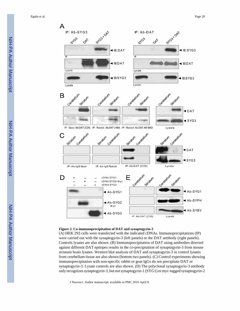
Figure 2. Co-immunoprecipitation of DAT and synaptogyrin-3(A) HEK 293 cells were transfected with the indicated cDNAs. Immunoprecipitations (IP)were carried out with the synaptogyrin-3 (left panels) or the DAT antibody (right panels).Controls lysates are also shown. (B) Immunoprecipitation of DAT using antibodies directedagainst different DAT epitopes results in the co-precipitation of synaptogyrin-3 from mousestriatum brain lysates. Western blot analysis of DAT and synaptogyrin-3 in control lysatesfrom cerebellum tissue are also shown (bottom two panels). (C) Control experiments showingimmunoprecipitation with non-specific rabbit or goat IgGs do not precipitate DAT orsynaptogyrin-3. Lysate controls are also shown. (D) The polyclonal synaptogyrin-3 antibodyonly recognizes synaptogyrin-3, but not synaptogyrin-1 (SYG1) or myc-tagged synaptogyrin-2
Egaña et al. Page 20
J Neurosci. Author manuscript; available in PMC 2010 April 8.
NIH
-PA Author Manuscript
NIH
-PA Author Manuscript
NIH
-PA Author Manuscript

(SYG2-Myc) in transfected HEK 293 cells. (E) Immunoprecipitation with the DAT antibodydoes not result in co-precipitation of synaptogyrin-1 (SYG1), synaptophysin (SYPH), orsynaptobrevin (SYBV) from striatum or cerebellum lysates.
Egaña et al. Page 21
J Neurosci. Author manuscript; available in PMC 2010 April 8.
NIH
-PA Author Manuscript
NIH
-PA Author Manuscript
NIH
-PA Author Manuscript

Figure 3. Immunocytochemical analysis of DAT and synaptogyrin-3 in striatumSlices were immunostained with a monoclonal rat anti-DAT (red, labeled with anti-rat AlexaFluor 555) and a polyclonal rabbit anti-synaptogyrin-3 (green, labeled with anti-rabbit AlexaFluor 488) antibodies. Low magnification views of the dorsal striatum (Str), overlying cortex(Ctx), and the arch of the corpus callosum (cc) extending between the two macroscopicstructures are shown in the top panels. (top panels, scale bar = 250 microns). High powerconfocal images (lower panels, scale bar = 10 microns) suggest the colocalization of the DATand synaptogyrin-3 signals with punctate varicosities, appearing yellow in the merged viewon the lower right. Five examples of colocalization are indicated with white arrows. Areas ofno signal in the internal capsule (labeled ic) are evident in all five images. Note the absence ofboth red and green fluorophores in these myelinated fiber bundles of the striatal internal capsuleand of the corpus callosum (cc) support the specificity of the primary and secondary antibodiesin use.
Egaña et al. Page 22
J Neurosci. Author manuscript; available in PMC 2010 April 8.
NIH
-PA Author Manuscript
NIH
-PA Author Manuscript
NIH
-PA Author Manuscript

Figure 4. Interaction between DAT and synaptogyrin-3 as visualized by Fluorescence ResonanceEnergy Transfer (FRET) microscopyHippocampal neurons from neonatal rats were co-transfected with CFP-DAT and YFP-synaptogyrin-3 (YFP-SYG3). (A) High magnification of a neuron transfected with CFP-DATand YFP-synaptogyrin-3. (top left panel: CFP channel, top right panel: YFP channel, bottomleft panels: FRET channel, bottom right panels: corrected FRET channel). The arrow headsindicate neuronal processes where FRET was detected. FRETC images are displayed usingpseudocolors with the spectrum inset representing fluorescence intensity. (B) The FRET wasanalyzed as described in the Methods. Control experiments included cells transfected with aCFP-YFP tandem construct, cells co-transfected with CFP and YFP, cells co-expressing CFP-DAT and YFP-DAT, and cells co-expressing CFP-DAT and YFP-synaptophysin (YFP-SYPH). The intensity of FRET was measured for the different combination of CFP/YFP pairsand expressed as normalized FRET values as described in Methods.
Egaña et al. Page 23
J Neurosci. Author manuscript; available in PMC 2010 April 8.
NIH
-PA Author Manuscript
NIH
-PA Author Manuscript
NIH
-PA Author Manuscript

Figure 5. The amino termini of both DAT and synaptogyrin-3 are sufficient for the DAT/synaptogyrin-3 interaction(A and B) Aliquots containing GST only, DAT-N, DAT-L1, or DAT-C fusion proteins (50 µgeach) were incubated with whole brain lysates and the precipitated proteins were analyzed bywestern blot using antibodies against synaptogyrin-3 (SYG3), synaptogyrin-1 (SYG1), orsynaptophysin (SYPH). (C) GST alone, DAT-N or NET-N were incubated with whole brainlysates and the pull-downs were analyzed by western blot using the anti-synaptogyrin-3antibody. The DAT-N, but not NET-N interacts with synaptogyrin-3. (D) Aliquots containingSYG3-N, -SYG3-L or SYG3-C GST-fusion proteins (50 µg) were incubated with striatumlysates and the precipitated proteins were analyzed by western blot using the anti-DATantibody. The SYG3-N interacts with DAT.
Egaña et al. Page 24
J Neurosci. Author manuscript; available in PMC 2010 April 8.
NIH
-PA Author Manuscript
NIH
-PA Author Manuscript
NIH
-PA Author Manuscript

Figure 6. The amino terminus of DAT interacts with synaptic vesicles(A) An enriched synaptic vesicle preparation (P4) was obtained from mouse brain and analyzedby western blot for synaptic vesicle markers. The final supernatant from the preparation of P4(S4 Fraction) is also shown. (B) Western blot analysis of P4 samples (with detergent)precipitated with GST alone or DAT-N. (C) Western blot analysis of P4 samples (withoutdetergent) precipitated with GST alone or DAT-N. (D) Synaptic vesicles separate in twodistinct populations when separated through sucrose gradients. Western blot analysis wasperformed with the indicated antibodies: synaptogyrin-3 (SYG3), synaptotagmin (SYTM),synaptobrevin (SYBV), synaptogyrin-1 (SYG1), synaptophysin (SYPH), vesicularmonoamine transporter 2 (VMAT2), post synaptic density protein 93 kDa (PSD93), transferrin
Egaña et al. Page 25
J Neurosci. Author manuscript; available in PMC 2010 April 8.
NIH
-PA Author Manuscript
NIH
-PA Author Manuscript
NIH
-PA Author Manuscript

receptor (TfR). Fractions 7–11 from the sucrose gradient were pooled and incubated with GSTalone or DAT-N in the presence (E) or absence (F) of detergent. Mixtures were treated with0.5mg/ml DSP for 10 min and solubilized in PBS. Bound proteins were precipitated withglutathione agarose beads binding either GST alone or DAT-N, separated by SDS-PAGE, andimmunoblotted with the indicated antibodies.
Egaña et al. Page 26
J Neurosci. Author manuscript; available in PMC 2010 April 8.
NIH
-PA Author Manuscript
NIH
-PA Author Manuscript
NIH
-PA Author Manuscript

Figure 7. Synaptogyrin-3 overexpression increases DAT activity in MN9D-DAT cells(A) MN9D-DAT cells transfected with synaptogyrin-3 vector (▲) show an increased DAuptake activity (Vmax = 2.37 ± 0.09 pmol/min/mg protein, n = 3) compared with MN9D-DATmock-transfected cells (●), (Vmax = 1.39 ± 0.17 pmol/min/mg protein, n = 3). Overexpressionof synaptogyrin-3 results in a 2-fold increase of protein levels (inset). (B) Total (top panel) andcell membrane (bottom panel) expression were analyzed by western blot for each transfectionand normalized to α-tubulin (TUB) levels. Densitometric analysis was performed for eachexperiment and plotted as a fraction of control. Results are presented as the mean ± SE (*,p<0.05).
Egaña et al. Page 27
J Neurosci. Author manuscript; available in PMC 2010 April 8.
NIH
-PA Author Manuscript
NIH
-PA Author Manuscript
NIH
-PA Author Manuscript

Figure 8. Changes in synaptogyrin-3 expression levels correlate with changes in DAT activity inPC12 cells(A) PC12 cells were transiently transfected with DAT (white) or co-transfected with bothsynaptogyrin-3 and DAT (black) and uptake activity was performed with [3H]-DA 96 h aftertransfections. The data represents the means ± SE of four independent experiments.Overexpression of synaptogyrin-3 was analyzed by western blot for each transfection andprotein levels were normalized to α-tubulin (TUB) levels. (B) Overexpression ofsynaptogyrin-3 has no effect on the cell surface (top panel) or total (bottom panel) levels ofDAT. Representative blot images of cells expressing DAT and cells co-expressing DAT andsynaptogyrin-3 are shown. Densitometric analysis was performed for each experiment and
Egaña et al. Page 28
J Neurosci. Author manuscript; available in PMC 2010 April 8.
NIH
-PA Author Manuscript
NIH
-PA Author Manuscript
NIH
-PA Author Manuscript

plotted as a fraction of control. Results are presented as the mean ± SE. (C) Densitometricanalysis of synaptogyrin-3 protein levels from cells transfected with a synaptogyrin-3 siRNA(black bar) or a scramble siRNA control (white bar). The expression levels of proteins involvedin regulated exocytosis or endocytosis were unaltered by synaptogyrin-3 siRNA. (D) DATactivity in synaptogyrin-3 knock-down cells. Uptake assays in PC12 cells expressing asynaptogyrin-3 siRNA (▲) or scramble siRNA (●) were performed in the presence of the NETinhibitor, nixosetine, with [3H]-DA 96h after transfections. The data represents the means ±SE of four independent experiments. (E) Levels of surface DAT remain unaltered insynaptogyrin-3 siRNA cells. AMPH: amphiphysin, SYPH: synaptophysin, SYG1:synaptogyrin-1, *: p<0.05.
Egaña et al. Page 29
J Neurosci. Author manuscript; available in PMC 2010 April 8.
NIH
-PA Author Manuscript
NIH
-PA Author Manuscript
NIH
-PA Author Manuscript

Figure 9. Synaptogyrin-3 overexpression does not increase NET activityA) Uptake assays in non-transfected PC12 cells (■), cells transfected with synaptogyrin-3 (▼),cells transfected with NET (●), and cells transfected with NET and synaptogyrin-3 cDNas (▲)were performed in the absence of the NET inhibitor, nixosetine 96h after transfections. Thedata represents the means ± SE of four independent experiments. (B) PC12 cells weretransfected with either DAT alone, NET alone, or NET and synaptogyrin-3.Immunoprecipitation experiments were performed on these lysates using either antibodiesdirected against DAT (left panels) or NET (right panels). Western blot analysis showedsuccessful transfection of DAT and NET (bottom panels), but show that synaptogyrin-3 onlyco-precipitates with DAT and not NET (top panels).
Egaña et al. Page 30
J Neurosci. Author manuscript; available in PMC 2010 April 8.
NIH
-PA Author Manuscript
NIH
-PA Author Manuscript
NIH
-PA Author Manuscript

Figure 10. Synaptogyrin-3 does not affect DAT activity in HEK 293A) Western blot analysis confirmed that HEK 293 cells were transiently transfected with eitherDAT alone or in combination of synaptogyrin-3. B) Uptake assays in cells expressing onlyDAT (black) or DAT and synaptogyrin-3 (red) were performed with [3H]-DA 96h aftertransfections. The kinetic values for DAT activity were Km = 3.61 ± 0.67 µM, Vmax = 3.63× 10⁶ ± 2.02 × 10⁵ pmol/min/mg protein in cells expressing DAT and synaptogyrin-3 vs. Km= 3.21 ± 1.13 µM and Vmax = 3.40 × 10⁶ ± 3.49 × 10⁵ pmol/min/mg protein in cells expressingonly DAT. The data represents the means ± SE of three independent experiments (*, p<0.05).
Egaña et al. Page 31
J Neurosci. Author manuscript; available in PMC 2010 April 8.
NIH
-PA Author Manuscript
NIH
-PA Author Manuscript
NIH
-PA Author Manuscript

Figure 11. The vesicular DA storage system has a role in synaptogyrin-3’s effect on DAT activity(A) The VMAT2 inhibitor reserpine (1 µM) reduces DAT activity and preventssynaptogyrin-3’s effect on DAT activity in PC12 cells transiently co-transfected with DATand synaptogyrin-3 cDNAs. Uptake assays were performed in the presence (red) or absence(black) of 1 µM reserpine. The data is representative of three independent experiments. B)Reserpine (100 µM) (red) fails to reduce DAT activity in HEK 293 cells transiently co-transfected with DAT and synaptogyrin-3 cDNAs compared to uptake assays performed in theabsence of reserpine (black). The data is representative of three independent experiments. C)Synaptogryin-3 fails to increase DAT activity in the presence of 1 µM reserpine in PC12 cells.[3H]-DA uptake was performed 96 h after transfection in the presence of 10 µM of unlabeled
Egaña et al. Page 32
J Neurosci. Author manuscript; available in PMC 2010 April 8.
NIH
-PA Author Manuscript
NIH
-PA Author Manuscript
NIH
-PA Author Manuscript

DA. The data represents the means ± SE of three independent experiments. D)Immunoprecipitation of DAT results in the co-precipitation of synaptogyrin-3 and VMAT2from mouse striatum The cross linking agent, DSP, was used. Synaptophysin (SYPH) and SV2did not co-precipitate with DAT, showing that all synaptic vesicle proteins were not cross-linked with DAT. Control experiments show that beads only or DAT antibody alone does notprecipitate any of the tested antibodies.
Egaña et al. Page 33
J Neurosci. Author manuscript; available in PMC 2010 April 8.
NIH
-PA Author Manuscript
NIH
-PA Author Manuscript
NIH
-PA Author Manuscript