
Codon Optimization of the Tat Antigen of Human Immunodeficiency Virus Type 1 Generates Strong Immune...
17
10.1128/JVI.78.17.9174-9189.2004. 2004, 78(17):9174. DOI: J. Virol. Kumarasamypet M. Mohankumar and Udaykumar Ranga Lakshmi Ramakrishna, Krishnamurthy Kumar Anand, Mice following Genetic Immunization Generates Strong Immune Responses in Human Immunodeficiency Virus Type 1 Codon Optimization of the Tat Antigen of http://jvi.asm.org/content/78/17/9174 Updated information and services can be found at: These include: REFERENCES http://jvi.asm.org/content/78/17/9174#ref-list-1 at: This article cites 108 articles, 41 of which can be accessed free CONTENT ALERTS more» articles cite this article), Receive: RSS Feeds, eTOCs, free email alerts (when new http://journals.asm.org/site/misc/reprints.xhtml Information about commercial reprint orders: http://journals.asm.org/site/subscriptions/ To subscribe to to another ASM Journal go to: on October 25, 2013 by guest http://jvi.asm.org/ Downloaded from on October 25, 2013 by guest http://jvi.asm.org/ Downloaded from on October 25, 2013 by guest http://jvi.asm.org/ Downloaded from on October 25, 2013 by guest http://jvi.asm.org/ Downloaded from on October 25, 2013 by guest http://jvi.asm.org/ Downloaded from on October 25, 2013 by guest http://jvi.asm.org/ Downloaded from on October 25, 2013 by guest http://jvi.asm.org/ Downloaded from on October 25, 2013 by guest http://jvi.asm.org/ Downloaded from on October 25, 2013 by guest http://jvi.asm.org/ Downloaded from on October 25, 2013 by guest http://jvi.asm.org/ Downloaded from on October 25, 2013 by guest http://jvi.asm.org/ Downloaded from on October 25, 2013 by guest http://jvi.asm.org/ Downloaded from on October 25, 2013 by guest http://jvi.asm.org/ Downloaded from on October 25, 2013 by guest http://jvi.asm.org/ Downloaded from on October 25, 2013 by guest http://jvi.asm.org/ Downloaded from on October 25, 2013 by guest http://jvi.asm.org/ Downloaded from on October 25, 2013 by guest http://jvi.asm.org/ Downloaded from
-
Upload
independent -
Category
Documents
-
view
0 -
download
0
Transcript of Codon Optimization of the Tat Antigen of Human Immunodeficiency Virus Type 1 Generates Strong Immune...

10.1128/JVI.78.17.9174-9189.2004.
2004, 78(17):9174. DOI:J. Virol. Kumarasamypet M. Mohankumar and Udaykumar RangaLakshmi Ramakrishna, Krishnamurthy Kumar Anand, Mice following Genetic ImmunizationGenerates Strong Immune Responses inHuman Immunodeficiency Virus Type 1 Codon Optimization of the Tat Antigen of
http://jvi.asm.org/content/78/17/9174Updated information and services can be found at:
These include:
REFERENCEShttp://jvi.asm.org/content/78/17/9174#ref-list-1at:
This article cites 108 articles, 41 of which can be accessed free
CONTENT ALERTS more»articles cite this article),
Receive: RSS Feeds, eTOCs, free email alerts (when new
http://journals.asm.org/site/misc/reprints.xhtmlInformation about commercial reprint orders: http://journals.asm.org/site/subscriptions/To subscribe to to another ASM Journal go to:
on October 25, 2013 by guest
http://jvi.asm.org/
Dow
nloaded from
on October 25, 2013 by guest
http://jvi.asm.org/
Dow
nloaded from
on October 25, 2013 by guest
http://jvi.asm.org/
Dow
nloaded from
on October 25, 2013 by guest
http://jvi.asm.org/
Dow
nloaded from
on October 25, 2013 by guest
http://jvi.asm.org/
Dow
nloaded from
on October 25, 2013 by guest
http://jvi.asm.org/
Dow
nloaded from
on October 25, 2013 by guest
http://jvi.asm.org/
Dow
nloaded from
on October 25, 2013 by guest
http://jvi.asm.org/
Dow
nloaded from
on October 25, 2013 by guest
http://jvi.asm.org/
Dow
nloaded from
on October 25, 2013 by guest
http://jvi.asm.org/
Dow
nloaded from
on October 25, 2013 by guest
http://jvi.asm.org/
Dow
nloaded from
on October 25, 2013 by guest
http://jvi.asm.org/
Dow
nloaded from
on October 25, 2013 by guest
http://jvi.asm.org/
Dow
nloaded from
on October 25, 2013 by guest
http://jvi.asm.org/
Dow
nloaded from
on October 25, 2013 by guest
http://jvi.asm.org/
Dow
nloaded from
on October 25, 2013 by guest
http://jvi.asm.org/
Dow
nloaded from
on October 25, 2013 by guest
http://jvi.asm.org/
Dow
nloaded from

JOURNAL OF VIROLOGY, Sept. 2004, p. 9174–9189 Vol. 78, No. 170022-538X/04/$08.00�0 DOI: 10.1128/JVI.78.17.9174–9189.2004Copyright © 2004, American Society for Microbiology. All Rights Reserved.
Codon Optimization of the Tat Antigen of Human ImmunodeficiencyVirus Type 1 Generates Strong Immune Responses in Mice
following Genetic ImmunizationLakshmi Ramakrishna, Krishnamurthy Kumar Anand, Kumarasamypet M. Mohankumar, and
Udaykumar Ranga*Molecular Virology Laboratory, Molecular Biology and Genetics Unit, Jawaharlal Nehru Centre
for Advanced Scientific Research, Jakkur, Bangalore, India
Received 22 November 2003/Accepted 19 March 2004
DNA vaccines have been successful in eliciting potent immune responses in mice. Their efficiency, however,is restricted in larger animals. One reason for the limited performance of the DNA vaccines is the lack ofmolecular strategies to enhance immune responses. Additionally, genes directly cloned from pathogenicorganisms may not be efficiently translated in a heterologous host expression system as a consequence of codonbias. To evaluate the influence of codon optimization on the immune response, we elected to use the Tatantigens of human immunodeficiency virus type 1 (HIV-1) (subtype C) and HIV-2, as these viral antigens arepoorly immunogenic in natural infection and in experimental immunization and they are functionally impor-tant in viral infectivity and pathogenesis. Substituting codons that are optimally used in the mammaliansystem, we synthetically assembled Tat genes and compared them with the wild-type counterparts in twodifferent mouse strains. Codon-optimized Tat genes induced qualitatively and quantitatively superior immuneresponses as measured in a T-cell proliferation assay, enzyme-linked immunospot assay, and chromium releaseassay. Importantly, while the wild-type genes promoted a mixed Th1-Th2-type cytokine profile, the codon-optimized genes induced a predominantly Th1 profile. Using a pepscan strategy, we mapped an immunodom-inant T-helper epitope to the core and basic domains of HIV-1 Tat. We also identified cross-clade immuneresponses between HIV-1 subtype B and C Tat proteins mapped to this T-helper epitope. Developing molecularstrategies to optimize the immunogenicity of DNA vaccines is critical for inducing strong immune responses,especially to antigens like Tat. Our identification of a highly conserved T-helper epitope in the first exon ofHIV-1 Tat of subtype C and the demonstration of a cross-clade immune response between subtypes B and Care important for a more rational design of an HIV vaccine.
DNA vaccine technology has emerged as a novel mode ofvaccination where a naked DNA construct, encoding one ormore foreign proteins or epitopes, is used for immunization.When injected into a host, the DNA vector elicits a cellular orhumoral immune response or both against the encoded anti-gen. Nucleic acid immunization offers several technical advan-tages over other formats of vaccination at the level of immu-nological outcome (25, 40). When administered intramuscularly,DNA vaccines elicit a predominantly T-helper cell Th1-typeimmune response, which is believed to be critical for confer-ring protection against several pathogens, especially viruses.Application of DNA vaccines, however, is limited, as they areusually unsuccessful in inducing strong immune responses inlarger animals (60, 97). Various molecular approaches havebeen explored to elicit potent immune responses through ge-netic immunization. These approaches include coadministra-tion of cytokines, such as interleukin-2 (IL-2), IL-15, gammainterferon (IFN-�), RANTES, and IL-18 (8, 49, 103, 104);coexpression of costimulatory molecules such as CD40L,CD86, and CTLA-4 (44, 48, 93); engineering CpG motifs into
the plasmid vectors (51, 52); expression of antigens as fusionproteins with molecular adjuvants, such as ubiquitin (34, 79),heat shock proteins (19), L-selectine (29), Flt3 ligand (84), andC3d (39, 80); adaptation of the prime-boost immunizationstrategies involving other vaccine formats in combination withDNA (41, 57); and many others (21, 85). Codon optimizationof the antigen-encoding gene is a powerful strategy to maxi-mize protein expression in a heterologous expression systemthat consequently leads to enhanced immune response (20, 94,107).
Selective use of specific codons for protein translation is acharacteristic feature of several species, a phenomenon calledcodon bias (87). Direct cloning of pathogen-derived genes intoexpression cassettes often leads to suboptimal expression ofthe wild-type genes in a heterologous system and may fail tostimulate strong immune responses. In a natural infection,codon bias of the wild-type genes may help reduce the magni-tude of the immune surveillance due to suboptimal antigenexpression in a host system, thus circumventing the inductionof strong immune responses against the pathogenic organism.Immunization strategies using genetic vaccines, therefore,must replace these suboptimal codons with those more fre-quently used in the host system to elicit strong immune re-sponses (20, 23, 91, 107).
Immunization with codon-optimized env (6) and gag (27,107) genes of human immunodeficiency virus type 1 (HIV-1)
* Corresponding author. Mailing address: Molecular Virology Lab-oratory, Molecular Biology and Genetics Unit, Jawaharlal Nehru Cen-tre for Advanced Scientific Research, Jakkur (PO), Bangalore 560 064,India. Phone: 91-80-2208 2830, ext. 2241. Fax: 91-80-2208 2766. E-mail: [email protected].
9174
led to enhanced expression of the genes and improved immuneresponses against the antigens. Similar studies conducted witha variety of other pathogenic organisms, such as Listeria (65),bacteria producing tetanus toxin (91), Plasmodium (65), hu-man papillomavirus (20, 59), and others (40), ascertained thepotential of codon optimization to enhance the efficiency ofthe DNA vaccines. The foreign genes or epitopes used inseveral of these studies were inherently immunodominant, thuspossibly underestimating the outcome of codon optimizationon the immune responses generated.
In an attempt to evaluate the influence of codon optimiza-tion on the immune response, we sought to use an inherentlynonimmunodominant antigen in our studies. We opted for thetransactivator protein (Tat) of HIV, as this viral antigen offersseveral technical advantages. Most important, the Tat proteinsof HIV-1 and HIV-2 are small molecules consisting of 101 and130 amino acids, respectively. The first exon of HIV-1 Tat(Tat-1), consisting of 72 amino acids, is functionally competentfor viral transactivation and a wide variety of other functionsascribed to this protein (98). The small size of the Tat proteinoffers a practical advantage of uncomplicated chemical synthe-sis of the expression cassette essential for codon optimization.Tat is expressed early in the viral life cycle and is functionallyimportant for its infectivity and pathogenicity (47). In additionto regulating viral gene expression, Tat modulates expressionof various genes of the host. Additionally, Tat is secretedextracellularly, and the extracellular Tat governs viral latencyand contributes to disease progression (66). Despite the func-tional importance of Tat for the virus, this antigen is notimmunodominant either in natural infection (53, 56, 58, 78,101) or in experimental immunization, especially when deliv-ered as a genetic vaccine (8, 15). Inducing cellular and humoral
immune responses against Tat is critical because of its earlyexpression in the viral life cycle and its extracellular secretion(37, 82). At the genetic level, the Tat gene is conserved to agreater extent than that of the env gene, thus permitting in-duction of cross-clade immune responses (54). Last, as a con-sequence of its pleiotropic biological functions, a variety offunctional assays are available for Tat to study the inhibitoryeffect of immune components on its biological functions.
The overall genetic content of HIV is AT-rich, suggestingsuboptimal codon usage of its genes in the human host. TheAT content of Tat-1 is approximately 54.3%, while that ofHIV-2 Tat (Tat-2) is 51.8%. This is in contrast to the relativelylow AT content (30 to 40%) in the coding regions of the hosthuman genome (86). To enhance the translational efficacy andimmunogenicity of HIV-1 and HIV-2 Tat proteins, we synthet-ically assembled the gene expression cassettes. We employedan amplification-based approach to generate codon-optimizedTat constructs. While designing the expression cassettes,codons frequently used in the mammalian system were substi-tuted for the original AT-rich codons. The synthetic Tat con-structs were evaluated in the mouse model for the generationof immune responses.
MATERIALS AND METHODS
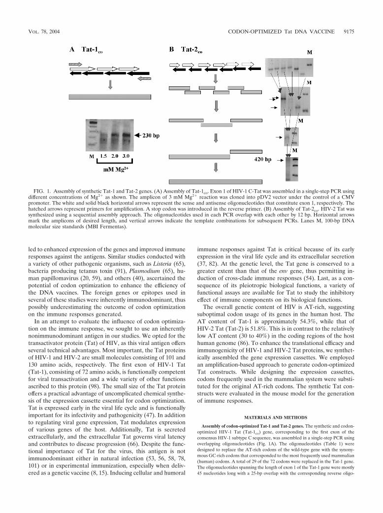
Assembly of codon-optimized Tat-1 and Tat-2 genes. The synthetic and codon-optimized HIV-1 Tat (Tat-1co) gene, corresponding to the first exon of theconsensus HIV-1 subtype C sequence, was assembled in a single-step PCR usingoverlapping oligonucleotides (Fig. 1A). The oligonucleotides (Table 1) weredesigned to replace the AT-rich codons of the wild-type gene with the synony-mous GC-rich codons that corresponded to the most frequently used mammalian(human) codons. A total of 29 of the 72 codons were replaced in the Tat-1 gene.The oligonucleotides spanning the length of exon 1 of the Tat-1 gene were mostly45 nucleotides long with a 25-bp overlap with the corresponding reverse oligo-
FIG. 1. Assembly of synthetic Tat-1 and Tat-2 genes. (A) Assembly of Tat-1co. Exon 1 of HIV-1 C-Tat was assembled in a single-step PCR usingdifferent concentrations of Mg2� as shown. The amplicon of 3 mM Mg2� reaction was cloned into pDV2 vector under the control of a CMVpromoter. The white and solid black horizontal arrows represent the sense and antisense oligonucleotides that constitute exon 1, respectively. Thehatched arrows represent primers for amplification. A stop codon was introduced in the reverse primer. (B) Assembly of Tat-2co. HIV-2 Tat wassynthesized using a sequential assembly approach. The oligonucleotides used in each PCR overlap with each other by 12 bp. Horizontal arrowsmark the amplicons of desired length, and vertical arrows indicate the template combinations for subsequent PCRs. Lanes M, 100-bp DNAmolecular size standards (MBI Fermentas).
VOL. 78, 2004 CODON-OPTIMIZED Tat DNA VACCINE 9175

nucleotide (Fig. 1A, schematic). The amplification reaction mixture of 100 �lcontained 25 nM concentrations of each oligonucleotide and 250 nM concen-trations of each primer (forward N298 [5�-CGG TAA GGT ACC GCC GCCGCC ATG GAG CCA GTA GAT CCT AAC CT] and reverse N180 [5�-TTTTCT AGA CTA TTG CTT TGA TAT AAG ATT TTG ATG]). The reactionmixture also contained 100 �M (each) deoxynucleoside triphosphates (dNTPs),1.25 U of Taq DNA polymerase, and 3.0 mM Mg2�. Amplification was per-formed using the following cycling conditions: (i) 2 min at 94°C; (ii) 2 cycles, with1 cycle consisting of 30 s at 42°C and 30 s at 72°C, followed by 2 min at 94°C; (iii)2 cycles, with 1 cycle consisting of 30 s at 50°C and 30 s at 72°C, followed by 30 sat 94°C; (iv) 35 cycles, with 1 cycle consisting of 30 s at 56°C and 30 s at 72°C. Theamplified product was purified using a column commercial kit (Qiagen), cloneddirectionally between KpnI and XbaI into pDV2, a mammalian expression vectorcontaining a cytomegalovirus (CMV) promoter.
Synthetic and codon-optimized HIV-2 Tat (Tat-2co) corresponding to thefull-length sequence of an Indian HIV-2 isolate was assembled using six pairs ofcodon-optimized oligonucleotides (Table 2). To assemble the Tat-2 gene, weused a sequential amplification strategy. The complementary oligonucleotides ofeach pair overlapped with each other by 12 bp. Six individual amplificationreactions were performed to amplify as many regions of Tat-2 (Fig. 1B). Theadjoining amplification products also overlapped with each other by 12 bp. Thesix first-round PCR products served as templates for three second-round ampli-fications. Each of the second-round amplification mixtures contained two adja-cent first-round amplicons and the two terminal oligonucleotides for primers.One-microliter portions from each of the first-round reaction mixtures were usedas templates for the second-round reactions. Further rounds of amplificationwere performed until the full-length Tat-2 product was obtained (Fig. 1B). Atotal of 81 of the 130 codons of the wild-type Tat-2 gene (Tat-2wt) were substi-tuted in the Tat-2co gene. A proofreading polymerase, Elongase (Gibco), was
used to ensure the fidelity of amplification. The reaction conditions were asfollows: preamplification denaturation (30 s at 94°C), followed by 40 cycles, with1 cycle consisting of 30 s at 94°C, 30 s at 20°C, and 30 s at 68°C. A 50-�l reactionmixture contained 1 �l of Elongase enzyme mixture (Gibco), 25 nM concentra-tion of each oligonucleotide, 100 �M dNTPs, and 1.8 mM Mg2�. The resultingamplicons from each step served as templates for the subsequent reactions. Thefinal 420-bp amplicon was cloned directionally between KpnI and XbaI sites onpDV2. The authenticity of the assembled expression cassettes was confirmed byrestriction digestion analysis and sequencing.
Histidine (His)-tagged subtype C Tat-1 (C-Tat-1) was amplified from a pri-mary Indian HIV isolate and cloned into the bacterial expression vectorpET28A. Glutathione S-transferase (GST)-tagged Tat-2 (GST-Tat-2) expressionvector was obtained from the National Institutes of Health AIDS Research andReference Reagent Program. The wild-type HIV-2 gene was a gift from RobinMukhopadhyaya.
SEAP assay. 293 cells were cotransfected with HIV secreted alkaline phos-phatase (SEAP) (a gift from Bryan Cullen) and HIV-1 Tat expression vectors bythe CaCl2 method (18). The culture supernatant (200 �l) was sampled at regularintervals and stored at �20°C until use. The reporter SEAP activity was mea-sured using 4-nitrophenyl phosphate as the substrate (24). In all transfections, weused CMV-�-galactosidase reporter vector as an internal standard for normal-ization. �-Galactosidase activity in cell extracts was measured by a colorimetricassay (90).
Western blot analysis. 293 cells were transiently transfected with Tat expres-sion vectors, and the cells were harvested 48 h after transfection. The cells werelysed in sodium dodecyl sulfate (SDS)-polyacrylamide gel electrophoresis samplebuffer, cell extract was resolved on an SDS–12% polyacrylamide gel, and theprotein was transferred to a polyvinylidene difluoride (PVDF) membrane (Im-mun-Blot PVDF; Bio-Rad) using a semidry transfer apparatus (Trans-Blot SD;
TABLE 1. Sequences of oligonucleotides used to assemble exon 1 of C-Tat-1
Oligonucleotide Sequence (5� to 3�)
SenseN223.................................................................................................CATGGAGCCAGTAGATCCTAACCTGGAGCCCTGGAACCACCCTGGCN224.................................................................................................AGCCAGCCCAAGACCGCCTGCAACAACTGCTACTGCAAGCACTGCN225.................................................................................................AGCTACCACTGCCTGGTGTGCTTCCAGACCAAGGGCCTGGGCATCN226.................................................................................................AGCTACGGCCGGAAGAAGCGGCGCCAGCGCCGGAGCGCTCCTCCAN227.................................................................................................AGCAGCGAGGACCACCAAAATCTTATATCAAAGCAGTAAT
Antisensea
N228.................................................................................................CTAGATTACTGCTTTGATATAAGATN229.................................................................................................TTTGGTGGTCCTCGCTGCTTGGAGGAGCGCTCCGGCGCTGGCN230.................................................................................................GCCGCTTCTTCCGGCCGTAGCTGATGCCCAGGCCCTTGGTCTGGAN231.................................................................................................AGCACACCAGGCAGTGGTAGCTGCAGTGCTTGCAGTAGCAGTTGTTN232.................................................................................................GCAGGCGGTCTTGGGCTGGCTGCCAGGGTGGTTCCAGGGCTN233.................................................................................................CCAGGTTAGGATCTACTGGCTCCATGGTAC
a The antisense oligonucleotides are presented as the sequences complementary to the sense oligonucleotides.
TABLE 2. Sequences of oligonucleotides used to assemble Tat-2
Oligonucleotide Sequence (5� to 3�)
SenseN267.................................................................................AGATCTGGTACCATGGAGACCCCCCTGAAGGCCCCCGAGAGCAGCCTGGAN269.................................................................................CCAGCGAGCAGGACGTCGCCACCCAGGGCAGCGCTAGCCAGGGCGAGGAGN270.................................................................................GAGGCCTGCGCCTGCGCCAGCACCTGCTACTGCAAGCTGTGCTGCTACCAN271.................................................................................GCCTGGGCATCTGCTACGAGCGGAAGGGCCGGCGGCGGCGGACCCCCAAGN272.................................................................................GTCCGACAAGAGCATCAGCACCCGGACCTGGAACAGCCAGCCCGAGAAGGN273.................................................................................GGAGGCCACCGTGGAGACCGACACCGGCCCCGGCCG
Antisensea
N275.................................................................................CCTGCTCGCTGGTGTGGCTGAAGGGCTCGTTGCAGCTCTCCAGGCTGCTCN276.................................................................................GGCGCAGGCCTCCAGGGGCCGGTACAGCTGGCTCAGGATCTCCTCGCCCTN277.................................................................................AGATGCCCAGGCCCTTCTGCAGGAAGCAGAGCTGGCAGTGGTAGCAGCACN278.................................................................................CTCTTGTCGGACGTCAACGTTGGGTGGGTCTTGGTCTTCTTGGGGGTCCGN279.................................................................................ACGGTGGCCTCCACGGGCTTCTTCTGCTCCTTCTCGGGCTN280.................................................................................GGATCCTCTAGATCACCGGCCGGGGCCG
a The antisense oligonucleotides are presented as the sequences complementary to the sense oligonucleotides.
9176 RAMAKRISHNA ET AL. J. VIROL.

Bio-Rad) at 25 V for 90 min. The cell extract was independently probed for�-actin (Oncogene), following the manufacturer’s instructions, to serve as aloading control. HIV-1 Tat was detected using a monoclonal antibody (2D1.1;catalog no. 4138, National Institutes of Health AIDS Research and ReferenceReagent Program) raised against the N-terminal 15 amino acid residues of theantigen (28). The membranes were incubated overnight at 4°C in 5 ml of mono-clonal antibody diluted 10,000 times in 0.5% skim milk powder solution inphosphate-buffered saline (PBS). HIV-2 Tat was detected using a mouse anti-serum raised against the full-length protein in our laboratory. After the mem-branes were washed three times with Tris-buffered saline (pH 8.0), they wereincubated with 5 ml of anti-mouse horseradish peroxidase conjugate (BD Pharm-ingen) (diluted 1:1,000) for 1 h at room temperature. The blots were developedby chemiluminescence reaction using a commercial kit (ECL�plus; Amersham).
HLM1 transfection and p24 ELISA. HLM1 cells, which contain a Tat-defec-tive provirus, were transfected with HIV-1 Tat constructs using a cationic lipid(Geneporter; Gene Therapy Systems). The culture supernatant was sampled atregular intervals and stored at �20°C until use. Empigen was added to eachsample to a final concentration of 1%, and the samples were incubated at 56°Cfor 30 min to inactivate the virus and to release the p24 antigen from the viralparticles. The quantity of p24 in the samples was measured using an in-housecapture enzyme-linked immunosorbent assay (ELISA). Briefly, 0.5 �g of captureantibody (ADP 410; National Institute for Biological Standards and Control) in100 �l of carbonate buffer was added to each well of a 96-well plate (Greiner),and the plates were incubated overnight at room temperature. The plates wereblocked with 5% skim milk powder in PBS for 1 h at room temperature. Solu-tions containing the antigen were added to each well (100 �l/well), and the plateswere incubated for 4 h at 37°C. A biotinylated monoclonal antibody (ARP 454;National Institute for Biological Standards and Control) specific for p24 wasdiluted to a final concentration of 1 �g/ml using a buffer containing Tris (10mM), Empigen (0.1%), Tween 20 (0.05%), and sheep serum (5%). Wells con-taining 100 �l of the detection antibody were incubated at 37°C for 1 h. Astreptavidin peroxidase conjugate (Sigma) was added to each well at a dilution of1:1,000 and incubated for 1 h. One hundred microliters of the substrate solutioncontaining o-phenylenediamine was added to each well, plates were incubatedfor 15 min at room temperature, and 50 �l of 1 N HCl was added to each wellto stop the enzyme reaction. The optical density was measured at 490 nm usingan ELISA reader (Molecular Dynamics).
Semiquantitative RT-PCR. Reverse transcriptase PCR (RT-PCR) was per-formed using random hexamers for reverse transcription and specific primers forDNA PCR. Total RNA was isolated using TRIzol reagent (Gibco) from 293 cellstransiently transfected with the Tat expression vectors. For amplification, totalRNA was used as a template at three different concentrations, 1.0, 0.33, and 0.11�g per reaction mixture. Reverse transcription was performed using 1 �Mconcentrations of random hexamers, 1 mM dNTPs, 3 mM Mg2�, Moloneymurine leukemia virus RT (Promega), and 100 U of RNase inhibitor (Promega)in a volume of 20 �l. The RT reaction conditions were 35 min at 42°C and 5 minat 94°C. Five-microliter portions of the RT reaction contents were transferred tothe DNA PCR mixture of 50 �l. DNA PCR was performed with the primer pairN177 and N180 using 1.25 U of Taq DNA polymerase by hot start PCR. ThePCR mixture contained 100 �M dNTPs, 250 nM concentrations of primers, and1.8 mM Mg2�. The following reaction conditions were used for amplification: (i)1 min at 94°C; (ii) 35 cycles, with 1 cycle consisting of 30 s at 56°C and 1 min at72°C. �-Actin transcript amplification was used as a positive control employingthe primers 139F (5�-GTG GGG CGC CCC AGG CAC CA) and 139R (5�-CTCCTT AAT GTC ACG CAC GAT TTC).
DNA immunization. The plasmid DNA intended for immunization was pre-pared using Qiagen endotoxin-free Giga kits and resuspended in endotoxin-freePBS (Manukirti Biogems, Bangalore, India; �0.06 endotoxin unit). The endo-toxin concentration was quantitated in a standard Limulus amebocyte lysateassay (QCL-1000; Biowhittaker) and found to be within recommended limits(�0.1 endotoxin unit/�g of DNA). One hundred micrograms of the DNA wasinjected into the tibialis anterior muscle of mice that were 8 to 12 weeks old.Each immunization consisted of four or five mice per group. The immunizationschedule involved one primary immunization, followed by one or three boosterimmunizations spaced 2 weeks apart. Animals were housed and maintained in afacility adhering to the recommendations of the Committee for the Purpose ofControl and Supervision of Experiments on Animals in India.
Lymphoproliferation assay. Splenocytes (5 � 106) from immunized mice wereincubated with 5 �g of recombinantly expressed antigen per ml for 5 days. Tat-1was purified using the His tag by Ni-nitrilotriacetic acid (Qiagen) affinity chro-matography, and Tat-2 was purified using the GST tag by glutathione-Sepharose(Amersham) affinity chromatography. The purity of the proteins was confirmedusing SDS-polyacrylamide gel electrophoresis. Control proteins containing iden-
tical tags (His-p24 and GST-PC4) were also purified by similar means and usedin the assays as controls for nonspecific proliferation. After incubation, the extentof cell proliferation was measured by adding [3H]thymidine (5 �Ci/ml)(NET520A; Perkin Elmer Life Sciences) to the wells, and the cultures wereincubated for 3 h at 37°C for incorporation of the label. The cells were harvested,washed, resuspended in 50 �l of PBS, and deposited onto filter paper disks(Whatman no. 3 filter paper). After thorough washing, the filters were dried, andradioactivity counts were determined using a �-scintillation counter (Wallac1409). Concanavalin A (ConA) was used as a positive control for cell prolifer-ation at a final concentration of 5 �g/ml.
Cytotoxicity assay. Splenocytes harvested from immunized mice were incu-bated with irradiated syngeneic stimulators (P815 cells expressing Tat and EL-4cells expressing Tat for BALB/c and C57BL/6 mice, respectively) for 5 days inRPMI 1640 medium supplemented with 10% fetal bovine serum (FBS). After invitro stimulation, the effector cells were incubated with 51Cr-labeled syngeneictarget cells (104 cells/200 �l) at different effector-to-target cell ratios. Nontrans-fected parental cell lines (P815 or EL-4) served as controls. The effector andtarget cells were coincubated for 5 h at 37°C, and 100 �l of the supernatant wasassayed using a gamma counter (LKB Rack gamma; Wallac). Counts fromspontaneous lysis usually remained below 30% of the maximal lysis. Values formaximal lysis were obtained by incubating the labeled target cells in the presenceof 2% Triton X-100. Percent specific lysis was calculated by the following for-mula: percent specific lysis (experimental lysis � spontaneous lysis)/(maximallysis � spontaneous lysis).
ELISPOT assay. Enzyme-linked immunospot (ELISPOT) assays were per-formed for the Th1 cytokine IFN-� (m IFN� Eli-spot; DIACLONE Research)and the Th2 cytokine IL-4 (mouse IL-4 ELISPOT set; BD Pharmingen) beforeand after in vitro stimulation. Briefly, the cytokine-specific capture antibody (1�g/100 �l of PBS) was adsorbed onto the PVDF-backed 96-well plates byincubating the antibody solution overnight at 4°C. The plates were blocked, andthe primed splenocytes (0.5 � 106 cells) were added along with the stimulatorcells (at a 1:3 ratio) in a final volume of 200 �l to the wells and incubated for 24to 36 h. The cells were then lysed, cell debris was removed by extensive washing,and the wells were incubated with a biotinylated anticytokine antibody (0.5�g/ml) for 2 h. The plates were washed extensively, and the wells were incubatedwith enzyme-conjugated avidin (HRP for IL-4 and alkaline phosphatase forIFN-�) for 1 h. Spots were developed using the appropriate substrate (3-amino-9-ethylcarbazole and nitroblue tetrazolium–5-bromo-4-chloro-3-indolylphos-phate, respectively) and incubating the plates for 20 min at room temperature.ConA (5 �g/ml) was used as a positive control for IL-4 secretion, and phorbolmyristate acetate (1 �g/ml) plus ionomycin (0.5 �g/ml) were used as a positivecontrol for IFN-� secretion. The spots were enumerated using a stereomicro-scope (MZ6; Leica) at a magnification of �16.
Tat-1 epitope mapping. Six 20-mer peptides spanning exon 1 of HIV-1 Tatwere synthesized (Peptron, Daejeon, Korea); these peptides overlapped eachother by 10 amino acids. The peptides represented the consensus sequence ofHIV-1 C-Tat. The peptides were used at a final concentration of 2 �g/ml.Pepscan analysis for IFN-� ELISPOT assay was performed by incubating thepeptides with 5 � 105 splenocytes from immunized BALB/c mice per well for36 h at 37°C. The controls were normalized for dimethyl sulfoxide concentration.Eventually, additional peptides were designed for fine mapping of the identifiedepitope (see Fig. 7).
Complement-mediated cell depletion of T-cell subsets. Splenocytes (40 � 106
cells) from three or more immunized BALB/c mice were pooled, and the pooledcells were incubated in 0.5 ml of culture supernatant of anti-mouse CD4 (GK1.5)or CD8 (3.155) hybridoma cell line for 1 h at 4°C. Serum samples collected from4-week-old rabbits were added to the cells as a source of complement to a final1:5 or 1:20 dilution and incubated for 30 min at 37°C. The cells were washed withRPMI 1640 medium containing 5% FBS. Lymphocytes were passed through aFicoll gradient (Histopaque-1083; Sigma) to isolate viable cells. The cells werecollected in 15-ml tubes (Corning), washed three times with 5 ml of RPMI 1640medium supplemented with 5% FBS, and used for IFN-� ELISPOT assay.
Statistical analysis. Results of lymphoproliferative assays are presented asstimulation index values as the mean standard deviation of at least threeindependent experiments, each of which was performed in triplicate. Results ofELISPOT assays are expressed as the number of spots per 106 cells, means oftriplicate wells. Comparisons between the Tat constructs were made using theStudent’s t test. P values less than 0.05 were considered statistically significant.
RESULTS
Codon optimization increases the translation efficiency ofthe Tat-1 and Tat-2 genes. HIV-1 and HIV-2 Tat genes were
VOL. 78, 2004 CODON-OPTIMIZED Tat DNA VACCINE 9177

synthetically assembled such that the suboptimal codons weresubstituted with codons frequently used in the mammaliansystem. As a result of codon optimization, the AT content ofthe Tat-1 and Tat-2 genes was reduced from 54.3 and 51.8% to40.1 and 30.0%, respectively. The protein expression efficien-cies of the synthetic genes were compared with those of thewild-type constructs in 293 cells. Western blot results demon-strated enhanced expression of synthetic genes, Tat-1co andTat-2co, at significantly higher quantities than the wild-type
genes (Tat-1wt and Tat-2wt genes, respectively) (Fig. 2A). Tat-1co expressed four times more antigen than Tat-1wt. Especiallywith Tat-2, the amount of antigen produced from the wild-typegene was so low that we failed to detect its presence in theWestern blot. Expression of Tat-2 from this construct, how-ever, was confirmed in a more sensitive reporter assay (resultsnot shown).
Transient transfection of 293 cells with Tat expression vec-tors and HIV-1 long terminal repeat (LTR)-green fluorescent
FIG. 2. Functional analyses of Tat expression vectors. (A) Western blot analysis for Tat-1 and Tat-2. 293 cells were transfected with 10 �g ofplasmid DNA by the CaCl2 method. The cells were harvested 48 h after transfection and lysed, and the lysate was resolved on an SDS–12%polyacrylamide gel. Tat-1 was detected using a monoclonal antibody (2D1.1; AIDS Research and Reference Reagent Program). �-Actin servedas a loading control and probed using a commercial kit (Oncogene). Tat-2 was detected using a mouse antiserum raised in the laboratory againstGST-Tat-2 protein. co, codon-optimized Tat; wt, wild-type Tat; pv, parental vector. (B) Expression of SEAP from 293 cells transiently transfectedwith 1 �g of LTR-SEAP and 10 �g of Tat-1 expression vectors by the CaCl2 method. Culture supernatants were collected after different periodsof time, and a colorimetric assay was used to quantitate SEAP in culture supernatants (24). CMV-�-galactosidase vector was used in alltransfections to control for differences in the transfection efficiency. �-Galactosidase levels in the cell extracts were quantitated by a colorimetricassay (90). Abs, absorbance. (C) Rescue of a Tat-defective provirus. HLM-1 cells, containing a single copy of a Tat-defective provirus, weretransfected with 1 �g of Tat expression vectors using a commercial lipid formulation. Spent medium was collected at different times, and p24concentration in the samples was measured using an in-house antigen capture assay. Results are presented as means 1 standard deviation (errorbars) of three samples, and the data are representative of two independent experiments. CMV-SEAP (0.3 �g) was cotransfected to serve as aninternal control for normalization of the data. C, empty vector. (D) Semiquantitative RT-PCR analysis of Tat-1 constructs. 293 cells in100-mm-diameter dishes were transfected with plasmid DNA (10 �g) by the CaCl2 method. Cells were harvested 48 h after transfection, and totalRNA was isolated using TRIzol reagent (Gibco). RNA was diluted in serial threefold dilutions starting at 1 �g, and each dilution served as atemplate for Tat gene and �-actin RT-PCRs using specific primers as described in Materials and Methods. The amplicons were resolved on a 1%agarose gel.
9178 RAMAKRISHNA ET AL. J. VIROL.

protein (GFP) reporter plasmid demonstrated that the syn-thetic Tat constructs were transcriptionally functional (datanot shown). Transfection of HOS-CD4-LTR-GFP cells withTat-2 expression vectors by electroporation confirmed thefunctional integrity of the Tat-2 constructs (data not shown).For a quantitative evaluation of Tat-1 expression, 293 cellswere cotransfected with different Tat vectors and a reportervector expressing SEAP, HIV-1 LTR-SEAP. Culture superna-tants were collected at different periods, and the SEAP con-centration was determined in a colorimetric assay (24). SEAPwas detected in culture medium starting from 12 h, and itsconcentration gradually increased up to 72 h (Fig. 2B). Atevery point of sampling, the quantity of SEAP released into themedium was significantly higher from Tat-1co than from thewild-type vector. Reporter expression from Tat-1co alsoreached saturation earlier, suggesting higher levels of proteinsynthesis. These results are in agreement with the Western blotanalysis (Fig. 2A) and illustrated the functional integrity of thesynthetic Tat constructs.
Codon-optimized Tat-1 is functional in the virological con-text. Reporter gene analysis is useful in evaluating Tat-medi-ated transactivation of the viral LTR. This analysis, however,may not directly correlate with the function of Tat in thecontext of the viral life cycle. We sought to examine whethercodon-optimized Tat-1 could also lead to enhanced virion pro-duction. HLM-1 cells, which contain a single copy of a Tat-defective provirus (83), were used in the transfection experi-ments. Tat-1wt and Tat-1co were delivered to the cells using acommercial lipid formulation (Geneporter; Gene Therapy Sys-tems), and the concentration of p24 in the medium was quan-titated using an in-house sandwich ELISA. Both Tat-1wt andTat-1co successfully complemented the defective provirus andreleased p24 into the medium. Although synthetic Tat-1 con-sistently released more p24 at every time point and earlier thanthe wild-type gene, the difference between the two constructswas not statistically significant (Fig. 2C). This observation in-dicated that an intracellular concentration of the transactivatorprotein above a threshold level is sufficient to drive virionproduction and that expression levels of Tat above this thresh-old need not translate to increased virion production.
Enhanced protein synthesis is not due to increased tran-scription. To examine whether the increased level of proteinsynthesis observed from synthetic Tat genes was the outcomeof gene expression regulation at the level of transcription ortranslation or both, we performed a semiquantitative RT-PCR.Total RNA was extracted from 293 cells transfected with theTat-1wt or Tat-1co construct. RT-PCR was performed on seri-ally diluted RNA samples as described in Materials and Meth-ods. As expected, the degrees of amplification of these two Tatconstructs were not significantly different (Fig. 2D). This ob-servation, therefore, supported our premise that the increasedprotein synthesis from the synthetic Tat vector was most prob-ably a result of enhanced protein translation, not more efficienttranscription.
Immunological evaluation. We evaluated the immunogenicpotential of the wild-type and codon-optimized Tat genes inmice. Plasmid DNA, extracted using Qiagen columns, was usedin all the immunization protocols. Mice (C57BL/6 and BALB/cmice, four or five animals per group) were immunized intra-muscularly using a one-prime–one-boost or one-prime–three-
boost regimen. DNA was administered at a low (10 �g/mouse)or high (100 �g/mouse) dose to study the influence of the doseon the immune response. After immunization, the animalswere sacrificed, and the splenocytes were used in various im-munological analyses.
Enhanced cell proliferation in mice immunized with codon-optimized Tat. Splenocytes harvested from mice immunizedwith the wild-type or codon-optimized Tat expression vectorsor the parental plasmid were incubated in vitro with recombi-nantly expressed antigen, and the extent of antigen-specific cellproliferation was measured using the [3H]thymidine incorpo-ration assay (Fig. 3). We observed antigen-specific and dose-dependent cell proliferation in response to both Tat-1 andTat-2 antigens in C57BL/6 (Fig. 3) and BALB/c (data notshown) mice. Cell proliferation was significantly higher in miceimmunized with 100 �g of DNA than in mice immunized with10 �g of DNA with both the wild-type and codon-optimizedTat constructs. Although cell proliferation in mice immunizedwith 100 �g of Tat-1co was higher than that of Tat-1wt, thisdifference was not statistically significant. Importantly, the ef-fect of codon optimization of Tat was striking at the lowerDNA concentration. At this dose, while splenocytes of Tat-1co
immunization proliferated, splenocytes of Tat-1wt immuniza-tion failed to respond to Tat protein, indicating a lack ofoptimal antigen expression to prime an efficient immune re-sponse (Fig. 3A). Cell proliferation was not observed in lym-phocytes harvested from control mice injected with the paren-tal vector or when nonspecific antigens were employed in theexperiment, demonstrating the specificity of the assay.
Codon-optimized Tat constructs elicit stronger CTL re-sponses. Considering the importance of Tat-specific cytotoxicT-lymphocyte (CTL) response to disease progression (1, 4, 30,95), we sought to evaluate the magnitude of the CTL responsegenerated in mice immunized with different Tat constructs.Conventional 51Cr release assay was used to compare the CTLresponses elicited by the wild-type and codon-optimized con-structs of Tat-1 and Tat-2 in C57BL/6 and BALB/c mice withor without in vitro stimulation. We observed antigen-specificand dose-dependent CTL responses in both mouse strains im-munized with Tat-1 or Tat-2. Cell lysis was significantly higherat the larger dose, and only data from the 100-�g immuniza-tions are presented (Fig. 4). DNA constructs encoding thesynthetic Tat genes elicited significantly higher CTL responsesthan the wild-type counterparts. The difference in the magni-tude of CTL responses between codon-optimized and wild-type vectors was statistically significant. No antigen-specific celllysis was observed in mice immunized with the parental controlvector.
Codon-optimized Tat vectors elicit optimal Th1 responses.Immune responses skewed toward the T-helper Th1 cell typeare considered to be immunoprotective in several infections,including HIV or AIDS (45, 50, 63). We sought to evaluate thenature of the cytokine profile generated, Th1 versus Th2, inmice immunized with two different doses of Tat-expressingDNA vectors. To this end, we used the ELISPOT technique toevaluate IFN-� and IL-4 production, which are signature cy-tokines for Th1- and Th2-type responses, respectively. Primedsplenocytes with or without in vitro stimulation were incubatedwith syngeneic target cells expressing Tat-1 or Tat-2 in a 96-well ELISPOT format.
VOL. 78, 2004 CODON-OPTIMIZED Tat DNA VACCINE 9179

Wild-type and codon-optimized expression vectors of Tat-1and Tat-2 induced antigen-specific immune responses in bothmouse strains (C57BL/6 and BALB/c) (Fig. 5). The immuneresponses were proportional to the dose of DNA injected, andonly the data from the 100-�g immunization are presented.Although the immune responses were detected without in vitrostimulation, antigen stimulation enhanced these responses to agreater magnitude. Of note, while IFN-�-secreting splenocyteswere identified prior to in vitro stimulation, IL-4-secreting cellswere detected only after stimulation (Fig. 5B and D). Immu-nization of mice with wild-type Tat vectors (one-prime–one-boost regimen) demonstrated a mixed type of cytokine profilein which both IFN-�- and IL-4-secreting cells were identified atcomparable frequencies (Fig. 5B and D). Importantly, immu-nization with codon-optimized Tat vectors by the identicalimmunization scheme induced a more pronounced IFN-� re-sponse. Although IL-4 producers were still present in thesemice, there was a significant reduction in their frequency com-pared with that of wild-type Tat immunization (Fig. 5B and D).A switch in the cytokine profile from a mixed Th1-Th2 re-sponse to a more pronounced Th1-type was observed in bothC57BL/6 (Fig. 5B) and BALB/c (Fig. 5D) mice. Identical re-sults were observed with Tat-2 immunizations in both mousestrains (Fig. 6), suggesting functional similarity between theTat-1 and Tat-2 antigens.
Triggering a switch in the cytokine profile by the codon-optimized Tat vectors alluded to the possibility of larger quan-tities of protein synthesized from these vectors that could havecaused the differential cytokine profile. Western blot resultsidentified enhanced levels of protein synthesis from the codon-optimized vectors (Fig. 2). To test the hypothesis that increas-
ing the number of booster immunizations with wild-type Tatvectors, thereby exposing the immune system to more antigen,could also bring about a similar switch in the cytokine profile,we immunized C57BL/6 mice with wild-type or codon-opti-mized Tat-1 expression vectors. Groups of mice received eitherone or three booster immunizations after the primary immu-nization. All the immunizations (100 �g per mouse) werespaced 2 weeks apart, and the splenocytes were harvested 1week after the last booster immunization. The splenocyteswere stimulated in vitro with syngeneic cells expressing Tat-1(EL-4/Tat-1) and tested for antigen-specific cytokine secretion.
Several important features emerged from this analysis. Theoverall frequency of antigen-specific cells increased signifi-cantly with increasing number of booster immunizations (com-pare Fig. 5B and E). The Tat-1co construct elicited a higherimmune response by both immunization schemes. A Th1-typeimmune response was observed with the synthetic Tat DNA byboth immunization schemes. In contrast, the wild-type Tatgene elicited a mixed Th1-Th2 immune response when a singlebooster dose was administered. Importantly, the immune re-sponse was skewed toward a more desirable Th1-type immuneresponse when more booster doses were administered with thewild-type Tat-1 DNA (Fig. 5E). Additional booster doses, how-ever, could have exposed the mice not only to larger quantitiesof synthesized Tat antigen but also to higher amounts of plas-mid DNA. When delivered intramuscularly, plasmid DNAcould promote a Th1-type cytokine profile. Although our datawould not permit us to assess the relative contribution of thesetwo factors, we believe that it is the larger quantities of the Tatantigen, not DNA, that tilted the cytokine profile from a mixedTh1-Th2 immune response to a predominantly Th1-type im-
FIG. 3. Tat-specific lymphoproliferative immune response. C57BL/6 mice (four mice per group) were genetically immunized with two differentdoses of Tat-1 (A) or Tat-2 (B) expression vector. The line diagram at the top of the figure depicts the immunization regimen; the white arrowrepresents the primary immunization, the black arrow represents the booster immunization, and the black arrow below the line represents the timeof harvest. Splenocytes were harvested 1 week after the booster immunization. In a 24-well plate, 5 � 106 primed splenocytes were incubated withrecombinantly expressed antigen, His-Tat-1, or GST-Tat-2 at a concentration of 5 �g/ml for 5 days. Following incubation, the cells were incubatedwith 5 �Ci of [3H]thymidine per ml for 3 h at 37°C. A stimulation index above 3 was considered a positive response. Results are presented as themeans of three samples 1 standard deviation (error bars), and the data are representative of two independent experiments. The mice had beenimmunized with 10 or 100 �g of wild-type Tat (wt), codon-optimized Tat (co), or parental vector (pv). The cells were grown in the absence (-ve)and presence of ConA as a control for cell proliferation.
9180 RAMAKRISHNA ET AL. J. VIROL.

mune response. We suggest this on the basis of the observationthat a single booster immunization with the codon-optimizedTat-1 vectors induced the optimal cytokine response.
Codon-optimized Tat vectors fail to elicit humoral immuneresponses. Eliciting a humoral immune response against Tatmay be important to attenuate various pathogenic propertiesascribed to the extracellular form of this viral factor (35, 37).DNA vaccines are intrinsically deficient in eliciting humoralimmune responses to the encoded antigen, despite stimulatingstrong cellular immune responses (15, 43). Since Tat DNAvectors elicited potent cell-mediated immune responses in ourexperiments, we sought to evaluate whether the immunizationsalso stimulated antibodies to Tat. Sera were collected frommice (C57BL/6 and BALB/c) immunized with Tat-1 DNA (100�g/immunization/mouse) using the one-prime–three-boostregimen. Anti-Tat antibodies were quantified in an indirect
ELISA. Both the Tat constructs elicited a low-level humoralimmune response (data not shown). The response elicited bythe synthetic construct was marginally higher than that of thewild-type DNA. This difference, however, was not statisticallysignificant. Unlike others who reported antibody response to aTat DNA vaccine in mice (17), we did not use bupivacain inour immunizations.
Immune responses are focused on the core region of Tat-1.Our studies with Tat-expressing DNA vectors generated po-tent cellular immune responses in BALB/c and C57BL/6 mice.Most of the previous studies of Tat protein immunization iden-tified the N-terminal domain of Tat as the immunodominantregion (67). In contrast, DNA immunizations elicited abroader immune response targeting several domains of Tat(42). We sought to identify and characterize the domains thatare immunodominant in C-Tat. This analysis was necessary
FIG. 4. CTL responses in C57BL/6 and BALB/c mice immunized with Tat-expressing DNA vectors. Mice (four or five mice per group) wereimmunized with two different quantities of DNA (10 and 100 �g/mouse); however, only data from the group immunized with 100 �g are presented.The mice were immunized with Tat-1 (A and B) or Tat-2 (C and D). The immunization schedule is depicted in the line diagram of Fig. 3.Splenocytes were stimulated in vitro with syngeneic cells stably expressing Tat-1 or Tat-2 antigen (EL-4 cells expressing Tat-1 or Tat-2 for C57BL/6mice and P815 cells expressing Tat-1 or Tat-2 for BALB/c mice) at a 10:1 ratio for 5 days. Activated splenocytes were incubated with appropriateTat-expressing EL-4 or P815 stable transfectants as target cells (104 labeled target cells per well) at different effector-to-target cell (E:T) ratios ina standard 5-h 51Cr release assay. Data are presented as the means of three samples, and the experiment was repeated two times. co,codon-optimized Tat; wt, wild-type Tat; pv, parental vector. An asterisk represents a statistically significant difference in the immune responsesbetween the codon-optimized and corresponding wild-type Tat genes. The P values are shown.
VOL. 78, 2004 CODON-OPTIMIZED Tat DNA VACCINE 9181

considering the number of natural variations identified in C-Tat (73b). We chemically synthesized six 20-mer peptides span-ning the entire length of exon 1 of Tat-1. The peptides over-lapped with each other by 10 amino acid residues. A consensusamino acid sequence of C-Tat protein, the same sequence asthat of the DNA vaccine, was used to design the peptides.Splenocytes from BALB/c mice that were primed with wild-type or codon-optimized Tat-1 vectors using a one-prime–one-boost immunization regimen were analyzed for cytokine pro-duction against individual peptides in an IFN-� ELISPOTassay.
Results from these experiments demonstrated that the mag-nitude of the immune response in mice immunized with thecodon-optimized vectors was higher than the response in miceimmunized with wild-type Tat-1. Immunizations with the wild-type vector appeared to have recognized several domains ofTat uniformly, although at a lower magnitude. Immunization
with the codon-optimized Tat, on the other hand, predomi-nantly recognized peptides 4 (residues 31 to 50) and 5 (resi-dues 41 to 60). The overlapping 10-amino-acid stretch betweenthese two peptides, encompassing the immunodominantepitope (Fig. 7A), corresponds to the core region of Tat-1. Thisresult was significant, as the core region represents a motifconserved across the viral subtypes. In addition to the coreregion, immune responses of significant magnitude were alsodirected against two other peptides, peptides 1 and 3, repre-senting the amino-terminal and cysteine-rich domains of Tat,respectively. This pattern of immune response, broaderspreading of the epitopes, is a characteristic feature of geneticimmunization (17, 42).
CD4� T cells mediate immune responses against the coreregion of Tat. Since both CD4� and CD8� T cells could se-crete IFN-� following antigenic stimulation, we wanted toknow which of the two subsets was the primary source of IFN-�
FIG. 5. ELISPOT response to Tat-1 in C57BL/6 (A, B, and E) and BALB/c (C and D) mice. Mice (four or five mice per group) were injectedwith two different quantities of DNA (10 and 100 �g/mouse); however, only data from the 100-�g immunization group are presented. Theimmunization schedules are shown in the line diagrams above the bar graphs; the white arrow represents the primary immunization, the blackarrows represent the booster immunizations, and the black arrow below the line represents the time of harvest. The assay was performed usingcells without stimulation or after in vitro stimulation with syngeneic cells stably expressing the Tat-1 antigen (EL-4 cells expressing Tat-1 forC57BL/6 mice and P815 cells expressing Tat-1 for BALB/c mice). For stimulation, splenocytes were incubated with the syngeneic antigen-presenting cells at a 10:1 ratio for 5 days. A cell pool consisting of 0.5 � 106 primed splenocytes and 0.16 � 106 syngeneic stable transfectant cellswas added to the ELISPOT wells, and the assay was performed as described in Materials and Methods. Each bar represents the mean for threeindividual wells 1 standard deviation (error bar), and the experiment was repeated two times. co, codon-optimized Tat; wt, wild-type Tat; pv,parental vector. (E) A switch in the Th profile due to additional booster immunizations (see the line diagram) with Tat-1wt. C57BL/6 mice (fourmice per group) were administered three booster immunizations after priming. The successive injections were spaced 2 weeks apart, and all theinjections contained 100 �g/dose. Note that the y axis is different from that of the other graphs due to elevated immune response induced as a resultof additional booster immunizations. The experiment was performed twice.
9182 RAMAKRISHNA ET AL. J. VIROL.

secretion. To answer this question, we used the strategy ofcomplement-mediated cell depletion to remove the CD4� orCD8� subset or both subsets from spleen cells and performedthe cytokine analysis. Immunodominant peptides 4 (residues31 to 50) and 5 (residues 41 to 60) were used in this experi-ment. Monoclonal antibodies specific to mouse CD4 or CD8,in combination with rabbit complement, were used for celldepletion, and cell depletion was confirmed by flow cytometryanalysis (data not presented). CD4� cell depletion of thesplenocytes completely eliminated IFN-�-secreting cells in re-sponse to both the immunodominant peptides (Fig. 7B). Incontrast, CD8 cell depletion of the splenocytes did not influ-ence the frequency of the cytokine-secreting cells. These re-sults indicated that CD4� T cells mediated the peptide-specificresponses and that the epitope was likely to be a T-helperepitope.
To further define the epitope encompassed by peptides 4and 5, we designed a new set of 13-mer peptides (peptides 7through 11) centered on the core region of Tat. Pepscan anal-ysis of these peptides using the IFN-� ELISPOT assay identi-fied peptide 8 (residues 39 to 51) as the one against whichmaximal responses were observed, indicating that the optimal
epitope was characterized by this peptide (Fig. 7C). A 10-merpeptide (residues 41 to 50) encompassing the amino acidsshared by peptides 4 and 5 elicited a partial immune response,indicating that the flanking residues are important for theoptimal epitope identified.
C-Tat-1 elicits cross-clade reactive immune responses. Anideal HIV vaccine must elicit cross-clade reactive immune re-sponse to confer protection against a wide range of viral sub-types and recombinants. Since C-Tat of HIV-1 shares signifi-cant homology (76% identity in exon 1) with subtype B Tat(B-Tat), we sought to examine the extent of cross-reactivitybetween these two antigens in an IFN-� ELISPOT assay.BALB/c mice were genetically immunized with C-Tat-1co usingthe one-prime–three-boost immunization regimen. Primedsplenocytes were assayed for IFN-� secretion in response toactivation by P815 cells expressing C-Tat-1 (homologous acti-vation) or P815 cells stably expressing B-Tat (heterologousactivation). The full-length Tat (86 amino acids) was clonedfrom HXB2 molecular clone. Under these experimental con-ditions, we identified significant levels of IFN-�-secreting cellswhen C-Tat-primed splenocytes were activated with B-Tat,although to a lesser extent than that of the C-Tat-specificresponse (Fig. 7D). This observation demonstrated the consid-erable magnitude of cross-clade reactivity between B- and C-Tat antigens.
Given that the overall immune response to Tat in a geneticimmunization is broadly distributed across multiple epitopesand that the difference in the magnitude of immunoreactivitybetween B-Tat-1 and C-Tat-1 antigens is statistically signifi-cant, we sought to identify the level of cross-reactivity betweenthese two antigens in the context of the immunodominant coreor basic region epitope. For this comparison, we used B- andC-Tat peptides encompassing the core sequence of the T-cellepitope (residues 37 to 51 for B-Tat and 31 to 50 for C-Tat).Each peptide represented a consensus amino acid sequence ofthe respective subtype. P815 cells pulsed with the C- or B-Tatpeptide were used in the assay as targets for the splenocytesfrom mice immunized with C-Tat DNA. In this analysis, al-though B-Tat peptide induced fewer IFN-�-secreting cells thanthe C-Tat peptide did (Fig. 7E), the difference was not statis-tically significant, suggesting a higher level of conservation ofthe epitope. A greater level of cross-reactivity between B- andC-Tat peptides, in contrast to that of antiprotein immune re-sponse, may be a reflection of the highly conserved nature ofthe T-helper epitope. The core epitope is conserved not onlybetween HIV subtype B and C clades but also among othersubtypes. While 17 of the 72 amino acid residues varied be-tween the consensus C-Tat and B-Tat sequences used in thisstudy and 5 of the 21 residues varied between these two pep-tides, only 2 of the 13 residues within the core epitope variedbetween the two peptides (Fig. 7F). These data indicate thatthe amino acid residues contributing to major histocompatibil-ity complex (MHC) recognition and anchorage of the peptidesare possibly conserved among the subtypes and that this func-tionally important region of Tat contained a highly conservedand immunologically important epitope.
DISCUSSION
Successful DNA immunization requires high-level expres-sion of pathogen-derived genes in mammalian cells. One po-
FIG. 6. ELISPOT response to Tat-2 in C57BL/6 (A and B) andBALB/c (C and D) mice. Mice (four or five mice per group) wereinjected with two different quantities of DNA (10 and 100 �g/mouse);however, only data from the 100-�g DNA immunization group arepresented. The immunization schedule used was the one-prime–one-boost immunization regimen depicted in Fig. 5. The assay was per-formed on cells without stimulation or after in vitro stimulation withsyngeneic cells (EL-4 cells expressing Tat-2 for C57BL/6 mice andP815 cells expressing Tat-2 for BALB/c mice) stably expressing Tat-2.The experimental details are provided in the legend to Fig. 5. Each barrepresents the mean of three individual wells standard deviation(error bar), and the experiment was performed two times. co, codon-optimized Tat; wt, wild-type Tat; pv, parental vector.
VOL. 78, 2004 CODON-OPTIMIZED Tat DNA VACCINE 9183

FIG. 7. Identification and characterization of a dominant T-helper epitope in Tat-1. (A) Identification of an epitope in the core and basic regionof Tat-1. The domain structure of Tat and the amino acid coordinates of the domains are illustrated in the schematic diagram at the top. N-term,N terminus; CRD, cysteine-rich domain; CD, core domain; BD, basic domain; C-term, C terminus. Six synthetic peptides, spanning the entirelength of exon 1 of Tat, were used in the ELISPOT assay for IFN-�. The 20-mer peptides (except the last one, which is a 22-mer) overlap eachother by 10 residues. The Tat gene used for immunization and the sequence of the peptides used in the analysis represent the consensus sequenceof C-Tat. Spleen cells (5 � 106 cells/assay) collected from BALB/c mice immunized with the wild-type or codon-optimized Tat-1 (using theone-prime–one-boost immunization regimen and 100 �g of DNA/mouse) were pulsed with the peptides (2 �g/ml) and used in the assay. Spleensfrom three mice per group were pooled for the analysis. The ELISPOT assay was performed as described in Materials and Methods. Each barrepresents the mean of three independent wells 1 standard deviation (error bar). The experiment was performed two times. The twoimmunodominant peptides, peptides 4 and 5, are shown as black bars for easy identification. co, codon-optimized Tat; wt, wild-type Tat. (B) Thecore or basic region epitope is a T-helper epitope. Primed splenocytes were initially incubated with monoclonal antibodies specific to mouse CD4(GK1.5) or CD8 (3.155) or with both antibodies. Cells were subsequently incubated with fresh rabbit serum as a source of complement, and livecells were purified on a Histopaque gradient. Complement-treated or control splenocytes were stained with fluorescein isothiocyanate-conjugatedanti-mouse CD4 or phycoerythrin-conjugated anti-mouse CD8-PE (BD Pharmingen) and analyzed by cytometry to confirm cell depletion.Splenocytes (0.5 � 106/assay) depleted of CD4� or CD8� cells or both subsets were incubated with peptides 4 and 5, and the ELISPOT assay wasperformed as described in Materials and Methods. Each bar represents the mean of three independent wells 1 standard deviation (error bar).The experiment was performed two times. (C) Identification of the core epitope. A new set of 13-mer peptides that span the core and basic domainswas used for the IFN-� ELISPOT assay. Peptide 4 (amino acid residues 31 to 50) and peptide 5 (amino acid residues 41 to 60) were included inthe same assay for comparison. Pulsing of the target cells and ELISPOT assay were performed as described above for panel A. These mice,however, received two additional booster immunizations unlike the mice in panel A. The core epitope is shaded. (D) Cross-clade immune reactivitybetween C-Tat and B-Tat antigens. Mice (n 4) were immunized with C-Tat DNA, and pooled splenocytes from these mice were stimulated invitro with P815 cells expressing C-Tat or B-Tat antigen. The ELISPOT assay for IFN-� was performed as described above for panel A. PMA,phorbol myristate acetate (5 �g/ml). (E) Cross-clade immune response against the core or basic region T-cell epitope. Pooled splenocytes fromBALB/c mice (n 3) immunized with C-Tat DNA were pulsed with 2 �g of the peptide per ml, and the cells were used in the ELISPOT assayfor IFN-� as described above in panel A. Each peptide represents the consensus amino acid sequence of the respective subtype. PMA, phorbolmyristate acetate (5 �g/ml); Medium, culture medium only. (F) The core T-helper epitope is highly conserved. A pairwise alignment of exon 1amino acid residues of subtypes B and C. Identical amino acids are indicated by the dashes. The core T-helper epitope exemplified by peptide 8is shaded. Boxes below the sequence alignment depict the peptides used in the experiment. Except for the two amino acid residues at theN-terminal region, the epitope is highly conserved across all the major subtypes of HIV-1.
9184 RAMAKRISHNA ET AL. J. VIROL.

tential obstacle for efficient expression of a heterologous genein the host is the interspecies difference in codon usage (87).Pathogens, especially viruses, are subjected to selection pres-sures to conserve such codons that are suboptimally utilized inthe host. Direct cloning of viral genes into the mammalianexpression vectors for the purpose of genetic immunizationcould limit protein synthesis in vivo and fail to stimulate strongimmune responses against the encoded antigens as a result ofcodon bias. We attempted to address the problem of subopti-mal codon usage in the context of a DNA vaccine by using thetransactivator proteins of HIV-1 and HIV-2.
Codon usage of Tat is strikingly different from that of thehuman genes, making Tat protein suitable for evaluating theinfluence of codon optimization on genetic immunization. Im-portantly, Tat antigens usually do not induce strong immuneresponses in a natural infection (53, 56, 58, 78, 92, 101). Ad-ditionally, when delivered through different formats, Tat elic-ited a limited magnitude of immune responses in humans (15)and experimental animals (8, 73). Other factors in favor ofselecting Tat for this study are the functional importance ofthis viral antigen to the infectivity of the virus (35, 47, 81, 82)and the existence of an inverse correlation between immuneresponses to Tat and disease progression (4, 75, 76, 78, 95,106).
To increase the immunopotency of Tat, we syntheticallyassembled the Tat genes and optimized the codons for mam-malian expression. The AT content of the Tat genes of HIV-1and HIV-2 was significantly reduced owing to codon optimi-zation. This also led to enhanced translational efficiency inmammalian cells without modulating the transcription rate(Fig. 2). Having characterized the Tat expression vectors at thefunctional and biochemical levels, we evaluated the antigenicpotential of the DNA vectors in a mouse model. Two differentmouse strains, two different DNA dosages, and two differentimmunization regimens were evaluated in these studies.
Overall, the codon-optimized Tat-1 and Tat-2 genes elicitedsignificantly higher levels of cellular immune responses. Im-portantly, the codon-optimized vectors promoted a predomi-nantly Th1-type immune response in all the immunizations. Incontrast, wild-type Tat vectors preferentially promoted a mixedtype of cytokine profile, in a one-prime–one-boost immuniza-tion regimen. This was unexpected, as the mode of immuniza-tion, intramuscular administration of the DNA, has beenshown to skew the immune response predominantly toward aTh1-type response (33, 55, 71, 99). Additionally, Tat has beendocumented to function as a T-cell adjuvant with a propensityto tilt the immune response in favor of a Th1-type profile (31,32). In contrast, experimental evidence also suggested that Tatcould promote antigen-specific immunosuppression by directlymodulating T-cell function (22, 96, 105) or indirectly modulat-ing T-cell function by stimulating the expression of cytokines,such as IL-10 (9), transforming growth factor �1 (77), andtumor necrosis factor (12), or by suppressing the expression ofcytokines, such as IL-12 (46).
These reports broadly suggested that Tat could select aTh2-type cytokine profile in natural infection and in vaccineimmunizations. Promotion of a Th2-type cytokine profile inintramuscular inoculation of DNA has not been documentedextensively. We ascribe the induction of a mixed Th1-Th2-typecytokine profile in our experiments by wild-type Tat to low-
level protein expression from the wild-type expression vectors,as demonstrated by the Western blot analysis (Fig. 2), ratherthan to the quantity of DNA delivered. The observation thatadministration of additional booster immunizations (one-prime–three-boost immunization regimen) with the wild-typevectors tilts the cytokine profile in favor of the Th1-type im-mune response lends support to this hypothesis. On the basisof our data, it is tempting to propose that Tat preferentiallypromotes a Th1-type cytokine profile only above a certainthreshold concentration (immunizations with codon-optimizedTat vectors) and not below this threshold (immunizations withwild-type Tat). Of note, promotion of a mixed cytokine profileby wild-type genes and reversal of the profile to a Th1 type bycodon-optimized genes was consistent with two independentantigens, Tat-1 and Tat-2, validating our observation. Our re-sults with the wild-type Tat constructs may partly explain whya mixed type of cytokine profile is predominant in the naturalcourse of HIV-1 infection (7).
It is difficult to speculate what type of cytokine profile Tatwould promote in natural infections as a viral protein secretedinto the body fluids. A reliable measure of the concentrationsof Tat in the body fluids of HIV-seropositive subjects is notavailable. Although in a minority of the subjects Tat was iden-tified at a concentration as high as 1 to 40 ng/ml (100, 102), itis not known if a correlation exists between the Th profileobserved in disease progression and Tat concentration in thebody fluids.
In the present work, we employed only exon 1 of Tat-1, asthis part of the viral antigen is adequate for supporting aplethora of the biological functions. Exon 2 of Tat-1 is not onlyconsidered less significant for immune response but also isgenetically more heterogeneous. However, a recent study dem-onstrated, for the first time, that exon 2 of Tat-1 could play animportant role in immune surveillance and viral escape (89). Inlight of this finding, it is important to evaluate the immunoge-nicity of the full-length Tat antigen. In our study, however, wealso used expression vectors encoding the full-length Tat-2antigen consisting of both the exons. Importantly, both Tat-1and the full-length Tat-2 expression vectors essentially inducedidentical patterns of immune response.
Identification of immunodominant domains within Tat is ofinterest for vaccine development and for the study of hostimmune response. In Tat, several T-cell, B-cell, and CTLepitopes have been identified in natural infections (10, 26, 62,67, 68, 74) and in experimental immunizations of mice (11, 17,42, 64) and rabbits (38). While studies of the immune responseto protein immunization with Tat primarily focused on theN-terminal region of Tat (17, 67), Tat genetic immunizationsbroadly recognized several epitopes within the amino-terminal,cysteine-rich, and core domains of the viral antigen (17, 42).Importantly, the core domain has been demonstrated to beimmunodominant in several studies, and overlapping B-cell,T-cell, and CTL epitopes have been mapped to this domain inhumans and experimental animals (1, 10, 42, 68, 74). Consid-ering the small size of Tat, it is not surprising that several of theepitopes overlap with one another. Of the various domains, thecore and basic domains of Tat are the regions that are highlyconserved among viral clades. Most of the previous studies,however, characterized the epitope profile of Tat antigen ofHIV subtype B origin. Although Tat is considered to be con-
VOL. 78, 2004 CODON-OPTIMIZED Tat DNA VACCINE 9185

served to a greater extent, a previous analysis from our labo-ratory identified a minimum of six natural variations within thefirst exon of C-Tat (73b). One of the variations at position 39is located in the core domain of Tat.
Given the importance of Tat for viral pathogenesis and C-Tat containing considerable number of variations compared tothe well-studied B-Tat, we sought to characterize the immu-nodominant epitope(s) in C-Tat using the pepscan strategy forIFN-� secretion in an ELISPOT assay. In agreement withprevious reports (17, 42), we observed that in BALB/c mice,multiple regions of Tat were targeted in genetic immunization,including the amino-terminal, cysteine-rich, and the core do-mains. Among these domains, the core region of the Tat an-tigen elicited the strongest immune response and contained aT-cell epitope. Complement-mediated depletion of the CD4�
cells, but not the CD8� cells, from splenocytes completelyeliminated the IFN-�-secreting cells, indicating that the puta-tive T-cell epitope has a T-helper nature.
Further characterization of the epitope, using 13-mer pep-tides within the core and basic region identified peptide 8,spanning residues 39 to 51, as the one containing the coreepitope. A comparison of immune responses to peptides 7 and8 on one hand and peptides 8 and 9 on the other hand revealedthat the optimal epitope is preferably flanked by a glutamine atpositions 39 and a lysine at 51 in C-Tat. Surprisingly, althoughpeptide 4 lacks lysine 51 and peptide 5 lacks glutamine 39 andlysine 40, they still induced immune responses at levels nearlycomparable to that of the optimal peptide 8 (Fig. 7C). On thebasis of this observation, we speculate that the optimal size ofthe epitope is longer than 13 amino acids and the flankingamino acid residues play a significant role in immune recogni-tion.
Peptides binding MHC class II molecules are usually longerthan 13 amino acids, and they are held in the groove of theMHC molecule by a series of bonds distributed along thelength of the peptide backbone. While the peptide is immobi-lized in this position through the interactions of the anchoringresidues within the core epitope, the two ends of the peptideare not bound but protrude out of the groove and are ofvariable length. Peptide 8 (residues 39 to 51) probably definesthe core of the T-helper epitope of the Tat core domain. Of the13 amino acids of this T-helper epitope, 10 residues at theN-terminal region are mapped to the core domain, while theremaining 3 residues map to the basic domain of Tat.
Most of the amino acid residues in the T-helper epitope thatwe identified here are highly conserved across the subtypes ofHIV-1 with the exception of the two N-terminal residues atpositions 39 and 40. Although several amino acids are found atposition 39 in Tat of non-subtype C origin, nearly 89% of thestrains designated as subtype C in the HIV-1 sequence data-base contained only a glutamine at this position. However, lessthan 1% of the non-subtype C strains contain a glutamine atthis location, and only the subtype A strains have a glutaminehere. Considering the terminal location of these two residuesand their highly polymorphic nature, it is possible that theseamino acids may constitute the T-cell receptor binding regionand may also determine antigen specificity.
To evaluate this possibility, we compared the cross-reactiveimmune response between B-Tat and C-Tat antigens and thecorresponding peptides. We observed considerable numbers of
IFN-�-secreting cells in response to heterologous antigen ac-tivation (C-Tat priming and B-Tat activation), either in thecontext of the Tat protein (Fig. 7D) or the peptides (Fig. 7E).The difference between homologous and heterologous antigenactivation, however, was statistically significant only in the con-text of the protein, not the epitope-containing peptide. Ineither case, we identified considerable magnitude of cross-clade immune reactivity between B- and C-Tat antigens. Itwould be interesting to see whether the cross-reactive immuneresponses would be sufficient to confer cross-clade immuneprotection.
Tat-1 and Tat-2 have a considerable degree of homology(�36% in exon 1), and it is possible that the basic domain ofTat-2 might also harbor a T-cell epitope. Since we did not haveTat-2-expressing target cells, we tested a possible cross-reac-tivity between Tat-1 and Tat-2 using a different experimentalformat. Splenocytes isolated from mice immunized with Tat-2were used in a CTL experiment with Tat-1-expressing targetcells. Under these experimental conditions, we did not observesignificant levels of cell lysis, suggesting that the immunodom-inant regions of Tat-1 and Tat-2 may be different (73a). How-ever, performing experiments with Tat-2 immunizations andTat-1- or Tat-2-expressing target cells is necessary to mapimmunodominant regions of Tat-2 and to evaluate the extentof cross-reactivity between these antigens.
Although the application of Tat as a potential candidatevaccine for HIV has been explored extensively, the efficacy ofTat vaccine in viral challenge studies is controversial. Whilesome studies reported complete (14, 30, 69) or partial (2, 3, 13,70) protection, others failed to observe such protection (3, 61,73, 88). Especially when delivered as a DNA, most of thestudies used the Tat gene directly cloned from the virus, whichmay have limited the efficacy of the vaccines. Although some ofthese studies reported strong immune responses, the cytokineprofile generated was not analyzed. While several DNA vac-cine studies encoding other viral proteins, such as Env, Gag,Pol, and Nef, used human versions of these genes (6, 23, 27, 36,107), only one such study reported the use of codon-optimizedB-Tat (2). Despite the limited success reported by DNA vac-cination approaches in general and Tat DNA vaccines in par-ticular, the most promising approach thus far appears to bepriming with DNA, followed by booster immunization withmodified vaccinia virus Ankara (5). The application of theprime-boost approach, however, may be limited because of thesafety concerns of the viral vectors on one hand and the pre-existing immunity to viruses in the populations on the otherhand. Considering the functional importance of Tat for viralpathogenicity (82, 98), the existence of an inverse correlationbetween disease progression and anti-Tat humoral immunity(53, 76, 78, 106) and the identification of mutations in Tat CTLepitopes in viral escape mutants (4, 16), the potential of Tat asa candidate vaccine cannot be undermined. Developing mo-lecular strategies, such as the one reported here and otherapproaches (8, 21, 72), is essential for DNA vaccine design toimmunopotentiate nonimmunodominant antigens like Tat.Importantly, the demonstration that a single booster immuni-zation with the codon-optimized Tat genes could elicit a rapidTh1-type cytokine response is important for vaccine design.
In summary, we have demonstrated that codon optimizationof an otherwise weak viral antigen could make it immunodom-
9186 RAMAKRISHNA ET AL. J. VIROL.

inant. While the wild-type Tat-1 and Tat-2 antigens induced amixed Th1-Th2-type immune profile, codon-optimized geneselicited a more desirable Th1 response. We also identified animmunodominant epitope in the core and basic region of Tat-1that is highly conserved among the viral subtypes. Althoughsubtype C strains of HIV-1 cause more than 50% of the totalinfections globally and more than 95% infections in India, fewstudies have attempted to characterize the HLA haplotype ofthe seropositive individuals and the cognate viral epitopes rec-ognized by them. In the absence of such information, identifi-cation of immunodominant epitopes highly conserved amongviral subtypes is useful for HIV vaccine design with broaderapplication. Molecular approaches that immunopotentiateDNA vaccines are essential for optimal performance of thistechnology.
ACKNOWLEDGMENTS
U.R. acknowledges financial support from the Department of Bio-technology of the Government of India (grant BT/MED/HIV/05/99).L.R. and K.K.A. are recipients of the C.S.I.R. fellowship from theGovernment of India.
We thank Karna Venkata Ramana for critically reading the manu-script and Nagendran Ramalingam for technical assistance. A numberof reagents used in this study were obtained through the AIDS Re-search and Reference Reagent Program, Division of AIDS, NationalInstitute of Allergy and Infectious Diseases, National Institutes ofHealth of the United States and the Centralised Facility for AIDSReagents, National Institute for Biological Standards and Control,United Nations Programme on HIV/AIDS.
REFERENCES
1. Addo, M. M., M. Altfeld, E. S. Rosenberg, R. L. Eldridge, M. N. Philips, K.Habeeb, A. Khatri, C. Brander, G. K. Robbins, G. P. Mazzara, P. J.Goulder, and B. D. Walker. 2001. The HIV-1 regulatory proteins Tat andRev are frequently targeted by cytotoxic T lymphocytes derived from HIV-1-infected individuals. Proc. Natl. Acad. Sci. USA 98:1781–1786.
2. Agwale, S. M., M. T. Shata, M. S. Reitz, Jr., V. S. Kalyanaraman, R. C.Gallo, M. Popovic, and D. M. Hone. 2002. A Tat subunit vaccine confersprotective immunity against the immune-modulating activity of the humanimmunodeficiency virus type-1 Tat protein in mice. Proc. Natl. Acad. Sci.USA 99:10037–10041.
3. Allen, T. M., L. Mortara, B. R. Mothe, M. Liebl, P. Jing, B. Calore, M.Piekarczyk, R. Ruddersdorf, D. H. O’Connor, X. Wang, C. Wang, D. B.Allison, J. D. Altman, A. Sette, R. C. Desrosiers, G. Sutter, and D. I.Watkins. 2002. Tat-vaccinated macaques do not control simian immunode-ficiency virus SIVmac239 replication. J. Virol. 76:4108–4112.
4. Allen, T. M., D. H. O’Connor, P. Jing, J. L. Dzuris, B. R. Mothe, T. U. Vogel,E. Dunphy, M. E. Liebl, C. Emerson, N. Wilson, K. J. Kunstman, X. Wang,D. B. Allison, A. L. Hughes, R. C. Desrosiers, J. D. Altman, S. M. Wolinsky,A. Sette, and D. I. Watkins. 2000. Tat-specific cytotoxic T lymphocytesselect for SIV escape variants during resolution of primary viraemia. Nature407:386–390.
5. Amara, R. R., F. Villinger, J. D. Altman, S. L. Lydy, S. P. O’Neil, S. I.Staprans, D. C. Montefiori, Y. Xu, J. G. Herndon, L. S. Wyatt, M. A.Candido, N. L. Kozyr, P. L. Earl, J. M. Smith, H. L. Ma, B. D. Grimm, M. L.Hulsey, J. Miller, H. M. McClure, J. M. McNicholl, B. Moss, and H. L.Robinson. 2001. Control of a mucosal challenge and prevention of AIDS bya multiprotein DNA/MVA vaccine. Science 292:69–74.
6. Andre, S., B. Seed, J. Eberle, W. Schraut, A. Bultmann, and J. Haas. 1998.Increased immune response elicited by DNA vaccination with a syntheticgp120 sequence with optimized codon usage. J. Virol. 72:1497–1503.
7. Barcellini, W., G. P. Rizzardi, M. O. Borghi, C. Fain, A. Lazzarin, and P. L.Meroni. 1994. TH1 and TH2 cytokine production by peripheral bloodmononuclear cells from HIV-infected patients. AIDS 8:757–762.
8. Billaut-Mulot, O., T. Idziorek, M. Loyens, A. Capron, and G. M. Bahr.2001. Modulation of cellular and humoral immune responses to a multie-pitopic HIV-1 DNA vaccine by interleukin-18 DNA immunization/viralprotein boost. Vaccine 19:2803–2811.
9. Blazevic, V., M. Heino, A. Lagerstedt, A. Ranki, and K. J. Krohn. 1996.Interleukin-10 gene expression induced by HIV-1 Tat and Rev in the cellsof HIV-1 infected individuals. J. Acquir. Immune Defic. Syndr. Hum. Ret-rovirol. 13:208–214.
10. Blazevic, V., A. Ranki, S. Mattinen, S. L. Valle, S. Koskimies, G. Jung, and
K. J. Krohn. 1993. Helper T-cell recognition of HIV-1 Tat synthetic pep-tides. J. Acquir. Immune Defic. Syndr. 6:881–890.
11. Brake, D. A., J. Goudsmit, W. J. Krone, P. Schammel, N. Appleby, R. H.Meloen, and C. Debouck. 1990. Characterization of murine monoclonalantibodies to the Tat protein from human immunodeficiency virus type 1.J. Virol. 64:962–965.
12. Buonaguro, L., G. Barillari, H. K. Chang, C. A. Bohan, V. Kao, R. Morgan,R. C. Gallo, and B. Ensoli. 1992. Effects of the human immunodeficiencyvirus type 1 Tat protein on the expression of inflammatory cytokines. J. Vi-rol. 66:7159–7167.
13. Cafaro, A., A. Caputo, C. Fracasso, M. T. Maggiorella, D. Goletti, S.Baroncelli, M. Pace, L. Sernicola, M. L. Koanga-Mogtomo, M. Betti, A.Borsetti, R. Belli, L. Akerblom, F. Corrias, S. Butto, J. Heeney, P. Verani,F. Titti, and B. Ensoli. 1999. Control of SHIV-89.6P-infection of cynomol-gus monkeys by HIV-1 Tat protein vaccine. Nat. Med. 5:643–650.
14. Cafaro, A., F. Titti, C. Fracasso, M. T. Maggiorella, S. Baroncelli, A.Caputo, D. Goletti, A. Borsetti, M. Pace, E. Fanales-Belasio, B. Ridolfi,D. R. Negri, L. Sernicola, R. Belli, F. Corrias, I. Macchia, P. Leone, Z.Michelini, P. ten Haaft, S. Butto, P. Verani, and B. Ensoli. 2001. Vaccina-tion with DNA containing tat coding sequences and unmethylated CpGmotifs protects cynomolgus monkeys upon infection with simian/humanimmunodeficiency virus (SHIV89.6P). Vaccine 19:2862–2877.
15. Calarota, S. A., A. C. Leandersson, G. Bratt, J. Hinkula, D. M. Klinman,K. J. Weinhold, E. Sandstrom, and B. Wahren. 1999. Immune responses inasymptomatic HIV-1-infected patients after HIV-DNA immunization fol-lowed by highly active antiretroviral treatment. J. Immunol. 163:2330–2338.
16. Cao, J., J. McNevin, U. Malhotra, and M. J. McElrath. 2003. Evolution ofCD8� T cell immunity and viral escape following acute HIV-1 infection.J. Immunol. 171:3837–3846.
17. Caselli, E., M. Betti, M. P. Grossi, P. G. Balboni, C. Rossi, C. Boarini, A.Cafaro, G. Barbanti-Brodano, B. Ensoli, and A. Caputo. 1999. DNA im-munization with HIV-1 tat mutated in the trans activation domain induceshumoral and cellular immune responses against wild-type Tat. J. Immunol.162:5631–5638.
18. Chen, C., and H. Okayama. 1987. High-efficiency transformation of mam-malian cells by plasmid DNA. Mol. Cell. Biol. 7:2745–2752.
19. Chen, C. H., T. L. Wang, C. F. Hung, Y. Yang, R. A. Young, D. M. Pardoll,and T. C. Wu. 2000. Enhancement of DNA vaccine potency by linkage ofantigen gene to an HSP70 gene. Cancer Res. 60:1035–1042.
20. Cid-Arregui, A., V. Juarez, and H. zur Hausen. 2003. A synthetic E7 geneof human papillomavirus type 16 that yields enhanced expression of theprotein in mammalian cells and is useful for DNA immunization studies.J. Virol. 77:4928–4937.
21. Cohen, A. D., J. D. Boyer, and D. B. Weiner. 1998. Modulating the immuneresponse to genetic immunization. FASEB J. 12:1611–1626.
22. Cohen, S. S., C. Li, L. Ding, Y. Cao, A. B. Pardee, E. M. Shevach, and D. I.Cohen. 1999. Pronounced acute immunosuppression in vivo mediated byHIV tat challenge. Proc. Natl. Acad. Sci. USA 96:10842–10847.
23. Corbet, S., L. Vinner, D. M. Hougaard, K. Bryder, H. V. Nielsen, C. Nielsen,and A. Fomsgaard. 2000. Construction, biological activity, and immunoge-nicity of synthetic envelope DNA vaccines based on a primary, CCR5-tropic, early HIV type 1 isolate (BX08) with human codons. AIDS Res.Hum. Retrovir. 16:1997–2008.
24. Cullen, B. R., and M. H. Malim. 1992. Secreted placental alkaline phos-phatase as a eukaryotic reporter gene. Methods Enzymol. 216:362–368.
25. Davis, H. L. 1997. Plasmid DNA expression systems for the purpose ofimmunization. Curr. Opin. Biotechnol. 8:635–646.
26. Demirhan, I., A. Chandra, O. Hasselmayer, P. Biberfeld, and P. Chandra.1999. Detection of distinct patterns of anti-tat antibodies in HIV-infectedindividuals with or without Kaposi’s sarcoma. J. Acquir. Immune Defic.Syndr. 22:364–368.
27. Deml, L., A. Bojak, S. Steck, M. Graf, J. Wild, R. Schirmbeck, H. Wolf, andR. Wagner. 2001. Multiple effects of codon usage optimization on expres-sion and immunogenicity of DNA candidate vaccines encoding the humanimmunodeficiency virus type 1 Gag protein. J. Virol. 75:10991–11001.
28. Dingwall, C., I. Ernberg, M. J. Gait, S. M. Green, S. Heaphy, J. Karn, A. D.Lowe, M. Singh, M. A. Skinner, and R. Valerio. 1989. Human immunode-ficiency virus 1 tat protein binds trans-activation-responsive region (TAR)RNA in vitro. Proc. Natl. Acad. Sci. USA 86:6925–6929.
29. Drew, D. R., J. S. Boyle, A. M. Lew, M. W. Lightowlers, P. J. Chaplin, andR. A. Strugnell. 2001. The comparative efficacy of CTLA-4 and L-selectintargeted DNA vaccines in mice and sheep. Vaccine 19:4417–4428.
30. Ensoli, B., and A. Cafaro. 2000. Control of viral replication and diseaseonset in cynomolgus monkeys by HIV-1 TAT vaccine. J. Biol. Regul. Ho-meost. Agents 14:22–26.
31. Fanales-Belasio, E., A. Cafaro, A. Cara, D. R. Negri, V. Fiorelli, S. Butto, S.Moretti, M. T. Maggiorella, S. Baroncelli, Z. Michelini, A. Tripiciano, L.Sernicola, A. Scoglio, A. Borsetti, B. Ridolfi, R. Bona, P. ten Haaft, I.Macchia, P. Leone, M. R. Pavone-Cossut, F. Nappi, E. Vardas, M. Mag-nani, E. Laguardia, A. Caputo, F. Titti, and B. Ensoli. 2002. HIV-1 Tat-based vaccines: from basic science to clinical trials. DNA Cell Biol. 21:599–610.
VOL. 78, 2004 CODON-OPTIMIZED Tat DNA VACCINE 9187

32. Fanales-Belasio, E., S. Moretti, F. Nappi, G. Barillari, F. Micheletti, A.Cafaro, and B. Ensoli. 2002. Native HIV-1 Tat protein targets monocyte-derived dendritic cells and enhances their maturation, function, and anti-gen-specific T cell responses. J. Immunol. 168:197–206.
33. Feltquate, D. M., S. Heaney, R. G. Webster, and H. L. Robinson. 1997.Different T helper cell types and antibody isotypes generated by saline andgene gun DNA immunization. J. Immunol. 158:2278–2284.
34. Fu, T. M., L. Guan, A. Friedman, J. B. Ulmer, M. A. Liu, and J. J. Donnelly.1998. Induction of MHC class I-restricted CTL response by DNA immu-nization with ubiquitin-influenza virus nucleoprotein fusion antigens. Vac-cine 16:1711–1717.
35. Gallo, R. C. 1999. Tat as one key to HIV-induced immune pathogenesis andTat toxoid as an important component of a vaccine. Proc. Natl. Acad. Sci.USA 96:8324–8326.
36. Gao, F., Y. Li, J. M. Decker, F. W. Peyerl, F. Bibollet-Ruche, C. M. Roden-burg, Y. Chen, D. R. Shaw, S. Allen, R. Musonda, G. M. Shaw, A. J. Zajac,N. Letvin, and B. H. Hahn. 2003. Codon usage optimization of HIV type 1subtype C gag, pol, env, and nef genes: in vitro expression and immuneresponses in DNA-vaccinated mice. AIDS Res. Hum. Retrovir. 19:817–823.
37. Goldstein, G. 1996. HIV-1 Tat protein as a potential AIDS vaccine. Nat.Med. 2:960–964.
38. Goldstein, G., G. Tribbick, and K. Manson. 2001. Two B cell epitopes ofHIV-1 Tat protein have limited antigenic polymorphism in geographicallydiverse HIV-1 strains. Vaccine 19:1738–1746.
39. Green, T. D., B. R. Newton, P. A. Rota, Y. Xu, H. L. Robinson, and T. M.Ross. 2001. C3d enhancement of neutralizing antibodies to measles hem-agglutinin. Vaccine 20:242–248.
40. Gurunathan, S., D. M. Klinman, and R. A. Seder. 2000. DNA vaccines:immunology, application, and optimization. Annu. Rev. Immunol. 18:927–974.
41. Hanke, T., T. J. Blanchard, J. Schneider, C. M. Hannan, M. Becker, S. C.Gilbert, A. V. Hill, G. L. Smith, and A. McMichael. 1998. Enhancement ofMHC class I-restricted peptide-specific T cell induction by a DNA prime/MVA boost vaccination regime. Vaccine 16:439–445.
42. Hinkula, J., C. Svanholm, S. Schwartz, P. Lundholm, M. Brytting, G.Engstrom, R. Benthin, H. Glaser, G. Sutter, B. Kohleisen, V. Erfle, K.Okuda, H. Wigzell, and B. Wahren. 1997. Recognition of prominent viralepitopes induced by immunization with human immunodeficiency virustype 1 regulatory genes. J. Virol. 71:5528–5539.
43. Hu, G. J., R. Y. Wang, D. S. Han, H. J. Alter, and J. W. Shih. 1999.Characterization of the humoral and cellular immune responses againsthepatitis C virus core induced by DNA-based immunization. Vaccine 17:3160–3170.
44. Ihata, A., S. Watabe, S. Sasaki, A. Shirai, J. Fukushima, K. Hamajima,J. Inoue, and K. Okuda. 1999. Immunomodulatory effect of a plasmidexpressing CD40 ligand on DNA vaccination against human immunodefi-ciency virus type-1. Immunology 98:436–442.
45. Imami, N., A. Pires, G. Hardy, J. Wilson, B. Gazzard, and F. Gotch. 2002.A balanced type 1/type 2 response is associated with long-term nonprogres-sive human immunodeficiency virus type 1 infection. J. Virol. 76:9011–9023.
46. Ito, M., T. Ishida, L. He, F. Tanabe, Y. Rongge, Y. Miyakawa, and H.Terunuma. 1998. HIV type 1 Tat protein inhibits interleukin 12 productionby human peripheral blood mononuclear cells. AIDS Res. Hum. Retrovir.14:845–849.
47. Jeang, K. T., H. Xiao, and E. A. Rich. 1999. Multifaceted activities of theHIV-1 transactivator of transcription, Tat. J. Biol. Chem. 274:28837–28840.
48. Kim, J. J., L. K. Nottingham, D. M. Wilson, M. L. Bagarazzi, A. Tsai, L. D.Morrison, A. Javadian, A. A. Chalian, M. G. Agadjanyan, and D. B. Weiner.1998. Engineering DNA vaccines via co-delivery of co-stimulatory moleculegenes. Vaccine 16:1828–1835.
49. Kim, J. J., J. Yang, K. H. Manson, and D. B. Weiner. 2001. Modulation ofantigen-specific cellular immune responses to DNA vaccination in rhesusmacaques through the use of IL-2, IFN-�, or IL-4 gene adjuvants. Vaccine19:2496–2505.
50. Klein, S. A., J. M. Dobmeyer, T. S. Dobmeyer, M. Pape, O. G. Ottmann,E. B. Helm, D. Hoelzer, and R. Rossol. 1997. Demonstration of the Th1 toTh2 cytokine shift during the course of HIV-1 infection using cytoplasmiccytokine detection on single cell level by flow cytometry. AIDS 11:1111–1118.
51. Kojima, Y., K. Q. Xin, T. Ooki, K. Hamajima, T. Oikawa, K. Shinoda, T.Ozaki, Y. Hoshino, N. Jounai, M. Nakazawa, D. Klinman, and K. Okuda.2002. Adjuvant effect of multi-CpG motifs on an HIV-1 DNA vaccine.Vaccine 20:2857–2865.
52. Krieg, A. M., A. K. Yi, J. Schorr, and H. L. Davis. 1998. The role of CpGdinucleotides in DNA vaccines. Trends Microbiol. 6:23–27.
53. Krone, W. J., C. Debouck, L. G. Epstein, P. Heutink, R. Meloen, and J.Goudsmit. 1988. Natural antibodies to HIV-tat epitopes and expression ofHIV-1 genes in vivo. J. Med. Virol. 26:261–270.
54. Kuiken, C. L., B. Foley, B. Hahn, B. Korber, F. McCutchan, P. A. Marx,J. W. Mellors, J. I. Mullins, J. Sodroski, and S. Wolinsky. 2002. Humanretroviruses and AIDS: a compilation and analysis of nucleic acid and
amino acid sequences. Los Alamos National Laboratory, Los Alamos,N.Mex.
55. Kwissa, M., V. von Kampen, R. Zurbriggen, R. Gluck, J. Reimann, and R.Schirmbeck. 2000. Efficient vaccination by intradermal or intramuscularinoculation of plasmid DNA expressing hepatitis B surface antigen underdesmin promoter/enhancer control. Vaccine 18:2337–2344.
56. Lamhamedi-Cherradi, S., B. Culmann-Penciolelli, B. Guy, M. Kieny, F.Dreyfus, A. Saimot, D. Sereni, D. Sicard, J. Levy, and E. Gomard. 1992.Qualitative and quantitative analysis of human cytotoxic T-lymphocyte re-sponses to HIV-1 proteins. AIDS 6:1249–1258.
57. Letvin, N. L., D. C. Montefiori, Y. Yasutomi, H. C. Perry, M. Davies, C.Lekutis, M. Alroy, D. C. Freed, C. I. Lord, L. K. Handt, M. A. Liu, and J. W.Shiver. 1997. Potent, protective anti-HIV immune responses generated bybimodal HIV envelope DNA plus protein vaccination. Proc. Natl. Acad.Sci. USA 94:9378–9383.
58. Lieberman, J., J. A. Fabry, D. M. Fong, and G. R. Parkerson. 1997. Rec-ognition of a small number of diverse epitopes dominates the cytotoxic Tlymphocyte response to HIV type 1 in an infected individual. AIDS Res.Hum. Retrovir. 13:383–392.
59. Liu, W., F. Gao, K. Zhao, W. Zhao, G. Fernando, R. Thomas, and I. Frazer.2002. Codon modified human papillomavirus type 16 E7 DNA vaccineenhances cytotoxic T-lymphocyte induction and anti-tumour activity. Virol-ogy 301:43–52.
60. MacGregor, R. R., J. D. Boyer, K. E. Ugen, K. E. Lacy, S. J. Gluckman,M. L. Bagarazzi, M. A. Chattergoon, Y. Baine, T. J. Higgins, R. B. Cic-carelli, L. R. Coney, R. S. Ginsberg, and D. B. Weiner. 1998. First humantrial of a DNA-based vaccine for treatment of human immunodeficiencyvirus type 1 infection: safety and host response. J. Infect. Dis. 178:92–100.
61. Matano, T., M. Kano, A. Takeda, H. Nakamura, N. Nomura, Y. Furuta, T.Shioda, and Y. Nagai. 2003. No significant enhancement of protection byTat-expressing Sendai viral vector-booster in a macaque AIDS model.AIDS 17:1392–1394.
62. McPhee, D. A., B. E. Kemp, S. Cumming, D. Stapleton, I. D. Gust, and R. R.Doherty. 1988. Recognition of envelope and tat protein synthetic peptideanalogs by HIV positive sera or plasma. FEBS Lett. 233:393–396.
63. Meyaard, L., E. Hovenkamp, I. P. Keet, B. Hooibrink, I. H. de Jong, S. A.Otto, and F. Miedema. 1996. Single cell analysis of IL-4 and IFN-� pro-duction by T cells from HIV-infected individuals: decreased IFN-� in thepresence of preserved IL-4 production. J. Immunol. 157:2712–2718.
64. Morris, C. B., A. Thanawastien, D. E. Sullivan, and J. D. Clements. 2001.Identification of a peptide capable of inducing an HIV-1 Tat-specific CTLresponse. Vaccine 20:12–15.
65. Nagata, T., M. Uchijima, A. Yoshida, M. Kawashima, and Y. Koide. 1999.Codon optimization effect on translational efficiency of DNA vaccine inmammalian cells: analysis of plasmid DNA encoding a CTL epitope derivedfrom microorganisms. Biochem. Biophys. Res. Commun. 261:445–451.
66. Noonan, D., and A. Albini. 2000. From the outside in: extracellular activitiesof HIV Tat. Adv. Pharmacol. 48:229–250.
67. Noonan, D. M., A. Gringeri, R. Meazza, O. Rosso, S. Mazza, M. Muca-Perja, H. Le Buanec, R. S. Accolla, A. Albini, and S. Ferrini. 2003. Identi-fication of immunodominant epitopes in inactivated Tat-vaccinated healthyand HIV-1-infected volunteers. J. Acquir. Immune Defic. Syndr. 33:47–55.
68. Novitsky, V., N. Rybak, M. F. McLane, P. Gilbert, P. Chigwedere, I. Klein,S. Gaolekwe, S. Y. Chang, T. Peter, I. Thior, T. Ndung’u, F. Vannberg, B. T.Foley, R. Marlink, T. H. Lee, and M. Essex. 2001. Identification of humanimmunodeficiency virus type 1 subtype C Gag-, Tat-, Rev-, and Nef-specificElispot-based cytotoxic T-lymphocyte responses for AIDS vaccine design.J. Virol. 75:9210–9228.
69. Osterhaus, A. D., C. A. van Baalen, R. A. Gruters, M. Schutten, C. H.Siebelink, E. G. Hulskotte, E. J. Tijhaar, R. E. Randall, G. van Amerongen,A. Fleuchaus, V. Erfle, and G. Sutter. 1999. Vaccination with Rev and Tatagainst AIDS. Vaccine 17:2713–2714.
70. Pauza, C. D., P. Trivedi, M. Wallace, T. J. Ruckwardt, H. Le Buanec, W. Lu,B. Bizzini, A. Burny, D. Zagury, and R. C. Gallo. 2000. Vaccination with tattoxoid attenuates disease in simian/HIV-challenged macaques. Proc. Natl.Acad. Sci. USA 97:3515–3519.
71. Pertmer, T. M., T. R. Roberts, and J. R. Haynes. 1996. Influenza virusnucleoprotein-specific immunoglobulin G subclass and cytokine responseselicited by DNA vaccination are dependent on the route of vector DNAdelivery. J. Virol. 70:6119–6125.
72. Peters, B. S. 2001. The basis for HIV immunotherapeutic vaccines. Vaccine20:688–705.
73. Putkonen, P., M. Quesada-Rolander, A. Leandersson, S. Schwartz, R.Thorstensson, K. Okuda, B. Wahren, and J. Hinkula. 1998. Immune re-sponses but no protection against SHIV by gene-gun delivery of HIV-1DNA followed by recombinant subunit protein boosts. Virology 250:293–301.
73a.Ramakrishna, L., K. K. Anand, M. Mahalingam, K. M. Mohankumar, R.Shilpa, N. B. Siddappa, and U. Ranga. 2004. Codon optimization andubiquitin conjugation of human immunodeficiency virus-1 Tat lead to en-hanced cell mediated immune responses. Vaccine 22:2586–2598.
73b.Ranga, U., R. Shankarappa, N. B. Siddappa, L. Ramakrishna, R. Nagen-
9188 RAMAKRISHNA ET AL. J. VIROL.
dran, M. Mahalingam, A. Mahadevan, J. Narayana, P. Satishchandra, S.K. Shankar, and V. R. Prasad. 2004. Tat protein of human immunodefi-ciency virus type 1 subtype C strains is a defective chemokine. J. Virol.78:2586–2590.
74. Ranki, A., J. Suni, V. Blazevic, P. Holmstrom, S. Mattinen, K. Krohn, andS. L. Valle. 1997. T-cell recognition of HIV antigens in HIV-serorevertedpersons. AIDS 11:132–133.
75. Re, M. C., G. Furlini, M. Vignoli, E. Ramazzotti, G. Roderigo, V. De Rosa,G. Zauli, S. Lolli, S. Capitani, and M. La Placa. 1995. Effect of antibody toHIV-1 Tat protein on viral replication in vitro and progression of HIV-1disease in vivo. J. Acquir. Immune Defic. Syndr. Hum. Retrovirol. 10:408–416.
76. Re, M. C., M. Vignoli, G. Furlini, D. Gibellini, V. Colangeli, F. Vitone, andM. La Placa. 2001. Antibodies against full-length Tat protein and somelow-molecular-weight Tat-peptides correlate with low or undetectable viralload in HIV-1 seropositive patients. J. Clin. Virol. 21:81–89.
77. Reinhold, D., S. Wrenger, T. Kahne, and S. Ansorge. 1999. HIV-1 Tat:immunosuppression via TGF-�1 induction. Immunol. Today 20:384–385.
78. Reiss, P., J. M. Lange, A. De Ronde, F. de Wolf, J. Dekker, C. Debouck, andJ. Goudsmit. 1990. Speed of progression to AIDS and degree of antibodyresponse to accessory gene products of HIV-1. J. Med. Virol. 30:163–168.
79. Rodriguez, F., J. Zhang, and J. L. Whitton. 1997. DNA immunization:ubiquitination of a viral protein enhances cytotoxic T-lymphocyte inductionand antiviral protection but abrogates antibody induction. J. Virol. 71:8497–8503.
80. Ross, T. M., Y. Xu, T. D. Green, D. C. Montefiori, and H. L. Robinson. 2001.Enhanced avidity maturation of antibody to human immunodeficiency virusenvelope: DNA vaccination with gp120-C3d fusion proteins. AIDS Res.Hum. Retrovir. 17:829–835.
81. Rubartelli, A., A. Poggi, R. Sitia, and M. R. Zocchi. 1998. HIV-1 Tat: apolypeptide for all seasons. Immunol. Today 19:543–545.
82. Rusnati, M., and M. Presta. 2002. HIV-1 Tat protein: a target for thedevelopment of anti-AIDS therapies. Drugs Fut. 27:481–493.
83. Sadaie, M. R., and G. L. Hager. 1994. Induction of developmentally pro-grammed cell death and activation of HIV by sodium butyrate. Virology202:513–518.
84. Sailaja, G., S. Husain, B. P. Nayak, and A. M. Jabbar. 2003. Long-termmaintenance of gp120-specific immune responses by genetic vaccinationwith the HIV-1 envelope genes linked to the gene encoding Flt-3 ligand.J. Immunol. 170:2496–2507.
85. Scheerlinck, J. Y. 2001. Genetic adjuvants for DNA vaccines. Vaccine19:2647–2656.
86. Sharp, P. M., M. Averof, A. T. Lloyd, G. Matassi, and J. F. Peden. 1995.DNA sequence evolution: the sounds of silence. Philos. Trans. R. Soc.Lond. B 349:241–247.
87. Sharp, P. M., E. Cowe, D. G. Higgins, D. C. Shields, K. H. Wolfe, and F.Wright. 1988. Codon usage patterns in Escherichia coli, Bacillus subtilis,Saccharomyces cerevisiae, Schizosaccharomyces pombe, Drosophila melano-gaster and Homo sapiens: a review of the considerable within-species diver-sity. Nucleic Acids Res. 16:8207–8211.
88. Silvera, P., M. W. Richardson, J. Greenhouse, J. Yalley-Ogunro, N. Shaw,J. Mirchandani, K. Khalili, J. F. Zagury, M. G. Lewis, and J. Rappaport.2002. Outcome of simian-human immunodeficiency virus strain 89.6p chal-lenge following vaccination of rhesus macaques with human immunodefi-ciency virus Tat protein. J. Virol. 76:3800–3809.
89. Smith, S. M., S. Pentlicky, Z. Klase, M. Singh, C. Neuveut, C. Y. Lu, M. S.Reitz, Jr., R. Yarchoan, P. A. Marx, and K. T. Jeang. 2003. An in vivoreplication-important function in the second coding exon of Tat is con-strained against mutation despite cytotoxic T lymphocyte selection. J. Biol.Chem. 278:44816–44825.
90. Spear, B. T., T. Longley, S. Moulder, S. L. Wang, and M. L. Peterson. 1995.A sensitive lacZ-based expression vector for analyzing transcriptional con-trol elements in eukaryotic cells. DNA Cell Biol. 14:635–642.
91. Stratford, R., G. Douce, L. Zhang-Barber, N. Fairweather, J. Eskola, and
G. Dougan. 2000. Influence of codon usage on the immunogenicity of aDNA vaccine against tetanus. Vaccine 19:810–815.
92. Tahtinen, M., A. Ranki, S. L. Valle, V. Ovod, and K. Krohn. 1997. B-cellepitopes in HIV-1 Tat and Rev proteins colocalize with T-cell epitopes andwith functional domains. Biomed. Pharmacother. 51:480–487.
93. Tsuji, T., K. Hamajima, N. Ishii, I. Aoki, J. Fukushima, K. Q. Xin, S.Kawamoto, S. Sasaki, K. Matsunaga, Y. Ishigatsubo, K. Tani, T. Okubo,and K. Okuda. 1997. Immunomodulatory effects of a plasmid expressingB7–2 on human immunodeficiency virus-1-specific cell-mediated immunityinduced by a plasmid encoding the viral antigen. Eur. J. Immunol. 27:782–787.
94. Uchijima, M., A. Yoshida, T. Nagata, and Y. Koide. 1998. Optimization ofcodon usage of plasmid DNA vaccine is required for the effective MHCclass I-restricted T cell responses against an intracellular bacterium. J. Im-munol. 161:5594–5599.
95. van Baalen, C. A., O. Pontesilli, R. C. Huisman, A. M. Geretti, M. R. Klein,F. de Wolf, F. Miedema, R. A. Gruters, and A. D. Osterhaus. 1997. Humanimmunodeficiency virus type 1 Rev- and Tat-specific cytotoxic T lymphocytefrequencies inversely correlate with rapid progression to AIDS. J. Gen.Virol. 78:1913–1918.
96. Viscidi, R. P., K. Mayur, H. M. Lederman, and A. D. Frankel. 1989.Inhibition of antigen-induced lymphocyte proliferation by Tat protein fromHIV-1. Science 246:1606–1608.
97. Wang, S., X. Liu, K. Fisher, J. G. Smith, F. Chen, T. W. Tobery, J. B. Ulmer,R. K. Evans, and M. J. Caulfield. 2000. Enhanced type I immune responseto a hepatitis B DNA vaccine by formulation with calcium- or aluminumphosphate. Vaccine 18:1227–1235.
98. Watson, K., and R. J. Edwards. 1999. HIV-1-trans-activating (Tat) protein:both a target and a tool in therapeutic approaches. Biochem. Pharmacol.58:1521–1528.
99. Weiss, R., S. Scheiblhofer, J. Freund, F. Ferreira, I. Livey, and J. Thal-hamer. 2002. Gene gun bombardment with gold particles displays a partic-ular Th2-promoting signal that overrules the Th1-inducing effect of immu-nostimulatory CpG motifs in DNA vaccines. Vaccine 20:3148–3154.
100. Westendorp, M. O., R. Frank, C. Ochsenbauer, K. Stricker, J. Dhein, H.Walczak, K. M. Debatin, and P. H. Krammer. 1995. Sensitization of T cellsto CD95-mediated apoptosis by HIV-1 Tat and gp120. Nature 375:497–500.
101. Wieland, U., J. E. Kuhn, C. Jassoy, H. Rubsamen-Waigmann, V. Wolber,and R. W. Braun. 1990. Antibodies to recombinant HIV-1 vif, tat, and nefproteins in human sera. Med. Microbiol. Immunol. 179:1–11.
102. Xiao, H., C. Neuveut, H. L. Tiffany, M. Benkirane, E. A. Rich, P. M.Murphy, and K. T. Jeang. 2000. Selective CXCR4 antagonism by Tat:implications for in vivo expansion of coreceptor use by HIV-1. Proc. Natl.Acad. Sci. USA 97:11466–11471.
103. Xin, K. Q., K. Hamajima, S. Sasaki, T. Tsuji, S. Watabe, E. Okada, and K.Okuda. 1999. IL-15 expression plasmid enhances cell-mediated immunityinduced by an HIV-1 DNA vaccine. Vaccine 17:858–866.
104. Xin, K. Q., Y. Lu, K. Hamajima, J. Fukushima, J. Yang, K. Inamura, andK. Okuda. 1999. Immunization of RANTES expression plasmid with aDNA vaccine enhances HIV-1-specific immunity. Clin. Immunol. 92:90–96.
105. Zagury, D., A. Lachgar, V. Chams, L. S. Fall, J. Bernard, J. F. Zagury, B.Bizzini, A. Gringeri, E. Santagostino, J. Rappaport, M. Feldman, A. Burny,and R. C. Gallo. 1998. Interferon alpha and Tat involvement in the immu-nosuppression of uninfected T cells and C-C chemokine decline in AIDS.Proc. Natl. Acad. Sci. USA 95:3851–3856.
106. Zagury, J. F., A. Sill, W. Blattner, A. Lachgar, H. Le Buanec, M. Richard-son, J. Rappaport, H. Hendel, B. Bizzini, A. Gringeri, M. Carcagno, M.Criscuolo, A. Burny, R. C. Gallo, and D. Zagury. 1998. Antibodies to theHIV-1 Tat protein correlated with nonprogression to AIDS: a rationale forthe use of Tat toxoid as an HIV-1 vaccine. J. Hum. Virol. 1:282–292.
107. zur Megede, J., M. C. Chen, B. Doe, M. Schaefer, C. E. Greer, M. Selby,G. R. Otten, and S. W. Barnett. 2000. Increased expression and immuno-genicity of sequence-modified human immunodeficiency virus type 1 gaggene. J. Virol. 74:2628–2635.
VOL. 78, 2004 CODON-OPTIMIZED Tat DNA VACCINE 9189