Clinical success rates for polyether crown impressions when mixed dynamically and statically
10
ORIGINAL ARTICLE Clinical success rates for polyether crown impressions when mixed dynamically and statically Marc Schmitter & Glen H. Johnson & Clovis Faggion Jr. & Christina Klose & Gergo Mitov & Frank P. Nothdurft & Peter R. Pospiech & Peter Rammelsberg & Brigitte Ohlmann & Stefanie Schwarz & Thomas Stober & Petra Schiller & Maria Pritsch Received: 9 August 2010 /Accepted: 11 May 2011 /Published online: 25 May 2011 # Springer-Verlag 2011 Abstract The purpose of this study is to compare success rates of dual-viscosity impressions for two types of mixing techniques of the polyether elastomeric impression materi- al. Additionally, influencing parameters on the success rates should be evaluated. The expectation was that there would be no difference between the success rates for the two mixing techniques. Two centres enrolled 290 subjects (727 teeth) into the trial. Patients were randomized for the two types of mixing techniques. One step, dual-viscosity impressions were made with either statically mixed Impre- gum Soft tray material (SAM) or dynamically mixed Impregum Penta H DuoSoft (DMM). Low viscosity Impregum Garant L DuoSoft was used for both groups. Gingival displacement involved the use of two braided cords. Full-arch trays were used exclusively. Both critical defects and operator errors were assessed for the first impression taken by trained dentists. The primary outcome was impression success. For comparison of the two mixing techniques, the odds ratio for success and the corresponding one-sided 95% confidence interval was calculated by a logistic regression model. To account for the dependence between several teeth within one patient, the method of general estimating equations was used. The overall impres- sion success rate was 35.4%. Both mixing techniques showed equal success rates indicated by an OR of 1.0 and a lower limit of the one-sided 95% confidence interval of 0.71. Using this result to develop the corresponding interval for the difference, it could be shown that the success rate using SAM was at most 8.2% lower than that when using DMM with a probability of 95%. Multivariate logistic regression analysis of other potential influencing factors showed position of finish line (p =0.008, supra compared to mixed), blood coagulation disorder (p =0.021) and the level of training of the clinician (student vs dentist, p=0.008) to Supported in part by 3M ESPE M. Schmitter : C. Faggion Jr. : P. Rammelsberg : B. Ohlmann : S. Schwarz : T. Stober Department of Prosthetic Dentistry, University Hospital Heidelberg, Heidelberg, Germany G. H. Johnson Department of Restorative Dentistry, University of Washington, Seattle, WA, USA C. Klose : P. Schiller : M. Pritsch Institute of Medical Biometry and Informatics, University of Heidelberg, Heidelberg, Germany G. Mitov : F. P. Nothdurft : P. R. Pospiech Department of Prosthetic Dentistry, University Clinic, Homburg, Saar, Germany M. Schmitter (*) Poliklinik für Zahnärztliche Prothetik Universitätsklinik für Mund-, Zahn- und Kieferkrankheiten, Ruprecht-Karls-Universität Heidelberg, Im Neuenheimer-Feld 400, 69120 Heidelberg, Germany e-mail: [email protected] M. Schmitter : C. Faggion Jr. : G. Mitov : F. P. Nothdurft : P. R. Pospiech : P. Rammelsberg : B. Ohlmann : S. Schwarz : T. Stober School of Dentistry, University Hospital Heidelberg, Heidelberg, Germany G. H. Johnson School of Dentistry, University Clinic, Seattle, WA, USA Clin Oral Invest (2012) 16:951–960 DOI 10.1007/s00784-011-0566-3
-
Upload
nicoletcollege -
Category
Documents
-
view
1 -
download
0
Transcript of Clinical success rates for polyether crown impressions when mixed dynamically and statically
ORIGINAL ARTICLE
Clinical success rates for polyether crown impressionswhen mixed dynamically and statically
Marc Schmitter & Glen H. Johnson & Clovis Faggion Jr. & Christina Klose &
Gergo Mitov & Frank P. Nothdurft & Peter R. Pospiech & Peter Rammelsberg &
Brigitte Ohlmann & Stefanie Schwarz & Thomas Stober & Petra Schiller & Maria Pritsch
Received: 9 August 2010 /Accepted: 11 May 2011 /Published online: 25 May 2011# Springer-Verlag 2011
Abstract The purpose of this study is to compare successrates of dual-viscosity impressions for two types of mixingtechniques of the polyether elastomeric impression materi-al. Additionally, influencing parameters on the success ratesshould be evaluated. The expectation was that there wouldbe no difference between the success rates for the twomixing techniques. Two centres enrolled 290 subjects (727teeth) into the trial. Patients were randomized for the twotypes of mixing techniques. One step, dual-viscosityimpressions were made with either statically mixed Impre-gum Soft tray material (SAM) or dynamically mixedImpregum Penta H DuoSoft (DMM). Low viscosityImpregum Garant L DuoSoft was used for both groups.Gingival displacement involved the use of two braidedcords. Full-arch trays were used exclusively. Both criticaldefects and operator errors were assessed for the firstimpression taken by trained dentists. The primary outcome
was impression success. For comparison of the two mixingtechniques, the odds ratio for success and the correspondingone-sided 95% confidence interval was calculated by alogistic regression model. To account for the dependencebetween several teeth within one patient, the method ofgeneral estimating equations was used. The overall impres-sion success rate was 35.4%. Both mixing techniquesshowed equal success rates indicated by an OR of 1.0 anda lower limit of the one-sided 95% confidence interval of0.71. Using this result to develop the corresponding intervalfor the difference, it could be shown that the success rateusing SAM was at most 8.2% lower than that when usingDMM with a probability of 95%. Multivariate logisticregression analysis of other potential influencing factorsshowed position of finish line (p=0.008, supra compared tomixed), blood coagulation disorder (p=0.021) and the levelof training of the clinician (student vs dentist, p=0.008) to
Supported in part by 3M ESPE
M. Schmitter :C. Faggion Jr. : P. Rammelsberg :B. Ohlmann :S. Schwarz : T. StoberDepartment of Prosthetic Dentistry, University Hospital Heidelberg,Heidelberg, Germany
G. H. JohnsonDepartment of Restorative Dentistry, University of Washington,Seattle, WA, USA
C. Klose : P. Schiller :M. PritschInstitute of Medical Biometry and Informatics,University of Heidelberg,Heidelberg, Germany
G. Mitov : F. P. Nothdurft : P. R. PospiechDepartment of Prosthetic Dentistry, University Clinic,Homburg, Saar, Germany
M. Schmitter (*)Poliklinik für Zahnärztliche Prothetik Universitätsklinik für Mund-,Zahn- und Kieferkrankheiten,Ruprecht-Karls-Universität Heidelberg,Im Neuenheimer-Feld 400,69120 Heidelberg, Germanye-mail: [email protected]
M. Schmitter :C. Faggion Jr. :G. Mitov : F. P. Nothdurft :P. R. Pospiech : P. Rammelsberg :B. Ohlmann : S. Schwarz :T. StoberSchool of Dentistry, University Hospital Heidelberg,Heidelberg, Germany
G. H. JohnsonSchool of Dentistry, University Clinic,Seattle, WA, USA
Clin Oral Invest (2012) 16:951–960DOI 10.1007/s00784-011-0566-3
have an independent influence on the success rate.Dynamic mechanical mixing and the new static mixing ofpolyether tray material showed nearly equal success rates inthe study even though success rates were comparativelylow (DMM, 35.3%; SAM, 35.4%).
Keywords Polyether crown impressions . Dual-viscosityimpressions . PE polymer
Introduction
Making an impression is a critical step for dentists inthe process of creating a laboratory processed restora-tion. Polyether (PE), introduced in the 1960s, has longbeen popular among clinicians for providing dimen-sionally stable impressions [1]. The inherent hydro-philicity of the PE polymer [2] and its accuracy [3] arewell established in literature. Additionally, PE has beenshown to be reliable in its ability to produce restorationsthat fit precisely even in challenging clinical situations[4]. Manual mixing of PE is tedious and can result insmall voids at the surface of impressions, thus compro-mising the usefulness of the impression clinically [5].Thus, automatic mixing devices can be advantageous. In1985, a static automixing (SAM) system was introducedwhich could reduce or eliminate voids in low-viscosityvinyl polysiloxane impression materials [6]. However,medium and heavy body polyether impression materialscould not be used in these automixing systems up to now,but were available for use with dynamic mechanicalmixing systems (DMM) as an alternative to spatulamixing which is known to produce some air voids [5].For PE, a dynamic mechanical mixing [7] system hasbeen available for some time and capable of producing athorough homogeneous mix, free of voids [8]. It was alsoshown that DMM mixing represented a marked improve-ment over the traditional hand-mixing methods [9]. ThisDMM technique can be regarded as the “gold standard”for mixing PE tray consistency material and it is wellreceived in many dental offices. However, the DMMmachine occupies counter space, is costly and requiresaccess to an electrical outlet.
Recently, a SAM system for PE tray consistency wasintroduced [10]. In a recent study, the preference of mixingtechniques was assessed and it was found that allparticipant groups preferred DMM mixing to auto or handmixing [8]. However, the SAM system for polyether mightbe ideal for dentists who have not yet made the investmentin a DMM unit, or who are limited to using a DMM unit inselected operatories [10]. The efficiency of mixing and theclinical application of this new device for PE, however,remain to be evaluated. The assessment of clinical successin impression techniques is challenging and has beenaddressed in several studies [4, 8]. Johnson et al. [4]proposed in their study dealing with the success rates ofpolyether and vinyl polysiloxane impressions, an evaluationform to assess the clinical success of impressions taken bystudents.
Some reformulation of the PE impression materialwas necessary for use in the SAM. For instance, theratio of PE catalyst to base paste was adjusted from 5:1to 2:1 for SAM cartridge mixing. The reactivecomponents are the same as those used in the polybagsof the DMM, but some changes were made to non-reactive components. Triglycerides are very importantin providing a unique rheology of PE. The amount oftriglycerides in SAM is only about one third of that inDMM. In SAM, a non-reactive polyether type is usedas a diluent in the base paste, whereas DMM employsa mixture of low viscous carbohydrates and highmolecular weight polymers. In addition, the overallfiller load in SAM is nearly twice as high as that inDMM. To improve stability of the set material, astabilizer has been added to the SAM formulation(information supplied by the manufacturer).
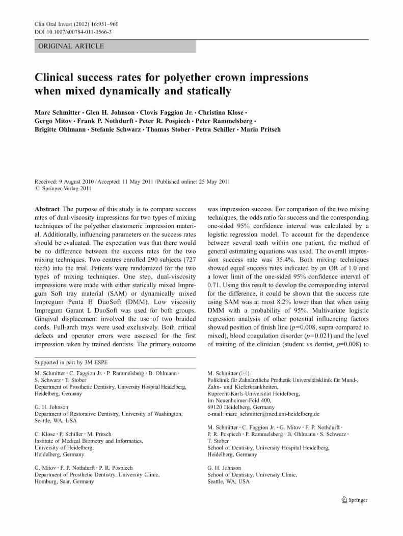
Thus, the purpose of this randomized controlledclinical trial was to compare first impression successrates of dual-viscosity impressions for two types ofmixing techniques (DMM versus SAM, see Fig. 1) ofthe PE tray material using a standardized evaluation form.Additionally, influencing parameters on the success rateswere evaluated. The expectation was that there would beno difference between the success rates for the twomixing techniques and that several independent variableshave an influence on the success rate.
Fig. 1 Dynamic mechanicalmixing unit (Pentamix 3,3M ESPE (a)) andhand-dispensed cartridgemixing system (3M ESPE (b))
952 Clin Oral Invest (2012) 16:951–960
Material and methods
Patients
Two centres participated in the trial, the Department ofProsthetic Dentistry of the University Hospital Heidel-berg and the Department of Prosthetic Dentistry of theUniversity Clinic Homburg/Saar. The study was ap-proved by the review board of the University ofHeidelberg (S-295/2008) and the ethics committee ofthe medical society of Saarland. The impressions weremade by fourth- and fifth-year dental students andfaculty dentists of the Department of Prosthodontics atthe University of Heidelberg and of the Department ofProsthodontics at the University of Homburg/Saar. Thestudents received didactic training, followed by practi-cal training which included placement of retractioncords and taking two impressions. All patients signedan informed consent form. The inclusion criteria for theparticipants were the following: need for at least onecrown (full metal crown, metal ceramic crown, all-ceramic crown and telescopic crown); age of subject≥18 years and a signed consent form. Exclusion criteriawere: pregnancy or lactation.
To assess the influence of several parameters on thesuccess rate of the impressions, relevant patient andtreatment characteristics were gathered using a casereport form. The following were documented as baselinedata: gender, age, number of teeth to be prepared, dentalarch, the presence of a blood coagulation disorder(which might complicate taking the impression) andthe status of the person taking the impression (studentvs. dentist). Additionally, the following data with respectto the treatment was gathered: position of the teeth,position of the finish line (supragingival, subgingival,mixed), use of cords with epinephrine, type of tray(standardized or individual tray) and the individual whoevaluated the impression.
Randomization
The allocation of the mixing technique to each patient wasdone by a stratified block randomization, the details ofwhich were not known by the treating dentists. A blockrandomisation with a variable block length was used toachieve equal group sizes for DDM and SAM for bothcentres. Furthermore, the randomisation was stratified forthe factors “centre” and “number of prepared teeth perpatient” which were expected to have an influence on theimpression success rate and therefore should be wellbalanced between both groups (six strata: two centres andthree levels for number of teeth: one to two, three to four orfive to six teeth). On the basis of the prepared randomiza-
tion list, sealed numbered envelops were created by anindependent person, for each centre and for the differentnumbers of teeth. For each patient the number of teeth to beprepared was identified and immediately before theimpression was taken, the envelope next in sequence withthe suitable number of teeth was opened. The mixingtechnique that was specified on the randomisation sheetwas applied.
Impression materials and clinical procedures
The impression technique and materials did not differ fromthose normally used at both study locations. The materialwas provided from the manufacturer ensuring that thematerial came from the same lot. Gingival displacementinvolved the use of two braided cords (Ultrapak Cord orUltrapak E Cord, Ultradent Products Inc, South Jordan,Utah; Surgident, Sigma Dental Systems-Emasdi GmbH,Jarplund-Weding, Germany) where the initial smaller cordremained in the sulcus at the time the impression was made[11]. Full-arch trays were used exclusively with either astock metal (Rim-Lock, Dentsply/Caulk, Milford DE,USA) or custom tray (Light-cured TrayMaterial, Omnident,Rodgau, Germany). Adhesive was applied to the impressiontray per manufacturer’s direction (Polyether Adhesive, 3MESPE).
A one-step, dual-viscosity impression technique wasused where the medium- or heavy-bodied material wasplaced in the full-arch impression tray while the lowviscosity material was injected onto the prepared tooth/teeth. The tray material was dispensed by random assign-ment either from the DMM device (Pentamix 3, 3M ESPE)or a static automixer (SAM) from cartridges. The colour ofthe two tray impression materials was identical. Theinjected low viscosity was the same for both groups:SAM mixed ISO Type 3 Impregum Garant L DuoSoft(3M ESPE). The tray viscosities consisted of either DMMISO Type 1 Impregum Penta H DuoSoft or SAM ISO Type2 Impregum Soft (3M ESPE).
Evaluation of the impressions
In order to use the same basis for the evaluation of thesuccess rates for the two impression (mixing) systems, onlythe first impressions taken were used in this study.Impression trays used for “first impressions” were markedto avoid confusing them with additional impressions madefor the same patient when needed.
Six experienced clinicians (all with more than 4 years ofexperience using PE impression materials), blinded withrespect to the impression mixing technique, evaluated theimpressions based on criteria modified from that originallydeveloped by Johnson et al. [4]. Both, critical impression
Clin Oral Invest (2012) 16:951–960 953
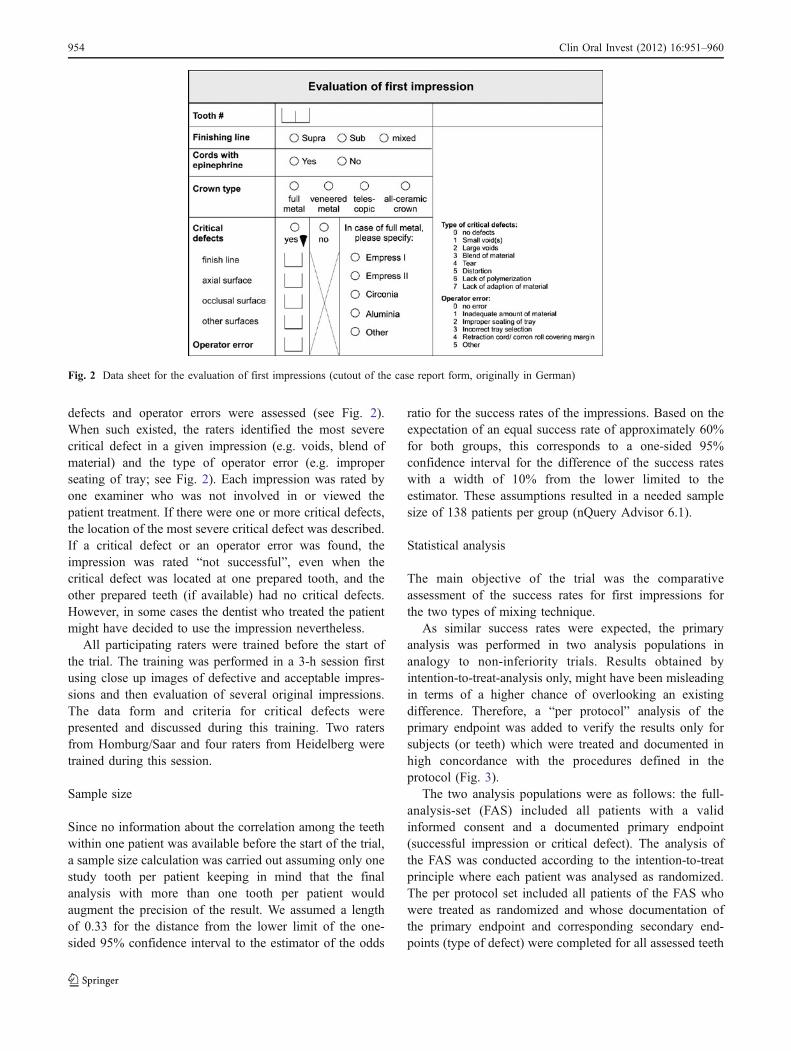
defects and operator errors were assessed (see Fig. 2).When such existed, the raters identified the most severecritical defect in a given impression (e.g. voids, blend ofmaterial) and the type of operator error (e.g. improperseating of tray; see Fig. 2). Each impression was rated byone examiner who was not involved in or viewed thepatient treatment. If there were one or more critical defects,the location of the most severe critical defect was described.If a critical defect or an operator error was found, theimpression was rated “not successful”, even when thecritical defect was located at one prepared tooth, and theother prepared teeth (if available) had no critical defects.However, in some cases the dentist who treated the patientmight have decided to use the impression nevertheless.
All participating raters were trained before the start ofthe trial. The training was performed in a 3-h session firstusing close up images of defective and acceptable impres-sions and then evaluation of several original impressions.The data form and criteria for critical defects werepresented and discussed during this training. Two ratersfrom Homburg/Saar and four raters from Heidelberg weretrained during this session.
Sample size
Since no information about the correlation among the teethwithin one patient was available before the start of the trial,a sample size calculation was carried out assuming only onestudy tooth per patient keeping in mind that the finalanalysis with more than one tooth per patient wouldaugment the precision of the result. We assumed a lengthof 0.33 for the distance from the lower limit of the one-sided 95% confidence interval to the estimator of the odds
ratio for the success rates of the impressions. Based on theexpectation of an equal success rate of approximately 60%for both groups, this corresponds to a one-sided 95%confidence interval for the difference of the success rateswith a width of 10% from the lower limited to theestimator. These assumptions resulted in a needed samplesize of 138 patients per group (nQuery Advisor 6.1).
Statistical analysis
The main objective of the trial was the comparativeassessment of the success rates for first impressions forthe two types of mixing technique.
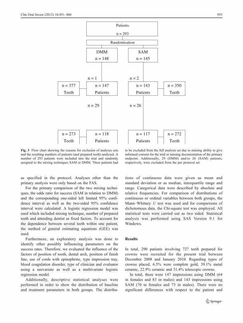
As similar success rates were expected, the primaryanalysis was performed in two analysis populations inanalogy to non-inferiority trials. Results obtained byintention-to-treat-analysis only, might have been misleadingin terms of a higher chance of overlooking an existingdifference. Therefore, a “per protocol” analysis of theprimary endpoint was added to verify the results only forsubjects (or teeth) which were treated and documented inhigh concordance with the procedures defined in theprotocol (Fig. 3).
The two analysis populations were as follows: the full-analysis-set (FAS) included all patients with a validinformed consent and a documented primary endpoint(successful impression or critical defect). The analysis ofthe FAS was conducted according to the intention-to-treatprinciple where each patient was analysed as randomized.The per protocol set included all patients of the FAS whowere treated as randomized and whose documentation ofthe primary endpoint and corresponding secondary end-points (type of defect) were completed for all assessed teeth
Fig. 2 Data sheet for the evaluation of first impressions (cutout of the case report form, originally in German)
954 Clin Oral Invest (2012) 16:951–960
as specified in the protocol. Analyses other than theprimary analysis were only based on the FAS.
For the primary comparison of the two mixing techni-ques, the odds ratio for success (SAM in relation to DMM)and the corresponding one-sided left limited 95% confi-dence interval as well as the two-sided 95% confidenceinterval were calculated. A logistic regression model wasused which included mixing technique, number of preparedteeth and attending dentist as fixed factors. To account forthe dependence between several teeth within one patient,the method of general estimating equations (GEE) wasused.
Furthermore, an exploratory analysis was done toidentify other possibly influencing parameters on thesuccess rates. Therefore, we evaluated the influence of thefactors of position of tooth, dental arch, position of finishline, use of cords with epinephrine, type impression tray,blood coagulation disorder, type of clinician and evaluatorusing a univariate as well as a multivariate logisticregression model.
Additionally, descriptive statistical analyses wereperformed in order to show the distribution of baselineand treatment parameters in both groups. The distribu-
tions of continuous data were given as mean andstandard deviation or as median, interquartile range andrange. Categorical data were described by absolute andrelative frequencies. For comparison of distributions ofcontinuous or ordinal variables between both groups, theMann–Whitney U test was used and for comparisons ofdichotomous data, the Chi-square test was employed. Allstatistical tests were carried out as two sided. Statisticalanalysis was performed using SAS Version 9.1 forWindows.
Results
In total, 290 patients involving 727 teeth prepared forcrowns were recruited for the present trial betweenDecember 2008 and January 2010. Regarding types ofcrowns placed, 6.5% were complete gold, 39.1% metalceramic, 22.9% ceramic and 31.4% telescopic crowns.
In total, there were 147 impressions using DMM (64in females and 83 in males) and 143 impressions usingSAM (70 in females and 73 in males). There were nosignificant differences with respect to the patient and
Patients
n = 293
Randomisation
DMM
n = 148
SAM
n = 145
n = 1 n = 2
n = 377 n = 147 n = 143 n = 350
Teeth Patients Patients Teeth
n = 29 n = 26
n = 273 n = 118 n = 117 n = 272
Teeth Patients Patients Teeth
Fig. 3 Flow chart showing the reasons for exclusion of analyses setsand the resulting numbers of patients (and prepared teeth) analysed. Anumber of 293 patients were included into the trial and randomlyassigned to the mixing techniques SAM or DMM. Three patients had
to be excluded from the full analysis set due to missing ability to giveinformed consent for the trial or missing documentation of the primaryendpoint. Additionally, 29 (DMM) and/or 26 (SAM) patients,respectively, were excluded from the per protocol set
Clin Oral Invest (2012) 16:951–960 955
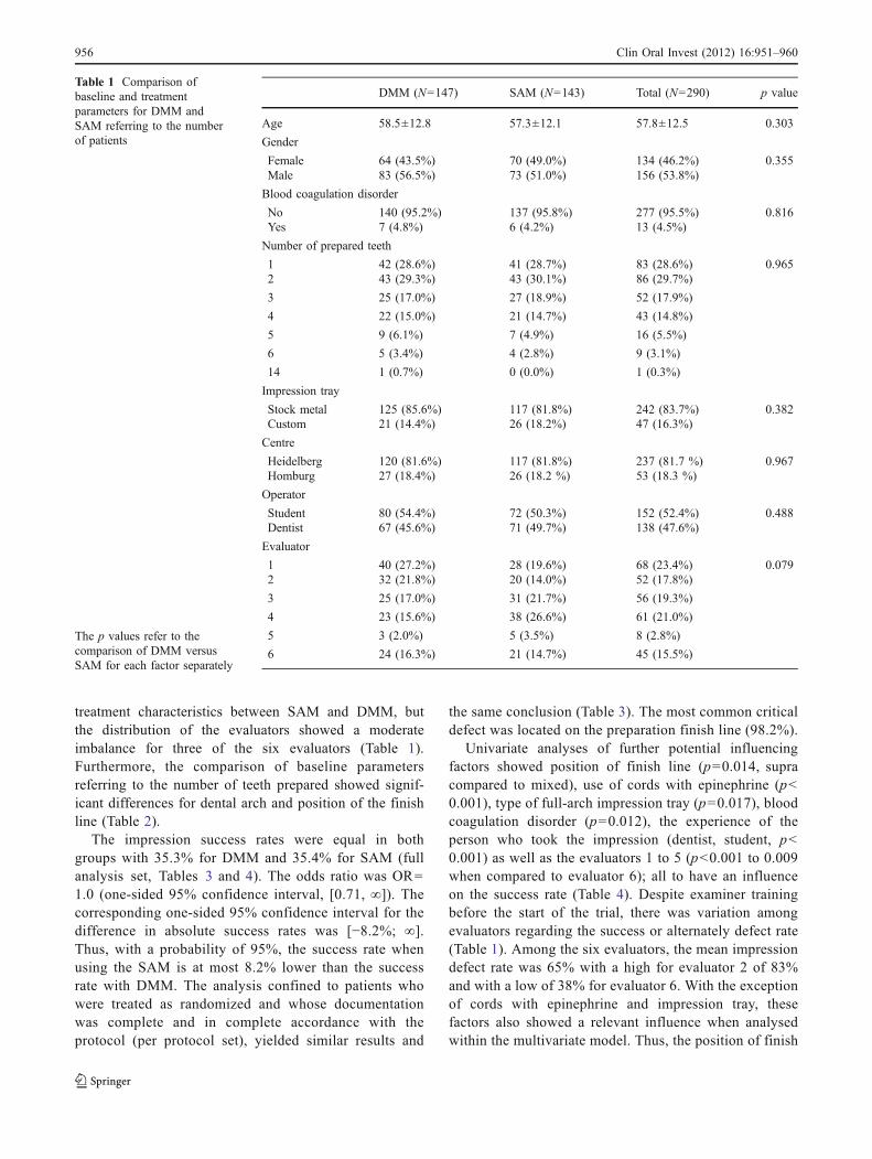
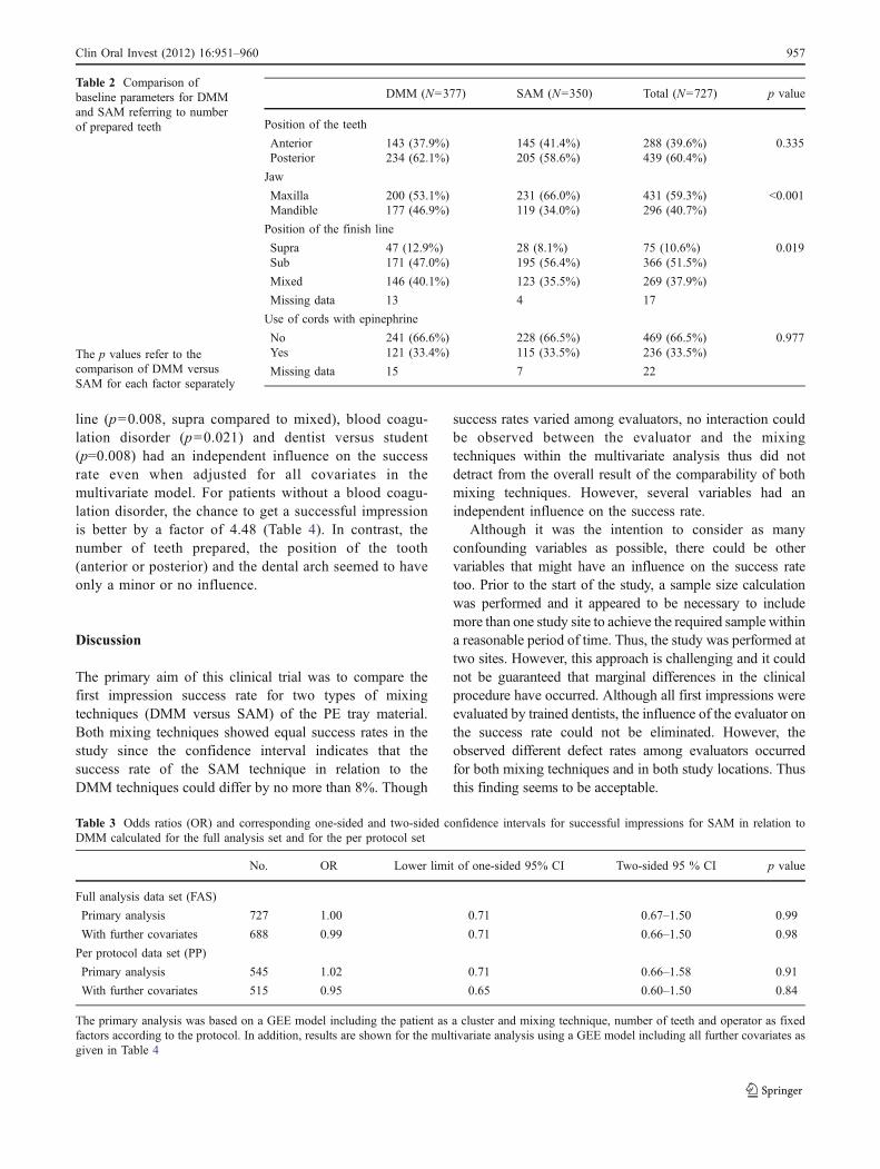
treatment characteristics between SAM and DMM, butthe distribution of the evaluators showed a moderateimbalance for three of the six evaluators (Table 1).Furthermore, the comparison of baseline parametersreferring to the number of teeth prepared showed signif-icant differences for dental arch and position of the finishline (Table 2).
The impression success rates were equal in bothgroups with 35.3% for DMM and 35.4% for SAM (fullanalysis set, Tables 3 and 4). The odds ratio was OR=1.0 (one-sided 95% confidence interval, [0.71, ∞]). Thecorresponding one-sided 95% confidence interval for thedifference in absolute success rates was [−8.2%; ∞].Thus, with a probability of 95%, the success rate whenusing the SAM is at most 8.2% lower than the successrate with DMM. The analysis confined to patients whowere treated as randomized and whose documentationwas complete and in complete accordance with theprotocol (per protocol set), yielded similar results and
the same conclusion (Table 3). The most common criticaldefect was located on the preparation finish line (98.2%).
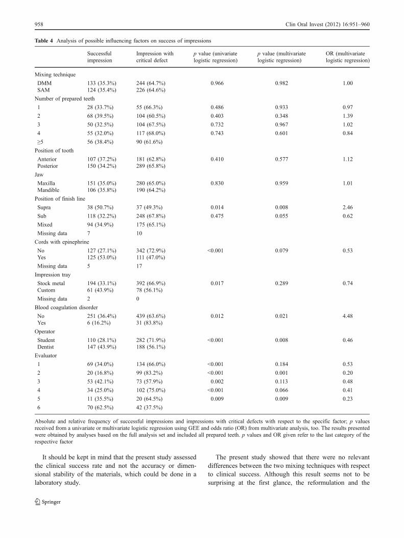
Univariate analyses of further potential influencingfactors showed position of finish line (p=0.014, supracompared to mixed), use of cords with epinephrine (p<0.001), type of full-arch impression tray (p=0.017), bloodcoagulation disorder (p=0.012), the experience of theperson who took the impression (dentist, student, p<0.001) as well as the evaluators 1 to 5 (p<0.001 to 0.009when compared to evaluator 6); all to have an influenceon the success rate (Table 4). Despite examiner trainingbefore the start of the trial, there was variation amongevaluators regarding the success or alternately defect rate(Table 1). Among the six evaluators, the mean impressiondefect rate was 65% with a high for evaluator 2 of 83%and with a low of 38% for evaluator 6. With the exceptionof cords with epinephrine and impression tray, thesefactors also showed a relevant influence when analysedwithin the multivariate model. Thus, the position of finish
DMM (N=147) SAM (N=143) Total (N=290) p value
Age 58.5±12.8 57.3±12.1 57.8±12.5 0.303
Gender
Female 64 (43.5%) 70 (49.0%) 134 (46.2%) 0.355Male 83 (56.5%) 73 (51.0%) 156 (53.8%)
Blood coagulation disorder
No 140 (95.2%) 137 (95.8%) 277 (95.5%) 0.816Yes 7 (4.8%) 6 (4.2%) 13 (4.5%)
Number of prepared teeth
1 42 (28.6%) 41 (28.7%) 83 (28.6%) 0.9652 43 (29.3%) 43 (30.1%) 86 (29.7%)
3 25 (17.0%) 27 (18.9%) 52 (17.9%)
4 22 (15.0%) 21 (14.7%) 43 (14.8%)
5 9 (6.1%) 7 (4.9%) 16 (5.5%)
6 5 (3.4%) 4 (2.8%) 9 (3.1%)
14 1 (0.7%) 0 (0.0%) 1 (0.3%)
Impression tray
Stock metal 125 (85.6%) 117 (81.8%) 242 (83.7%) 0.382Custom 21 (14.4%) 26 (18.2%) 47 (16.3%)
Centre
Heidelberg 120 (81.6%) 117 (81.8%) 237 (81.7 %) 0.967Homburg 27 (18.4%) 26 (18.2 %) 53 (18.3 %)
Operator
Student 80 (54.4%) 72 (50.3%) 152 (52.4%) 0.488Dentist 67 (45.6%) 71 (49.7%) 138 (47.6%)
Evaluator
1 40 (27.2%) 28 (19.6%) 68 (23.4%) 0.0792 32 (21.8%) 20 (14.0%) 52 (17.8%)
3 25 (17.0%) 31 (21.7%) 56 (19.3%)
4 23 (15.6%) 38 (26.6%) 61 (21.0%)
5 3 (2.0%) 5 (3.5%) 8 (2.8%)
6 24 (16.3%) 21 (14.7%) 45 (15.5%)
Table 1 Comparison ofbaseline and treatmentparameters for DMM andSAM referring to the numberof patients
The p values refer to thecomparison of DMM versusSAM for each factor separately
956 Clin Oral Invest (2012) 16:951–960
line (p=0.008, supra compared to mixed), blood coagu-lation disorder (p=0.021) and dentist versus student(p=0.008) had an independent influence on the successrate even when adjusted for all covariates in themultivariate model. For patients without a blood coagu-lation disorder, the chance to get a successful impressionis better by a factor of 4.48 (Table 4). In contrast, thenumber of teeth prepared, the position of the tooth(anterior or posterior) and the dental arch seemed to haveonly a minor or no influence.
Discussion
The primary aim of this clinical trial was to compare thefirst impression success rate for two types of mixingtechniques (DMM versus SAM) of the PE tray material.Both mixing techniques showed equal success rates in thestudy since the confidence interval indicates that thesuccess rate of the SAM technique in relation to theDMM techniques could differ by no more than 8%. Though
success rates varied among evaluators, no interaction couldbe observed between the evaluator and the mixingtechniques within the multivariate analysis thus did notdetract from the overall result of the comparability of bothmixing techniques. However, several variables had anindependent influence on the success rate.
Although it was the intention to consider as manyconfounding variables as possible, there could be othervariables that might have an influence on the success ratetoo. Prior to the start of the study, a sample size calculationwas performed and it appeared to be necessary to includemore than one study site to achieve the required sample withina reasonable period of time. Thus, the study was performed attwo sites. However, this approach is challenging and it couldnot be guaranteed that marginal differences in the clinicalprocedure have occurred. Although all first impressions wereevaluated by trained dentists, the influence of the evaluator onthe success rate could not be eliminated. However, theobserved different defect rates among evaluators occurredfor both mixing techniques and in both study locations. Thusthis finding seems to be acceptable.
DMM (N=377) SAM (N=350) Total (N=727) p value
Position of the teeth
Anterior 143 (37.9%) 145 (41.4%) 288 (39.6%) 0.335Posterior 234 (62.1%) 205 (58.6%) 439 (60.4%)
Jaw
Maxilla 200 (53.1%) 231 (66.0%) 431 (59.3%) <0.001Mandible 177 (46.9%) 119 (34.0%) 296 (40.7%)
Position of the finish line
Supra 47 (12.9%) 28 (8.1%) 75 (10.6%) 0.019Sub 171 (47.0%) 195 (56.4%) 366 (51.5%)
Mixed 146 (40.1%) 123 (35.5%) 269 (37.9%)
Missing data 13 4 17
Use of cords with epinephrine
No 241 (66.6%) 228 (66.5%) 469 (66.5%) 0.977Yes 121 (33.4%) 115 (33.5%) 236 (33.5%)
Missing data 15 7 22
Table 2 Comparison ofbaseline parameters for DMMand SAM referring to numberof prepared teeth
The p values refer to thecomparison of DMM versusSAM for each factor separately
Table 3 Odds ratios (OR) and corresponding one-sided and two-sided confidence intervals for successful impressions for SAM in relation toDMM calculated for the full analysis set and for the per protocol set
No. OR Lower limit of one-sided 95% CI Two-sided 95 % CI p value
Full analysis data set (FAS)
Primary analysis 727 1.00 0.71 0.67–1.50 0.99
With further covariates 688 0.99 0.71 0.66–1.50 0.98
Per protocol data set (PP)
Primary analysis 545 1.02 0.71 0.66–1.58 0.91
With further covariates 515 0.95 0.65 0.60–1.50 0.84
The primary analysis was based on a GEE model including the patient as a cluster and mixing technique, number of teeth and operator as fixedfactors according to the protocol. In addition, results are shown for the multivariate analysis using a GEE model including all further covariates asgiven in Table 4
Clin Oral Invest (2012) 16:951–960 957
It should be kept in mind that the present study assessedthe clinical success rate and not the accuracy or dimen-sional stability of the materials, which could be done in alaboratory study.
The present study showed that there were no relevantdifferences between the two mixing techniques with respectto clinical success. Although this result seems not to besurprising at the first glance, the reformulation and the
Table 4 Analysis of possible influencing factors on success of impressions
Successfulimpression
Impression withcritical defect
p value (univariatelogistic regression)
p value (multivariatelogistic regression)
OR (multivariatelogistic regression)
Mixing technique
DMM 133 (35.3%) 244 (64.7%) 0.966 0.982 1.00SAM 124 (35.4%) 226 (64.6%)
Number of prepared teeth
1 28 (33.7%) 55 (66.3%) 0.486 0.933 0.97
2 68 (39.5%) 104 (60.5%) 0.403 0.348 1.39
3 50 (32.5%) 104 (67.5%) 0.732 0.967 1.02
4 55 (32.0%) 117 (68.0%) 0.743 0.601 0.84
≥5 56 (38.4%) 90 (61.6%)
Position of tooth
Anterior 107 (37.2%) 181 (62.8%) 0.410 0.577 1.12Posterior 150 (34.2%) 289 (65.8%)
Jaw
Maxilla 151 (35.0%) 280 (65.0%) 0.830 0.959 1.01Mandible 106 (35.8%) 190 (64.2%)
Position of finish line
Supra 38 (50.7%) 37 (49.3%) 0.014 0.008 2.46
Sub 118 (32.2%) 248 (67.8%) 0.475 0.055 0.62
Mixed 94 (34.9%) 175 (65.1%)
Missing data 7 10
Cords with epinephrine
No 127 (27.1%) 342 (72.9%) <0.001 0.079 0.53Yes 125 (53.0%) 111 (47.0%)
Missing data 5 17
Impression tray
Stock metal 194 (33.1%) 392 (66.9%) 0.017 0.289 0.74Custom 61 (43.9%) 78 (56.1%)
Missing data 2 0
Blood coagulation disorder
No 251 (36.4%) 439 (63.6%) 0.012 0.021 4.48Yes 6 (16.2%) 31 (83.8%)
Operator
Student 110 (28.1%) 282 (71.9%) <0.001 0.008 0.46Dentist 147 (43.9%) 188 (56.1%)
Evaluator
1 69 (34.0%) 134 (66.0%) <0.001 0.184 0.53
2 20 (16.8%) 99 (83.2%) <0.001 0.001 0.20
3 53 (42.1%) 73 (57.9%) 0.002 0.113 0.48
4 34 (25.0%) 102 (75.0%) <0.001 0.066 0.41
5 11 (35.5%) 20 (64.5%) 0.009 0.009 0.23
6 70 (62.5%) 42 (37.5%)
Absolute and relative frequency of successful impressions and impressions with critical defects with respect to the specific factor; p valuesreceived from a univariate or multivariate logistic regression using GEE and odds ratio (OR) from multivariate analysis, too. The results presentedwere obtained by analyses based on the full analysis set and included all prepared teeth. p values and OR given refer to the last category of therespective factor
958 Clin Oral Invest (2012) 16:951–960
effectiveness of mixing of the tray material could have hadan influence on the clinical success. For instance, operatorerrors could have been higher using the hand-dispensedcartridge mixing system. However, both the handling of thecartridge system and the clinical performance of the newlyformulated material seems to be acceptable compared to thegold-standard of machine mixing.
Furthermore, variables which have an independent influ-ence on impression success have been identified. Luthardt etal. [12] assessed the clinical parameters influencing theaccuracy of one- and two-stage impressions and found thepresence of blood as an important factor. The present studyshowed that the presence of a blood coagulation disordernegatively affects the success rate for impressions. Thus, theabsence of blood appears be essential for the success and theaccuracy of the impression. Additionally, the present studyshowed that the use of retraction cords with epinephrinetended to have a positive effect on the success rate.Kombuloglu et al. [13] found that epinephrine-impregnatedcord systems were clinically successful, thus consistent withthe results of the present study and found that these cordswere more effective than nonmedicated cords. Weir et al. [14]assessed the effectiveness of mechanical–chemical tissuedisplacement methods and confirm the results of the presentstudy with respect to the usage of cords with epinephrine.
Moreover, the degree of experience/educationwas shown tohave an influence on clinical success rates. Johnson et al. [4]assessed the success rates for polyether and vinyl polysilox-ane impressions using a standardized evaluation form andfound that 50% of the PE impressions using full-arch trayswere successful. In the present study, the success rate waslower in both operating groups (student, dentist). However,the results for dentists in the present study are comparable tothe results of Johnson et al. (43.9% versus 50%), especiallyconsidering that 166 out of 191 impressions in the cited studyinvolved a single prepared tooth whereas only 29% of theimpressions in the present study were of a single preparedtooth. Additionally, it could be speculated that the lowersuccess rate in the students of the present study compared tothe students in the study of Johnson et al. [4] could be a resultof the different experience levels. In the present study, thestudents had average experience with impressioning, whereasin the study of Johnson et al. the students had extensiveexperience. Additionally, it has to be taken into considerationthat in the present study, about 90% of the teeth wereprepared completely or partially subgingival and conse-quently the impression was challenging [15, 16]. In thiscontext it has to be taken into consideration that in theaforementioned study, most of the critical defects ofimpressions were observed on the finish line which isconfirmed by the present study. This area is examined byclinicians first since this area is thought to be the mostimportant for clinical success [17]. Thus, the raters might
have tended to categorize defects at this location more oftenas “critical defects” than at other locations.
A reason for the lower success rate in the present studyin general could be that the clinical situation for makingimpressions is challenging in the dental school environmentsince many patients who seek dental treatment haveneglected dental care for some time and present withsignificant loss of tooth structure [4].
The finding that both mixing techniques under examinationdid not show different success rates might be of interest topractitioners who wish to use static automixing with PEmaterial. Additionally, the present study demonstrated thatseveral factors have an influence on the success rates. This mayalso be important to many dental practitioners since theselection of the most suitable displacement cord is essentialin everyday practice. Future studies could focus on theassessment of other variables which might have an additionalinfluence on the success rate of dental impressions.
Conclusions
Within the limitations of this clinical trial, the following couldbe concluded: dynamic mechanical mixing and the new staticmixing of polyether tray impression material showed nearlyequal success (DMM, 35.3%; SAM, 35.4%) with a confidenceinterval indicating that static automixing could be no more than8% less successful than dynamicmechanical mixing (one-sided95% confidence interval, [−8.2%; ∞]). Also, the position offinish line (p=0.008, supra compared to mixed), bloodcoagulation disorder (p=0.021) and the experience of theclinician who took the impression (dentist, student; p=0.008)had an independent influence on the success rate even whenadjusted for all covariates in the multivariate model.
Acknowledgement The authors acknowledge and thank the followingfrom the Institute of Medical Biometry and Informatics, Heidelberg:Judith Munzinger for data management and Annette Kretz and AstridWallenwein for providing data entry. Additionally, the authors thank 3 MESPE (Dr. Thomas Klettke) for the financial support of the study.
Conflict of interest The authors declare that they have no conflict ofinterest.
References
1. Stewardson DA (2005) Trends in indirect dentistry: 5. Impressionmaterials and techniques. Dent Update; 32: 374–376, 379–380,382–374
2. Michalakis KX, Bakopoulou A, Hirayama H, Garefis DP, GarefisPD (2007) Pre- and post-set hydrophilicity of elastomericimpression materials. J Prosthodont 16:238–248
3. Stober T, Johnson GH, Schmitter M (2010) Accuracy of the newlyformulated vinyl siloxanether elastomeric impression material. JProsthet Dent 103:228–239
Clin Oral Invest (2012) 16:951–960 959
4. Johnson GH, Mancl LA, Schwedhelm ER, Verhoef DR, LepeX (2010) Clinical trial investigating success rates forpolyether and vinyl polysiloxane impressions made withfull-arch and dual-arch plastic trays. J Prosthet Dent 103:13–22
5. Stackhouse JA Jr (1983) Voids in a mixed elastomeric impressionmaterial. J Prosthet Dent 50:762–766
6. Keck SC (1985) Automixing: a new concept in elastomericimpression material delivery systems. J Prosthet Dent 54:479–483
7. Powers J, Sakaguchi R (2006) Craig’s restorative dental materials.Elsevier, St. Louis
8. Kugel G, Swift EJ, Jr., Sorensen JA, Tucker JH, Dunne JT, Jr.(1999) A prospective clinical evaluation of electronically mixedpolyvinyl siloxane impression materials: results from the pros-thetic "SuperStudy"—a consumer evaluation. Compend ContinEduc Dent Suppl; S3–21; quiz S22
9. Di Felice R, Scotti R, Belser UC (2002) The influence of themixing technique on the content of voids in two polyether impressionmaterials. Schweiz Monatsschr Zahnmed 112:12–16
10. Margeas R (2009) Creating precision restorations using a hand-dispensed polyether. Dent Today 28(106):108–109
11. Perakis N, Belser UC, Magne P (2004) Final impressions: areview of material properties and description of a currenttechnique. Int J Periodontics Restor Dent 24:109–117
12. Luthardt RG, Walter MH, Weber A, Koch R, Rudolph H (2008)Clinical parameters influencing the accuracy of 1- and 2-stageimpressions: a randomized controlled trial. Int J Prosthodont21:322–327
13. Kumbuloglu O, User A, Toksavul S, Boyacioglu H (2007)Clinical evaluation of different gingival retraction cords.Quintessence Int 38:e92–e98
14. Weir DJ, Williams BH (1984) Clinical effectiveness ofmechanical-chemical tissue displacement methods. J ProsthetDent 51:326–329
15. Beier US, Grunert I, Kulmer S, Dumfahrt H (2007) Quality ofimpressions using hydrophilic polyvinyl siloxane in a clinicalstudy of 249 patients. Int J Prosthodont 20:270–274
16. Radjaeipour G (2007) Improving decision-making in restorations:evaluation of impressions and working casts. J Calif Dent Assoc35:637–640
17. Winstanley RB, Carrotte PV, Johnson A (1997) The quality ofimpressions for crowns and bridges received at commercial dentallaboratories. Br Dent J 183:209–213
960 Clin Oral Invest (2012) 16:951–960