Clinical and Magnetic Resonance Imaging (MRI) Features ...
18
Citation: Pons-Sorolla, M.; Dominguez, E.; Czopowicz, M.; Suñol, A.; Maeso Ordás, C.; Morales Moliner, C.; Pérez Soteras, M.; Montoliu, P. Clinical and Magnetic Resonance Imaging (MRI) Features, Tumour Localisation, and Survival of Dogs with Presumptive Brain Gliomas. Vet. Sci. 2022, 9, 257. https://doi.org/10.3390/ vetsci9060257 Academic Editors: Sara Belluco, Maria Teresa Mandara and Martí Pumarola Received: 5 April 2022 Accepted: 24 May 2022 Published: 27 May 2022 Publisher’s Note: MDPI stays neutral with regard to jurisdictional claims in published maps and institutional affil- iations. Copyright: © 2022 by the authors. Licensee MDPI, Basel, Switzerland. This article is an open access article distributed under the terms and conditions of the Creative Commons Attribution (CC BY) license (https:// creativecommons.org/licenses/by/ 4.0/). veterinary sciences Article Clinical and Magnetic Resonance Imaging (MRI) Features, Tumour Localisation, and Survival of Dogs with Presumptive Brain Gliomas Marta Pons-Sorolla 1 , Elisabet Dominguez 1 , Michal Czopowicz 2 , Anna Suñol 1 , Christian Maeso Ordás 1 , Carles Morales Moliner 1 , Marc Pérez Soteras 1 and Patrícia Montoliu 1, * 1 AniCura Ars Veterinaria Hospital Veterinari, Carrer dels Cavallers 37, 08034 Barcelona, Spain; [email protected] (M.P.-S.); [email protected] (E.D.); [email protected] (A.S.); [email protected] (C.M.O.); [email protected] (C.M.M.); [email protected] (M.P.S.) 2 Division of Veterinary Epidemiology and Economics, Institute of Veterinary Medicine, Warsaw University of Life Sciences—SGGW, Nowoursynowska 159c, 02-776 Warsaw, Poland; [email protected] * Correspondence: [email protected] Abstract: Brain gliomas are common tumours diagnosed in dogs. However, limited information is available on the clinical features and overall survival time (OS) in dogs receiving palliative treatment. The aim of this study was to evaluate possible associations between presenting complaint, tumour localisation, Magnetic Resonance Imaging (MRI) features, survival times, and reason for the death of dogs with suspected intracranial glioma treated palliatively. Sixty dogs from a single institution were retrospectively included (from September 2017 to December 2021). Dogs were included if a presumptive diagnosis of brain glioma was obtained based on an MRI scan and medical history. French Bulldogs were overrepresented (40/60); 46 out of 60 dogs (77%) presented due to epileptic seizures (ES) and in 25/60 dogs (42%), cluster seizures or status epilepticus were the first manifestation of the disease. Dogs with suspected gliomas located in the piriform lobe showed a higher probability of presenting due to epilepsy compared to dogs with glioma in other regions, and more frequently died or were euthanised because of increased ES. Magnetic Resonance Imaging (MRI) features differed between localisations. Fronto-olfactory tumours were more frequently, whereas piriform tumours were less frequently, classified as suspected high-grade glioma. The median survival time was 61 days. Dogs with contrast-enhancing suspected gliomas had significantly shorter OS. This study provides additional information on the clinical features and survival of dogs with suspected brain gliomas treated palliatively. Keywords: glioma; epileptic seizures (ES); piriform; dog; Magnetic Resonance Imaging (MRI) 1. Introduction Gliomas are the second most common brain tumour in dogs, after meningiomas [1–3]. An overall prevalence rate of 0.9% has been reported, accounting for approximately 30% of primary intracranial neoplasms in dogs [3]. Brachycephalic dogs have shown a predisposition [4–8], and males are reported in many studies to be overrepresented [7,8]. Most brain gliomas are rostrotentorial, and neurological signs are variable, with epileptic seizures (ES) being the most frequently described clinical sign. Histopathologically, gliomas are classified as oligodendrogliomas, astrocytomas, or undefined gliomas, and are further subclassified as low or high grade [8]. Due to financial and safety considerations, tentative antemortem diagnosis of intracranial gliomas is most commonly based on signalment, clinical signs, and compatible magnetic resonance imaging (MRI) findings [9,10]. Vet. Sci. 2022, 9, 257. https://doi.org/10.3390/vetsci9060257 https://www.mdpi.com/journal/vetsci
-
Upload
khangminh22 -
Category
Documents
-
view
0 -
download
0
Transcript of Clinical and Magnetic Resonance Imaging (MRI) Features ...
Citation: Pons-Sorolla, M.;
Dominguez, E.; Czopowicz, M.;
Suñol, A.; Maeso Ordás, C.; Morales
Moliner, C.; Pérez Soteras, M.;
Montoliu, P. Clinical and Magnetic
Resonance Imaging (MRI) Features,
Tumour Localisation, and Survival of
Dogs with Presumptive Brain
Gliomas. Vet. Sci. 2022, 9, 257.
https://doi.org/10.3390/
vetsci9060257
Academic Editors: Sara Belluco,
Maria Teresa Mandara and
Martí Pumarola
Received: 5 April 2022
Accepted: 24 May 2022
Published: 27 May 2022
Publisher’s Note: MDPI stays neutral
with regard to jurisdictional claims in
published maps and institutional affil-
iations.
Copyright: © 2022 by the authors.
Licensee MDPI, Basel, Switzerland.
This article is an open access article
distributed under the terms and
conditions of the Creative Commons
Attribution (CC BY) license (https://
creativecommons.org/licenses/by/
4.0/).
veterinarysciences
Article
Clinical and Magnetic Resonance Imaging (MRI) Features,Tumour Localisation, and Survival of Dogs with PresumptiveBrain GliomasMarta Pons-Sorolla 1 , Elisabet Dominguez 1, Michał Czopowicz 2 , Anna Suñol 1 , Christian Maeso Ordás 1,Carles Morales Moliner 1, Marc Pérez Soteras 1 and Patrícia Montoliu 1,*
1 AniCura Ars Veterinaria Hospital Veterinari, Carrer dels Cavallers 37, 08034 Barcelona, Spain;[email protected] (M.P.-S.); [email protected] (E.D.); [email protected] (A.S.);[email protected] (C.M.O.); [email protected] (C.M.M.); [email protected] (M.P.S.)
2 Division of Veterinary Epidemiology and Economics, Institute of Veterinary Medicine, Warsaw University ofLife Sciences—SGGW, Nowoursynowska 159c, 02-776 Warsaw, Poland; [email protected]
* Correspondence: [email protected]
Abstract: Brain gliomas are common tumours diagnosed in dogs. However, limited information isavailable on the clinical features and overall survival time (OS) in dogs receiving palliative treatment.The aim of this study was to evaluate possible associations between presenting complaint, tumourlocalisation, Magnetic Resonance Imaging (MRI) features, survival times, and reason for the deathof dogs with suspected intracranial glioma treated palliatively. Sixty dogs from a single institutionwere retrospectively included (from September 2017 to December 2021). Dogs were included if apresumptive diagnosis of brain glioma was obtained based on an MRI scan and medical history.French Bulldogs were overrepresented (40/60); 46 out of 60 dogs (77%) presented due to epilepticseizures (ES) and in 25/60 dogs (42%), cluster seizures or status epilepticus were the first manifestationof the disease. Dogs with suspected gliomas located in the piriform lobe showed a higher probabilityof presenting due to epilepsy compared to dogs with glioma in other regions, and more frequentlydied or were euthanised because of increased ES. Magnetic Resonance Imaging (MRI) featuresdiffered between localisations. Fronto-olfactory tumours were more frequently, whereas piriformtumours were less frequently, classified as suspected high-grade glioma. The median survival timewas 61 days. Dogs with contrast-enhancing suspected gliomas had significantly shorter OS. Thisstudy provides additional information on the clinical features and survival of dogs with suspectedbrain gliomas treated palliatively.
Keywords: glioma; epileptic seizures (ES); piriform; dog; Magnetic Resonance Imaging (MRI)
1. Introduction
Gliomas are the second most common brain tumour in dogs, after meningiomas [1–3].An overall prevalence rate of 0.9% has been reported, accounting for approximately 30% ofprimary intracranial neoplasms in dogs [3].
Brachycephalic dogs have shown a predisposition [4–8], and males are reported inmany studies to be overrepresented [7,8]. Most brain gliomas are rostrotentorial, andneurological signs are variable, with epileptic seizures (ES) being the most frequentlydescribed clinical sign. Histopathologically, gliomas are classified as oligodendrogliomas,astrocytomas, or undefined gliomas, and are further subclassified as low or high grade [8].Due to financial and safety considerations, tentative antemortem diagnosis of intracranialgliomas is most commonly based on signalment, clinical signs, and compatible magneticresonance imaging (MRI) findings [9,10].
Vet. Sci. 2022, 9, 257. https://doi.org/10.3390/vetsci9060257 https://www.mdpi.com/journal/vetsci
Vet. Sci. 2022, 9, 257 2 of 18
The reported overall survival times (OS) in canine gliomas are mainly related tothe chosen treatment [6,7,11,12]. Described OS for dogs with brain gliomas that receivepalliative treatment are short, ranging from 26 to 94 days [7,12–14]. However, little isknown about possible differences in OS between specific localisations or MRI features insuspected gliomas.
In humans, the predisposition for developing ES depends on the localisation andgrade of the tumour [15]. Mesial temporal gliomas are associated with higher epilep-togenicity [16]. In addition, low-grade gliomas are more epileptogenic than high-gradegliomas [17,18]. Since low-grade gliomas have an increased affinity for the mesial temporalarea in adults [16,19], drug-resistant epilepsy is their most common symptom [17,18]. In-vestigations of animal models have also indicated that the piriform lobe, located at the edgeof the olfactory and mesial temporal cortex, is one of the most epileptogenic regions in thebrain [20,21]. Information about the influence of the localisation and nature of the lesionon epileptogenicity of gliomas is very limited in veterinary medicine. Veterinary studiesassociating localisation of the tumour and MRI features with clinical signs are limited, andmost commonly include various types of tumours.
The aim of this study was to evaluate the relationship between tumour localisation andMRI features, as well as presenting clinical signs, and to link these variables with survivaltimes in a population of dogs with suspected intracranial glioma that have undergone apalliative treatment.
We hypothesised that the specific localisation of the suspected glioma in the rostro-tentorial region influences presenting clinical signs, MRI features, and survival time. Ourmain hypothesis was that dogs with suspected gliomas located in the piriform lobe presentmore frequently due to severe ES compared to other rostrotentorial regions.
2. Materials and Methods2.1. Study Design and Medical Records
This retrospective study was carried out including dogs from Anicura Ars Veterinariaveterinary hospital that presented between September 2017 and December 2021. Dogswith a final presumptive diagnosis of brain glioma based on signalment, clinical history,neurological signs, progression, and compatible MRI characteristics were included.
The term “glioma” was searched within the author’s institution’s MRI reports database.All cases with a presumptive diagnosis or main differential diagnosis of glioma in the MRIreport conclusions were selected for revision. Complete medical records were reviewed,and the following data were recorded for each case: age, sex, body weight and breed,owner’s main complaint, date of first presentation of clinical signs, physical and neuro-logical examination, type of treatment, overall survival time (time from MRI diagnosis todeath, OS), and cause of death or euthanasia when available. When complete informationregarding follow up was not included in the medical records, data were obtained by meansof a telephone call with the first opinion veterinary or owners.
The findings in the neurological examination were classified by a board-certifiedneurologist as mild/moderate, severe, or post-ictal following the criteria in Table 1. Post-ictal signs were considered when neurological deficits were symmetrical, resolved in thesecond neurological examination before additional specific treatment, and the ownersreported no behavioural or neurological changes before the presentation.
For dogs with ES, information about the type of ES (focal or generalised) and severity(isolated ES or cluster seizures/status epilepticus (CS/SE)) was recorded. Clustered ES orstatus epilepticus were included in the same group due to the lack of specific information insome clinical history records to differentiate between them. It was also recorded whetherCS/SE was the first manifestation of neurological disease.
Vet. Sci. 2022, 9, 257 3 of 18
Status epilepticus and CS were defined following the International Veterinary EpilepsyTask Force Consensus proposal. Status epilepticus was defined clinically as (a) an ES lastingmore than 5 min or (b) two or more discrete ES between which there is incomplete recovery ofconsciousness. Cluster seizures were defined as two or more seizures within a 24-h period [22].
Table 1. Classification of neurological findings in 60 dogs with presumed brain glioma.
Neurological Findings Description
Absence Only seizures.
Mild/ModerateAt least one of the following: mildly altered mentation, head tilt,ambulatory status, any type of ataxia, proprioceptive deficits, andcranial nerve deficits.
Severe At least one of the following: severe obtundation, stupor or coma,or non-ambulatory tetraparesis.
Post-ictalAt least one of the following: altered mental status, compulsivebehaviour, tetraparesis or paraparesis, bilateral symmetricalproprioceptive deficits, or bilateral menace response deficits.
Status epilepticus and CS were defined following the International Veterinary EpilepsyTask Force Consensus proposal. Status epilepticus was defined clinically as (a) an ES lastingmore than 5 min or (b) two or more discrete ES between which there is incomplete recovery ofconsciousness. Cluster seizures were defined as two or more seizures within a 24-h period [22].
When available, additional diagnostic tests including cerebrospinal fluid (CSF) anal-ysis, thoracic radiographs, abdominal ultrasound, or computed tomography (CT) werereviewed for the presence of signs of neoplasia or severe systemic disease.
Dogs were excluded if they showed other neoplasia or severe systemic disease thatcould influence neurological signs or OS. Dogs that received chemotherapy or radiotherapy,dogs that died or were euthanised <24 h after MRI without any intent of treatment, and dogswith a caudotentorial localisation were included in the demographic study but excludedfrom the survival analysis. Dogs that were alive at the end of the study, died of conditionsunrelated to suspected glioma, and dogs whose date of death was unknown were censored.
2.2. MRI and Tumour Localisation
All MRI scans were performed under general anaesthesia using a high-field 1.5TMRI scanner (Vantage Elan, Canon Medical Systems Corporation, Tochigi, Japan). Themost common MRI protocol used included T2-weighted (T2W) sagittal and dorsal planes,transverse planes of T2W, T1-weighted (T1W), T2 fluid-attenuated inversion recovery(FLAIR), and dorsal and transverse planes of T1W post-gadolinium sequences (Table S1).Some patients had additional sequences including transverse diffusion-weighted echo-planar sequences, T2-weighted gradient echo (T2*), or susceptibility-weighted images. Forpost-contrast images, gadoteridol (Clariscan®-GE Healthcare AS, Oslo, Norway- injection0.5 mmol/mL) was administered at a dose of 0.2 mL/kg intravenously (IV). The termspresumptive and suspected are used in the study since histopathological confirmation ofdiagnosis was not available.
All MRI images were reviewed for the purpose of the study by one board-certifiedradiologist (E.D.) and one board-certified neurologist (P.M.) who were blinded to the initialimaging report and patient’s clinical signs.
MRI features were considered consistent with glial tumours when intra-axial singlelesions, often heterogeneous, ovoid to amorphous, well-defined to infiltrative, T1 isointenseto hypointense, and T2 isointense to hyperintense mass lesions with variable contrastenhancement (CE) were observed. Other considered findings were cyst-like lesions, intrale-sional haemorrhage, vasogenic oedema, and mass effect [23].
Vet. Sci. 2022, 9, 257 4 of 18
Additional criteria including clinical history, neurological signs, case follow-up, CSF,and follow-up MRI, if available, were reviewed at a later stage in the case of diagnosticdiscordance between the imaging report and the review performed by the board-certifiedradiologist and neurologist. Cases initially diagnosed as most consistent with glioma wereexcluded if no consensus was reached after reviewing the images. Patients with severeconcomitant brain lesions or other lesions that could cause ES or affect the survival timewere excluded from the study.
For each case included, the following MRI features were recorded: localisation, lateral-isation, margins, cavitation, presumed invasiveness, and invaded areas, presumed invasionor oedema of the hippocampus, predominant MRI signal intensities in T1W, T2W, andFLAIR sequences, presence of signal void on T2* sequences, presence of abnormalities in thebones adjacent to the lesion, presence of suspected perilesional and/or vasogenic oedema,CE, presence of suspected post-ictal changes, and MRI features evaluating mass effect(subarachnoid CSF effacement, midline shift, ventricular compression/displacement, andbrain herniation). Additionally, a board-certified radiologist (E.D.), who was blinded forany clinical information, was asked for a subjective evaluation of tumour grade given theoverall MRI features with three options: low-grade, high-grade, or equivocal. High-gradegliomas were suspected when the lesions had ill-defined margins, demonstrated the het-erogeneous signal intensity and CE, and showed signs of internal haemorrhage, cavitation,and/or ventricular/intraventricular extension. Low-grade gliomas were suspected whenthe lesions had better-defined margins, an overall more uniform signal intensity, and anabsence of CE. Low-grade and equivocal groups were later merged into “non-high-grade”for meaningful statistical analysis. To ensure comparable subjective grading, images werereviewed consecutively for three days.
Suspected gliomas were divided into groups based on the localisation of the majority ofthe volume of the lesion as follows: fronto-olfactory, piriform, temporal, parietal, occipital,intraventricular, diencephalic, and caudotentorial (brainstem or cerebellum).
2.3. Statistical Analysis
Numerical variables were presented as a median, interquartile range (IQR), andrange, and compared between two groups using the Mann–Whitney U test and between>2 groups with the Kruskal–Wallis H test, followed by Dunn’s post-hoc test. Categoricalvariables were presented as counts and percentages and compared between groups usingthe maximum likelihood G test or Fisher exact test if a count in any cell of the contingencytable was <5. The 95% confidence intervals (CI 95%) for proportions were calculated usingthe Wilson score method. The relationship of demographic and clinical characteristicswith the localisation in the central nervous system was investigated first in the univariableanalysis and variables with p < 0.1 were entered into the multivariable multinomial logisticregression (Table S7). For the multivariable analysis, tumour localisation was mergedinto three categories: fronto-olfactory, piriform, and other rostrotentorial localisations.The relationship of demographic and clinical characteristics with the high-grade wasinvestigated first in the univariable analysis, and variables with p < 0.1 were entered into themultivariable binary logistic regression. The strength and direction of the relationship wereexpressed using the odds ratio (OR). The univariable and multivariable Cox proportionalhazard models were used to investigate the relationship of demographic and clinicalcharacteristics with the overall survival time (OS) and to estimate the hazard ratio (HR).Survival curves were presented for significant variables using the Kaplan–Meier product-limit estimator. Survival analysis included only dogs with rostrotentorial suspected gliomastreated palliatively and only glioma-related deaths were considered complete records. Dogswere censored if: (1) they were still alive when the final database was built, (2) they died ofconditions unrelated to suspected glioma, or (3) the date of death was unknown (in this case,the date of the last follow-up was used as the date of censoring). All multivariable analyseswere carried out according to the backward stepwise procedure. A significance level (α) of0.05 was assumed in all statistical tests except for the univariable analyses in which α was
Vet. Sci. 2022, 9, 257 5 of 18
0.1. The statistical analysis was performed in TIBCO Statistica 13.3 (TIBCO Software Inc., PaloAlto, CA, USA) and IBM SPSS Statistics 26 (IBM Corporation, Armonk, NY, USA).
3. Results3.1. Demographics
In the study period, 85 dogs with MRI consistent with suspected glioma were eligiblefor inclusion in the study. In total, 25 dogs were excluded due to any of the followingreasons: equivocal MRI diagnosis with other suspected brain tumours, vascular accidents,inflammatory diseases, or post-ictal changes (18 dogs); clinical progression not consistentwith brain neoplasia (2 dogs); concomitant congenital hydrocephalus that may have affectedOS and clinical signs (2 dogs); lack of clinical information (2 dogs); and abdominal effusionand suspected lymphoma (1 dog).
Sixty dogs met the inclusion criteria. There were 32 males (19/32 neutered) and28 females (23/28 spayed). The male-to-female ratio was 1.14 (CI 95%: 0.69–1.89). Ageat presentation ranged from 4 to 15 years with the median (IQR) of 9 (7–10) years. Bodyweight ranged from 4 to 55 kg with the median (IQR) of 14 (12–18) kg. Breeds includedFrench Bulldogs (40 dogs), Boxers (6 dogs), mixed breed dogs (5 dogs), Yorkshire Terrier(2 dogs), and one each of the following breeds: English Bulldog, Boston Terrier, CavalierKing Charles Spaniel, West Highland White Terrier, Staffordshire Bull Terrier, Cane Corso,and Dogue de Bordeaux. Detailed demographic characteristics of the study population arepresented in Table S2.
3.2. Clinical Presentation and Clinicopathologic Findings
The time from the first signs to diagnosis ranged from 1 to 730 days with the median(IQR) of 13 (3–35) days. The diagnosis was made within 2 weeks of presentation of clinicalsigns in 50% of dogs, and within 5 weeks in 75% of dogs.
Epileptic seizures were the most common reason for presentation, recorded for46/60 dogs (77%; CI 95%: 65–86%). Thirty-four of sixty dogs (57%; CI 95%: 44–68%)had CS/SE. In 25/60 dogs (42%; CI 95%: 30–54%), CS/SE was also the first manifestationof the disease. Epileptic seizures were described by the owner as being generalised tonic-clonic in 36/46 dogs (78%), 8/46 dogs (17%) had suffered both from focal and generalisedtonic-clonic ES, and 2/46 dogs had shown only focal ES. In only 8 of 46 dogs (17%) thatpresented due to ES, other neurological signs (obtundation, circling, weakness, or alteredbehaviour) were also described by the owner.
A total of 13 of 60 dogs (22%; CI 95%: 13–34%) presented due to neurological signs withouta history of ES. In one dog, suspected glioma was considered an incidental finding, and MRIwas performed due to an acute onset of facial paralysis, which was considered idiopathic.
On neurological examination, 31/60 dogs (52%; CI 95%: 39–64%) had neurologicaldeficits and in only 1 dog they were classified as severe. Neurological examination wasnormal at the time of presentation or deficits were considered post-ictal in 29/60 (48%) dogs.
Results of CSF analysis were available for review in only 6 dogs (10%). Mild pleocytosiswas observed in 2 dogs and albumin-cytological dissociation in the other 2 dogs.
3.3. MRI Features and Localisation of Suspected Glioma
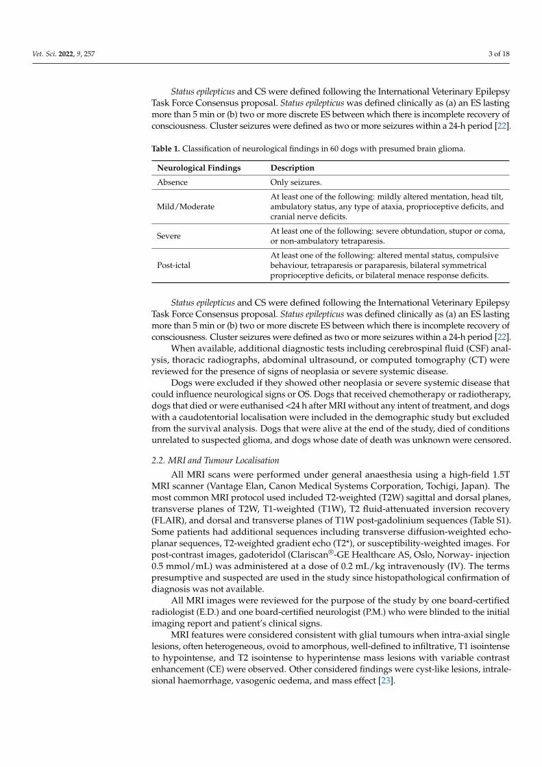
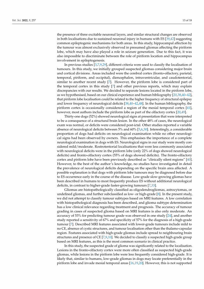
Most suspected gliomas (48/60 dogs; 80%) were located in the fronto-olfactory cortex(20/60 dogs; 33%), piriform lobe (18/60 dogs; 30%), and temporal cortex (10/60 dogs; 17%).The remaining 12 presumptive gliomas (20%) were located in the thalamus (4 dogs), parietalcortex (3 dogs), occipital cortex (1 dog), and lateral ventricle (1 dog), or were caudotentorial(3 dogs). (Figure 1). Detailed clinical characteristics of the study population are presentedin Table S2.
Vet. Sci. 2022, 9, 257 6 of 18
Tumour invasion or oedema of the hippocampus was observed in 15/18 dogs withpiriform lobe suspected gliomas, 1 dog with the tumour in the temporal cortex, and1 dog with an intraventricular presumptive glioma. Detailed MRI features of the studypopulation are presented in Table S3.
Additionally, 9 dogs underwent a second MRI scan, reaffirming the suspected diag-nosis of brain tumour consistent with glioma in 8 of them. One dog had a second MRIperformed after radiotherapy treatment.
Vet. Sci. 2022, 9, x FOR PEER REVIEW 6 of 19
Figure 1. Suspected glioma location in the 60 dogs studied.
Tumour invasion or oedema of the hippocampus was observed in 15/18 dogs with piriform lobe suspected gliomas, 1 dog with the tumour in the temporal cortex, and 1 dog with an intraventricular presumptive glioma. Detailed MRI features of the study popula-tion are presented in Table S3.
Additionally, 9 dogs underwent a second MRI scan, reaffirming the suspected diag-nosis of brain tumour consistent with glioma in 8 of them. One dog had a second MRI performed after radiotherapy treatment.
3.4. Association among Suspected Glioma Localisation, Demographic Data, Clinical Features, and MRI Features
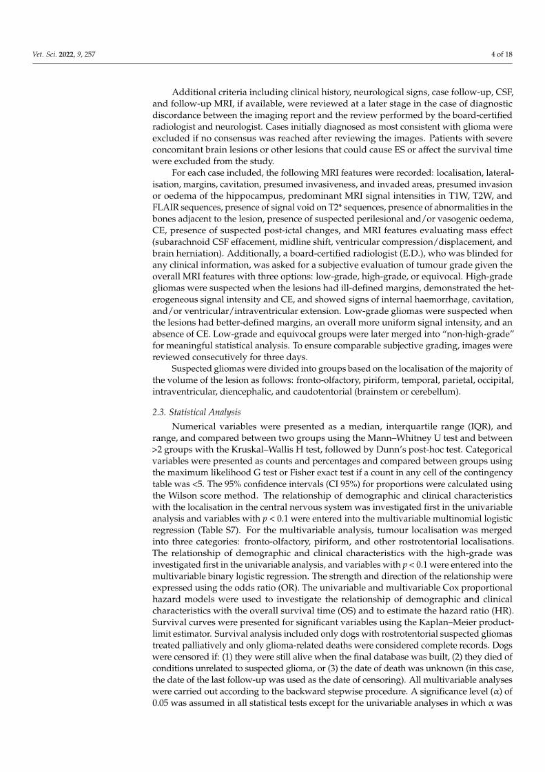
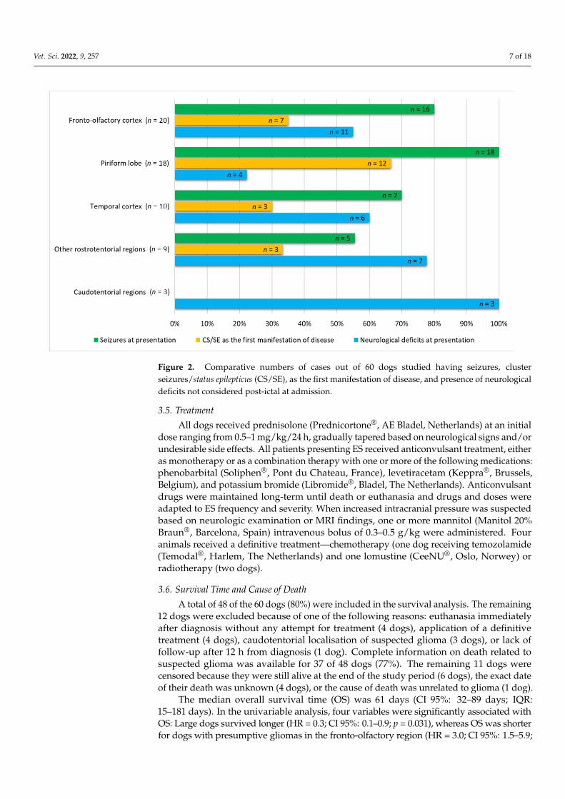
In the univariable analysis of variables associated with glioma location, suspected gliomas in the fronto-olfactory localisation were significantly more often diagnosed in fe-males (p = 0.003), and dogs with presumptive gliomas in this localisation were signifi-cantly lighter than dogs with gliomas in other localisations (p = 0.029). In dogs with sus-pected gliomas in the piriform localisation, ES were observed significantly more often (p = 0.010), and neurological deficits were observed significantly less often (p = 0.024) com-pared to dogs with suspected gliomas in other localisations (Table S4; Figure 2). In the multivariable analysis, two variables remained significantly and independently related to the tumour localisation: fronto-olfactory localisation was more common in females (p = 0.011), and ES were more common in tumours located in the piriform lobe (p = 0.006).
Figure 1. Suspected glioma location in the 60 dogs studied.
3.4. Association among Suspected Glioma Localisation, Demographic Data, Clinical Features, andMRI Features
In the univariable analysis of variables associated with glioma location, suspectedgliomas in the fronto-olfactory localisation were significantly more often diagnosed in fe-males (p = 0.003), and dogs with presumptive gliomas in this localisation were significantlylighter than dogs with gliomas in other localisations (p = 0.029). In dogs with suspectedgliomas in the piriform localisation, ES were observed significantly more often (p = 0.010),and neurological deficits were observed significantly less often (p = 0.024) compared todogs with suspected gliomas in other localisations (Table S4; Figure 2). In the multivariableanalysis, two variables remained significantly and independently related to the tumourlocalisation: fronto-olfactory localisation was more common in females (p = 0.011), and ESwere more common in tumours located in the piriform lobe (p = 0.006).
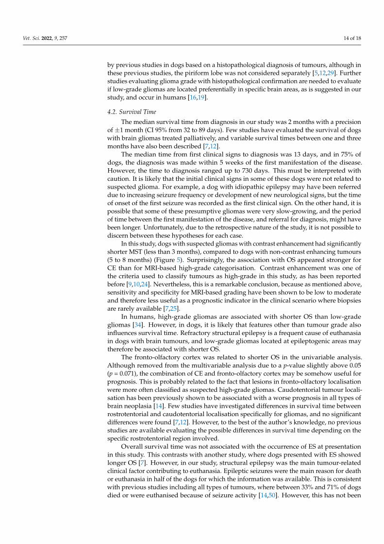
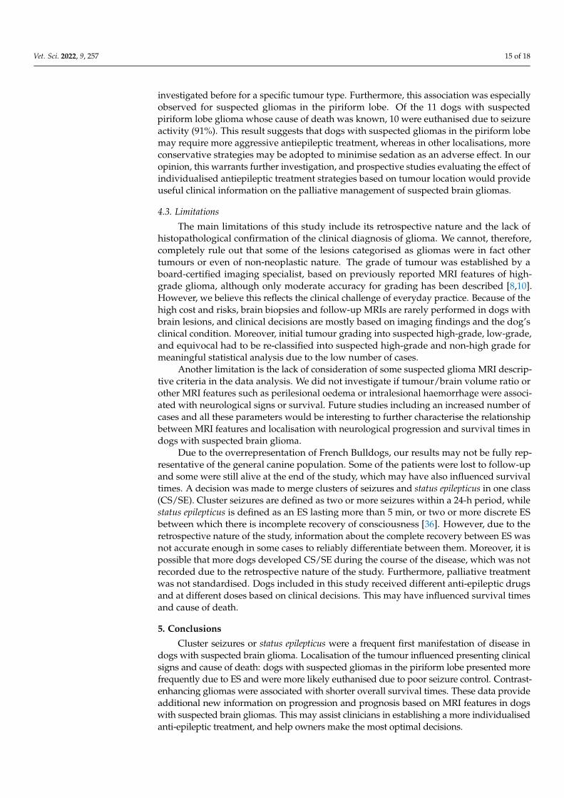
The following variables were significantly linked to the MRI diagnosis of high-gradepresumptive glioma in the univariable analysis (Table S5): high-grade suspected gliomaswere less often observed in large breed dogs (p = 0.036); neurological deficits at presen-tation were more frequent in dogs with suspected high-grade glioma (p = 0.038); andsuspected high-grade gliomas were more often located in the fronto-olfactory cortex(p < 0.001) and less often located in the piriform lobe (p < 0.001). In the multivariable analy-sis, only localisation of the suspected tumour remained significantly associated with pre-sumed glioma grade: tumours in the fronto-olfactory cortex were significantly more often(OR = 9.00; CI 95%: 1.67–48.4; p = 0.011) classified as suspected high-grade gliomas andtumours in the piriform lobe were significantly less likely (OR = 0.13; CI 95%: 0.02–0.68;p = 0.016) to be considered high-grade, compared to tumours located in other regions(Figures 3 and 4). The multivariable model fit the data well (Hosmer-Lemeshowchi-squaredH&L χ2 = 0.00, p = 0.999; Nagelkerke’s pseudo-R2 coefficient = 0.48).
Vet. Sci. 2022, 9, 257 7 of 18Vet. Sci. 2022, 9, x FOR PEER REVIEW 7 of 19
Figure 2. Comparative numbers of cases out of 60 dogs studied having seizures, cluster seizures/sta-tus epilepticus (CS/SE), as the first manifestation of disease, and presence of neurological deficits not considered post-ictal at admission.
The following variables were significantly linked to the MRI diagnosis of high-grade presumptive glioma in the univariable analysis (Table S5): high-grade suspected gliomas were less often observed in large breed dogs (p = 0.036); neurological deficits at presenta-tion were more frequent in dogs with suspected high-grade glioma (p = 0.038); and sus-pected high-grade gliomas were more often located in the fronto-olfactory cortex (p < 0.001) and less often located in the piriform lobe (p < 0.001). In the multivariable analysis, only localisation of the suspected tumour remained significantly associated with pre-sumed glioma grade: tumours in the fronto-olfactory cortex were significantly more often (OR = 9.00; CI 95%: 1.67–48.4; p = 0.011) classified as suspected high-grade gliomas and tumours in the piriform lobe were significantly less likely (OR = 0.13; CI 95%: 0.02–0.68; p = 0.016) to be considered high-grade, compared to tumours located in other regions (Fig-ures 3 and 4). The multivariable model fit the data well (Hosmer-Lemeshowchi-squared H&L χ2 = 0.00, p = 0.999; Nagelkerke’s pseudo-R2 coefficient = 0.48).
Figure 2. Comparative numbers of cases out of 60 dogs studied having seizures, clusterseizures/status epilepticus (CS/SE), as the first manifestation of disease, and presence of neurologicaldeficits not considered post-ictal at admission.
3.5. Treatment
All dogs received prednisolone (Prednicortone®, AE Bladel, Netherlands) at an initialdose ranging from 0.5–1 mg/kg/24 h, gradually tapered based on neurological signs and/orundesirable side effects. All patients presenting ES received anticonvulsant treatment, eitheras monotherapy or as a combination therapy with one or more of the following medications:phenobarbital (Soliphen®, Pont du Chateau, France), levetiracetam (Keppra®, Brussels,Belgium), and potassium bromide (Libromide®, Bladel, The Netherlands). Anticonvulsantdrugs were maintained long-term until death or euthanasia and drugs and doses wereadapted to ES frequency and severity. When increased intracranial pressure was suspectedbased on neurologic examination or MRI findings, one or more mannitol (Manitol 20%Braun®, Barcelona, Spain) intravenous bolus of 0.3–0.5 g/kg were administered. Fouranimals received a definitive treatment—chemotherapy (one dog receiving temozolamide(Temodal®, Harlem, The Netherlands) and one lomustine (CeeNU®, Oslo, Norwey) orradiotherapy (two dogs).
3.6. Survival Time and Cause of Death
A total of 48 of the 60 dogs (80%) were included in the survival analysis. The remaining12 dogs were excluded because of one of the following reasons: euthanasia immediatelyafter diagnosis without any attempt for treatment (4 dogs), application of a definitivetreatment (4 dogs), caudotentorial localisation of suspected glioma (3 dogs), or lack offollow-up after 12 h from diagnosis (1 dog). Complete information on death related tosuspected glioma was available for 37 of 48 dogs (77%). The remaining 11 dogs werecensored because they were still alive at the end of the study period (6 dogs), the exact dateof their death was unknown (4 dogs), or the cause of death was unrelated to glioma (1 dog).
The median overall survival time (OS) was 61 days (CI 95%: 32–89 days; IQR:15–181 days). In the univariable analysis, four variables were significantly associated withOS: Large dogs survived longer (HR = 0.3; CI 95%: 0.1–0.9; p = 0.031), whereas OS was shorterfor dogs with presumptive gliomas in the fronto-olfactory region (HR = 3.0; CI 95%: 1.5–5.9;

Vet. Sci. 2022, 9, 257 8 of 18
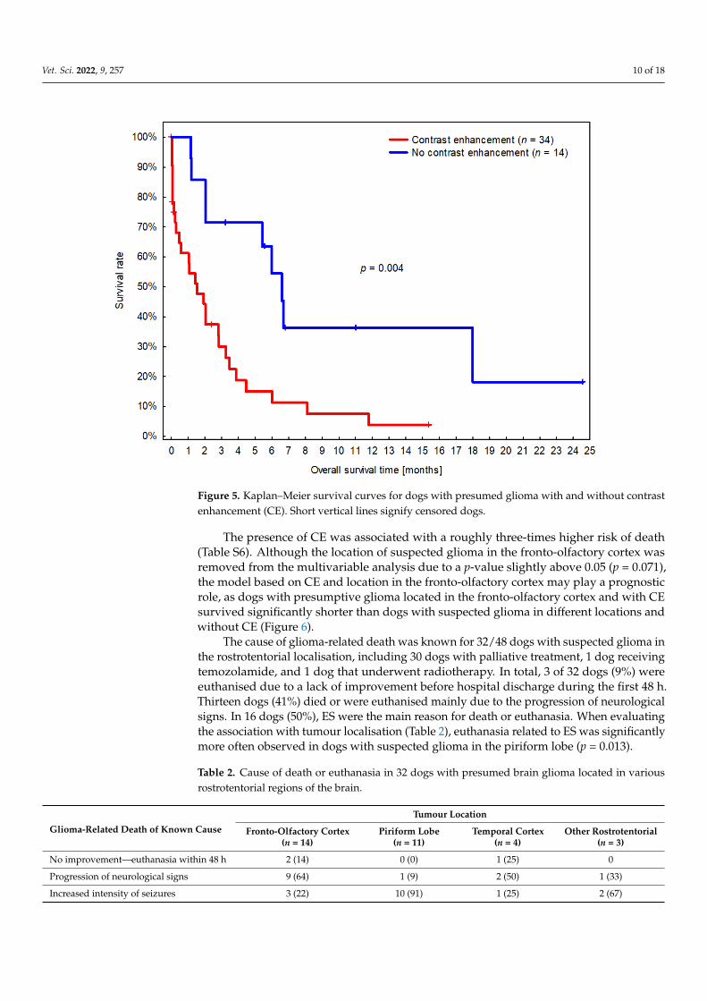
p = 0.002), for dogs with contrast-enhancing presumptive glioma (HR = 3.2; CI 95: 1.5–7.2;p = 0.004), and dogs with MRI-based high-grade glioma (HR = 2.1; CI 95: 1.1–4.2; p = 0.032).The only variable that remained significant in the multivariable analysis was CE. Dogs withsuspected gliomas with CE had significantly shorter MST (46 days, CI 95%: 10–81 days)compared to dogs with non-CE gliomas (198 days, CI 95%: 159–237 days) (Figure 5).
Vet. Sci. 2022, 9, x FOR PEER REVIEW 8 of 19
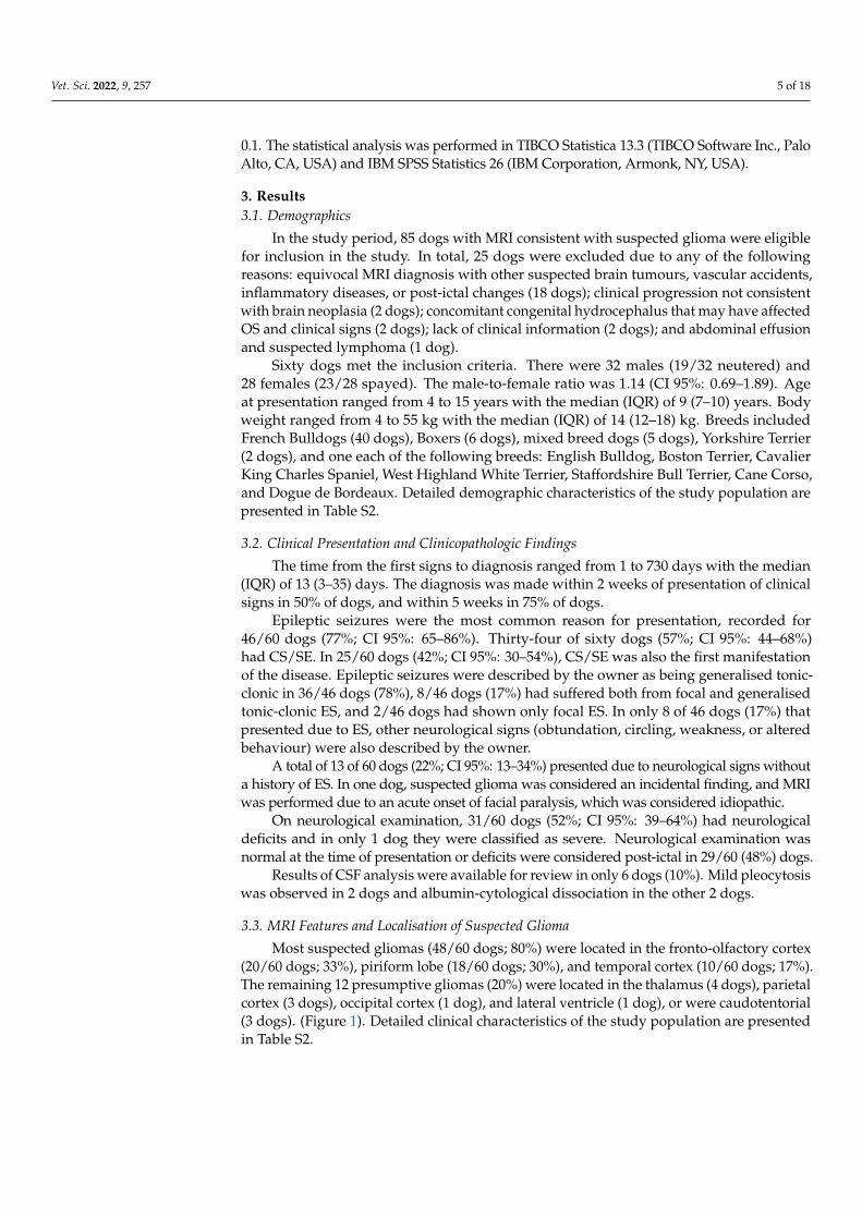
Figure 3. MRI images of suspected high-grade glioma in the left fronto-olfactory cortex in an 8-year-old female French Bulldog: Dorsal (A) and left parasagittal (B), T2-w. Transverse T2-w (C), T2-FLAIR (D), T1-w (E), and T1 post-contrast (F). The left side of the patient is on the right side of the image. Arrows point to the lesion.
Figure 3. MRI images of suspected high-grade glioma in the left fronto-olfactory cortex in an 8-year-old female French Bulldog: Dorsal (A) and left parasagittal (B), T2-w. Transverse T2-w (C), T2-FLAIR(D), T1-w (E), and T1 post-contrast (F). The left side of the patient is on the right side of the image.Arrows point to the lesion.
Vet. Sci. 2022, 9, 257 9 of 18Vet. Sci. 2022, 9, x FOR PEER REVIEW 9 of 19
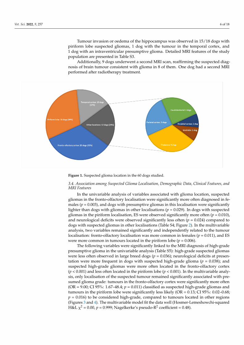
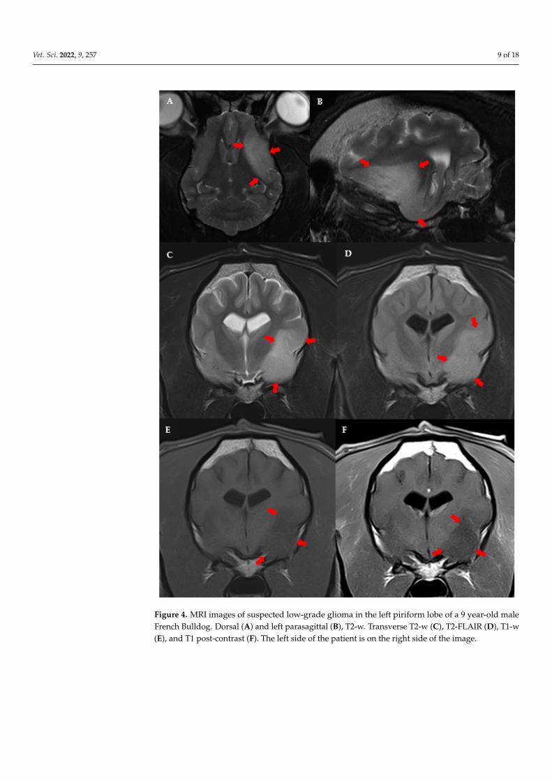
Figure 4. MRI images of suspected low-grade glioma in the left piriform lobe of a 9 year-old male French Bulldog. Dorsal (A) and left parasagittal (B), T2-w. Transverse T2-w (C), T2-FLAIR (D), T1-w (E), and T1 post-contrast (F). The left side of the patient is on the right side of the image.
3.5. Treatment All dogs received prednisolone (Prednicortone®, AE Bladel, Netherlands) at an initial
dose ranging from 0.5–1 mg/kg/24 h, gradually tapered based on neurological signs and/or undesirable side effects. All patients presenting ES received anticonvulsant
Figure 4. MRI images of suspected low-grade glioma in the left piriform lobe of a 9 year-old maleFrench Bulldog. Dorsal (A) and left parasagittal (B), T2-w. Transverse T2-w (C), T2-FLAIR (D), T1-w(E), and T1 post-contrast (F). The left side of the patient is on the right side of the image.
Vet. Sci. 2022, 9, 257 10 of 18Vet. Sci. 2022, 9, x FOR PEER REVIEW 11 of 19
Figure 5. Kaplan–Meier survival curves for dogs with presumed glioma with and without contrast enhancement (CE). Short vertical lines signify censored dogs.
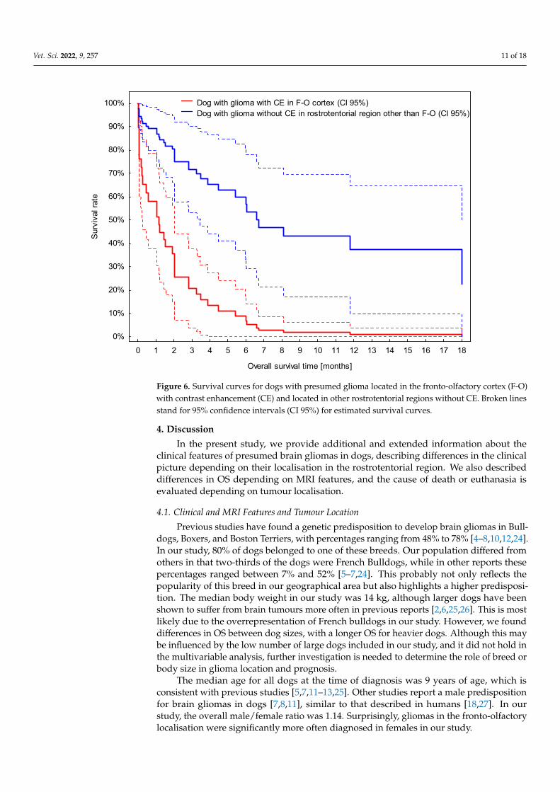
Figure 6. Survival curves for dogs with presumed glioma located in the fronto-olfactory cortex (F-O) with contrast enhancement (CE) and located in other rostrotentorial regions without CE. Broken lines stand for 95% confidence intervals (CI 95%) for estimated survival curves.
0 1 2 3 4 5 6 7 8 9 10 11 12 13 14 15 16 17 18
Overall survival time [months]
0%
10%
20%
30%
40%
50%
60%
70%
80%
90%
100%
Sur
viva
l rat
e
Dog with glioma with CE in F-O cortex (CI 95%) Dog with glioma without CE in rostrotentorial region other than F-O (CI 95%)
Figure 5. Kaplan–Meier survival curves for dogs with presumed glioma with and without contrastenhancement (CE). Short vertical lines signify censored dogs.
The presence of CE was associated with a roughly three-times higher risk of death(Table S6). Although the location of suspected glioma in the fronto-olfactory cortex wasremoved from the multivariable analysis due to a p-value slightly above 0.05 (p = 0.071),the model based on CE and location in the fronto-olfactory cortex may play a prognosticrole, as dogs with presumptive glioma located in the fronto-olfactory cortex and with CEsurvived significantly shorter than dogs with suspected glioma in different locations andwithout CE (Figure 6).
The cause of glioma-related death was known for 32/48 dogs with suspected glioma inthe rostrotentorial localisation, including 30 dogs with palliative treatment, 1 dog receivingtemozolamide, and 1 dog that underwent radiotherapy. In total, 3 of 32 dogs (9%) wereeuthanised due to a lack of improvement before hospital discharge during the first 48 h.Thirteen dogs (41%) died or were euthanised mainly due to the progression of neurologicalsigns. In 16 dogs (50%), ES were the main reason for death or euthanasia. When evaluatingthe association with tumour localisation (Table 2), euthanasia related to ES was significantlymore often observed in dogs with suspected glioma in the piriform lobe (p = 0.013).
Table 2. Cause of death or euthanasia in 32 dogs with presumed brain glioma located in variousrostrotentorial regions of the brain.
Glioma-Related Death of Known Cause
Tumour Location
Fronto-Olfactory Cortex(n = 14)
Piriform Lobe(n = 11)
Temporal Cortex(n = 4)
Other Rostrotentorial(n = 3)
No improvement—euthanasia within 48 h 2 (14) 0 (0) 1 (25) 0
Progression of neurological signs 9 (64) 1 (9) 2 (50) 1 (33)
Increased intensity of seizures 3 (22) 10 (91) 1 (25) 2 (67)
Vet. Sci. 2022, 9, 257 11 of 18
Vet. Sci. 2022, 9, x FOR PEER REVIEW 11 of 19
Figure 5. Kaplan–Meier survival curves for dogs with presumed glioma with and without contrast enhancement (CE). Short vertical lines signify censored dogs.
Figure 6. Survival curves for dogs with presumed glioma located in the fronto-olfactory cortex (F-O) with contrast enhancement (CE) and located in other rostrotentorial regions without CE. Broken lines stand for 95% confidence intervals (CI 95%) for estimated survival curves.
0 1 2 3 4 5 6 7 8 9 10 11 12 13 14 15 16 17 18
Overall survival time [months]
0%
10%
20%
30%
40%
50%
60%
70%
80%
90%
100%S
urvi
val r
ate
Dog with glioma with CE in F-O cortex (CI 95%) Dog with glioma without CE in rostrotentorial region other than F-O (CI 95%)
Figure 6. Survival curves for dogs with presumed glioma located in the fronto-olfactory cortex (F-O)with contrast enhancement (CE) and located in other rostrotentorial regions without CE. Broken linesstand for 95% confidence intervals (CI 95%) for estimated survival curves.
4. Discussion
In the present study, we provide additional and extended information about theclinical features of presumed brain gliomas in dogs, describing differences in the clinicalpicture depending on their localisation in the rostrotentorial region. We also describeddifferences in OS depending on MRI features, and the cause of death or euthanasia isevaluated depending on tumour localisation.
4.1. Clinical and MRI Features and Tumour Location
Previous studies have found a genetic predisposition to develop brain gliomas in Bull-dogs, Boxers, and Boston Terriers, with percentages ranging from 48% to 78% [4–8,10,12,24].In our study, 80% of dogs belonged to one of these breeds. Our population differed fromothers in that two-thirds of the dogs were French Bulldogs, while in other reports thesepercentages ranged between 7% and 52% [5–7,24]. This probably not only reflects thepopularity of this breed in our geographical area but also highlights a higher predisposi-tion. The median body weight in our study was 14 kg, although larger dogs have beenshown to suffer from brain tumours more often in previous reports [2,6,25,26]. This is mostlikely due to the overrepresentation of French bulldogs in our study. However, we founddifferences in OS between dog sizes, with a longer OS for heavier dogs. Although this maybe influenced by the low number of large dogs included in our study, and it did not hold inthe multivariable analysis, further investigation is needed to determine the role of breed orbody size in glioma location and prognosis.
The median age for all dogs at the time of diagnosis was 9 years of age, which isconsistent with previous studies [5,7,11–13,25]. Other studies report a male predispositionfor brain gliomas in dogs [7,8,11], similar to that described in humans [18,27]. In ourstudy, the overall male/female ratio was 1.14. Surprisingly, gliomas in the fronto-olfactorylocalisation were significantly more often diagnosed in females in our study.
Vet. Sci. 2022, 9, 257 12 of 18
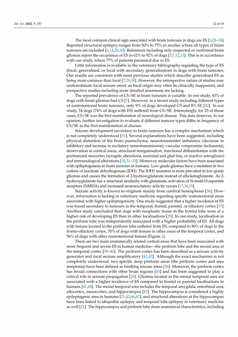
The most common clinical sign associated with brain tumours in dogs are ES [3,28–30].Reported structural epilepsy ranges from 50% to 75% in studies where all types of braintumours are included [3,14,29,30]. References including only suspected or confirmed braingliomas report the occurrence of ES in 62% to 92% of dogs [5,7,12,13]. This is in accordancewith our study, where 77% of patients presented due to ES.
Little information is available in the veterinary bibliography regarding the type of ES(focal, generalised, or focal with secondary generalisation) in dogs with brain tumours.Our results are consistent with most previous studies which describe generalised ES asbeing more common than focal [7,29,30]. However, the retrospective nature of studies mayunderestimate focal seizure onset, as focal origin may often be clinically inapparent, andprospective studies including more detailed anamnesis are lacking.
The reported prevalence of CS/SE in brain tumours is variable. In one study, 43% ofdogs with brain gliomas had CS [7]. However, in a recent study including different typesof rostrotentorial brain tumours, only 9% of dogs developed CS and 8% SE [30]. In ourstudy, 34 dogs (74% of dogs with ES) suffered from CS/SE. Interestingly, for 25 of thosecases, CS/SE was the first manifestation of neurological disease. This data deserves, in ouropinion, further investigation to evaluate if different tumour types differ in frequency ofCS/SE as the first manifestation of disease.
Seizure development secondary to brain tumours has a complex mechanism whichis not completely understood [31]. Several explanations have been suggested, includingphysical distortion of the brain parenchyma, neurotransmitter imbalance (decrease ininhibitory and increase in excitatory neurotransmission), vascular compromise (ischaemia),denervation in cortical areas, structural reorganisation, functional differentiation with theperitumoral neocortex (synaptic alterations, neuronal and glial loss, or reactive astrogliosis)and immunological alterations [18,31–33]. Moreover, molecular factors have been associatedwith epileptogenesis in brain tumours in humans. Low-grade gliomas have a mutation in twocodons of isocitrate dehydrogenase (IDH). The IDH1 mutation is more prevalent in low-gradegliomas and causes the formation of 2-hydroxyglutarate instead of alfa-ketoglutarate. As 2-hydroxyglutarate has a structural similarity with glutamate, activation of N-metil-D-aspartatereceptors (NMDA) and increased neuroexcitatory activity occurs [17,34,35].
Seizure activity is known to originate mainly from cerebral hemispheres [36]. How-ever, information is lacking in veterinary medicine regarding specific rostrotentorial areasassociated with higher epileptogenicity. One study suggested that a higher incidence of ESwas found secondary to tumours in the temporal, frontal, parietal, or olfactory cortex [37].Another study concluded that dogs with neoplastic tissue in the frontal lobe were at ahigher risk of developing ES than in other localisations [29]. In our study, localisation inthe piriform lobe was independently associated with a higher probability of ES. All dogswith lesions located in the piriform lobe suffered from ES, compared to 80% of dogs in thefronto-olfactory cortex, 70% of dogs with lesions in other areas of the temporal cortex, and56% of dogs with other rostrotentorial lesions (Figure 2).
There are two main anatomically related cortical areas that have been associated withmore frequent and severe ES in human medicine—the piriform lobe and the mesial area atthe temporal cortex [38–40]. The piriform cortex has been described as a seizure activitygenerator and focal seizure amplificatory [41,42]. Although the exact mechanism is notcompletely understood, two specific deep piriform areas (the piriform cortex and areatempestas) have been defined as kindling seizure areas [38]. Moreover, the piriform cortexhas broad connections with other brain regions [43] and has been suggested to play acritical role in seizure propagation [20]. Gliomas located in the mesial temporal area areassociated with a higher incidence of ES compared to frontal or parietal localisations inhumans [41,44]. The mesial temporal area includes the temporal amygdala, entorhinal area,allocortex, mesocortex, and hippocampus [45]. The hippocampus is considered a highlyepileptogenic area in humans [21,42,46,47], and structural alterations at the hippocampushave been linked to idiopathic epilepsy and temporal lobe epilepsy in veterinary medicineas well [21]. The hippocampus and piriform lobe share anatomical characteristics, including
Vet. Sci. 2022, 9, 257 13 of 18
the presence of three excitable neuronal layers, and similar structural changes are observedin both localisations due to sustained neuronal injury in humans with ES [39,42] suggestingcommon epileptogenic mechanisms for both areas. In this study, hippocampal affection bythe tumour was almost exclusively observed in presumed gliomas affecting the piriformlobe, which may have also played a role in seizure generation. Due to this fact, it wasalso impossible to discriminate between the role of piriform location and hippocampusinvolvement in epileptogenesis.
In previous studies [3,7,9,29], different criteria were used to classify the localisation oftumours. In this study, we initially grouped suspected gliomas considering major brainand cortical divisions. Areas included were the cerebral cortex (fronto-olfactory, parietal,temporal, piriform, and occipital), diencephalon, intraventricular, and caudotentorial,similar to another recent study [7]. However, the piriform lobe is considered part ofthe temporal cortex in this study [7] and other previous reports, which may explaindiscrepancies with our results. We decided to separate lesions located in the piriform lobe,as we hypothesised, based on our clinical experience and human bibliography [20,38,40–42],that piriform lobe localisation could be related to the higher frequency of structural epilepsyand lower frequency of neurological deficits [38,40–42,48]. In the human bibliography, thepiriform cortex is occasionally considered a region of the mesial temporal cortex [41],however, most authors include the piriform lobe as part of the olfactory cortex [20,49].
Thirty-one dogs (52%) showed neurological signs at presentation that were interpretedto be a consequence of a structural brain lesion. In the other 48% of cases, the neurologicalexam was normal, or deficits were considered post-ictal. Other studies reported a variableabsence of neurological deficits between 5% and 60% [5,6,30]. Interestingly, a considerableproportion of dogs had deficits on neurological examination while no other neurologi-cal signs had been observed by owners. This emphasises the importance of a completeneurological examination in dogs with ES. Neurological signs in our study were mostly con-sidered mild/moderate. Rostrotentorial localisations that were less commonly associatedwith neurological deficits were in the piriform lobe (only 22% of dogs showed neurologicaldeficits) and fronto-olfactory cortex (55% of dogs showed deficits). The fronto-olfactorycortex and piriform lobe have been previously described as “clinically silent regions” [40].However, to the best of the author’s knowledge, no studies have investigated in detailthe prevalence of neurological deficits depending on the specific brain area affected. Apossible explanation is that dogs with piriform lobe tumours may be diagnosed before dueto ES occurrence early in the course of the disease. Low-grade slow-growing gliomas havebeen described in humans to most frequently produce ES without additional neurologicaldeficits, in contrast to higher-grade faster-growing tumours [7,33].
Gliomas are histopathologically classified as oligodendrogliomas, astrocytomas, orundefined gliomas, and further subclassified as low- or high-grade [8]. In the present study,we did not attempt to classify tumour subtypes based on MRI features. A low correlationwith histopathological diagnosis has been described, and glioma subtype determinationhas a low clinical relevance regarding treatment and prognosis. The accuracy of tumourgrading in cases of suspected glioma based on MRI features is also only moderate. Anaccuracy of 53% for predicting tumour grade was observed in one study [24], and anotherstudy reported a sensitivity of 67% and specificity of 57% for the diagnosis of a high-gradetumour [7]. Described MRI features associated with lower-grade tumours include mild tono CE, absence of cystic structures, and tumour localisation other than the thalamo-capsularregion. Features associated with high-grade gliomas include spread to neighbouring brainstructures and presence of CE [7,9,10]. We decided to classify a suspected high-grade groupbased on MRI features, as this is the most common scenario in clinical practice.
In this study, the suspected grade of glioma was significantly related to the localisation.Lesions in the fronto-olfactory cortex were more often classified as suspected high-gradegliomas, while lesions in the piriform lobe were less frequently considered high-grade. It islikely that, similar to humans, low-grade gliomas in dogs may locate preferentially in thepiriform lobe and invade mesial temporal areas [16,19,20]. However, this is not supported
Vet. Sci. 2022, 9, 257 14 of 18
by previous studies in dogs based on a histopathological diagnosis of tumours, although inthese previous studies, the piriform lobe was not considered separately [5,12,29]. Furtherstudies evaluating glioma grade with histopathological confirmation are needed to evaluateif low-grade gliomas are located preferentially in specific brain areas, as is suggested in ourstudy, and occur in humans [16,19].
4.2. Survival Time
The median survival time from diagnosis in our study was 2 months with a precisionof ±1 month (CI 95% from 32 to 89 days). Few studies have evaluated the survival of dogswith brain gliomas treated palliatively, and variable survival times between one and threemonths have also been described [7,12].
The median time from first clinical signs to diagnosis was 13 days, and in 75% ofdogs, the diagnosis was made within 5 weeks of the first manifestation of the disease.However, the time to diagnosis ranged up to 730 days. This must be interpreted withcaution. It is likely that the initial clinical signs in some of these dogs were not related tosuspected glioma. For example, a dog with idiopathic epilepsy may have been referreddue to increasing seizure frequency or development of new neurological signs, but the timeof onset of the first seizure was recorded as the first clinical sign. On the other hand, it ispossible that some of these presumptive gliomas were very slow-growing, and the periodof time between the first manifestation of the disease, and referral for diagnosis, might havebeen longer. Unfortunately, due to the retrospective nature of the study, it is not possible todiscern between these hypotheses for each case.
In this study, dogs with suspected gliomas with contrast enhancement had significantlyshorter MST (less than 3 months), compared to dogs with non-contrast enhancing tumours(5 to 8 months) (Figure 5). Surprisingly, the association with OS appeared stronger forCE than for MRI-based high-grade categorisation. Contrast enhancement was one ofthe criteria used to classify tumours as high-grade in this study, as has been reportedbefore [9,10,24]. Nevertheless, this is a remarkable conclusion, because as mentioned above,sensitivity and specificity for MRI-based grading have been shown to be low to moderateand therefore less useful as a prognostic indicator in the clinical scenario where biopsiesare rarely available [7,25].
In humans, high-grade gliomas are associated with shorter OS than low-gradegliomas [34]. However, in dogs, it is likely that features other than tumour grade alsoinfluences survival time. Refractory structural epilepsy is a frequent cause of euthanasiain dogs with brain tumours, and low-grade gliomas located at epileptogenic areas maytherefore be associated with shorter OS.
The fronto-olfactory cortex was related to shorter OS in the univariable analysis.Although removed from the multivariable analysis due to a p-value slightly above 0.05(p = 0.071), the combination of CE and fronto-olfactory cortex may be somehow useful forprognosis. This is probably related to the fact that lesions in fronto-olfactory localisationwere more often classified as suspected high-grade gliomas. Caudotentorial tumour locali-sation has been previously shown to be associated with a worse prognosis in all types ofbrain neoplasia [14]. Few studies have investigated differences in survival time betweenrostrotentorial and caudotentorial localisation specifically for gliomas, and no significantdifferences were found [7,12]. However, to the best of the author’s knowledge, no previousstudies are available evaluating the possible differences in survival time depending on thespecific rostrotentorial region involved.
Overall survival time was not associated with the occurrence of ES at presentationin this study. This contrasts with another study, where dogs presented with ES showedlonger OS [7]. However, in our study, structural epilepsy was the main tumour-relatedclinical factor contributing to euthanasia. Epileptic seizures were the main reason for deathor euthanasia in half of the dogs for which the information was available. This is consistentwith previous studies including all types of tumours, where between 33% and 71% of dogsdied or were euthanised because of seizure activity [14,50]. However, this has not been
Vet. Sci. 2022, 9, 257 15 of 18
investigated before for a specific tumour type. Furthermore, this association was especiallyobserved for suspected gliomas in the piriform lobe. Of the 11 dogs with suspectedpiriform lobe glioma whose cause of death was known, 10 were euthanised due to seizureactivity (91%). This result suggests that dogs with suspected gliomas in the piriform lobemay require more aggressive antiepileptic treatment, whereas in other localisations, moreconservative strategies may be adopted to minimise sedation as an adverse effect. In ouropinion, this warrants further investigation, and prospective studies evaluating the effect ofindividualised antiepileptic treatment strategies based on tumour location would provideuseful clinical information on the palliative management of suspected brain gliomas.
4.3. Limitations
The main limitations of this study include its retrospective nature and the lack ofhistopathological confirmation of the clinical diagnosis of glioma. We cannot, therefore,completely rule out that some of the lesions categorised as gliomas were in fact othertumours or even of non-neoplastic nature. The grade of tumour was established by aboard-certified imaging specialist, based on previously reported MRI features of high-grade glioma, although only moderate accuracy for grading has been described [8,10].However, we believe this reflects the clinical challenge of everyday practice. Because of thehigh cost and risks, brain biopsies and follow-up MRIs are rarely performed in dogs withbrain lesions, and clinical decisions are mostly based on imaging findings and the dog’sclinical condition. Moreover, initial tumour grading into suspected high-grade, low-grade,and equivocal had to be re-classified into suspected high-grade and non-high grade formeaningful statistical analysis due to the low number of cases.
Another limitation is the lack of consideration of some suspected glioma MRI descrip-tive criteria in the data analysis. We did not investigate if tumour/brain volume ratio orother MRI features such as perilesional oedema or intralesional haemorrhage were associ-ated with neurological signs or survival. Future studies including an increased number ofcases and all these parameters would be interesting to further characterise the relationshipbetween MRI features and localisation with neurological progression and survival times indogs with suspected brain glioma.
Due to the overrepresentation of French Bulldogs, our results may not be fully rep-resentative of the general canine population. Some of the patients were lost to follow-upand some were still alive at the end of the study, which may have also influenced survivaltimes. A decision was made to merge clusters of seizures and status epilepticus in one class(CS/SE). Cluster seizures are defined as two or more seizures within a 24-h period, whilestatus epilepticus is defined as an ES lasting more than 5 min, or two or more discrete ESbetween which there is incomplete recovery of consciousness [36]. However, due to theretrospective nature of the study, information about the complete recovery between ES wasnot accurate enough in some cases to reliably differentiate between them. Moreover, it ispossible that more dogs developed CS/SE during the course of the disease, which was notrecorded due to the retrospective nature of the study. Furthermore, palliative treatmentwas not standardised. Dogs included in this study received different anti-epileptic drugsand at different doses based on clinical decisions. This may have influenced survival timesand cause of death.
5. Conclusions
Cluster seizures or status epilepticus were a frequent first manifestation of disease indogs with suspected brain glioma. Localisation of the tumour influenced presenting clinicalsigns and cause of death: dogs with suspected gliomas in the piriform lobe presented morefrequently due to ES and were more likely euthanised due to poor seizure control. Contrast-enhancing gliomas were associated with shorter overall survival times. These data provideadditional new information on progression and prognosis based on MRI features in dogswith suspected brain gliomas. This may assist clinicians in establishing a more individualisedanti-epileptic treatment, and help owners make the most optimal decisions.
Vet. Sci. 2022, 9, 257 16 of 18
Supplementary Materials: The following supporting information can be downloaded at: https://www.mdpi.com/article/10.3390/vetsci9060257/s1, Table S1: Magnetic resonance imaging (MRI)sequence variable ranges for the transverse T2w, T1w and FLAIR sequences included in this study;Table S2: Demographic and clinical characteristics of the study population; Table S3: Magneticresonance imaging features of 60 suspected gliomas; Table S4: Univariable analysis of the relationshipbetween demographic & clinical characteristics and MRI location. Table S5: Univariable analysisof the relationship between demographic & clinical characteristics and MRI-based high grade (all60 dogs included); Table S6: Univariable survival analysis including 48 dogs with rostrotentorialgliomas; Table S7: Multivariable survival analysis including 48 dogs with rostrotentorial gliomas.
Author Contributions: M.P.-S., P.M. and C.M.O. participated in the design of the study. P.M., M.P.-S.,C.M.O., A.S., M.P.S., E.D. and C.M.M. participated in the acquisition of data. M.P.-S., P.M. and E.D.visualised Magnetic Resonance Imaging. M.P.-S., M.C., P.M. and A.S. analysed, and interpreted thedata. M.P.-S. and P.M. participated in drafting the manuscript. All authors provided critical manuscriptrevisions. P.M. coordinated the study. M.C. performed the statistics. M.C. and M.P.-S. developed thefigure and the tables. All authors have read and agreed to the published version of the manuscript.
Funding: This research received no external funding.
Institutional Review Board Statement: Ethical review and approval were waived for this study dueto the retrospective nature of the present study.
Informed Consent Statement: Not applicable.
Data Availability Statement: The data presented in this study are available on reasonable requestfrom the corresponding author.
Acknowledgments: The authors thank to Sergi Fuxa, MRI technician of AniCura Ars Veterinaria,who contributed to the acquisition of MRI parameters and images.
Conflicts of Interest: The authors declare no potential conflict of interest.
Abbreviations
SE Status epilepticusCE Contrast enhancementCI Confidence intervalCS Cluster seizuresCSF Cerebrospinal fluidCT Computed tomographyES Epileptic seizuresFLAIR Fluid-attenuated inversion recoveryHR Hazard ratioIDH Isocitrate dehydrogenaseIQR Interquartile rangeMST Median overall survival timeMRI Magnetic Resonance ImagingOR Odds ratioOS Overall survival timeT1W T1-weightedT2W T2-weightedT2* T2-weighted gradient echo
References1. Hayes, H.M.; Priester, W.A.; Pendergrass, T.W. Occurrence of nervous-tissue tumors in cattle, horses, cats and dogs. Int. J. Cancer
1975, 15, 39–47. [CrossRef] [PubMed]2. Song, R.B.; Vite, C.H.; Bradley, C.W.; Cross, J.R. Postmortem Evaluation of 435 Cases of Intracranial Neoplasia in Dogs and
Relationship of Neoplasm with Breed, Age, and Body Weight. J. Vet. Intern. Med. 2013, 27, 1143–1152. [CrossRef] [PubMed]3. Snyder, J.M.; Shofer, F.S.; Van Winkle, T.J.; Massicotte, C. Canine intracranial primary neoplasia: 173 Cases (1986–2003). J. Vet.
Intern. Med. 2006, 20, 669–675.
Vet. Sci. 2022, 9, 257 17 of 18
4. Truvé, K.; Dickinson, P.; Xiong, A.; York, D.; Jayashankar, K.; Pielberg, G.; Koltookian, M.; Muren, E.; Fuxelius, H.-H.;Weishaupt, H.; et al. Utilizing the Dog Genome in the Search for Novel Candidate Genes Involved in Glioma Development—Genome Wide Association Mapping followed by Targeted Massive Parallel Sequencing Identifies a Strongly Associated Locus.PLoS Genet. 2016, 12, e1006000. [CrossRef]
5. Dolera, M.; Malfassi, L.; Bianchi, C.; Carrara, N.; Finesso, S.; Marcarini, S.; Mazza, G.; Pavesi, S.; Sala, M.; Urso, G. Framelessstereotactic radiotherapy alone and combined with temozolomide for presumed canine gliomas. Vet. Comp. Oncol. 2018,16, 90–101. [CrossRef] [PubMed]
6. Debreuque, M.; De Fornel, P.; David, I.; Delisle, F.; Ducerveau, M.N.; Devauchelle, P.; Thibaud, J.L. Definitive-intent uniformmegavoltage fractioned radiotherapy protocol for presumed canine intracranial gliomas: Retrospective analysis of survival andprognostic factors in 38 cases (2013–2019). BMC Vet. Res. 2020, 16, 412. [CrossRef]
7. José-López, R.; Gutierrez-Quintana, R.; de la Fuente, C.; Manzanilla, E.G.; Suñol, A.; Pi Castro, D.; Anor, S.; Sanchez-Masian, D.;Fernandez-Flores, F.; Ricci, E.; et al. Clinical features, diagnosis, and survival analysis of dogs with glioma. J. Vet. Intern. Med.2021, 35, 1902–1917. [CrossRef]
8. Koehler, J.W.; Miller, A.D.; Miller, C.R.; Porter, B.; Aldape, K.; Beck, J.; Brat, D.; Cornax, I.; Corps, K.; Frank, C.; et al. A reviseddiagnostic classification of canine glioma: Towards validation of the canine glioma patient as a naturally occurring preclinicalmodel for human glioma. J. Neuropathol. Exp. Neurol. 2018, 77, 1039–1054. [CrossRef]
9. Bentley, R.T.; Ober, C.P.; Anderson, K.L.; Feeney, D.A.; Naughton, J.F.; Ohlfest, J.R.; O’Sullivan, M.G.; Miller, M.A.; Constable, P.D.;Pluhar, G.E. Canine intracranial gliomas: Relationship between magnetic resonance imaging criteria and tumor type and grade.Vet. J. 2013, 198, 3921–3932. [CrossRef]
10. Young, B.D.; Levine, J.M.; Porter, B.F.; Chen-Allen, A.V.; Rossmeisl, J.H.; Platt, S.R.; Kent, M.; Fosgate, G.T.; Schatzberg, S.J.Magnetic resonance imaging features of intracranial astrocytomas and oligodendrogliomas in dogs. Vet. Radiol. Ultrasound 2011,52, 132–141. [CrossRef]
11. Suñol, A.; Mascort, J.; Font, C.; Bastante, A.R.; Pumarola, M.; Feliu-Pascual, A.L. Long-term follow-up of surgical resection alonefor primary intracranial rostrotentorial tumors in dogs: 29 cases (2002–2013). Open Vet. J. 2017, 7, 375–383. [CrossRef] [PubMed]
12. Moirano, S.J.; Dewey, C.W.; Wright, K.Z.; Cohen, P.W. Survival times in dogs with presumptive intracranial gliomas treated withoral lomustine: A comparative retrospective study (2008–2017). Vet. Comp. Oncol. 2018, 16, 459–466. [CrossRef] [PubMed]
13. Moirano, S.J.; Dewey, C.W.; Haney, S.; Yang, J. Efficacy of frameless stereotactic radiotherapy for the treatment of presumptivecanine intracranial gliomas: A retrospective analysis (2014–2017). Vet. Comp. Oncol. 2020, 18, 528–537. [CrossRef] [PubMed]
14. Rossmeisl, J.H.; Jones, J.C.; Zimmerman, K.L.; Robertson, J.L. Survival time following hospital discharge in dogs with palliativelytreated primary brain tumors. J. Am. Vet. Med. Assoc. 2013, 242, 193–198. [CrossRef]
15. Van Breemen, M.S.; Wilms, E.B.; Vecht, C.J. Epilepsy in patients with brain tumours: Epidemiology, mechanisms, and management.Lancet Neurol. 2007, 6, 421–430. [CrossRef]
16. Akgun, M.Y.; Cetintas, S.C.; Kemerdere, R.; Yeni, S.N.; Tanriverdi, T. Are low-grade gliomas of mesial temporal area alone? Surg.Neurol. Int. 2019, 10, 170. [CrossRef]
17. Vecht, C.J.; Kerkhof, M.; Duran-Pena, A. Seizure Prognosis in Brain Tumors: New Insights and Evidence-Based Management.Oncologist 2014, 19, 751–759. [CrossRef]
18. Pallud, J.; Audureau, E.; Blonski, M.; Sanai, N.; Bauchet, L.; Fontaine, D.; Mandonnet, E.; Dezamis, E.; Psimaras, D.;Guyotat, J.; et al. Epileptic seizures in diffuse low-grade gliomas in adults. Brain 2014, 137, 449–462. [CrossRef]
19. Kemerdere, R.; Yuksel, O.; Kacira, T.; Yeni, N.; Ozkara, C.; Tanriverdi, T.; Uzan, M.; Ozyurt, E. Low-grade temporal gliomas:Surgical strategy and long-term seizure outcome. Clin. Neurol. Neurosurg. 2014, 126, 196–200. [CrossRef]
20. Young, J.C.; Vaughan, D.N.; Nasser, H.M.; Jackson, G.D. Anatomical imaging of the piriform cortex in epilepsy. Exp. Neurol. 2019,320, 113013. [CrossRef]
21. Czerwik, A.; Płonek, M.; Podgórski, P.; Wrzosek, M. Comparison of electroencephalographic findings with hippocampal magneticresonance imaging volumetry in dogs with idiopathic epilepsy. J. Vet. Intern. Med. 2018, 32, 2037–2044. [CrossRef] [PubMed]
22. Hetch, S. Brain Neoplasia. In Diagnostic MRI in Dogs and Cats, 1st ed.; Mai, W., Ed.; CRC Press: Boca Raton, FL, USA, 2018;pp. 216–218.
23. Berendt, M.; Farquhar, R.G.; Mandigers, P.J.J.; Pakozdy, A.; Bhatti, S.F.M.; De Risio, L.; Fischer, A.; Long, S.; Matiasek, K.;Munana, K.; et al. International veterinary epilepsy task force consensus report on epilepsy definition, classification and terminol-ogy in companion animals. BMC Vet. Res. 2015, 11, 182. [CrossRef] [PubMed]
24. Fernández, F.; Deviers, A.; Dally, C.; Mogicato, G.; Delverdier, M.; Cauzinille, L.; Gnirs, K.; Anor, S.; de la Fuente, C.;Fondevila, D.; et al. Presence of neural progenitors in spontaneous canine gliomas: A histopathological and immunohisto-chemical study of 20 cases. Vet. J. 2016, 209, 125–132. [CrossRef] [PubMed]
25. Stadler, K.L.; Ruth, J.D.; Pancotto, T.E.; Were, S.R.; Rossmeisl, J.H. Computed tomography and magnetic resonance imaging areequivalent in mensuration and similarly inaccurate in grade and type predictability of canine intracranial gliomas. Front. Vet. Sci.2017, 4, 157. [CrossRef] [PubMed]
26. Wolff, C.A.; Holmes, S.P.; Young, B.D.; Chen, A.V.; Platt, S.R.; Savage, M.Y.; Schatzberg, S.J.; Fosgate, G.T.; Levine, J.M. MagneticResonance Imaging for the differentiation of Neoplastic Inflammatory, and cerebrovascular brain disease in dogs. J. Vet. Intern.Med. 2012, 26, 589–597. [CrossRef]
27. Maschio, M. Brain Tumor-Related Epilepsy. Curr. Neuropharmacol. 2012, 10, 124–133. [CrossRef]
Vet. Sci. 2022, 9, 257 18 of 18
28. Heidner, G.L.; Kornegay, J.N.; Page, R.L.; Dodge, R.K.; Thrall, D.E. Analysis of Survival in a Retrospective Study of 86 Dogs withBrain Tumors. J. Vet. Intern. Med. 1991, 5, 219–226. [CrossRef]
29. Schwart, M.; Lamb, C.R.; Brodbelt, D.C.; Volk, H.A. Canine intracranial neoplasia: Clinical risk factors for development ofepileptic seizures. Small Anim. Pract. 2011, 52, 632–637. [CrossRef]
30. Parker, R.L.; Hsu, F.; Du, J.; Shinn, R.L.; Drury, A.G.; Roberston, J.L.; Cecere, T.E.; Arendse, A.U.; Rossmeisl, J.H. Incidence, riskfactors, and outcomes for early postoperative seizures in dogs with rostrotentorial brain tumors after intracranial surgery. J. Vet.Intern. Med. 2022, 36, 694–701. [CrossRef]
31. Liebel, F.X.; Smith, P.M. Cental nervous system neoplasia. FOCUS 2014, 36, 24–29.32. Pallud, J.; McKhann, G.M. Diffuse Low-Grade Glioma-Related Epilepsy. Neurosurg. Clin. N. Am. 2019, 30, 43–54. [CrossRef]
[PubMed]33. Pallud, J.; Capelle, L.; Huberfeld, G. Tumors and tumoral epilepsy. Tumoral epileptogenicity: How does it happen? Epilepsia 2013,
54, 30–34. [CrossRef] [PubMed]34. Louis, D.N.; Perry, A.; Reifenberger, G.; Von Deimling, A.; Figarella-Branger, D.; Cavenee, W.K.; Ohgaki, H.; Wiestler, O.D.;
Kleihues, P.; Ellison, D.W. The 2021 World Health Organization Classification of Tumors of the Central Nervous System: Asummary. Acta Neuropathol. 2021, 23, 1231–1251.
35. Nagashima, N.; Tanaka, K.; Sasayama, T.; Irino, Y.; Sato, N.; Takeuchi, Y.; Kyotani, K.; Mukasa, A.; Mizukawa, K.; Sakata, J.; et al.Diagnostic value of glutamate with 2-hydroxyglutarate in magnetic resonance spectroscopy for IDH1 mutant glioma. NeuroOncol. 2016, 18, 1559–1568. [PubMed]
36. De Risio, L.; Bhatti, S.; Muñana, K.; Penderis, J.; Stein, V.; Tipold, A.; Berendt, M.; Farqhuar, R.; Fischer, A.; Long, S.; et al.International veterinary epilepsy task force consensus proposal: Diagnostic approach to epilepsy in dogs. BMC Vet. Res. 2015,11, 148. [CrossRef] [PubMed]
37. Moore, M.P.; Bagley, R.S.; Harrington, M.L.; Gavin, P.R. Intracranial tumors. Vet. Clin. N. Am. Small Anim. Pract. 1996, 26, 759–777.[CrossRef]
38. Piredda, S.; Gale, K. A crucial epileptogenic site in the deep prepiriform cortex. Nature 1985, 317, 623–625. [CrossRef]39. Curia, G.; Lucchi, C.; Vinet, J.; Gualtieri, F.; Marinelli, C.; Torsello, A.; Constantino, L.; Biagini, G. Pathophysiogenesis of Mesial
Temporal Lobe Epilepsy: Is Prevention of Damage Antiepileptogenic? Curr. Med. Chem. 2014, 21, 663–688. [CrossRef]40. Blumcke, I.; Spreafico, R.; Haaker, G.; Coras, R.; Kobow, K.; Bien, C.G.; Pfafflin, M.; Elger, C.; Widman, G.; Schramm, J.; et al.
Histopathological Findings in Brain Tissue Obtained during Epilepsy Surgery. N. Engl. J. Med. 2017, 377, 1648–1656. [CrossRef]41. Pereira, P.M.G.; Insausti, R.; Artacho-Pérula, E.; Salmenperä, T.; Kälviäinen, R.; Pitkänen, A. MR volumetric analysis of the
piriform cortex and cortical amygdala in drug-refractory temporal lobe epilepsy. Am. J. Neuroradiol. 2005, 26, 319–332.42. Vaughan, D.N.; Jackson, G.D. The piriform cortex and human focal epilepsy. Front. Neurol. 2014, 5, 259. [CrossRef] [PubMed]43. Miodonski, R. The structure of the posterior piriform cortex in the dog. Acta Anat. 1974, 88, 556–573. [CrossRef] [PubMed]44. Wick, W.; Menn, O.; Meisner, C.; Steinbach, J.; Hermisson, M.; Tatagiba, M.; Weller, M. Pharmacotherapy of epileptic seizures in
glioma patients: Who, when, why and how long? Onkologie 2005, 28, 391–396. [CrossRef] [PubMed]45. Wen, H.T.; Rhoton, A.L.; De Oliveira, E.; Cardoso, A.C.C.; Tedeschi, H.; Baccanelli, M.; Marino, R., Jr. Microsurgical anatomy of
the temporal lobe: Part 1: Mesial temporal lobe anatomy and its vascular relationships as applied to amygdalohippocampectomy.Neurosurgery 1999, 45, 549–592. [CrossRef] [PubMed]
46. Asada, R.; Hamamoto, Y.; Yu, Y.; Mizuno, S.; Chambers, J.K.; Uchida, K.; Hasegawa, D. Ventrolateral temporal lobectomyin normal dogs as a counterpart to human anterior temporal lobectomy: A preliminary study on the surgical procedure andcomplications. J. Vet. Med. Sci. 2021, 83, 1513–1520. [CrossRef]
47. Shihab, N.; Summers, B.A.; Benigni, L.; Mcevoy, A.W.; Volk, H.A. Novel approach to temporal lobectomy for removal of acavernous hemangioma in a dog. Vet. Surg. 2014, 43, 877–881. [CrossRef]
48. De Risio, L.S.P. Canine and Feline Epilepsy: Diagnosis and Management; CABI: Boston, MA, USA, 2014; 101p.49. Jia, H.; Pustovyy, O.M.; Waggoner, P.; Beyers, R.J.; Schumacher, J.; Wildey, C.; Barrett, J.; Morrison, E.; Salibi, N.; Denney, T.S.; et al.
Functional MRI of the olfactory system in conscious dogs. PLoS ONE 2014, 9, e86362. [CrossRef]50. Monteiro, S.R.M.; Rossmeisl, J.H.; Russell, J.; Holmes, M.A.; Wessmann, A.; Morris, J.; Dobson, J.M.; Vanhaesebrouck, A.E. Effect
of radiotherapy on freedom from seizures in dogs with brain tumors. J. Vet. Intern. Med. 2020, 34, 821–827. [CrossRef]