Circadian Preference Modulates the Neural Substrate of Conflict Processing across the Day
9
Circadian Preference Modulates the Neural Substrate of Conflict Processing across the Day Christina Schmidt 1,2 , Philippe Peigneux 1,5 , Yves Leclercq 1 , Virginie Sterpenich 1 , Gilles Vandewalle 1 , Christophe Phillips 1 , Pierre Berthomier 3 , Christian Berthomier 3 , Gilberte Tinguely 1 , Steffen Gais 1 , Manuel Schabus 1 , Martin Desseilles 1 , Thanh Dang-Vu 1 , Eric Salmon 1 , Christian Degueldre 1 , Evelyne Balteau 1 , Andre ´ Luxen 1 , Christian Cajochen 4 , Pierre Maquet 1 , Fabienne Collette 1,2 * 1 Cyclotron Research Centre, University of Lie `ge, Lie `ge, Belgium, 2 Cognitive and Behavioral Neuroscience Centre, University of Lie `ge, Lie `ge, Belgium, 3 PHYSIP SA, Paris, France, 4 Centre for Chronobiology, Psychiatric Hospital of the University of Basel, Basel, Switzerland, 5 Neuropsychology and Functional Neuroimaging Research Unit (UR2NF), Universite ´ Libre de Bruxelles, Brussels, Belgium Abstract Human morning and evening chronotypes differ in their preferred timing for sleep and wakefulness, as well as in optimal daytime periods to cope with cognitive challenges. Recent evidence suggests that these preferences are not a simple by- product of socio-professional timing constraints, but can be driven by inter-individual differences in the expression of circadian and homeostatic sleep-wake promoting signals. Chronotypes thus constitute a unique tool to access the interplay between those processes under normally entrained day-night conditions, and to investigate how they impinge onto higher cognitive control processes. Using functional magnetic resonance imaging (fMRI), we assessed the influence of chronotype and time-of-day on conflict processing-related cerebral activity throughout a normal waking day. Sixteen morning and 15 evening types were recorded at two individually adapted time points (1.5 versus 10.5 hours spent awake) while performing the Stroop paradigm. Results show that interference-related hemodynamic responses are maintained or even increased in evening types from the subjective morning to the subjective evening in a set of brain areas playing a pivotal role in successful inhibitory functioning, whereas they decreased in morning types under the same conditions. Furthermore, during the evening hours, activity in a posterior hypothalamic region putatively involved in sleep-wake regulation correlated in a chronotype-specific manner with slow wave activity at the beginning of the night, an index of accumulated homeostatic sleep pressure. These results shed light into the cerebral mechanisms underlying inter-individual differences of higher-order cognitive state maintenance under normally entrained day-night conditions. Citation: Schmidt C, Peigneux P, Leclercq Y, Sterpenich V, Vandewalle G, et al. (2012) Circadian Preference Modulates the Neural Substrate of Conflict Processing across the Day. PLoS ONE 7(1): e29658. doi:10.1371/journal.pone.0029658 Editor: Shin Yamazaki, Vanderbilt University, United States of America Received August 9, 2011; Accepted December 1, 2011; Published January 4, 2012 Copyright: ß 2012 Schmidt et al. This is an open-access article distributed under the terms of the Creative Commons Attribution License, which permits unrestricted use, distribution, and reproduction in any medium, provided the original author and source are credited. Funding: This study was supported by the Belgian Fonds National de la Recherche Scientifique (FNRS), the Fondation Me ´dicale Reine Elisabeth, the Po ˆle d’Attraction Interuniversitaire and the University of Lie ` ge. CS, FC, YL, VS, CP, GV, GT, AD, MD, TD-V, ES, EB & PM were supported by the FNRS. MS was supported by an Austrian Science Fund Erwin Schro ¨ dinger Fellowship J2470-B02. CS was supported by the LUNDBECK BCNBP grant; PB and CB are co-founders, employees and shareholders of Physip Company that setup Aseega software, CC was supported by Grants of the Swiss National Science Foundation and the Daimler-Benz Foundation. The funders had no role in study design, data collection and analysis, decision to publish, or preparation of the manuscript. Competing Interests: PB and CB are co-founders, employees and shareholders of Physip Company that setup Aseega software. This does not alter the authors’ adherence to all the PLoS ONE policies on sharing data and materials. * E-mail: [email protected] Introduction Morning and evening types differ in preferred sleep-wake times, phase of circadian rhythm markers (e.g. melatonin and core body temperature), dynamics of homeostatic sleep pressure, and time of day for optimal cognitive performance [1,2,3]. Thus, individual timing preferences of sleep-wake cycles and cognitive performance may come from a differential interaction of circadian and homeostatic processes. We recently tested this hypothesis in extreme chronotypes [4]. Optimal sustained attention in the subjective evening hours (high sleep pressure) was associated with more brain activity in evening than in morning types in a region of the brainstem compatible with the locus coeruleus and the anterior hypothalamus, putatively encompassing the suprachiasmatic area. Both represent key elements for the generation of the circadian wake-promoting signal [5]. Furthermore, task-related activity in the SCA decreased with accumulating homeostatic sleep pressure, which was reminiscent to the reported reduction of suprachias- matic nuclei activity with increased homeostatic sleep pressure in mice [6,7]. These results provide first evidence that circadian wake promotion and homeostatic sleep pressure interact within the hypothalamus to modulate human cognitive performance. Besides arousal and basic forms of attention, performance in higher cognitive domains are also affected by the circadian system and sleep-wake homeostat, depending on task complexity [8]. Thus, the next question is whether those sleep-wake regulatory processes account for differences in more demanding cognitive processing and their neuronal correlates. Cognitive interference is crucial for maintaining a coherent stream of thought and thus represents a cognitive aspect required for behaving suitably in many daily live functions [9,10]. However, how such cognitive processing and its cerebral correlates are modulated by circadian and homeostatic sleep-wake processes under normally entrained day-night conditions remains unknown. To address this question, PLoS ONE | www.plosone.org 1 January 2012 | Volume 7 | Issue 1 | e29658
Transcript of Circadian Preference Modulates the Neural Substrate of Conflict Processing across the Day
Circadian Preference Modulates the Neural Substrate ofConflict Processing across the DayChristina Schmidt1,2, Philippe Peigneux1,5, Yves Leclercq1, Virginie Sterpenich1, Gilles Vandewalle1,
Christophe Phillips1, Pierre Berthomier3, Christian Berthomier3, Gilberte Tinguely1, Steffen Gais1,
Manuel Schabus1, Martin Desseilles1, Thanh Dang-Vu1, Eric Salmon1, Christian Degueldre1, Evelyne
Balteau1, Andre Luxen1, Christian Cajochen4, Pierre Maquet1, Fabienne Collette1,2*
1 Cyclotron Research Centre, University of Liege, Liege, Belgium, 2 Cognitive and Behavioral Neuroscience Centre, University of Liege, Liege, Belgium, 3 PHYSIP SA, Paris,
France, 4 Centre for Chronobiology, Psychiatric Hospital of the University of Basel, Basel, Switzerland, 5 Neuropsychology and Functional Neuroimaging Research Unit
(UR2NF), Universite Libre de Bruxelles, Brussels, Belgium
Abstract
Human morning and evening chronotypes differ in their preferred timing for sleep and wakefulness, as well as in optimaldaytime periods to cope with cognitive challenges. Recent evidence suggests that these preferences are not a simple by-product of socio-professional timing constraints, but can be driven by inter-individual differences in the expression ofcircadian and homeostatic sleep-wake promoting signals. Chronotypes thus constitute a unique tool to access the interplaybetween those processes under normally entrained day-night conditions, and to investigate how they impinge onto highercognitive control processes. Using functional magnetic resonance imaging (fMRI), we assessed the influence of chronotypeand time-of-day on conflict processing-related cerebral activity throughout a normal waking day. Sixteen morning and 15evening types were recorded at two individually adapted time points (1.5 versus 10.5 hours spent awake) while performingthe Stroop paradigm. Results show that interference-related hemodynamic responses are maintained or even increased inevening types from the subjective morning to the subjective evening in a set of brain areas playing a pivotal role insuccessful inhibitory functioning, whereas they decreased in morning types under the same conditions. Furthermore, duringthe evening hours, activity in a posterior hypothalamic region putatively involved in sleep-wake regulation correlated in achronotype-specific manner with slow wave activity at the beginning of the night, an index of accumulated homeostaticsleep pressure. These results shed light into the cerebral mechanisms underlying inter-individual differences of higher-ordercognitive state maintenance under normally entrained day-night conditions.
Citation: Schmidt C, Peigneux P, Leclercq Y, Sterpenich V, Vandewalle G, et al. (2012) Circadian Preference Modulates the Neural Substrate of Conflict Processingacross the Day. PLoS ONE 7(1): e29658. doi:10.1371/journal.pone.0029658
Editor: Shin Yamazaki, Vanderbilt University, United States of America
Received August 9, 2011; Accepted December 1, 2011; Published January 4, 2012
Copyright: � 2012 Schmidt et al. This is an open-access article distributed under the terms of the Creative Commons Attribution License, which permitsunrestricted use, distribution, and reproduction in any medium, provided the original author and source are credited.
Funding: This study was supported by the Belgian Fonds National de la Recherche Scientifique (FNRS), the Fondation Medicale Reine Elisabeth, the Poled’Attraction Interuniversitaire and the University of Liege. CS, FC, YL, VS, CP, GV, GT, AD, MD, TD-V, ES, EB & PM were supported by the FNRS. MS was supported byan Austrian Science Fund Erwin Schrodinger Fellowship J2470-B02. CS was supported by the LUNDBECK BCNBP grant; PB and CB are co-founders, employees andshareholders of Physip Company that setup Aseega software, CC was supported by Grants of the Swiss National Science Foundation and the Daimler-BenzFoundation. The funders had no role in study design, data collection and analysis, decision to publish, or preparation of the manuscript.
Competing Interests: PB and CB are co-founders, employees and shareholders of Physip Company that setup Aseega software. This does not alter the authors’adherence to all the PLoS ONE policies on sharing data and materials.
* E-mail: [email protected]
Introduction
Morning and evening types differ in preferred sleep-wake times,
phase of circadian rhythm markers (e.g. melatonin and core body
temperature), dynamics of homeostatic sleep pressure, and time of
day for optimal cognitive performance [1,2,3]. Thus, individual
timing preferences of sleep-wake cycles and cognitive performance
may come from a differential interaction of circadian and
homeostatic processes. We recently tested this hypothesis in
extreme chronotypes [4]. Optimal sustained attention in the
subjective evening hours (high sleep pressure) was associated with
more brain activity in evening than in morning types in a region of
the brainstem compatible with the locus coeruleus and the anterior
hypothalamus, putatively encompassing the suprachiasmatic area.
Both represent key elements for the generation of the circadian
wake-promoting signal [5]. Furthermore, task-related activity in
the SCA decreased with accumulating homeostatic sleep pressure,
which was reminiscent to the reported reduction of suprachias-
matic nuclei activity with increased homeostatic sleep pressure in
mice [6,7]. These results provide first evidence that circadian wake
promotion and homeostatic sleep pressure interact within the
hypothalamus to modulate human cognitive performance.
Besides arousal and basic forms of attention, performance in
higher cognitive domains are also affected by the circadian system
and sleep-wake homeostat, depending on task complexity [8].
Thus, the next question is whether those sleep-wake regulatory
processes account for differences in more demanding cognitive
processing and their neuronal correlates. Cognitive interference is
crucial for maintaining a coherent stream of thought and thus
represents a cognitive aspect required for behaving suitably in
many daily live functions [9,10]. However, how such cognitive
processing and its cerebral correlates are modulated by circadian
and homeostatic sleep-wake processes under normally entrained
day-night conditions remains unknown. To address this question,
PLoS ONE | www.plosone.org 1 January 2012 | Volume 7 | Issue 1 | e29658

we assessed the neural bases of performance maintenance in
chronotypes with the Stroop paradigm [11], which challenges
continuous control over conflicting information. Extreme morning
and evening types underwent fMRI at two individually adapted
time points within a normal waking day, when homeostatic sleep
pressure and circadian alertness levels are low (morning session)
and high (evening session; Figure 1). Ultimately, we aimed at
linking these outputs to specific sleep homeostatic and circadian
markers to understand the processes underlying the diurnal
modulation of cognitive interference. We hypothesized that
chronotypes can predict daily fluctuations in interference-related
cortical responses, through a differential expression of subcortical
driven wake-promoting signals throughout a normal waking day.
Results
Part of the experimental protocol is detailed elsewhere [4]. Only
essential information related to the present study is presented here.
PopulationDemographic data for all participants (16 morning and 15
evening types) are provided in Table S1. As expected, morning-
ness and eveningness differed between the groups, as indexed by
two chronotype questionnaires [12,13].
Timing of sleep and circadian phase markersSleep and wake times were significantly earlier in morning than
in evening chronotypes (t(29) = 216.26; p,0.001; Table S1).
Similarly, circadian phase, as indexed by mid-range crossing
(MRC) times [14] of salivary melatonin samples while awake, were
significantly earlier in morning than in evening types
(t(29) = 212.61; p,0.001; Table S1, Figure 2). Conversely, the
phase angle between circadian phase and the sleep episode was
similar in morning and evening type subjects, suggesting similar
entrainment to the adopted sleep-wake cycle in both groups
(t(29) = 1.28; p . 0.1; Table S1).
Electrophysiological markers of sleep homeostasisPolysomnographical recordings were taken during nocturnal
sleep prior to fMRI sessions (data with poor recording quality were
excluded; nights prior scan session: 16 for morning and 13 for
evening types). All visually and automatically [15] scored sleep
parameters did not significantly differ between chronotypes (Table
S2).
Figure 1. Overview of the experimental design. Subjects came tothe lab 7 h before scheduled sleep time and stayed for 2 consecutivenights monitored via polysomnography (black box). They stayed undercontrolled light (,10 lux for wake periods and <0 lux for sleep periods)conditions and body posture (dashed lines). During wakefulness,sleepiness and vigilance measures were collected at hourly intervalsas well as saliva samples (for melatonin assay). One and a half (morningsession) and 10.5 (evening session) hours after scheduled wake up timesubjects underwent an fMRI session while performing a Stroop task.Order of morning and evening sessions was counterbalanced betweengroups and subjects.doi:10.1371/journal.pone.0029658.g001
Figure 2. Time course (±SEM) of (a) subjective sleepiness (KSS), (b) objective vigilance (overall median RTs on PVT) and (c) salivarymelatonin in morning (red) and evening (blue) chronotypes plotted according to time spent awake. One and a half (morning session)and 10.5 (evening session) hours after scheduled wake up time subjects underwent an fMRI session. Black bars indicate scheduled sleep.doi:10.1371/journal.pone.0029658.g002
Stroop-Related BOLD Activity and Chronotype
PLoS ONE | www.plosone.org 2 January 2012 | Volume 7 | Issue 1 | e29658
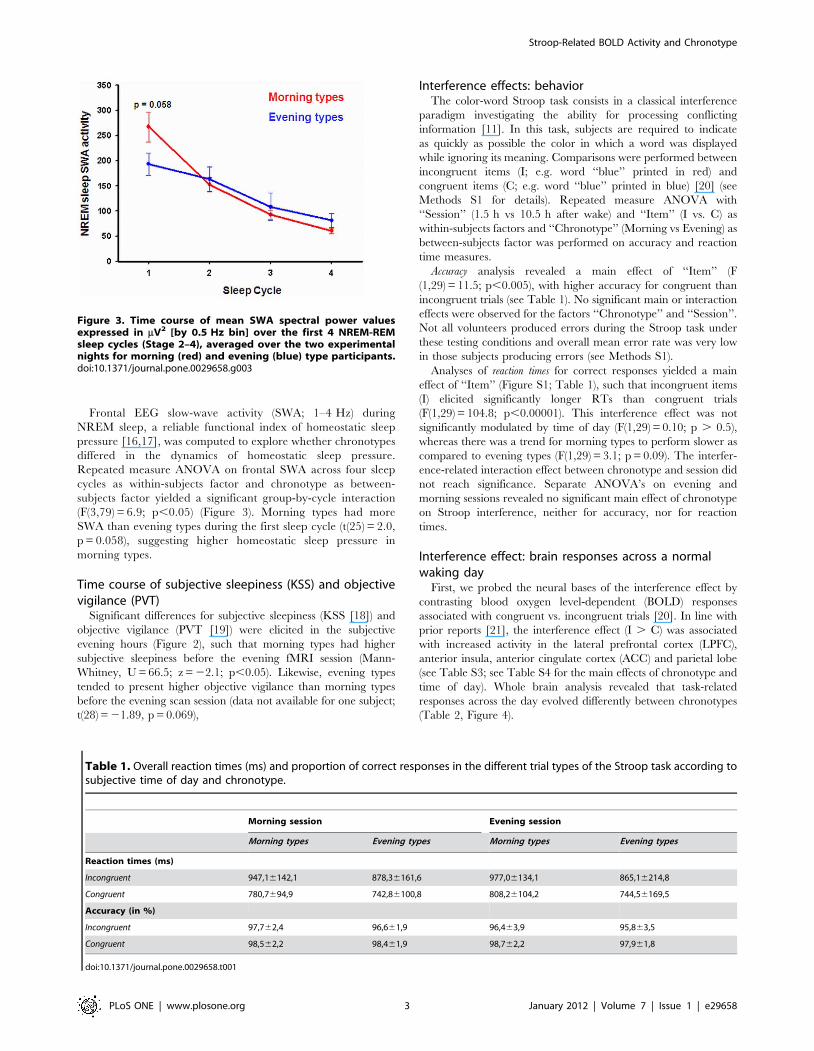
Frontal EEG slow-wave activity (SWA; 1–4 Hz) during
NREM sleep, a reliable functional index of homeostatic sleep
pressure [16,17], was computed to explore whether chronotypes
differed in the dynamics of homeostatic sleep pressure.
Repeated measure ANOVA on frontal SWA across four sleep
cycles as within-subjects factor and chronotype as between-
subjects factor yielded a significant group-by-cycle interaction
(F(3,79) = 6.9; p,0.05) (Figure 3). Morning types had more
SWA than evening types during the first sleep cycle (t(25) = 2.0,
p = 0.058), suggesting higher homeostatic sleep pressure in
morning types.
Time course of subjective sleepiness (KSS) and objectivevigilance (PVT)
Significant differences for subjective sleepiness (KSS [18]) and
objective vigilance (PVT [19]) were elicited in the subjective
evening hours (Figure 2), such that morning types had higher
subjective sleepiness before the evening fMRI session (Mann-
Whitney, U = 66.5; z = 22.1; p,0.05). Likewise, evening types
tended to present higher objective vigilance than morning types
before the evening scan session (data not available for one subject;
t(28) = 21.89, p = 0.069),
Interference effects: behaviorThe color-word Stroop task consists in a classical interference
paradigm investigating the ability for processing conflicting
information [11]. In this task, subjects are required to indicate
as quickly as possible the color in which a word was displayed
while ignoring its meaning. Comparisons were performed between
incongruent items (I; e.g. word ‘‘blue’’ printed in red) and
congruent items (C; e.g. word ‘‘blue’’ printed in blue) [20] (see
Methods S1 for details). Repeated measure ANOVA with
‘‘Session’’ (1.5 h vs 10.5 h after wake) and ‘‘Item’’ (I vs. C) as
within-subjects factors and ‘‘Chronotype’’ (Morning vs Evening) as
between-subjects factor was performed on accuracy and reaction
time measures.
Accuracy analysis revealed a main effect of ‘‘Item’’ (F
(1,29) = 11.5; p,0.005), with higher accuracy for congruent than
incongruent trials (see Table 1). No significant main or interaction
effects were observed for the factors ‘‘Chronotype’’ and ‘‘Session’’.
Not all volunteers produced errors during the Stroop task under
these testing conditions and overall mean error rate was very low
in those subjects producing errors (see Methods S1).
Analyses of reaction times for correct responses yielded a main
effect of ‘‘Item’’ (Figure S1; Table 1), such that incongruent items
(I) elicited significantly longer RTs than congruent trials
(F(1,29) = 104.8; p,0.00001). This interference effect was not
significantly modulated by time of day (F(1,29) = 0.10; p . 0.5),
whereas there was a trend for morning types to perform slower as
compared to evening types (F(1,29) = 3.1; p = 0.09). The interfer-
ence-related interaction effect between chronotype and session did
not reach significance. Separate ANOVA’s on evening and
morning sessions revealed no significant main effect of chronotype
on Stroop interference, neither for accuracy, nor for reaction
times.
Interference effect: brain responses across a normalwaking day
First, we probed the neural bases of the interference effect by
contrasting blood oxygen level-dependent (BOLD) responses
associated with congruent vs. incongruent trials [20]. In line with
prior reports [21], the interference effect (I . C) was associated
with increased activity in the lateral prefrontal cortex (LPFC),
anterior insula, anterior cingulate cortex (ACC) and parietal lobe
(see Table S3; see Table S4 for the main effects of chronotype and
time of day). Whole brain analysis revealed that task-related
responses across the day evolved differently between chronotypes
(Table 2, Figure 4).
Figure 3. Time course of mean SWA spectral power valuesexpressed in mV2 [by 0.5 Hz bin] over the first 4 NREM-REMsleep cycles (Stage 2–4), averaged over the two experimentalnights for morning (red) and evening (blue) type participants.doi:10.1371/journal.pone.0029658.g003
Table 1. Overall reaction times (ms) and proportion of correct responses in the different trial types of the Stroop task according tosubjective time of day and chronotype.
Morning session Evening session
Morning types Evening types Morning types Evening types
Reaction times (ms)
Incongruent 947,16142,1 878,36161,6 977,06134,1 865,16214,8
Congruent 780,7694,9 742,86100,8 808,26104,2 744,56169,5
Accuracy (in %)
Incongruent 97,762,4 96,661,9 96,463,9 95,863,5
Congruent 98,562,2 98,461,9 98,762,2 97,961,8
doi:10.1371/journal.pone.0029658.t001
Stroop-Related BOLD Activity and Chronotype
PLoS ONE | www.plosone.org 3 January 2012 | Volume 7 | Issue 1 | e29658
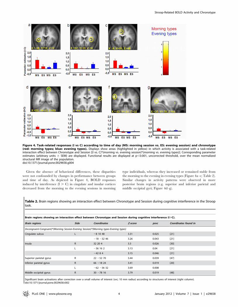
Given the absence of behavioral differences, these disparities
were not confounded by changes in performance between groups
and time of day. As depicted in Figure 4, BOLD responses
induced by interference (I . C) in cingulate and insular cortices
decreased from the morning to the evening sessions in morning
type individuals, whereas they increased or remained stable from
the morning to the evening in evening types (Figure 4a–c; Table 2).
Similar changes in activity patterns were observed in more
posterior brain regions (e.g. superior and inferior parietal and
middle occipital gyri; Figure 4d–g).
Figure 4. Task-related responses (I vs C) according to time of day (MS: morning session vs. ES: evening session) and chronotype(red: morning types; blue: evening types). Displays show areas (highlighted in yellow) in which activity is associated with a task-relatedinteraction effect between Chronotype and Session [(I vs. C)*(morning vs. evening session)*(morning vs. evening types)]. Corresponding parameterestimates (arbitrary units 6 SEM) are displayed. Functional results are displayed at p,0.001, uncorrected threshold, over the mean normalizedstructural MR image of the population.doi:10.1371/journal.pone.0029658.g004
Table 2. Brain regions showing an interaction effect between Chronotype and Session during cognitive interference in the Strooptask.
Brain regions showing an interaction effect between Chronotype and Session during cognitive interference (I.C).
Brain regions Side Coordinates Z-score psvc Coordinates found in
(Incongruent-Congruent)*(Morning Session-Evening Session)*(Morning types-Evening types)
Cingulate sulcus L 28 10 48 3.31 0.025 [21]
218 232 46 3.26 0.043 [21]
Insula R 32 20 4 3.3 0.026 [30]
L 236 16 2 3.13 0.04 [21]
242 8 4 3.15 0.046 [21]
Superior parietal gyrus R 22 252 70 3.44 0.039 [47]
Inferior parietal gyrus R 66 218 24 3.41 0.019 [20]
L 262 236 32 3.69 0.008
Middle occipital gyrus R 30 278 16 3.79 0.019 [48]
Significant brain activations after correction over a small volume of interest (svc; 10 mm radius) according to structures of interest (right column).doi:10.1371/journal.pone.0029658.t002
Stroop-Related BOLD Activity and Chronotype
PLoS ONE | www.plosone.org 4 January 2012 | Volume 7 | Issue 1 | e29658
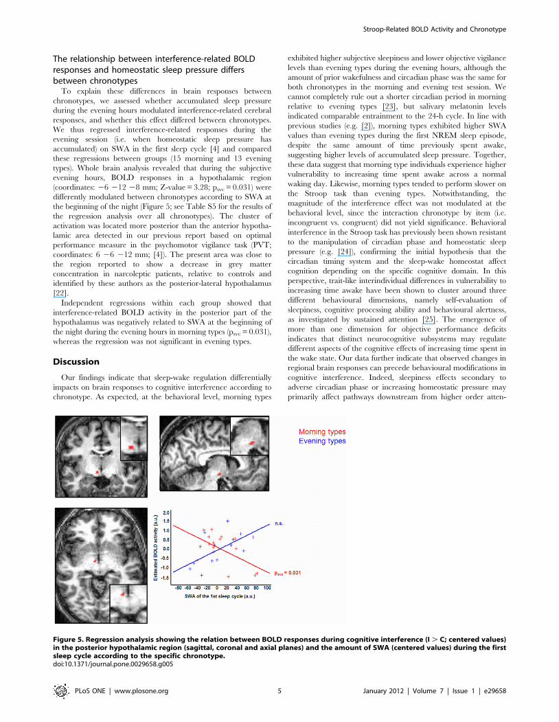
The relationship between interference-related BOLDresponses and homeostatic sleep pressure differsbetween chronotypes
To explain these differences in brain responses between
chronotypes, we assessed whether accumulated sleep pressure
during the evening hours modulated interference-related cerebral
responses, and whether this effect differed between chronotypes.
We thus regressed interference-related responses during the
evening session (i.e. when homeostatic sleep pressure has
accumulated) on SWA in the first sleep cycle [4] and compared
these regressions between groups (15 morning and 13 evening
types). Whole brain analysis revealed that during the subjective
evening hours, BOLD responses in a hypothalamic region
(coordinates: 26 212 28 mm; Z-value = 3.28; psvc = 0.031) were
differently modulated between chronotypes according to SWA at
the beginning of the night (Figure 5; see Table S5 for the results of
the regression analysis over all chronotypes). The cluster of
activation was located more posterior than the anterior hypotha-
lamic area detected in our previous report based on optimal
performance measure in the psychomotor vigilance task (PVT;
coordinates: 6 26 212 mm; [4]). The present area was close to
the region reported to show a decrease in grey matter
concentration in narcoleptic patients, relative to controls and
identified by these authors as the posterior-lateral hypothalamus
[22].
Independent regressions within each group showed that
interference-related BOLD activity in the posterior part of the
hypothalamus was negatively related to SWA at the beginning of
the night during the evening hours in morning types (psvc = 0.031),
whereas the regression was not significant in evening types.
Discussion
Our findings indicate that sleep-wake regulation differentially
impacts on brain responses to cognitive interference according to
chronotype. As expected, at the behavioral level, morning types
exhibited higher subjective sleepiness and lower objective vigilance
levels than evening types during the evening hours, although the
amount of prior wakefulness and circadian phase was the same for
both chronotypes in the morning and evening test session. We
cannot completely rule out a shorter circadian period in morning
relative to evening types [23], but salivary melatonin levels
indicated comparable entrainment to the 24-h cycle. In line with
previous studies (e.g. [2]), morning types exhibited higher SWA
values than evening types during the first NREM sleep episode,
despite the same amount of time previously spent awake,
suggesting higher levels of accumulated sleep pressure. Together,
these data suggest that morning type individuals experience higher
vulnerability to increasing time spent awake across a normal
waking day. Likewise, morning types tended to perform slower on
the Stroop task than evening types. Notwithstanding, the
magnitude of the interference effect was not modulated at the
behavioral level, since the interaction chronotype by item (i.e.
incongruent vs. congruent) did not yield significance. Behavioral
interference in the Stroop task has previously been shown resistant
to the manipulation of circadian phase and homeostatic sleep
pressure (e.g. [24]), confirming the initial hypothesis that the
circadian timing system and the sleep-wake homeostat affect
cognition depending on the specific cognitive domain. In this
perspective, trait-like interindividual differences in vulnerability to
increasing time awake have been shown to cluster around three
different behavioural dimensions, namely self-evaluation of
sleepiness, cognitive processing ability and behavioural alertness,
as investigated by sustained attention [25]. The emergence of
more than one dimension for objective performance deficits
indicates that distinct neurocognitive subsystems may regulate
different aspects of the cognitive effects of increasing time spent in
the wake state. Our data further indicate that observed changes in
regional brain responses can precede behavioural modifications in
cognitive interference. Indeed, sleepiness effects secondary to
adverse circadian phase or increasing homeostatic pressure may
primarily affect pathways downstream from higher order atten-
Figure 5. Regression analysis showing the relation between BOLD responses during cognitive interference (I . C; centered values)in the posterior hypothalamic region (sagittal, coronal and axial planes) and the amount of SWA (centered values) during the firstsleep cycle according to the specific chronotype.doi:10.1371/journal.pone.0029658.g005
Stroop-Related BOLD Activity and Chronotype
PLoS ONE | www.plosone.org 5 January 2012 | Volume 7 | Issue 1 | e29658
tional processes [26] such that, decreased alertness probably
participates in performance deterioration to the task, which is
unlikely to result only from specific effects of sleep loss on higher
order control functions [27].
Chronotype-specific brain responses to cognitiveinterference throughout a normal waking day
One of our key findings is the significant influence of
chronotype and time-of-day on brain responses to cognitive
interference. Indeed, responses were maintained or even increased
in evening types from morning to evening sessions in a set of brain
areas known to be involved in Stroop performance, whereas they
decreased in morning types in the same areas and under the same
conditions. The identified brain areas have been previously
reported to play a pivotal role in successful inhibitory functioning,
particularly the anterior cingulate cortex, involved in monitoring
and resolution of cognitive conflict [20,28]. Likewise, the insula
has been associated with error processing [21]. In the present
study, ceiling effects in accuracy measures during performance on
the Stroop task, commonly reported in healthy young participants
(e.g. [29]), prevented an analysis of changes in error-related
cerebral activity. However, besides its putative implication in error
processing, insular activity has been shown to play a generic role in
conflict processing (Wager et al., 2005). Also, responses related to
cognitive inhibition in the insula area has been previously found to
increase in subjects with low vulnerability to sleep deprivation, and
to decrease in subjects more vulnerable to sleep loss [30]. In this
latter study, different time courses in cortical task-related BOLD
responses during task performance differentiated more resilient
individuals from those participants more susceptible to the
detrimental effects of sleep loss. Our results suggest that a
differential impact of sleep pressure on hypothalamic wake
promotion may be one of the systems underlying those inter-
individual differences in the maintenance of cortical cognition-
related cerebral activity.
Hypothalamic integration of wake promoting signals forthe consolidation of cognitive interference in thesubjective evening hours
Another key finding of our study is the identification of
differential interference-related responses to accumulated sleep
pressure between morning and evening types in a region located in
the posterior hypothalamus. This relationship suggests that with
accumulated homeostatic sleep pressure, present during the evening
hours, morning type individuals become progressively less and less
apt to recruit this hypothalamic region during Stroop performance.
An exact delineation of the specific hypothalamic nuclei responsible
for this diminishing response is beyond the current spatial resolution
of fMRI. It is worth noticing, that this region is located in a more
posterior hypothalamic part than the area we previously observed to
be negatively related to accumulated homeostatic sleep pressure
during optimal sustained attention performance. The posterior
hypothalamic portion contains neurons actively involved in the
promotion of wakefulness [31], making this area a potential
candidate for modulating brain responses underlying cognitive
effort. It is tempting to assume that the arousal stabilizing
neuropeptide hypocretin produced in the postero-lateral hypothal-
amus [32] has some characteristics that suitably fit into the
framework of our data. Chief among these is hypocretin expression
that follows a circadian variation in rats [33,34], monkeys [35] and
humans [36], but also appears to be regulated by a reactive
homeostatic mechanism, opposing to the accumulating sleep drive
during the day to maintain wakefulness [35].
In the context of the present study, we surmise that BOLD
activity in the posterior hypothalamic region allows the promotion
and maintenance of appropriate cognitive interference abilities at
the cortical level during the late part of a normal waking day,
when homeostatic pressure for sleep is high. The posterior
hypothalamic region widely projects to the entire cortex [37,38]
where these projections enhance cortical responses either directly
[39] or through projections of the intralaminar thalamus [40]. We
suggest a relative disruption in the transmission of similar alerting
signals generated by the circadian system into cortical levels in
morning types during the evening. Still, the maintenance of
preserved behavioral effects also indicates that activity in other
brain structures may provide support for the processing of
cognitive interference.
ConclusionOur results indicate that in the subjective evening hours, conflict
processing-related cortical responses are maintained or even
enhanced in evening types, whereas they decrease in morning
types. We speculate that this effect is driven by changes in the
wake-promoting signal generated by hypothalamic structures to
ensure sustained alertness and cortical activity despite accumulat-
ing sleep pressure. Conversely, in morning chronotypes, the
alerting signal may be less pronounced and thus less able to
counteract increasing homeostatic sleep pressure in the evening,
potentially leading to less stable arousal states and thus challenging
cognitive performance and favoring sleep occurrence at earlier
times of the day. Along with genetic studies [27,41], our results
shed light on the mechanisms underlying inter-individual differ-
ences in behavioral and cerebral responses to changes in circadian
and sleep pressure and strengthens chronotype as a strong
predictor for such vulnerability.
Materials and Methods
Supplemental details are presented in Methods S1
Ethics statementAll volunteers fulfilled a written consent form to participate in
this study approved by the Ethics Committee of the Faculty of
Medicine of the University of Liege, and performed in accordance
with the ethical standards described in the Declaration of Helsinki
(1964).
PopulationSixteen extreme morning and 15 extreme evening type subjects
gave their written informed consent to participate in this study. All
volunteers were healthy and between 22 and 32 years of age
(morning types: 9 women/7 men; evening types: 8 women/7
men). Exclusion criteria were reports of medical, psychiatric and
sleep disorders, medication or drug use, alcohol abuse, excessive
caffeine consumption or physical activity, shift work within the
three past months, and transmeridian travel or disturbances in the
sleep-wake cycle within one month before the experiment.
Subjects were screened according to their timing preferences as
defined by the morningness-eveningness questionnaire (MEQ;
[12]), in which scores . 70 identify extreme morning types and
scores ,30 identify extreme evening types. The two groups were
matched according to age, sex and educational level (Table 1).
ProceduresThe experimental design is illustrated in Figure 1.
In a first step, participants reported to the sleep facility for a
habituation night. After this night, they were requested to follow
Stroop-Related BOLD Activity and Chronotype
PLoS ONE | www.plosone.org 6 January 2012 | Volume 7 | Issue 1 | e29658
during one week the sleep schedule (630 minutes) they would
spontaneously adopt while free from any socio-professional
constraints while averaging their bedtime to 8 hours (61 hour).
Actimetry (Cambridge Neurotechnologies, UK) assessed partici-
pants’ compliance to the selected rest-activity patterns. Afterwards,
subjects entered the sleep laboratory for 2 subsequent nights
(Figure 1). Subjects came to the laboratory 7 hours before habitual
lights off on day 1. After the hook-up of electrodes, subjects stayed
under controlled conditions in dim light (,10 lux). Subjective
sleepiness (Visual analogue scale (VAS) and the Karolinska
Sleepiness Scale (KSS; [18]) and objective vigilance (a modified
version of the PVT; [19]) were assessed at hourly intervals while
awake. Furthermore, hourly collected saliva samples while awake
were assayed for melatonin, and polygraphic data were recorded
during the nights. After lights off, subjects were allowed to sleep for
8 hours. One and a half (morning session) and 10.5 (evening
session) hours after wake up of scheduled sleep timing, subjects
underwent an fMRI session during the performance of various
cognitive tasks. We report here results related to the Stroop task;
results regarding a psychomotor vigilance task are reported
elsewhere [4]. For half of the subjects, the morning session
followed the first experimental night and the evening session the
second one while for the other half of the volunteers, the morning
session followed the second experimental night and the evening
session the first night, thus controlling for a potential practice effect
on session-related Stroop performance. The order of selected
cognitive tasks was counterbalanced across subjects and sessions
and parallel versions of the Stroop task were used for the repeated
administrations.
Stroop taskThe color-word Stroop paradigm [11] investigates the ability to
process conflicting information. Participants are required to
indicate the color in which a word is printed as quickly as possible
while ignoring the word’s meaning, an interference occurring
when the word is a color name but printed in a different color (e.g.
the word ‘‘RED’’ printed in blue). We used a computerized trial by
trial, 4 colors version of the task adapted to the fMRI
environment. The test comprised 196 items divided into four
different categories: congruent (C), incongruent (I) and neutral (NE
and NT) trials. Neutral items consisted of a non-verbal stimulus,
i.e. a sequence of X’s, printed in a particular color. Trials with the
same printed color and word meaning were defined as congruent
items (e.g. the word RED printed in the color red). By contrast,
incongruent trials were represented by color words printed in a
different color than the meaning of the word (e.g. RED printed in
the color yellow). We used incongruent and congruent items in
order to investigate the Stroop interference effect (see Methods S1
for details of the task). Stimuli were presented on a black screen
and each color was represented in equal proportions within each
trial type. Responses were made by manipulating a 4-buttons
response keypad with the dominant hand.
Polysomnographic recordingsDuring sleep, EEG was recorded digitally with Brainamp MR
Family and V-amp EEG amplifiers (Brain Products Inc. Gilching,
Germany), using AgCl electrodes. Recording was done at 250 Hz
sampling frequency, with a 0.1 Hz high-pass filter, a 80 Hz low-
pass filter, and a 50 Hz notch filter. Electrodes were placed
according to the 10–20-System with 4 channels (Fz, Cz, Pz, Oz)
referenced to linked mastoids. Additionally, horizontal and vertical
eye movements and a submental electromyogram were recorded.
Sleep stages during scheduled nights were visually scored on a 20-s
epoch basis [42] according to standard criteria [43]. EEG artifacts
were visually detected. Finally, NREM–REM sleep cycles were
determined according to the criteria of Feinberg & Floyd [44].
Abnormally long first NREM episodes, where evidence of a
‘skipped’ first REM period was observed, were divided into two
separate NREM episodes [45].
Quantitative EEG analysisEEGs were subjected to spectral analysis (Welch’s method, 4-s
window, 50% overlap) using a fast Fourier transforms resulting in
a 0.5 Hz bin resolution. EEG power spectra were calculated
during NREM sleep in the frequency range between 0.5 and
25 Hz. Results were averaged over all epochs of NREM sleep
(stage 2–4), which did not contain movement artifacts. The time
course of EEG slow wave activity (1–4 Hz) during non-REM sleep
was investigated throughout sleep cycles of the nights preceding
fMRI acquisitions.
MRI data acquisitionFunctional MRI-series (fMRI) were acquired using a head-only
3T scanner (Siemens Allegra). Multislice T2*-weighted fMRI
images were obtained with a gradient echo-planar sequence using
axial slice orientation (TR = 2130 ms, TE = 40 ms, FA = 90u, 32
transverse slices, 3 mm slice thickness, 30% inter-slice gap,
FoV = 2206220 mm2, matrix size = 64664632, voxel si-
ze = 3.463.463.0 mm3). Structural T1-weighted brain images
were also acquired [46].
Functional MRI data analysisA detailed description of fMRI data analysis can be found in
Methods S1.
fMRI data from morning and evening sessions were analyzed
using SPM5 (http://www.fil.ion.ucl.ac.uk) implemented in MA-
TLAB 7 (Mathworks, Sherbom, MA). Volumes were corrected
for head motion, spatially normalized, and smoothed. Data were
processed using two-step statistical analyses, taking into account
intra-individual then inter-individual variance, respectively. For
each subject, brain responses were modeled at each voxel, using a
general linear model. Five separate regressors were included:
events associated with congruent trials (C), events related to
incongruent trials (I), events associated with neutral items that
were used for comparison with other trial conditions (NE), events
linked to neutral items used in order to annihilate undesired
priming effects (NT), and response errors over all trial types (for
those subjects committing errors). Linear contrasts tested the
main effect of interference in the Stroop task as revealed by
comparing I to C trials (I.C). At the random-effect level, the
interference-related effect of chronotype [(I vs. C)*(morning vs.
evening types)], of time of day [(I vs. C)*(morning vs. evening
session)] and most importantly the interference-related interac-
tion effects between chronotype and time of day [(I vs.
C)*(morning vs. evening session)*(morning vs. evening type)]
were tested. In a second analysis, a covariate representing the
amount of SWA during the first sleep cycle for each group
separately was included into the model in order to investigate
whether accumulated sleep pressure modulated task-related
BOLD responses and whether this effect differed between
chronotypes. The resulting set of voxel values constituted a
map of t statistics [SPM(T)] thresholded at p,0.001 (uncorrected;
over the whole brain). Statistical inferences were performed after
correction for multiple comparisons at a threshold of p,0.05,
using Gaussian random field theory at the voxel level in small
spherical volumes (radius, 10 mm) around a priori locations of
structures of interest, taken from the literature.
Stroop-Related BOLD Activity and Chronotype
PLoS ONE | www.plosone.org 7 January 2012 | Volume 7 | Issue 1 | e29658
Supporting Information
Figure S1 Reaction times (±SEM) during the Strooptask according to trial type (congruent [C] and incon-gruent [I]), testing time (morning versus eveningsession) and chronotype (blue: evening types; red:morning types).
(TIF)
Table S1 Demographic, sleep and circadian character-istics (mean ± SD) in morning and evening types.
(DOCX)
Table S2 Visually scored sleep stages (mean ± SD)expressed in percentages according to chronotype(averaged over the two experimental nights). Comparison
between groups: all ps . 0.1).
(DOCX)
Table S3 Brain regions involved in the Stroop interfer-ence effect (Incongruent . Congruent items), all chron-otypes and testing sessions confounded. Reported brain
activations are significant after correction for multiple comparisons
over the entire volume (*) or over a small volume of interest (svc).
R: right hemisphere; L: left hemisphere.
(DOCX)
Table S4 Task-related main effect of time of day andchronotype. R: right hemisphere; L: left hemisphere.
(DOCX)
Table S5 Negative regression between relative SWA inthe first sleep cycle (all chronotypes) and BOLD activityinvolved in the main effect of the Stroop task (I . Ctrials) during the evening scan session. psvc: significance
after small volume correction (radius 10 mm) according to
structures of interest reported in the literature. R: right
hemisphere; L: left hemisphere. * p,0.001 uncorrected.
(DOCX)
Methods S1
(DOC)
Acknowledgments
We thank Dr. Sarah Chellappa for helpful comments and language
revision of the manuscript.
Author Contributions
Conceived and designed the experiments: CS PP CP EB CC PM FC.
Performed the experiments: CS PP YL VS GV GT SG MS MD TD-V ES
FC. Analyzed the data: CS PP YL CP CB PB EB CC PM FC. Contributed
reagents/materials/analysis tools: YL CP CB PB CD AL. Wrote the paper:
CS FC PP CC PM GV.
References
1. Kerkhof GA (1991) Differences between morning-types and evening-types in thedynamics of EEG slow wave activity during night sleep. Electroencephalogr Clin
Neurophysiol 78: 197–202.
2. Mongrain V, Carrier J, Dumont M (2006) Difference in sleep regulationbetween morning and evening circadian types as indexed by antero-posterior
analyses of the sleep EEG. Eur J Neurosci 23: 497–504.
3. Mongrain V, Dumont M (2007) Increased homeostatic response to behavioral
sleep fragmentation in morning types compared to evening types. Sleep 30:
773–780.
4. Schmidt C, Collette F, Leclercq Y, Sterpenich V, Vandewalle G, et al. (2009)
Homeostatic sleep pressure and responses to sustained attention in the
suprachiasmatic area. Science 324: 516–519.
5. Aston-Jones G, Chen S, Zhu Y, Oshinsky ML (2001) A neural circuit for
circadian regulation of arousal. Nat Neurosci 4: 732–738.
6. Deboer T, Detari L, Meijer JH (2007) Long term effects of sleep deprivation onthe mammalian circadian pacemaker. Sleep 30: 257–262.
7. Deboer T, Vansteensel MJ, Detari L, Meijer JH (2003) Sleep states alter activity
of suprachiasmatic nucleus neurons. Nat Neurosci 6: 1086–1090.
8. Chee MW, Chuah LY (2008) Functional neuroimaging insights into how sleep
and sleep deprivation affect memory and cognition. Curr Opin Neurol 21:
417–423.
9. Collette F, Van der Linden M, Laureys S, Delfiore G, Degueldre C, et al. (2005)
Exploring the unity and diversity of the neural substrates of executive
functioning. Hum Brain Mapp 25: 409–423.
10. Collette F, Salmon E, Van der Linden M (2006) Exploration of the Neural
Substrates of Executive Functioning by Functional Neuroimaging. Neuroscience139: 209–221.
11. Stroop JR (1935) Studies of interference in serial verbal reactions. Journal of
Experimental Psychology 18: 643–662.
12. Horne JA, Ostberg O (1976) A self-assessment questionnaire to determine
morningness-eveningness in human circadian rhythms. International Journal of
Chronobiology 4: 97–110.
13. Roenneberg T, Wirz-Justice A, Merrow M (2003) Life between clocks: daily
temporal patterns of human chronotypes. J Biol Rhythms 18: 80–90.
14. Krauchi K, Cajochen C, Wirz-Justice A (1997) A relationship between heat lossand sleepiness: effects of postural change and melatonin administration. J Appl
Physiol 83: 134–139.
15. Berthomier C, Drouot X, Herman-Stoica M, Berthomier P, Prado J, et al.(2007) Automatic analysis of single-channel sleep EEG: validation in healthy
individuals. Sleep 30: 1587–1595.
16. Cajochen C, Foy R, Dijk DJ (1999) Frontal predominance of a relative increasein sleep delta and theta EEG activity after sleep loss in humans. Sleep Res
Online 2: 65–69.
17. Finelli LA, Achermann P, Borbely AA (2001) Individual ‘fingerprints’ in human
sleep EEG topography. Neuropsychopharmacology 25: S57–62.
18. Akerstedt T, Gillberg M (1990) Subjective and objective sleepiness in the activeindividual. Int J Neurosci 52: 29–37.
19. Dinges DF, Powell JW (1985) Microcomputer analyses of performance on a
portable, simple visual RT task during sustained operations. Behavior Research
Methods, Instruments, & Computers: A Journal of the Psychonomic Society 17:
625–655.
20. Fan J, Flombaum JI, McCandliss BD, Thomas KM, Posner MI (2003) Cognitive
and brain consequences of conflict. Neuroimage 18: 42–57.
21. Roberts KL, Hall DA (2008) Examining a supramodal network for conflict
processing: a systematic review and novel functional magnetic resonance
imaging data for related visual and auditory stroop tasks. J Cogn Neurosci 20:
1063–1078.
22. Draganski B, Geisler P, Hajak G, Schuierer G, Bogdahn U, et al. (2002)
Hypothalamic gray matter changes in narcoleptic patients. Nat Med 8:
1186–1188.
23. Duffy JF, Rimmer DW, Czeisler CA (2001) Association of intrinsic circadian
period with morningness-eveningness, usual wake time, and circadian phase.
Behav Neurosci 115: 895–899.
24. Sagaspe P, Sanchez-Ortuno M, Charles A, Taillard J, Valtat C, et al. (2006)
Effects of sleep deprivation on Color-Word, Emotional, and Specific Stroop
interference and on self-reported anxiety. Brain Cogn 60: 76–87.
25. Van Dongen HP, Baynard MD, Maislin G, Dinges DF (2004) Systematic
interindividual differences in neurobehavioral impairment from sleep loss:
evidence of trait-like differential vulnerability. Sleep 27: 423–433.
26. Horowitz TS, Cade BE, Wolfe JM, Czeisler CA (2003) Searching night and day:
a dissociation of effects of circadian phase and time awake on visual selective
attention and vigilance. Psychol Sci 14: 549–557.
27. Vandewalle G, Archer SN, Wuillaume C, Balteau E, Degueldre C, et al. (2009)
Functional magnetic resonance imaging-assessed brain responses during an
executive task depend on interaction of sleep homeostasis, circadian phase, and
PER3 genotype. J Neurosci 29: 7948–7956.
28. Botvinick MM, Braver TS, Barch DM, Carter CS, Cohen JD (2001) Conflict
monitoring and cognitive control. Psychol Rev 108: 624–652.
29. Compton RJ, Banich MT, Mohanty A, Milham MP, Herrington J, et al. (2003)
Paying attention to emotion: an fMRI investigation of cognitive and emotional
stroop tasks. Cogn Affect Behav Neurosci 3: 81–96.
30. Chuah YM, Venkatraman V, Dinges DF, Chee MW (2006) The neural basis of
interindividual variability in inhibitory efficiency after sleep deprivation.
J Neurosci 26: 7156–7162.
31. Saper CB, Chou TC, Scammell TE (2001) The sleep switch: hypothalamic
control of sleep and wakefulness. Trends Neurosci 24: 726–731.
32. Adamantidis A, de Lecea L (2008) Physiological arousal: a role for hypothalamic
systems. Cell Mol Life Sci 65: 1475–1488.
33. Yoshida Y, Fujiki N, Nakajima T, Ripley B, Matsumura H, et al. (2001)
Fluctuation of extracellular hypocretin-1 (orexin A) levels in the rat in relation to
the light-dark cycle and sleep-wake activities. Eur J Neurosci 14: 1075–1081.
34. Zhang S, Zeitzer JM, Yoshida Y, Wisor JP, Nishino S, et al. (2004) Lesions of the
suprachiasmatic nucleus eliminate the daily rhythm of hypocretin-1 release.
Sleep 27: 619–627.
Stroop-Related BOLD Activity and Chronotype
PLoS ONE | www.plosone.org 8 January 2012 | Volume 7 | Issue 1 | e29658
35. Zeitzer JM, Buckmaster CL, Parker KJ, Hauck CM, Lyons DM, et al. (2003)
Circadian and homeostatic regulation of hypocretin in a primate model:implications for the consolidation of wakefulness. J Neurosci 23: 3555–3560.
36. Salomon RM, Ripley B, Kennedy JS, Johnson B, Schmidt D, et al. (2003)
Diurnal variation of cerebrospinal fluid hypocretin-1 (Orexin-A) levels in controland depressed subjects. Biol Psychiatry 54: 96–104.
37. Goutagny R, Verret L, Fort P, Salvert D, Leger L, et al. (2004) Posteriorhypothalamus and regulation of vigilance states. Arch Ital Biol 142: 487–500.
38. Peyron C, Tighe DK, van den Pol AN, de Lecea L, Heller HC, et al. (1998)
Neurons containing hypocretin (orexin) project to multiple neuronal systems.J Neurosci 18: 9996–10015.
39. Bayer L, Serafin M, Eggermann E, Saint-Mleux B, Machard D, et al. (2004)Exclusive postsynaptic action of hypocretin-orexin on sublayer 6b cortical
neurons. J Neurosci 24: 6760–6764.40. Bayer L, Eggermann E, Saint-Mleux B, Machard D, Jones BE, et al. (2002)
Selective action of orexin (hypocretin) on nonspecific thalamocortical projection
neurons. J Neurosci 22: 7835–7839.41. Viola AU, Archer SN, James LM, Groeger JA, Lo JC, et al. (2007) PER3
polymorphism predicts sleep structure and waking performance. Curr Biol 17:613–618.
42. Leclercq Y, Schrouff J, Noirhomme Q, Maquet P, Phillips C (2011) FMRI
artefact rejection and sleep scoring toolbox. Comput Intell Neurosci 2011:
598206.
43. Rechtschaffen A, Kales AA (1968) A manual of standardized terminology,
techniques and scoring system of sleep stages of human subjects. BethesdaMD:
US Dept of Health, Education and Welfare, Public Health Service.
44. Feinberg I, Floyd TC (1979) Systematic trends across the night in human sleep
cycles. Psychophysiology 16: 283–291.
45. Feinberg I, Campbell IG (2003) Kinetics of non-rapid eye movement delta
production across sleep and waking in young and elderly normal subjects:
theoretical implications. Sleep 26: 192–200.
46. Deichmann R, Schwarzbauer C, Turner R (2004) Optimisation of the 3D
MDEFT sequence for anatomical brain imaging: technical implications at 1.5
and 3 T. Neuroimage 21: 757–767.
47. Drummond SP, Bischoff-Grethe A, Dinges DF, Ayalon L, Mednick SC, et al.
(2005) The neural basis of the psychomotor vigilance task. Sleep 28: 1059–1068.
48. Milham MP, Banich MT, Claus ED, Cohen NJ (2003) Practice-related effects
demonstrate complementary roles of anterior cingulate and prefrontal cortices in
attentional control. Neuroimage 18: 483–493.
Stroop-Related BOLD Activity and Chronotype
PLoS ONE | www.plosone.org 9 January 2012 | Volume 7 | Issue 1 | e29658