Cholesteric liquid crystals in living matter - Archive ouverte HAL
93
HAL Id: hal-01994869 https://hal.archives-ouvertes.fr/hal-01994869 Submitted on 6 Oct 2021 HAL is a multi-disciplinary open access archive for the deposit and dissemination of sci- entific research documents, whether they are pub- lished or not. The documents may come from teaching and research institutions in France or abroad, or from public or private research centers. L’archive ouverte pluridisciplinaire HAL, est destinée au dépôt et à la diffusion de documents scientifiques de niveau recherche, publiés ou non, émanant des établissements d’enseignement et de recherche français ou étrangers, des laboratoires publics ou privés. Cholesteric liquid crystals in living matter Michel Mitov To cite this version: Michel Mitov. Cholesteric liquid crystals in living matter. Soft Matter, Royal Society of Chemistry, 2017, 13 (23), pp.4176-4209. 10.1039/C7SM00384F. hal-01994869
-
Upload
khangminh22 -
Category
Documents
-
view
0 -
download
0
Transcript of Cholesteric liquid crystals in living matter - Archive ouverte HAL

HAL Id: hal-01994869https://hal.archives-ouvertes.fr/hal-01994869
Submitted on 6 Oct 2021
HAL is a multi-disciplinary open accessarchive for the deposit and dissemination of sci-entific research documents, whether they are pub-lished or not. The documents may come fromteaching and research institutions in France orabroad, or from public or private research centers.
L’archive ouverte pluridisciplinaire HAL, estdestinée au dépôt et à la diffusion de documentsscientifiques de niveau recherche, publiés ou non,émanant des établissements d’enseignement et derecherche français ou étrangers, des laboratoirespublics ou privés.
Cholesteric liquid crystals in living matterMichel Mitov
To cite this version:Michel Mitov. Cholesteric liquid crystals in living matter. Soft Matter, Royal Society of Chemistry,2017, 13 (23), pp.4176-4209. �10.1039/C7SM00384F�. �hal-01994869�

Accepted version published in: Soft Matter, 2017, 13, 4176-4209
Review
1
Cholesteric liquid crystals in living matter
Michel Mitov*
Liquid crystals play an important role in biology because the combination of order and mobility is a
basic requirement for self-organization and structure formation in living systems. Cholesteric liquid
crystals are omnipresent in the living matter in both in vivo and in vitro conditions and address the
major types of molecules essential to life. In animal and plant kingdoms, the cholesteric structure is a
recurring design, suggesting a convergent evolution to an optimised left-handed helix. Herein, we
review the recent advances in the cholesteric organisation of DNA, chromatin, chitin, cellulose,
collagen, viruses, silk and cholesterol esters deposition in atherosclerosis. Cholesteric structures can
be found in bacteriophages, archaea, eukaryotes, bacteria nucleoids, chromosomes of unicellular
algae, sperm nuclei of many vertebrates, cuticles of crustaceans and insects, bone, tendon, cornea,
fish scales and scutes, cuttlebone and squid pen, plant cell wall, virus suspensions, silk produced by
spiders and silkworms, and arterial wall lesions. This article specifically aims at describing the
consequences of the cholesteric geometry in living matter, which are far from being fully defined and
understood, and discusses various perspectives. The roles and functions of biological cholesteric
liquid crystals include maximisation of packing efficiency, morphogenesis, mechanical stability,
optical information, radiation protection and evolution pressure.
Article history: Received 23rd Feb 2017 / Accepted 22nd May 2017 / First published (online) 07 Jun 2017.
https://doi.org/10.1039/C7SM00384F
*Centre d’Elaboration de Matériaux et d’Etudes Structurales (CEMES), CNRS, BP 94347, 29 rue Jeanne-Marvig, F-31055
Toulouse cedex 4. E-mail : mitov AT cemes.fr

Accepted version published in: Soft Matter, 2017, 13, 4176-4209
Review
2
Cholesteric liquid crystals are ubiquitous in animal and plant kingdoms. We review the advances and
open questions in the cholesteric organisation of DNA, chromatin, chitin, collagen, cellulose, viruses,
silk fibroin and cholesterol esters.
Michel Mitov graduated from the University of Nice–Sophia Antipolis (France),
where he received his PhD in condensed matter physics. He is Director of
Research at CNRS (National Center for Scientific Research) and leader of the
Liquid Crystal Group at Centre d’Elaboration de Matériaux et d’Etudes
Structurales (CEMES) in Toulouse. His current areas of interest are the design
and optical response of complex cholesteric liquid crystal structures (pitch
gradient, double helicity, spatially-variable helicoidal axis), and bio-inspired
versions from insect carapaces. He is the inventor of patents related to smart reflective windows to
control solar light and heat. He has published a science popularisation essay on soft matter
(“Sensitive Matter—Foams, Gels, Liquid Crystals and Other Miracles”, Harvard University Press,
2012).

Accepted version published in: Soft Matter, 2017, 13, 4176-4209
Review
3
1. Introduction
1.1. Historical background
In 1957, Glenn H. Brown and Wilfrid G. Shaw from the University of Cincinnati, Ohio, published a
notable 100-page long article reviewing the knowledge on the states of the matter once named as
liquid crystals (LCs) [1]. During those times, LCs were neglected or even forgotten in textbooks. The
first book on the structure and properties of LCs was published in 1962 by George Gray from the
University of Hull, England [2]. In August 1965, the first meeting of the International Liquid Crystal
Conference series was organised under the leadership of Brown at the Kent State University, Ohio.
On 28 May 1968, a press conference was held in New York City at the 30 Rockefeller Center, where
the RCA management revealed their LC research results and showed prototype displays. These
displays included a numeric indicator, small electronic window, television test pattern and fully
functional digital clock [3]. These devices were revolutionary and they marked the beginning of a new
era for a novel industry. Therefore, the mid-1960s was considered as a period of the great revival of
LC research, in the USA, after the pioneering work conducted in Germany and France between 1888
and 1920s.
The seminal book “Crystals that flow” primarily focused on compiling classical papers on the history
of LC research with commentary [4]. The readers may refer to the book “Soap, science, & flat-screen
TVs” to understand the background behind the discoveries and the communications between the
scientists, which also provides social and political contexts [5]. The milestones of the pioneering
research on LCs from 1888 to 1922 have been summarised in a five-page report [6].
1.2. Motivation of the Review
From 1968 to around 1990s, much attention was focused on the chemistry, physics and technology
of flat panel LC displays (LCDs) in both public and private sectors, and a huge amount of funding was
available to solve fundamental problems and to develop the flourishing market of flat screens. The
Accepted version published in: Soft Matter, 2017, 13, 4176-4209
Review
4
response time of cholesteric LCs (CLCs) was too slow to display video frame rate dynamic images, and
the driving voltages were high. Therefore, these chiral phases probably could not gather the
attraction they deserved with regard to the scientific questions they raise to gain fundamental
knowledge and find innovative (non-display) applications.
Furthermore, compared with LCs for flat screens, biological LCs have received very less attention
from the scientific community although they are highly important as no life is possible without them.
LCs are commonly classified as thermotropic and lyotropic LCs. The most encountered LCs in nature
are lyotropic [7], for which the organization is determined or changed by concentration, rather than
temperature like in the case of thermotropic LCs [8] found in LCDs. For example, in the cell
membrane, the LC phase forms from the dissolution of phospholipids in water. The phospholipid
bilayer in the cell membrane is a smectic LC that is reduced to two layers [9]. Amylopectin side chains
in starch—a glucose polymer of vast importance found in wheat, rice and potatoes—may adopt
smectic structures [10]. Solanine, a glycoalkaloid found in small quantity in potatoes, tomatoes and
eggplants, exhibits a smectic A* phase [11]. Muscles have been long considered as biological
analogues of LCs [12]. Protein filaments (myosin and actin) in smooth and striated muscles have an
organisation reminiscent from nematic and smectic structures [13]. The difference between cross-
striated muscles (arthropods and vertebrates) and oblique-striated muscles (annelids and molluscs)
corresponds to the difference between smectic A and C phases. The helicity observed in certain
obliquely striated muscles has its counterpart with smectic C* phase [14]. Nevertheless, smectic
phases were rarely found, probably due to polydispersity in the length of fibrils composed of
biological macromolecules [15]. During this review, columnar phases (in DNA) or nematic phases (in
chitin, collagen, cellulose, viruses and silk) will incidentally appear. However, the LC structure, which
is ubiquitously present in living matter and whose chirality is one of the most important features, is a
cholesteric structure found in almost all types of animals and plants. The unifying structure of
biological CLCs is attributed to the helicoidal organisation of elementary bricks such as molecules,

Accepted version published in: Soft Matter, 2017, 13, 4176-4209
Review
5
macromolecules, and microfibrils as anisotropic aggregates of macromolecules (chitin, collagen and
cellulose)—with no positional order for these bricks. This is the basic structure of CLC phase.
Therefore, it is necessary to dedicate a review to the cholesteric organisation in living organisms,
regardless of the nature of the constituent molecules or taxa. Due to the large bibliographic domains
involved, there is a huge dispersion of data. One can be tempted to apply to biological CLCs the main
concepts introduced by physicists in the field of synthetic LCs; however, reviewing and discussing
these concepts is needed. This review may be of interest to a large audience in the fields of soft
matter physics, LC science, biological material science and engineering, biophysics, developmental
biology, plant and animal physiology and botany.
1.3. Methods
CLC phases are the most common LC phases formed by biopolymers; therefore, this article focuses
on the cholesteric arrangements found in nucleic acids (e.g. DNA), chromatin (i.e. macromolecule
complex consisting of DNA and proteins), polysaccharides (e.g. chitin and cellulose) and proteins (e.g.
collagen, silk fibroin). In addition, virus and cholesterol ester depositions in atherosclerosis are
addressed. The cholesteric structure found in the following organisms is discussed in this study:
bacteriophages, archaea, eukaryotes, bacteria nucleoids, chromosomes of unicellular algae
dinoflagellates, sperm nuclei of many vertebrates, exoskeleton of arthropods (such as crustaceans
and insects), bone, dentin, tendon, cornea (such as that of fishes, amphibians, reptiles and birds), fish
armour, coleoid cephalopods (such as cuttlebone and squid pen), molluscan nacre, plant cell wall
(such as that from leaves of tropical ferns, berry-like fruits, wood or cotton pulp), virus suspensions,
silk (such as that from spiders and silkworms) and arterial wall lesions.
The following questions were taken into consideration while analysing the recent advances:
Which elementary bricks are responsible for the CLC organisation?
In which living organisms are they found?

Accepted version published in: Soft Matter, 2017, 13, 4176-4209
Review
6
At what scale does the CLC organisation occur?
What is the state-of-the-art before the 2000s?
What kinds of cases are found in the literature over last 10 years?
What are the current trends in the field?
Does the CLC organisation differ from one organism to another and from animals to plants?
What are the accepted or debatable biological functions?
To what extent biological CLC structures may inspire artificial structures and for which
applications?
Which are the least investigated topics?
Can fresh research directions be suggested and future directions be speculated?
1.4. Article Content
Section 2 gives a historical perspective on the early days of research in LC science and states the
speculations on liquid-crystalline organisations in living matter as essential features for the
realisation of processes inherent to life. Section 3 describes the following main features of the CLC
phase: structural parameters, optical properties, applications and the twisted plywood model, which
is of paramount importance for analysing different scale textures and the textures found in different
organisms or tissues. All the sections are then organised around the main biological materials, and
discuss and highlight the current progress and problems using a few studies from the literature that
are of particular interest and significance. Sections 4 to 10 discuss the basic understanding of
cholesteric organisation, its main features, functions and (when applied) evolutionary context in the
DNA and chromatin, chitin (in vivo and in vitro), collagen (in situ and ex situ), cellulose, suspensions of
rod-like viruses, silk fibroin, and cholesterol ester deposits in atherosclerosis. At the end of each
section, conclusions are drawn or interesting questions are posed to make the readers aware of the
future prospects. In the last, Section 11 summarises the previous findings and concludes with general
perspectives.

Accepted version published in: Soft Matter, 2017, 13, 4176-4209
Review
7
2. Historical perspective
Friedrich Reinitzer (1857–1927) was a botanist, known as a biochemist in the modern terms, at the
Institute of Plant Physiology, German University of Prague, in 1888. His primary work focused on
cholesterol derived crystals extracted from carrot roots [16] and he found that cholesteryl benzoate
had ‘two melting points’, cited in Reinitzer’s letter dated 14 March 1888. This letter was addressed to
the crystallographer Otto Lehmann (1855–1922), a physicist at the Polytechnical School of Aachen
(Germany) who specialised in the growth phenomena and phase transitions of crystalline materials.
In his letter, Reinitzer explained that the small crystals lose their rigidity at 145.5°C; this ‘first’ melting
point is the temperature at which the solid turned into a milky fluid. The ‘second’ melting point was
at 178.5°C, later named the clearing point; at this temperature, the material became transparent. On
cooling, the material exhibited violet and blue colours, which then disappeared, leaving the liquid
substance cloudy. This is starkly different from the current knowledge on the crystalline matter,
which believes that a crystal should lose both its colour and solidity at a single, unique temperature.
In 1854, Rudolf Virchow [17] described similar unusual behaviour for myelin—the sheath that
surrounds nerve fibres. Furthermore, Carl von Mettenheimer [18] showed that myelin is birefringent.
Between 1861 and 1887, different chemists, like Julius Planer [19], reported strange colour effects in
cholesterol esters, but the complexity of their observations and the difficulty of duplicating them did
not encourage them to pursue their investigations. For this reason, science historians consider that
LCs were discovered in 1888 by Reinitzer, who was the first to give a detailed description of his
observations and to recognise that the strange behaviour of these ‘crystals’ was a significant, novel
phenomenon and not an artefact.
Lehmann reproduced the phenomena observed by Reinitzer in a large number of substances and
concluded that it was a general phenomenon. He was the main promoter of ‘liquid crystals’; he
promoted not only the term but also the research activities in this field. In addition to his use of
heterodox terms, Lehmann’s links with the ultra-Darwinist Ernst Haeckel (1834–1919) detracted him

Accepted version published in: Soft Matter, 2017, 13, 4176-4209
Review
8
from a credibility. Haeckel was a zoologist whose view on the theory of evolution was more than
controversial. He believed that matter, including inert one, was endowed with a soul. He spoke
literally on ‘crystal souls’ [20]. He was convinced that Lehmann’s LCs contained the essence of life
itself, i.e. the vital force, that they linked the living and the dead; it was a link he was desperately
seeking. Apart from the non-scientific claims of Lehmann and Haeckel, they were probably the first
scientists to propound upon the idea that the LC state is responsible for many of the biological
processes. The roots of research on LCs lie in living matter.
The liquid crystalline phases that Reinitzer observed were chiral phases, later named as cholesteric
phases and blue phases. The term cholesteric was coined in 1922 by Georges Friedel (1865–1933)
[21] as this state of matter was discovered in cholesterol esters by Reinitzer. However, post the
discovery, cholesteric-liquid-crystalline states were found in other substances not connected with
cholesterol. The CLC phase is also known as the chiral nematic phase, which stems from the fact that
the phase is like the twisted version of the nematic phase. A small amount of a chiral (non-racemic)
compound, dissolved in a nematic LC, may transform it into a CLC [21].
3. Main features of the cholesteric liquid crystal phase
3.1. Structure
The CLC phase exhibits a helicoidal structure with a twist axis perpendicular to the local director (Fig.
1.a), which comes in whole or in part from the molecular chirality (rod-like molecules here). If the
structure is cut in a direction perpendicular to the helicoidal axis, local nematic order would appear.
A nematic LC phase is an achiral phase that exhibits a purely orientational order of elongated
molecules. The CLC structure is often drawn as a stack of layers with an orientational order of
molecules in each plane and rotation by a constant angle of each plane with respect to its
neighbours. However, this representation is only a guide for the observer and these layers do not
Accepted version published in: Soft Matter, 2017, 13, 4176-4209
Review
9
have a physical reality because the CLC phase is not a layered system. When the CLC structure is
modelled as a layered system, each layer behaves like a uniaxial anisotropic medium with the slow
axis parallel to the rod-like molecules and the fast axis perpendicular to them. The two axes twist
regularly without discontinuity from ‘layer’ to ‘layer’.
The cholesteric phase is characterised by the following two structural parameters: the helical pitch
and twist sense. The pitch gives the distance along the helicoidal axis that corresponds to a rotation
of 360° in the orientation of the rod-like molecules (Fig. 1.b). Due to the head–tail symmetry of the
molecules, the periodicity of the cholesteric phase along the axis is given by half the pitch. The
magnitude of the pitch can considerably vary from a few tens of nanometres to many micrometres.
The twist sense determines a left- or right-handed helix. To date, there is no method to predict which
chiral molecular configuration will give a certain helix handedness. Furthermore, quantitative
prediction of the pitch based on molecular features is another unsolved problem. One of the greatest
challenges in LC science is understanding and anticipating the connection between the molecular
chirality and the structure chirality of LC phases.
Topological defects are key features in LCs, and the connection between symmetry and defects is a
contemporary research subject in biology. The spatial confinement of the cholesteric structure in
complex curved geometries, like in chromosomes, imposes restrictions on the configurations of the
order parameter and requires the appearance of topological defects in the ground state [22]. It leads
to a rich palette of defect structures such as focal conic domains and oily streaks. The complexity of
twisted structures with defects makes the CLCs an important subject to investigate the relationship
between the symmetry of molecular interactions and the mesoscopic organization.

Accepted version published in: Soft Matter, 2017, 13, 4176-4209
Review
10
Fig. 1. The helicoidal structure of the cholesteric liquid crystal phase. (a) The twist in the arrangement
of rod-like molecules is shown with the help of the rotating white arrow. A partial twist is
represented. (b) A complete revolution of the arrow along the helicoidal axis occurs over a length
equal to the pitch. Adapted with permission of authors from a figure by K. G. Yager (Brookhaven
National Laboratory) and C. J. Barrett (McGill University).
3.2. Main optical properties
CLC material is multifunctional in a proper orientation (i.e. when it presents a planar Bragg texture,
for which the helicoidal axis is perpendicular to the surfaces of the sandwich cell). At the same time,
it is a reflector, a notch filter, a polariser and an optical rotator.
When light propagates through a CLC slab in the Bragg regime, the medium produces interference
colours and gives rise to the fundamental property of selective light reflection. At the normal
incidence (i.e. for light propagating along the helicoidal axis), the maximum selective reflection
occurs at a wavelength of 0 which is directly related to p by 0 = np, where n is the average
refractive index [n = (no + ne)/2 and no and ne are the ordinary and extraordinary indices or refraction,

Accepted version published in: Soft Matter, 2017, 13, 4176-4209
Review
11
respectively, which are measured in directions perpendicular and parallel to the local (uniaxial)
director, respectively].
The light reflected by a CLC is circularly polarised (CP). Consider the incident light as linearly
polarised, which can be hypothesised to consisting of a left- and right-handed CP component. At 0
and normal incidence, one of these components is fully reflected by the CLC structure and the
electric field pattern in the reflected wave is a helix that is identical in shape to the cholesteric helix,
which strongly contrasts with the reflection from a common mirror that undergoes an 180° phase
shift upon reflection and changes handedness. The other component is simply transmitted. The fact
that the reflected light is CP with the same handedness as that of the CLC structure constitutes the
polarisation-selectivity rule, which is valid only at normal incidence. At oblique incidence, the
reflected or transmitted light is elliptically polarised. Therefore, a CLC cannot reflect >50% of
normally incident unpolarised light and reflects 0% when the incident beam is CP with a handedness
opposite that of the CLC.
The helicoidal structure of CLCs gives rise to an exceptionally large optical activity, occurring on each
side of the reflection band. An optical rotation up to 103 to 105°/mm may occur in the visible
spectrum compared with typical values between 0.01 and 100°/mm for a chiral liquid (e.g. sugar
water) or a crystal (e.g. quartz). The wavelength regions for optical activity are separated by the
Bragg band and have opposite signs of rotation.
3.3. Applications
Applications which take advantage of the optical properties of the cholesteric phase in thermotropic
LCs include temperature [23], gas [24], flow [25] and humidity [26] sensors based on colour changes;
super-twisted nematic LCDs [27]; tunable bandpass filters and rewritable colour recordings [28];
thermal printable e-paper [29]; polariser-free reflective displays [30]; lasing applications [31] and
reflective smart (field-switchable) window prototypes [32,33]. In lyotropic LCs, which are very widely

Accepted version published in: Soft Matter, 2017, 13, 4176-4209
Review
12
used in beauty cares [34], CLC formulations might be chosen to screen UV light. In make-up
cosmetics, CLC formulations can be used to obtain iridescent visual effects, depending on the angle
of view or illumination. CLC-based applications are reviewed in Refs. [35,36]. Medical applications of
(synthetic) CLCs, which are mainly in thermography [37], are out of the scope of the present review.
3. 4. The twisted plywood model
A series of arced patterns are recurrently observed in the cuts of cells and tissues by optical or
electron microscopy. The twisted plywood model, as introduced by Yves Bouligand (1935-2011)
[38,39], is of paramount importance for explaining how the cholesteric organisation of elementary
bricks results in these patterns [40-42]. The term universal is often associated with the geometry of
the twisted plywood because it is ubiquitously present in different phyla.
Bouligand showed that the arced patterns do not result from authentic curved filaments, as
interpreted earlier, but they originate from oblique sections of a cholesteric arrangement. The
geometry of the twisted plywood can be understood by a pyramidal model (Fig. 2.a), where the
molecular orientations are represented by parallel and equidistant straight lines on a series of
rectangles; the direction of the lines rotates from one rectangle to another by a small and constant
angle. A periodicity is visible with each 180° rotation of the molecular orientations and corresponds
to the half-pitch p/2. The helicoidal axis is normal to the stratification. The rotation, shown in Fig. 2.a,
is chosen to be left handed as it is found in biological materials. A superposed series of parallel
nested arcs appear to be directly visible on the oblique sides of the pyramid (Fig. 2.b, right). The
concavities of the arcs are reversed on opposite sides of the pyramid, as shown in Fig. 2.a. Therefore,
arcs are the visual artefacts of the cholesteric texture. If the section is strictly perpendicular to the
rectangles, a network of parallel lines (Fig. 2b, left), appearing as periodically dark and bright under a
microscope,—the so-called fingerprint texture—would appear. If the section is strictly parallel to the
rectangles, no periodic pattern would be visible. Importantly, when the section of the biological

Accepted version published in: Soft Matter, 2017, 13, 4176-4209
Review
13
material is not realised in the right direction, the arced patterns and the fingerprint texture as a
consequence of the cholesteric geometry may be missed [43].
Fig. 2. The twisted plywood model. (a) Superimposed cards represent equidistant and parallel planes
forming a pyramidal model. On each card the orientations of the molecules (or elementary bricks in
general) are represented as parallel lines; their direction rotates by a small and constant angle from
one plane to the other. An 180° rotation of the line direction defines the helicoidal half-pitch. The
superimposed series of nested arcs are visible on the oblique sides of the model [44]. Copyright ©
1995 Academic Press. (b) When the section is perpendicular (left), the molecules appear as dots or
parallel segments of different length as a consequence of the twist of the molecular orientation.
When the section is oblique, the molecules appear as superposed series of nested arcs. [45]
Copyright © 2003, Springer-Verlag Berlin/Heidelberg.
The chitin, collagen or cellulosic fibres are organised by self-assembly outside the cells as a fluid LC
phase, and many animals and plants would collapse without the control of fibre architecture [46]. A
progressive loss of fluidity then occurs in the extracellular matrices due to molecular crosslinks
and/or mineralisation. The most common organisations are orthogonal and helicoidal plywood,
which may be imitated in vitro with molecules obtained by chemical synthesis or extracted from
biomaterials.
a b

Accepted version published in: Soft Matter, 2017, 13, 4176-4209
Review
14
Architectures of different macromolecules or microfibrils as anisotropic aggregates of
macromolecules show arced patterns, possibly extending over hundreds of micrometres in oblique
sections of biological materials such as DNA in certain nuclei and chromosomes or fibrils in many
extracellular matrices such as chitin, collagen or cellulose. During the morphogenesis,
macromolecules and fibrils are at first in a fluid CLC phase, and then they self-assemble into a twisted
state. In thermodynamic terms, self-assembly is a free energy minimisation process driven by
entropy. The free energy of the system is lowered as the excluded volume between the molecules is
reduced; reduction is favourably operated by twisted packing because most of the biological
macromolecules are chiral. A model based on the Landau–de Gennes theory helps understand the
structure formation process of the twisted plywood. Theoretical models based on soft matter physics
employed to investigate biological LCs are reviewed in Refs. [47-49].
4. DNA
4.1. Overview
DNA (deoxyribonucleic acid) encodes the genetic instructions used in the development and
functioning of living organisms and many viruses. Most DNA molecules are double-stranded forming
a right-handed (RH) double helix as a consequence of the chirality conformation of the sugar–
phosphate backbone. A few years after the discovery of the helical structure of DNA in 1953, liquid
crystalline organisations in DNA concentrated solutions were discovered as columnar hexagonal in
1959 by Luzzati and Nicolaieff [50] and as cholesteric in 1961 by Robinson [51].
RH closely packed helices of linear DNA form a left-handed (LH) CLC phase in a range of DNA
concentrations [52]. The mutual alignment of DNA molecules is affected by many factors such as salt
composition, temperature, DNA length, DNA charge pattern, DNA-bound charged polymers and DNA

Accepted version published in: Soft Matter, 2017, 13, 4176-4209
Review
15
supercoiling. Taking all these parameters into account within a single theoretical model is currently
out of reach.
The two strands of the helix are complementary in their nucleotide sequence, with approximately 10
nucleotide pairs per helical turn. Different organisms achieve different DNA compaction levels. The
evolution has favoured the following three fundamental packaging (i.e. genome condensation)
strategies in different kingdoms [53]:
LC organisation of DNA and proteins (such as that observed in viruses), depending on the
presence of multivalent ions and osmotic stress;
DNA over- or under-winding, i. e. DNA supercoiling (such as that observed in bacteria);
The building of architectures mediated by DNA-protein interactions (i.e. DNA-binding
proteins), such as those observed in archaea (unicellular organisms without nucleus or
membrane-bound structures, i.e. organelles) and eukaryotes (unicellular or multicellular
organisms with a nucleus and membrane-bound structures).
These strategies may interplay and act synergistically, resulting in numerous possibilities. All of them
have to deal with the elementary physical constraint that hinders compaction, i.e. the electrostatic
repulsion due to the high line charge density of DNA [53]. Double-stranded DNA is a highly charged
polyelectrolyte of two negative charges per base pair (bp). The bps are the building blocks of the
double helix and contribute to the folded structure of both DNA and RNA (ribonucleic acid). Each
elementary charge belongs to one phosphate backbone. These charges repel each other and act
against the double helix bending. On the other hand, the stacking interaction between DNA bps also
contributes to the stability of the double helix and, thus in turn, to its rigidity.
DNA molecules form dense LC phases both in vivo and in vitro.

Accepted version published in: Soft Matter, 2017, 13, 4176-4209
Review
16
In vivo, during the cell cycle, DNA undergoes alternate condensation and decondensation processes.
Its local concentration remains high due to the presence of macromolecules in the nucleus that
produces crowding conditions. Further studies are warranted to understand the consequences of
crowding conditions on the functional properties of the molecule.
In vitro, condensed DNA solutions can be obtained by multiple methods. They are always liquid
crystalline, irrespective of the method used. LC phases have the following three properties that are
required for DNA to function [54]: high local DNA concentration (to facilitate contact between the
molecules), fluidity (to help the components to come into contact) and order (which is required in
certain reactions such as homologous pairing). Self-organised DNA-based structures depend on the
properties of DNA–DNA interactions. These structures are built by exploiting the programmable
selectivity of DNA interactions and the modularity of their geometry and strength. Therefore, there is
a wide variety of possible patterns in amorphous, liquid-crystalline and crystalline phases and in one,
two or three dimensions. Locally, the DNA concentration can vary from 50 to 800 mg/mL. In the
same concentration range, linear DNA fragments in aqueous solution form multiple LC phases whose
nature depends on the concentration. DNA condensation increases its activity in replication,
recombination and transcription. When increasing the DNA concentration, the polymorphism
sequence is typically (Fig. 3): isotropic solution; blue phases or precholesteric stages within a narrow
range; CLC phase with a pitch typically ranges from 2 to 3 m; and finally, when DNA molecules
become densely packed, the CLC structure unwinds and a columnar hexagonal phase is formed. In
the most concentrated solutions, DNA arrangements are crystalline. The ionic strength and the
characteristics of the DNA affect the concentrations at which the different transitions occur.
So-called blue phases are composed of regions with double twist [55]. For some chiral LCs, a double
twist may be energetically preferable to a one-dimensional twist. Double twist cylinders are known
to be the building blocks of the three blue phases usually found in a narrow temperature range
between the isotropic and the cholesteric phases in chiral thermotropic LCs. These phases differ one

Accepted version published in: Soft Matter, 2017, 13, 4176-4209
Review
17
from another by the long-range ordering of the double twist cylinders [52]. Both blue phases and
precholesteric stages are observed in DNA solutions between the isotropic and cholesteric phases
with local double twist configurations — double twist cylinders and double twist helical cords
respectively [52].
Fig. 3. The typical sequence of liquid crystalline textures as observed by polarised light microscopy
with corresponding structures for an increasing DNA (50-nm fragments) concentration [56] Copyright
© The authors.
DNA is a highly charged anionic polyelectrolyte; therefore, several characteristics of the molecule
change with the ionic concentration, such as the persistence length and the effective diameter of the
DNA molecule. Both these parameters influence the LC behaviour of DNA molecules. The molecule
length influences the preferred defect configurations, phase transitions and phase boundaries. It is

Accepted version published in: Soft Matter, 2017, 13, 4176-4209
Review
18
difficult to obtain large amounts of well-defined long molecules, thus limiting the investigation of the
role of the molecule length on the phase diagram.
The formation of chiral LC phases from DNA might have played a key role in the earliest development
of life in the prebiotic era [57].
4.2. DNA cholesterics
The molecular chirality is responsible for the twist of the molecular director in the CLC phase.
Molecules align in parallel and a slight displacement of some of them with respect to their
neighbours creates oblique grooves along which other molecules can align, inducing the formation of
a twist [58]. In this way, RH helices would give rise to a LH twist. Several theories on DNA cholesteric
ordering have been developed on the basis of the helical nature of the DNA charge pattern [59].
Some geometrical models imply that the RH twist is favoured by DNA–DNA steric hindrance, while LH
CLC phases originate from electrostatic interactions (Fig. 4).

Accepted version published in: Soft Matter, 2017, 13, 4176-4209
Review
19
Fig. 4. Scheme showing how steric (top) and electrostatic (bottom) interactions can affect the
handedness of the cholesteric phase of densely packed RH helices with large diameters. The ridges
on the contacting sides of the helices are drawn as thicker curves. The helix inclination angle is and
the mutual twist angle of helices is 60] Copyright © 2008, American Chemical Society.
CLC phase can be found in vivo in condensed forms of chromatin in bacteriophages, bacteria
nucleoids, dinoflagellate chromosomes and the sperm nuclei of many vertebrates (such as human,

Accepted version published in: Soft Matter, 2017, 13, 4176-4209
Review
20
horse, rabbit and whip scorpion) [58]. The cholesteric geometry is visible as a series of nested arcs
(Fig. 5).
Fig. 5. Cholesteric geometry of DNA seen as a series of nested arcs using electron microscopy.
Oblique sections: (a) DNA-binding proteins from starved cells (Dps) of Escherichia coli. Dark particles
at the cell periphery are phase-separated ribosomes. Scale bar: 150 nm [61]. Copyright © 2002,
Rights Managed by Nature Publishing Group. (b) In vitro DNA sample (225 mg/mL) of dinoflagellate
Prorocentrum micans. Scale bar: 1 m. [62] Copyright © 1989, Springer-Verlag.
Bouligand, by investigating the chromosomes of primitive unicellular algae dinoflagellates using
optical and electron microscopy, found evidence of CLC arrangement of highly concentrated DNA in
vivo [63]. The dinoflagellates have the largest known genomes among living organisms. A very high
level of DNA condensation must be attained to sequester these genomes within the bounds of the
nucleus, and this can be achieved through the CLC state. The DNA concentration in the dinoflagellate
nucleus is up to 80 times more than a human cell, falling within the range observed for in vitro CLC
DNA formation [64]. Furthermore, CLC order of DNA was found in other chromosomes, bacteria,
a b

Accepted version published in: Soft Matter, 2017, 13, 4176-4209
Review
21
viruses and sperm heads, where its concentration reaches extremely high values up to 800 mg/mL,
suggesting a correlation between the liquid crystalline packing and the biological activity, particularly
with respect to protection from external stress or damage.
In nuclear DNA (i.e. DNA contained within a nucleus of eukaryotic organisms), a wide set of
sequences with n between 8 and 20 bps were explored and both RH and LH CLC arrangements were
found [65]. The handedness appears to be strongly dependent on the parameters such as
oligonucleotide length, mode of terminal interaction, oligomer sequence or concentration, which
reflects small changes in the helix–helix interactions.
Using the molecular theory and coarse-grained modelling, the relationship between the sequence of
oligonucleotides and their organisation in the CLC phase was investigated [66]; hard-core
interactions promoted an RH twist, whereas a change to an LH twist was produced by electrostatic
interactions. The chirality of local interactions, which determines the geometry and stability of DNA–
DNA crossovers, affects the physical properties and subsequently the topological state of supercoiled
DNA. The local chirality plays a vital role in the organisation of DNA inside bacteriophages. Above a
critical concentration, an LH CLC phase is formed by the solutions of double stranded B-DNA, with
bps >130. Even the concentrated solutions of oligomers of double stranded nucleic acids with 6 to 20
bps exhibit the CLC phase. This behaviour, observed even in the absence of multivalent ions, was
ascribed to end-to-end stacking interactions between short duplexes; these promote the formation
of long linear aggregates having a sufficiently high length-to-width ratio to induce CLC ordering. LH
and RH CLC phases were found depending on the sequence, length and nature of oligomer ends. This
result is quite remarkable because the same handedness would be expected for perfect RH helices
with the same periodicity and charge distribution. The following reasons may underlie the observed
behaviour [66]: structural differences between the linear aggregates, such as those derived from the
intrinsic curvature of duplexes or from defects in the molecular helix at the junction between the
oligomers, and influence of the sequence on the flexibility of oligomers and of their aggregates or on

Accepted version published in: Soft Matter, 2017, 13, 4176-4209
Review
22
the interactions of duplexes with counter ions and salts. The effects of these factors on helix–helix
interactions are unknown. The molecular origin of the CLC organisation of nucleic acid duplexes
remains controversial.
4.3. Pitch
Forces arising from the electrostatic interactions between helical distributions of charges (or
hydration force equivalents) can be chiral only in the presence of azimuthal correlations [67]. In
order to understand the physics of CLC phase, it is particularly important to establish the relationship
between the azimuthal correlations and pitch. All the factors influencing electrostatic and steric
DNA–DNA forces affect the pitch [60,68]. In vitro, the pitch typically varies from 0.2 to >5 m with
average values of 2.2–3 m. In vivo, the pitch varies from approximately 50 nm to 0.45 m [58];
sperm nuclei have the smallest values (50–70 nm). The pitch varies with the species and physiological
conditions affect the pitch within a given species. The different pitch values correspond to different
degrees of DNA compaction, i.e. different local concentrations.
Long DNA CLC phases show pitch values of 2–4 m. Short DNA oligomers form a submicrometer
pitch, depending on the ionic strength and temperature. The Kornyshev–Leikin [68] and Tombolato–
Ferrarini [69] models show that the pitch decreases with the increasing ionic strength and increases
with the increasing temperature. The behaviour of a long DNA molecule is correctly predicted by the
TF model; however, it is missed by the KL model. The azimuthal frustration of helices with two
different arrangements minimises the free energy in the latter model, supporting the possibility of an
LH CLC phase [70].
Although the helicity sense obtained by this theory is opposite to that commonly reported in
experiments, the pitch and its dependence on DNA lattice density are in qualitative agreement with
the experimental data [60]. Other types of interactions might have led to this discrepancy, which

Accepted version published in: Soft Matter, 2017, 13, 4176-4209
Review
23
might have been neglected in current investigations. To better understand the DNA CLC phases,
attention should be focused on a combined theory of electrostatic and steric DNA–DNA forces.
4.4. Defects
Defects (disclinations) possibly play some important role in the in vivo chromatin activity [54]. They
are rarely seen in cholesteric germs in vitro because they tend to move to the surface of the germs
and get eliminated for energetic reasons. In the chromosomes of dinoflagellates, which are
cholesteric germs in equilibrium with an isotropic state, defects are frequently observed. These
defects can either be involved in specific biological functions as they correspond to particular
molecular configurations or they can correspond to a geometrical solution of a local overcrowding,
resulting from DNA replication.
4.5. Outlook
There is a lack of information regarding the role of diverse parameters on the determination of
nature of the phases, the effect of the ionic environment and the characteristics of the DNA molecule
(length, monodispersity vs. polydispersity and circularity vs. linearity). Further insights on LC
organisations of the DNA can provide new perspectives for understanding the genetic material
organisation and, consequently, the regulation of the functional activity of DNA. So far, there is no
satisfactory model connecting the molecular chiral interactions and the cholesteric pitch. The
relationship between the molecular chirality of DNA and the helicity handedness and pitch of the
supramolecular organisation has not been completely understood.
5. Chitin
After cellulose, chitin is the second most abundant biopolymer. It is found in arthropods, molluscs,
worms, mushrooms, algae and lichens. The mains sources of chitin are crabs and shrimps. Chitin has

Accepted version published in: Soft Matter, 2017, 13, 4176-4209
Review
24
a wide range of applications, such as in the treatment of industrial pollutants, biosensors,
antibacterial coatings and in the medical and pharmaceutical fields (it is used for the production of
wound-dressing material, bone-filling material and control-released drugs), due to its
biodegradability, nontoxicity, physiological inertness, antibacterial properties, hydrophilicity, gel-
forming properties and affinity for proteins [71]. For industrial applications, it is essential to obtain
liquid-crystalline structures of chitin molecules as they fundamentally make up the organisation of a
given material via a simple self-assembly process inherent to LCs.
Chitin has a structural role in the exoskeleton of arthropods, such as insects, crustaceans, arachnids
and myriapods. The arthropods represent approximately 80% of the animal species. The other
constituents of the exoskeleton are water, proteins and sometimes crystals of calcium and uric acid.
Arthropods are adapted to all types of ecological niches in water, land and air. The cuticles of this
phylum are majorly responsible for the evolutionary success of members. Cuticles cover the animal
body by forming an interface with its environment and consist of three main layers, i.e. epicuticle,
exocuticle and endocuticle, secreted by a single layer of epidermal cells. The epicuticle (outer skin) is
usually a wax layer, which acts as a waterproofing barrier. The structure of the exoskeleton is
determined by genetics and the prevailing conditions during its formation. The cholesteric structure
is present in the exocuticle and endocuticle, which consists of a hard mineralised fibrous chitin–
protein tissue. Emphasis should be given on the geometry of the cholesteric structure and the
related physical properties by discussing known or supposed biological functions.
5.1. Mechanical stability
CLCs with a pitch gradient are omnipresent in the living matter [72]. The American lobster
exoskeleton provides a representative example of a biomaterial with graded mechanical properties
related to a graded cholesteric structure [73-76]. Fig. 6 shows the hierarchical organisation of the

Accepted version published in: Soft Matter, 2017, 13, 4176-4209
Review
25
American lobster exoskeleton. Chitin is a polysaccharide composed of repeating units of N-acetyl-D-
glucosamine. Approximately 18–25 molecular chains assemble in narrow and long crystalline units.
Proteins are bound to the periphery of these parallel chitin crystals, which aggregate into fibrils. The
cholesteric structure is the consequence of the helical stacking sequence of fibrous chitin–protein
layers [39,77-79].
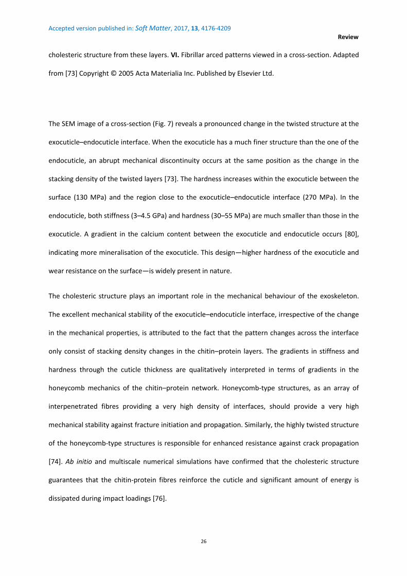
Fig. 6. The exoskeleton of Homarus americanus. The hierarchical organisation reveals six main levels.
From the bottom to top and from the left to the right: I. Chitin molecules, whose antiparallel
alignment forms rod-like -chitin crystals. II. The arrangement of 18–25 chitin molecules as
crystalline units that are wrapped around by proteins, forming nanofibrils of approximately 2–5 nm
diameter and 300 nm length. III. Clustering of these nanofibrils into long fibres of approximately 50–
300 nm diameter. IV. Formation of a planar network of these clusters. The spacing between the
strands is filled with proteins and minerals (calcite or amorphous calcium carbonate in crab
exoskeletons). The chitin–protein layers form flat honeycomb-type arrays. V. Formation of
chitin molecules
N-acetyl-D-glucosamine
chitin-protein crystals as nanofibrils
nanofibril clusters
network of nanofibril clusterscholesteric structure
twisted plywood model with arcedpatterns at oblique incidence
Accepted version published in: Soft Matter, 2017, 13, 4176-4209
Review
26
cholesteric structure from these layers. VI. Fibrillar arced patterns viewed in a cross-section. Adapted
from [73] Copyright © 2005 Acta Materialia Inc. Published by Elsevier Ltd.
The SEM image of a cross-section (Fig. 7) reveals a pronounced change in the twisted structure at the
exocuticle–endocuticle interface. When the exocuticle has a much finer structure than the one of the
endocuticle, an abrupt mechanical discontinuity occurs at the same position as the change in the
stacking density of the twisted layers [73]. The hardness increases within the exocuticle between the
surface (130 MPa) and the region close to the exocuticle–endocuticle interface (270 MPa). In the
endocuticle, both stiffness (3–4.5 GPa) and hardness (30–55 MPa) are much smaller than those in the
exocuticle. A gradient in the calcium content between the exocuticle and endocuticle occurs [80],
indicating more mineralisation of the exocuticle. This design—higher hardness of the exocuticle and
wear resistance on the surface—is widely present in nature.
The cholesteric structure plays an important role in the mechanical behaviour of the exoskeleton.
The excellent mechanical stability of the exocuticle–endocuticle interface, irrespective of the change
in the mechanical properties, is attributed to the fact that the pattern changes across the interface
only consist of stacking density changes in the chitin–protein layers. The gradients in stiffness and
hardness through the cuticle thickness are qualitatively interpreted in terms of gradients in the
honeycomb mechanics of the chitin–protein network. Honeycomb-type structures, as an array of
interpenetrated fibres providing a very high density of interfaces, should provide a very high
mechanical stability against fracture initiation and propagation. Similarly, the highly twisted structure
of the honeycomb-type structures is responsible for enhanced resistance against crack propagation
[74]. Ab initio and multiscale numerical simulations have confirmed that the cholesteric structure
guarantees that the chitin-protein fibres reinforce the cuticle and significant amount of energy is
dissipated during impact loadings [76].

Accepted version published in: Soft Matter, 2017, 13, 4176-4209
Review
27
Fig. 7. SEM micrograph of the cross-section of Homarus americanus exoskeleton at the interface
between the exocuticle and endocuticle, where a change occurs in the periodicity of the fingerprint
texture and in its granular aspect. In the fingerprint texture, the distance between lines of equal
contrast is related to the half-pitch and the helical axis is perpendicular to all the lines. [73] Copyright
© 2005 Acta Materialia Inc. Published by Elsevier Ltd.
The mechanical properties in the longitudinal and transverse directions were measured in the sheep
crab exoskeleton [80]. When tensile loading is applied in the transverse direction, a fracture tends to
occur at the exocuticle–endocuticle interface with a flat cleavage appearance. When the endocuticle
alone is solicited, the fracture morphology is irregular and the stress–strain curve shows a non-linear
(irreversible) plastic deformation.
The hammer-like dactyl clubs of the stomatopod Odontodactylus scyllarus, who are known to smash
its heavily shelled prey with high accelerations, exhibits several microstructural characteristics that

Accepted version published in: Soft Matter, 2017, 13, 4176-4209
Review
28
permit the infliction of crippling impacts while minimising the internal damage [81]; a pitch-graded
CLC structure dissipates the energy released by microcracks which propagate. By combining the
propagator matrix formalism to represent the CLC structure with the Bloch–Floquet periodic
boundary conditions to account for the graded pitch (from several m to several tens of m), a
dispersive response with bandgaps corresponding to propagation modes related to shear waves was
identified [82]. Strikingly, these frequency bandgaps correspond to those generated from the stress
pulse experienced by the dactyl club during the stomatopod’s hunting activities.
In addition to possible inelastic and damage effects, the dactyl club can sustain high-intensity
dynamical loads through a shear wave filtering capability introduced by the pitch–gradient CLC
structure.
Cuttlebone, the sophisticated buoyancy device of cuttlefish, is made of superposed chambers with a
complex internal arrangement of calcified pillars and organic membranes. In order to assemble the
membranes, Sepia officinalis secretes a chitin–protein complex, which self-organises, ‘layer’ by
‘layer’, as a CLC [83]. It is argued that the twisted plywood is the solution to a mechanical adaptation
for a better performance against the hydrostatic load. CLC organisation appears to be ubiquitous
within coleoid cephalopods. Arced patterns have been observed in the pen of the squid Loligo [84] or
in the septum (thin wall between the internal chambers of the shell of a cephalopod) of Spirula
spirula [83].
In order to explain the formation of the interlamellar membranes of molluscan nacre [85], the CLC
organisation is invoked. This constructional strategy could be traced even further back to at least the
Upper Ordovician when the first nautiloid nacre was recorded (445 million of years).
Computational modelling by the finite difference time domain method provides an accurate
prediction of the optical response in structure-gradient LC structures [86]. Complex pitch gradients in
the cuticles have been studied using a geometric model and computational visualisation tool [41,87].

Accepted version published in: Soft Matter, 2017, 13, 4176-4209
Review
29
In summary, chitin-based materials in crustaceans or molluscs are anisotropic in structure and the
mechanical properties have a recurrent role of the CLC organisation, which may often exhibit a pitch
gradient.
5.2. Optical information
The role of optical information is of paramount importance in the evolution of most day-living
animals [88]. Animals use coloration for sexual communication, cryptic behaviour or warning
predators [89], relaying information about the species [90] or age when iridescence changes or
deterioration occurs over time [91,92]. Iridescent coloration produces spectacular visual displays [93-
99]. The term iridescence is derived from Iris, the Greek goddess associated with the rainbow. It is
currently used to describe metallic colours, spectral light dispersion and opal-like effects. In fact,
iridescence is fundamentally related to a surface whose hue changes with the angle of view or
illumination.
In 1911, Michelson discovered that the scarab beetle Plusiotis resplendens displayed CP reflection
[100]. In 1924, Gaubert correlated it with the CP reflections in synthetic CLCs [101]. However, this
topic of research was revived much later in the 1960s [102-104].
The cholesteric structures in living matter display unique optical signatures with selective light
reflection property, vivid hues, strong angle-dependency of reflected colours and polarisation-
dependent reflection patterns. In nature, the ‘tour de force’ of cholesteric structures consists in
producing a broad set of dramatic lighting effects from biological materials whose refractive indices
vary in a narrow range; this behaviour is attributed to the helicoidal geometry and not the
composition.
Single-pitch or graded-pitch cholesteric reflectors in insect cuticles are iridescent. However, some
beetles do not have this characteristic. For silver metallic beetles, the cholesteric structure reflects a
broader spectrum than the visible one; therefore, the human eye cannot detect this iridescence. For

Accepted version published in: Soft Matter, 2017, 13, 4176-4209
Review
30
narrow-band reflectors, a diffuse reflection may arise from the irregularities in the cuticle (bumps,
pits and indentations) resulting in the matte or dull appearance of the cuticle. The armour of insects
plays a major role in their huge diversity (Fig. 8), especially for beetles because they have a thicker
cuticle than most other insects [105]. There are several publications which review the iridescent
colours in the insect world [106-110].
Fig. 8. Iridescent beetles. (a) Loxandrus rectus (Carabidae: Harpalinae), (b) Phalacridae sp., (c)
Cicindela scutellaris scutellaris (Carabidae: Cicindelinae), (d) Amarygminae sp. (Tenebrionidae), (e)
Phanaeus vindex (Scarabaeidae: Phanainae), (f) Eupholus sp. (Curculionidae: Entiminae). [108]
Copyright © 2009, © 2008 The Royal Society.
5.2.1. Camouflage—Beetles from the genus Chrysina, also named jewel scarabs, show very vivid light
reflections in a variety of colours, from bright green to metallic silver–gold, including broadband

Accepted version published in: Soft Matter, 2017, 13, 4176-4209
Review
31
reflections [111-113]. For camouflage strategies, the organisms that reflect light similarly to a
specular broadband mirror would match their background from any angle of observation; in other
words, the colour of the broadband reflectors is less directionally dependent when a full range of
wavelengths is reflected. In addition, the surrounding environment would be reflected from the
mirrored surface so that the animal cannot be seen [105]. Camouflage may also be achieved in an
environment with diffuse light to prevent a strong, direct reflection from the sun [114]. One of the
most spectacular Chrysina beetles is Chrysina gloriosa (Fig. 9.a), which is found from southwestern
North America to Central America in pine, pine-oak and juniper forests. C. gloriosa exhibits
iridescence and selectively reflects the left CP light. Its exoskeleton includes alternate green and
silver (mirror-like) stripes.

Accepted version published in: Soft Matter, 2017, 13, 4176-4209
Review
32
Fig. 9. (a) Chrysina gloriosa beetle. Its chitin-based cuticle is made of alternating green and silver
stripes. (b) Optical micrograph (unpolarised light) at the interface between the green (top–polygonal
texture) and silver (bottom) stripes. (c) Cholesteric fingerprint texture, as observed by scanning
electron microscopy, in the cross-section of a green stripe. [115] © 2016 Acta Materialia Inc.
Published by Elsevier Ltd
Since the foliage of the juniper tree is green with white flecks (resin spots), green-and-white (or
silver)-striped insects are relatively cryptic in the juniper foliage with their frequent natural perch
[116]. C. gloriosa belongs to a large group of insects with a tessellated exocuticle, in whole or in part,
exhibiting an array of cells in the form of bumps, pits or indentations. Since the mid-2000s, several
studies on these insects, such as Manuka beetles [117], June beetles [118], Plusiotis boucardi [119],
Pyronata festiva [120], Chlorophila obscuripennis [121], several beetles from the genus Cicindela
[108,122], Calidea panaethiopica [123], Chrysochroa fulgidissima [124], Chrysina aurora [125] and
Anomala dimidata [97], have been undertaken. In the field of microlenses and micromirrors [126-
128] and in photonics [95,129], micro-textured exocuticles of these scarabs may inspire researchers
and engineers to make their replicas as optical materials. The polygonal texture of C. gloriosa cuticle
was compared to its equivalent in synthetic cholesteric oligomers and the fundamental differences
were studied [115]. The cuticle has concave cells, whereas the artificial films have convex cells, which
is contrary to expectation and assumption in the literature.
The bright glare from silver stripes might temporarily blind a potential predator, enabling C. gloriosa
beetle to escape [130]. Since the silver stripes mirror the colour of its environment, C. gloriosa beetle
may hide among the leaves when they are brown during the dry season. However, if dealing with a
generic function, this hypothesis is not entirely satisfactory to other authors, e.g. Thomas et al. [116]
observed that gold and silver species are found in evergreen tropical cloud forests.

Accepted version published in: Soft Matter, 2017, 13, 4176-4209
Review
33
The green stripes have a polygonal texture (Fig. 9.b), and each polygonal cell contains a bright yellow
core in a greenish cell with yellowish border according to bright field unpolarised microscopy. The
reflectance exhibits, at a normal incidence, a broad halo from 500 to 600 nm with two peaks at 530
nm (green) and 580 nm (yellow) [111]. This kind of diffuse reflection, when an incident ray is
reflected at many angles rather than at just one angle as in the case of specular reflection, arises
from a combination of a continuous pitch gradient and irregularities on the cuticle surface below the
flat wax layer (Fig. 9.c). Below the epicuticle (waxy layer), the cholesteric organisation presents two
successive pitch gradients occurring in the IR spectrum in the endocuticle and in the visible spectrum
in the exocuticle, producing the structural colour. The fingerprint texture in the endocuticle
appears—as observed in the case of the Homarus americanus exoskeleton (Fig. 7)—to be less regular
(stripes are interrupted) and grainy. The presence of a cholesteric grating with a large pitch might
avoid overheating. This kind of thermal regulation could be offered to (many) arthropods (such as
insects or beach-dwelling crabs), whose cuticle presents a pitch gradient in the IR range [72]. Dorsal
colouration is associated with thermoregulation in diurnal beetles. Niche differentiation associated
with thermoregulation is well documented in tiger beetles (Ref. [131] and references therein).
According to Pace, the TEM images of C. gloriosa cuticle showed a cellular pattern with concentric
rings [132], which is similar to that found in the tubercle of crabs [133]. Fluorescence confocal
microscopy images confirmed these patterns [134,135]. The polarising properties of C. gloriosa
cuticle in the visible spectrum were investigated by using Mueller matrix spectroscopic ellipsometry
[136,137]. Over the visible and near-IR spectra, the polygonal cells in the tessellated green stripes
behave as multi-wavelength selective micromirrors [115]; these cells can be used in the future for
fabricating multi-wavelength selective micromirrors or spatial wavelength-specific light modulators.
5.2.2. Communication—Humans have a trichromatic vision, while insects have a tetrachromatic or
pentachromatic vision, depending on the species [138]. Their eyes and brains are able to perceive
polarised light, which we humans cannot [139]. The ability of C. gloriosa to detect CP light has been

Accepted version published in: Soft Matter, 2017, 13, 4176-4209
Review
34
evidenced [140]. C. gloriosa presents different flight orientations in accordance with linear and CP
light stimuli of equal intensities and shows a discrimination between CP light and unpolarised light of
different intensities. Circular polarisation sensitivity might allow C. gloriosa to communicate with
conspecifics while remaining cryptic to predators with eye geometries not capable of CP light
detection. Although linearly polarised light is quite common in nature, the production of CP light
from unpolarised light appears limited to only a few groups of organisms including scarab beetles
(Scarabaeidae, predominantly in subfamilies Rutelinae, Scarabaeinae and Cetoniinae [108]) and
marine stomatopods [141,142]. It is still unknown whether the scarab beetles use the CP light as a
recognition mechanism or for performing any other survival function. However, the rarity of CP light
in nature presents itself as an opportunity for unique signal evolution. Perceiving polarised light
might play a role in animal signalling, serve as a selective communication channel to specific
polarisation-sensitive targets such as potential mates and can be used for navigation and orientation.
Another possible biological advantage of CP light for communication is that there is no preferential
angle from which polarisation can be detected; animals can then send and receive signals regardless
of their respective orientations (while the directionality of linearly polarised light represents a
challenge; the orientation of the sender and receiver may have to be coordinated). In other cases,
cholesteric colours may be brightest from certain directions and the body orientation could direct a
visual signal to particular receivers [143].
The CP light reflected by the cuticle of scarab beetles is LH. However, RH circular polarisations have
been reported. They are very rare and restricted to single individuals such as C. resplendens in the
Rutelinae [100,111,144,145]. Furthermore, Hegedüs et al. found examples of RH circular
polarisations, but concluded that the situation was complicated [112]. Neville and Caveney reported
that LH CP light was reflected by all the investigated species [104]. There is a mixed support to the
observation of RH CP reflections. Therefore, it is difficult to conclude anything about RH CP light from
different discussions found in the literature. Polarisation may occasionally occur in patterns (patches,

Accepted version published in: Soft Matter, 2017, 13, 4176-4209
Review
35
stripes or edgings) and RH circular polarisations have been reported for a peculiar wavelength band;
this discrepancy makes it difficult to draw any firm conclusions. The problem of spectrum-dependent
handedness deserves detailed studies, requires elaborate equipment and suggests reproducing
experiments on samples from not only same scarab species but also different climes. Thus, the
research on the role of polarisation in light reflection by insect cuticles is still in its nascent phases.
The angle dependence of the cholesteric colour of the insect cuticle can slow or hinder the
recognition process by predators or make the signal ineffective if predators approach from the
directions relative to the cuticle surface where there is no apparent reflectance [146]. A high visual
contrast may also confuse the predator leading to the escape of the prey [107]. Aposematism, a
name given to antipredator adaptations, is commonly known in the context of warning colouration
that may deter predators from attacking distasteful or toxic prey [147], which may sometimes be a
deception. Although birds can exert selection on the dorsal colouration of tiger beetles [148], dorsal
colours appear less significant for escaping to terrestrial predators because these beetles avoid
lizards by running or flying. Despite the long history of interest in warning colouration, many
questions remain unanswered regarding the tactical design features of warning signals that
contribute to their effectiveness.
5.2.3. Evolutionary context—The complete role of light reflection by the cholesteric cuticle of insects
still needs to be elucidated; better knowledge of physical properties and an evolutionary context is
needed. The colour can be changed only by changing the pitch (or layer thickness if no twist) and not
by changing of material; therefore, the selective pressure can be considered as lower in return. The
cuticle of larvae is normally deposited in a cholesteric state [149,150]; therefore, polarised light
reflection is simply brought in the same time. With regards to the evolution of visual function in any
event, the way to evolve is to retain the twist from the larval cuticle formation in the adult cuticle,
possibly leading to the evolution of cholesteric reflectors.

Accepted version published in: Soft Matter, 2017, 13, 4176-4209
Review
36
The earliest known reflectors (diffraction gratings) in living matter are 515 million years (Ma) old
[114]. The subsequent fossil record preserves multilayer reflectors, including cholesteric mirrors,
because structural colouration is generally permanent and does not bleach like pigmental colours.
Fossil beetle specimens ranging from 15–47 Ma in age exhibit several reflection colours [151] (Fig.
10). Reconstruction of the original colours of the fossils based on the structure of the reflector show
that the preserved colours correspond to longer wavelengths; however, the conditions of
fossilisation of these samples and their preservation must be exceptional. These fossilised beetles
should have been rapidly buried by sediments before the breakdown by bacterias, and they should
be preferably preserved in a fine-grained sediment [88].
Fig. 10. Fossil of a 40 million-years-old leaf beetle (Chrysomelidae) found in Eckfeld (Germany), with
blue elytra that are still reflective. [151] Copyright © 2012, This journal is © 2011 The Royal Society.
Fossil records show that eyes evolved around 521 Ma ago [88]. The so-called ‘Light Switch Theory’
states that the introduction of vision to the behavioural system of animals caused the Cambrian
explosion. This event introduced vision as a selection pressure in the evolution of animals, which led
to the evolution of adapted optical devices. The evolution of the eye and probably active predators

Accepted version published in: Soft Matter, 2017, 13, 4176-4209
Review
37
can explain why animals suddenly changed their appearance in the Lower Cambrian. Cambrian
animals would have possessed efficient and coloured (cholesteric) reflectors to be visually adapted.
5.3. Concluding remarks
The cholesteric structure appears to play an important role in the evolution and the daily life of
crustaceans and insects, with a benefit of mechanical strength and optical properties. The dispatch of
cholesteric information including vivid reflection colours and polarisation, with angle dependence,
possibly helps the animal for social signalling (recognition with conspecifics), sexual selection,
aposematism (camouflage for avoiding predation) and thermoregulation. However, the cholesteric
structure is not the only factor responsible for the structural colouration in insect cuticles [152]. The
chitin fibres may assemble without twist like chirped layers with a nematic structure (parallel fibres)
or as orthogonal plywood [153]. Such organisations may lead to an interferential network or a
diffraction grating. Layers may sometimes be porous and filled with air or liquid (hydrochromic
effects) [154].
In some beetles, uric acid plays a role in imparting the colour by enhancing the reflectivity [107]. In
Plusiotis optima and Plusiotis resplendens, uric acid accounts for 0.6 and 0.7 volume fraction of the
exocuticle, respectively [155]. Chitin-based structures can also be combined with pigments or light-
emitting compounds; absorption colouration and bioluminescence are possible. Nevertheless,
structural colouration has recently received more attention in the literature.
When evaluating the proposed function of a colour, it is important to analyse the visual system of the
animal and its potential predators, the light conditions and the optical characteristics of the habitat
(ground, foliage or sea). The knowledge of the phylogenetic and ecological context is also required to
elucidate the role of cholesteric structures in an animal. For example, the environment plays a major
role in the colour polymorphism exhibited by the southern African dung beetle, Gymnopleurus
humanus [156]. The population samples of G. humanus were dominated by blue individuals in the

Accepted version published in: Soft Matter, 2017, 13, 4176-4209
Review
38
cooler south, by cupreous individuals in the warmer north, and by locally co-occurring blue, green
and cupreous individuals in intermediate situations. The differences in mean reflectance values
between 24 populations were strongly correlated with the average annual temperatures at the study
sites. The adaptability of physical CLC properties to thermal conditions can generate different
exocuticle structures responsible for different light reflections. Colour polymorphism could be
advantageous across a gradient from cooler to warmer climate due to the different thermal
properties of different colours. Interestingly, it is hypothesised that the prevailing reflected colour
balance in southern populations would shift in response to global climatic change. In this situation
also, it could be argued that colour changes are easily accessible by changing the cholesteric pitch
without the requirement of changing the material.
For a given cholesteric structure, it is difficult to affirm that the evolution has endowed the animal
with optical features, increased mechanical strength or reduced weight. In some cases the
cholesteric structure could be an artefact. It can be debated that the structural colouration may be
an incidental consequence of the evolution of a periodic structure [157]. At the same time, a huge
amount of literature directly addresses the critical importance of colouration in living organisms. The
evolution of a colour-producing structure may be driven by several factors simultaneously, and the
consequence would be the occurrence of a multifunctional material responding to several
requirements, which may or may not be optimal for them. For example, the optimisation of both
mechanical and optical functions may lead to a conflict.
In general, bio-inspired fabrication from cholesteric cuticles of insects can be applied in textiles, in
cosmetics, for vehicle shells, in anti-counterfeiting technologies for banknote production, in
microphotonics and as optical components for multifunctional materials [107].

Accepted version published in: Soft Matter, 2017, 13, 4176-4209
Review
39
6. Collagen
6.1. Liquid crystalline nature of collagen
Collagen accounts for one-third of the total body proteins and is present in almost all tissues [158]. It
is the major part of the skin, bone, tendon and cornea, where collagen macromolecules organise into
fibrillary networks. Type I collagen is predominant in connective tissues and plays a major role in
morphogenesis. Type I collagen macromolecules are composed of three polypeptide chains that wind
up into an RH triple helix (1.5 nm in diameter and 300 nm in length) which is known as tropocollagen
(Fig. 11). They are first synthesised in a precursor form by cells as fibroblasts, the most common cells
of connective tissues, or osteoblasts, the cells with a single nucleus that synthesise bone.
Subsequently, they aggregate into individual fibrils in the extracellular matrix.
Fig. 11. (a) Hierarchical organisation of bone. Collagen fibrils are made from type I collagen
macromolecules (~300 nm long and ~1.5 nm in diameter) and hydroxyapatite (HA) Ca10(PO4)6(OH)2
nanocrystals (plate-shaped, 50 nm × 25 nm in size and 1.5–4 nm thick) are distributed along the
collagen fibrils. Compact bone is composed of osteons. Each osteon consists of lamellae arranged
into coaxial cylinders and surround a central canal, the Haversian canal, which contains blood vessels.
Lamellae consist of parallel collagen fibrils, whose orientation varies for one cylinder to the next. This
geometrical construction results in a helical arrangement of the osteon. Therefore, the twisted

Accepted version published in: Soft Matter, 2017, 13, 4176-4209
Review
40
plywood is cylindrical and not planar. Macroscale arrangements involve both compact bone at the
surface and spongy bone (foam-like material with ~100-μm thick struts) in the interior [159].
Copyright © 2014, Rights Managed by Nature Publishing Group. (b) The SEM image of a transverse
fracture of the human compact bone. Each osteon appears to be formed by concentric lamellae
around the Haversian canal (bar = 100 m) [160]. Copyright © 2003 Elsevier Science Ltd.
Collagen has essential functions at different levels, ranging from molecules to fibrils and suprafibrillar
networks [161]. Tissue properties such as elasticity in the dermis, resistance to load in the bone,
resistance to stretch or shear in tendons or transparency and resistance to pressure in the cornea
depend on the molecular composition, size and the arrangement of fibrils and organisation of the
fibrillary network [162]. As an implantable material, collagen is of major interest because it is highly
biocompatible and biodegradable, shows low immunogenicity, favours cell adhesion and guides the
cell growth and tissue repair [163].
Collagen macromolecules assemble into fibrils both in vitro (by raising the pH) and in vivo (through
enzymatic control). The passage from individual fibrils to ordered fibrillar arrays relies on the self-
assembly processes driven by the LC organisation of collagen. The LC organisation of collagen has
received much attention because it plays an important role in determining tissue form [164-169] and
can orient the cell growth [170].
In compact bone osteons or in the cornea, a continuous twist occurs between individual collagen
fibrils or between groups of fibrils [40,171,172]. Highly uniform CLC collagen films are capable of
inducing contact guidance for osteoblasts, which are aligned with the orientation of bands in the
cholesteric texture [161]. In the tendons, intervertebral disc or intestinal wall, crimp morphologies, in
which a regular and planar undulation of fibrils occurs, resemble a precholesteric phase [173,174].
Molecular orientation in a precholesteric arrangement is oblique to the helical axis, whereas it is
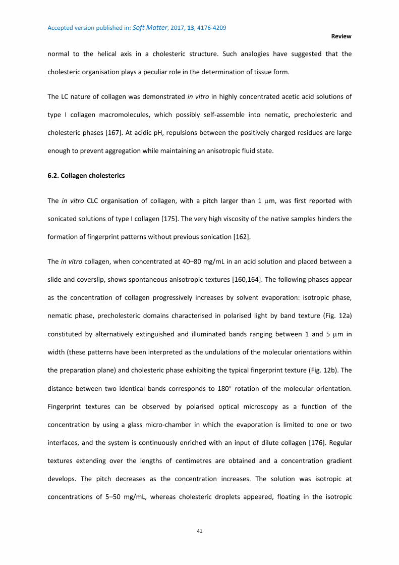
Accepted version published in: Soft Matter, 2017, 13, 4176-4209
Review
41
normal to the helical axis in a cholesteric structure. Such analogies have suggested that the
cholesteric organisation plays a peculiar role in the determination of tissue form.
The LC nature of collagen was demonstrated in vitro in highly concentrated acetic acid solutions of
type I collagen macromolecules, which possibly self-assemble into nematic, precholesteric and
cholesteric phases [167]. At acidic pH, repulsions between the positively charged residues are large
enough to prevent aggregation while maintaining an anisotropic fluid state.
6.2. Collagen cholesterics
The in vitro CLC organisation of collagen, with a pitch larger than 1 m, was first reported with
sonicated solutions of type I collagen [175]. The very high viscosity of the native samples hinders the
formation of fingerprint patterns without previous sonication [162].
The in vitro collagen, when concentrated at 40–80 mg/mL in an acid solution and placed between a
slide and coverslip, shows spontaneous anisotropic textures [160,164]. The following phases appear
as the concentration of collagen progressively increases by solvent evaporation: isotropic phase,
nematic phase, precholesteric domains characterised in polarised light by band texture (Fig. 12a)
constituted by alternatively extinguished and illuminated bands ranging between 1 and 5 m in
width (these patterns have been interpreted as the undulations of the molecular orientations within
the preparation plane) and cholesteric phase exhibiting the typical fingerprint texture (Fig. 12b). The
distance between two identical bands corresponds to 180 rotation of the molecular orientation.
Fingerprint textures can be observed by polarised optical microscopy as a function of the
concentration by using a glass micro-chamber in which the evaporation is limited to one or two
interfaces, and the system is continuously enriched with an input of dilute collagen [176]. Regular
textures extending over the lengths of centimetres are obtained and a concentration gradient
develops. The pitch decreases as the concentration increases. The solution was isotropic at
concentrations of 5–50 mg/mL, whereas cholesteric droplets appeared, floating in the isotropic

Accepted version published in: Soft Matter, 2017, 13, 4176-4209
Review
42
phase, at higher concentrations, i.e., 50–400 mg/mL, with long helical pitches. At concentrations of
400–800 mg/mL or more, the solutions were cholesteric, showing a much smaller helical pitch and so
tight in some areas that the helical periodicity was not resolved by optical microscopy. To stabilise
these solutions into gels, fibrillogenesis was induced by ammonia vapours. The images showed
similarities with those obtained with thin sections of natural collagen matrices, for instance, in a
compact bone. In the bone, the collagen fibrils showed a relatively constant diameter from 30 to 120
nm, whereas the fibril diameter varied from 10 nm in the highly twisted regions to 500 nm in less
twisted regions in these ammoniacal gels of collagen.
Fig. 12. Liquid crystal textures of collagen observed by polarised light microscopy between crossed
polariser and analyser (P and A). (a) Band texture in a precholesteric phase. Molecular directions
undulate in the section plane and extinction occurs when they lie parallel to one of the polarisers.
Bar: 10 m. (b) Cholesteric fingerprint texture. Molecular directions regularly rotate along a direction
normal to the lines. Bar: 10 m. [160] Copyright © 2003 Elsevier Science Ltd.
Thermodynamics, texture, structure and rheology of cholesteric collagen I solutions are discussed in
Ref. [177].
6.2.1. Bone—Bone comprises organic and inorganic components in an advanced composite form
that confers remarkable ability to withstand a high mechanical loading. Moreover, it comprises
approximately 30% organics and 70% minerals by dry weight [178]. The toughness and stiffness are
bestowed by the organic and mineral phases, respectively. The former is a matrix of proteins—

Accepted version published in: Soft Matter, 2017, 13, 4176-4209
Review
43
mainly collagen—and other macromolecules including proteoglycans that are rich in acidic
glycosaminoglycans (which polysaccharides dominate the organic–mineral interfaces) [179]. The
structure of a compact bone consists of adjacent cylindrical units of about 0.2 mm in diameter called
osteons (Fig. 11). Osteons are formed by concentric lamellae disposed around the Haversian canal
containing blood vessels. The lamellae consist of collagen fibrils moving helically around the central
canal with regular changes in the orientation of the fibrils.
Similar to the fish scales (discussed below), the bone is thus a collagen–mineral composite. Its
strength comes primarily from its ability to absorb energy at small length scales through the mineral
and protein phases; its anisotropic organisation at higher structural levels allows the development of
the specific mechanical properties for peculiar functions. Two main osteon organizations may be
found in a compact bone [42,171]. The first organization conforms to the classical description of
osteons following an orthogonal (or a quasi-orthogonal) plywood model, i.e. with two principal
orientations of fibrils. The second organization conforms to the twisted plywood model, where fibrils
follow small and regular changes in their orientation; in this case, the structure is continuous. The
passage between the orthogonal and twisted lamellae may be observed within the same osteon,
with the arced patterns corresponding to the most recently secreted levels [171]. It is suggested that
the twisted plywood appears when the collagen network is first produced by the cells, as it is always
present in the newly secreted layers. The mechanical constraints further modulate the fibrillar
arrangement before stabilisation of the collagen network.
Within the same context as that of the arthropod carapaces (Section 5.1), a plywood structure that is
continuously twisted or exhibiting two directions of the preferred alignment is a resistant material
[180]. The plywood structure may confer an enhanced resistance to the bone against fracture
initiation and propagation in one or several directions. It provides several advantages over a perfect
alignment: It increases the crack-extension energy per area, transports energy into a higher volume
and leads to a more ductile fracture behaviour of the bone tissues [181]. It was reported that the

Accepted version published in: Soft Matter, 2017, 13, 4176-4209
Review
44
fracture energy changes by two orders of magnitude depending on the collagen orientation; and the
angle between the collagen orientation and the crack propagation direction is decisive in switching
between different toughening mechanisms [181]. The structure–property relationship in the bone
plywood is an evolving subject that requires supplementary investigations to be fully understood. A
similar remark might be made for dentin (one of the four major components of teeth) whose
toughness comes from its structure close to that of bone consisting of a cholesteric matrix of highly
mineralised collagen fibrils [159].
Understanding how different structural features, acting at several length scales, interact
simultaneously to generate the mechanical properties and biological responses is still a work in
progress. Scaglione et al. [182] compared the in vivo cellular response with two osteoconductive
materials that maximise the differences in the internal structure while displaying similar chemical
compositions (a highly porous hydroxyapatite foam and a hydroxyapatite–collagen composite
sponge). The pattern of the new bone tissue, deposited by bone-marrow-derived cells loaded on the
two scaffolds, was assessed through a combined multimodal 3D optical microscope. Whenever a
regular and geometrically ordered scaffold is present to the cells as a template for tissue
regeneration, the lamellar bone tissue is formed and a cholesteric organisation of collagen fibrils is
observed. A cholesteric scaffold design may contribute to drive properly the collagen fibre assembly
to target in vivo bone regeneration [183,184].
6.2.2. Tendon—A tendon connects muscle to bone and is essentially bundles of collagen fibres. A
balance of repulsive and attractive pair interactions between collagen macromolecules determines
the formation and swelling of fibres, which plays a crucial role in the tendon mechanics [67]. The
twisted (rope like) organisation of tendons and ligaments provides them with the capacity to
withstand large forces without stretching more than a few percent of their length making possible
the movement. Cholesteric geometries are not clearly visible in the tendon. However, an unusual
periodic change in the pattern of the bundles of collagen fibres occurs in tendons, which has been

Accepted version published in: Soft Matter, 2017, 13, 4176-4209
Review
45
explained by a planar crimp of the collagen fibrils along the tendon axis [158,185] (Fig. 13). This
pattern allows for the extension of tendinous structures without the extension of collagen fibrils. This
undulation does not occur in bone collagen, which is often subjected to compressive forces, but it
occurs in the tissues that are subjected to tensile forces such as the tendon. The fact whether or not
the crimp in the tendon might originate from similar mechanisms as those inducing the in vitro
formation of the banded texture is not demonstrated [177].
Fig. 13. The crimp patterns of collagen fibrils in a section of the rattail. Bar: 10 m. [186] Copyright ©
2008 Elsevier Ltd.
6.2.3. Cornea—The structure of collagen in the cornea is especially complex. It exhibits discontinuous
rotation angles between the adjacent collagen bundles and a twist at a higher range [186]. The last
part of the cornea consists of helicoidal layers of collagen fibres in fishes (carp and goldfish),
amphibians (frogs), reptiles (turtle) and birds (domestic fowl, quail, pheasant and duck) [187]. The
cholesteric structure, by providing equal stiffness in all directions, likely contributes to make and
maintain, with the aid of intraocular pressure, a spherical shape to the cornea and thus reducing
spherical aberrations. The corneal lenses are helicoidally constructed in the bird’s (and insect’s)
corneas with the same sense of rotation in both eyes [188]. This means that the construction of

Accepted version published in: Soft Matter, 2017, 13, 4176-4209
Review
46
fibrils in the left and right eyes is asymmetrical with respect to the bilateral symmetry of the body.
Could this situation have any significance for the navigation and homing abilities, perhaps by the use
of polarised light from the sky? There is no firm answer to this question.
In the dense solutions of collagen in 5 mM acetic acid (pH 3.5), the fibrils may assemble in a complex
cornea-like organisation coupling together the smectic and cholesteric organisations [189]. This
result is explained by the propensity of fibrils, which are in a repulsive regime, to form a cholesteric
phase and an achiral (smectic or hexagonal) phase at the same time. The abrupt rotations between
the different domains of fibres might result from such a frustration. A similar mechanism might exist
in vivo. These results are of interest in the field of biomimetic corneal matrices.
6.2.4. Fish armour—The organisation of the collagen network in fish scales and scutes exhibits a
diversity of the plywood structures: orthogonal, twisted, or double-twisted plywood (i.e. the
orthogonal plywood where every second layer, instead of being strictly parallel to the first one,
rotates by a small angle in a given sense so that the whole geometry is twisted) and many
intermediate situations, in between discontinuous and continuous structures, depending on the
species or the region under consideration in the fish armour. A series of arced patterns have been
observed by the optical or electron microscopy in the fish scales such as coelacanths [40,190],
freshwater carp Cyprinus carpio communis [191], goldfish Carassius auratus [192,193] and in the
dermal scutes of boxfish [194] (arc patterns were not recovered by Yang et al. [195]). In the latter
case two cholesteric organisations were described: one with continuously rotating fibrils and the
other with discrete steps of rotation [194]. The two geometric systems were separated by a 3D
collagen lattice reminiscent to the array of double-twist cylinders in chiral LC phases called blue
phases [55, 196]. The cholesteric organisation can be evidenced by using small-angle X-ray scattering
(SAXS), the same used for the teleost fish scales [197].
The cholesteric structure may specifically offer resistant armour against predators. Arapaima gigas is
a freshwater fish found in the Amazon Basin, which can resist the appetite of piranhas through the
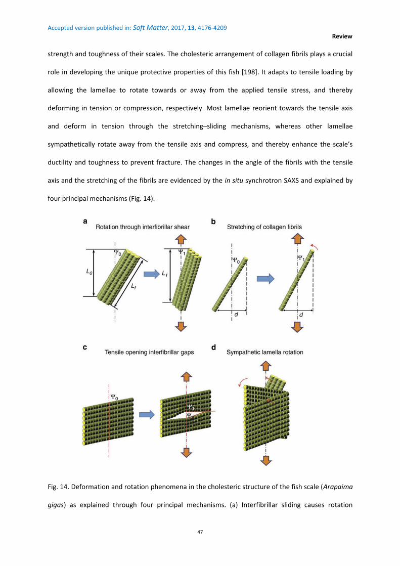
Accepted version published in: Soft Matter, 2017, 13, 4176-4209
Review
47
strength and toughness of their scales. The cholesteric arrangement of collagen fibrils plays a crucial
role in developing the unique protective properties of this fish [198]. It adapts to tensile loading by
allowing the lamellae to rotate towards or away from the applied tensile stress, and thereby
deforming in tension or compression, respectively. Most lamellae reorient towards the tensile axis
and deform in tension through the stretching–sliding mechanisms, whereas other lamellae
sympathetically rotate away from the tensile axis and compress, and thereby enhance the scale’s
ductility and toughness to prevent fracture. The changes in the angle of the fibrils with the tensile
axis and the stretching of the fibrils are evidenced by the in situ synchrotron SAXS and explained by
four principal mechanisms (Fig. 14).
Fig. 14. Deformation and rotation phenomena in the cholesteric structure of the fish scale (Arapaima
gigas) as explained through four principal mechanisms. (a) Interfibrillar sliding causes rotation

Accepted version published in: Soft Matter, 2017, 13, 4176-4209
Review
48
towards the tensile axis, where fibrils at an original orientation from the tensile axis of 0 reorient to
1 with a corresponding increase in the projected length from L0 before loading to L1 under loading
with respect to the fibril length, Lf. (b) The stretching of the lamellae could also cause the fibrils to
rotate towards the tensile axis from 0 to 1, whereas the projected length d is assumed to remain
unchanged. Other mechanisms could be responsible for rotation away from the tensile axis. (c)
Tensile separation of fibrils at an initial orientation of 0 could force some fibrils to reorient towards
the tensile axis (2), whereas other fibrils rotate away from them (1). (d) Sympathetic rotation of
lamellae could occur, where lamellae rotating towards the tensile axis force other lamellae to
reorient away from the tension. [198] Copyright © 2013, Rights Managed by Nature Publishing
Group.
The light-reflecting structures of fishes are useful to communicate the information on relative
positions, orientations and movements between neighbours, especially in a school of fish [199,200].
For example, the silvered patches on the tail or the pectoral fins may become much brighter or
darker with movement, and the colours of the ventral flanks may change from bright red to blue with
the viewing direction. Similarly to that happens in the insect world (Sections 5.2.1 and 5.2.2), it may
be assumed that the optical properties unique to the cholesteric organisation (not found in non-
continuously twisted plywood structure)—the exceptionally large optical activity and the
polarisation–selectivity rule—might provide unique benefits to a fish for camouflage (the silvery
armour plays an important role in open water camouflage; broadband mirrors provide camouflage to
fishes taking advantage of diffuse light conditions [201]) and communication and navigation inside a
school of fish. The investigations on the specific role of cholesteric colours versus colours from achiral
periodic structures like a simple stacking of platelets [202] are in a preliminary stage, in the world of
sea animals.

Accepted version published in: Soft Matter, 2017, 13, 4176-4209
Review
49
6.3. Outlook
The exact mechanisms that determine the tissue architecture in a compact bone, skin or cornea are
still major open problems in morphogenesis. In vivo, the cholesteric organisation of fibrils in compact
bone osteons and, in tendons, the crimp morphology—which is akin to a precholesteric phase—
suggest that CLC organisation plays a crucial role in the determination of the tissue form. In vitro,
collagen macromolecules in acid solution form nematic, precholesteric and cholesteric phases, but
the relevance of it to the in vivo assembly mechanisms is unclear.
Many indirect cues suggest that an intermediate cholesteric phase could exist prior to fibril self-
assembly (fibrillogenesis) and could, therefore, constitute a basic morphogenetic principle of the
collagen patterning in tissues [167,186]. For example, cells produce collagen within the closed
compartments (in the cell vesicles or the extracellular matrix) at very high concentrations, and the
volume fraction of collagen in tissues and collagen LC phases is very similar. Advantageously the
‘cholesteric-driven control’ of fibrillogenesis could not require orchestrated cell movement but high
cell density and high collagen concentration [203]. In the concentrated solutions of procollagen
(precursor molecules of collagen), precholesteric arrangements are obtained at physiological pH and
ionic strength in the form of large domains exhibiting regular precholesteric cords, but typical
cholesteric textures are not observed.
The mechanism of the in vivo formation of collagen fibrils, the fixation of their diameter, the
relationship between their compositions and the functional requirements in different tissues are still
open questions. The differences in the long-scale organisation of collagen—twisted in bone, crimpy
in tendon and orthogonal in cornea—are difficult to explain.
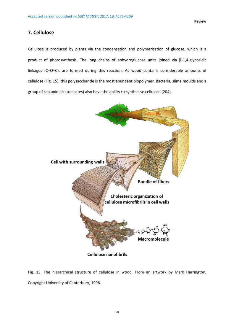
Accepted version published in: Soft Matter, 2017, 13, 4176-4209
Review
50
7. Cellulose
Cellulose is produced by plants via the condensation and polymerisation of glucose, which is a
product of photosynthesis. The long chains of anhydroglucose units joined via -1,4-glycosidic
linkages (C–O–C), are formed during this reaction. As wood contains considerable amounts of
cellulose (Fig. 15), this polysaccharide is the most abundant biopolymer. Bacteria, slime moulds and a
group of sea animals (tunicates) also have the ability to synthesise cellulose [204].
Fig. 15. The hierarchical structure of cellulose in wood. From an artwork by Mark Harrington,
Copyright University of Canterbury, 1996.

Accepted version published in: Soft Matter, 2017, 13, 4176-4209
Review
51
7.1. In situ cholesterics
7.1.1. The plant cell wall—In contrast to the animal cells, the plant cells possess a wall, which acts
like an extracellular compartment (box) surrounding the cell. The thickness of plant cell walls
generally lies in the range of 0.1–0.3 μm and can exceed 1 μm [205]. The walls form a continuous
network through the whole plant. The growth and form are controlled by the oriented expansion of
the walls. The driving force for cell expansion is osmotic. The cells grow by turgor-driven
enlargement. The primary task of the wall is to provide a skeletal support to the plant. The wall
shape depends on the types of cells. For example, the compression wood cells of spruce are cylinders
of the circular or elliptical cross-section, whereas normal wood cells of spruce are often cylinders
with super-elliptical cross-section [49]. The cell wall also plays a dominant role in cell morphogenesis,
disease resistance, cell recognition and signalling, digestibility by herbivores and humans, and plant
decay [206].
The wall is made of sequentially deposited layers of different thicknesses, chemical compositions and
structural organisations. There are two types of walls: primary and secondary, which differ in
function and in composition. The primary wall is the first layer to be laid down after the cell division
and its formation coincides with the cell growth. It provides mechanical strength (it has to resist the
internal turgor pressure of the cell) and must expand to allow the cell to grow and divide. It behaves
like a fibrous composite (‘fibreglass’) with an ordered array of stiff cellulose microfibrils embedded in
a less stiff matrix of polysaccharides and glycoproteins (hemicellulose, pectin and other
macromolecules) (Fig. 16). Once the cell has ceased to grow, three layers of the secondary wall are
laid over the primary wall in an inward direction. The central layer, called secondary layer S2,
comprises about 80–90% (of the entire cell wall volume). The layers of the secondary wall are much
thicker and stronger than the primary wall; they contain lignin. Lignification follows the organisation
of microfibrils in the primary walls and provokes the hardening of the secondary walls. The secondary
walls are not found in all cell types.

Accepted version published in: Soft Matter, 2017, 13, 4176-4209
Review
52
Cellulose microfibrils are composed of approximately 36 hydrogen-bonded chains and each chain
contains 500–14 000 glucose units. These cable-like structures are laid down by apposition on the
inner surface of the growing wall, whereas pectin, hemicellulose and glycoproteins are secreted via
the Golgi apparatus into the network of microfibrils.
Fig. 16. Plant cell wall structure. The plasma membrane forms the innermost layer surrounding a cell.
The primary wall is outside it. Cellulose microfibrils are the major components. They are represented
by cylinders that, in this particular case, lie on the top of one another in a crisscross fashion. The
middle lamella forms the outermost layer. It contains many pectin molecules and soluble proteins
similar to those in the primary cell wall. Lignin is not shown here. Source: Wikimedia Commons
(adapted from M. B. Sticklen, Nature Reviews Genetics, 2008, 9, 433–443).
Microfibrils may self-assemble in the cell wall with different kinds of organisations: axial (nematic-
like), transverse, crisscrossed, so-called helical, and helicoidal (cholesteric-like) [207] (Fig. 17a). The
cholesteric organisation (Fig. 17b) can be found both in the primary and secondary walls [206]. The
walls may exhibit arc patterns that deviate from the monotonic helicoidal plywood as depicted in Fig.

Accepted version published in: Soft Matter, 2017, 13, 4176-4209
Review
53
2, revealing different CLC structures with the graded pitch, random pitch, discontinued pitch, etc.
[49], from which architectures could generate complex optical responses.
Fig. 17. (a) Basic organisations of cellulose microfibrils in the cell wall. The successive wall layers have
been peeled off from the top to bottom to reveal the internal structure. (b) The focus is on the
principal geometry of the helicoidal deposition of cellulose microfibrils. For representation purposes,
the deposition is described as tapes that wind around a cylinder. A small number of microfibrils are
drawn at a highly expanded scale. Four generations of microfibrils are spun. Each of them contributes
locally to a single ‘layer’ in the cholesteric structure. The inset shows that the angle of deposition
between the adjacent layers is nearly constant, which is the hallmark of the cholesteric organisation.
[207] Copyright © 2000 Elsevier Science Ltd.
The growth mechanisms of plant cells are not fully described. More information on the control of
microfibrils orientation, inter alia, is required [208].

Accepted version published in: Soft Matter, 2017, 13, 4176-4209
Review
54
7.1.2. Early work—The cholesteric organisation is distributed throughout the plant taxa [209] and
found in a range of cell types: spores, collenchyma, sclerenchyma, tracheids, sclereids and
parenchyma [210-213]. In many supporting cells (sclerenchyma and wood cells), the CLC structure
appears only temporarily; the process, which is inherent to growth, is irreversible and its regulation is
determinant for the cell physiology [211].
The readers, for further information on in situ cholesteric cellulose reported till 1990, may refer to
the must-read book “Biology of fibrous composites” by Neville [46]. The cholesteric cellulose
concerns lower plants, such as mosses, horsetails, ferns, fungi and lichens (cholesteric chitin can also
be found in cell walls of fungi and lichen), algae, coniferous gymnosperms, flowering plants, such as
angiosperms including monocotyledons and dicotyledons (quince, hazel, pear, prune, cherry, walnut
and olive), bacteria, cyanobacteria, and tunicates (Halocynthia papillosa). Neville's monography
includes several tables classifying cholesteric structures found in different groups, genera and
species, with references. For example, and beyond cellulose, Tables 2.1–2.6 [214] list arthropods
other than insects and crustaceans (Scorpionida, Araneida), butterfly and moth eggshells,
invertebrates excluding arthropodan phyla (Protista, Rotifera, Nematoda, Echinoderma and
Mollusca), fishes and amphibia (eggshell or corneal collagen), reptiles (eggshell or corneal collagen),
birds (corneal collagen), etc.
To visualise the cholesteric patterns, dedicated techniques for the preparation of specimens and
electron microscopy are required; and, as mentioned by Neville, these patterns may remain
undetected if the observation conditions are not correct [43].
The CLC structure provides better resistance to the plant in multiple directions against environmental
stresses such as the wind [209]. Similarly to the case of chitin-based and collagen-based biomaterials,
resistance to fracture is increased because the path of any crack is diverted by the change in
orientation, which results in an increased capacity to dissipate energy [215]. Therefore, the CLC
structure combines a balance of strength in all directions with flexibility. The cholesteric wall

Accepted version published in: Soft Matter, 2017, 13, 4176-4209
Review
55
structure resists the turgor pressure equally in different directions [216], and similarly, the
cholesteric collagen in the cornea of lower vertebrates resists the pressure of fluid within the eye
(Section 6.2.3).
7.1.3. Iridescence in plants—Structural colour in general has been mainly studied in the animal
kingdom, and largely ignored in plants [217], where research has focused on the analysis of pigments
and the subtractive colouring they produce. Multilayer interferences, thin-film interferences, grating
diffraction, scattering or light reflection by photonic crystals like cholesteric LCs may also be at the
origin of colouration in plants [218,219].
Iridescent plants are generally found in the shaded forests and at tropical latitudes. Iridescence
found in leaves, fruits and petals can span to different colours of spectra that are perceptible to a
variety of animals, including UV [217] and the visible range for humans [220]. The structures
employed to achieve iridescent colours may (i) make plants attractive to pollinators by leaves and
flowers and, by fruits, to birds and mammals, which then disperse the seeds (display function); e.g.
the bumblebees can use iridescence as a cue to detect flowers [221,222]; (ii) deter herbivores
(defence function), the changeability of colour with the viewing angle could corrupt the object
identity, making it harder for insects to form a search image [223], and colouration could even be
visible only to certain species of insects [224]; (iii) help in light management with a crucial role either
in photoprotection or by focusing light on the chloroplasts (Ref. [225], and references therein), and
the plant surface topography not only affects the visual appearance but also controls the amount of
light coupled into the lower lying tissue [226] with the following consequences: the increased
capture of photosynthetically important wavelengths and polarisation filtering that enhances the
orientation of the photosynthetic apparatus within the cell and protects the shade-adapted plants
against sun flecks. A diverse range of photosynthetic organisms from diatoms to algae and then to
angiosperms produce a vivid iridescence over their surface, which suggests that iridescence has
evolved multiple times and has some adaptive advantages for life in deep shade [223].

Accepted version published in: Soft Matter, 2017, 13, 4176-4209
Review
56
The recent publications on the physical properties of the in situ cholesteric cellulose are much rarer
than those on cholesteric chitin. We devote this section to structural colour since most of them have
remarkably focused on this subject.
7.1.4. Leaves—The leaves of Selaginella willdenowii produce UV–blue light iridescence. The
underlying mechanism arises from the multilayer interference of two or more materials with
different refractive indices [223,227]. The iridescence of Selaginella species might aid the capture of
photosynthetically active wavelengths in low light conditions with the leaf iridescence acting as an
anti-reflective coating. This coating would produce constructive interferences for certain
wavelengths (by increasing the transmission of them), and other (non-transmitted) wavelengths
would be strongly reflected because of destructive interferences [217].
The UV–blue colouration arising from a CLC structure is found in the leaves of Malaysian iridescent
understory tropical fern species such as Danaea nodosa [225,228] (Fig. 18), Lindsaea lucida and
Diplazium tomentosum [224,225,228,229], where the cholesteric organisation is absent in the older
leaves, which show no iridescence [217], and in the flowering plants such as Phyllagathis rotundifolia
[225,229].
Plant leaves are expected to be green because the chlorophyll pigments absorb visible light but little
in the green range. The leaf colour other than green shows that wavelengths captured by chlorophyll
are absorbed or reflected by other pigments. Consequently, less energy is captured for the
photosynthesis and growth. Why blue plants living in a dark environment reflect light that could
contribute to the photosynthetic efficiency [230]? The selectivity of light reflection by a cholesteric
structure might provide photo-protection because these plants, adapted to low light conditions,
suffer damages whenever encounter sunflecks. Some iridescent blue leaves may recover significantly
more rapidly from light exposure than non-iridescent green leaves [217]. However, no difference was
found between the iridescent and non-iridescent leaves of D. tomentosum [225].

Accepted version published in: Soft Matter, 2017, 13, 4176-4209
Review
57
Fig. 18. (a) The photograph of juvenile (blue) and adult (green) leaves of D. nodosa. The adult leaves
have lost iridescence. (b) The cholesteric structure as shown in a TEM cross-sectional view of the cell
wall of a juvenile leaf. Reproduced with permission from D. W. Lee.
The Malaysian tropical rainforest understorey herb Mapania caudata (Fig. 19a and b) provides an
unusual case of the iridescent cholesteric cellulose-enriched with silica nanoparticles [231]. Granular
silica nanoparticles are organised inside the graded-pitch cholesteric organisation of microfibrils (Fig.
19c and d) and contribute to the whole structure producing the blue iridescence. The diffuse
reflection peak in the visible spectrum is quite broad from 400 to 700 nm. The removal of silica from
the walls eliminates the blue colour. Two partially overlapping periodicities might account for the
blue structural colouration: the helicoidal periodicity of microfibrils and the periodicity for the
layered aggregation of nanoparticles. The cholesteric colouration involving silica might not be limited
to M. caudata. In addition to the cholesteric fingerprint texturation of platinum [232,233] and gold
[234] nanoparticles in synthetic cholesteric oligomers, this study remarkably presents the
simultaneous nucleation and fingerprint patterning of bio-nanoparticles in a biopolymer.

Accepted version published in: Soft Matter, 2017, 13, 4176-4209
Review
58
Fig. 19. Self-organisation of nanoparticles into the cholesteric cellulose of Mapania caudata leaves.
(a) The intense blue–green iridescent colour from the edge of leaf to midrib. (b) Light reflected by the
surface of the leaf reveals the iridescence of individual epidermal cells. (c) A TEM image of the
transverse cell wall showing the cholesteric fingerprint texture and a thicker granular layer near the
surface. (d) Enlarged image showing the layering of granular nanoparticles inside the fingerprint
texture near the surface. Reproduced with permission from D. W. Lee.
7.1.5. Fruits—The primary function of fruits and flower iridescence is likely to attract animals,
particularly to those species whose visual systems are attuned to iridescence in the context of
animal–animal communication [217].
The shiny, metallic, blue colour of the fruits of Pollia condensata (Fig. 20.a), an African forest
understory species, arises from the cholesteric cellulose [97,235]. The epicarp (outermost skin)
exhibits cellular motifs under a microscope. The pixelated iridescence of the fruit, with purple, blue,

Accepted version published in: Soft Matter, 2017, 13, 4176-4209
Review
59
green and red speckles, is revealed by epi-illumination using circular polarisation filters (Fig. 20b and
c). The TEM transverse section image of epicarp shows arc patterns (Fig. 20.d). The colour difference
from cell to cell corresponds to different pitches. Each cell shows colour in a specific polarisation
channel (Fig. 20.e), i.e. a specific twist handedness. P. condensata is a parental biological version of
synthetic cholesteric gels in which RH and LH helicity senses coexist [236,237]. The property of
double helicity, increasing the reflected intensity at a given wavelength in the Bragg band, added to
the existence of different colours makes the P. condensata fruit the most brilliant land-based
biological organism to date.
Fig. 20. (a) Pollia condensata fruit. The diameter is about 5 mm. (b, c) Optical micrographs in epi-
illumination (unpolarised incident light) of the single area of epicarp by using left-handed (b) and
right-handed (c) circular polarisers. The pointillist colouration originates from individually coloured
cellular motifs. (d) Cholesteric structure is revealed in a cross-sectional TEM view of the epicarp; the
pink line enhances an arced pattern. (e) Reflection spectra for two different cells (continuous and
dotted lines) collected by the left- and right-handed circular polarisation channels. [97] Copyright ©
2014 The Authors. Published by Elsevier Ltd.

Accepted version published in: Soft Matter, 2017, 13, 4176-4209
Review
60
Although more than 50-year old, the fruit shown in Fig. 20a appears like a fresh one. By mimicking
the appearance of a fresh nutritious fruit, some plants like P. condensata may have evolved to
mislead their seed dispersers, without offering them any nutritious reward [238]. This strategy could
avoid the energy cost of producing the fresh pulp. The structural colours, in contrast to the pigmental
colours, give to the fruit a brilliant and intense appearance that is maintained even after it falls from
the plant, increasing the probability of attracting an animal. Alternatively to the promise of fresh
fruits, P. condensata might also attract birds for the research of shiny objects to decorate nests or
arenas to attract potential mates [239].
7.1.6. Mechanism of the helicoidal formation of the cell wall—In a study by Murugesan and Rey
[215], a model based on the Landau-de Gennes theory was used to simulate the dynamic self-
assembly of a lyotropic CLC and to understand the mechanism of the helicoidal formation of the cell
wall. This mechanism is still a matter of debate [240,241]. (i) One hypothesis is that the cellulose self-
assembly is guided by hemicellulose [209,242-244]. Cellulose is crystalline, strong and resistant to
hydrolysis, whereas hemicellulose is amorphous, shows weak strength and may be hydrolysed. The
unbranched homopolymers like cellulose crystallise with backbone chains closely packed to form
parallel crystallites. The branched heteropolymers like hemicellulose show a pipe-cleaner structure:
flexible side chains (which can contain different sugar monomers such as xylose, mannose, galactose,
rhamnose and arabinose) are attached to a stiff backbone that may be composed of glucose (as in
xyloglucans) or xylose (as in gluronoxylans). Such a structure offers the possibility to have rotatory
movements in hemicellulose molecules, not in cellulose molecules, and to adopt a twisted
organisation due to their chirality (the presence of asymmetric carbons). Since hemicellulose
molecules are hydrogen bonded to the periphery of cellulose microfibrils (Fig. 16), they may entrain
cellulose microfibrils into the CLC structure. In this model, the handedness of the material is
determined by the intrinsic chirality of the molecules that compose the cell wall. (ii) In the other

Accepted version published in: Soft Matter, 2017, 13, 4176-4209
Review
61
model, the helicoidal structure of cellulose microfibrils may be determined by the rotation of
microtubules in the underlying cell [241,245]. Chan [237] focused on the relationship between the
growth and the orientation of microtubules and cellulose microfibrils. The microtubules provide a
template for cellulose-synthesising particles that glide along the plasma membrane extruding
cellulose microfibrils into the wall [246]. Microtubules can change the position during the cell growth
and rotate in both the clockwise and anticlockwise directions. The mechanism that determines the
direction of the microtubule rotation is unknown. It could lead to both the LH and RH CLC structures
because the helicoidal arrangement would be independent of the chirality of the microfibrils.
According to Vignolini et al. [235], this mechanism could explain why both helicity senses coexist in P.
condensata. However, with this scenario, no change would happen in the direction of microtubule
rotation during the growth of a single cellular motif. More clarification is expected on the origin of
cholesteric domains with both helicities in P. condensata.
7.1.7. Outlook— Is the production of colour a primary function of a periodic and chiral structure or a
by-product of a structure that performs a different (non-optical) function? The same question was
also raised for chitin and collagen. However, limited information is available about the
developmental mechanisms and the genetic controls used by plants or animals in producing
cholesteric structures.
Light plays a major role in the plant life. The columns of cells in plant seedlings (oats, corn, mung
bean) can act like a bundle of fibre optics [247]. Laser experiments have shown that plants can
transmit light up to the tips of root hairs even when they are bent. Do cholesteric cell walls play a
role in the internal light reflection? In the plant fibre optics, the light may coordinate various aspects
of the physiology of the growing plants.
Although much is known about the chemistry of plant cell walls, a lot yet to be unravelled about the
control of their morphology. The relationship between the cholesteric organisation and other
components requires to be further exemplified.

Accepted version published in: Soft Matter, 2017, 13, 4176-4209
Review
62
Structural colours are probably quite frequent in the plant kingdom but we have only a fraction of
the knowledge about their distribution, development and function. The reason why not so many
plant tissues with cholesteric-based coloration are known, in contrast to insects, might simply be that
nobody has yet looked for them [224]. Many iridescent plants might be awaiting an exploration [248]
that could reveal novel mechanisms of colour production. A multitude of plants for which the
structural origin of the colouration effects has not yet been identified or characterised has been
compiled by Gebeshuber and Lee [218]. The enormous morphological diversity of more than 300 000
angiosperm species has to be kept in mind. It demands a mutual interaction between the scientists
belonging to different horizons to understand the significance of chiro-optical phenomena in plants.
As Kutschera wrote the cholesteric organisation of cellulose looks like an example of ‘complex design
in biological systems without an intelligent designer’ [244]. More experimental work is required to
support further this concept of the generation of ordered structures from disordered cable-like
biopolymers in the extracellular compartments of living organisms like plants.
7.2. Ex situ cholesterics
A number of review papers have compiled the information available on the structures, properties
and applications of cellulose-based nanomaterials [240,249-258]. In this section, our prime attention
is on the cholesteric structures artificially produced from natural cellulose nanocrystals (CNCs). Being
promising optical materials and templates [259,260], they resulted an avalanche of publications from
several research groups, in particular from 2003–2004.
7.2.1. Nanocrystals suspensions—CNCs are produced by the sulphuric acid based hydrolysis of
natural cellulose such as wood or cotton pulp, bacteria and tunicates ([249] and references therein).
A cellulose microfibril contains both amorphous and crystalline parts. The acid degrades the
amorphous regions, leaving CNCs as rod-like particles. CNCs were also referred to as cellulose
microcrystals (in the early literature), nanocrystalline cellulose and cellulose whiskers (or

Accepted version published in: Soft Matter, 2017, 13, 4176-4209
Review
63
nanowhiskers). CNCs resemble whiskers because of the tapering at the ends of the crystals
consequent to the hydrolysis. Another consequence is that the surface of CNCs becomes negatively
charged by the sulphate groups, which serve both for the stabilisation of colloidal suspensions of
CNCs and the formation of a CLC phase [261]. CNCs have a high aspect ratio—typically 3–50 nm wide
and 50–1000 nm long [262]. Their dimensions depend on the origin of cellulose. Cotton CNCs have
typical dimensions of 100 to 300 nm in length and 8 to 10 nm in diameter, while tunicate CNCs range
from 100 nm to few micrometers in the length and 10 to 20 nm in diameter [263].
At very low concentrations, CNCs randomly suspend in water and form an isotropic phase. A nematic
or cholesteric organisation of CNCs above a critical concentration is the result of favourable excluded
volume interactions leading to higher packing entropy than in the isotropic phase [264]. The CLC
phase is the most commonly observed LC phase in the CNC suspensions. In 1992, Revol, Derek G.
Gray et al. discovered that CNCs (from wood pulp, cotton pulp, ramie, bacteria) in aqueous
suspension might form CLC phases [261]. The cholesteric structure of the suspensions can be
preserved by the slow evaporation of water, which means that solid iridescent films with optical
properties similar to CLCs can be prepared. The final structure strongly depends on the drying
conditions [261,265,266].
Fig. 21.a shows the fingerprint texture of a eucalyptus suspension [267]. The pitch decreases with an
increasing cellulose concentration and the rod length. At the same volume fraction than the short
rods, the long rods tend to align in a nematic state instead of a cholesteric state. Fig. 21.b shows the
fingerprint texture of a bacterial suspension for comparison [268]. A monodomain texture is
promoted. This situation is due to a larger aspect ratio, the rectangular shape of the cross-section of
bacterial CNCs (typically 54 nm × 10 nm vs. 5 nm × 5 nm for wood), and a lower surface charge
density.

Accepted version published in: Soft Matter, 2017, 13, 4176-4209
Review
64
Fig. 21. Cholesteric fingerprint texture of cellulose nanocrystals from: (a) Eucalyptus in suspension in
water (cellulose concentration = 10 wt.%) [267]. Copyright © 2005, American Chemical Society. (b)
Bacteria in suspension in water (cellulose concentration = 3 wt.%) [268]. Copyright © 2009, American
Chemical Society.
Although the case of synthetic cellulose is beyond the scope of this review, it has to be noted that
CLC phases appear in concentrated solutions of many cellulose derivatives such as ethyl cellulose,
hydroxypropylcellulose (HPC), acetoxypropylcellulose, and other esters of propyl cellulose
(propanoate, butanoate, benzoate) [269]. Both LH and RH structures have been observed [269], but
to date only LH CLCs have been observed for CNC suspensions. Much higher concentrations of
cellulosic derivative are needed to form the CLC phase in comparison to native CNCs (typically 7 wt.%
for CNCs vs. 50–75 wt.% for HPC and 40 wt.% for ethyl cellulose) [270,271].
7.2.2. Pitch tuning—The size, shape, dispersity or charge of CNCs, type of electrolyte, conditions of
film preparation and external stimuli can affect liquid crystallinity, pitch and domain size.
Pitch can vary from less than 1 m to 50 m (and even beyond) and depends on the shape
anisotropy of the CNCs, polydispersity in length and width [272,273]—longer CNCs give longer pitch
[265,268]—, concentration that drives the minimisation of excluded volume [273], ionic strength of
the CNCs–solvent suspension which regulates the charges on CNCs [265,275,276], drying

Accepted version published in: Soft Matter, 2017, 13, 4176-4209
Review
65
temperature [276] and application of a shear alignment [277]. The glass formation after the
quenching (drying) is a non-equilibrium process, which is delicate to control, thus strong pitch
variations may occur (Fig. 22) in the quenched state with time and from one region to another. A
competition between glass formation and LC self-assembly governs the formation of the targeted
helical structure. Both processes are promoted by the high aspect ratio of CNC rods. The
fundamental issues related to this sensitive balance have yet to be fully understood; they have been
discussed by Lagerwall et al. [278]. The kinetic arrest, leading to the glass formation, in the later
stages of evaporation is of importance while producing uniform films and reproducibly controlling
the pitch [279].
Fig. 22. Dry cholesteric film in a Petri dish formed from a 4 wt.% eucalyptus CNCs–water suspension.
The rainbow-like variation of colour is related to the pitch gradient. It comes from the gradient in the
concentration and thickness consequent to the slow drying at an ambient temperature. Both CNC
concentration and film thickness decrease from the edge of the dish towards the centre. When the
water has completely evaporated the colour variation is locked into a solid film [273]. Creative
Commons Attribution (CC-BY).
The CNCs extracted from filter paper [275], wood pulp [267] and bacterial cellulose [268] showed a
decrease in the pitch in suspensions with an increasing CNC concentration. Although the origin of

Accepted version published in: Soft Matter, 2017, 13, 4176-4209
Review
66
twist in lyotropic CLCs [280] is still debateable, it is generally accepted that the proximity between
chiral particles decreases the pitch, whereas an increasing inter-particle distance increases it. The
film colour can be shifted toward shorter wavelengths by increasing the concentration prior to film
casting [265] or toward longer wavelengths by the ultrasonication of the suspension [281]. The
added electrolyte partially masks the negative charges of the sulphate ester groups, reducing inter-
particle electrostatic repulsions and shortening the pitch in a predictable manner.
Vacuum-assisted drying can play some role in the film colour [282]. The films that are prepared at
ambient conditions generally show a polydomain structure but applying a magnetic field during the
drying can induce homogeneity in the size and orientation of domains [266,283]. The drying of
suspension with the control of time and in the presence of 0.2 T magnetic field increases the pitch
[284]. The colour can be changed by applying electric field [285] and could also be affected by
humidity, so the films might be used as moisture sensors [286,287].
7.2.3. Origin of twist and other questions to be answered—The CLC phase formation in CNC
suspensions can be explained by the phase equilibrium theories related to colloidal LCs such as the
Onsager theory [264] or its extended version—the Stroobants, Lekkerkerker, and Odijk (SLO) theory
[288]. However, the explanations for this formation remain speculative, as the electrostatically-
stabilised CNC suspensions contain only pure water and crystallites with charged surface groups. The
question is, 'What is the source of chiral interactions?' On the basis of in situ small angle neutron
scattering measurements in CNC suspensions under magnetic field and shear alignment, Orts et al.
[277] confirmed that CNCs are screw-like rods. Araki and Kuga [289] showed that the CNCs from
bacterial cellulose (produced by Acetobacter xylinum in Nata de coco gel) form a nematic phase in an
electrolyte-free suspension and a CLC phase consecutive to the addition of a trace electrolyte (<1
mM NaCl). This phenomenon was explained by morphological changes from a plain cylindrical
configuration to a twisted rod, an outcome of surface charge masking (Fig. 23). In conditions of
extreme deionisation, the charge double-layer may alter the screw-like character of the crystalloids

Accepted version published in: Soft Matter, 2017, 13, 4176-4209
Review
67
from some sources of cellulose and lead to the formation of a nematic phase. The authors concluded
that the twisted particle geometry is the most likely mechanism of the formation of the CLC phase. In
other words, the chiral interactions would arise from the shape of the rods and not from the chiral
nature of cellulose chains. It has to be kept in mind that the films of CNCs bear chirality on three
different scales: on the molecular level because of the asymmetric carbon atoms of each D-glucose
unit; on the nanoscale because of the screw-shape morphology of the individual crystals; and on the
mesoscopic scale with the LH CLC structure. A chronological and detailed review of different
contributions about the origin of twist in cellulosic materials has been discussed by Conley et al.
[290].
Fig. 23. Schematic illustration of cellulose nanocrystals with surface charge showing the change in
effective particle shape. (a) In water, repulsion by surface charge extends to a long range. The
particle (nanocrystal) appears as an achiral rod. (b) The addition of NaCl decreases the repulsion
range and the particle appears as a twisted rod [289]. Copyright © 2001, American Chemical Society.
In addition to the controversies regarding the self-organisation of CNCs in aqueous suspensions, the
attribution of colour to Bragg's band has been challenged on the basis of observations of pitch that is

Accepted version published in: Soft Matter, 2017, 13, 4176-4209
Review
68
too long for the reflection to take place in the visible range [291]; and a surprising dependence of the
phase sequence and pitch variation with the ionic strength has been observed in some systems [268].
The open questions about the stumbling blocks in the progress in the field of CNCs-based materials
and their applications are discussed by Lagerwall et al. [278]. The exact role of factors influencing the
optical properties of CLC films prepared from CNC suspensions remains to be a qualitative one and
the interplay between the chiral interactions and kinetic factors during film formation is still poorly
understood [260].
7.2.4. Potential applications—Cellulose-based products are widespread but are still primarily used
for low-tech applications such as banknotes, information storage and packaging [292]. There is a
growing interest in applying cellulose to advanced technologies as it is a widely available and
renewable resource. The cholesteric design of cellulose may help in transforming it into an advanced
material. The solid films with the attractive optical properties of cholesteric structures render them
as promising candidates for novel applications [255,263,278,284,292] such as in anti-counterfeiting
components in security papers (banknotes, passports, certificates)—since optical features cannot be
reproduced by photocopying or usual printing—, polarising mirrors, mirrorless lasing lasers,
intelligent packaging materials, printed paper electronic devices, additives for paints, inks and
cosmetic products and optical encryption.
The films made from CNCs may be problematic in certain applications due to their inherent
susceptibility to re-dispersion in water, and it is yet challenging to produce uniform planar textures
leading to highly reflective films with high pitch reproducibility.
Accepted version published in: Soft Matter, 2017, 13, 4176-4209
Review
69
8. Viruses
There is a debate as to whether viruses can be considered as living organisms [293-295]. A virus
cannot grow or multiply on its own (it can replicate inside the living cells of other organisms) but at
the same time, it contains genes, which give it an ability to mutate and evolve.
The filamentous viruses like tobacco mosaic virus (TMV), M13 and fd are present as rod-like particles.
As for CNCs, the suspensions of rod-like viruses may exhibit LC phases. TMV was the first virus to be
isolated and purified [296]. Onsager showed theoretically that the anisotropic phase of TMV
suspensions is a nematic phase for purely entropic reasons [264].
Since the year 2000, the experimental studies on LC phases in virus suspensions have mainly focused
on fd suspensions [297]. Fd virus is a micrometer-length semi-flexible polyelectrolyte with a diameter
of 6.6 nm. It contains single stranded DNA helicoidally wrapped by proteins, making it a chiral
particle with a helical charge distribution. Isotropic, cholesteric [298], smectic A and smectic C [299]
phases are displayed by increasing rod concentration ([300] and references therein). A set of
characteristics, such as rich polymorphism including CLC phase, high monodispersity, surface
properties ([301] and references therein), and ease for technical investigations (e.g. optical
microscopy) due to the large size of fd particles, have made them highly valuable systems to explore
the complex relationship between the chirality of individual bricks and the chirality of their
self-assembly [302]. While TMV [296] and pf1 [297] viruses have a helical structure extremely similar
to fd, they exhibit a nematic phase. This behaviour is not understood. A reversible temperature-
driven transition between the cholesteric and nematic states is possible in M13 virus suspensions by
heating it above 40 °C and incubating for several days [303].
Various chemical modifications of fd virus capsid (protein shell of the virus) have shown that the
particle interactions due to surface charges, in addition to particle chirality, play an important role in
the origin of the twist [301]. When fd virus is grafted with the neutral polymer polyethylene glycol

Accepted version published in: Soft Matter, 2017, 13, 4176-4209
Review
70
(PEG), the fd-PEG suspensions yet exhibit a CLC phase, which means that chiral interactions between
the rods are not hampered [302]. Slight differences in the surface properties of rod-like viruses with
very similar molecular structures can strongly change the self-assembly behaviour.
The origin of twist in the cholesteric phase of virus suspensions is an open question.
9. Silk
Silk is produced by spiders, the larvae of Bombyx mori mulberry silkworm and numerous other
arthropods [304]. Silk fibre is mainly composed of fibroin proteins.
A CLC phase in fibroin was observed in the early duct portion of the major silk-producing gland in
Nephila clavipes spider and Bombyx mori [305]. The investigations were performed by cryogenically
quenching the animals during the spinning process. The morphology of the material within the silk
gland was studied by TEM, electron diffraction, and atomic force microscopy. Until this study, only a
nematic texture was reported in silk solutions [306,307], as the consequence of rod-like aggregates
of globular or coiled protein chains [307]. However, these ex situ observations may differ from the
structure present in the gland under natural spinning conditions, and a material in a cholesteric state
at equilibrium may untwist to a nematic state while flowing [308]. Fibroin may form chiral secondary
structures, which could explain the CLC organisation [305]. A band texture assigned to a CLC phase
was also observed in a film formed by an aqueous solution of pure fibroin prepared from Bombyx
mori cocoon at the interface with hexane [309].
Apart from these early investigations [305,309], there have been no further studies exploring
cholesteric silk. The discussion on the liquid crystallinity of silk has mainly focused on the nematic
state [310,311]. Owing to the liquid crystallinity of spinning dope (material from which silk is spun),
spiders can draw it during the extrusion into a hardened fibre using minimal forces [312]. Liquid
Accepted version published in: Soft Matter, 2017, 13, 4176-4209
Review
71
crystallinity reduces the fibre formation energy budget by minimising a viscous drag. This process
involves an unusual internal drawdown within the spider's spinneret followed by a conventional
external drawdown after the dope has left the spinneret. A better understanding of this process,
including the detailed LC behaviour and properties of silk, may help industrial production of silk-
based fibres with unique properties under benign conditions. Spider silk has indeed outstanding
mechanical properties while being spun at close to an ambient temperature and pressure, and simply
with water as solvent. Viscoelastic modelling of silk spinning process sheds some light on the textural
transitions in the duct of spiders and silkworms [313]. The concerted structuring action of capillary
confinement, strong anchoring, and nematic flow leads to the predictions that agree with
experimental textural transitions [314].
10. Cholesterol ester deposition in atherosclerosis
Atherosclerosis—the primary cause of heart disease and stroke—is characterised by the progressive
accumulation of lipids and fibrous plaques in large arteries [315]. The lipid composition of the normal
young aortic intima (the innermost layer of the aorta) mainly includes phospholipids. Free cholesterol
is also present but cholesterol esters are either absent or present in trace amounts [316]. During the
development of atherosclerosis, cholesterol esters become increasingly abundant. In the more
advanced cases, cholesterol and its esters predominate. The relationship between phospholipids,
cholesterol, cholesterol esters and water could be displayed by a quaternary phase diagram [316],
which helps define the states of lipids in relation to the environment of the intima. Cholesterol esters
of long chain fatty acids may exhibit at body temperature isotropic, cholesteric, lamellar (smectic-
like), and crystal phases [317-319], according to the type of esters and relative concentration.
The knowledge of LC phases in the lesions of arterial wall may allow predicting the possible
reversibility of atherosclerosis disease. The fluid lipid phases—isotropic phase and LC phases—may

Accepted version published in: Soft Matter, 2017, 13, 4176-4209
Review
72
be removed metabolically while the crystallised lipids may be more problematic and require surgery.
The reversibility of lipid lesions might depend partly on the lipid phases present and on the
composition of these phases. However, it remains unclear that how to promote isotropic and LC
phases and the stability of LC phases, at the detriment of crystalline deposits. How to promote the
presence of the CLC phase at the detriment of (more ordered) lamellar phases?
Except for a few studies in 1970s and 1980s, we could not find any recent studies on LC phases
involved in arterial diseases. It seems that little attention has been paid to the physical states of
cholesterol esters in last few decades.
11. Summary and concluding remarks
The cholesteric organisation is a recurring design in the animal and plant kingdoms. Its biological
importance suggests the convergent evolution of an optimised left-handed helicoidal structure.
Nucleic acids, polysaccharides, proteins and lipids have a predominant cholesteric organisation in the
living matter. These are four major types of molecules essential to life. Cholesteric organisation is
mainly found in:
DNA and chromatin;
polysaccharide fibres in polysaccharide matrix (plant cell wall) or in protein matrix (arthropod
cuticle);
protein fibres in polysaccharide matrix (bone) or in protein matrix (eggshells: fish, brine
shrimp, moth [320]).
The multifunctionality of biological CLCs is objectified by the combination of maximising packing
efficiency and reduction in the energy costs incurred in the processing, with mechanical and optical

Accepted version published in: Soft Matter, 2017, 13, 4176-4209
Review
73
properties. In summary, we gather and categorise the roles and possible functions brought by the
CLC structure:
Packing efficiency:
Cholesteric structure offers a high local DNA concentration, which is required for DNA to
function.
Cholesteric defects could correspond to a geometrical solution of a local overcrowding
consecutive to the replication of the DNA molecule.
Highly uniform cholesteric collagen films are capable of inducing contact guidance for
osteoblasts in bone.
Morphogenesis:
A cholesteric structure can exist prior to collagen fibrillogenesis and offer a basic
morphogenetic scaffold for collagen patterning in tissues. Cholesteric organisation of
collagen may play a peculiar role in the determination of tissue form.
A cholesteric scaffold may contribute to the properly driven collagen fibre assembly to
target in vivo bone regeneration.
Mechanical stability:
The cholesteric structure:
guarantees that the chitin–protein fibres reinforce the cuticle and is suitable to dissipate
significant amounts of energy during the impact loadings with the benefit of a reduced
weight; in cephalopods, the cholesteric structure looks like the solution to a mechanical
adaptation against the hydrostatic load;

Accepted version published in: Soft Matter, 2017, 13, 4176-4209
Review
74
adapts to tensile loading by allowing the ‘layers’ to rotate towards or away from the
applied tensile stress and prevents fracture and its propagation; cholesteric collagen lends to
sea animals a specifically resistant armour against predators;
provides the plant better resistance against environmental stresses, by combining a
balance of strength in all directions with flexibility;
may offer resistance of plant cell wall to turgor pressure equally in different directions.
Cholesteric collagen may offer resistance to:
load in bone: cholesteric structure may confer an enhanced resistance against fracture
initiation and propagation in one or several directions;
stretch or shear in tendons and ligaments: cholesteric organisation provides them with
the capacity to withstand large forces without stretching more than a few percent of their
length, making movement possible;
pressure in the cornea, by providing equal stiffness in all directions, which maintains the
spherical shape of the cornea and so reduces spherical aberrations.
Optical information:
Unique optical signatures of CLC structure may help:
animals in sexual communication and selection, social signalling (recognition with
conspecifics or intra-species communication), warning against predators, camouflage to
avoid predation, communication and navigation within a fish school;
plants to maintain attractiveness to animals (for iridescent fruits), since structural
colours, unlike the pigmental colours, do not fade with time (cf. cholesteric fossils of beetles)
or indicate aging while it happens that iridescence changes over time (in adult fern leaves).

Accepted version published in: Soft Matter, 2017, 13, 4176-4209
Review
75
Radiation protection:
IR thermoregulation for diurnal beetles and beach-dwelling crabs;
photo-protection against sun-flecks for plants adapted to low light conditions.
Evolutionary context: The biological CLCs offer a set of optical features using the materials
whose refractive indices vary in a narrow range. The reason, rather than in the composition,
lies in the helicoidal geometry. Since colour can be changed only by changing the pitch, the
selective pressure of evolution can be considered as lower in return.
Open questions have been mentioned and discussed in appropriate contexts throughout the article
and thus will not be repeated in this section. However, as concluding remarks, we would like to recall
a selective set of considerations of possible interest for current and future investigations. (i) It would
be of great interest to explore why a similar organisation is found in distant biological materials,
differing in their chemical composition, function and phylogenetic positions. Arced patterns are
found in several diverse tissues of animal and plant groups, mainly extracellular matrices, connective
and skeletal tissues, and in several intracellular differentiations. (ii) The CLC organisation is involved
in most physical aspects of biological morphogenesis with specific defects and textures whose
investigation has to be pursued in details at a small scale, probably with updated or novel imaging
techniques. The problem is the comprehension of the differentiation of biological CLCs as what
comes from liquid crystallinity of the initial secretion and what is controlled by cells. (iii) It might also
be interesting to make models of CLC structures for biomimicry. CLCs are rarely considered in applied
biology, although there are considerable perspectives. New mineralised biocomposites with a CLC
organisation of the organic part could serve as implants. The investigation of nucleation and growth
of mineral phases in the confined environment of matrices mimicking cholesteric organisations has
to be continued, which could give rise to interesting problems of physics in confined geometry with
inorganic–organic interfaces. Several issues for future investigations, of which some are relevant,
have been compiled elsewhere [72,162,180,321-326].

Accepted version published in: Soft Matter, 2017, 13, 4176-4209
Review
76
Acknowledgements
I gratefully acknowledge Prof. Derek G. Gray (McGill University, Montreal, Canada) and Prof.
Young-Hwa Goo (Albany Medical College, New York, USA) for fruitful discussions on cholesteric
cellulose and atherosclerosis, respectively.
References
[1] G. H. Brown and W. G. Shaw, Chem. Rev., 1957, 57, 1049-1157.
[2] G. W. Gray, Molecular Structure and Properties of Liquid Crystals, Academic Press, London and
New York, 1962.
[3] J. A. Castellano, Liquid Gold—The Story of Liquid Crystal Displays and the Creation of an
Industry, World Scientific, Singapore, 2005, p. 48.
[4] T. J. Sluckin, D. A. Dunmur, and H. Stegemeyer, Crystals that flow—Classic papers from the
history of liquid crystals, Taylor & Francis, 2004.
[5] D. Dunmur and T. Sluckin, Soap, science, & flat-screen TVs—A history of liquid crystals, Oxford
University Press, 2011.
[6] M. Mitov, ChemPhysChem, 2014, 15, 1245-1250.
[7] A. Figueiredo Neto and S. R. A. Salinas, The Physics of Lyotropic Liquid Crystals, Oxford Science
Publications, 2005.
[8] Thermotropic Liquid Crystals, ed. A. Ramamoorthy, Springer, New York, 2010.
[9] V. Luzzati, in Biological membranes, ed. D. Chapman, vol. 1, Academic Press, New York, 1968,
p. 71.
[10] T. A. Waigh ,P. Perry ,C. Riekel, M. J. Gidley and A. M. Donald, Macromol., 1998, 31, 7980–
7984.

Accepted version published in: Soft Matter, 2017, 13, 4176-4209
Review
77
[11] V. Grassert and V. Vill, Liq. Cryst. Today, 1994, 4, 4-5.
[12] O. Lehmann, Die neue welt der flüssigen kristalle, Akademische Verlagsgesellschaft m.b.H.,
Leipzig, 1911.
[13] Y. Bouligand, in Liquid Crystal Order in Polymers, ed. A. Blumstein, Academic Press, New York,
1978, pp. 269-274.
[14] D. W. L. Hukins and J. Woodhead-Galloway, Mol. Cryst. Liq. Cryst., 1977, 41, 33-39.
[15] I. Hamley, Soft Matter, 2010, 6, 1863-1871.
[16] F. Reinitzer, Liq. Cryst. 1989, 5, 7-18 (translation from the German of the original paper
published in Monatshefte für Chemie 1888, 9, 421-441).
[17] R. Virchow, Virchows Archiv., 1854, 6, 571.
[18] C. Mettenheimer, Corr. blatt d. Vereins gem. Arbeit z. Förd. d. wiss. Heilkunde, 1857, 24, 331.
[19] A. Trokhymchuk, Cond. Matt. Phys., 2010, 13, 37002: 1-4.
[20] E. Haeckl, in foreword of Crystal Souls–Studies of Inorganic Life (1917), translated by A. L.
Mackay, Forma, 1999, 14, 1-240.
[21] G. Friedel, Ann. de Phys., XVIII, 273-474, 1922.
[22] O. D. Lavrentovich and M. Kleman, in Chirality in Liquid Crystals, Eds. H.-S. Kitzerow & C. Bahr,
Springer, New York, 2001, pp. 115-158.
[23] R. Smith, D. R. Sabatino and T. J. Praisner, Exp. in Fluids, 2001, 30, 190.
[24] Y. Han, K. Pacheco, C. W. M. Bastiaansen, D. J. Broer and R. P. Sijbesma, J. Am. Chem. Soc.,
2010, 132, 2961-2967.
[25] J. Stasiek, A. Stasiek, M. Jewartowski and M. W. Collins, Optics & Laser Tech., 2006, 38, 243-
256.
[26] A. Saha, Y. Tanaka, Y. Han, C. M. W. Bastiaansen, D. J. Broer and R. P. Sijbesma, Chem.
Commun., 2012, 48, 4579-4581.
[27] H. Hirschmann and V. Reiffenrath, in Handbook of Liquid Crystals, Vol. 2A (Eds. D. Demus, J.
Goodby, G. W. Gray, H.-W. Spiess, V. Vill), Wiley-VCH, Weinheim, Germany, 1998, p. 199.

Accepted version published in: Soft Matter, 2017, 13, 4176-4209
Review
78
[28] N. Tamaoki, Adv. Mat., 2001, 13, 1135.
[29] J. Chen and C. T. Liu, IEEE Access, 2013, 1, 150-158.
[30] S.-T. Wu and D.-K. Yang, Reflective Liquid Crystal Displays, John Wiley & Sons Ltd., Chichester,
England, 2001.
[31] I. P. Ilchishin and E. A. Tikhonov, Progr. in Quant. Elec., 2015, 41, 1–22
[32] H. Khandelwal, R. C. G. M. Loonen, J. L. M. Hensen, M. G. Debije and A. P. H. J. Schenning,
Scientif. Reports, 2015, 5, 11773.
[33] L. Y. Zhang, Y. Z. Gao, P. Song, X. J. Wu, X. Yuan, B. F. He, X. W. Chen, W. Hu, R. W. Guo, H. J.
Ding, J. M. Xiao and H. Yang, Chin. Phys. B, 2016, 25, 096101.
[34] T. Engels and W. von Rybinski, J. of Mat. Chem., 1998, 8, 1313.
[35] D. Coates, Liq. Cryst., 2015, 42, 653-665.
[36] R. Balamurugan and J.-H. Liu, Reac. Func. Polym., 2016, 105, 9–34.
[37] F. Y. Biga, F. I. Luks, J. W. McMurdy and G. P. Crawford, in Liquid Crystals — Frontiers in
Biomedical Applications, Eds. S. J. Woltman, G. D. Jay and G. P. Crawford, World Scientific,
2007, pp. 81-147.
[38] Y. Bouligand, C. R. Acad. Sci. Hebd. Seances Acad. Sci. D, 1965, 261, 4864–4867.
[39] Y. Bouligand, Tissue & Cell, 1972, 4, 189-217.
[40] M.-M. Giraud-Guille, Int. Rev. of Cytology, 1996, 166, 59-101.
[41] O. F. Aguilar Gutierrez and A. D. Rey, Soft Matter, 2014, 10, 9446-9453.
[42] O. F. Aguilar Gutierrez and A. D. Rey, Soft Matter, 2016, 12, 1184-1191.
[43] M.-M. Giraud-Guille, Tissue & Cell, 1986, 18, 603-620.
[44] L. Besseau and M.-M. Giraud Guille, J. Mol. Biol., 1995, 251, 197-202.
[45] G. De Luca and A. D. Rey, Eur. Phys. J. E, 2003, 12, 291-302.
[46] A. C. Neville, Biology of fibrous composites—Development beyond the cell membrane,
Cambridge University Press, 1993.
[47] A. D. Rey, Soft Matter, 2010, 6, 3402-3429.

Accepted version published in: Soft Matter, 2017, 13, 4176-4209
Review
79
[48] A. D. Rey, E. Herrera-Valencia and Y. K. Murugesan, Liq. Cryst., 2014, 41, 430.
[49] Y. K. Murugesan, D. Pasini and A. D. Rey, J. Renew. Mater., 2015, 3, 56-72.
[50] V. Luzzati and A. Nicolaieff, J. Mol. Biol., 1959, 1, 127-133.
[51] C. Robinson, Tetrahedron, 1961, 13, 219-234.
[52] F. Livolant and A. Leforestier, Prog. Polym. Sci., 1996, 21, 1115-1164.
[53] P. Carrivain, A. Cournac, C. Lavelle, A. Lesne, J. Mozziconacci, F. Paillusson, L. Signon, J.-M.
Victor and M. Barbi, Soft Matter 8, 9285-9301 (2012).
[54] A. Leforestier and F. Livolant, Biophys. J., 1993, 65, 56-72.
[55] P. P. Crooker, in Chirality in Liquid Crystals, Eds. H.-S. Kitzerow & C. Bahr, Springer, New York,
2001, pp. 186-222.
[56] M. de Frutos, A. Leforestier and F. Livolant, Biophys. Rev. and Lett., 2014, 9, 81-104.
[57] T. P. Fraccia, G. Zanchetta, V. Rimoldi, N. A. Clark and T. Bellini, Orig. Life Evol. Biosph., 2015,
45, 51-68.
[58] F. Livolant, Physica A, 1991, 176, 117-137.
[59] A. G. Cherstvy, Phys. Chem. Chem. Phys., 2011, 13, 9942-9968.
[60] A. G. Cherstvy, J. Phys. Chem. B, 2008, 112, 12585-12595.
[61] A. Minksy, E. Shimoni and D. Frenkiel-Krispin, Nature Rev. Molec. Cell Biol., 2002, 3, 50-60.
[62] R. L. Rill, F. Livolant, H. C. Aldrich and M. W. Davidson, Chromosoma, 1989, 98, 280-286.
[63] Y. Bouligand, Tissue & Cell, 1972, 4, 189-217.
[64] M. H. Chow, K. T. H. Yan, M. J. Bennett and J. T. Y. Wong, Eukaryotic Cell, 2010, 9, 1577-1587.
[65] G. Zanchetta, F. Giavazzi, M. Nakata et al., Proc. Natl. Acad. Sci. USA, 2010, 107, 17497-17502.
[66] E. Frezza, F. Tombolato and A. Ferrarini, Soft Matter, 2011, 7, 9291-9296.
[67] A. B. Harris, R. D. Kamien and T. C. Lubensky, Phys. Rev. Lett., 1997, 78, 1476-1479.
[68] A. Kornyshev, D. J. Lee, S. Leikin and A. Wynveen, Rev. Mod. Phys., 2007, 79, 943-996.
[69] F. Tombolato and A. Ferrarini, J. Chem. Phys., 2005, 122, 054908.
[70] T. Bellini, R. Cerbino and G. Zanchetta, Top. Curr. Chem., 2011, 318, 225-279.

Accepted version published in: Soft Matter, 2017, 13, 4176-4209
Review
80
[71] M. Rinaudo, Prog. Polym. Sci., 2006, 31, 603-632.
[72] M. Mitov, Adv. Mater., 2012, 24, 6260-6276.
[73] D. Raabe, C. Sachs and P. Romano, Acta Materialia, 2005, 53, 4281–4292.
[74] D. Raabe,P. Romano, C. Sachs, A. Al-Sawalmih, H.-G. Brokmeier, S.-B. Yi, G. Servos and H.G.
Hartwig, J. Cryst. Growth, 2005, 283, 1–7.
[75] P. Romano, H. Fabritius, D. Raabe, Acta Biomater., 2007, 3, 301–309.
[76] S. Nikolov, M. Petrov, L. Lymperakis, M. Friak, C. Sachs, H.-O. Fabritius, D. Raabe and J.
Neugebauer, Adv. Mater., 2010, 22, 519-526.
[77] M. M. Giraud-Guille, Tissue & Cell, 1984, 16, 75.
[78] M. M. Giraud-Guille, J. Struct. Biol., 1990, 103, 232.
[79] M. M. Giraud-Guille, Curr. Opin. Solid State Mater. Sci., 1998, 3, 221.
[80] P.-Y. Chen, A. Y.-M. Lin, J. McKittrick and M. A. Meyers, Acta Biomater., 2008, 4, 587-596.
[81] J. C. Weaver, G. W. Milliron, A. Miserez, K. Evans-Lutterodt, S. Herrera, I. Gallana, W. J.
Mershon, B. Swanson, P. Zavattieri, E. DiMasi and D. Kisailus, Science, 2012, 336, 1275-1280.
[82] N. Guarín-Zapata, J. Gomez, N. Yaraghi, D. Kisailus and P. D. Zavattieri, Acta Biomater., 2015,
23, 11-20.
[83] A. G. Checa, J. H. E. Cartwright, I. Sanchez-Alamzo, J. P. Andrade and F. Ruiz-Raya, Scientific
Reports, 2015, 5, 11513.
[84] S. Hunt and A. El Sherief, Tissue & Cell, 1990, 22, 191–197.
[85] J. H. E. Cartwright and A.G. Checa, J. R. Soc. Interface, 2007, 4, 491–504.
[86] K. Hwang and A. D. Rey, Appl. Optics, 2005, 44, 4513-4522.
[87] O. F. Aguilar Gutierrez and A. D. Rey, Colloids Interface Sci. Commun., 2014, 3, 18-22.
[88] A. R. Parker, Optics & Laser Tech., 2011, 43, 323-329.
[89] M. Théry and D. Gomez, Adv. Insect. Physiol., 2010, 38, 267-353.
[90] R. E. Silberglied and O. R. Taylor, Behav. Ecol. and Sociobiol., 1978, 3, 203–243.
[91] D. J. Kemp, Evol. Ecol. Res., 2006, 8, 515–527.

Accepted version published in: Soft Matter, 2017, 13, 4176-4209
Review
81
[92] P. P. Bitton and R. D. Dawson, J. Avian Biol., 2008, 39, 446–452.
[93] S. Kinoshita, Structural Colors in the Realm of Nature, World Scientific Singapore, 2008.
[94] S.M. Doucet and M. G. Meadows, J. R. Soc. Interface, 2009, 6, S115–S132.
[95] L. P. Biro and J.-P. Vigneron, Laser Photonics Rev., 2011, 5, 27-51.
[96] K. Yu, T. Fan, S. Lou and D. Zhang, Prog. Mat. Sci., 2013, 58, 825-873.
[97] B. D. Wilts, H.M. Whitney, B. J. Glover, Ullrich Steiner and S. Vignolini, Mat. Today: Proc., 2014,
1S, 177–185.
[98] D. G. Stavenga, Mat. Today: Proc., 2014, 1S, 109 – 121.
[99] Y. Fu, C. A. Tippets, E. U. Donev and R. Lopez, WIREs Nanomed. Nanobiotechnol., 2016, 8, 758–
775.
[100] A. A. Michelson, Phil. Mag., 1911, 21, 554-567.
[101] P. Gaubert, C. R. Acad. Sci., 1924, 179, 1148-1150.
[102] C. Robinson, Mol. Cryst. Liq. Cryst., 1966, 1, 467-494.
[103] Y. Bouligand, J. Microscopie, 1967, 6, 41a.
[104] A. C. Neville and S. Caveney, Biol. Rev. Cambridge Phil. Soc., 1969, 44, 531-562.
[105] A. R. Parker, D. Mckenzie and M. Large, J. Exp. Biol., 1998, 201, 1307.
[106] S. Berthier, Iridescences, Springer New York, 2007.
[107] T. Lenau and M. Barfoed, Adv. Eng. Mat., 2008, 10, 299-314.
[108] A. E. Seago, P. Brady, J.-P. Vigneron and T. D. Schultz, J. R. Soc. Interface, 2009, 6, S165-S184.
[109] M. Srinivasarao, M. Crne, V. Sharma and J. O. Park, Scarab Beetle Iridescence, in Yearbook of
Science and Technology 2011, McGraw-Hill, 2011.
[110] H. Arwin, R. Magnusson, J. Landin and K. Järrendahl, Phil. Mag., 2012, 92, 1583-1599.
[111] H. Goldstein, Appl. Optics, 2006, 45, 7944-7950.
[112] R. Hegedüs. G. Szél and G. Horváth, Vis. Res., 2006, 46, 2786–2797.
[113] D. E. Azofeifa, M. Hernandez-Jimenez, E. Libby, A. Solis, C. Barboza-Aguilar and W. E. Vargas, J.
Quant. Spectrosc. Radiat., 2015, 160, 63-74.

Accepted version published in: Soft Matter, 2017, 13, 4176-4209
Review
82
[114] A. R. Parker, J. R. Soc. Interface, 2005, 2, 1.
[115] G. Agez, C. Bayon and M. Mitov, Acta Biomater., 2017, 48, 357–367
[116] D. B. Thomas, A. Seago and D. C. Robacker, Amer. Entomol., 2007, 53, 224-230.
[117] L. de Silva, I. Hodkinson, P. Murray, Q. H. Wu, M. Arnold, J. Leader and A. McNaughton,
Electromag., 2005, 25, 391-408.
[118] K. Buhl, Z. Roth, P. Srinivasan, R. Rumpf and E. Johnson, Proc. SPIE, 2008, 7057, 1-8.
[119] S. A. Jewell, P. Vukusic and N. W. Roberts, N. J. of Phys., 2007, 9, 1-10.
[120] A. Crowe, Which New Zealand Insect?, Penguin, New Zealand, 2002.
[121] Liu, H. Yin, B. Dong, Y. Qing, L. Zhao, S. Meyer, X. Liu, J. Zi and B. Chen, Phys. Rev. E, 2008, 77,
012901.
[122] T. D. Schultz and M. A. Rankin, J. Exp. Biol., 1985, 117, 87-110.
[123] J. P. Vigneron, M. Ouedraogo, J.-F. Colomer and M. Rassart, Phys. Rev. E, 2009, 79, 021907.
[124] D. G. Stavenga, B. D. Wilts, H. L. Leertouwer and T. Hariyama, Phil. Trans. B, 2011, 366, 709-
723.
[125] M. Hernandez-Jimenez, D. E. Azofeifa, E. Libby, C. Barboza-Aguilar, A. Solis, L. Arce-Marenco, I.
Garcia-Aguilar, A. Hernandez and W. E. Vargas, Opt. Mat. Expr., 2014, 4, 2632-2645.
[126] G. Agez, R. Bitar and M. Mitov, Soft Matter, 2011, 7, 2841-2847.
[127] C. Bayon, G. Agez and M. Mitov, Lab Chip, 2014, 14, 2063-2071.
[128] C. Bayon, G. Agez and M. Mitov, Optics Lett., 2015, 40, 4763-4766.
[129] I. Musevic, Liq. Cryst. Rev., 2016, 4, 1-34.
[130] H.E. Hinton, Proc. & Trans. Brit. Entomol. Soc.,1973, 6, 43-54.
[131] T. R. Brosius and L. G. Higley, PeerJ, 2013, 1, e169.
[132] A. Pace, Science, 1972, 176, 678-680.
[133] Y. Bouligand, J. Phys. Colloq., 1969, 30, 90-103.
[134] V. Sharma, M. Crne, J. O. Park and M. Srinivasarao, Science, 2009, 325, 449-451.
[135] V. Sharma, M. Crne, J. O. Park and M. Srinivasarao, Mat. Today: Proc., 2014, 1S, 161-171.

Accepted version published in: Soft Matter, 2017, 13, 4176-4209
Review
83
[136] L. Fernandez del Rio, A. Arwin and K. Järrendahl, Thin Solid Films, 2014, 571, 410-415.
[137] L. Fernandez del Rio, A. Arwin and K. Järrendahl, Mat. Today: Proc., 2014, 1S, 172-176.
[138] M. Srinivasarao, Chem. Rev., 1999, 99, 1935-1961.
[139] R. Wehner, Scientific American, 1976, 23,106-115.
[140] P. Brady and M. Cummings, Amer. Nat., 2010, 175, 614-620.
[141] T.-H. Chiou, S. Kelinlogel, T. Cronin, R. Caldwell, B. Loeffler, A. Siddiqi, A. Goldizen and J.
Marshall, Curr. Biol., 2008, 18, 429-434.
[142] Y. L. Gagnon, R. M. Templin, M. J. How and N. J. Marshall, Curr. Biol., 2015, 25, 3074-3078.
[143] K. Vulinec, Florida Entomol., 1997, 80, 132-141.
[144] D. Pye, Biol. J. Linnean Soc., 2010, 100, 585-596.
[145] H. Arwin, T. Berlind, B. Johs and K. Järrendahl, Optics Expr., 2013, 21, 22645-22656.
[146] R. L. Rutowski, A. C. Nahm and J. M. Macedonia, Funct. Ecol., 2010, 24, 767—775.
[147] V. Pegram, H. A. Han and R. L. Rutowski, Ethology, 2015, 121, 861–873
[148] T. D. Schultz, Coleop. Bull., 2001, 55, 153-163.
[149] A. C. Neville, in Insect Ultrastructure, ed. A. C. Neville, Blackwell, Oxford, 1970, 17–39.
[150] R. Crowson, The biology of the Coleoptera, Academic Press London, 1981.
[151] E. McNamara, D. E. G. Briggs, P. J. Orr, H. Noh and H. Cao, Proc. R. Soc. B, 2012, 279, 1114-
1121.
[152] J. Sun, B. Bhushan and J. Tong, RSC Advances, 2013, 3, 14862-14889.
[153] Ref. [46], pp. 25-27.
[154] J. P. Vigneron, J. M. Pasteels, D. M. Windsor, Z. Vertesy, M. Rassart, Th. Seldrum, J. Dumont, O.
Deparis, V. Lousse, L. P. Biro, D. Ertz and V. Welch, Phys. Rev. E, 2007, 76, 031907.
[155] S. Caveney, Proc. Roy. Soc. Lond. B., 1971, 178, 205.
[156] A. L. V. Davis, D. J. Brink, C. H. Scholtz, L. C. Prinsloo and C. M. Deschodt, Ecol. Entomol., 2008,
33, 771-779.
[157] V. L. Welch and J.-P. Vigneron, Opt. Quant. Electron, 2007, 39, 295-303.

Accepted version published in: Soft Matter, 2017, 13, 4176-4209
Review
84
[158] S. C. Cowin, J. Biomech. Eng., 2000, 122, 553-569.
[159] U. G. K. Wegst, H. Bai, E. Saiz, A. P. Tomsia and R. O. Ritchie, Nature Mater., 2015, 14, 23-36.
[160] M. M. Giraud-Guille, L. Besseau and R. Martin, J. Biomech., 2003, 36, 1571-1579.
[161] M. Tang, S. Ding, X. Min, Y. Jiao, L. Li, H. Li and C. Zhou, Mat. Sci. Eng. C, 2016, 58, 977-985.
[162] M. M. Giraud-Guille, E. Belamie, G. Mosser, C. Helary, F. Gobeaux and S. Vigier, C. R. Chimie,
2008, 11, 245-252.
[163] M.M. Giraud Guille, C. Helary, S. Vigier and N. Nassif, Soft Matter, 2010, 6, 4963–4967.
[164] M.M. Giraud-Guille, J. Mol. Biol., 1992, 224, 861–873.
[165] L. Besseau and M.M. Giraud-Guille, J. Mol. Biol., 1995, 251, 197–202.
[166] M.M. Giraud-Guille, L. Besseau, C. Chopin, P. Durand and D. Herbage, Biomater., 2000, 21,
899–906.
[167] R. Martin, J. Farjanel, D. Eichenberger, A. Colige, E. Kessler, D.J. Hulmes and M.-M. Giraud-
Guille, J. Mol. Biol., 2000, 301, 11–17.
[168] D. Eglin, G. Mosser, M.-M. Giraud-Guille, J. Livage and T. Coradin, Soft Matter, 2005, 1, 129–
131.
[169] M.M. Giraud Guille, G. Mosser, C. Helary and D. Eglin, Micron, 2005, 36, 602–608.
[170] J.E. Kirkwood and G.G. Fuller, Langmuir, 2009, 25, 3200–3206.
[171] M. M. Giraud-Guille, Calcif. Tissue Int., 1988, 42, 167–180
[172] Ref. [46], pp. 138-141.
[173] E. Baer, J. J. Cassidy and A. Hiltner, in Biomimetics, Design and Processing of Materials, eds. M.
Sarikaya and I. A. Aksay, American Institute of Physics Press, New York, 1995, 13-34.
[174] M. M. Giraud-Guille and L. Besseau, Connect. Tissue Res., 1998, 37, 183-193.
[175] M.-M. Giraud-Guille, Biol. Cell., 1989, 67, 97.
[176] G. Mosser, A. Anglo, C. Helary, Y. Bouligand and M. M. Giraud-Guille, Matrix Biol., 2006, 25, 3–
13.

Accepted version published in: Soft Matter, 2017, 13, 4176-4209
Review
85
[177] F. Gobeaux, E. Belamie, G. Mosser, P. Davidson, P. Panine and M.-M. Giraud-Guille, Langmuir,
2007, 23, 6411-6417.
[178] A. J. Salgado, O. P. Coutinho and R. L. Reis, Macromol. Biosci., 2004, 4, 743–765
[179] R. Wise, S. Maltsev, M. E. Davies, M. J. Duer, C. Jaeger, N. Loveridge, R. C. Murray and D. G.
Reid, Chem. Mater., 2007, 19, 5055-5057.
[180] Y. Bouligand, C. R. Chimie, 2008, 11, 281-296.
[181] H. Peterlik, P. Roschger, K. Klaushoffer and P. Fratzl, Nature Mater., 2006, 5, 52-55.
[182] S. Scaglione, P. Giannoni, P. Bianchini, M. Sandri and R. Marotta, G. Firpo, U. Valbusa, A.
Tampieri, A. Diaspro, P. Bianco and R. Quarto, Scientif. Reports, 2012, 2, 00274.
[183] Y. Wang, T. Azaïs, M. Robin, A. Vallée, C. Catania, P. Legriel, G. Pehau-Arnaudet, F. Babonneau,
M. M Giraud-Guille and N. Nassif, Nature Mater., 2012, 11, 724-733.
[184] B. Wingender, P. Bradley, N. Saxena, J. W. Rubertiand and L. Gower, Matrix Biol., 2016, 52-54,
384-396.
[185] V. L. C. Feitosa, F. P. Reis, M. A. M. Esquisatto, P. P. Joazeiro, B. C. Vidal and E. R. Pimentel,
Micron, 2006, 37, 518-525.
[186] M. M. Giraud-Guille, G. Mosser and E. Belamie, Curr. Opin. Colloid Sci., 2008, 13, 303-313.
[187] Ref. [46], pp. 95-96.
[188] Ref. [46], p. 36.
[189] P. De Sa Peixoto, A. Deniset-Besseau, M. Schmutz, A.Anglo, C. Illoul, M.-C. Schanne-Kleinb and
G. Mosser, Soft Matter, 2013, 9, 11241.
[190] M.M. Giraud-Guille, J. Castanet, F. J. Meunier and Y. Bouligand, Tissue & Cell, 1978, 10, 671-
686.
[191] Ravneet, M. L. Sharma and H.P.S. Kang, J. Biomim., Biomater. and Tissue Engin., 2009, 4, 13–
20.
[192] N. Komatsu, N. Ogawa, K. Limura, K. Ura and Y. Takagi, Fisheries Science, 2014, 80, 1249-1256.

Accepted version published in: Soft Matter, 2017, 13, 4176-4209
Review
86
[193] Z. Fang, Y. Wang, Q. Fenga, A. Kienzle and W. E.G. Müller, Mat. Sci. and Eng., 2014, C43, 145–
152
[194] L. Besseau and Y. Bouligand, Tissue & Cell, 1998, 30, 251-260.
[195] W. Yang, S. E. Naleway, M. M. Porter, M. A. Meyers and J. McKittrick, Acta Biomater., 2015, 23,
1-10.
[196] H.-S. Kitzerow, ChemPhysChem, 2006, 7, 63-66.
[197] A. Bigi, M. Burghammer, R. Falconi, M. H. J. Koch, S. Panzavolta and C. Riekel, J. of Struct. Biol.,
2001, 136, 137–143.
[198] E. A. Zimmermann, B. Gludovatz, E. Schaible, N.K.N. Dave, W. Yang, M. A. Meyers and R. O.
Ritchie, Nature Commun., 2013, 4, 2634.
[199] E. J. Denton, Phil. Trans. R. Soc. Lond. B, 1970, 258, 285-313.
[200] M. Rowe and E. J. Denton, Phil. Trans. R. Soc. Lond. B, 1997, 352, 531-549.
[201] J. Denton, in Light and life in the sea, Eds. P. J. Herring, A. K. Cambell, M. Whitfield & L.
Maddock, Cambridge University Press, Cambridge, 1990, pp. 127–148.
[202] S. Zhao, P. C. Brady , M. Gao, R. I. Etheredge, G. W. Kattawar and M. E. Cummings, J. R. Soc.
Interf., 2015, 12, 20141390.
[203] X. Guo, A. E. K. Hutcheon, S. A. Melotti, J. D. Zieske, V. Trinkaus-Randall and J. W. Ruberti,
Invest. Ophthalmology & Visual Sci., 2007, 48, 4050-4060.
[204] A. G. Matthysse, K. Deschet, M. Williams, M. Marry, A. R. White and W. C. Smith, Proc. Nat.
Acad. Sci. USA, 2004, 101, 986–991.
[205] L. Dupuy, J. Mackenzie and J. Haseloff, Proc. Nat. Acad. Sci. USA, 2010, 107, 2711-2716.
[206] Ref. [46], p. 65.
[207] A. M. C. Emons and B.M. Mulder, Trends in Plant Sci., 2000, 5, 35-40.
[208] L. H. Thomas, V. T. Forsyth, A. Šturcová, C. J. Kennedy, R. P. May, C. M. Altaner, D. C. Apperley,
T. J. Wess and M. C. Jarvis, Plant Physiol., 2013, 161, 465–476.
[209] A. C. Neville, BioEssays, 1985, 3, 4-8.

Accepted version published in: Soft Matter, 2017, 13, 4176-4209
Review
87
[210] A. C. Neville, Planta, 1984, 162, 370-384.
[211] D. Reis, Mol. Cryst. Liq. Cryst., 1987, 153, 43-53.
[212] J. C. Roland, D. Reis, B. Vian, B. Satiat-Jeunemaitre and M. Mosiniak, Protoplasma, 1987, 140,
75-91.
[213] B. Vian, J. C. Roland and D. Reis, Int. J. Plant Sci., 1993, 154, 1-9.
[214] Ref. [46], pp. 28-29 and 42-45.
[215] Y. K. Murugesan and A. D. Rey, Polymers, 2010, 2, 766-785.
[216] Ref. [46], p. 97.
[217] J. Glover and H. M. Whitney, Annals of Botany, 2010, 105, 505–511.
[218] I. C. Gebeshuber and D. W. Lee, in Springer Encyclopedia of Nanotechnology, Eds. B. Bushan
and M. Nosonovsky, Springer, Heidelberg, 2012, pp. 1790-1803.
[219] S. Vignolini, E. Moyroud, B. J. Glover and U. Steiner, J. Roy. Soc. Interface, 2013, 10, 20130394.
[220] P. Vukusic and D. G. Stavenga, J. Roy. Soc. Interface, 2009, 6, S133–S148.
[221] M. Whitney, M. Kolle, P. Andrew, L. Chittka, U. Steiner and B. J. Glover, Science, 2009, 323,
130-133.
[222] M. Whitney, M. Kolle, R. Alvarez-Fernandez R, U. Steiner and B. J. Glover, Commun. Integr.
Biol., 2009, 2, 230-232.
[223] R. Thomas, M. Kolle, H. M. Whitney, B. J. Glover and U. Steiner, J. Roy. Soc. Interface, 2010, 7,
1699–1707.
[224] S. M. Diah, S. B. Karman and I. C. Gebeshuber, J. Nanomater., 2014, 878409.
[225] D. W. Lee, Nature’s Palette: The Science of Plant Color, The University of Chicago Press,
Chicago, Illinois, USA, 2007.
[226] J. Glover, J. Exp. Bot., 2000, 51, 497–505.
[227] C. Hébant and D. W Lee, Am. J. Bot., 1984, 71, 216–219.
[228] M. Graham, D. W. Lee and K. Norstog, Am. J. Bot., 1993, 80, 198-203.
[229] S. Gould and D. W. Lee, Am. J. Bot., 1996, 83, 45–50.

Accepted version published in: Soft Matter, 2017, 13, 4176-4209
Review
88
[230] D. W. Lee, Amer. Scient., 1997, 85, 56-63.
[231] G. Strout, S. D. Russell, D. P. Pulsifer, S. Erten, A. Lakhtakia and D. W. Lee, Ann. Bot., 2013, 112,
1141-1148.
[232] M. Mitov, C. Portet, C. Bourgerette, E. Snoeck and M. Verelst, Nature Mater., 2002, 1, 229-231.
[233] M. Mitov, C. Bourgerette and F. de Guerville, J. Phys. : Condens. Matter, 2004, 16, S1981-
S1988.
[234] R. Bitar, G. Agez and M. Mitov, Soft Matter, 2011, 7, 8198-8206.
[235] S. Vignolini, P. J. Rudall, A. V. Rowland, A. Reed, E. Moyroud, R. B. Faden, J. J. Baumberg, B. J.
Glover and U. Steiner, Proc. Nat. Acad. Sci. USA, 2012, 109, 15712-15715.
[236] M. Mitov and N. Dessaud, Nature Mater., 2006, 5, 361-364.
[237] M. Mitov and N. Dessaud, Liq. Cryst., 2007, 34, 183-193.
[238] M. Galetti, in Seed dispersal and frugivory: Ecology, Evolution and Conservation, Eds. D. Levey,
W. Silva and M. Galetti, CABI Publishing, Wallingford, Oxfordshire, UK, 2002, Chap. 12.
[239] E. Cazetta, L. S. Zumstein, T. A. Melo and M. Galetti, Rev. Bras. Bot., 2008, 31, 303–308.
[240] Y. Habibi, L. A. Lucia and O. J. Rojas, Chem. Rev., 2010, 110, 3479–3500.
[241] J. Chan, J. Microsc., 2012, 247, 23–32.
[242] A. C. Neville, J. Theor. Biol., 1988, 131, 243–254.
[243] Ref. [46], pp. 85-123.
[244] U. Kutschera, Ann. Bot., 2008, 101, 615–621.
[245] J. Chan, E. Crowell, M. Eder, G. Calder, S. Bunnewell, K. Findlay, S. Vernhettes, H. Höfte and C.
Lloyd, J. Cell Sci., 2010, 123, 3490-3495.
[246] I. B. Heath, J. Theor. Biol., 1974, 48, 445 -449.
[247] F. Mandoli and W. R. Briggs, Photochem. Photobiol., 1984, 39, 419-424.
[248] S. McPherson, Plantsman, 2010, 9, 120–125.
[249] J. Eichhorn, Soft Matter, 2011, 7, 303–315

Accepted version published in: Soft Matter, 2017, 13, 4176-4209
Review
89
[250] D. Klemm, F. Kramer, S. Moritz, T. Lindström, M. Ankerfors, D. Gray and A. Dorris, Angew.
Chemie Int. Ed., 2011, 50, 5438-5466.
[251] R. J. Moon, A. Martini, J. Nairn, J. Simonsen and J. Youngblood, Chem. Soc. Rev., 2011, 40,
3941–3994.
[252] A. Dufresne, Nanocellulose, Walter de Gruyter, Berlin, Germany, 2012.
[253] A. Salas, T. Nypelö, C. Rodriguez-Abreu, C. Carrillo and O. J. Rojas, Curr. Opin. in Colloid &
Interface Sci., 2014, 19, 383–396
[254] M. Mariano, N. El Kissi and A. Dufresne, J. Pol. Sci., Part B: Polym. Phys., 2014, 52, 791-806.
[255] M. Giese, L. K. Blusch, M. K. Khan and M. J. MacLachlan, Angew. Chem. Int. Ed., 2015, 54, 2888-
2910.
[256] R. J. Moon, G. T. Schueneman and J. Simonsen, JOM, 2016, 68, 2383–2394.
[257] J. Y. Zhu, W. Luo, P. N. Ciesielski, Z. Fang, J. Y. Zhu, G. Henriksson, M. E. Himmel and L. Hu,
Chem. Rev., 2016, 116, 9305−9374
[258] T. Abitbol, A. Rivkin, Y. Cao, Y. Nevo, E. Abraham, T. Ben-Shalom, S. Lapidot and O. Shoseyov,
Curr. Opin. in Biotech., 2016, 39, 76–88.
[259] A. G. Dumanli, G. Kamita, J. Landman, H. M. van der Kooij, B. J. Glover, J. J. Baumberg, U.
Steiner and S. Vignolini, Adv. Optical Mater., 2014, 2, 646–650.
[260] D. G. Gray, Nanomater., 2016, 6, 1-9.
[261] J.-F. Revol, H. Bradford, J. Giasson, R.H. Marchessault and D. G. Gray, Int. J. Biol. Macromol.,
1992, 14, 170-172.
[262] B. L. Peng, N. Dhar, H. L. Liu and K. C. Tam, Can. J. Chem. Eng., 2011, 89, 1191 – 1206.
[263] M. M. de Souza Lima and R. Borsali, Macromol. Rapid Commun., 2004, 25, 771–787.
[264] L. Onsager, Ann. N.Y. Acad. Sci., 1949, 51, 627.
[265] J.-F. Revol, L. Godbout and D. G. Gray, J. Pulp Paper Sci., 1998, 24, 146.
[266] C. D. Edgar and D. G. Gray, Cellulose, 2001, 8, 5.
[267] S. Beck-Candanedo, M. Roman and D. G. Gray, Biomacromol., 2005, 6, 1048-1054.

Accepted version published in: Soft Matter, 2017, 13, 4176-4209
Review
90
[268] A. Hirai, O. Inui, F. Horii and M. Tsuji, Langmuir, 2009, 25, 497–502.
[269] D. G. Gray, Carbohydr. Polym., 1994, 25, 277-284
[270] R. S. Werbowyj and D. G. Gray, Macromolecules, 1980, 13, 69 – 73.
[271] L. Yan, Q. Zhu and T. Ikeda, J. Appl. Polym. Sci., 2001, 82, 2770 –2774.
[272] S. Elazzouzi-Hafraoui, Y. Nishiyama, J.-L. Putaux, L. Heux, F. Dubreuil and C. Rochas,
Biomacromol., 2008, 9, 57–65.
[273] A. G. Dumanli, H. M. van der Kooij, G. Kamita, E. Reisner, J. J. Baumberg, U. Steiner and S.
Vignolini, ACS Appl. Mater. Interfaces, 2014, 6, 12302−12306.
[274] J. F. Revol, L. Godbout, X. M. Dong, D. G. Gray, H. Chanzy and G. Maret, Liq. Cryst., 1994, 16,
127-134.
[275] X. M. Dong, T. Kimura, J. F. Revol and D. G. Gray, Langmuir, 1996, 12, 2076.
[276] S. Beck, J. Bouchard, G. Chauve and R. Berry, Cellulose, 2013, 20, 1401 – 1411.
[277] J. Orts, L. Godbout, R. H. Marchessault and J.-F. Revol, Macromolecules, 1998, 31, 5717–5725.
[278] J. P. F. Lagerwall, C. Schütz, M. Salajkova, J. Noh, J. H. Park, G. Scalia and L. Bergström, NPG
Asia Materials, 2014, 6, e80; doi:10.1038/am.2013.69.
[279] C. Honorato-Rios, A. Kuhnhold, J. Bruckner, R. Dannert, T. Schilling and J. P. F. Lagerwall, Front.
Mater., 2016, 3, 1-13.
[280] E. Figgemeier and K. Hiltrop, Liq. Cryst., 1999, 26, 1301–1305.
[281] S. Beck, J. Bouchard and R. Berry, Biomacromol., 2011, 12, 167–172.
[282] Q. Chen, P.Liu, F. Nan, L. Zhou and J. Zhang, Biomacromol., 2014, 15, 4343−4350
[283] M. Dong and D. G. Gray, Langmuir, 1997, 13, 3029–3034.
[284] J. Pan, W. Hamad and S. K. Straus, Macromol., 2010, 43, 3851–3858.
[285] Y. Nishio, T. Kai, N. Kimura, K. Oshima and H. Suzuki, Macromol., 1998, 31, 2384.
[286] P. Zhang, V. P. Chodavarapu, A. G. Kirk and M. P. Andrews, Sens. Actuators B, 2013, 176, 692 –
697.
[287] T. Wu, J. Li, J. Li, S. Ye, J. Wei and J.Guo, J. Mater. Chem. C, 2016, 4, 9687-9696.

Accepted version published in: Soft Matter, 2017, 13, 4176-4209
Review
91
[288] A. Stroobants, H. N. W. Lekkerkerker and T. Odijk, Macromol., 1986, 19, 2232.
[289] J. Araki and S. Kuga, Langmuir, 2001, 17, 4493-4496.
[290] K. Conley, L. Godbout, M.A. Whitehead and T. G.M. van de Ven, Carbohydrate Polym., 2016,
135, 285–299.
[291] J. Majoinen, E. Kontturi, O. Ikkala and D. G. Gray, Cellulose, 2012, 19, 1599–1605.
[292] M. Giese, L. K. Blusch, M. K. Khan, W. Y. Hamad and M. J. MacLachlan, Angew. Chem. Int. Ed.,
2014, 53, 8880 –8884.
[293] D. Moreira and P. López-García, Nature Reviews Microbiology, 2009, 7, 306–311.
[294] N. R. Hegde, M. S. Maddur, S. V. Kaveri and J. Bayry, Nature Reviews Microbiology, 2009, 7,
615-617.
[295] S. Luketa, Protistology, 2012, 7, 218–237.
[296] F.C. Bawden, N.W. Pirie, J. D. Bernal and I. Fankuchen, Nature, 1936, 138, 1051-1052.
[297] Z. Dogic and S. Fraden, Langmuir, 2000, 16, 7820-7824.
[298] J. Lapointe and D. A. Marvin, Mol. Cryst. Liq. Cryst., 1973, 19, 269-78.
[299] Z. Dogic and S. Fraden, Phys. Rev. Lett., 1997, 78, 2417-2420.
[300] Z. Dogic and S. Fraden, Curr. Opin. Coll. & Interf.Sci., 2006, 11, 47-55.
[301] Z. Zhang and E. Grelet, Soft Matter, 2013, 9, 1015.
[302] E. Grelet and S. Fraden, Phys. Rev. Lett., 2003, 90, 198302.
[303] S. Liu, T. Zan, S. Chen, X. Pei,H. Li and Z. Zhang, Langmuir, 2015, 31, 6995−7005.
[304] C. L. Craig, Spiderwebs and Silk, Oxford University Press, Oxford, 2003.
[305] J. Willcox, S. P. Gido, W. Muller and D. L. Kaplan, Macromol., 1996, 29, 5106-5110.
[306] K. Kerkam, C. Viney, D. Kaplan and S. Lombardi, Nature, 1991, 349, 596-598.
[307] C. Viney, A. E. Huber, D. L. Dunaway, K. Kerkam and S. T. Case, in Silk Polymers: Materials
Science and Biotechnology, Eds. D. Kaplan, W. W. Adams, B. Farmer & C. Viney, American
Chemical Society, Washington, 1994.
[308] G. Kiss and R. S. Porter, J. Polym .Sci.: Polym. Phys. Ed., 1980, 18, 361-388.

Accepted version published in: Soft Matter, 2017, 13, 4176-4209
Review
92
[309] R. Valluzzi, S .J. He, S. P. Gido and D. Kaplan, Int. J. Biol. Macromol., 1999, 24, 227-236.
[310] D. P. Knight and F. Vollrath, Proc. R. Soc. Lond. B, 1999, 266, 519-523.
[311] T. Asakura, K. Umemura,Y. Nakazawa,H. Hirose, J. Higham and D. Knight, Biomacromol., 2007,
8, 175-181.
[312] F. Vollrath and D. P. Knight, Nature, 2001, 410, 541-548.
[313] A. D. Rey and E. E. Herrera-Valencia, Biopolymers, 2012, 97, 374–396.
[314] A. D. Rey, Soft Matter, 2010, 6, 3402–3429.
[315] J. Lusis, Nature, 2000, 407, 233-241.
[316] D. M. Small and G. G. Shipley, Science, 1974, 185, 222-229.
[317] D. M. Small, in Surface Chemistry of Biological Systems, ed. M. Blank, Plenum Press, New York,
1970, p. 55.
[318] M. Hillman and D. M. Engelman, J. Clin. lnvest., 1976, 58, 1008-1018.
[319] F. A. A. Sayeed and H. Schott, J. Colloid Interface Sci., 1986, 109, 140-149.
[320] Not addressed here in the absence of recent investigations and identified perspectives. Early
studies can be found in Ref. [46] pp. 53-56 and 61-62.
[321] Y. Bouligand, C. R. Palevol., 2004, 3, 617-628.
[322] F. Livolant, S. Mangenot, A. Leforestier, A. Bertin, M. de Frutos, E. Raspaud and D. Durand,
Phil.Trans. R. Soc. A, 2006, 364, 2615-2633.
[323] J. Woltman, G. D. Jay and G. P. Crawford, Nature Mater., 2007, 6, 929-938.
[324] A. R. Parker and H. E. Townley, Nature Nanotech., 2007, 2, 347-353 .
[325] D. Deamer, Chem. Soc. Rev., 2012, 41, 5375-5379 .
[326] K. Yu, T. Fan, S. Lou and D. Zhang, Progr. Mat. Sci., 2013, 58, 825–873.