Article Reference - Archive ouverte UNIGE
19
Article Reference Nasal and paranasal sinus carcinoma: are we making progress? A series of 220 patients and a systematic review DULGUEROV, Pavel, et al. Abstract The authors reviewed treatment results in patients with nasal and paranasal sinus carcinoma from a large retrospective cohort and conducted a systematic literature review. DULGUEROV, Pavel, et al. Nasal and paranasal sinus carcinoma: are we making progress? A series of 220 patients and a systematic review. Cancer, 2001, vol. 92, no. 12, p. 3012-29 DOI : 10.1002/cncr.10131 PMID : 11753979 Available at: http://archive-ouverte.unige.ch/unige:26083 Disclaimer: layout of this document may differ from the published version. 1 / 1
-
Upload
khangminh22 -
Category
Documents
-
view
1 -
download
0
Transcript of Article Reference - Archive ouverte UNIGE
Article
Reference
Nasal and paranasal sinus carcinoma: are we making progress? A
series of 220 patients and a systematic review
DULGUEROV, Pavel, et al.
Abstract
The authors reviewed treatment results in patients with nasal and paranasal sinus carcinoma
from a large retrospective cohort and conducted a systematic literature review.
DULGUEROV, Pavel, et al. Nasal and paranasal sinus carcinoma: are we making progress? A
series of 220 patients and a systematic review. Cancer, 2001, vol. 92, no. 12, p. 3012-29
DOI : 10.1002/cncr.10131
PMID : 11753979
Available at:
http://archive-ouverte.unige.ch/unige:26083
Disclaimer: layout of this document may differ from the published version.
1 / 1
Nasal and Paranasal Sinus Carcinoma: Are WeMaking Progress?A Series of 220 Patients and a Systematic Review
Pavel Dulguerov, M.D., Ph.D.1,2
Michael S. Jacobsen, M.D.1
Abdelkarim S. Allal, M.D.3
Willy Lehmann, M.D.2
Thomas Calcaterra, M.D.1
1 Division of Head and Neck Surgery, Departmentof Surgery, University of California-Los Angeles,Los Angeles, California.
2 Division of Head and Neck Surgery, Geneva Uni-versity Hospital, Geneva, switzerland.
3 Division of Radio-Oncology, Geneva UniversityHospital, Geneva, Switzerland.
Presented at the fifth International Conference onHead and Neck Cancer, San Francisco, California,August, 2000.
Address for reprints: Pavel Dulguerov, M.D., Ph.D.,Division of Head and Neck Surgery, Geneva Uni-versity Hospital, 24, rue Micheli-du-Crest, 1205Geneva, Switzerland; Fax: (�4122) 372 8240;E-mail: [email protected]
Received March 27, 2001; revision received July25, 2001; accepted August 30, 2001.
BACKGROUND. The authors reviewed treatment results in patients with nasal and
paranasal sinus carcinoma from a large retrospective cohort and conducted a
systematic literature review.
METHODS. Two hundred twenty patients who were treated between 1975 and 1994
with a minimum follow-up of 4 years were reviewed retrospectively. A systematic
review of published articles on patients with malignancies of the nasal and para-
nasal sinuses during the preceding 40 years was performed.
RESULTS. The 5-year survival rate was 40%, and the local control rate was 59%. The
5-year actuarial survival rate was 63%, and the local control rate was 57%. Factors
that were associated statistically with a worse prognosis, with results expressed as
5-year actuarial specific survival rates, included the following: 1) histology, with
rates of 79% for patients with glandular carcinoma, 78% for patients with adeno-
carcinoma, 60% for patients with squamous cell carcinoma, and 40% for patients
with undifferentiated carcinoma; 2) T classification, with rates of 91%, 64%, 72%,
and 49% for patients with T1, T2, T3, and T4 tumors, respectively; 3) localization,
with rates of 77% for patients with tumors of the nasal cavity, 62% for patients with
tumors of the maxillary sinus, and 48% for patients with tumors of the ethmoid
sinus; 4) treatment, with rates of 79% for patients who underwent surgery alone,
66% for patients who were treated with a combination of surgery and radiation,
and 57% for patients who were treated exclusively with radiotherapy. Local exten-
sion factors that were associated with a worse prognosis included extension to the
pterygomaxillary fossa, extension to the frontal and sphenoid sinuses, the erosion
of the cribriform plate, and invasion of the dura. In the presence of an intraorbital
invasion, enucleation was associated with better survival. In multivariate analysis,
tumor histology, extension to the pterygomaxillary fossa, and invasion of the dura
remained significant. Systematic review data demonstrated a progressive improve-
ment of results for patients with squamous cell and glandular carcinoma, maxillary
and ethmoid sinus primary tumors, and most treatment modalities.
CONCLUSIONS. Progress in outcome for patients with nasal and paranasal carci-
noma has been made during the last 40 years. These data may be used to make
baseline comparisons for evaluating newer treatment strategies. Cancer 2001;92:
3012–29. © 2001 American Cancer Society.
KEYWORDS: carcinoma, nose, paranasal sinus, multivariate, meta–analysis.
Malignancies of the nasal cavity and paranasal sinuses are rareneoplasms that account for only 3% of head and neck carcino-
mas and about 0.5% of all malignant diseases. The annual incidencerate is 0.5–1.0 per 100,000 population.1,2 This small incidence rate andthe great variety of histologic types3 explain the fact that few centershave had extensive experience with the treatment of patients with
3012
© 2001 American Cancer SocietyDOI 10.1002/cncr.10131
these tumors.4 –7 Furthermore, the complexity of theanatomy and the proximity of the eye, brain, andcranial nerves render radical surgery8 –10 and radiationtherapy11–13 delicate, and such treatments are associ-ated with numerous complications. Although the firstresections of the maxilla were described nearly 200years ago,14 surgical treatment of nasal and paranasalcarcinomas remained for a long time a piecemealextirpation associated with a high rates of recurrenceand low success rates.15 The results obtained duringthat period are well illustrated in two publicationsfrom the Memorial Hospital16,17: In 677 patients whowere treated over the preceding 30 years with radia-tion (20%) or surgery (80%), the global cure rate was28%. Similar results can be found in radiation therapyseries from the same period.18,19
Although several studies have underlined the lackof improvement in disease mortality,20 we hypothe-sized that advancements within the last decades mayhave resulted in improved survival for patients withnasal and paranasal carcinoma. Progress in treatmentmodalities for these patients during the last 30 yearsinclude more extensive and radical base-of-skull sur-gical procedures21–24; the use of treatments combiningsurgery and radiotherapy; developments in radiationtherapy, such as hyperfractionation,25 better field de-lineation by three-dimensional dosimetry,25–27 andproton therapy26; as well as better preoperative assess-ment of the extent of disease by imaging modalities.28
We present a retrospective review of 220 patientswith carcinoma of the nasal cavity and paranasal si-nuses who were treated at two institutions during thepreceding 20 years. A systematic review of the litera-ture also was conducted to determine whether anyimprovement in treatment results was achieved dur-ing the last 40 years. Data from patient subgroupsdivided by such factors as tumor histology, tumorsites, and treatment modalities may be used as a ref-erence for future developments.
MATERIALS AND METHODSPatientsA retrospective chart review was conducted of patientswho were treated for carcinoma of the nasal cavity andparanasal sinuses at two centers: the University ofCalifornia-Los Angeles and the University Hospital ofGeneva, Switzerland. The study period spanned 20years from January 1975 to December 1994.
Inclusion criteriaPatients with benign tumors, such as inverted papil-loma, and with palate or skin primary tumors withsecondary invasion of the sinuses and nose were ex-cluded. Patients with nasal vestibule primary tumors
also were excluded, because these tumors probablyare related more to skin primary tumors than to nasalcarcinoma. Only patients who were treated primarilyand who had a minimal follow-up of 4 years wereincluded. The data collected include the age and gen-der of patients; the side, site, and T classification of theprimary tumor; tumor histology; the adjacent struc-tures involved; the treatment modalities used; the pos-sible recurrences and their treatment; and survivaldata.
Site and stagingThe tumor site was determined from the epicenter ofthe disease, as determined at the time of diagnosis or,more rarely, from an analysis of the clinical, radio-logic, or operative data. The sites considered were themaxillary sinus, the ethmoid sinus, the sphenoid si-nus, the frontal sinus, and the nasal cavity.29 For eachsite, the invasion of each adjacent anatomic structurewas noted and analyzed separately.
Patients who were not classified at the time ofdiagnosis according to the International UnionAgainst Cancer (UICC) TNM classification system30 formalignancies of the maxillary or ethmoid sinuses werereclassified retrospectively by reevaluating the clinicaland radiologic data. Because of the low incidence ofsphenoid and frontal primary tumors, these were an-alyzed with the primary tumors of the ethmoid sinusand were grouped with advanced (T4) primary tumorsof the ethmoid sinus. Nasal cavity primary tumorswere reclassified retrospectively according to the 1993UICC classification system.29 Nonsquamous cell pri-mary tumors were assigned a T classification by anal-ogy to a similar squamous cell carcinoma.
HistologyThe tumors were divided in four histologic groups:squamous cell carcinoma (squamous cell, transitional,and verrucous), adenocarcinoma, glandular carci-noma (adenoid cystic carcinoma and mucoepider-moid carcinoma), and undifferentiated carcinoma.
TreatmentTreatment consisted of surgery, radiation, chemother-apy, or various combinations of these modalities. Inthe absence of clinical and radiologic evidence of cer-vical lymph node involvement, no prophylactic treat-ment was administered to the neck. Surgical resec-tions were grouped into six types: inferior, median, ortotal maxillectomy; orbital exenteration; craniofacialresection; and infratemporal fossa resection. Depend-ing on the extent of disease, a combination of thesedifferent surgeries was used.
Radiotherapy was administered with daily doses
Nasal and Paranasal Carcinoma—Retrospective Review/Dulguerov et al. 3013
of 1.8 –2.0 grays (Gy) 5 days per week for a total dose of60 – 65 Gy. The technique used most often combinedone anterior field and two lateral fields of Co60 gammaphotons or 6 MV photons X. Until 1991, the irradiationisodoses were determined by conventional dosimetrytechniques; since then, computed tomography-based,three-dimensional dosimetry, determined in the treat-ment position, has allowed for better mapping of thetarget volume and aims to preserve the surroundingstructures. The chemotherapy regimen used for mostpatients was a combination of cisplatin and 5-fluorou-racil.
Patient Data: Statistical AnalysisResults were analyzed in terms of actuarial local con-trol and survival according to the Kaplan–Meier prod-uct limit method. Survival and local control profileswere examined for univariate statistical differencesusing the log-rank test. Multivariate survival analysiswas performed with the Cox proportional hazardsmethod. The statistical analysis software used wasSPSS (version 9.0; SPSS, Inc., Chicago, IL).
Systematic ReviewHypothesisThe main hypothesis of this systematic review31–33 wasthat the survival of patients with nasal and paranasalcarcinoma has improved over the last 40 years. Asecondary hypothesis was that differences in survivalwould be found in histology, disease site, stage, andtreatment subgroups.
Literature search and article selectionThe MEDLINE data base was searched from 1960 to1999 with a Boolean combination (nasal carcinoma orparanasal carcinoma and treatment). To locate recentarticles not yet indexed in MEDLINE, the current con-tent issues for the last 3 months of 1999 were re-viewed. The search was supplemented by cross check-ing the references in each article, a strategyresponsible for 30% of all references included. Twoinvestigators conducted the search independently.
To be included, articles had to present resultsfrom the treatment of patients with carcinoma of thenasal fossa and/or one of the paranasal sinuses. Arti-cles that reported on patients with primary tumors ofthe nasal vestibule were excluded for the reasons dis-cussed above. Articles with a population of fewer than20 patients were excluded, because they often repre-sented case reports or studies on experimental treat-ments, or they originated from centers with small re-cruitment and, thus, possibly insufficient experience.In addition, articles with unclear follow-up or withminimal follow-up (� 2 years) were excluded. Numer-
ous articles reported treatment results for patientswith tumors of mixed histology, not only carcinoma:To be included, at least two-thirds of the patients’histology in a given article had to be carcinoma. Inthese articles, only data specifically addressing theoutcome of patients with carcinoma were tabulated.Several centers have published recurrently on thissubject34 –58 and, most probably, on similar (if notidentical) patients. We arbitrarily decided to includeone article per 5-year period unless the focus of studyclearly was different. Finally, only articles in five lan-guages (English, French, German, Italian, and Span-ish) were selected. No contacts were made with indi-vidual authors, and no effort was made to look forunpublished studies.33
Data extractionThe treatment results from each article selected wereextracted and grouped into five categories: global, siteof the primary tumor, histology, T classification, andtreatment modality. The definitions for each categorywere similar to those described for our patients. Che-motherapy was used rarely (5% of patients), and, toprovide a population of sufficient size, patients whoreceived the various combined treatments, includingchemotherapy, were grouped together. Because fewstudies used a T classification for tumors of the nasalcavity and ethmoid sinus, only T classification data forthe maxillary sinus were collected. In addition, thelymph node status and the sites of recurrence wereanalyzed.
Few studies provided clear data in all categories:Some studies provided only global results, others pro-vided only site specific results, etc. In each publica-tion, the available data were collected and expressedas a percent of the population treated for each cate-gory; thus, studies with larger numbers of patientscarried more weight in the category average. Whenavailable, 5-year actuarial survival data were used;otherwise, 5-year crude survival data were collected.
Data extraction was performed independently bytwo investigators, and any difference was reconciledthrough discussions. No blinding of the authors orinstitution for individual articles was carried out.59
Although it is obvious that the quality of the individualarticles differed substantially, we decided against rat-ing article quality because of the lack of an acceptedscale for oncology articles and the uncertain benefit ofthe process.59
Statistical analysisFor each of the five categories, the available data weregrouped according to the year of publication in fourdecades: 1960s, 1970s, 1980s, and 1990s. The treat-
3014 CANCER December 15, 2001 / Volume 92 / Number 12
ment results were expressed as a 5-year survival aver-age and standard deviation for each variable. Becausedata for most of these groups did not follow a normaldistribution, more conservative nonparametric testswere used (the Kruskal–Willis test for independentsamples, as implemented by SPSS software; version9.0). It seemed obvious that the studies analyzed wereheterogeneous; thus, no heterogeneity or sensitivitytests were performed.33,59
RESULTSFrom 1975 to 1995, 386 patients with carcinoma of thenasal cavity and paranasal sinuses were diagnosedand treated at both institutions: 74 patients in Genevaand 312 patients at the University of California-LosAngeles. Patients with disease types other than carci-noma, such as lymphoma (n � 38 patients), mela-noma (n � 34 patients), sarcoma (n � 52 patients),and esthesioneuroblastoma (n � 42 patients), wereexcluded. The remaining 220 patients with primarilytreated nasal and paranasal carcinoma represent thestudy population. The minimal, average, and medianfollow-up were 48 months, 87 months, and 72 months,respectively.
The overall crude survival data show that 88 pa-tients (40%) were alive without disease, 5 patients(2.3%) were alive with disease, there were 32 intercur-rent deaths (14.5%), and 95 patients (43.2%) died ofdisease. Thus, 93 patients (42.3%) were alive, and 127patients (57.7%) had died at the time of this report.The crude 5-year disease specific survival rate was54.5% (120 patients). For the entire population, theactuarial overall survival rate was 75% � 3% at 2 years,60% � 3% at 5 years, and 47% � 4% at 10 years. Thecarcinoma specific actuarial survival (CSAS) rate was76% � 3% at 2 years, 63% � 3% at 5 years, and 56% �4% at 10 years.
Disease recurrences or metastases were present in114 patients (52%), whereas 106 patients (48%) neverexperienced disease recurrence. Local control wasachieved in 129 patients (59%). The actuarial locore-gional control (ALRC) rate was 67% � 3 at 2 years, 59%� 3 at 5 years, and 54% � 4 at 10 years.
GenderAmong 220 patients, 84 were female (38%), and 136were male (62%). The 5-year ALRC rate was better infemale patients (69% � 5%) compared with male pa-tients (53% � 5%), and this difference was statisticallysignificant (P � 0.02). The 5-year CSAS rate was 72% �5% in female patients and 57% � 4% in male patients(not significant).
AgeThe average age was 56.7 years � 14.8 years. Themedian age was 58 years (range, 9 – 86 years). Severalage groupings were computed, but no statistical asso-ciation between ALRC/CSAS and age could be found.
SideThe right side was involved in 100 patients (45.5%), theleft side in was involved in 108 patients (49%), and thetumors were bilateral in 12 patients (5.5%). The ALRCrates at 5 years were 70% � 5%, 53% � 5%, and 25% �12% for patients with right, left, and bilateral involve-ment, respectively (P � 0.002). The 5-years CSAS rateswere 73% � 5%, 57% � 5%, and 30% � 14%, forpatients with right, left, and bilateral involvement,respectively (P � 0.03). This statistical difference per-sisted after patients with bilateral involvement wereexcluded.
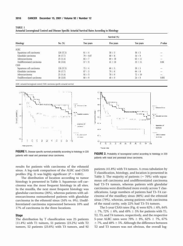
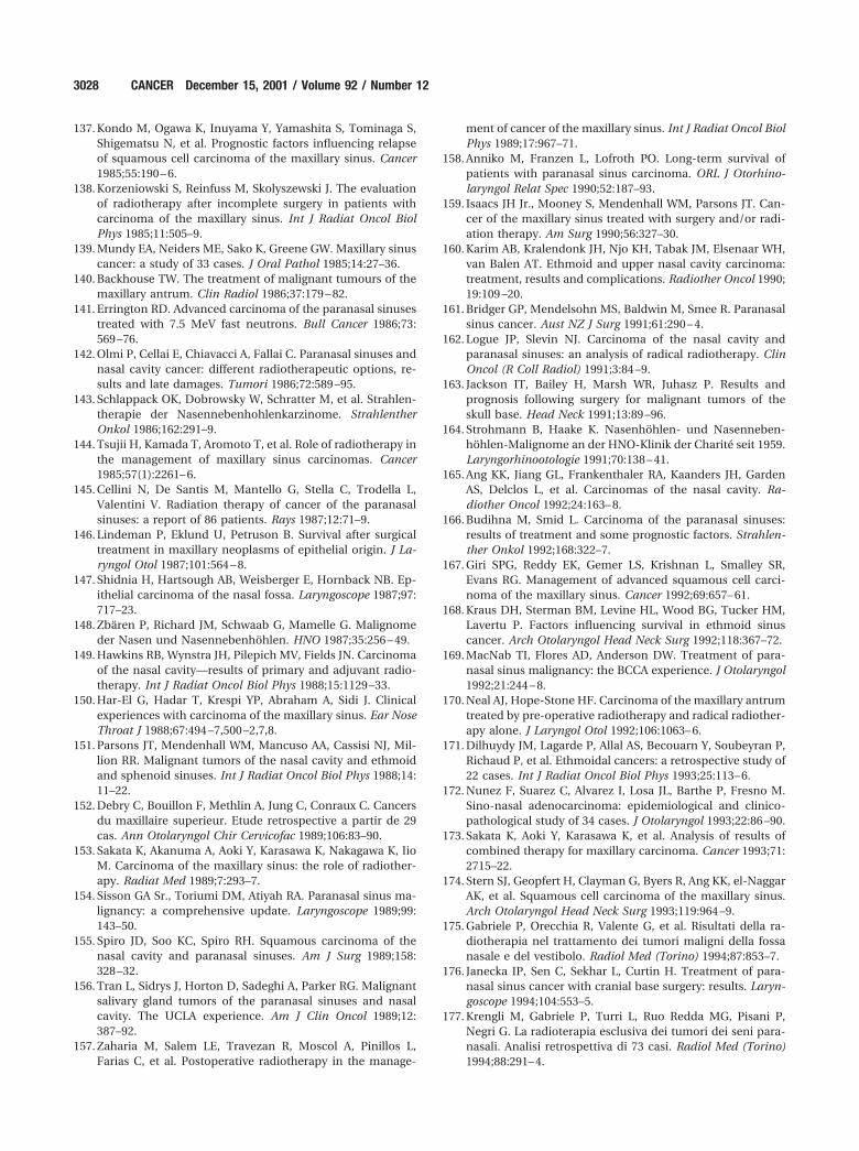
HistologySquamous cell carcinoma was the most frequent his-tologic type and was found in 126 patients (57.3%).There were 39 patients (17.7%) with glandular carci-noma, most of whom had adenoid cystic carcinoma(35 patients). There also were 25 patients (11.4%) withadenocarcinoma and 30 patients (13.6%) with undif-ferentiated carcinoma. The ALRC and CSAS rates forthese four histologic groups are shown in Table 1.Adenocarcinoma and glandular carcinoma treatmentresults were the best, with a 5-year CSAS rate of� 78%, followed by squamous cell carcinoma (60%)and undifferentiated carcinoma (40%). The Kaplan–Meier survival curves for the four histologic groups areshown in Figures 1 and 2. The log-rank test was sig-nificant for CSAS (0.001) but not for ALRC (P � 0.06).
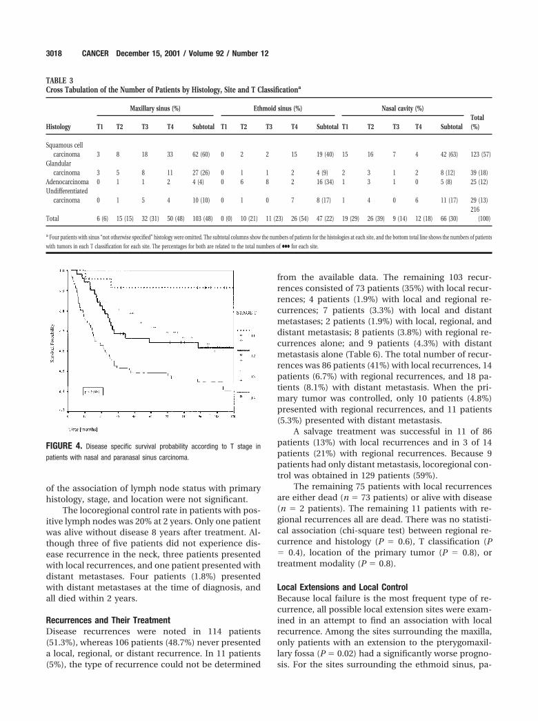
LocationThe site of origin was the maxillary sinus in 103 pa-tients (47%), the nasal cavity in 66 patients (30%), theethmoid sinus in 38 patients (17.3%), the sphenoidsinus in 7 patients (3.2%), and the frontal sinus in 2patients (0.9%). For the analysis, patients with carci-noma of the sphenoid and frontal sinuses weregrouped with patients with ethmoid sinus carcinomaand classified as T4. In 4 patients, the tumors were solarge that the exact locus of origin could not be deter-mined.
The ALRC and CSAS data for the four locations areshown in Table 2. Patients with carcinoma of the nasalcavity exhibited higher control and actuarial survivalrates compared with the rates in patients with sinuscarcinoma. In addition, the results for patients withmaxillary carcinoma were better compared with the
Nasal and Paranasal Carcinoma—Retrospective Review/Dulguerov et al. 3015
results for patients with carcinoma of the ethmoidsinus. A log-rank comparison of the ALRC and CSASprofiles (Fig. 3) was highly significant (P � 0.001).
The distribution of location according to tumorhistology is presented in Table 3. Squamous cell car-cinoma was the most frequent histology in all sites.In the maxilla, the next most frequent histology wasglandular carcinoma (26%), whereas patients with ad-enocarcinoma outnumbered patients with glandularcarcinoma in the ethmoid sinus (34% vs. 9%). Undif-ferentiated carcinoma represented between 10% and17% of carcinoma in the three locations.
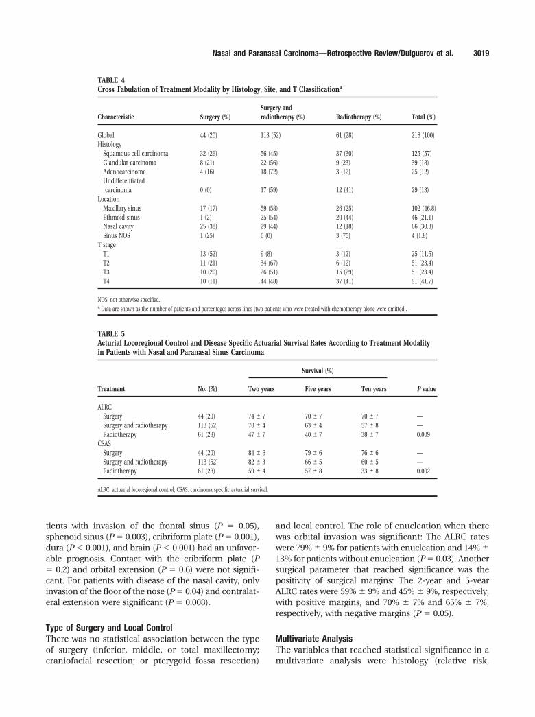
StageThe distribution by T classification was 25 patients(11.4%) with T1 tumors, 51 patients (23.2%) with T2tumors, 52 patients (23.6%) with T3 tumors, and 92
patients (41.8%) with T4 tumors. A cross tabulation byT classification, histology, and location is presented inTable 3. The majority of patients (� 70%) with squa-mous cell carcinoma and undifferentiated carcinomahad T3–T4 tumors, whereas patients with glandularcarcinoma were distributed more evenly across T clas-sifications. Large numbers of patients had T3–T4 car-cinoma of the maxillary sinus (80%) and the ethmoidsinus (79%), whereas, among patients with carcinomaof the nasal cavity, only 22% had T3–T4 tumors.
The 5-year CSAS rates (Fig. 4) were 92% � 6%, 64%� 7%, 72% � 6%, and 49% � 5% for patients with T1,T2, T3, and T4 tumors, respectively, and the respective5-year ALRC rates were 79% � 9%, 62% � 7%, 67%� 7%, and 48% � 5%. Although the difference betweenT2 and T3 tumors was not obvious, the overall log-
TABLE 1Actuarial Locoregional Control and Disease Specific Acturial Survival Rates According to Histology
Histology No. (%)
Survival (%)
P valueTwo years Five years Ten years
ALRCSquamous cell carcinoma 126 (57.3) 61 � 4 58 � 5 56 � 5 —Glandular carcinoma 39 (17.7) 79 � 0.07 68 � 8 54 � 9 —Adenocarcinoma 25 (11.4) 84 � 7 69 � 10 63 � 11 —Undifferentiated carcinoma 30 (13.6) 57 � 9 41 � 10 33 � 11 0.06
CSASSquamous cell carcinoma 126 (57.3) 73 � 4 60 � 5 59 � 5 —Glandular carcinoma 39 (17.7) 87 � 5 79 � 6 64 � 8 —Adenocarcinoma 25 (11.4) 92 � 5 78 � 9 72 � 10 —Undifferentiated carcinoma 30 (13.6) 60 � 9 40 � 9 24 � 9 0.001
ALRC: actuarial locoregional control; CSAS: carcinoma specific actuarial survival.
FIGURE 1. Disease specific survival probability according to histology in 220
patients with nasal and paranasal sinus carcinoma.FIGURE 2. Probability of locoregional control according to histology in 220
patients with nasal and paranasal sinus carcinoma.
3016 CANCER December 15, 2001 / Volume 92 / Number 12
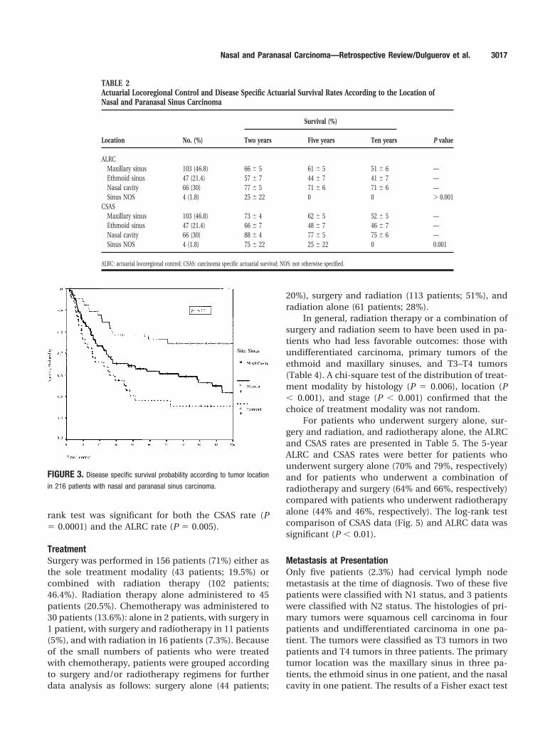
rank test was significant for both the CSAS rate (P� 0.0001) and the ALRC rate (P � 0.005).
TreatmentSurgery was performed in 156 patients (71%) either asthe sole treatment modality (43 patients; 19.5%) orcombined with radiation therapy (102 patients;46.4%). Radiation therapy alone administered to 45patients (20.5%). Chemotherapy was administered to30 patients (13.6%): alone in 2 patients, with surgery in1 patient, with surgery and radiotherapy in 11 patients(5%), and with radiation in 16 patients (7.3%). Becauseof the small numbers of patients who were treatedwith chemotherapy, patients were grouped accordingto surgery and/or radiotherapy regimens for furtherdata analysis as follows: surgery alone (44 patients;
20%), surgery and radiation (113 patients; 51%), andradiation alone (61 patients; 28%).
In general, radiation therapy or a combination ofsurgery and radiation seem to have been used in pa-tients who had less favorable outcomes: those withundifferentiated carcinoma, primary tumors of theethmoid and maxillary sinuses, and T3–T4 tumors(Table 4). A chi-square test of the distribution of treat-ment modality by histology (P � 0.006), location (P� 0.001), and stage (P � 0.001) confirmed that thechoice of treatment modality was not random.
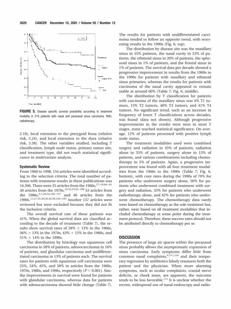
For patients who underwent surgery alone, sur-gery and radiation, and radiotherapy alone, the ALRCand CSAS rates are presented in Table 5. The 5-yearALRC and CSAS rates were better for patients whounderwent surgery alone (70% and 79%, respectively)and for patients who underwent a combination ofradiotherapy and surgery (64% and 66%, respectively)compared with patients who underwent radiotherapyalone (44% and 46%, respectively). The log-rank testcomparison of CSAS data (Fig. 5) and ALRC data wassignificant (P � 0.01).
Metastasis at PresentationOnly five patients (2.3%) had cervical lymph nodemetastasis at the time of diagnosis. Two of these fivepatients were classified with N1 status, and 3 patientswere classified with N2 status. The histologies of pri-mary tumors were squamous cell carcinoma in fourpatients and undifferentiated carcinoma in one pa-tient. The tumors were classified as T3 tumors in twopatients and T4 tumors in three patients. The primarytumor location was the maxillary sinus in three pa-tients, the ethmoid sinus in one patient, and the nasalcavity in one patient. The results of a Fisher exact test
TABLE 2Actuarial Locoregional Control and Disease Specific Actuarial Survival Rates According to the Location ofNasal and Paranasal Sinus Carcinoma
Location No. (%)
Survival (%)
P valueTwo years Five years Ten years
ALRCMaxillary sinus 103 (46.8) 66 � 5 61 � 5 51 � 6 —Ethmoid sinus 47 (21.4) 57 � 7 44 � 7 41 � 7 —Nasal cavity 66 (30) 77 � 5 71 � 6 71 � 6 —Sinus NOS 4 (1.8) 25 � 22 0 0 � 0.001
CSASMaxillary sinus 103 (46.8) 73 � 4 62 � 5 52 � 5 —Ethmoid sinus 47 (21.4) 66 � 7 48 � 7 46 � 7 —Nasal cavity 66 (30) 88 � 4 77 � 5 75 � 6 —Sinus NOS 4 (1.8) 75 � 22 25 � 22 0 0.001
ALRC: actuarial locoregional control; CSAS: carcinoma specific actuarial survival; NOS: not otherwise specified.
FIGURE 3. Disease specific survival probability according to tumor location
in 216 patients with nasal and paranasal sinus carcinoma.
Nasal and Paranasal Carcinoma—Retrospective Review/Dulguerov et al. 3017
of the association of lymph node status with primaryhistology, stage, and location were not significant.
The locoregional control rate in patients with pos-itive lymph nodes was 20% at 2 years. Only one patientwas alive without disease 8 years after treatment. Al-though three of five patients did not experience dis-ease recurrence in the neck, three patients presentedwith local recurrences, and one patient presented withdistant metastases. Four patients (1.8%) presentedwith distant metastases at the time of diagnosis, andall died within 2 years.
Recurrences and Their TreatmentDisease recurrences were noted in 114 patients(51.3%), whereas 106 patients (48.7%) never presenteda local, regional, or distant recurrence. In 11 patients(5%), the type of recurrence could not be determined
from the available data. The remaining 103 recur-rences consisted of 73 patients (35%) with local recur-rences; 4 patients (1.9%) with local and regional re-currences; 7 patients (3.3%) with local and distantmetastases; 2 patients (1.9%) with local, regional, anddistant metastasis; 8 patients (3.8%) with regional re-currences alone; and 9 patients (4.3%) with distantmetastasis alone (Table 6). The total number of recur-rences was 86 patients (41%) with local recurrences, 14patients (6.7%) with regional recurrences, and 18 pa-tients (8.1%) with distant metastasis. When the pri-mary tumor was controlled, only 10 patients (4.8%)presented with regional recurrences, and 11 patients(5.3%) presented with distant metastasis.
A salvage treatment was successful in 11 of 86patients (13%) with local recurrences and in 3 of 14patients (21%) with regional recurrences. Because 9patients had only distant metastasis, locoregional con-trol was obtained in 129 patients (59%).
The remaining 75 patients with local recurrencesare either dead (n � 73 patients) or alive with disease(n � 2 patients). The remaining 11 patients with re-gional recurrences all are dead. There was no statisti-cal association (chi-square test) between regional re-currence and histology (P � 0.6), T classification (P� 0.4), location of the primary tumor (P � 0.8), ortreatment modality (P � 0.8).
Local Extensions and Local ControlBecause local failure is the most frequent type of re-currence, all possible local extension sites were exam-ined in an attempt to find an association with localrecurrence. Among the sites surrounding the maxilla,only patients with an extension to the pterygomaxil-lary fossa (P � 0.02) had a significantly worse progno-sis. For the sites surrounding the ethmoid sinus, pa-
TABLE 3Cross Tabulation of the Number of Patients by Histology, Site and T Classificationa
Histology
Maxillary sinus (%) Ethmoid sinus (%) Nasal cavity (%)Total(%)T1 T2 T3 T4 Subtotal T1 T2 T3 T4 Subtotal T1 T2 T3 T4 Subtotal
Squamous cellcarcinoma 3 8 18 33 62 (60) 0 2 2 15 19 (40) 15 16 7 4 42 (63) 123 (57)
Glandularcarcinoma 3 5 8 11 27 (26) 0 1 1 2 4 (9) 2 3 1 2 8 (12) 39 (18)
Adenocarcinoma 0 1 1 2 4 (4) 0 6 8 2 16 (34) 1 3 1 0 5 (8) 25 (12)Undifferentiated
carcinoma 0 1 5 4 10 (10) 0 1 0 7 8 (17) 1 4 0 6 11 (17) 29 (13)
Total 6 (6) 15 (15) 32 (31) 50 (48) 103 (48) 0 (0) 10 (21) 11 (23) 26 (54) 47 (22) 19 (29) 26 (39) 9 (14) 12 (18) 66 (30)216
(100)
a Four patients with sinus “not otherwise specified” histology were omitted. The subtotal columns show the numbers of patients for the histologies at each site, and the bottom total line shows the numbers of patients
with tumors in each T classification for each site. The percentages for both are related to the total numbers of ●●● for each site.
FIGURE 4. Disease specific survival probability according to T stage in
patients with nasal and paranasal sinus carcinoma.
3018 CANCER December 15, 2001 / Volume 92 / Number 12
tients with invasion of the frontal sinus (P � 0.05),sphenoid sinus (P � 0.003), cribriform plate (P � 0.001),dura (P � 0.001), and brain (P � 0.001) had an unfavor-able prognosis. Contact with the cribriform plate (P� 0.2) and orbital extension (P � 0.6) were not signifi-cant. For patients with disease of the nasal cavity, onlyinvasion of the floor of the nose (P � 0.04) and contralat-eral extension were significant (P � 0.008).
Type of Surgery and Local ControlThere was no statistical association between the typeof surgery (inferior, middle, or total maxillectomy;craniofacial resection; or pterygoid fossa resection)
and local control. The role of enucleation when therewas orbital invasion was significant: The ALRC rateswere 79% � 9% for patients with enucleation and 14% �13% for patients without enucleation (P � 0.03). Anothersurgical parameter that reached significance was thepositivity of surgical margins: The 2-year and 5-yearALRC rates were 59% � 9% and 45% � 9%, respectively,with positive margins, and 70% � 7% and 65% � 7%,respectively, with negative margins (P � 0.05).
Multivariate AnalysisThe variables that reached statistical significance in amultivariate analysis were histology (relative risk,
TABLE 4Cross Tabulation of Treatment Modality by Histology, Site, and T Classificationa
Characteristic Surgery (%)Surgery andradiotherapy (%) Radiotherapy (%) Total (%)
Global 44 (20) 113 (52) 61 (28) 218 (100)Histology
Squamous cell carcinoma 32 (26) 56 (45) 37 (30) 125 (57)Glandular carcinoma 8 (21) 22 (56) 9 (23) 39 (18)Adenocarcinoma 4 (16) 18 (72) 3 (12) 25 (12)Undifferentiatedcarcinoma 0 (0) 17 (59) 12 (41) 29 (13)
LocationMaxillary sinus 17 (17) 59 (58) 26 (25) 102 (46.8)Ethmoid sinus 1 (2) 25 (54) 20 (44) 46 (21.1)Nasal cavity 25 (38) 29 (44) 12 (18) 66 (30.3)Sinus NOS 1 (25) 0 (0) 3 (75) 4 (1.8)
T stageT1 13 (52) 9 (8) 3 (12) 25 (11.5)T2 11 (21) 34 (67) 6 (12) 51 (23.4)T3 10 (20) 26 (51) 15 (29) 51 (23.4)T4 10 (11) 44 (48) 37 (41) 91 (41.7)
NOS: not otherwise specified.a Data are shown as the number of patients and percentages across lines (two patients who were treated with chemotherapy alone were omitted).
TABLE 5Acturial Locoregional Control and Disease Specific Actuarial Survival Rates According to Treatment Modalityin Patients with Nasal and Paranasal Sinus Carcinoma
Treatment No. (%)
Survival (%)
P valueTwo years Five years Ten years
ALRCSurgery 44 (20) 74 � 7 70 � 7 70 � 7 —Surgery and radiotherapy 113 (52) 70 � 4 63 � 4 57 � 8 —Radiotherapy 61 (28) 47 � 7 40 � 7 38 � 7 0.009
CSASSurgery 44 (20) 84 � 6 79 � 6 76 � 6 —Surgery and radiotherapy 113 (52) 82 � 3 66 � 5 60 � 5 —Radiotherapy 61 (28) 59 � 4 57 � 8 33 � 8 0.002
ALRC: actuarial locoregional control; CSAS: carcinoma specific actuarial survival.
Nasal and Paranasal Carcinoma—Retrospective Review/Dulguerov et al. 3019
2.18), local extension to the pterygoid fossa (relativerisk, 2.24), and local extension to the dura (relativerisk, 3.38). The other variables studied, including Tclassification, lymph node status, primary tumor site,and treatment type, did not reach statistical signifi-cance in multivariate analysis.
Systematic ReviewFrom 1960 to 1998, 154 articles were identified accord-ing to the selection criteria. The total number of pa-tients with treatment results in these publications was16,396. There were 25 articles from the 1960s,17–19,60 – 81
30 articles from the 1970s,16,53,55,82–108 53 articles fromthe 1980s,5,7,48,54,109 –157 and 46 articles from the1990s.11,27,35,40,50,56,58,158 –195 Another 137 articles werereviewed but were excluded because they did not fitthe inclusion criteria.
The overall survival rate of these patients was41%. When the global survival data are classified ac-cording to the decade of treatment (Table 7), the re-sults show survival rates of 28% � 13% in the 1960s,36% � 13% in the 1970s, 43% � 15% in the 1980s, and51% � 14% in the 1990s.
The distribution by histology was squamous cellcarcinoma in 58% of patients, adenocarcinoma in 16%of patients, and glandular carcinoma and undifferen-tiated carcinoma in 13% of patients each. The survivalrates for patients with squamous cell carcinoma were25%, 34%, 45%, and 50% in articles from the 1960s,1970s, 1980s, and 1990s, respectively (P � 0.001). Sim-ilar improvements in survival were found for patientswith glandular carcinoma, whereas data for patientswith adenocarcinoma showed little change (Table 7).
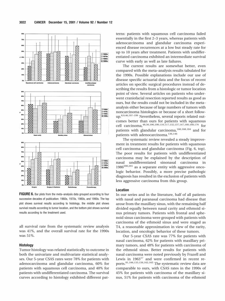
The results for patients with undifferentiated carci-noma tended to follow an opposite trend, with wors-ening results in the 1990s (Fig. 6, top).
The distribution by disease site was the maxillarysinus in 55% patients, the nasal cavity in 23% of pa-tients, the ethmoid sinus in 20% of patients, the sphe-noid sinus in 1% of patients, and the frontal sinus in1% of patients. The survival data per decade showed aprogressive improvement in results from the 1960s tothe 1990s for patients with maxillary and ethmoidsinus primaries, whereas the results for patients withcarcinoma of the nasal cavity appeared to remainstable at around 60% (Table 7; Fig. 6, middle).
The distribution by T classification for patientswith carcinoma of the maxillary sinus was 4% T1 tu-mors, 15% T2 tumors, 40% T3 tumors, and 41% T4tumors. No significant trend, such as an increase infrequency of lower T classifications across decades,was found (data not shown). Although progressiveimprovements in the results were seen in most Tstages, none reached statistical significance. On aver-age, 12% of patients presented with positive lymphnode status.
The treatment modalities used were combinedsurgery and radiation in 45% of patients, radiationalone in 35% of patients, surgery alone in 15% ofpatients, and various combinations including chemo-therapy in 5% of patients. Again, a progressive im-provement was found with all four treatment modal-ities from the 1960s to the 1990s (Table 7; Fig. 6,bottom), with cure rates during the 1990s of 70% forpatients who underwent surgery alone, 56% for pa-tients who underwent combined treatment with sur-gery and radiation, 33% for patients who underwentradiotherapy alone, and 42% for patients who under-went chemotherapy. The chemotherapy data rarelywere based on chemotherapy as the sole treatment but,rather, were based on all treatment modalities that in-cluded chemotherapy at some point during the treat-ment protocol. Therefore, these success rates should notbe attributed directly to chemotherapy per se.
DISCUSSIONThe presence of large air spaces within the paranasalsinus probably allows the asymptomatic expansion ofsinus carcinoma. Early symptoms differ little fromcommon nasal complaints,6,117,154 and their tempo-rary regression by antibiotics falsely reassures both thepatient and the physician. When more alarmingsymptoms, such as ocular complaints, cranial nervedeficits, or cheek mass, are apparent, the outcometends to be less favorable.111 It is unclear whether therecent, widespread use of nasal endoscopy and radio-
FIGURE 5. Disease specific survival probability according to treatment
modality in 218 patients with nasal and paranasal sinus carcinoma. Rxth:
radiotherapy.
3020 CANCER December 15, 2001 / Volume 92 / Number 12
logic studies will result in earlier diagnoses of nasaland paranasal sinus carcinoma.
Overall ResultsIn our series, the overall survival rate was 40%, andintercurrent deaths occurred in 14.5% of patients, fora disease specific survival rate of 54.5%. The 5-yearCSAS rate was 63%. The results in terms of locore-gional control paralleled the survival data, with a5-year ALRC rate of 57%. The close relation between
survival and local control underscores the fact that theprognosis for patients with nasal and paranasal sinuscarcinoma is related directly to local control of thedisease.4,5,103,117,154,157,174,186,196
In the literature, global results, most often ex-pressed in terms of crude survival, vary between10%60,61,63,70,77,80,82,152 and 75%,40,106,109,132,135,160,161,165,176
with better results in carefully selected patients, inpatients with primary tumors of the nasal cavity, andin more recently published articles. The average over-
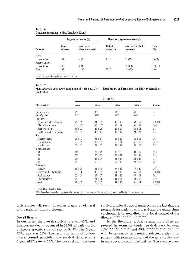
TABLE 6Outcome According to First Oncologic Eventa
Outcome
Regional recurrence (%) Absence of regional recurrence (%)
Total(%)
Distantmetastasis
Absence ofdistant metastasis
Distantmetastasis
Absence of distantmetastasis
Localrecurrence 2 (1) 4 (2) 7 (3) 73 (35) 86 (41)
Absence of localrecurrence 0 (0) 8 (4) 9 (4) 106 (51) 123 (59)
Total 2 (1) 12 (6) 16 (7) 179 (86) 209
a Eleven patients with insufficient data were excluded.
TABLE 7Meta–Analysis Data: Cross Tabulation of Histology, Site, T-Classification, and Treatment Modality by Decade ofPublication
Characteristic
Decade (%)
P value1960s 1970s 1980s 1990s
No. of studies 25 30 53 46 —No. of patients 3137 3877 5966 3416 —Histology
Squamous cell carcinoma 25 � 13 34 � 15 45 � 19 50 � 19 � 0.001Glandular carcinoma 42 � 33a 30 � 17a 45 � 21 60 � 24 0.09Adenocarcinoma 46 � 33 48 � 28 49 � 29 50 � 14 0.83Undifferentiated carcinoma 23 � 17 42 � 21 30 � 17 28 � 21 0.41
SiteMaxillary sinus 26 � 13 31 � 8 39 � 14 45 � 11 0.02Ethmoid sinus 27 � 13 37 � 13 56 � 24 51 � 15 0.002Nasal cavity 63 � 23 54 � 25 59 � 12 66 � 15 0.71
T classificationT1 28a 83 � 29 87 � 23 90 � 19 0.25T2 22a 53 � 22 62 � 27 70 � 30 0.28T3 10a 28 � 14 44 � 17 44 � 29 0.13T4 0a 18 � 11 19 � 10 28 � 18 0.31
TreatmentSurgery 36 � 25 54 � 15 57 � 26 70 � 20 0.034Surgery and radiotherapy 33 � 18 42 � 15 54 � 15 56 � 13 � 0.001Radiotherapy 21 � 13 19 � 17 28 � 20 33 � 18 0.048Chemotherapyb 0a 21 � 18 34 � 24 42 � 18 0.10
Overall 28 � 13 36 � 13 44 � 15 51 � 14 � 0.001
a Included fewer than five studies.b The chemotherapy data include patients who received chemotherapy as part of their treatment, usually combined with other modalities.
Nasal and Paranasal Carcinoma—Retrospective Review/Dulguerov et al. 3021
all survival rate from the systematic review analysiswas 41%, and the overall survival rate for the 1990swas 51%.
HistologyTumor histology was related statistically to outcome inboth the univariate and multivariate statistical analy-ses. Our 5-year CSAS rates were 78% for patients withadenocarcinoma and glandular carcinoma, 60% forpatients with squamous cell carcinoma, and 40% forpatients with undifferentiated carcinoma. The survivalcurves according to histology exhibited different pat-
terns: patients with squamous cell carcinoma failedessentially in the first 2–3 years, whereas patients withadenocarcinoma and glandular carcinoma experi-enced disease recurrences at a low but steady rate forup to 10 years after treatment. Patients with undiffer-entiated carcinoma exhibited an intermediate survivalcurve with early as well as late failures.
The current results are somewhat better, evencompared with the meta–analysis results tabulated forthe 1990s. Possible explanations include our use ofdisease specific actuarial data and the focus of recentarticles on specific surgical procedures instead of de-scribing the results from a histologic or tumor locationpoint of view. Several articles on patients who under-went craniofacial resection reported results as good asours, but the results could not be included in the meta–analysis either because of large numbers of tumors withnoncarcinoma histologies or because of a short follow-up.8,9,46,197–199 Nevertheless, several reports related out-comes better than ours for patients with squamouscell carcinoma,48,56,106,100,110,117,132,137,147,160,168,176 forpatients with glandular carcinoma,160,168,184 and forpatients with adenocarcinoma.136,146
The systematic review revealed a steady improve-ment in treatment results for patients with squamouscell carcinoma and glandular carcinoma (Fig. 6, top).The poor results for patients with undifferentiatedcarcinoma may be explained by the description ofnasal undifferentiated sinonasal carcinoma in1986200,201 as a separate entity with aggressive onco-logic behavior. Possibly, a more precise pathologicdiagnosis has resulted in the exclusion of patients withless aggressive carcinoma from this group.
LocationIn our series and in the literature, half of all patientswith nasal and paranasal carcinoma had disease thatarose from the maxillary sinus, with the remaining halfdivided equally between nasal cavity and ethmoid si-nus primary tumors. Patients with frontal and sphe-noid sinus carcinoma were grouped with patients withcarcinoma of the ethmoid sinus and were staged asT4, a reasonable approximation in view of the rarity,location, and oncologic behavior of these tumors.
Our 5-year CSAS rate was 77% for patients withnasal carcinoma, 62% for patients with maxillary pri-mary tumors, and 48% for patients with carcinoma ofthe ethmoid sinus. Better results for patients withnasal carcinoma were noted previously by Frazell andLewis in 196317 and were confirmed in recent re-ports.81,106,133,156,162,165 The systematic review data arecomparable to ours, with CSAS rates in the 1990s of45% for patients with carcinoma of the maxillary si-nus, 51% for patients with carcinoma of the ethmoid
FIGURE 6. Bar plots from the meta–analysis data grouped according to four
successive decades of publication: 1960s, 1970s, 1980s, and 1990s. The top
plot shows survival results according to histology, the middle plot shows
survival results according to tumor location, and the bottom plot shows survival
results according to the treatment used.
3022 CANCER December 15, 2001 / Volume 92 / Number 12
sinus, and 66% for patients with nasal carcinoma.There was a steady improvement in the treatmentresults for patients with maxillary and ethmoid pri-mary tumors, while the results for patients with nasalcarcinoma have stagnated, for unclear reasons,around 60% since the 1960s.
T ClassificationThe diagnosis of nasal and paranasal carcinoma oc-curred at an advanced stage in our patients, and thisdistinction also was seen in other publica-tions,5,11,103,111,117,154,176 with 70 – 80% of patients diag-nosed with T3 or T4 tumors. It has been reported thatimprovements in treatment results for patients withmalignant disease represent spurious effects of diag-nosis at an early stage202; however, no such trend forlower T classifications could be found in the systemicreview.
A clear correlation between T stage and survival wasfound in univariate analysis for all locations. This was notedin previous articles on patients with primary tumors of themaxillary sinus.5,90,92,95,104,117,126,137,144,154,155,157,162,173 Our5-year CSAS rates were 91%, 64%, 70%, and 50% forpatients with T1, T2, T3, and T4 tumors, respectively.These results are close to the meta–analysis data fromthe 1990s for patients with T1 tumors (94%) and T2tumors (55%), but they are much better comparedwith the results for patients with T3 tumors (50%) andT4 tumors (27%). Possibly, the use of more extensivesurgical resections in our series, the presence of nu-merous exclusive radiotherapy reports in the literaturewith less favorable outcomes for patients with ad-vanced-stage disease, and the bias of recent surgicalpublications discussed above may provide explana-tions for these differences.
Articles that employed a staging system for pa-tients with nasal and ethmoid carcinoma were sparse,and the classification systems used varied4,29,30,77,100,
109,151,165,171,188,196,203 and sometimes were arbi-trary57,74,107,108,110,130,156,162,204 –206 Only one previousstudy used a similar classification system in 54 pa-tients with nasal and paranasal carcinoma183: The5-year survival rates found were 100% for patientswith T1 tumors, 87.5% for patients with T2 tumors,92.3% for patients with T3 tumors, 28% for patientswith T4 tumors.
It is difficult to compare our data in terms ofdisease stage for these locations until a universal stag-ing system is adopted. For this study, we reluctantlyabandoned the staging system that we proposed sev-eral years ago for patients with esthesioneuroblas-toma4,207 in favor of the UICC classification system inthe search for such a consensus. However, the pro-posed UICC system for classifying patients with tu-
mors of the ethmoid and nasal cavity has numerousshortcomings and may explain the paucity of differ-ences between the results for patients with T2 and T3tumors.
Treatment ModalityOur data show a 5-year CSAS rate of 57% for patientswho underwent radiation alone, 66% for patientswho underwent combined radiation and surgery,and 79% for patients who underwent surgeryalone. The difference was highly significant in thelog-rank analysis, as suggested in previous re-ports.17,66,76,95,96,104,111,134,135,139,154,167,169,180
The meta–analysis confirmed that surgery (70%)and combined surgery and radiation (56%) offer betterlocal control and cure rates than radiotherapy alone(33%). Most series, including ours, are biased in pa-tient selection, and no randomized study has beenpublished. In general, patients with favorable lesionsare found mainly in the surgery alone groups, whereaspatients with large lesions and those who are treatedfor palliation are in the exclusive radiation or chemo-radiation groups. Nevertheless, except for a few arti-cles,76,77,113,160,184 the results of radiation alone arepoorer than treatments that include surgery. Further-more, radical radiotherapy protocols have resulted in20%11 to 30%56 unilateral blindness and 6%11 to 10%56
bilateral blindness. Despite the inherent patient selec-tion bias of retrospective studies, the available datasuggest that surgery should be included in the treat-ment strategy for patients with nasal and paranasalcarcinoma who are treated with a curative intent.
The sequence of surgery and radiotherapy in themanagement of these patients has remained open todebate since the work of Jesse,71 who showed no cleardifference. Although most centers prefer primary sur-gery, some continue to choose primary radiothera-py.120,146,154,162,167,184,188 Because a high incidence ofresidual disease is found after primary radia-tion,74,137,147 the main goal of primary radiation oftenis to shrink the tumor so that the surgical resection isless extensive and vital structures, such as the eye, canbe spared.154,184,185 The use of hyperfractionation11,142
or neutron beam irradiation142 does not seem to mod-ify the cure rates drastically. Although the results of afew recent articles that included chemotherapy in thetreatment protocol are encouraging, with 5-year sur-vival rates � 50%,34,40,136,137,173 it remains unclearwhether the addition of chemotherapy to other ag-gressive treatment regimens provides a clear advan-tage in local control or survival.
Nasal and Paranasal Carcinoma—Retrospective Review/Dulguerov et al. 3023
Local ExtensionThe air-filled sinus cavities offer little resistance totumor growth. Sinus carcinoma probably expands byfilling the cavity first, before eroding the adjacent bonywalls in a centrifugal pattern. Fibroelastic connectivetissue present in periosteum, perichondrium, anddura is considered the most efficient barrier to diseaseexpansion.6 e pathologic studies addressing the natu-ral barriers to sinonasal carcinoma invasion are lack-ing.
In the current series, the patients with extensionstoward the anterior base of skull and toward the ptery-gomaxillary fossa had a worse prognosis in both uni-variate and multivariate analyses. Keeping in mind theimportance of local control, further advancement intreatment should address these structures specifically.For patients who undergo surgery, en bloc resection,including surrounding noninvaded osseous walls, isfavored. In patients with high nasal and ethmoid car-cinoma, craniofacial resection by combined cranialand transfacial approaches has become a routine pro-cedure4,22,48,174,176,178 and probably is responsible forthe improved results in this and other series.154,176
Similarly, patients with invasion beyond the maxillarywalls undergo mandibulectomy for the lateral wall andinfratemporal fossa23 and sphenoid wing resection176
for the posterior wall. The evolution from the piece-meal surgical procedures performed in the 1960s canbe seen in Table 7: Note the improvements in the5-year survival rate from 36% in the 1960s to 70% inthe 1990s, notwithstanding patient selection.
Orbital invasion is always dramatic and, despitemodern radiologic techniques, often is diagnosed cor-rectly only during exploration.208 Since the report byLarsson et al.,209 several authors have advocated aneye-sparing approach.45,174,210 –212 Although it seemsreasonable that an intact periorbit should mandateeye preservation, the available data are inconclusive atbest,191 and preservation does not necessarily result inan intact functional eye.45 Our data show that, inpatients with orbital invasion, the local control rate is79% with enucleation and 14% without enucleation.Despite the biases inherent to a retrospective study, itprobably is unwise to conclude that the orbit can bespared in all patients.211
Metastasis and RecurrencesFortunately, neck lymph metastases remain infre-quent either at the time of presentation or after treat-ment. In our data, 2.3% of patients presented with aneck metastasis, and 7% of patients developed a neckmetastasis. In the meta–analysis, the correspondingrates were 12% and 13%. Neck lymph node recurrence
alone was present in 4% of our patients and in 5%of patients in the meta–analysis data. In some seriesthat were weighted heavily toward patients withadvanced-stage maxillary squamous cell carcino-ma,5,117,137,148,174,213 the rate of neck metastasis at thetime of presentation was �20 –25%, and prophylactictreatment of the neck should have been consid-ered.5,56,174,213 Several studies137,194,214 have indicateda higher incidence of neck recurrence with involve-ment of the alveolus and cheek.
The results of treatment for patients with meta-static neck disease were disappointing, However, pa-tients with primary neck metastases had a 20% 5-yearsurvival rate in the current series and a 32% 5-yearsurvival rate in the systematic review. For patientswith post-treatment neck metastases, the 5-year sur-vival rates were 21% in the current series and 25% inthe meta–analysis. Our success rate in the treatment ofpatients with local recurrences was 13% and was com-parable to the 16% rate found in the meta–analysis.
Shortcomings of the StudyAlthough this patient series was relatively large, it suf-fered from the usual shortcomings of any retrospec-tive study: mainly, a retrospective staging in somepatients and nonrandomized treatment selection. Thesystematic review theoretically may strengthen theconclusions, and this represents the rationale for un-dertaking it. Notwithstanding criticisms of meta–anal-ysis in general and specific and pertinent criticisms ofmeta–analysis of observational studies, we tend tothink that our systemic review may be the only meansof gaining a global perspective.32,33 Because of theheterogeneity of the studies and the lack of possiblequality control, the results of the systematic reviewshould be taken as a general indication of our currentachievements in the treatment of patients with nasaland paranasal carcinoma.
REFERENCES1. Muir CS, Nectoux J. Descriptive epidemiology of malignant
neoplasms of nose, nasal cavities, middle ear and accessorysinuses. Clin Otolaryngol 1980;5:195–211.
2. Ayiomamitis A, Parker L, Havas T. The epidemiology ofmalignant neoplasms of the nasal cavities, the paranasalsinuses and the middle ear in Canada. Arch Otorhinolaryn-gol 1988;244:367–71.
3. Shanmugaratnam K, Sobin LH. Histological typing of tu-mours of the upper respiratory tract and ear. Berlin: Springer-Verlag, 1991.
4. Dulguerov P, Calcaterra TC. Esthesioneuroblastoma—theUCLA experience. Laryngoscope 1992;102:843–9.
5. Lavertu P, Roberts JK, Kraus DH, Levine HL, Wood BG,Mendendorp SV, et al. Squamous cell carcinoma of theparanasal sinuses: the Cleveland Clinic experience 1977–1986. Laryngoscope 1989;99:1130 – 6.
3024 CANCER December 15, 2001 / Volume 92 / Number 12
6. Lyons BM, Donald PJ. Radical surgery for nasal cavity andparanasal sinus tumors. Otolaryngol Clin North Am 1991;24:1499 –521.
7. Ketcham AS, Van Buren JM. Tumors of the paranasal si-nuses: a therapeutic challenge. Am J Surg 1985;150:406 –13.
8. Catalano PJ, Hecht CS, Biller HF, Lawson W, Post KD,Sachdev V, et al. Craniofacial resection. An analysis of 73cases. Arch Otolaryngol Head Neck Surg 1994;120:1203– 8.
9. Richtsmeier WJ, Briggs RJS, Koch WM, Eisele DW, Loury MC,Price JC, et al. Complications and early outcome of anteriorcraniofacial resection. Arch Otolaryngol Head Neck Surg1992;118:913–7.
10. Donald PJ. Complications in skull base surgery for malig-nancy. Laryngoscope 1999;109:1959 – 66.
11. Parsons JT, Kimsey FC, Mendenhall WM, Million RR, CassisiNJ, Stringer SP. Radiation therapy for sinus malignancies.Otolaryngol Clin North Am 1995;28:1259 – 68.
12. Shukovsky LJ, Fletcher GH. Retinal and optic nerve compli-cations in a high dose irradiation technique of ethmoidsinus and nasal cavity. Radiology 1972;104:629 –34.
13. Takeda A, Shigematsu N, Suzuki S, Fujii M, Kawata T,Kawaguchi O, et al. Late retinal complications of radiationtherapy for nasal and paranasal malignancies: relationshipbetween irradiated-dose area and severity. Int J Radiat On-col Biol Phys 1999;44:599 – 605.
14. Syme J. Excision of upper jaw bones. Lancet 1829;2:667– 8.15. Schuknecht HF. The surgical management of carcinoma of
the paranasal sinuses. Laryngoscope 1951:874 –90.16. Lewis JS, Castro EB. Cancer of the nasal cavity and paranasal
sinuses. J Laryngol Otol 1972;86:255– 86.17. Frazell EL, Lewis JS. Cancers of the nasal cavity and acces-
sory sinuses. A report of the management of 416 patients.Cancer 1963;16:1293–301.
18. Baclesse F, Ennuyer A, Calle R. Les epitheliomas du sinusmaxillaire traites par roentgentherapie transcutanee seule(resultats a cinq ans). J Radiol Electrol 1960;41:368 –75.
19. Pointon RCS. Neoplasia of the nose and sinuses. J LaryngolOtol 1969;83:407–15.
20. Bailar JC, Gornik HL. Cancer undefeated. N Engl J Med1997;336:1569 –74.
21. Smith RR, Klopp CT, Williams JM. Surgical treatment ofcancer of the frontal sinus and adjacent areas. Cancer 1954;7:991–9.
22. Ketcham AS, Wilkins RH, Van Buren JM, Smith RR. A com-bined intracranial facial approach to the paranasal sinuses.Am J Surg 1963;106:698 –703.
23. Terz JJ, Alksne JF, Lawrence W. Craniofacial resection fortumors invading the pterygoid fossa. Am J Surg 1969;118:732–9.
24. Janecka IP, Nuss DW, Sen CN. Facial translocation approachto the cranial base. Acta Neurochir 1991;53(Suppl):193– 8.
25. Gademann G, Schlegel W, Debus J, Schad L, Bortfeld T,Hover KH, et al. Fractionated stereotactically guided radio-therapy of head and neck tumors: a report on clinical use ofa new system in 195 cases. Radiother Oncol 1993;29:205–13.
26. Miralbell R, Crowell C, Suit HD. Potential improvement ofthree dimension treatment planning and proton therapy inthe outcome of maxillary sinus cancer. Int J Radiat OncolBiol Phys 1991;22:305–10.
27. Roa WH, Hazuka MB, Sandler HM, Martel MK, Thornton AF,Turrisi AT, et al. Results of primary and adjuvant CT-based3-dimensional radiotherapy for malignant tumors of theparanasal sinuses. Int J Radiat Oncol Biol Phys 1994;28:857– 65.
28. Mosesson RE, Som PM. The radiographic evaluation of si-nonasal tumors: an overview. Otolaryngol Clin North Am1995;28:1097–115.
29. Hermanek P, Henson DE, Hutter RVP, Sobin LH. Interna-tional Union Against Cancer: TNM supplement 1993. Berlin:Springer, 1993.
30. Sobin LH, Wittekind C. International Union Against Cancer:TNM classification of malignant tumors. Berlin: Springer,1997.
31. Egger M, Smith GD. Meta-analysis. Potentials and promise.BMJ 1997;315:1371– 4.
32. Egger M, Schneider M, Davey Smith G. Spurious precision?Meta-analysis of observational studies. BMJ 1998;316:140 – 4.
33. Stroup DF, Berlin JA, Morton SC, Olkin I, Williamson GD,Rennie D, et al. Meta-analysis of observational studies inepidemiology: a proposal for reporting. Meta-analysis ofObservational Studies in Epidemiology (MOOSE) group.JAMA 2000;283:2008 –12.
34. Roux FX, Brasnu D, Menard M, et al. Adenocarcinoma of theethmoid sinuses. Results of a new protocol based on induc-tive chemotherapy combined with surgery. Four years ex-perience. Acta Neurochir (Wien) 1989;98:129 –34.
35. Roux FX, Brasnu D, Menard M, et al. Les abords combinesdes tumeurs malignes de l’ethmoıde et autres sinus parana-saux. Principes et resultats. Ann Otolaryngol Chir Cervicofac1991;108:292–7.
36. Roux FX, Brasnu D, Devaux B, Chabardes E, Schwaab G,Laccourreye O, et al. Ethmoid sinus carcinomas: results andprognosis after neoadjuvant chemotherapy and combinedsurgery—a 10-year experience. Surg Neurol 1994;42:98 –104.
37. Roux FX, Devaux B, Nataf F, Pages JC, Laccourreye O, Me-nard M, et al. Malignant tumors of the ethmoid region.Neurosurgical techniques. Neurochirurgie 1997;43:92–9.
38. Roux FX, Pages JC, Nataf F, Devaux B, Laccourreye O, Me-nard M, et al. Les tumeurs malignes ethmoıdo-sphenoı-dales. 130 cas. Etude retrospective. Neurochirurgie 1997;43:100 –10.
39. Brasnu D, Roux FX, Fabre A, Menard M, Manolopoulos L,Chodkiewicz JP, et al. La voie d’abord combinee ORL etNeurochirurgicale dans les adenocarcinomes de l’ethmoıde.Ann Otolaryngol Chir Cervicofac 1987;104:347–51.
40. Brasnu D, Laccourreye O, Bassot V, Laccourreye L, Naudo P,Roux FX. Cisplatin-based neoadjuvant chemotherapy andcombined resection for ethmoid sinus adenocarcinomareaching and/or invading the skull base. Arch OtolaryngolHead Neck Surg 1996;122:765– 8.
41. Brasnu D, Laccourreye O, Menard M, Devaux B, Roux FX.Approches transfaciales des cancers de l’ethmoıde. Neuro-chirurgie 1997;43:88 –91.
42. Schwaab G, Marandas P. Les problemes du traitementchirurgical des tumeurs malignes de l’ethmoide par abordmixte endocranien et facial. Ann Otolaryngol Chir Cervicofac1983;100:159 – 61.
43. Schwaab G, Julieron M, Janot F. Epidemiologie des cancersde la fosse nasale et sinus paranasaux. Neurochirurgie 1997;43:61–3.
44. George B, Salvan D, Luboinski B, Boissonnet H, Lot G.Tumeurs malignes de l’ethmoıde. Serie homogene de 41 casoperes par voie mixte. Neurochirurgie 1997;43:121– 4.
45. Andersen PE, Kraus DH, Arbit E, Shah JP. Management ofthe orbit during anterior fossa craniofacial resection. ArchOtolaryngol Head Neck Surg 1996;122:1305–7.
Nasal and Paranasal Carcinoma—Retrospective Review/Dulguerov et al. 3025
46. Bilsky MH, Kraus DH, Strong EW, Harrison LB, Gutin PH,Shah JP. Extended anterior craniofacial resection for intra-cranial extension of malignant tumors. Am J Surg 1997;174:565– 8.
47. Harrison LB, Pfister DG, Kraus D, et al. Management ofunresectable malignant tumors at the skull base using con-comitant chemotherapy and radiotherapy with acceleratedfractionation. Skull Base Surg 1994;4:127–31.
48. Shah JP, Sundaresan N, Galicich J, Strong EW. Craniofacialresections for tumors involving the base of the skull. Am JSurg 1987;154:352– 8.
49. Shah JP, Kraus DH, Arbit E, Galicich JH, Strong EW. Cranio-facial resection for tumors involving the anterior skull base.Otolaryngol Head Neck Surg 1992;106:387–93.
50. Shah JP, Kraus DH, Bilsky MH, Gutin PH, Harrison LH,Strong EW. Craniofacial resection for malignant tumors in-volving the anterior skull base. Arch Otolaryngol Head NeckSurg 1997;123:1312–7.
51. Blacklock JB, Weber RS, Lee YY, Goepfert H. Transcranialresection of tumors of the paranasal sinuses and nasal cav-ity. J Neurosurg 1989;71:10 –5.
52. McCutcheon IE, Blacklock JB, Weber RS, DeMonte F, MoserRP, Byers M, et al. Anterior transcranial (craniofacial) resec-tion of tumors of the paranasal sinuses: surgical techniqueand results. Neurosurgery 1996;38:471– 80.
53. Goepfert H, Jesse RH, Lindberg RD. Arterial infusion andradiation therapy in the treatment of advanced cancer of thenasal cavity and paranasal sinuses. Am J Surg 1973;126:464 – 8.
54. Goepfert H, Luna MA, Lindberg RD, White AK. Malignantsalivary gland tumors of the paranasal sinuses and nasalcavity. Arch Otolaryngol 1983;109:662– 8.
55. Jesse RH, Goepfert H, Linberg RD. Squamous cell carcinomaof maxillary and ethmoid sinuses. Proc Natl Cancer Conf1973;7:193–7.
56. Jiang GL, Ang KK, Peters LJ, Wendt CD, Oswald MJ, GoepfertH. Maxillary sinus carcinoma: natural history and results ofpostoperative radiotherapy. Radiother Oncol 1991;21:193–200.
57. Lee YY, Dimery IW, Van Tassel P, De Pena C, Blacklock JB,Goepfert H. Superselective intra–arterial chemotherapy ofadvanced paranasal sinus tumors. Arch Otolaryngol HeadNeck Surg 1989;115:503–11.
58. Salvan D, Julieron M, Marandas P, Janot F, Leridant AM,Domenge C, et al. Combined transfacial and neurosurgicalapproach to malignant tumours of the ethmoid sinus. JLaryngol Otol 1998;112:446 –50.
59. Clarke M, Oxman AD. Cochrane reviewers’s handbook 4.0.Review manager (RevMan) computer program. Oxford, En-gland: The Cochrane Collaboration, 1999.
60. Chaudry AP, Gorlin RJ, Moser DG. Carcinoma of the antrum.A clinical and histopathologic study. Oral Surg Oral MedOral Pathol 1960;13:269 – 81.
61. Cocchi VU. Die Strahlentherapie des malignen Tumoren derinneren Nase und der Nasennebenholen. Oncologia 1960;13:370 – 80.
62. Fitz-Hugh G, Gorman J. Cancer of the nasal accessory si-nuses. South Med J 1960;53:155– 61.
63. Markowitz AM. Malignant tumors of the upper jaw. Surgery1960;47:443–52.
64. Osborn DA, Winston P. Carcinoma of the paranasal sinuses.J Laryngol Otol 1961;75:387– 405.
65. Rossi G, Demichelis G, Cherubini E. Primary malignant tu-
mours of the maxillary sinus. Acta Otolaryngol 1962;S181:1– 43.
66. Tabah EJ. Cancer of the paranasal sinuses. A study of theresults of various methods of treatment in fifty-four pa-tients. Am J Surg 1962;104:741–5.
67. Vaheri E, Setala H. Malignant tumors of paranasal sinuses.Acta Otolaryngol 1962;54:561–72.
68. Deeley TJ, Morrison R. Treatment of malignant disease ofthe nasal sinuses by supervoltage radiotherapy. J LaryngolOtol 1963;77:43–9.
69. Salem LE, Zaharia M, Travezan R. Carcinoma of the para-nasal sinuses. Am J Surg 1963;106:826 –30.
70. Spratt JS, Mercado R. Therapy and staging in advancedcancer of the maxillary antrum. Am J Surg 1965;110:502–9.
71. Jesse RH. Preoperative versus postoperative radiation in thetreatment of squamous carcinoma of the paranasal sinuses.Am J Surg 1965;110:552– 6.
72. Macbeth R. Malignant disease of the paranasal sinuses. JLaryngol Otol 1965;79:592– 612.
73. Baker R, Cherry J, Lott S, Bischofberger WB. Carcinoma ofthe maxillary sinus. Arch Otolaryngol 1966;84:201– 4.
74. Ireland PE, Bryce DP. Carcinoma of the accessory nasalsinuses. Ann Otol Rhinol Laryngol 1966;75:698 –713.
75. Holsti LR, Rinne R. Treatment of malignant tumours ofparanasal sinuses. Acta Radiol 1967;6:337–50.
76. Boone MLM, Harle TS, Higholt HW, Fletcher GH. Malignantdisease of the paranasal sinuses and nasal cavity. Am JRoentgenol 1968;102:627–36.
77. Badib AO, Kurohara SS, Webster JH, Shedd DP. Treatment ofcancer of the nasal cavity. Am J Roentgenol Radium TherNucl Med 1969;106:824 –30.
78. Badib AO, Kurohara SS, Webster JH, Shedd DP. Treatment ofcancer of the paranasal sinuses. Cancer 1969;23:533–7.
79. Leroux-Robert J. Epitheliomas du massif ethmoıdo-maxil-laire. Etude statistique de 215 cas operes plus de 5 ans. AnnOtolaryngol (Paris) 1969;86:5–12.
80. Marchetta FC, Sako K, Mattick WL, Stinziano GD. Squamouscell carcinoma of the maxillary antrum. Am J Surg 1969;118:805–7.
81. Rafla S. Mucous gland tumors of paranasal sinuses. Cancer1969;24:683–91.
82. Bennett M. Paranasal sinus malignancies (a review of sixtycases). Laryngoscope 1970;80:933– 43.
83. Gallagher TM, Boles R. Symposium: treatment of malignan-cies of paranasal sinuses. I. Carcinoma of the maxillaryantrum. Laryngoscope 1970;80:924 –32.
84. Hamberger CA, Martensson G. Carcinoma of the paranasalsinuses, combined approach. Radiat Ther Oncol 1970;5:130 –46.
85. Lederman M. Tumors of the upper jaw: natural history andtreatment. J Laryngol Otol 1970;84:369 – 401.
86. Bataini JP, Ennuyer A. Advanced carcinoma of the maxillaryantrum treated by cobalt teletherapy and electron beamirradiation. Br J Radiol 1971;44:590 – 8.
87. Tabb HG, Barranco SJ. Cancer of the maxillary sinus. Laryn-goscope 1971;81:818 –27.
88. Sanchez-Casis G, Devine KD, Weiland LH. Nasal adenocar-cinoma that closely simulate colonic carcinomas. Cancer1971;28:714 –20.
89. Birkhead BM, Scott RM. integrated therapy of cancer of themaxillary antrum. Cancer 1972;30:665–7.
90. De La Cruz A, Chandler JR. Malignant neoplasms of theparanasal sinuses. South Med J 1972;65:727–31.
3026 CANCER December 15, 2001 / Volume 92 / Number 12
91. Kurohara SS, Webster JH, Ellis. Role of radiation therapy andsurgery in the management of localized epidermoid carci-noma of the maxillary antrum. AJR 1972;114:35– 43.
92. Schechter GL, Ogura JH. Maxillary sinus malignancy. Laryn-goscope 1972;82:796 – 807.
93. Ketcham AS, Chretien PB, Van Buren JM, Hoze RC, BeazleyRM, Herdt JR. The ethmoid sinuses: a re-evaluation of sur-gical resection. Am J Surg 1973;126:469 –76.
94. Matsumura Y, Soda T, Motomura K. Combined treatmentfor carcinoma of the paranasal sinuses with irradiation andbleomycin. Ear Nose Throat J 1973;4:4 –11.
95. Rybak LP, Lehman RH. Nasal cavity and paranasal sinusmalignancies: review of 41 cases. Wisconsin Med J 1973;72:257– 60.
96. Adams G, Duvall AJ, Smith D, Pollak K. Malignant tumors ofthe paranasal sinuses. Minn Med 1974;57:562– 8.
97. Som ML. Surgical management of carcinoma of the maxilla.Arch Otolaryngol 1974;99:270 –3.
98. Gamez-Araujo JJ, Ayala AG, Guillamondegui O. Mucous ad-enocarcinoma of nose and paranasal sinuses. Cancer 1975;36:1100 –5.
99. Barley VL, Paine CH. Carcinoma of the maxillary antrum.Proc R Soc Med 1976;69:697–700.
100. Bosch A, Vallecillo L, Frias Z. Cancer of the nasal cavity.Cancer 1976;37:1458 – 63.
101. Helman P, Sealy R. The treatment of locally advanced can-cer of the head and neck, with special reference to upper jawtumours. J Laryngol Otol 1976;90:49 –58.
102. Saunders SH, Ruff T. Adenocarcinoma of the para-nasalsinuses. J Laryngol Otol 1976;90:157– 66.
103. Cheng VST, Wang CC. Carcinomas of the paranasal sinuses.A study of sixty-six cases. Cancer 1977;40:3038 – 41.
104. Jackson RT, Fitz-Hugh GS, Constable WC. Malignant neo-plasms of the nasal cavity and paranasal sinuses: a retro-spective study. Laryngoscope 1977;87:726 –36.
105. Bridger MWM, Beale FA, Bryce DP. Carcinoma of the para-nasal sinuses—a review of 158 cases. J Otolaryngol 1978;7:379 – 88.
106. Ellingwood KE, Million RR. Cancer of the nasal cavity andethmoid/sphenoid sinuses. Cancer 1979;43:1517–26.
107. Muller RP, Castrup W, Baumeister S, Burkhardtsmaier G.Zur Therapie der Malignome der Nasenhaupt- und neben-holen. Stralentherapie 1979;155:149 –53.
108. Young JR. Malignant tumors of the nasal septum. J LaryngolOtol 1979;93:817–32.
109. Chung CT, Rabuzzi DD, Sagerman RH, King GA, Gacek RR.Radiotherapy for carcinoma of the nasal cavity. Arch Oto-laryngol 1980;106:763– 6.
110. Konno A, Togawa K, Inoue S. Analysis of the results of ourcombined therapy for maxillary cancer. Acta Otolaryngol1980;372(Suppl):1–15.
111. Weymuller EA, Reardon EJ, Nash D. A comparison of treat-ment modalities in carcinoma of the maxillary antrum. ArchOtorhinolaryngol 1980;106:625–9.
112. Ahmad R, Cordoba RB, Fayos JV. Squamous cell carcinomaof the maxillary sinus. Arch Otorhinolaryngol 1981;107:48 –51.
113. Amendola BE, Eisert D, Harza TA, King ER. Carcinoma of themaxillary antrum. Int J Radiat Oncol Biol Phys 1981;7:743– 6.
114. Lee F, Ogura JH. Maxillary sinus carcinoma. Laryngoscope1981;91:133–9.
115. Robin PE, Powell DJ. Treatment of carcinoma of the nasalcavity and paranasal sinuses. Clin Otolaryngol 1981;6:401–14.
116. Beatty CW, Pearson BW, Kern EB. Carcinoma of the nasal
septum: experience with 85 cases. Otolaryngol Head NeckSurg 1982;90:90 – 4.
117. Bush SE, Bagshaw MA. Carcinoma of the paranasal sinuses.Cancer 1982;50:154 – 8.
118. Frich JC. Treatment of advanced squamous cell carcinomaof the maxillary sinus by irradiation. Int J Radiat Oncol BiolPhys 1982;8:1453–9.
119. Heffner DK, Hyams VJ, Hauck KW, Lingeman C. Low-gradeadenocarcinoma of the nasal cavity and paranasal sinuses.Cancer 1982;50:312–22.
120. Hu YH, Tu GY, Qi YQ, Xu GS, Wu XL, Cai WM, et al. Com-parison of pre- and postoperative radiation in the combinedtreatment of carcinoma of the maxillary sinus. Int J RadiatOncol Biol Phys 1982;8:1045–9.
121. Beale FA, Garett PG. Cancer of the paranasal sinuses withparticular reference to maxillary sinus cancer. J Otolaryngol1983;12:377– 82.
122. Gullane P, Conley J. Carcinoma of the maxillary sinus. JOtolaryngol 1983;12:141–5.
123. Marandas P, Lecointre F, Micheau C, Wibault P, Eschwege F,Richard J, et al. Cancers des cavites naso-sinusiennes—interet de la chimiotherapie intra-arterielle. J Otolaryngol1983;12:45–9.
124. Lund VJ. Malignant tumors of the nasal cavity and paranasalsinuses. ORL J Otorhinolaryngol Relat Spec 1983;45:1–12.
125. Majumdar B, Kent S. Malignant neoplasms of the nose andparanasal sinuses. A survey of cases treated in a regionalcentre. Clin Otolaryngol 1983;8:97–102.
126. St Pierre S, Baker SR. Squamous cell carcinoma of the max-illary sinus: analysis of 66 cases. Head Neck Surg 1983;5:508 –13.
127. Sakai S, Hohki A, Fuchihata H, Tanaka Y. Multidisciplinarytreatment of maxillary sinus carcinoma. Cancer 1983;52:1360 – 4.
128. Sakai S, Murata M, Sasaki R, Tsujimoto T, Miyaguchi M,Hohki A. Combined therapy for maxillary sinus carcinomawith special reference to cryosurgery. Rhinology 1983;21:179 – 84.
129. Flores AD, Anderson DW, Doyle PJ, Jackson SM, MorrisonMD. Paranasal sinus malignancies—a retrospective analysisof treatment methods. J Otolaryngol 1984;13:141– 6.
130. Gadeberg CC, Hjelm-Hansen M, Sogaard H, Elbrond O.Malignant tumors of the paranasal sinuses and nasal cavity.Acta Radiol 1984;23:181–7.
131. Klintenberg C, Olofsson J, Hellquist H, Sokjer H. Adenocar-cinoma of the ethmoid sinuses. A review of 28 cases withspecial reference to wood dust exposure. Cancer 1984;54:482– 8.
132. LeLiever JC, Bailey BJ, Griffiths C. Carcinoma of the nasalseptum. Arch Otolaryngol 1984;110:748 –51.
133. McNicoll W, Hopkin N, Dalley VM, Shaw HJ. Cancer of theparanasal sinuses and nasal cavities. Part II: results of treat-ment. J Laryngol Otol 1984;98:707–18.
134. Shidnia H, Hornback NB, Saghafi N, Sayoc E, Lingeman R,Hamaker R. The role of radiation therapy in treatment ofmalignant tumors of the paranasal sinuses. Laryngoscope1984;94:102– 6.
135. Hordijk GJ, Brons EN. Carcinomas of the maxillary sinus: aretrospective study. Clin Otolaryngol 1985;10:285– 8.
136. Knegt PP, de Jong PC, van Andel JG, de Boer MF, Eyken-boom W, van der Schans E. Carcinoma of the paranasalsinuses. Results of a prospective pilot study. Cancer 1985;56:57– 62.
Nasal and Paranasal Carcinoma—Retrospective Review/Dulguerov et al. 3027
137. Kondo M, Ogawa K, Inuyama Y, Yamashita S, Tominaga S,Shigematsu N, et al. Prognostic factors influencing relapseof squamous cell carcinoma of the maxillary sinus. Cancer1985;55:190 – 6.
138. Korzeniowski S, Reinfuss M, Skolyszewski J. The evaluationof radiotherapy after incomplete surgery in patients withcarcinoma of the maxillary sinus. Int J Radiat Oncol BiolPhys 1985;11:505–9.
139. Mundy EA, Neiders ME, Sako K, Greene GW. Maxillary sinuscancer: a study of 33 cases. J Oral Pathol 1985;14:27–36.
140. Backhouse TW. The treatment of malignant tumours of themaxillary antrum. Clin Radiol 1986;37:179 – 82.
141. Errington RD. Advanced carcinoma of the paranasal sinusestreated with 7.5 MeV fast neutrons. Bull Cancer 1986;73:569 –76.
142. Olmi P, Cellai E, Chiavacci A, Fallai C. Paranasal sinuses andnasal cavity cancer: different radiotherapeutic options, re-sults and late damages. Tumori 1986;72:589 –95.
143. Schlappack OK, Dobrowsky W, Schratter M, et al. Strahlen-therapie der Nasennebenhohlenkarzinome. StrahlentherOnkol 1986;162:291–9.
144. Tsujii H, Kamada T, Aromoto T, et al. Role of radiotherapy inthe management of maxillary sinus carcinomas. Cancer1985;57(1):2261– 6.
145. Cellini N, De Santis M, Mantello G, Stella C, Trodella L,Valentini V. Radiation therapy of cancer of the paranasalsinuses: a report of 86 patients. Rays 1987;12:71–9.
146. Lindeman P, Eklund U, Petruson B. Survival after surgicaltreatment in maxillary neoplasms of epithelial origin. J La-ryngol Otol 1987;101:564 – 8.
147. Shidnia H, Hartsough AB, Weisberger E, Hornback NB. Ep-ithelial carcinoma of the nasal fossa. Laryngoscope 1987;97:717–23.
148. Zbaren P, Richard JM, Schwaab G, Mamelle G. Malignomeder Nasen und Nasennebenhohlen. HNO 1987;35:256 – 49.
149. Hawkins RB, Wynstra JH, Pilepich MV, Fields JN. Carcinomaof the nasal cavity—results of primary and adjuvant radio-therapy. Int J Radiat Oncol Biol Phys 1988;15:1129 –33.
150. Har-El G, Hadar T, Krespi YP, Abraham A, Sidi J. Clinicalexperiences with carcinoma of the maxillary sinus. Ear NoseThroat J 1988;67:494 –7,500 –2,7,8.
151. Parsons JT, Mendenhall WM, Mancuso AA, Cassisi NJ, Mil-lion RR. Malignant tumors of the nasal cavity and ethmoidand sphenoid sinuses. Int J Radiat Oncol Biol Phys 1988;14:11–22.
152. Debry C, Bouillon F, Methlin A, Jung C, Conraux C. Cancersdu maxillaire superieur. Etude retrospective a partir de 29cas. Ann Otolaryngol Chir Cervicofac 1989;106:83–90.
153. Sakata K, Akanuma A, Aoki Y, Karasawa K, Nakagawa K, IioM. Carcinoma of the maxillary sinus: the role of radiother-apy. Radiat Med 1989;7:293–7.
154. Sisson GA Sr., Toriumi DM, Atiyah RA. Paranasal sinus ma-lignancy: a comprehensive update. Laryngoscope 1989;99:143–50.
155. Spiro JD, Soo KC, Spiro RH. Squamous carcinoma of thenasal cavity and paranasal sinuses. Am J Surg 1989;158:328 –32.
156. Tran L, Sidrys J, Horton D, Sadeghi A, Parker RG. Malignantsalivary gland tumors of the paranasal sinuses and nasalcavity. The UCLA experience. Am J Clin Oncol 1989;12:387–92.
157. Zaharia M, Salem LE, Travezan R, Moscol A, Pinillos L,Farias C, et al. Postoperative radiotherapy in the manage-
ment of cancer of the maxillary sinus. Int J Radiat Oncol BiolPhys 1989;17:967–71.
158. Anniko M, Franzen L, Lofroth PO. Long-term survival ofpatients with paranasal sinus carcinoma. ORL J Otorhino-laryngol Relat Spec 1990;52:187–93.
159. Isaacs JH Jr., Mooney S, Mendenhall WM, Parsons JT. Can-cer of the maxillary sinus treated with surgery and/or radi-ation therapy. Am Surg 1990;56:327–30.
160. Karim AB, Kralendonk JH, Njo KH, Tabak JM, Elsenaar WH,van Balen AT. Ethmoid and upper nasal cavity carcinoma:treatment, results and complications. Radiother Oncol 1990;19:109 –20.
161. Bridger GP, Mendelsohn MS, Baldwin M, Smee R. Paranasalsinus cancer. Aust NZ J Surg 1991;61:290 – 4.
162. Logue JP, Slevin NJ. Carcinoma of the nasal cavity andparanasal sinuses: an analysis of radical radiotherapy. ClinOncol (R Coll Radiol) 1991;3:84 –9.
163. Jackson IT, Bailey H, Marsh WR, Juhasz P. Results andprognosis following surgery for malignant tumors of theskull base. Head Neck 1991;13:89 –96.
164. Strohmann B, Haake K. Nasenhohlen- und Nasenneben-hohlen-Malignome an der HNO-Klinik der Charite seit 1959.Laryngorhinootologie 1991;70:138 – 41.
165. Ang KK, Jiang GL, Frankenthaler RA, Kaanders JH, GardenAS, Delclos L, et al. Carcinomas of the nasal cavity. Ra-diother Oncol 1992;24:163– 8.
166. Budihna M, Smid L. Carcinoma of the paranasal sinuses:results of treatment and some prognostic factors. Strahlen-ther Onkol 1992;168:322–7.
167. Giri SPG, Reddy EK, Gemer LS, Krishnan L, Smalley SR,Evans RG. Management of advanced squamous cell carci-noma of the maxillary sinus. Cancer 1992;69:657– 61.
168. Kraus DH, Sterman BM, Levine HL, Wood BG, Tucker HM,Lavertu P. Factors influencing survival in ethmoid sinuscancer. Arch Otolaryngol Head Neck Surg 1992;118:367–72.
169. MacNab TI, Flores AD, Anderson DW. Treatment of para-nasal sinus malignancy: the BCCA experience. J Otolaryngol1992;21:244 – 8.
170. Neal AJ, Hope-Stone HF. Carcinoma of the maxillary antrumtreated by pre-operative radiotherapy and radical radiother-apy alone. J Laryngol Otol 1992;106:1063– 6.
171. Dilhuydy JM, Lagarde P, Allal AS, Becouarn Y, Soubeyran P,Richaud P, et al. Ethmoidal cancers: a retrospective study of22 cases. Int J Radiat Oncol Biol Phys 1993;25:113– 6.
172. Nunez F, Suarez C, Alvarez I, Losa JL, Barthe P, Fresno M.Sino-nasal adenocarcinoma: epidemiological and clinico-pathological study of 34 cases. J Otolaryngol 1993;22:86 –90.
173. Sakata K, Aoki Y, Karasawa K, et al. Analysis of results ofcombined therapy for maxillary carcinoma. Cancer 1993;71:2715–22.
174. Stern SJ, Geopfert H, Clayman G, Byers R, Ang KK, el-NaggarAK, et al. Squamous cell carcinoma of the maxillary sinus.Arch Otolaryngol Head Neck Surg 1993;119:964 –9.
175. Gabriele P, Orecchia R, Valente G, et al. Risultati della ra-diotherapia nel trattamento dei tumori maligni della fossanasale e del vestibolo. Radiol Med (Torino) 1994;87:853–7.
176. Janecka IP, Sen C, Sekhar L, Curtin H. Treatment of para-nasal sinus cancer with cranial base surgery: results. Laryn-goscope 1994;104:553–5.
177. Krengli M, Gabriele P, Turri L, Ruo Redda MG, Pisani P,Negri G. La radioterapia esclusiva dei tumori dei seni para-nasali. Analisi retrospettiva di 73 casi. Radiol Med (Torino)1994;88:291– 4.
3028 CANCER December 15, 2001 / Volume 92 / Number 12
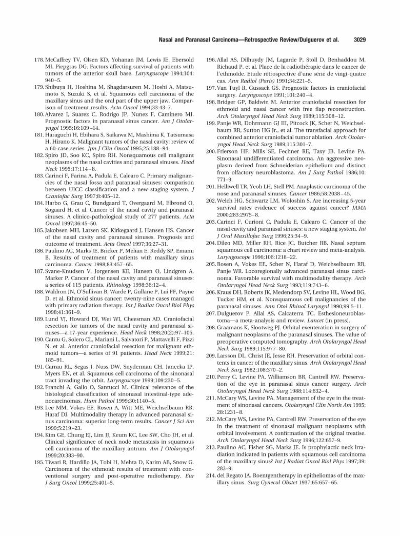
178. McCaffrey TV, Olsen KD, Yohanan JM, Lewis JE, EbersoldMJ, Piepgras DG. Factors affecting survival of patients withtumors of the anterior skull base. Laryngoscope 1994;104:940 –5.
179. Shibuya H, Hoshina M, Shagdarsuren M, Hoshi A, Matsu-moto S, Suzuki S, et al. Squamous cell carcinoma of themaxillary sinus and the oral part of the upper jaw. Compar-ison of treatment results. Acta Oncol 1994;33:43–7.
180. Alvarez I, Suarez C, Rodrigo JP, Nunez F, Caminero MJ.Prognostic factors in paranasal sinus cancer. Am J Otolar-yngol 1995;16:109 –14.
181. Haraguchi H, Ebihara S, Saikawa M, Mashima K, TatsumasaH, Hirano K. Malignant tumors of the nasal cavity: review ofa 60-case series. Jpn J Clin Oncol 1995;25:188 –94.
182. Spiro JD, Soo KC, Spiro RH. Nonsquamous cell malignantneoplasms of the nasal cavities and paranasal sinuses. HeadNeck 1995;17:114 – 8.
183. Carinci F, Farina A, Padula E, Calearo C. Primary malignan-cies of the nasal fossa and paranasal sinuses: comparisonbetween UICC classification and a new staging system. JCraniofac Surg 1997;8:405–12.
184. Harbo G, Grau C, Bundgaard T, Overgaard M, Elbrond O,Sogaard H, et al. Cancer of the nasal cavity and paranasalsinuses. A clinico-pathological study of 277 patients. ActaOncol 1997;36:45–50.
185. Jakobsen MH, Larsen SK, Kirkegaard J, Hansen HS. Cancerof the nasal cavity and paranasal sinuses. Prognosis andoutcome of treatment. Acta Oncol 1997;36:27–31.
186. Paulino AC, Marks JE, Bricker P, Melian E, Reddy SP, EmamiB. Results of treatment of patients with maxillary sinuscarcinoma. Cancer 1998;83:457– 65.
187. Svane-Knudsen V, Jorgensen KE, Hansen O, Lindgren A,Marker P. Cancer of the nasal cavity and paranasal sinuses:a series of 115 patients. Rhinology 1998;36:12– 4.
188. Waldron JN, O’Sullivan B, Warde P, Gullane P, Lui FF, PayneD, et al. Ethmoid sinus cancer: twenty-nine cases managedwith primary radiation therapy. Int J Radiat Oncol Biol Phys1998;41:361–9.
189. Lund VJ, Howard DJ, Wei WI, Cheesman AD. Craniofacialresection for tumors of the nasal cavity and paranasal si-nuses—a 17-year experience. Head Neck 1998;20(2):97–105.
190. Cantu G, Solero CL, Mariani L, Salvatori P, Mattavelli F, PizziN, et al. Anterior craniofacial resection for malignant eth-moid tumors—a series of 91 patients. Head Neck 1999;21:185–91.
191. Carrau RL, Segas J, Nuss DW, Snyderman CH, Janecka IP,Myers EN, et al. Squamous cell carcinoma of the sinonasaltract invading the orbit. Laryngoscope 1999;109:230 –5.
192. Franchi A, Gallo O, Santucci M. Clinical relevance of thehistological classification of sinonasal intestinal-type ade-nocarcinomas. Hum Pathol 1999;30:1140 –5.
193. Lee MM, Vokes EE, Rosen A, Witt ME, Weichselbaum RR,Haraf DJ. Multimodality therapy in advanced paranasal si-nus carcinoma: superior long-term results. Cancer J Sci Am1999;5:219 –23.
194. Kim GE, Chung EJ, Lim JJ, Keum KC, Lee SW, Cho JH, et al.Clinical significance of neck node metastasis in squamouscell carcinoma of the maxillary antrum. Am J Otolaryngol1999;20:383–90.
195. Tiwari R, Hardillo JA, Tobi H, Mehta D, Karim AB, Snow G.Carcinoma of the ethmoid: results of treatment with con-ventional surgery and post-operative radiotherapy. EurJ Surg Oncol 1999;25:401–5.
196. Allal AS, Dilhuydy JM, Lagarde P, Stoll D, Benhaddou M,Richaud P, et al. Place de la radiotherapie dans le cancer del’ethmoıde. Etude retrospective d’une serie de vingt-quatrecas. Ann Radiol (Paris) 1991;34:221–5.
197. Van Tuyl R, Gussack GS. Prognostic factors in craniofacialsurgery. Laryngoscope 1991;101:240 – 4.
198. Bridger GP, Baldwin M. Anterior craniofacial resection forethmoid and nasal cancer with free flap reconstruction.Arch Otolaryngol Head Neck Surg 1989;115:308 –12.
199. Panje WR, Dohrmann GJ III, Pitcock JK, Scher N, Weichsel-baum RR, Sutton HG Jr., et al. The transfacial approach forcombined anterior craniofacial tumor ablation. Arch Otolar-yngol Head Neck Surg 1989;115:301–7.
200. Frierson HF, Mills SE, Fechner RE, Taxy JB, Levine PA.Sinonasal undifferentiated carcinoma. An aggressive neo-plasm derived from Schneiderian epithelium and distinctfrom olfactory neuroblastoma. Am J Surg Pathol 1986;10:771–9.
201. Helliwell TR, Yeoh LH, Stell PM. Anaplastic carcinoma of thenose and paranasal sinuses. Cancer 1986;58:2038 – 45.
202. Welch HG, Schwartz LM, Woloshin S. Are increasing 5-yearsurvival rates evidence of success against cancer? JAMA2000;283:2975– 8.
203. Carinci F, Curioni C, Padula E, Calearo C. Cancer of thenasal cavity and paranasal sinuses: a new staging system. IntJ Oral Maxillofac Surg 1996;25:34 –9.
204. Dileo MD, Miller RH, Rice JC, Butcher RB. Nasal septumsquamous cell carcinoma: a chart review and meta-analysis.Laryngoscope 1996;106:1218 –22.
205. Rosen A, Vokes EE, Scher N, Haraf D, Weichselbaum RR,Panje WR. Locoregionally advanced paranasal sinus carci-noma. Favorable survival with multimodality therapy. ArchOtolaryngol Head Neck Surg 1993;119:743– 6.
206. Kraus DH, Roberts JK, Medendorp SV, Levine HL, Wood BG,Tucker HM, et al. Nonsquamous cell malignancies of theparanasal sinuses. Ann Otol Rhinol Laryngol 1990;99:5–11.
207. Dulguerov P, Allal AS, Calcaterra TC. Esthesioneuroblas-toma—a meta-analysis and review. Lancet (in press).
208. Graamans K, Slootweg PJ. Orbital exenteration in surgery ofmalignant neoplasms of the paranasal sinuses. The value ofpreoperative computed tomography. Arch Otolaryngol HeadNeck Surg 1989;115:977– 80.
209. Larsson DL, Christ JE, Jesse RH. Preservation of orbital con-tents in cancer of the maxillary sinus. Arch Otolaryngol HeadNeck Surg 1982;108:370 –2.
210. Perry C, Levine PA, Williamson BR, Cantrell RW. Preserva-tion of the eye in paranasal sinus cancer surgery. ArchOtolaryngol Head Neck Surg 1988;114:632– 4.
211. McCary WS, Levine PA. Management of the eye in the treat-ment of sinonasal cancers. Otolaryngol Clin North Am 1995;28:1231– 8.
212. McCary WS, Levine PA, Cantrell RW. Preservation of the eyein the treatment of sinonasal malignant neoplasms withorbital involvement. A confirmation of the original treatise.Arch Otolaryngol Head Neck Surg 1996;122:657–9.
213. Paulino AC, Fisher SG, Marks JE. Is prophylactic neck irra-diation indicated in patients with squamous cell carcinomaof the maxillary sinus? Int J Radiat Oncol Biol Phys 1997;39:283–9.
214. del Regato JA. Roentgentherapy in epitheliomas of the max-illary sinus. Surg Gynecol Obstet 1937;65:657– 65.
Nasal and Paranasal Carcinoma—Retrospective Review/Dulguerov et al. 3029