Cholera toxin impairs the differentiation of monocytes into dendritic cells, inducing professional...
11
INFECTION AND IMMUNITY, Mar. 2011, p. 1300–1310 Vol. 79, No. 3 0019-9567/11/$12.00 doi:10.1128/IAI.01181-10 Copyright © 2011, American Society for Microbiology. All Rights Reserved. Cholera Toxin Impairs the Differentiation of Monocytes into Dendritic Cells, Inducing Professional Antigen-Presenting Myeloid Cells Filippo Veglia, Ester Sciaraffia, Antonella Riccomi, Dora Pinto, Donatella R. M. Negri, Maria Teresa De Magistris, and Silvia Vendetti* Department of Infectious, Parasitic, and Immune-Mediated Diseases, Istituto Superiore di Sanita `, Rome, Italy Received 7 July 2010/Returned for modification 20 July 2010/Accepted 1 December 2010 Cholera toxin (CT) is a potent adjuvant for mucosal vaccination; however, its mechanism of action has not been clarified completely. It is well established that peripheral monocytes differentiate into dendritic cells (DCs) both in vitro and in vivo and that monocytes are the in vivo precursors of mucosal CD103 proinflam- matory DCs. In this study, we asked whether CT had any effects on the differentiation of monocytes into DCs. We found that CT-treated monocytes, in the presence of granulocyte-macrophage colony-stimulating factor (GM-CSF) and interleukin 4 (IL-4), failed to differentiate into classical DCs (CD14 low CD1a high ) and acquired a macrophage-like phenotype (CD14 high CD1a low ). Cells differentiated in the presence of CT expressed high levels of major histocompatibility complex class I (MHC-I) and MHC-II and CD80 and CD86 costimulatory molecules and produced larger amounts of IL-1, IL-6, and IL-10 but smaller amounts of tumor necrosis factor alpha (TNF-) and IL-12 than did monocytes differentiated into DCs in the absence of CT. The enzymatic activity of CT was found to be important for the skewing of monocytes toward a macrophage-like phenotype (Ma-DCs) with enhanced antigen-presenting functions. Indeed, treatment of monocytes with scalar doses of forskolin (FSK), an activator of adenylate cyclase, induced them to differentiate in a dose-dependent manner into a population with phenotype and functions similar to those found after CT treatment. Monocytes differentiated in the presence of CT induced the differentiation of naïve T lymphocytes toward a Th2 phenotype. Interestingly, we found that CT interferes with the differentiation of monocytes into DCs in vivo and promotes the induction of activated antigen-presenting cells (APCs) following systemic immunization. Adjuvant design has been mainly empirical, and the mech- anism of action of the most efficacious molecules has remained obscure. It is important to investigate the mechanisms of action of already known adjuvants to facilitate the design of new effective molecules with high potency and decreased side ef- fects. The bacterial enterotoxin cholera toxin (CT) from Vibrio cholerae is extraordinarily effective as a mucosal and systemic adjuvant, yet its capacity to amplify the immune response has not been completely clarified (12, 24, 25). It is a holotoxin, composed of an enzymatically active A subunit, noncovalently linked to a pentameric B subunit, which binds to the ganglio- side GM1 on host cell membranes. Once internalized, the A subunit ADP-ribosylates the subunit of the GTP-binding regulatory protein Gs, thereby inducing permanent adenylate cyclase activation, resulting in an increase of intracellular cyclic AMP (cAMP) (55). The mechanism of the adjuvanticity of CT may be a complex phenomenon resulting from the interaction of the toxin with different cell types present in the architecture of the mucosa. Dissecting the effect of CT on different cells types could help in understanding the contribution of each to its adjuvant activity. Given the crucial role of dendritic cells (DCs) and other professional antigen-presenting cells (APCs) (monocytes/mac- rophages and B cells) in the induction of adaptive immunity (26), the potentiation of APC function can be a major aspect of adjuvant action. Cholera toxin upregulates expression of the B7-2 (CD86) costimulatory molecule and stimulates antigen presentation through enhancement of major histocompatibility complex class II (MHC-II) expression and interleukin 1 (IL- 1) production (7, 11, 37, 54). CT also interacts with lympho- cytes and promotes B cell isotype-switch differentiation toward IgG1 and IgA in mice (2, 54) and enhances the antigen-pre- senting function of human B cells (38). We and others showed that CT, by inducing maturation of human DCs, polarizes a mixed Th1/Th2 T cell response, with a strong bias toward Th2 cells (15, 16). The capacity of CT to interact with DCs or monocytes as innate immune cells or as precursors of DCs may represent a crucial step for its adjuvant mechanism. DCs represent hetero- geneous populations that comprise distinct subtypes that vary in hematopoietic origin, life cycle, and function (4, 52). It is well established that peripheral monocytes differentiate into DCs both in vitro and in vivo (19, 20, 43–45, 48) and that monocytes are the in vivo precursors of mucosal CD103 proinflammatory DCs (5, 27, 58). More important, the role of monocytes as precursors of DCs in mediating the in vivo ad- juvant effects has been described (29, 60). Therefore, in this study, we asked whether CT affected the differentiation of monocytes into DCs. By using human DCs generated from monocytes (48), we found that CT interferes with the differ- entiation of monocytes into DCs, giving rise to a distinct population (Ma-DCs), which displays an activated macro- phage-like phenotype, induces a strong allogeneic and antigen- specific response, and promotes the polarization of naive CD4 T lymphocytes toward a Th2 profile. Interestingly, we * Corresponding author. Mailing address: Department of Infectious, Parasitic, and Immune-Mediated Diseases, Istituto Superiore di Sanita `, Viale Regina Elena, 00161 Rome, Italy. Phone: 39-06- 49903145. Fax: 39-06-49387112. E-mail: [email protected]. Published ahead of print on 13 December 2010. 1300
Transcript of Cholera toxin impairs the differentiation of monocytes into dendritic cells, inducing professional...
INFECTION AND IMMUNITY, Mar. 2011, p. 1300–1310 Vol. 79, No. 30019-9567/11/$12.00 doi:10.1128/IAI.01181-10Copyright © 2011, American Society for Microbiology. All Rights Reserved.
Cholera Toxin Impairs the Differentiation of Monocytes into DendriticCells, Inducing Professional Antigen-Presenting Myeloid Cells�
Filippo Veglia, Ester Sciaraffia, Antonella Riccomi, Dora Pinto, Donatella R. M. Negri,Maria Teresa De Magistris, and Silvia Vendetti*
Department of Infectious, Parasitic, and Immune-Mediated Diseases, Istituto Superiore di Sanita, Rome, Italy
Received 7 July 2010/Returned for modification 20 July 2010/Accepted 1 December 2010
Cholera toxin (CT) is a potent adjuvant for mucosal vaccination; however, its mechanism of action has notbeen clarified completely. It is well established that peripheral monocytes differentiate into dendritic cells(DCs) both in vitro and in vivo and that monocytes are the in vivo precursors of mucosal CD103� proinflam-matory DCs. In this study, we asked whether CT had any effects on the differentiation of monocytes into DCs.We found that CT-treated monocytes, in the presence of granulocyte-macrophage colony-stimulating factor(GM-CSF) and interleukin 4 (IL-4), failed to differentiate into classical DCs (CD14low CD1ahigh) and acquireda macrophage-like phenotype (CD14high CD1alow). Cells differentiated in the presence of CT expressed highlevels of major histocompatibility complex class I (MHC-I) and MHC-II and CD80 and CD86 costimulatorymolecules and produced larger amounts of IL-1�, IL-6, and IL-10 but smaller amounts of tumor necrosisfactor alpha (TNF-�) and IL-12 than did monocytes differentiated into DCs in the absence of CT. Theenzymatic activity of CT was found to be important for the skewing of monocytes toward a macrophage-likephenotype (Ma-DCs) with enhanced antigen-presenting functions. Indeed, treatment of monocytes with scalardoses of forskolin (FSK), an activator of adenylate cyclase, induced them to differentiate in a dose-dependentmanner into a population with phenotype and functions similar to those found after CT treatment. Monocytesdifferentiated in the presence of CT induced the differentiation of naïve T lymphocytes toward a Th2 phenotype.Interestingly, we found that CT interferes with the differentiation of monocytes into DCs in vivo and promotesthe induction of activated antigen-presenting cells (APCs) following systemic immunization.
Adjuvant design has been mainly empirical, and the mech-anism of action of the most efficacious molecules has remainedobscure. It is important to investigate the mechanisms of actionof already known adjuvants to facilitate the design of neweffective molecules with high potency and decreased side ef-fects. The bacterial enterotoxin cholera toxin (CT) from Vibriocholerae is extraordinarily effective as a mucosal and systemicadjuvant, yet its capacity to amplify the immune response hasnot been completely clarified (12, 24, 25). It is a holotoxin,composed of an enzymatically active A subunit, noncovalentlylinked to a pentameric B subunit, which binds to the ganglio-side GM1 on host cell membranes. Once internalized, the Asubunit ADP-ribosylates the � subunit of the GTP-bindingregulatory protein Gs, thereby inducing permanent adenylatecyclase activation, resulting in an increase of intracellular cyclicAMP (cAMP) (55). The mechanism of the adjuvanticity of CTmay be a complex phenomenon resulting from the interactionof the toxin with different cell types present in the architectureof the mucosa. Dissecting the effect of CT on different cellstypes could help in understanding the contribution of each toits adjuvant activity.
Given the crucial role of dendritic cells (DCs) and otherprofessional antigen-presenting cells (APCs) (monocytes/mac-rophages and B cells) in the induction of adaptive immunity(26), the potentiation of APC function can be a major aspect of
adjuvant action. Cholera toxin upregulates expression of theB7-2 (CD86) costimulatory molecule and stimulates antigenpresentation through enhancement of major histocompatibilitycomplex class II (MHC-II) expression and interleukin 1� (IL-1�) production (7, 11, 37, 54). CT also interacts with lympho-cytes and promotes B cell isotype-switch differentiation towardIgG1 and IgA in mice (2, 54) and enhances the antigen-pre-senting function of human B cells (38). We and others showedthat CT, by inducing maturation of human DCs, polarizes amixed Th1/Th2 T cell response, with a strong bias toward Th2cells (15, 16).
The capacity of CT to interact with DCs or monocytes asinnate immune cells or as precursors of DCs may represent acrucial step for its adjuvant mechanism. DCs represent hetero-geneous populations that comprise distinct subtypes that varyin hematopoietic origin, life cycle, and function (4, 52). It iswell established that peripheral monocytes differentiate intoDCs both in vitro and in vivo (19, 20, 43–45, 48) and thatmonocytes are the in vivo precursors of mucosal CD103�
proinflammatory DCs (5, 27, 58). More important, the role ofmonocytes as precursors of DCs in mediating the in vivo ad-juvant effects has been described (29, 60). Therefore, in thisstudy, we asked whether CT affected the differentiation ofmonocytes into DCs. By using human DCs generated frommonocytes (48), we found that CT interferes with the differ-entiation of monocytes into DCs, giving rise to a distinctpopulation (Ma-DCs), which displays an activated macro-phage-like phenotype, induces a strong allogeneic and antigen-specific response, and promotes the polarization of naiveCD4� T lymphocytes toward a Th2 profile. Interestingly, we
* Corresponding author. Mailing address: Department of Infectious,Parasitic, and Immune-Mediated Diseases, Istituto Superiore diSanita, Viale Regina Elena, 00161 Rome, Italy. Phone: 39-06-49903145. Fax: 39-06-49387112. E-mail: [email protected].
� Published ahead of print on 13 December 2010.
1300
found that CT interferes with the differentiation of monocytesinto DCs in vivo and promotes the induction of activated an-tigen-presenting cells following systemic immunization.
MATERIALS AND METHODS
Media and reagents. RPMI 1640 supplemented with 2 mM L-glutamine, 1%nonessential amino acids, 1% pyruvate, 100 U/ml penicillin, 100 �g/ml strepto-mycin (Gibco, NY), and 10% fetal bovine serum (FBS) (HyClone Laboratories,Logan, UT) was used as complete medium in all cell cultures. Cholera toxin andcholera toxin B subunit (CT-B) were purchased from Calbiochem-NovabiochemCo. (San Diego, CA), and forskolin (FSK), ionomycin (Ion), phorbol 12-myris-tate 13-acetate (PMA), brefeldin A (BFA), lipopolysaccharide (LPS), andovalbumin (OVA) were purchased from Sigma Chemicals Co., St. Louis, MO.GM-CSF and IL-4 were purchased from Immunological Science (Rome, Italy),and murine GM-CSF (mGM-CSF) was purchased from Peprotech, (Rocky Hill,NJ). Carboxyfluorescein succiniminidyl ester (CFSE) was purchased from Mo-lecular Probes (Eugene, OR).
Animals. Female BALB/c mice and DO11.10 TCR transgenic mice aged 6 to8 weeks were obtained from Harlan Nossan (Correzzana, Italy) and CharlesRiver (Calco, Italy), respectively. All mice were maintained in our animal facil-ities for the duration of the experiments, and all procedures were in accordancewith the institutional guidelines and have been approved by the institutionalcommittee.
Monocyte purification and cell cultures. Human monocytes were purifiedfrom healthy donors’ peripheral blood mononuclear cells (PBMC) by positiveselection using anti-CD14-conjugated magnetic microbeads (Miltenyi Biotec,Bergisch Gladbach, Germany). The recovered cells were 95% to 99% CD14� asdetermined by flow cytometry using the FITC conjugate anti-human CD14monoclonal antibody (MAb) (BD Biosciences, San Diego, CA). Monocytes werecultured at 1 � 106/ml in RPMI-FBS supplemented with GM-CSF (50 ng/ml)and IL-4 (35 ng/ml), in the presence of 0.03 �g/ml CT, 0.03 �g/ml CT-B, orescalating doses of FSK (10, 20, and 40 �M or 10 �M for 3 days). All stimuli werekept in culture up to 6 days, and then the cells were washed and stained withFITC or phycoerythrin (PE) conjugate anti-human CD14, CD1a, HLA-DR,HLA-I, CD80, CD86, CD209 (DC-Sign), CD64, CD32, CD89, CCR7, and CD83(BD Biosciences, San Diego, CA) to verify their differentiation, activation, andmaturation status.
Murine CD11b� monocytes were obtained from single-cell suspensions ofBALB/c femur bone marrow by positive selection using anti-mouse CD11b-conjugated magnetic microbeads (Miltenyi Biotec, Bergisch Gladbach, Ger-many). The recovered cells were 95% to 99% CD11b� as determinated by flowcytometry using the FITC conjugate anti-mouse CD11b MAb (BD Biosciences,San Diego, CA), and they were cultured (1 � 106/ml) in RPMI-FBS supple-mented with mGM-CSF (30 ng/ml), 2-mercaptoethanol (50 �M) in the presenceof 0.3 �g/ml CT. At day 2, mGM-CSF (15 ng/ml) was added again to cellcultures. After 6 days, cells were washed and stained with FITC or PE conjugateanti-mouse CD11c, MHC-II, and CD86 (BD Biosciences, San Diego, CA). Cellswere acquired on a FACSCalibur instrument running CellQuest software (BDBiosciences, San Diego, CA).
Naïve CD4� T cell purification. PBMC were isolated from healthy donors byFicoll-Hypaque (Pharmacia, Uppsala, Sweden) density centrifugation. CD4� Tlymphocytes were purified by negative selection using immunomagnetic cellsorting (Miltenyi Biotec, Bergisch Gladbach, Germany). Briefly, PBMC werelabeled using a cocktail of the hapten-conjugated MAbs anti-CD8, -CD11b,-CD14, -CD16, -CD19, -CD36, -CD56 -CD123, -TCR�/�, and -glycophorin A incombination with MACS microbeads coupled to an anti-hapten monoclonalantibody. The magnetically labeled cells were depleted by retaining them on acolumn using a MidiMACS cell separator. CD4� T cells were further purified inCD4� CD45RO� and CD4� CD45RA� T cells by positive selection usinganti-CD45RA microbeads (Miltenyi Biotec, Bergisch Gladbach, Germany).
Cytokine production. Supernatants from monocytes differentiated in the pres-ence or in the absence of CT or CT-B for 6 days and further cultured for 48 hwith or without LPS (250 ng/ml) were collected and stored at �80°C. The levelsof IL-1�, IL-6, TNF-�, IL-10, and IL-12 were detected by Quantikine immuno-assay kits (Endogene, Cambridge, MA) and were measured as absorbance (450nm) on an enzyme-linked immunosorbent assay (ELISA) reader.
Proliferation and polarization assays. The ability of monocytes differentiatedin the presence or in the absence of CT (0.03 �g/ml) to activate T cells wasevaluated by the use of the mixed lymphocyte reaction or antigen-specific T cellresponse. At day 6, cells were washed, starved for 8 h, and cocultured withallogeneic PBMC (1 � 105) at different PBMC/DC ratios (4:1, 2:1, and 1:1) for
5 days in round-bottom, 96-well plates. Proliferation was evaluated by [3H]thy-midine incorporation. Plates were incubated for 48 h at 37°C with 5% CO2, and[3H]thymidine (Amersham, Aylesbury, United Kingdom) was added (1 �Ci/well). After 18 h, cells were harvested, and the incorporated radioactivity wasmeasured by Micro� counting. To evaluate the ability of monocytes differenti-ated in the presence or in the absence of CT (0.03 �g/ml) or FSK (10 �M) tostimulate an antigen-specific T cell response, cells were isolated from donorsimmune to the tetanus toxoid (TT) antigen. DCs were cocultured with autolo-gous CFSE-labeled peripheral blood lymphocytes (PBLs) (1 � 105 at a ratio of1:1) in the presence of TT (2 �g/ml). Briefly, autologous PBLs (107/ml) werewashed in phosphate-buffered saline (PBS) 1% fetal calf serum (FCS), incubatedwith 0.5 �M CFSE for 10 min at 37°C, and then washed three times withcomplete medium. After 5 days, antigen-specific proliferation was evaluated byCFSE dilution, and cells were double stained with anti-CD4-PE and anti-CD8-Cy5.5 MAbs and acquired and analyzed on a FACSCalibur instrument runningCellQuest software.
The ability of monocytes differentiated in the presence or in the absence of CT(0.03 �g/ml) to stimulate and polarize naïve T cells was evaluated. At day 6, cellswere washed, starved for 8 h, and cocultured with purified naïve CD4�
CD45RA� T cells (ratio of 1:5) in 48-well plates, at 37°C for 11 days. IL-2 (20UI/ml) was added to the culture at days 5, 7, and 9 of the cocultures, and after11 days, cells were harvested, washed, and analyzed for cytokine production byintracellular staining. In brief, cells were stimulated with 50 ng/ml PMA and 1�g/ml ionomycin; after 1 h, 10 �g/ml of brefeldin (Sigma Chemicals Co., St.Louis, MO) was added, and they were incubated for a further 5 h at 37°C. Cellswere washed twice in PBS, 1% bovine serum albumin (BSA), and 0.1% sodiumazide and stained with anti-CD4 MAb for 15 min at 4°C, and then they were fixedwith lysing solution (BD Biosciences, San Diego, CA), permeabilized with per-meabilizing solution (BD Biosciences, San Diego, CA), and stained with PE orFITC conjugate anti-human IL-4 and gamma interferon (IFN-�) (BD Bio-sciences, San Diego, CA). Samples were acquired and analyzed on a FACSCaliburinstrument running CellQuest software.
Adoptive transfer experiments. Purified CD11b� monocytes were labeled withCFSE (0.5 �M) at room temperature (RT) in the dark for 8 min, washed threetimes with RPMI-10% FBS and intraperitoneally (i.p.) injected (8 � 106) intoBALB/c recipient mice. In all experiments, OVA-specific T cells (2 � 106)obtained from the spleen and lymph nodes of DO11.10 TCR transgenic micewere intravenously injected. Adoptively transferred mice were immunized i.p.with OVA (5 �g/dose) or OVA plus CT (1 �g/dose). After 3 days, the medias-tinal lymph nodes were collected, stained with anti-CD11b, -CD11c, -MHC-I(H2Kd), -MHC-II (I-A/I-E), -CD86, -CD4 (BD Biosciences, San Diego, CA),and -DO11.10 TCR (eBioscience, San Diego, CA) and acquired on a FACS-Canto instrument running Diva software (BD Biosciences, San Diego, CA).
Statistical analysis. Microsoft Excel (Microsoft Corp., Redmond, WA) wasused for statistical analysis. Data were expressed as the mean standard devi-ation (SD), and statistical significance was determined by Student’s t test; a Pvalue of 0.05 was considered statistically significant.
RESULTS
Cholera toxin interferes with the differentiation of mono-cytes into DCs, giving rise to a macrophage-like population(Ma-DCs). To investigate the effects of CT on the differenti-ation of monocytes into DCs, human monocytes were culturedwith GM-CSF and IL-4 for 6 days in the presence or absenceof CT. In the absence of CT, monocytes differentiated intoimmature DCs characterized by the expression of CD1a andthe loss of CD14 molecules. In contrast, cells derived fromCT-treated monocytes did not express CD1a and retained theexpression of CD14, suggesting that they acquired a monocyte/macrophage-like phenotype (Ma-DCs) (Fig. 1A and B). Toinvestigate whether the GM1-binding B subunit (CT-B) or theenzymatic activity of the A subunit of CT was important for thedifferentiation toward this monocyte/macrophage-like popula-tion, monocytes were induced to differentiate into DCs in thepresence of CT-B or forskolin (FSK), an activator of adenylatecyclases. Monocytes treated with CT-B showed a phenotypesimilar to that of untreated cells, whereas FSK-stimulated
VOL. 79, 2011 CHOLERA TOXIN IMPAIRS DC DIFFERENTIATION 1301

monocytes differentiate into a macrophage-like populationsimilar to that derived from CT-treated monocytes (Fig. 1Cand D). This suggests that the enzymatic activity of CT and itscapacity to increase intracellular cAMP levels is important forthe skewing of monocytes toward a macrophage-like pheno-type.
Ma-DCs express DC-Sign and FcR patterns typical of ter-minally differentiated myeloid cells. We evaluated the effect ofCT on surface expression of DC-Sign (CD209), a DC-re-stricted C-type lectin involved in the early interaction betweenDCs and naïve T cells and also the DC trafficking and inter-nalization of Ag (17, 56). DC-Sign is upregulated during thedifferentiation of monocytes into DCs (18), and this upregula-tion was not completely inhibited by treatment with CT or FSK(Fig. 2B); however, CT- or FSK-treated monocytes expressedsignificantly (P � 0.0004, n � 4 for CT and P � 0.0009, n � 4for FSK) lower levels of DC-Sign than control cells did. Wealso analyzed the expression of Fc-�RI (CD64), the high-af-finity IgG Fc receptor (FcR), which is constitutively expressedon monocytes and down-modulated during DC differentiation.CT-treated or untreated monocytes induced to differentiate
into DCs did not express CD64 molecules, showing that thedown-modulation of CD64 was not affected by CT treatment(Fig. 2B).
Terminal differentiation of myeloid cells is characterized bya switch of expression from Fc-�RI/CD64 to low-affinity Fc-�RII/CD32 and other FcRs, such as CD89, the IgA-specificFcR (8, 41). Therefore, we compared the expression of thelow-affinity receptor CD32 and, because of the peculiar capac-ity of CT to promote mucosal IgA production, the expressionof CD89 in cells differentiated from CT-treated and untreatedmonocytes. CD32 is expressed on monocytes and upregulatedduring either macrophage or, to lesser extent, DC differentia-tion. Treatment with CT during monocyte differentiation intoDCs caused an increase of CD32 greater than that of controlcells (P � 0.009, n � 6), a further indication of a macrophage-like phenotype (Fig. 2A and B). Once again treatment withFSK induced the expression of CD32 at levels comparable tothat of CT-treated cells (P � 0.0003, n � 6) (Fig. 2D), sug-gesting that the modulation of CD32 is dependent on theadenylate cyclase activation capacity of the toxin. Further, astrong upregulation of CD89 FcR was observed in cells differ-
FIG. 1. Cholera toxin impairs differentiation of human monocytes into DCs. Dot and histogram plots show the phenotype of human CD14monocytes isolated from healthy donors’ PBMC and cultured for 6 days with GM-CSF (50 ng/ml) and IL-4 (35 ng/ml) in the presence of mediumalone (A), CT (0.03 �g/ml) (B), CT-B (0.03 �g/ml) (C), and FSK (10 �M) (D). Cells were double stained using anti-CD14-FITC and anti-CD1a-PEMAbs and analyzed by flow cytometry. Dot and histogram plots are representative of one experiment out of six performed. The histogram barsshow the means (�SD) of the relative mean fluorescence intensity (MFI) of CD14 and CD1a expression from six independent experimentsperformed. The numbers within the plots indicate the percentage of positive cells (dot plots) and the mean fluorescence intensity (histogram plots).
1302 VEGLIA ET AL. INFECT. IMMUN.
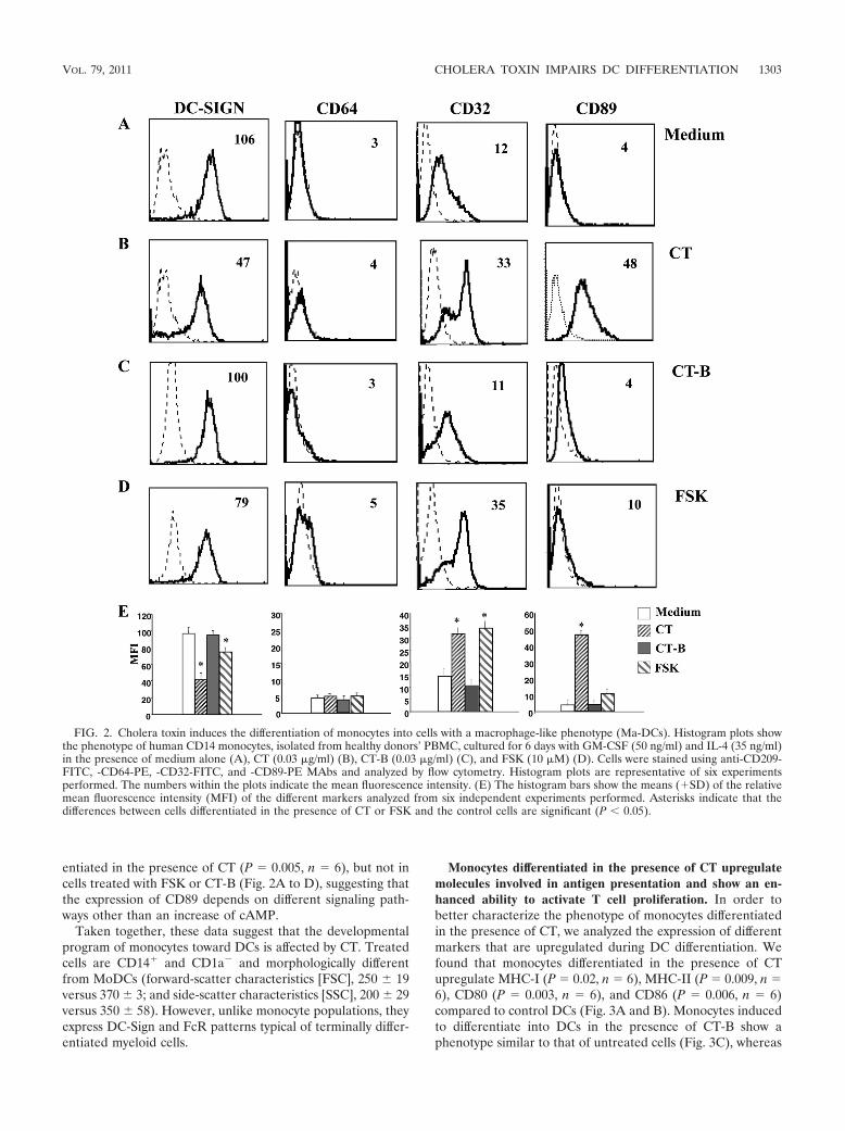
entiated in the presence of CT (P � 0.005, n � 6), but not incells treated with FSK or CT-B (Fig. 2A to D), suggesting thatthe expression of CD89 depends on different signaling path-ways other than an increase of cAMP.
Taken together, these data suggest that the developmentalprogram of monocytes toward DCs is affected by CT. Treatedcells are CD14� and CD1a� and morphologically differentfrom MoDCs (forward-scatter characteristics [FSC], 250 19versus 370 3; and side-scatter characteristics [SSC], 200 29versus 350 58). However, unlike monocyte populations, theyexpress DC-Sign and FcR patterns typical of terminally differ-entiated myeloid cells.
Monocytes differentiated in the presence of CT upregulatemolecules involved in antigen presentation and show an en-hanced ability to activate T cell proliferation. In order tobetter characterize the phenotype of monocytes differentiatedin the presence of CT, we analyzed the expression of differentmarkers that are upregulated during DC differentiation. Wefound that monocytes differentiated in the presence of CTupregulate MHC-I (P � 0.02, n � 6), MHC-II (P � 0.009, n �6), CD80 (P � 0.003, n � 6), and CD86 (P � 0.006, n � 6)compared to control DCs (Fig. 3A and B). Monocytes inducedto differentiate into DCs in the presence of CT-B show aphenotype similar to that of untreated cells (Fig. 3C), whereas
FIG. 2. Cholera toxin induces the differentiation of monocytes into cells with a macrophage-like phenotype (Ma-DCs). Histogram plots showthe phenotype of human CD14 monocytes, isolated from healthy donors’ PBMC, cultured for 6 days with GM-CSF (50 ng/ml) and IL-4 (35 ng/ml)in the presence of medium alone (A), CT (0.03 �g/ml) (B), CT-B (0.03 �g/ml) (C), and FSK (10 �M) (D). Cells were stained using anti-CD209-FITC, -CD64-PE, -CD32-FITC, and -CD89-PE MAbs and analyzed by flow cytometry. Histogram plots are representative of six experimentsperformed. The numbers within the plots indicate the mean fluorescence intensity. (E) The histogram bars show the means (�SD) of the relativemean fluorescence intensity (MFI) of the different markers analyzed from six independent experiments performed. Asterisks indicate that thedifferences between cells differentiated in the presence of CT or FSK and the control cells are significant (P 0.05).
VOL. 79, 2011 CHOLERA TOXIN IMPAIRS DC DIFFERENTIATION 1303

FSK induces monocytes to differentiate into an activated mac-rophage-like population similar to that derived from CT-treated monocytes (Fig. 3D). Although the expression levels ofthe differentiation markers elicited by FSK were lower than
those in CT-treated cells, their expression scaled up withhigher doses of FSK in a dose-dependent manner (Table 1),suggesting that the effects on DC differentiation by CT aredependent on the increase of intracellular cAMP levels. Fur-
FIG. 3. Monocytes differentiated in the presence of CT upregulate molecules involved in antigen presentation and activation of T cells. Histogramplots show the phenotype of human CD14 monocytes isolated from healthy donors’ PBMC, cultured for 6 days with GM-CSF (50 ng/ml) and IL-4 (35ng/ml) in the presence of medium alone (A), CT (0.03 �g/ml) (B), CT-B (0.03 �g/ml) (C), and FSK (10 �M) (D). Cells were stained using anti-HLA-IPE,-HLA-DR-FITC, -CD80-PE, -CD86-FITC, and -CCR7-PE MAbs and analyzed by flow cytometry. Histogram plots are representative of six experimentsperformed. The numbers within the plots indicate the mean fluorescence intensity. (E) The histogram bars show the means (�SD) of the relative meanfluorescence intensity (MFI) of the different markers analyzed from six independent experiments performed. Asterisks indicate that the differencesbetween cells differentiated in the presence of CT or FSK and the control cells are significant (P 0.05).
TABLE 1. Expression of differentiation markers on monocytes differentiated in the presence of scalar doses of FSK is dose dependent
TreatmentExpression of differentiation markersa
CD14 CD1a HLA-I HLA-DR CD80 CD86
Medium 6.5 1.3 102 29 31 5.2 14 4.3 11.6 3.5 7.3 2.3CT, 0.03 �g/ml 49.3 14.4 9.3 1.5 80 10 170 38 24.6 6.5 32.6 13
FSK10 �M 25.3 4.6 57 8.5 61 8 72 3.5 19 5 22.6 10.620 �M 39 4.2 31.5 21 72 7 140 35 16 2.8 32 1440 �M 41 2 27.5 17.6 75 3.5 160 56 19 5.6 46 20.53 � 10 �Mb 43 4.3 25 17 71 1 167 53 18 4.2 51 29.7
a Values are mean fluorescence intensity (MFI) standard deviation (three samples per group).b Forskolin was added to the cultures daily at 10 mM for 3 days.
1304 VEGLIA ET AL. INFECT. IMMUN.
thermore, CCR7, the key chemokine receptor involved in themigration of mature DCs to the lymph nodes is upregulated byCT treatment (P � 0.002, n � 6) and to a lesser extent by FSK(P � 0.03, n � 6) (Fig. 3).
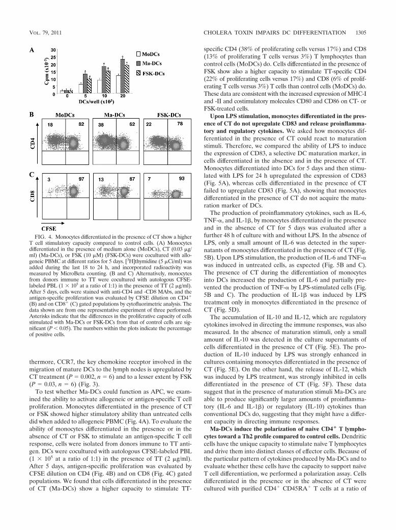
To test whether Ma-DCs could function as APC, we exam-ined the ability to activate allogeneic or antigen-specific T cellproliferation. Monocytes differentiated in the presence of CTor FSK showed higher stimulatory ability than untreated cellsdid when added to allogeneic PBMC (Fig. 4A). To evaluate theability of monocytes differentiated in the presence or in theabsence of CT or FSK to stimulate an antigen-specific T cellresponse, cells were isolated from donors immune to TT anti-gen. DCs were cocultured with autologous CFSE-labeled PBL(1 � 105 at a ratio of 1:1) in the presence of TT (2 �g/ml).After 5 days, antigen-specific proliferation was evaluated byCFSE dilution on CD4 (Fig. 4B) and on CD8 (Fig. 4C) gatedpopulations. We found that cells differentiated in the presenceof CT (Ma-DCs) show a higher capacity to stimulate TT-
specific CD4 (38% of proliferating cells versus 17%) and CD8(13% of proliferating T cells versus 3%) T lymphocytes thancontrol cells (MoDCs) do. Cells differentiated in the presence ofFSK show also a higher capacity to stimulate TT-specific CD4(22% of proliferating cells versus 17%) and CD8 (6% of prolif-erating T cells versus 3%) T cells than control cells (MoDCs) do.These data are consistent with the increased expression of MHC-Iand -II and costimulatory molecules CD80 and CD86 on CT- orFSK-treated cells.
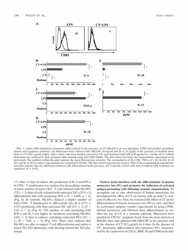
Upon LPS stimulation, monocytes differentiated in the pres-ence of CT do not upregulate CD83 and release proinflamma-tory and regulatory cytokines. We asked how monocytes dif-ferentiated in the presence of CT could react to maturationstimuli. Therefore, we compared the ability of LPS to inducethe expression of CD83, a selective DC maturation marker, incells differentiated in the absence and in the presence of CT.Monocytes differentiated into DCs for 5 days and then stimu-lated with LPS for 24 h upregulated the expression of CD83(Fig. 5A), whereas cells differentiated in the presence of CTfailed to upregulate CD83 (Fig. 5A), showing that monocytesdifferentiated in the presence of CT do not acquire the matu-ration marker of DCs.
The production of proinflammatory cytokines, such as IL-6,TNF-�, and IL-1�, by monocytes differentiated in the presenceand in the absence of CT for 5 days was evaluated after afurther 48 h of culture with and without LPS. In the absence ofLPS, only a small amount of IL-6 was detected in the super-natants of monocytes differentiated in the presence of CT (Fig.5B). Upon LPS stimulation, the production of IL-6 and TNF-�was induced in untreated cells, as expected (Fig. 5B and C).The presence of CT during the differentiation of monocytesinto DCs increased the production of IL-6 and partially pre-vented the production of TNF-� by LPS-stimulated cells (Fig.5B and C). The production of IL-1� was induced by LPStreatment only in monocytes differentiated in the presence ofCT (Fig. 5D).
The accumulation of IL-10 and IL-12, which are regulatorycytokines involved in directing the immune responses, was alsomeasured. In the absence of maturation stimuli, only a smallamount of IL-10 was detected in the culture supernatants ofcells differentiated in the presence of CT (Fig. 5E). The pro-duction of IL-10 induced by LPS was strongly enhanced incultures containing monocytes differentiated in the presence ofCT (Fig. 5E). On the other hand, the release of IL-12, whichwas induced by LPS treatment, was strongly inhibited in cellsdifferentiated in the presence of CT (Fig. 5F). These datasuggest that in the presence of maturation stimuli Ma-DCs areable to produce significantly larger amounts of proinflamma-tory (IL-6 and IL-1�) or regulatory (IL-10) cytokines thanconventional DCs do, suggesting that they might have a differ-ent capacity in directing immune responses.
Ma-DCs induce the polarization of naive CD4� T lympho-cytes toward a Th2 profile compared to control cells. Dendriticcells have the unique capacity to stimulate naïve T lymphocytesand drive them into distinct classes of effector cells. Because ofthe particular pattern of cytokines produced by Ma-DCs and toevaluate whether these cells have the capacity to support naïveT cell differentiation, we performed a polarization assay. Cellsdifferentiated in the presence or in the absence of CT werecultured with purified CD4� CD45RA� T cells at a ratio of
FIG. 4. Monocytes differentiated in the presence of CT show a higherT cell stimulatory capacity compared to control cells. (A) Monocytesdifferentiated in the presence of medium alone (MoDCs), CT (0.03 �g/ml) (Ma-DCs), or FSK (10 �M) (FSK-DCs) were cocultured with allo-geneic PBMC at different ratios for 5 days. [3H]thymidine (5 �Ci/ml) wasadded during the last 18 to 24 h, and incorporated radioactivity wasmeasured by MicroBeta counting. (B and C) Alternatively, monocytesfrom donors immune to TT were cocultured with autologous CFSE-labeled PBL (1 � 105 at a ratio of 1:1) in the presence of TT (2 �g/ml).After 5 days, cells were stained with anti-CD4 and -CD8 MAbs, and theantigen-specific proliferation was evaluated by CFSE dilution on CD4�
(B) and on CD8� (C) gated populations by cytofluorimetric analysis. Thedata shown are from one representative experiment of three performed.Asterisks indicate that the differences in the proliferative capacity of cellsstimulated with Ma-DCs or FSK-DCs from that of control cells are sig-nificant (P 0.05). The numbers within the plots indicate the percentageof positive cells.
VOL. 79, 2011 CHOLERA TOXIN IMPAIRS DC DIFFERENTIATION 1305
1:5. After 11 days of culture, the production of IL-4 and IFN-�by CD4� T lymphocytes was analyzed by intracellular staining.A lower number of naïve CD4� T cells cultured with Ma-DCs(32% 2) than of cells cultured with untreated DCs (55%3)differentiated into cells producing IFN-� (P � 0.008, n � 6)(Fig. 6). In contrast, Ma-DCs induced a higher number ofnaïve CD4� T lymphocytes to differentiate into IL-4 (15% 0.2%)-producing cells than untreated DC did (2% 1) (P �0.02, n � 6) (Fig. 6). The number of cells producing bothIFN-� and IL-4 was higher in cocultures containing Ma-DCs(10% 2) than in cultures containing untreated DCs (2% 1) (P � 0.02, n � 6) (Fig. 6A). These data indicate thatMa-DCs are able to support T cell differentiation and induce amixed Th1/Th2 phenotype, with skewing toward the Th2 phe-notype.
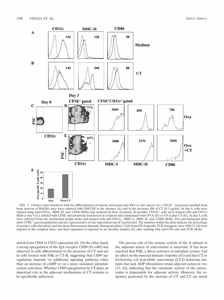
Cholera toxin interferes with the differentiation of murinemonocytes into DCs and promotes the induction of activatedantigen-presenting cells following systemic immunization. Tostrengthen our in vitro observation of human monocytes, weinvestigated the effect of CT on murine cells in order to eval-uate its effect in vivo. First, we evaluated the effect of CT on thedifferentiation of murine monocytes into DCs in vitro, and thenwe performed adoptive transfer experiments by using CFSE-labeled monocytes and followed their differentiation in vivoafter the use of CT as a systemic adjuvant. Monocytes werepurified by CD11b� magnetic beads from the bone marrow ofBALB/c mice and cultured with GM-CSF in the presence andin the absence of CT (0.3 �g/ml) for 6 days. In the absence ofCT, monocytes differentiated into immature DCs character-ized by the expression of CD11c, MHC-II and CD86 molecules
FIG. 5. Upon LPS stimulation, monocytes differentiated in the presence of CT (Ma-DCs) do not upregulate CD83 and produce proinflam-matory and regulatory cytokines. (A) Monocytes were cultured with GM-CSF (50 ng/ml) and IL-4 (35 ng/ml) in the presence of medium alone(left) or CT (0.03 �g/ml) (right). After 6 days, cells were washed, starved for 8 h, and incubated with LPS (250 ng/ml) for a further 48 h. Cellularphenotype was analyzed by flow cytometry after staining using anti-CD83 MAbs. The data shown are from one representative experiment of sixperformed. The numbers within the plots indicate the mean fluorescence intensity. The accumulation of IL-6 (B), TNF-� (C), IL-1� (D), IL-10(E), and IL-12 (F) in culture supernatants was evaluated by ELISA. The data shown represent the mean (�SD) of three independent experiments.Asterisks indicate that the differences between cells differentiated in the presence of CT and the control cells with or without LPS treatment aresignificant (P 0.05).
1306 VEGLIA ET AL. INFECT. IMMUN.

(Fig. 7A). In contrast, cells derived from CT-treated mono-cytes showed a lower expression of CD11c, but higher levels ofboth MHC-II and CD86 molecules than control cells did (Fig.7B), suggesting that they acquired an activated phenotype.Next, to follow the differentiation of monocytes into DCs invivo, CD11b� cells were labeled with CFSE and injected in-traperitoneally in mice immunized with OVA (5 �g/dose) orOVA plus CT (1 �g/dose). OVA-specific CD4� T lymphocytes(2 � 106) isolated from DO.11.10 transgenic mice were alsoinjected intravenously. At day 3, the phenotype of CFSE-pos-itive (CFSE�) cells and the number of OVA-specific CD4� Tcells were analyzed in the mediastinal lymph nodes, which havebeen described as the draining lymph nodes after intraperito-neal immunization (29). We found that CFSE� cells migratedto the mediastinal lymph nodes and upregulated CD11c,MHC-I, MHC-II, and CD86 costimulatory molecules. As ob-served during the differentiation of monocytes in vitro, in miceimmunized with CT, the level of CD11c on CFSE� cells waslower, but the expression of MHC-I, MHC-II, and CD86 onthe CFSE� CD11c� gated population was higher than that in
mice immunized in the absence of CT (Fig. 7D and E). Inaddition, the absolute number of OVA-specific CD4� T cellswas also higher in mice immunized with CT (Fig. 7F), showingthat a good presentation of the antigen occurred. These dataindicate that CT interferes with the differentiation of mono-cytes into DCs also in vivo and gives rise to an activated pop-ulation that could play a role in the adjuvant activity of CT.
DISCUSSION
Understanding the mechanisms of action of CT, one of themost effective mucosal adjuvants, could be important for thedesign of new effective adjuvants. The strong adjuvant activityof CT may be due to the interaction of the toxin with differentcell types, including epithelial cells, B and T lymphocytes,monocytes/macrophages, and DCs, present in the mucosa (1,16, 22, 53). Recently, it has been established that circulatingmonocytes differentiate into mucosal, but not splenic or lym-phoid, DCs (30, 33, 57, 60) and that monocytes under thecontrol of GM-CSF give rise to the proinflammatory CD103�
CX3CR1� mucosal DCs (46, 58). In this study, by using humanDCs generated from monocytes cultured with GM-CSF andIL-4, we found that CT interferes with the differentiation ofmonocytes into DCs, giving rise to a population with a macro-phage-like phenotype (Ma-DCs). These cells display an acti-vated phenotype, induce a strong allogeneic and antigen-spe-cific immune response, and have a higher capacity to inducethe polarization of naive CD4� T lymphocytes toward Th2cells than control cells do. Interestingly, we found that CTinterferes with the differentiation of monocytes into DCs invivo and promotes the induction of activated antigen-present-ing cells following systemic immunization.
Monocytes are circulating precursors of both macrophagesand DCs, which can be recruited into tissues and differentiatedepending on the microenvironment of the inflammatory sites(43, 47). The differentiation process is complex and regulatedby cytokines (9, 10, 49, 50) and also by the interaction withpathogens, such as viruses or bacteria (35, 36, 39). Althoughmany factors affecting DC and macrophage differentiationhave been identified, the intracellular signaling pathways thatregulate these processes are poorly understood. Here, by usingCT, which induces permanent adenylate cyclase activation,resulting in an increase of intracellular cyclic AMP (cAMP),we found that the cAMP pathway affects differentiation ofmonocytes into DCs by inhibiting CD14 down-modulation andCD1a upregulation. These data are consistent with previousfindings showing that the intracellular increase of cAMP im-pairs the differentiation of monocytes into DCs (21, 28, 40).We have previously shown that CT induces the release ofcAMP in the extracellular compartment (59), and we have alsoobserved that extracellular cAMP in turn affects monocytedifferentiation (unpublished data). Consistent with the effect ofthe activation of the cAMP pathway during the differentiationof monocytes into DCs, CT upregulated DC-Sign, although toa lesser extent than the upregulation in control cells. Interest-ingly, Fc-�RI/CD64 was down-modulated, and the expressionof CD32 was enhanced by CT compared to the expression inuntreated cells, leading to a cell type resembling phenotypi-cally a macrophage. Similarly, during DC and macrophagedevelopment, cAMP analogues have been found to promote a
FIG. 6. Monocytes differentiated in the presence of CT (Ma-DCs)induce the polarization of naive CD4� T lymphocytes toward a Th2profile. Monocytes were cultured with GM-CSF (50 ng/ml) and IL-4(35 ng/ml) in the presence of medium alone (MoDCs) or CT (0.03�g/ml) (Ma-DCs). After 6 days, cells were washed, starved for 8 h, andcocultured with purified CD4� CD45RA� T cells at a ratio of 1:5.After 11 days of culture, the production of IL-4 and IFN-� by CD4� Tlymphocytes cocultured with control cells (MoDCs) (A) or Ma-DCs(B) was analyzed by flow cytometry after intracellular staining usinganti-CD4-Cy5, -IFN-�-FITC, and -IL-4-PE MAbs. Dot plots show thepercentage of CD4� cytokine-producing cells. The data shown arefrom one representative experiment of six performed. The numberswithin the plots indicate the percentage of positive cells. (C) Thehistogram bars (�SD) show the means of six independent experimentsperformed. Asterisks indicate that the differences between cells differ-entiated in the presence of CT and the control cells are significant (P 0.05).
VOL. 79, 2011 CHOLERA TOXIN IMPAIRS DC DIFFERENTIATION 1307
switch from CD64 to CD32 expression (8). On the other hand,a strong upregulation of the IgA receptor CD89 (Fc-�RI) wasobserved in cells differentiated in the presence of CT and notin cells treated with FSK or CT-B, suggesting that CD89 up-regulation depends on additional signaling pathways otherthan an increase of cAMP or on a more sustained adenylatecyclase activation. Whether CD89 upregulation by CT plays animportant role in the adjuvant mechanisms of CT remains tobe specifically addressed.
The precise role of the enzyme activity of the A subunit inthe adjuvant action of enterotoxins is uncertain. It has beenreported that FSK, a direct activator of adenylate cyclase, hadno effect on the mucosal immune response (61) and that CT orEscherichia coli heat-labile enterotoxin (LT-I) holotoxin mu-tants that lack ADP ribosylation retain adjuvant action in vivo(13, 42), indicating that the enzymatic activity of the entero-toxins is dispensable for adjuvant activity. However, the re-sponses generated by the mutants of CT and LT are much
FIG. 7. Cholera toxin interferes with the differentiation of murine monocytes into DCs in vitro and in vivo. CD11b� monocytes purified frombone marrow of BALB/c mice were cultured with GM-CSF in the absence (A) and in the presence (B) of CT (0.3 �g/ml). At day 6, cells werestained using anti-CD11c, -MHC-II, and -CD86 MAbs and analyzed by flow cytometry. In parallel, CD11b� cells were stained with anti-CD11cMAb at day 0 (C), labeled with CFSE and adoptively transferred in recipient mice immunized with OVA (D) or OVA plus CT (E). At day 3, cellswere collected from the mediastinal lymph nodes and stained with anti-CD11c, -MHC-I, -MHC-II, and -CD86 MAbs. Dot and histogram plotsshow CFSE� gated populations and are representative of one experiment out of 2 performed. The numbers within the plots indicate the percentageof positive cells (dot plots) and the mean fluorescence intensity (histogram plots). Cells from OVA-specific TCR transgenic mice (DO.11.10) wereinjected in the recipient mice, and their expansion is reported as an absolute number (F) after staining with anti-CD4 and anti-TCR MAbs.
1308 VEGLIA ET AL. INFECT. IMMUN.
weaker than those induced by the wild-type toxins, and mu-tants that retain partial enzymatic activity have an intermediatecapacity to enhance the responses (13, 14, 42), suggesting thatthe capacity of increasing cAMP plays a role in the adjuvanteffects. Furthermore, constructs based on the enzymatic activ-ity of CT are strong adjuvants (3, 34). We found that theincrease of intracellular cAMP levels is important for the skew-ing of monocytes toward professional antigen-presenting my-eloid cells, and this is consistent with the cAMP-dependentadjuvant effects noted in vivo. On the other hand, it is unclearhow the enzymatically inactive A subunit would support adju-vant activity, but LT and CT mutants LTK63 and CTK63maintained the ability to bind the ADP-ribosylation factor(ARF), which is important in vesicular membrane trafficking(42). This ARF-binding activity is independent of the catalyticactivity of the A subunit, although it has not yet been linked toany adjuvant mechanism. Additional studies are needed toverify the contribution of the cAMP pathway versus otherintracellular pathways in CT-mediated adjuvanticity. Further-more, when using the CT-B subunit, which lacks the enzymaticactivity, we did not observe the upregulation of the activationmarkers. It has been reported that commercially availablepreparations of CT-B before the 1990s were active as adju-vants, but later it was realized that they were contaminatedwith traces of intact CT (22). Therefore, some attention shouldbe given to the different sources of CT-B used. However, amore recent study by Schnitzler et al. demonstrated that in theabsence of the A subunit, CT-B induces intracellular signalingassociated with in vitro activation of murine B cells and peri-toneal macrophages (51). In our experimental setting, whenusing CT-B during the differentiation of monocytes into DCs,we have not found the upregulation of activation markers, andthis could be explained by having analyzed different cell typesin the human rather than in the murine system. In addition,with human B cells we also did not observe the upregulation ofactivation markers by CT-B (38); therefore, the effects of CT-Bon human versus murine cells should be further investigated.
Cells differentiated from CT-treated monocytes, despitehaving lower levels of specific DC markers, do acquire potentAPC capability and produce proinflammatory and regulatorycytokines. The release of proinflammatory cytokines may pro-mote the recruitment and activation of additional leukocytes,playing a central role in amplifying the immune responses. Inaddition, upon maturation stimuli, cells differentiated fromCT-treated monocytes produced high levels of IL-10 and werenot able to produce IL-12. These findings are consistent withothers showing that CT through the cAMP pathway inhibitsTNF-� and IL-12 production in different APCs both in vitroand in vivo (6, 23, 31, 38). The secretion of IL-10 and theinhibition of IL-12 production can account for the induction ofthe Th2-biased immune response induced by CT (6, 16, 32).Indeed, when cells derived from CT-treated monocytes werecultured with naïve CD4� CD45RA� T lymphocytes, a higherpercentage of CD4� T lymphocytes producing IL-4 and alower percentage of IFN-�-producing cells were induced thanwhen untreated cells were used.
Interestingly, we found that CT interferes with the differen-tiation of monocytes into DCs in vivo and promotes the induc-tion of activated antigen-presenting cells following systemicimmunization. We hypothesize that in the initial step of CT
interaction with the tissues, different cell types, including res-idents of freshly recruited monocytes, may come into contactwith the toxin, and the monocytes, during their differentiationinto DCs, could be induced to differentiate into a distinctpopulation that has an activated phenotype and strong antigen-presenting capacity and promotes the polarization of naiveCD4� T lymphocytes toward Th2 cells. However, additionalstudies need to be performed to better clarify the role of thispopulation in the adjuvant activity of CT following either mu-cosal or systemic immunization.
ACKNOWLEDGMENTS
We thank R. Lindstedt for helpful discussions.This work was supported by a grant from the collaboration program
ISS/NIH, no. 530/0F24, and from Muvapred Exploration project no.L76/7.
We have no conflicting financial interests.
REFERENCES
1. Anosova, N. G., et al. 2008. Cholera toxin, E. coli heat-labile toxin, andnon-toxic derivatives induce dendritic cell migration into the follicle-associ-ated epithelium of Peyer’s patches. Mucosal Immunol. 1:59–67.
2. Arce, S., H. F. Nawar, G. Muehlinghaus, M. W. Russell, and T. D. Connell.2007. In vitro induction of immunoglobulin A (IgA)- and IgM-secretingplasma blasts by cholera toxin depends on T-cell help and is mediated byCD154 up-regulation and inhibition of gamma interferon synthesis. Infect.Immun. 75:1413–1423.
3. Bagley, K. C., et al. 2003. Immunogenicity of DNA vaccines that direct thecoincident expression of the 120 kDa glycoprotein of human immunodefi-ciency virus and the catalytic domain of cholera toxin. Vaccine 21:3335–3341.
4. Banchereau, J., and R. M. Steinman. 1998. Dendritic cells and the control ofimmunity. Nature 392:245–252.
5. Bogunovic, M., et al. 2009. Origin of the lamina propria dendritic cell net-work. Immunity 31:513–525.
6. Braun, M. C., J. He, C. Y. Wu, and B. L. Kelsall. 1999. Cholera toxinsuppresses interleukin (IL)-12 production and IL-12 receptor beta1 andbeta2 chain expression. J. Exp. Med. 189:541–552.
7. Bromander, A., J. Holmgren, and N. Lycke. 1991. Cholera toxin stimulatesIL-1 production and enhances antigen presentation by macrophages in vitro.J. Immunol. 146:2908–2914.
8. Cameron, A. J., M. M. Harnett, and J. M. Allen. 2001. Differential recruit-ment of accessory molecules by FcgammaRI during monocyte differentia-tion. Eur. J. Immunol. 31:2718–2725.
9. Chomarat, P., J. Banchereau, J. Davoust, and A. K. Palucka. 2000. IL-6switches the differentiation of monocytes from dendritic cells to macro-phages. Nat. Immunol. 1:510–514.
10. Chomarat, P., C. Dantin, L. Bennett, J. Banchereau, and A. K. Palucka.2003. TNF skews monocyte differentiation from macrophages to dendriticcells. J. Immunol. 171:2262–2269.
11. Cong, Y., C. T. Weaver, and C. O. Elson. 1997. The mucosal adjuvanticity ofcholera toxin involves enhancement of costimulatory activity by selectiveup-regulation of B7.2 expression. J. Immunol. 159:5301–5308.
12. De Magistris, M. T. 2006. Mucosal delivery of vaccine antigens and itsadvantages in pediatrics. Adv. Drug Deliv. Rev. 58:52–57.
13. De Magistris, M. T., et al. 1998. Adjuvant effect of non-toxic mutants of E.coli heat-labile enterotoxin following intranasal, oral and intravaginal immu-nization. Dev. Biol. Stand. 92:123–126.
14. Dickinson, B. L., and J. D. Clements. 1995. Dissociation of Escherichia coliheat-labile enterotoxin adjuvanticity from ADP-ribosyltransferase activity.Infect. Immun. 63:1617–1623.
15. Gagliardi, M. C., et al. 2000. Cholera toxin induces maturation of humandendritic cells and licences them for Th2 priming. Eur. J. Immunol. 30:2394–2403.
16. Gagliardi, M. C., et al. 2002. Effects of the adjuvant cholera toxin on den-dritic cells: stimulatory and inhibitory signals that result in the amplificationof immune responses. Int. J. Med. Microbiol. 291:571–575.
17. Geijtenbeek, T. B., J. den Dunnen, and S. I. Gringhuis. 2009. Pathogenrecognition by DC-SIGN shapes adaptive immunity. Future Microbiol.4:879–890.
18. Geijtenbeek, T. B., et al. 2000. Identification of DC-SIGN, a novel dendriticcell-specific ICAM-3 receptor that supports primary immune responses. Cell100:575–585.
19. Geissmann, F., et al. 2008. Blood monocytes: distinct subsets, how theyrelate to dendritic cells, and their possible roles in the regulation of T-cellresponses. Immunol. Cell Biol. 86:398–408.
20. Ginhoux, F., et al. 2006. Langerhans cells arise from monocytes in vivo. Nat.Immunol. 7:265–273.
VOL. 79, 2011 CHOLERA TOXIN IMPAIRS DC DIFFERENTIATION 1309
21. Giordano, D., D. M. Magaletti, E. A. Clark, and J. A. Beavo. 2003. Cyclicnucleotides promote monocyte differentiation toward a DC-SIGN�
(CD209) intermediate cell and impair differentiation into dendritic cells.J. Immunol. 171:6421–6430.
22. Hajishengallis, G., S. Arce, C. M. Gockel, T. D. Connell, and M. W. Russell.2005. Immunomodulation with enterotoxins for the generation of secretoryimmunity or tolerance: applications for oral infections. J. Dent. Res. 84:1104–1116.
23. Hajishengallis, G., H. Nawar, R. I. Tapping, M. W. Russell, and T. D.Connell. 2004. The type II heat-labile enterotoxins LT-IIa and LT-IIb andtheir respective B pentamers differentially induce and regulate cytokineproduction in human monocytic cells. Infect. Immun. 72:6351–6358.
24. Harandi, A. M., J. Sanchez, K. Eriksson, and J. Holmgren. 2003. Recentdevelopments in mucosal immunomodulatory adjuvants. Curr. Opin. Inves-tig. Drugs 4:156–161.
25. Holmgren, J., et al. 2005. Mucosal adjuvants and anti-infection and anti-immunopathology vaccines based on cholera toxin, cholera toxin B subunitand CpG DNA. Immunol. Lett. 97:181–188.
26. Itano, A. A., and M. K. Jenkins. 2003. Antigen presentation to naive CD4 Tcells in the lymph node. Nat. Immunol. 4:733–739.
27. Jakubzick, C., et al. 2008. Blood monocyte subsets differentially give rise toCD103� and CD103� pulmonary dendritic cell populations. J. Immunol.180:3019–3027.
28. Kalinski, P., J. H. Schuitemaker, C. M. Hilkens, and M. L. Kapsenberg.1998. Prostaglandin E2 induces the final maturation of IL-12-deficientCD1a�CD83� dendritic cells: the levels of IL-12 are determined during thefinal dendritic cell maturation and are resistant to further modulation. J. Im-munol. 161:2804–2809.
29. Kool, M., et al. 2008. Alum adjuvant boosts adaptive immunity by inducinguric acid and activating inflammatory dendritic cells. J. Exp. Med. 205:869–882.
30. Landsman, L., C. Varol, and S. Jung. 2007. Distinct differentiation potentialof blood monocyte subsets in the lung. J. Immunol. 178:2000–2007.
31. la Sala, A., et al. 2009. Cholera toxin inhibits IL-12 production andCD8alpha� dendritic cell differentiation by cAMP-mediated inhibition ofIRF8 function. J. Exp. Med. 206:1227–1235.
32. Lavelle, E. C., et al. 2004. Effects of cholera toxin on innate and adaptiveimmunity and its application as an immunomodulatory agent. J. Leukoc.Biol. 75:756–763.
33. Liu, K., et al. 2009. In vivo analysis of dendritic cell development andhomeostasis. Science 324:392–397.
34. Lycke, N., and M. Bemark. 2010. Mucosal adjuvants and long-term memorydevelopment with special focus on CTA1-DD and other ADP-ribosylatingtoxins. Mucosal Immunol. 3:556–566.
35. Martino, A., et al. 2004. Dendritic cells derived from BCG-infected precur-sors induce Th2-like immune response. J. Leukoc. Biol. 76:827–834.
36. Martino, A., et al. 2005. Non-pathogenic Mycobacterium smegmatis inducesthe differentiation of human monocytes directly into fully mature dendriticcells. J. Clin. Immunol. 25:365–375.
37. Matousek, M. P., J. G. Nedrud, and C. V. Harding. 1996. Distinct effects ofrecombinant cholera toxin B subunit and holotoxin on different stages ofclass II MHC antigen processing and presentation by macrophages. J. Im-munol. 156:4137–4145.
38. Negri, D. R., et al. 2009. Cholera toxin and Escherichia coli heat-labileenterotoxin, but not their nontoxic counterparts, improve the antigen-pre-senting cell function of human B lymphocytes. Infect. Immun. 77:1924–1935.
39. Niiya, H., et al. 2006. Human herpesvirus 6 impairs differentiation of mono-cytes to dendritic cells. Exp. Hematol. 34:642–653.
40. Novitskiy, S. V., et al. 2008. Adenosine receptors in regulation of dendriticcell differentiation and function. Blood 112:1822–1831.
41. Otten, M. A., and M. van Egmond. 2004. The Fc receptor for IgA (FcalphaRI,CD89). Immunol. Lett. 92:23–31.
42. Pizza, M., et al. 2001. Mucosal vaccines: non toxic derivatives of LT and CTas mucosal adjuvants. Vaccine 19:2534–2541.
43. Randolph, G. J., S. Beaulieu, S. Lebecque, R. M. Steinman, and W. A.Muller. 1998. Differentiation of monocytes into dendritic cells in a model oftransendothelial trafficking. Science 282:480–483.
44. Randolph, G. J., K. Inaba, D. F. Robbiani, R. M. Steinman, and W. A.Muller. 1999. Differentiation of phagocytic monocytes into lymph node den-dritic cells in vivo. Immunity 11:753–761.
45. Randolph, G. J., J. Ochando, and S. Partida-Sanchez. 2008. Migration ofdendritic cell subsets and their precursors. Annu. Rev. Immunol. 26:293–316.
46. Rescigno, M. 2009. Before they were gut dendritic cells. Immunity 31:454–456.
47. Sacchi, A., et al. 2007. Differentiation of monocytes into CD1a� dendriticcells correlates with disease progression in HIV-infected patients. J. Acquir.Immune Defic. Syndr. 46:519–528.
48. Sallusto, F., and A. Lanzavecchia. 1994. Efficient presentation of solubleantigen by cultured human dendritic cells is maintained by granulocyte/macrophage colony-stimulating factor plus interleukin 4 and downregulatedby tumor necrosis factor alpha. J. Exp. Med. 179:1109–1118.
49. Sanarico, N., et al. 2006. Human monocyte-derived dendritic cells differen-tiated in the presence of IL-2 produce proinflammatory cytokines and primeTh1 immune response. J. Leukoc. Biol. 80:555–562.
50. Santini, S. M., et al. 2000. Type I interferon as a powerful adjuvant formonocyte-derived dendritic cell development and activity in vitro and inHu-PBL-SCID mice. J. Exp. Med. 191:1777–1788.
51. Schnitzler, A. C., J. M. Burke, and L. M. Wetzler. 2007. Induction of cellsignaling events by the cholera toxin B subunit in antigen-presenting cells.Infect. Immun. 75:3150–3159.
52. Shortman, K., and S. H. Naik. 2007. Steady-state and inflammatory dendritic-cell development. Nat. Rev. Immunol. 7:19–30.
53. Shreedhar, V. K., B. L. Kelsall, and M. R. Neutra. 2003. Cholera toxininduces migration of dendritic cells from the subepithelial dome region to T-and B-cell areas of Peyer’s patches. Infect. Immun. 71:504–509.
54. Simmons, C. P., et al. 2001. Immunomodulation using bacterial enterotoxins.Scand. J. Immunol. 53:218–226.
55. Spangler, B. D. 1992. Structure and function of cholera toxin and the relatedEscherichia coli heat-labile enterotoxin. Microbiol. Rev. 56:622–647.
56. Steinman, R. M. 2000. DC-SIGN: a guide to some mysteries of dendriticcells. Cell 100:491–494.
57. Varol, C., et al. 2007. Monocytes give rise to mucosal, but not splenic,conventional dendritic cells. J. Exp. Med. 204:171–180.
58. Varol, C., et al. 2009. Intestinal lamina propria dendritic cell subsets havedifferent origin and functions. Immunity 31:502–512.
59. Vendetti, S., M. Patrizio, A. Riccomi, and M. T. De Magistris. 2006. HumanCD4� T lymphocytes with increased intracellular cAMP levels exert regu-latory functions by releasing extracellular cAMP. J. Leukoc. Biol. 80:880–888.
60. Willart, M. A., et al. 2009. The lung vascular filter as a site of immuneinduction for T cell responses to large embolic antigen. J. Exp. Med. 206:2823–2835.
61. Wilson, A. D., A. Robinson, L. Irons, and C. R. Stokes. 1993. Adjuvant actionof cholera toxin and pertussis toxin in the induction of IgA antibody responseto orally administered antigen. Vaccine 11:113–118.
Editor: S. R. Blanke
1310 VEGLIA ET AL. INFECT. IMMUN.