Chitosan-dibasic orthophosphate hydrogel: A potential drug delivery system
8
International Journal of Pharmaceutics 371 (2009) 134–141 Contents lists available at ScienceDirect International Journal of Pharmaceutics journal homepage: www.elsevier.com/locate/ijpharm Chitosan-dibasic orthophosphate hydrogel: A potential drug delivery system Hang T. Ta a , Han Han a , Ian Larson b , Crispin R. Dass c , Dave E. Dunstan a,∗ a Department of Chemical and Biomolecular Engineering, University of Melbourne, Vic. 3010, Australia b Faculty of Pharmacy, Monash University, Vic. 3052, Australia c Departments of Orthopaedics and Surgery, St Vincent’s Hospital Melbourne, Vic. 3065, Australia article info Article history: Received 16 May 2008 Received in revised form 3 November 2008 Accepted 19 January 2009 Available online 29 January 2009 Keywords: Chitosan Orthophosphate Hydrogel Gelation Delivery Cytocompatible abstract Injectable thermo-activated hydrogels have shown great potential in biomedical applications including use in therapeutic delivery vehicles. In addition to their biocompatibility, the feasibility of these deliv- ery systems is significantly contributed by their ability to gel at physiological conditions and to release entrapped molecules in a sustained manner. In this study, parameters affecting the gelling behavior and the release characteristics of a neutral hydrogel system based on chitosan and an inorganic orthophos- phate salt have been investigated. Monobasic and tribasic phosphate salts were not effective in inducing gelation of chitosan solution. However, in the presence of dibasic phosphate salt such as dipotassium hydrogen orthophosphate (DHO), the acidic chitosan solution was neutralized and gelling at tempera- ture and time regulated by varying chitosan and salt concentrations in the formulation. The release rate of the entrapped macromolecules depended on chitosan concentration, DHO concentration, structural conformation and molecular weight of entrapped agents. The relationship between the morphology of the hydrogel and the release profiles are discussed. Chitosan/DHO (Chi/DHO) hydrogels were found to be cytocompatible as evaluated in an in vitro study using a human cell line. These results indicate the poten- tial of Chi/DHO hydrogels as delivery systems for different therapeutic agents with controlled release kinetics. Crown Copyright © 2009 Published by Elsevier B.V. All rights reserved. 1. Introduction In a variety of therapeutic contexts, it is necessary to adminis- ter therapeutic agents to patients parentally, and in a manner that allows for sustained local delivery of the active agent. For exam- ple, local release of therapeutic agents may be desirable in the case where an agent has a narrow therapeutic index, as a means of maintaining efficacy at the organ or tissue where an effective dose is required, while avoiding systemic toxicity and unwanted side- effects. One of the most efficient methods of parental delivery of macromolecules is via thermo-sensitive in-situ gels (Ruel-Gariepy et al., 2000). These gels avoid the need for encapsulation of the active compound in organic solvents or heat treatment which often reduces drug activity (Martini and Lauria, 2003). As well, they have a lower cost and associated risk compared to surgical implantation of drug delivery devices in the body. Recently, much attention has been paid towards the application of chitosan in such drug delivery systems due to its many desirable properties. Chitosan is derived from chitin, a naturally occurring amino polysaccharide found in various organisms including the cell walls of fungi and exoskeletons of arthropods such as crabs, shrimps ∗ Corresponding author. Tel.: +61 3 8344 8261; fax: +61 3 8344 4153. E-mail address: [email protected] (D.E. Dunstan). and insects (Kumar, 2000; Chenite et al., 2001). Chitosan is a poly- mer composed of -(1–4)-linked glucosamine residues and it is cationic, nontoxic, biocompatible and biodegradable (Dhanikula and Panchagnula, 2004; Kashyap et al., 2007; Weska, 2007). In addition, chitosan is mucoadhesive, susceptible to enzymatic degradation and has antibacterial activity and intrinsic wound healing properties (He et al., 1998; Helander et al., 2001; Ueno et al., 2001). Due to these characteristics and the fact that chi- tosan is abundant and economical to produce (Peter, 1995; Kumar, 2000), it has attracted a wide range of medical and pharmaceuti- cal applications (Oungbho and Muller, 1997; Dodane and Vilivalam, 1998; Bernkop-Schnurch, 2000; Borchard, 2001; Shu et al., 2001; Okamoto, 2002; Shu and Zhu, 2002; Khor and Lim, 2003). At pH below its pKa (pH 6.2), chitosan is water-soluble and positively charged due to protonation of amine groups present on the polymeric chains (Cho et al., 2005). This results in electro- static repulsion between the polymeric chains and hence chitosan is retained in its solution form. When the pH exceeds 6.2, a gel- like precipitate forms from chitosan aqueous solutions due to the neutralization of chitosan amine groups, leading to the removal of repulsive interchain electrostatic forces, allowing for hydrogen bonding and hydrophobic interactions between chains. Chenite et al. (2001) have reported the neutralization of chitosan solution by a organic polyol counterionic monohead salt, -glycerophosphate (GP), leading to the gelation of this polymer system. 0378-5173/$ – see front matter. Crown Copyright © 2009 Published by Elsevier B.V. All rights reserved. doi:10.1016/j.ijpharm.2009.01.018
-
Upload
independent -
Category
Documents
-
view
3 -
download
0
Transcript of Chitosan-dibasic orthophosphate hydrogel: A potential drug delivery system

C
Ha
b
c
a
ARRAA
KCOHGDC
1
tapcmiemearaobs
po
0d
International Journal of Pharmaceutics 371 (2009) 134–141
Contents lists available at ScienceDirect
International Journal of Pharmaceutics
journa l homepage: www.e lsev ier .com/ locate / i jpharm
hitosan-dibasic orthophosphate hydrogel: A potential drug delivery system
ang T. Taa, Han Hana, Ian Larsonb, Crispin R. Dassc, Dave E. Dunstana,∗
Department of Chemical and Biomolecular Engineering, University of Melbourne, Vic. 3010, AustraliaFaculty of Pharmacy, Monash University, Vic. 3052, AustraliaDepartments of Orthopaedics and Surgery, St Vincent’s Hospital Melbourne, Vic. 3065, Australia
r t i c l e i n f o
rticle history:eceived 16 May 2008eceived in revised form 3 November 2008ccepted 19 January 2009vailable online 29 January 2009
eywords:hitosanrthophosphate
a b s t r a c t
Injectable thermo-activated hydrogels have shown great potential in biomedical applications includinguse in therapeutic delivery vehicles. In addition to their biocompatibility, the feasibility of these deliv-ery systems is significantly contributed by their ability to gel at physiological conditions and to releaseentrapped molecules in a sustained manner. In this study, parameters affecting the gelling behavior andthe release characteristics of a neutral hydrogel system based on chitosan and an inorganic orthophos-phate salt have been investigated. Monobasic and tribasic phosphate salts were not effective in inducinggelation of chitosan solution. However, in the presence of dibasic phosphate salt such as dipotassiumhydrogen orthophosphate (DHO), the acidic chitosan solution was neutralized and gelling at tempera-
ydrogelelationeliveryytocompatible
ture and time regulated by varying chitosan and salt concentrations in the formulation. The release rateof the entrapped macromolecules depended on chitosan concentration, DHO concentration, structuralconformation and molecular weight of entrapped agents. The relationship between the morphology ofthe hydrogel and the release profiles are discussed. Chitosan/DHO (Chi/DHO) hydrogels were found to becytocompatible as evaluated in an in vitro study using a human cell line. These results indicate the poten-tial of Chi/DHO hydrogels as delivery systems for different therapeutic agents with controlled release
kinetics.. Introduction
In a variety of therapeutic contexts, it is necessary to adminis-er therapeutic agents to patients parentally, and in a manner thatllows for sustained local delivery of the active agent. For exam-le, local release of therapeutic agents may be desirable in thease where an agent has a narrow therapeutic index, as a means ofaintaining efficacy at the organ or tissue where an effective dose
s required, while avoiding systemic toxicity and unwanted side-ffects. One of the most efficient methods of parental delivery ofacromolecules is via thermo-sensitive in-situ gels (Ruel-Gariepy
t al., 2000). These gels avoid the need for encapsulation of thective compound in organic solvents or heat treatment which ofteneduces drug activity (Martini and Lauria, 2003). As well, they havelower cost and associated risk compared to surgical implantationf drug delivery devices in the body. Recently, much attention haseen paid towards the application of chitosan in such drug delivery
ystems due to its many desirable properties.Chitosan is derived from chitin, a naturally occurring aminoolysaccharide found in various organisms including the cell wallsf fungi and exoskeletons of arthropods such as crabs, shrimps
∗ Corresponding author. Tel.: +61 3 8344 8261; fax: +61 3 8344 4153.E-mail address: [email protected] (D.E. Dunstan).
378-5173/$ – see front matter. Crown Copyright © 2009 Published by Elsevier B.V. All rigoi:10.1016/j.ijpharm.2009.01.018
Crown Copyright © 2009 Published by Elsevier B.V. All rights reserved.
and insects (Kumar, 2000; Chenite et al., 2001). Chitosan is a poly-mer composed of �-(1–4)-linked glucosamine residues and it iscationic, nontoxic, biocompatible and biodegradable (Dhanikulaand Panchagnula, 2004; Kashyap et al., 2007; Weska, 2007).In addition, chitosan is mucoadhesive, susceptible to enzymaticdegradation and has antibacterial activity and intrinsic woundhealing properties (He et al., 1998; Helander et al., 2001; Uenoet al., 2001). Due to these characteristics and the fact that chi-tosan is abundant and economical to produce (Peter, 1995; Kumar,2000), it has attracted a wide range of medical and pharmaceuti-cal applications (Oungbho and Muller, 1997; Dodane and Vilivalam,1998; Bernkop-Schnurch, 2000; Borchard, 2001; Shu et al., 2001;Okamoto, 2002; Shu and Zhu, 2002; Khor and Lim, 2003).
At pH below its pKa (pH 6.2), chitosan is water-soluble andpositively charged due to protonation of amine groups present onthe polymeric chains (Cho et al., 2005). This results in electro-static repulsion between the polymeric chains and hence chitosanis retained in its solution form. When the pH exceeds 6.2, a gel-like precipitate forms from chitosan aqueous solutions due to theneutralization of chitosan amine groups, leading to the removal
of repulsive interchain electrostatic forces, allowing for hydrogenbonding and hydrophobic interactions between chains. Chenite etal. (2001) have reported the neutralization of chitosan solution bya organic polyol counterionic monohead salt, �-glycerophosphate(�GP), leading to the gelation of this polymer system.hts reserved.

l of Ph
ouppe
2
2
Bg(ffb(Atcfasup
2
Matfrr
2
dmh
2
wu53oo1a
2
ttaptt
H.T. Ta et al. / International Journa
The purpose of this study was to evaluate the feasibility of devel-ping a thermo-activated in-situ gelling chitosan delivery systemsing an inorganic salt present in the body fluid such as potassiumhosphate as the gelling agent. Parameters affecting the gelationroperties and the release characteristics were investigated. Thevaluation on the cytotoxicity of these hydrogels is also reported.
. Materials and methods
.1. Materials
Low molecular weight (MW) chitosan was obtained from FlukaioChemika (Switzerland). Analytical grade potassium dihydro-en phosphate (KH2PO4), dipotassium hydrogen orthophosphateK2HPO4) and tripotassium phosphate (K3PO4) were acquiredrom GPR (BDH, England). Acetic acid (CH3COOH) was purchasedrom Ajax Finechem (Australia). FITC-dextran, �-lactoglobulin andovine serum albumin (BSA) were obtained from Sigma–AldrichAustralia). Micro BCA Protein assay kit was purchased from Pierce,ustralia. The human SaOS-2 osteoblast-like cell line attained fromhe American Type Culture Collection (ATCC) was employed andultured in �-MEM (Invitrogen, Australia) supplemented with 10%etal calf serum (FCS, Invitrogen, giving complete medium, CM) inhumidified 5% CO2 atmosphere. Cells were used within 20 pas-
ages. A seeding cell population of greater than 95% viability wassed for cytocompatibility assay. Cell Titer Blue (CTB) assay kit wasurchased from Promega (Australia).
.2. Preparation of chitosan solution
2.5% (w/w) chitosan solution was prepared by dissolving lowW chitosan flakes in 0.1 M acetic acid overnight at room temper-
ture under constant magnetic stirring. The solution (pH ∼5.5) washen filtered through 100 �m pore membranes and heated at 85 ◦Cor 9 h to reduce its viscosity from 600 to 200 cps, measured at shearate of 10 s−1 (Carri-Med CSL2 100 controlled stress rheometer). Theesultant solution was finally stored under refrigeration at 4 ◦C.
.3. Preparation of hydrogel solution
An appropriate amount of orthophosphate salt in either pow-er or solution form was added to cold chitosan solutions. Theixture was then magnetically stirred at room temperature until
omogeneous.
.4. Gelation determination
Samples containing different molar ratios of chitosan to saltere prepared such that they can be physically tested for gelationnder three different temperatures: room temperature, 37 ◦C and0 ◦C. Water bath was used to heat and maintain the samples at7 ◦C and 50 ◦C. At specified time intervals within 1 h, samples werebserved for gelation by checking the flowability of the solution. pHf Chi/DHO solutions was measured at 25 ◦C using Carri-Med CSL2
00 controlled stress rheometer with cone (4 cm diameter, 1.59o
ngle) and plate geometry.
.5. Rheology analysis
Dipotassium hydrogen orthophosphate (DHO) was addedo low molecular weight chitosan solutions with various chi-
osan/orthophosphate molar ratios. The mixtures were stirred tollow the salt to totally dissolve. Rheological measurements wereerformed using a Carri-Med CSL2 100 controlled stress rheome-er with cone (4 cm diameter, 1.59o angle) and plate geometry. Theemperature of the plate was controlled by a Peltier unit. For allarmaceutics 371 (2009) 134–141 135
measurements, approximately 1 mL of each freshly prepared sam-ple was introduced onto the plate at 18 ◦C. Evaporation of samplewas prevented by using a solvent trap in conjunction with silicon oilsealing. All the measurements were performed in oscillation modeat a fixed frequency of 1 Hz and a strain of 1%, which is well withinthe measured linear viscoelastic region. The temperature evolutionof G′ and G′′ moduli were measured with change in temperature ata rate of 1 ◦C/min. The time evolution of G′ and G′′ were measuredat constant temperature of 37 ◦C. The gelation temperature and thegelation time were taken as the temperature and the time at whichG′ and G′′ were equivalent in value (Winter and Chambon, 1986).
2.6. Scanning electron microscopy (SEM)
Hydrogel samples were gelled at 37 ◦C for 1 h before beingquenched with liquid nitrogen and freeze dried under vacuum for24 h in a LABCONCO Lyph-Lock 6 Freeze Dry System. The sampleswere then adhered onto 25 mm aluminium stubs with carbon tabs,and gold coated with an Edwards S150B sputter coater before SEMwas performed using a Philips XL30 FEG scanning electron micro-scope with an accelerating voltage of 20 kV and a magnification of500 times.
2.7. In vitro release studies
Either FITC-dextran of different molecular weights or �-lactoglobulin or BSA was added to low MW chitosan solutions andmixed thoroughly. The mixtures were kept cold on ice. Appropri-ate amounts of ice-cold DHO solution were added into ice-coldchitosan solution and mixed until homogeneous. 20 �L of the resul-tant Chi/DHO solution containing either FITC-dextran or proteinwas pipetted into a 1.5 mL eppendorf tube which was incubated at37 ◦C for 1 h at which temperature the solution gelled. 1 mL PBSpH 7.2 was added. The formed gels occupied the bottom of thetubes and exposed to the release buffer by the same surface areas(∼19.6 mm2). The tubes were then placed in a shaking incubatormaintained at 37 ◦C under gentle shaking at 100 rpm. At predeter-mined time intervals, 900 �L of the release buffer was sampled.Subsequently, 900 �L of fresh buffer was added to the tubes in orderto maintain constant volume of release medium. The amount ofmacromolecules released from the gel matrix was determined asbelow. The percentage cumulative release was calculated based onthe total macromolecule content.
2.8. Determination of released FITC-dextran amount
The amount of FITC-Dextran released was determined by mea-suring the fluorescence intensity of release buffer using Carryfluorimeter with excitation wavelength at 490 nm and emissionwavelength at 593 nm. Briefly, a standard curve with fluorescenceintensity plotted versus FITC-dextran concentration was preparedand the amount of FITC-dextran released was determined based onthis curve.
2.9. Determination of released protein amount
�-Lactoglobulin and BSA released was quantified using MicroBCA Protein assay kit (Pierce, Australia). Briefly, Micro BCA workingreagent was prepared according to the manufacturer’s instructions.500 �L of reagent solution was added to a tube containing 500 �Lof collected release buffer. The tube was covered and incubated at
60 ◦C for 1 h. Absorbance of the resultant solution was measuredat 562 nm using a spectrophotometer. Amount of released proteinin buffer was calculated based on a standard curve prepared byplotting absorbance reading at 562 nm of protein standards versustheir concentrations.
1 l of Pharmaceutics 371 (2009) 134–141
2
cbDc22thivACtw5uws
2
(
3
3
tTugtfsofd
3
gattctt
Tpr
M
36 H.T. Ta et al. / International Journa
.10. Cytotoxicity studies
Cytotoxicity of Chi/DHO hydrogels was studied by using SaOS-2ells. The extent of toxicity was evaluated by measuring the via-ility of cells cultured in the media containing Chi/DHO hydrogels.ifferent hydrogels were prepared by varying either chitosan con-entration (2%, 2.5%, and 2.9%) or phosphate concentration (1.75%,.5%, and 3.5%) in the formulations. The solutions were pipetted into4-well plates and allowed to gel at 37 ◦C. 250 �L of medium con-aining SaOS-2 cells was pipetted into each well (on top of Chi/DHOydrogels). Cells were seeded at 20,000 cells/well. After 7 days of
ncubation in a humidified atmosphere and 5% CO2 at 37 ◦C, celliability in each well was measured by the CTB assay (Promega,ustralia) according to manufacturer’s instruction. Briefly, 50 �L ofTB reagent solution was added to cells in each well. The wells werehen incubated at 37 ◦C for 1 h. 100 �L of resulting solution in eachell was transferred to 96-well plate. The absorbance was read at
70 nm with 600 nm as a reference wavelength (Promega, 2006)sing BioRad Model 680 Microplate Reader. Ratio of OD570/OD600as plotted versus concentration of chitosan or orthophosphate
alt.
.11. Statistical analysis
Data were reported as mean ± SEM. Statistical significanceP < 0.05) was determined using Student’s t-test (1-tailed).
. Results
.1. Effect of basic degree of orthophosphate salt on gelation
It was found that samples containing KH2PO4 at molar ratiosested could not gel at any of the evaluated temperature settings.he pH of these solutions was between 5.0 and 5.2. In contrast,pon heating, chitosan/K2HPO4 solutions became more viscous andelation occurred with gel strength observed to be proportional toemperature (data not shown). The pH range of these solutions wasound to be between 7.0 and 7.6. For samples containing K3PO4, atrong basic salt, local precipitation of chitosan occurred insteadf the homogeneous gelation. A pH range of 11.9–12.7 was foundor these chitosan/K3PO4 solutions. pH values and ability to gel ofifferent chitosan/salt solutions are presented in Table 1.
.2. Adjustable gelation temperature and gelation time
The gelation temperature and gelation time of Chi/DHO hydro-els are adjustable and depended on the concentrations of chitosannd DHO in their formulations. Decreasing either chitosan concen-ration or DHO concentration led to the increase of both gelation
emperature and time (Fig. 1A and B). Therefore, by varying theombinations of chitosan and DHO in the formulations, the gela-ion properties of these hydrogels can be controlled and adjustedo be acceptable for a particular utility.able 1H and ability to gel of chitosan/orthophosphate solutions with different molaratios of chitosan to salt.
olar ratio of chitosan to salt pH of chitosan/salt solution at 25 ◦C
KH2PO4 K2HPO4 K3PO4
1:1 5.2 7.1* 11.91:1.5 5.2 7.2* 12.3
1:2 5.2 7.3* 12.51:2.5 5.2 7.6* 12.7
* Solutions were able to gel.
Fig. 1. Dependence of (A) gelation temperature and (B) gelation time (at 37 ◦C) ofChi/DHO solutions on chitosan concentration and DHO concentration.
3.3. In vitro release of macromolecules in PBS (pH 7.2) at 37 ◦C
The release of protein such as �-lactoglobulin was significantlyaffected by chitosan concentration: the higher the concentrationof chitosan, the slower the release of protein and the lower theinitial burst (Fig. 2A). Formulations Chi/DHO-2 and Chi/DHO-2.5released 35% protein within 1 day while formulation Chi/DHO-2.9released only 25% protein. The cumulative percentage of proteinreleased from Chi/DHO-2, Chi/DHO-2.5, and Chi/DHO-2.9 after 10days were about 60%, 53%, and 35%, respectively. These results werein agreement with the morphology study (Fig. 2B). As the chitosanconcentration increased, the pore size of gel matrix became smaller,which led to the slower release of entrapped protein.
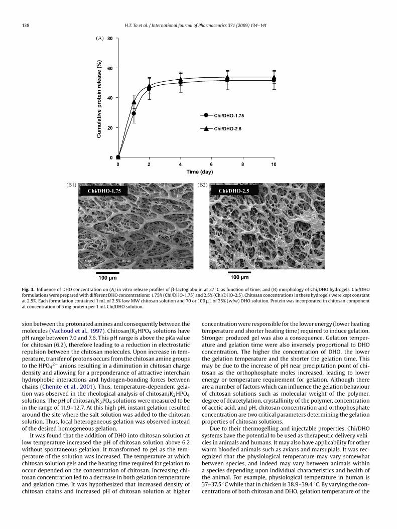
In contrast, salt concentration did not significantly influencethe release profile of �-lactoglobulin (Fig. 3A) but it did affect themorphology of the hydrogel matrix. Much larger outer pores wereobserved in hydrogels with a smaller salt content (Fig. 3B1 and B2).While the size of outer pores was significantly different betweenhydrogels containing different salt amounts, that of inner poreswere not. As the amount of orthophosphate increased, the outerpores of Chi/DHO matrix became smaller. For example, size distri-bution of outer pores in Chi/DHO-70 was from 50 to 200 �m whilethat in Chi/DHO-100 was from 10 to 70 �m. Smaller outer poreswere thought to be associated with the lower initial burst effect.However, in this study, the formulation containing a higher concen-tration of salt exhibited a slightly larger initial burst effect although
much smaller outer pores were observed in this hydrogel. At day 4 ofthe study, approximately 50% of �-lactoglobulin was released fromboth hydrogel formulations and afterwards, a plateau was reached.Structural conformation of entrapped compounds played animportant role on the release rate. Initially, the release rates of

H.T. Ta et al. / International Journal of Pharmaceutics 371 (2009) 134–141 137
F globulf ): 2%t hitosac
FFnwooa
tiBFwolb
3
it(Cpsaa
4
eS
ig. 2. Influence of chitosan concentration on (A) in vitro release profiles of �-lactoormulations were prepared with different low MW chitosan concentrations (w/whese hydrogels were kept constant at 2.5%. Each formulation contained 1 mL of component at concentration of 5 mg protein per 1 mL Chi/DHO solution.
ITC-dextran (FD) molecules with different MWs (4 kDa FD4, 10 kDaD10, and 20 kDa FD20) were the same but later on they varied sig-ificantly (Fig. 4A). FD20, a flexible polymer, was released fastestith the cumulative release percentage at nearly 90% after 6 days
f the study. FD10, existing as random flexible chain/coil, diffusedut of the gel matrix faster than FD4 but less than FD20. And FD4,coil polymer in solution, was released at the slowest rate.
The molecular weight of loaded macromolecules also affectedhe release profile. The release profiles of globular proteins includ-ng �-lactoglobulin (MW: 36.6 kDa, Stokes’ radius: 1.83 nm) andSA (MW: 66.4 kDa, Stokes’ radius: 3.18 nm) were presented inig. 4B. It was found that �-lactoglobulin, a lower-MW protein,as released faster than BSA, a higher-MW protein. After 10 daysf the experiment, approximately 55% and 45% of entrapped �-actoglobulin and BSA, respectively, was detected in the releaseuffer.
.4. Cytotoxicity of CHOR hydrogels
Fig. 5 shows the metabolic activity of cells cultured for 7 daysn media containing CHOR hydrogels formed by various concen-rations of chitosan (Fig. 5A) and various concentration of DHOFig. 5B). As shown in the figure, cells grew well in the presence ofHOR hydrogels. Increasing the concentration of chitosan and/orhosphate salt did not affect the viability of these cells. Fig. 5Chows the photomicrograph of SaOS-2 cells in direct contact withCHOR hydrogel. As can be seen in this figure, cells looked healthynd normal and grew around the hydrogel.
. Discussion
Chitosan is a pH-dependent cationic, non-toxic, antibacterial,asily bioabsorbable, biodegradable, biocompatible (Chandy andharma, 1990; Hirano et al., 1990) and mucoadhesive biopolymer
in at 37 ◦C as function of time; and (B) morphology of Chi/DHO hydrogels. Chi/DHO(Chi/DHO-2), 2.5% (Chi/DHO-2.5), and 2.9% (Chi/DHO-2.9). DHO concentrations inn solution and 100 �L of 25% DHO solution. Protein was incorporated in chitosan
(Henriksen et al., 1996; He et al., 1998). It was ranked as GRAS(generally recognised as safe) by FDA (Food and Drug Administra-tion). Therefore, chitosan can be widely used in pharmaceuticalpreparations and food products without concern for patient orconsumer safety. Chitosan remains dissolved in aqueous solutionsup to a pH of 6.2. When the pH exceeds 6.2, a gel-like precip-itate forms from chitosan aqueous solutions. This gel formationis due to neutralization of chitosan amine groups, leading to theremoval of repulsive interchain electrostatic forces, allowing forhydrogen bonding and hydrophobic interactions between chains.Therefore, in addition to the use of poly- or multivalent anions (suchas polyphosphate) to develop ionically cross-linked chitosan hydro-gels, increasing the pH of chitosan solution above 6.2 is anotherway to induce the physical gelation of chitosan. Organic phosphatesuch as �-glycerophosphate was investigated for this purpose. Inthis study, we investigated the use of inorganic phosphate saltssuch as potassium phosphate salts on the development of thermal-activated neutral injectable chitosan solutions. Both potassium andphosphate ions are naturally present in the body fluid. Therefore,the combination of chitosan and this salt are expected to be bio-compatible.
Monobasic, dibasic and tribasic potassium orthophosphate saltswere investigated for their abilities to form chitosan hydrogels. Onlydibasic orthophosphate salt could induce the gelation of chitosansolution. Samples containing monobasic phosphate salt could notgel at any temperature settings. For samples containing tribasicphosphate, local precipitation of chitosan occurred spontaneouslyupon salt addition instead of a homogeneous gelation. These obser-vations indicate a strong relationship between pH and gelation
ability of chitosan/orthophosphate solutions.KH2PO4 was incapable of causing gelation due to their low pHranges of 5.0–5.2. These pH ranges are below the pKa value of chi-tosan (6.2). Hence chitosan is still positively charged due to theammonium group (–NH3
+) which leads to an electrostatic repul-
138 H.T. Ta et al. / International Journal of Pharmaceutics 371 (2009) 134–141
F bulin ◦
f 5) anda or 10a
smpfrptdhctsiaso
lwpcotac
ig. 3. Influence of DHO concentration on (A) in vitro release profiles of �-lactogloormulations were prepared with different DHO concentrations: 1.75% (Chi/DHO-1.7t 2.5%. Each formulation contained 1 mL of 2.5% low MW chitosan solution and 70t concentration of 5 mg protein per 1 mL Chi/DHO solution.
ion between the protonated amines and consequently between theolecules (Vachoud et al., 1997). Chitosan/K2HPO4 solutions have
H range between 7.0 and 7.6. This pH range is above the pKa valueor chitosan (6.2), therefore leading to a reduction in electrostaticepulsion between the chitosan molecules. Upon increase in tem-erature, transfer of protons occurs from the chitosan amine groupso the HPO4
2− anions resulting in a diminution in chitosan chargeensity and allowing for a preponderance of attractive interchainydrophobic interactions and hydrogen-bonding forces betweenhains (Chenite et al., 2001). Thus, temperature-dependent gela-ion was observed in the rheological analysis of chitosan/K2HPO4olutions. The pH of chitosan/K3PO4 solutions were measured to ben the range of 11.9–12.7. At this high pH, instant gelation resultedround the site where the salt solution was added to the chitosanolution. Thus, local heterogeneous gelation was observed insteadf the desired homogeneous gelation.
It was found that the addition of DHO into chitosan solution atow temperature increased the pH of chitosan solution above 6.2
ithout spontaneous gelation. It transformed to gel as the tem-erature of the solution was increased. The temperature at which
hitosan solution gels and the heating time required for gelation toccur depended on the concentration of chitosan. Increasing chi-osan concentration led to a decrease in both gelation temperaturend gelation time. It was hypothesized that increased density ofhitosan chains and increased pH of chitosan solution at higherat 37 C as function of time; and (B) morphology of Chi/DHO hydrogels. Chi/DHO2.5% (Chi/DHO-2.5). Chitosan concentrations in these hydrogels were kept constant0 �L of 25% (w/w) DHO solution. Protein was incorporated in chitosan component
concentration were responsible for the lower energy (lower heatingtemperature and shorter heating time) required to induce gelation.Stronger produced gel was also a consequence. Gelation temper-ature and gelation time were also inversely proportional to DHOconcentration. The higher the concentration of DHO, the lowerthe gelation temperature and the shorter the gelation time. Thismay be due to the increase of pH near precipitation point of chi-tosan as the orthophosphate moles increased, leading to lowerenergy or temperature requirement for gelation. Although thereare a number of factors which can influence the gelation behaviourof chitosan solutions such as molecular weight of the polymer,degree of deacetylation, crystallinity of the polymer, concentrationof acetic acid, and pH, chitosan concentration and orthophosphateconcentration are two critical parameters determining the gelationproperties of chitosan solutions.
Due to their thermogelling and injectable properties, Chi/DHOsystems have the potential to be used as therapeutic delivery vehi-cles in animals and humans. It may also have applicability for otherwarm blooded animals such as avians and marsupials. It was rec-ognized that the physiological temperature may vary somewhat
between species, and indeed may vary between animals withina species depending upon individual characteristics and health ofthe animal. For example, physiological temperature in human is37–37.5 ◦C while that in chicken is 38.9–39.4 ◦C. By varying the con-centrations of both chitosan and DHO, gelation temperature of the
H.T. Ta et al. / International Journal of Ph
Fig. 4. Influence of entrapped macromolecule properties on in vitro release profilesat 37 ◦C as function of time. (A) Influence of structural conformation of FITC-dextranwith different molecular weights: 4 kDa (FD4), 10 kDa (FD10) and 20 KDa (FD20) For-mulation contained 1 mL of 2.5% low MW chitosan solution and 100 �L of 25% (w/w)DHO solution. FITC-dextran was incorporated in chitosan component at concentra-tion of 1.5 mg FITC-dextran per 1 mL Chi/DHO solution (30 �g FITC-dextran/20 �Lformulation). (B) Influence of molecular weight of proteins including �-lactoglobulin(Mpf
Cotc
dficmott
�mstc(
MW: 36.6 kDa) and BSA (MW: 66.4 kDa). Formulation contained 1 mL of 2.5% lowW chitosan solution and 100 �L of 25% (w/w) DHO solution. Protein was incor-
orated in chitosan component at concentration of 5 mg protein per 1 mL Chi/DHOormulation (100 �g protein/20 �L formulation).
HOR compositions can be adjusted to be optimal for the physi-logical temperature of a particular species. The desired gelationime after having been administered to the patient could be alsoontrolled.
Properties of the hydrogel delivery systems and properties of therug agents were expected to affect the release of macromoleculesrom gel matrix under physiological conditions. In this study, thempact of parameters constructing the gel including chitosan con-entration and DHO concentration was investigated. Effects ofolecular weight and structural conformation of entrapped agents
n the release profile were also studied. In each experiment, onlyhe investigated factor was varied, the others were kept constant inhe formulations.
Chitosan concentration significantly affected the release of-lactoglobulin but DHO concentration did not. However, the for-
ulation containing a higher concentration of salt exhibited alightly larger initial burst effect. These results can be explained byhe negative charge of �-lactoglobulin at neutral pH and the pro-edure used to prepare the hydrogels. At acidic pH, amine groups–NH2) on the chitosan chain are protonated, leading to an overall
armaceutics 371 (2009) 134–141 139
positive charge on chitosan. As �-lactoglobulin was mixed with thechitosan solution before the addition of DHO, negatively charged�-lactoglobulin was attracted to positive-charged chitosan due toelectrostatic interaction. When salt was added, it neutralized thepositive charges of chitosan and free protein. The more salt wasadded, the more protein was released.
The molecular weight of loaded molecules affected the releaseprofile. However, structural conformation of entrapped compoundsalso played an important role in the diffusion and release rate.Based on product information (Sigma–Aldrich), dextrans of MWgreater than 10 kDa behave as typically branched flexible polymers,while those of 2–10 kDa exhibit the properties of an expandablecoil. At MW below 2 kDa, dextran is more stiff chain, rod-likemolecules. Dextran with MW at 10 kDa can be best described asrandom flexible chain/expandable coil. The approximate Stokes’radii (hydrodynamic radii) of FD4, FD10, and FD20 are 14, 23, and33 Å, respectively. In our study, although MWs and Stoke’s radii ofFD4, FD10 and FD20 were different from each other, they seemednot to influence the diffusion and release of these dextrans. Fewstudies on dextran diffusion through a range of membranes alsosuggested that dextran was less hindered than would be predictedon the basis of Stokes’ radius (Bohrer et al., 1984; De Belder, 2003).On the other hand, geometrical structure of dextrans seemed toplay an important role in their diffusion and release characteristics.The gel is considered as a heterogeneous network of overlapping orcross-linked chains in which “pore size” or “mesh size” is a defin-ing parameter of the system. Since FD4 exists as coil in solution,it might quickly become and remain trapped in pores. Conversely,as FD20 appeared as flexible polymers, it diffused through the gelmatrix more easily. Since FD10 existed as random flexible chain/coil,its diffusion through the gel matrix was better than FD4 but lessthan FD20. These explanations are supported by a study reported byPluen et al. (1999) that the diffusion coefficients of flexible macro-molecules in agarose gels were greater than those of rigid and/orspherical macromolecules. It has also been demonstrated that flex-ibility allows rods to escape enmeshment more easily (Cush, 2003).
Proteins with different MW values but existing in a same con-formation were further employed in the release test to investigatethe influence of MW of entrapped compounds on the release char-acteristics. They were globular proteins including �-lactoglobulinand BSA. It was found that �-lactoglobulin was released faster thanBSA. However, despite the significant gaps in MWs and Stokes’ radiiof �-lactoglobulin and BSA, the difference in the release patternsof these proteins was not significant, which was exhibited in theoverlap of error bars. It may be due to the much larger pore sizes ofhydrogel than the molecular size or Stokes’ radius of these proteins.
It was noted that the release of the neutral dextrans fromChi/DHO gel matrix was quicker and better in terms of sus-tained release rather than that of the negative-charged proteins.�-Lactoglobulin and BSA both carry a net negative charge at pH 7.4and hence could interact with the cationic chitosan backbone. Inaddition, it is probably due to the difference in structural conforma-tion of these macromolecules in which dextran existed as flexiblechain while �-lactoglobulin and BSA were globular in shape.
It was observed that the release or the diffusion of proteins fromthe hydrogel matrix to media quickly reached plateau after the ini-tial burst effect within a few days of the study (4 days). Since thethree-dimensional network of the gel did not significantly changeover time, it indicated that there was no substantial erosion of thehydrogel matrix in PBS (pH 7.4) within 10 days of the study. Thusproteins were released mainly by diffusion mechanism in PBS. Since
only 50% of entrapped proteins were released after 10 days, it sug-gested that part of the protein was linked to the polymeric network.However, in vivo, it was expected that the release rate of proteinswould be higher due to the degradation of the chitosan networkcaused by various enzymes and macrophages present in the body.
140 H.T. Ta et al. / International Journal of Pharmaceutics 371 (2009) 134–141
F (A) ChC ) PhotC
Ile(iyoa
iHcgw
5
cgtiSvBtcstfqtmm
ig. 5. Viability of SaOS-2 cells cultured in media containing various Chi/DHO gels.hi/DHO gels prepared by different concentrations of DHO (2.2%, 2.8% and 3.5%). (Chi/DHO gel containing 2.5% chitosan and 2.5% DHO.
n fact, the addition of lysozyme at concentration of 8 �g/mL – simi-ar to the enzyme concentration found in human serum (Montagnet al., 1998) – at day 10 resulted in a further release of these proteinsdata not shown). Chitosan is known to be degraded or metabolizedn vivo by certain enzymes, especially lysozyme through the hydrol-sis of the acetylated residues (Tomihata and Ikada, 1997). The ratef this degradation inversely depends on the degree of acetylationnd crystallinity of the polymer.
The cytotoxicity of gel study using SaOS-2 cells preliminar-ly demonstrated that Chi/DHO hydrogels are cytocompatible.owever, another study using other various cell lines should beonducted to confirm the cytocompatibility of these systems. Theelling Chi/DHO solution is therefore a potential candidate for aide range of biomedical applications due to its biocompatibility.
. Conclusion
The ability of DHO to induce the thermo-activated gelation ofhitosan solutions was demonstrated. Gelation temperature andelation time of Chi/DHO systems are adjustable and can be con-rolled for use in a particular utility. The in vitro cytotoxicity studyndicates that neutral Chi/DHO hydrogels are non-toxic towardaOS-2 cells. The potential of Chi/DHO as a prolonged drug deliveryehicle was demonstrated using FITC-dextran, �-lactoglobulin andSA. The release of these macromolecules depends on key parame-ers constructing the gel including chitosan concentration and DHOoncentration; and the properties of entrapped macromoleculesuch as structural conformation and molecular weight. The neu-ral hydrophilic macromolecule FITC-dextran showed a fast release
rom the gel while proteins showed a slow partial release whichuickly reached plateau. Because of the ionic interactions of pro-eins with the hydrogel polymer, the complete release of proteinsight be possible only after the enzymatic degradation of the CHORatrix. The results obtained from this study show a strong potential
i/DHO gels prepared by different concentrations of chitosan (2%, 2.5% and 3%). (B)omicrograph showing the viability of SaOS-2 cells cultured in direct contact with a
of these biocompatible hydrogel systems as sustained drug deliverysystem.
Acknowledgements
This project was supported by the University of Melbourne. Wewould like to thank Dr Simon Crawford (School of Botany, Universityof Melbourne) for his assistance with SEM imaging.
References
Bernkop-Schnurch, A., 2000. Chitosan and its derivatives: potential excipients forperoral peptide delivery systems. Int. J. Pharm. 194, 1–13.
Bohrer, M.P., Patterson, G.D., Carrol, P.J., 1984. Hindered diffusion of dextran and ficollin microporous membranes. Macromolecules 17, 1170–1173.
Borchard, G., 2001. Chitosans for gene delivery. Adv. Drug Deliv. Rev. 52, 145–150.Chandy, T., Sharma, C.P., 1990. Chitosan as a biomaterial. Biomater. Artif. Cells Artif.
Organs 18, 1–24.Chenite, A., Buschmann, M., Wang, D., Chaput, C., Kandani, N., 2001. Rheological char-
acterisation of thermogelling chitosan/glycerol-phosphate solutions. Carbohydr.Polym. 46, 39–47.
Cho, J., Heuzey, M.-C., Begin, A., Carreau, P.J., 2005. Physical gelation of chitosan in thepresence of �-glycerophosphate: the effect of temperature. Biomacromolecules6, 3267–3275.
Cush, R.C., 2003. PhD thesis: Diffusion of a rodlike virus in complex solutions. Depart-ment of Chemistry, Louisiana State University, p. 113.
De Belder, A.N., 2003. Dextran. Amersham Biosciences Limited, Uppsala, Sweden.Dhanikula, A.B., Panchagnula, R., 2004. Development and characterization of
biodegradable chitosan films for local delivery of paclitaxel. AAPS J. 6, e27.Dodane, V., Vilivalam, V.D., 1998. Pharmaceutical applications of chitosan. Pharm.
Sci. Technol. Today 1, 246–253.He, P., Davis, S.S., Illum, L., 1998. In vitro evaluation of the mucoadhesive properties
of chitosan microspheres. Int. J. Pharm. 166, 75–88.Helander, I.M., Nurmiaho-Lassila, E.L., Ahvenainen, R., Rhoades, J., Roller, S., 2001.
Chitosan disrupts the barrier properties of gram-negative bacteria. Int. J. FoodMicrobiol. 71, 235–244.
Henriksen, I., Green, K.L., Smart, J.D., 1996. Bioadhesion of hydrated chitosans: an invitro study. Int. J. Pharm. 145, 231–240.
Hirano, S., Yamaguchi, R., Fukui, N., 1990. A chitosan oxalate gel: its conversion to anN-acetylchitosan gel via a chitosan gel. Carbohydr. Res. 201, 145–149.

l of Ph
K
K
K
M
M
O
O
P
P
physical chitin. Carbohydr. Res. 302, 169–177.
H.T. Ta et al. / International Journa
ashyap, N., Viswanad, B., Sharma, G., Bhardwaj, V., Ramarao, P., Ravi Kumar, M.N.,2007. Design and evaluation of biodegradable, biosensitive in situ gelling systemfor pulsatile delivery of insulin. Biomaterials 28, 2051–2060.
hor, E., Lim, L.Y., 2003. Implantable applications of chitin and chitosan. Biomaterials272, 65–78.
umar, M.N.V.R., 2000. A review of chitin and chitosan applications. React. Funct.Polym. 46, 1–27.
artini, A., Lauria, S., 2003. Sustained Release Injectable Products. Am. Pharm. Rev.,http://www.americanpharmaceuticalreview.com/current issue/3 APR Fall2003/Martini article.htm.
ontagne, P., Cuiliere, M.L., Mole, C., Bene, M.C., Faure, G., 1998. Microparticle-enhanced nephelometric immunoassay of lysozyme in milk and other humanbody fluids. Clin. Chem. 44, 1610–1615.
kamoto, Y., 2002. Analgesic effects of chitin and chitosan. Carbohydr. Polym. 49,249–252.
ungbho, K., Muller, B.W., 1997. Chitosan sponges as sustained release drug carriers.
Int. J. Pharm. 156, 229–237.eter, M.G., 1995. Applications and environmental aspects of chitin and chitosan. J.Macromol. Sci. Pure Appl. Chem. A32, 629–640.
luen, A., Netti, P.A., Jain, R.K., Berk, D.A., 1999. Diffusion of macromolecules inagarose gels: comparison of linear and globular configurations. Biophys. J. 77,542–552.
armaceutics 371 (2009) 134–141 141
Promega, 2006. Cell viability: Protocols and Applications guide, Promega Corpora-tion.
Ruel-Gariepy, E., Chenite, A., Chaput, C., Guirguis, S., Leroux, J.C., 2000. Characteri-zation of thermosensitive chitosan gels for the sustained delivery of drugs. Int.J. Pharm. 203, 89–98.
Shu, X.Z., Zhu, K.J., Song, W., 2001. Novel pH-sensitive citrate cross-linked chitosanfilms for drug controlled release. Int. J. Pharm. 212, 19–28.
Shu, X.Z., Zhu, K.J., 2002. Controlled drug release properties of ionically cross-linked chitosan beads: the influence of anion structure. Int. J. Pharm. 233,217–225.
Tomihata, K., Ikada, Y., 1997. In vitro and in vivo degradation of films of chitin and itsdeacetylated derivatives. Biomaterials 18, 567–575.
Ueno, H., Mori, T., Fujinaga, T., 2001. Topical formulations and wound healing appli-cations of chitosan. Adv. Drug Deliv. Rev. 52, 105–115.
Vachoud, L., Zydowicz, N., Domard, A., 1997. Formation and characterization of a
Weska, R.F., 2007. Optimization of deacetylation in the production of chitosanfrom shrimp wastes: use of response surface methodology. J. Food Eng. 80,749–753.
Winter, H.H., Chambon, F., 1986. Analysis of linear viscoelasticity of crosslinkingpolymer at gel point. J. Rheol. 30, 367–382.