Chemical silicon surface modification and bioreceptor attachment to develop competitive integrated...
10
ORIGINAL PAPER Chemical silicon surface modification and bioreceptor attachment to develop competitive integrated photonic biosensors Jorge Escorihuela & María José Bañuls & Javier García Castelló & Veronica Toccafondo & Jaime García-Rupérez & Rosa Puchades & Ángel Maquieira Received: 13 April 2012 / Revised: 7 June 2012 / Accepted: 17 July 2012 # Springer-Verlag 2012 Abstract Methodology for the functionalization of silicon- based materials employed for the development of photonic label-free nanobiosensors is reported. The studied function- alization based on organosilane chemistry allowed the direct attachment of biomolecules in a single step, maintaining their bioavailability. Using this immobilization approach in probe microarrays, successful specific detection of bacterial DNA is achieved, reaching hybridization sensitivities of 10 pM. The utility of the immobilization approach for the functionalization of label-free nanobiosensors based on pho- tonic crystals and ring resonators was demonstrated using bovine serum albumin (BSA)/anti-BSA as a model system. Keywords Photonic nanobiosensor . Silicon-based materials . Label free . DNA microarray Introduction In recent years, the development of new heterogeneous microarray methods for the immobilization of nucleic acid probes onto solid surfaces has become of the utmost interest owing to the applications of these systems in the diagnosis of diseases [1], gene expression analysis [2], mutations detection [3], forensic science [4], toxicology [5], pharma- cogenomics [6], drug discovery [7], biosensing [8] or envi- ronmental monitoring [9]. Optical biosensors are powerful detection instruments and versatile analytical tools, which use a variety of signal transduction pathways based on photonic attributes. The biosensors can provide chemical information ranging from analyte concentration or binding kinetics to microscopic imaging and molecular structure. Among optical biosensors, those based on integrated nano- photonic structures currently attract huge interest mainly owing to their extremely high sensitivity and reduced size, making it possible to integrate many of them on a single chip to perform a multianalyte detection [10]. In terms of optical nanobiosensing for point of need applications, several requirements, such as, reliability, ro- bustness, selectivity, sensitivity, low cost and portability, must be fulfilled. Besides, in many cases, an interpretable signal without instrumental intervention is desired. Most of these requirements are highly dependent on the functional- ization strategy employed to link the capture agent onto the support. Among the solid supports used in biosensing, sili- con has proven to be exceptionally versatile for sensors development, being the key material in the microelectronics industry. Furthermore, silicon is also advantageous as a substrate for biosensor building because it is readily deriv- atized and biocompatible [11, 12]. Published in the special paper collection Optical Biochemical and Chemical Sensors with guest editor Laura M. Lechuga. Electronic supplementary material The online version of this article (doi:10.1007/s00216-012-6280-4) contains supplementary material, which is available to authorized users. J. Escorihuela : M. J. Bañuls : R. Puchades : Á. Maquieira (*) Centro de Reconocimiento Molecular y Desarrollo Tecnológico, Departamento de Química, Universidad Politécnica de Valencia, Camino de Vera s/n, 46022 Valencia, Spain e-mail: [email protected] J. G. Castelló : V. Toccafondo : J. García-Rupérez Nanophotonics Technology Center, Universidad Politécnica de Valencia, Camino de Vera s/n, 46022 Valencia, Spain Anal Bioanal Chem DOI 10.1007/s00216-012-6280-4
Transcript of Chemical silicon surface modification and bioreceptor attachment to develop competitive integrated...

ORIGINAL PAPER
Chemical silicon surface modification and bioreceptorattachment to develop competitive integrated photonicbiosensors
Jorge Escorihuela & María José Bañuls &
Javier García Castelló & Veronica Toccafondo &
Jaime García-Rupérez & Rosa Puchades &
Ángel Maquieira
Received: 13 April 2012 /Revised: 7 June 2012 /Accepted: 17 July 2012# Springer-Verlag 2012
Abstract Methodology for the functionalization of silicon-based materials employed for the development of photoniclabel-free nanobiosensors is reported. The studied function-alization based on organosilane chemistry allowed the directattachment of biomolecules in a single step, maintainingtheir bioavailability. Using this immobilization approach inprobe microarrays, successful specific detection of bacterialDNA is achieved, reaching hybridization sensitivities of10 pM. The utility of the immobilization approach for thefunctionalization of label-free nanobiosensors based on pho-tonic crystals and ring resonators was demonstrated usingbovine serum albumin (BSA)/anti-BSA as a model system.
Keywords Photonic nanobiosensor . Silicon-basedmaterials . Label free . DNAmicroarray
Introduction
In recent years, the development of new heterogeneousmicroarray methods for the immobilization of nucleic acidprobes onto solid surfaces has become of the utmost interestowing to the applications of these systems in the diagnosisof diseases [1], gene expression analysis [2], mutationsdetection [3], forensic science [4], toxicology [5], pharma-cogenomics [6], drug discovery [7], biosensing [8] or envi-ronmental monitoring [9]. Optical biosensors are powerfuldetection instruments and versatile analytical tools, whichuse a variety of signal transduction pathways based onphotonic attributes. The biosensors can provide chemicalinformation ranging from analyte concentration or bindingkinetics to microscopic imaging and molecular structure.Among optical biosensors, those based on integrated nano-photonic structures currently attract huge interest mainlyowing to their extremely high sensitivity and reduced size,making it possible to integrate many of them on a singlechip to perform a multianalyte detection [10].
In terms of optical nanobiosensing for point of needapplications, several requirements, such as, reliability, ro-bustness, selectivity, sensitivity, low cost and portability,must be fulfilled. Besides, in many cases, an interpretablesignal without instrumental intervention is desired. Most ofthese requirements are highly dependent on the functional-ization strategy employed to link the capture agent onto thesupport. Among the solid supports used in biosensing, sili-con has proven to be exceptionally versatile for sensorsdevelopment, being the key material in the microelectronicsindustry. Furthermore, silicon is also advantageous as asubstrate for biosensor building because it is readily deriv-atized and biocompatible [11, 12].
Published in the special paper collection Optical Biochemical andChemical Sensors with guest editor Laura M. Lechuga.
Electronic supplementary material The online version of this article(doi:10.1007/s00216-012-6280-4) contains supplementary material,which is available to authorized users.
J. Escorihuela :M. J. Bañuls : R. Puchades :Á. Maquieira (*)Centro de Reconocimiento Molecular y Desarrollo Tecnológico,Departamento de Química, Universidad Politécnica de Valencia,Camino de Vera s/n,46022 Valencia, Spaine-mail: [email protected]
J. G. Castelló :V. Toccafondo : J. García-RupérezNanophotonics Technology Center,Universidad Politécnica de Valencia,Camino de Vera s/n,46022 Valencia, Spain
Anal Bioanal ChemDOI 10.1007/s00216-012-6280-4

The bioreceptor immobilization constitutes one of thekey steps in the biosensor development [13]. Althoughseveral methodologies for surface biofunctionalization havebeen described including adsorption, entrapment, electro-static and covalent attachment, the last one is widely pre-ferred because it provides the best stability towards pH andionic strength variations, temperature and flow rate. Forphotonic biosensing, the most common modification ofsilicon surface materials is accomplished through a silaniza-tion reaction, which introduces active groups, mainly aminomoieties [14–16], and other functionalities like epoxy [17,18], carboxylic acid or thiol [19]. Silane coupling to silicasurfaces by grafting is a well-understood process, and iseasily carried out using organic solvents or vapour deposi-tion technique [20, 21]. The resulting surface functionalitiescan be further modified for the attachment of the captureagent by bioconjugation procedures [22]. However, this stepoften requires the use of cross-linkers for covalent probeanchoring. Glutaraldehyde [23, 24], succinimidyl-6-hydrazino-nicotinamine/succinimidyl-4-formyl benzoate[25, 26] and 1-ethyl-3-(3-dimethylaminopropyl)carbodii-mide (EDC)/N-hydroxysuccinimide (NHS) [27] are widelyemployed for this purpose. Unfortunately, in many cases theimines or succinimidyl esters formed can be hydrolysedunder the bioconjugation conditions, affecting considerablythe reproducibility of the process. Furthermore, the use ofcross-linkers involves additional steps in the biosensor prep-aration process, increasing the variability and decreasingconsiderably the immobilization yield. Thus, there is still aneed to find direct and effective chemical functionalizationsfor silicon-based materials allowing the direct covalent attach-ment of bioreceptors and maintaining their biofunctionality.
Among different organosilane compounds, 3-isocyanatepropyltriethoxysilane (ICPTS) is gaining popularity, as the isocy-anate group reacts with many nucleophiles including aminesand thiols. Isocyanate organosilane compounds show goodselectivity when reacting with amino groups in lysine sidechains and N-terminal α-amines in proteins or other primaryaminated compounds such as oligonucleotides. The reactivityof the isocyanate group increases in alkaline pH, especially incarbonate buffer, affording good yields in aqueous media andin a single step.
In this article, we report the chemical modification ofsilicon oxide with ICPTS to develop label-free photonicnanobiosensors. The described methodology includes apre-optimization and surface characterization using planarchips, followed by the transfer of the optimized biofunction-alization procedures to the nanostructured photonic structures,and the experimental demonstration of label-free biosensingby monitoring a proof-of-concept recognition event. The pro-cedure presents several advantages when compared to otherapproaches because it can be carried out easily in a single step,allowing the direct covalent attachment of aminated capture
bioreceptors without the use of cross-linking reagents, and it iscompatible with aqueous media chemistry, which is crucial forretaining bioactivity.
Materials and methods
Chemicals
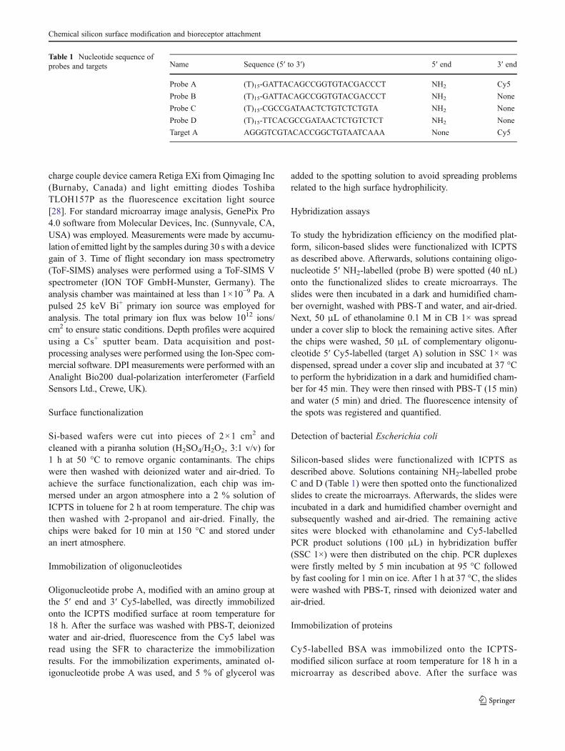
The silicon-based wafers were provided by the ValenciaNanophotonics Technology Center (NTC) at the UniversidadPolitécnica de Valencia (Spain) and consisted on a 3-μm-thicksilicon oxide layer grown on silicon (100) wafer. Hydrogenperoxide (35 % w/w), albumin from bovine serum ≥98 %lyophilized powder, anti-bovine albumin antibody pro-duced in rabbit, 3-(triethoxysilyl)propyl isocyanate, and2-aminoethanol were purchased from Sigma-Aldrich Quí-mica (Madrid, Spain). Toluene, dichloromethane and sulfuricacid (95–98 %) were purchased from Scharlau (Madrid,Spain). Rabbit IgG, Cy5-Linked (from goat) was suppliedby GE Healthcare (Buckinghamshire, UK). Oligonucleotideswere acquired from Thermo Fischer (Madrid, Spain) and theirsequences are shown in Table 1. Escherichia coli PCR prod-ucts were kindly provided by Mobidiag (Helsinki, Finland).The phosphate buffer saline (PBS, 0.008M sodium phosphatedibasic, 0.002 M sodium phosphate monobasic, 0.137 Msodium chloride, 2.7 M potassium chloride, pH 7.5), PBS-T(10× PBS containing 0.05 % Tween 20), saline sodium citratebuffer (10× SSC, 0.9 M sodium chloride, 0.09 M sodiumcitrate, pH 7), carbonate buffer (10× CB, 0.5 M sodiumcarbonate, pH 9.6), and washing solutions were filteredthrough a 0.22-μm pore size nitrocellulose membrane fromWhatman GmbH (Dassel, Germany) before use.
Instruments
Microarray printing was carried out with a low volume non-contact dispensing system from Biodot (Irvine, CA, USA),model AD1500. A OCA20 contact angle system equippedwith SCA20 software from Dataphysics Instruments GmbH(Filderstadt, Germany) was used. X-ray photoelectron spec-tra were recorded with a Sage 150 spectrophotometer fromSPECS Surface Nano Analysis GmbH (Berlin, Germany).Atomic force microscopy (AFM) images were obtained witha Veeco model Dimension 3100 Nanoman from VeecoMetrology (Santa Barbara, CA) using tapping mode at300 kHz. Scanning electron microscopy (SEM) was carriedout with an S-4500 Hitachi High-Technologies apparatus(Krefeld, Germany) with the upper detector at 15 kV. AnEVG620 Automated Mask Alignment System from EVGwas employed to irradiate UV light. The fluorescence signalof the spots was detected using a home-made surface fluo-rescence reader (SFR), equipped with a high sensitive
J. Escorihuela et al.
charge couple device camera Retiga EXi from Qimaging Inc(Burnaby, Canada) and light emitting diodes ToshibaTLOH157P as the fluorescence excitation light source[28]. For standard microarray image analysis, GenePix Pro4.0 software from Molecular Devices, Inc. (Sunnyvale, CA,USA) was employed. Measurements were made by accumu-lation of emitted light by the samples during 30 s with a devicegain of 3. Time of flight secondary ion mass spectrometry(ToF-SIMS) analyses were performed using a ToF-SIMS Vspectrometer (ION TOF GmbH-Munster, Germany). Theanalysis chamber was maintained at less than 1×10⁻⁹ Pa. Apulsed 25 keV Bi+ primary ion source was employed foranalysis. The total primary ion flux was below 1012 ions/cm2 to ensure static conditions. Depth profiles were acquiredusing a Cs+ sputter beam. Data acquisition and post-processing analyses were performed using the Ion-Spec com-mercial software. DPI measurements were performed with anAnalight Bio200 dual-polarization interferometer (FarfieldSensors Ltd., Crewe, UK).
Surface functionalization
Si-based wafers were cut into pieces of 2×1 cm2 andcleaned with a piranha solution (H2SO4/H2O2, 3:1 v/v) for1 h at 50 °C to remove organic contaminants. The chipswere then washed with deionized water and air-dried. Toachieve the surface functionalization, each chip was im-mersed under an argon atmosphere into a 2 % solution ofICPTS in toluene for 2 h at room temperature. The chip wasthen washed with 2-propanol and air-dried. Finally, thechips were baked for 10 min at 150 °C and stored underan inert atmosphere.
Immobilization of oligonucleotides
Oligonucleotide probe A, modified with an amino group atthe 5′ end and 3′ Cy5-labelled, was directly immobilizedonto the ICPTS modified surface at room temperature for18 h. After the surface was washed with PBS-T, deionizedwater and air-dried, fluorescence from the Cy5 label wasread using the SFR to characterize the immobilizationresults. For the immobilization experiments, aminated ol-igonucleotide probe A was used, and 5 % of glycerol was
added to the spotting solution to avoid spreading problemsrelated to the high surface hydrophilicity.
Hybridization assays
To study the hybridization efficiency on the modified plat-form, silicon-based slides were functionalized with ICPTSas described above. Afterwards, solutions containing oligo-nucleotide 5′ NH2-labelled (probe B) were spotted (40 nL)onto the functionalized slides to create microarrays. Theslides were then incubated in a dark and humidified cham-ber overnight, washed with PBS-T and water, and air-dried.Next, 50 μL of ethanolamine 0.1 M in CB 1× was spreadunder a cover slip to block the remaining active sites. Afterthe chips were washed, 50 μL of complementary oligonu-cleotide 5′ Cy5-labelled (target A) solution in SSC 1× wasdispensed, spread under a cover slip and incubated at 37 °Cto perform the hybridization in a dark and humidified cham-ber for 45 min. They were then rinsed with PBS-T (15 min)and water (5 min) and dried. The fluorescence intensity ofthe spots was registered and quantified.
Detection of bacterial Escherichia coli
Silicon-based slides were functionalized with ICPTS asdescribed above. Solutions containing NH2-labelled probeC and D (Table 1) were then spotted onto the functionalizedslides to create the microarrays. Afterwards, the slides wereincubated in a dark and humidified chamber overnight andsubsequently washed and air-dried. The remaining activesites were blocked with ethanolamine and Cy5-labelledPCR product solutions (100 μL) in hybridization buffer(SSC 1×) were then distributed on the chip. PCR duplexeswere firstly melted by 5 min incubation at 95 °C followedby fast cooling for 1 min on ice. After 1 h at 37 °C, the slideswere washed with PBS-T, rinsed with deionized water andair-dried.
Immobilization of proteins
Cy5-labelled BSA was immobilized onto the ICPTS-modified silicon surface at room temperature for 18 h in amicroarray as described above. After the surface was
Table 1 Nucleotide sequence ofprobes and targets Name Sequence (5′ to 3′) 5′ end 3′ end
Probe A (T)15-GATTACAGCCGGTGTACGACCCT NH2 Cy5
Probe B (T)15-GATTACAGCCGGTGTACGACCCT NH2 None
Probe C (T)15-CGCCGATAACTCTGTCTCTGTA NH2 None
Probe D (T)15-TTCACGCCGATAACTCTGTCTCT NH2 None
Target A AGGGTCGTACACCGGCTGTAATCAAA None Cy5
Chemical silicon surface modification and bioreceptor attachment

washed with PBS-T, deionized water and air-dried, thefluorescence was read with the home-made SFR.
BSA immunoassay
Different solutions containing BSA (from 10 to 0.001 μg/mL)were spotted onto the silicon-based slides, previously func-tionalized with ICPTS as described above, creating the micro-arrays. Afterwards, the slides were incubated in a dark andhumidified chamber overnight and subsequently washed andair-dried. The remaining active sites were blocked with etha-nolamine 0.1 M or OVA 1 % in PBS and a solution of anti-BSA (50 μL) in PBS-T was then added on the chip andincubated for 15 min. The chip was rinsed with PBS-T(15 min) and water (5 min) and dried, after which Cy5-labelled goat anti-rabbit IgG (0.5 μg/mL in PBS-T) wasapplied with a cover slip and incubated for 15 min. Finally,the chip was washed with PBS-T, rinsed with deionized waterand air-dried. Fluorescence measurements were made usingthe SFR.
Dual polarization interferometry measurements
Dual polarization interferometry (DPI) was used to character-ize the layers of biomolecules immobilized on the sensor chipsurface. DPI’s Anachip sensing chips are based on a dual-slabwaveguide made of an upper sensing waveguide and a lowerreference waveguide. The physical principle of this techniquewas described by Cross et al. [29]. Briefly, laser light isswitched between two polarizations: transverse magnetic(TM) and transverse electric (TE); the light travels along thesensor chip producing interference fringe patterns in the farfield which are detected by a CCD camera. To this end,unmodified Anachip (Farfield Sensors) was functionalizedwith ICPTS according to the procedure described above.The running buffer for the first experiment was CB 1× at aflow rate of 50 μL/min, and the temperature was set at 20 °C.Before calibration, 1 μM of probe 1 was flowed over the chipfor 100 min at a flow rate of 2 μL/min. The chip was calibratedand the experiment stopped. Then, after a blocking step with 2-aminoethanol, a second experiment was started. The tempera-ture was set at 37 °C, the buffer was PBS 1× and the flow rate50 μL/min. The chip was calibrated and two successive addi-tions of 200μL 500 nM complementary DNA in PBS 1× weremade at a flow rate of 2 μL/min and 10 μL/min, respectively.
Results and discussion
Optimization of conditions in planar chips
The main method for chemical functionalization of glass orsilicon slides uses reactive silanol groups (Si–OH) that can
be generated by pretreatment of the surface with e.g. piranhasolution. In this regard, the surface was functionalized byreaction between the native silicon oxide present on siliconand an isocyanate-capped organosilane. Isocyanates caneasily react with several functional groups which are presentin nucleic acids, proteins and other biomolecules (Fig. 1).For that purpose, the silicon surface was first treated withthe piranha solution and subsequently immersed under argoninto a solution containing ICPTS to generate isocyanate func-tions on the surface of the substrate.
As a starting point, the optimal buffer and pH conditionsfor immobilization of probe A were investigated, as thereactivity of the isocyanate group toward a number ofnucleophiles can be controlled by tuning the pH (from 4 to12). The results showed the influence of pH on the immo-bilization of probe A, reaching a maximum yield with PBS1× at pH 10 (see Electronic Supplementary Material,Fig. S1). Thus, alkaline conditions were needed to haveenough free amine moieties to perform the nucleophilicattack. As a control, probe A was also spotted onto unmod-ified substrates, which exhibited no detectable fluorescenceunder immobilization conditions. Another negative controlemploying Cy5-labelled oligonucleotide without aminefunctionalization (target A) was printed onto an ICPTS-functionalized surface; and after incubation and washing,no measurable fluorescence was observed. The incubationtime for the immobilization was then studied. For thatpurpose, probe A (5 μM) was spotted and incubated atdifferent times. After the slides were washed, low signal-to-noise (S/N) ratios were obtained for short times; betterimmobilization efficiencies and S/N ratios were achieved forlonger times, reaching maximum values from 8 h (seeElectronic Supplementary Material, Fig. S2).
The immobilization efficiency of probe A was evaluatedunder the optimized conditions. Microarray immobilizationquantification was carried out by using a standard calibra-tion curve (see Electronic Supplementary Material, Fig. S3).The functionalized chips were spotted with probe A (con-centrations ranging from 0 to 10 μM), and incubated topermit the covalent attachment. After the chips werewashed, the fluorescence was read and immobilization effi-ciencies up to 24.7 % were achieved (calculated from theinterpolation of the calibration curve and referred to theamount of spotted probe). The immobilization density in-creased linearly with the probe concentration and reached aplateau at 6 μM (see Electronic Supplementary Material,Fig. S4). The maximum probe density was about 2.5 pmol/cm2, which compares favourably with the state of the art [30].
The performance of the DNA microarrays fabricated onICPTS-modified silicon slides was then evaluated for hy-bridization arrays using probe B. Hybridization was per-formed at different probe concentrations (0.1 to 2 μM),varying target concentrations from 100 pM to 20 μM
J. Escorihuela et al.

(Fig. 2a). Different times and temperatures were assayedand the best results were obtained at 60 min and 40 °C.Hybridization signal sharply increases up to 100 nM con-centration of target oligonucleotide beyond which no furtherincrement in hybridization signal is observed for all theprobe B concentrations tested. As expected, the hybridiza-tion signal intensities are lower when the capture probeconcentration decreases. This was attributed to a crowdingeffect due to an excessively high probe immobilization onthe surface. Under the studied conditions, a good immobili-zation yield (ca. 25 %), S/N ratio (ca. 87) and hybridizationefficiency (ca. 26–30 %) were achieved. The lowest detect-able target concentration was only 0.02 nM and the S/Nratio was around 15 on average. This concentration is of thesame order of magnitude as that achieved by Vigano et al.for microarray glass slides functionalized with isocyanate–γ-
propyltrimethoxysilane [31]. The interchip reproducibilitywas studied and the standard deviation for measurements ondifferent days was below 20 %.
As a proof-of-concept of the proposed methodology, itwas used in the development of a biochip to detect bacterialinfection. For that, a specific E. coli probe (probe C, 1 μM)and another non-specific sequence (probe D, 1 μM), used ascontrol, were immobilized onto an ICPTS-functionalizedslide. After the slide was blocked and washed, the arrayswere hybridized for 45 min at 37 °C with the PCR products(100 nM in SSC 1×) obtained from the lysis of E. colibacteria and labelled with Cy5. After the arrays werewashed and dried, the fluorescence was read with the SFR(Fig. 2b). As can be seen, the resulting assay was highlyspecific for the bacteria; the rows corresponding to thespecific probe showed fluorescence, whereas in control
ETHANOLAMINEBLOCKING
DNAFUNCTIONALIZATION
a b c
Fig. 1 Schematic representation of the chemical reactions: a isocyanate derivatized surface, b DNA immobilization, c ethanolamine blocking. Thethickness of the different layers is not to scale
row 1
row 2
row 3
row 5
row 4
row 6
ba
0 25 50 75 100 125 150 175 200
0
2000
4000
6000
8000
10000
12000
Flu
ore
scen
ce In
ten
sity
(a.
u.)
Target A (nM)
Probe B2 M 1 M 0.5 M 0.1 M
Fig. 2 a Fluorescence intensity observed after hybridization with different concentrations of target A and probe B. b Detection of E. coli; odd rowsprobe sequence, even rows control sequence
Chemical silicon surface modification and bioreceptor attachment
rows no fluorescence was detected. This demonstrated thesuitability of the described modification for the constructionof DNA-based biosensors.
We then evaluated the performance of the immobilizationmethodology for another big family of capture agents—proteins. For this study, the model system BSA/anti-BSAwas selected because it has been widely used and extensivedata are available in the literature for comparison [32]. Cy5-labelled BSA protein (PBS 1×, from 0.25 to 5 μg/mL) wasincubated overnight with the ICPTS-functionalized surface.Quantification of immobilized protein was carried out byusing a standard calibration curve (see Electronic Supple-mentary Material, Fig. S6). Under the studied conditions, amaximum immobilization density of 3.8 pmol/cm2 wasreached for a protein concentration of 4 μg/mL (see ElectronicSupplementary Material, Fig. S7), which means an immobili-zation efficiency of 27 % with regards to the spotted captureagent, and a surface coverage of 88 % with regards to a close-packed protein monolayer (4.32 pmol/cm2) [33]. This surfacedensity was similar to that reported by others authors workingon different materials [34].
Arrays of BSA were created with the optimized spottingconcentrations, and the surface was then blocked with OVA1 % in PBS 1× and different concentrations of rabbit anti-BSA IgGs in PBS-T were assayed (from 0.5 to 20 μg/mL).Finally, the marker (Cy5 labelled goat anti-rabbit antibody0.5 μg/mL in PBS-T) was added and microarray fluores-cence was read with the SFR, detecting signal for all theanti-BSA concentrations assayed. The interchip reproduc-ibility was studied and the standard deviation for measure-ments on different days was around 18 %.
Surface characterization
The water contact angle (WCA) of all the raw material andtreated surfaces was measured (see Electronic Supplemen-tary Material, Table S1). The raw silicon substrate had aWCA of about 27°, which decreased considerably to valuesaround 0 after piranha treatment; this behaviour is attributedto the abundant surface hydroxyl groups generated in thepiranha solution. Upon reaction with ICPTS, the WCAincreased to 70°, in accordance with the presence of a morehydrophobic surface. After bioreceptor immobilization theWCA decreased to 62°, as observed for similar systems[35].
The chemical composition of the silicon surfaces afterfunctionalization was also investigated by X-ray photoelec-tron spectroscopy (XPS) (see Electronic SupplementaryMaterial, Fig. S8). Functionalization with ICPTS resultedin a decrease in the Si signal, and an increase in the C 1s(284.5 eV) and N 1s (399.5 eV) signals compared to thebare silicon material, demonstrating the successful surfacemodification after organosilane treatment. For both
functionalized surfaces, an overlapping resulting from thedifferent chemical environments was observed in the C 1speak spectra (see Electronic Supplementary Material,Fig. S9). XPS data reveal that the C 1s signal can bedeconvoluted into four components at 289, 287, 286 and285 eV that are assigned to C0O, C–O, C–N and C–Ccarbon atoms, respectively. More interestingly, a significantincrease in the C 1sC–O band, and in the bands assigned to–CONH– and C–N in the deconvolution of the N 1s signal,was observed after the attachment of BSA to the ICPTS-functionalized surface. All of this indicates the effectivebonding of protein (see Electronic Supplementary Material,Fig. S10). The C 1s high-resolution spectrum of the DNA-immobilized surface showed further increased intensity at286.2, 287.5 and 288.5 eV. In particular, the peaks at 287.5and 288.5 eV represent carbon species specific to the DNAbases [36], thus confirming the binding of oligonucleotideson the functionalized surface.
The surface chemical composition calculated from high-resolution XPS spectra is given in Table S2 (ElectronicSupplementary Material). The Si content decreased fromapproximately 54.6 to 40.5 %, after modification withICPTS and subsequent covalent attachment of protein orDNA, whereas significant increases of N (0 to 6.7 %) and C(10.2 to 20.6 %) contents were observed after ICPTS func-tionalization. This step was also confirmed by attenuated totalreflection infrared (ATR-IR) spectroscopy (see ElectronicSupplementary Material, Fig. S11). After DNA immobiliza-tion, Si, N, C and O content slightly changed compared to theICPTS-modified surface. Measurable phosphorus wasdetected only on the DNA-immobilized substrates. Forthe BSA-immobilized surface, the same slight change forSi, N, C and O was observed, although no phosphorus wasdetected.
Finally, surface topographic analysis was done by AFM(see Electronic Supplementary Material, Fig. S12). For theraw silicon, a root mean square (RMS) surface roughness of0.17 nm was measured; the surface was really smooth anduniform as expected because no treatment was applied to thechip. After the ICPTS silanization, an increase in the RMSvalue was observed, which confirmed the deposition of theICPTS monolayer, with an RMS of 0.37 nm. Finally, afterBSA deposition, the RMS slightly increased to 0.47 nm,indicating the attachment of protein onto the surface.
Transfer to the nanostructured materials
As the main goal of this research was the set up of achemical surface modification with potential applicationsin the development of competitive photonic nanobiosensors,the characterization of silicon nanowaveguides after ICPTStreatment was performed by ToF-SIMS, SEM and fluores-cence confocal microscopy (FCM).
J. Escorihuela et al.

Using ToF-SIMS, the analysis for the isocyanate-treatedsurface containing nanowaveguides showed less than 10 %of defects (Fig. 3a, b), which indicated the successful orga-nosilane anchoring on the nanostructured material, coveringnearly 90 % of the surface. On the other hand, SEM imagesshowed the presence of protein on the waveguides (seebright areas on top and the walls in Fig. 3d) after thechemical treatment and protein immobilization. SEMimages of the control chip (prepared avoiding the ICPTSsilanization) showed that the protein did not remain attached
to the walls of the nanowaveguides (Fig. 3c), demonstratingthe role of this reagent and the effectiveness of the cleaningstep. Protein accumulation in the gap between the accesswaveguide and that of the ring resonator was observed as aresult of the difficulty in washing when a layer of protein isdeposited on the waveguides walls.
Finally, FCM was used to evaluate the attachment ofaminated oligonucleotide probes on the silicon nanostruc-tures surface and their suitability for hybridization. Since thephotonic nanostructures were made of silicon and there is
Fig. 3 ToF-SIM spectra of ICPTS-functionalized microring resonator:total ion image (a) and CN−/CON− (b). SEM cross-section images ofuntreated (c) and protein-functionalized (d) ring resonator. FCM
images and lambda scan performed upon probe A immobilization ontoICPTS-functionalized surface and non-functionalized surface (e)
Time (s) Time (s)0 1000 2000 3000 4000 5000 6000 7000 8000
0.0
0.1
0.2
0.3
0.4
TM TE
a b
4000 6000 8000 10000 12000 140000.0
0.1
0.2
0.3
0.4
0.5
0.6
0.7
0.8
TM TE
TE
/TM
(ra
d)
Fig. 4 Measurement of the TMand TE values for the sensorchip upon injection of a 1 μMof oligonucleotide probe andb 100 nM of target
Chemical silicon surface modification and bioreceptor attachment

only a thin silicon oxide layer on top [37, 38], a fluorescencequenching effect was produced. Thus, it was necessary toincrease the excitation light intensity, causing a reflection ofthe laser light and making very difficult to distinguish be-tween the reflected light and the probe signal. Finally, awavelength scan permitted us to discriminate the lightsource reflection from the Cy5 emission (Fig. 3e). In thisway, the presence of the fluorophore, and thus the successfulattachment of the probe, was confirmed. In a similar man-ner, hybridization with the Cy5-labelled complementarystrand was measured. Again the wavelength scan showedthe presence of Cy5 on the surface, indicating the hybrid-ization success. When the same measurements were per-formed on the control surface (treated in a similar way, butwithout ICPTS treatment), only laser light reflection wasdetected.
Dual polarization interferometry experiments
Two different approaches were employed to evaluate theability of the developed methodology towards the captureimmobilization and subsequent analyte recognition in label-free systems working in flow. The first one was based onDPI [39] working with silicon oxynitride chips. The secondone consisted of the use of silicon nanophotonic structuresmeasured in the laboratory optical set-up.
DPI allows the quantitative monitoring of changes inmass, refractive index (RI) and thickness on a sensor surfacebecause of the binding of the target analyte. Thus, it wasused to investigate the covalent immobilization of the DNAprobe on an ICPTS-silanized surface. Direct covalent attach-ment was monitored using probe A and, as shown in Fig. 4a,a change in the TM and TE values was observed uponinjection of the DNA probe. The thickness of the probelayer after washing increased by 0.56 nm (see ElectronicSupplementary Material, Table S3), which suggests that theprobe molecule attached to the surface is in a flattenedconfiguration [40]. The overall increase in mass of the layerwas 0.33 ng/mm2, which corresponds to probe coverage of1.8×1011 molecules/mm2. The maximum packing densityof perfectly tethered double-stranded DNA is ca. 3×10¹³ molecules/cm2. The reported mass values here corre-sponded to a surface coverage value of ca. 10 % (Table S3).
After DNA probe immobilization, the surface wasblocked to prevent non-specific interactions of non-complementary DNA. The changes in measured thicknessand density upon injection of 100 nM of target A were0.42 nm and 0.10 ng/mm2, respectively (Fig. 4b). Takinginto account the amount of immobilized capture probe, thechange in mass after addition of target DNA corresponds toa hybridization efficiency around 43 %, which was slightlyhigher than the one obtained in the microarray format.
Fig. 5 a Representation of thesilicon on insulator (SOI)photonic crystal waveguide. bWavelength shift versus timefor anti-BSA sensing. Reprintedwith permission from ref. [41]
Fig. 6 a SEM image of the SOI ring resonator waveguide. b Response of the functionalized chip using different concentrations of anti-BSA. cResponse of the four rings to different concentrations of anti-BSA
J. Escorihuela et al.

Label-free integrated photonic biosensing applications
Regarding the second approach, nanophotonic biosensorsbased on both photonic crystals and ring resonators, andthe model system BSA/anti-BSA, were selected to evaluatethe performance of these biofunctionalized structures assensing devices. In the last few years, photonic crystal(PhC) biosensors, which consist of a periodic arrangementof dielectric material which present a photonic bandgap intheir transmission spectrum, have become a novel type oflabel-free photonic biosensing platform. Structures contain-ing PhC with the following parameters were used: height0250 nm, hole radius0110 nm, period0390 nm, PC wave-guide length020 μm and access waveguides width0450 nm. The silicon integrated PhC waveguide was activat-ed by ICPTS, functionalized with BSA and subsequentlyblocked (Fig. 5a) as described for the experiments withplanar PhC structure. Anti-BSA (10 μg/mL, PBS) bindingwas then monitored using a microfluidic system and anoptical characterization set-up described elsewhere [41].While the antibody solution flowed, a permanent shift inthe peak position was observed (Fig. 5b). In order to verifythe specificity of the binding, anti-digoxigenin (anti-DIG)antibody was also flowed and, as expected, no peak shiftwas registered. An estimated surface mass density detectionlimit below 2.1 pg/mm2 and a total mass detection limitbelow 0.7 fg were reached using the planar PhC structure.
Optical microring resonators are another emerging inte-grated photonic sensing technology that has recently beenunder intensive investigation [42]. In a ring resonator, thelight propagates in the form of whispering gallery modes orcirculating waveguide modes [43], which result from totalinternal reflection of light along the curved boundary be-tween the high and low refractive index media (Fig. 6a). Inthis case, the simultaneous monitoring of four ring resona-tors was possible. The microring resonators used in theexperiments have a racetrack configuration with a radiusof 5 μm and straight sections of 2 μm, and the waveguidedimensions are 220 nm×450 nm. Two rings were function-alized with BSA, whereas the other two were funtionalizedwith ovoalbumin (OVA) protein in a similar manner to thatin PhC and at the same concentration, in order to test thespecificity of the binding. Several dilutions of anti-BSA,ranging from 10 to 1,000 ng/mL in PBS, were then flowedover the sensing structure from the lowest to the highestconcentration. Under these conditions binding was onlydetected for BSA-functionalized rings and not for OVA-functionalized ones (Fig. 6c). The slope of the wavelengthshift for each anti-BSA injection was in accordance with thedifferent concentrations (Fig. 6b), and the estimated massdetection limit was the same as that for the PhC. The resultsobtained allowed us to conclude that the developed meth-odology was suitable for its application in label-free
integrated photonic nanobiosensors with competitiveperformances.
Conclusions
A chemical modification with an isocyanate-capped organo-silane for silicon-based materials was developed and char-acterized for the covalent attachment of bioreceptors. Themethod has proven to be simple, quick and effective, allow-ing the direct attachment of aminated probes by formingurea bonds. The success in the chemical modification andcapture agent anchoring is supported by contact angle, XPSand infrared analysis, among other techniques. The func-tionalization has been used to immobilize oligonucleotideprobes in a microarray format and to specifically detect E.coli PCR products with high selectivity and good sensitivity,demonstrating the ability of the system to prepare biochipsfor the detection or identification of bacterial infections. Theprocedure can be also used to functionalize nanostructuredsurfaces showing good performance, with a silanizationsurface coverage around 90 %. Furthermore, the captureagents immobilized maintain their bioavailability.
Finally, the approach was applied to optical sensors basedon photonic crystals and ring resonators for the label-freerecognition of anti-BSA by immobilizing BSA probes.Good sensitivity results were obtained and demonstratedthat this approach is truly suitable for nanophotonic appli-cations. The developed methodology allows the chemicalanchoring of biomolecules at nanoscale, and has the desiredrequirements of compatibility with biological functional-ities, specificity, versatility and nanoresolution; all theseconsiderations are essential to provide a competitive manu-facturing process.
Acknowledgments The authors thank European Union (INTOPSENSFP7-ICT-223932), Ministerio de Ciencia e Innovación (project no.FEDER CTQ2010-15943) and Generalitat Valenciana (project no.PROMETEO/2010/008 and GV-2010-031) for financial support.
References
1. Wallace RW (1997) Mol Med Today 3:384–3892. Lockhart DJ, Winzeler EA (2000) Nature 405:827–8363. Wang Y, Vaidya B, Farquar HD, Stryjewski W, Hammer RP,
McCarley RL, Sope SA (2003) Anal Chem 75:1130–11404. Hopwood AJ, Hurth C, Yang J, Cai Z, Moran N, Lee-Edghill JG,
Nordquist A, Lenigk R, Estes MD, Haley JP, McAlister CR, ChenX, Brooks C, Smith S, Elliott K, Koumi P, Zenhausern F, Tully G(2010) Anal Chem 82:6991–6999
5. Lettieri T (2006) Environ Health Perspect 114:4–96. Villeneuve DJ, Parissenti AM (2004) Curr Top Med Chem
4:1329–13457. Debouck C, Goodfellow PN (1999) Nat Genet 21:48–50
Chemical silicon surface modification and bioreceptor attachment
8. Sendroiu IE, Gifford LK, Lupták A, Corn RM (2011) J Am ChemSoc 133:4271–4273
9. Palchetti I, Mascini M (2008) Analyst 133:846–85410. Fan XD, White IM, Shopova SI, Zhu H, Suter JD, Sun Y (2008)
Anal Chim Acta 620:8–2611. Luchansky MS, Bailey RC (2012) Anal Chem 84:793–82112. Zourob M, Lakhtakia A (2010) Optical guided-wave chemical and
biosensors II, chap 1. Springer-Verlag, Heidelberg13. Hunta HK, Armani AM (2010) Nanoscale 2:1544–155914. Mandal S, Goddard JM, Erickson D (2009) Lab Chip 9:2924–
293215. Washburn AL, Gunn LC, Bailey RC (2009) Anal Chem 81:9499–
950616. De Vos K, Bartolozzi I, Schacht E, Bienstman P, Baets R (2007)
Opt Express 15:7610–761517. Ramachandran A, Wang S, Clarke J, Ja SJ, Goad D, Wald L,
Flood EM, Knobbe E, Hryniewicz JV, Chu ST, Gill D, ChenW, King O, Little BE (2008) Biosens. Bioelectron 23:939–944
18. Claes T, Molera JG, De Vos K, Schacht E, Baets R, Bienstman P(2009) IEEE Photon J 1:197–204
19. Sagiv J (1980) J Am Chem Soc 102:92–9820. George S, Block ID, Jones SI, Mathias PC, Chaudhery V,
Vuttipittayamongkol P, Wu HY, Vodkin LO, CunninghamBT (2010)Anal Chem 82:8551–8557
21. Huang CS, George S, Lu M, Chaudhery V, Tan R, Zangar RC,Cunningham BT (2011) Anal Chem 83:1425–1430
22. Hermanson GT (2008) Bioconjugate techniques, chap 13. Aca-demic, New York
23. Zhu HY, White IM, Suter JD, Fan XD (2008) Biosens Bioelectron24:461–466
24. Zhu H, Dale PS, Caldwell CW, Fan X (2009) Anal Chem81:9858–9865
25. Qavi AJ, Bailey RC (2010) Angew Chem Int Ed 49:4608–461126. Byeon JY, Limpoco FT, Bailey RC (2010) Langmuir 26:15430–15435
27. White IM, Zhu H, Suter JD, Fan X, Zourob M (2009) In: RasoolyA, Herold KE (eds) Methods in molecular biology: biosensors andbiodetection, vol 503. Humana, New York, pp 139–165
28. Mira D, Llorente R, Morais S, Puchades R, Maquieira A, Marti J(2004) Proc SPIE 5617:364–373
29. Cross GH, Reeves A, Brand S, Swann MJ, Peel LL, Freeman NJ,Lu JRJ (2004) Phys D Appl Phys 37:74–80
30. Chrisey LA, Lee GU, O’Ferrall EC (1996) Nucleic Acids Res24:3031–3039
31. Viganò M, Suriano R, Levi M, Turri S, Chiari M, Damin F (2007)Surf Sci 601:1365–1367
32. JrT P (1985) Adv Protein Chem 37:231–23733. Stobiecka M, Hepel M, Radecki J (2005) Electrochim Acta
50:4873–488734. Liao W, Wei F, Qian MX, Zhao XS (2004) Sens Actuators B
101:361–36735. Ham HO, Liu Z, Aaron Lau KH, Lee, Messersmith PB (2011)
Angew Chem Int Ed 50:732–73636. Graf N, Gross T, Wirth T, Weigel W, Unger W (2009) Anal
Bioanal Chem 393:1907–191237. Bras M, Dugas V, Bessueille F, Cloarec JP, Martin JR, Cabrera M,
Chauvet JP, Souteyrand E, Garrigues M (2004) Biosens Bioelec-tron 20:797–806
38. Danos L, Greef R,Markvart T (2008) Thin Solid Films 516:7251–725539. Marks RS, Lowe CR, Cullen DC, Weetall HH, Karube I (2007)
Handbook of biosensors and biochips, vol 1, chap 32. Wiley, NewYork
40. Tinland B, Pluen A, Sturm J, Weill G (1997) Macromolecules30:5763–5765
41. García-Rupérez J, Toccafondo Bañuls MJ, Castelló JG, Griol A,Peransi-Llopis S, Maquieira A (2010) Opt Express 18:24276–24286
42. Armani AM, Kulkarni RP, Fraser SE, Flagan RC, Vahala KJ(2007) Science 317:783–787
43. Bohren CF, Huffman DR (1998) Absorption and scattering of lightby small particles. Wiley, New York
J. Escorihuela et al.