Genetic Background Drives Transcriptional Variation ... - PLOS
Upload
khangminh22Category
view
0download
0
CHD4 AND THE NURD COMPLEX ORCHESTRATE A TRANSCRIPTIONAL NETWORK TO CONTROL CARDIAC SARCOMERE FORMATION
Caralynn M. Wilczewski
A dissertation submitted to the faculty at the University of North Carolina at Chapel Hill in partial fulfillment of the requirements for the degree of Doctor in Philosophy in the
Curriculum in Genetics and Molecular Biology.
Chapel Hill 2018
Approved by:
Frank L. Conlon
Li Qian
Brian Strahl
Paul A. Wade
Scott Williams

ii
© 2018 Caralynn M. Wilczewski
ALL RIGHTS RESERVED

iii
ABSTRACT
Caralynn M. Wilczewski: CHD4 and the NuRD complex orchestrate a transcriptional network to control cardiac sarcomere formation
(Under the direction of Frank L. Conlon)
Cardiac development relies on proper cardiomyocyte differentiation, including
expression and assembly of cell-type specific actomyosin subunits into a functional
cardiac sarcomere. Control of this process involves not only promoting expression of
cardiac sarcomere subunits but also repressing expression of non-cardiac myofibril
paralogs. This level of transcriptional control requires broadly expressed multiprotein
machines that modify and remodel the chromatin landscape to restrict transcription
machinery access. Prominent among these is the Nucleosome Remodeling and
Deacetylase (NuRD) complex, which includes the catalytic core subunit CHD4. Here,
we demonstrate that direct CHD4-mediated repression of skeletal and smooth muscle
myofibril isoforms is required for normal cardiac sarcomere formation, function, and
embryonic survival early in gestation. Through transcriptomic and genome-wide
analyses of CHD4 localization, we identified novel CHD4 binding sites in smooth muscle
myosin heavy chain, fast skeletal α-actin, and the fast skeletal troponin complex genes.
We further demonstrate that in the absence of CHD4, cardiomyocytes in the developing
heart form a hybrid muscle cell that contains cardiac, skeletal and smooth muscle
myofibril components. These misexpressed paralogs intercalate into the nascent
cardiac sarcomere to disrupt sarcomere formation and cause impaired cardiac function

iv
in utero. We further identify two new binding partners of CHD4, GATA4 and NKX2-5,
which are predicted to recruit the NuRD complex to these regulatory loci to mediate
non-cardiac myofibril isoform repression in cardiomyocytes. These results demonstrate
the genomic and physiological requirements for CHD4 in mammalian cardiac
development and cardiomyocyte differentiation.

v
To my mom, for her unconditional love and support, and for demonstrating what hard
work and service to others looks like every day.

vi
ACKNOWLEDGEMENTS
There are many people without whom this work would not have been possible.
The members of the Conlon lab have been my most valuable sounding board, teachers,
friends, and coworkers. I would like to personally thank Chris Slagle, Leslie Kennedy,
Lauren Kuchenbrod, Zachary Robbe, Haley Davies, Caroline Tarallo, Ross Carroll,
Meng Zou, Katie Sampuda, and Tia Andrade. I would especially like to thank Kerry
Dorr, Panna Tandon and Lauren Waldron for teaching me everything I know, making
themselves available to answer every question I had, and assuring me that I am
capable and know more than I think I do. I am so grateful for the lab happy hours that
kept things fun, the practice talks that made me a better speaker, the lab meetings that
made me a better scientist, and the friends that made me want to come into lab every
day.
I would like to thank and acknowledge the collaborators that made this research
possible. The Cristea, Wade, and Davis labs were all instrumental in not only
completing this research but also patiently explaining the underlying why and how so I
could formulate more intelligent questions and design better experiments. I would
particularly like to thank Austin Hepperla, Joel Federspiel, Todd Greco, and Takashi
Shimbo for their time and expertise.
I would like to thank my committee for their time and advice developing this
project and ensuring its success. Brian Strahl, Li Qian, Paul Wade and Scott Williams

vii
have been supportive, inquisitive and interested in this project since its inception, and
for that I am very grateful.
I cannot express how grateful I am to Frank Conlon for being my mentor and
advisor. His wisdom and guidance have shaped the way I think, communicate, and do
science and has made me more articulate, thoughtful, and purposeful. He has always
been supportive of my endeavors and given me the space and independence to explore
and try new things on my own. I will truly miss our meetings, discussions, and chats
from which I always walked away having learned something new.
Finally, I would like to thank my family and friends for their boundless
encouragement. My family has always been supportive of my seemingly endless pursuit
of knowledge and education, and I thank them for being with me every step of the way. I
would like to thank the incredible friends who have made graduate school the best
experience with their companionship, commiseration, laughter, fun-loving attitudes and
availability to drop everything and go for a run when things got tough. I want to thank
James for everything, including but not limited to keeping me fed, happy, and laughing
throughout this whole process. His confidence in me kept me going and encouraged me
to push beyond what I thought was possible.

viii
TABLE OF CONTENTS
LIST OF FIGURES ........................................................................................................ xiii
CHAPTER 1: Introduction ............................................................................................... 1
Cardiomyocyte differentiation ...................................................................................... 2
Topological chromatin organization ............................................................................. 5
Chromatin compartmentalization .............................................................................. 5
Interactions between topologically associated domains (TADs) and cis-regulatory regions .............................................................................................. 7
Influencing chromatin architecture through histone modifications ............................... 9
Effects of the histone code on cardiomyocyte differentiation.................................. 11
Chromatin remodelers shift chromatin organization .................................................. 15
BRM/BRG1 complexes .......................................................................................... 16
CHD complexes ..................................................................................................... 17
ISWI complexes ..................................................................................................... 18
INO80 complexes ................................................................................................... 18
Dissertation goals ...................................................................................................... 19
REFERENCES .......................................................................................................... 22
CHAPTER 2: CHD4 and the NuRD complex directly control cardiac sarcomere formation ..................................................................................................... 31
Introduction ................................................................................................................ 31
Results ....................................................................................................................... 34
CHD4 is required for cardiac development and myocardial growth ........................ 34

ix
CHD4 regulates transcription of the skeletal- and smooth muscle- specific programs in the developing heart .............................................................. 35
CHD4 binds proximal gene elements to regulate myofibril assembly ..................... 37
CHD4 coordinates a transcriptional network to repress non-cardiac myofibril gene expression....................................................................................... 39
Misexpression of non-cardiac myofibril paralogs leads to sarcomere disarray and impaired cardiac function ................................................................... 39
Co-expression of cardiac, smooth muscle and skeletal muscle paralogs compromises cardiac contractility and function ...................................................... 40
Discussion ................................................................................................................. 41
Materials and Methods .............................................................................................. 44
REFERENCES .......................................................................................................... 73
CHAPTER 3: CHD4 and the NuRD complex interact with GATA4 and NKX2-5 in the developing heart .................................................................................... 80
Introduction ................................................................................................................ 80
Results ....................................................................................................................... 82
Cardiac transcription factor motifs overrepresented in CHD4-bound genomic elements .................................................................................................. 82
CHD4 physically interacts with GATA4, NKX2-5, SMAD3 and TBX5 ..................... 83
NKX2-5 and GATA4 bind putative regulatory regions for fast skeletal and smooth muscle myofibril isoforms ................................................................... 84
Discussion ................................................................................................................. 84
Materials and Methods .............................................................................................. 87
REFERENCES .......................................................................................................... 92
CHAPTER 4: Discussion and Future Directions ............................................................ 97
CHD4 and the NuRD complex directly control cardiac sarcomere formation............. 97
CHD4 directly modulates transcription at active and repressed target genes ........ 98
CHD4 and the NuRD complex repress alternative cell fates in cardiomyocyte differentiation ................................................................................ 100

x
Cardiac function defects observed in the presence of skeletal and smooth muscle myofibril proteins ......................................................................... 101
Implications for human cases of cardiomyopathy ................................................. 102
CHD4 and the NuRD complex interact with GATA4 and NKX2-5 in the developing heart ................................................................................................ 104
Predicted cardiac co-factors of CHD4 and the NuRD complex ............................ 104
Transcriptional repression through CHD4/GATA4 and CHD4/NKX2-5 interactions ........................................................................................................... 106
REFERENCES ........................................................................................................ 108
APPENDIX 1: The cardiac TBX5 interactome reveals a chromatin remodeling network essential for cardiac septation ..................................................... 115
Introduction .............................................................................................................. 115
Results ..................................................................................................................... 117
Generation of the Tbx5Avi allele ............................................................................ 117
Isolation and characterization of the endogenous TBX5 interactome .................. 117
TBX5 interacts with components of the Nucleosome Remodeling and Deacetylase (NuRD) complex in vivo ............................................................ 118
TBX5 and MTA1 and cardiac septation ................................................................ 119
TBX5 binds to the same consensus DNA site in activated and repressed target genes ........................................................................................ 120
TBX5 acts to directly repress inappropriate expression of genes in cardiac tissue ....................................................................................................... 121
TBX5 interaction with CHD4 does not require either the CHD4 chromo domains or the Phd domains. .................................................................. 122
TBX5 interacts with the NuRD complex through a coil-α-helix domain ................ 122
The TBX5-NuRD interaction domain is essential for cardiac development and function in humans ................................................................... 123
TBX5 can repress target genes in a NuRD dependent manner ........................... 125
The TBX5-NuRD interaction arose concomitantly with the evolution of cardiac septation .............................................................................................. 125

xi
Discussion ............................................................................................................... 126
Experimental Proceedures ...................................................................................... 128
Supplemental Experimental Proceedures................................................................ 154
REFERENCES ........................................................................................................ 160
APPENDIX 2: The Lhx9-Integrin pathway is essesntial for positioning of the proepicardial organ...................................................................................................... 167
Introduction .............................................................................................................. 167
Results ..................................................................................................................... 169
Lhx9 is expressed in a temporally dynamic pattern during epicardial formation. ............................................................................................................. 169
Lhx9 is required for epicardial formation. ............................................................. 171
Lhx9 is required for clustering of proepicardial cells. ............................................ 172
Proepicardial cell cluster positioning is driven by Integrin-mediated mechanisms. ........................................................................................................ 173
Lhx9 is required for proepicardial clustering via an Integrin-Paxillin interaction. .. 174
Lhx9-regulated Integrin signaling is essential for correct formation of the epicardial layer. .............................................................................................. 175
Discussion ............................................................................................................... 177
Itga4 and epicardial development ........................................................................ 178
Lhx9 and Tcf21 during epicardial formation ......................................................... 179
Materials and Methods ............................................................................................ 180
REFERENCES ........................................................................................................ 211
APPENDIX 3: A Diverged Cardiac Program in Vertebrates ........................................ 223
Introduction .............................................................................................................. 223
Results ..................................................................................................................... 223
Identification of a vertebrate core cardiac program .............................................. 223
Mammalian and Xenopus hearts pathways that have diverged ........................... 224

xii
Species specific pathways ................................................................................... 225
Cell cycle enrichment in Xenopus laevis .............................................................. 225
Targeted mass spectrometry validation of cell cycle protein enrichment ............. 226
Choice of species in human disease modeling .................................................... 227
Material and Methods .............................................................................................. 227
REFERENCES ........................................................................................................ 264

xiii
LIST OF FIGURES
Figure 1.1. Cardiomyocyte differentiation from pluripotent stem cells ........................... 21
Figure 2.1. CHD4 is required for transcriptional repression of non-cardiac myofibril genes during cardiac development ................................................................. 53
Figure 2.2. CHD4 regulates sarcomere assembly through direct binding to gene regulatory regions in the developing heart ........................................................... 54
Figure 2.3. CHD4 binds genomic regions linked to Myh11, Acta1, Tnnc2, Tnnt3 and Tnni2 ............................................................................................................ 56
Figure 2.4. Misexpression of non-cardiac myofibril paralogs in the absence of CHD4 leads to sarcomere malformation and altered cardiac function during development ....................................................................................................... 58
Figure S2.1. CHD4 is required for myocardial growth before E10.5 .............................. 59
Figure S2.2. CHD4 ablation does not increase non-cardiomyocyte cell types in the developing heart .................................................................................................. 61
Figure S2.3. CHD4 is depleted from the myocardium at E9.5 ....................................... 62
Figure S2.4. CHD4 temporally restricts gene expression in the developing heart ......... 63
Figure S2.5. CHD4 is required to repress expression of non-cardiac myofibril isoforms in the embryonic heart .................................................................................... 65
Figure S2.6. Acta2 is not differentially expressed in the absence of CHD4................... 67
Figure S2.7. Smooth muscle myosin heavy chain antibody specifically stains smooth muscle cells ...................................................................................................... 68
Figure S2.8. Ablation of CHD4 in the myocardium results in misexpression of non-cardiac myofibril paralogs .................................................................................. 69
Figure S2.9. CHD4 directly binds genomic elements of transcriptional targets ............. 70
Figure S2.10. CHD4 binds genomic regions linked to cardiac Mylk3 ............................ 71
Figure S2.11. Misexpression of non-cardiac myofibril paralogs contribute to sarcomere disarray in the absence of CHD4 ............................................................. 72
Figure 3.1. CHD4 bound genomic regions are enriched for cardiac transcription factor motifs. ............................................................................................. 89
xiv
Figure 3.2. CHD4 interacts with a subset of cardiac transcription factors in the developing heart .................................................................................................. 90
Figure 3.3. CHD4 and cardiac transcription factors are both present at putative regulatory loci .................................................................................................. 91
Figure A1.1. TBX5 interacts with the NuRD complex .................................................. 138
Figure A1.2. TBX5 and the NuRD complex genetically interact .................................. 140
Figure A1.3. Analysis of TBX5 binding motifs in activated vs repressed genes .......... 142
Figure A1.4. TBX5 functions to represses misexpression of genes in cardiac tissue .. 144
Figure A1.5. Congenital heart disease associated mutations of TBX5 disrupt TBX5-NuRD complex interaction and activity .................................................. 146
Figure A1.6. Congenital heart disease associated mutations of TBX5 disrupt TBX5-NuRD complex activity .......................................................................... 147
Figure A1.7. The TBX5-NuRD interaction evolved concurrently with cardiac septation ......................................................................................................... 148
Figure SA1.1. The TBX5 transcription interaction network .......................................... 149
Figure SA1.2. TBX5 and MTA1 and cardiac septation ................................................ 150
Figure SA1.3. Mta1-/- embryos undergo normal heart chamber septation ................... 151
Figure SA1.4. Transcriptional targets not repressed by TBX5 .................................... 152
Figure SA1.5. Targets repressed by TBX5 in a NuRD independent manner .............. 153
Figure A2.1. Spatio-temporal analysis of lhx9 isoforms during Xenopus epicardial development ............................................................................................... 184
Figure A2.2. Lhx9 is required for proper epicardial layer and PE cluster formation ..... 185
Figure A2.3. Integrin-Paxillin association is required for PE clustering ....................... 187
Figure A2.4. Lhx9 regulates Integrin α4-Paxillin signaling in PE cluster ...................... 188
Figure A2.5. Disrupted Lhx9-Integrin signaling alters epicardial ECM environment .... 190
Figure A2.6. Model depicting role for Lhx9 in epicardial development in Xenopus ..... 192
Figure SA2.1. Lhx9 genomic loci and isoform organization ......................................... 194
Figure SA2.2. Spatio-temporal analysis of lhx9α during Xenopus embryogenesis ..... 196

xv
Figure SA2.3. Spatio-temporal analysis of tbx18 and itga4 during Xenopus embryogenesis ............................................................................................................ 198
Figure SA2.4. Spatio-temporal analysis of lhx9 during Xenopus embryogenesis ....... 200
Figure SA2.5. Spatio-temporal analysis of lhx9HD during Xenopus embryogenesis .. 202
Figure SA2.6. Spatio-temporal analysis of lhx2 during Xenopus embryogenesis ....... 204
Figure SA2.7. Validation of Lhx9 depletion assays ..................................................... 205
Figure SA2.8. RT-PCR validation of epicardial marker expression in Lhx9-depleted hearts................................................................................................... 206
Figure SA2.9. Lhx9 splice-blocking MO depletion strategy gives comparable PE clustering defects to translation-blocking MO .................................... 208
Figure SA2.10. Lhx9 depletion has no obvious effects on vcam1 expression ............. 209
Figure SA2.11. Lhx9α expression correlates with epicardial maker Integrin β1 .......... 210
Figure A3.1. Generating multispecies proteomes ...................................................... 234
Figure A3.2. Analysis of shared protein expression ................................................... 237
Figure A3. 3. Analysis of species unique proteins ...................................................... 239
Figure A3.4. Gene set enrichment analysis reveals an increased presence of cell cycle proteins in Xenopus laevis ....................................................... 241
Figure A3.5. PRM validation of cell cycle enrichment in X. laevis .............................. 243
Figure A3.6. Links to human heart disease ................................................................ 245
Figure SA3.1. Total proteins identified ........................................................................ 246
Figure SA3.2. Number of proteins identified in each replicate by species ................... 247
Figure SA3.3. PCA analysis of proteins identified in each extract from each species ....................................................................................................... 248
Figure SA3.4. Multiscatter plot of median-normalized precursor values across the four species ............................................................................................... 249
Figure SA3.5. All identified proteins that could be mapped to human entries ............. 250
Figure SA3.6. Cell cycle GO enrichments in every pairwise comparison ................... 251
Figure SA3.7. Pairwise GSEA analysis of heart proteomes ....................................... 257

xvi
Figure SA3.8. Normalized peptide peak areas from PRM assays targeting cell cycle proteins enriched in X. laevis ....................................................................... 263

1
CHAPTER 1: Introduction
DNA encodes the instructions that permit multicellular life to exist. Remarkably,
the sequence of an organism’s DNA is identical in all of its somatic cells; however,
within one multicellular organism, there are many specialized cell types that express
distinct gene sets. The process by which one pluripotent progenitor cell generates a
multiplicity of tissue-specific cells in a developing embryo requires exquisite
coordination of gene regulation and expression. This process occurs in several stages:
As pluripotency is limited, lineage-determining factors are activated, and terminally
differentiated cells express the genes necessary to carry out their ultimate cellular
functions. This dynamic process involves a balance between the activation and
repression of certain genes. This temporal flux in gene expression is exemplified by the
differentiation of the first specialized cell type in the earliest developing organ in
vertebrates—a cardiomyocyte in the developing heart.
One mechanism by which the cell restricts the differentiation of pluripotent cells
into cardiomyocytes is the regulation of gene accessibility. By wrapping DNA around
histone proteins to form nucleosomes, the cell is able to regulate which DNA sequences
are accessible for transcription and which are hidden and unavailable. Nucleosomal
DNA, known as chromatin, can be further organized and packaged into highly ordered
structures that affect its accessibility. Structural proteins such as histones can also be
modified to encourage or discourage gene expression within these regions.

2
These organizational processes undergo dynamic changes throughout
differentiation, as the cell requires access to different genes during the pluripotency,
mesodermal, cardiac progenitor, and cardiomyocyte stages. Chromatin structure can
thus carefully guide the cell along its intended path by permitting access to certain
genes while restricting access to genes that are either no longer needed or expressed
in parallel lineages. This process requires the careful coordination of many aspects of
chromatin biology, including topological structure organization, biochemical modification
of structural scaffolding proteins such as histones, and dynamic alterations in genome
accessibility mediated by chromatin remodeling complexes. The precise control of each
of these elements permits the progression from a pluripotent progenitor cell to a
functional cardiomyocyte, capable of contraction to drive cardiac function and support
continued embryonic growth.
Cardiomyocyte differentiation
Cardiomyocyte differentiation relies on the progressive induction and integration
of transcriptional and signaling cues (Brade et al. 2013; Noseda et al. 2011; Evans et al.
2010). The information below will therefore serve as a brief summary of the events
required for the progression of pluripotent stem cells through mesodermal lineage
commitment, cardiac progenitor specification, and cardiomyocyte differentiation. The
molecular mechanisms underlying these transitions have been elucidated using both in
vitro differentiation and in vivo developmental genetics tools in several species,
including humans; for the sake of simplicity, mouse gene and protein nomenclature will
be used throughout the text below.

3
The first step in cardiomyocyte differentiation from a pluripotent state is
mesoderm specification, which is accomplished via the combination of BMP4 (Kattman
et al. 2011), Activin A/Nodal (Conlon et al. 1994; Johansson and Wiles 1995; Kattman
et al. 2011), and canonical Wnt3a/β-catenin (Nakamura et al. 2003; Ueno et al. 2007;
Flaim et al. 2008) signals (Figure 1.1). The subsequent activation of Flk-1, Pdgfr-α
(Kattman et al. 2011), and Brachyury or T (Conlon et al. 1994; Liu et al. 2007), and later
Eomes (Aramaki et al. 2013), is demonstrative of successful mesoderm specification.
Wnt3a signaling also activates the expression of Sox17, which allows the cell to
progress to cardiogenic specification by arresting canonical Wnt signaling via noncell-
autonomous means (Liu et al. 2007).
In turn, these mesodermal factors (particularly Brachyury) drive the specified cell
toward a cardiogenic mesodermal state by inducing Mesp1, which is the first definitive
cardiogenic marker expressed (David et al. 2011; Kattman et al. 2011). The
continuation of cardiogenic specification is further aided by the Mesp1-mediated
activation of Dkk1, an inhibitor of canonical Wnt/β-catenin signaling (David et al. 2008).
Thereafter, the continuation of BMP2 signaling, together with FGF and noncanonical
Wnt11 signaling, is required to induce the expression of Gata4, a key transcription
factor in cardiogenesis (Schlange et al. 2000).
The transcriptional activities of GATA4 and BMP-dependent SMAD induce the
expression of the determinant cardiac progenitor markers Nkx2-5 and Isl1 (Lien et al.
1999; Lien et al. 2002; Ueno et al. 2007; Cagavi et al. 2014; Quaranta et al. 2018).
NKX2-5 reinforces Gata4 expression and activates Tbx5 expression to signal the
transition into the cardiac progenitor stage (Kennedy et al. 2017; Luna-Zurita et al.

4
2016). These transcription factors activate the expression of both downstream
cardiomyocyte-specific transcriptional mediators and markers of terminal cardiomyocyte
differentiation (Luna-Zurita et al. 2016; Quaranta et al. 2018). The induced expression of
structural proteins that are characteristic of cardiomyocyte differentiation begins very
early during the cardiac progenitor stage, although robust expression and functional
cellular differentiation do not occur until later (Kokkinopoulos et al. 2015). Markers of
robust cardiomyocyte differentiation include subunits of the cardiac sarcomere, the
functional contractile unit of the cardiomyocyte, such as Myl2, Myl7, Tnnt2, Myh6,
Actc1, and others.
Cardiomyocyte differentiation is distinguished by formation of the contractile
apparatus, the cardiac sarcomere. The sarcomere is assembled in several stages in a
gradual process that undergoes refinement and maturation over time (Ehler et al. 1999;
Hirschy et al. 2006). Molecules of cardiac α-actin and tropomyosin intercalate to form
the thin filament, which is then held in alignment by structural proteins such as α-actinin
at the Z-disk (Hewett et al. 1994). The thin filament is bound by the cardiac troponin
complex composed of TnC1, TnT2 and TnI1 (with a low abundance of skeletal TnI2)
(Nishii et al. 2008; Wang et al. 2001; Zhu et al. 1995). The thick filament, composed of
myosin-binding proteins and myosin chain isoforms (such as β-myosin heavy chain in
embryonic cardiomyocytes), interdigitates between thin filaments and is held in
alignment by proteins such as myomesin at the M-line (England and Loughna 2013). At
the opposite pole of the thick filament, titin anchors the thick filament to the Z-disc.
Formation of these stereotypical sarcomere structures denotes the final stages of
cardiomyocyte differentiation and the initiation of specialized cellular function.

5
Topological chromatin organization
The activation or repression of genes required for cardiomyocyte differentiation is
determined by the ability of regulatory elements to access the target gene’s promoter to
start, stop, or prevent transcription. These regulatory elements can be trans-activating
factors, such as transcription factors or transcriptional machinery such as RNA
polymerase II. Regulatory elements can also take the form of cis-regulatory
components, such as enhancers, by recruiting trans-activating factors to target gene
promoters via DNA looping or the relaxation or condensation of the surrounding
chromatin. The spatial relationships between these elements and their targets
determine their efficacy; thus, the process by which chromatin is organized determines
whether a gene program is subsequently executed.
Chromatin compartmentalization
The organization of chromatin into distinct domains within the nucleus is the
highest level of genomic organization. These domains form zones of interactions
between genomic regulatory regions that control gene activation and repression during
cardiomyocyte differentiation. As the cell transitions through differentiation and its
transcriptional profile changes, these zones sequester genes that will no longer or never
need to be activated. Recent research has found that chromatin exists in a relatively
relaxed state after fertilization in embryonic stem cells (ESCs) and gradually moves
toward higher levels of compartmentalization and packaging as differentiation proceeds

6
(Park et al. 2004; Du et al. 2017; Hug et al. 2017; Ke et al. 2017). Compartmentalization
begins at the chromosome level, with chromosome regions packed into “shell-like”
layers of dense, inactive perinuclear compartments and a less dense, active internal
nuclear compartment (Cremer et al. 2015; Szalaj and Plewczynski 2018). The active
compartment, or compartment A, contains a higher gene density, transcriptional activity,
and greater chromatin accessibility, hallmarks of euchromatin, than the inactive regions
(Rao et al. 2014; Lieberman-Aiden et al. 2009). Compartment B contains genomic
regions associated with chromatin packaging and formation of heterochromatin; it is
often associated with the nuclear lamina. These features manifest in the reduced
expression of genes contained within this compartment (Rao et al. 2014; Ryba et al.
2010). Previous research findings suggest that chromatin within these compartments is
arranged into disordered fibers of 5–24 nm in length that are uniformly varied across
regions of additional compaction (Ou et al. 2017).
Research detailing the process of chromatin compartmentalization during
cardiomyocyte differentiation is currently lacking. However, the importance of
establishing nuclear compartments during cardiomyocyte differentiation is emphasized
by clinical phenotypes exhibited by patients with mutations in nuclear lamin proteins
(Fidzianska et al. 2008). These mutations show disproportionate phenotypic effects on
cardiac and skeletal muscle, leading to such diagnoses as dilated cardiomyopathy.
Myocardial biopsies reveal misshapen nuclear deformation, randomly distributed
clumps of heterochromatin, and leakage of chromatin outside of the nuclear envelope.
Such cases highlight the importance of normal chromatin organization.

7
Interactions between topologically associated domains (TADs) and cis-regulatory
regions
Within these compartments, chromatin is further organized into loops by proteins
such as CTCF and cohesion (Tang et al. 2015; Rubio et al. 2008; Parelho et al. 2008).
These loops are organized into 5–20-megabase (Mb) megadomains, which are further
arranged into approximately 1-Mb TADs with distinct boundaries between them (Rao et
al. 2014). While research into the dynamic properties of TAD formation during
cardiomyocyte differentiation is scarce, knowledge can be inferred from extensive
studies in other cell types. These studies have indicated that TADs are maintained
throughout differentiation, without substantial changes in genomic regions contained
within each TAD (Dixon et al. 2015; Rao et al. 2014). Furthermore, TADs appear to be
the primary unit for compartment A/B switching during gene expression transitions
throughout differentiation (Rao et al. 2014; Dixon et al. 2015; Schoenfelder et al. 2015).
These findings indicate that TADs may stabilize the genome during expression profile
switching throughout cellular differentiation.
Chromatin loops within TADs function to bring cis-regulatory elements into close
spatial proximity with one another and with target gene promoters. Conversely, these
loops may also keep cis-regulatory regions sequestered from each other and target
promoters when the activation of gene expression would be inappropriate (Dixon et al.
2015). These chromatin loops allow distal elements to bypass the nearest promoters
and interact with three-dimensionally local but linearly distant promoters, thereby
extending their regulatory influence. They may also allow multiple cis-regulatory regions
to interact simultaneously to affect gene expression; previous research has

8
demonstrated that gene activation can be amplified by one promoter binding multiple
enhancers. Indeed, highly active promoters have been shown to engage in a greater
number of long-range interactions than less active promoters, suggesting an additive
effect of enhancer interactions (Schoenfelder et al. 2015).
Paradoxically, increasing the number of promoter interactions with one enhancer
does not necessarily correlate with the activation of target gene expression. These
interconnected “hubs” of one enhancer bound to multiple promoters may instead reveal
a spatial, physical method of coordinating gene expression (Schoenfelder et al. 2015).
Fittingly, prior research has found that TADs segregate chromatin into regions of similar
expression patterns (Rao et al. 2014). Within these domains, promoter-promoter
interactions can also be facilitated, with preferential interactions between promoters at a
similar expression level and with similar gene ontological terms (Schoenfelder et al.
2015). Chromatin loops within TADs are thereby important physical mediators of
transcriptional regulation via the bringing of cis-regulatory elements into close proximity
with one another.
When examining how changes in cis-regulatory region interactions contribute to
dynamic gene expression changes during cardiomyocyte differentiation, little evidence
exists. However, information gleaned from other models of differentiation may provide a
provisional blueprint. Differentiation of murine fetal liver cells demonstrates that the
number of promoter interactions within TADs increases as differentiation proceeds.
Furthermore, during differentiation, there is an extensive rewriting of enhancer-promoter
interactions, as very few enhancers interact with the same promoters in both ESCs and
differentiated cells (Schoenfelder et al. 2015). Analyzing enhancer-promoter interactions

9
between different cell types has yielded similar results, with striking differences in cis-
regulatory region interactions between nine different cell types (Rao et al. 2014). These
dynamic changes in cis-regulatory interactions influence transcriptional activity, which in
turn changes chromatin compartmentalization with expansion of the compartment B
upon lineage differentiation into mesendoderm, mesenchymal, and neural progenitor
cells. As lineage-specific genes are activated, they move into compartment A where
there is greater chromatin accessibility and access to transcriptional hubs (Dixon et al.
2015; Schoenfelder et al. 2015). It is likely that a similar process occurs during
cardiomyocyte differentiation, as mesoderm is specified, cell fate is determined,
cardiomyocyte differentiation proceeds, and other cell fate programs are restricted.
Additional experimental evidence is needed to confirm these hypotheses.
Influencing chromatin architecture through histone modifications
How these TADs and cis-regulatory region interactions regulate gene expression
during cardiomyocyte differentiation is intricately connected to the histone modifications
present at these genetic loci. Other models of cellular differentiation have shown that, in
tandem with the regulation of gene expression, histones play an important role in the
regulation of interactions between cis-regulatory elements within and between TADs,
and create a chromatin signature to mediate intracompartmental shuffling (Schoenfelder
et al. 2015; Rao et al. 2014; Dixon et al. 2015). Furthermore, modifications of histones
change their affinity to DNA, leading to the opening or closing of genomic regions, as
well as their affinity to the trans-activating factors of nucleosomal DNA.

10
Each subunit of the histone octomer [which comprises two subunits each of
histones H2A,H2B, H3, and H4, with potential substitutions such as H2A.Z at
transcriptional start sites (TSSs)] (Sutcliffe et al. 2009; Peterson and Laniel 2004) can
be reversibly altered by biochemical modifications of amino acids in the N-terminal tail.
These modifications serve as distinguishing features for a genomic locus to bind or
repel chromatin- or DNA-associated proteins, such as chromatin remodelers,
transcription factors, and/or transcriptional machinery, and facilitate interactions
between cis-regulatory elements. Common modifications include, but are not limited to,
acetylation, methylation, phosphorylation, ubiquitination, or additive modifications of the
above.
Considering how dynamic histone modifications regulate chromatin architecture
and gene expression, their role in the regulation of highly ordered chromatin is relatively
static. Previous research indicates that TAD boundaries are typically marked by
trimethylated (me3) H3K36 and H3K4me3 modifications (Rao et al. 2014). Given that
TADs do not tend to shift during differentiation but instead more commonly change
interactions within or between domains, it can be surmised that a broader regulatory
role for histone modifications lies in making smaller changes within these domains to
regulate these interactions. Histone modifications may also serve to recruit
transcriptional machinery in a dynamic fashion, as gene expression patterns change
over the course of cardiomyocyte differentiation (Schoenfelder et al. 2015; Rao et al.
2014).

11
Effects of the histone code on cardiomyocyte differentiation
While histone modifications are an ongoing topic of research and discovery, a
topographic map of their effects on regulatory elements and levels of gene expression
has emerged in recent years. Vast knowledge within the chromatin biology field has
proven applicable to many models of differentiated cells and has been useful in the
establishment of a “histone code” that plays a role in the regulation of gene expression
during cardiomyocyte differentiation. As one stage of differentiation concludes and
another begins, individual genes or cis-regulatory elements lose and/or gain histone
modifications (Papait et al. 2013). In addition, certain histone changes are preserved
throughout multiple stages of differentiation, in order to retain potential activity,
depending on the receipt of certain differentiation cues. These changes in the histone
code influence the cell’s gene expression profile, allowing it to leave the pluripotent
stage, activate genes that signify cardiac lineage commitment while decommissioning
alternative cell fate genes, and ultimately permit the transcription of markers of
cardiomyocyte differentiation. Therefore, understanding the effects of the most common
modifications on transcriptional or regulatory activity informs how the histone code
influences progression through cardiomyocyte differentiation.
Research has demonstrated that the H3K4me3 modification is present in
promoters of actively transcribed genes during discrete stages of gene expression over
the course of cardiomyocyte differentiation (Schoenfelder et al. 2015). It is then
removed when the transcription of a stage-specific gene is no longer necessary. For
example, H3K4me3 is present in promoters of genes such as Nodal in ESCs before
mesoderm induction but is removed at the cardiac progenitor stage as differentiation

12
progresses (Wamstad et al. 2012; Paige et al. 2012). Conversely, H3K4me3 is not
present in markers of differentiated cardiomyocytes such as Tnnt2 during the ESC and
mesodermal stages, preventing the ectopic expression of differentiation markers before
proper specification and determination stages have taken place. These genes are only
expressed during the late cardiac progenitor and cardiomyocyte stages after gaining the
H3K4me3 modification during the late cardiac progenitor stage (Paige et al. 2012;
Wamstad et al. 2012).
H3K36me3 is another “active” histone modification that marks transcriptional
elongation; when it is lost, it signifies that the gene’s expression has been terminated,
and the cell is transitioning out of that stage of differentiation (Schoenfelder et al. 2015;
Rao et al. 2014). For example, H3K36me3 is present in the genes Eomes and
Brachyury/T in mesoderm cells during cardiac differentiation but is lost as differentiation
proceeds into the cardiac progenitor stage (Paige et al. 2012).
Other histone modifications are dynamically regulated within cis-regulatory
regions, such as enhancers or proximal promoter regions. For example, H3K27
acetylation (ac) marks the activation of enhancers for genes that are transcribed or will
be imminently expressed; it is removed as the enhancer is decommissioned during the
transition into another differentiation stage (Wamstad et al. 2012; Creyghton et al. 2010;
Schoenfelder et al. 2015; Rao et al. 2014; Dixon et al. 2015). For example, H3K27ac is
present in the proximal promoter region of Isl1 during the mesodermal stage of
cardiomyocyte differentiation, which precedes Isl1 expression in cardiac progenitor cells
(Wamstad et al. 2012).

13
H3K4 mono-methylation (me1) is another modification found in proximal
promoter regions or enhancers, and has the slightly more ambiguous role of indicating
so-called “bivalent” or “poised” promoters; that is, research has demonstrated that it
may mark regions capable of being activated during differentiation following deposition
of a more definitive activating marker, such as H3K27ac (Rada-Iglesias et al. 2010;
Creyghton et al. 2010; Heintzman et al. 2009; Schoenfelder et al. 2015; Rao et al. 2014;
Dixon et al. 2015). Enhancers associated with Actc1 (cardiac α-actin), which is
expressed in differentiated cardiomyocytes, demonstrate the presence of this bivalent
state throughout differentiation from the ESC to the cardiac progenitor stage, but Actc1
expression is only induced after H3K27ac deposition during the mesodermal and
cardiac progenitor stages (Wamstad et al. 2012). Without deposition of coactivating
markers, regions containing H3K4me1 tend to remain silent and do not activate cell-
type-specific gene expression programs (Creyghton et al. 2010; Heintzman et al. 2009).
In differentiating cardiomyocytes, these regions tend to be associated with genes in
noncardiac lineages, such as genes related to neural development (Wamstad et al.
2012).
Certain histone modifications have been found to correlate with the repression of
gene expression. Acquisition of H3K27me3 modifications of enhancers or proximal
promoter regulatory regions correlates with the repression of genes that are no longer
necessary, as the cell progresses throughout cardiomyocyte differentiation,
demonstrating the dynamic control of gene expression via histone modifications. For
example, ESC genes such as NANOG gain H3K27me3 after the ESC stage; expression

14
of such genes coordinately drops after this stage and disappears completely after the
cardiac progenitor stage of differentiation (Wamstad et al. 2012).
H3K27me3 is also a constant signal of repression throughout cellular
differentiation. It has been found in bivalent promoters or enhancers of inactive genes
with H3K4me1; research has shown that over the course of differentiation, the cell can
resolve these bivalent enhancers or promoters through histone modification removal to
guide the cell along the intended differentiation gene expression pattern (Rada-Iglesias
et al. 2010; Cui et al. 2009). Interestingly, the repression of noncardiac lineage genes,
such as skeletal or smooth muscle-specific transcription factors or structural proteins,
during cardiomyocyte differentiation correlates with either the constant presence of
H2K27me3 or the absence of detectable H3K27me3 deposition, indicating multiple
avenues by which the cell can mediate transcriptional repression (Paige et al. 2012).
While their roles regulating gene expression during cardiomyocyte differentiation have
not yet been examined, H3K9 dimethylation (me2) and H3K9me3 are two other histone
modifications that are demonstrated to correlate with tissue-specific gene silencing in
other models of differentiation (Wen et al. 2009). H3K9me2 and H3K9me3 deposition
has been shown to correlate with gene repression in a model of cardiac hypertrophy
(Papait et al. 2013). Taken together, this research demonstrates that dynamic
alterations of certain histone modifications play a significant role in the regulation of
gene expression throughout the stages of cardiomyocyte differentiation.

15
Chromatin remodelers shift chromatin organization
If histone modifications potentiate alterations in chromatin conformation, leading
to changes in gene accessibility and activation, the mechanism by which these
structural changes occur remains an unanswered question.
Chromatin remodeling complexes interact with cofactors that read the histone
code established at these loci and use ATP-dependent helicase subunits to change the
local chromatin architecture, either promoting or repressing gene expression. These
complexes are responsible for opening or closing the chromatin by moving
nucleosomes along the DNA strand to either permit or restrict gene access by trans-
activating factors or transcriptional machinery. The chromatin remodeling enzymes in
each complex are hypothesized to move nucleosomes along the DNA strand by two
mechanisms: remaining in a fixed position on a nucleosome while rotating the DNA
around this position, thus changing the DNA bound to the histone, or by binding the
histone and turning the enzyme-histone complex around a fixed DNA position like a
screw (Peterson 2000).
There are four major chromatin remodeling complexes that are distinguishable by
the ATP-dependent remodeling subunit they utilize: vertebrate BRM/BRG1-associated
factor (BAF) complexes, which are homologs of the yeast switch/sucrose
nonfermentable (SWI/SNF) complexes; chromodomain-helicase-DNA-binding (CHD)
complexes, including the nucleosome remodeling and deacetylase (NuRD) complex;
imitation SWI (ISWI) complexes; and inositol-requiring 80 (INO80) complexes (Hota and
Bruneau 2016). Each of these complexes contains multiple subunits with distinct roles
in complex stability, cofactor binding and recruitment, and/or chromatin modification

16
during cellular differentiation (Chen and Dent 2013; Ho and Crabtree 2010). For the
purposes of examining the dynamic alterations in chromatin conformation during
cardiomyocyte differentiation, the information presented below will focus on each
complex’s ATP-dependent chromatin remodeling subunit.
BRM/BRG1 complexes
Vertebrate BAF complexes can contain either the ATP-dependent chromatin
remodeling subunit Brahma (BRM) or Brahma-related gene 1 (BRG1). Brahma has
been shown to be dispensable for embryonic survival, indicating functional redundancy
with BRG1, as BRM-null mice survive to adulthood with only slight proliferation defects
that lead to increased body mass, although the fidelity of this mouse model has recently
been called into question (Reyes et al. 1998; Hota and Bruneau 2016). BRG1 has been
shown to be required for ESC renewal and pluripotency via the activation of
pluripotency genes such as Oct4 and Sox2, among others. BRG1 may also bind to
bivalent promoters and enhancers containing H3K4me3 and H3K27me3 modifications
in genes related to differentiated ESC lineages, demonstrating that BRG1 may also act
to transcriptionally repress the premature differentiation of these pluripotent cells
(Kidder, Palmer, and Knott 2009). Once mesoderm specification signals are received,
BRG1 binds to H3K27ac-modified enhancer regions of Mesp1 and Flk1 to contribute to
the active expression of mesodermal lineage markers via chromatin remodeling. At this
stage, BRG1 also represses transcription factors such as Tbx5 and Nkx2-5 as well as
nonmesodermal lineage markers by binding H3K27me3-modified enhancers to repress
premature differentiation or differentiation down the wrong lineage (Alexander et al.

17
2015). As cardiomyocyte differentiation proceeds, BRG1 ensures the proper timing of
cardiomyocyte maturation via the repression of adult, mature cardiomyocyte sarcomere
subunits such as Myh6 (α-myosin heavy chain) while activating the expression of the
developmentally predominant Myh7 (β-myosin heavy chain) (Hang et al. 2010).
Interestingly, BRG1 is not required in differentiated cardiomyocytes to maintain beating
and differentiation (Alexander et al. 2015), and is not expressed in adult cardiomyocytes
(Hang et al. 2010), indicating that establishment of chromatin architecture for some
genes may not require constant maintenance.
CHD complexes
CHD complexes may contain any one of the CHD family members 1–9 that can
function either in a complex with other proteins or independently (Marfella and
Imbalzano 2007; Hota and Bruneau 2016). Predominant among CHD nucleosome
remodeling complexes is the NuRD complex, which utilizes either CHD3 (also known as
Mi-2α) or CHD4 (also known as Mi-2β) as the ATP-dependent DNA helicase subunit
(Wade et al. 1999; Wade et al. 1998; Zhang et al. 1998). CHD3 and CHD4 have been
demonstrated to bind H3K9me2 and H3K9me3, as well as H3K9ac (Mansfield et al.
2011; Musselman et al. 2009; Musselman et al. 2012). CHD4 and the NuRD complex
are required in ESCs to repress the early expression of mesoderm genes such as
Eomes, Brachyury/T, and NANOG (Beyer et al. 2013). CHD4 also represses the
inappropriate early expression of cardiac lineage markers such as Gata4 and modulates
the expression of certain ESC genes such as Oct4 (O'Shaughnessy-Kirwan et al. 2015).
Research has shown that CHD4 is required for normal mesoderm specification via the

18
repression of the neural fate regulator Sip1, which permits Xbra (Brachyury/T)
expression and mesoderm specification (Linder et al. 2007). The role of CHD4 in later
stages of cardiac progenitor specification have yet to be elucidated (Hota and Bruneau
2016), whereas the role of CHD4 in cardiomyocyte differentiation is a topic that is
currently under investigation.
ISWI complexes
ISWI complexes contain either the ATP-dependent chromatin remodeler SNF2L
or SNF2H (Lazzaro and Picketts 2001). SNF2L is expressed in differentiating cells;
however, Snf2l (Smarca1)-null mice develop normally (Yip et al. 2012), indicating that it
may not play a significant role in the dynamic regulation of chromatin architecture during
cardiomyocyte differentiation. SNF2H (Smarca5) ablation is embryonic lethal due to
proliferation defects (Stopka and Skoultchi 2003); however, studies ascertaining its role
in mesoderm specification or cardiomyocyte differentiation are lacking, leaving its
precise role in these processes unknown (Han et al. 2011; Hota and Bruneau 2016).
INO80 complexes
INO80-containing complexes remodel chromatin by pairing the ATP-dependent
catalytic activity of INO80 with the DNA helicase action of RVB1 (Rvbl1), also known as
Pontin, or RVB2 (Rvbl2), also known as Reptin (Huen et al. 2010; Han et al. 2011).
INO80 is required for ESC maintenance via chromatin remodeling, specifically
nucleosome depletion, of stem cell pluripotency genes (Wang et al. 2014). Its role in
mesoderm specification or cardiomyocyte differentiation has yet to be determined,

19
although studies in zebrafish have identified a requirement for both Pontin and Reptin in
the regulation of cardiomyocyte proliferation (Rottbauer et al. 2002).
Dissertation goals
While dynamic changes in chromatin architecture are clearly required for
cardiomyocyte differentiation and embryonic heart development, mechanisms
underlying these changes remain relatively unknown. The role of chromatin remodeling
complexes in this process is a particularly interesting area of research, as they
incorporate the dynamic changes in chromatin accessibility and histone modifications
with differentiation cues in the form of stage-specific co-factors that change where the
complex is targeted. Incorporation of all these signals results in alterations of the local
chromatin architecture that lead to modulation of stage-specific gene expression profiles
that permit continuation of the cardiomyocyte differentiation program. Execution of this
program is required for embryonic heart development and survival, as cardiomyocyte
formation is essential to initiate systolic function to circulate oxygen and nutrients
throughout the growing embryo.
The role of the NuRD complex in this process has generated considerable
interest as it interacts with several cardiac transcription factors mutated in patients with
congenital heart defects, such as TBX5, FOG-2 and TBX20 (Waldron et al. 2016;
Kaltenbrun et al. 2013; Garnatz et al. 2014; Roche et al. 2008; Basson et al. 1997; Kirk
et al. 2007; Tevosian et al. 2000; De Luca et al. 2011). While the NuRD complex’s role
in ESCs and mesoderm specification has been studied in vitro, its role in cardiomyocyte
differentiation and contribution to heart development remain relatively unknown. Some

20
studies have interrogated its role in a limited context by investigating its interactions with
individual co-factors (Kaltenbrun et al. 2013; Waldron et al. 2016; Garnatz et al. 2014;
Roche et al. 2008); however, the field lacks an explanation of how the NuRD complex
contributes to this process outside these specific interactions.
We therefore sought to investigate the role of the NuRD complex during
cardiomyocyte differentiation and cardiac development. We elected to use an in vivo
mouse model system to identify the requirement for the NuRD complex within the
endogenous cardiac developmental context. Chapter 2 describes a comprehensive
study using systems-level genomic, transcriptomic and phenotypic tools that found
CHD4 and the NuRD complex are required to repress fast skeletal and smooth muscle
myofibril isoforms during cardiac development. Transcriptional repression of these
isoforms via the NuRD complex is required for normal cardiac sarcomere formation. In
the absence of CHD4-mediated repression, cardiomyocytes form a hybrid cardiac,
skeletal, and smooth muscle type that cannot properly initiate systolic function in the
developing heart, leading to early embryonic lethality. In Chapter 3, we identify the
transcriptional co-factors that recruit CHD4 to these regulatory targets using
computational biology and proteomics. We uncover two new co-factors for CHD4,
GATA4 and NKX2-5, which have been previously shown to be required for normal
cardiac development and cardiomyocyte differentiation. Collectively, this work begins to
unravel the requirement of the NuRD complex in regulating cardiomyocyte identity and
embryonic heart development.

21
Figure 1.1. Cardiomyocyte differentiation from pluripotent stem cells. Exogenous
and endogenous signaling cues induce sequential mesoderm specification, cardiac
progenitor specification, and cardiomyocyte differentiation in pluripotent stem cells. As
the cell progresses through this process, its gene expression profile will change and
unique stage-specific genes will be expressed while genes specific to earlier stages are
repressed.

22
REFERENCES
Alexander, J. M., S. K. Hota, D. He, S. Thomas, L. Ho, L. A. Pennacchio, and B. G. Bruneau. 2015. 'Brg1 modulates enhancer activation in mesoderm lineage commitment', Development, 142: 1418-30.
Aramaki, Shinya, Katsuhiko Hayashi, Kazuki Kurimoto, Hiroshi Ohta, Yukihiro Yabuta, Hiroko Iwanari, Yasuhiro Mochizuki, Takao Hamakubo, Yuki Kato, Katsuhiko Shirahige, and Mitinori Saitou. 2013. 'A Mesodermal Factor, T, Specifies Mouse Germ Cell Fate by Directly Activating Germline Determinants', Dev Cell, 27: 516-29.
Basson, C. T., D. R. Bachinsky, R. C. Lin, T. Levi, J. A. Elkins, J. Soults, D. Grayzel, E. Kroumpouzou, T. A. Traill, J. Leblanc-Straceski, B. Renault, R. Kucherlapati, J. G. Seidman, and C. E. Seidman. 1997. 'Mutations in human TBX5 [corrected] cause limb and cardiac malformation in Holt-Oram syndrome', Nat Genet, 15: 30-5.
Beyer, Tobias A, Alexander Weiss, Yuliya Khomchuk, Kui Huang, Abiodun A Ogunjimi, Xaralabos Varelas, and Jeffrey L Wrana. 2013. 'Switch Enhancers Interpret TGF-β and Hippo Signaling to Control Cell Fate in Human Embryonic Stem Cells', Cell Reports, 5: 1611-24.
Brade, Thomas, Luna S. Pane, Alessandra Moretti, Kenneth R. Chien, and Karl-Ludwig Laugwitz. 2013. 'Embryonic Heart Progenitors and Cardiogenesis', Cold Spring Harbor Perspectives in Medicine, 3.
Cagavi, Esra, Oscar Bartulos, Carol Y. Suh, Baonan Sun, Zhichao Yue, Zhengxin Jiang, Lixia Yue, and Yibing Qyang. 2014. 'Functional Cardiomyocytes Derived from Isl1 Cardiac Progenitors via Bmp4 Stimulation', PLoS One, 9: e110752.
Chen, Taiping, and Sharon Y. R. Dent. 2013. 'Chromatin modifiers and remodellers: regulators of cellular differentiation', Nature Reviews Genetics, 15: 93.
Conlon, F. L., K. M. Lyons, N. Takaesu, K. S. Barth, A. Kispert, B. Herrmann, and E. J. Robertson. 1994. 'A primary requirement for nodal in the formation and maintenance of the primitive streak in the mouse', Development, 120: 1919-28.
Cremer, T., M. Cremer, B. Hubner, H. Strickfaden, D. Smeets, J. Popken, M. Sterr, Y. Markaki, K. Rippe, and C. Cremer. 2015. 'The 4D nucleome: Evidence for a dynamic nuclear landscape based on co-aligned active and inactive nuclear compartments', FEBS Lett, 589: 2931-43.
Creyghton, Menno P., Albert W. Cheng, G. Grant Welstead, Tristan Kooistra, Bryce W. Carey, Eveline J. Steine, Jacob Hanna, Michael A. Lodato, Garrett M. Frampton, Phillip A. Sharp, Laurie A. Boyer, Richard A. Young, and Rudolf Jaenisch. 2010. 'Histone H3K27ac separates active from poised enhancers and predicts developmental state', Proceedings of the National Academy of Sciences, 107: 21931-36.

23
Cui, K., C. Zang, T. Y. Roh, D. E. Schones, R. W. Childs, W. Peng, and K. Zhao. 2009. 'Chromatin signatures in multipotent human hematopoietic stem cells indicate the fate of bivalent genes during differentiation', Cell Stem Cell, 4: 80-93.
David, R., C. Brenner, J. Stieber, F. Schwarz, S. Brunner, M. Vollmer, E. Mentele, J. Müller-Höcker, S. Kitajima, H. Lickert, R. Rupp, and W. M. Franz. 2008. 'MesP1 drives vertebrate cardiovascular differentiation through Dkk-1-mediated blockade of Wnt-signalling', Nat Cell Biol, 10: 338.
David, Robert, Veronica Barbara Jarsch, Florian Schwarz, Petra Nathan, Moritz Gegg, Heiko Lickert, and Wolfgang-Michael Franz. 2011. 'Induction of MesP1 by Brachyury(T) generates the common multipotent cardiovascular stem cell', Cardiovasc Res, 92: 115-22.
De Luca, A., A. Sarkozy, R. Ferese, F. Consoli, F. Lepri, M. L. Dentici, P. Vergara, A. De Zorzi, P. Versacci, M. C. Digilio, B. Marino, and B. Dallapiccola. 2011. 'New mutations in ZFPM2/FOG2 gene in tetralogy of Fallot and double outlet right ventricle', Clin Genet, 80: 184-90.
Dixon, Jesse R., Inkyung Jung, Siddarth Selvaraj, Yin Shen, Jessica E. Antosiewicz-Bourget, Ah Young Lee, Zhen Ye, Audrey Kim, Nisha Rajagopal, Wei Xie, Yarui Diao, Jing Liang, Huimin Zhao, Victor V. Lobanenkov, Joseph R. Ecker, James A. Thomson, and Bing Ren. 2015. 'Chromatin architecture reorganization during stem cell differentiation', Nature, 518: 331.
Du, Z., H. Zheng, B. Huang, R. Ma, J. Wu, X. Zhang, J. He, Y. Xiang, Q. Wang, Y. Li, J. Ma, X. Zhang, K. Zhang, Y. Wang, M. Q. Zhang, J. Gao, J. R. Dixon, X. Wang, J. Zeng, and W. Xie. 2017. 'Allelic reprogramming of 3D chromatin architecture during early mammalian development', Nature, 547: 232-35.
Ehler, E., B. M. Rothen, S. P. Hammerle, M. Komiyama, and J. C. Perriard. 1999. 'Myofibrillogenesis in the developing chicken heart: assembly of Z-disk, M-line and the thick filaments', J Cell Sci, 112 ( Pt 10): 1529-39.
England, J., and S. Loughna. 2013. 'Heavy and light roles: myosin in the morphogenesis of the heart', Cell Mol Life Sci, 70: 1221-39.
Evans, S. M., D. Yelon, F. L. Conlon, and M. L. Kirby. 2010. 'Myocardial lineage development', Circ Res, 107: 1428-44.
Fidzianska, A., E. Walczak, Z. Glinka, and G. Religa. 2008. 'Nuclear architecture remodelling in cardiomyocytes with lamin A deficiency', Folia Neuropathol, 46: 196-203.
Flaim, Christopher J., Dayu Teng, Shu Chien, and Sangeeta N. Bhatia. 2008. 'Combinatorial Signaling Microenvironments for Studying Stem Cell Fate', Stem Cells and Development, 17: 29-40.

24
Garnatz, A. S., Z. Gao, M. Broman, S. Martens, J. U. Earley, and E. C. Svensson. 2014. 'FOG-2 mediated recruitment of the NuRD complex regulates cardiomyocyte proliferation during heart development', Dev Biol, 395: 50-61.
Han, Pei, Calvin T. Hang, Jin Yang, and Ching-Pin Chang. 2011. 'Chromatin Remodeling in Cardiovascular Development and Physiology', Circ Res, 108: 378-96.
Hang, Calvin T., Jin Yang, Pei Han, Hsiu-Ling Cheng, Ching Shang, Euan Ashley, Bin Zhou, and Ching-Pin Chang. 2010. 'Chromatin regulation by Brg1 underlies heart muscle development and disease', Nature, 466: 62-67.
Heintzman, Nathaniel D., Gary C. Hon, R. David Hawkins, Pouya Kheradpour, Alexander Stark, Lindsey F. Harp, Zhen Ye, Leonard K. Lee, Rhona K. Stuart, Christina W. Ching, Keith A. Ching, Jessica E. Antosiewicz-Bourget, Hui Liu, Xinmin Zhang, Roland D. Green, Victor V. Lobanenkov, Ron Stewart, James A. Thomson, Gregory E. Crawford, Manolis Kellis, and Bing Ren. 2009. 'Histone modifications at human enhancers reflect global cell-type-specific gene expression', Nature, 459: 108.
Hewett, T. E., I. L. Grupp, G. Grupp, and J. Robbins. 1994. 'Alpha-skeletal actin is associated with increased contractility in the mouse heart', Circ Res, 74: 740-6.
Hirschy, A., F. Schatzmann, E. Ehler, and J. C. Perriard. 2006. 'Establishment of cardiac cytoarchitecture in the developing mouse heart', Dev Biol, 289: 430-41.
Ho, Lena, and Gerald R. Crabtree. 2010. 'Chromatin remodelling during development', Nature, 463: 474.
Hota, Swetansu K., and Benoit G. Bruneau. 2016. 'ATP-dependent chromatin remodeling during mammalian development', Development, 143: 2882-97.
Huen, J., Y. Kakihara, F. Ugwu, K. L. Cheung, J. Ortega, and W. A. Houry. 2010. 'Rvb1-Rvb2: essential ATP-dependent helicases for critical complexes', Biochem Cell Biol, 88: 29-40.
Hug, C. B., A. G. Grimaldi, K. Kruse, and J. M. Vaquerizas. 2017. 'Chromatin Architecture Emerges during Zygotic Genome Activation Independent of Transcription', Cell, 169: 216-28.e19.
Johansson, B M, and M V Wiles. 1995. 'Evidence for involvement of activin A and bone morphogenetic protein 4 in mammalian mesoderm and hematopoietic development', Mol Cell Biol, 15: 141-51.
Kaltenbrun, E., T. M. Greco, C. E. Slagle, L. M. Kennedy, T. Li, I. M. Cristea, and F. L. Conlon. 2013. 'A Gro/TLE-NuRD corepressor complex facilitates Tbx20-dependent transcriptional repression', J Proteome Res, 12: 5395-409.
Kattman, S. J., A. D. Witty, M. Gagliardi, N. C. Dubois, M. Niapour, A. Hotta, J. Ellis, and G. Keller. 2011. 'Stage-specific optimization of activin/nodal and BMP signaling

25
promotes cardiac differentiation of mouse and human pluripotent stem cell lines', Cell Stem Cell, 8: 228-40.
Ke, Y., Y. Xu, X. Chen, S. Feng, Z. Liu, Y. Sun, X. Yao, F. Li, W. Zhu, L. Gao, H. Chen, Z. Du, W. Xie, X. Xu, X. Huang, and J. Liu. 2017. '3D Chromatin Structures of Mature Gametes and Structural Reprogramming during Mammalian Embryogenesis', Cell, 170: 367-81.e20.
Kennedy, Leslie, Erin Kaltenbrun, Todd M. Greco, Brenda Temple, Laura E. Herring, Ileana M. Cristea, and Frank L. Conlon. 2017. 'Formation of a TBX20-CASZ1 protein complex is protective against dilated cardiomyopathy and critical for cardiac homeostasis', PLoS Genet, 13: e1007011.
Kidder, B. L., S. Palmer, and J. G. Knott. 2009. 'SWI/SNF-Brg1 regulates self-renewal and occupies core pluripotency-related genes in embryonic stem cells', Stem Cells, 27: 317-28.
Kirk, E. P., M. Sunde, M. W. Costa, S. A. Rankin, O. Wolstein, M. L. Castro, T. L. Butler, C. Hyun, G. Guo, R. Otway, J. P. Mackay, L. B. Waddell, A. D. Cole, C. Hayward, A. Keogh, P. Macdonald, L. Griffiths, D. Fatkin, G. F. Sholler, A. M. Zorn, M. P. Feneley, D. S. Winlaw, and R. P. Harvey. 2007. 'Mutations in cardiac T-box factor gene TBX20 are associated with diverse cardiac pathologies, including defects of septation and valvulogenesis and cardiomyopathy', Am J Hum Genet, 81: 280-91.
Kokkinopoulos, Ioannis, Hidekazu Ishida, Rie Saba, Prashant Ruchaya, Claudia Cabrera, Monika Struebig, Michael Barnes, Anna Terry, Masahiro Kaneko, Yasunori Shintani, Steven Coppen, Hidetaka Shiratori, Torath Ameen, Charles Mein, Hiroshi Hamada, Ken Suzuki, and Kenta Yashiro. 2015. 'Single-Cell Expression Profiling Reveals a Dynamic State of Cardiac Precursor Cells in the Early Mouse Embryo', PLoS One, 10: e0140831.
Lazzaro, M. A., and D. J. Picketts. 2001. 'Cloning and characterization of the murine Imitation Switch (ISWI) genes: differential expression patterns suggest distinct developmental roles for Snf2h and Snf2l', J Neurochem, 77: 1145-56.
Lieberman-Aiden, E., N. L. van Berkum, L. Williams, M. Imakaev, T. Ragoczy, A. Telling, I. Amit, B. R. Lajoie, P. J. Sabo, M. O. Dorschner, R. Sandstrom, B. Bernstein, M. A. Bender, M. Groudine, A. Gnirke, J. Stamatoyannopoulos, L. A. Mirny, E. S. Lander, and J. Dekker. 2009. 'Comprehensive mapping of long-range interactions reveals folding principles of the human genome', Science, 326: 289-93.
Lien, C. L., C. Wu, B. Mercer, R. Webb, J. A. Richardson, and E. N. Olson. 1999. 'Control of early cardiac-specific transcription of Nkx2-5 by a GATA-dependent enhancer', Development, 126: 75-84.
Lien, Ching-Ling, John McAnally, James A. Richardson, and Eric N. Olson. 2002. 'Cardiac-Specific Activity of an Nkx2–5 Enhancer Requires an Evolutionarily Conserved Smad Binding Site', Developmental Biology, 244: 257-66.

26
Linder, B., E. Mentele, K. Mansperger, T. Straub, E. Kremmer, and R. A. Rupp. 2007. 'CHD4/Mi-2beta activity is required for the positioning of the mesoderm/neuroectoderm boundary in Xenopus', Genes Dev, 21: 973-83.
Liu, Yu, Masanori Asakura, Hironori Inoue, Teruya Nakamura, Motoaki Sano, Zhiyv Niu, Michelle Chen, Robert J. Schwartz, and Michael D. Schneider. 2007. '<em>Sox17</em> is essential for the specification of cardiac mesoderm in embryonic stem cells', Proceedings of the National Academy of Sciences, 104: 3859-64.
Luna-Zurita, L., C. U. Stirnimann, S. Glatt, B. L. Kaynak, S. Thomas, F. Baudin, M. A. Samee, D. He, E. M. Small, M. Mileikovsky, A. Nagy, A. K. Holloway, K. S. Pollard, C. W. Muller, and B. G. Bruneau. 2016. 'Complex Interdependence Regulates Heterotypic Transcription Factor Distribution and Coordinates Cardiogenesis', Cell.
Mansfield, R. E., C. A. Musselman, A. H. Kwan, S. S. Oliver, A. L. Garske, F. Davrazou, J. M. Denu, T. G. Kutateladze, and J. P. Mackay. 2011. 'Plant homeodomain (PHD) fingers of CHD4 are histone H3-binding modules with preference for unmodified H3K4 and methylated H3K9', J Biol Chem, 286: 11779-91.
Marfella, C. G., and A. N. Imbalzano. 2007. 'The Chd family of chromatin remodelers', Mutat Res, 618: 30-40.
Musselman, C. A., R. E. Mansfield, A. L. Garske, F. Davrazou, A. H. Kwan, S. S. Oliver, H. O'Leary, J. M. Denu, J. P. Mackay, and T. G. Kutateladze. 2009. 'Binding of the CHD4 PHD2 finger to histone H3 is modulated by covalent modifications', Biochem J, 423: 179-87.
Musselman, C. A., J. Ramirez, J. K. Sims, R. E. Mansfield, S. S. Oliver, J. M. Denu, J. P. Mackay, P. A. Wade, J. Hagman, and T. G. Kutateladze. 2012. 'Bivalent recognition of nucleosomes by the tandem PHD fingers of the CHD4 ATPase is required for CHD4-mediated repression', Proc Natl Acad Sci U S A, 109: 787-92.
Nakamura, Teruya, Motoaki Sano, Zhou Songyang, and Michael D. Schneider. 2003. 'A Wnt- and β-catenin-dependent pathway for mammalian cardiac myogenesis', Proceedings of the National Academy of Sciences, 100: 5834-39.
Nishii, K., S. Morimoto, R. Minakami, Y. Miyano, K. Hashizume, M. Ohta, D. Y. Zhan, Q. W. Lu, and Y. Shibata. 2008. 'Targeted disruption of the cardiac troponin T gene causes sarcomere disassembly and defects in heartbeat within the early mouse embryo', Dev Biol, 322: 65-73.
Noseda, M., T. Peterkin, F. C. Simoes, R. Patient, and M. D. Schneider. 2011. 'Cardiopoietic factors: extracellular signals for cardiac lineage commitment', Circ Res, 108: 129-52.
O'Shaughnessy-Kirwan, A., J. Signolet, I. Costello, S. Gharbi, and B. Hendrich. 2015. 'Constraint of gene expression by the chromatin remodelling protein CHD4 facilitates lineage specification', Development, 142: 2586-97.

27
Ou, H. D., S. Phan, T. J. Deerinck, A. Thor, M. H. Ellisman, and C. C. O'Shea. 2017. 'ChromEMT: Visualizing 3D chromatin structure and compaction in interphase and mitotic cells', Science, 357.
Paige, Sharon L, Sean Thomas, Cristi L Stoick-Cooper, Hao Wang, Lisa Maves, Richard Sandstrom, Lil Pabon, Hans Reinecke, Gabriel Pratt, Gordon Keller, Randall T Moon, John Stamatoyannopoulos, and Charles E Murry. 2012. 'A Temporal Chromatin Signature in Human Embryonic Stem Cells Identifies Regulators of Cardiac Development', Cell, 151: 221-32.
Papait, Roberto, Paola Cattaneo, Paolo Kunderfranco, Carolina Greco, Pierluigi Carullo, Alessandro Guffanti, Valentina Viganò, Giuliano Giuseppe Stirparo, Michael V. G. Latronico, Gerd Hasenfuss, Ju Chen, and Gianluigi Condorelli. 2013. 'Genome-wide analysis of histone marks identifying an epigenetic signature of promoters and enhancers underlying cardiac hypertrophy', Proceedings of the National Academy of Sciences, 110: 20164-69.
Parelho, V., S. Hadjur, M. Spivakov, M. Leleu, S. Sauer, H. C. Gregson, A. Jarmuz, C. Canzonetta, Z. Webster, T. Nesterova, B. S. Cobb, K. Yokomori, N. Dillon, L. Aragon, A. G. Fisher, and M. Merkenschlager. 2008. 'Cohesins functionally associate with CTCF on mammalian chromosome arms', Cell, 132: 422-33.
Park, S. H., S. H. Park, M. C. Kook, E. Y. Kim, S. Park, and J. H. Lim. 2004. 'Ultrastructure of human embryonic stem cells and spontaneous and retinoic acid-induced differentiating cells', Ultrastruct Pathol, 28: 229-38.
Peterson, C. L. 2000. 'ATP-dependent chromatin remodeling: going mobile', FEBS Lett, 476: 68-72.
Peterson, Craig L., and Marc-André Laniel. 2004. 'Histones and histone modifications', Current Biology, 14: R546-R51.
Quaranta, Roberto, Jakob Fell, Frank Rühle, Jyoti Rao, Ilaria Piccini, Marcos J. Araúzo-Bravo, Arie O. Verkerk, Monika Stoll, and Boris Greber. 2018. 'Revised roles of ISL1 in a hES cell-based model of human heart chamber specification', eLife, 7: e31706.
Rada-Iglesias, Alvaro, Ruchi Bajpai, Tomek Swigut, Samantha A. Brugmann, Ryan A. Flynn, and Joanna Wysocka. 2010. 'A unique chromatin signature uncovers early developmental enhancers in humans', Nature, 470: 279.
Rao, Suhas S P., Miriam H Huntley, Neva C Durand, Elena K Stamenova, Ivan D Bochkov, James T Robinson, Adrian L Sanborn, Ido Machol, Arina D Omer, Eric S Lander, and Erez Lieberman Aiden. 2014. 'A 3D Map of the Human Genome at Kilobase Resolution Reveals Principles of Chromatin Looping', Cell, 159: 1665-80.
Reyes, J. C., J. Barra, C. Muchardt, A. Camus, C. Babinet, and M. Yaniv. 1998. 'Altered control of cellular proliferation in the absence of mammalian brahma (SNF2alpha)', Embo j, 17: 6979-91.

28
Roche, A. E., B. J. Bassett, S. A. Samant, W. Hong, G. A. Blobel, and E. C. Svensson. 2008. 'The zinc finger and C-terminal domains of MTA proteins are required for FOG-2-mediated transcriptional repression via the NuRD complex', J Mol Cell Cardiol, 44: 352-60.
Rottbauer, W., A. J. Saurin, H. Lickert, X. Shen, C. G. Burns, Z. G. Wo, R. Kemler, R. Kingston, C. Wu, and M. Fishman. 2002. 'Reptin and pontin antagonistically regulate heart growth in zebrafish embryos', Cell, 111: 661-72.
Rubio, E. D., D. J. Reiss, P. L. Welcsh, C. M. Disteche, G. N. Filippova, N. S. Baliga, R. Aebersold, J. A. Ranish, and A. Krumm. 2008. 'CTCF physically links cohesin to chromatin', Proc Natl Acad Sci U S A, 105: 8309-14.
Ryba, T., I. Hiratani, J. Lu, M. Itoh, M. Kulik, J. Zhang, T. C. Schulz, A. J. Robins, S. Dalton, and D. M. Gilbert. 2010. 'Evolutionarily conserved replication timing profiles predict long-range chromatin interactions and distinguish closely related cell types', Genome Res, 20: 761-70.
Schlange, Thomas, Birgit Andrée, Hans-Henning Arnold, and Thomas Brand. 2000. 'BMP2 is required for early heart development during a distinct time period', Mechanisms of Development, 91: 259-70.
Schoenfelder, Stefan, Mayra Furlan-Magaril, Borbala Mifsud, Filipe Tavares-Cadete, Robert Sugar, Biola-Maria Javierre, Takashi Nagano, Yulia Katsman, Moorthy Sakthidevi, Steven W. Wingett, Emilia Dimitrova, Andrew Dimond, Lucas B. Edelman, Sarah Elderkin, Kristina Tabbada, Elodie Darbo, Simon Andrews, Bram Herman, Andy Higgs, Emily LeProust, Cameron S. Osborne, Jennifer A. Mitchell, Nicholas M. Luscombe, and Peter Fraser. 2015. 'The pluripotent regulatory circuitry connecting promoters to their long-range interacting elements', Genome Res, 25: 582-97.
Stopka, T., and A. I. Skoultchi. 2003. 'The ISWI ATPase Snf2h is required for early mouse development', Proc Natl Acad Sci U S A, 100: 14097-102.
Sutcliffe, E. L., I. A. Parish, Y. Q. He, T. Juelich, M. L. Tierney, D. Rangasamy, P. J. Milburn, C. R. Parish, D. J. Tremethick, and S. Rao. 2009. 'Dynamic histone variant exchange accompanies gene induction in T cells', Mol Cell Biol, 29: 1972-86.
Szalaj, P., and D. Plewczynski. 2018. 'Three-dimensional organization and dynamics of the genome', Cell Biol Toxicol.
Tang, Z., O. J. Luo, X. Li, M. Zheng, J. J. Zhu, P. Szalaj, P. Trzaskoma, A. Magalska, J. Wlodarczyk, B. Ruszczycki, P. Michalski, E. Piecuch, P. Wang, D. Wang, S. Z. Tian, M. Penrad-Mobayed, L. M. Sachs, X. Ruan, C. L. Wei, E. T. Liu, G. M. Wilczynski, D. Plewczynski, G. Li, and Y. Ruan. 2015. 'CTCF-Mediated Human 3D Genome Architecture Reveals Chromatin Topology for Transcription', Cell, 163: 1611-27.
Tevosian, S. G., A. E. Deconinck, M. Tanaka, M. Schinke, S. H. Litovsky, S. Izumo, Y. Fujiwara, and S. H. Orkin. 2000. 'FOG-2, a cofactor for GATA transcription factors, is
29
essential for heart morphogenesis and development of coronary vessels from epicardium', Cell, 101: 729-39.
Ueno, Shuichi, Gilbert Weidinger, Tomoaki Osugi, Aimee D. Kohn, Jonathan L. Golob, Lil Pabon, Hans Reinecke, Randall T. Moon, and Charles E. Murry. 2007. 'Biphasic role for Wnt/β-catenin signaling in cardiac specification in zebrafish and embryonic stem cells', Proceedings of the National Academy of Sciences, 104: 9685-90.
Wade, P. A., P. L. Jones, D. Vermaak, and A. P. Wolffe. 1998. 'A multiple subunit Mi-2 histone deacetylase from Xenopus laevis cofractionates with an associated Snf2 superfamily ATPase', Curr Biol, 8: 843-6.
Wade, PA, A Gegonne, PL Jones, E Ballestar, F Aubry, and AP Wolffe. 1999. 'Mi-2 complex couples DNA methylation to chromatin remodelling and histone deacetylation', Nat Genet, 23: 62 - 66.
Waldron, L., J. D. Steimle, T. M. Greco, N. C. Gomez, K. M. Dorr, J. Kweon, B. Temple, X. H. Yang, C. M. Wilczewski, I. J. Davis, I. M. Cristea, I. P. Moskowitz, and F. L. Conlon. 2016. 'The Cardiac TBX5 Interactome Reveals a Chromatin Remodeling Network Essential for Cardiac Septation', Dev Cell, 36: 262-75.
Wamstad, Joseph A, Jeffrey M Alexander, Rebecca M Truty, Avanti Shrikumar, Fugen Li, Kirsten E Eilertson, Huiming Ding, John N Wylie, Alexander R Pico, John A Capra, Genevieve Erwin, Steven J Kattman, Gordon M Keller, Deepak Srivastava, Stuart S Levine, Katherine S Pollard, Alisha K Holloway, Laurie A Boyer, and Benoit G Bruneau. 2012. 'Dynamic and Coordinated Epigenetic Regulation of Developmental Transitions in the Cardiac Lineage', Cell, 151: 206-20.
Wang, Li, Ying Du, James M Ward, Takashi Shimbo, Brad Lackford, Xiaofeng Zheng, Yi-liang Miao, Bingying Zhou, Leng Han, David C Fargo, Raja Jothi, Carmen J Williams, Paul A Wade, and Guang Hu. 2014. 'INO80 Facilitates Pluripotency Gene Activation in Embryonic Stem Cell Self-Renewal, Reprogramming, and Blastocyst Development', Cell Stem Cell, 14: 575-91.
Wang, Q., R. S. Reiter, Q. Q. Huang, J. P. Jin, and J. J. Lin. 2001. 'Comparative studies on the expression patterns of three troponin T genes during mouse development', Anat Rec, 263: 72-84.
Wen, Bo, Hao Wu, Yoichi Shinkai, Rafael A. Irizarry, and Andrew P. Feinberg. 2009. 'Large histone H3 lysine 9 dimethylated chromatin blocks distinguish differentiated from embryonic stem cells', Nat Genet, 41: 246.
Yip, D. J., C. P. Corcoran, M. Alvarez-Saavedra, A. DeMaria, S. Rennick, A. J. Mears, M. A. Rudnicki, C. Messier, and D. J. Picketts. 2012. 'Snf2l regulates Foxg1-dependent progenitor cell expansion in the developing brain', Dev Cell, 22: 871-8.

30
Zhang, Y, G LeRoy, HP Seelig, WS Lane, and D Reinberg. 1998. 'The dermatomyositis-specific autoantigen Mi2 is a component of a complex containing histone deacetylase and nucleosome remodeling activities', Cell, 95: 279 - 89.
Zhu, L., G. E. Lyons, O. Juhasz, J. E. Joya, E. C. Hardeman, and R. Wade. 1995. 'Developmental regulation of troponin I isoform genes in striated muscles of transgenic mice', Dev Biol, 169: 487-503.

31
CHAPTER 2: CHD4 and the NuRD complex directly control cardiac sarcomere
formation1
Introduction
Congenital heart disease remains the most common congenital malformation,
and as such, attaining a mechanistic understanding of cardiomyocyte formation is
crucial for improving outcomes to structural heart disease (Heron et al. 2009; Dolk,
Loane, and Garne 2010). Though much emphasis in the last few years has been
placed on transcription factor networks that control cardiomyocyte differentiation,
these studies have largely focused on transcriptional activation. However, there is
growing recognition that alterations in transcriptional repression also lead to
congenital heart disease (Waldron et al. 2016; Homsy et al. 2015; Zaidi et al. 2013).
Transcriptional repression involves not only cardiac transcription factors but also
broadly expressed multiprotein machines that modify and remodel chromatin.
Prominent among these is the Nucleosome Remodeling and Deacetylase (NuRD)
complex.
The Nucleosome Remodeling and Deacetylase (NuRD) complex is one of
the major transcriptional complexes that function to repress gene expression. The
1This chapter previously appeared as an article in the journal Proceedings of the National Academy of Sciences. The original citation is as follows: Wilczewski CM, Hepperla AJ, Shimbo T, Wasson L, Robbe ZL, Davis IJ, Wade PA, Conlon FL. “CHD4 and the NuRD complex directly control cardiac sarcomere formation,” Proceedings of the National Academy of Science (June 2018).

32
NuRD complex has been reported in most instances to function as a chromatin
modifying complex and been demonstrated to act by combining histone deacetylase
activity with an ATP-dependent chromatin remodeling helicase to modulate
chromatin states at target genes (Wade et al. 1999; Zhang et al. 1998; Xue et al.
1998; Wade et al. 1998). The NuRD complex is essential for numerous
developmental events, including ensuring proper timing of the switch from stem-cell
lineages to differentiated cell types, maintaining cell differentiation, and activating
DNA damage response pathways (Zhang 2011; O'Shaughnessy-Kirwan et al. 2015;
Hung, Kohnken, and Svaren 2012; Kashiwagi, Morgan, and Georgopoulos 2007;
Williams et al. 2004; Yoshida et al. 2008; Luo et al. 2013).
The components of the NuRD complex can vary but it is typically composed
of the ATP-dependent chromodomain helicase DNA-binding protein (CHD) 3/4,
histone deacetylase (HDAC) 1/2, metastasis-associated protein (MTA) 1/2/3,
retinoblastoma binding protein (RBBP) 4/7 (also known as RbAp48/46),
GATAD2A/B, and the mCpG-binding domain protein (MBD) 2/3 (Zhang et al. 1998;
Xue et al. 1998; Wade et al. 1998; Marhold et al. 2004; Kim et al. 1999). NuRD
complex target specificity can be conferred by the association of components of the
NuRD complex with tissue-specific co-factors that target the complex to a defined
set of loci. Factors include three proteins associated with congenital heart disease:
FOG-2, TBX5 and TBX20 (Waldron et al. 2016; Garnatz et al. 2014; Roche et al.
2008; Aguayo-Gomez et al. 2015; Basson et al. 1997; Bruneau et al. 1999; Bruneau
et al. 2001; Kirk et al. 2007; Li et al. 1997; Mori and Bruneau 2004; Yoshida et al.
2016; Brown et al. 2005; Kaltenbrun et al. 2013; Yang et al. 2000; Svensson et al.

33
2000). Consistently, mutations in CHD4 have been found to be causative to
congenital heart disease including atrial and ventricular septal defects (Homsy et al.
2015). Although the functions of the NuRD complex have been studied in a limited
context in vivo, its role in cardiac development has yet to be defined and no studies
to date have directly addressed the requirement or mechanism for CHD4 in cardiac
tissue.
Here we report CHD4 is essential for cardiac development as mice cardiac
conditionally null for Chd4 die during mid-gestation. By performing a systems-level
analysis of CHD4 target genes combined with temporal transcriptional profiling, we
provide evidence CHD4 directly binds proximal-promoter and distal gene regulatory
elements to directly repress many fast skeletal and smooth muscle myofibril genes.
Moreover, we find mis-expression of fast skeletal and smooth muscle myofibril genes in
Chd4 null embryos is direct and not associated with a reactivation of the embryonic
skeletal or embryonic smooth muscle program. We report skeletal and smooth muscle
proteins are incorporated into cardiomyocytes forming a hybrid of all three muscle
types. Using a non-invasive in utero embryonic echocardiography technique, we show
expression of all three muscle types impairs cardiomyocyte function leading to a
decrease in blood flow and ultimately, embryonic lethality. Collectively these studies
define molecular, biochemical, anatomical, and physiological mechanisms for CHD4
and the NuRD complex in repressing the inappropriate expression of the skeletal and
smooth muscle programs in the developing heart.

34
Results
CHD4 is required for cardiac development and myocardial growth
To determine the requirement for CHD4 in heart development, we generated
Chd4 cardiac conditional null mice, Chd4Δflox/Δflox, by mating Chd4flox/flox female mice to
Chd4flox/+;Nkx2-5Cre/+ male mice (Moses et al. 2001; Williams et al. 2004). Heterozygote
(Chd4Δflox/+) mice were viable, fertile and displayed no obvious phenotypic
abnormalities. By contrast, no mice homozygous for Chd4 ablation were recovered
postnatally. Analysis of timed intercrosses to generate Chd4Δflox/Δflox mice failed to
identify viable homozygous Chd4Δflox/Δflox embryos subsequent to embryonic day (E)12.5
(Table S2.1).
At E11.5 we observed Chd4Δflox/Δflox mice were viable but displayed pericardial
edema, pericardial hemorrhage and stunted growth compared to Chd4flox/flox (no Cre
recombinase) littermate controls (Figure S1.1A-D). Ultrastructural analysis showed
Chd4flox/flox and Chd4Δflox/Δflox hearts initiate cardiac chamber formation at E10.5 (Figure
2.1A,B). However, by E11.5 Chd4Δflox/Δflox hearts exhibited hallmarks of cardiac failure
including enlarged left and right atria, a reduced right ventricle and an enlarged left
ventricle (Figure 2.1C,D) (Conway et al. 2003). Histological analysis on Chd4Δflox/Δflox
and Chd4flox/flox hearts at E10.5 revealed a decrease in complexity of the trabecular
layer of the right and left ventricles and a significant decrease in the thickness of the
compact layer by E10.5 (Figure S2.1E-I). This myocardial growth defect was concurrent
with a decrease in the mitotic index (Figure S2.1J). However, there was no increase in
the levels of apoptosis (Figure S2.1K) or any change in the number of endocardial cells
relative to cardiomyocytes (Figure S2.2). Since we confirmed Chd4Δflox/Δflox mice lack

35
any cardiac CHD4 protein at E9.5 (Figure S2.3), these data imply CHD4 is required for
cardiac development at or prior to E10.5.
CHD4 regulates transcription of the skeletal- and smooth muscle-specific programs in
the developing heart
To explore the molecular mechanism by which CHD4 functions, we performed
transcriptomic analysis (RNA-seq) on E9.5 and E10.5 hearts to reflect states prior to
and at the early stage of the observed cardiac defects in Chd4 null hearts (Slagle and
Conlon). Comparing transcript abundances in the presence (Chd4flox/flox) or absence
(Chd4Δflox/Δflox) of CHD4 revealed 1285 differentially expressed genes at E9.5 and 1318
differentially expressed genes at E10.5 (adjusted p-value<0.05, log2(fold change) ≥±0.5)
(Figure S2.4A,B). In agreement with the primary role of the NuRD complex as a
transcriptional repressor, nearly three times as many genes were upregulated in the
absence of CHD4 relative to downregulated genes (913 upregulated versus 372
downregulated at E9.5; 920 upregulated versus 398 downregulated at E10.5, Figure
S2.4C,D). Of these genes, 327 were coordinately upregulated, or shared between E9.5
and E10.5, while 65 were coordinately downregulated at both E9.5 and E10.5 (Figure
S2.4E,F). These genes tended to be those with the highest degree of change in either
dataset. Gene ontology (GO) analyses were performed to investigate the roles and
pathways of differentially expressed genes in Chd4 null hearts. Surprisingly, the most
significant over-represented biological process associated with genes transcriptionally
regulated by CHD4 was striated muscle contraction (Figure 2.1E) (Mi et al. 2017).

36
In cardiomyocytes at early embryonic stages (E8.5-E9.5) myofibril subunits
become organized and function as a contractile apparatus that will ultimately develop
into mature cardiac sarcomeres. These sarcomeres bear contractile stress to drive the
heartbeat and thus circulate nutrients and oxygen throughout the growing embryo
(Hirschy et al. 2006). Contractility is achieved by cardiomyocyte-specific myofibril
subunits, including α-actin (cardiac Actc1 and to a lesser degree skeletal Acta1), β-
myosin heavy chain (β-MHC, Myh7) and the troponin complex proteins: cardiac/slow
skeletal troponin C1 (Tnnc1), cardiac troponin TnT2 (Tnnt2), and slow skeletal troponin
TnI1 (Tnni1 and to a low degree TnI2, Tnni2) (Nishii et al. 2008; Wang et al. 2001;
England and Loughna 2013; Saggin et al. 1989; Ilkovski et al. 2005). We found Chd4
null hearts deviate from this normal developmental gene expression pattern by
upregulating the non-cardiac paralogs for all of these genes, including smooth muscle
Myh11 and fast skeletal Acta1, Tnnc2, Tnnt3 and Tnni2 (Figure 2.1F). We confirmed
misexpression of non-cardiac myofibril isoforms by quantitative PCR (RT-qPCR) (Figure
S2.5). Interestingly, aberrant expression of these fast skeletal and smooth muscle
paralogs is not accompanied by any significant misexpression of skeletal or smooth
muscle transcriptional regulators (Figure 2.1F). CHD4 ablation did not significantly alter
expression of α-smooth muscle actin (Acta2) or the majority of cardiac myofibril subunits
(Figure 2.1F, Figure S1.6) (Clement et al. 2007). We further confirmed increased fast
skeletal TnI2 and smooth-muscle myosin heavy chain protein levels by
immunohistochemistry (Figure 2.1G-DD) (antibody specificity demonstrated in Figure
S2.7).

37
To address whether the misexpression of non-cardiac myofibril isoforms was due
to CHD4-mediated repression in cardiomyocytes, or a stress response to hemodynamic
forces, we conditionally ablated Chd4 with Tnnt2-cre. Results phenocopied that of
Nkx2-5Cre/+ in the misexpression of TnI2 and smooth muscle myosin heavy chain
(Myh11) (Figure S2.8). Taken together, these studies imply in the absence of CHD4,
cardiomyocytes form a hybrid of cardiac, skeletal and smooth muscle.
CHD4 binds proximal gene elements to regulate myofibril assembly
To identify genes directly regulated by CHD4, we performed chromatin
immunoprecipitation followed by high-throughput sequencing (ChIP-seq) for CHD4
using embryonic hearts collected at E10.0. To our knowledge, this is the first report of
CHD4 ChIP-seq using non-cultured in vivo tissue. We identified 43,818 regions of signal
enrichment and found that CHD4 preferentially localizes to proximal-promoter regions
(16.0% over 0.9% baseline genome composition) and introns (40.8% over 33.8%
baseline genome composition) (Figure 2.2A, right panel compared to baseline genome
composition in left panel).
To elucidate the relationship between CHD4 binding and the previously identified
transcriptional changes, CHD4 ChIP-seq peaks were assigned genes by
computationally predicted association (McLean et al. 2010). GO analysis of these genes
revealed striking concordance with processes predicted to be transcriptionally regulated
by CHD4, specifically sarcomere organization, striated muscle contraction and myofibril
assembly (Figure 2.2B). This prompted us to compare genes transcriptionally
misregulated in Chd4Δflox/Δflox hearts with genes predicted to contain CHD4 ChIP-seq

38
peaks. Suggestive of direct CHD4-mediated transcriptional regulation, we find a
significant enrichment for genes predicted to be directly bound by CHD4 in genes up or
downregulated in the absence of CHD4 (p<0.001) (Figure S2.9).
For the E9.5 dataset, 71.4% (652 of 913) of upregulated genes and 86.3% (321
of 372) of downregulated genes associated with at least one CHD4 peak; 81.1% (746 of
920) upregulated and 83.4% (333 of 398) downregulated genes were associated with at
least one CHD4 peak at E10.5. Genes coordinately up- or downregulated (shared)
across E9.5 and E10.5 were also significantly enriched for genes predicted to be
directly bound by CHD4; 74.3% (243 of 327) and 84.6% (55 of 65) of genes associating
with at least one CHD4 peak respectively (Figure S2.9). For example, CHD4 peaks are
present at regions predicted to associate with the cardiac Mylk3 gene, one of two
cardiac sarcomere subunits downregulated in the absence of CHD4 (Figure 2.1F,
Figure S2.10). These data are in agreement with studies that have shown CHD4 and
the NuRD complex can function in both transcriptional activation and repression
(Shimbo et al. 2013; Williams et al. 2004; Yamada et al. 2014; Hung, Kohnken, and
Svaren 2012; Miccio et al. 2010; Allen, Wade, and Kutateladze 2013).
As CHD4 appears to directly influence gene regulation, we queried if there was a
difference in the magnitude of regulation based on the location of CHD4 binding,
specifically between genes with at least one proximal-promoter site (within 1500 base
pairs [bp] upstream and 500bp downstream of transcriptional start sites [TSSs])
compared to genes with only distal sites. We observed genes associated with only distal
binding sites demonstrated greater differential mis-regulation in the absence of CHD4
relative to genes with at least one proximal-promoter binding site (Figure 2.2C). This

39
unexpected finding may be due to CHD4 localizing at distal active enhancers, as has
been previously reported for the NuRD complex (Shimbo et al. 2013). Collectively,
these data demonstrate CHD4 directly transcriptionally regulates cardiac muscle cell
development when cardiac function becomes indispensable for continued embryonic
growth.
CHD4 coordinates a transcriptional network to repress non-cardiac myofibril gene
expression
Gene annotation of CHD4-bound regions and visual inspection of browser tracks
identified CHD4 as directly bound to regulatory regions for fast skeletal and smooth
muscle myofibril paralogs Myh11, Acta1, Tnnt3, Tnnc2, and Tnni2, (Figure 2.3A-F).
Combined with the previous transcriptomic analyses, these data suggest loss of CHD4
causes de-repression of the skeletal and smooth muscle program in cardiomyocytes.
Misexpression of non-cardiac myofibril paralogs leads to sarcomere disarray and
impaired cardiac function
The observation that CHD4 suppresses non-cardiac myofibril paralogs led us to
hypothesize that misexpression of fast skeletal and smooth muscle myofibril paralogs
affects sarcomere organization and ultimately, cardiac function in the developing
embryo. To test this hypothesis, we analyzed sarcomere formation in the absence of
CHD4-mediated transcriptional regulation by transmission electron microscopy. By
E10.5 control ventricular cardiomyocytes organize myofibril subunits into discrete
sarcomere units in series that are anchored to each other at the Z-disk (Figure 2.4A,

40
wedge arrows). To provide contractile force, units are arranged in parallel in a higher
order structure observed by the alignment of the Z-disks across the muscle (Figure
2.4B, arrows). In contrast, Chd4Δflox/Δflox hearts show a severe reduction in sarcomere
organization and formation of Z-disks. This coincides with a decrease in the parallel
arrangement of sarcomeres (Figure 2.4C, D). These defects are further associated with
a significant decrease in sarcomere organization in Chd4Δflox/Δflox hearts as quantified by
the degree of Z-disk and A-band alignment (Figure S2.11).
To determine if the skeletal and smooth muscle proteins are incorporated into the
developing cardiac muscle, we analyzed co-localization of a cardiac sarcomere Z-disk
protein, α-actinin, with mis-expressed smooth-muscle myosin heavy chain.
Fluorescence intensity was measured and quantified over the length of individual
sarcomeres. Analysis revealed smooth muscle myosin heavy chain protein organizes
into nascent sarcomere in Chd4Δflox/Δflox cardiomyocytes (Figure 2.4E-L). Taken
together, our data imply that by intercalating into the nascent cardiac sarcomere, non-
cardiac myofibril isoforms displace normal cardiac sarcomere proteins during myofibril
assembly.
Co-expression of cardiac, smooth muscle and skeletal muscle paralogs compromises
cardiac contractility and function
To determine the physiological consequences of sarcomere disorganization as a
result of fast skeletal and smooth muscle myofibril protein mis-expression on cardiac
function, we developed non-invasive in utero embryonic echocardiography
methodologies. We performed ultrasound pulsed-wave (PW) Doppler on E10.5

41
littermates in utero without surgical manipulation of the dam or embryos. This approach
enabled us to measure the effect of sarcomere malformation on cardiac function in the
context of the developing heart in situ while avoiding artificial manipulation of cardiac
fluid dynamics or maternal stress. PW Doppler recordings of E10.5 control hearts
demonstrate consistently strong atrial and ventricular contractions (Figure 2.4M,
Supplemental Video 2.1). In stark contrast, Chd4Δflox/Δflox hearts with significant cardiac
sarcomere disarray show severely reduced ventricular contractions with significant
decreases in ventricular outflow peak velocity and velocity time integral (Figure 2.4N-P
and Supplemental Video 2.2). Collectively, these data demonstrate CHD4 is required to
repress expression of non-cardiac myofibril paralogs in the developing heart. In the
absence of CHD4, mis-expressed fast skeletal and smooth muscle myofibril paralogs
intercalate into the cardiac sarcomere resulting in impaired cardiac function at the point
at which cardiac function becomes indispensable for continued embryonic growth.
Discussion
Here we demonstrate CHD4 and the NuRD complex repress both smooth
muscle and fast skeletal myofibril paralogs in the developing heart. We find the
consequences of activating and incorporating skeletal or smooth muscle myofibrils in
cardiomyocytes is a gross disorganization of the sarcomere, a failure of proper
contraction and ultimately death of the embryo. There are multiple functional differences
between these cell types. For example, cardiac and skeletal muscle have differences in
calcium cycling and sensitivity of the troponin complex, as well as cooperativity and
activation of the thin filament (Yang et al. 2009). In contrast to cardiac muscle, smooth

42
muscle cells do not primarily rely on a troponin complex-mediated system of
contraction, and instead utilize ATP-mediated phosphorylation of the myosin head to
induce contraction. Furthermore, unlike cardiac and skeletal muscle, smooth muscle cell
myofibrils do not arrange into a striated sarcomere structure and instead organize into
an oblique actomyosin cytoarchitecture (Craig and Woodhead 2006). In striated muscle
cells, such as developing cardiomyocytes, myosin heavy chain intercalation is
particularly crucial for formation of the thick filament and the associated M-line structure,
which is then anchored in alignment between structural Z-disks by titin. Substitution of
smooth muscle myosin heavy chain into the nascent cardiac sarcomere may therefore
be causing the sarcomere formation defects observed in CHD4-null cardiomyocytes,
while incorporation of the fast skeletal sarcomere paralogs may further explain the
impaired cardiac function in the absence of CHD4-mediated transcriptional regulation.
While some studies have reported misexpression of skeletal or smooth muscle
myofibril components in the developing heart at the transcript level, their presence at
the protein level had not yet been confirmed (Gómez-del Arco et al. ; Heidersbach et al.
2013). Furthermore, the mechanism by which misexpression leads to cardiac
developmental defects had not yet been explored. In this report, we have demonstrated
that in the absence of CHD4-mediated repression, misexpression of smooth muscle
myosin heavy chain, fast skeletal α-actin, and the fast skeletal troponin complex leads
to sarcomere disarray and impaired cardiac function. Collectively, this study begins to
answer how misexpression of skeletal and smooth muscle myofibril subunits during
cardiac development negatively affects sarcomere formation and cardiac function.

43
We further identified the genomic loci targeted by CHD4 to repress non-cardiac
myofibril paralogs through an unbiased approach. While some genomic regions have
been documented as contributing to repression of these paralogs, these studies have
not been performed in a comprehensive fashion using endogenous tissue at the
relevant developmental stages of cardiac sarcomere formation (Gómez-del Arco et al. ;
Montgomery et al. 2007). Also, the importance of cis-regulatory regions in regulating
cardiac development and disease has only recently been emphasized (Postma,
Bezzina, and Christoffels 2016). The regulatory regions discovered here thus represent
a significant step forward in identifying sites that are crucial for transcriptional regulation
and normal heart development.
There is misexpression of many genes in the absence of CHD4-mediated
repression in the developing heart. However, at this stage of cardiac development in
mouse, continued embryonic growth is dependent on the formation of functional cardiac
sarcomere units to initiate systolic function. Our data support a model in which CHD4
loss impedes this process during early heart development. This poses an intriguing
question whether in humans impaired cardiac systolic function associated with certain
cardiomyopathies or cardiac failure, in the absence of mutations in cardiac sarcomere
subunits, may be due to improper expression and intercalation of non-cardiac myofibril
paralogs in the cardiac sarcomere. Screening for misexpression of non-cardiac myofibril
paralogs in cardiac tissue, or for mutations in these putative regulatory elements that
may impair CHD4 recruitment or activity would address this hypothesis and suggest
interventions for these patients.

44
Materials and Methods
Mice
Chd4flox/flox mice were obtained from Dr. Katia Georgeopolos(Williams et al.
2004). Nkx2-5Cre/+ mice were obtained from Robert Schwartz (Moses et al. 2001).
Tg(Tnnt2-cre)5Blh/JiaoJ (strain 024240) mice were obtained from JAX. Chd4
conditional knockout mice and control littermates were obtained by breeding female
Chd4flox/flox mice to male Chd4flox/+;Nkx2-5Cre/+ mice. Research was approved by the
Institutional Animal Care and Use Committee at the University of North Carolina and
conforms to the Guide for the Care and Use of Laboratory Animals.
Histology and Immunohistochemistry
Embryos were fixed in 4% paraformaldehyde/0.1% Tween-20 overnight at 4°C.
Embryos were prepared for histology and immunohistochemistry as previously
described (Dorr et al. 2015). 10μm paraffin sections were stained with hematoxylin and
eosin using standard practices and were imaged on an Olympus BX61 microscope with
a Retiga 400R camera. Immunohistochemistry was performed with antigen retrieval on
10μm cryosections as previously described (Dorr et al. 2015) with the following primary
antibodies: mouse anti-tropomyosin (DSHB clone CH1), 1:50; rabbit anti-CHD4 (Abcam
#ab72418) 1:500; rabbit anti-smooth muscle myosin heavy chain (Abcam ab53219),
1:50; mouse anti-α-actinin (Sigma #A7732), 1:500; rabbit anti-phospho-histone H3
(Millipore #06-570), 1:200. Immunohistochemistry with rabbit anti-troponin I2 (Abcam
#ab183508), 1:250; rat anti-PECAM1 (CD31) (BD Biosciences #553370), 1:50 and
rabbit anti-cleaved caspase 3 (Cell Signalling #9661), 1:50 did not require antigen

45
retrieval. Secondary antibodies were Alexa Fluor 488 goat anti-mouse IgG H+L (Thermo
#A11001), 1:1000; Alexa Fluor 546 goat anti-mouse IgG1 (Thermo #A21123), 1:1000;
Alexa Fluor 647 goat anti-rabbit IgG H+L (Thermo #A21245), 1:1000.
Immunohistochemistry images were captured on a Zeiss LSM 700 laser scanning
confocal microscope. Whole-mount images were captured using a Leica MZ 16F
dissection microscope with a Retiga 4000RV camera. ImageJ (NIH) was used for image
analysis and standard image processing.
Image Quantification
Thickness of the compact layer was quantified using hematoxylin and eosin
stained paraffin with ImageJ on three separate regions per ventricle on three sections
(anterior, mid, and posterior heart) per embryo, from three embryos per genotype for
total N=243 measurements per genotype. Measurements were averaged separately for
each ventricle. Sarcomere organization was quantified by classifying sarcomere
structure by α-actinin staining in twenty-five 2μm vectors per field, with three fields per
ventricle measured in three embryos per genotype for total N=225 vectors per ventricle
per genotype. Statistical comparison between genotypes was performed by χ2 test.
Z-stacks of smooth muscle myosin heavy chain and α-actinin double-stained sections
were deconvolved using AutoQuantX3 software (Media Cybernetics). Fluorescent
signals for each channel were measured on 10 vectors per embryo from three embryos
per genotype in ImageJ for total N=30 vectors. Correlations between signals were
determined using Spearman’s rank correlation. Percentage of pHH3 or cleaved caspase
3 positive cardiomyocytes were assessed by counting pHH3 or cleaved caspase 3
46
positive cells co-stained with tropomyosin from three separate sections per embryo,
three independent embryos per genotype for N=9 whole sections counted per genotype.
Percentage of PECAM1+/tropomyosin+ cells was calculated by counting PECAM1
positive cells and dividing by tropomyosin positive cells from three separate sections per
embryo, three independent embryos per genotype for N=9 whole sections counted per
genotype. Statistical comparison between genotypes was performed by Student’s t-test
except where noted.
Scanning electron microscopy (SEM)
A standardized procedure for SEM for the heart was used (Dorr et al. 2015).
Briefly, the pericardium was removed before embryos were fixed in 2%
paraformaldehyde/2.5% glutaraldehyde in 1X PBS (EM grade, Electron Microscopy
Services). Embryos were washed in 1X PBS, dehydrated into 100% ethanol and subject
to critical point drying. Dried specimens were mounted ventral side up and ion sputtered
with gold palladium to 20nm thickness before scanning with Zeiss Supra 25 FESEM
microscope with accelerating voltage of 5000V. SEM photomicrographs were taken in
standard orientations and magnifications.
RNA-sequencing and Analysis
E10.5 hearts were collected from three Chd4flox/flox and three Chd4Δflox/Δflox
embryos. E9.5 hearts were collected from five Chd4flox/flox and five Chd4Δflox/Δflox
embryos. RNA was isolated from individual hearts using standard Trizol extraction.
Poly-A selected RNA-seq libraries were prepared at the Vanderbilt Genomic Core using

47
standard library preparation protocols for mRNA. Samples were run on a HiSeq2500
(Illumina) with 50 base-pair single-end reads.
Fastq files were first filtered for reads containing adapter sequences using
TagDust (v1.12) (Lassmann, Hayashizaki, and Daub 2009) with an FDR of 0.001.
Reads were then mapped to the mm10 genome using Bowtie (v1.2.0) (Langmead et al.
2009) and options: -m 1, --best, --seed=123, and --nomaqround. Post-alignment,
samtools (v1.6) (Li et al. 2009) and BedTools (v2.25.0) (Quinlan 2014) were used to
interconvert and manipulate files. HTSeq (v0.6.2) (Anders, Pyl, and Huber 2015), using
the refFlat gtf from UCSC Genome Browser (Kent et al. 2002), was run to get read
counts over genes and these counts were used in DESeq2 (v1.6.3) (Love, Huber, and
Anders 2014) to determine differential transcripts. Genes with an adjusted p-value <
0.05 and a log2(Fold Change) > 0.5 in either direction. Gene ontology (GO) terms were
analyzed for genes with an adjusted p-value<0.05 using PANTHER (v12.0) Biological
Process using Fisher’s Exact with FDR multiple test correction (Mi et al. 2007).
Morpheus was used for heatmap visualization
(http://software.broadinstitute.org/morpheus/).
Reverse Transcription and Quantitative PCR
RNA from individual E10.5 hearts (N=3 per genotype) was isolated using Trizol
(Invitrogen) and purified on RNeasy columns (Qiagen). cDNA synthesis was performed
using random primers and SuperScript II reverse transcriptase (Invitrogen). Quantitative
PCR was performed using the SensiFAST SYBR Hi-ROX Kit (Bioline) with the following
primers: Myh11F: GCTAATCCACCCCCGGAGTA, Myh11R:

48
TCGCTGAGCTGCCCTTTCT; Acta1F: GTGACCACAGCTGAACGTG, Acta1R:
CCAGGGAGGAGGAAGAGG; Tnnt3F: ACAGATTGGCGGAGGAGAAG, Tnnt3R:
CATGGAGGACAGAGCCTTTT; Tnnc2F: GAGATGATCGCTGAGTTCAAG, Tnnc2R:
GTCTGCCCTAGCATCCTCAT; Tnni2F: CACCTGAAGAGTGTGATGCTC, Tnni2R:
GGGCAGTGTTCTGACAGGTAG; RPS29F: TGGGTCACCAGCAGCTCTAC, RPS29R:
GTACTGCCGGAAGCACTGG.
Chromatin Immunoprecipitation (ChIP)-sequencing and Analysis
E10.0 hearts from wild-type CD1 mice (thirty hearts per biological replicate, two
biological replicates with three technical replicates total) were fixed for chromatin
immunoprecipitation as described previously (Shimbo et al. 2013). Briefly, hearts were
dissected in cold PBS and crosslinked with 1.5mM disuccinimidyl glutarate (Thermo) for
45 minutes at room temperature followed by 1% paraformaldehyde (Electron
Microscopy Services) for 10 minutes at room temperature. Fixation was quenched with
125mM glycine. Hearts were washed twice with cold PBS, snap frozen and stored at -
80°C. Hearts were prepared for ChIP as described (Shimbo, Takaku, and Wade 2016)
with some modifications. Hearts were resuspended in TD600 buffer (10mM Tris pH7.4,
2mM MgCl2, 600mM NaCl, 1% Triton-100, 0.1% sodium deoxycholate, protease
inhibitor (Roche)), dounced 20 times on ice, and sonicated in a Covaris S220 with the
following settings: 145 watts, 20% duty cycle, 200 counts for 1200 seconds at 4°C.
Following sonication, samples were centrifuged at 16.1xg at 4°C for 5 minutes. The
soluble chromatin fraction was diluted 1:4 with TD0 buffer (10mM Tris pH7.4, 2mM
MgCl2, 1% Triton-100, 0.1% sodium deoxycholate, 1X protease inhibitor). 10% of the

49
input was stored at -80°C for input library sequencing. 800μl of the chromatin fraction
was mixed with 3μg mouse anti-CHD4 antibody (Abcam #ab70469) and rotated
overnight at 4°C. 225μg each Protein A and Protein G Dynabeads (Life Technologies)
were blocked overnight at 4°C with 2.6μg/μl bovine serum albumin (Jackson Immuno).
The next day, blocked beads were rotated with the chromatin immunoprecipitate for 45
minutes at 4°C, then washed three times with SDS-free RIPA buffer (25mM Tris-HCl
pH7.4, 150mM NaCl, 1% NP-40, 1% sodium deoxycholate) followed by three washes
with high-salt RIPA buffer (25mM Tris-HCl pH7.4, 600mM NaCl, 1% NP-40, 1% sodium
deoxycholate), followed by two washes with TM2 buffer (10mM Tris pH7.4, 2mM
MgCl2). The beads were prepared for ChIPmentation using the Nextera XT DNA Library
Preparation Kit (Illumina) with the following modifications: 10μl Resuspension Buffer,
20μl Tagment DNA Buffer and 10μl Amplicon Tagment Mix with incubation at 37°C for
15 minutes in a thermal cycler (Schmidl et al. 2015). Beads were washed with SDS-free
RIPA buffer, then incubated in 100μl SDS-free RIPA with 40μg Proteinase K at 65°C for
45 minutes. DNA was purified with Zymo DNA Clean & Concentrator-5 columns (Zymo
Research). CHD4 ChIP and input libraries were prepared using the Nextera XT DNA
Library Kit and purified using the Agencourt AMPure XP PCR Purification Kit (Beckman
Coulter). Libraries were sequenced using the NextSeq 500 at the National Institute of
Environmental Health Sciences Epigenomics Core Facility with 36 base pair paired-end
reads per sample. Fastq files were first filtered for reads containing adapter sequences
using TagDust (v1.12) (Lassmann, Hayashizaki, and Daub 2009) with an FDR of 0.001.
In-house scripts were used to ensure TagDust filtered reads were maintained in the
proper order for alignment. Reads were then mapped to the mm10 genome using

50
Bowtie (v1.2.0) (Langmead et al. 2009) using options: -m 1, --best, --seed=123, --
nomaqround. Aligned fragments were then filtered for any duplicate reads based on
alignment position using in-house scripts. Samtools (v1.6) (Li et al. 2009) and BedTools
(v2.25.0) (Quinlan 2014) were used to interconvert and manipulate files. Peaks were
called with MACS2 (Feng, Liu, and Zhang 2011), and only high confidence peaks
(found in at least two of the three replicates) were used in downstream analyses. Cis-
regulatory Element Annotation System (CEAS) (v0.9.9.7) (Shin et al. 2009) was used to
assign ChIP-seq peaks to genomic regions after making the mm10 annotation. Gene
associations and GO terms for CHD4-bound elements were generated using GREAT
(McLean et al. 2010). Gene tracks were visualized with UCSC Genome Browser (Kent
et al. 2002). Permutations were done using in-house scripts based on the bedtools
shuffle tool.
Transmission Electron Microscopy
Embryos were collected and fixed in 2% formaldehyde/2.5% glutaraldehyde in
0.15M sodium phosphate buffer, pH 7.4, overnight at 4°C. Following several washes in
sodium phosphate buffer, samples were post-fixed for 1 hour in 1% buffered osmium
tetroxide, dehydrated through a graded series of ethanol and embedded in PolyBed 812
epoxy resin (Polysciences). Using a diamond knife, 1μm cross-sections were cut,
stained with 1% toluidine blue and examined by light microscopy to isolate the area of
interest. Ultrasthin sections were cut with a diamond knife (70-80nm thick), mounted on
200 mesh copper grids and stained with 4% aqueous uranyl acetate for 15 minutes
followed by Reynolds’ lead citrate for 8 minutes (Reynolds 1963). The sections were
51
observed using a LEO EM-910 transmission electron microscrope (LEO Electron
Microscopy Inc.), accelerating voltage of 80kV and micrographs were taken using a
Gatan Orius SC1000 CCD Camera.
Embryonic Echocardiography
A pregnant dam carrying Chd4flox/flox and Chd4Δflox/Δflox embryos at E10.5 was
anesthetized using 1.5% isoflurane and hair was removed from abdomen using topical
hair removal agent. The female was placed under a warming lamp and ultrasound gel
was applied to the abdomen. Embryos were imaged according to established protocols
(Aristizabal et al. 1998; Srinivasan et al. 1998; Turnbull et al. 1995; Golden et al. 2012).
Briefly, embryos were located using the VisualSonics Vevo 2100 Ultrasound machine
(Visual Sonics, Inc.) using a 30MHz pediatric probe. B-mode images were used to guide
pulsed-wave (PW) Doppler imaging in-line at the point of the atrioventricular canal.
Post-imaging, embryos were injected with fluorescent colored microspheres (Cospheric,
LLC) diluted in PBS for post-dissection identification and genotyping. Nine cycles of
inflow/outflow peaks per recording were measured for peak velocity and velocity time
integral (VTI) using VisualSonics image processing software from 5 embryos per
genotype for total N=45 measurements per genotype and statistically compared by
Student’s t-test.

52

53
Figure 2.1. CHD4 is required for transcriptional repression of non-cardiac
myofibril genes during cardiac development. (A-D) Scanning electron micrographs
of Chd4Δflox/Δflox hearts at E10.5 compared to Chd4flox/flox controls demonstrate normal
initiation of chamber formation (A,B). By E11.5 Chd4Δflox/Δflox hearts exhibit enlarged
atria, a smaller right ventricle and an enlarged left ventricle compared to Chd4flox/flox
hearts (C,D). (E) PANTHER gene ontology (GO) overrepresentation test in biological
processes terms for genes up-regulated (orange columns) or down-regulated (pink
columns) in Chd4Δflox/Δflox hearts at E9.5 and E10.5. Column height represents log2(Fold
Enrichment) of genes associated with each GO term and green line represents –
log10(FDR adjusted p-value) of each GO term. (F) Heatmap of fast skeletal, smooth
muscle, and cardiac gene expression in Chd4Δflox/Δflox and Chd4flox/flox hearts at E9.5 row-
scaled to show relative expression reveals lack of transcriptional repression of a set of
fast skeletal and smooth muscle myofibril paralogs in the absence of CHD4. (G-R) Fast
skeletal Troponin I2 (TnI2) misexpression in cardiomyocytes costained for tropomyosin
(TMY) in E10.5 Chd4Δflox/Δflox hearts compared to Chd4flox/flox controls in the right
ventricle (I-J compared to O-P) and left ventricle (K-L compared to Q-R). (S-DD)
Smooth muscle myosin heavy chain (SM-MHC; Myh11 gene) misexpression in
cardiomyocytes costained for tropomyosin (TMY) in E10.5 Chd4Δflox/Δflox hearts
compared to Chd4flox/flox controls in the right ventricle (U-V compared to AA-BB) and left
ventricle (W-X compared to CC-DD). Boxed regions denote area of higher
magnification. la, left atria; lv, left ventricle; oft, outflow tract; ra, right atria; rv, right
ventricle.

54
Figure 2.2. CHD4 regulates sarcomere assembly through direct binding to gene
regulatory regions in the developing heart. (A) Left, composition of Mus musculus
genome by distance to nearest gene transcription start site (TSS) based on mm10
genome build. Right, composition of genomic regions bound by CHD4 in wild-type
E10.0 hearts by ChIP-seq demonstrates high enrichment at promoter and intronic
regions. (B) GO Biological Processes terms for regions bound by CHD4 ranked by log2
Fold Enrichment) (purple column) and –log10 (FDR adjusted p-value) (yellow line)
indicates CHD4 binds genes required for sarcomere organization and myofibril
assembly. (C) Comparing the magnitude of gene expression change in Chd4Δflox/Δflox

55
hearts between genes containing proximal promoter (1500bp upstream≥TSS≤500bp
downstream) or distal intergenic CHD4 ChIP-peaks demonstrates a higher degree of
change in genes associated with distal regulatory peaks in E9.5, E10.5, and shared
upregulated genes and E10.5 downregulated genes. ***=p-value ≤0.001, **=p-value
≤0.01.

56
Figure 2.3. CHD4 binds genomic regions linked to Myh11, Acta1, Tnnc2, Tnnt3
and Tnni2. (A) Diagram of CHD4 ChIP-seq approach representing methodology. TF,
transcription factor. (B-F) CHD4 binding sites identified by ChIP-seq reads enriched
over input DNA at non-cardiac myofibril paralog genes are highlighted in yellow.

57
Figure 2.4. Misexpression of non-cardiac myofibril paralogs in the absence of
CHD4 leads to sarcomere malformation and altered cardiac function during

58
development. (A-D) Transmission electron microscopy reveals weak, deficient Z-disk
formation (yellow wedges) and decreased sarcomere formation and alignment (yellow
arrows) in Chd4Δflox/Δflox hearts. (E-J) Co-staining for α-actinin (E-F) and smooth muscle
myosin heavy chain (SM-MHC) (G-H) demonstrates organization of SM-MHC into
striated sarcomere structures and integration into the cardiac sarcomere in Chd4Δflox/Δflox
cardiomyocytes compared to Chd4flox/flox controls (I,J). (K) Relative fluorescent signal of
SM-MHC and α-actinin plotted against distance in E10.5 Chd4Δflox/Δflox cardiomyocytes
reveals intercalation of SM-MHC into the nascent sarcomere. (L) Spearman correlation
between SM-MHC/α-actinin signal in Chd4Δflox/Δflox cardiomyocytes is significant
compared to Chd4flox/flox controls by Student’s t-test, N=27 vectors per genotype. (M-N)
Non-invasive in utero embryonic echocardiography by pulsed-wave (PW) Doppler on
E10.5 embryos show Chd4Δflox/Δflox embryos have pronounced decreases in ventricular
(outflow) velocity (N) compared to Chd4flox/flox controls (M). (O-P) Quantification of blood
flow velocity from PW Doppler shows significant decrease in ventricular cardiac function
in Chd4Δflox/Δflox hearts by peak velocity (O) and velocity time integral (VTI) (P) by
Student’s t-test, N=5 embryos per genotype, SEM ± 14.35, 18.12, 25.12 and 16.93 (O)
and 3.57, 2.30, 7.11 and 2.85 (P). *=p-value<0.05, ***=p-value<0.001.

59
Figure S2.1. CHD4 is required for myocardial growth before E10.5. (A-D) Gross
morphology of Chd4Δflox/Δflox and Chd4flox/flox embryos at E10.5 (A,B) and E11.5 (C,D)
show growth arrest at E10.5 in the absence of CHD4 in the developing heart with
concomitant pericardial edema and hemorrhaging. (E-H) Histological analysis at E10.5
reveals decreased trabecular complexity in the ventriclular myocardium in Chd4Δflox/Δflox
hearts (E, F) with a thinner myocardial compact layer compared to Chd4flox/flox controls
(wedge arrow) (G, H). Boxed area denotes region of higher magnification. (I)
Quantification demonstrates significant decrease in thickness of the right and left
ventricle compact layer in Chd4Δflox/Δflox compared to Chd4flox/flox controls by Student’s t-
test, N=27 vectors per genotype, error bars represent SEM ± 1.31, 1.27, 1.83, 1.77. (J)
60
Quantification of phosphorylated histone H3+/tropomyosin+ cells indicates a significant
decrease in the mitotic index in Chd4Δflox/Δflox hearts by Student’s t-test, N=9 sections per
genotype, SEM ± 0.238 and 0.271. (J) Quantification of cleaved
caspase3+/tropomyosin+ cells indicates no significant change in apoptosis in
Chd4Δflox/Δflox hearts by Student’s t-test, N=9 sections per genotype, SEM ± 0.0009 and
0.0007. *=p-value<0.05,**=p-value<0.01. oft, outflow tract; lv, left ventricle; la, left
atrium; ra, right atrium; rv, right ventricle.
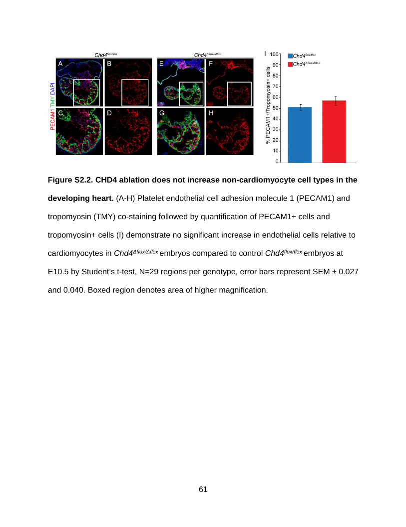
61
Figure S2.2. CHD4 ablation does not increase non-cardiomyocyte cell types in the
developing heart. (A-H) Platelet endothelial cell adhesion molecule 1 (PECAM1) and
tropomyosin (TMY) co-staining followed by quantification of PECAM1+ cells and
tropomyosin+ cells (I) demonstrate no significant increase in endothelial cells relative to
cardiomyocytes in Chd4Δflox/Δflox embryos compared to control Chd4flox/flox embryos at
E10.5 by Student’s t-test, N=29 regions per genotype, error bars represent SEM ± 0.027
and 0.040. Boxed region denotes area of higher magnification.

62
Figure S2.3. CHD4 is depleted from the myocardium at E9.5. (A-D) CHD4 is
depleted from cardiomyocytes costained with tropomyosin (TMY) at E9.5 in
Chd4Δflox/Δflox embryos (B,D) compared to Chd4flox/flox controls (A,C).

63
Figure S2.4. CHD4 temporally restricts gene expression in the developing heart.
(A-B) Ward’s two-way hierarchical clustering of differential gene expression profiles of
E9.5 (A) and E10.5 (B) Chd4Δflox/Δflox and Chd4flox/flox hearts show a distinct molecular
signature in the absence of CHD4 at both time points. Gene expression is row-scaled to
show relative expression. (C-D) MA plots of log2(fold change) versus log2(RNA count
mean) show a greater number of genes upregulated (913 genes) than downregulated
(372 genes) in the absence of CHD4 at E9.5 (C) and a greater number of genes

64
upregulated (920 genes) than downregulated (398 genes) in the absence of CHD4 at
E10.5 (D). Red points indicate adjusted p-value of less than 0.05. (E-F) Plotting E10.5
log2(fold change) versus E9.5 log2(fold change), with shared upregulated (E) or
downregulated (F) genes colored in blue demonstrate distinct requirements in
transcriptional regulation dependent on developmental stage. Black points represent
genes misexpressed unique to that stage.

65
Figure S2.5. CHD4 is required to repress expression of non-cardiac myofibril
isoforms in the embryonic heart. (A-E) Reverse transcription of RNA followed by
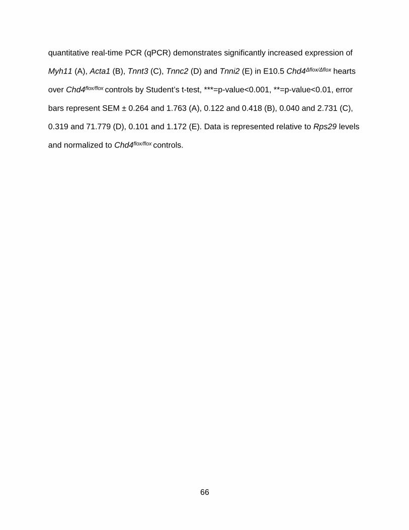
66
quantitative real-time PCR (qPCR) demonstrates significantly increased expression of
Myh11 (A), Acta1 (B), Tnnt3 (C), Tnnc2 (D) and Tnni2 (E) in E10.5 Chd4Δflox/Δflox hearts
over Chd4flox/flox controls by Student’s t-test, ***=p-value<0.001, **=p-value<0.01, error
bars represent SEM ± 0.264 and 1.763 (A), 0.122 and 0.418 (B), 0.040 and 2.731 (C),
0.319 and 71.779 (D), 0.101 and 1.172 (E). Data is represented relative to Rps29 levels
and normalized to Chd4flox/flox controls.

67
Figure S2.6. Acta2 is not differentially expressed in the absence of CHD4. Plotting
log2(RPKM) values from RNA-seq of Chd4flox/flox and Chd4Δflox/Δflox hearts at E9.5 and
E10.5 demonstrate that Acta2 (alpha-smooth muscle actin) is highly expressed
compared to median overall gene expression but is not significantly up- or down-
regulated in the absence of CHD4.

68
Figure S2.7. Smooth muscle myosin heavy chain antibody specifically stains
smooth muscle cells. (A-D) Smooth muscle myosin heavy chain staining of smooth
muscle cells surrounding the pharyngeal arch arteries in an E10.5 Chd4flox/flox control
embryo. Boxed region denotes area of higher magnification.

69
Figure S2.8. Ablation of CHD4 in the myocardium results in misexpression of
non-cardiac myofibril paralogs. Immunohistochemistry for fast skeletal TnI2 (A-H)
and smooth muscle myosin heavy chain (I-P) reveals increased expression of fast
skeletal and smooth muscle myofibril subunits in the myocardium of
Chd4Δflox/Δflox;Tg(Tnnt2-cre) hearts (E-H, M-P) at E10.5 compared to controls (A-D, I-L).
White boxes denote regions of higher magnification in adjacent panels.
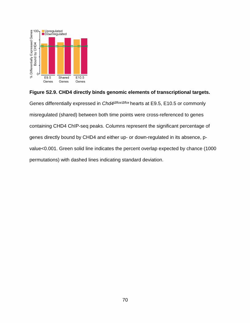
70
Figure S2.9. CHD4 directly binds genomic elements of transcriptional targets.
Genes differentially expressed in Chd4Δflox/Δflox hearts at E9.5, E10.5 or commonly
misregulated (shared) between both time points were cross-referenced to genes
containing CHD4 ChIP-seq peaks. Columns represent the significant percentage of
genes directly bound by CHD4 and either up- or down-regulated in its absence, p-
value<0.001. Green solid line indicates the percent overlap expected by chance (1000
permutations) with dashed lines indicating standard deviation.
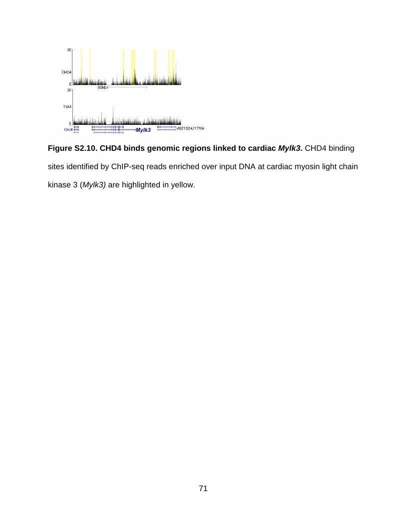
71
Figure S2.10. CHD4 binds genomic regions linked to cardiac Mylk3. CHD4 binding
sites identified by ChIP-seq reads enriched over input DNA at cardiac myosin light chain
kinase 3 (Mylk3) are highlighted in yellow.

72
Figure S2.11. Misexpression of non-cardiac myofibril paralogs contribute to
sarcomere disarray in the absence of CHD4. (A-D) α-actinin staining reveals
disorganized sarcomere structure (wedge arrow) in Chd4Δflox/Δflox cardiomyocytes at
E10.5 compared to Chd4flox/flox controls. Box denotes region of higher magnification. (E)
Measurement of sarcomere organization by classification of Z-disk and A-band
formation reveals significant decreases in sarcomere organization in Chd4Δflox/Δflox
cardiomyocytes by χ2 test, N= 225 vectors per ventricle per genotype, ***=p-
value<0.001.

73
REFERENCES
Aguayo-Gomez, A., J. Arteaga-Vazquez, Y. Svyryd, J. Calderon-Colmenero, C. Zamora-Gonzalez, G. Vargas-Alarcon, and O. M. Mutchinick. 2015. 'Identification of Copy Number Variations in Isolated Tetralogy of Fallot', Pediatr Cardiol, 36: 1642-6.
Allen, H. F., P. A. Wade, and T. G. Kutateladze. 2013. 'The NuRD architecture', Cell Mol Life Sci, 70: 3513-24.
Anders, S., P. T. Pyl, and W. Huber. 2015. 'HTSeq--a Python framework to work with high-throughput sequencing data', Bioinformatics, 31: 166-9.
Aristizabal, O., D. A. Christopher, F. S. Foster, and D. H. Turnbull. 1998. '40-MHZ echocardiography scanner for cardiovascular assessment of mouse embryos', Ultrasound Med Biol, 24: 1407-17.
Basson, C. T., D. R. Bachinsky, R. C. Lin, T. Levi, J. A. Elkins, J. Soults, D. Grayzel, E. Kroumpouzou, T. A. Traill, J. Leblanc-Straceski, B. Renault, R. Kucherlapati, J. G. Seidman, and C. E. Seidman. 1997. 'Mutations in human TBX5 [corrected] cause limb and cardiac malformation in Holt-Oram syndrome', Nat Genet, 15: 30-5.
Brown, D. D., S. N. Martz, O. Binder, S. C. Goetz, B. M. Price, J. C. Smith, and F. L. Conlon. 2005. 'Tbx5 and Tbx20 act synergistically to control vertebrate heart morphogenesis', Development, 132: 553-63.
Bruneau, B. G., M. Logan, N. Davis, T. Levi, C. J. Tabin, J. G. Seidman, and C. E. Seidman. 1999. 'Chamber-specific cardiac expression of Tbx5 and heart defects in Holt-Oram syndrome', Dev Biol, 211: 100-8.
Bruneau, B.G., G. Nemer, J.P. Schmitt, F. Charron, L. Robitaille, S. Caron, D. A. Conner, M. Gessler, M. Nemer, C. E. Seidman, and J.G. Seidman. 2001. 'A Murine Model of Holt-Oram Syndrome Defines Roles of the T-Box Transcription Factor Tbx5 in Cardiogenesis and Disease', Cell, 106: 709-21.
Clement, S., M. Stouffs, E. Bettiol, S. Kampf, K. H. Krause, C. Chaponnier, and M. Jaconi. 2007. 'Expression and function of alpha-smooth muscle actin during embryonic-stem-cell-derived cardiomyocyte differentiation', J Cell Sci, 120: 229-38.
Conway, S. J., A. Kruzynska-Frejtag, P. L. Kneer, M. Machnicki, and S. V. Koushik. 2003. 'What cardiovascular defect does my prenatal mouse mutant have, and why?', Genesis, 35: 1-21.
Craig, Roger, and John L. Woodhead. 2006. 'Structure and function of myosin filaments', Current Opinion in Structural Biology, 16: 204-12.

74
Dolk, H., M. Loane, and E. Garne. 2010. 'The prevalence of congenital anomalies in Europe', Adv Exp Med Biol, 686: 349-64.
Dorr, Kerry M., Nirav M. Amin, Lauren M. Kuchenbrod, Hanna Labiner, Marta S. Charpentier, Larysa H. Pevny, Andy Wessels, and Frank L. Conlon. 2015. 'Casz1 is required for cardiomyocyte G1-to-S phase progression during mammalian cardiac development', Development.
England, J., and S. Loughna. 2013. 'Heavy and light roles: myosin in the morphogenesis of the heart', Cell Mol Life Sci, 70: 1221-39.
Feng, J., T. Liu, and Y. Zhang. 2011. 'Using MACS to identify peaks from ChIP-Seq data', Curr Protoc Bioinformatics, Chapter 2: Unit 2.14.
Garnatz, A. S., Z. Gao, M. Broman, S. Martens, J. U. Earley, and E. C. Svensson. 2014. 'FOG-2 mediated recruitment of the NuRD complex regulates cardiomyocyte proliferation during heart development', Dev Biol, 395: 50-61.
Golden, H. B., S. Sunder, Y. Liu, X. Peng, and D. E. Dostal. 2012. 'In utero assessment of cardiovascular function in the embryonic mouse heart using high-resolution ultrasound biomicroscopy', Methods Mol Biol, 843: 245-63.
Gómez-del Arco, Pablo, Eusebio Perdiguero, Paula Sofia Yunes-Leites, Rebeca Acín-Pérez, Miriam Zeini, Antonio Garcia-Gomez, Krishnamoorthy Sreenivasan, Miguel Jiménez-Alcázar, Jessica Segalés, Dolores López-Maderuelo, Beatriz Ornés, Luis Jesús Jiménez-Borreguero, Gaetano D’Amato, David Enshell-Seijffers, Bruce Morgan, Katia Georgopoulos, Abul B M. M. K. Islam, Thomas Braun, José Luis de la Pompa, Johnny Kim, José A Enriquez, Esteban Ballestar, Pura Muñoz-Cánoves, and Juan Miguel Redondo. 'The Chromatin Remodeling Complex Chd4/NuRD Controls Striated Muscle Identity and Metabolic Homeostasis', Cell Metabolism, 23: 881-92.
Heidersbach, Amy, Chris Saxby, Karen Carver-Moore, Yu Huang, Yen-Sin Ang, Pieter J. de Jong, Kathryn N. Ivey, and Deepak Srivastava. 2013. 'microRNA-1 regulates sarcomere formation and suppresses smooth muscle gene expression in the mammalian heart', eLife, 2: e01323.
Heron, M., D. L. Hoyert, S. L. Murphy, J. Xu, K. D. Kochanek, and B. Tejada-Vera. 2009. 'Deaths: final data for 2006', Natl Vital Stat Rep, 57: 1-134.
Hirschy, A., F. Schatzmann, E. Ehler, and J. C. Perriard. 2006. 'Establishment of cardiac cytoarchitecture in the developing mouse heart', Dev Biol, 289: 430-41.
Homsy, J., S. Zaidi, Y. Shen, J. S. Ware, K. E. Samocha, K. J. Karczewski, S. R. DePalma, D. McKean, H. Wakimoto, J. Gorham, S. C. Jin, J. Deanfield, A. Giardini, G. A. Porter, Jr., R. Kim, K. Bilguvar, F. Lopez-Giraldez, I. Tikhonova, S. Mane, A. Romano-Adesman, H. Qi, B. Vardarajan, L. Ma, M. Daly, A. E. Roberts, M. W. Russell, S. Mital, J. W. Newburger, J. W. Gaynor, R. E. Breitbart, I. Iossifov, M. Ronemus, S. J. Sanders, J. R. Kaltman, J. G. Seidman, M. Brueckner, B. D. Gelb, E. Goldmuntz, R. P.

75
Lifton, C. E. Seidman, and W. K. Chung. 2015. 'De novo mutations in congenital heart disease with neurodevelopmental and other congenital anomalies', Science, 350: 1262-6.
Hung, H., R. Kohnken, and J. Svaren. 2012. 'The nucleosome remodeling and deacetylase chromatin remodeling (NuRD) complex is required for peripheral nerve myelination', J Neurosci, 32: 1517-27.
Ilkovski, B., S. Clement, C. Sewry, K. N. North, and S. T. Cooper. 2005. 'Defining alpha-skeletal and alpha-cardiac actin expression in human heart and skeletal muscle explains the absence of cardiac involvement in ACTA1 nemaline myopathy', Neuromuscul Disord, 15: 829-35.
Kaltenbrun, E., T. M. Greco, C. E. Slagle, L. M. Kennedy, T. Li, I. M. Cristea, and F. L. Conlon. 2013. 'A Gro/TLE-NuRD corepressor complex facilitates Tbx20-dependent transcriptional repression', J Proteome Res, 12: 5395-409.
Kashiwagi, M., B. A. Morgan, and K. Georgopoulos. 2007. 'The chromatin remodeler Mi-2beta is required for establishment of the basal epidermis and normal differentiation of its progeny', Development, 134: 1571-82.
Kent, W. James, Charles W. Sugnet, Terrence S. Furey, Krishna M. Roskin, Tom H. Pringle, Alan M. Zahler, Haussler, and David. 2002. 'The Human Genome Browser at UCSC', Genome Res, 12: 996-1006.
Kim, J., S. Sif, B. Jones, A. Jackson, J. Koipally, E. Heller, S. Winandy, A. Viel, A. Sawyer, T. Ikeda, R. Kingston, and K. Georgopoulos. 1999. 'Ikaros DNA-binding proteins direct formation of chromatin remodeling complexes in lymphocytes', Immunity, 10: 345-55.
Kirk, E. P., M. Sunde, M. W. Costa, S. A. Rankin, O. Wolstein, M. L. Castro, T. L. Butler, C. Hyun, G. Guo, R. Otway, J. P. Mackay, L. B. Waddell, A. D. Cole, C. Hayward, A. Keogh, P. Macdonald, L. Griffiths, D. Fatkin, G. F. Sholler, A. M. Zorn, M. P. Feneley, D. S. Winlaw, and R. P. Harvey. 2007. 'Mutations in cardiac T-box factor gene TBX20 are associated with diverse cardiac pathologies, including defects of septation and valvulogenesis and cardiomyopathy', Am J Hum Genet, 81: 280-91.
Langmead, B., C. Trapnell, M. Pop, and S. L. Salzberg. 2009. 'Ultrafast and memory-efficient alignment of short DNA sequences to the human genome', Genome Biol, 10: R25.
Lassmann, T., Y. Hayashizaki, and C. O. Daub. 2009. 'TagDust--a program to eliminate artifacts from next generation sequencing data', Bioinformatics, 25: 2839-40.
Li, H., B. Handsaker, A. Wysoker, T. Fennell, J. Ruan, N. Homer, G. Marth, G. Abecasis, and R. Durbin. 2009. 'The Sequence Alignment/Map format and SAMtools', Bioinformatics, 25: 2078-9.

76
Li, Q. Y., R. A. Newbury-Ecob, J. A. Terrett, D. I. Wilson, A. R. Curtis, C. H. Yi, T. Gebuhr, P. J. Bullen, S. C. Robson, T. Strachan, D. Bonnet, S. Lyonnet, I. D. Young, J. A. Raeburn, A. J. Buckler, D. J. Law, and J. D. Brook. 1997. 'Holt-Oram syndrome is caused by mutations in TBX5, a member of the Brachyury (T) gene family', Nat Genet, 15: 21-9.
Love, M. I., W. Huber, and S. Anders. 2014. 'Moderated estimation of fold change and dispersion for RNA-seq data with DESeq2', Genome Biol, 15: 550.
Luo, M., T. Ling, W. Xie, H. Sun, Y. Zhou, Q. Zhu, M. Shen, L. Zong, G. Lyu, Y. Zhao, T. Ye, J. Gu, W. Tao, Z. Lu, and I. Grummt. 2013. 'NuRD blocks reprogramming of mouse somatic cells into pluripotent stem cells', Stem Cells, 31: 1278-86.
Marhold, J., K. Kramer, E. Kremmer, and F. Lyko. 2004. 'The Drosophila MBD2/3 protein mediates interactions between the MI-2 chromatin complex and CpT/A-methylated DNA', Development, 131: 6033-9.
McLean, C. Y., D. Bristor, M. Hiller, S. L. Clarke, B. T. Schaar, C. B. Lowe, A. M. Wenger, and G. Bejerano. 2010. 'GREAT improves functional interpretation of cis-regulatory regions', Nat Biotechnol, 28: 495-501.
Mi, H, N Guo, A Kejariwal, and PD Thomas. 2007. 'PANTHER version 6: protein sequence and function evolution data with expanded representation of biological pathways', Nucleic Acids Res, 35: D247 - D52.
Mi, Huaiyu, Xiaosong Huang, Anushya Muruganujan, Haiming Tang, Caitlin Mills, Diane Kang, and Paul D. Thomas. 2017. 'PANTHER version 11: expanded annotation data from Gene Ontology and Reactome pathways, and data analysis tool enhancements', Nucleic Acids Res, 45: D183-D89.
Miccio, Annarita, Yuhuan Wang, Wei Hong, Gregory D Gregory, Hongxin Wang, Xiang Yu, John K Choi, Suresh Shelat, Wei Tong, Mortimer Poncz, and Gerd A Blobel. 2010. 'NuRD mediates activating and repressive functions of GATA‐1 and FOG‐1 during blood development', Embo j, 29: 442-56.
Montgomery, Rusty L., Christopher A. Davis, Matthew J. Potthoff, Michael Haberland, Jens Fielitz, Xiaoxia Qi, Joseph A. Hill, James A. Richardson, and Eric N. Olson. 2007. 'Histone deacetylases 1 and 2 redundantly regulate cardiac morphogenesis, growth, and contractility', Genes Dev, 21: 1790-802.
Mori, A. D., and B. G. Bruneau. 2004. 'TBX5 mutations and congenital heart disease: Holt-Oram syndrome revealed', Curr Opin Cardiol, 19: 211-5.
Moses, K. A., F. DeMayo, R. M. Braun, J. L. Reecy, and R. J. Schwartz. 2001. 'Embryonic expression of an Nkx2-5/Cre gene using ROSA26 reporter mice', Genesis, 31: 176-80.

77
Nishii, K., S. Morimoto, R. Minakami, Y. Miyano, K. Hashizume, M. Ohta, D. Y. Zhan, Q. W. Lu, and Y. Shibata. 2008. 'Targeted disruption of the cardiac troponin T gene causes sarcomere disassembly and defects in heartbeat within the early mouse embryo', Dev Biol, 322: 65-73.
O'Shaughnessy-Kirwan, A., J. Signolet, I. Costello, S. Gharbi, and B. Hendrich. 2015. 'Constraint of gene expression by the chromatin remodelling protein CHD4 facilitates lineage specification', Development, 142: 2586-97.
Postma, Alex V., Connie R. Bezzina, and Vincent M. Christoffels. 2016. 'Genetics of congenital heart disease: the contribution of the noncoding regulatory genome', J Hum Genet, 61: 13-19.
Quinlan, A. R. 2014. 'BEDTools: The Swiss-Army Tool for Genome Feature Analysis', Curr Protoc Bioinformatics, 47: 11.12.1-34.
Reynolds, E. S. 1963. 'The use of lead citrate at high pH as an electron-opaque stain in electron microscopy', J Cell Biol, 17: 208-12.
Roche, A. E., B. J. Bassett, S. A. Samant, W. Hong, G. A. Blobel, and E. C. Svensson. 2008. 'The zinc finger and C-terminal domains of MTA proteins are required for FOG-2-mediated transcriptional repression via the NuRD complex', J Mol Cell Cardiol, 44: 352-60.
Saggin, L., L. Gorza, S. Ausoni, and S. Schiaffino. 1989. 'Troponin I switching in the developing heart', J Biol Chem, 264: 16299-302.
Schmidl, Christian, Andre F. Rendeiro, Nathan C. Sheffield, and Christoph Bock. 2015. 'ChIPmentation: fast, robust, low-input ChIP-seq for histones and transcription factors', Nat Meth, 12: 963-65.
Shimbo, T., Y. Du, S. A. Grimm, A. Dhasarathy, D. Mav, R. R. Shah, H. Shi, and P. A. Wade. 2013. 'MBD3 localizes at promoters, gene bodies and enhancers of active genes', PLoS Genet, 9: e1004028.
Shimbo, T., M. Takaku, and P. A. Wade. 2016. 'High-quality ChIP-seq analysis of MBD3 in human breast cancer cells', Genom Data, 7: 173-4.
Shin, H., T. Liu, A. K. Manrai, and X. S. Liu. 2009. 'CEAS: cis-regulatory element annotation system', Bioinformatics, 25: 2605-6.
Slagle, Christopher E., and Frank L. Conlon. 'Emerging Field of Cardiomics: High-Throughput Investigations into Transcriptional Regulation of Cardiovascular Development and Disease', Trends in Genetics, 32: 707-16.
Srinivasan, S., H. S. Baldwin, O. Aristizabal, L. Kwee, M. Labow, M. Artman, and D. H. Turnbull. 1998. 'Noninvasive, in utero imaging of mouse embryonic heart development with 40-MHz echocardiography', Circulation, 98: 912-8.

78
Svensson, Eric C., Gordon S. Huggins, Hua Lin, Cynthia Clendenin, Fang Jiang, Rachel Tufts, Fred B. Dardik, and Jeffrey M. Leiden. 2000. 'A syndrome of tricuspid atresia in mice with a targeted mutation of the gene encoding Fog-2', Nat Genet, 25: 353-56.
Turnbull, D. H., T. S. Bloomfield, H. S. Baldwin, F. S. Foster, and A. L. Joyner. 1995. 'Ultrasound backscatter microscope analysis of early mouse embryonic brain development', Proc Natl Acad Sci U S A, 92: 2239-43.
Wade, P. A., A. Gegonne, P. L. Jones, E. Ballestar, F. Aubry, and A. P. Wolffe. 1999. 'Mi-2 complex couples DNA methylation to chromatin remodelling and histone deacetylation', Nat Genet, 23: 62-6.
Wade, P. A., P. L. Jones, D. Vermaak, and A. P. Wolffe. 1998. 'A multiple subunit Mi-2 histone deacetylase from Xenopus laevis cofractionates with an associated Snf2 superfamily ATPase', Curr Biol, 8: 843-6.
Waldron, L., J. D. Steimle, T. M. Greco, N. C. Gomez, K. M. Dorr, J. Kweon, B. Temple, X. H. Yang, C. M. Wilczewski, I. J. Davis, I. M. Cristea, I. P. Moskowitz, and F. L. Conlon. 2016. 'The Cardiac TBX5 Interactome Reveals a Chromatin Remodeling Network Essential for Cardiac Septation', Dev Cell, 36: 262-75.
Wang, Q., R. S. Reiter, Q. Q. Huang, J. P. Jin, and J. J. Lin. 2001. 'Comparative studies on the expression patterns of three troponin T genes during mouse development', Anat Rec, 263: 72-84.
Williams, C. J., T. Naito, P. G. Arco, J. R. Seavitt, S. M. Cashman, B. De Souza, X. Qi, P. Keables, U. H. Von Andrian, and K. Georgopoulos. 2004. 'The chromatin remodeler Mi-2beta is required for CD4 expression and T cell development', Immunity, 20: 719-33.
Xue, Y., J. Wong, G. T. Moreno, M. K. Young, J. Cote, and W. Wang. 1998. 'NURD, a novel complex with both ATP-dependent chromatin-remodeling and histone deacetylase activities', Mol Cell, 2: 851-61.
Yamada, Tomoko, Yue Yang, Martin Hemberg, Toshimi Yoshida, Ha Young Cho, J. Patrick Murphy, Diasynou Fioravante, Wade G Regehr, Steven P Gygi, Katia Georgopoulos, and Azad Bonni. 2014. 'Promoter Decommissioning by the NuRD Chromatin Remodeling Complex Triggers Synaptic Connectivity in the Mammalian Brain', Neuron, 83: 122-34.
Yang, J., D. Hu, J. Xia, Y. Yang, B. Ying, J. Hu, and X. Zhou. 2000. 'Three novel TBX5 mutations in Chinese patients with Holt-Oram syndrome', Am J Med Genet, 92: 237-40.
Yang, Zhenyun, Marie Yamazaki, Qingwu W. Shen, and Darl R. Swartz. 2009. 'Differences between Cardiac and Skeletal Troponin Interaction with the Thin Filament Probed by Troponin Exchange in Skeletal Myofibrils', Biophysical Journal, 97: 183-94.
Yoshida, A., H. Morisaki, M. Nakaji, M. Kitano, K. S. Kim, K. Sagawa, S. Ishikawa, I. Satokata, Y. Mitani, H. Kato, K. Hamaoka, S. Echigo, I. Shiraishi, and T. Morisaki. 2016.

79
'Genetic mutation analysis in Japanese patients with non-syndromic congenital heart disease', J Hum Genet, 61: 157-62.
Yoshida, T, I Hazan, J Zhang, SY Ng, T Naito, HJ Snippert, EJ Heller, X Qi, LN Lawton, CJ Williams, and K Georgopoulos. 2008. 'The role of the chromatin remodeler Mi-2beta in hematopoietic stem cell self-renewal and multilineage differentiation', Genes Dev, 22: 1174 - 89.
Zaidi, S., M. Choi, H. Wakimoto, L. Ma, J. Jiang, J. D. Overton, A. Romano-Adesman, R. D. Bjornson, R. E. Breitbart, K. K. Brown, N. J. Carriero, Y. H. Cheung, J. Deanfield, S. DePalma, K. A. Fakhro, J. Glessner, H. Hakonarson, M. J. Italia, J. R. Kaltman, J. Kaski, R. Kim, J. K. Kline, T. Lee, J. Leipzig, A. Lopez, S. M. Mane, L. E. Mitchell, J. W. Newburger, M. Parfenov, I. Pe'er, G. Porter, A. E. Roberts, R. Sachidanandam, S. J. Sanders, H. S. Seiden, M. W. State, S. Subramanian, I. R. Tikhonova, W. Wang, D. Warburton, P. S. White, I. A. Williams, H. Zhao, J. G. Seidman, M. Brueckner, W. K. Chung, B. D. Gelb, E. Goldmuntz, C. E. Seidman, and R. P. Lifton. 2013. 'De novo mutations in histone-modifying genes in congenital heart disease', Nature, 498: 220-3.
Zhang, Y, G LeRoy, HP Seelig, WS Lane, and D Reinberg. 1998. 'The dermatomyositis-specific autoantigen Mi2 is a component of a complex containing histone deacetylase and nucleosome remodeling activities', Cell, 95: 279 - 89.
Zhang, Y. 2011. 'Biology of the Mi-2/NuRD Complex in SLAC (Stemness, Longevity/Ageing, and Cancer)', Gene Regul Syst Bio, 5: 1-26.

80
CHAPTER 3: CHD4 and the NuRD complex interact with GATA4 and NKX2-5 in the
developing heart
Introduction
Congenital heart disease (CHD) is the leading cause of infant mortality in the
United States (Joann et al. 2002; Petrini, Damus, and Johnston 1997). Despite
extensive research identifying mutations in cardiac transcription factors such as Nkx2-5,
Gata4, and Tbx5 as responsible for a number of these cases, there is a persistent lack
of understanding how these transcription factors function to instigate normal heart
development (Chen et al. 2010; Garg et al. 2003; Hirayama-Yamada et al. 2005;
Benson et al. 1999; Basson et al. 1999). Recent research on the roles of chromatin
remodeling complexes in cardiac development may provide some explanation
(Takeuchi et al. 2011; Bruneau 2010; Homsy et al. 2015; Bevilacqua, Willis, and
Bultman 2014; Hang et al. 2010). However, interactions between these complexes and
cardiac transcription factors causative to CHD remain incompletely understood. One
such complex required for cardiac development is the Nucleosome Remodeling and
Deacetylase (NuRD) complex.
The NuRD complex is composed of the ATP-dependent chromodomain-helicase-
DNA binding protein 3 or 4 (CHD3/4), histone deacetylase 1 or 2 (HDAC1/2), methyl-
CpG-binding domain protein 2 or 3 (MBD2/3), retinoblastoma-binding protein 4 or 7
(RBBP4/7), metastasis-associated protein 1, 2 or 3 (MTA1/2/3), GATA zinc-finger
81
domain containing protein 2A or B (GATAD2A/B) (Wade et al. 1999; Wade et al. 1998;
Zhang et al. 1998). The critical nature of CHD4 in cardiac development is highlighted by
the observation that mutations in CHD4 are causative to CHDs including atrial and
ventricular septal and morphological defects (Homsy et al. 2015). Recent research from
our lab demonstrated that CHD4 is required early in cardiac development to directly
repress expression of non-cardiac myofibril isoforms, and loss of CHD4-mediated
repression leads to misexpression of fast skeletal and smooth muscle myofibril
isoforms, cardiac sarcomere malformation and early embryonic lethality (Wilczewski et
al. 2018).
Several NuRD subunits have DNA-binding capabilities; however, the NuRD
complex must be recruited to its target genes through interactions with sequence-
specific co-factors. Recent research has revealed cooperative interactions between
cardiac transcription factors causative to CHD and the NuRD complex during heart
development (Waldron et al. 2016; Kaltenbrun et al. 2013; Garnatz et al. 2014; Roche et
al. 2008). Ablation of these interactions leads to misexpression of target genes and
structural defects in the heart. This raises the possibility of discovering other cofactors
of this complex which may further inform the genetic etiology of CHD.
In this report, we identify three previously unknown transcriptional cofactors of
CHD4 and the NuRD complex in the developing heart. Using an unbiased
bioinformatics approach, we found cardiac transcription factor motifs to be highly
overrepresented in CHD4-bound regions of the genome. Parallel reaction monitoring
mass spectrometry (PRM-MS) in embryonic cardiac tissue uncovered interactions
between CHD4 and NKX2-5, GATA4, SMAD3 and TBX5. Further analysis revealed
82
CHD4 and GATA4 or NKX2-5 converge on regulatory targets for transcriptional
repression, including genes for fast skeletal and smooth muscle myofibril isoforms.
These results represent a substantial advance in our understanding of how these well-
studied transcription factors carry out their regulatory function.
Results
Cardiac transcription factor motifs overrepresented in CHD4-bound genomic elements
Previous work determining the role of CHD4 in cardiac development uncovered
the direct regulatory targets of CHD4 during heart development through chromatin
immunoprecipication followed by high-throughput sequencing (ChIP-seq) for CHD4 and
transcriptional profiling (Wilczewski et al. 2018).To uncover transcriptional co-factors
that could guide CHD4 to its regulatory targets through sequence-specific binding, we
performed de novo motif discovery with HOMER (Heinz et al. 2010) using CHD4 ChIP-
seq data from E10.0 wild-type hearts (Figure 3.1A). This analysis revealed a striking
abundance of cardiac transcription factor consensus motifs in CHD4-bound regions
(Figure 3.1B).
TBX5, ISL1, NKX2-5, and GATA4 are required for human heart development
(Luo et al. 2014; Baban et al. 2014; Basson et al. 1997; Garg et al. 2003; Hirayama-
Yamada et al. 2005). SMAD2/3/4 and Brachyury/T play critical roles in cardiac
differentiation (Beyer et al. 2013; Johansson and Wiles 1995; Aramaki et al. 2013;
David et al. 2011) while MEIS1, CRE-binding protein (CREB), and TEAD4, as a
member of the Hippo signaling pathway, have been implicated in regulating cardiac
homeostasis (Mahmoud et al. 2013; Xin et al. 2011; Fentzke et al. 1998). MEF2c is

83
expressed early in the cardiac lineage and is capable of reprograming fibroblasts to
induced cardiomyocytes in conjunction with GATA4 and TBX5 (Edmondson et al. 1994;
Ieda et al. 2010; Qian et al. 2013; Lin et al. 1997). CTCF is important in establishing
chromatin architecture through formation of topologically associated domains (Bell,
West, and Felsenfeld 1999; Parelho et al. 2008; Rubio et al. 2008). These motifs
indicate CHD4 may interact with many more cardiac-specific transcriptional co-factors
than previously thought.
CHD4 physically interacts with GATA4, NKX2-5, SMAD3 and TBX5
We wanted to identify whether these predicted associations physically occur the
developing heart. We isolated E10.5 hearts from wild-type mice and performed
immunoisolation of CHD4 complexes using anti-GFP immunoprecipitation as a negative
control. Since many of these potential co-factors are low-abundance transcription
factors, we utilized parallel reaction monitoring mass spectrometry (PRM-MS) to capture
these infrequent and transient interactions by monitoring unique peptides for each
potential co-factor. This analysis revealed interactions between CHD4 and GATA4,
NKX2-5, SMAD3 and TBX5 in the developing heart (Figure 3.2). Interactions between
CHD4 and SMAD3 have been previously demonstrated in human embryonic stem cells
(Beyer et al. 2013), and we have previously reported an interaction between CHD4 and
TBX5 in the developing heart (Waldron et al. 2016), which supports the validity of these
results. To our knowledge, this is the first report of an interaction between GATA4 and
NKX2-5 and CHD4.

84
NKX2-5 and GATA4 bind putative regulatory regions for fast skeletal and smooth
muscle myofibril isoforms
To identify regulatory targets of these interactions, we analyzed genomic regions
directly bound by CHD4 that contained GATA4 or NKX2-5 consensus motifs. We
assessed whether these transcription factors bound these regulatory regions in mature
cardiomyocytes or cardiac progenitor cells differentiated in culture using previously
published ChIP-seq data (Luna-Zurita et al. 2016). Strikingly, we found that GATA4
binds a putative regulatory region for Myh11 (encoding smooth muscle myosin heavy
chain) in mature cardiomyocytes and cardiac precursors (Figure 3.3A). We also found
that NKX2-5 binds a putative regulatory region for Acta1 (encoding fast skeletal α-actin)
in mature cardiomyocytes but not cardiac progenitor cells (Figure 3.3B). This data
demonstrates that CHD4 likely interacts with these cardiac transcription factors to
mediate repression of non-cardiac myofibril isoforms during cardiac development.
Discussion
We have demonstrated that CHD4 interacts with the transcription factors GATA4,
NKX2-5, SMAD3 and TBX5 in the developing heart. We also present evidence
indicating that CHD4 is recruited to repress fast skeletal and smooth muscle myofibril
isoforms through its interactions with GATA4 and NKX2-5.
GATA4 is a well-studied transcription factor in cardiac development and
cardiomyocyte differentiation. GATA4 has been demonstrated to be a non-histone
target of NuRD complex member HDAC2; however, this interaction did not include a
role for direct transcriptional regulation of target genes. In comparison, GATA4

85
interaction with co-repressor FOG-2 leads to transcriptional repression of some of its
target genes (Svensson et al. 1999). FOG-2 has been shown to interact with members
of the NuRD complex, particularly MTA1 and RBBP 4/7, to repress gene expression of
cell cycle inhibitors; loss of this interaction leads to perinatal lethality with septal defects
and a thin myocardium (Garnatz et al. 2014; Roche et al. 2008). The data presented in
this report demonstrates that GATA4 can also interact with the NuRD complex through
CHD4; however, it remains unclear whether this interaction is dependent on FOG-2 or if
GATA4 can interact with NuRD in a FOG-2 independent manner to modulate
expression of a different set of target genes. GATA4 binding to the direct CHD4 target
Myh11, whose upregulation was not reported in FOG-2 mutants, suggests this may be
the case.
NKX2-5 is another well-studied transcription factor in cardiac development and
differentiation demonstrated to act through transcriptional activation and repression. The
mechanism by which NKX2-5 engages in transcriptional repression has not been
thoroughly explored. This report therefore demonstrates one potential avenue for NKX2-
mediated repression of regulatory targets. Interestingly, a separate study found NKX2-5
acts to directly repress expression of different fast skeletal myofibril subunits (Tnnt3 and
Tnni2) in the developing heart but did not mention upregulation of Acta1 (Dupays et al.
2015). While CHD4 has been demonstrated to bind putative regulatory regions for both
Tnnt3 and Tnni2 in the developing heart, these regions differ from the proposed NKX2-5
binding site (Wilczewski et al. 2018). Therefore, whether NKX2-5 interacts with the
NuRD complex to repress these targets or acts through an alternative, unknown
mechanism remains to be determined.
86
PRM-MS analysis in embryonic cardiac tissue found that the previously
discovered interaction between CHD4 and SMAD3 in human embryonic stem cells
(hESC) is maintained in the developing heart. In hESCs, the NuRD complex interacts
with OCT4, TEAD, and SMAD2/3 to repress mesendoderm genes to maintain
pluripotency (Beyer et al. 2013). Induction of mesoderm specification lead to the loss of
TEAD binding at these sites and SMAD2/3-induced gene activation. Interestingly, de
novo motif discovery also predicted TEAD4 as a co-factor of CHD4; however, this
interaction was not detected in embryonic cardiac tissue. This result could indicate that
CHD4 and the NuRD complex maintain their interaction with SMAD3 through
cardiomyocyte differentiation after mesoderm specification without TEAD to regulate a
different set of target genes during cardiac development. Further exploration of CHD4-
SMAD3 targets is needed to identify what role this interaction plays during the later
stages of differentiation and cardiac development.
This report also validated the interaction we previously reported between CHD4
and TBX5, albeit one day later in cardiac development (Waldron et al. 2016). Further
analysis is necessary to determine whether direct targets predicted by CHD4 ChIP-seq
at E10.0 uncovers new gene regulatory targets for this interaction as cardiac
development proceeds.
This report demonstrates that several cardiac transcription factors engage CHD4
and the NuRD complex to repress gene targets through interactions at cis-regulatory
regions. Evaluating other potential targets of these interactions may yield novel targets
of these well-studied transcription factors and inform the molecular mechanisms
underlying cases of congenital heart disease.

87
This work was completed in collaboration with Xinlei Sheng, Austin J. Hepperla,
Todd Greco, Ian J. Davis and Ileana M. Cristea. This work was funded by R01
HL112618 and R01 HL127640 to F.L.C., and 5T32 HL069768 and 1F31 HL136100 to
C.M.W.
Materials and Methods
Motif Discovery
De novo motif discovery on CHD4 chromatin immunoprecipitation followed by
high-throughput sequencing (ChIP-seq) regions (Wilczewski et al. 2018) (GSE109012)
from embryonic day (E)10.0 cardiac tissue was performed using Hypergeometric
Optimization of Motif EnRichment (HOMER) (Heinz et al. 2010).
CHD4 Immunoprecipitation
E10.5 wild-type CD1 hearts (minimum twenty-eight hearts per IP, three separate
biological replicates) were harvested in cold PBS, snap frozen and cryolysed as
previously described (Kaltenbrun et al. 2013). Frozen tissue powder was resuspended
in optimized lysis buffer (5ml/1g powder) (20mM K-HEPES pH7.4, 0.11M KOAC, 2mM
MgCl2, 0.1% Tween 20, 1μM ZnCl2, 1mM CaCl2, 0.5% Triton-X 100, 150mM NaCl2,
protease inhibitor (Sigma), phosphatase inhibitor (Sigma)). CHD4 complexes were
solubilized and immunoprecipitated as previously described (Kaltenbrun et al. 2013;
Cristea and Chait 2011) using rabbit anti-CHD4 antibody (Abcam #ab72418) or
negative control custom rabbit anti-GFP antibody (Cristea et al. 2005) with elution at
95°C for 10 minutes. The immunoisolated proteins were resolved (~ 1 cm) by SDS-

88
PAGE, and visualized by Coomassie blue. Each lane was subjected to in-gel digestion
with trypsin as previously reported (Waldron et al. 2016).
Parallel Reaction Monitoring (PRM) targeted Mass Spectrometry (MS)
Desalted peptides (2ul each) were analyzed by nano-liquid chromatography
(nLC)-MS/MS using a Dionex Ultimate 3000 nRSLC system directly coupled to a Q
exactive HF orbitrap mass spectrometer (ThermoFisher Scientific) equipped with an
EASY-Spray ion source (Thermo, Fisher Scientific). Peptide mixtures were evaporated
in vacuo and resuspended in 1% Trifluoroacetic acid/4%acetonitrile/95% water and
loaded onto an 50 cm long column with a 75 um internal diameter (PepMap) and
separated over a 60 min gradient with a mobile phase containing aqueous acetonitrile
and 0.1% formic acid programmed from 1% to 3% acetonitrile over 12 min, then 3 to
35% acetonitrile over 60 min, then 35 to 97% acetonitrile over 1 min, followed by 10 min
at 97% acetonitrile and 97 to 70% over 1 min, all at a flow rate of 250 nL/min. The PRM
method consisted of targeted MS2 scans at a resolution of 60,000 and with an AGC
value of 1x10E6, a max injection time of 500 ms, a 0.8 m/z isolation window, a fixed first
mass of 150 m/z, and normalized collision energy of 27, which were recorded as profile
data. The targeted MS2 methods were controlled with a timed inclusion list containing
the target precursor m/z value, charge, and a retention time window that were
determined from shotgun analysis.

89
Figure 3.1. CHD4 bound genomic regions are enriched for cardiac transcription
factor motifs. (A) Schematic for de novo motif discovery from CHD4 ChIP-seq data. (B)
HOMER motif results from E10.0 CHD4 ChIP-seq peaks. q-value represents Benjamini-
Hotchberg correction for multiple comparisons.

90
Figure 3.2. CHD4 interacts with a subset of cardiac transcription factors in the
developing heart. (A) Schematic showing CHD4 interaction partners detected by
parallel reaction monitoring mass spectrometry at E10.5. (B) Table showing average
enrichment for each monitored peptide in CHD4 immunoprecipitation over GFP
negative control immunoprecipitation. Percent covariance represents the covariance of
the average enrichment over three independent biological replicates.

91
Figure 3.3. CHD4 and cardiac transcription factors are both present at putative
regulatory loci. (A) CHD4 bound regulatory region in Myh11 with GATA4 consensus
motif in E10.0 cardiac tissue is also bound by GATA4 in mature cardiomyocytes (CM)
and cardiac progenitors (CP). (B) CHD4 bound regulatory region in Acta1 with NKX2-5
consensus motif in E10.0 cardiac tissue is also bound by NKX2-5 in mature
cardiomyocytes (CM) but not cardiac progenitors (CP).

92
REFERENCES
Aramaki, Shinya, Katsuhiko Hayashi, Kazuki Kurimoto, Hiroshi Ohta, Yukihiro Yabuta, Hiroko Iwanari, Yasuhiro Mochizuki, Takao Hamakubo, Yuki Kato, Katsuhiko Shirahige, and Mitinori Saitou. 2013. 'A Mesodermal Factor, T, Specifies Mouse Germ Cell Fate by Directly Activating Germline Determinants', Dev Cell, 27: 516-29.
Baban, A., A. V. Postma, M. Marini, G. Trocchio, A. Santilli, M. Pelegrini, P. Sirleto, M. Lerone, S. B. Albanese, P. Barnett, C. J. Boogerd, B. Dallapiccola, M. C. Digilio, R. Ravazzolo, and G. Pongiglione. 2014. 'Identification of TBX5 mutations in a series of 94 patients with Tetralogy of Fallot', Am J Med Genet A, 164a: 3100-7.
Basson, C. T., D. R. Bachinsky, R. C. Lin, T. Levi, J. A. Elkins, J. Soults, D. Grayzel, E. Kroumpouzou, T. A. Traill, J. Leblanc-Straceski, B. Renault, R. Kucherlapati, J. G. Seidman, and C. E. Seidman. 1997. 'Mutations in human TBX5 [corrected] cause limb and cardiac malformation in Holt-Oram syndrome', Nat Genet, 15: 30-5.
Basson, C. T., T. Huang, R. C. Lin, D. R. Bachinsky, S. Weremowicz, A. Vaglio, R. Bruzzone, R. Quadrelli, M. Lerone, G. Romeo, M. Silengo, A. Pereira, J. Krieger, S. F. Mesquita, M. Kamisago, C. C. Morton, M. E. Pierpont, C. W. Muller, J. G. Seidman, and C. E. Seidman. 1999. 'Different TBX5 interactions in heart and limb defined by Holt-Oram syndrome mutations', Proc Natl Acad Sci U S A, 96: 2919-24.
Bell, A. C., A. G. West, and G. Felsenfeld. 1999. 'The protein CTCF is required for the enhancer blocking activity of vertebrate insulators', Cell, 98: 387-96.
Benson, D. W., G. M. Silberbach, A. Kavanaugh-McHugh, C. Cottrill, Y. Zhang, S. Riggs, O. Smalls, M. C. Johnson, M. S. Watson, J. G. Seidman, C. E. Seidman, J. Plowden, and J. D. Kugler. 1999. 'Mutations in the cardiac transcription factor NKX2.5 affect diverse cardiac developmental pathways', J Clin Invest, 104: 1567-73.
Bevilacqua, A., M. S. Willis, and S. J. Bultman. 2014. 'SWI/SNF chromatin-remodeling complexes in cardiovascular development and disease', Cardiovasc Pathol, 23: 85-91.
Beyer, Tobias A, Alexander Weiss, Yuliya Khomchuk, Kui Huang, Abiodun A Ogunjimi, Xaralabos Varelas, and Jeffrey L Wrana. 2013. 'Switch Enhancers Interpret TGF-β and Hippo Signaling to Control Cell Fate in Human Embryonic Stem Cells', Cell Reports, 5: 1611-24.
Bruneau, Benoit G. 2010. 'Chromatin remodeling in heart development', Current Opinion in Genetics & Development, 20: 505-11.
Chen, Y., Z. Q. Han, W. D. Yan, C. Z. Tang, J. Y. Xie, H. Chen, and D. Y. Hu. 2010. 'A novel mutation in GATA4 gene associated with dominant inherited familial atrial septal defect', J Thorac Cardiovasc Surg, 140: 684-7.

93
Cristea, I. M., R. Williams, B. T. Chait, and M. P. Rout. 2005. 'Fluorescent proteins as proteomic probes', Mol Cell Proteomics, 4: 1933-41.
Cristea, Ileana M., and Brian T. Chait. 2011. 'Conjugation of Magnetic Beads for Immunopurification of Protein Complexes', Cold Spring Harb Protoc, 2011: pdb.prot5610.
David, Robert, Veronica Barbara Jarsch, Florian Schwarz, Petra Nathan, Moritz Gegg, Heiko Lickert, and Wolfgang-Michael Franz. 2011. 'Induction of MesP1 by Brachyury(T) generates the common multipotent cardiovascular stem cell', Cardiovasc Res, 92: 115-22.
Dupays, Laurent, Catherine Shang, Robert Wilson, Surendra Kotecha, Sophie Wood, Norma Towers, and Timothy Mohun. 2015. 'Sequential Binding of MEIS1 and NKX2-5 on the Popdc2 Gene: A Mechanism for Spatiotemporal Regulation of Enhancers during Cardiogenesis', Cell Reports, 13: 183-95.
Edmondson, D. G., G. E. Lyons, J. F. Martin, and E. N. Olson. 1994. 'Mef2 gene expression marks the cardiac and skeletal muscle lineages during mouse embryogenesis', Development, 120: 1251-63.
Fentzke, R. C., C. E. Korcarz, R. M. Lang, H. Lin, and J. M. Leiden. 1998. 'Dilated cardiomyopathy in transgenic mice expressing a dominant-negative CREB transcription factor in the heart', J Clin Invest, 101: 2415-26.
Garg, V., I. S. Kathiriya, R. Barnes, M. K. Schluterman, I. N. King, C. A. Butler, C. R. Rothrock, R. S. Eapen, K. Hirayama-Yamada, K. Joo, R. Matsuoka, J. C. Cohen, and D. Srivastava. 2003. 'GATA4 mutations cause human congenital heart defects and reveal an interaction with TBX5', Nature, 424: 443-7.
Garnatz, A. S., Z. Gao, M. Broman, S. Martens, J. U. Earley, and E. C. Svensson. 2014. 'FOG-2 mediated recruitment of the NuRD complex regulates cardiomyocyte proliferation during heart development', Dev Biol, 395: 50-61.
Hang, Calvin T., Jin Yang, Pei Han, Hsiu-Ling Cheng, Ching Shang, Euan Ashley, Bin Zhou, and Ching-Pin Chang. 2010. 'Chromatin regulation by Brg1 underlies heart muscle development and disease', Nature, 466: 62-67.
Heinz, S., C. Benner, N. Spann, E. Bertolino, Y. C. Lin, P. Laslo, J. X. Cheng, C. Murre, H. Singh, and C. K. Glass. 2010. 'Simple combinations of lineage-determining transcription factors prime cis-regulatory elements required for macrophage and B cell identities', Mol Cell, 38: 576-89.
Hirayama-Yamada, K., M. Kamisago, K. Akimoto, H. Aotsuka, Y. Nakamura, H. Tomita, M. Furutani, S. Imamura, A. Takao, M. Nakazawa, and R. Matsuoka. 2005. 'Phenotypes with GATA4 or NKX2.5 mutations in familial atrial septal defect', Am J Med Genet A, 135: 47-52.

94
Homsy, J., S. Zaidi, Y. Shen, J. S. Ware, K. E. Samocha, K. J. Karczewski, S. R. DePalma, D. McKean, H. Wakimoto, J. Gorham, S. C. Jin, J. Deanfield, A. Giardini, G. A. Porter, Jr., R. Kim, K. Bilguvar, F. Lopez-Giraldez, I. Tikhonova, S. Mane, A. Romano-Adesman, H. Qi, B. Vardarajan, L. Ma, M. Daly, A. E. Roberts, M. W. Russell, S. Mital, J. W. Newburger, J. W. Gaynor, R. E. Breitbart, I. Iossifov, M. Ronemus, S. J. Sanders, J. R. Kaltman, J. G. Seidman, M. Brueckner, B. D. Gelb, E. Goldmuntz, R. P. Lifton, C. E. Seidman, and W. K. Chung. 2015. 'De novo mutations in congenital heart disease with neurodevelopmental and other congenital anomalies', Science, 350: 1262-6.
Ieda, M., J. D. Fu, P. Delgado-Olguin, V. Vedantham, Y. Hayashi, B. G. Bruneau, and D. Srivastava. 2010. 'Direct reprogramming of fibroblasts into functional cardiomyocytes by defined factors', Cell, 142: 375-86.
Joann, Petrini, Damus Karla, Russell Rebecca, Poschman Karalee, Davidoff Michael J., and Mattison Donald. 2002. 'Contribution of birth defects to infant mortality in the United States', Teratology, 66: S3-S6.
Johansson, B M, and M V Wiles. 1995. 'Evidence for involvement of activin A and bone morphogenetic protein 4 in mammalian mesoderm and hematopoietic development', Mol Cell Biol, 15: 141-51.
Kaltenbrun, E., T. M. Greco, C. E. Slagle, L. M. Kennedy, T. Li, I. M. Cristea, and F. L. Conlon. 2013. 'A Gro/TLE-NuRD corepressor complex facilitates Tbx20-dependent transcriptional repression', J Proteome Res, 12: 5395-409.
Lin, Q., J. Schwarz, C. Bucana, and E. N. Olson. 1997. 'Control of mouse cardiac morphogenesis and myogenesis by transcription factor MEF2C', Science, 276: 1404-7.
Luna-Zurita, L., C. U. Stirnimann, S. Glatt, B. L. Kaynak, S. Thomas, F. Baudin, M. A. Samee, D. He, E. M. Small, M. Mileikovsky, A. Nagy, A. K. Holloway, K. S. Pollard, C. W. Muller, and B. G. Bruneau. 2016. 'Complex Interdependence Regulates Heterotypic Transcription Factor Distribution and Coordinates Cardiogenesis', Cell.
Luo, Z. L., H. Sun, Z. Q. Yang, Y. H. Ma, Y. Gu, Y. Q. He, D. Wei, L. B. Xia, B. H. Yang, and T. Guo. 2014. 'Genetic variations of ISL1 associated with human congenital heart disease in Chinese Han people', Genet Mol Res, 13: 1329-38.
Mahmoud, A. I., F. Kocabas, S. A. Muralidhar, W. Kimura, A. S. Koura, S. Thet, E. R. Porrello, and H. A. Sadek. 2013. 'Meis1 regulates postnatal cardiomyocyte cell cycle arrest', Nature, 497: 249-53.
Parelho, V., S. Hadjur, M. Spivakov, M. Leleu, S. Sauer, H. C. Gregson, A. Jarmuz, C. Canzonetta, Z. Webster, T. Nesterova, B. S. Cobb, K. Yokomori, N. Dillon, L. Aragon, A. G. Fisher, and M. Merkenschlager. 2008. 'Cohesins functionally associate with CTCF on mammalian chromosome arms', Cell, 132: 422-33.
95
Petrini, J., K. Damus, and R. B. Johnston, Jr. 1997. 'An overview of infant mortality and birth defects in the United States', Teratology, 56: 8-10.
Qian, L., E. C. Berry, J. D. Fu, M. Ieda, and D. Srivastava. 2013. 'Reprogramming of mouse fibroblasts into cardiomyocyte-like cells in vitro', Nat Protoc, 8: 1204-15.
Roche, A. E., B. J. Bassett, S. A. Samant, W. Hong, G. A. Blobel, and E. C. Svensson. 2008. 'The zinc finger and C-terminal domains of MTA proteins are required for FOG-2-mediated transcriptional repression via the NuRD complex', J Mol Cell Cardiol, 44: 352-60.
Rubio, E. D., D. J. Reiss, P. L. Welcsh, C. M. Disteche, G. N. Filippova, N. S. Baliga, R. Aebersold, J. A. Ranish, and A. Krumm. 2008. 'CTCF physically links cohesin to chromatin', Proc Natl Acad Sci U S A, 105: 8309-14.
Svensson, Eric C., Rachel L. Tufts, Christine E. Polk, and Jeffrey M. Leiden. 1999. 'Molecular cloning of FOG-2: A modulator of transcription factor GATA-4 in cardiomyocytes', Proceedings of the National Academy of Sciences, 96: 956-61.
Takeuchi, Jun K., Xin Lou, Jeffrey M. Alexander, Hiroe Sugizaki, Paul Delgado-Olguín, Alisha K. Holloway, Alessandro D. Mori, John N. Wylie, Chantilly Munson, Yonghong Zhu, Yu-Qing Zhou, Ru-Fang Yeh, R. Mark Henkelman, Richard P. Harvey, Daniel Metzger, Pierre Chambon, Didier Y. R. Stainier, Katherine S. Pollard, Ian C. Scott, and Benoit G. Bruneau. 2011. 'Chromatin remodelling complex dosage modulates transcription factor function in heart development', Nat Commun, 2: 187.
Wade, P. A., P. L. Jones, D. Vermaak, and A. P. Wolffe. 1998. 'A multiple subunit Mi-2 histone deacetylase from Xenopus laevis cofractionates with an associated Snf2 superfamily ATPase', Curr Biol, 8: 843-6.
Wade, PA, A Gegonne, PL Jones, E Ballestar, F Aubry, and AP Wolffe. 1999. 'Mi-2 complex couples DNA methylation to chromatin remodelling and histone deacetylation', Nat Genet, 23: 62 - 66.
Waldron, L., J. D. Steimle, T. M. Greco, N. C. Gomez, K. M. Dorr, J. Kweon, B. Temple, X. H. Yang, C. M. Wilczewski, I. J. Davis, I. M. Cristea, I. P. Moskowitz, and F. L. Conlon. 2016. 'The Cardiac TBX5 Interactome Reveals a Chromatin Remodeling Network Essential for Cardiac Septation', Dev Cell, 36: 262-75.
Wilczewski, Caralynn M., Austin J. Hepperla, Takashi Shimbo, Lauren Wasson, Zachary L. Robbe, Ian J. Davis, Paul A. Wade, and Frank L. Conlon. 2018. 'CHD4 and the NuRD complex directly control cardiac sarcomere formation', Proceedings of the National Academy of Sciences.
Xin, M., Y. Kim, L. B. Sutherland, X. Qi, J. McAnally, R. J. Schwartz, J. A. Richardson, R. Bassel-Duby, and E. N. Olson. 2011. 'Regulation of insulin-like growth factor signaling by Yap governs cardiomyocyte proliferation and embryonic heart size', Sci Signal, 4: ra70.

96
Zhang, Y, G LeRoy, HP Seelig, WS Lane, and D Reinberg. 1998. 'The dermatomyositis-specific autoantigen Mi2 is a component of a complex containing histone deacetylase and nucleosome remodeling activities', Cell, 95: 279 - 89.
97
CHAPTER 4: Discussion and Future Directions
Dynamic alterations in chromatin accessibility is one mechanism of gene
regulation during cardiomyocyte differentiation and embryonic heart development.
CHD4 and the NuRD complex are essential components of this regulatory framework,
as previously demonstrated in ESCs and mesoderm specification (Beyer et al. 2013;
O'Shaughnessy-Kirwan et al. 2015; Linder et al. 2007). However, its role in later stages
of cardiomyocyte differentiation and its functional significance on embryonic heart
development remain relatively unknown. This set of studies sought to examine the
requirement for the NuRD complex during cardiac development by conditionally ablating
its catalytic core subunit, CHD4, and utilized transcriptomic, genomic, proteomic, and
phenotypic methodology to uncover the NuRD complex’s functional significance during
early heart development. We uncovered a requirement for CHD4 in the repression of
skeletal and smooth muscle cells in cardiomyocytes that is essential for cardiac function
and embryonic development.
CHD4 and the NuRD complex directly control cardiac sarcomere formation
Chapter 2 established a role for CHD4 and the NuRD complex as a negative
regulator of skeletal and smooth muscle myofibril subunits. We demonstrated that
CHD4 binds to proximal and distal regulatory regions in the developing heart to both
positively and negatively regulate gene expression. We found CHD4 directly represses

98
smooth muscle myosin heavy chain, fast skeletal α-actin, and the fast skeletal troponin
complex in cardiomyocytes in the developing heart. In the absence of CHD4-mediated
repression, these non-cardiac myofibril subunits are expressed in cardiomyocytes,
intercalate into the nascent cardiac sarcomere and interfere with sarcomere formation.
Ultimately, this leads to a failure to properly initiate systolic function in the developing
heart and embryonic lethality.
CHD4 directly modulates transcription at active and repressed target genes
The NuRD complex has in many cases been demonstrated to act as a
transcriptional repressor (Musselman et al. 2012; O'Shaughnessy-Kirwan et al. 2015;
Pfefferli et al. 2014; Yamada et al. 2014). However, its role modulating the expression of
actively transcribed genes has recently become more appreciated (Shimbo et al. 2013;
Allen, Wade, and Kutateladze 2013; Hung, Kohnken, and Svaren 2012; Ingram et al.
2013). While our findings suggest that the cardiac defects and embryonic lethality in the
absence of CHD4 are due to lost repression of non-cardiac myofibril genes, we also
found a significant number of genes that are directly bound by CHD4 and are
downregulated upon its ablation. While these directly regulated targets are not
represented by any significant gene ontology terms, it is an interesting finding that fits
with the emerging model of the NuRD complex as a transcriptional modulator, rather
than solely a transcriptional repressor.
One proposed hypothesis is that the NuRD complex is responsible for
participating in the rapid repositioning of nucleosomes and fast turnover of histone
modifications in the active regions of the genome, which balances the activating

99
properties of the BAF complex and histone acetyltransferases (Allen, Wade, and
Kutateladze 2013; Hu and Wade 2012; Shimono et al. 2003). An example of this
interplay between activation and repression at actively expressed genes has been
demonstrated in vascular development (Curtis and Griffin 2012). It would be intriguing to
assess the state of histone modifications at these directly regulated and actively
expressed targets of CHD4 using previously published datasets in cardiomyocytes
(Paige et al. 2012; Wamstad et al. 2012). This analysis could clarify whether these sites
contain signatures of actively expressed, repressed or bivalent histone modifications
and provide context for when CHD4 will act as a transcriptional activator or repressor in
cardiomyocytes. Furthermore, assessing whether the BAF complex is present by
analyzing BRG1 ChIP-seq data from embryonic cardiac tissue (Hang et al. 2010) would
inform potential mechanisms of transcriptional modulation at these target genes.
Another potential explanation for decreased expression of these direct targets
could be a decrease in recruitment of shared co-factors upon CHD4 depletion, such as
TBX5, GATA4, or NKX2-5, which have all been demonstrated to have dual roles in
transcriptional activation and repression (Dupays et al. 2015; Svensson et al. 1999; Dai
and Markham 2001; Waldron et al. 2016). This could be evaluated by performing ChIP-
seq for each of these factors in the presence and absence of CHD4 in the developing
heart.

100
CHD4 and the NuRD complex repress alternative cell fates in cardiomyocyte
differentiation
With our conditional knockout mouse model system, we were able to ascertain
the role of CHD4 in cardiac development and cardiomyocyte differentiation in an
endogenous developmental context. Utilizing Nkx2-5 as the conditional Cre driver
enabled us to ablate CHD4 early in this process, as Cre recombinase expression is
activated at cardiac precursor specification upon induction of Nkx2-5 expression (Moses
et al. 2001; Lien et al. 1999).
This approach allowed us to circumvent the early embryonic requirements for
CHD4 in gastrulation and mesoderm specification (Linder et al. 2007; O'Shaughnessy-
Kirwan et al. 2015). However, due to the timing of Cre activation and Chd4 ablation, we
cannot draw conclusions regarding the role of CHD4 and the NuRD complex in cardiac
precursor specification. To investigate the role of CHD4 during this step of
cardiomyocyte differentiation, the Chd4 conditional allele could be crossed to a Mesp1
Cre driver (Saga et al. 1999). Classification of the effects of this conditional ablation on
cardiac precursor specification would provide information regarding the role of CHD4
during these earlier stages of cardiac progenitor specification in the endogenous context
of cardiac development.
This study demonstrates a critical role for CHD4/NuRD in cardiomyocyte
differentiation through the repression of other muscle type genes. Previous research
has demonstrated the existence of a bipotential myocardial and smooth muscle cell
precursor in murine development as late as E9.5 (Wu et al. 2006). Our research
implicates CHD4 and the NuRD complex as a determining factor for cardiomyocyte cell

101
fate in this process as we demonstrated that CHD4 is required for repression of the
smooth muscle gene Myh11. Furthermore, the NuRD subunits HDAC1/2 have been
shown to be directly responsible for repression of fast skeletal Tnni2 and repression of
fast skeletal Tnnt3 in the adult heart (Montgomery et al. 2007). These findings were
recapitulated by ablating CHD4 in adult mice, which led to severe cardiomyopathy,
impaired cardiac function and sudden death (Gómez-del Arco et al.). These findings
indicate that CHD4 and the NuRD complex are required to repress markers of
alternative cell fates, specifically smooth and skeletal muscle, to maintain
cardiomyocyte differentiation, which complement our studies demonstrating its
requirement for the establishment of differentiated cardiomyocytes.
Cardiac function defects observed in the presence of skeletal and smooth muscle
myofibril proteins
Cardiomyocyte differentiation is required to form the first functional organ in
developing vertebrates capable of circulating oxygen and nutrients throughout the
rapidly growing embryo (Hirschy et al. 2006). Differentiation is characterized by
expression of cardiac-specific myofibril proteins that arrange in a stereotypical manner
to form a functional cardiac sarcomere capable of contracting in response to calcium
(Ehler et al. 1999; Nishii et al. 2008; Sehnert et al. 2002; Takimoto and Kass 2012; van
Eldik and Passier 2013). We have demonstrated that in the absence of CHD4,
differentiation is impaired by skeletal and smooth muscle myofibril subunits disrupting
sarcomere formation and cardiomyocyte function.

102
While other studies have found misexpression of skeletal and smooth muscle
genes in genetic models of heart development and disease, these studies failed to
address the functional significance of non-cardiac myofibril protein expression
(Heidersbach et al. 2013; Montgomery et al. 2007; Gómez-del Arco et al. 2016; Petchey
et al. 2014; Risebro et al. 2009). Through ultramicroscopy, quantitative
immunofluorescence and embryonic echocardiography, we established the functional
consequences of alternate muscle myofibril misexpression. An interesting question
these studies did not address was how each different myofibril type contributes to
cardiomyocyte function defects To address this question, we could parse out the effect
of fast skeletal troponin complex misexpression on calcium cycling rates from
contractility defects by performing calcium imaging on CHD4-null cardiomyotyes using
commercially available imaging techniques or patch-clamp experiments (Qian et al.
2012; Golebiewska and Scarlata 2011; Guatimosim, Guatimosim, and Song 2011). This
would address whether hybrid cardiac and fast skeletal troponin complexes altered
cardiac function in CHD4 null cardiomyocytes, or whether substituting smooth muscle
myosin heavy chain and skeletal α-actin, or a combination of both mechanisms, resulted
in cardiac function defects.
Implications for human cases of cardiomyopathy
For patients affected by cardiac defects such as dilated cardiomyopathy, left
ventricular noncompaction, right ventricular volume overload, and hypertrophic
cardiomyopathy, this study may provide mechanistic insight to the development of their
disease. Several studies have identified mutations in cardiac sarcomere genes as

103
causative to these types of conditions (Kamisago et al. 2000; Morita et al. 2008; Gerull
et al. 2002; Klaassen et al. 2008; Kaski et al. 2008). However, the results of our
research demonstrate that misexpression of skeletal or smooth muscle myofibril
isoforms is equally capable of causing cardiac sarcomere defects that lead to impaired
cardiac function. To this point, research on human cases of cardiac hypertrophy and
cardiomyopathy has found upregulation of skeletal α-actin in myocardial tissue
(Suurmeijer et al. 2003). This evidence suggests a compelling hypothesis that
previously unexamined genetic causes, such as mutations in transcriptional repressors
such as CHD4, may be at the root of some of these cases.
These studies therefore provide a potential mechanism for congenital heart
disease patients with mutations in CHD4 (Homsy et al. 2015; Sifrim et al. 2016; Weiss
et al. 2016). A subset of these patients show defects such as dilated right atrium with
right ventricular volume overload, which could be attributed to defects in cardiac
sarcomere contractility. If it were possible to obtain samples of myocardial tissue from
these patients, assessing the expression of skeletal and/or smooth muscle myofibril
proteins may provide a mechanistic insight to the pathogenesis of their disease. In lieu
of these difficult to obtain tissue samples, we have initiated a collaboration with
Jonathan and Christine Seidman’s laboratories at Harvard University to engineer these
patient missense mutations into CHD4 in human ESCs. By differentiating these cells
into cardiomyocytes, then assaying direct transcriptional control by CHD4 ChIP-seq and
transcriptomic analysis we aim to address whether CHD4 engages in the same gene
regulatory mechanisms during human cardiomyocyte differentiation as in murine cardiac
development. In the future, it may be possible to engineer gene therapies to treat

104
human cardiac disease by targeting these misexpressed gene products for degradation
before they cause detrimental effects to cardiac sarcomere formation and cardiac
function.
CHD4 and the NuRD complex interact with GATA4 and NKX2-5 in the developing
heart
Considering CHD4 and the NuRD complex require sequence-specific co-factors
to localize to regulatory targets, we were interested in identifying co-factors responsible
for guiding CHD4 to its regulatory targets during cardiomyocyte differentiation. In
Chapter 3, we used proteomic and computational bioinformatics to establish an
interaction between CHD4 and the cardiac transcription factors GATA4 and NKX2-5 in
the developing heart. We found that in differentiating cardiomyocytes, these
transcription factors localize to putative regulatory regions for skeletal and smooth
muscle myofibril isoforms which are directly repressed by CHD4 in the developing heart.
We found that CHD4 maintains its interaction with TBX5 in the developing heart from
E9.5 to E10.5 (Waldron et al. 2016). Furthermore, we found that CHD4 interacts with
SMAD3, a finding previously reported in ESCs but reported for the first time here in the
heart (Beyer et al. 2013).
Predicted cardiac co-factors of CHD4 and the NuRD complex
De novo motif discovery of CHD4-bound genomic regions revealed DNA binding
motifs for several transcription factors with demonstrated roles in cardiac development
and disase (Luo et al. 2014; Baban et al. 2014; Basson et al. 1997; Garg et al. 2003;

105
Hirayama-Yamada et al. 2005; Beyer et al. 2013; Johansson and Wiles 1995; Aramaki
et al. 2013; David et al. 2011; Mahmoud et al. 2013; Xin et al. 2011; Fentzke et al. 1998;
Edmondson et al. 1994; Ieda et al. 2010; Qian et al. 2012; Lin et al. 1997). While
proteomic analysis of E10.5 hearts demonstrated CHD4 interacts with at least four of
these predicted co-factors, several predicted co-factors such as ISL1, Brachyury/T,
TEAD4, CREB1, CTCF, MEIS1, and SMAD2 were not evident in CHD4 immunoisolated
complexes by parallel reaction monitoring mass spectrometry. It is important not to rule
out interaction of these potential co-factors with CHD4 in the developing heart based on
this incidence of absence of evidence; while PRM-MS is an advanced technology
capable of detecting specific proteins from small amounts of input material, it remains a
possibility that the candidate peptides selected for monitoring were not ideally suited for
detection by the mass spectrometer. Furthermore, the stringent criteria for candidate
peptide selection meant detection in most cases relied on one single candidate peptide
per protein, increasing the likelihood that some potential co-factors may miss detection.
Increasing the input material could circumvent some of these technical issues and result
in increased detection of these predicted co-factors.
Alternatively, it remains a possibility that while these predicted co-factors have
DNA binding motifs within CHD4-bound regions, they do not interact with CHD4 in the
developing heart to regulate those targets. For example, ISL1 DNA binding motifs were
found in CHD4 ChIP-seq peaks; however, research suggests that this transcription
factor is expressed in cardiac progenitors but its expression is extinguished in
cardiomyocytes (Cai et al. 2003). Two possible explanations exist: 1) CHD4 is recruited
to these regulatory targets by ISL1 early in cardiac progenitor stages and maintains its

106
association at that locus after ISL1 exits, or 2) CHD4 is guided to these regulatory
targets by other transcriptional co-factors and the presence of ISL1 motifs at the same
locus represent the demonstrated likelihood of finding different cardiac transcription
factor motifs in close proximity to one another in genomic regulatory elements (Luna-
Zurita et al. 2016). To address the first hypothesis, CHD4 complexes could be isolated
in cardiac progenitor cells to identify whether an interaction exists at these stages. We
have begun to address the second hypothesis by beginning a computational analysis of
CHD4-bound elements to identify all transcription factor motifs present within each
ChIP-seq peak. This will address whether other transcription factor motifs may be
responsible for recruiting CHD4 to loci that also contain an ISL1 motif, which could
indicate other binding partners responsible for CHD4 recruitment at these loci.
Transcriptional repression through CHD4/GATA4 and CHD4/NKX2-5 interactions
Chapter 2 demonstrated that CHD4 directly represses non-cardiac myofibril
isoforms in the developing heart; Chapter 3 showed that repression of two of these
isoforms in cardiomyocytes may be due to GATA4 and NKX2-5 recruitment of CHD4
and the NuRD complex. Previous research demonstrated that NKX2-5 may be
responsible for repression of fast skeletal Tnni2 and Tnnt3 (Dupays et al. 2015);
however, this study did not show how NKX2-5 was acting as a transcriptional repressor.
This research begins to address that important question. An intriguing question raised
by the present study is whether human congenital heart disease patients with mutations
in GATA4 and NKX2-5 also misexpress skeletal and smooth muscle myofibril paralogs,
and whether this plays into the pathogenesis of their disease.

107
Mutations in CHD4, GATA4 and NKX2-5 result in similar congenital heart defects
in some cases, particularly Tetrology of Fallot and atrial and ventricular septal defects
(Luo et al. 2014; Baban et al. 2014; Basson et al. 1997; Garg et al. 2003; Hirayama-
Yamada et al. 2005; Weiss et al. 2016; Homsy et al. 2015; Sifrim et al. 2016). However,
our knowledge of the molecular mechanisms underlying these defects remains
underwhelming. It is possible that mutations in these genes disrupt the interaction
between these transcription factors and CHD4, akin to our recent findings with TBX5
Holt-Oram syndrome patient mutations (Waldron et al. 2016). To address this
hypothesis, future work will map the regions of each protein required for these
interactions. After mapping the congenital heart disease patient mutations that lie within
these domains, one could test whether these mutations abolish interactions between
CHD4 and GATA4 or CHD4 and NKX2-5. Using the CHD4 ChIP-seq data generated in
this study, as well as publicly available GATA4 and NKX2-5 ChIP-seq datasets in
differentiated cardiomyocytes (Luna-Zurita et al. 2016) it would be possible to identify
genes directly regulated by each of these interactions. This research would address the
requirement for these interactions in cardiac differentiation and cardiac development
within the context of normal cardiac development and disease.

108
REFERENCES
Allen, H. F., P. A. Wade, and T. G. Kutateladze. 2013. 'The NuRD architecture', Cell Mol Life Sci, 70: 3513-24.
Aramaki, Shinya, Katsuhiko Hayashi, Kazuki Kurimoto, Hiroshi Ohta, Yukihiro Yabuta, Hiroko Iwanari, Yasuhiro Mochizuki, Takao Hamakubo, Yuki Kato, Katsuhiko Shirahige, and Mitinori Saitou. 2013. 'A Mesodermal Factor, T, Specifies Mouse Germ Cell Fate by Directly Activating Germline Determinants', Dev Cell, 27: 516-29.
Baban, A., A. V. Postma, M. Marini, G. Trocchio, A. Santilli, M. Pelegrini, P. Sirleto, M. Lerone, S. B. Albanese, P. Barnett, C. J. Boogerd, B. Dallapiccola, M. C. Digilio, R. Ravazzolo, and G. Pongiglione. 2014. 'Identification of TBX5 mutations in a series of 94 patients with Tetralogy of Fallot', Am J Med Genet A, 164a: 3100-7.
Basson, C. T., D. R. Bachinsky, R. C. Lin, T. Levi, J. A. Elkins, J. Soults, D. Grayzel, E. Kroumpouzou, T. A. Traill, J. Leblanc-Straceski, B. Renault, R. Kucherlapati, J. G. Seidman, and C. E. Seidman. 1997. 'Mutations in human TBX5 [corrected] cause limb and cardiac malformation in Holt-Oram syndrome', Nat Genet, 15: 30-5.
Beyer, Tobias A, Alexander Weiss, Yuliya Khomchuk, Kui Huang, Abiodun A Ogunjimi, Xaralabos Varelas, and Jeffrey L Wrana. 2013. 'Switch Enhancers Interpret TGF-β and Hippo Signaling to Control Cell Fate in Human Embryonic Stem Cells', Cell Reports, 5: 1611-24.
Cai, Chen-Leng, Xingqun Liang, Yunqing Shi, Po-Hsien Chu, Samuel L. Pfaff, Ju Chen, and Sylvia Evans. 2003. 'Isl1 Identifies a Cardiac Progenitor Population that Proliferates Prior to Differentiation and Contributes a Majority of Cells to the Heart', Dev Cell, 5: 877-89.
Curtis, C. D., and C. T. Griffin. 2012. 'The chromatin-remodeling enzymes BRG1 and CHD4 antagonistically regulate vascular Wnt signaling', Mol Cell Biol, 32: 1312-20.
Dai, Y. S., and B. E. Markham. 2001. 'p300 Functions as a coactivator of transcription factor GATA-4', J Biol Chem, 276: 37178-85.
David, Robert, Veronica Barbara Jarsch, Florian Schwarz, Petra Nathan, Moritz Gegg, Heiko Lickert, and Wolfgang-Michael Franz. 2011. 'Induction of MesP1 by Brachyury(T) generates the common multipotent cardiovascular stem cell', Cardiovasc Res, 92: 115-22.
Dupays, Laurent, Catherine Shang, Robert Wilson, Surendra Kotecha, Sophie Wood, Norma Towers, and Timothy Mohun. 2015. 'Sequential Binding of MEIS1 and NKX2-5 on the Popdc2 Gene: A Mechanism for Spatiotemporal Regulation of Enhancers during Cardiogenesis', Cell Reports, 13: 183-95.

109
Edmondson, D. G., G. E. Lyons, J. F. Martin, and E. N. Olson. 1994. 'Mef2 gene expression marks the cardiac and skeletal muscle lineages during mouse embryogenesis', Development, 120: 1251-63.
Ehler, E., B. M. Rothen, S. P. Hammerle, M. Komiyama, and J. C. Perriard. 1999. 'Myofibrillogenesis in the developing chicken heart: assembly of Z-disk, M-line and the thick filaments', J Cell Sci, 112 ( Pt 10): 1529-39.
Fentzke, R. C., C. E. Korcarz, R. M. Lang, H. Lin, and J. M. Leiden. 1998. 'Dilated cardiomyopathy in transgenic mice expressing a dominant-negative CREB transcription factor in the heart', J Clin Invest, 101: 2415-26.
Garg, V., I. S. Kathiriya, R. Barnes, M. K. Schluterman, I. N. King, C. A. Butler, C. R. Rothrock, R. S. Eapen, K. Hirayama-Yamada, K. Joo, R. Matsuoka, J. C. Cohen, and D. Srivastava. 2003. 'GATA4 mutations cause human congenital heart defects and reveal an interaction with TBX5', Nature, 424: 443-7.
Gerull, B., M. Gramlich, J. Atherton, M. McNabb, K. Trombitas, and S. Sasse-Klaassen. 2002. 'Mutations of TTN, encoding the giant muscle filament titin, cause familial dilated cardiomyopathy', Nat Genet, 30.
Golebiewska, Urszula, and Suzanne Scarlata. 2011. 'Measuring Fast Calcium Fluxes in Cardiomyocytes', Journal of Visualized Experiments : JoVE: 3505.
Gómez-del Arco, Pablo, Eusebio Perdiguero, Paula Sofia Yunes-Leites, Rebeca Acín-Pérez, Miriam Zeini, Antonio Garcia-Gomez, Krishnamoorthy Sreenivasan, Miguel Jiménez-Alcázar, Jessica Segalés, Dolores López-Maderuelo, Beatriz Ornés, Luis Jesús Jiménez-Borreguero, Gaetano D’Amato, David Enshell-Seijffers, Bruce Morgan, Katia Georgopoulos, Abul B M. M. K. Islam, Thomas Braun, José Luis de la Pompa, Johnny Kim, José A Enriquez, Esteban Ballestar, Pura Muñoz-Cánoves, and Juan Miguel Redondo. 'The Chromatin Remodeling Complex Chd4/NuRD Controls Striated Muscle Identity and Metabolic Homeostasis', Cell Metabolism, 23: 881-92.
Guatimosim, Silvia, Cristina Guatimosim, and Long-Sheng Song. 2011. 'Imaging Calcium Sparks in Cardiac Myocytes', Methods Mol Biol, 689: 205-14.
Hang, Calvin T., Jin Yang, Pei Han, Hsiu-Ling Cheng, Ching Shang, Euan Ashley, Bin Zhou, and Ching-Pin Chang. 2010. 'Chromatin regulation by Brg1 underlies heart muscle development and disease', Nature, 466: 62-67.
Heidersbach, Amy, Chris Saxby, Karen Carver-Moore, Yu Huang, Yen-Sin Ang, Pieter J. de Jong, Kathryn N. Ivey, and Deepak Srivastava. 2013. 'microRNA-1 regulates sarcomere formation and suppresses smooth muscle gene expression in the mammalian heart', eLife, 2: e01323.
Hirayama-Yamada, K., M. Kamisago, K. Akimoto, H. Aotsuka, Y. Nakamura, H. Tomita, M. Furutani, S. Imamura, A. Takao, M. Nakazawa, and R. Matsuoka. 2005. 'Phenotypes
110
with GATA4 or NKX2.5 mutations in familial atrial septal defect', Am J Med Genet A, 135: 47-52.
Hirschy, A., F. Schatzmann, E. Ehler, and J. C. Perriard. 2006. 'Establishment of cardiac cytoarchitecture in the developing mouse heart', Dev Biol, 289: 430-41.
Homsy, J., S. Zaidi, Y. Shen, J. S. Ware, K. E. Samocha, K. J. Karczewski, S. R. DePalma, D. McKean, H. Wakimoto, J. Gorham, S. C. Jin, J. Deanfield, A. Giardini, G. A. Porter, Jr., R. Kim, K. Bilguvar, F. Lopez-Giraldez, I. Tikhonova, S. Mane, A. Romano-Adesman, H. Qi, B. Vardarajan, L. Ma, M. Daly, A. E. Roberts, M. W. Russell, S. Mital, J. W. Newburger, J. W. Gaynor, R. E. Breitbart, I. Iossifov, M. Ronemus, S. J. Sanders, J. R. Kaltman, J. G. Seidman, M. Brueckner, B. D. Gelb, E. Goldmuntz, R. P. Lifton, C. E. Seidman, and W. K. Chung. 2015. 'De novo mutations in congenital heart disease with neurodevelopmental and other congenital anomalies', Science, 350: 1262-6.
Hu, Guang, and Paul A Wade. 2012. 'NuRD and Pluripotency: A Complex Balancing Act', Cell Stem Cell, 10: 497-503.
Hung, H., R. Kohnken, and J. Svaren. 2012. 'The nucleosome remodeling and deacetylase chromatin remodeling (NuRD) complex is required for peripheral nerve myelination', J Neurosci, 32: 1517-27.
Ieda, M., J. D. Fu, P. Delgado-Olguin, V. Vedantham, Y. Hayashi, B. G. Bruneau, and D. Srivastava. 2010. 'Direct reprogramming of fibroblasts into functional cardiomyocytes by defined factors', Cell, 142: 375-86.
Ingram, Kyle G., Carol D. Curtis, Robert Silasi-Mansat, Florea Lupu, and Courtney T. Griffin. 2013. 'The NuRD Chromatin-Remodeling Enzyme CHD4 Promotes Embryonic Vascular Integrity by Transcriptionally Regulating Extracellular Matrix Proteolysis', PLoS Genet, 9: e1004031.
Johansson, B M, and M V Wiles. 1995. 'Evidence for involvement of activin A and bone morphogenetic protein 4 in mammalian mesoderm and hematopoietic development', Mol Cell Biol, 15: 141-51.
Kamisago , Mitsuhiro, Sapna D. Sharma , Steven R. DePalma , Scott Solomon , Pankaj Sharma , Barbara McDonough , Leslie Smoot , Mary P. Mullen , Paul K. Woolf , E. Douglas Wigle , J.G. Seidman , John Jarcho , Lawrence R. Shapiro , and Christine E. Seidman 2000. 'Mutations in Sarcomere Protein Genes as a Cause of Dilated Cardiomyopathy', New England Journal of Medicine, 343: 1688-96.
Kaski, J P, P Syrris, M Burch, M-T Tomé-Esteban, M Fenton, M Christiansen, P S Andersen, N Sebire, M Ashworth, J E Deanfield, W J McKenna, and P M Elliott. 2008. 'Idiopathic restrictive cardiomyopathy in children is caused by mutations in cardiac sarcomere protein genes', Heart, 94: 1478-84.

111
Klaassen, S., S. Probst, E. Oechslin, B. Gerull, G. Krings, P. Schuler, M. Greutmann, D. Hurlimann, M. Yegitbasi, L. Pons, M. Gramlich, J. D. Drenckhahn, A. Heuser, F. Berger, R. Jenni, and L. Thierfelder. 2008. 'Mutations in sarcomere protein genes in left ventricular noncompaction', Circulation, 117: 2893-901.
Lien, C.L., C. Wu, B. Mercer, R. Webb, J.A. Richardson, and E.N. Olson. 1999. 'Control of early cardiac-specific transcription of Nkx2-5 by a GATA-dependent enhancer', Development, 126: 75-84.
Lin, Q., J. Schwarz, C. Bucana, and E. N. Olson. 1997. 'Control of mouse cardiac morphogenesis and myogenesis by transcription factor MEF2C', Science, 276: 1404-7.
Linder, B., E. Mentele, K. Mansperger, T. Straub, E. Kremmer, and R. A. Rupp. 2007. 'CHD4/Mi-2beta activity is required for the positioning of the mesoderm/neuroectoderm boundary in Xenopus', Genes Dev, 21: 973-83.
Luna-Zurita, L., C. U. Stirnimann, S. Glatt, B. L. Kaynak, S. Thomas, F. Baudin, M. A. Samee, D. He, E. M. Small, M. Mileikovsky, A. Nagy, A. K. Holloway, K. S. Pollard, C. W. Muller, and B. G. Bruneau. 2016. 'Complex Interdependence Regulates Heterotypic Transcription Factor Distribution and Coordinates Cardiogenesis', Cell.
Luo, Z. L., H. Sun, Z. Q. Yang, Y. H. Ma, Y. Gu, Y. Q. He, D. Wei, L. B. Xia, B. H. Yang, and T. Guo. 2014. 'Genetic variations of ISL1 associated with human congenital heart disease in Chinese Han people', Genet Mol Res, 13: 1329-38.
Mahmoud, A. I., F. Kocabas, S. A. Muralidhar, W. Kimura, A. S. Koura, S. Thet, E. R. Porrello, and H. A. Sadek. 2013. 'Meis1 regulates postnatal cardiomyocyte cell cycle arrest', Nature, 497: 249-53.
Montgomery, Rusty L., Christopher A. Davis, Matthew J. Potthoff, Michael Haberland, Jens Fielitz, Xiaoxia Qi, Joseph A. Hill, James A. Richardson, and Eric N. Olson. 2007. 'Histone deacetylases 1 and 2 redundantly regulate cardiac morphogenesis, growth, and contractility', Genes Dev, 21: 1790-802.
Morita , Hiroyuki, Heidi L. Rehm , Andres Menesses , Barbara McDonough , Amy E. Roberts , Raju Kucherlapati , Jeffrey A. Towbin , J.G. Seidman , and Christine E. Seidman 2008. 'Shared Genetic Causes of Cardiac Hypertrophy in Children and Adults', New England Journal of Medicine, 358: 1899-908.
Moses, K. A., F. DeMayo, R. M. Braun, J. L. Reecy, and R. J. Schwartz. 2001. 'Embryonic expression of an Nkx2-5/Cre gene using ROSA26 reporter mice', Genesis, 31: 176-80.
Musselman, C. A., J. Ramirez, J. K. Sims, R. E. Mansfield, S. S. Oliver, J. M. Denu, J. P. Mackay, P. A. Wade, J. Hagman, and T. G. Kutateladze. 2012. 'Bivalent recognition of nucleosomes by the tandem PHD fingers of the CHD4 ATPase is required for CHD4-mediated repression', Proc Natl Acad Sci U S A, 109: 787-92.

112
Nishii, K., S. Morimoto, R. Minakami, Y. Miyano, K. Hashizume, M. Ohta, D. Y. Zhan, Q. W. Lu, and Y. Shibata. 2008. 'Targeted disruption of the cardiac troponin T gene causes sarcomere disassembly and defects in heartbeat within the early mouse embryo', Dev Biol, 322: 65-73.
O'Shaughnessy-Kirwan, A., J. Signolet, I. Costello, S. Gharbi, and B. Hendrich. 2015. 'Constraint of gene expression by the chromatin remodelling protein CHD4 facilitates lineage specification', Development, 142: 2586-97.
Paige, Sharon L, Sean Thomas, Cristi L Stoick-Cooper, Hao Wang, Lisa Maves, Richard Sandstrom, Lil Pabon, Hans Reinecke, Gabriel Pratt, Gordon Keller, Randall T Moon, John Stamatoyannopoulos, and Charles E Murry. 2012. 'A Temporal Chromatin Signature in Human Embryonic Stem Cells Identifies Regulators of Cardiac Development', Cell, 151: 221-32.
Petchey, L. K., C. A. Risebro, J. M. Vieira, T. Roberts, J. B. Bryson, L. Greensmith, M. F. Lythgoe, and P. R. Riley. 2014. 'Loss of Prox1 in striated muscle causes slow to fast skeletal muscle fiber conversion and dilated cardiomyopathy', Proc Natl Acad Sci U S A, 111: 9515-20.
Pfefferli, Catherine, Fritz Muller, Anna Jazwinska, and Chantal Wicky. 2014. 'Specific NuRD components are required for fin regeneration in zebrafish', BMC Biol, 12: 30.
Qian, Li, Yu Huang, C. Ian Spencer, Amy Foley, Vasanth Vedantham, Lei Liu, Simon J. Conway, Ji-dong Fu, and Deepak Srivastava. 2012. 'In vivo reprogramming of murine cardiac fibroblasts into induced cardiomyocytes', Nature, 485: 593-98.
Risebro, C. A., R. G. Searles, A. A. Melville, E. Ehler, N. Jina, S. Shah, J. Pallas, M. Hubank, M. Dillard, N. L. Harvey, R. J. Schwartz, K. R. Chien, G. Oliver, and P. R. Riley. 2009. 'Prox1 maintains muscle structure and growth in the developing heart', Development, 136: 495-505.
Saga, Y., S. Miyagawa-Tomita, A. Takagi, S. Kitajima, Ji Miyazaki, and T. Inoue. 1999. 'MesP1 is expressed in the heart precursor cells and required for the formation of a single heart tube', Development, 126: 3437-47.
Sehnert, A. J., A. Huq, B. M. Weinstein, C. Walker, M. Fishman, and D. Y. Stainier. 2002. 'Cardiac troponin T is essential in sarcomere assembly and cardiac contractility', Nat Genet, 31: 106-10.
Shimbo, T., Y. Du, S. A. Grimm, A. Dhasarathy, D. Mav, R. R. Shah, H. Shi, and P. A. Wade. 2013. 'MBD3 localizes at promoters, gene bodies and enhancers of active genes', PLoS Genet, 9: e1004028.
Shimono, Y., H. Murakami, K. Kawai, P. A. Wade, K. Shimokata, and M. Takahashi. 2003. 'Mi-2 beta associates with BRG1 and RET finger protein at the distinct regions with transcriptional activating and repressing abilities', J Biol Chem, 278: 51638-45.

113
Sifrim, A., M. P. Hitz, A. Wilsdon, J. Breckpot, S. H. Turki, B. Thienpont, J. McRae, T. W. Fitzgerald, T. Singh, G. J. Swaminathan, E. Prigmore, D. Rajan, H. Abdul-Khaliq, S. Banka, U. M. Bauer, J. Bentham, F. Berger, S. Bhattacharya, F. Bu'Lock, N. Canham, I. G. Colgiu, C. Cosgrove, H. Cox, I. Daehnert, A. Daly, J. Danesh, A. Fryer, M. Gewillig, E. Hobson, K. Hoff, T. Homfray, A. K. Kahlert, A. Ketley, H. H. Kramer, K. Lachlan, A. K. Lampe, J. J. Louw, A. K. Manickara, D. Manase, K. P. McCarthy, K. Metcalfe, C. Moore, R. Newbury-Ecob, S. O. Omer, W. H. Ouwehand, S. M. Park, M. J. Parker, T. Pickardt, M. O. Pollard, L. Robert, D. J. Roberts, J. Sambrook, K. Setchfield, B. Stiller, C. Thornborough, O. Toka, H. Watkins, D. Williams, M. Wright, S. Mital, P. E. Daubeney, B. Keavney, J. Goodship, R. M. Abu-Sulaiman, S. Klaassen, C. F. Wright, H. V. Firth, J. C. Barrett, K. Devriendt, D. R. FitzPatrick, J. D. Brook, and M. E. Hurles. 2016. 'Distinct genetic architectures for syndromic and nonsyndromic congenital heart defects identified by exome sequencing', Nat Genet, 48: 1060-5.
Suurmeijer, A. J., S. Clement, A. Francesconi, L. Bocchi, A. Angelini, D. J. Van Veldhuisen, L. G. Spagnoli, G. Gabbiani, and A. Orlandi. 2003. 'Alpha-actin isoform distribution in normal and failing human heart: a morphological, morphometric, and biochemical study', J Pathol, 199: 387-97.
Svensson, Eric C., Rachel L. Tufts, Christine E. Polk, and Jeffrey M. Leiden. 1999. 'Molecular cloning of FOG-2: A modulator of transcription factor GATA-4 in cardiomyocytes', Proceedings of the National Academy of Sciences, 96: 956-61.
Takimoto, Eiki, and David A. Kass. 2012. 'Chapter 21 - Regulation of Cardiac Systolic Function and Contractility A2 - Hill, Joseph A.' in Eric N. Olson (ed.), Muscle (Academic Press: Boston/Waltham).
van Eldik, W., and R. Passier. 2013. 'Signalling in sarcomeres in development and disease', Netherlands Heart Journal, 21: 367-71.
Waldron, L., J. D. Steimle, T. M. Greco, N. C. Gomez, K. M. Dorr, J. Kweon, B. Temple, X. H. Yang, C. M. Wilczewski, I. J. Davis, I. M. Cristea, I. P. Moskowitz, and F. L. Conlon. 2016. 'The Cardiac TBX5 Interactome Reveals a Chromatin Remodeling Network Essential for Cardiac Septation', Dev Cell, 36: 262-75.
Wamstad, Joseph A, Jeffrey M Alexander, Rebecca M Truty, Avanti Shrikumar, Fugen Li, Kirsten E Eilertson, Huiming Ding, John N Wylie, Alexander R Pico, John A Capra, Genevieve Erwin, Steven J Kattman, Gordon M Keller, Deepak Srivastava, Stuart S Levine, Katherine S Pollard, Alisha K Holloway, Laurie A Boyer, and Benoit G Bruneau. 2012. 'Dynamic and Coordinated Epigenetic Regulation of Developmental Transitions in the Cardiac Lineage', Cell, 151: 206-20.
Weiss, K., P. A. Terhal, L. Cohen, M. Bruccoleri, M. Irving, A. F. Martinez, J. A. Rosenfeld, K. Machol, Y. Yang, P. Liu, M. Walkiewicz, J. Beuten, N. Gomez-Ospina, K. Haude, C. T. Fong, G. M. Enns, J. A. Bernstein, J. Fan, G. Gotway, M. Ghorbani, K. van Gassen, G. R. Monroe, G. van Haaften, L. Basel-Vanagaite, X. J. Yang, P. M. Campeau, and M. Muenke. 2016. 'De Novo Mutations in CHD4, an ATP-Dependent

114
Chromatin Remodeler Gene, Cause an Intellectual Disability Syndrome with Distinctive Dysmorphisms', Am J Hum Genet.
Wu, S. M., Y. Fujiwara, S. M. Cibulsky, D. E. Clapham, C. L. Lien, T. M. Schultheiss, and S. H. Orkin. 2006. 'Developmental origin of a bipotential myocardial and smooth muscle cell precursor in the mammalian heart', Cell, 127: 1137-50.
Xin, M., Y. Kim, L. B. Sutherland, X. Qi, J. McAnally, R. J. Schwartz, J. A. Richardson, R. Bassel-Duby, and E. N. Olson. 2011. 'Regulation of insulin-like growth factor signaling by Yap governs cardiomyocyte proliferation and embryonic heart size', Sci Signal, 4: ra70.
Yamada, Tomoko, Yue Yang, Martin Hemberg, Toshimi Yoshida, Ha Young Cho, J. Patrick Murphy, Diasynou Fioravante, Wade G Regehr, Steven P Gygi, Katia Georgopoulos, and Azad Bonni. 2014. 'Promoter Decommissioning by the NuRD Chromatin Remodeling Complex Triggers Synaptic Connectivity in the Mammalian Brain', Neuron, 83: 122-34.

115
APPENDIX 1: The cardiac TBX5 interactome reveals a chromatin remodeling
network essential for cardiac septation2
Introduction
Congenital malformations, or structural birth defects, are the leading cause of
infant mortality in the US and Europe (Dolk et al., 2010; Heron and Tejada-Vera, 2009).
Congenital heart disease (CHD) remains the most common congenital malformation,
with atrial septal defects (ASD) present in 116 per 100,000 live births and ventricular
septal defects (VSD) present in 307 per 100,000 live births (Gittenberger-de Groot et al.,
2014). Holt Oram Syndrome (HOS) is an autosomal disorder associated with cardiac
septal defects and is caused by dominant mutations in the T-box transcription factor
Tbx5 (Basson et al., 1997; Basson et al., 1994; Basson et al., 1999; Cross et al., 2000;
Holt and Oram, 1960; Li et al., 1997). Mice heterozygous for a null mutation in Tbx5
display many of the phenotypic abnormalities of CHD, while mice homozygous for a null
mutation in Tbx5 die by E10.5 due to cardiac defects (Bruneau et al., 1999; Bruneau et
al., 2001; Moskowitz et al., 2007; Moskowitz et al., 2004). In addition to its role in
cardiogenesis, Tbx5 is one of the key factors essential for cellular reprogramming of
fibroblasts into induced cardiomyocytes (Ieda et al., 2010; Qian et al., 2012).
While Tbx5 is an essential transcription factor for heart development and
reprogramming, and its disease relevance is well established, there are many critical
2This appendix previously appeared as an article in the journal Developmental Cell. The original citation is as follows: Waldron L, Steimle JD, Greco TM, Gomez NC, Dorr KM, Kweon J, Temple B, Yang XH, Wilczewski CM, Davis IJ, Cristea IM, Moskowitz IP, Conlon FL. “The cardiac TBX5 interactome reveals a chromatin remodeling network essential for cardiac septation,” Developmental Cell Volume 36 (February 2016): 3.

116
unanswered questions about the mechanism of TBX5 function. In particular, the TBX5
interactome has not been defined in-vivo, leaving unanswered the question of how
TBX5 interacting proteins regulate TBX5's choice of distinct transcriptional targets, or
how these interactions function to activate and/or repress target gene transcription. It is
also not known how these events, when disrupted, contribute to CHD. To these ends,
we initiated a directed proteomic-based approach to identify endogenous cardiac
proteins that function in physical association with TBX5.
We report the discovery that TBX5 interacts biochemically and genetically with
the transcriptional repression machinery of the Nucleosome Remodeling and
Deacetylase (NuRD) complex to repress inappropriate expression of gene programs in
the developing heart. We have further uncovered a novel domain of TBX5, show that it
forms an α-Helix, and that TBX5 mis-sense mutations associated with septal defects in
human patients map to this domain. Furthermore, HOS patient mis-sense mutations in
this domain disrupt the TBX5-NuRD interaction and elicit misexpression of inappropriate
gene programs that are normally repressed by TBX5. Through phylogenetic analysis
across numerous vertebrate species, we find that the NuRD-interaction domain of
TBX5, and hence the interaction of TBX5 with NuRD, evolved during the early
diversification of vertebrates, simultaneous with the evolution of cardiac septation. Thus,
the TBX5-NuRD interaction identified here plays a central role in both cardiac
development and human congenital heart defects, as well as the evolution of the
mammalian heart.

117
Results
Generation of the Tbx5Avi allele
Past attempts to isolate endogenous T-box protein complexes have been
hampered by their relative low abundance and insolubility. Therefore, we introduced an
Avitag epitope into the murine Tbx5 locus via homologous recombination to generate a
Tbx5Avi allele (Figure A1.1A-D). The Avitag is a small peptide tag that is biotinylated in
the presence of the bacterial enzyme BirA. Thus, the Avitag/BirA system combines
minimal structural invasiveness with the specificity and strength of the biotin-streptavidin
interaction, the strongest non-covalent peptide-ligand interaction identified to date
(Bayer and Wilchek, 1980; Maine et al., 2010; Roesli et al., 2006; van Werven and
Timmers, 2006; Wang et al., 2006); Tbx5Avi mice were mated to mice ubiquitously
expressing BirA (Rosa26BirA) (Driegen et al., 2005). The Tbx5Avi; Rosa26BirA compound
homozygous mice show no overt phenotype, have no cardiac phenotype, and are
fertile. Thus, the Avitag appears to have no effect on TBX5 activity, and expression of
biotinylated TBX5Avi is not deleterious to embryonic development.
Isolation and characterization of the endogenous TBX5 interactome
To define the TBX5 cardiac interactome, we performed mass spectrometry
analysis of affinity-purified TBX5 complexes from Tbx5Avi/Avi;Rosa26BirA/BirA cardiac nuclei
(Figure A1.1E). We used an unbiased gene ontology-based bioinformatics classification
to screen the functions of proteins associated with TBX5 at a statistical relevance
versus Tbx5Avi/Avi alone (no BirA) and Rosa26BirA/BirA alone. This analysis defined a
subset of 58 candidate interactions (Table SA1.1). We analyzed these candidates by

118
functional network analysis; drawing upon the STRING database of protein-protein
interactions, 40 out of the 58 candidates were assigned to a single interconnected
network (Figure A1.1F, SA1.1A).
TBX5 interacts with components of the Nucleosome Remodeling and Deacetylase
(NuRD) complex in vivo
Analysis of the TBX5 interaction network identified the Heterodimeric Facilitates
Chromatin Transcription (FACT) complex and several members of the transcription
initiation factor TFIID complex, both of which play roles in RNA Pol II-dependent gene
transcription (Berk, 1999; Saunders et al., 2003) (Figure A1.1F). TBX5 has been long
considered to be a cardiac transcriptional activator. Surprisingly, in our analysis we
identified TBX5 in association with multiple components of the NuRD chromatin-
modifying complex (Figure A1.1F, G), which in most instances functions to repress gene
transcription (Wade et al., 1998; Xue et al., 1998). We find TBX5 in association with six
components of the NuRD transcriptional repression complex: CHD4, MTA1, RBBP4,
GATAD2a, GATAD2b, and HDAC2. All the components required for a functional NuRD
complex were identified in association with TBX5, as CHD3 commonly acts in a
mutually exclusive manner with CHD4, RBBP7 and RBBP4 act redundantly, and an
additional component of the NuRD complex, MBD3, has been suggested to be
dispensable for transcription factor recruitment (e.g., by hunchback (Kehle et al., 1998)).
We confirmed the interaction of TBX5 with multiple members of the NuRD complex in
adult cardiac nuclear extracts (Figure A1.1G).

119
As TBX5 has been shown to be required for cardiogenesis at E9.5 (Bruneau et
al., 2001), we established expression of components of the NuRD complex in cardiac
tissue at E9.5. We found that members of the NuRD complex are strongly expressed in
the heart at this stage by RT-PCR (Figure SA1.1B). We further demonstrate that
endogenous TBX5 (untagged TBX5) interacts with CHD4 at E9.5 (Figure A1.1H).
TBX5 and MTA1 and cardiac septation
To assess whether the TBX5-NuRD complex interaction was physiologically
relevant, we analyzed mutant alleles of Tbx5 and a central component of the NuRD
complex, Mta1, for a genetic interaction (Figure A1.2, SA1.2). At E13.5, Mta1+/- mice
have no apparent cardiac defects (Figure A1.2C). Tbx5+/- mice exhibit partially penetrant
cardiac defects as previously described, including ASD, VSD, and complete common
atrioventricular canal (CCAVC), which includes both ASD and VSD components (Figure
A1.2D) (Baban et al., 2014; Basson et al., 1994; Basson et al., 1999; Benson et al.,
1996; Bruneau et al., 1999; Bruneau et al., 2001; McDermott et al., 2005). Tbx5+/-;
Mta1+/- heterozygous null embryos all demonstrated septal defects (19/19), a higher
frequency of septal defects than either single heterozygote alone (0/14 for Mta1+/- and
12/16 for Tbx5+/-) (p = 7.1E-9 vs. WT and p = 0.035 vs Tbx5+/-) (Figure A1.2E-J).
Furthermore, Mta1-/- hearts also do not have septal defects at this stage (Figure SA1.3).
We observed no change in the left ventricular chamber width or left ventricular wall
thickness (Figure SA1.2D-E) or inferior or superior cushions size (Figure A1.2F-I). To
rule out a developmental delay in septation in the Tbx5+/-; Mta1+/- heterozygous null
embryos, we examined Tbx5+/-; Mta1+/- heterozygous null embryos at E14.5 and

120
consistently observe CCAVCs at this time point (Figure SA1.2F-G). These results
demonstrate a genetic interaction between Tbx5 and Mta1 in intro-cardiac septation
providing genetic evidence for a role for the TBX5-NuRD interaction.
.
TBX5 binds to the same consensus DNA site in activated and repressed target genes
The finding that TBX5 and NuRD complex components interact biochemically
and genetically led us to hypothesize that TBX5 can function to repress aberrant gene
expression in the developing heart. To test this hypothesis, we performed RNA-seq
analysis and identified genes significantly downregulated and significantly upregulated
in Tbx5 null verses wildtype heart tissue (Figure A1.3, Table SA1.2). To determine
which genes were direct targets of TBX5, we overlaid our data set to that of a TBX5
ChIP-seq data set (He et al., 2011). From our ChIP-seq data, 71,624 TBX5 peak
regions were identified by enrichment of signal using MACS2 (Zhang et al., 2008).
These peak regions demonstrated a significant association with genes that were
differentially regulated between Tbx5 wild type and Tbx5-/- hearts.
Given a large number of peaks can skew results in gene-association analyses,
we varied the number of analyzed peaks from 10,000 to 70,000 in increments of 10,000.
In every instance, TBX5 was significantly associated with differentially expressed genes
(data not shown). We selected the top 40,000 peaks for subsequent analyses since the
inclusion of additional peaks minimally increased the number of associated genes (data
not shown). We identified an association between TBX5 and 82% of genes differentially
expressed between Tbx5 wild type and Tbx5-/ -hearts (2988 of 3634, Figure A1.3A). Of
these genes, about half were activated and half were repressed by TBX5. Both groups

121
demonstrated a statistically significant enrichment in TBX5 binding sites in the TBX5
ChIP-peak regions (activated 4.88 x 10-141, repressed p = 1.29 x 10-142, KS test, Figure
A1.3B). Genes repressed by TBX5 were enriched in pathways involved in cancer as
well as cardiac diseases, while genes activated by TBX5 were enriched in pathways
involved in cell cycle and DNA replication, a known function of TBX5 (Figure A1.3C)
(Hatcher et al., 2001, Goetz et al., 2006).
TBX5 acts to directly repress inappropriate expression of genes in cardiac tissue
To validate the genes putatively repressed by TBX5, we rank ordered our list of
repressed genes based first on relative change in expression between wild-type and
Tbx5-/- hearts and the number of reads from the ChIP-seq data set (Table SA1.3). From
this rank ordered list, we cloned the top 15 of the ChIP peak sequence elements from
14 genes and performed transcriptional assays (Figure A1.4, SA1.4). Of the candidate
genes tested, 11 were repressed by TBX5. This set of genes could be divided into two
categories: Casz1, Fgf11, Gad1, Kcne3, Kctd16, Klf4, Nxph4, Plekha2, and Sncb are
expressed in neural lineages (Diez-Roux et al., 2011; Dorr et al., 2015; Liu et al., 2011;
Metz et al., 2011; Smallwood et al., 1996; Sopher et al., 2001; Trifonov et al., 2014;
Wang et al., 2014), while Casz1, Col20a1, Fgf11, Gad1, Klf4, Mef2b, and Plekha2 are
expressed in a divergent subset of cancers (Costantini et al., 2009; Evans and Liu,
2008; Hu et al., 2015; Huang et al., 2013; Kimura et al., 2013; Ying et al., 2013).
Altogether, these data suggest that TBX5 function is essential for in vivo transcriptional
repression and prevention of ectopic expression of inappropriate gene programs in the
heart.

122
TBX5 interaction with CHD4 does not require either the CHD4 chromo domains or the
Phd domains.
CHD4 is a highly conserved protein that serves as the central catalytic core
component of the NuRD complex (Denslow and Wade, 2007). Studies across plants
and animals have shown that CHD4 rather functions through interaction with DNA
binding proteins by its pair of chromo domains. In addition CHD4 also contains a
tandem set of Phd (plant homology domains) and an ATPase cassette (Denslow and
Wade, 2007). To gain insight into the mechanisms of TBX5-NuRD mediated repression,
we mapped the protein interaction domains of TBX5 of CHD4 (Figure A1.5A-F).
Streptavidin pulldown of TBX5 complexes demonstrated that an N-terminal human
CHD4 region containing the pair of Phd domains and the pair of chromo domains was
required for interaction with mouse TBX5 (Figure A1.5A, C). Surprisingly, we found
CHD4 deletions of either the Phd domains or the chromo domains still interact with
TBX5(Figure A1.5D) implying that TBX5 interacts with a previously undefined set of
residues in the amino terminal portion of CHD4.
TBX5 interacts with the NuRD complex through a coil-α-helix domain
In vitro based approaches have led to the identification of a number of proteins
that interact with TBX5 through two regions of TBX5; one in the amino-terminal portion
of the protein including the most amino-terminal portion of the T-box DNA binding
domain (Brown et al., 2005; Garg et al., 2003; Ghosh et al., 2009; Hiroi et al., 2001;
Wang et al., 2011) and a second in the C-terminus of TBX5 containing a putative

123
nuclear localization signal (Krause et al., 2004; Murakami et al., 2005). To determine if
TBX5 associates with the NuRD complex through these regions we mapped the
residues of TBX5 that was essential for interaction with CHD4. From these studies we
found a region C-terminal to the T-box was necessary and sufficient for interaction with
CHD4 (amino acids 238-340) (Figure A1.5B, E-F). A conserved function for this region
is supported by our observation that the minimal region of TBX5 that interacts with
CHD4 is highly conserved across 48 TBX5 orthologues (Figure A1.5H).
Since the region of TBX5 that interacts with CHD4 has not been included in
TBX5 crystal structural analysis (Stirnimann et al., 2010) we sought to determine if the
region of TBX5 that interacted with CHD4 represented a true TBX5 structural domain.
We therefore conducted molecular 3D structural modeling of the entire TBX5 protein as
well as the minimal region of TBX5 that we showed interacted with CHD4. Results from
these analyses revealed that TBX5 residues 255-264 is highly predicted to form a small
coil-to α-Helix region (Figure A1.5I,J). We have termed this structural-functional region
of TBX5 the NuRD Interaction Domain (TBX5NID) (Figure A1.5G).
The TBX5-NuRD interaction domain is essential for cardiac development and function in
humans
Having demonstrated TBX5 and the NuRD complex genetically and
biochemically interact, and having established that TBX5NID represents a true structural
functional domain, we next determined if the interaction between TBX5 and the NuRD
complex is essential for TBX5 function. For these studies we undertook a CHD patient
driven approach based on the observations that the vast majority of molecularly

124
characterized TBX5 mutations associated with CHD are mis-sense mutations within the
coding region of TBX5. Most of the CHD associated mutations are located within the
TBX5 T-box causing either a loss of DNA binding or degradation by non-sense
mediated decay (Fan et al., 2003a; Fan et al., 2003b; Mori and Bruneau, 2004).
However in addition, there exists a subset of CHD-associated mutations that lie in the
C-terminus of TBX5, outside the T-box; the resulting mutant forms of TBX5 are stable,
nuclear localized and bind DNA. The molecular basis for these CHD-associated
mutations however, has been unknown.
Multiple mis-sense mutations that correspond to TBX5-associated CHD in the C-
terminal region of TBX5 that fall within the TBX5NID including: TBX5S252I, TBX5S261C,
TBX5V263M, TBX5K266R, and TBX5Q292R (Heinritz et al., 2005) (Figure A1.5G). We
investigated the physical location of these TBX5 amino acids on the predicted NID
structural domain and observed the amino acids align along a single interaction surface
of the TBX5NID α-Helix (Figure A1.5I-J). We hypothesized the CHD mis-sense mutations
in the TBX5NID disrupt the TBX5-CHD4 interaction.
We tested the hypothesis that TBX5 mis-sense mutations in the TBX5NID alter the
TBX5-NuRD association. Three single mis-sense mutations within the TBX5NID
(TBX5S261C, TBX5V263M, and TBX5K266R) each abolished the interaction between TBX5
and both CHD4 and MTA1, and an additional two other CHD single mis-sense
mutations (TBX5S252I and TBX5Q292R) reduced the interactions (Figure A1.5K). These
results suggest that disruption of the TBX5-NuRD complex in vivo causes cardiac
abnormalities associated with HOS.

125
TBX5 can repress target genes in a NuRD dependent manner
Having demonstrated that the TBX5-NuRD interaction is disrupted by CHD-
associated mutations in the NID, we queried if repression of TBX5 target genes was
NuRD dependent. We performed transcriptional assays and observed, of the 11
validated targets repressed by TBX5, Kctd16 and Mef2b fail to be repressed by TBX5
containing the CHD associated mutation TBX5S261C (Figure A1.6A-B, S5). Furthermore,
TBX5S261C failed to repress transcription of multiple elements of the Mef2b gene (Figure
A1.6B-D). Furthermore, we were able to ChIP CHD4 in the presence of TBX5 to these
elements (Figure A1.6E-F). Taken together, these results imply the TBX5-NuRD
complex is required to repress inappropriate gene expression in the heart and further
suggest a subset of CHD mutants leads to the mis-expression of genes including Mef2b
in the developing heart.
The TBX5-NuRD interaction arose concomitantly with the evolution of cardiac septation
Phylogenetic analysis of forty-eight TBX5 orthologues demonstrated there is
complete conservation of the TBX5NID amino acids mutated in TBX5-associated CHD in
animals with a septated heart (TBX5S261, TBX5V263, TBX5K266) (e.g. Xenopus to human)
(Figure A1.7). In contrast, orthologues of TBX5 in vertebrates that lack a septated heart
(e.g. zebrafish) have an amino acid substitution present in human CHD patients, and
furthermore, this substitution ablates the TBX5-NuRD interaction (Figure A1.5K). We
hypothesized that the Xenopus ortholog of Tbx5 would interact with the NuRD complex,
but a Xenopus Tbx5 ortholog with zebrafish NID amino acid substitutions would fail to
interact with the NuRD complex. To test this hypothesis we generated the K266R

126
mutation in the Xenopus ortholog of Tbx5 (xTbx5K266R). We observed that while mTBX5
and xTbx5 interact with the NuRD complex, xTbx5K266R has a disrupted interaction with
CHD4 (Figure A1.7B). Thus, our data suggested that the TBX5-NuRD interaction arose
coincidentally with the evolution of cardiac septation and it is an essential component of
an evolutionarily conserved mechanism for cardiac septum formation.
Discussion
Here we report the isolation and characterization of the first in vivo TBX5
interactome. For over 20 years, studies have defined TBX5 as an activator of
transcription. However, our analysis demonstrates that TBX5 interacts biochemically and
genetically with the transcriptional repression machinery of the NuRD complex. We
further report TBX5 acts to repress inappropriate gene programs in cardiac tissue. We
observed that only a subset of TBX5-repressed target genes failed to be repressed by
the mutation of the TBX5-NuRD interacting domain (e.g. HOS mutation TBX5S261C; Figure
6A-D, S5) implying that TBX5-mediated repression occurs in both a NuRD-dependent
and NuRD-independent manner. We favor a model by which TBX5 regulates growth and
differentiation of cardiomyocytes in part through the repression of alternate lineage-
specific gene regulatory networks.
To date, all other predicted TBX5 interactions have been identified through in
vitro studies, such as GST pulldowns in bacterial or cell lysates or yeast 2-hybrid
approaches (Brown et al., 2005; Garg et al., 2003; Ghosh et al., 2009; Hiroi et al., 2001;
Wang et al., 2011)(Krause et al., 2004; Murakami et al., 2005). In our study we isolate
and characterize the endogenous TBX5 interactome from cardiac tissue under

127
physiological conditions. Therefore, this is the first in vivo isolation of any T-box protein in
any species, which is highly significant given mutations in T-box genes are causative for
over 30 human diseases. Though there is no in vivo evidence for interaction of proteins
previously identified as being associated with TBX5 (BRG1, NKX2.5, GATA4, TBX20,
Myocardin, TAZ and BAF60C), it remains formally possible that the proteins also interact
with TBX5 in vivo at different time points of cardiac development then the period we
analyzed or may not have identified in our study for technical reasons (e.g. distinct lysis
buffer conditions for different studies). To the later point we note if we slightly relax the
statistical cutoffs, we identified BRG1 (SMARCA4) (Takeuchi et al., 2011) in complex with
TBX5.
TBX5 and cardiac septation
Our data suggest the TBX5-NuRD interaction arose coincidentally with cardiac
septation. This hypothesis is further supported by our observations that Tbx5+/-; Mta1+/-
compound heterozygous mice have a higher frequency of cardiac septal defects than
either single heterozygote alone (Figure 2J). AVSDs have been reported in 18 HOS
patients, and 10 have been characterized to date (Baban et al., 2014). Of these, 6 of the
mutations lead to the introduction of a stop codon before the TBX5NID, and one mis-sense
mutation maps within the TBX5NID and ablates the TBX5-NuRD interaction (Figure 5K).
Thus, we find a correlation between the phenotype of patients displaying intracardiac
septal defects and the specific genotype of TBX5 patient mutations that causes absent
or altered TBX5NID.

128
Conclusions
Our studies demonstrate that a proteomic-based approach coupled with protein
modeling provides a powerful predictive strategy in prioritizing patient mutations. This
type of approach should be broadly applicable to the analysis of protein complexes in
any cell or tissue type. The power of such an approach is underscored by recent high-
throughput sequencing studies that have led to the identification of a vast number of
somatic mutations in coding regions of potential disease causing genes. Exploiting such
large datasets has proved problematic because it has not been possible to determine
which mutations are naturally occurring, non-disease associated variants and which are
disease-causing. Results presented here demonstrate a proteomic-modeling based
strategy that can serve to prioritize mis-sense mutations and, therefore, correlate
disease associated phenotypes with specific mutations.
Experimental Proceedures
Generation of Tbx5Avi mice
The targeting construct to create Tbx5Avi was created in collaboration with the
UNC Animal Models Core and the UNC BAC Core (Chapel Hill). Briefly, the biotin
acceptor peptide (Avi) targeting cassette was inserted in frame to the terminal exon of
the Tbx5 gene. Targeted ES cells were selected and introduced into B6 mouse
blastocysts. High-chimera males were mated with B6 females to produce F1
heterozygotes (Tbx5Avi-Neo/+). Incorporation of the Tbx5Avi construct was confirmed by
PCR and Southern analysis (Figure 1B), presence of Tbx5Avi mRNA confirmed by RT-
PCR (Figure 1D). F1 heterozygotes were crossed with Sox2-Cre mice (Hayashi et al.,

129
2002) to remove the neomycin cassette in the F2 generation (Figure 1C). F2 compound
heterozygous mice were intercrossed to remove the Sox2-Cre allele and establish
Tbx5Avi/Avi homozygous lines. Tbx5Avi/Avi mice were crossed to Rosa26BirA mice (JAX),
(Driegen et al., 2005). Resultant progeny were intercrossed to obtain compound
homozygotes. Genotyping was performed by PCR. Genotyping primers in Supplemental
Experimental Procedures. Southern probe was designed to Tbx5 intron 8. Genomic
DNA was digested with EcoRV or BamHI. Animal care and animal experiments were
conducted in accordance with the Animal Care Committee at the University of North
Carolina, Chapel Hill.
Preparation of cardiac nuclei
Frozen hearts from 4 week old Tbx5Avi/Avi; Rosa26BirA/BirA (n=3, 2 replicates) and
Tbx5Avi/Avi or Rosa26BirA/BirA control mice (n=3, 1 replicate per genotype) were
homogenized using a mortar and pestle in liquid nitrogen. Nuclei were prepared as
previously described (Franklin et al., 2011) and snap frozen in liquid N2.
Solubilization of protein complexes
Plasmids were transfected into HEK-293 cells, harvested and lysed as previously
described (Kaltenbrun et al., 2013). E9.5 hearts were harvested in PBS and lysed as
described (Kaltenbrun et al., 2013). Cells and nuclear pellets were resuspended in lysis
buffer (200mM K-HEPES pH 7.4, 1.1M KOAc, 20mM MgCl2, 1% Tween-20, 10µM
ZnCl2, 10mM CaCl2, 0.5% Triton X-100, 500 mM NaCl, 1X protease inhibitors (Roche),
1X phosphatase inhibitors (Sigma), optimized for TBX5 complexes. Nuclei were

130
homogenized using a Polytron (Kinematica), and processed for affinity purification or
immunoprecipitation as described (Kaltenbrun et al., 2013).
Conjugation of magnetic beads and affinity purification
Conjugation of V5 (Invitrogen R960-CUS) or TBX5 (Aviva ARP33403-P050)
antibody to magnetic beads (Invitrogen), and immunoisolation of protein complexes was
performed as described (Cristea et al., 2005; Kaltenbrun et al., 2013), with following
modifications for streptavidin-coated beads. Affinity purifications performed using
streptavidin conjugated Dynabeads (Invitrogen) by slow rocking at RT, 30 minutes. The
isolated protein complex was eluted for 20 min at 95°C in 40µL (80mM NaOAc pH 9.0,
95% formamide). Western blots were probed with mouse anti-V5 (1:5000, Invitrogen
R960-CUS), rabbit anti-CHD4 (1:500, Active Motif 39289), mouse anti-HDAC2 (1:1000,
Abcam Ab51832), rabbit anti-MTA1 (1:5000, Bethyl A300-280A), rabbit anti-TBX5
(1:1000, Aviva ARP33403-P050, using light chain-specific rabbit secondary), goat anti-
TBX5 (1:500, Santa Cruz SC-17866), and mouse anti-GFP (1:10000, Clontech 63281).
Mass spectrometry
Isolated protein complexes were analyzed by mass spectrometry as previously
described (Kaltenbrun et al., 2013). Briefly, tandem mass spectra were extracted by
Proteome Discoverer (Thermo Fisher Scientific), and all MS/MS samples were analyzed
with SEQUEST (Thermo Fisher Scientific; version 1.2.0.208), set up to search the
human and mouse UniProt-SwissProt protein sequence database, assuming digestion
pattern with trypsin. Scaffold (version Scaffold_3_00_06, Proteome Software Inc.,

131
Portland, OR) was used to validate MS/MS-based peptide and protein identifications.
Peptide sequences were deemed a match if they could be established at >95.0%
probability as specified by the Peptide Prophet algorithm (Keller et al., 2002). In turn,
protein identifications were deemed a match if they could be established at >99.0%
probability by the ProteinProphet algorithm and have at least two sequenced peptides.
Protein identifications and associated spectral counts were exported to Excel for further
data processing.
Interaction bioinformatics analysis
Protein identifications were filtered to exclude non-specific associations. Proteins
were retained as specific candidates if they (1) had at least two spectral counts in both
Tbx5Avi/Avi; Rosa26BirA/BirA replicates with at least 4 spectral counts in one replicate, and
(2) were uniquely identified or had at least a 2.5-fold spectral count enrichment in the
Tbx5Avi/Avi; Rosa26BirA/BirA condition versus the controls. Candidates were assigned gene
ontology classification using the UniProt GOA annotations from within the Cytoscape
(v3.2) platform (Cline et al., 2007). Gene symbols were submitted to the web-based
STRING database (Franceschini et al., 2013) for interaction network analysis.
Interactions with a combined STRING score of > 0.4 (medium confidence) were
retained, exported, and further visualized in Cytoscape.
RNA extraction and RT-PCR
Adult (n=2) hearts were cryolysed (2.5 min/cycle, 30 Hz, 20 cycles) in Trizol
(Invitrogen) and RNA extracted using Trizol and purified on RNeasy columns (Qiagen).

132
cDNA synthesis was performed from 0.5-1 μg of RNA using random primers and
SuperScript II reverse transcriptase (Invitrogen). Expression levels assessed using
GoTaq Green Master Mix (Promega) and Taq polymerase by GeneAmp PCR System
(Applied Biosystems). PCR products were analyzed by gel electrophoresis. RT-PCR
primer sequences are provided in supplemental Experimental Procedures.
Transcriptional assays
ChIP peak regions from candidate genes were amplified from mouse genomic
DNA and subcloned into the PGL3-Promoter Luciferase vector (Promega). Primer
sequences in Supplemental Experimental Procedures. HEK-293 cells were transfected
with 600ng total DNA in 12 well plates using FuGene6 (Promega) and harvested 48
hours later for transcription assay which were performed using Dual Reporter Assay
System (Promega) according to the manufacturer’s protocol in three biological and
three technical replicates per assay.
DNA constructs
Generation of the Tbx5Avi targeting construct for embryonic stem cells and
generation of chimeric mice was performed by JrGang Chen (UNC BAC Core-Chapel
Hill). Tbx5-Avi-V5 was generated by synthesizing an oligo encoding the Avi tag (5’ ggc
ctg aac gac atc ttc gag gct cag aaa atc gaa tgg cac gaa 3’) and subcloning it into the
pcDNA3.1/V5-His TOPO vector (Invitrogen). Full length and truncated versions of
mouse Tbx5 were then subcloned into pcDNA3.1-Avi-V5. pEF1α-BirA-V5-Neo was
generously provided by Stuart Orkin (Harvard University) (Kim et al., 2009). GFP tagged

133
full length, C-terminal, and N-terminal human CHD4 constructs were generously
provided by Stephen Jackson (Cambridge) (Polo et al., 2010). Site-directed
mutagenesis primers are available in Supplemental Experimental Procedures.
Genetic interaction and histology
Tbx5fl/fl mice were generously provided by Jon and Christine Seidman (Harvard
Medical School). The Tbx5+/- mouse has been reported (Bruneau et al., 2001). The
Mta1+/- mouse line was generated by Harold Olivey (Indiana University Northwest) and
generously provided by Eric Svensson (Novartis Institutes for Biomedial Research). All
mice are in a mixed B6/129/SvEv/CD-1 background and experiments were performed
according to a protocol reviewed and approved by the Institutional Animal Care and Use
Committee of the University of Chicago, in compliance with the USA Public Health
Service Policy on Humane Care and Use of Laboratory Animals. E13.5 and E14.5 wild
type, Tbx5+/-, Mta1+/,- and Tbx5+/-;Mta1+/- embryos were dissected from timed matings.
Embryos fixed in 4% PFA overnight and sent to the Human Tissue Resource Center at
the University of Chicago for paraffin embedding, sectioning, and H&E staining. Slides
were imaged on a Leica DM2500 microscope and QImaging Retiga 2000R 1394 Color
Cooled camera. Mta1-/- null hearts were sectioned and prepared as previously
described (Dorr et al., 2015).
RNA-seq
Heart tube dissections were performed as described (Hoffmann et al., 2014).
Briefly, E9.5 Tbx5+/+ and Tbx5-/- heart tubes were dissected away from the body nearest
134
the myocardial reflections and four heart tubes were pooled to isolate sufficient RNA.
Genotypes assigned via PCR. Pools of tissue were mechanically homogenized in TRIzol
Reagent (Invitrogen, Carlsbad CA) and RNA was isolated in accordance with the
manufacturer’s instructions. RNA resuspended in water was further purified with 500 µl
of 1-butanol (4 times) and 500 µl diethyl ether (twice) (Krebs et al., 2009). Isolated RNA
underwent library preparation and 51-bp single-ended RNA sequencing at the Genomics
Core Facility at The University of Chicago. Briefly, mRNA was selected using an oligo-dT
pulldown, and barcoded libraries were prepared according to Illumina's instructions
accompanying the TruSeq RNA Sample prep kit v2 (Part# RS-122-2001). Library
fragments of ~275 bps (insert plus adaptor and PCR primer sequences) were quantitated
using the Agilent Bio-analyzer 2100 and pooled in equimolar amounts. The pooled
libraries were sequenced on the HiSeq2500 in Rapid Run Mode following the
manufacturer's protocols (Invitrogen, 2013).
The raw data output from the Illumina Genome Analyzer was in phred-scaled base-
64 fastq format, representing the sequence and quality scores for each read. The single-
ended 51-bp reads were filtered based on their quality scores using FastQC toolkit
(Andrews S, 2010) , selecting for the 37-bp reads on the right with a minimum quality
score of 35 in 75% of the bases of the read. The reads passing the filter were aligned to
the mouse genome mm10 assembly by Tophat (version 2.0.6) using default parameters
(Kim et al., 2013). The output SAM files were filtered for alignment sorted, and converted
to BAM files using Samtools (Li et al., 2009). Hit counts were estimated and normalized
by both each transcript's length and the total yield of the sequencer using Cufflinks
(version 2.1.1, default parameters and “-p 2”) (Trapnell et al., 2012). Among all Cufflinks
135
reported transcript abundances, the Fragments Per Kilobase of exon per Million
fragments mapped (FPKM), values larger than 0.125 in all samples were kept for the
following statistical analysis. This filter guaranteed the median correlation coefficient
>0.96 between any two biological replicates while kept ~65% of all transcripts, including
11789 coding genes (Ensembl annotation, mm10, 2014 April version). Differential
expression between mutant samples (n=2) and wild types (n=4) was tested using Cuffdiff
(v2.1.1) (Trapnell et al., 2012) Although Cuffdiff lacks power for designs with fewer than
three replicates per group, the low variability of the two biological replicates in all cases
(r>0.96) guarantees the statistic power of a fold change to reveal biologically meaningful
results. All genes with an adjusted p-value less than 0.05 and the absolute fold change
larger than 1.57 were called significant.
Large Scale bioinformatics analysis of TBX5 binding sites
Differentially expressed genes were determined by q-value (<0.05) and status
(“OK”). Those genes that did not meet the status threshold were given a non-significant
q-value (qval=1). TBX5 ChIP-Seq FASTQ data was downloaded from GEO (GSE21529).
Tags were filtered using Tag-dust (Lassmann et al., 2009) and aligned to the mouse
genome (mm9) using bowtie (Langmead et al., 2009). Peaks were called using MACS2
(Zhang et al., 2008) with default parameters. Peaks were associated with differentially
expressed genes and differential motif enrichment was determined using BETA (Wang et
al., 2013) with the following options –-pn 40000 and –-df 0.05 –-da 1. Because BETA
uses a curated database that lacks the TBX5 motif, TBX5 peaks associated with
differentially regulated genes were also analyzed for motif enrichment using HOMER

136
(Heinz et al., 2010). Genes associated with TBX5 regulation were then analyzed for
pathway enrichment using DAVID (Huang da et al., 2009). The top 10 (by p-value) KEGG
pathways are shown for both activated and repressed genes.
Chromatin IP
HEK-293 cells were transfected with the Kctd16-Luciferase construct used in
transcriptional assays, as well as wild type TBX5 or TBX5S261C (20 µg total DNA) using
FuGene 6 (Promega). Cells were fixed in 1% PFA for 10 minutes at RT. 10 million cells
were used per ChIP. Cells were lysed in ChIP Buffer (50mM Tris pH 7.5, 140 mM NaCl,
1mM EDTA, 1 mM EGTA, 0.1% sodium deoxycholate, 0.1% SDS), sonicated using a
Branson 450d, 12 cycles, 30s/cycle (1s on, 0.5s off), 30% amplitude), and spun at top
speed to isolate chromatin. Triton X-100 was added to 1%, and the chromatin was
added to CHD4-conjugated Protein G beads (Life Technologies) at 4°C O/N. ChIP was
resumed using ChIP Protocol (Agilent). ChIP primer sequence is available in
Supplemental Experimental Procedures.
TBX5 NID Modelling
The mouse TBX5 sequence was submitted to the DisProt PONDR-FIT disorder
predictor in order to identify disordered regions in the protein. Both the entire protein
and just the NID region were submitted to the fold recognition server HHpred
(http://toolkit.tuebingen.mpg.de/hhpred) to determine if any suitable templates were
available for structural modeling. A small coil-to-helical region involving residues 255-
264 was predicted by HHpred. This region of TBX5 was modeled based on the

137
correspondingly small region of the structure of the CRISPR-associated protein [PDB ID
3VZI].
Author Contributions:
K.D. performed Southern analysis to determine Tbx5Avi positive clones. J.K. and
J.S. performed all experiments and analysis related to the Tbx5+/-; Mta1+/- mouse, and
C.W. performed the Mta1-/- experiment and analysis. B.T. modeled the TBX5 protein
structure, and generated the TBX5 evolutionary tree, T.G. analyzed proteomics data.
N.C.G. performed large scale bioinformatics analysis of TBX5 binding motifs and
pathway analysis. I.J.D. provided additional bioinformatics expertise. I.L.C. provided
additional proteomics expertise. I.P.M provided RNA-seq data. L.W. performed all other
experiments. L.W. and F.L.C. designed the experiments and wrote the paper. All
authors discussed the results and commented on the manuscript.

138
Figure A1.1. TBX5 interacts with the NuRD complex. (A) (Top) The Tbx5-Avi
targeting cassette contains the biotin acceptor peptide (Avi) fused in frame to the
terminal coding exon of Tbx5. Neo-neomycin resistance gene. PGK- phosphoglycerate
kinase-1 promoter. Amp-Ampicillan resistance gene. Exons colored brown represent
untranslated exons (5’ and 3’ UTR). Exons colored orange represent the T-box DNA
binding domain. Exons colored green represent the nuclear localization signal. Exons
colored pink represent the activation domain. (Middle) Schematic of the Tbx5
recombined locus (Tbx5Avi-Neo) upon homologous recombination. (Bottom) Schematic of
the Tbx5 locus upon introduction of Cre (Tbx5Avi). (B) Southern blot of positive

139
embryonic stem cell clone confirms homologous recombination. (C) Southern blot of F2
mice confirms removal of Neo cassette upon introduction of Cre. Samples were run on
the same gel but were noncontiguous. (D) RT-PCR analysis of Tbx5Avi/+ adult hearts
demonstrates presence of Tbx5Avi mRNA. (E) Overview of the isolation and
characterization of the endogenous cardiac TBX5 interactome. (F) TBX5 transcriptional
interaction network. Network nodes, labeled with mouse gene symbols, are candidate
direct or indirect TBX5 protein interactions identified from affinity enrichment of
biotinylated TBX5. Network edges represent known and/or predicted functional
interactions in the STRING database. Edge thickness reflects the combined STRING
evidence score for each binary relationship. Thicker edges represent increased
interaction evidence. Selected transcriptional complexes within the network are
highlighted in red dashed boxes. (G) Affinity isolation of endogenous TBX5Avi from 4
week cardiac nuclei confirms interaction of TBX5 with endogenous components of the
NuRD complex. (H) Affinity isolation of TBX5 from E9.5 hearts further confirms
interaction of TBX5 with the NuRD component CHD4 in the embryonic heart.

140
Figure A1.2. TBX5 and the NuRD complex genetically interact. Histology
(Hematoxylin and Eosin) of embryonic hearts in transverse section at E13.5 (A) Low
magnification of a wild type heart. High magnification of (B) wild type, (C) Mta1+/- , (D)
Tbx5+/- and, (E) Tbx5+/-; Mta1+/ hearts. Tbx5+/-; Mta1+/ hearts exhibited cardiac septal
defects including ASD, VSD, and CCAVC (asterisk). (F-I) Histology at E13.5 through a
single Tbx5+/-; Mta1+/- heart from inferior to superior revealing ASD (G) (black asterisk)
and VSD (red asterisk) (H, I) components of CCAVC. (J) Quantification of cardiac
defects observed in each genotype. Fisher’s exact test showed that Tbx5+/-; Mta1+/-

141
mice demonstrated significantly more septal defects than either wild type or Tbx5+/-
mice. as = atrial septum, ivs = intervectricular septum, la = left atrium, lv = left ventricle,
ra = right atrium, rv = right ventricle.

142
Figure A1.3. Analysis of TBX5 binding motifs in activated vs repressed genes. (A)
Schematic overlay between TBX5 peaks (He et al., 2011) and differentially expressed
genes between wild-type and Tbx5 null heart tissue. 2988 genes are differentially
expressed and associated with a TBX5 peak. Inset is number of up and downregulated
genes. (B) Activating/Repressive function prediction of TBX5 peaks. Red and purple
lines represent up and down-regulated genes. Black dashed line represents background
(static genes). Genes are ranked from high to low based on their regulatory potential
score (RPS) determined by BETA. P values represent significance of up or down-
regulated group relative to background (static genes) by Kolmogorov-Smirnov test. (C)

143
Kegg Pathway enrichment of down-regulated genes associated with TBX5 binding
ranked by –log10(pvalue). (D) TBX5 consensus motif (top) and top motif from de novo
identification (bottom) and its presence at ChIP-seq peaks associated with activated or
repressed genes.

144
Figure A1.4. TBX5 functions to represses misexpression of genes in cardiac
tissue. (A) Summary of fold change (RNA-seq) and ChIP-seq peak values of the 15 top
rank ordered genes. (B-L) Gene reporter elements cloned from potential TBX5 targets
in the presence or absence of TBX5. Transcriptional assays of target genes show TBX5
acts as a transcriptional repressor in 11 of 15 elements tested (additional data in Figure
S4). Genes defined as neural are shown in red, expressed in cancer as blue, or both as
purple. Graphs are plotted as the mean luciferase value ± SEM. Student’s two tailed t-
test was used to perform statistical analysis. * p< 0.05 *** p < 0.001.
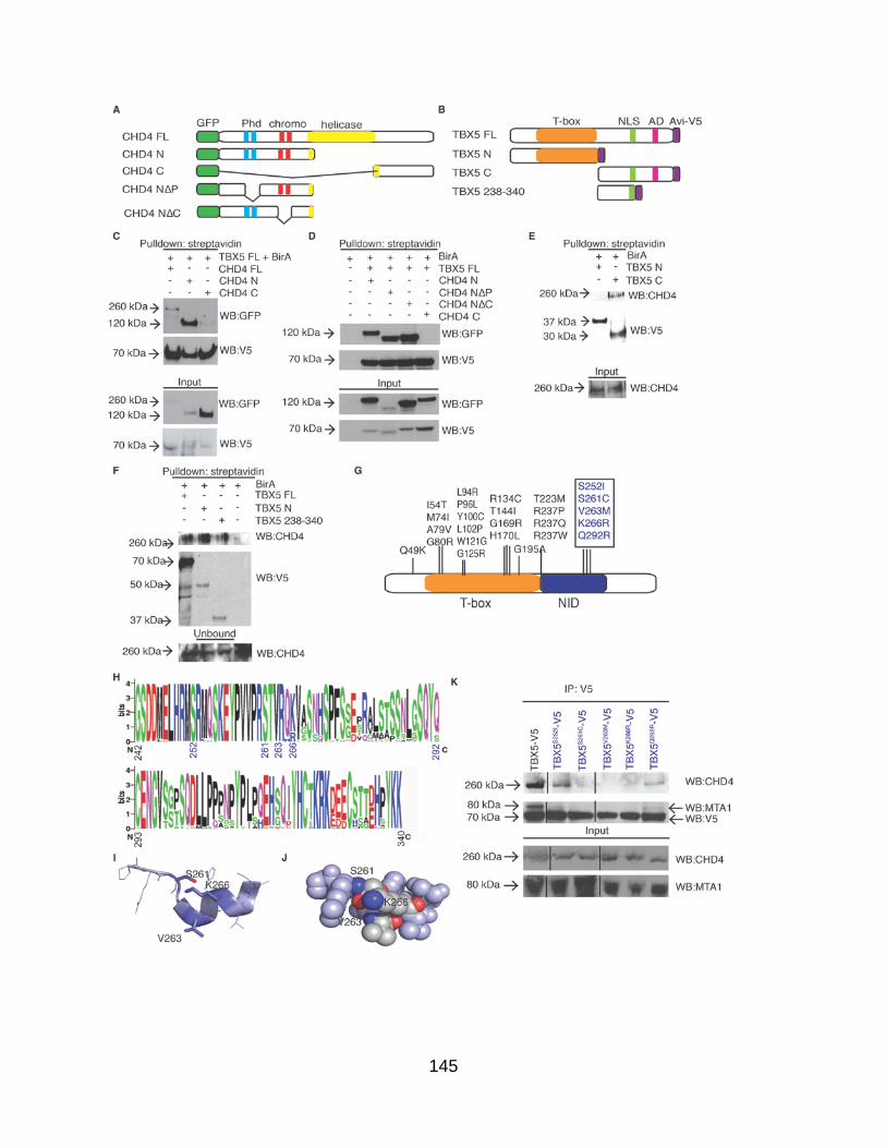
145

146
Figure A1.5. Congenital heart disease associated mutations of TBX5 disrupt
TBX5-NuRD complex interaction and activity. (A) Schematic of CHD4 deletion
constructs. Phd= Plant homeo domain. CHD4 FL = aa 1-1937, CHD4 N = aa 1-758,
CHD4 C = aa 1183-1937, TBX5 FL = aa 1-518, TBX5 N = aa 1-237, TBX5 C = aa 238-
518. (B) Schematic of TBX5 deletion constructs. NLS = Nuclear Localization Signal. AD
= Activation domain. (C-D) Affinity isolation of full-length TBX5-Avi-V5 with GFP-tagged
CHD4 deletions reveals that CHD4 interacts with TBX5 via sequences in the N-terminus
of CHD4, but that the Phd and chromo domains are not individually required for the
TBX5-CHD4 interaction. (E-F) Affinity isolation of CHD4 with a TBX5-Avi-V5 deletion
series demonstrates TBX5 sequences aa 238-340 are both necessary and sufficient for
interaction with CHD4. This region has been termed the NuRD interaction domain
(NID). (G) Schematic of human TBX5 protein showing location of a subset of CHD
associated mis-sense mutations. NID= NuRD interacting domain (aa 238-340). (H)
Sequence alignment of the TBX5NID from 48 TBX5 ortholgues. Height of letters is
relative conservation at that residue. (I) Structural prediction of the TBX5NID shows the
TBX5NID is comprised of a coil region followed an α-helix. (J) CHD associated mis-
sense mutations align along a single surface of the α-helix and are predicted to disrupt
protein-protein interactions (representative residues TBX5S261, TBX5V263, and TBX5K266
shown in dark blue). (K) Immunoisolation of V5-tagged CHD associated mis-sense
mutations of TBX5 probed for CHD4 and MTA1.

147
Figure A1.6. Congenital heart disease associated mutations of TBX5 disrupt
TBX5-NuRD complex activity. (A-D) Transcription of the regulatory regions of TBX5
target genes in response to wild type TBX5 (second column) or TBX5S261C (third
column). Additional gene targets shown in Figure S7. n= 9 per transcriptional assay.
Graphs are plotted as the mean luciferase value ± SEM. Student’s two tailed t-test was
used to perform statistical analysis. *** p < 0.001. (E) Schematic of ChIP primer location
on Kctd16 element. The T-box binding element (asterisk) is noted. (F) ChIP of CHD4
from cells co-transfected with Kctd16-Luciferase in the presence of absence of TBX5.

148
Figure A1.7. The TBX5-NuRD interaction evolved concurrently with cardiac
septation. (A) Phylogenetic comparison of TBX5 orthologues with boot-strap values.
Dark green brackets represent animals with a septated heart. Light green brackets
represent animals without a septated heart. Sequence of core amino acid residues
required for the TBX5-NuRD interaction are shown to the right. Amino acid residues
mutated in TBX5-associated CHD are bolded, and a K->R substitution from fish to frog
is highlighted in pink. (B) Immunoisolation of V5-tagged TBX5 orthologs probed for
CHD4.

149
Figure SA1.1. The TBX5 transcription interaction network. (A)Sixty mouse genes
corresponding to TBX5 candidate interactions as identified by the gene ontology
function of transcription were uploaded to the STRING database (www.string-db.org).
STRING functional relationships were evaluated with default settings with text mining
disabled. Forty proteins were clustered in a single protein interaction network, reflecting
a minimum combined STRING score of > 0.4 (medium confidence) between each
binary relationship (left). This network was imported to Cytoscape for further processing
(see Fig 1). The remaining 20 nodes with scores < 0.4 are illustrated as disconnected
(bottom). (B) RT-PCR analysis of all possible members of the NuRD complex
demonstrates the NuRD complex is present in both the adult and embryonic heart.

150
Figure SA1.2. TBX5 and MTA1 and cardiac septation. Histology staining
(Hematoxylin and Eosin) of (A) Mta1+/-, (B) Tbx5+/- and, (C) Tbx5+/-; Mta1+/- mice at
E13.5. (D-E) Measurements of left ventricular (LV) chamber width (D) and wall
thickness (E) of wild type and Tbx5+/-; Mta1+/- embryonic hearts (n=11 per genotype) at
E13.5 from the 4-chamber view at the widest part. Data is reported as either chamber
width or wall thickness in millimeters ± SEM. P value was determined by Student’s T-
test. (F-G) Histology staining of (F) wild type and (G) Tbx5+/-; Mta1+/- mice at E14.5
demonstrates a CCAVC at this stage (asterisk).

151
Figure SA1.3. Mta1-/- embryos undergo normal heart chamber septation. Histology
staining (Hematoxylin and Eosin) of heart tissue at E13.5. Low magnification transverse
section through (A) wild-type heart and (B) Mta1-/- heart. Asterisks denote region of
higher magnification, arrows denote leaflets of venous valve of inferior vena cava, scale
bar=310μm. High magnification of asterisk region in (C) wild-type heart and (D) Mta1-/-
heart at point of chamber septation. Scale bar=130μm. la = left atrium, lv = left
ventricle, ra = right atrium, rv = right ventricle.

152
Figure SA1.4. Transcriptional targets not repressed by TBX5. (A-D) Gene reporter
elements cloned from potential TBX5 targets in the presence or absence of TBX5. Data
is reported as the mean ± SEM. Student’s T-test was used to determine significance.
*** p < 0.001.

153
Figure SA1.5. Targets repressed by TBX5 in a NuRD independent manner. (A-I)
TBX5-repressed gene reporter elements and in the co-transfected with wild type TBX5
or TBX5S261C. Genes sorted into neural (red), cancer (blue), or both (purple). Data is
reported as the mean ± SEM. Student’s T-test was used to determine significance * p<
0.05 *** p < 0.001.

154
Table S1. Spectral counts for TBX5 interacting proteins, related to Figure 1 (Excel
File upon request)
Table S2. Complete list of genes misregulated in Tbx5-/- embryonic heart, related
to Figures 3-4 (Excel File upon request)
Table S3. TBX5 ChIP data for genes upregulated in Tbx5-/- embryonic hearts,
related to Figures 3-4. (Excel File upon request)
Supplemental Experimental Proceedures
Generation of Tbx5Avi mice
Genotyping primers for Tbx5Avi/Avi; Rosa26BirA/BirA mice.
Genotyping Primers Sequence
Wild type Tbx5 F: CGGCGTGCCCAGGACCCTGT
R: GGCACAGGTCAGCCTTTAGC Tbx5Avi F: CGGCGTGCCCAGGACCCTGT
R: GCGCGCCTTCGTGCCATTCGA Sox2-Cre F: GTCCCCTTCTCCATCTCCAG
R: GCAAACGGACAGAAGCATTT Neo F: AGGATCTCCTGTCATCTCACCTT
R: CTAGAGAATTGATCCCCTCAGAAG Presence of BirA F: TTACGCAAGCTGGGTGCAGA
R: TTACGCAAGCTGGGTGCAGA Absence of BirA F: GTGTAACTGTGGACAGAGGAG
R: GAACTTGATGTGTAGACCAGG

155
Transcriptional assays
TBX5 target gene primers for transcriptional assays.
ChIP cloning PCR Primers Sequence
Casz1-1 F: ATCGGGTACCTGTCCCTTTCTTCTGTCCCA
R: ATCGGGTACCAGGCATAAGATCGCTGGAAA
Casz1-2 F: ATCGGGTACCACCATCCATCCTCGTGTAGC
R: ATCGGCTAGCGCCAAAAACAGTGGCCTAAA
Col20a1 F: ATCGGCTAGCATCGAGTGTGTCAGCTGTGG
R: ATCGAAGCTTCCGTGGTGCTGTGTTCTTAC
Fbxl20 F: GATCACGCGTGGCCCAAGGAATGGGTACT
R: GATCACGCGTCCACAGTCGCCTCTATCGTC
Fgf8 F: CGAGGCTAGCCCAGCTCCCTCCTTCCTTTA
R: CGAGGGTACCGGCAGCTTTCTGTCTTGGTT
Fgf11 F:
GCACAGATCTGGGACTCCCTAACTGTCGTACC
R: GCACACGCGTCCGCTGGAGCTAGTCAGG
Gad1 F: CGAGGGTACCCACTGGAATTCCTACCGTGA
R: CGAGGCTAGCCAAGGCGTTCTGGTCTAAGG
Kcne3 F: CGCTACGCGTGTGCCACACAGCTACAATAA
R: CGCTCTCGAGTCCGGTGAACACTGCAATAA
Kcnn2 F: GATCGGTACCCTGGTCTCCCTGCAGCTTTA
R: GATCGCTACGGTTGTCCTGGCTCTGTTGCT
Kctd16 F: AGGACGCGTTGCTTGTGCAAACTCCAGAC
R: GACCAGATCTAGGCAGGGCGACAGATAGAT
Klf4 F: ATCGACGCGTCTTGGCGTGAGGAACTCTCT
R: ATCGAGATCTCGCTCAAATGGGCCTCTA
Mef2b-1 F: CGAGGTACCTCTTCTCTCGGACGGACCTA
R: CGAGGCTAGCCACACACACTCACGGCTCTC
Mef2b-2 F: CGAGGCTAGCCTGCACAGGGACCCACAG
R: CGAGGCTAGCCTGCACAGGGACCCACAG

156
Mef2b-3 F: CGAGGGTACCGCGTGCGATCACCATAACT
R: CGAGGCTAGCAGGGGCTGCGATGTAGGT
Nxph-4 F: CGAGGGTACCGCTTGCAGAAGGGTATCTGG
R: CGAGGCTAGCTAAGCCTCCAGGCATTCAAC
Plekha2 F: GATCGGTACCTAACTTGGAACCGACCTACG
R: GATCGCTAGCTTTCACACCTGAGCGAACTG
Sncb F: CACGGGTACCTCCCTCCACTGCCTCCAC
R: CAGCGCTAGCGCTCCAGGGTCCTCCTAGTC

157
DNA constructs
Site directed mutagenesis primer pairs. Amino acid changes are represented in bold.
mTBX5S252I F: GATGTCTCGGATGCAAATTAAAGAGTATCCTGTGGTTC
R: GAACCACAGGATACTCTTTAATTTGCATCCGAGACATC
mTBX5S261C F: GTATCCTGTGGTTCCCAGGTCGACAGTGAGGCACAAAG R: CTTTGTGCCTCACTGTCGACCTGGGAACCACAGGATAC
mTBX5V263M F: GGTTCCCAGGAGCACAATGAGGCACAAAGTCACCTCC
R: GGAGGTGACTTTGTGCCTCATTGTGCTCCTGGGAACC
mTBX5K266R F: GAGCACAGTGAGGCACAGAGTCACCTCCAACCACAGC
R: GCTGTGGTTGGAGGTGACTCTGTGCCTCACTGTGCTC
mTBX5Q292R F: CAATTTAGGGTCCCAGTACCGGTGTGAGAATGGTGTC
R: GACACCATTCTCACACCGGTACTGGGACCCTAAATTG
xTbx5K266R F: GTCCGTCAGAGGGTGTCCTCT
R: TGTGCATCTTGGGACTAC

158
Chromatin IP
Kctd16 intron 1 ChIP-PCR primer sequences
Kctd16 intron 1 F: GTTTCTTGCCTGCACTGCT
R: GAAGGAAAAGCAGCCGGGAA

159
RT-PCR
Primer sequences for NuRD complex members
Chd3 F: AGAAGGAAAACAAGCCAGG
R: TTGTGTCCCCCATCCCCTT Chd4 F: GGGTGAAATGAATCGTGGC
R: TTGTGTAGGACTGCATTGGC
Rbbp4 F: ACTGTTGCCTTGTGGGATCT
R: AGATCCCAGACATTCAGCCT
Rbbp7 F: GGTGGTTGCTCGAGTTCATA
R: GTGGCAATGATGTGAGGATTC
Mta1 F: AGAGCCCACTGGTGCTGAAG
R: CATCTACCCCATTGTGCTGC
Mta2 F: GCTCGATCCGACTTCCTAAG
R: GTCTCATAGGAACGTTTCACC
Mta3 F: TGAGAATTCCTCCAGCAACCC
R: TTAGCATGCTTGTCGGCGAG
Gatad2a F: GCCCAGTGCTGCCAATAATG
R: CAGCGGCATGTGAAGTCTGT
Gatad2b F: TTACAGCAGCAGGCAGCA
R: GCCACCTGGCACAGACAATT
Mbd2/3 F: GGAGAAGAACCCTGGTGTGT
R: ACAGGCCTTGTCCAGTGGT
Hdac2 F: ATGAGGCTTCATGGGATGAC
R: CATGGCGTACAGTCAAGGAG

160
REFERENCES
Andrews S. FastQC: a quality control tool for high throughput sequence data. 2010. Available from: http://www.bioinformatics.babraham.ac.uk/projects/fastqc.
Baban, A., Pitto, L., Pulignani, S., Cresci, M., Mariani, L., Gambacciani, C., Digilio, M.C., Pongiglione, G., and Albanese, S. (2014). Holt-Oram syndrome with intermediate atrioventricular canal defect, and aortic coarctation: Functional characterization of a de novo TBX5 mutation. Am J Med Genet A.
Basson, C.T., Bachinsky, D.R., Lin, R.C., Levi, T., Elkins, J.A., Soults, J., Grayzel, D., Kroumpouzou, E., Traill, T.A., Leblanc-Straceski, J., et al. (1997). Mutations in human TBX5 [corrected] cause limb and cardiac malformation in Holt-Oram syndrome. Nat Genet 15, 30-35.
Basson, C.T., Cowley, G.S., Solomon, S.D., Weissman, B., Poznanski, A.K., Traill, T.A., Seidman, J.G., and Seidman, C.E. (1994). The clinical and genetic spectrum of the Holt-Oram syndrome (heart-hand syndrome). N Engl J Med 330, 885-891.
Basson, C.T., Huang, T., Lin, R.C., Bachinsky, D.R., Weremowicz, S., Vaglio, A., Bruzzone, R., Quadrelli, R., Lerone, M., Romeo, G., et al. (1999). Different TBX5 interactions in heart and limb defined by Holt-Oram syndrome mutations. Proc Natl Acad Sci U S A 96, 2919-2924.
Bayer, E.A., and Wilchek, M. (1980). The use of the avidin-biotin complex as a tool in molecular biology. Methods Biochem Anal 26, 1-45.
Benson, D.W., Basson, C.T., and MacRae, C.A. (1996). New understandings in the genetics of congenital heart disease. Curr Opin Pediatr 8, 505-511.
Berk, A.J. (1999). Activation of RNA polymerase II transcription. Curr Opin Cell Biol 11, 330-335.
Brown, D.D., Martz, S.N., Binder, O., Goetz, S.C., Price, B.M., Smith, J.C., and Conlon, F.L. (2005). Tbx5 and Tbx20 act synergistically to control vertebrate heart morphogenesis. Development 132, 553-563.
Bruneau, B.G., Logan, M., Davis, N., Levi, T., Tabin, C.J., Seidman, J.G., and Seidman, C.E. (1999). Chamber-specific cardiac expression of Tbx5 and heart defects in Holt-Oram syndrome. Dev Biol 211, 100-108.
Bruneau, B.G., Nemer, G., Schmitt, J.P., Charron, F., Robitaille, L., Caron, S., Conner, D.A., Gessler, M., Nemer, M., Seidman, C.E., et al. (2001). A murine model of Holt-Oram syndrome defines roles of the T-box transcription factor Tbx5 in cardiogenesis and disease. Cell 106, 709-721.

161
Cline, M.S., Smoot, M., Cerami, E., Kuchinsky, A., Landys, N., Workman, C., Christmas, R., Avila-Campilo, I., Creech, M., Gross, B., et al. (2007). Integration of biological networks and gene expression data using Cytoscape. Nat Protoc 2, 2366-2382.
Costantini, J.L., Cheung, S.M., Hou, S., Li, H., Kung, S.K., Johnston, J.B., Wilkins, J.A., Gibson, S.B., and Marshall, A.J. (2009). TAPP2 links phosphoinositide 3-kinase signaling to B-cell adhesion through interaction with the cytoskeletal protein utrophin: expression of a novel cell adhesion-promoting complex in B-cell leukemia. Blood 114, 4703-4712.
Cristea, I.M., Williams, R., Chait, B.T., and Rout, M.P. (2005). Fluorescent proteins as proteomic probes. Mol Cell Proteomics 4, 1933-1941.
Cross, S.J., Ching, Y.H., Li, Q.Y., Armstrong-Buisseret, L., Spranger, S., Lyonnet, S., Bonnet, D., Penttinen, M., Jonveaux, P., Leheup, B., et al. (2000). The mutation spectrum in Holt-Oram syndrome. J Med Genet 37, 785-787.
Denslow, S.A., and Wade, P.A. (2007). The human Mi-2/NuRD complex and gene regulation. Oncogene 26, 5433-5438.
Diez-Roux, G., Banfi, S., Sultan, M., Geffers, L., Anand, S., Rozado, D., Magen, A., Canidio, E., Pagani, M., Peluso, I., et al. (2011). A high-resolution anatomical atlas of the transcriptome in the mouse embryo. PLoS Biol 9, e1000582.
Dolk, H., Loane, M., and Garne, E. (2010). The prevalence of congenital anomalies in Europe. Adv Exp Med Biol 686, 349-364.
Dorr, K.M., Amin, N.M., Kuchenbrod, L.M., Labiner, H., Charpentier, M.S., Pevny, L.H., Wessels, A., and Conlon, F.L. (2015). Casz1 is required for cardiomyocyte G1-to-S phase progression during mammalian cardiac development. Development 142, 2037-2047.
Driegen, S., Ferreira, R., van Zon, A., Strouboulis, J., Jaegle, M., Grosveld, F., Philipsen, S., and Meijer, D. (2005). A generic tool for biotinylation of tagged proteins in transgenic mice. Transgenic Res 14, 477-482.
Evans, P.M., and Liu, C. (2008). Roles of Krupel-like factor 4 in normal homeostasis, cancer and stem cells. Acta Biochim Biophys Sin (Shanghai) 40, 554-564.
Fan, C., Duhagon, M.A., Oberti, C., Chen, S., Hiroi, Y., Komuro, I., Duhagon, P.I., Canessa, R., and Wang, Q. (2003a). Novel TBX5 mutations and molecular mechanism for Holt-Oram syndrome. J Med Genet 40, e29.
Fan, C., Liu, M., and Wang, Q. (2003b). Functional analysis of TBX5 missense mutations associated with Holt-Oram syndrome. J Biol Chem 278, 8780-8785.
Franceschini, A., Szklarczyk, D., Frankild, S., Kuhn, M., Simonovic, M., Roth, A., Lin, J., Minguez, P., Bork, P., von Mering, C., et al. (2013). STRING v9.1: protein-protein

162
interaction networks, with increased coverage and integration. Nucleic Acids Res 41, D808-815.
Franklin, S., Zhang, M.J., Chen, H., Paulsson, A.K., Mitchell-Jordan, S.A., Li, Y., Ping, P., and Vondriska, T.M. (2011). Specialized compartments of cardiac nuclei exhibit distinct proteomic anatomy. Mol Cell Proteomics 10, M110 000703.
Garg, V., Kathiriya, I.S., Barnes, R., Schluterman, M.K., King, I.N., Butler, C.A., Rothrock, C.R., Eapen, R.S., Hirayama-Yamada, K., Joo, K., et al. (2003). GATA4 mutations cause human congenital heart defects and reveal an interaction with TBX5. Nature 424, 443-447.
Ghosh, T.K., Song, F.F., Packham, E.A., Buxton, S., Robinson, T.E., Ronksley, J., Self, T., Bonser, A.J., and Brook, J.D. (2009). Physical interaction between TBX5 and MEF2C is required for early heart development. Mol Cell Biol 29, 2205-2218.
Gittenberger-de Groot, A.C., Calkoen, E.E., Poelmann, R.E., Bartelings, M.M., and Jongbloed, M.R. (2014). Morphogenesis and molecular considerations on congenital cardiac septal defects. Ann Med 46, 640-652.
Goetz, S.C., Brown, D.D., and Conlon, F.L. (2006). TBX5 is required for embryonic cardiac cell cycle progression. Development 133, 2575-2584.
Hatcher, C.J., Kim, M.S., Mah, C.S., Goldstein, M.M., Wong, B., Mikawa, T., and Basson, C.T. (2001). TBX5 transcription factor regulates cell proliferation during cardiogenesis. Dev Biol 230, 177-188.
Hayashi, S., Lewis, P., Pevny, L., and McMahon, A.P. (2002). Efficient gene modulation in mouse epiblast using a Sox2Cre transgenic mouse strain. Mech Dev 119 Suppl 1, S97-S101.
He, A., Kong, S.W., Ma, Q., and Pu, W.T. (2011). Co-occupancy by multiple cardiac transcription factors identifies transcriptional enhancers active in heart. Proc Natl Acad Sci U S A 108, 5632-5637.
Heinritz, W., Shou, L., Moschik, A., and Froster, U.G. (2005). The human TBX5 gene mutation database. Hum Mutat 26, 397.
Heinz, S., Benner, C., Spann, N., Bertolino, E., Lin, Y.C., Laslo, P., Cheng, J.X., Murre, C., Singh, H., and Glass, C.K. (2010). Simple combinations of lineage-determining transcription factors prime cis-regulatory elements required for macrophage and B cell identities. Mol Cell 38, 576-589.
Heron, M., and Tejada-Vera, B. (2009). Deaths: leading causes for 2005. Natl Vital Stat Rep 58, 1-97.

163
Hiroi, Y., Kudoh, S., Monzen, K., Ikeda, Y., Yazaki, Y., Nagai, R., and Komuro, I. (2001). Tbx5 associates with Nkx2-5 and synergistically promotes cardiomyocyte differentiation. Nat Genet 28, 276-280.
Hoffmann, A.D., Yang, X.H., Burnicka-Turek, O., Bosman, J.D., Ren, X., Steimle, J.D., Vokes, S.A., McMahon, A.P., Kalinichenko, V.V., and Moskowitz, I.P. (2014). Foxf genes integrate tbx5 and hedgehog pathways in the second heart field for cardiac septation. PLoS Genet 10, e1004604.
Holt, M., and Oram, S. (1960). Familial heart disease with skeletal malformations. Br Heart J 22, 236-242.
Hu, S., Li, L., Yeh, S., Cui, Y., Li, X., Chang, H.C., Jin, J., and Chang, C. (2015). Infiltrating T cells promote prostate cancer metastasis via modulation of FGF11-->miRNA-541-->androgen receptor (AR)-->MMP9 signaling. Mol Oncol 9, 44-57.
Huang, C.C., Tu, S.H., Lien, H.H., Jeng, J.Y., Huang, C.S., Huang, C.J., Lai, L.C., and Chuang, E.Y. (2013). Concurrent gene signatures for han chinese breast cancers. PLoS One 8, e76421.
Huang da, W., Sherman, B.T., and Lempicki, R.A. (2009). Systematic and integrative analysis of large gene lists using DAVID bioinformatics resources. Nat Protoc 4, 44-57.
Ieda, M., Fu, J.D., Delgado-Olguin, P., Vedantham, V., Hayashi, Y., Bruneau, B.G., and Srivastava, D. (2010). Direct reprogramming of fibroblasts into functional cardiomyocytes by defined factors. Cell 142, 375-386.
Kaltenbrun, E., Greco, T.M., Slagle, C.E., Kennedy, L.M., Li, T., Cristea, I.M., and Conlon, F.L. (2013). A Gro/TLE-NuRD corepressor complex facilitates Tbx20-dependent transcriptional repression. J Proteome Res 12, 5395-5409.
Kehle, J., Beuchle, D., Treuheit, S., Christen, B., Kennison, J.A., Bienz, M., and Muller, J. (1998). dMi-2, a hunchback-interacting protein that functions in polycomb repression. Science 282, 1897-1900.
Keller, A., Nesvizhskii, A.I., Kolker, E., and Aebersold, R. (2002). Empirical statistical model to estimate the accuracy of peptide identifications made by MS/MS and database search. Anal Chem 74, 5383-5392.
Kim, D., Pertea, G., Trapnell, C., Pimentel, H., Kelley, R., and Salzberg, S.L. (2013). TopHat2: accurate alignment of transcriptomes in the presence of insertions, deletions and gene fusions. Genome Biol 14, R36.
Kim, J., Cantor, A.B., Orkin, S.H., and Wang, J. (2009). Use of in vivo biotinylation to study protein-protein and protein-DNA interactions in mouse embryonic stem cells. Nat Protoc 4, 506-517.

164
Kimura, R., Kasamatsu, A., Koyama, T., Fukumoto, C., Kouzu, Y., Higo, M., Endo-Sakamoto, Y., Ogawara, K., Shiiba, M., Tanzawa, H., et al. (2013). Glutamate acid decarboxylase 1 promotes metastasis of human oral cancer by beta-catenin translocation and MMP7 activation. BMC Cancer 13, 555.
Krebs, S., Fischaleck, M., and Blum, H. (2009). A simple and loss-free method to remove TRIzol contaminations from minute RNA samples. Anal Biochem 387, 136-138.
Langmead, B., Trapnell, C., Pop, M., and Salzberg, S.L. (2009). Ultrafast and memory-efficient alignment of short DNA sequences to the human genome. Genome Biol 10, R25.
Lassmann, T., Hayashizaki, Y., and Daub, C.O. (2009). TagDust--a program to eliminate artifacts from next generation sequencing data. Bioinformatics 25, 2839-2840.
Li, H., Handsaker, B., Wysoker, A., Fennell, T., Ruan, J., Homer, N., Marth, G., Abecasis, G., and Durbin, R. (2009). The Sequence Alignment/Map format and SAMtools. Bioinformatics 25, 2078-2079.
Li, Q.Y., Newbury-Ecob, R.A., Terrett, J.A., Wilson, D.I., Curtis, A.R., Yi, C.H., Gebuhr, T., Bullen, P.J., Robson, S.C., Strachan, T., et al. (1997). Holt-Oram syndrome is caused by mutations in TBX5, a member of the Brachyury (T) gene family. Nat Genet 15, 21-29.
Liu, Z., Yang, X., Li, Z., McMahon, C., Sizer, C., Barenboim-Stapleton, L., Bliskovsky, V., Mock, B., Ried, T., London, W.B., et al. (2011). CASZ1, a candidate tumor-suppressor gene, suppresses neuroblastoma tumor growth through reprogramming gene expression. Cell Death Differ 18, 1174-1183.
Maine, G.N., Li, H., Zaidi, I.W., Basrur, V., Elenitoba-Johnson, K.S., and Burstein, E. (2010). A bimolecular affinity purification method under denaturing conditions for rapid isolation of a ubiquitinated protein for mass spectrometry analysis. Nat Protoc 5, 1447-1459.
McDermott, D.A., Bressan, M.C., He, J., Lee, J.S., Aftimos, S., Brueckner, M., Gilbert, F., Graham, G.E., Hannibal, M.C., Innis, J.W., et al. (2005). TBX5 genetic testing validates strict clinical criteria for Holt-Oram syndrome. Pediatr Res 58, 981-986.
Metz, M., Gassmann, M., Fakler, B., Schaeren-Wiemers, N., and Bettler, B. (2011). Distribution of the auxiliary GABAB receptor subunits KCTD8, 12, 12b, and 16 in the mouse brain. J Comp Neurol 519, 1435-1454.
Mori, A.D., and Bruneau, B.G. (2004). TBX5 mutations and congenital heart disease: Holt-Oram syndrome revealed. Curr Opin Cardiol 19, 211-215.
Morris, C. (2010). Epidemiology of congenital heart disease. In Cardiology, D.J. Crawford MH, Paulus WJ, ed. (Philadelphia, Mosby), pp. 1381-1389.

165
Moskowitz, I.P., Kim, J.B., Moore, M.L., Wolf, C.M., Peterson, M.A., Shendure, J., Nobrega, M.A., Yokota, Y., Berul, C., Izumo, S., et al. (2007). A molecular pathway including Id2, Tbx5, and Nkx2-5 required for cardiac conduction system development. Cell 129, 1365-1376.
Moskowitz, I.P., Pizard, A., Patel, V.V., Bruneau, B.G., Kim, J.B., Kupershmidt, S., Roden, D., Berul, C.I., Seidman, C.E., and Seidman, J.G. (2004). The T-Box transcription factor Tbx5 is required for the patterning and maturation of the murine cardiac conduction system. Development 131, 4107-4116.
Polo, S.E., Kaidi, A., Baskcomb, L., Galanty, Y., and Jackson, S.P. (2010). Regulation of DNA-damage responses and cell-cycle progression by the chromatin remodelling factor CHD4. EMBO J 29, 3130-3139.
Qian, L., Huang, Y., Spencer, C.I., Foley, A., Vedantham, V., Liu, L., Conway, S.J., Fu, J.D., and Srivastava, D. (2012). In vivo reprogramming of murine cardiac fibroblasts into induced cardiomyocytes. Nature 485, 593-598.
Roesli, C., Neri, D., and Rybak, J.N. (2006). In vivo protein biotinylation and sample preparation for the proteomic identification of organ- and disease-specific antigens accessible from the vasculature. Nat Protoc 1, 192-199.
Saunders, A., Werner, J., Andrulis, E.D., Nakayama, T., Hirose, S., Reinberg, D., and Lis, J.T. (2003). Tracking FACT and the RNA polymerase II elongation complex through chromatin in vivo. Science 301, 1094-1096.
Sedmera, D., and Thompson, R.P. (2011). Myocyte proliferation in the developing heart. Dev Dyn 240, 1322-1334.
Smallwood, P.M., Munoz-Sanjuan, I., Tong, P., Macke, J.P., Hendry, S.H., Gilbert, D.J., Copeland, N.G., Jenkins, N.A., and Nathans, J. (1996). Fibroblast growth factor (FGF) homologous factors: new members of the FGF family implicated in nervous system development. Proc Natl Acad Sci U S A 93, 9850-9857.
Sopher, B.L., Koszdin, K.L., McClain, M.E., Myrick, S.B., Martinez, R.A., Smith, A.C., and La Spada, A.R. (2001). Genomic organization, chromosome location, and expression analysis of mouse beta-synuclein, a candidate for involvement in neurodegeneration. Cytogenet Cell Genet 93, 117-123.
Stirnimann, C.U., Ptchelkine, D., Grimm, C., and Muller, C.W. (2010). Structural basis of TBX5-DNA recognition: the T-box domain in its DNA-bound and -unbound form. J Mol Biol 400, 71-81.
Trapnell, C., Roberts, A., Goff, L., Pertea, G., Kim, D., Kelley, D.R., Pimentel, H., Salzberg, S.L., Rinn, J.L., and Pachter, L. (2012). Differential gene and transcript expression analysis of RNA-seq experiments with TopHat and Cufflinks. Nat Protoc 7, 562-578.

166
Trifonov, S., Yamashita, Y., Kase, M., Maruyama, M., and Sugimoto, T. (2014). Glutamic acid decarboxylase 1 alternative splicing isoforms: characterization, expression and quantification in the mouse brain. BMC Neurosci 15, 114.
van Werven, F.J., and Timmers, H.T. (2006). The use of biotin tagging in Saccharomyces cerevisiae improves the sensitivity of chromatin immunoprecipitation. Nucleic Acids Res 34, e33.
Wade, P.A., Jones, P.L., Vermaak, D., and Wolffe, A.P. (1998). A multiple subunit Mi-2 histone deacetylase from Xenopus laevis cofractionates with an associated Snf2 superfamily ATPase. Curr Biol 8, 843-846.
Wang, J., Rao, S., Chu, J., Shen, X., Levasseur, D.N., Theunissen, T.W., and Orkin, S.H. (2006). A protein interaction network for pluripotency of embryonic stem cells. Nature 444, 364-368.
Wang, C., Cao, D., Wang, Q., and Wang, D.Z. (2011). Synergistic activation of cardiac genes by myocardin and tbx5. PLoS One 6, e24242.
Wang, S., Sun, H., Ma, J., Zang, C., Wang, C., Wang, J., Tang, Q., Meyer, C.A., Zhang, Y., and Liu, X.S. (2013). Target analysis by integration of transcriptome and ChIP-seq data with BETA. Nat Protoc 8, 2502-2515.
Wang, W., Kim, H.J., Lee, J.H., Wong, V., Sihn, C.R., Lv, P., Perez Flores, M.C., Mousavi-Nik, A., Doyle, K.J., Xu, Y., et al. (2014). Functional significance of K+ channel beta-subunit KCNE3 in auditory neurons. J Biol Chem 289, 16802-16813.
Xue, Y., Wong, J., Moreno, G.T., Young, M.K., Cote, J., and Wang, W. (1998). NURD, a novel complex with both ATP-dependent chromatin-remodeling and histone deacetylase activities. Mol Cell 2, 851-861.
Ying, C.Y., Dominguez-Sola, D., Fabi, M., Lorenz, I.C., Hussein, S., Bansal, M., Califano, A., Pasqualucci, L., Basso, K., and Dalla-Favera, R. (2013). MEF2B mutations lead to deregulated expression of the oncogene BCL6 in diffuse large B cell lymphoma. Nat Immunol 14, 1084-1092.
Zhang, Y., Liu, T., Meyer, C.A., Eeckhoute, J., Johnson, D.S., Bernstein, B.E., Nusbaum, C., Myers, R.M., Brown, M., Li, W., et al. (2008). Model-based analysis of ChIP-Seq (MACS). Genome Biol 9, R137.

167
APPENDIX 2: The Lhx9-Integrin pathway is essesntial for positioning of the
proepicardial organ3
Introduction
Cardiovascular disease is one of the highest causes of mortality and reduced
quality of life in the US (Roger et al., 2012). To better optimize therapeutics to preserve
and maintain heart physiology it is pertinent that we understand the mechanisms
involved in all aspects of heart development, growth and repair. This will further enable
us to target resident cardiac cell populations and gene programs for clinical
applications.
The heart initially forms in vertebrates as a bilaminar tube comprised of an inner
endocardium and outer myocardial layer. At later stages of development a third layer is
added to the heart termed the epicardium. In many vertebrates this layer forms from a
dynamic precursor cell population, the proepicardial (PE) organ, which arises as a
cluster of cells on the right side of the septum transversum adjacent to the heart
(Schulte et al., 2007, Jahr et al., 2008, Manner, 1992, Pombal et al., 2008, Serluca,
2008). Whilst FGF and BMP signaling, as well as the transcription factors Snai1 and
Twist, have been implicated in PE induction (Schlueter and Brand, 2009, Schulte et al.,
2007, Schlueter and Brand, 2013), there is little known about the mechanisms that
direct the formation of the PE cell cluster. Once the cluster has formed and is positioned
correctly it is induced to bridge towards the developing heart, directed by BMP signaling
3This appendix previously appeared as an article in the journal Development. The original citation is as follows: Tandon P, Wilczewski CM, Williams CE, Conlon FL. “The Lhx9-integrin pathway is essential for positioning of the proepicardial organ,” Development no. 143 (March 2016): 5.

168
from the atrioventricular sulcus (AVS) (Ishii et al., 2010). Upon attachment, epicardial
cells then traverse over the heart surface as an epithelial-like sheet termed the
epicardium.
The epicardial layer is an essential cardiac component as it provides mitogenic
stimulation to the developing myocardium (Kang et al., 2008, Lavine et al., 2005, Li et
al., 2011, Pennisi et al., 2003, Stuckmann and Lassar, 2002). In addition, the
epicardium is the source of epicardial-derived cells (EPDCs) whereby subpopulations of
this layer undergo epithelial-mesenchymal transition and invade the underlying
myocardium. Here they differentiate into vital cardiac cells such as smooth muscle cells
and pericytes of the vasculature and fibroblasts that contribute to the scaffold and
support of the heart (Gittenberger-de Groot et al., 2010, Lie-Venema et al., 2007,
Manner et al., 2001, Ratajska et al., 2008, von Gise and Pu, 2012). Current
understanding is that EPDC lineage specificity is determined early within the precursor
PE cell cluster prior to epithelial-mesenchymal transition (EMT) (Acharya et al., 2012,
Guadix et al., 2006, Manner, 1999, Mikawa and Gourdie, 1996, Katz et al., 2012, Wei et
al., 2015). In the event of a cardiac infarction or damage in the adult, research has also
demonstrated the utility of the epicardium in providing regenerative support and cardiac
cells to the damaged tissue (Lepilina et al., 2006, Limana et al., 2010, Winter and
Gittenberger-de Groot, 2007, Winter et al., 2009). Therefore, better comprehension of
how this source of pluripotent cells is specified and develops is vitally important to
understanding mechanisms of cardiac development, disease and repair.
Here we report that LIM Homeobox transcription factor 9 (Lhx9) is essential for
the formation and function of the epicardium. We show that loss of Lhx9 in Xenopus

169
leads to defects in the assembly of the PE cluster and subsequently failed migration and
spreading of epicardial cells onto the surface of the developing heart. Using a small
pharmacological molecule screen in Xenopus we ascertained that Integrin-Paxillin
signaling is required for proper PE clustering, which is confirmed by a significant
decrease in epicardial integrin alpha 4 (itga4) expression upon loss of Lhx9 function.
Given that interactions between cells and the surrounding ECM are vital for epicardial
formation and its role in cardiac repair (Kalman et al., 1995, Nahirney et al., 2003, Pae
et al., 2008, Wang et al., 2013, Benesh et al., 2013, Fransen and Lemanski, 1991,
Mercer et al., 2013, Rongish et al., 1996, Yang et al., 1995), we further show a novel
function for Lhx9 in mediating Integrin-adhesion mechanisms for correct PE cell
clustering. Therefore Lhx9 is vital for the formation of the epicardium and development
of the heart.
Results
Lhx9 is expressed in a temporally dynamic pattern during epicardial formation.
Animals lacking a functional epicardium die prematurely and display
hemorrhaging hearts with thin ventricular walls (Moore et al., 1998, Lie-Venema et al.,
2005, Gittenberger-de Groot et al., 2000, Acharya et al., 2012, Combs et al., 2011).
Many vertebrates show a right side clustering of PE cells during epicardial formation
(Viragh et al., 1993, Kalman et al., 1995, Nahirney et al., 2003, Serluca, 2008, Pombal
et al., 2008, Jahr et al., 2008). Recently we have demonstrated that the LIM
homeodomain transcription factor family lhx9 is transiently expressed in the Xenopus
PEO (Tandon et al., 2013). We therefore conducted a detailed analysis to establish the

170
relationship between Lhx9 expression and formation of the PEO on the septum
transversum.
The sequence and structure of Lhx9 has been shown to be highly conserved
across vertebrates (Bertuzzi et al., 1999, Retaux et al., 1999, Failli et al., 2000, Bachy et
al., 2001, Ottolenghi et al., 2001, Alunni et al., 2007, Oshima et al., 2007, Peukert et al.,
2011) being comprised of two LIM protein binding domains and a DNA-binding
homeodomain (HD). Consistent with other vertebrates we identified two isoform variants
of lhx9 in Xenopus, one with a full length HD (lhx9HD) and one with a truncated HD
(lhx9α) generated by alternative splicing of exon 5 (Failli et al., 2000, Molle et al., 2004)
(Fig. SA2.1).
To ascertain the endogenous role of Lhx9 during epicardial development we
performed a detailed spatio-temporal analysis of both lhx9 isoforms. Results from these
studies show lhx9α is initially expressed bilaterally on the septum transversum prior to
PEO formation (Fig. A2.1A, E, Fig. SA2.2-SA2.4). In all vertebrates, the lateral
confinement of the PEO cells to the right side of the embryo can be observed by the
expression of the conserved PEO marker tbx18 (Fig. A2.1K-N, Fig. SA2.3) (Tandon et
al., 2013). Consistently we observed a dramatic restriction of lhx9α expression to the
right side of the septum transversum (stages 39-46) (Fig. A2.1B-D, F) and subsequently
lhx9α is down regulated, relative to tbx18, as the PEO cells mature and migrate onto the
heart (Fig. A2.1D, N, Fig. SA2.3) (Tandon et al., 2013). An identical pattern of
expression was obtained with an independent probe recognizing the LIM domains of
lhx9α and lhx9HD (Fig.A2.1E, F, Fig. SA2.1, Fig. SA2.4, Fig. SA2.11). We further
observed a highly similar pattern of expression for lhx9HD in the septum transversum

171
(Fig. A2.1G-J, Fig. SA2.5) however we note lhx9HD is more weakly expressed and
dramatically down regulated upon PE clustering compared to lhx9α (Fig. A2.A1I, Fig.
SA2.5). We also note we did not observe detectable expression of lhx2, a closely
related LIM-HD transcription factor to Lhx9, in the developing epicardium at any stage
(Fig. SA2.6) (Viczian et al., 2006). Taken together these studies demonstrate lhx9α and
lhx9HD isoforms have overlapping dynamic expression during epicardial development
and lhx9α is the predominant isoform expressed during PEO formation.
Lhx9 is required for epicardial formation.
To determine the requirement for Lhx9 in the vertebrate PEO we depleted Lhx9
in Xenopus laevis (X. laevis) using Morpholinos (see Supplementary Methods for
details).
Depletion of Lhx9 function in Xenopus does not impede early embryonic
development. However, analysis of the developing heart at tadpole stages revealed a
severe epicardial defect. Whilst control embryos displayed PE cells clustering to the
AVS on the right side of the heart and a layer of epicardial cells on the ventricle surface
(Fig. A2.2A, C, red arrowheads marking PE cell cluster, tbx18 in situ hybridization),
embryos lacking Lhx9 failed to accumulate PE cells to the right-side AVS and displayed
a reduced and discontinuous epicardial layer on the ventricular surface (Fig. A2.2B, D,
E, F, red arrowheads).
172
Lhx9 is required for clustering of proepicardial cells.
Having demonstrated proper epicardial formation requires Lhx9 function and
because lhx9 expression is detected on the septum transversum and PE cluster (Fig.
A2.1A-F, Fig. SA2.2), we assessed whether Lhx9 functions during the earlier stages of
epicardial specification. At stage 38 the conserved pan-marker of the epicardial lineage,
tbx18 was observed on the septum transversum and deemed indistinguishable between
control and Lhx9-depleted embryos (Fig. A2.2G, H). Therefore Lhx9 is not required for
specification of the epicardial lineage. However, by stage 41 when PE cells begin to
cluster to the right side of embryos (Fig. A2.2I, red arrowhead), Lhx9-depleted stage-
matched sibling embryos maintained diffuse tbx18 expression bilaterally along the
septum transversum (Fig. A2.2J, green arrowheads, Fig. SA2.9A). By stage 42, the PE
cluster had attached to the AVS on the right side of the heart in controls whereas in
Lhx9-depleted embryos there was either no detectable cluster and tbx18 expression
persisted on the anterior endoderm/septum transversum or the cluster was
mispositioned caudal to the heart (Fig. A2.2L, M, N).
Consistently, we observed the spatial distribution of two independent epicardial
markers, tcf21 and wt1, were similarly disrupted in Lhx9-depleted embryos. Both tcf21
and wt1 expression marks the punctate cluster of PE cells at stage 41 on the right side
of the embryo (Fig. A2.2O, Q, S, red arrowhead), whilst expression of these epicardial
markers is reduced and mislocalized in Lhx9-depleted embryos (Fig. A2.2P, R, T, U, red
arrowheads, Fig. SA2.9B). This data collectively implies Lhx9 function is critical for
normal clustering of PE cells during early stages of development and in its absence
epicardial formation is disrupted.

173
Proepicardial cell cluster positioning is driven by Integrin-mediated mechanisms.
The cellular and molecular mechanisms which lead to the clustering of the PEO
on the right side of the septum transversum remain entirely unknown. To assess
whether active cell migration is required for asymmetric PE clustering we conducted a
screen in wild type embryos with a defined bank of small molecule inhibitors known to
disrupt cell migration and adhesion in Xenopus (Harata et al., 2013, Broders-Bondon et
al., 2007). Whilst the inhibition of myosin II (Blebbistatin) resulted in tadpoles with
severe pericardial edema (possibly due to reduced heart beat) and paralysis, we did
observe clustered PE cells (marked by tbx18) to the right of the embryo, albeit due to
the pronounced edema we were unable to determine attachment to the heart (data not
shown). Inhibition of Rac1 activity (NSC23766 trihydrochloride) which has been
extensively shown to disrupt cell migration (Diaz et al., 2014, Huang et al., 2013) did not
affect PE cell clustering compared to controls as depicted by tbx18 expression on the
AVS (Fig. A2.3A, B, G-J). Little tbx18 expression was preserved on the septum
transversum in these embryos implying PE clustering is not dependent on classical cell
migration mechanisms involving actin-myosin interactions and GTPase activity.
Interestingly, exposure from stages 38-41 when PE cell clustering is occurring, to
a small molecule that disrupts Integrin-Paxillin interactions (6-B345TTQ) led to a PE
cluster positioning defect phenocopying loss of Lhx9 function. Integrin binds to ECM
components and induces the recruitment of proteins, such as Paxillin, to focal
adhesions at the site of cell attachment. These protein complexes then function to
reorganize the actin cytoskeleton and are essential for the coordinated adhesion and

174
motility of cells through an ECM environment (Bellis et al., 1995). Paxillin is a multi-LIM
domain protein has been shown to directly and specifically bind to the cytoplasmic tail of
Itgα4 to coordinate cell spreading, adhesion and migration (Liu et al., 1999, Alon et al.,
2005). Whilst some clustering of PE cells was evident in cultured tadpoles, we observed
mispositioning of the cluster to the caudal side of the heart as well as persistent tbx18
expression on the septum transversum (Fig. A2.3C-F, K) as seen in Lhx9-depleted
embryos (Fig. A2.2H, J, L, M). This data therefore suggests Integrin-mediated
mechanisms plays an important role in the correct positioning of the PEO cell cluster in
developing embryos.
Lhx9 is required for proepicardial clustering via an Integrin-Paxillin interaction.
Our data demonstrate Lhx9 as well as Integrin-mediated cellular functions are
essential for the correct clustering and positioning of PE cells. We previously reported
lhx9 and itgα4 are co-regulated in response to loss of the epicardial transcription factor
Tcf21 (Tandon et al., 2013). Based on these observations we hypothesized Lhx9 acts
upstream of itgα4 to correctly position the PEO. Consistent with this hypothesis we find
itgα4 expression is detected subsequent to lhx9 in the clustered PE cells from stage 39-
46 (Fig.A2.4A-D, red arrowheads, Fig. SA2.3). We thus assessed the expression of
itgα4 in Lhx9-depleted hearts. Consistent with itgα4 acting downstream of Lhx9 we find
itgα4 is reduced and misplaced in Lhx9-depleted embryos (Fig. A2.4E, F, O, Fig. SA2.8,
Fig. SA2.9C). By contrast, the expression of Integrin β1 (Itgβ1) is strongly localized to
the epicardium (as marked by tcf21) and appears to be independent of Lhx9 function
(Fig. A2.4G-J’, Fig. SA2.11) therefore enabling its use as a marker of epicardial cells.

175
Due to the clustering defects observed upon reducing the Itgα4-Paxillin interaction
biochemically (Fig. A2.3) and reduced itgα4 expression in Lhx9-depleted embryos, we
sought to determine if Lhx9 is responsible for Integrin-mediated cell adhesion and
migration processes in PE cells. To address this possibility we assayed for
phosphorylated Paxillin (Y118) which has been used to demonstrate Integrin activation
at focal adhesions and shown to modulate cell spreading, migration and invasion (Bellis
et al., 1995, Burridge et al., 1992, Turner, 1991, Nakamura et al., 2000, Lewis and
Schwartz, 1998, Zaidel-Bar et al., 2007, Sachdev et al., 2009). Our data indicated a
significant decrease in phosphorylated Y118 Paxillin expression specifically in the
attached PE cluster in Lhx9-depleted embryos compared to controls (Fig. A2.4K-N’, P).
Taken together these data strongly imply Lhx9 acts through Itgα4 to correctly position
the PEO.
Lhx9-regulated Integrin signaling is essential for correct formation of the epicardial
layer.
Collectively our data demonstrates a role for Lhx9-Itgα4 in the correct positioning
and clustering of the PEO. Our data also suggests Integrin activity at focal adhesions is
required for the clustering of PE cells. Previous data has implicated Itgα4 as being
essential for epicardial development particularly in regulating PE cell clustering,
epicardial adhesion and epicardial migration (Sengbusch et al., 2002, Pinco et al., 2001,
Yang et al., 1995, Dettman et al., 2003, Dokic and Dettman, 2006, Hirose et al., 2006,
Pae et al., 2008, Bax et al., 2010). Integrin heterodimers have been shown to
coordinate cytoskeletal interactions and adhesion with the surrounding ECM

176
environment (Gardiner, 2011, Gehler et al., 2013, Huttenlocher and Horwitz, 2011,
Manninen, 2015, Wolfenson et al., 2013), pertinent processes for epicardial formation
and function (Kalman et al., 1995, Nahirney et al., 2003).
To determine if the depleted adhesive properties of the PEO and its mis-
positioning have persistent biological consequence in the development of the vertebrate
epicardium and heart, we examined the epicardial ECM environment of control and
Lhx9-depleted hearts. Using Itgβ1 as a marker of epicardial cells we analyzed the
expression of known Itgα4 ligands and ECM components. Whilst we observed no
discernible alterations in the expression of the Itgα4-Itgβ1 (Very Late Antigen 4, VLA-4)
ligand vascular cell adhesion molecule 1 (vcam1, Fig.S10) in Lhx9-depleted embryos,
we did detect a decrease in Fibronectin within the PE cluster as well as in the heart
relative to controls (Fig. A2.5A-E) (Humphries et al., 1995, Wu et al., 1995). This
suggests alterations to Integrin function in response to loss of Lhx9 is specific to Itgα4
pathway components. In addition these data further implies the adhesive qualities and
positioning of the PEO is critical for the proper deposition of epicardial ECM.
To address this further we examined the localization of epicardial-ECM markers
Laminin and β-dystroglycan. In control embryos we observed weak Laminin deposition
surrounding attached PE cells (Fig. A2.5F, F’, white arrowheads) but a continuous
smooth layer of Laminin was present under the mature epicardial layer (Fig. A2.5F”,
white arrowheads). However in Lhx9-depleted embryos, when epicardial cells were
present on the myocardial surface, we observed an increased accumulation of Laminin
in the basal portion of attached PE cells (Fig. A2.6G, G', white arrowheads) as well as
discontinuous deposits within epicardial cells on the surface of the heart (Fig. A2.5G”,

177
white arrowheads). To further confirm these findings, we analyzed β-dystroglycan, a
transmembrane glycoprotein that attaches cells to the basement membrane through
Laminin binding (Ervasti and Campbell, 1993, Klietsch et al., 1993, Smalheiser and
Schwartz, 1987). In Lhx9-depleted embryos β-dystroglycan is reduced or disrupted at
the point of contact between the PE cluster and heart surface (as indicated by tcf21)
(Fig. A2.5J-K’) compared to controls (Fig. A2.5H-I’). Note β-dystroglycan is prominent in
the endocardium of both control and Lhx9-depleted hearts showing this defect is
specific to the epicardium (Fig. A2.5I’, K’, yellow arrowheads). Taken together these
results demonstrate PE cell adhesive integrity is required for the proper formation of the
epicardial layer.
Collectively, our data are consistent with a role for Lhx9 in the clustering and
positioning of the PEO (Fig. A2.6A-B). Our data further show the ability of Lhx9 to
regulate Itgα4 signaling is vital for the correct establishment of clustering, attachment
and migration of PE cells to the heart (Fig, A2.6A, C, D). These signals are in turn
necessary for the correct deposition of ECM components thus, facilitating epicardial
cells to form a cohesive layer over the myocardial surface (Fig. A2.6E, F).
Discussion
Our data collectively shows Lhx9 is essential for proepicardial positioning and
that loss of Lhx9 is associated with aberrant deposition of essential ECM components
during crucial stages of epicardial clustering and migration. We further observe that loss
of Lhx9 correlates with a decrease in Integrin-ECM signaling and the ability of PE cells
to form cohesive adhesions downstream of Integrin activity. Because the PE cluster

178
defect in Lhx9-depleted embryos closely resembles the defect caused by disturbing the
Itgα4-Paxillin interaction biochemically, we propose and demonstrate Lhx9 functions to
modulate itgα4 expression. Taken together this data establishes a role for Lhx9 in
Integrin-mediated cell adhesion and motility crucial for the proper positioning and
clustering of PE cells.
Though most vertebrates show a right side clustering of PE cells during
epicardial formation (Viragh et al., 1993, Kalman et al., 1995, Nahirney et al., 2003,
Serluca, 2008, Pombal et al., 2008, Jahr et al., 2008) it remains to be established if the
sidedness of epicardial formation is essential for its function. In our studies we further
find that alterations in the positioning of the epicardium on the septum transversum is
essential for the correct attachment of the epicardium to the heart at the AVS. Thus,
these imply that alterations in PE clustering predispose an embryo to cardiac defects.
Itga4 and epicardial development
The function of Itgα4 during epicardial development has been characterized in
mice mutants who displayed absence of the epicardial layer and lack of coronary
vessels at the AVS (Yang et al., 1995). This was shown to be due to defects in the
migratory process (Sengbusch et al., 2002) and as demonstrated by this work.
Therefore the role of Itgα4 in epicardial migration appears to be conserved regardless of
the mode of epicardial formation; cluster bridge in frog or free-floating cysts in mouse.
Our data provides new evidence that the expression of itga4 is restricted to immature
PE cells and downregulated as the epicardial layer matures, similar to the
hematopoietic lineage (Arroyo et al., 1999, Lobb and Hemler, 1994). Using Itgβ1 as a
179
marker of epicardial cells we analyzed the expression of known Itgα4 ligands and ECM
components. We report a down regulation of Fibronectin in embryos lacking Lhx9 but
observe no alterations in expression of vcam1, the Itgα4-Itgβ1 ligand. Taken together
these findings demonstrate that loss of Lhx9 functions upstream of itgα4 and that the
function of Lhx9 is specific to Itgα4 function.
Lhx9 and Tcf21 during epicardial formation
Our previous studies identified lhx9 and itgα4 as genes upregulated upon Tcf21
loss. The role of Lhx9 in cardiac development to date has been limited to studies on the
effect of its persistent expression upon FOG2 ablation (Smagulova et al., 2008). Whilst
these studies provide insight into the effects of potentially prolonged Lhx9 activity, the
requirement for Lhx9 has not been addressed. Consistently with our finding on Tcf21,
we observe here that depletion of Lhx9 results in decreased itgα4 with a sparse and
disorganized epicardial layer on the heart. Given their similar patterns of expression in
the developing epicardium, this data suggests therefore that Tcf21 and Lhx9 function
coordinately together to enable the correct clustering and migration of PE cells onto the
heart surface through Integrin-directed mechanisms. It is of interesting note that Tcf21
and LIM homeodomain transcription factors are both found to be essential for the
development of the gonad and craniofacial structures (Moncaut et al., 2012, Harel et al.,
2012, Cui et al., 2004, Birk et al., 2000) suggesting that the transcriptional targets of
these two genes and the pathways they regulate may unveil a conserved mechanism
into regulating cell motility and thus, have implications in cancer as well as cardiac and
organ development (Vladimirova et al., 2009, Yang et al., 2014, Miller et al., 2014,

180
Weiss et al., 2013, Miller et al., 2013, Zhang et al., 2012, Ye et al., 2012, Richards et al.,
2011, Arab et al., 2011).
Materials and Methods
In situ hybridization
Whole-mount in situ hybridization (ISH) was carried out as previously described
(Tandon et al., 2013, Harland, 1991, Charpentier et al., 2015), the pericardial cavity
membrane in late tadpole stage embryos being removed post-fixation to improve
resolution. Embryos were processed for gelatin (30 µm) sectioning as previously
reported (Gessert and Kuhl, 2009, Tandon et al., 2013, Charpentier et al., 2015). All
probes were previously used (Tandon et al., 2013) or generated by PCR cloning from
Xenopus embryonic cDNA (Supplemental Table 1). Information on constructs is
available upon request. All phenotypes were quantified and statistically assessed using
Fishers Exact test or Chi-Square (Graphpad Prism 6). Figure images show
representative phenotypes quantified. For spatiotemporal analysis of lhx9, lhx2, tbx18
and itga4 transcripts in whole wild type embryos (Supplemental Figs. 2-6), multiple focal
plane images were compiled using auto-blend function (Photoshop CS4). Original
images are available upon request.
Xenopus manipulations
Xenopus embryos were cultured, microinjected and staged according to
Niewkoop and Faber (Nieuwkoop and Faber, 1967, Tandon et al., 2013).
Xla.Tg(Cardiac-actin:GFP)Mohun transgenic frogs were used as previously reported
181
(Latinkic et al., 2002, Tandon et al., 2013). The 5’UTR from both short and long X.
laevis lhx9 alloalleles was cloned and sequenced to verify design of the translation-
blocking Morpholino (MOT). A 5-base mismatch MO was used as a control
(Supplemental Table 2). Two transcription-blocking Morpholinos were designed against
the splice donor-site of exon 1 from both lhx9 alloallele genomic loci (MO1, Table 2)
(Tandon et al., 2012) (Genetools). 30 ng of each MO was injected at the one-cell stage,
with both MO1 being co-injected (Tandon et al., 2013, Tandon et al., 2012). To assess
the specificity and efficiency of MOT, 1ng lhx9-5’UTR-GFP RNA (designed against each
genomic loci) was co-injected together with the stated concentrations of MO. Stage 11
embryos were collected, lysed and analyzed using western-blot as previously performed
(Tandon et al., 2012, Tandon et al., 2013) using GFP antibody (JL8, #632381, Clontech,
1:10,000) and Shp2 antibody (#610622, BD Transduction, 1:2500) as a loading control.
To assess the efficiency of MO1, embryos were injected at the one-cell stage, and the
hearts from 25 embryos collected at stage 42 from each condition. RT-PCR was
performed (Superscript II, Invitrogen) for lhx9 with gapdh as a loading control
(Supplemental Table 1). See also Supplemental Methods.
Immunohistochemistry
Embryos were processed for agarose vibratome sectioning (100 µm) as reported
(Wallingford, 2010, Tandon et al., 2013, Charpentier et al., 2015). Sections were then
washed with PBS + 1% Triton X-100, blocked with wash buffer + 10% fetal calf serum
and incubated with primary antibody as previously reported (Langdon et al., 2012,
Tandon et al., 2013, Charpentier et al., 2015). Antibodies included mouse anti-
182
Tropomyosin (CH1, DSHB 1:25), rabbit anti-Laminin (L9393, Sigma, 1:100), mouse
anti- β-dystroglycan (MANDAG2, 7D11, DSHB, 1:100), rabbit anti-Fibronectin (F3648,
Sigma, 1:250), rabbit anti-phospho-Paxillin pTyr118 (44-722G, Invitrogen, 1:100) and
mouse anti-Integrin β1 (8C8, DHSB, 1:100). CH1 was deposited to the DSHB by Lin
J.J.-C, MANDAG2(7D11) by Morris G.E and 8C8 by Hausen P/ Gawantka V. Sections
were then incubated in DAPI/PBS (200 ng/ml) and imaged using a Zeiss LSM700
Spectral Confocal Laser Scanning Microscope, with representative figures compiled
using ImageJ and Photoshop. Image fluorescence, gauged by integrated pixel density,
was measured using ImageJ and quantified with standard two-tailed student’s t-test
(Graphpad).
Live tadpole small molecule culture assay
Xenopus wild type embryos were incubated in the appropriate concentration of
small molecule or DMSO control in 0.1 X MBS from stage 38 to stage 41 at room
temperature in the dark. Blebbistatin (B0560, Sigma) was used at 10 µM (Straight et al.,
2003), 6-B345TTQ (B7438, Sigma) was used at 5 and 10 µM (Kummer and Ginsberg,
2006, Kummer et al., 2010, Hung et al., 2013) and NSC23766 trihydrochloride
(SML0952, Sigma) was used at 5 and 50 µM. Embryos were briefly rinsed in 0.1x MBS,
fixed in 4% PFA and subjected to in situ hybridization as described.
Funding

183
We are thankful for funding from the National Institutes of Health (NICHD,
R21HD073044 to FLC) and the American Heart Association (13POST16950044,
postdoctoral fellowship to PT).
Author Contributions
PT developed the concepts, performed experiments and analyzed data for the
manuscript as well as prepared and edited the paper prior to submission. CMW and
CEW performed experiments and phenotypic analysis, FLC prepared and edited
manuscript.

184
Figure A2.1. Spatio-temporal analysis of lhx9 isoforms during Xenopus epicardial
development. Whole embryo in situ hybridization was performed on Xenopus embryos
at stages 36 to 46, using probes specific for lhx9a (A-D), lhx9LIM (E, F), lhx9HD (G-L)
and tbx18 (K-N) (See Fig. S2, S3, S4, S5). Views display embryos facing right, left and
ventral with anterior to the top. Images show magnified cardiac regions as depicted by
red box on tadpole schematic (bottom). Red arrowheads depict staining of septum
transversum and PE clusters. Cardiac region is depicted with *. White dashed line
outlines heart in ventral views. (E, F) Transverse gelatin section in situ hybridization
images of cardiac region demonstrate lhx9LIM expression exclusively in the septum
transversum (stage 36) and proepicardial tissue (stage 41). en, endocardium; h, heart;
peo, proepicardial organ; st, septum transversum.

185
Figure A2.2. Lhx9 is required for proper epicardial layer and PE cluster formation.
Epicardial layer formation (as shown with tbx18) analysis in control (A, C) and Lhx9-
depleted (B, D) embryos at stage 45. (A, B) Images show lateral view of cardiac region,
dorsal to the top and anterior to right. (C, D) Transverse gelatin sections through
representative embryo cardiac regions showing tbx18 expression, dorsal to the top. PE
cell clustering (red arrowheads) and epicardial cell layer on the ventricular surface is
shown. (E) Epicardial layer formation defects were quantified as abnormal as ≤ 50%
ventricular coverage as depicted in schematic. (F) Quantification of observed epicardial
layer formation defects, as represented in A-D, from three independent experiments, p =
<0.0001 by two-tailed Fisher’s exact test. Proepicardial clustering morphology was
analyzed using in situ hybridization for tbx18 in control (G, I, K) and Lhx9-depleted (H, J,

186
L) embryos at stage 38 (G, H), stage 41 (I, J) and stage 42 (K, L). Ventral view showing
cardiac region, anterior to the top. Tbx18 expression is detected as a cluster of cells in
control embryos on the right of the embryo near the atrioventricular sulcus (I, K, red
arrowheads) whereas in Lhx9-depleted embryos, the cluster is either not detected and
tbx18 expression remains throughout the septum transversum region or is mis-
positioned to the caudal side of the heart (abnormal clustering) (J, L, green
arrowheads). (M) Quantification of PE clustering defects is depicted in the schematic as
being abnormal by bilateral tbx18 expression retention on septum transversum and/or
cluster mis-positioning of ≥ 45o caudal to the AVS compared to controls. (N)
Quantification of observed clustering phenotype at stage 41 represented in C and D.
Data taken from seven independent experiments, p = <0.0001 by two-tailed Fisher’s
exact test. (O-T) In situ hybridization analysis for tcf21 (O, P, U) and wt1 (Q-T)
proepicardial expression at stage 41. Images depict lateral view of the cardiac region,
dorsal to the top and anterior to the right (M-P) or ventral view, dorsal to top (Q, R). Red
arrowheads mark clustered PE cells. (U) Quantification of observed phenotypes as
depicted in M, data taken from six independent experiments, p = <0.0001 by two-tailed
Fisher’s exact test. avs, atrioventricular sulcus; pe/peo; proepicardial organ, st; septum
transversum, v; ventricle.

187
Figure A2.3. Integrin-Paxillin association is required for PE clustering. In situ
hybridization for tbx18 on whole embryos after incubation in stated concentrations of
small molecules between stages 38 and 41. Lateral views (A, C, E, G, I) and ventral
views (B, D, F, H, J) of the cardiac region from representative embryos are shown, red
arrowheads mark PE cells. (C-F) Images depict maintained tbx18 expression on septum
transversum and clustering abnormalities as shown Fig.2M when embryos incubated in
6-B345TTQ. (K) Embryos taken from two independent experiments, p = 0.000248 by
Chi Square test.v, ventricle.

188
Figure A2.4. Lhx9 regulates Integrin α4-Paxillin signaling in PE cluster. (A-D)
Whole embryo in situ hybridization for itgα4, stages 36 to 46. Lateral views of anterior
portion (left panels) with magnified image of cardiac region (right panels). Red
arrowhead marks PE cluster, * marks heart. (E, F) In situ hybridization for itgα4 in
control (E) and Lhx9-depleted embryos (F) at stage 41, representative cardiac region
shown, anterior to right, dorsal to top, red arrowhead marks PE cluster. (O)
Quantification of observed itgα4 expression (reduced expression denoting ≤ 50% stain
intensity to controls) and clustering defects shown in E, F (see Fig. 2M for phenotype
assessment), , embryos taken from six independent experiments, p = <0.0001 by Chi-
square. (G-J) Transverse cardiac region agarose sections from control (G, H) and Lhx9-
depleted (I, J) stage 41 embryos post-in situ hybridization for tcf21 (G, I), nuclei
expression with DAPI (H, J, blue) and Itgβ1 immunohistochemistry (H, J, magenta).

189
White outlines (H, J) indicate PE cluster as depicted by tcf21 and Itgβ1. (K-N)
Transverse cardiac region agarose sections from control (K, L) and Lhx9-depleted (M,
N) stage 45 Xla.Tg(Cardiac-actin:GFP)Mohun embryos depicting representative
immunohistochemical analysis for DAPI (blue), GFP (green) to mark cardiomyocytes,
Itgβ1 (magenta) to mark epicardial cells and phosphorylated Y118-Paxillin (red).
Magnified images (L, N) from white boxes (K, M). White arrowheads mark PE cluster.
(P) Pixel intensity (integrated density) levels for phosphorylated Y118-Paxillin (M) from
five control and ten Lhx9-depleted embryos, p = 0.0134 by two-tailed student t-test.v,
ventricle.

190
Figure A2.5. Disrupted Lhx9-Integrin signaling alters epicardial ECM environment.
(A-E) Transverse cardiac agarose sections from control (A, B) and Lhx9-depleted (C, D)
Xla.Tg(Cardiac-actin:GFP)Mohun embryos at stage 45. Nuclei stained with DAPI (blue),
GFP marking cardiomyocytes (green) and immunohistochemical expression for Itgβ1
(B, D, magenta) to mark epicardial cells, and Fibronectin (B”, D”, red). Threshold binary
images (Image J) in B’ and D’ show Itgβ1-positive epicardial cells used to quantify pixel
intensity in B” and D”. (E) Pixel intensity (integrated density) levels for Fibronectin from
five control and ten Lhx9-depleted embryos, p = 0.064 by two-tailed student t-test. (F-K)
Transverse cardiac agarose sections from control (F, H, I) and Lhx9-depleted (G, J, K)
embryos at stage 45. Nuclei stained with DAPI (blue) and immunohistochemical stain
for tropomyosin (cardiomyocytes, F, G, green), laminin (F, G, magenta) and β-
dystroglycan (H’-K’, magenta). Tcf21 (H-K) demonstrates PE cell attachment to the

191
heart. White and red boxes depict magnified images in ‘ and “ panels. White
arrowheads mark PE (F’, G’, I’, K’) and migrating epicardial cells (F”, G”). Yellow
arrowheads mark expression in endocardial tissue (B”, D”, I’, K’). v; ventricle.
Representative images from seven (laminin) and six (β-dystroglycan) embryos per
condition, from two independent experiments.

192
Figure A2.6. Model depicting role for Lhx9 in epicardial development in Xenopus.
(A) During early tadpole stages the epicardial lineage is determined and marked by
transcription factors tcf21, tbx18, wt1 and lhx9 (blue) as a bilateral population of cells on
the septum transversum caudal to the heart (red). (B) Lhx9 functions to drive clustering
of cells to form the proepicardial cluster on the right side of the embryo (blue), whereby
itgα4 expression is activated. At this stage signaling factors, most likely BMP (yellow
arrows), from the heart atrioventricular sulcus (AVS) direct epicardial migration. (C, D)
Lhx9-Integrin-mediated signaling, including focal adhesion (FA) formation and

193
phosphorylation of Paxillin (pPaxillin), allow the PEO bridge (blue) to attach to the heart
(red) at the AVS. (E, F) Once the PEO has attached to the heart, deposition of essential
ECM components such as Fibronectin and Laminin are required for the epicardial layer
to adhere and spread over the heart surface.

194
Figure SA2.1. Lhx9 genomic loci and isoform organization. Schematics of X. laevis
Lhx9 genomic locus (top) and mRNA (bottom), showing localization of LIM protein
binding domains (red) in exon 2 and 3, and the DNA-binding homeodomain in exons 4
and 5 (blue). Not to scale. Note that lhx9α isoform harbors a truncated HD due to
alternative splicing to exon 5a. Translation-blocking (purple) and splice-blocking
(orange) Morpholinos are depicted on exon 1 of genomic locus. Green bars on mRNA
schematics depict localization of in situ hybridization probes.

195

196
Figure SA2.2. Spatio-temporal analysis of lhx9α during Xenopus embryogenesis.
AIn situ hybridization right, left, ventral and dorsal views of wild-type embryos showing
lhx9a expression of the anterior region, from stage 34 to stage 46. 1; neural tube, 2;
retina, 3; kidney, 4; septum transversum, 5; proepicardial cluster, 6; jaw cartilage.

197

198
Figure SA2.3. Spatio-temporal analysis of tbx18 and itga4 during Xenopus
embryogenesis. (A-H) In situ hybridization of tbx18 showing right, left and ventral
views of wild-type embryos from stage 34 to 46, anterior region of embryo. (I-P) In situ
hybridization of itga4 showing right and left views of anterior region from stage 34-46. 1;
cranial mesoderm, 2; somites, 3; branchial arches, 4; septum transversum, 5;
proepicardial cluster, 6; epicardium.

199

200
Figure SA2.4. Spatio-temporal analysis of lhx9 during Xenopus embryogenesis. In
situ hybridization of whole embryo anterior region using probe specific for the LIM
domains of lhx9. Right, left, ventral and dorsal views of wild-type embryos from stage 34
to stage 46. 1; neural tube, 2; septum transversum, 3; retina, 4; kidney, 5; nasal
placode, 6; proepicardial cluster, 7; otic placode, 8; jaw cartilage.

201

202
Figure SA2.5. Spatio-temporal analysis of lhx9HD during Xenopus
embryogenesis. In situ hybridization right, left, ventral and dorsal views of wild-type
embryos showing lhx9HD expression of the anterior region over time, from stage 34 to
stage 46. 1; septum transversum, 2; retina, 3; neural tube, 4; nasal placode, 5; otic
placode.
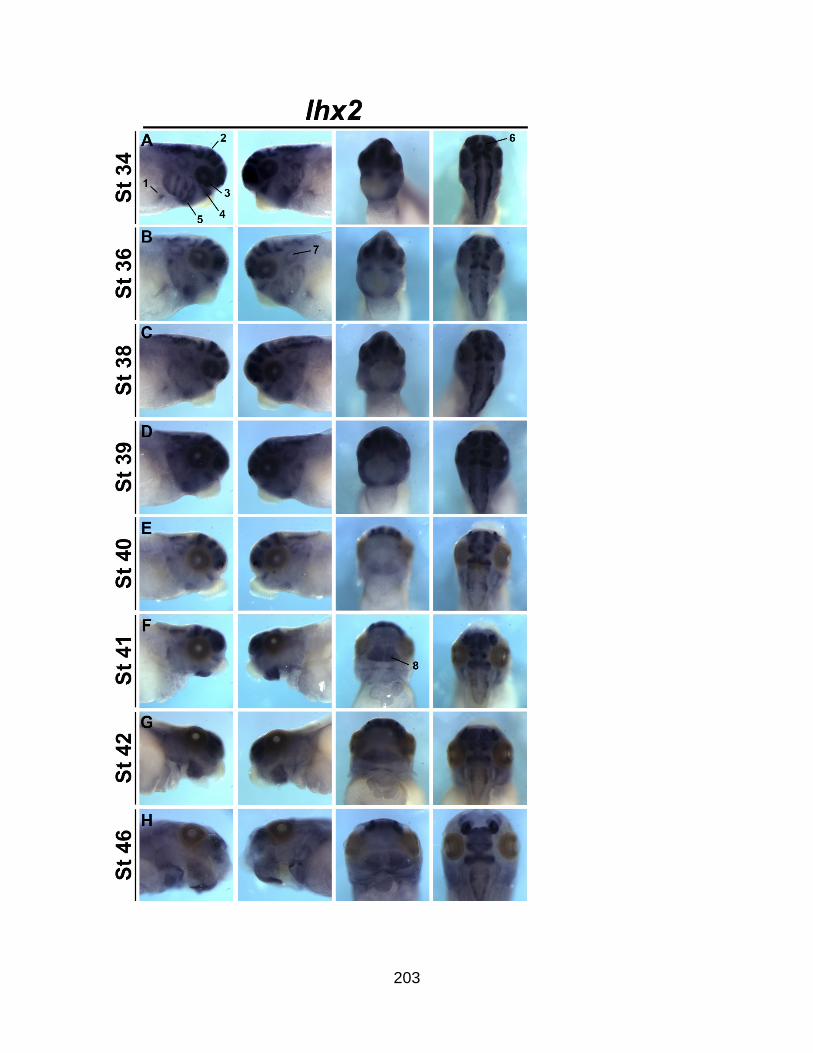
203
204
Figure SA2.6. Spatio-temporal analysis of lhx2 during Xenopus embryogenesis. In
situ hybridization right, left, ventral and dorsal views of wild-type embryos showing lhx2
expression of the anterior region over time, from stage 34 to stage 46. 1; lung bud, 2;
neural tube, 3; retina, 4; mandibular arch, 5; branchial arches, 6; pineal gland, 7; otic
placode, 8; lower jaw.

205
Figure SA2.7. Validation of Lhx9 depletion assays. (A) Validation of MO-specific
inhibition of Lhx9 translation by GFP western blot on stage 11 embryos, injected with
1ng Lhx9-5’UTR-GFP RNA and MO at various concentrations (20-40 ng). Shp2 is used
as a protein loading control. MOT targets the translational start site from both the short
and long genomic versions of X. laevis Lhx9. (B) RT-PCR analysis of cardiac cDNA
from stage 42 embryos injected with both MO1 (30ng each) targeting the short and long
genomic versions of X. laevis Lhx9. Negative control lanes (-) are without superscript II
enzyme, GAPDH PCR as loading control.

206
Figure SA2.8. RT-PCR validation of epicardial marker expression in Lhx9-depleted
hearts. RT-PCR was performed on equivalent amounts of RNA from control or Lhx9-
MO1-depleted hearts from stage 41-42. Lhx9 is significantly depleted by two PCR
amplification methods, as well as decreased itg4. Tbx18 and tcf21 appeared
indistinguishable. Gapdh used as loading control.

207

208
Figure SA2.9. Lhx9 splice-blocking MO depletion strategy gives comparable PE
clustering defects to translation-blocking MO. Clustering defects at stage 41 as
assessed by (A) tbx18 and (B) tcf21 whole embryo in situ hybridization expression were
present in Lhx9-MO1-depleted embryos (Fishers exact test p = <0.0001). (C) Defects
observed in itga4 expression and localization was significant by Chi-square test (p =
<0.0001) in Lhx9-MO1-depleted embryos. From three independent experiments.

209
Figure SA2.10. Lhx9 depletion has no obvious effects on vcam1 expression. Lhx9-
depletion at stage 41(B) did not significantly alter the expression of vcam1 in the heart
compared to controls (A) by in situ hybridization, 6 embryos per condition. v; ventricle.

210
Figure SA2.11. Lhx9α expression correlates with epicardial maker Integrin β1.
Transverse agarose sections demonstrate the co-localization of lhx9α in situ
hybridization (A, C) with the epicardial cell marker Itgβ1 (B, D, magenta) and DAPI
(blue) at stage 40. Magnified images (C, D) shown in boxes (A, B). h, heart; peo,
proepicardial organ.

211
REFERENCES
ACHARYA, A., BAEK, S. T., HUANG, G., ESKIOCAK, B., GOETSCH, S., SUNG, C. Y., BANFI, S., SAUER, M. F., OLSEN, G. S., DUFFIELD, J. S., OLSON, E. N. & TALLQUIST, M. D. 2012. The bHLH transcription factor Tcf21 is required for lineage-specific EMT of cardiac fibroblast progenitors. Development, 139, 2139-49.
ALON, R., FEIGELSON, S. W., MANEVICH, E., ROSE, D. M., SCHMITZ, J., OVERBY, D. R., WINTER, E., GRABOVSKY, V., SHINDER, V., MATTHEWS, B. D., SOKOLOVSKY-EISENBERG, M., INGBER, D. E., BENOIT, M. & GINSBERG, M. H. 2005. Alpha4beta1-dependent adhesion strengthening under mechanical strain is regulated by paxillin association with the alpha4-cytoplasmic domain. J Cell Biol, 171, 1073-84.
ALUNNI, A., MENUET, A., CANDAL, E., PENIGAULT, J. B., JEFFERY, W. R. & RETAUX, S. 2007. Developmental mechanisms for retinal degeneration in the blind cavefish Astyanax mexicanus. J Comp Neurol, 505, 221-33.
ARAB, K., SMITH, L. T., GAST, A., WEICHENHAN, D., HUANG, J. P., CLAUS, R., HIELSCHER, T., ESPINOSA, A. V., RINGEL, M. D., MORRISON, C. D., SCHADENDORF, D., KUMAR, R. & PLASS, C. 2011. Epigenetic deregulation of TCF21 inhibits metastasis suppressor KISS1 in metastatic melanoma. Carcinogenesis, 32, 1467-73.
ARROYO, A. G., YANG, J. T., RAYBURN, H. & HYNES, R. O. 1999. Alpha4 integrins regulate the proliferation/differentiation balance of multilineage hematopoietic progenitors in vivo. Immunity, 11, 555-66.
AVRAHAM, O., HADAS, Y., VALD, L., ZISMAN, S., SCHEJTER, A., VISEL, A. & KLAR, A. 2009. Transcriptional control of axonal guidance and sorting in dorsal interneurons by the Lim-HD proteins Lhx9 and Lhx1. Neural Dev, 4, 21.
BACHY, I., VERNIER, P. & RETAUX, S. 2001. The LIM-homeodomain gene family in the developing Xenopus brain: conservation and divergences with the mouse related to the evolution of the forebrain. J Neurosci, 21, 7620-9.
BALASUBRAMANIAN, R., BUI, A., DING, Q. & GAN, L. 2014. Expression of LIM-homeodomain transcription factors in the developing and mature mouse retina. Gene Expr Patterns, 14, 1-8.
BAX, N. A., BLEYL, S. B., GALLINI, R., WISSE, L. J., HUNTER, J., VAN OORSCHOT, A. A., MAHTAB, E. A., LIE-VENEMA, H., GOUMANS, M. J., BETSHOLTZ, C. & GITTENBERGER-DE GROOT, A. C. 2010. Cardiac malformations in Pdgfralpha mutant embryos are associated with increased expression of WT1 and Nkx2.5 in the second heart field. Dev Dyn, 239, 2307-17.

212
BELLIS, S. L., MILLER, J. T. & TURNER, C. E. 1995. Characterization of tyrosine phosphorylation of paxillin in vitro by focal adhesion kinase. J Biol Chem, 270, 17437-41.
BENESH, E. C., MILLER, P. M., PFALTZGRAFF, E. R., GREGA-LARSON, N. E., HAGER, H. A., SUNG, B. H., QU, X., BALDWIN, H. S., WEAVER, A. M. & BADER, D. M. 2013. Bves and NDRG4 regulate directional epicardial cell migration through autocrine extracellular matrix deposition. Mol Biol Cell, 24, 3496-510.
BERTUZZI, S., PORTER, F. D., PITTS, A., KUMAR, M., AGULNICK, A., WASSIF, C. & WESTPHAL, H. 1999. Characterization of Lhx9, a novel LIM/homeobox gene expressed by the pioneer neurons in the mouse cerebral cortex. Mech Dev, 81, 193-8.
BIRK, O. S., CASIANO, D. E., WASSIF, C. A., COGLIATI, T., ZHAO, L., ZHAO, Y., GRINBERG, A., HUANG, S., KREIDBERG, J. A., PARKER, K. L., PORTER, F. D. & WESTPHAL, H. 2000. The LIM homeobox gene Lhx9 is essential for mouse gonad formation. Nature, 403, 909-13.
BLAIR, S. S., BROWER, D. L., THOMAS, J. B. & ZAVORTINK, M. 1994. The role of apterous in the control of dorsoventral compartmentalization and PS integrin gene expression in the developing wing of Drosophila. Development, 120, 1805-15.
BRODERS-BONDON, F., CHESNEAU, A., ROMERO-OLIVA, F., MAZABRAUD, A., MAYOR, R. & THIERY, J. P. 2007. Regulation of XSnail2 expression by Rho GTPases. Dev Dyn, 236, 2555-66.
BURRIDGE, K., TURNER, C. E. & ROMER, L. H. 1992. Tyrosine phosphorylation of paxillin and pp125FAK accompanies cell adhesion to extracellular matrix: a role in cytoskeletal assembly. J Cell Biol, 119, 893-903.
CHARPENTIER, M. S., TANDON, P., TRINCOT, C. E., KOUTLEVA, E. K. & CONLON, F. L. 2015. A distinct mechanism of vascular lumen formation in Xenopus requires EGFL7. PLoS One, 10, e0116086.
COMBS, M. D., BRAITSCH, C. M., LANGE, A. W., JAMES, J. F. & YUTZEY, K. E. 2011. NFATC1 promotes epicardium-derived cell invasion into myocardium. Development, 138, 1747-57.
CUI, S., ROSS, A., STALLINGS, N., PARKER, K. L., CAPEL, B. & QUAGGIN, S. E. 2004. Disrupted gonadogenesis and male-to-female sex reversal in Pod1 knockout mice. Development, 131, 4095-105.
DETTMAN, R. W., PAE, S. H., MORABITO, C. & BRISTOW, J. 2003. Inhibition of alpha4-integrin stimulates epicardial-mesenchymal transformation and alters migration and cell fate of epicardially derived mesenchyme. Dev Biol, 257, 315-28.

213
DIAZ, J., MENDOZA, P., ORTIZ, R., DIAZ, N., LEYTON, L., STUPACK, D., QUEST, A. F. & TORRES, V. A. 2014. Rab5 is required in metastatic cancer cells for Caveolin-1-enhanced Rac1 activation, migration and invasion. J Cell Sci, 127, 2401-6.
DOKIC, D. & DETTMAN, R. W. 2006. VCAM-1 inhibits TGFbeta stimulated epithelial-mesenchymal transformation by modulating Rho activity and stabilizing intercellular adhesion in epicardial mesothelial cells. Dev Biol, 299, 489-504.
ERVASTI, J. M. & CAMPBELL, K. P. 1993. A role for the dystrophin-glycoprotein complex as a transmembrane linker between laminin and actin. J Cell Biol, 122, 809-23.
FAILLI, V., ROGARD, M., MATTEI, M. G., VERNIER, P. & RETAUX, S. 2000. Lhx9 and Lhx9alpha LIM-homeodomain factors: genomic structure, expression patterns, chromosomal localization, and phylogenetic analysis. Genomics, 64, 307-17.
FERAL, C. C., ROSE, D. M., HAN, J., FOX, N., SILVERMAN, G. J., KAUSHANSKY, K. & GINSBERG, M. H. 2006. Blocking the alpha 4 integrin-paxillin interaction selectively impairs mononuclear leukocyte recruitment to an inflammatory site. J Clin Invest, 116, 715-23.
FRANSEN, M. E. & LEMANSKI, L. F. 1991. Extracellular matrix of the developing heart in normal and cardiac lethal mutant axolotls, Ambystoma mexicanum. Anat Rec, 230, 387-405.
GARDINER, N. J. 2011. Integrins and the extracellular matrix: key mediators of development and regeneration of the sensory nervous system. Dev Neurobiol, 71, 1054-72.
GEHLER, S., PONIK, S. M., RICHING, K. M. & KEELY, P. J. 2013. Bi-directional signaling: extracellular matrix and integrin regulation of breast tumor progression. Crit Rev Eukaryot Gene Expr, 23, 139-57.
GESSERT, S. & KUHL, M. 2009. Comparative gene expression analysis and fate mapping studies suggest an early segregation of cardiogenic lineages in Xenopus laevis. Dev Biol, 334, 395-408.
GITTENBERGER-DE GROOT, A. C., VRANCKEN PEETERS, M. P., BERGWERFF, M., MENTINK, M. M. & POELMANN, R. E. 2000. Epicardial outgrowth inhibition leads to compensatory mesothelial outflow tract collar and abnormal cardiac septation and coronary formation. Circ Res, 87, 969-71.
GITTENBERGER-DE GROOT, A. C., WINTER, E. M. & POELMANN, R. E. 2010. Epicardium-derived cells (EPDCs) in development, cardiac disease and repair of ischemia. J Cell Mol Med, 14, 1056-60.
GUADIX, J. A., CARMONA, R., MUNOZ-CHAPULI, R. & PEREZ-POMARES, J. M. 2006. In vivo and in vitro analysis of the vasculogenic potential of avian proepicardial and epicardial cells. Dev Dyn, 235, 1014-26.

214
HARATA, A., MATSUZAKI, T., NISHIKAWA, A. & IHARA, S. 2013. The cell sorting process of Xenopus gastrula cells involves the acto-myosin system and TGF-beta signaling. In Vitro Cell Dev Biol Anim, 49, 220-9.
HAREL, I., MAEZAWA, Y., AVRAHAM, R., RINON, A., MA, H. Y., CROSS, J. W., LEVIATAN, N., HEGESH, J., ROY, A., JACOB-HIRSCH, J., RECHAVI, G., CARVAJAL, J., TOLE, S., KIOUSSI, C., QUAGGIN, S. & TZAHOR, E. 2012. Pharyngeal mesoderm regulatory network controls cardiac and head muscle morphogenesis. Proc Natl Acad Sci U S A, 109, 18839-44.
HARLAND, R. M. 1991. In situ hybridization: an improved whole-mount method for Xenopus embryos. Methods Cell Biol, 36, 685-95.
HIROSE, T., KARASAWA, M., SUGITANI, Y., FUJISAWA, M., AKIMOTO, K., OHNO, S. & NODA, T. 2006. PAR3 is essential for cyst-mediated epicardial development by establishing apical cortical domains. Development, 133, 1389-98.
HUANG, X. G., CHEN, Y. Z., ZHANG, Z. T., WEI, Y. T., MA, H. Z., ZHANG, T. & ZHANG, S. C. 2013. Rac1 modulates the vitreous-induced plasticity of mesenchymal movement in retinal pigment epithelial cells. Clin Experiment Ophthalmol, 41, 779-87.
HUMPHRIES, M. J., SHERIDAN, J., MOULD, A. P. & NEWHAM, P. 1995. Mechanisms of VCAM-1 and fibronectin binding to integrin alpha 4 beta 1: implications for integrin function and rational drug design. Ciba Found Symp, 189, 177-91; discussion 191-9.
HUNG, W. C., CHEN, S. H., PAUL, C. D., STROKA, K. M., LO, Y. C., YANG, J. T. & KONSTANTOPOULOS, K. 2013. Distinct signaling mechanisms regulate migration in unconfined versus confined spaces. J Cell Biol, 202, 807-24.
HUTTENLOCHER, A. & HORWITZ, A. R. 2011. Integrins in cell migration. Cold Spring Harb Perspect Biol, 3, a005074.
ISHII, Y., GARRIOCK, R. J., NAVETTA, A. M., COUGHLIN, L. E. & MIKAWA, T. 2010. BMP signals promote proepicardial protrusion necessary for recruitment of coronary vessel and epicardial progenitors to the heart. Dev Cell, 19, 307-16.
IWAI, L. & KAWASAKI, H. 2009. Molecular development of the lateral geniculate nucleus in the absence of retinal waves during the time of retinal axon eye-specific segregation. Neuroscience, 159, 1326-37.
IWASAKI, T., NAKATA, A., MUKAI, M., SHINKAI, K., YANO, H., SABE, H., SCHAEFER, E., TATSUTA, M., TSUJIMURA, T., TERADA, N., KAKISHITA, E. & AKEDO, H. 2002. Involvement of phosphorylation of Tyr-31 and Tyr-118 of paxillin in MM1 cancer cell migration. Int J Cancer, 97, 330-5.
JAHR, M., SCHLUETER, J., BRAND, T. & MANNER, J. 2008. Development of the proepicardium in Xenopus laevis. Dev Dyn, 237, 3088-96.

215
KALMAN, F., VIRAGH, S. & MODIS, L. 1995. Cell surface glycoconjugates and the extracellular matrix of the developing mouse embryo epicardium. Anat Embryol (Berl), 191, 451-64.
KANAI, Y., KANAI-AZUMA, M., TAJIMA, Y., BIRK, O. S., HAYASHI, Y. & SANAI, Y. 2000. Identification of a stromal cell type characterized by the secretion of a soluble integrin-binding protein, MFG-E8, in mouse early gonadogenesis. Mech Dev, 96, 223-7.
KANG, J., GU, Y., LI, P., JOHNSON, B. L., SUCOV, H. M. & THOMAS, P. S. 2008. PDGF-A as an epicardial mitogen during heart development. Dev Dyn, 237, 692-701.
KATZ, T. C., SINGH, M. K., DEGENHARDT, K., RIVERA-FELICIANO, J., JOHNSON, R. L., EPSTEIN, J. A. & TABIN, C. J. 2012. Distinct compartments of the proepicardial organ give rise to coronary vascular endothelial cells. Dev Cell, 22, 639-50.
KLIETSCH, R., ERVASTI, J. M., ARNOLD, W., CAMPBELL, K. P. & JORGENSEN, A. O. 1993. Dystrophin-glycoprotein complex and laminin colocalize to the sarcolemma and transverse tubules of cardiac muscle. Circ Res, 72, 349-60.
KUMMER, C. & GINSBERG, M. H. 2006. New approaches to blockade of alpha4-integrins, proven therapeutic targets in chronic inflammation. Biochem Pharmacol, 72, 1460-8.
KUMMER, C., PETRICH, B. G., ROSE, D. M. & GINSBERG, M. H. 2010. A small molecule that inhibits the interaction of paxillin and alpha 4 integrin inhibits accumulation of mononuclear leukocytes at a site of inflammation. J Biol Chem, 285, 9462-9.
LANGDON, Y., TANDON, P., PADEN, E., DUDDY, J., TAYLOR, J. M. & CONLON, F. L. 2012. SHP-2 acts via ROCK to regulate the cardiac actin cytoskeleton. Development, 139, 948-57.
LATINKIC, B. V., COOPER, B., TOWERS, N., SPARROW, D., KOTECHA, S. & MOHUN, T. J. 2002. Distinct enhancers regulate skeletal and cardiac muscle-specific expression programs of the cardiac alpha-actin gene in Xenopus embryos. Dev Biol, 245, 57-70.
LAVINE, K. J., YU, K., WHITE, A. C., ZHANG, X., SMITH, C., PARTANEN, J. & ORNITZ, D. M. 2005. Endocardial and epicardial derived FGF signals regulate myocardial proliferation and differentiation in vivo. Dev Cell, 8, 85-95.
LEPILINA, A., COON, A. N., KIKUCHI, K., HOLDWAY, J. E., ROBERTS, R. W., BURNS, C. G. & POSS, K. D. 2006. A dynamic epicardial injury response supports progenitor cell activity during zebrafish heart regeneration. Cell, 127, 607-19.
LEWIS, J. M. & SCHWARTZ, M. A. 1998. Integrins regulate the association and phosphorylation of paxillin by c-Abl. J Biol Chem, 273, 14225-30.

216
LI, P., CAVALLERO, S., GU, Y., CHEN, T. H., HUGHES, J., HASSAN, A. B., BRUNING, J. C., PASHMFOROUSH, M. & SUCOV, H. M. 2011. IGF signaling directs ventricular cardiomyocyte proliferation during embryonic heart development. Development, 138, 1795-805.
LIE-VENEMA, H., ERALP, I., MAAS, S., GITTENBERGER-DE GROOT, A. C., POELMANN, R. E. & DERUITER, M. C. 2005. Myocardial heterogeneity in permissiveness for epicardium-derived cells and endothelial precursor cells along the developing heart tube at the onset of coronary vascularization. Anat Rec A Discov Mol Cell Evol Biol, 282, 120-9.
LIE-VENEMA, H., VAN DEN AKKER, N. M., BAX, N. A., WINTER, E. M., MAAS, S., KEKARAINEN, T., HOEBEN, R. C., DERUITER, M. C., POELMANN, R. E. & GITTENBERGER-DE GROOT, A. C. 2007. Origin, fate, and function of epicardium-derived cells (EPDCs) in normal and abnormal cardiac development. ScientificWorldJournal, 7, 1777-98.
LIMANA, F., CAPOGROSSI, M. C. & GERMANI, A. 2010. The epicardium in cardiac repair: From the stem cell view. Pharmacol Ther.
LIU, S., THOMAS, S. M., WOODSIDE, D. G., ROSE, D. M., KIOSSES, W. B., PFAFF, M. & GINSBERG, M. H. 1999. Binding of paxillin to alpha4 integrins modifies integrin-dependent biological responses. Nature, 402, 676-81.
LOBB, R. R. & HEMLER, M. E. 1994. The pathophysiologic role of alpha 4 integrins in vivo. J Clin Invest, 94, 1722-8.
MANNER, J. 1992. The development of pericardial villi in the chick embryo. Anat Embryol (Berl), 186, 379-85.
MANNER, J. 1999. Does the subepicardial mesenchyme contribute myocardioblasts to the myocardium of the chick embryo heart? A quail-chick chimera study tracing the fate of the epicardial primordium. Anat Rec, 255, 212-26.
MANNER, J., PEREZ-POMARES, J. M., MACIAS, D. & MUNOZ-CHAPULI, R. 2001. The origin, formation and developmental significance of the epicardium: a review. Cells Tissues Organs, 169, 89-103.
MANNINEN, A. 2015. Epithelial polarity - Generating and integrating signals from the ECM with integrins. Exp Cell Res.
MERCER, S. E., ODELBERG, S. J. & SIMON, H. G. 2013. A dynamic spatiotemporal extracellular matrix facilitates epicardial-mediated vertebrate heart regeneration. Dev Biol, 382, 457-69.
MIKAWA, T. & GOURDIE, R. G. 1996. Pericardial mesoderm generates a population of coronary smooth muscle cells migrating into the heart along with ingrowth of the epicardial organ. Dev Biol, 174, 221-32.
217
MILLER, C. L., ANDERSON, D. R., KUNDU, R. K., RAIESDANA, A., NURNBERG, S. T., DIAZ, R., CHENG, K., LEEPER, N. J., CHEN, C. H., CHANG, I. S., SCHADT, E. E., HSIUNG, C. A., ASSIMES, T. L. & QUERTERMOUS, T. 2013. Disease-related growth factor and embryonic signaling pathways modulate an enhancer of TCF21 expression at the 6q23.2 coronary heart disease locus. PLoS Genet, 9, e1003652.
MILLER, C. L., HAAS, U., DIAZ, R., LEEPER, N. J., KUNDU, R. K., PATLOLLA, B., ASSIMES, T. L., KAISER, F. J., PERISIC, L., HEDIN, U., MAEGDEFESSEL, L., SCHUNKERT, H., ERDMANN, J., QUERTERMOUS, T. & SCZAKIEL, G. 2014. Coronary heart disease-associated variation in TCF21 disrupts a miR-224 binding site and miRNA-mediated regulation. PLoS Genet, 10, e1004263.
MOLLE, B., PERE, S., FAILLI, V., BACH, I. & RETAUX, S. 2004. Lhx9 and lhx9alpha: differential biochemical properties and effects on neuronal differentiation. DNA Cell Biol, 23, 761-8.
MONCAUT, N., CROSS, J. W., SILIGAN, C., KEITH, A., TAYLOR, K., RIGBY, P. W. & CARVAJAL, J. J. 2012. Musculin and TCF21 coordinate the maintenance of myogenic regulatory factor expression levels during mouse craniofacial development. Development, 139, 958-67.
MOORE, A. W., SCHEDL, A., MCINNES, L., DOYLE, M., HECKSHER-SORENSEN, J. & HASTIE, N. D. 1998. YAC transgenic analysis reveals Wilms' tumour 1 gene activity in the proliferating coelomic epithelium, developing diaphragm and limb. Mech Dev, 79, 169-84.
MORENO, N., BACHY, I., RETAUX, S. & GONZALEZ, A. 2004. LIM-homeodomain genes as developmental and adult genetic markers of Xenopus forebrain functional subdivisions. J Comp Neurol, 472, 52-72.
MORENO, N., GONZALEZ, A. & RETAUX, S. 2008. Evidences for tangential migrations in Xenopus telencephalon: developmental patterns and cell tracking experiments. Dev Neurobiol, 68, 504-20.
NAHIRNEY, P. C., MIKAWA, T. & FISCHMAN, D. A. 2003. Evidence for an extracellular matrix bridge guiding proepicardial cell migration to the myocardium of chick embryos. Dev Dyn, 227, 511-23.
NAKAMURA, K., YANO, H., UCHIDA, H., HASHIMOTO, S., SCHAEFER, E. & SABE, H. 2000. Tyrosine phosphorylation of paxillin alpha is involved in temporospatial regulation of paxillin-containing focal adhesion formation and F-actin organization in motile cells. J Biol Chem, 275, 27155-64.
NIEUWKOOP, P. D. & FABER, J. 1967. Normal table of Xenopus laevis (Daudin). Amsterdam: North-Holland Publishing.
OSHIMA, Y., NOGUCHI, K. & NAKAMURA, M. 2007. Expression of Lhx9 isoforms in the developing gonads of Rana rugosa. Zoolog Sci, 24, 798-802.

218
OTTOLENGHI, C., MOREIRA-FILHO, C., MENDONCA, B. B., BARBIERI, M., FELLOUS, M., BERKOVITZ, G. D. & MCELREAVEY, K. 2001. Absence of mutations involving the LIM homeobox domain gene LHX9 in 46,XY gonadal agenesis and dysgenesis. J Clin Endocrinol Metab, 86, 2465-9.
PAE, S. H., DOKIC, D. & DETTMAN, R. W. 2008. Communication between integrin receptors facilitates epicardial cell adhesion and matrix organization. Dev Dyn, 237, 962-78.
PENNISI, D. J., BALLARD, V. L. & MIKAWA, T. 2003. Epicardium is required for the full rate of myocyte proliferation and levels of expression of myocyte mitogenic factors FGF2 and its receptor, FGFR-1, but not for transmural myocardial patterning in the embryonic chick heart. Dev Dyn, 228, 161-72.
PERALTA, M., STEED, E., HARLEPP, S., GONZALEZ-ROSA, J. M., MONDUC, F., ARIZA-COSANO, A., CORTES, A., RAYON, T., GOMEZ-SKARMETA, J. L., ZAPATA, A., VERMOT, J. & MERCADER, N. 2013. Heartbeat-driven pericardiac fluid forces contribute to epicardium morphogenesis. Curr Biol, 23, 1726-35.
PETIT, V., BOYER, B., LENTZ, D., TURNER, C. E., THIERY, J. P. & VALLES, A. M. 2000. Phosphorylation of tyrosine residues 31 and 118 on paxillin regulates cell migration through an association with CRK in NBT-II cells. J Cell Biol, 148, 957-70.
PEUKERT, D., WEBER, S., LUMSDEN, A. & SCHOLPP, S. 2011. Lhx2 and Lhx9 determine neuronal differentiation and compartition in the caudal forebrain by regulating Wnt signaling. PLoS Biol, 9, e1001218.
PINCO, K. A., LIU, S. & YANG, J. T. 2001. alpha4 integrin is expressed in a subset of cranial neural crest cells and in epicardial progenitor cells during early mouse development. Mech Dev, 100, 99-103.
PLAVICKI, J. S., HOFSTEEN, P., YUE, M. S., LANHAM, K. A., PETERSON, R. E. & HEIDEMAN, W. 2014. Multiple modes of proepicardial cell migration require heartbeat. BMC Dev Biol, 14, 18.
POMBAL, M. A., CARMONA, R., MEGIAS, M., RUIZ, A., PEREZ-POMARES, J. M. & MUNOZ-CHAPULI, R. 2008. Epicardial development in lamprey supports an evolutionary origin of the vertebrate epicardium from an ancestral pronephric external glomerulus. Evol Dev, 10, 210-6.
RATAJSKA, A., CZARNOWSKA, E. & CISZEK, B. 2008. Embryonic development of the proepicardium and coronary vessels. Int J Dev Biol, 52, 229-36.
RETAUX, S., ROGARD, M., BACH, I., FAILLI, V. & BESSON, M. J. 1999. Lhx9: a novel LIM-homeodomain gene expressed in the developing forebrain. J Neurosci, 19, 783-93.
RICHARDS, K. L., ZHANG, B., SUN, M., DONG, W., CHURCHILL, J., BACHINSKI, L. L., WILSON, C. D., BAGGERLY, K. A., YIN, G., HAYES, D. N., WISTUBA, II & KRAHE,

219
R. 2011. Methylation of the candidate biomarker TCF21 is very frequent across a spectrum of early-stage nonsmall cell lung cancers. Cancer, 117, 606-17.
RICHARDSON, A., MALIK, R. K., HILDEBRAND, J. D. & PARSONS, J. T. 1997. Inhibition of cell spreading by expression of the C-terminal domain of focal adhesion kinase (FAK) is rescued by coexpression of Src or catalytically inactive FAK: a role for paxillin tyrosine phosphorylation. Mol Cell Biol, 17, 6906-14.
ROGER, V. L., GO, A. S., LLOYD-JONES, D. M., BENJAMIN, E. J., BERRY, J. D., BORDEN, W. B., BRAVATA, D. M., DAI, S., FORD, E. S., FOX, C. S., FULLERTON, H. J., GILLESPIE, C., HAILPERN, S. M., HEIT, J. A., HOWARD, V. J., KISSELA, B. M., KITTNER, S. J., LACKLAND, D. T., LICHTMAN, J. H., LISABETH, L. D., MAKUC, D. M., MARCUS, G. M., MARELLI, A., MATCHAR, D. B., MOY, C. S., MOZAFFARIAN, D., MUSSOLINO, M. E., NICHOL, G., PAYNTER, N. P., SOLIMAN, E. Z., SORLIE, P. D., SOTOODEHNIA, N., TURAN, T. N., VIRANI, S. S., WONG, N. D., WOO, D. & TURNER, M. B. 2012. Heart disease and stroke statistics--2012 update: a report from the American Heart Association. Circulation, 125, e2-e220.
RONGISH, B. J., HINCHMAN, G., DOTY, M. K., BALDWIN, H. S. & TOMANEK, R. J. 1996. Relationship of the extracellular matrix to coronary neovascularization during development. J Mol Cell Cardiol, 28, 2203-15.
ROSE, D. M. 2006. The role of the alpha4 integrin-paxillin interaction in regulating leukocyte trafficking. Exp Mol Med, 38, 191-5.
ROSE, D. M., LIU, S., WOODSIDE, D. G., HAN, J., SCHLAEPFER, D. D. & GINSBERG, M. H. 2003. Paxillin binding to the alpha 4 integrin subunit stimulates LFA-1 (integrin alpha L beta 2)-dependent T cell migration by augmenting the activation of focal adhesion kinase/proline-rich tyrosine kinase-2. J Immunol, 170, 5912-8.
SACHDEV, S., BU, Y. & GELMAN, I. H. 2009. Paxillin-Y118 phosphorylation contributes to the control of Src-induced anchorage-independent growth by FAK and adhesion. BMC Cancer, 9, 12.
SCHLUETER, J. & BRAND, T. 2009. A right-sided pathway involving FGF8/Snai1 controls asymmetric development of the proepicardium in the chick embryo. Proc Natl Acad Sci U S A, 106, 7485-90.
SCHLUETER, J. & BRAND, T. 2013. Subpopulation of proepicardial cells is derived from the somatic mesoderm in the chick embryo. Circ Res, 113, 1128-37.
SCHULTE, I., SCHLUETER, J., ABU-ISSA, R., BRAND, T. & MANNER, J. 2007. Morphological and molecular left-right asymmetries in the development of the proepicardium: a comparative analysis on mouse and chick embryos. Dev Dyn, 236, 684-95.

220
SENGBUSCH, J. K., HE, W., PINCO, K. A. & YANG, J. T. 2002. Dual functions of [alpha]4[beta]1 integrin in epicardial development: initial migration and long-term attachment. J Cell Biol, 157, 873-82.
SERLUCA, F. C. 2008. Development of the proepicardial organ in the zebrafish. Dev Biol, 315, 18-27.
SMAGULOVA, F. O., MANUYLOV, N. L., LEACH, L. L. & TEVOSIAN, S. G. 2008. GATA4/FOG2 transcriptional complex regulates Lhx9 gene expression in murine heart development. BMC Dev Biol, 8, 67.
SMALHEISER, N. R. & SCHWARTZ, N. B. 1987. Cranin: a laminin-binding protein of cell membranes. Proc Natl Acad Sci U S A, 84, 6457-61.
STRAIGHT, A. F., CHEUNG, A., LIMOUZE, J., CHEN, I., WESTWOOD, N. J., SELLERS, J. R. & MITCHISON, T. J. 2003. Dissecting temporal and spatial control of cytokinesis with a myosin II Inhibitor. Science, 299, 1743-7.
STUCKMANN, I. & LASSAR, A. B. 2002. Erythropoietin and retinoic acid signaling in the epicardium is required for cardiac myocyte proliferation. Cold Spring Harb Symp Quant Biol, 67, 45-8.
TANDON, P., MITEVA, Y. V., KUCHENBROD, L. M., CRISTEA, I. M. & CONLON, F. L. 2013. Tcf21 regulates the specification and maturation of proepicardial cells. Development, 140, 2409-21.
TANDON, P., SHOWELL, C., CHRISTINE, K. & CONLON, F. L. 2012. Morpholino injection in Xenopus. Methods Mol Biol, 843, 29-46.
TURNER, C. E. 1991. Paxillin is a major phosphotyrosine-containing protein during embryonic development. J Cell Biol, 115, 201-7.
TURNER, C. E. 1998. Paxillin. Int J Biochem Cell Biol, 30, 955-9.
TZCHORI, I., DAY, T. F., CAROLAN, P. J., ZHAO, Y., WASSIF, C. A., LI, L., LEWANDOSKI, M., GORIVODSKY, M., LOVE, P. E., PORTER, F. D., WESTPHAL, H. & YANG, Y. 2009. LIM homeobox transcription factors integrate signaling events that control three-dimensional limb patterning and growth. Development, 136, 1375-85.
UNO, Y., NISHIDA, C., TAKAGI, C., UENO, N. & MATSUDA, Y. 2013. Homoeologous chromosomes of Xenopus laevis are highly conserved after whole-genome duplication. Heredity (Edinb), 111, 430-6.
VICZIAN, A. S., BANG, A. G., HARRIS, W. A. & ZUBER, M. E. 2006. Expression of Xenopus laevis Lhx2 during eye development and evidence for divergent expression among vertebrates. Dev Dyn, 235, 1133-41.

221
VIRAGH, S., GITTENBERGER-DE GROOT, A. C., POELMANN, R. E. & KALMAN, F. 1993. Early development of quail heart epicardium and associated vascular and glandular structures. Anat Embryol (Berl), 188, 381-93.
VLADIMIROVA, V., MIKESKA, T., WAHA, A., SOERENSEN, N., XU, J., REYNOLDS, P. C. & PIETSCH, T. 2009. Aberrant methylation and reduced expression of LHX9 in malignant gliomas of childhood. Neoplasia, 11, 700-11.
VON GISE, A. & PU, W. T. 2012. Endocardial and epicardial epithelial to mesenchymal transitions in heart development and disease. Circ Res, 110, 1628-45.
WALLINGFORD, J. B. 2010. Preparation of fixed Xenopus embryos for confocal imaging. Cold Spring Harb Protoc, 2010, pdb prot5426.
WANG, J., KARRA, R., DICKSON, A. L. & POSS, K. D. 2013. Fibronectin is deposited by injury-activated epicardial cells and is necessary for zebrafish heart regeneration. Dev Biol, 382, 427-35.
WEI, K., DIAZ-TRELLES, R., LIU, Q., DIEZ-CUNADO, M., SCIMIA, M. C., CAI, W., SAWADA, J., KOMATSU, M., BOYLE, J. J., ZHOU, B., RUIZ-LOZANO, P. & MERCOLA, M. 2015. Developmental origin of age-related coronary artery disease. Cardiovasc Res, 107, 287-94.
WEISS, D., STOCKMANN, C., SCHRODTER, K. & RUDACK, C. 2013. Protein expression and promoter methylation of the candidate biomarker TCF21 in head and neck squamous cell carcinoma. Cell Oncol (Dordr), 36, 213-24.
WILSON, S. I., SHAFER, B., LEE, K. J. & DODD, J. 2008. A molecular program for contralateral trajectory: Rig-1 control by LIM homeodomain transcription factors. Neuron, 59, 413-24.
WINTER, E. M. & GITTENBERGER-DE GROOT, A. C. 2007. Epicardium-derived cells in cardiogenesis and cardiac regeneration. Cell Mol Life Sci, 64, 692-703.
WINTER, E. M., VAN OORSCHOT, A. A., HOGERS, B., VAN DER GRAAF, L. M., DOEVENDANS, P. A., POELMANN, R. E., ATSMA, D. E., GITTENBERGER-DE GROOT, A. C. & GOUMANS, M. J. 2009. A new direction for cardiac regeneration therapy: application of synergistically acting epicardium-derived cells and cardiomyocyte progenitor cells. Circ Heart Fail, 2, 643-53.
WOLFENSON, H., LAVELIN, I. & GEIGER, B. 2013. Dynamic regulation of the structure and functions of integrin adhesions. Dev Cell, 24, 447-58.
WU, C., FIELDS, A. J., KAPTEIJN, B. A. & MCDONALD, J. A. 1995. The role of alpha 4 beta 1 integrin in cell motility and fibronectin matrix assembly. J Cell Sci, 108 ( Pt 2), 821-9.

222
YANG, J. T., RAYBURN, H. & HYNES, R. O. 1995. Cell adhesion events mediated by alpha 4 integrins are essential in placental and cardiac development. Development, 121, 549-60.
YANG, Y. & WILSON, M. J. 2015. Lhx9 gene expression during early limb development in mice requires the FGF signalling pathway. Gene Expr Patterns.
YANG, Z., LI, D. M., XIE, Q. & DAI, D. Q. 2014. Protein expression and promoter methylation of the candidate biomarker TCF21 in gastric cancer. J Cancer Res Clin Oncol.
YE, Y. W., JIANG, Z. M., LI, W. H., LI, Z. S., HAN, Y. H., SUN, L., WANG, Y., XIE, J., LIU, Y. C., ZHAO, J., TANG, A. F., LI, X. X., GUAN, Z. C., GUI, Y. T. & CAI, Z. M. 2012. Down-regulation of TCF21 is associated with poor survival in clear cell renal cell carcinoma. Neoplasma, 59, 599-605.
ZAIDEL-BAR, R., MILO, R., KAM, Z. & GEIGER, B. 2007. A paxillin tyrosine phosphorylation switch regulates the assembly and form of cell-matrix adhesions. J Cell Sci, 120, 137-48.
ZHANG, H., GUO, Y., SHANG, C., SONG, Y. & WU, B. 2012. miR-21 downregulated TCF21 to inhibit KISS1 in renal cancer. Urology, 80, 1298-302 e1.

223
APPENDIX 3: A Diverged Cardiac Program in Vertebrates
Introduction
The pathologies and ultimately treatment of human heart disease has relied on
vertebrate model systems with a dual circulatory system including Xenopus laevis,
Xenopus tropicalis, Mus musculus, and Sus scrofa. However, which protein and protein
pathways are conserved and which have diverged between these species and humans
has not been established. Here we conduct an evolutionary comparative analysis of
these cardiac proteomes to that of human. We find that all species with a pulmonary
and systemic circulatory system contain a conserved set of protein and protein
pathways that correlate with a common cardiac set of metabolic pathways. We further
find mammalian and amphibian specific as well as species specific enriched proteins
and protein pathways. Surprisingly, these include pathways that are conserved between
individual species and those involved in human heart disease. Integration of these
finding with those proteins causative to cardiac disease will therefore facilitate the
optimization of species specific models for distinct disease states.
Results
Identification of a vertebrate core cardiac program
To define those protein and protein pathways that are conserved and diverged in
abundance between Mus musculus, Sus scrofa, Xenopus laevis, and Xenopus tropicalis
and human, we performed a differential extraction protocol of heart tissue (Fig
A3.1A)(Fig A3.1B, Fig SA3.1). The resulting three extracts were fractionated by SDS

224
PAGE, generating 15 fractions per biological replicates and analyzed them by LC-
MS/MS. To ensure assignment to different species, identified proteins were assembled
and clustered in Scaffold. In total, 9067 protein clusters were observed, with
approximately 4500 proteins identified for each species in each replicate (Fig SA3.2).
Quantitation was performed using summed abundance-based precursor data with
median normalized across replicates. Principle component analysis (PCA) (Fig A3.1C,
Fig SA3.2) revealed M. musculus and S. scrofa samples separated while the two
Xenopus species remained clustered. Multiscatter plot analysis confirmed that the two
Xenopus species had the highest degree of similarity (Pearson correlation of 0.83, Fig
SA3.4). Strikingly, signatures of the proteomes were still observed when the PCA was
conducted using different extract samples as inputs verses summed data from all
extracts (Fig SA3.3) To identify shared protein and protein pathways, we removed all
remaining redundancies (i.e., where products of the same gene might exist in two or
more clusters for the different species) (Fig A3.1D, Fig A3.2A, Fig SA3.5) resulting in
4710 quantifiable proteins (Fig A3.2A) with 1770 (38%) found in all species. We find the
core cardiac program shows an increase in abundance of proteins (p≤0.05) in pathways
that include metabolism, translation, and cellular organization (Fig A3.2C).
Mammalian and Xenopus hearts pathways that have diverged
In addition to the identification of a core cardiac program, our results identified
differential species’ enrichments (Fig A3.2B). This analysis revealed sets of proteins
shared between mammals (cluster 3) and a second set shared between Xenopus
species (cluster 4). The cluster showing commonalities among mammalian proteomes

225
contained enrichments in metabolic processes related to lipid and carbohydrate
metabolism, while the Xenopus proteomes revealed enrichments in cell cycle proteins,
intracellular signal transduction, and cell communication (Fig A3.2C). To determine
functional differences of mammalian verses Xenopus cardiac proteomes we used
ClueGO, (Fig A3.3B) which revealed 63 terms to be significantly enriched (adjusted p
≤0.01). The results of this analysis show increased numbers of metabolic and
mitochondrial proteins for the mammals, consistent with energy production being more
critical and more complex four-chambered hearts versed the three chambered heart of
frogs.
Species specific pathways
Our analysis revealed a high degree of species specific proteins and protein
pathways. For example, M. musculus-driven cluster was enriched in vesicle mediated
transport functions, the S. scrofa enriched for mitochondrial and transcriptional
categories, the X. tropicalis proteome was enriched in RNA processing, while X. laevis
had enrichment in cell adhesion proteins (Fig A3.3A). Analysis further revealed
species-specific abundance of protein and protein pathways (Fig A3.3A, clusters in Fig
A3.2B). For example, the M. musculus proteome had enrichments for immune signaling
pathways, while X. laevis had further enrichments for RNA related processes.
Cell cycle enrichment in Xenopus laevis
One surprising finding is protein and protein pathways have diverged between X.
tropicalis and X. laevis. One of the most striking was a X. laevis enrichment in cell cycle

226
proteins (Fig A3.4A) which fell into eleven different GO categories all being significantly
higher in X. laevis across all species in three pairwise comparisons (Fig A3.4B, top, Fig
SA3.6). These proteins were found to be in a diverse set of cell cycle pathways
including cell cycle checkpoints, G2 and M transition, cell cycle regulation and cell cycle
signaling. Though enrichment in X. laevis was consistent and the most prominent
difference by GSEA (Fig SA3.7) we find not all proteins were uniformly higher in X.
laevis (Fig A3.4B, bottom). For example, mitochondrial proteins were enriched in S.
scrofa compared to the other species. Thus changes in cardiac protein abundance in a
species specific manner with the most pronounced being an increase in cell cycle
proteins in X. laevis.
Targeted mass spectrometry validation of cell cycle protein enrichment
To validate our discovery of enriched cell cycle proteins in X. laevis, we
performed an orthogonal assay based on quantitative targeted mass spectrometry. Our
analysis focused on cell cycle proteins identified as enriched in X laevis by at least 5
fold in comparison to other species. To minimize sample processing and improve
accuracy of quantification, we developed a parallel reaction monitoring (PRM) assay
(Fig A3.5A). A key component of this assay was the presence of one or more tryptic
peptides shared across all four species. Following an iterative optimization process, we
generated a final list of 33 high-confidence cell cycle protein targets. The scheduled
PRM assays were run on a new set of tissues in three biological replicates, and loading
was normalized based on the average MS1 signals. The resulting peak areas for each
protein are illustrated as grouped by functional categories (Fig A3.5B). The

227
quantification graphs for individual peptides for each protein are illustrated in Fig SA3.8.
Two-thirds of the proteins targeted had a higher average peak area in X. laevis (shown
in bold) than the other three species, confirming our finding of enriched cell cycle
proteins.
Choice of species in human disease modeling
Given the observation that the cardiac proteome of vertebrates has diverged in
the abundance of a subset of protein pathways, we queried which pathways are
conserved between Xenopus laevis, Xenopus tropicalis, Mus musculus, and Sus scrofa
and those associated with human disease (Fig A3.6). Our data shows an
interconnectedness of heart disease proteins and further, allows for identification of hub
proteins that are important in multiple disease states, i.e. TNNT2 (Troponin T2).
This work was performed in collaboration with Joel D. Federspiel, Laura E.
Herring, Samvida Venkatesh, Lauren Wasson, Ileana M. Cristea, and Frank L. Conlon.
Material and Methods
Sample preparation for data dependent analysis (DDA)
Heart tissues were frozen in liquid nitrogen and cryogenically homogenized using
a mortar and pestle. The Sus scrofa hearts were received (Pel-Freez) on wet ice, diced
into pieces, snap frozen in liquid nitrogen and then blended in a stainless-steel Waring
blender. For each species, frozen powder from the following number of hearts were
combined equally for each biological replicate: Mus musculus: 3, Xenopus laevis: 3,
Xenopus tropicalis: 7, Sus scrofa: 2. The mixed tissue powder was then dissolved in

228
homogenization buffer (10 mM Tris pH 7.4, 250 mM sucrose, 1 mM EDTA, 0.15% NP-
40, 10 mM sodium butyrate, 1 mM PMSF, 1X protease inhibitors, and 1X phosphatase
inhibitors). Insoluble material was pelleted out and the remaining supernatant
(Cytoplasmic fraction) was put aside for further processing. The pellet (Nuclear fraction)
was resuspended in lysis buffer (20 mM K-HEPES pH 7.4, 110 mM KOAc, 2 mM MgCl2,
0.1% Tween-20, 1 µM ZnCl2, 1 mM CaCl2, 150 mM NaCl, 0.5% Triton X100 1X
protease inhibitors, and 1X phosphatase inhibitors) and then homogenized via Polytron
(Kinematica). Remaining insoluble material (Nuclear insoluble) was pelleted out and
the supernatant (Nuclear Soluble) was subjected to acetone precipitation to concentrate
the protein. The resulting acetone pellet was resuspended in lysis buffer (as above).
The Nuclear Insoluble pellet was resuspended in lysis buffer (as above). Protein yields
of all three subcellular fractions were assessed by Bradford assay and 25 µg of protein
from each fraction was reduced with 25 mM iodoacetimde at RT in the dark for 1h. LDS
buffer and 3 µL of reducing agent (Novex) was added to each sample and boiled for 10
min at 70°C. Each sample was then resolved by 4-12% Bis-Tris SDS PAGE in MOPS
buffer for a length of 1.5 cm into the gel. The gels were then Coomassie stained, cut
into 5 fractions and then digested overnight with trypsin (125 ng/gel slice) in 25 mM
ammonium bicarbonate, pH 8.0. Peptides were extracted from the gel using 3
consecutive washes of 60% acetonitrile (ACN), 1% formic acid (FA), 39% H2O with 20
min incubations. The extracted peptides were dried in vacuo and resuspended in 10 µL
of 1% FA, 1% ACN, 98% H2O for LC-MS/MS analysis.

229
Sample preparation for Parallel Reaction Monitoring (PRM) targeted analysis.
Heart tissues were thawed on ice, minced into small pieces, and then
homogenized in 50 mM Tris-HCl, pH 8.0, 100 mM NaCl, 0.5 mM EDTA by douncing in a
Tenbroeck homogenizer. The homogenate was then mixed 1:1 with the same buffer
containing 4% SDS for a final SDS concentration of 2%. One whole heart per replicate
was used apart from Sus scrofa hearts where 300 mg of ground powder (as above for
the data dependent samples) from a single heart was used instead of intact heart
tissue. The homogenized tissues in the SDS buffer were then heated at 95°C for 5 min
and then sonicated in a cup horn sonicator (1s pulses, medium power) for 20s. The
heating and sonication process was repeated three times until little or no insoluble
material was visible. Samples were centrifuged to pellet any remaining insoluble
material and a BCA assay (Pierce) was performed to assess protein concentration. 50
µg of protein from each sample was simultaneously reduced and alkylated with 20 mM
tris(2-carboxyethyl)phosphine (Pierce) and chloroacetamide respectively for 20 min at
70°C. Protein samples were then cleaned up by methanol-chloroform precipitation.
Briefly, LC-MS grade methanol, chloroform, and water (at a 4:1:3 ratio) were added to
the sample with vortexing following each addition. The samples were spun at 2,000 × g
for 5 min at room temperature and the top phase was removed. Three volumes of cold
methanol were then added, and the samples were spun at 9,000 × g for 2 min at 4°C.
All liquid was removed, and the protein pellets were washed with five volumes of cold
methanol and then spun at 9,000 × g for 2 min at 4°C. All liquid was removed again,
and the dried protein pellets were resuspended in 50 mM HEPES pH 8.5 at a 0.5 µg/µL
concentration. Trypsin (Pierce) was added at a 1:50 protease:protein ratio and the

230
samples were incubated at 37°C overnight. Peptides were cleaned up via SDB-RPS
StageTip as described (Sauls, et al., 2018), briefly, peptides were acidified, bound to
StageTips, washed with 0.5% trifluoroacetic acid in H2O, and eluted directly into
autosampler vials with 5% ammonium hydroxide, 80% ACN, 15% H2O. Peptides were
dried in vacuo and resuspended in 1% ACN, 1% FA, 98% H2O and peptide
concentrations were assessed by A205 assay on a Nanodrop One (Thermo Scientific).
Samples were adjusted to equal concentrations and analyzed by LC-MS/MS.
Mass spectrometry analyses for DDA
Peptides (6µL) were analyzed by LC-MS/MS using an EasyNano nLC1000 UPLC
(Thermo Scientific) coupled online to an EASYSpray ion source and Q Exactive HF
(Thermo Scientific). Peptides were separated on an EASYSpray C18 column (75µm x
50cm) heated to 50°C using a gradient of solvents A (0.1% FA in H2O) and B (0.1% FA,
2.9% H2O in ACN) at a flow rate of 250 nL/min as follows: 5% B to 20% B over 40 min,
then to 32% B in 11 min, then to 90% B in 2 min, and hold at 90% B for 12 min.
Peptides were ionized at 2.5 kV and an MS1 survey scan was performed from 400 to
1600 m/z at 120,000 resolution with an automatic gain control (AGC) setting of 3e6 and
a maximum injection time (MIT) of 100ms recorded in profile. The top 15 precursors
were then selected for fragmentation and MS2 scans were acquired at a resolution of
15,000 with an AGC setting of 2e4, a MIT of 50ms, an isolation window of 1.6 m/z,
normalized collision energy of 27, intensity threshold of 2e4, peptide match set to
preferred, and a dynamic exclusion of 30s recorded in profile. MS/MS data were
analyzed by Proteome Discoverer (Thermo Fisher Scientific, v2.1.1.21). A fully tryptic

231
Sequest HT search against a Uniprot database containing human, Mus musculus,
Xenopus laevis, Xenopus tropicalis, and Sus scrofa sequences appended with common
contaminants (141,645 entries, downloaded 2016_12) was conducted. Sequest
settings required peptides to have 5ppm accuracy on the precursor and 0.02Da
accuracy on the fragments. Allowed peptide modifications included static
carbamidomethyl modifications to cysteine, dynamic oxidation of methionine, dynamic
deamidation of asparagine, and dynamic methionine loss and acetylation of protein n-
termini. The precursor ions area detector node in Proteome Discoverer was used to
allow MS1 quantitation. The resulting peptide spectrum matches were imported into
Scaffold (Proteome Software, v4.7.3) with an X!Tandem rescore with Phosphorylations
on STY included as additional modifications. The protein clustering algorithm in
Scaffold was used and the data was assembled at a 2 peptide/protein level with a
99.9% protein probability and 96% peptide probability. This resulted in a final protein
and peptide FDR of 0.9% and 0.11% respectively. The total (summed abundance)
precursor intensity quantitation in Scaffold was utilized for all subsequent analysis.
Targeted quantification by PRM
Peptides (2µL) were analyzed by LC-MS/MS using a Dionex Ultimate 3000
UPLC (Thermo Scientific) coupled online to an EASYSpray ion source and Q Exactive
HF. Peptides were separated on an EASYSpray C18 column (75µm x 25cm) heated to
50°C at a flow rate of 250nL/min as follows: 1% B to 4% B over 9 min, then to 14% B
over 40 min, then to 25% B over 20 min, then to 50% B over 4 min, then to 97% B over
0.5 min, then hold at 97% B for 6 min, then to 70% B over 0.5 min. A peptide inclusion
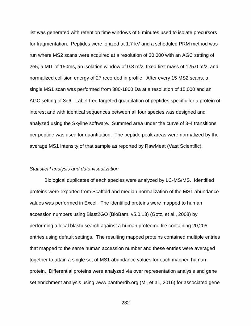
232
list was generated with retention time windows of 5 minutes used to isolate precursors
for fragmentation. Peptides were ionized at 1.7 kV and a scheduled PRM method was
run where MS2 scans were acquired at a resolution of 30,000 with an AGC setting of
2e5, a MIT of 150ms, an isolation window of 0.8 m/z, fixed first mass of 125.0 m/z, and
normalized collision energy of 27 recorded in profile. After every 15 MS2 scans, a
single MS1 scan was performed from 380-1800 Da at a resolution of 15,000 and an
AGC setting of 3e6. Label-free targeted quantitation of peptides specific for a protein of
interest and with identical sequences between all four species was designed and
analyzed using the Skyline software. Summed area under the curve of 3-4 transitions
per peptide was used for quantitation. The peptide peak areas were normalized by the
average MS1 intensity of that sample as reported by RawMeat (Vast Scientific).
Statistical analysis and data visualization
Biological duplicates of each species were analyzed by LC-MS/MS. Identified
proteins were exported from Scaffold and median normalization of the MS1 abundance
values was performed in Excel. The identified proteins were mapped to human
accession numbers using Blast2GO (BioBam, v5.0.13) (Gotz, et al., 2008) by
performing a local blastp search against a human proteome file containing 20,205
entries using default settings. The resulting mapped proteins contained multiple entries
that mapped to the same human accession number and these entries were averaged
together to attain a single set of MS1 abundance values for each mapped human
protein. Differential proteins were analyzed via over representation analysis and gene
set enrichment analysis using www.pantherdb.org (Mi, et al., 2016) for associated gene

233
ontology enrichments. Example proteins of different classes were graphed in GraphPad
Prism v5.04 and heatmaps of protein classes were generated using Morpheus (Broad
Institute). Principle component analysis was performed in Perseus (Tyanova, et al.,
2016). Network diagrams were created in Cytoscape (Shannon, et al., 2003) using the
Reactome (Wu, et al., 2010) and ClueGO (Bindea, et al., 2009) plugins. Protein
complex clustering was performed by mapping the identified proteins to human protein
complexes in CORUM and then calculating a mean complex score (the mean value of
the precursor area in all quantified components). The scored complexes were then
clustered in R using the hclust algorithm with the ward method. The resulting
dendrogram was cut at a level that generated 7 clusters based on evaluation of the
clustering via PCA. The clustered complexes were then assessed for functional
enrichments.

234
Figure A3.1. Generating multispecies proteomes. Heart tissue from M. musculus,
S. scrofa, X. laevis, and X. tropicalis were collected and subjected to differential protein
extraction as detailed. The resulting three extracts were fractionated by SDS PAGE. (B)

235
Number of proteins identified in the five gel fractions for each extract per species.
These numbers include proteins found in two or more extracts. (C) PCA analysis of
MS1 proteome data demonstrates that the mammalian sample separate from each
other and the Xenopus samples but the Xenopus samples maintain a tight association.
(D) Data analysis workflow.

236

237
Figure A3.2. Analysis of shared protein expression. (A) Identified proteins were
mapped to human accession numbers using Blast2GO. (B) The 1770 proteins that are
shared across all four species were clustered using k means with k=6 revealing clusters
driven by higher protein expression in one or more species. Each of the clusters from
the heatmap were analyzed for overrepresentation of GO biological process terms and
the p-values of the significantly enriched terms were shown in a heatmap.

238

239
Figure A3. 3. Analysis of species unique proteins. (A) Proteins found uniquely in
one species only were combined with the cluster of proteins that were more highly
expressed in that species and analyzed for overrepresentation of GO biological process
terms. The p-values of the significantly enriched terms are shown in a heatmap. (B)
The proteins uniquely identified in the mammalian and Xenopus species were used to
examine evolutionary-driven protein expression differences in these organisms using
ClueGO.

240

241
Figure A3.4. Gene set enrichment analysis reveals an increased presence of cell
cycle proteins in Xenopus laevis. (A) Pairwise GSEA was carried out between X.
laevis and the other three species which revealed an enrichment in cell cycle related
proteins in X. laevis. (B) A heatmap of all proteins found in any enriched cell cycle
related category by GSEA and clustered using kmeans. Relative protein expression is
shown. The proteins found in cluster 2 were further analyzed in Cytoscape and
grouped by function to show the interrelated nature of the proteins enriched in this
cluster.

242

243
Figure A3.5. PRM validation of cell cycle enrichment in X. laevis. (A) Workflow of
target protein and peptide selection for PRM validation. (B) Averaged protein
expression with SEM in each species by PRM assay are shown. The data were
grouped by protein functional categories and proteins graphed in blue have a higher
average expression in X. laevis than all other species.

244

245
Figure A3.6. Links to human heart disease. A diagram of detected proteins with
known links to human heart disease was created in Cytoscape showing both relative
protein expression data across the four species examined here as well as known
protein-protein interactions between these heart disease related proteins.

246
Figure SA3.1. Total proteins identified. Number of proteins identified in each extract
for (A) M. musculus (B) S. scrofa (C) X. laevis (D) X. tropicalis.

247
Figure SA3.2. Number of proteins identified in each replicate by species.

248
Figure SA3.3. PCA analysis of proteins identified in each extract from each
species.

249
Figure SA3.4. Multiscatter plot of median-normalized precursor values across the
four species. The two Xenopus species exhibit the greatest degree of similarity.

250
Figure SA3.5. All identified proteins that could be mapped to human entries. The
data in figure 2 includes quantified proteins only, whereas this figure includes any
identified but not quantified proteins.

251
Figure SA3.6. Cell cycle GO enrichments in every pairwise comparison. X. laevis.
(A) M. musculus / X. laevis (B) S. scrofa / X. laevis (C) X. tropicalis / X. laevis.

252
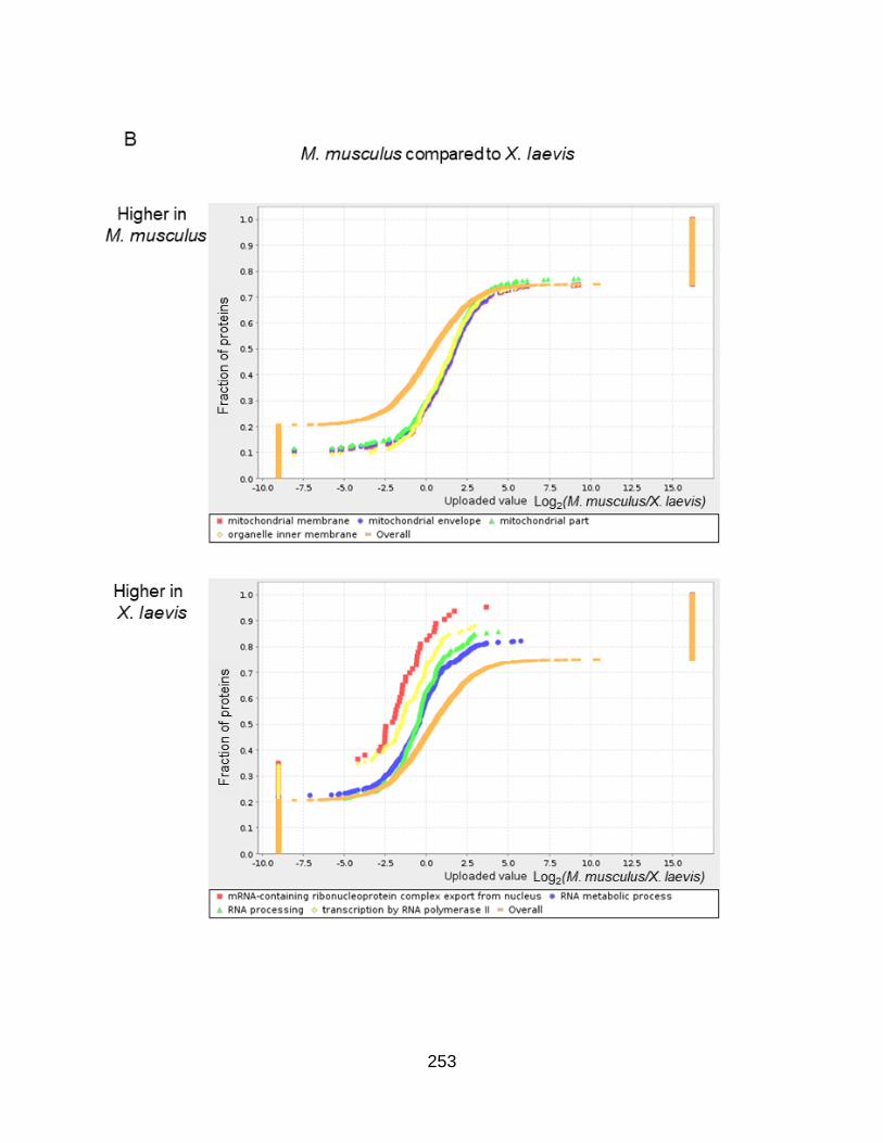
253

254
255

256

257
Figure SA3.7. Pairwise GSEA analysis of heart proteomes. M. musculus compared
to S. scrofa (A), M. musculus compared to X. laevis (B), M. musculus compared to X.

258
tropicalis (C), S. scrofa compared to X. laevis (D), S. scrofa compared to X. tropicalis
(E), X. tropicalis compared to X. laevis (F).

259

260

261

262

263
Figure SA3.8. Normalized peptide peak areas from PRM assays targeting cell
cycle proteins enriched in X. laevis. Gene names are listed above the graphs and
the first three amino acids for each peptide are listed to the right of the graph.

264
REFERENCES
Bindea, G., Mlecnik, B., Hackl, H., Charoentong, P., Tosolini, M., Kirilovsky, A., Fridman, W.H., Pages, F., Trajanoski, Z., and Galon, J. (2009). ClueGO: a Cytoscape plug-in to decipher functionally grouped gene ontology and pathway annotation networks. Bioinformatics 25, 1091-3.
Gotz, S., Garcia-Gomez, J.M., Terol, J., Williams, T.D., Nagaraj, S.H., Nueda, M.J., Robles, M., Talon, M., Dopazo, J., and Conesa, A. (2008). High-throughput functional annotation and data mining with the Blast2GO suite. Nucleic Acids Res 36, 3420-35.
McCauley, M.D., and Wehrens, X.H. (2009). Animal models of arrhythmogenic cardiomyopathy. Dis Model Mech 2, 563-70.
Mi, H., Poudel, S., Muruganujan, A., Casagrande, J.T., and Thomas, P.D. (2016). PANTHER version 10: expanded protein families and functions, and analysis tools. Nucleic Acids Res 44, D336-42.
Sauls, K., Greco, T.M., Wang, L., Zou, M., Villasmil, M., Qian, L., Cristea, I.M., and Conlon, F.L. (2018). Initiating Events in Direct Cardiomyocyte Reprogramming. Cell Rep 22, 1913-1922.
Shannon, P., Markiel, A., Ozier, O., Baliga, N.S., Wang, J.T., Ramage, D., Amin, N., Schwikowski, B., and Ideker, T. (2003). Cytoscape: a software environment for integrated models of biomolecular interaction networks. Genome Res 13, 2498-504.
Tyanova, S., Temu, T., Sinitcyn, P., Carlson, A., Hein, M.Y., Geiger, T., Mann, M., and Cox, J. (2016). The Perseus computational platform for comprehensive analysis of (prote)omics data. Nat Methods 13, 731-40.
Wu, G., Feng, X., and Stein, L. (2010). A human functional protein interaction network and its application to cancer data analysis. Genome Biol 11, R53.
Copyright © 2022 FDOKUMEN