Characterization of the novel antifungal chitosanase PgChP and the encoding gene from Penicillium...
10
BIOTECHNOLOGICALLY RELEVANT ENZYMES AND PROTEINS Characterization of the novel antifungal chitosanase PgChP and the encoding gene from Penicillium chrysogenum Andrea Rodríguez-Martín & Raquel Acosta & Susan Liddell & Félix Núñez & María José Benito & Miguel A. Asensio Received: 14 May 2010 / Revised: 29 June 2010 / Accepted: 1 July 2010 / Published online: 23 July 2010 # Springer-Verlag 2010 Abstract The protein PgChP is a new chitosanase pro- duced by Penicillium chrysogenum AS51D that showed antifungal activity against toxigenic molds. Two isoforms were found by SDS-PAGE in the purified extract of PgChP. After enzymatic deglycosylation, only the smaller isoform was observed by SDS-PAGE. Identical amino acid sequen- ces were obtained from the two isoforms. Analysis of the molecular mass by electrospray ionization-mass spectrom- etry revealed six major peaks from 30 to 31 kDa that are related to different levels of glycosylation. The pgchp gene has 1,146 bp including four introns and an open reading frame encoding a protein of 304 amino acids. The translated open reading frame has a predicted mass of 32 kDa, with the first 21 amino acids comprising a signal peptide. Two N glycosylation consensus sequences are present in the protein sequence. The deduced sequence showed high identity with fungal chitosanases. A high level of catalytic activity on chitosan was observed. PgChP is the first chitosanase described from P. chrysogenum. Given that enzymes produced by this mold species are granted generally recognized as safe status, PgChP could be used as a food preservative against toxigenic molds and to obtain chitosan oligomers for food additives and nutraceuticals. Keywords Antifungal protein . Food preservative . Glycosylation . Protective culture . Toxigenic mold Introduction Fungal infections and contaminations have led to an increasing demand for antifungal drugs in diverse fields including agriculture, medicine, and food protection. Many antifungal drugs have low efficacy rates, show severe side effects, and can even be toxic to humans (Meyer 2008). This makes it necessary to develop new antifungal com- pounds. Antifungal proteins have been described from diverse organisms including plants and animals, but only scant information is available about antifungal proteins of fungal origin. Many antifungal proteins have useful applications because they attack components of fungal walls that are not present in mammalian cells, such as glucanases, chitinases, and chitosanases (Adams 2004). Penicillium chrysogenum AS51D was isolated from dry- cured ham and produces an antifungal protein of 37.7 kDa. This protein, named PgChP, was active against toxigenic Penicillium commune (Acosta et al. 2009). Since the known antifungal proteins from ascomycetes are small proteins between 5.8 and 6.6 kDa (Marx 2004; Rodríguez-Martín et al. 2010; Skouri-Gargouri et al. 2009), PgChP cannot belong to this group of proteins. Antifungal proteins showing chitosanase activity have been isolated from different sources, including vegetables (Cuero and Osuji 1995), cyanobacteria (Prasanna et al. 2008), and actinomycetes (Saito et al. 2009). A. Rodríguez-Martín : R. Acosta : F. Núñez : M. A. Asensio (*) Higiene y Seguridad Alimentaria, Facultad de Veterinaria, Universidad de Extremadura, Avda. de la Universidad, s/n. 10071, Cáceres, Spain e-mail: [email protected] S. Liddell Division of Animal Sciences, University of Nottingham, Sutton Bonington Campus, Loughborough, Leicestershire LE12 5RD, United Kingdom M. J. Benito Nutrición y Bromatología, Escuela de Ingenierías Agrarias, Universidad de Extremadura, Carretera de Cáceres s/n. 06071, Badajoz, Spain Appl Microbiol Biotechnol (2010) 88:519–528 DOI 10.1007/s00253-010-2767-0
Transcript of Characterization of the novel antifungal chitosanase PgChP and the encoding gene from Penicillium...

BIOTECHNOLOGICALLY RELEVANT ENZYMES AND PROTEINS
Characterization of the novel antifungal chitosanase PgChPand the encoding gene from Penicillium chrysogenum
Andrea Rodríguez-Martín & Raquel Acosta &
Susan Liddell & Félix Núñez & María José Benito &
Miguel A. Asensio
Received: 14 May 2010 /Revised: 29 June 2010 /Accepted: 1 July 2010 /Published online: 23 July 2010# Springer-Verlag 2010
Abstract The protein PgChP is a new chitosanase pro-duced by Penicillium chrysogenum AS51D that showedantifungal activity against toxigenic molds. Two isoformswere found by SDS-PAGE in the purified extract of PgChP.After enzymatic deglycosylation, only the smaller isoformwas observed by SDS-PAGE. Identical amino acid sequen-ces were obtained from the two isoforms. Analysis of themolecular mass by electrospray ionization-mass spectrom-etry revealed six major peaks from 30 to 31 kDa that arerelated to different levels of glycosylation. The pgchp genehas 1,146 bp including four introns and an open readingframe encoding a protein of 304 amino acids. Thetranslated open reading frame has a predicted mass of32 kDa, with the first 21 amino acids comprising a signalpeptide. Two N glycosylation consensus sequences arepresent in the protein sequence. The deduced sequenceshowed high identity with fungal chitosanases. A high levelof catalytic activity on chitosan was observed. PgChP is thefirst chitosanase described from P. chrysogenum. Given thatenzymes produced by this mold species are granted
generally recognized as safe status, PgChP could be usedas a food preservative against toxigenic molds and to obtainchitosan oligomers for food additives and nutraceuticals.
Keywords Antifungal protein . Food preservative .
Glycosylation . Protective culture . Toxigenic mold
Introduction
Fungal infections and contaminations have led to anincreasing demand for antifungal drugs in diverse fieldsincluding agriculture, medicine, and food protection. Manyantifungal drugs have low efficacy rates, show severe sideeffects, and can even be toxic to humans (Meyer 2008).This makes it necessary to develop new antifungal com-pounds. Antifungal proteins have been described fromdiverse organisms including plants and animals, but onlyscant information is available about antifungal proteins offungal origin. Many antifungal proteins have usefulapplications because they attack components of fungalwalls that are not present in mammalian cells, such asglucanases, chitinases, and chitosanases (Adams 2004).
Penicillium chrysogenum AS51D was isolated from dry-cured ham and produces an antifungal protein of 37.7 kDa.This protein, named PgChP, was active against toxigenicPenicillium commune (Acosta et al. 2009). Since the knownantifungal proteins from ascomycetes are small proteinsbetween 5.8 and 6.6 kDa (Marx 2004; Rodríguez-Martín etal. 2010; Skouri-Gargouri et al. 2009), PgChP cannotbelong to this group of proteins.
Antifungal proteins showing chitosanase activity havebeen isolated from different sources, including vegetables(Cuero and Osuji 1995), cyanobacteria (Prasanna et al.2008), and actinomycetes (Saito et al. 2009).
A. Rodríguez-Martín :R. Acosta : F. Núñez :M. A. Asensio (*)Higiene y Seguridad Alimentaria, Facultad de Veterinaria,Universidad de Extremadura,Avda. de la Universidad, s/n. 10071,Cáceres, Spaine-mail: [email protected]
S. LiddellDivision of Animal Sciences, University of Nottingham,Sutton Bonington Campus,Loughborough, Leicestershire LE12 5RD, United Kingdom
M. J. BenitoNutrición y Bromatología, Escuela de Ingenierías Agrarias,Universidad de Extremadura,Carretera de Cáceres s/n. 06071,Badajoz, Spain
Appl Microbiol Biotechnol (2010) 88:519–528DOI 10.1007/s00253-010-2767-0

Given that PgChP differs from other described antifun-gal proteins produced by molds, the objective of this paperwas to characterize the antifungal protein PgChP, includingamino acid and genetic sequencing.
Materials and methods
Microbial strain
P. chrysogenum AS51D used in this study was isolatedfrom dry-cured ham and deposited in the Spanish TypeCulture Collection (CECT 20753).
Protein purification
The antifungal protein from P. chrysogenum AS51D wasobtained after growing in malt extract broth by FPLC usinga cationic exchange column. Fractions showing the anti-fungal activity were gel-filtered by FPLC following themethod previously described (Acosta et al. 2009). Themold-inhibiting fraction was desalted and concentrated withYM-10 Microcon Centrifugal Filter Units (Millipore Iberica,Madrid, Spain), and stored at−20°C.
Sodium dodecyl sulphate-polyacrylamidegel electrophoresis
Sodium dodecyl sulphate-polyacrylamide gel electrophoresis(SDS-PAGE) was performed according to Laemmli methodas described previously (Acosta et al. 2009). Gels werestained with Imperial Protein Stain (Pierce Biotechnology,Rockford, USA). The molecular mass marker was PrecisionPlus Protein Standards with ten proteins between 10 and250 kDa (Bio-Rad).
Digestion with enzymes for amino acid sequencing
Reduction and alkylation prior to enzymatic digestion wereperformed with the protein in SDS-PAGE gel pieces or in-solution. For in-gel deglycosylations, the protein waselectrophoresed (25 μg of protein per well) and stainedusing Imperial Protein Stain. The band was excised fromthe gel and washed, reduced, alkylated, and dehydrated asdescribed previously (Rodríguez-Martín et al. 2010). In-solution digestion was also carried out adding 1.25, 2.5,and 6.25 μg of the protein before digestion with eachenzyme.
Digestions with trypsin and chymotrypsin of the reducedand alkylated protein were performed in-gel and in-solutionfollowing a protocol described previously (Rodríguez-Martín et al. 2010). For further automated in-gel digestions,
gel pieces were de-stained, dehydrated, reduced, alkylated,washed, and digested using the MassPREP robotic liquidhandling station (Waters, Manchester, United Kingdom).After addition of 25 μl per well of Trypsin Gold (Promega,Hampshire, United Kingdom) at 10 ng/μl, the plate wasincubated at 6°C for 20 min and then at 37°C for 4 h.
Analysis for glucidic residues
Both chemical and enzymatic treatments for deglycosylationwere carried out after reduction and alkylation of the proteinin solution as described above.
Chemical deglycosylation was carried out using twodifferent chemical methods: acid hydrolysis with HCl and β-elimination with NaOH, as described previously (Rodríguez-Martín et al. 2010). Finally, both samples were analyzed byelectrospray ionization-mass spectrometry (ESI-MS).
For enzymatic deglycosylation, PNGase F (New EnglandBiolabs, Ipswich, United Kingdom) and E-DEGLY degly-cosylation kit (Sigma, Madrid, Spain) were used. Reactionswith E-DEGLY kit were carried out under native anddenaturing conditions according to the manufacturer’sinstructions, and products were subjected to SDS-PAGEanalysis.
ESI-MS analysis for peptide sequence determination
After deglycosylation and enzymatic digestion, formic acidwas added to a final concentration of 0.1% (v/v); the samplewas desalted and analyzed in a Q-TOF2 mass spectrometer(Waters) by ESI-MS as described previously (Rodríguez-Martín et al. 2010). Tandem MS fragmentation spectra werecollected typically from 50 to 1,600 m/z. Peptide tandemMS spectra were deconvoluted and de novo peptidesequences were searched against the public databases usingBLASTP with parameter settings for “short and nearlyexact matches”. A multiple sequence alignment of fungalchitosanases was produced using the program T-Coffee(www.ebi.ac.uk).
Molecular mass determination by ESI-MS
The molecular mass of the desalted protein (0.125 μg/μl) wasdetermined by electrospray mass spectrometry (Rodríguez-Martín et al. 2010). Spectra were deconvoluted with anoutput range of 20,000 and 40,000 Da, at a resolution of1 Da.
Evaluation of chitosanase activity
Chitosanase activity was determined by measuring thereducing sugars liberated during the hydrolysis of chitosan
520 Appl Microbiol Biotechnol (2010) 88:519–528

following a modification of the 3,5-dinitrosalicylic acidmethod (Su et al. 2006). Chitosan predissolved in aceticacid was added to a PgChP solution (10 μg/ml) andincubated for 2–4 h at 37°C. Hydrolysis reactions wereterminated by adding dinitrosalicylic acid reagent, and thesample was boiled at 100°C for 45 min. Finally, 1 ml of 40%(w/v) potassium sodium tartrate was added, the solution wascentrifuged, and the optical density of the supernatant wasmeasured at 520 and 575 nm. To quantify the reducingsugars liberated, a calibration curve was obtained fromdifferent concentrations of D-(+)-glucosamine hydrochlo-ride (Sigma-Aldrich). A chitinase from Streptomycesgriseus (Sigma-Aldrich) with chitosanase activity was usedat 50 μg/ml as positive control.
DNA isolation
P. chrysogenum AS51D was grown in MEB (Pitt, 1986),pH 4.5 at 25°C under continuous shaking, for 5 days. DNAwas obtained as previously described (Benito et al. 2006).For this, 2 g of filteredmyceliumwere broken and treated withproteinase K. DNA was extracted with phenol-chloroform-isoamyl alcohol, precipitated with sodium acetate and ethanol,and treated with RNase.
Partial amplification of genomic DNAwith degenerate primers
PCRs were performed with 100 ng genomic DNA(Rodríguez-Martín et al. 2010). Degenerate primerspgchp-DPF1 and pgchp-DPR2A (Table 1) were designedfrom amino acid sequences obtained by mass spectrometry.For pgchp-DPR2A, the contiguous residues conserved infungal chitosanases (Shimosaka et al. 2005) were included(Table 1).
Three reactions were done, one with both forward andreverse primers and two with either forward or reverseprimers. The PCR program consisted of initial denaturation(94°C for 5 min), 35 cycles of denaturation (94°C for1 min), annealing (56–60°C range for 30 s, gradient 12),
and extension (72°C for 30 s), followed by a final extensionat 72°C for 4 min. Products detected only in the reactionswith both reverse and forward primers were gel-purified,cloned, and sequenced as described previously (Rodríguez-Martín et al. 2010).
Rapid amplification of cDNA ends
RNA was obtained after growing P. chrysogenum AS51Din malt extract broth, pH 4.5 at 25°C under continuousshaking for 4 days. Chitosanase production was induced ina medium with glucosamine (Zhang et al. 2000) made with0.3% (w/v) NaNO3, 0.05% (w/v) KCl, 0.05% (w/v)MgSO4·7H2O, 0.01% (w/v) FeSO4·7H2O, and 2% (w/v) D
(+)-glucosamine hydrochloride (Sigma, Dorset, UnitedKingdom), dissolved in 25 mM phosphate buffer pH 5.8.Mold was cultured in glucosamine medium at 25°C undercontinuous shaking for 17 h. Subsequently, mycelium wasquickly frozen until RNA extraction with RNeasy PlantMini Kit (Qiagen, Crawley, UK) following the manufac-turer’s instructions.
Amplification of 5′ and 3′ ends of the pgchp gene wascarried out according to Rodríguez-Martín et al. (2010)using the SMART rapid amplification of cDNA ends(RACE) cDNA Amplification Kit (Clontech Laboratories,Saint Germain en Laye, France). Two gene-specific primers(GSPs) were designed for amplifying the 5′ and 3′- ends (5′-RACE GSP: CCATTGCAGATAACGGCACCAACG and3′-RACE GSP: GTTCGACAAGGGTGCGGCATATG).Polymerase chain reactions were performed with 50 ng 5′-or 3′-RACE-Ready cDNAs (Rodríguez-Martín et al. 2010).Finally, 5′- and 3′-RACE-PCR products were gel-purified,cloned, and sequenced as described above.
The full cDNA fragment of the gene pgchp was obtainedby PCR with specific primers Fwd-pgchp (ATGATGACCTACAGCCGCTTAATCCC) and Rev-pgchp (TCAATCACTGGGGCATTTTCCAC) designed from 5′ and 3′ends. PCR products from 50 ng 5′-RACE-Ready cDNAwere obtained, gel-purified, cloned, and sequenced aspreviously described (Rodríguez-Martín et al. 2010).
Table 1 Degenerate primers used to amplify the pgchp gene
Primer names Amino acid sequences and designed primersa
pgchp-DPF1
pgchp-DPR2A
Conserved amino acids used for designing the primer pgchp-DPR2A are underlineda R=purine, Y=pirimidine, N=A, C, T, or G
Appl Microbiol Biotechnol (2010) 88:519–528 521
Amplification of the complete gene from genomic DNA
The gene sequence was obtained by PCR amplificationusing 100 ng of genomic DNA and both Fwd-pgchp andRev-pgchp primers as described for full cDNA. Alignmentbetween the sequences obtained from genomic DNA andcDNA was performed.
Sequence analysis
The NCBI database (http://blast.ncbi.nlm.nih.gov/Blast.cgi)was used to compare the obtained data against other genomicsequences or proteins. Intron sequences were confirmed byGenScan software (http://genes.mit.edu/GENSCAN.html)(Burge 1998; Burge and Karlin 1997; 1998). To translatethe open reading frame to protein and to estimate themolecular mass and the isoelectric point of the predictedprotein, Translate tool and Compute pI/Mw tool fromExPASy proteomics server at the Swiss Institute of Bio-informatics (http://www.expasy.org) (Gasteiger et al. 2005)were used. A search for the presence of signal peptides wasperformed with SignalP version 3.0 (http://www.cbs.dtu.dk/services/SignalP/) (Bendtsen et al. 2004; Nielsen et al. 1997).
The nucleotide sequence of the pgchp gene has beendeposited in the National Center for Biotechnology Infor-mation, GenBank database (http://www.ncbi.nlm.nih.gov/Genbank/index.html), under accession number HM044146.
Results
Analysis of PgChP protein
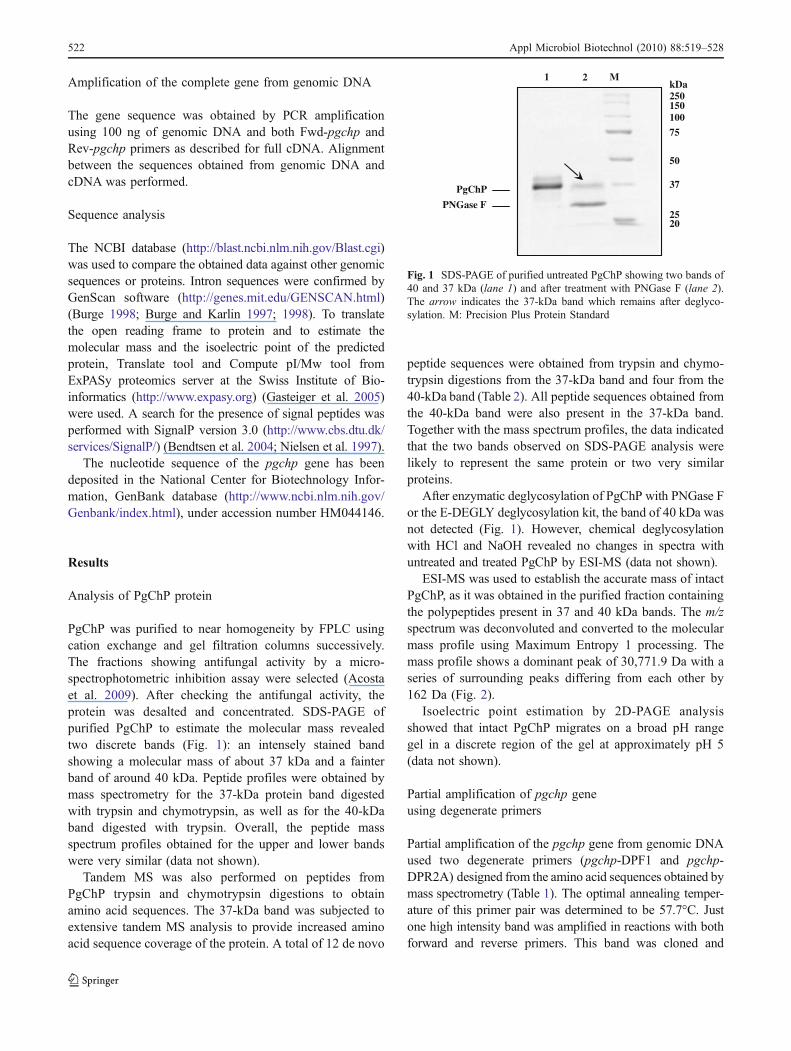
PgChP was purified to near homogeneity by FPLC usingcation exchange and gel filtration columns successively.The fractions showing antifungal activity by a micro-spectrophotometric inhibition assay were selected (Acostaet al. 2009). After checking the antifungal activity, theprotein was desalted and concentrated. SDS-PAGE ofpurified PgChP to estimate the molecular mass revealedtwo discrete bands (Fig. 1): an intensely stained bandshowing a molecular mass of about 37 kDa and a fainterband of around 40 kDa. Peptide profiles were obtained bymass spectrometry for the 37-kDa protein band digestedwith trypsin and chymotrypsin, as well as for the 40-kDaband digested with trypsin. Overall, the peptide massspectrum profiles obtained for the upper and lower bandswere very similar (data not shown).
Tandem MS was also performed on peptides fromPgChP trypsin and chymotrypsin digestions to obtainamino acid sequences. The 37-kDa band was subjected toextensive tandem MS analysis to provide increased aminoacid sequence coverage of the protein. A total of 12 de novo
peptide sequences were obtained from trypsin and chymo-trypsin digestions from the 37-kDa band and four from the40-kDa band (Table 2). All peptide sequences obtained fromthe 40-kDa band were also present in the 37-kDa band.Together with the mass spectrum profiles, the data indicatedthat the two bands observed on SDS-PAGE analysis werelikely to represent the same protein or two very similarproteins.
After enzymatic deglycosylation of PgChP with PNGase For the E-DEGLY deglycosylation kit, the band of 40 kDa wasnot detected (Fig. 1). However, chemical deglycosylationwith HCl and NaOH revealed no changes in spectra withuntreated and treated PgChP by ESI-MS (data not shown).
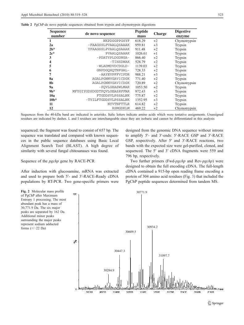
ESI-MS was used to establish the accurate mass of intactPgChP, as it was obtained in the purified fraction containingthe polypeptides present in 37 and 40 kDa bands. The m/zspectrum was deconvoluted and converted to the molecularmass profile using Maximum Entropy 1 processing. Themass profile shows a dominant peak of 30,771.9 Da with aseries of surrounding peaks differing from each other by162 Da (Fig. 2).
Isoelectric point estimation by 2D-PAGE analysisshowed that intact PgChP migrates on a broad pH rangegel in a discrete region of the gel at approximately pH 5(data not shown).
Partial amplification of pgchp geneusing degenerate primers
Partial amplification of the pgchp gene from genomic DNAused two degenerate primers (pgchp-DPF1 and pgchp-DPR2A) designed from the amino acid sequences obtained bymass spectrometry (Table 1). The optimal annealing temper-ature of this primer pair was determined to be 57.7°C. Justone high intensity band was amplified in reactions with bothforward and reverse primers. This band was cloned and
250
100
1 2 M
150
PNGase F
PgChP
75
50
37
2520
kDa
Fig. 1 SDS-PAGE of purified untreated PgChP showing two bands of40 and 37 kDa (lane 1) and after treatment with PNGase F (lane 2).The arrow indicates the 37-kDa band which remains after deglyco-sylation. M: Precision Plus Protein Standard
522 Appl Microbiol Biotechnol (2010) 88:519–528
sequenced; the fragment was found to consist of 657 bp. Thesequence was translated and compared with known sequen-ces in the public sequence databases using Basic LocalAlignment Search Tool (BLAST). A high degree ofsimilarity with several fungal chitosanases was found.
Sequence of the pgchp gene by RACE-PCR
After induction with glucosamine, mRNA was extractedand used to prepare both 5′- and 3′-RACE-Ready cDNApopulations by RT-PCR. Two gene-specific primers were
designed from the genomic DNA sequence without intronsto amplify 5′- and 3′-ends: 5′-RACE GSP and 3′-RACEGSP, respectively. After 5′ and 3′-RACE reactions, twobands with the expected size were gel-purified, cloned, andsequenced. The 5′ and 3′ cDNA fragments were 559 and796 bp, respectively.
Two further primers (Fwd-pgchp and Rev-pgchp) weredesigned to obtain the full encoding cDNA. The full-lengthcDNA contained a 915-bp open reading frame encoding aprotein of 304 amino acid residues (Fig. 3) that included thePgChP peptide sequences determined from tandem MS.
31097.7
30934.2
30771.9
30609.5
30447.3
30284.9
Fig. 2 Molecular mass profileof PgChP after MaximumEntropy 1 processing. The mostabundant peak has a mass of30,771.9 Da. The six majorpeaks are separated by 162 Da.Additional minor peakssurrounding the major peaksrepresent sodium adductedforms (+/−22 Da)
Table 2 PgChP-de novo peptide sequences obtained from trypsin and chymotrypsin digestions
Sequence number de novo sequence Peptide
mass Charge Digestive enzyme
1 618.29 +2 Chymotrypsin 2a 959.81 +3 Trypsin 2b* 911.48 +2 Trypsin 2c* 1026.61 +1 Trypsin3 866.40 +2 Trypsin 4 528.79 +2 Trypsin 5 1139.03 +2 Trypsin 6 728.33 +2 Trypsin 7 988.21 +3 Trypsin 8a 771.40 +2 Trypsin 8b 720.89 +2 Chymotrypsin 9a 1053.50 +2 Trypsin 9b* 972.43 +3 Trypsin 10a 775.87 +2 Trypsin 10b* 1352.95 +3 Trypsin 11 614.82 +2 Trypsin 12 469.22 +2 Chymotrypsin
Sequences from the 40-kDa band are indicated in asterisks. Italic letters indicate amino acids which were tentative assignments. Unassignedresidues are indicated by dashes. L and I residues are interchangeable since they are isobaric and cannot be differentiated in this analysis
Appl Microbiol Biotechnol (2010) 88:519–528 523
The genomic sequence of pgchp, amplified using thesame primers used for the full-length cDNA, was 1,146 bplength. Comparison of cDNA and genomic sequencesrevealed that pgchp gene had four introns 60, 53, 59, and59 bp in length (Fig. 3). The position and lengths of theseintrons were confirmed by GenScan program.
The open reading frame was translated by ExPASyproteomics server at the Swiss Institute of Bioinformatics
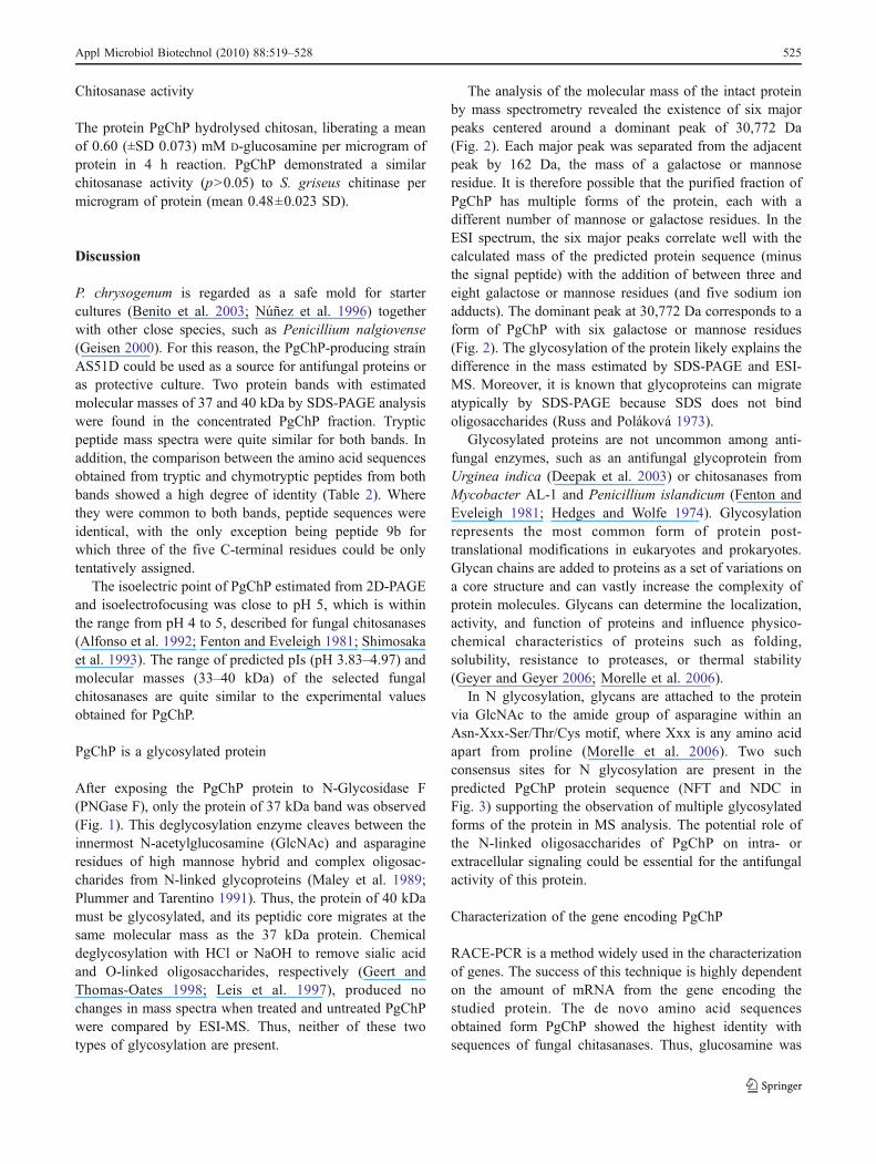
(http://www.expasy.org). Prediction of the presence of asignal peptide by the SignalP program (Nielsen et al. 1997)indicated that the pre-sequence consisted of the first 21amino acids (MMTYSRLIPFALFALFGTGLA). The calcu-lated mass of the PgChP protein after signal peptide cleavageis 29,689 Da. The deduced amino acid sequences of PgChP(Table 2) showed the highest identity with sequences ofseven described or predicted fungal chitosanases (Fig. 4).
20 40 60 80 100PgChP --MMTYSRLIPFALFALFG-TGLAQTVDGSKFNKPDGGPPGSYFAAGSSIPVAALQSAAAKARTPVPDATYPINGDNGAKKVTIHSDWAKFDKGAAYVWIADMDVDCDGI 107 A.fumigatus MPSTTIIRQLAIS-LALCN-SALGQVVNGADYNKPNGGPPASFFAAASTMPVAALQAAAAKATKVPSLATYPVSQDKGAAKSTIHTDWASFSEGASISWVADMDVDCDGL 108 N.fischeri MPSKTIIRQLAIS-VALCN-SALGQVVNGADYNKPNGGPPASFFAAASTMPVAALQAAAAKASKVPSLATYPVSQDSGAAKSTIHTDWASFSEGASISWVADMDVDCDGL 108 A.niger MAFKTTAG--LAF-LALAG-SVKAQSVDGSKYNSPSNGPPASYFAAATTLPVAALQSAAAKASSVPSKATYPVNTDDDSPKSTIHSDWVKFNQGAALSWVADMDVDCDGI 106 A.oryzae-1 MPIKSFASRLALS-LAICG-TAMGQKVNGADYNKPDGGPPAKFFQASSSIPVAAIQAAAAKASKVPSHATYPIGQ--GSTKSTIHSDWAGFSEGAAFSFIADMDVDCDGL 106 A.oryzae-2 MPIKSFASRLALS-LAICG-TAMGQKVNGADYNKPDGGPPAKFFQASSSIPVAAIQAAAAKASKVPSHATYPIGQ--GSTKSTIHSDWAGFSEGAAFSFIADMDVDCDGL 106 A.terreus MVFKKAAIGLTLP-FALFSSVALGQTVDGSDYDSPNGGPPGSYFAAASTMPVAALQAAAAKATKVPSYATYPVSQDDNAKKSTIHSDWASFSQGAAISWVADMDVDCDGI 109 A.clavatus MIFKSSLSHAAAS-LLLLT-PALAQKVQGPEYNKPSAGPPASFFAAAPTMPVAALKSAVARASVVPKNAAYPVNQD-GGPTATIHADWASLPTAAAYVYTADMDVDCDGL 107
PgChP DYKC-------KGNPDGQHQTNFGALAAYEVPFFVIPDRFGTKYAKQLPGNNVGAVIC----NGKMFYGIYGDSDGDTPQVIGEASWLMARTCFPNDDLNGNSGHGDVDV 206 A.fumigatus NSGC-------QGNPDGQPQTNWGALSAYEVPFIVIPDKYLSANSGALPGNNIAAVIC----NGKMFYGILGDSNGDSPQVTGEASWLMARTCFPNEGLNGNNGHTGVDV 207 N.fischeri NSGC-------QGNPDGQPQTNWGALSAYEVPFIVIPDKYLSANTGALPGNNIAAVIC----NGKMFYGILGDSNGDSPQVTGEASWLMARTCFPNEGLNGNNGHTGVDV 207 A.niger DYKC-------KGNGDGLPETNWGALSAYEVPWIVIPDQFLTANEDLLPGNNVAAVIC----NGKMYYGILGDSNGDDPEVTGEASWLMARTCFPDDDLNGAEGHAEADV 205 A.oryzae-1 NHGC-------KGNPDGQKETNWGALSAYEVPFIVIPQEFLDANKGTLKGNAVAAVIC----NGKMFYGIFGDSNGDSPQVTGEASWLMARTCFPKEDLNGNKGHTAADV 205 A.oryzae-2 NHGC-------KGNPDGQKETNWGALSAYEVPFIVIPQEFLDANKGTLKGNAVAAVICATSSNGKMFYGIFGDSNGDSPQVTGEASWLMARTCFPEEDLNGNKGHTAADV 209 A.terreus DSGCEHEIKTSQGNPDGQDATNWGALAAYEVPFIVIPQKYLDHNGNALKGNNIAAVIC----NGKMFYGILGDANGDEPQVTGEASWLMARTCFPNEGLNGNKGHTAADV 215 A.clavatus DHNC-------KGNPDGQPDTNFGALAAYEVPFVVIPDRFATTYASALPGNNIVAVIC----DGKMFYGIFGDTDGDHPQVIGEASWLMARTCFPNDNLNGDSGHVPADV 206
* * PgChP TYILFTGDDSVLPSSALNKNYVTNFTTLRSMGDKLMTALAKNLKLVDGGDGG---------SPT--TTAGSNPT--SGSCEWEGHCA-----GASCKDENDCSDQLVCKS 298 A.fumigatus TYIVFTGKNAVLPSSALTKNYITNFTTLRSMGDKLVNALVSNLGLSGTPPTK---------TTTRVTTTTTKPT-SAASCSWAGHCL-----GASCSSDDDCADALVCTA 302 N.fischeri TYIVFTGKDAVLPSSALTKNYITNFTTLRSMGDKLVNALASKLGLSGTPPTK---------TTTLVTTTTTKPT-STASCSWAGHCL-----GASCSSNDDCADALVCAA 302 A.niger TCKPYT-------RSALNKNYITNFSTLRSMGDKLVGALASNLGLTSSA------------------------SGSTATCSWQGHCE-----GAICSTEDDCSDDLVCDS 279 A.oryzae-1 TYIVFTGDKAVLPSSALNKNYITNFDTLRSMGDSLVGALAKNLNLGGGGGNP---------PTTLTTTSIPEPTGGSGSCSWPGHCA-----GATCSSNDDCSDDLTCQN 301 A.oryzae-2 TYIVFTGDKAVLPSSALNKNYITNFDTLRSMGDSLVGALAKNLNLGGGGGNP---------PTTLTTTSIPEPTGGSGSCSWPGHCAGFKNKGATCSSNDDCSDDLACQN 310 A.terreus TYILFTGDESVLPSSALNENYITNFSTLRSMGDKLVNALASNLGLSGGSGGT---------PTT--TTTATQPT-STGTCSWAGHCE-----GASCSTNDDCSDQLACKN 308 A.clavatus TYIFFTGKDSVLPSSAVNKNYITDFTKLRSMGDSLVNAFASQLGISSGGGNGGGDGGHTTLHTTTATRTATAPT-STATCDWEGHCL-----GTACSNNGECSDPFGCIN 310
PgChP GKCPSD---------------------------------------------------------------------------- 304 A.fumigatus GKCSVDGAA-TCSWEGHCEGASCSSDDDCSDDLYCKSGSCTA------------------------------------P--- 344N.fischeri GKCSVDGAA-TCSWEGHCEGVYS----------------------------------------------------------- 324A.niger GKCSSPDED--------------------------------------DGDDEDDEDEEENDEDDEDDDDEDDEDDE------ 317A.oryzae-1 GKCASDGSAETCSWEGHCKGATCSSNDDCSDELACISGICSVDNGVETCEWEGHCEGASCSSHDDCDGNLACKNGKCSA--- 380A.oryzae-2 GKCASDGSAETCSWEGHCKGATCSSNDDCSDELACISGICSVDNGVETCEWEGHCEGASCSSHDDCDGNLACKNGKCSA--- 389A.terreus GVCSTDGEV-VCSWEGHCEGATCSSNDDCSDELYCAGGACTSA--------------------------------------- 350A.clavatus GFCNYDPTL-TCQWAGHCVGASCSSNDQCSDPFACIDGACAVDT-SLDCSRKGHCAGTTCLSDSDCSRPLSCILGVCANQSG 390
Fig. 4 Alignment of PgChP with other fungal chitosanases. Commonamino acids are denoted by background shaded letters. Asterisksindicate essential amino acid residues for catalytic activity of fungalchitosanases. Accession numbers in GenBank are XP_754126
(Aspergillus fumigatus), XP_001262949 (Neosartorya fischeri),XP_001390407 (Aspergillus niger), BAD08218 (Aspergillus oryzae-1),XP_001824257 (Aspergillus oryzae-2), XP_001209034 (Aspergillusterreus), XP_001274664 (Aspergillus clavatus)
1 atgatgacctacagccgcttaatccccttcgctctctttgccttgtttggcacagggctcgcgcaaacagtcgatggctccaaattcaacaagccagacggcggtccaccaggaagctac 120
M M T Y S R L I P F * A L F A L F G T G L A Q T V D G S K F N K P D G G P P G S Y
121 ttcgctgctggctcgtctattcccgtcgctgctttgcagagcgctgctgcaaaggctcgcactcccgtgccagatgcaacctatcctattaatggcgataacggtgctaagaaagttacc 240
F A A G S S I P V A A L Q S A A A K A R T P V P D A T Y P I N G D N G A K K V T
241 atccacagcgactgggctaagttcgacaaggtagatactctcactggtcggttaattgcaaggtccgcatgcttattaaatggatcatagggtgcggcatatgtttggattgcggatatg 360
I H S D W A K F D K << Intron 1 >>G A A Y V W I A D M
361 gacgtcgactgcgacggcattgactacaagtgcaaggtacggtcactattgttgtctgtctgagatgttattgctgacccgatgttcagggaaacccggatggtcagcaccaaaccaact 480
D V D C D G I D Y K C K << Intron 2 >>G N P D G Q H Q T N F
481 tcggagctttggctgcgtatgaagtgccgttctttgtgattcccgacaggtttggaaccaagtacgcgaagcagcttcctggaaacaacgttggtgccgttatctg gtatggtttcctac 600
G A L A A Y E V P F F V I P D R F G T K Y A K Q L P G N N V G A V I C <<
601 ctttgtaagatatctcagcggcgtcttgttctaaccgaaagctagcaatggaaagatgttctacggaatttacggagactccgatggcgatacccctcaggtcatcggcgaggcctcatg 720
Intron 3 >> N G K M F Y G I Y G D S D G D T P Q V I G E A S W
721 gttaatggcccggacctgcttccctaatgatgacttgaatggcaatagtggccatggtgatgttgatgtgacctgtaagtttgcagccttggcacatctagatccgtattctgtcaacta 840
L M A R T C F P N D D L N G N S G H G D V D V T Y<< Intron 4
841 ctaatctaaccagatatcctcttcactggcgacgactcggtgctccccagcagcgctctcaacaagaattatgtcaccaacttcaccactctgcgctctatgggagacaaactcatgact 960
>> I L F T G D D S V L P S * S A L N K N Y V T N F T T L R S M G D K L M T
961 gctcttgcgaagaacctcaagttggtagatggaggtgatggaggttctccaacaactacggctgggtccaaccctactagcgggtcttgtgagtgggagggacactgtgctggtgcgtct 1080
A L A K N L K L V D G G D G G S P T T T A G S N P T S G S C E W E G H C A G A S
1081 tgcaaagatgaaaatgattgctccgatcaactggtctgcaaaagtggaaaatgccccagtgattga 1146
C K D E N D C S D Q L V C K S G K C P * S D* -
Fig. 3 Full-length DNA sequence of pgchp gene. The open readingframe has been translated to amino acids. Typical sequences of fungalintrons are underlined. Shaded background indicates typical consensussites for glycosylation. *Open squares indicate the differences with the
predicted chitosanase from P. chrysogenum Wisconsin 54-1255(accession number CAP80409): F, S, P, and D in PgChP change toS, N, A, and H, respectively
524 Appl Microbiol Biotechnol (2010) 88:519–528
Chitosanase activity
The protein PgChP hydrolysed chitosan, liberating a meanof 0.60 (±SD 0.073) mM D-glucosamine per microgram ofprotein in 4 h reaction. PgChP demonstrated a similarchitosanase activity (p>0.05) to S. griseus chitinase permicrogram of protein (mean 0.48±0.023 SD).
Discussion
P. chrysogenum is regarded as a safe mold for startercultures (Benito et al. 2003; Núñez et al. 1996) togetherwith other close species, such as Penicillium nalgiovense(Geisen 2000). For this reason, the PgChP-producing strainAS51D could be used as a source for antifungal proteins oras protective culture. Two protein bands with estimatedmolecular masses of 37 and 40 kDa by SDS-PAGE analysiswere found in the concentrated PgChP fraction. Trypticpeptide mass spectra were quite similar for both bands. Inaddition, the comparison between the amino acid sequencesobtained from tryptic and chymotryptic peptides from bothbands showed a high degree of identity (Table 2). Wherethey were common to both bands, peptide sequences wereidentical, with the only exception being peptide 9b forwhich three of the five C-terminal residues could be onlytentatively assigned.
The isoelectric point of PgChP estimated from 2D-PAGEand isoelectrofocusing was close to pH 5, which is withinthe range from pH 4 to 5, described for fungal chitosanases(Alfonso et al. 1992; Fenton and Eveleigh 1981; Shimosakaet al. 1993). The range of predicted pIs (pH 3.83–4.97) andmolecular masses (33–40 kDa) of the selected fungalchitosanases are quite similar to the experimental valuesobtained for PgChP.
PgChP is a glycosylated protein
After exposing the PgChP protein to N-Glycosidase F(PNGase F), only the protein of 37 kDa band was observed(Fig. 1). This deglycosylation enzyme cleaves between theinnermost N-acetylglucosamine (GlcNAc) and asparagineresidues of high mannose hybrid and complex oligosac-charides from N-linked glycoproteins (Maley et al. 1989;Plummer and Tarentino 1991). Thus, the protein of 40 kDamust be glycosylated, and its peptidic core migrates at thesame molecular mass as the 37 kDa protein. Chemicaldeglycosylation with HCl or NaOH to remove sialic acidand O-linked oligosaccharides, respectively (Geert andThomas-Oates 1998; Leis et al. 1997), produced nochanges in mass spectra when treated and untreated PgChPwere compared by ESI-MS. Thus, neither of these twotypes of glycosylation are present.
The analysis of the molecular mass of the intact proteinby mass spectrometry revealed the existence of six majorpeaks centered around a dominant peak of 30,772 Da(Fig. 2). Each major peak was separated from the adjacentpeak by 162 Da, the mass of a galactose or mannoseresidue. It is therefore possible that the purified fraction ofPgChP has multiple forms of the protein, each with adifferent number of mannose or galactose residues. In theESI spectrum, the six major peaks correlate well with thecalculated mass of the predicted protein sequence (minusthe signal peptide) with the addition of between three andeight galactose or mannose residues (and five sodium ionadducts). The dominant peak at 30,772 Da corresponds to aform of PgChP with six galactose or mannose residues(Fig. 2). The glycosylation of the protein likely explains thedifference in the mass estimated by SDS-PAGE and ESI-MS. Moreover, it is known that glycoproteins can migrateatypically by SDS-PAGE because SDS does not bindoligosaccharides (Russ and Poláková 1973).
Glycosylated proteins are not uncommon among anti-fungal enzymes, such as an antifungal glycoprotein fromUrginea indica (Deepak et al. 2003) or chitosanases fromMycobacter AL-1 and Penicillium islandicum (Fenton andEveleigh 1981; Hedges and Wolfe 1974). Glycosylationrepresents the most common form of protein post-translational modifications in eukaryotes and prokaryotes.Glycan chains are added to proteins as a set of variations ona core structure and can vastly increase the complexity ofprotein molecules. Glycans can determine the localization,activity, and function of proteins and influence physico-chemical characteristics of proteins such as folding,solubility, resistance to proteases, or thermal stability(Geyer and Geyer 2006; Morelle et al. 2006).
In N glycosylation, glycans are attached to the proteinvia GlcNAc to the amide group of asparagine within anAsn-Xxx-Ser/Thr/Cys motif, where Xxx is any amino acidapart from proline (Morelle et al. 2006). Two suchconsensus sites for N glycosylation are present in thepredicted PgChP protein sequence (NFT and NDC inFig. 3) supporting the observation of multiple glycosylatedforms of the protein in MS analysis. The potential role ofthe N-linked oligosaccharides of PgChP on intra- orextracellular signaling could be essential for the antifungalactivity of this protein.
Characterization of the gene encoding PgChP
RACE-PCR is a method widely used in the characterizationof genes. The success of this technique is highly dependenton the amount of mRNA from the gene encoding thestudied protein. The de novo amino acid sequencesobtained form PgChP showed the highest identity withsequences of fungal chitasanases. Thus, glucosamine was
Appl Microbiol Biotechnol (2010) 88:519–528 525
used to induce PgChP production, as it has been reportedfor other chitosanases (Zhang et al. 2000). After the mRNAextraction, both populations 5′- and 3′-RACE-ReadycDNAs were satisfactorily obtained by RT-PCR. Usinggene-specific primers designed from the sequence amplifiedwith degenerated primers, the 5′- and 3′-ends were obtainedby RACE-PCR.
When the complete genomic (1,146 bp) and cDNA(915 bp) sequences were compared, four introns of 60, 53,59, and 59 bp were found (Fig. 3). The small size of theseintrons compared to mammalian introns is a typical featureof fungal genes (Gurr et al. 1987). The 5′- and 3′-ends ofthe four introns are very similar and conform to theconsensus splice sequences for fungal introns 5′-splicedonor site (GT), the 3′-splice acceptor site (AG). Inaddition, three of them have the internal CTRAC sequence,typical of fungal introns (Wiesner et al. 1988).
The translated sequence of the gene pgchp encodes 304amino acids of the PgChP protein. Comparing the deducedamino acid sequence of pgchp gene with seven fungalchitosanases, the level of identity was about 60% (Fig. 4).
The calculated mass of the predicted, unprocessed proteinsequence was 31.9 and 29.7 kDa for the signal peptidasecleaved form, close to the 30–31 kDa determined for themajor excreted isoforms of the protein by ESI-MS. Thediscrepancy between the predicted mass and the determinedweight can be explained by the lack of the signal peptide(2,321 Da) in the excreted protein and the presence of N-linked oligosaccharides. The predicted isoelectric point usingthe Compute pI/Mw tool (Gasteiger et al. 2005) was 4.8,which is close to the value of five determined by 2D-PAGE.
PgChP is a chitosanase
As it is shown in Fig. 4, the highest identity of the deducedamino acid sequence of PgChP was obtained with sequen-ces of seven fungal chitosanases. Three highly conservedamino acid sequences have been described in all fungalchitosanases. These three regions are located in the vicinityof the center of the protein sequence and are considered tobe important for catalytic function (Shimosaka et al. 2005).In each of these three regions, there is one conservedcarboxylic amino acid (Glu or Asp) thought to be essentialfor the catalytic function by acting as a proton donor in alarge number of glycosyl hydrolases (Monzingo et al. 1996;Robertus et al. 1998).
Several common sequences were found among all theseproteins (Fig. 4). PgChP and the other seven fungalchitosanases show eleven common residues of aspartic acidas well as two of glutamic acid. Moreover, the ten cysteinesof PgChP are found in all these fungal chitosanases.Cysteine residues are known to play an important role onthe stabilization of the tertiary structure by the formation of
disulfide bridges in fungal proteins, contributing to main-tain protein integrity in the extracellular environment (Battaet al. 2009; Lacadena et al. 1995; Lee et al. 1999; Marx etal. 1995; Nakaya et al. 1990). In addition, the disulfidebonds can be essential for the antifungal activity insensitive fungi, playing an active role in the internalizationprocess, as it has been proposed for PAF (Batta et al. 2009).The high degree of identity with fungal chitosanases andthe conserved region identified suggest that PgChP belongsto this group of proteins. In addition, the protein showeda significant chitosanase activity. Chitosanases (E.C.3.2.1.132) from viruses, bacteria, and fungi are classifiedinto five glycoside hydrolase families, GH-5, GH-8, GH-46, GH-75, and GH-80, according to amino acid sequenceidentity. Fungal chitosanases belong to the GH-75 family(Cheng et al. 2005), which is composed mainly ofchitosanases from Aspergillus (Cheng and Li 2000; Zhanget al. 2000) and Fusarium (Cantarel et al. 2009; Shimosakaet al. 1996). Most chitosanases are characterized by a lowmolecular mass in the range of 10–50 kDa (Alfonso et al.1992; Fenton and Eveleigh 1981; Somashekar and Joseph1992). Chitosanases can degrade chitosan, a polysaccharidefound in some fungal walls (Synowiecki and Al-Khateeb1997). Degradation of chitosan or the deacetylated portionof chitin polymers in the fungal cell wall would explain theinhibition of target molds by chitosanases (Alfonso et al.1992; Saito et al. 2009; Zhang et al. 2000). In addition,enzymatic hydrolysis of chitosan allows production ofoligomers that show antimicrobial activity (Shahidi et al.1999). Nevertheless, the production of other cationicantifungal compounds should not be disregarded to under-stand the antifungal activity described for P. chrysogenumAS51D (Acosta et al. 2009). The combined contribution ofsuch compounds together with other bioactive compounds,such as hydrolytic enzymes, seems to be essential toexplain the full inhibition spectrum of this mold strain.
Recently, the genome sequence of P. chrysogenumWisconsin 54-1255 has been published (Van den Berg etal. 2008). A deduced protein can be found in BLAST(accession number CAP80409) that differs from theprecursor of PgChP in only four amino acids. However,until now, the production of the protein by this mold hasnot been reported. Previously, only two chitosanases havebeen described from the genus Penicillium, one fromPenicillium spinulosum and another from P. islandicum(Ak et al. 1998; Fenton and Eveleigh 1981).
In conclusion, the antifungal protein PgChP from P.chrysogenum is a glycosylated protein showing chitosanaseactivity, and the gene encoding the PgChP has beensequenced. Given that P. chrysogenum is used to obtainenzymes classified as generally recognized as safe by theU.S. Food and Drug Administration, PgChP could be usedas a food preservative against toxigenic molds and to obtain
526 Appl Microbiol Biotechnol (2010) 88:519–528

chitosan oligomers for food additives and nutraceuticals.However, before using this novel chitosanase as an antifungalprotein in foods, an in-depth toxicological evaluation ofPgChP is a prerequisite for its application. In addition, the roleof N-linked oligosaccharides from PgChP on antifungalactivity deserves further investigation.
Acknowledgements This work was supported by the SpanishMinistry of Education and Science (AGL2004-06546-ALI), INIA(RM2006-00013-00-00), and FEDER. Andrea Rodríguez-Martín wasthe recipient of a FPI grant from the Spanish Ministry of Educationand Science. Thanks to Alex Rathbone, University of Nottingham, fortuition in 2D gels.
References
Acosta R, Rodríguez-Martín A, Martín A, Núñez F, Asensio MA(2009) Selection of antifungal protein-producing molds from dry-cured meat products. Int J Food Microbiol 135:39–46
Adams DJ (2004) Fungal cell wall chitinases and glucanases.Microbiology 150:2029–2035
Ak O, Bakir U, Guray T (1998) Production, purification andcharacterization of chitosanase from Penicillium spinulosum.Biochem Arch 14:221–225
Alfonso C, Martinez MJ, Reyes F (1992) Purification and propertiesof two endochitosanases from Mucor rouxii implicated in its cellwall degradation. FEMS Microbiol Lett 95:187–194
Batta G, Barna T, Gáspári Z, Sándor S, Kövér KE, Binder U, Sarg B,Kaiserer L, Chhillar AK, Eigentler A, Leiter E, Hegedüs N, PócsiI, Lindner H, Marx F (2009) Functional aspects of the solutionstructure and dynamics of PAF—a highly-stable antifungalprotein from Penicillium chrysogenum. FEBS J 276:2875–2890
Bendtsen JD, Nielsen H, von Heijne G, Brunak S (2004) Improvedprediction of signal peptides: signalP 3.0. J Mol Biol 340:783–795
Benito MJ, Córdoba JJ, Alonso M, Asensio MA, Núñez F (2003)Hydrolytic activity of Penicillium chrysogenum Pg222 on porkmyofibrillar proteins. Int J Food Microbiol 89:155–161
Benito MJ, Connerton IF, Córdoba JJ (2006) Genetic characterizationand expression of the novel fungal protease, EPg222 active indry-cured meat products. Appl Microbiol Biotechnol 73:356–365
Burge CB (1998) Modeling dependencies in pre-mRNA splicingsignals. In: Salzberg S, Searls D, Kasif S (eds) Computationalmethods in molecular biology. Elsevier Science BV, Amsterdam,pp 127–163
Burge CB, Karlin S (1997) Prediction of complete gene structures inhuman genomic DNA. J Mol Biol 268:78–94
Burge CB, Karlin S (1998) Finding the genes in genomic DNA. CurrOpin Struct Biol 8:346–354
Cantarel BL, Coutinho PM, Rancurel C, Bernard T, Lombard V,Henrissat B (2009) The carbohydrate-active enzymes database(CAZy): an expert resource for glycogenomics. Nucleic AcidsRes 37:233–238
Cheng C, Li YK (2000) An Aspergillus chitosanase with potential forlarge-scale preparation of chitosan oligosaccharides. BiotechnolAppl Biochem 32:197–203
Cheng C, Chang C, Wu Y, Li Y (2005) Exploration of glycosylhydrolase family 75, a chitosanase from Aspergillus fumigatus. JBiol Chem 281:3137–3144
Cuero RG, Osuji GO (1995) Aspergillus flavus-induced chitosanase ingerminating corn and peanut seeds: A. flavus mechanism forgrowth dominance over associated fungi and concomitantaflatoxin production. Food Addit Contam 12:479–483
Deepak AV, Thippeswamy G, Shivakameshwari MN, Salimath BP(2003) Isolation and characterization of a 29-kDa glycoproteinwith antifungal activity from bulbs of Urginea indica. BiochemBiophys Res Commun 311:735–742
Fenton DM, Eveleigh DE (1981) Purification and mode of action of achitosanase from Penicillium islandicum. J Gen Microbiol126:151–165
Gasteiger E, Hoogland C, Gattiker A, Duvaud S, Wilkins MR, AppelRD, Bairoch A (2005) Protein identification and analysis tools onthe ExPASy server. In: Walker JM (ed) The proteomics protocolshandbook. Humana Press Inc, Totowa, pp 571–607
Geert JD, Thomas-Oates J (1998) Analysis of glycoproteins andglycopeptides using fast-atom bombardment. Methods Mol Biol61:231–241
Geisen R (2000) P. nalgiovense carries a gene which is homologous tothe paf gene of P. chrysogenum which codes for an antifungalpeptide. Int J Food Microbiol 62:95–101
Geyer H, Geyer R (2006) Strategies for analysis of glycoproteinglycosylation. Biochim Biophys Acta 1764:1853–1869
Gurr SJ, Uncles SE, Kinghorn JR (1987) The structure andorganization of nuclear genes of filamentous fungi. In: KinghornJR (ed) Gene structure in eukaryotic microbes. IRL Press,Oxford, pp 93–193
Hedges A, Wolfe RS (1974) Extracellular enzyme from MyxobacterAl-1 that exhibits both β-1, 4 glucanase and chitosanaseactivities. J Bacteriol 120:844–853
Lacadena J, Martínez del Pozo A, Gasset M, Patiño B, Campos-OlivasR, Vázquez C, Martínez-Ruiz A, Mancheño JM, Oñaderra M,Gavilanes JG (1995) Characterization of the antifungal proteinsecreted by the mould Aspergillus giganteus. Arch BiochemBiophys 20:273–281
Lee DG, Shin SY, Maeng C, Jin ZZ, Kim KL, Hahm K (1999)Isolation and characterization of a novel antifungal peptide fromAspergillus niger. Biochem Biophys Res Commun 263:646–651
Leis O, Madrid JF, Ballesta J, Hernández F (1997) N- and O-linkedoligosaccharides in the secretory granules of rat paneth cells: anultrastructural cytochemical study. J Histochem Cytochem45:285–294
Maley F, Trimble RB, Tarentino AL, Plummer TH (1989) Character-ization of glycoproteins and their associated oligosaccharidesthrough the use of endoglycosidases. Anal Biochem 180:195–204
Marx F (2004) Small, basic antifungal proteins secreted fromfilamentous ascomycetes: a comparative study regarding expres-sion, structure, function and potential application. Appl MicrobiolBiotechnol 65:133–142
Marx F, Haas H, Reindl M, Stöffler G, Lottspeich F, Redl B (1995)Cloning, structural organization and regulation of expression ofthe Penicillium chrysogenum paf gene encoding an abundantlysecreted protein with antifungal activity. Gene 167:167–171
Meyer V (2008) A small protein that fights fungi: AFP as a newpromising antifungal agent of biotechnological value. ApplMicrobiol Biotechnol 78:17–28
Monzingo AF, Marcotte EM, Hart PJ, Robertus JD (1996) Chitinases,chitosanases, and lysozymes can be divided into procaryotic andeucaryotic families sharing a conserved core. Nat Struct Biol3:133–140
Morelle W, Canis K, Chirat F, Faid V, Michalski JC (2006) The use ofmass spectrometry for the proteomic analysis of glycosylation.Proteomics 6:3993–4015
Nakaya N, Omata K, Okahashi I, Nakamura Y, Kolkenbrock HJ,Ulbrich N (1990) Amino-acid sequence and disulphide bridges ofan antifungal protein isolated from Aspergillus giganteus. Eur JBiochem 193:31–38
Nielsen H, Engelbrecht J, Brunak S, Von Heijne G (1997) Identifi-cation of prokaryotic and eukaryotic signal peptides andprediction of their cleavage sites. Protein Eng 10:1–6
Appl Microbiol Biotechnol (2010) 88:519–528 527
Núñez F, Rodríguez MM, Bermúdez ME, Córdoba JJ, Asensio MA(1996) Composition and toxigenic potential of the mold popula-tion on dry-cured Iberian ham. Int J Food Microbiol 32:185–197
Prasanna R, Nain L, Tripathi R, Gupta V, Chaudhary V, Middha S,Joshi M, Ancha R, Kaushik BD (2008) Evaluation of fungicidalactivity of extracellular filtrates of cyanobacteria—possible roleof hydrolytic enzymes. J Basic Microbiol 48:186–194
Pitt JI (1986) A laboratory guide to common Penicillium species.Commonwealth Scientific and Industrial Research Organization,Division of Food Research and Processing, North Ryde, NewSouth Wales, Australia
Plummer TH, Tarentino AL (1991) Purification of the oligosaccharide-cleaving enzymes ofFlavobacterium meningosepticum. Glycobiology1:257–263
Robertus JD, Monzingo AF, Marcotte EM, Hart PJ (1998) Structuralanalysis shows five glycohydrolase families diverged from acommon ancestor. J Exp Zool 282:127–132
Rodríguez-Martín A, Acosta R, Liddell S, Núñez F, Benito MJ,Asensio MA (2010) Characterization of the novel antifungalprotein PgAFP and the encoding gene of Penicillium chrysoge-num. Peptides 31:541–547
Russ G, Poláková K (1973) Molecular-weight determination ofproteins and glycoproteins of RNA enveloped viruses bypolyacrylamide-gel electrophoresis in SDS. Biochem BiophysRes Commun 55:666–672
Saito A, Ooya T, Miyatsuchi D, Fuchigami H, Terakado K, NakayamaS, Watanabe T, Nagata Y, Ando A (2009) Molecular character-ization and antifungal activity of a family 46 chitosanase fromAmycolatopsis sp. CsO-2. FEMS Microbiol Lett 293:79–84
Shahidi F, Arachchi JKV, Jeon YJ (1999) Food applications of chitinand chitosans. Trends Food Sci Technol 10:37–51
ShimosakaM, Nogawa M, Ohno Y, Okazaki M (1993) Chitosanase fromthe plant pathogenic fungus, Fusarium solani f. sp. phaseoli—purification and some properties. Biosci Biotechnol Biochem57:231–235
Shimosaka M, Kumehara M, Zhang X, Nogawa M, Okazaki M (1996)Cloning and characterization of a chitosanase gene from the plantpathogenic fungus Fusarium solani. J Ferment Bioeng 82:426–431
Shimosaka M, Sato K, Nishiwaki N, Miyazawa T, Okazaki M (2005)Analysis of essential carboxylic amino acid residues for catalyticactivity of fungal chitosanases by site-directed mutagenesis. JBiosci Bioeng 5:545–550
Skouri-Gargouri H, Ali MB, Gargouri A (2009) Molecular cloning,structural analysis and modelling of the antifungal peptide fromAspergillus clavatus. Peptides 30:1798–1804
Somashekar D, Joseph R (1992) Partial purification and properties ofa novel chitosanase secreted by Rhodotorula gracilis. Lett ApplMicrobiol 14:1–4
Su C, Wang D, Yao L, Yu Z (2006) Purification, characterization, andgene cloning of a chitosanase from Bacillus species strain S65. JAgric Food Chem 54:4208–4214
Synowiecki J, Al-Khateeb NAAQ (1997) Mycelia of Mucor rouxii asa source of chitin and chitosan. Food Chem 60:605–610
Van den Berg MA, Albang R, Albermann K, Badger JH, Daran JM,Driessen AJ, Garcia-Estrada C, Fedorova ND, Harris DM,Heijne WH, Joardar V, Kiel JA, Kovalchuk A, Martin JF,Nierman WC, Nijland JG, Pronk JT, Roubos JA, van der Klei IJ,van Peij NN, Veenhuis M, von Dohren H, Wagner C, WortmanJ, Bovenberg RA (2008) Genome sequencing and analysis of thefilamentous fungus Penicillium chrysogenum. Nat Biotechnol26:1161–1168
Wiesner P, Beck J, Beck KF, Ripka S, Müller G, Lücke S, SchweizerE (1988) Isolation and sequence analysis of the fatty acidsynthetase FAS2 gene from Penicillium patulum. Eur J Biochem177:69–79
Zhang X, Dai A, Zhang X, Kuroiwa K (2000) Purification andcharacterization of chitosanase and exo-β-D-glucosaminidasefrom a Koji Mold, Aspergillus oryzae IAM2660. BiosciBiotechnol Biochem 64:1896–1902
528 Appl Microbiol Biotechnol (2010) 88:519–528