The oxidative inactivation of FeFe hydrogenase reveals the flexibility of the H-cluster

JOURNAL OF BACTERIOLOGY, Aug. 2010, p. 4022–4030 Vol. 192, No. 150021-9193/10/$12.00 doi:10.1128/JB.01446-09Copyright © 2010, American Society for Microbiology. All Rights Reserved.
Characterization of Energy-Conserving Hydrogenase Bin Methanococcus maripaludis�
Tiffany A. Major,† Yuchen Liu, and William B. Whitman*Department of Microbiology, University of Georgia, Athens, Georgia 30602-2605
Received 4 November 2009/Accepted 7 May 2010
The Methanococcus maripaludis energy-conserving hydrogenase B (Ehb) generates low potential electronsrequired for autotrophic CO2 assimilation. To analyze the importance of individual subunits in Ehb structureand function, markerless in-frame deletions were constructed in a number of M. maripaludis ehb genes. Thesegenes encode the large and small hydrogenase subunits (ehbN and ehbM, respectively), a polyferredoxin andferredoxin (ehbK and ehbL, respectively), and an ion translocator (ehbF). In addition, a gene replacementmutation was constructed for a gene encoding a putative membrane-spanning subunit (ehbO). When grown inminimal medium plus acetate (McA), all ehb mutants had severe growth deficiencies except the �ehbO::pacstrain. The membrane-spanning ion translocator (�ehbF) and the large hydrogenase subunit (�ehbN) deletionstrains displayed the severest growth defects. Deletion of the ehbN gene was of particular interest because thisgene was not contiguous to the ehb operon. In-gel activity assays and Western blots confirmed that EhbN waspart of the membrane-bound Ehb hydrogenase complex. The �ehbN strain was also sensitive to growthinhibition by aryl acids, indicating that Ehb was coupled to the indolepyruvate oxidoreductase (Ior), furthersupporting the hypothesis that Ehb provides low potential reductants for the anabolic oxidoreductases in M.maripaludis.
Hydrogenotrophic methanococci specialize in utilizing H2 as anelectron donor, and these organisms possess six different Ni-Fehydrogenases. These enzymes include two F420--reducing hydro-genases, two non-F420-reducing hydrogenases, and two mem-brane-bound hydrogenases (Eha and Ehb [5]). The F420-reducinghydrogenases reduce coenzyme F420, which subsequently re-duces methenyltetrahydromethanopterin and methylenetetra-hydromethanopterin, intermediates in the pathway of methano-genesis. In Methanococcus voltae, the F420-reducing hydrogenaseis also reported to reduce the 2-mercaptoethanesulfonate:7-mer-captoheptanoylthreonine phosphate heterodisulfide formed inthe final step of methanogenesis (2). In contrast, Methanother-mobacter marburgensis utilizes the non-F420-reducing hydroge-nase to reduce the heterodisulfide (22, 25).
The two membrane-bound hydrogenases couple the chemi-osmotic energy of ion gradients to H2 oxidation and ferredoxinreduction. In the aceticlastic methanogen Methanosarcinabarkeri, the homologous enzyme is called energy conservinghydrogenase or Ech and performs a variety of physiologicalfunctions, including the generation of a proton motive forceduring CO oxidation and concomitant proton reduction inaceticlastic methanogenesis and the generation of low poten-tial electron donors for CO2 reduction to formylmethanofuranin the first step of methanogenesis and the reductive carboxy-lation of acetyl coenzyme A (acetyl-CoA) to pyruvate incarbon assimilation (11, 12). In the hydrogenotrophic methan-ogens, it is predicted that the two energy-conserving hydroge-
nases (Eha and Ehb) have distinct roles (26). The Ehb appearsto reduce low potential electron carriers utilized in autotrophicCO2 fixation (16). Anabolic enzymes likely to be coupled toEhb in this manner include (i) the carbon monoxide dehydro-genase/acetyl-CoA synthase (CODH/ACS) and the pyruvateoxidoreductase (Por), which catalyze the first two steps ofcarbon assimilation; (ii) the �-ketoglutarate oxidoreductase(Kor), which catalyzes the final step in the incomplete reduc-tive tricarboxylic acid cycle; and (iii) the indolepyruvate oxi-doreductase (Ior) and the 2-oxoisovalerate oxidoreductase(Vor), which are involved in amino acid biosynthesis from aryland branched-chain acids, respectively. Support for these con-clusions comes in large part from the phenotype of an M.maripaludis ehb gene replacement mutant S40, which was onlycapable of limited growth in the absence of acetate and aminoacids (16). Furthermore, expression of CODH/ACS, Por, andVor were significantly upregulated in the mutant, providingfurther evidence for a role of Ehb in these processes (16). Incontrast, there is no direct evidence for the role of Eha. Byanalogy with the Methanosarcina Ech, it could be involved ingenerating reducing equivalents for the reduction of CO2 toformylmethanofuran. Alternatively, hydrogenotrophic methano-gens may have an alternative method of CO2 reduction (27), andEha could have another function entirely.
In spite of some functional similarities between the Ech ofthe aceticlastic methanogens and Eha or Ehb of hydro-genotrophs, the structures of their operons are very different(Fig. 1). Based upon sequence comparisons, all of these mem-brane-bound hydrogenases possess conserved large and smallhydrogenase subunits, a 2[4Fe-4S] ferredoxin, and an integralmembrane ion translocator (3, 8, 26). Otherwise, the structuresare very different. The purified Ech from Methanosarcina bark-eri contains six polypeptides encoded by the six genes of the echoperon (8, 11). The Eha and Ehb hydrogenases have never
* Corresponding author. Mailing address: Department of Microbi-ology, University of Georgia, Athens, GA 30602-2605. Phone: (706)542-4219. Fax: (706) 542-2674. E-mail: [email protected].
† Present address: College of Nursing, University of Cincinnati, Cin-cinnati, OH 45219.
� Published ahead of print on 28 May 2010.
4022

been purified. The eha and ehb operons from the hydro-genotrophic methanogen Methanothermobacter thermautotro-phicus comprise 20 and 17 genes, respectively (23, 26). Most ofthese genes are predicted to encode transmembrane proteins,although there are also several polyferredoxins and hydrophilicproteins (26). Many of these genes are not homologous to theM. barkeri ech genes. The Methanococcus maripaludis genomecontains homologs to the M. thermautotrophicus eha and ehbgenes, although only nine of the ehb genes are contiguous onthe genome (Fig. 1). In the present study, the Ehb from thehydrogenotrophic methanogen Methanococcus maripaludiswas analyzed. M. maripaludis is a model organism that can beeasily genetically modified. Furthermore, its genome has beensequenced, and many of its biochemical pathways have beencharacterized.
MATERIALS AND METHODS
Strains, media, and growth conditions. Strains used in the present study arelisted in Table 1. Methanococcus maripaludis cultures were grown as described byJones et al. (6). The media used included McA, a minimal medium containing 10mM sodium acetate; McC, McA plus 0.2% (wt/vol) Casamino Acids; and McCV,McC plus 1% (vol/vol) vitamin mixture (29). M. maripaludis cultures were grownwith 275 kPa of H2-CO2 (80:20 [vol/vol]). The antibiotics puromycin (2.5 �gml�1) or neomycin (500 �g ml�1 in plates and 1 mg ml�1 in broth) were usedwhere necessary. To reduce the selection for revertants, inocula for growthexperiments with the mutants were taken from frozen stocks into prewarmedMcC tubes. Cultures were grown to an absorbance at 600 nm of 0.4 to 0.5. Toinitiate the growth experiments, cultures were diluted in McA medium, and 105
cells were inoculated into the experimental medium.Escherichia coli TOP10 strains utilized in genetic manipulations were obtained
from Invitrogen (Carlsbad, CA). E. coli cultures were maintained in low-salt(0.5% [wt/vol]) Luria-Bertani medium containing 50 �g of ampicillin ml�1 whenappropriate.
Construction of ehb plasmids. Standard molecular biology techniques wereused. M. maripaludis genomic DNA was purified by using a Wizard GenomicDNA purification kit (Promega, Madison, WI). Plasmids were purified usingeither the Wizard Plus Minipreps DNA purification system (Promega) for plas-mids larger than 10 kb or the Qiaprep spin miniprep kit (Qiagen, Valencia, CA)for plasmids smaller than 10 kb. Gel extraction was performed by using aQiaQuick gel extraction kit (Qiagen).
The pWDK60 plasmid, which generated an ehbO::pac gene insertion into theM. maripaludis chromosomal DNA, was constructed as described by Lin andWhitman (9, 24). Briefly, amplification of genomic M. maripaludis DNA wasperformed by using a Ready-To-Go kit (Amersham Pharmacia Biotech, Piscat-away, NJ), with the primers and PCR conditions as described by Major (10).
Other ehb mutants were constructed by using the marklerless in-frame deletionmethod of Moore and Leigh (13). The pCRPrtNeo vector contained two mul-tiple cloning regions for generating in-frame, markerless deletions in M. mari-paludis. Upstream and downstream flanking regions were cloned in tandem on thepCRPrtNeo vector at one of these multiple cloning sites. Primers for DNAamplification of ehb flanking regions were described by Major (10). The openreading frames (ORFs) were reduced to four codons: the ATG start codon,either GGATCC (BamHI site) or TCTAGA (XbaI site), and the native stopcodon. DNA was amplified by using Herculase enhanced DNA polymerase(Stratagene, La Jolla, CA) in a gradient thermocycler (Major [10]). The cyclingprogram proceeded as follows. After 1 min of denaturation at 92°C, the followingsteps were performed for 15 cycles: a denaturation step was performed at 92°Cfor 30 s; a gradient extension step of 45 to 55°C was performed for 30 s, with anincrease of 1°C per cycle; and a gradient annealing step was performed at 60 to70°C for 1 min, with an increase of 0.5°C per cycle (10). The first 15 cycles wereimmediately followed by a second round of 25 cycles. The second round includeda denaturation step at 92°C for 30 s, an extension step for 30 s at 52°C, and agradient annealing step performed at 63 to 73°C for 1 min, with a 10 s increasein duration per cycle (10). Amplified DNA was subcloned with a TOPO TAcloning kit (Invitrogen) for ease of subsequent manipulation. The upstream anddownstream ehb flanking regions were inserted into the pCRPrtNeo vector in astepwise manner. Typically, the upstream flanking region was digested, ligatedinto the pCRPrtNeo vector, electroporated, and cloned. Correct constructs con-taining the upstream flanking region were then digested for insertion of thedownstream flanking region.
Ehb mutants were complemented by using the pMEV2 vector containing therelevant ehb gene (9). Individual ehb genes were amplified as described aboveusing the primers described by Major (10). The primers were constructed tocontain an NsiI site at the 5� start site of the gene and an XbaI site at or after the3� terminus of the gene to facilitate cloning into the pMEV2 vector.
Correct plasmid construction was confirmed by sequencing performed at theUniversity of Michigan Sequencing Core Facility (Ann Arbor, MI). Primers usedfor sequencing are available upon request.
Transformations. M. maripaludis chemical transformations were performed aspreviously described (28). Selection of in-frame deletion mutants was performedas described by Moore and Leigh (13). To avoid selection for second-site rever-tants, transformants were cultured on rich McCV medium. After the initialplating, well isolated colonies were restreaked for purity. After growth, well-isolated colonies were picked into McCV broth. A portion of the broth culturewas used for preparation of glycerol stocks, and a portion was used for confir-mation of the genotype. E. coli was transformed as described by Lin and Whit-man (9).
Confirmation of ehb mutant genotypes. Southern hybridization was used toconfirm the �ehbO::pac mutation. The DIG High Prime DNA Labeling andDetection Starter Kit I (Roche, Mannheim, Germany) was used for Southernblotting. The S40 probe was generated by digestion of pWDK60 using EcoRV/
TABLE 1. ehb homologs in Methanothermobacter thermautotrophicus�H and Methanococcus maripaludis S2
Gene
Homolog% aa
identitya Predicted gene productM.thermautotrophicus
M.maripaludis
ehbA mth1251 mmp1469 57.7 Transmembrane proteinehbB mth1250 mmp1049 38.7 Transmembrane proteinehbC mth1249 mmp1073 46.7 Transmembrane proteinehbD mth1248 mmp1074 49.8 Transmembrane proteinehbE mth1247 mmp1629 52.0 Transmembrane proteinehbF mth1246 mmp1628 49.1 Transmembrane protein,
ion translocatorehbG mth1245 mmp1627 41.6 Transmembrane proteinehbH mth1244 mmp1626b 38.5 Transmembrane proteinehbI mth1243 mmp1626 38.5 Transmembrane proteinehbJ mth1242 mmp1625 38.9 Transmembrane proteinehbK mth1241 mmp1624 47.4 PolyferredoxinehbL mth1240 mmp1623 49.6 FerredoxinehbM mth1239 mmp1622 61.8 Hydrogenase small subunitehbN mth1238 mmp1153 52.1 Hydrogenase large subunitehbO mth1237 mmp1621 50.0 Transmembrane proteinehbP mth1236 mmp0940 46.6 Hydrophilic proteinehbQ mth1235 mmp0400 57.1 Hydrophilic protein
a Percent amino acid (aa) sequence identity.b mmp1626 is a gene fusion of mth1244 and mth1243.
FIG. 1. Genetic map of Methanosarcina barkeri ech (A), Methano-thermobacter marburgensis ehb (MTH1235-1251) (B), and Methanococ-cus maripaludis ehb (MMP1631-1629) (C) operons. Genes encodingintegral membrane proteins found only in Ehb are indicated in blue,integral membrane proteins conserved in both Ech and Ehb are bluewith diagonal stripes, hydrogenase small subunits are yellow, hydroge-nase large subunits are red, 4Fe-4S motif-containing proteins arebrown, and other hydrophilic proteins present in Ehb but absent fromEch are gray. Notably, M. maripaludis contains homologs to all of theM. marburgensis ehb genes, but many are unlinked to the major genecluster and not shown. Based upon references 5, 8, 11, and 26.
VOL. 192, 2010 Ehb STRUCTURE AND FUNCTION 4023

KpnI to generate the downstream flanking region of the ehbO gene (�880 bp).Genomic DNA was digested using EcoRV. Markerless ehb mutants were con-firmed via PCR screening and DNA sequencing.
Sample preparation for Blue Native PAGE. To prepare whole-cell proteinextracts, M. maripaludis was cultured in 5 ml of McA medium to an absorbanceof �0.4. Proteins were prepared by a modification of the method of Schagger andvon Jagow (21). All sample preparation steps were performed anaerobically. Thecells were collected by centrifugation and lysed by osmotic shock in 0.5 ml ofsuspension buffer containing 50 mM NaCl, 5 mM 6-aminocaproic acid, 50 mMBistris-HCl (pH 7.0), and 10% glycerol (19). To inhibit protease, 0.5 �l of 0.5 Mphenylmethylsulfonyl fluoride (PMSF) in dimethyl sulfoxide was added. The celllysate was then incubated with 5 U of RQ1 DNase (Promega) at room temper-ature for 1 h to digest DNA. To solubilize membrane proteins, 62.5 �l of 10%(wt/vol) laurylmaltoside was added. After 15 min of incubation at room temper-ature, the sample was centrifuged at 16,000 � g for 10 min. The supernatant wasthen transferred to a new microfuge tube and mixed with 10% glycerol.
The protein concentrations were determined with a BCA protein assay kit(Pierce, Rockford, IL). Before electrophoresis, 2.5 �l of a 5% (wt/vol) solutionof Coomassie blue G in 500 mM 6-aminocaproic acid was added to 40 �l ofsample containing 60 �g of protein, which was loaded in each lane.
Blue Native PAGE. Blue Native gel electrophoresis was performed as de-scribed on 7% polyacrylamide gels at 80 V (15, 21). The gel buffer contained 0.5M 6-aminocaproic acid and 50 mM Bis-Tris/HCl (pH 7.0). The anode buffercontained 50 mM Bis-Tris/HCl (pH 7.0). The beginning cathode buffer contained50 mM Tricine, 15 mM Bis-Tris/HCl (pH 7.0), and 0.02% (wt/vol) Coomassieblue G. When proteins had migrated to the middle of the gels, the cathode bufferwas replaced with the same buffer without the dye. The typical total running timewas �4 h. Silver staining was then performed as described by Nesterenko et al.(14).
Western blotting of Blue Native gels. The proteins were transferred by elec-troblotting from Blue Native gels to Immun-Blot polyvinylidene difluoride(PVDF) membranes (Bio-Rad, Hercules, CA) using the Mini Trans-Blot system(Bio-Rad) at a constant voltage of 100 V for 1 h. The transfer buffer contained25 mM Tris (pH 8.3), 192 mM glycine, and 10% methanol (vol/vol). Aftertransfer, the membrane was washed with a solution of 30% methanol–10% aceticacid (vol/vol) overnight to destain the background.
For the preparation of antisera, multiple antigenic peptides (MAP) of the 15amino acids of the N-terminal sequence of the hydrogenase large subunit(EhbN) were synthesized (Sigma-Genosys, St. Louis, MO) and injected intorabbits (Cocalico Biologicals, Reamstown, PA). Protein complexes containingEhbN were probed with rabbit polyclonal antiserum and a goat anti-rabbitsecondary antibody (Bio-Rad). The secondary antibody was conjugated to alka-line phosphatase, and the bound antibodies were visualized with nitroblue tet-razolium chloride and BCIP (5-bromo-4-chloro-3-indolylphosphate).
In-gel hydrogenase activity assay. For the hydrogenase activity assay, the BlueNative PAGE was performed anaerobically under an atmosphere of 95% N2–5%H2 in a Coy anaerobic chamber. The hydrogenase activity bands on the gels werelocated as previously described (1). The gels were immersed with 10 mM meth-ylviologen in 25 mM potassium phosphate buffer (pH 7.5). An equal volume of2,3,5-triphenyl tetrazolium chloride solution (2.5% [wt/vol]) was added after bluebands of hydrogenases appeared. The anaerobic incubation was continued untilred bands were clearly formed.
Preparation of membrane fraction. To prepare membrane fractions, M. mari-paludis was cultured in 150 ml of McCV medium to an absorbance of �0.4. Thecells were collected anaerobically by centrifugation at 10,000 � g for 30 min at4°C and resuspended in 5 ml of buffer A containing 10 mM magnesium acetate,30 mM KCl, 2 mM dithiothreitol, and 25 mM potassium PIPES (pH 7.0). Toinhibit protease, 20 �l of 0.5 M PMSF in dimethyl sulfoxide was added. The cellswere lysed by freezing at �20°C. Then, the cell lysate was incubated with 20 Uof RQ1 DNase (Promega) at 37°C for 15 min to digest DNA. Unbroken cellswere removed by centrifugation at 8,000 � g for 30 min at 4°C. Then, 0.5 ml ofthe supernatant was collected as whole-cell extract, and the rest was centrifugedat 200,000 � g for 1 h at 4°C. After ultracentrifugation, the supernatant wascollected as the soluble fraction, and the pellet was resuspended in 0.2 ml ofbuffer A with 1 mM PMSF. The pellet suspension was loaded onto a 3.9 ml of 10to 60% sucrose gradient in buffer A and centrifuged at 32,000 � g for 18 h at20°C. The membrane fraction (�1 ml) was collected at 30 to 50% sucrose.
Phylogenetic analysis. The amino acid sequences of large hydrogenase sub-units and relevant homologs were acquired from the NCBI database. Sequenceswere aligned within the MEGA3.1 program using the CLUSTAL X algorithm(7). Phylogenetic trees were generated by using the minimum evolution algo-rithm under default MEGA3.1 settings.
RESULTS AND DISCUSSION
Phylogenetic analysis of hydrogenotrophic methanogen ehbgenes. To identify the M. maripaludis ehb genes, the M. mari-paludis genome was searched for homologs to the Methano-thermobacter thermautotrophicus ehb genes by using BLAST(Table 1) (5, 26). M. thermautotrophicus has 17 ehb genes,which form one transcriptional unit (26). Although homologsto all of the M. thermautotrophicus ehb genes were found atvarious loci around the genome, only nine ehb genes clusteredin M. maripaludis (Fig. 1). It was particularly noteworthy thatthe ORF mmp1153, which possessed the highest sequencesimilarity to the gene for the large hydrogenase subunit of Ehbor ehbN, was not part of the ehb gene cluster. Nevertheless,phylogenetic comparisons of the genes encoding homologs ofthe large hydrogenase subunits of Eha and Ehb from repre-sentative archaea, other hydrogenases, and the NADH:ubiqui-none oxidoreductase support the hypothesis that mmp1153 isthe likely ehbN in M. maripaludis (Fig. 2).
The phylogenetic analyses also illustrated the uniqueness ofEhb. For instance, EchE, which encodes the large hydrogenasesubunit of the methanosarcinal Ech, is more similar to thebacterial enzyme from Desulfovibrio than the archaeal Eha andEhb enzymes. Likewise, the Eha and Ehb hydrogenase largesubunits are no more similar to the archaeal MbhL fromPyrococcus than the bacterial EchE (Fig. 2). These observa-tions are consistent with the large differences seen in operonstructure and support the hypothesis that the Ehb and Eha ofthe hydrogenotrophic methanogens represent a separate fam-ily of energy-conserving hydrogenases (8, 26).
A search of the M maripaludis genome also revealed that thehyp and other hydrogenase maturation genes were not linked tothe ehb or other hydrogenase operons. Apparent homologs were(numbers reflect the relative position of their genes on the ge-nome): hypF, MMP0140; hypE, MMP0274; hypD, MMP0289;hypA, MMP0301; hycI, MMP1185; hypC, MMP1330; and hypB,MMP1520. In addition, the genome possessed MMP0291, whichwas annotated as “hydrogenase expression/formation protein re-lated,” and MMP1337, which was annotated as “hydrogenasematuration protease, related.” This result is not unusual in M.maripaludis, where functionally related genes are not often linkedunless they are subunits of a larger protein complex. Because thegenome encodes six different Ni-Fe hydrogenases, the maturationproteins are probably active with multiple enzyme systems.
Functional characterization of ehb deletion mutants. Be-cause many hydrogenases are abundant in M. maripaludis, it isdifficult to evaluate Ehb activity biochemically (16). An alter-native, albeit qualitative, method for monitoring physiologicalfunction is to measure growth rates in minimal medium. SinceEhb plays an important role in acetate and amino acid biosyn-thesis, ehb mutants grow poorly in the absence of acetate andamino acids (16). However, unlike some strains of M. mari-paludis, the wild-type strain S2 grows poorly in minimal me-dium without acetate as a carbon source. When the inoculumsize is small, cultures in minimal medium frequently fail togrow at all. For this reason, the mutants were tested in mediumcontaining acetate. Moreover, preliminary experiments indi-cated that cultures of ehb mutants frequently recovered thewild-type phenotype after several transfers in minimal me-dium, presumably through the accumulation of mutations at
4024 MAJOR ET AL. J. BACTERIOL.
other loci. Therefore, mutants were stored as frozen stocksimmediately after isolation from rich medium.
EhbF is a hypothetical transmembrane protein with se-quence similarity to ion translocators and is hypothesized to bean essential component of Ehb (16). S40, a mutant where theehbF gene was replaced with the antibiotic-resistant cassettepac, was used to evaluate its role in Ehb (16). As expected, S40grew poorly in minimal medium with only acetate (Fig. 3A).Although amino acids were stimulatory, growth in rich mediumwas still much poorer than that of the wild type. For instance,the mean growth rates and lag times with standard deviations(n � 3) of the wild type in minimal and rich media were 0.28 0.01 h�1 and 11 0 h versus 0.31 0.02 h�1 and 7 0 h,respectively. In contrast, for the mutant S40 the values were0.22 0.01 h�1 and 59 11 h in minimal medium versus0.27 0.01 h�1 and 10 0 h in rich medium, respectively. Thepoor growth of the mutant in minimal medium confirmed theimportance of EhbF in carbon assimilation. For comparison,ehbO, which encodes a small hydrophobic protein and is at the3� end of the ehb gene cluster, was replaced with the paccassette to generate strain S42. Growth of this strain wasgreatly improved in rich medium, but it still did not grow aswell as wild type (Fig. 3B). For instance, the mean growth rate
and lag times with standard deviations (n � 3) of the mutantS42 in minimal and rich media were 0.28 0.07 h�1 and 23 2 h versus 0.24 0.03 h�1 and 10 0 h, respectively. Becausethe pac cassette is found in both mutants, the major growthdeficit of S40 cannot be attributed to expression of the puro-mycin transacetylase per se. Therefore, the differences ingrowth must be attributed to either differences in the roles ofEhbF and EhbO or the effects of polar expression of differentregions of the gene cluster. Growth of S42 was also impaired inminimal medium but not as severely as the ehbF mutant. Thus,EhbO appeared to play a role in Ehb, but its function was notessential.
An �ehbF in-frame deletion strain (S940) without pac wasgenerated to more clearly identify the effect of the ehbF dele-tion in the absence of polarity from the pac insertion. Thismutation was constructed in a derivative of S2 called S900.This strain is a �hpt mutant that contains a wild-type ehb locus(13). Growth of S900 was the same as S2 in minimal and richmedia (data not shown). Growth of S940 was indistinguishablefrom the parental strain in rich medium, confirming that thegrowth defects of the gene replacement strain S40 in richmedium were not due to the ehbF mutation (Fig. 3C). Presum-ably, they resulted from overexpression of the remainder of the
FIG. 2. Phylogenetic relationships of NiFe large hydrogenase subunits and NADH:ubiquionone oxidoreductase. Amino acid sequence align-ments and trees were generated by using the MEGA3.1 program. Alignments were generated by using the CLUSTAL � algorithm. Thephylogenetic tree was generated by using the minimum evolutionary distance matrix method, although similar trees were found by using theneighbor-joining and maximum-parsimony methods (data not shown). The bootstrap values are indicated by circles at the branch points: F, 70%;E, 50 to 70%; unlabeled, �50%. The scale bar is 0.1 expected amino acid substitutions per site. The accession numbers for the amino acidsequences (from the top to the bottom of the tree) are NP_621827, AAS94913, AAM32020, YP_426513, NC_007503, AAS96764, AAL81566,AAC75763, AAC75540, CAF31018, NP_247493, CAB52770, NP_613748, CAB52791, YP_448457, CAF30709, NP_248021, AAL81558,YP_005886, YP_360254, AAC75346, and YP_426645.
VOL. 192, 2010 Ehb STRUCTURE AND FUNCTION 4025

ehb gene cluster in S40. Moreover, S940 growth in minimalmedium was severely impaired, which confirmed the impor-tance of EhbF in Ehb function and carbon assimilation. Smallinocula were utilized in these experiments to eliminate thepossibility that the long lags were due to selection for low levelsof second-site revertants (see above). Complementation of�ehbF with pMEV2�ehbF in strain S943 restored the ability togrow in minimal medium. Therefore, the poor growth of S940in minimal medium was due to �ehbF and not a secondarymutation.
In-frame deletions were constructed for additional ehbgenes. Phylogenetic analyses predicted that the large and smallsubunits of the hydrogenase component, EhbN and EhbM,were encoded by mmp1153 and mmp1622, respectively. SinceEhbN was not encoded within the ehb gene cluster, it was ofspecial interest to confirm its role in Ehb. Mutants with dele-tions in either ehbM or ehbN grew poorly in minimal medium(Fig. 4). The growth of the �ehbN mutant was comparable tothat of �ehbF, suggesting that both proteins played major roles
in Ehb function and confirming the annotation of this ORF. Incontrast, growth of the �ehbM mutant (S960) was less severelyimpaired. It was unclear why this mutation caused a less severephenotype because the small hydrogenase subunit is also es-sential for hydrogenase activity. In addition, the growth phe-notypes of the complementation strains for both mutants werecomplex. Although growth in minimal medium always im-proved, it never equaled that of the wild-type strain, andgrowth in rich medium was also impaired.
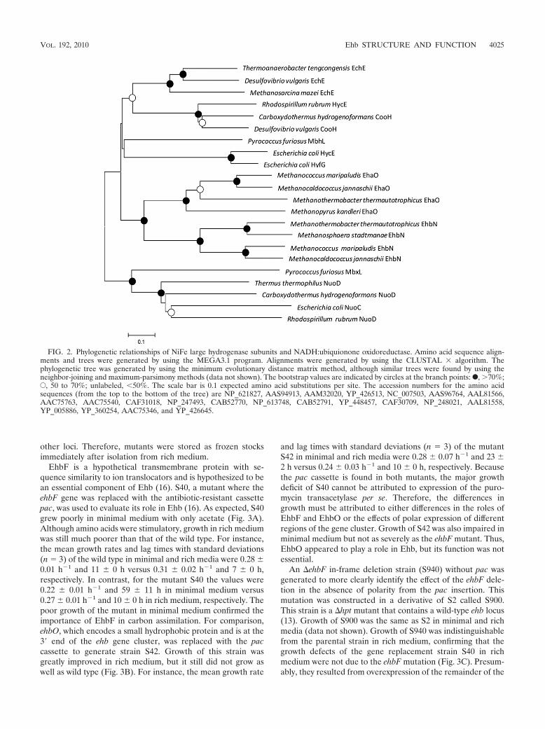
In the Methanosarcina barkeri Ech, 4Fe-4S clusters play cen-tral roles in electron transfer and, possibly, proton transloca-tion (4). In M. maripaludis, EhbK and EhbL contain the 4Fe-4Smotifs CX2CX2CX3C and are predicted to be a polyferredoxinand ferredoxin, respectively. To determine the importance ofEhbL in Ehb function, the deletion mutant �ehbL (S955) wasgenerated. In minimal medium, the growth of this mutant wasinhibited but not as severely as the �ehbF and �ehbN mutants(Fig. 5). Upon complementation of the mutation with the ex-pression vector for ehbL (strain S945), growth was actuallybetter than the wild type in minimal medium. Similarly, in richmedium the complemented strain possessed a shorter growthlag but longer generation time. Thus, expression of EhbL fromthe strong promoter in the pMEV vector had complex conse-quences under both growth conditions.
Although the double-mutant �ehbKL (S945) was isolated,an �ehbK mutant was not isolated after numerous attempts.Growth of the �ehbKL mutant in minimal medium was inter-
FIG. 3. Evaluation of gene replacement (�ehbF::pac and�ehbO::pac) and deletion (�ehbF) mutants of the conserved integralmembrane proteins in rich (McCA, closed symbols) and minimal(McA, open symbols) media. (A) Growth of the �ehbF::pac strain S40(triangles) and the parental strain S2 (circles). (B) Growth of the�ehbO::pac strain S42 (squares) and the parental strain S2 (circles).For panels A and B, the experiment was begun with �2 � 106 cellsupon inoculation into prewarmed media. (C) Growth of the �ehbFstrain S940 (triangles), strain S943 containing the �ehbF mutationcomplemented with pMEV2�ehbF (squares), and the parental strainS900 (circles). In these experiments, prewarmed McA media wereinoculated with �105 cells. Growth curves are representative of exper-iments that were performed at least twice in duplicate.
FIG. 4. Growth of deletion mutants of the small hydrogenase sub-unit (�ehbM) and the large hydrogenase subunit (�ehbN) in rich(McCA, closed symbols) and minimal (McA, open symbols) media.(A) Growth of the �ehbM strain S960 (squares), strain S963 containingthe �ehbM mutation complemented with pMEV2�ehbM (triangles),and the parental strain S900 (circles). (B) Growth of the �ehbN strainS965 (squares), strain S968 containing the �ehbN mutation comple-mented with pMEV2�ehbN (triangles), and the parental strain S900(circles). For other experimental conditions, see the legend to Fig. 3.
4026 MAJOR ET AL. J. BACTERIOL.
mediate between that of the wild type and �ehbF and �ehbNmutants and, in that regard, resembled that of the �ehbLmutant. Complementation of the �ehbKL with the ehbL generestored growth to wild-type levels in minimal medium but didnot yield the growth stimulation observed in the �ehbL back-ground. In rich medium, growth was somewhat impaired butnot to the same extent as in the �ehbL background (Fig. 5).These results suggest that EhbK is necessary for proper Ehbfunction even with EhbL overexpression.
The complex phenotypes of the �ehbL and �ehbKL mutantsand the ehbL overexpression strains suggest complex interac-tions for EhbL and EhbK in the cell. Although sufficient in-formation for definitive conclusions is lacking, this circumstan-tial evidence suggests that EhbL and EhbK interact. Theeffects of the overexpression of EhbL were greatly attenuatedin the double-deletion mutant �ehbKL, suggesting that EhbKmitigates the effects of EhbL. Second, �ehbK mutants wereonly isolated in the presence of a �ehbL mutation. If the�ehbK mutation is in fact lethal in the presence of normallevels of EhbL, as these results seem to suggest, it could beevidence for functional coupling where EhbL becomes toxic inthe absence of EhbK. Although it is premature to speculate onthe details, interactions of EhbK and EhbL, either with theEha homologs or with other enzyme systems, could be respon-sible for these complex phenotypes. In conclusion, althoughEhbL and EhbK both appear to be important in Ehb function,
it is not possible to assign their roles without additional exper-imentation.
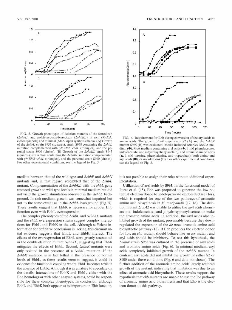
Utilization of aryl acids by S965. In the functional model ofPorat et al. (15), Ehb was proposed to generate the low po-tential electron donor to indolepyruvate oxidoreductase (Ior),which is required for one of the two pathways of aromaticamino acid biosynthesis in M. maripaludis (17, 18). The dele-tion mutant �iorA2 was unable to utilize the aryl acids phenyl-acetate, indoleacetate, and p-hydroxyphenylacetate to makethe aromatic amino acids. In addition, the aryl acids also in-hibited growth of the mutant, presumably because they down-regulated the expression of the de novo aromatic amino acidbiosynthetic pathway (18). If Ehb produces the electron donorfor Ior, an ehb mutant should behave like an ior mutant andaryl acids should be inhibitory. To test this hypothesis, the�ehbN strain S965 was cultured in the presence of aryl acidsand aromatic amino acids (Fig. 6). In minimal medium, arylacids completely inhibited growth of the �ehbN mutant. Incontrast, aryl acids did not inhibit the growth of either S2 orS900 under these conditions (Fig. 6 and data not shown). Thefurther addition of the aromatic amino acids largely restoredgrowth of the mutant, indicating that inhibition was due to aneffect of aromatic acid biosynthesis. These results support thehypothesis that ehb mutants are unable to use the Ior pathwayof aromatic amino acid biosynthesis and that Ehb is the elec-tron donor to this pathway.
FIG. 5. Growth phenotypes of deletion mutants of the ferredoxin(�ehbL) and polyferredoxin-ferredoxin (�ehbKL) in rich (McCA,closed symbols) and minimal (McA, open symbols) media. (A) Growthof the �ehbL strain S955 (squares), strain S958 containing the �ehbLmutation complemented with pMEV2�ehbL (triangles), and the pa-rental strain S900 (circles). (B) Growth of the �ehbKL strain S945(squares), strain S948 containing the �ehbKL mutation complementedwith pMEV2�ehbL (triangles), and the parental strain S900 (circles).For other experimental conditions, see the legend to Fig. 3.
FIG. 6. Requirement for Ehb during conversion of the aryl acids toamino acids. The growth of wild-type strain S2 (A) and the �ehbNmutant S965 (B) was evaluated. Media included complex McCA me-dium (F), McA medium containing aryl acids (}, 1 mM phenylacetate,indoleacetate, and p-hydroxyphenylacetate), and aromatic amino acids(Œ, 1 mM tyrosine, phenylalanine, and tryptophan), both amino andaryl acids (f), or no additions (E). For other experimental conditions,see the legend to Fig. 3.
VOL. 192, 2010 Ehb STRUCTURE AND FUNCTION 4027

Analyses of the proteome of the S40 strain had suggestedthat Vor was upregulated and that Ehb was involved inbranched-chain amino acid biosynthesis (16). Therefore, a sim-ilar growth experiment using branched-chain fatty acids wasperformed. However, a difference between growth in the pres-ence of the fatty acids or amino acids was not detected (datanot shown). Presumably, branched-chain fatty acids do notinhibit the de novo biosynthetic pathway in the same manner asthe aryl acids.
Structural characterization of Ehb. Originally identified byhomology to other membrane-bound hydrogenases and ge-netic screens (26, 30), the physical structure of Ehb has notbeen characterized. Purified membranes of M. maripaludiscontained high levels of hydrogenase activity, but this activitywas not diminished in S40, the ehbF::pac mutant (16). Al-though there are a number of possible explanations, one likelypossibility is that Ehb represented only a small fraction of thetotal hydrogenase activity. To test this possibility, whole pro-tein extracts of M. maripaludis were fractionated by Blue Na-tive PAGE (Fig. 7). EhbN, which encodes the large subunit ofthe hydrogenase, was identified in wild-type cells by its cross-reactivity with anti-EhbN antibodies (Fig. 7C). Hydrogenaseswere identified by activity staining, and a minor band of activityhad the same electrophoretic mobility as EhbN from wild-typebut not �ehbN mutant cells (Fig. 7B). Moreover, staining withsilver or Coomassie blue failed to reveal the protein complexeven at the fairly large amounts of protein necessary for theactivity staining and Western blotting (Fig. 7A and data notshown). Therefore, Ehb was not an abundant protein complexand represented only a minor component of the hydrogenaseactivity. This result was consistent with an anabolic role forEhb in these hydrogenotrophs.
In Blue Native PAGE, the native Ehb complex migratedwith an apparent molecular mass of �110 kDa (Fig. 7). How-
ever, upon summing the masses of all of the subunits listed inTable 1, a molecular mass of �360 kDa was expected. Becausethe migration in Blue Native PAGE is sensitive to the isoelec-tric point of the proteins in addition to charge (20), the appar-ent low molecular weight may be misleading. To test whetherEhbF, EhbK, EhbL, and EhbM were part of the complex, theformation of the Ehb complex was examined in the ehb mu-tants. On sodium dodecyl sulfate (SDS) gels, two proteins werereadily detected by Western blotting of extracts of wild-typecells with anti-EhbN antibodies (Fig. 8). However, only thelower-molecular-weight protein, whose size was identical tothat expected for EhbN, was absent in extracts of the �ehbNmutant. Therefore, this protein was identified as the authenticEhbN subunit, and the higher-molecular-weight band was across-reacting protein. Presumably, this protein was not ob-served in Western blotting of the Blue Native PAGE becauseits native form did not cross-react with the antibodies. In anycase, ehbN is the only gene in M. maripaludis with sufficientsequence similarity to the MAP peptide to be detected byBLAST searches. Therefore, it is unlikely that the cross-react-ing material is the subunit of another hydrogenase. EhbN wasreadily detected in cell extracts of all of the other ehb mutants.Thus, the levels of EhbN were not affected, even though thegrowth was severely impaired in some cases. However, only thewild-type cells contained the Ehb membrane complex identi-fied by Blue Native PAGE (Fig. 8B). Therefore, either thecomplex failed to form or was unstable during electrophoresisin these mutants.
The presence of Ehb in membranes was also determined byWestern blotting of the membrane fraction with anti-EhbNantibodies. EhbN was pelleted by centrifugation at 200,000 �
FIG. 7. Hydrogenase activity comigrates with Ehb. A 7% Blue Na-tive PAGE gel was prepared anaerobically and loaded with triplicatecell extracts of the wild-type S2 and the �ehbN mutant S965 (60 �g ofprotein in each lane). Lane 1, S2; lane 2, S965. Standard proteins (5 �gof each), including albumin from bovine serum (66 kDa, monomer;132 kDa, dimer), -amylase (200 kDa), and apoferritin (443 kDa),were used as molecular mass markers. After electrophoresis, the gelwas sliced into three pieces for silver staining (A), in-gel hydrogenaseactivity staining (B), and Western blotting with antibody targetingEhbN (C). The position of the Ehb complex (marked by arrows) isindicated by the band detected by Western blotting in lane 1 ofpanel C.
FIG. 8. Formation of the Ehb membrane complex in ehb mutants.Whole-cell extract samples were separated in an SDS–12% PAGE gel(20 �g of protein each lane) (A) and 7% Blue Native PAGE gela(B) (120 �g of protein each lane). Lane 1, S2 (WT); lane 2, S965(�ehbN); lane 3, S940 (�ehbF); lane 4, S955 (�ehbL); lane 5, S945(�ehbKL); lane 6, S960 (�ehbM). (C) Whole-cell extract, soluble frac-tion, and membrane fraction samples were separated in SDS–12.5%PAGE gel (20 �g of protein each lane). Lane 1, whole-cell extract ofS2 (WT); lane 2, soluble fraction of S2 (WT); lane 3, membranefraction of S2 (WT); lane 4, whole-cell extract of S965 (�ehbN). Pro-teins were then transferred to PVDF membranes. EhbN and proteincomplexes containing EhbN were subsequently detected with rabbitpolyclonal antiserum and a secondary goat anti-rabbit alkaline phos-phatase conjugate. The position of a 47-kDa marker protein in panelsA and C is given on the right.
4028 MAJOR ET AL. J. BACTERIOL.

g for 1 h, and only ca. 3% of the initial cross-reactive materialremained in the soluble fraction of cell extracts (Fig. 8C).Upon centrifugation of the membrane pellet in sucrose gradi-ents, cross-reactive material was recovered in the membranefraction.
These results provide direct evidence of the formation of amultisubunit Ehb membrane complex. However, they do notdistinguish between the possibilities that the Ehb proteins ei-ther participate as subunits or are required for biosynthesis orassembly of the complex. For instance, EhbK and EhbL mightbe required for insertion of key Fe-S clusters into an apoen-zyme, which would be unable to form a stable complex in theirabsence. If this were true, they might perform a similar role inother enzyme systems, which could explain the complex phe-notype of their deletion mutants. If these proteins are compo-nents of the complex, the molecular mass would be expected toexceed �360 kDa, the minimum size of a complex comprisedof one of each of these subunits.
It is unclear how a �ehb mutant grows autotrophically if theEhb is required to produce low potential electrons for severaloxidoreductases. Because these organisms possess a limitedcapacity to assimilate organic substrates such as pyruvate,these mutations might be expected to be lethal (9, 16, 31). Ifmutations in the large subunit of hydrogenase or the ion trans-locator completely inactivate Ehb as expected, then anotherenzyme system or pathway must compensate. Possibly, Ehapossesses low levels of Ehb functionality. Alternatively, Ehasubunits might function in heterologous complexes with Ehb.For instance, the intermediate phenotype of the �ehbM muta-tion could be explained if the small subunit of the Eha hydro-genase was capable of forming an active enzyme with EhbN.This hybrid complex might have low Ehb activity and be un-stable during electrophoresis.
A variety of energy-conserving hydrogenases have been de-scribed in both bacteria and archaea, such as Escherichia coli(Hyc and Hyf), the sulfate-reducing bacterium Desulfovibriogigas (Ech and Coo), the phototroph Rhodospirillum rubrum(Coo), the hyperthermophilic archaeon Pyrococcus furiosus(Mbh and Mbx), the aceticlastic methanoarchaeon Methano-sarcina barkeri (Ech), and the hydrogenotrophic methanoar-chaeaon Methanococcus maripaludis (Eha and Ehb). The func-tions of these enzymes are diverse: the oxidation of formate orcarbon monoxide in bacteria, the oxidation of glyceraldehyde3-phosphate in Pyrococcus, or the oxidation of acetate and thereduction of carbon dioxide in methanogenesis in Methanosar-cina. To group enzymes with such diverse functions wouldsuggest that there is a great deal of structural or sequencesimilarity. However, these enzyme systems vary widely in thenumber of genes (from 21 in the Methanococcus eha to 6 in theDesulfovibrio ech) and the percent sequence similarity betweenvarious protein homologs. Many energy-conserving hydroge-nases also possess subunits for which there are no homologs inthe other systems. Furthermore, the hydrogenotrophic methan-ogens have developed a unique form of energy-conservinghydrogenase that couples energy transduction to autotrophicanabolism. The primary function of the other energy conservinghydrogenases appears to be energy transduction via catabo-lism. From this general synopsis, it seems that the energy-conserving hydrogenases are loosely grouped. As more energy-conserving hydrogenases are identified and characterized,
understanding of the genetic, structural, and functional diver-sity of this group of enzymes is sure to increase.
ACKNOWLEDGMENT
This study was supported by grant DE-FG02-05ER15709 from theU.S. Department of Energy.
REFERENCES
1. Adams, M. W. W., and D. O. Hall. 1979. Purification of the membrane-boundhydrogenase of Escherichia coli. Biochem. J. 183:11–22.
2. Brodersen, J., G. Gottschalk, and U. Deppenmeier. 1999. Membrane-boundF420H2-dependent heterodisulfide reduction in Methanococcus voltae. Arch.Microbiol. 171:115–121.
3. Bult, C. J., O. White, G. J. Olsen, L. Zhou, R. D. Fleischmann, G. G. Sutton,J. A. Blake, L. M. FitzGerald, R. A. Clayton, J. D. Gocayne, A. R. Kerlavage,B. A. Dougherty, J. F. Tomb, M. D. Adams, C. I. Reich, R. Overbeek, E. F.Kirkness, K. G. Weinstock, J. M. Merrick, A. Glodek, J. L. Scott, N. S. M.Geoghagen, J. F. Weidman, J. L. Fuhrmann, D. Nguyen, T. R. Utterback,J. M. Kelley, J. D. Peterson, P. W. Sadow, M. C. Hanna, M. D. Cotton, K. M.Roberts, M. A. Hurst, B. P. Kaine, M. Borodovsky, H.-P. Klenk, C. M.Fraser, H. O. Smith, C. R. Woese, and J. C. Venter. 1996. Complete genomesequence of the methanogenic archaeon, Methanococcus jannaschii. Science273:1058–1073.
4. Forzi, L., J. Koch, A. M. Guss, C. G. Radosevich, W. W. Metcalf, and R.Hedderich. 2005. Assignment of the [4Fe-4S] clusters of Ech hydrogenasefrom Methanosarcina barkeri to individual subunits via the characterizationof site-directed mutants. FEBS J. 272:4741–4753.
5. Hendrickson, E. L., R. Kaul, Y. Zhou, D. Bovee, P. Chapman, J. Chung, E.Conway de Macario, J. A. Dodsworth, W. Gillett, D. E. Graham, M. Hackett,A. K. Haydock, A. Kang, M. L. Land, R. Levy, T. J. Lie, T. A. Major, B. C.Moore, I. Porat, A. Palmeiri, G. Rouse, C. Saenphimmachak, D. Soll, S. VanDien, T. Wang, W. B. Whitman, Q. Xia, Y. Zhang, F. W. Larimer, M. V.Olson, and J. A. Leigh. 2004. Complete genome sequence of the geneticallytractable hydrogenotrophic methanogen Methanococcus maripaludis. J. Bac-teriol. 186:6956–6969.
6. Jones, W. J., M. J. B. Paynter, and R. Gupta. 1983. Characterization ofMethanococcus maripaludis sp. nov., a new methanogen isolated from saltmarsh sediment. Arch. Microbiol. 135:91–97.
7. Kumar, S., K. Tamura, and M. Nei. 2004. MEGA3: integrated software formolecular evolutionary genetics analysis and sequence alignment. Brief.Bioinform. 5:150–163.
8. Kunkel, A., J. A. Vorholt, R. K. Thauer, and R. Hedderich. 1998. An Esch-erichia coli hydrogenase-3-type hydrogenase in methanogenic archaea. Eur.J. Biochem. 252:467–476.
9. Lin, W. C., and W. B. Whitman. 2004. The importance of porE and porF inthe anabolic pyruvate oxidoreductase of Methanococcus maripaludis. Arch.Microbiol. 179:444–456.
10. Major, T. A. 2006. The role of the energy conserving hydrogenase B inautotrophy and the characterization of sulfur metabolism in Methanococcusmaripaludis. Ph.D. dissertation. University of Georgia, Athens.
11. Meuer, J., S. Bartoschek, J. Koch, A. Kunkel, and R. Hedderich. 1999.Purification and catalytic properties of Ech hydrogenase from Methanosar-cina barkeri. Eur. J. Biochem. 265:325–335.
12. Meuer, J., H. C. Kuettner, J. K. Zhang, R. Hedderich, and W. W. Metcalf.2002. Genetic analysis of the archaeon Methansarcina barkeri fusaro revealsa central role for Ech hydrogenase and ferredoxin in methanogenesis andcarbon fixation. Proc. Natl. Acad. Sci. U. S. A. 99:5632–5637.
13. Moore, B. C., and J. A. Leigh. 2005. Markerless mutagenesis in Methano-coccus maripaludis demonstrates roles for alanine dehydrogenase, alanineracemase, and alanine permease. J. Bacteriol. 187:972–979.
14. Nesterenko, M. V., M. Tilley, and S. J. Upton. 1994. A simple modificationof Blum’s silver stain method allows for 30 minute detection of proteins inpolyacrylamide gels. J. Biochem. Biophys. Methods 28:239–242.
15. Nijtmans, L. G., S. M. Artal, L. A. Grivell, and P. J. Coates. 2002. BlueNative electrophoresis to study mitochondrial and other protein complexes.Methods 26:327–334.
16. Porat, I., W. Kim, E. L. Hendrickson, Q. Xia, Y. Zhang, T. Wang, F. Taub,B. C. Moore, I. J. Anderson, M. Hackett, J. A. Leigh, and W. B. Whitman.2006. Disruption of the operon encoding Ehb hydrogenase limits anabolicCO2 assimilation in the archaeon Methanococcus maripaludis. J. Bacteriol.188:1373–1380.
17. Porat, I., M. Sieprawska-Lupa, Q. Teng, F. J. Bohanon, R. H. White, andW. B. Whitman. 2006. Biochemical and genetic characterization of an earlystep in a novel pathway for the biosynthesis of aromatic amino acids andp-aminobenzoic acid in the archaeon Methanococcus maripaludis. Mol. Mi-crobiol. 62:1117–1131.
18. Porat, I., B. W. Waters, Q. Teng, and W. B. Whitman. 2004. Two biosyntheticpathways for aromatic amino acids in the archaeon Methanococcus maripalu-dis. J. Bacteriol. 186:4940–4950.
VOL. 192, 2010 Ehb STRUCTURE AND FUNCTION 4029

19. Schagger, H. 2001. Blue-native gels to isolate protein complexes from mito-chondria. Methods Cell Biol. 65:231–244.
20. Schagger, H., W. A. Cramer, and G. von Jagow. 1994. Analysis of molecularmasses and oligomeric states of protein complexes by blue native electro-phoresis and isolation of membrane protein complexes by two-dimensionalnative electrophoresis. Anal. Biochem. 217:220–230.
21. Schagger, H., and G. von Jagow. 1991. Blue native electrophoresis for iso-lation of membrane protein complexes in enzymatically active form. Anal.Biochem. 2:223–231.
22. Setzke, E., R. Hedderich, S. Heiden, and R. K. Thauer. 1994. H2:heterodis-ulfide oxidoreductase complex from Methanobacterium thermautotrophicum:composition and properties. Eur. J. Biochem. 220:139–148.
23. Smith, D. R., L. A. Doucette-Stamm, C. Deloughery, H. Lee, J. Dubois, T.Aldredge, R. Bashirzadeh, D. Blakely, R. Cook, K. Gilbert, D. Harrison, L.Hoang, P. Keagle, W. Lumm, B. Pothier, D. Qiu, R. Spadafora, R. Vicaire,Y. Wang, J. Wierzbowski, R. Gibson, N. Jiwani, A. Caruso, D. Bush, H. Safer,D. Patwell, S. Prabhakar, S. McDougall, G. Shimer, A. Goyal, S. Pietrok-ovski, G. M. Church, C. J. Daniels, J. Mao, P. Rice, J. Nolling, and J. N.Reeve. 1997. Complete genome sequence of Methanobacterium thermoau-totrophicum �H: functional analysis and comparative genomics. J. Bacteriol.179:7135–7155.
24. Stathopoulos, C., W. Kim, T. Li, I. Anderson, B. Deutsch, S. Palioura, W.Whitman, and D. Soll. 2001. Cysteinyl-tRNA synthetase is not essential for
viability of the archaeon Methanococcus maripaludis. Proc. Natl. Acad. Sci.U. S. A. 98:14292–14297.
25. Stojanowic, A., G. J. Mander, E. C. Duin, and R. Hedderich. 2003. Physio-logical role of the F420-non-reducing hydrogenase (Mvh) from Methanother-mobacter marburgensis. Arch. Microbiol. 180:194–203.
26. Tersteegen, A., and R. Hedderich. 1999. Methanobacterium thermoautotro-phicum encodes two multisubunit membrane-bound [NiFe] hydrogenases.Eur. J. Biochem. 264:930–943.
27. Thauer, R. K., A.-K. Kaster, H. Seedorf, W. Buckel, and R. Hedderich. 2008.Methanogenic archaea: ecologically relevant differences in energy conserva-tion. Nat. Rev. Microbiol. 6:579–590.
28. Tumbula, D. L., R. A. Makula, and W. B. Whitman. 1994. Transformation ofMethanococcus maripaludis and identification of a PstI-like restriction sys-tem. FEMS Microbiol. Lett. 121:309–314.
29. Whitman, W. B., J. S. Sheih, S. H. Sohn, D. S. Caras, and U. Premachan-dran. 1986. Isolation and characterization of 22 mesophilic methanococci.Syst. Appl. Microbiol. 7:235–240.
30. Whitman, W. B., D. L. Tumbula, J.-P. Yu, and W. Kim. 1997. Developmentof genetic approaches for the methane-producing archaebacterium Methano-coccus maripaludis. BioFactors 6:37–46.
31. Yang, Y. L., J. Gluska, and W. B. Whitman. 2002. Intracellular pyruvate fluxin the methane-producing archaeon Methanococcus maripaludis. Arch. Mi-crobiol. 178:493–498.
4030 MAJOR ET AL. J. BACTERIOL.
Copyright © 2022 FDOKUMEN


![The mechanism of hydrogen uptake in [NiFe] hydrogenase: first-principles molecular dynamics investigation of a model compound](https://static.fdokumen.com/doc/165x107/6345b765df19c083b108312d/the-mechanism-of-hydrogen-uptake-in-nife-hydrogenase-first-principles-molecular.jpg)