Chapter 3 Membrane Nanotubes in Urothelial Cell Line T24
31
Provided for non-commercial research and educational use only. Not for reproduction, distribution or commercial use. This chapter was originally published in the book Advances in Planar Lipid Bilayers and Liposomes, Vol. 10, published by Elsevier, and the attached copy is provided by Elsevier for the author's benefit and for the benefit of the author's institution, for non- commercial research and educational use including without limitation use in instruction at your institution, sending it to specific colleagues who know you, and providing a copy to your institution’s administrator. All other uses, reproduction and distribution, including without limitation commercial reprints, selling or licensing copies or access, or posting on open internet sites, your personal or institution’s website or repository, are prohibited. For exceptions, permission may be sought for such use through Elsevier's permissions site at: http://www.elsevier.com/locate/permissionusematerial From: Maruša Lokar, Šárka Perutková, Veronika Kralj-Iglič, Aleš Iglič, and Peter Veranič, Membrane Nanotubes in Urothelial Cell Line T24. In A. Leitmannova Liu and Aleš Iglič, editors: Advances in Planar Lipid Bilayers and Liposomes, Vol. 10, Burlington: Academic Press, 2009, pp. 65-94. ISBN: 978-0-12-374823-2 © Copyright 2009 Elsevier Inc. Academic Press.
Transcript of Chapter 3 Membrane Nanotubes in Urothelial Cell Line T24

Provided for non-commercial research and educational use only. Not for reproduction, distribution or commercial use.
This chapter was originally published in the book Advances in Planar Lipid Bilayers and Liposomes, Vol. 10, published by Elsevier, and the attached copy is provided by Elsevier for the author's benefit and for the benefit of the author's institution, for non-commercial research and educational use including without limitation use in instruction at your institution, sending it to specific colleagues who know you, and providing a copy to your institution’s administrator.
All other uses, reproduction and distribution, including without limitation commercial reprints, selling or licensing copies or access, or posting on open internet sites, your personal or institution’s website or repository, are prohibited. For exceptions, permission may be sought for such use through Elsevier's permissions site at:
http://www.elsevier.com/locate/permissionusematerial
From: Maruša Lokar, Šárka Perutková, Veronika Kralj-Iglič, Aleš Iglič, and Peter Veranič, Membrane Nanotubes in Urothelial Cell Line T24. In A. Leitmannova Liu and Aleš Iglič, editors:
Advances in Planar Lipid Bilayers and Liposomes, Vol. 10, Burlington: Academic Press, 2009, pp. 65-94.
ISBN: 978-0-12-374823-2 © Copyright 2009 Elsevier Inc.
Academic Press.

Author’s personal copy
C H A P T E R T H R E E
A
IS
1
2
3
*E
dvance
SN 1
LaborSI-10LaborVrazoInstitSlove
Corr-mail a
Membrane Nanotubes in
Urothelial Cell Line T24
Marusa Lokar,1,* Sarka Perutkova,1 Veronika Kralj-Iglic,2
Ales Iglic,1 and Peter Veranic3
Contents
1. In
s in
554
ato00atov trutenia
espoddr
troduction
Planar Lipid Bilayers and Liposomes, Volume 10 # 2009
-4516, DOI: 10.1016/S1554-4516(09)10003-0 All rig
ry of Biophysics, Faculty of Electrical Engineering, University of Ljubljana, Trzaska 2Ljubljana, Sloveniary of Clinical Biophysics, Institute of Biophysics, Faculty of Medicine, University of Lg 2, SI-1000 Ljubljana, Sloveniaof Cell Biology, Faculty of Medicine, University of Ljubljana, Lipiceva 2, SI-1000 Lju
nding author. Tel.: þ 386 1 4768 235;ess: [email protected]
Else
hts
5,
jub
blj
66
2. T
24 Cell Line and Membrane Nanotubes 722
.1. T ypes of Membrane Nanotubes, Their Structuraland Functional Properties
722
.2. V esicular Dilatations on Membrane Nanotubes 783. F
ormation and Stability of Type I Membrane Nanotubes 823
.1. O n the Role of Small Anisotropic Protein–Lipid Nanodomainsin Formation and Stabilization of Membrane Nanotubes
854. C
oncluding Remarks 91Refe
rences 91Abstract
Membrane nanotubes (also referred as tunnelling nanotubes—TNTs, nanotu-
bules, cytonemes), that directly connect separated neighboring cells, may offer
a very specific and effective way of intercellular transport and communication.
Our experiments on T24 cell line show that TNTs can be divided into two types
with respect to their biochemical and biophysical characteristics and the nature
of their formation. As type I were characterized the nanotubes which are
shorter, more dynamic and contain actin filaments. These structures remain
stabile even if underlying actin cytoskeleton is disintegrated by cytochalasin D.
The nanotubes of type II are much longer, appear more stable and contain
cytokeratin filaments. In both types microtubules can be found, but this type of
cytoskeleton is present in only a small fraction of the TNTs. On the nanotubes of
vier Inc.
reserved.
ljana,
ana,
65

66 M. Lokar et al.
Author’s personal copy
both types small vesicular dilatations were found as an integral part of the
nanotubes (i.e., dilatations of the nanotubes, gondolas). Vesicular dilatations of
type I nanotubes move along the nanotubes in both directions, while the
vesicular dilatations of type II nanotubes do not move along the nanotubes.
Both TNTs by themselves and the transporting gondolas were proposed to be
involved in intercellular communication and transport. The possible mechan-
isms of stabilization of membrane nanotubular protrusions and TNTs are also
discussed.
Abbreviations
CMFDA
5-Chloromethylfluorescein diacetate DiD 1,10-Dioctadecyl-3,3,3,30-tetramethylindodicarbocyanine
DiI 1,10-Dioctadecyl-3,3,3030-tetramethylindocarbocyanine
DiO 3,30-Dilinoleyloxacarbocyanine EFGP Enhanced green fluorescent protein FITC Fluorescein isothiocyanate GFP Green fluorescent protein GPI Glycosyl-phosphatidylinositol HLA, -B, -C Human leukocyte class I antigens TNTs Tunneling nanotubes TRITC Tetramethylrhodamine isothiocyanate1. Introduction
Cell-to-cell communication is one of the fundamental processes in thedevelopment and homeostasis of multicellular organisms. For this purposeorganisms have evolved diverse mechanisms to communicate on the level ofconnected or/and spatially separated cells. The most common mechanismsdepend on secretion of diffusible signal molecules (like hormones, growthfactors) that bind to specific receptors in/on target cells [1]. A few years agoa novel type of cell-to-cell connection was discovered, where two spatiallyseparated cells are connected by a long, thin tubular membranous struc-tures [43]. They were named tunnelling nanotubes (TNTs) and are nowknown as membrane nanotubes. The membrane nanotubes were found incultures and cocultures of both permanent cell lines and primary cultures(see Tables 1 and 2), mostly between cells that are weakly connected to each

Table 1 Human and animal cell lines shown to form TNTs and their characteristics
Cell lines forming TNTs Morphology Transferred cellular components (markers)
(From)
F (diameter)
l (length) Prevalence; form
Cytosolic
continuity Junctiona
Cytoskeleton
(filament binding
proteins)
Cytoplasmic
components
(dyes)
Membrane
components
(dyes)
Rat pheochromacytoma
cells (PC12) [43]
F ¼ 50–200 nm
l ¼ 16–60 mmFew; straight,
rarely
branched,
above
substratum
ND Yes F-actin (myosin Va) EGFP-actin f-EGFP
Human embionic kidney
cells (Hek-293) [15]
ND Few ND ND F-actin (myosin Va) ND (DiI, DiO, DiD)
Normal rat kidney cells
(NRK) [15]
ND Many: form ND ND ND ND Endocytic
organelles
(DiD)
(DiI, DiO, DiD)
Primary human
T-cells [48]
F ¼ 180–
380 nma
l ¼ 30–50 mm,
some�200 mm
Many; straight,
curved in 3D
environment,
rarely
branched,
above
substratum
Nob Yes F-actin ND ND
Jurkat T-cells [48] F ¼ ND
l ¼ � 22 mm,
some
� 100 mm in a
3D mimic of
ECM
Many: straight,
curved in 3D
environment,
rarely
branched,
above
substratum
Nob Yes F-actin GFP, (CFDA,
calcein)
GFP-I-CAM1,
GFP-HLA-
Cw7, GPI-
GFP (DiO,
DiD), HIV-1
Gag-GFP
(continued)
Author’s personal copy

Table 1 (continued)
Cell lines forming TNTs Morphology Transferred cellular components (markers)
(From)
F (diameter)
l (length) Prevalence; form
Cytosolic
continuity Junctiona
Cytoskeleton
(filament binding
proteins)
Cytoplasmic
components
(dyes)
Membrane
components
(dyes)
EBV-transformed human
B cell line (721.221)
[41]
ND ND ND ND ND ND GPI-GFP,
HLA-Cw6 in
coculture
with human
peripheral
blood NK
cells
Transitional cell
carcinoma of urinary
bladder cells (T24) [51]
F¼ 60–200 nm
l ¼ most
< 30 mm,
some
� 120 mm
Many: multiple,
straight, on
the
substratum,
rare above
substratum
Yesb Yes Only F-actin
cytokeratin 7,
F-actinþmicrotubules
(a-tubulin)
Actin-GFP,
(CMFDA)
Choleratoxin-
GFP
(DiI, DiO)
Transitional cell
papillomaa of urinary
bladder cells (RT4)
(Lokar et al.,
unpublished)
ND Few, on the
substratum
ND ND F-actin ND ND
DU 145 human prostate
cancer cells [52]
F: thinner100–200 nm,
thicker
(�1 mm)
l ¼ few mm –
100 mm)
Yes ND Microtubules Mitochondria
(MitoTracker)
lysosomes
ND
Human glioblastoma cells
(U-87 MG) [42]
F ¼ 42–54 nm
l ¼ tens of mmStraight ND ND F-actin Vesicle
trafficking
under stress
conditions
ND
Author’s personal copy

THP-1 monocytes [40] F ¼ � 35–
250 nm
l ¼ <100 mm
Many Yesb ND F-actin Ca2þ (Lucifer
Yellow)
HLA-A,B, C
class I MHC
Human hepatic cells
(Hep G2) [54]
ND Few ND ND ND ND ND
Dendritic cells from
peripheral blood
monocytes [53]
F ¼ � 35–
250 nm
l ¼ <100 mm
Many Yesb ND ND Ca2þ (Lucifer
Yellow)
HLA-A,B, C
class I MHC
Human monocyte-
derived macrophages
[40, 41]
thin (<0.7 mm)
thick (>0.7 mm)
Many: thick
prevalent,
most are
connecting
apical parts of
the cells,
some are
branched
ND No F-actin, F-actin þmicrotubules
Endosomes
(DiD)
mitochondria
F ¼ (Mito
Tracker)
lysosomes
(LysoTracker),
EB1-GFP
ND
Primary rat
astrocytes [17]
F ¼ � 100 nm
l ¼ several mmMany, branched ND ND F-actin ND ND
l ¼ <1 mml ¼ >100 mm
Hematopoetic stem
and progenitor
cells [14]
No data No data
Bovine mammary
gland epithelial cells
(BMGE) [54]
No data No data
Primary cultures
of mouse
medulla [15]
No data No data
Murine macrophage
J477 cells [40]
No data No data
ND — not determined.a Was assessed by transmission electron microscopy.b Was proved indirectly by measuring calcium fluxes.
Author’s personal copy

Table 2 Cocultures of cell lines shown to form TNTs between different cell types
Coculture (from)
Transferred cellular
component (dye)
PC12 and NRK cells [15] Endosome related cell
organelle (DiI, DiO, DiD)
PC12 and Hek-293 cells [15] Endosome related cell
organelle (DiI, DiO, DiD)
Hek-293 and NRK cells [15] Endosome related cell
organelle (DiI, DiO, DiD)
722.221 and NK cells cells [40] Surface receptor
HLA-Cw6-GFP
macrophages and NK cells [40] ND, seen upon separation
Neonatal rat cardiomycites and adult human
endothelial progenitor (EPC) cells [29]
Mitochondria
(MytoTracker), GFP
Dendritic and THP-1 cells [40] Ca2þ
70 M. Lokar et al.
Author’s personal copy
other or in those which are actively migrating and seeking for bacteria orattachment to neighboring eukaryotic cells. Nanotubes exist also in cellswith limited ability of movement and strong intercellular connections likeepithelial cells. The diameter of these membrane nanostructures ranges from30 to 400 nm, in some cases up to 1 mm, and their length can span for morecell diameters, depending on the cell type. They are versatile in ultra-structure and formation, and consequently also in function (reviewed byRefs. [9, 15]). Although TNTs are versatile, they share some commonfeatures like the presence of cytoskeleton and cytoskeleton-associated pro-teins. Through them different cellular material is being transported—fromsignaling molecules, soluble cytoplasmic proteins, membrane proteins andcell organelles [15, 16, 20, 21, 43, 51], to viruses [46, 47, 49] and bacteria.Viruses and bacteria are being transported only upon nanotube surfaces [41].
There is a considerable heterogeneity present between membrane nano-tubes. They differ in their cytoskeletal composition, diameter, length,proposed function, ability to form cytoplasmic continuity. Ongoing studiesof relatively young field in different cellular models are focused primarily onmorphology, transported material and their frequency, but little is knownabout their biophysical properties, what factors influence their formation,what is the driving force of their formation, which protein complexes areinvolved in their dynamics and initiation of cell-to-cell contacts.
Considering their heterogeneity it is not an easy task to subdivide themubiquitously into distinct types, since membrane nanotubes differ muchfrom one cell type to another and their properties sometimes overlapbetween designated forms. Our classification of membrane nanotubes, inaccordance with other authors, is presented in Fig. 1.

Membrane nanotubes
Actin containingtypically up to a cell diameter long
Cytosolic continuity
Tunneling nanotubes
No membrane continuity
Cargo transferred:
Through the channel:- by selective transport- via acto-myosin transportsystem
Along continous outermembrane:- bound to membraneresident species- ordered patches ofmembrane
Membrane continuity
Cytonemes
Junction to target cell is present
No junction to target cell No microtubules
Components of cytoplasm present
No data for the presence of microtubules
No cytosolic continuity
Filopodial-likeprotrusions
Cargo transferred via:- bound to membrane resident species- ordered patches of membrane alongthe outer surface of the membrane
Type ICytokeratin containing
typically several cell diameters long
Have microtubules (shorter)a
No microtubules
Type II
Central channel(no data on the nature of the channel)
Figure 1 Classification and forms of membrane nanotubes. Criteria used for classification and their main properties are briefly described.Additional remarks are described in brackets. aUnpublished observation.
Author’s personal copy

72 M. Lokar et al.
Author’s personal copy
The focus of this chapter will be on the properties and formation ofnanotubes found in urothelial cell line T24. In our studies phase contrast,fluorescence, time-lapse and electron microscopy were employed to studystructural characteristics, formation, stability and dynamics of nanotubes thatbridge two neighboring urinary bladder epithelial cells T24. Theoreticalmodels of mechanical stabilization of membrane nanotubes are suggested.
2. T24 Cell Line and Membrane Nanotubes
Cells in a permanent cell line isolated from transitional cell carcinomaof urinary bladder epithelia are heterogenous, consisting of at least twomorphologically distinct types [4]. Cells grow in a disorderly manner,forming a monolayer of cells, where individual cells are lying partly overone other. Cell population consists of two main types of cells: (i) large cellswith large, round, light nuclei with scarce chromatin and numerousnucleoli, abundant and slightly pyronin-positive cytoplasm (pyronin is aribonucleic acid dye) which is rather poor in organelles, with a few mito-chondria and ribosomes; (ii) elongated cells with oval, darker nuclei, withmany protrusions, abundant in chromatin and several nucleoli, morestrongly pyroninphylic cytoplasm with numerous mitochondria and abun-dant endoplasmic reticulum. Cells contain numerous vacuoles of differentsizes with inclusion bodies sometimes present. Cell membrane hasmany protrusions, especially at the basal edges of the cells, with nanotubesconnecting cells close together (Fig. 2).
Membrane nanotubes are readily formed only between closely posi-tioned cells of a subconfluent culture, where cells have a certain degree offreedom to move around on the substratum. The most appropriate condi-tions for studying their properties were found to be 70–80% confluentovernight cultures grown on glass coverslips, where cells were not yetmitotic and have enough space to form membrane protrusions.
2.1. Types of Membrane Nanotubes, Their Structuraland Functional Properties
In subconfluent culture of T24 cells two distinct types of membranenanotubes were identified and classified with respect to their cytoskeletalcontent, origin, stability and consequently their proposed function (Fig. 3).
2.1.1. Type I nanotubesType I nanotubes are shorter, usually not longer than 30 mm, dynamicstructures and contain actin filaments. Actin filaments give them theirdynamic properties. The protruding type I nanotubes are formed when a

BA
Figure 2 T24 cell line, grown in normal conditions. Cells are growing partly one overother. Note that the membrane of the cells on the edge of the cell island (indicated byarrowhead in (A) are not and firmly attached to the substratum along entire edge of thecell body, but at some places rather undulating with the cell membrane floating in themedium. Cells in the middle of the cell island are tightly connected with tubularconnection forming between separating cells. Cells have numerous vacuoles (gray-white circular structures in cells, marked by white arrow in (B) and inclusion bodies(black circular structures in cells, marked by black arrows). Individual cells areconnected by membranous nanotubes (wider white arrow).
Figure 3 T24 cell stained for actin (gray) and cytokeratin (white) filaments. White arrows arepointing at multiple, acting containing type I nanotubes, whereas gray arrow is pointingat longer, cytokeratin containing type II nanotube (see also Fig. 11).
Membrane Nanotubes in Urothelial Cell Line T24 73
Author’s personal copy
cell explores its surroundings, through a thin filopodial-like tubular mem-brane protrusion extending from basal part of the cell body in order to makecontact with another cell (Figs. 4 and 5). This type of actin-containingnanotubes can also bridge cells at distances of less than 30 mm and is mostlikely derived from the adherence cell–cell contacts of cells that move apartas they appear higher on the cell body (Fig. 6). Type I nanotubes have nocytokeratin filaments (Fig. 5). Microtubules are present in some of type Inanotubes (Figs. 6B and 7). The primary function of these nanotubes seemsto be intercellular communication by initiation of cell-to-cell contacts and

A B
C1 C1
C2
C3 C3
5 mm5 mm
C2
Figure 4 Type I nanotubes. (A) is a phase contrast image of live T24 cells while (B) is afluorescence micrograph showing actin-TRITC labeling of the same cells as in A, after15 min of paraformaldehyde fixation. Cell C1 is approaching the cells C2 and C3 (foranimation see Supplementary material I in Ref. [51]). The white arrows in (A) and (B)indicate short and dynamic membrane protrusion with which the approaching cellexplores its surroundings. Black arrow in (A) points at protrusions that have alreadyconnected to the target cell. In all these multiple tubular connections actin filaments arepresent (arrow in B). Adapted from Veranic et al. [51].
B
10 mm
A
Figure 5 T24 cells stained for actin filaments with phalloidin-FITC (A) and cytoker-atin filaments with anti-cytokeratin 7 antibodies (B). Arrows (in A and B) are indicatingtype I containing nanotubes that bridge two cells. These structures have no cytokeratinfilaments.
74 M. Lokar et al.
Author’s personal copy
subsequent transport of cellular components from one cell to another once acontact has been made. Their formation and stability is discussed inSection 3.

B
10 mm
A
Figure 6 T24 cells stained for actin filaments with phalloidin (A) and microtubuleswith anti-alpha-tubulin antibodies (B). White arrows are pointing at actin containingnanotube connecting two separated cells. Dashed arrows are pointing at dynamic actingcontaining nanotubes with which cell is exploring its surroundings. These structureshave no microtubules. Arrows in (B) are pointing at the same parts as in (A).
10 mm
10 mm
DC
BA
Figure 7 T24 cells stained for actin filaments with phalloidin (A and C) and micro-tubules with anti-alpha-tubulin antibodies (B and D). In type I nanotubules, that searchthe surroundings (indicated with dashed arrows in (A) and corresponding places in (B))or connecting two neighboring cells (indicated with arrows in (A) and correspondingplaces in (B)), there is no microtubules. Microtubules can be found in intercellularconnections of cells that are separating (arrows in (C) and corresponding places in (D)).See also Fig. 12A and B.
Membrane Nanotubes in Urothelial Cell Line T24 75
Author’s personal copy
The protruding type I nanotubes start growing as filopodia at the basallevel of the cell surface and continue to grow until they reach the target cell(Fig. 5), where the nanotube can attach by an anchoring type of intercellular

500 nm
Figure 8 A TEM micrograph showing an anchoring type of intercellular junction(arrow) connecting a nanotubule to the protrusion of a neighboring cell. Adapted formVeranic et al. [51].
76 M. Lokar et al.
Author’s personal copy
junction [43, 51] (Fig. 8). However, it is not much known about the natureof these contacts. The connection via nanotubules may not be initiateddirectly by contact of the tip of the membrane nanotube with the targetmembrane, but may require a nanotube first to slide along target cellmembrane and connect to the cell via several adherens junctions at thelateral region of nanotube (Fig. 8) before the cytosolic continuity can beestablished via communication junction at the nanotube tip. These adhesioncontacts might also be necessary for the nanotube to stabilize the contactwith the target cell when it is ‘‘searching’’ for appropriate docking site forthe communication junction to be formed.
Proteins responsible for formation of lateral connection of the nanotubesto the target cell most likely belongs to cadherin family, since these proteinswere found at both tips and tubular regions of type I nanotubular protru-sions (Fig. 9).
Nanotubes in T24 cell line can mediate cytosolic continuity [16, 51]even though no exchange of membrane labels could be found between cells[51] (Fig. 10).
2.1.2. Type II nanotubesIn comparison to abundant type I nanotubes, type II nanotubes are quitescarce. They are much longer, up to several 100 mm and appear to be morestable (Fig. 11). They are usually located more apically on the cell body. TypeII nanotubes differ from all previously described nanotubes (which can bedetermined as type I nanotubes) by having no actin filaments but onlycytokeratin filaments, which are probably responsible for their stability [37]and longer life span. Actin network is still present at both ends of the

* *
C2 C1 C1C2
BA
10mm
Figure 9 T24 cells stained for actin filaments with phalloidin-FITC (A) and anti-pan-cadherin antibodies (B). Cell C1 is crawling upon cell C2. Cadherins are present atcell–cell contacts (arrow) and actin containing nanotubes (asterix), even though notat all tips of the nanotubes.
10mm10mm
B
CA
Figure 10 (A) Urothelial cells T24 labeled with lipophilic stain DiI were coculturedwith unlabelled T24 cells. The nanotubes (arrow) of stained cells (white) becameprotruded and attached to unstained cells (gray) in 3 h. However, even after 24 h DiIstain did not spread to the connected cells. (B) and (C) Live urothelial cells T24colabeled with lipophilic membrane stain DiI (B) and cytoplasmic dye CMFDA (C).Nanotubes are mediating cytoplasmic connection between two neighboring cells(arrows). Adapted from Veranic et al. [51].
Membrane Nanotubes in Urothelial Cell Line T24 77
Author’s personal copy
nanotube, but as the nanotube narrows actin disappears. They may be formedwhen two already connected cells start to move apart (Fig. 12A and B). As isthe case of type I nanotubes, microtubules are present in only some of type IInanotubes, mainly those which connect separating cells (Fig. 12C).
C
c1
10mm20mm50mm
c2BA
Figure 11 In urothelial line T24 a long tubular structure connects cells of the two cellclusters C1 and C2 (A). (B) is a magnified region of the area in the black frame in (A).Such long singular tubes of type II contain thin cytokeratin filaments (arrow in (C)).In C cytokeratin 7 is labeled in white, and actin in gray. From Veranic et al. [51].
C
10 mm 10 mm
BA
Ccl
C1 C1
C2
C
C2
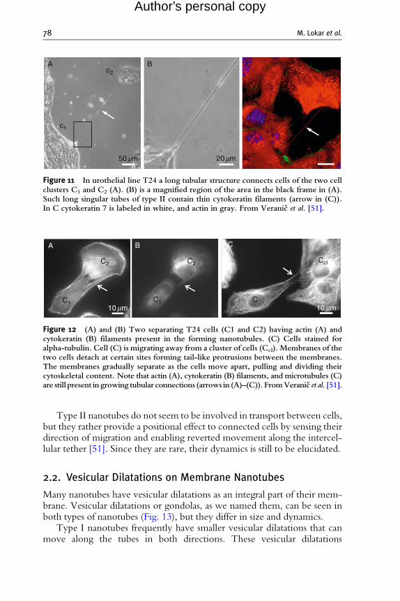
Figure 12 (A) and (B) Two separating T24 cells (C1 and C2) having actin (A) andcytokeratin (B) filaments present in the forming nanotubules. (C) Cells stained foralpha-tubulin. Cell (C) is migrating away from a cluster of cells (Ccl). Membranes of thetwo cells detach at certain sites forming tail-like protrusions between the membranes.The membranes gradually separate as the cells move apart, pulling and dividing theircytoskeletal content. Note that actin (A), cytokeratin (B) filaments, and microtubules (C)are still present in growing tubular connections (arrows in (A)–(C)). FromVeranic et al. [51].
78 M. Lokar et al.
Author’s personal copy
Type II nanotubes do not seem to be involved in transport between cells,but they rather provide a positional effect to connected cells by sensing theirdirection of migration and enabling reverted movement along the intercel-lular tether [51]. Since they are rare, their dynamics is still to be elucidated.
2.2. Vesicular Dilatations on Membrane Nanotubes
Many nanotubes have vesicular dilatations as an integral part of their mem-brane. Vesicular dilatations or gondolas, as we named them, can be seen inboth types of nanotubes (Fig. 13), but they differ in size and dynamics.
Type I nanotubes frequently have smaller vesicular dilatations that canmove along the tubes in both directions. These vesicular dilatations

10mm
Ccl
C1
*
Figure 13 A scanning electron micrograph of T24 cell line. Nanotubular structures connectadjacent cells between cell cluster (Ccl) and cell C1. Cell C1 radially extends many thinfilopodial-like tubular protrusions upon the substratum in the leading part of cell(asterix). It is connected to cell cluster by a longer nanotube, located higher on thecell body. On all nanotubes vesicular dilatations are present (arrows).
1.5 mm 0.5 mm
0 s 3 s 9 s6 s 12 s
Figure 14 Vesicular dilatation (gondola) on type I membrane nanotube (arrow). Fusion of agondola (black arrow) with a cell body is seen after a time-lapse sequence showingdirectional movement of the gondola along a nanotube. The time sequence in secondsis indicated on the upper left side of each micrograph. Adapted from Veranic et al. [53].
Membrane Nanotubes in Urothelial Cell Line T24 79
Author’s personal copy
(gondolas) move for 5–15 mm in certain direction with an average speed of40 nm/s [51]. They sometimes appear in the middle of the nanotube andtravel along the nanotube until they fuse with the cell body (Fig. 14).
On the other hand, the dilatations on type II nanotubes are larger,usually placed in the middle of the nanotube and do not move along thenanotube (Fig. 15).
2.2.1. Possible origins of vesicular dilatations and mechanismsof their propagation along nanotubes
The observed vesicular dilatations of the nanotubes (gondolas) movingalong the type I nanotubes (Fig. 16) may be formed in different ways.In some cases the formation of gondolas, corresponding to transient excitedstates, may be induced by a sudden tension (caused, e.g., by diverging or

Gondolas
Figure 15 Vesicular dilatations (gondolas) on type II membrane nanotubes.
A B C
Figure 16 Possible origins of gondola formation and its movement along a nanotube. Thedirection of its movement is indicated by arrows. The distention of the nanotubule(gondola) may be formed in different ways. In illustration (A) the distention is formedbecause of sudden tension (caused by diverging cells) in the membrane at specificsites on the nanotube, where local constituents enable and favor the formation of thisstructure. This kind of distention may appear anywhere along the nanotube and travelsin the direction that is energetically favorable. In illustrations (B) the distention isformed because material inside the gondola is actively transported along the filamentsby motor proteins ([41, 43]). The total volume of the enclosed material (an organelle ora vesicle) is larger than the inner diameter of the nanotube. Transported material(multiple small particles moving synchronously within the distension) may be enclosedwithin gondola or may be a part of the gondola membrane. Illustration C schematicallyindicates nanotubule-directed movement of the gondola, formed in the buddingprocess.
80 M. Lokar et al.
Author’s personal copy
approaching cells) in the membrane nanotubes at specific sites where thelocal membrane constituents of the nanotubes enable and favor the forma-tion of such dilatations. The tension-induced dilatation of the nanotubesmay appear anywhere along the nanotube and then travels as a wave along

Membrane Nanotubes in Urothelial Cell Line T24 81
Author’s personal copy
the nanotube in the direction that is energetically favorable. The tensionmight be the most probable reason of gondolas that suddenly appear in themiddle of the nanotube [51]. These tension-induced dilatations are as anyother excited states of the membrane relaxed after a certain time. It has beenreported that slight undulations are relaxed in seconds while sphere-likeblobs are relaxed in minutes [2].
The vesicular dilatations of the nanotubes may also be formed because ofa small organelle, vesicle or supramolecular assembly (multiple small parti-cles moving synchronously within the distension) is being transported insidethe nanotubes, if their effective diameter is larger than the inner diameter ofthe nanotube [15, 27, 51]. The material inside the nanotubes may beactively transported by different actomyosin-dependent mechanisms([15, 40], reviewed by Ref. [8]). These dilatations are forming at thebeginning of the nanotube of one cell as a vesicle-like structures, aretransported along the nanotube and are then released into the cytoplasmof the second cell (see Fig. 17).
The observed vesicular dilatations of the nanotubes moving along themembrane nanotubes of type I show striking similarity to the dilatations ofphospholipid nanotubes, which move along these nanotubes [24]. There-fore it is also possible that the initiation of gondola formation (Fig. 17A) maybe based on similar physical mechanisms as those governing the formationof free membrane daughter vesicles, which are created in the processes ofbudding. In contrast to the latter process however, in gondolas, the con-nection to the parent membrane, from which they originate is not disruptedwhen the dilatation is detached from the parent cell (Fig. 17B). Once thegondola is formed, its movement along the nanotube (Fig. 17C) requires noadditional bending energy. Nevertheless, a mechanism is still needed toprovide the energy for the dilatation to travel along the nanotube. It ispossible that the gondola movement is driven by the difference in chemicalpotentials between the molecules packed inside the gondola and the
A B DC
Figure 17 Schematic illustration of nanotubule-directed transport of small vesiculardilatations (gondolas) transporting granular content and membrane particles.

82 M. Lokar et al.
Author’s personal copy
molecules in the interior of the target cell, or the difference in chemicalpotential between the molecules composing the membrane of the gondolaand the molecules in the membrane of the target cell. The final event of thetransport is the fusion of the gondola with the target membrane [24]. In thisprocess, molecules of the gondola’s membrane which originate from theparent, nearly flat membrane, redistribute again in an almost flat target mem-brane (Fig. 17D). This may be energetically favorable and therefore also part ofa driving mechanism to facilitate fusion of the gondola with the membrane.Prior to fusion of the gondola with the target cell membrane, no neckformation is needed (Fig. 17C) since the neck is already part of the nanotubeconnecting the gondola to the membrane of the target cell. This is contrary tothe case of a free transport vesicle. It can therefore be concluded that thetransport of material in dilatations (or the transport of molecules composingthemembrane of dilatations) may bemore efficient, since it is guided either byactomyosin transport system or passive diffusion along the nanotube, andtherefore energetically advantageous over free vesicle transport.
3. Formation and Stability of Type I
Membrane Nanotubes
Membrane nanotubes are thin, dynamic structures, but nevertheless atleast transiently stable structures. We presume they are formed on (andbetween) specific surface regions on the cell membrane where the localenvironment favors their molding and stabilization (or attachment). Thereare several factors that influence their dynamics and stability, the majorplayers being cytoskeleton and membrane constituents.
Although the underlying cytoskeleton greatly influences their shape,molding and mechanical stability, its primary role may not be in formationof very first steps of nanotubules but rather in strengthening the alreadyformed membrane protrusions (nanotubular buds), to provide the corewhich pushes the membrane outward and on which transport of materialis being conduced once the nanotube is connecting two cells. The protrud-ing type I nanotubes in the beginning of their formation resemble thegrowth of flilopodia (for review see Ref. [36]). Therefore it is likely thatan actin cross-linker, like facsin [57], may be involved in the growth of typeI membrane nanotube, by helping to organize actin filaments into paralleland nearly aligned filamentous bundles which push the membrane outwardand in the stabilization of the nanotubes by increasing the stiffness of thesebundles, therefore giving the tubular part of the nanotube the necessarymechanical support ([15, 40, 53, 57]). However, protrusions of thiskind remain stable even after disintegration of the actin filaments withcytochalasin D (Fig. 18), that is, without the force of cytoskeleton
80 s 90 s2 mm5 mm
A B C D
E F G
20 s 40 s 60 s
100 s
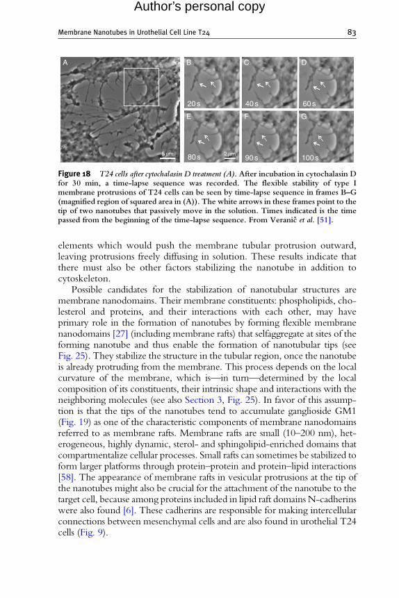
Figure 18 T24 cells after cytochalasin D treatment (A). After incubation in cytochalasin Dfor 30 min, a time-lapse sequence was recorded. The flexible stability of type Imembrane protrusions of T24 cells can be seen by time-lapse sequence in frames B–G(magnified region of squared area in (A)). The white arrows in these frames point to thetip of two nanotubes that passively move in the solution. Times indicated is the timepassed from the beginning of the time-lapse sequence. From Veranic et al. [51].
Membrane Nanotubes in Urothelial Cell Line T24 83
Author’s personal copy
elements which would push the membrane tubular protrusion outward,leaving protrusions freely diffusing in solution. These results indicate thatthere must also be other factors stabilizing the nanotube in addition tocytoskeleton.
Possible candidates for the stabilization of nanotubular structures aremembrane nanodomains. Their membrane constituents: phospholipids, cho-lesterol and proteins, and their interactions with each other, may haveprimary role in the formation of nanotubes by forming flexible membranenanodomains [27] (including membrane rafts) that selfaggregate at sites of theforming nanotube and thus enable the formation of nanotubular tips (seeFig. 25). They stabilize the structure in the tubular region, once the nanotubeis already protruding from the membrane. This process depends on the localcurvature of the membrane, which is—in turn—determined by the localcomposition of its constituents, their intrinsic shape and interactions with theneighboring molecules (see also Section 3, Fig. 25). In favor of this assump-tion is that the tips of the nanotubes tend to accumulate ganglioside GM1(Fig. 19) as one of the characteristic components of membrane nanodomainsreferred to as membrane rafts. Membrane rafts are small (10–200 nm), het-erogeneous, highly dynamic, sterol- and sphingolipid-enriched domains thatcompartmentalize cellular processes. Small rafts can sometimes be stabilized toform larger platforms through protein–protein and protein–lipid interactions[58]. The appearance of membrane rafts in vesicular protrusions at the tip ofthe nanotubes might also be crucial for the attachment of the nanotube to thetarget cell, because among proteins included in lipid raft domains N-cadherinswere also found [6]. These cadherins are responsible for making intercellularconnections between mesenchymal cells and are also found in urothelial T24cells (Fig. 9).

A B
Figure 19 Membrane rafts are present at the tips and entire length of the nanotubes aswell as on cell body. Cells were stained with a membrane raft marker choleratoxin B(arrows) that binds GM1, a membrane raft resident ganglioside.
A B
Figure 20 Control (A) and cholesterol depleted cells (B) and their morphology. In cholesteroldepleted cells the cells round and their membrane appears more rigid with no dynami-cally fluctuating protrusions at the cell edges like in control (arrow in (A)). Their shapeis rather determined by the underlying cytoskeleton. Cells do not appear firmlyattached to each other anymore, although they still preserve some connections(arrow in (B)).
84 M. Lokar et al.
Author’s personal copy
Since nanodomains are thought to be important in both formation andstability of nanotubes and these nanostuctures are shown to be enriched incholesterol (Pike et al., 2006) it is expected that content of cholesterol inthe membrane will influence also the dynamic of nanotubes. Cholesteroldecreases membrane fluidity, locally increases membrane thickness and candirectly modulate the dynamics of membrane rafts-associated proteins(reviewed in Ref. [39]) as well as cell-to-cell junctions [10, 38].
Our preliminary results show that depletion of cholesterol by b-methylcyclodextin changes morphology of T24 cells as well as almost totallyinhibits formation of type I membrane nanotubes, whereas the effect ontype II nanotubes is not known (no experimental data) (Fig. 20).

Membrane Nanotubes in Urothelial Cell Line T24 85
Author’s personal copy
3.1. On the Role of Small Anisotropic Protein–LipidNanodomains in Formation and Stabilization ofMembrane Nanotubes
The observed stability of tubular membrane protrusions after disintegra-tion of the actin filaments within tubular protrusions (Fig. 18) can beexplained by coupling between the nonhomogeneous lateral distributionof the membrane nanodomains and the specific membrane curvatures[5, 22, 25, 45, 50]. The proposed mechanism of mechanical stabilizationof tubular membrane protrusions is a part of the general mechanism ofstabilization of highly curved membrane structures (spherical buds, necks,tubular protrusions) [5, 18, 23, 26, 28, 49, 50]. For example, it wassuggested that due to its specific molecular shape the prominin molecules[56] may form small anisotropic protein–lipid nanodomains [26, 27]which may associate into larger two-dimensional aggregates accumulatedin tubular membrane protrusions (Lubrol rafts) [23, 26, 28, 59]. Lubrolrafts, formed by clustering of prominin nanodomains, are considered to bea novel type of membrane rafts (microdomains) that are distinct fromthe cholesterol-sphingolipid (Triton resistant) rafts in the planar parts ofthe membrane [23, 28, 59].
The prevalent force of the origin of membrane protrusion is usually theforce exerted by the cytoskeleton elements [3]. However, also in this case,the accumulation of anisotropic membrane nanodomains in tubular mem-brane protrusions may offer an additional physical mechanism for stabiliza-tion of tubular membrane protrusions [26, 32, 55]. The observed stability ofthin tubular membrane protrusions without the inner supporting rod-likeskeleton (Fig. 18) is in line with the assumption that prominin nanodomains(and other anisotropic membrane inclusions) have an important role ingeneration and stabilization of plasma membrane protrusions and TNTs;[26, 28] (Fig. 21).
In accordance with above proposed mechanism of stabilization of tubu-lar membrane protrusions we proposed [51] that membrane nanodomains(Fig. 21) which compose the membrane of bridging nanotubes energeticallyprefer highly curved cylindrical geometry (C1> 0 and C2¼ 0) (for defini-tion of the principal membrane curvatures C1 and C2 see Fig. 22).
In our model [12, 27] we divide the flexible membrane nanodomains intwo groups. In the first are small molecular complexes composed of proteinsand lipids where the proteins are often chain-like biopolymers that cross themembrane bilayer a few times (Fig. 21A) [22, 26]. Membrane nanodomainsand raft elements of biological membranes usually fall into this category.The second group of flexible membrane inclusions are induced by a singlerigid globular membrane protein, which can be described in the firstapproximation as a rigid object of a simple geometrical shape (Fig. 21B)[7, 8, 11, 12, 19, 30, 31].

A
B
Figure 21 Schematic illustration of membrane nanodomains (shaded area): A flexible lipid-protein membrane nanodomain containing transmembrane proteins (A) and a flexiblemembrane nanodomain induced by single membrane-embedded rigid (globular)protein (B) [12].
R2
R1
nY
C1=
C2= R2
1R1
1
Figure 22 Schematic presentation of the two principal membrane curvatures C1 andC2 (for the case of saddle-like membrane shape) defined in the origin of the membranenormal n. The principal curvatures C1 and C2 are inversely proportional to principalradii of curvatures R1 and R2, respectively.
86 M. Lokar et al.
Author’s personal copy

Membrane Nanotubes in Urothelial Cell Line T24 87
Author’s personal copy
In the following we assume that membrane nanodomains (Fig. 21), as aresult of their structure and local interactions energetically prefer a localgeometry that is described by the two intrinsic principal curvatures(C1m and C2m) [13, 31, 36]. The intrinsic principal curvatures (spontaneouscurvatures) (C1m and C2m) are in general different (C1m 6¼C2m) (Fig. 23).If they are identical (C1m¼C2m), the nanodomain is called isotropic.If C1m 6¼C2m the nanodomain is called anisotropic. The location andorientation of the anisotropic nanodomain are important for its energy.An anisotropic nanodomain (Fig. 24) will therefore prefer to accumulate inthe membrane region with the principal curvatures C1 and C2 close to thevalues of its intrinsic principal curvatures C1m and C2m [22, 26] and on theaverage also spend more time in the orientation that is energetically morefavorable than in any other orientation. A coupling between the membraneshape (i.e., curvature) and the nonhomogeneous lateral distribution ofmembrane nanodomains has been predicted [5, 18, 22, 25, 27, 45, 50].
The elastic energy of a small anisotropic membrane nanodomain derivesfrom the mismatch between the actual local curvature of the membrane(Fig. 22) and the intrinsic (spontaneous) curvature of the constituents(Fig. 23) which can be characterized by the mismatch tensor
�M ¼
�R�Cm
�R�1 �
�C [27]. Here the tensor
�C describes the actual curvature
(see Fig. 22), while the tensor�Cm describes the intrinsic curvature of the
constituents:
�C ¼ C1 0
0 C2
� �;�Cm ¼ C1m 0
0 C2m
� �; ð1Þ
where
�R ¼ coso �sino
sino coso
� �; ð2Þ
is the rotation matrix. The angle o describes the orientation of the principalaxes system of a single membrane nanodomain with respect to the localprincipal axes system of the membrane [28, 32]. In the respective principal
C1m> 0C2m= 0 C2m= 0
C1m= 0 C1m> 0C2m< 0
Anisotropic:Anisotropic: Isotropic:
Figure 23 Schematic representation of different intrinsic shapes of larger of membranenanodomains described by the two intrinsic principal (spontaneous) curvaturesC1m and C2m.
500
−1000
−1000
−500
−500
500
500
1000
1000
f.104/K
Z [nm]
Hm= Dm
Hm= 1/100 [nm-1]
Hm= 0 [nm-1]
Hm= 1/200 [nm-1]Hm= 1/300 [nm-1]
Cm1> 0
Cm 2= 0
� [nm]
� [nm]
0
A
B
0
3
2
1
0
400
300
200
100
0
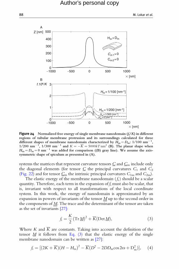
Figure 24 Normalized free energy of single membrane nanodomain (fi/K) in differentregions of tubular membrane protrusion and its surroundings calculated for threedifferent shapes of membrane nanodomain characterized by Hm¼Dm: 1/100 nm� 1,1/200 nm� 1, 1/300 nm� 1 and K ¼ �K ¼ 5000kT nm2 (B). The planar shape whenHm¼Dm¼ 0 nm� 1 was added for comparison ((B) gray line). We assume the axis-symmetric shape of spiculum as presented in (A).
88 M. Lokar et al.
Author’s personal copy
systems the matrices that represent curvature tensors�C and
�Cm include only
the diagonal elements (for tensor�C the principal curvatures C1 and C2
(Fig. 22) and for tensor�Cm the intrinsic principal curvatures C1m and C2m).
The elastic energy of the membrane nanodomain ( fi ) should be a scalarquantity. Therefore, each term in the expansion of fi must also be scalar, thatis, invariant with respect to all transformations of the local coordinatesystem. In this work, the energy of nanodomain is approximated by anexpansion in powers of invariants of the tensorM up to the second order inthe components ofM. The trace and the determinant of the tensor are takenas the set of invariants [27]:
fi ¼ K
2ðTr�MÞ2 þ KðDet
�MÞ; ð3Þ
Where K and K are constants. Taking into account the definition of thetensor
�M it follows from Eq. (3) that the elastic energy of the single
membrane nanodomain can be written as [27]:
fi ¼ ½ð2K þ KÞðH �HmÞ2 � KðD2 � 2DDmcos2oþD2mÞ�; ð4Þ

Membrane Nanotubes in Urothelial Cell Line T24 89
Author’s personal copy
where H¼ (C1þC2)/2 and D¼ |C1�C2|/2 are the mean curvature andthe curvature deviator of the membrane (see also Fig. 22), Hm¼ (C1mþC2m)/2 is the intrinsic (spontaneous) mean curvature and Dm¼ |C1m�C2m|/2 is the intrinsic (spontaneous) curvature deviator. The constants Kand K are proportional to the area of the single membrane nanodomain[12, 27]. In the case of a simple flexible membrane nanodomain composed ofa rigid core (protein) and the surrounding lipids which are distorted in orderto fit with the rigid core (Fig. 21), the constants K and K were estimatedusing a microscopic model [12] while in the case lipid molecules they wereestimated from the bending constant [34, 35]. The optimal values of themembrane mean curvature H, the curvature deviator D and the membraneconstituent orientation angle o corresponding to the minimum of the func-tion fi can be calculated from the necessary and sufficient conditions for theextremum of fi [27]: H¼Hm, D¼Dm, o¼ 0, p, 2p where o¼ 0 ando¼ 2p describe the same orientation and where K > �K=2, K < 0.
The partition function of a single anisotropic membrane nanodomain:
Q ¼ 1
oo
ð2p0
exp � f iðoÞkT
� �do; ð5Þ
The free energy of the single anisotropic nanodomain is then obtainedby considering that fi¼�kT lnQ [27]:
fi ¼ ð2K þ KÞðH �HmÞ2 � KðD2 þD2mÞ � kT ln I0
2KDDm
kT
� �� �;
ð6Þwhere I0 is the modified Bessel function. In the limit j2KDDm=kT j > 1,Eq. (6) becomes:
fi ¼ ð2K þ KÞðH �HmÞ2 � KðD�DmÞ2; ð7Þwhere we took into account that ln I0(x)� |x| for x> 1 and K < 0. In thelimit of small j2KDDm=kT j, Eq. (6) transforms into:
fi ¼ 2K � K2D2
m
kT
!ðH �H0Þ2 þ K þ K
2D2
m
kT
!C1C2; ð8Þ
H0 ¼ Hmð2K þ KÞ2K � K
2D2
m
kT
� � ; ð9Þ
where we took into account ln I0(x)� x2/4 for x<< 1, D2¼H2�C1C2
and omitted the constant term.Figure 24 shows the influence of the anisotropy of the intrinsic shape of
the membrane nanodomain (described by intrinsic mean curvature

90 M. Lokar et al.
Author’s personal copy
Hm ¼ ðC1m þ C2mÞ=2 and intrinsic curvature deviator Dm¼ (C1m�C2m)/2) on the energy of the nanodomain fi (Eq. (6)) in the different parts of thetubular membrane protrusions and the surrounding membrane. It can be seenin Fig. 24 that the energy of anisotropic nanodomains ( fi ) with the intrinsicshape characterized by Hm¼Dm (or equivalently C1m> 0 and C1m¼ 0,see Fig. 23) may be strongly decreased in the region of tubular protrusionwhich leads to accumulation of such nanodomains in the tubular membraneprotrusion and consequently to mechanical stabilization of tubular membraneprotrusions as shown elsewhere [26, 27, 32, 33, 51].
Based on the results presented in Fig. 24 and our previous theoreticalconsideration of the stability of tubular membrane protrusions [26, 27, 32, 33]we suggest that nanotubular membrane protrusions andmembrane nanotubesare in addition to stabilization forces of cytoskeleton elements mechanicallystabilized also by energetically favorable clustering of anisotropic (flexible)membrane nanodomains in nanotubes [17, 26, 27, 51] (Fig. 25).
Membrane
Actin filaments
Flexible nanodomain
C1m> 0 C2m= 0
C1m≈ C2m> 0
Figure 25 Schematic illustration of stabilization of type I nanotubular membrane pro-trusions by accumulation of anisotropic membrane nanodomains in the tubular region.Bending deformation and rotation of the nanodomain allow the nanodomain to adapt itsshape and orientation to the actual membrane curvature, which in turn is influenced bythe nanodomains [23,28]. Growing actin filaments push the membrane outward. Theprotrusion is additionally stabilized by accumulated anisotropic nanodomains with mem-brane curvatures that favor anisotropic cylindrical geometry of the membrane. Thecylindrical-shaped anisotropic membrane domains, once assembled in the membraneregion of a nanotubular membrane protrusion, keeps the protrusion mechanically stableeven if the cytoskeletal components (actin filaments) are disintegrated. Adaped from [46].

Membrane Nanotubes in Urothelial Cell Line T24 91
Author’s personal copy
4. Concluding Remarks
In urothelial T24 cell line at least two different kinds of membranenanotubes exist. These two types differ in their structural components(type I having actin cytoskeleton and type II having cytokeratins) stability,dynamics and consequently also in function. Type II nanotubes do providecytosolic and membrane continuity between two cells, at least in thebeginning, since they are presumably formed in nonmitotic separation oftwo cells. As for type I nanotubes cytosolic continuity can be establishedafter an adherens and communication junctions between a protrudingnanotube and acceptor cell is assembled even though their protein compo-nents have not been undoubtedly determined. Which proteins make thispossible need to be further defined. Also the stability of these nanotubules isnot well understood, but ongoing studies are suggesting that both choles-terol and lipid constituents that determine the local geometry of the mem-brane are important in this process.
REFERENCES
[1] B. Alberts, A. Johnson, J. Lewis, M. Raff, K. Roberts, P. Walter, Molecular Biology ofthe Cell, 4th ed., Garland Science, New York, 2002.
[2] R. Bar-Ziv, E. Moses, Instability and ‘‘pearling’’ states produced in tubular membranesby competition of curvature and tension, Phys. Rev. Lett. 73 (1994) 1392–1395.
[3] A.A. Boulbitch, Deflection of a cell membrane under application of local force, Phys.Rev. E 57 (1998) 1–5.
[4] J. Bubenık, M. Baresova, V. Viklicky, J. Jakoubkova, H. Sainerova, J. Donner, Estab-lished cell line of urinary bladder carcinoma (T24) containing tumour-specific antigen,Int. J. Cancer 11 (1973) 765–773.
[5] L. Cantu’, M. Corti, P. Brocca, E. del Favero, Structural aspects of ganglioside-containing membranes, Biochim. Biophy. Acta 1788 (2009) 202–208.
[6] M. Causeret, N. Taulet, F. Comunale, C. Favard, C. Gauthier-Rouviere, N-cadherinassociation with lipid rafts regulates its dynamic assembly at cell-cell junctions inC2C12 myoblasts, Mol. Biol. Cell. 16 (2005) 2168–2180.
[7] N. Dan, P. Pincus, S.A. Safran, Membrane-induced interactions between inclusions,Langmuir 9 (1993) 2768–2771.
[8] N. Dan, S.A. Safran, Effect of lipid characteristics on the structure of transmembraneproteins, Biophys. J. 75 (1998) 1410–1414.
[9] D.M. Davies, S. Sowinski, Membrane nanotubes: dynamic long-distance connectionsbetween animal cells, Nat. Rev. Mol. Cell Biol. 9 (2008) 431–436.
[10] S.A. Francis, J.M. Kelly, J. McCormack, R.A. Rogers, J. Lai, E.E. Schneeberger,R.D. Lynch, Rapid reduction of MDCK cell cholesterol by methyl-beta-cyclodextrinalters steady state transepithelial electrical resistance, Eur. J. Cell Biol. 78 (1999)473–484.
[11] M. Fosnaric, A. Iglic, S. May, Influence of rigid inclusions on the bending elasticity of alipid membrane, Phys. Rev. E 174 (2006) 051503.

92 M. Lokar et al.
Author’s personal copy
[12] M. Fosnaric, A. Iglic, T. Slivnik, V. Kralj-Iglic, Flexible membrane inclusions andmembrane inclusions induced by rigid globular proteins, Advances in planar lipidbilayers and liposomes, Elsevier, (2008) 143–168.
[13] J.B. Fournier, P. Galatola, Bilayer membranes with 2-D nematic order of the surfactantpolar heads, Braz. J. Phys. 28 (1998) 329–338.
[14] D. Freund, N. Bauer, S. Boxberger, S. Feldmann, U. Streller, G. Ehninger, C.Werner,M. Bornhauser, J. Oswald, D. Corbeil, Polarization of human hematopoietic progeni-tors during contact with multipotent mesenchymal stromal cells: effects on proliferationand clonogenicity, Stem Cells Dev. 15 (2006) 815–829.
[15] H.H. Gerdes, N.V. Bukoreshtliev, J.F. Barroso, Tunneling nanotubes: a new route forthe exchange of components between animal cells, FEBS Lett. 581 (2007) 2194–2201.
[16] H.H. Gerdes, R.N. Carvalho, Intercellular transfer mediated by tunneling nanotubes,Curr. Opin. Cell Biol. 20 (2008) 470–475.
[17] U. Gimsa, A. Iglic, S. Fiedler, M. Zwanzig, V. Kralj-Iglic, L. Jonas, J. Gimsa, Actin isnot required for nanotubular protrusions of primary astroctes grown on metal nano-lawn, Mol. Membr. Biol. 24 (2007) 243–255.
[18] W.T. Gozdz, Diffusion of macromolecules on lipid vesicles, Langmuir 24 (2008)12458–12468.
[19] H. Gruler, Chemoelastic effect of membranes, Z. Naturforsch. [C] 30 (1975) 608–614.[20] S. Gurke, J.F. Barroso, E. Hodneland, N.V. Bukoreshtliev, O. Schlicker, H.H. Gerdes,
Tunneling nanotube (TNT)-like structures facilitate a constitutive, actomyosin-dependent exchange of endocytic organelles between normal rat kidney cells, Exp.Cell Res. 314 (2008) 3669–3683.
[21] S. Gurke, J.F. Barroso, H.H. Gerdes, The art of cellular communication: tunnelingnanotubes bridge the divide, Histochem. Cell Biol. 129 (2008) 539–550.
[22] H. Hagerstrand, L. Mrowczynska, U. Salzer, R. Prohaska, K. Michelsen, V. Kralj-Iglic,A. Iglic, Curvature-dependent lateral distribution of raft markers in the human eryth-rocyte membrane, Mol. Membr. Biol. 23 (2006) 277–288.
[23] J.C. Holthius, G. vanMeer, K. d’Huitema, Lipid microdomains, lipid translocation andthe organization of intracellular membrane transport (review), Mol. Membr. Biol.20 (2003) 231–241.
[24] A. Iglic, H. Hagerstrand, M. Bobrowska-Hagerstrand, V. Arrigler, V. Kralj-Iglic,Possible role of phospholipid nanotubes in directed transport of membrane vesicles,Phys. Lett. 310 (2003) 493–497.
[25] A. Iglic, M. Fosnaric, H. Hagerstrand, V. Kralj-Iglic, Coupling between vesicle shapeand the non-homogeneous lateral distribution of membrane constituents in Golgibodies, FEBS Lett. 574/1–3 (2004) 9–12.
[26] A. Iglic, H. Hagerstrand, P. Veranic, A. Plemenitas, V. Kralj-Iglic, Curvature inducedaccumulation of anisotropic membrane components and raft formation in cylindricalmembrane protrusions, J. Theor. Biol. 240 (2006) 368–373.
[27] A. Iglic, M. Lokar, B. Babnik, T. Slivnik, P. Veranic, H. Hagerstrand, V. Kralj-Iglic,Possible role of flexible red blood cell membrane nanodomains in the growth andstability of membrane nanotubes, Blood Cells Mol. Dis. 39 (2007) 14–23.
[28] P. Janich, D. Corbeil, GM1 and GM3 gangliosides highlight distinct lipid microdo-mains with the apical domain of epithelial cells, FEBS Lett. 581 (2007) 1783–1787.
[29] M. Koyanagi, R.P. Brandes, J. Haendeler, A.M. Zeiher, S. Dimmeler, Cell-to-cell 31.connection of endothelial progenitor cells with cardiac myocytes by nanotubes:A novel mechanism for cell fate changes? Circ. Res. 96 (2005) 1039–1041.
[30] V. Kralj-Iglic, S. Svetina, B. Zeks, Shapes of bilayer vesicles with membrane embeddedmolecules, Eur. Biophy. J. 24 (1996) 311–321.
[31] V. Kralj-Iglic, V. Heinrich, S. Svetina, B. Zeks, Free energy of closed membrane withanisotropic inclusions, Eur. Phys. J. B 10 (1999) 5–8.

Membrane Nanotubes in Urothelial Cell Line T24 93
Author’s personal copy
[32] V. Kralj-Iglic, A. Iglic, H. Hagerstrand, P. Peterlin, Stable tubular microexovesicles ofthe erythrocyte membrane induced by dimeric amphiphiles, Phys. Rev. E 61 (2000)4230–4234.
[33] V. Kralj-Iglic, H. Hagerstrand, P. Veranic, K. Jezernik, B. Babnik, D.R. Gauger, A. Iglic,Amphiphile-induced tubular budding of the bilayermembrane, Eur. Biophys. J. 34 (2005)1066–1070.
[34] V. Kralj-Iglic, B. Babnik, R.D. Gauger, S. May, A. Iglic, Quadrupolar ordering ofphospholipid molecules in narrow necks of phospholipid vesicles, J. Stat. Phys.125 (2006) 727–752.
[35] T. Mares, M. Daniel, S. Perutkova, A. Perne, G. Dolinar, A. Iglic, M. Rappolt,V. Kralj-Iglic, Role of phospholipid asymmetry in the stability of inverted hexagonalmesoscopic phases, J. Phys. Chem. B 112 (2008) 16575–16584.
[36] P.K. Mattila, P. Lappalainen, Filopodia: Molecular architecture and cellular functions,Nat. Rev. Mol. Cell Biol. 9 (2008) 446–454.
[37] T.J. Mitcinson, Actin based motility on retraction fibers in mitotic PtK2 cells, CellMotil. Cytoskeleton 22 (1992) 135–151.
[38] A. Nusrat, C.A. Parkos, P. Verkade, C.S. Foley, T.W. Liang, W. Innis-Whitehouse,K.K. Eastburn, J.L. Madara, Tight junctions are membrane microdomains, J. Cell Sci.113 (2000) 1771–1781.
[39] H. Ohvo-Rekila, B. Ramstedt, P. Leppimaki, J.P. Slotte, Cholesterol interactions withphospholipids in membranes, Prog. Lipid Res. 41 (2002) 66–97.
[40] B. Onfelt, S. Nedvetzki, K. Yanagi, D.M. Davis, Cutting edge: Membrane nanotubesconnect immune cells, J. Immunol. 173 (2004) 1511–1513.
[41] B. Onfelt, S. Nedvetzki, R.K. Benninger, M.A. Purbhoo, S. Sowinski, A.N. Hume,M.C. Seabra, M.A. Neil, P.M. French, D.M. Davis, Structurally distinct membranenanotubes between human macrophages support long-distance vesicular traffic orsurfing of bacteria, J Immunol. 177 (2006) 8476–8483.
[42] B. Pontes, N.B. Viana, L. Campanti, M. Farina, V.M. Neto, H.M. Nussenzveig,Structure and elastic properties of tunneling nanotubes, Eur. Biophys. J. 37 (2008)121–129.
[43] A. Rustom, R. Saffrich, I. Markovic, P. Walther, H.H. Gerdes, Nanotubular highwaysfor intercellular organelle transport, Science 303 (2004) 1007–1010.
[44] K. Schara, V. Jansa, V. Sustar, D. Dolinar, J.I Pavlic, M. Lokar, V. Kralj-Iglic,P. Veranic, A. Iglic, Mechanisms for the formation of membranous nanostructures incell-to-cell communication, Cell. Mol. Biol. Lett. 2009.
[45] P. Sens, M.S. Turner, The forces that shape caveolae, In: Lipid Rafts and Caveolae,(C.J. Fielding, Ed.), 2006, pp. 25–44. Wiley-VCH Verlag, Weinheim.
[46] N.M. Sherer, M.J. Lehmann, L.F. Jimenez-Soto, C. Horensavitz, M. Pypaert,W. Mothes, Retroviruses can establish filopodial bridges for efficient cell-to-celltransmission, Nat. Cell Biol. 9 (2007) 310–315.
[47] N.M. Sherer, W. Mothes, Cytonemes and tunneling nanotubules in cell-cell-commu-nication and viral pathogenesis, Trends Cell Biol. 18 (2008) 414–420.
[48] S. Sowinski, C. Jolly, O. Berninghausen, M.A. Purbhoo, A. Chauveau, K. Kohler,S. Oddos, P. Eissmann, F.M. Brodsky, C. Hopkins, B. Onfelt, Q. Sattentau,D.M. Davis, Membrane nanotubes physically connect T cells over long distancespresenting a novel route for HIV-1 transmission, Nat. Cell Biol. 10 (2008) 211–219.
[49] C. Thiele, M.J. Hannah, F. Fahrenholz, W.B. Huttner, Cholesterol binds to synapto-physin and is required for biogenesis of synaptic vesicles, Nat. Cell Biol. 2 (2000)42–49.
[50] A. Tian, T. Baumgart, Sorting of lipids and proteins in membrane curvature gradients,Bipohys. J. 96 (2009) 2676–2688.
94 M. Lokar et al.
Author’s personal copy
[51] P. Veranic, M. Lokar, G.J. Schutz, J. Weghuber, S. Wieser, H. Hagerstrand,V. Kralj-Iglic, A. Iglic, Different types of cell-to-cell connections mediated by nano-tubular structures, Biophys. J. 95 (2008) 4416–4425.
[52] C. Vidulescu, S. Clejan, K.C. O’connor, Vesicle traffic through intercellular bridges inDU 145 human prostate cancer cells, J. Cell. Mol. Med. 8 (2004) 388–396.
[53] S.C.Watkins, R.D. Salter, Functional connectivity between immune cells mediated bytunneling nanotubes, Immunity 23 (2005) 309–318.
[54] D. Wustner, Plasma membrane sterol distribution resembles the surface topography ofliving cells, Mol. Biol. Cell. 18 (2007) 211–228.
[55] Y. Yamashita, S.M. Masum, T. Tanaka, Y. Tamba, M. Yamazaki, Shape changes ofgiant unilamellar vesicles of phosphatidiylcholine induced by a de novo designedpeptide interacting with their membrane interface, Langmuir 18 (2002) 9638–9641.
[56] S. Zacchigna, H. Oh, M. Wilsch-Brauninger, E. Missol-Kolka, J. Jaszai, S. Jansen,N. Tanimoto, F. Tonagel, M. Seeliger, W.B. Huttner, D. Corbeil, M. Dewerchin,et al. Loss of the cholesterol-binding protein prominin-1/CD133 causes disk dysmor-phogenesis and photoreceptor degeneration, J. Neurosci. 29 (2009) 2297–2308.
[57] D. Vignjevic, S. Kojima, Y. Aratyn, O. Danciu, T. Svitkina, G.G. Borisy, Role offascin in filopodial protrusion, J. Cell Biol. 11 (2006) 863–875.
[58] L.J. Pike, Rafts defined: a report on the Keystone Symposium on Lipid Rafts and CellFunction, J. Lipid Res. 47 (2006) 1597–1598.
[59] W.B. Huttner, J. Zimmerberg, Implications of lipid microdomains for membranecurvature, budding and fission, Curr. Opin. Cell Biol. 13 (2001) 478–484.