Changes in numbers and kinds of bacteria during a chickpea submerged fermentation used as a...
7
Changes in numbers and kinds of bacteria during a chickpea submerged fermentation used as a leavening agent for bread production M. Hatzikamari a , M. Yiangou b , N. Tzanetakis a , E. Litopoulou-Tzanetaki a, ⁎ a Laboratory of Food Microbiology and Hygiene, Faculty of Agriculture, Aristotle University of Thessaloniki, 541 24 Thessaloniki, Greece b Department of Genetic, Development and Molecular Biology, Faculty of Biology, Aristotle University of Thessaloniki, 541 24, Thessaloniki, Greece Received 19 July 2006; received in revised form 8 September 2006; accepted 10 December 2006 Abstract The microflora developed during a submerged fermentation of coarsely ground chickpea (Cicer arietinum L.) in water (primary starter) and during raising a dough from wheat flour (adapted starter) was studied. In the fermenting liquid, only populations of Bacillus and Clostridium developed. Bacilli increased their loads significantly ( p b 0.05) during fermentation for 8–12 h and then remained constant. Clostridia developed ( p b 0.05) subsequently to levels of 10 7 cfu/ml at 18 h, when the pH of the fermenting liquid had decreased ( p b 0.05) to ∼ 5.4. It also seems that the rise of the adapted starter within 2 h was caused by enzymes present in the primary starter and those liberated after cell death by the declining populations of bacilli and clostridia. The principal groups of isolates in all fermentation experiments (with chickpea seeds from five different areas) seemed to have the phenotypic characteristics of Bacillus cereus group and Clostridium perfringens. SDS-PAGE of whole-cell proteins elucidated the taxonomic position of the B. cereus group of strains as B. cereus and Bacillus thuringiensis and confirmed the phenotypic allocation of C. perfringens isolates. Strains phenotypically characterized as Bacillus licheniformis and Clostridium beijerinckii were also found to belong to these same species by SDS-PAGE. In addition, results showed that the fermenting broth was not toxic to mice when inoculated intraperitoneally and the product can thus be considered as safe for consumption. © 2007 Elsevier B.V. All rights reserved. Keywords: Chickpea fermentation; Characterization; Bacillus; Clostridium; Dough; Leavening 1. Introduction Food legumes have been well recognized as one of the most important sources of proteins, calories, minerals and vitamins. Among them, lentils and chickpeas make a significant con- tribution to the Middle Eastern diet. Traditional processing and preparation methods of foods based on chickpeas (Cicer arietinum L.) include soaking, decortication, grinding, sprouting, fermentation, boiling, mashing, roasting, parching, frying and steaming (Desphande and Damodaran, 1990). In some Mediter- ranean countries fermented chickpeas are being used as a leavening agent to make traditional breads and rusks. By adding fermented chickpea to the flour, the nutritional quality and shelf- life of these products is enhanced (Tulbek et al., 2003). In Greece a traditional type of bread is made with fermented chickpea in water as a leavening agent. The bread is called “chickpea bread” in the Western Macedonia region of northern Greece, but is more commonly known as “eftazymo” (from the Greek word “aftozymo”, something that is self-fermented). This type of bread was prepared in the past as a celebration bread of Easter, local religious celebrations, and weddings. Previous study of chickpea fermentation in water has shown that changes in enzyme activities and in the chemical compo- sition of the fermenting liquid are caused by indigenous bacilli and clostridia populations (Hatzikamari et al., in press). Changes in the chemical constituents of the fermenting liquid can be attributed mainly to bacilli until 8–10 h of fermentation and then to clostridia until 18 h. Bacilli (Bacillus subtilis) are important starter cultures for alkaline fermentations observed in traditional fermented legumes, such as the Indian Kinema (from spontaneously fermented soybeans), the African Soumbala (from locust beans) and others (Sarkar et al., 2002). However, throughout the fermentation of chickpea extracts, a significant decrease of pH is observed (Hatzikamari et al., in press). Similarly, during Gergoush-making from milk, lentils and International Journal of Food Microbiology 116 (2007) 37 – 43 www.elsevier.com/locate/ijfoodmicro ⁎ Corresponding author. Tel./fax: +30 2310 473622. E-mail address: [email protected] (E. Litopoulou-Tzanetaki). 0168-1605/$ - see front matter © 2007 Elsevier B.V. All rights reserved. doi:10.1016/j.ijfoodmicro.2006.12.030
Transcript of Changes in numbers and kinds of bacteria during a chickpea submerged fermentation used as a...

obiology 116 (2007) 37–43www.elsevier.com/locate/ijfoodmicro
International Journal of Food Micr
Changes in numbers and kinds of bacteria during a chickpea submergedfermentation used as a leavening agent for bread production
M. Hatzikamari a, M. Yiangou b, N. Tzanetakis a, E. Litopoulou-Tzanetaki a,⁎
a Laboratory of Food Microbiology and Hygiene, Faculty of Agriculture, Aristotle University of Thessaloniki, 541 24 Thessaloniki, Greeceb Department of Genetic, Development and Molecular Biology, Faculty of Biology, Aristotle University of Thessaloniki, 541 24, Thessaloniki, Greece
Received 19 July 2006; received in revised form 8 September 2006; accepted 10 December 2006
Abstract
The microflora developed during a submerged fermentation of coarsely ground chickpea (Cicer arietinum L.) in water (primary starter) andduring raising a dough from wheat flour (adapted starter) was studied. In the fermenting liquid, only populations of Bacillus and Clostridiumdeveloped. Bacilli increased their loads significantly ( pb0.05) during fermentation for 8–12 h and then remained constant. Clostridia developed( pb0.05) subsequently to levels of 107 cfu/ml at 18 h, when the pH of the fermenting liquid had decreased ( pb0.05) to ∼5.4. It also seems thatthe rise of the adapted starter within 2 h was caused by enzymes present in the primary starter and those liberated after cell death by the decliningpopulations of bacilli and clostridia. The principal groups of isolates in all fermentation experiments (with chickpea seeds from five differentareas) seemed to have the phenotypic characteristics of Bacillus cereus group and Clostridium perfringens. SDS-PAGE of whole-cell proteinselucidated the taxonomic position of the B. cereus group of strains as B. cereus and Bacillus thuringiensis and confirmed the phenotypicallocation of C. perfringens isolates. Strains phenotypically characterized as Bacillus licheniformis and Clostridium beijerinckii were also foundto belong to these same species by SDS-PAGE. In addition, results showed that the fermenting broth was not toxic to mice when inoculatedintraperitoneally and the product can thus be considered as safe for consumption.© 2007 Elsevier B.V. All rights reserved.
Keywords: Chickpea fermentation; Characterization; Bacillus; Clostridium; Dough; Leavening
1. Introduction
Food legumes have been well recognized as one of the mostimportant sources of proteins, calories, minerals and vitamins.Among them, lentils and chickpeas make a significant con-tribution to the Middle Eastern diet. Traditional processingand preparation methods of foods based on chickpeas (Cicerarietinum L.) include soaking, decortication, grinding, sprouting,fermentation, boiling, mashing, roasting, parching, frying andsteaming (Desphande and Damodaran, 1990). In some Mediter-ranean countries fermented chickpeas are being used as aleavening agent to make traditional breads and rusks. By addingfermented chickpea to the flour, the nutritional quality and shelf-life of these products is enhanced (Tulbek et al., 2003).
In Greece a traditional type of bread is made with fermentedchickpea in water as a leavening agent. The bread is called
⁎ Corresponding author. Tel./fax: +30 2310 473622.E-mail address: [email protected] (E. Litopoulou-Tzanetaki).
0168-1605/$ - see front matter © 2007 Elsevier B.V. All rights reserved.doi:10.1016/j.ijfoodmicro.2006.12.030
“chickpea bread” in the Western Macedonia region of northernGreece, but is more commonly known as “eftazymo” (from theGreek word “aftozymo”, something that is self-fermented). Thistype of bread was prepared in the past as a celebration bread ofEaster, local religious celebrations, and weddings.
Previous study of chickpea fermentation in water has shownthat changes in enzyme activities and in the chemical compo-sition of the fermenting liquid are caused by indigenous bacilliand clostridia populations (Hatzikamari et al., in press).Changes in the chemical constituents of the fermenting liquidcan be attributed mainly to bacilli until 8–10 h of fermentationand then to clostridia until 18 h. Bacilli (Bacillus subtilis) areimportant starter cultures for alkaline fermentations observed intraditional fermented legumes, such as the Indian Kinema (fromspontaneously fermented soybeans), the African Soumbala(from locust beans) and others (Sarkar et al., 2002). However,throughout the fermentation of chickpea extracts, a significantdecrease of pH is observed (Hatzikamari et al., in press).Similarly, during Gergoush-making from milk, lentils and

38 M. Hatzikamari et al. / International Journal of Food Microbiology 116 (2007) 37–43
wheat flour, the dominant bacteria are bacilli, clostridia andlactic acid bacteria, and the pH of the fermenting product isreduced (Sherfi and Hamad, 2001).
The purpose of the present work was to study underlaboratory standardized conditions the microbiological changesthat occur during production of a traditional dough made in twosteps: a chickpea water extract is fermented in the first stepwhich is used for leavening the dough from wheat flour in thesecond step.
2. Materials and methods
2.1. Preparation of the starter
Chickpea seeds deriving from an area of central Macedonia,northern Greece were used to make a starter in four fermentationexperiments. In addition, in order to study microfloral diversity,chickpea seeds from four other areas of northern Greece wereobtained and used for fermentation experiments. Simulating thetraditional method, a two-step starter was prepared and in-vestigated. In the first step, seeds were coarsely ground in amortar and pestle and subsequently soaked with boiling water(2:10) in cupped glass vessels. The vessels were incubated forfermentation at 37 °C for 18 h. A series of vessels was preparedto serve for analyses at time 0 h and at 2 h time points until 18 h.The adapted starters were prepared by mixing the fermentedliquid with wheat flour and were left at 37 °C to double theirinitial volume (within ∼2 h).
2.2. Microbiological examinations
Decimal dilutions were made in saline solution (NaCl,0.85% w/v), except for clostridia, where a peptone diluent(peptone, 0.1% w/v) was used. The following microbiologicalexaminations were completed during fermentation: total viablecounts on plate count agar (PCA), after incubation at 30 °C for72 h; Bacillus spp. on nutrient agar (NA), incubated at 37 °C for18 h; lactic acid bacteria on MRS agar (at pH 6.2; Reuter, 1985)incubated anaerobically (Gas-Pak system, BBL, Cockeysville,Maryland, USA) at 30 °C for 5 days; Gram-positive, catalasenegative cocci on M17 agar, after incubation at 30 °C for 48 h;enterococci on citrate azide agar (CAA; Saraswat et al., 1963)after 3 days at 37 °C; staphylococci and micrococci on mannitolsalt agar (MSA) incubated at 37 °C for 48 h; yeasts on acidifiedpotato dextrose agar (PDA) at 25 °C for 5 days; clostridia werecounted by the MPN method in differential reinforcedclostridial medium (DRCM; Freame and Fitzpatrick, 1971)incubated at 37 °C for 3 days under anaerobic conditions (withagar seals). All media were obtained from Merck except forCAA, which was made from its individual ingredients.
2.3. Isolation of the microflora involved in the fermentation
Isolates (10) were taken at random from NA plates innutrient broth and from MRS agar plates in MRS broth, at 10and 18 h of fermentation. Since no growth was noticed for theisolates in MRS broth, colonies were picked again from MRS
agar into brain heart infusion broth (BHIB) and they were foundmicroscopically to be sporeformers. The isolates from NAwerestreaked on NA plates and well isolated colonies were stored onNA supplemented with MnSO4·H2O (50 mg l−1; Claus andBerkeley, 1986) slopes at 4 °C, as well as in nutrient broth plusglycerol (70:30) at −80 °C. For isolating and purifyingclostridia (developed at 10 and 18 h of fermentation inDRCM tubes), the sulphite cycloserine azide medium (SCA;Eisgruber and Reuter, 1995) in deep agar tubes (Freame andFitzpatrick, 1971) was used. Isolates of clostridia were finallykept in cooked meat medium (CMM), covered with liquidparaffin, at room temperature.
2.4. Identification of bacilli
The isolates of bacilli were identified according to criteria ofClaus and Berkeley (1986) and information from Gordon et al.(1973), Logan and Berkeley (1984), Batt (1998) and Harmonet al. (1992). The following tests were applied on all isolates:Gram-staining (position and shape of spores); motility; growthat 50, 55 and 60 °C; growth in 7% NaCl broth; growth in pH 5.7broth and agar; utilization of citrate; nitrate reduction; anaerobicgrowth; Voges-Proskauer test; hydrolysis of starch and casein;gelatine liquefication; egg-yolk lecithinase; H2S formation;urease formation; heamolysis; acid from D-glucose, L-arabinose,D-xylose, D-mannitol. Isolates of Bacillus cereus group werealso examined for formation of parasporal crystals and rhizoidgrowth.
2.5. Identification of clostridia
The isolates of clostridia were identified according to thecriteria of Cato et al. (1986) and Sebald and Petit (1994). Thefollowing tests were applied on all isolates: Gram staining;motility; acid from glucose; gelatine hydrolysis; aerobic growth;indole production; lecithinase production; lipase production;esculin and starch hydrolysis; nitrate reduction; milk reaction;meat digestion; acid production from: cellobiose, fructose,galactose, lactose, maltose, raffinose, sorbitol, starch, sucroseand xylose. All tests, except for aerobic growth were performedunder anaerobic conditions (Gas-Pak system).
2.6. Characterization by sodium dodecyl sulphate-polyacryla-mide gel electrophoresis (SDS-PAGE) of whole cell proteins
As an additional taxonomic tool, SDS-PAGE of whole-cellproteins was used to identify the isolates of bacilli and clostridiafrom one of the four fermentation experiments. The isolates wereactivated (two transfers in broth) and subsequently grown onbrain heart infusion agar (BHIA) plates and tryptone glucoseyeast extract broth (TGY), for bacilli and clostridia, respectively.The growth of bacilli was collected from the surface of the plateswith 0.01 M potassium-phosphate buffer (pH 6.5) and was thencentrifuged (12,000×g, 10 min, 4 °C; Sigma 3K centrifuge,Sigma Chemical Co. St. Louis, MO). The cells of clostridia wereharvested by centrifugation (12,000×g, 10 min, 4 °C). The cellpellets (∼0.150 g wet weight) were then washed twice with

Table 1Counts 1 (log cfu/g; x±S.D.) of bacteria and pH values during chickpeafermentation for the production of primary and adapted starter culture
Time(h)
pH Bacterial counts in
PCA NA DRCM
Primarystarter
0 6.67±0.12 a 1.66±0.16 a 1.37±0.35 a 0 a2 6.52±0.08 a 2.77±0.26 b 2.60±0.24 b4 6.53±0.05 a 2.97±0.52 b 2.89±0.41 b 1.04±0.65 a,b6 6.49±0.02 a 4.80±0.60 c 4.56±0.51 c8 6.45±0.05 a,b 5.90±0.80 d 6.46±0.37 d 2.00±0.51 b10 6.25±0.17 b 6.61±0.75 d,e 6.63±0.39 d,e 3.42±0.56 c12 5.99±0.10 c 7.47±0.39 e,f 7.25±0.71 e,f14 5.78±0.08 d 7.36±0.50 e,f 7.60±0.70 e,f 6.33±1.40 d16 5.57±0.03 d,e 7.94±0.12 f 7.91±0.26 f18 5.36±0.21 e 7.94±0.37 f 7.72±0.08 f 7.81±0.92 e
Adaptedstarter
0 7.76±0.63 7.54±0.72 8.34±1.722 7.63±0.95 7.18±1.09 7.90±1.09
a–f: Means in the same column with different letters differ significantly( pb0.05).1 Mean of four fermentations.
39M. Hatzikamari et al. / International Journal of Food Microbiology 116 (2007) 37–43
phosphate buffer and were finally transferred in eppendorf tubes.The cells were resuspended in 1 ml of potassium-phosphatebuffer containing protease inhibitor (PMSF, 2 mM; Sigma) and0.150 g of 100 μm diameter glass beads were then added. Thetubes were placed in an ice bath and the cells were disrupted byvigorous shaking for two 2-min periods (Vortex-Genie 2T,Scientific Industries, Inc., NY, USA). Beads and cells wereseparated by centrifugation (12,000×g, 10 min, 4 °C). Thesupernatant fluids obtained were designated as crude cell-freeextracts. The protein content of these extracts was determinedaccording to Lowry et al. (1951) and appropriate amounts weresubjected to SDS-PAGE according to Laemmli (1970), with amodification for bacilli only (use of 2X stacking and resolving gelbuffer). Registration of the protein electrophoretic patterns,normalization of the densitometric traces, grouping of strains bythe Pearson product moment correlation coefficient (r) andUPGMA (Unweighted Pair Group Method using AveragesLinkages) cluster analysis were performed by the techniquesdescribed by Pot et al. (1994) using the software package of GelCompar (version 4.0, Applied Maths, Belgium). Identification ofthe isolates was performed by comparison of their protein patternsto the fingerprints of type strains obtained from DeutscheSammlung von Mikroorganismen und Zellculturen GmbH(DSMZ, Braunschweig, Germany). Type strains of B. cereus(DSM31),Bacillus thuringiensis (DSM2046),Bacillusmycoides(DSM 2048), B. licheniformis (DSM 13), Clostridium perfrin-gens (DSM 756), Clostridium absonum (DSM 599), Clostridiumbaratii (DSM 601), Clostridium beijerinckii (DSM 791), Clos-tridium aurantibutyricum (DSM 793), Clostridium felsineum(DSM 794), Clostridium clostridioforme (DSM 933), Clostri-dium sardiniense (DSM 2632), Clostridium chauvoei (DSM7528), Clostridium acetobutylicum (DSM 792) and Clostridiumbutyricum (DSM 10702) were used.
2.7. pH changes
The changes in the pH of the fermenting liquid weremeasured electrometrically by a Hanna pHmeter (HANNAInstruments, Padova, Italy).
2.8. Toxigenicity test
Male Balb/c mice 2–3 months old (20–25 g) from our colonywere housed under standard laboratory conditions (12–24 hlight–dark cycle) and received a diet of commercial food pelletsand water ad lib. Fermented chickpea liquids from four ex-perimental fermentations were centrifuged at 12,000×g at 4 °C for10 min, filter sterilized through a 0.22 μm filter and toxigenicitywas then determined as follows. Groups of 5 mice were injectedintraperitoneally (i.p.) with various amounts (0.1–2 ml) offermented chickpea liquid and monitored for 72 h. A group of 5mice injected with various doses of chickpea liquid beforefermentation served as the control. Chickpea liquids before orafter fermentation were treated for 30 min at 37 °C with trypsin(1% final concentration) and toxigenicity was tested as above(Baldassi et al., 2002) This study compliedwith the current ethicalregulations on animal research of our university that is according
to EEC ethical regulations and all mice used in the experimentsreceived humane care.
3. Results and discussion
3.1. Evolution of microflora during fermentation
No growth was observed on media other than PCA, NA,MRS agar and DRCM. As shown in Table 1, the microbialnumbers on both PCA and NA were almost identical. Gramstaining of isolates randomly obtained from PCA, NA and/orMRS agar plates showed that these microbial populationsconsisted of sporeforming rods. It seems, therefore, that theconditions of manufacture of the primary starter (use of boilingwater to soak the ground chickpea seeds) allowed sporeformersonly to survive and grow in the liquid during the fourfermentation experiments with chickpea seeds from the samearea. Similar results were obtained for fermentations withchickpea seeds from four other areas (data not shown). InDhokla, Dosa and Khaman, three popular chickpea-containingfermented foods made in India, the main microorganismsinvolved are Leuconostoc, Lactobacillus, Streptococcus, Ped-iococcus, Bacillus and yeasts (Reddy et al., 1982). There arealso reports suggesting that the Lactobacillus spp. and Pedio-coccus spp. (Zamora and Fields, 1979) or a gas-producingClostridium species (Katsaboxakis and Mallidis, 1996), dom-inate during fermenting chickpea in water.
As shown in Table 1, at time zero (after the addition of theboiling water) the counts of bacilli were low and no clostridiawere detected. Bacilli on NA then increased rapidly ( pb0.05)by 5.09 log10 cfu/ml until 8 h of fermentation, when the pH haddecreased only slightly (0.32 pH units). Bacilli then developed( pb0.05) by 0.8 log10 cfu/ml until 12 h and remained almostconstant until the end of fermentation. Clostridial growth wasslow initially and remained slow until 8 h. From this timeonwards, the fermenting liquid was fortified by amino acids andsugars from the activity of the bacilli (Hatzikamari et al., inpress), possibly resulting in the accelerated growth of clostridia;

Table 2Biochemical characterization of Bacillus isolates a obtained after 10 and 18 h offermentation of a chickpea water extract from four fermentation experiments
Characteristic Number of isolates having the characteristics of
B. cereus group B. licheniformis
10 h(28 strains)
18 h(28 strains)
10 h(1 strain)
18 h(2 strains)
Parasporal crystals 0 0 NTb NTRhizoid growth 0 0 NT NTAnaerobic growth 28 28 1 2Voges-Proskauer test 28 28 1 2Egg yolk lechithinase 26 (2w c) 25 (3w) 1w 2wAcid from
D-glucose 28 28 1 2L-arabinose 0 0 1 2D-xylose 0 0 0 1wD-mannitol 0 0 1 1w
Hydrolysis ofStarch 22 28 1 2Casein 28 28 1 2Gelatine 28 28 1 2
Nitrate reduction 28 28 1 2Growth at50 °C 5 4 1 255 °C 0 0 1 260 °C 0 0 0 0pH 5.7 28 28 1 2NaCl 7% 2 4 1 2
Utilization of citrate 9 9 0 0Production of H2S 10w 1 0 0Urease 2 1w 1 2Heamolysis 24 (4w) 25 (3w) NT NT
a All the isolates formed ellipsoidal spores that do not distend the sporangium(Group I).b Not tested.c Weak reaction.
40 M. Hatzikamari et al. / International Journal of Food Microbiology 116 (2007) 37–43
at the end of fermentation the clostridial population was higher( pb0.05) by 5.81 log10 cfu/ml than at 8 h. Coincidentally, from8 h onwards the pH of the fermenting liquid decreased ( pb0.05)by ∼1.1 pH units to 5.36 after 18 h. In addition, visible gas-bubbles started appearing at about 8 to 10 h, and a significantamount of foam had been formed over the surface of the liquidby the end of the fermentation. The fermented liquid was mixedwith wheat flour and left at 37 °C until its initial volume doubled,within about 2 h. During this time the population of bacilliand clostridia underwent a non significant decrease by 0.36 and0.44 log10 cfu/g, respectively, from initial counts.
These results suggest that the leavening of the adapted startercan be attributed to enzymatic rather than bacterial activity.Sherfi and Hamad (2001) found Bacillus spp., Clostridium spp.and lactic acid bacteria to participate in a similar two-step starterpreparation from a legume and boiling milk in order to makeGergoush, a traditional product of Sudan. In this product,however, during preparation of primary and adapted starters for12 and 2 h respectively, the population of bacilli and clostridiaseemed to develop progressively and at a similar rate from thebeginning until the end of the fermentation. Thus, high amountsof gases evolved even from the first hours of fermentation, dueto the activity of these microbial groups as well as hetero-fermentative lactic acid bacteria.
3.2. Identification of bacilli and clostridia isolates
A total of 59 bacilli were obtained from NA plates at 10 and18 h of incubation in four fermentation experiments. Examina-tion and comparison of their characteristics indicated that 56were similar and could be assigned to the same group of strains(Table 2). On the whole, isolates of this group formed oval sporesthat did not appreciably distend their sporangia; the majority ofthem also fermented starch. The aggregate of the isolates wascharacterized by: anaerobic growth; positive VP test; egg-yolklecithinase production; hydrolysis of casein and gelatin; nitratereduction; growth at pH 5.7; production of heamolysin; acidproduction from glucose; inability to form acid from mannitol.Therefore, these isolates were assigned to the B. cereus group(Claus and Berkeley, 1986; Batt, 1998) and their charac-teristics were close to those of B. cereus, B. mycoides andB. thuringiensis. Key features that distinguish B. cereus fromB. mycoides and B. thuringiensis are the absence of rhizoidgrowth and spore crystals, respectively (Claus and Berkeley,1986; Harmon et al., 1992; Batt, 1998). None of the strainsformed rhizoid colonies or crystals. It is well known, however,that B. mycoides may lose its rhizoid growth during laboratoryculture, and B. thuringiensismay similarly lose its ability to formcrystals (Harmon et al., 1992; Batt, 1998). Three out of 59isolates, able to grow at 50 and 55 °C and to form spores that donot distend their sporangia were characterized according to theirbiochemical characteristics as B. licheniformis (Gordon et al.,1973; Claus and Berkeley, 1986). Both, B. cereus group andB. licheniformis are commonly found in spontaneous fermentedcereals and pulses (Blakey and Priest, 1980; Omafuvbe et al.,2000; Sarkar et al., 2002) and multiply during soaking the seedsin water.
Using the GelCompar software package, all 19 proteinpatterns of bacilli isolates obtained from one fermentation ex-periment at 10 and 18 h were compared with protein fingerprintsof type strains. The resulting dendrogram is shown in Fig. 1. Theintragel reproducibility (r) was about 0.96. The isolates werephenotypically characterized as B. cereus group (16 isolates) andB. licheniformis (3 isolates). Numerical analysis of the SDS-PAGE profiles elucidated the taxonomic position of all teststrains except three (B3.10.6, B3.18.7 and B3.18.8) which didnot cluster with any of the type strains. Three clusters of strains,A, B, and C, were distinguished. The isolates (B3.18.4, B3.18.5and B3.18.10) phenotypically characterized as B. licheniformis,were grouped with the B. licheniformis type strain at a similarityof 54.6%, forming cluster C. Cluster A comprised six strains andthe type strain of B. cereus (correlation level r=0.66). Cluster Bis composed of two subclusters, of three and four isolates,respectively, and constituted the B. thuringiensis group ofstrains (at r=0.66). None of the isolates was grouped with theB. mycoides type strain. Our results suggest that SDS-PAGE ofwhole cell proteins, was able to distinguish isolates of B. cereusgroup (Matar et al., 1996), and is thus more efficient than the totalfree fatty acid analysis and carbohydrate patterns, which wereunable to differentiate among B. cereus isolates (Lawrence et al.,1991; Fox et al., 1993).
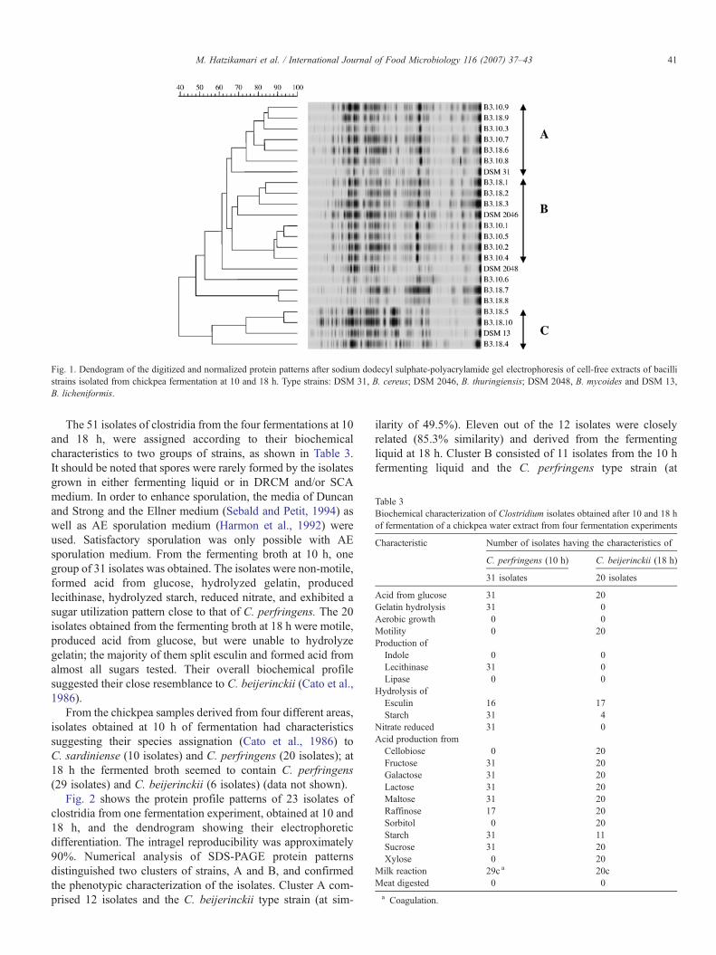
Table 3Biochemical characterization of Clostridium isolates obtained after 10 and 18 hof fermentation of a chickpea water extract from four fermentation experiments
Characteristic Number of isolates having the characteristics of
C. perfringens (10 h) C. beijerinckii (18 h)
31 isolates 20 isolates
Acid from glucose 31 20Gelatin hydrolysis 31 0Aerobic growth 0 0Motility 0 20Production ofIndole 0 0Lecithinase 31 0Lipase 0 0
Hydrolysis ofEsculin 16 17Starch 31 4
Nitrate reduced 31 0Acid production fromCellobiose 0 20Fructose 31 20Galactose 31 20Lactose 31 20Maltose 31 20Raffinose 17 20Sorbitol 0 20Starch 31 11Sucrose 31 20Xylose 0 20
Milk reaction 29c a 20cMeat digested 0 0a Coagulation.
Fig. 1. Dendogram of the digitized and normalized protein patterns after sodium dodecyl sulphate-polyacrylamide gel electrophoresis of cell-free extracts of bacillistrains isolated from chickpea fermentation at 10 and 18 h. Type strains: DSM 31, B. cereus; DSM 2046, B. thuringiensis; DSM 2048, B. mycoides and DSM 13,B. licheniformis.
41M. Hatzikamari et al. / International Journal of Food Microbiology 116 (2007) 37–43
The 51 isolates of clostridia from the four fermentations at 10and 18 h, were assigned according to their biochemicalcharacteristics to two groups of strains, as shown in Table 3.It should be noted that spores were rarely formed by the isolatesgrown in either fermenting liquid or in DRCM and/or SCAmedium. In order to enhance sporulation, the media of Duncanand Strong and the Ellner medium (Sebald and Petit, 1994) aswell as AE sporulation medium (Harmon et al., 1992) wereused. Satisfactory sporulation was only possible with AEsporulation medium. From the fermenting broth at 10 h, onegroup of 31 isolates was obtained. The isolates were non-motile,formed acid from glucose, hydrolyzed gelatin, producedlecithinase, hydrolyzed starch, reduced nitrate, and exhibited asugar utilization pattern close to that of C. perfringens. The 20isolates obtained from the fermenting broth at 18 h were motile,produced acid from glucose, but were unable to hydrolyzegelatin; the majority of them split esculin and formed acid fromalmost all sugars tested. Their overall biochemical profilesuggested their close resemblance to C. beijerinckii (Cato et al.,1986).
From the chickpea samples derived from four different areas,isolates obtained at 10 h of fermentation had characteristicssuggesting their species assignation (Cato et al., 1986) toC. sardiniense (10 isolates) and C. perfringens (20 isolates); at18 h the fermented broth seemed to contain C. perfringens(29 isolates) and C. beijerinckii (6 isolates) (data not shown).
Fig. 2 shows the protein profile patterns of 23 isolates ofclostridia from one fermentation experiment, obtained at 10 and18 h, and the dendrogram showing their electrophoreticdifferentiation. The intragel reproducibility was approximately90%. Numerical analysis of SDS-PAGE protein patternsdistinguished two clusters of strains, A and B, and confirmedthe phenotypic characterization of the isolates. Cluster A com-prised 12 isolates and the C. beijerinckii type strain (at sim-
ilarity of 49.5%). Eleven out of the 12 isolates were closelyrelated (85.3% similarity) and derived from the fermentingliquid at 18 h. Cluster B consisted of 11 isolates from the 10 hfermenting liquid and the C. perfringens type strain (at

Fig. 2. Dendogram of the digitized and normalized protein patterns after sodium dodecyl sulphate-polyacrylamide gel electrophoresis of cell-free extracts of clostridialstrains isolated from chickpea fermentation at 10 and 18 h. Type strains: DSM 756, C. perfringens; DSM 599, C. absonum; DSM 601, C. baratii; DSM 791, C.beijerinckii; DSM 793, C. aurantibutyricum; DSM 794, C. felsineum; DSM 933, C. clostridioforme; DSM 2632, C. sardiniense; DSM 7528, C. chauvoei; DSM 792,C. acetobutylicum and DSM 10702, C. butyricum.
42 M. Hatzikamari et al. / International Journal of Food Microbiology 116 (2007) 37–43
similarity of 67.5%). Thus, our results suggest that SDS-PAGEof whole cell proteins is reliable and quick method for theaccurate identification of clostridia isolates.
The B. cereus and C. perfringens populations developedduring fermentation were probably unable to produce toxins,since i.p. injection of 0.1 to 2 ml chickpea fermentation liquidtreated or not treated with trypsin were not toxic to mice. Thevarious beans used for cooking are soaked in water for 18–24 hat room temperature, during which B. cereus is able to multiply(Blakey and Priest, 1980). The emetic toxin is stable at 126 °Cfor at least 90 min (Melling and Capel, 1978), and the B. cereusisolates forming it are unable to degrade starch or fermentsalicin, and additionally exhibit only weak or no hemolysis(Ehling-Schulz et al., 2005). Very few isolates in our study wereunable to ferment starch or exhibited weak hemolysis. Thediarrhoeal enterotoxins, on the other hand, are heat-labiletoxins produced during vegetative growth of B. cereus inthe small intestine (Granum, 1994). Moreover, bacilli rarelyformed spores, in the chickpea fermenting liquid and thevegetative cells would be killed by the baking process; thiswould minimize the risk of either food poisoning or breadspoilage by B. cereus. The spore heat resistance D100 ofB. cereus is 1.2–8 min (Gibbs, 2002) and it takes almost 30 minfor the baking bread to obtain a temperature of 100 °C in the
center of the crumb (Singh, 2005) while the baking is com-pleted within 45 min. On the other hand, wild type strains ofC. perfringens isolated from the environment are almost alwaysenterotoxin negative (Skjelkvåle et al., 1979) and it is likely thatonly strains of C. perfringens that have been through repetitiveheat treatments (kitchen strains) are enterotoxin positive(Anderson et al., 1995). It is therefore tempting to speculatethat proper baking of bread made with adapted starter culturesminimizes any risk of food poisoning and any health hazardafter its consumption seems improbable.
4. Conclusions
This study has shown that during production of a traditionaldough in two steps, in the first step, when the chickpea waterextract is fermented, bacilli initially and subsequently clostridiagrow to high levels and degrade the constituents liberated fromthe chickpea into the water (Hatzikamari et al., in press) withgas as a major end product. The enzyme activities in thefermenting liquid decrease with the progress of fermentation(Hatzikamari et al., in press). The leavening of dough in thesecond step can be therefore attributed to residual enzymeactivity and enzymes liberated after cell lysis by the decliningmicrobial population of bacilli and clostridia during dough
43M. Hatzikamari et al. / International Journal of Food Microbiology 116 (2007) 37–43
raising. B. cereus and C. perfringens predominantly growingduring fermentation do not seem to form toxins, and any healthhazard after consumption of the bread, properly baked, seemsimprobable.
References
Anderson, A., Rönner, U., Granum, P.E., 1995. What problems does the foodindustry have with the spore-forming pathogens Bacillus cereus and Clos-tridium perfringens? International Journal of Food Microbiology 28,145–155.
Baldassi, L., Barbosa, M.L., Bach, E.E., Iaria, S.T., 2002. Toxigenicitycharacterisation of Clostridium perfringens from bovine isolates. Journalof Venomous Animals and Toxins 8, 112–126.
Batt, C.A., 1998. Bacillus cereus. In: Robinson, R.K., Batt, C.A., Patel, P.D.(Eds.), Encyclopedia of Food Microbiology. Academic Press, London,pp. 119–124.
Blakey, L.J., Priest, F.G., 1980. The occurrence of Bacillus cereus in some driedfoods including pulses and cereals. Journal of Applied Bacteriology 48,297–302.
Cato, P.E., George, W.L., Finegold, S.M., 1986. Genus Clostridium. In:Sneath, P.H.A., Mair, N.S., Sharpe, M.E., Holt, J.G. (Eds.), Bergey'sManual of Systematic Bacteriology, vol. 2.Williams andWilkins, Baltimore,pp. 1141–1200.
Claus, D., Berkeley, R.C.W., 1986. Genus Bacillus. In: Sneath, P.H.A., Mair, N.S.,Sharpe, M.E., Holt, J.G. (Eds.), Bergey's Manual of Systematic Bacteriology,vol. 2. Williams and Wilkins, Baltimore, pp. 1105–1138.
Desphande, S.S., Damodaran, S., 1990. Food legumes: chemistry andtechnology. In: Pomeranz, X. (Ed.), Advances in Cereal Science andTechnology, vol. X. American Association of Cereal Chemists, St. Paul,MN, pp. 147–241.
Ehling-Schulz, M., Svensson, B., Guinebretiere, M.H., Lindbäck, T., Andersson,M., Schultz, A., Fricker, M., Christiansson, A., Granum, P.E., Märtlbauer, E.,Nguyen-The, C., Salkinoja-Salonen, M., Scherer, S., 2005. Emetic toxinformation of Bacillus cereus is restricted to a single evolutionary lineage ofclosely related strains. Microbiology 151, 183–197.
Eisgruber, H., Reuter, G., 1995. A selective medium for the detection andenumeration of mesophilic sulphite-reducing clostridia in food monitoringprograms. Food Research International 28, 219–226.
Fox, A., Black, G.E., Fox, K., Rostovtseva, S., 1993. Determination ofcarbohydrate patterns of Bacillus anthracis and Bacillus cereus includingidentification of O-methyl methylpentoses by using gas chromatography-mass spectrometry. Journal of Clinical Microbiology 31, 887–894.
Freame, B., Fitzpatrick, B.W.F., 1971. The use of differential reinforcedclostridial medium for the isolation and enumeration of clostridia from food.In: Shapton, D.A., Board, R.G. (Eds.), Isolation of Anaerobes. AcademicPress, London, pp. 49–55.
Gibbs, P., 2002. Characteristics of spore-forming bacteria. In: Blackburn, C.de W.,McClure, P. (Eds.), Foodborne Pathogens.Hazards, RiskAnalysis andControl.CRC Press, New York, pp. 417–435.
Gordon, R.E., Haynes, W.C., Pang, C.H.-N., 1973. The Genus Bacillus.Agriculture Handbook, vol. 427. Agricultural Research Service, UnitedStates Department of Agriculture, Washington, D.C.
Granum, P.E., 1994. Bacillus cereus and its toxins. Journal of AppliedBacteriology Symposium Supplement 23, 61S–66S.
Harmon, S.M., Kauter, D.A., Golden, D.A., Rhodehamel, E.J., 1992. Bacilluscereus, FDA Bacteriological Analytical Manual, 7th ed. AOAC Interna-tional, USA. Chapter 14.
Hatzikamari, M., Kyriakidis, D.A., Tzanetakis, N., Biliaderis, C.G., Litopoulou-Tzanetaki, E., in press. Biochemical changes during a submerged chickpeafermentation used as a leavening agent for bread production. European FoodResearch and Technology. doi:10.1007/s00217-006-0363-4.
Katsaboxakis, K., Mallidis, K., 1996. The microflora of soak water duringnatural fermentation of coarsely ground chickpea (Cicer arietinum) seeds.Letters in Applied Microbiology 23, 261–265.
Laemmli, U.K., 1970. Cleavage of structural protein during the assembly of thehead of bacteriophage T4. Nature 227, 680–685.
Lawrence, D., Heitefuss, S., Seifert, H.S., 1991. Differentiation of Bacillusanthracis from Bacillus cereus by gas chromatographic whole-cell fatty acidanalysis. Journal of Clinical Microbiology 29, 1508–1512.
Logan, N.A., Berkeley, R.C.W., 1984. Identification of Bacillus strains usingAPI systematic. Journal of General Microbiology 130, 1871–1882.
Lowry, O.H., Rosebrough, N.J., Farr, A.L., Randall, R.J., 1951. Proteinmeasurement with folin phenol reagent. Journal of Biological Chemistry193, 265–275.
Matar, G.M., Slieman, T.A., Nabbut, N.H., 1996. Subtyping Bacillus cereus bytotal protein patterns and arbitrary primer polymerase chain reaction.European Journal of Epidemiology 12, 309–314.
Melling, J., Capel, B.J., 1978. Characteristics of Bacillus cereus emetic toxin.FEMS Microbiological Letters 4, 133–135.
Omafuvbe, B.O., Shonukan, O.O., Abiose, S.H., 2000. Microbiological andbiochemical changes in the traditional fermentation of soybean for ‘soy-daddawa’ — Nigerian food condiment. Food Microbiology 17, 469–474.
Pot, B., Vandame, P., Kersters, K., 1994. Analysis of electrophoretic, wholeorganism protein fingerprints. In: Goodfellow, M., O'Donner, A.G. (Eds.),Chemical Methods in Prokaryotic Systematics. J. Wiley, Chichester,pp. 493–521.
Reddy, N.R., Pierson, M.D., Sathe, S.K., Salunke, D.K., 1982. Legume-basedfermented foods: their preparation and nutritional quality. Critical Reviewsin Food Science and Nutrition 17, 335–355.
Reuter, G., 1985. Elective and selective media for lactic acid bacteria.International Journal of Food Microbiology 2, 55–68.
Saraswat, D.S., Clark Jr., W.S., Reinbold, G.W., 1963. Selection of a medium forthe isolation and enumeration of enterococci in dairy products. Journal ofMilk and Food Technology 26, 114–118.
Sarkar, P.K., Hasenack, B., Nout, M.J.R., 2002. Diversity and functionality ofBacillus and related genera isolated from spontaneously fermented soybeans(Indian Kinema) and locust bean (African Soubala). International Journal ofFood Microbiology 77, 175–186.
Sebald, M., Petit, J.-C., 1994. Laboratory Methods for Anaerobic Bacteria andtheir Identification. Institute Pasteur, France.
Sherfi, S.A., Hamad, S.H., 2001. Microbiological and biochemical studies onGergoush fermentation. International Journal of Food Microbiology 67,247–252.
Singh, H., 2005. A study of changes in wheat protein during bread baking usingSE-HPLC. Food Chemistry 90, 247–250.
Skjelkvåle, R., Stringer, M.F., Smart, J.L., 1979. Enterotoxin production bylecithinase-positive and lecithinase-negative Clostridium perfringens isolatedfrom food poisoning outbreaks and other sources. Journal of AppliedBacteriology 69, 550–556.
Tulbek, M.C., Hall, C., Schwarz, J.G., 2003. The use of fermented chickpea ondough rheology and wheat pan bread quality. Abstract in IFT AnnualMeeting Chicago, July 13–16.
Zamora, A.F., Fields, M.L., 1979. Nutritive quality of fermented cowpeas(Vinga sinensis) and chickpeas (Cicer arietinum L.). Journal of FoodScience 44, 234–236.