
Cerebral Cortex Principles of Operation - Oxford Centre for ...
154
OUP-FIRST UNCORRECTED PROOF, June 17, 2016 Cerebral Cortex Principles of Operation Edmund T. Rolls Oxford Centre for Computational Neuroscience, Oxford, UK 3
-
Upload
khangminh22 -
Category
Documents
-
view
1 -
download
0
Transcript of Cerebral Cortex Principles of Operation - Oxford Centre for ...
OUP-FIRST UNCORRECTED PROOF, June 17, 2016
Cerebral CortexPrinciples ofOperation
Edmund T. RollsOxford Centre for Computational Neuroscience, Oxford, UK
3

3
Great Clarendon Street, Oxford, OX2 6DP,United Kingdom
Oxford University Press is a department of the University of Oxford.It furthers the University’s objective of excellence in research, scholarship,and education by publishing worldwide. Oxford is a registered trade mark ofOxford University Press in the UK and in certain other countries
© Edmund Rolls 2016
The moral rights of the author have been asserted
First Edition published in 2016Impression: 1
All rights reserved. No part of this publication may be reproduced, stored ina retrieval system, or transmitted, in any form or by any means, without theprior permission in writing of Oxford University Press, or as expressly permittedby law, by licence or under terms agreed with the appropriate reprographicsrights organization. Enquiries concerning reproduction outside the scope of theabove should be sent to the Rights Department, Oxford University Press, at theaddress above
You must not circulate this work in any other formand you must impose this same condition on any acquirer
Published in the United States of America by Oxford University Press198 Madison Avenue, New York, NY 10016, United States of America
British Library Cataloguing in Publication DataData available
Library of Congress Control Number: 2016944945
ISBN 978–0–19–878485–2
Printed and bound byCPI Group (UK) Ltd, Croydon, CR0 4YY
Oxford University Press makes no representation, express or implied, that thedrug dosages in this book are correct. Readers must therefore always checkthe product information and clinical procedures with the most up-to-datepublished product information and data sheets provided by the manufacturersand the most recent codes of conduct and safety regulations. The authors andthe publishers do not accept responsibility or legal liability for any errors in thetext or for the misuse or misapplication of material in this work. Except whereotherwise stated, drug dosages and recommendations are for the non-pregnantadult who is not breast-feeding
Links to third party websites are provided by Oxford in good faith andfor information only. Oxford disclaims any responsibility for the materialscontained in any third party website referenced in this work.
Preface
The overall aim of this book is to provide insight into the principles of operation of the cerebralcortex. These are key to understanding how we, as humans, function.
There have been few previous attempts to set out some of the important principles ofoperation of the cortex, and this book is pioneering. I have asked some of the leading investigators in neuroscience about their views on this, and most have not had many well formulatedanswers or hypotheses. As clear hypotheses are needed in this most important area of 21stcentury science, how our brains work, I have formulated a set of hypotheses to guide thinkingand future research. I present evidence for many of the hypotheses, but at the same time wemust all recognise that hypotheses and theory in science are there to be tested, and hopefully refined rather than rejected. Nevertheless, such theories and hypotheses are essential toprogress, and it is in this frame of reference that I present the theories, hypotheses, and ideasthat I have produced and collected together.
This book focusses on the principles of operation of the cerebral cortex, because at thistime it is possible to propose and describe many principles, and many are likely to stand thetest of time, and provide a foundation I believe for further developments, even if some needto be changed. In this context, I have not attempted to produce an overall theory of operationof the cerebral cortex, because at this stage of our understanding, such a theory would beincorrect or incomplete. I believe though that many of the principles will be important, andthat many will provide the foundations for more complete theories of the operation of thecerebral cortex.
Given that many different principles of operation of the cortex are proposed in this book,with often several principles in each Chapter, the reader may find it convenient to take oneChapter at a time, and think about the issues raised in each Chapter, as the overall enterpriseis large. The Highlights sections provided at the end of each Chapter may be useful in helpingthe reader to appreciate the different principles being considered in each Chapter.
To understand how the cortex works, including how it functions in perception, memory,attention, decisionmaking, and cognitive functions, it is necessary to combine differentapproaches, including neural computation. Neurophysiology at the single neuron level isneeded because this is the level at which information is exchanged between the computingelements of the brain. Evidence from the effects of brain damage, including that availablefrom neuropsychology, is needed to help understand what different parts of the system do,and indeed what each part is necessary for. Neuroimaging is useful to indicate where in thehuman brain different processes take place, and to show which functions can be dissociatedfrom each other. Knowledge of the biophysical and synaptic properties of neurons is essentialto understand how the computing elements of the brain work, and therefore what the buildingblocks of biologically realistic computational models should be. Knowledge of the anatomicaland functional architecture of the cortex is needed to show what types of neuronal networkactually perform the computation. And finally the approach of neural computation is needed,as this is required to link together all the empirical evidence to produce an understandingof how the system actually works. This book utilizes evidence from all these disciplinesto develop an understanding of how different types of memory, perception, attention, anddecisionmaking are implemented by processing in the cerebral cortex.
Prefaceiv |
I emphasize that to understand how memory, perception, attention, decisionmaking, cognitive functions, and actions are produced in the cortex, we are dealing with largescalecomputational systems with interactions between the parts, and that this understanding requires analysis at the computational and global level of the operation of many neurons toperform together a useful function. Understanding at the molecular level is important forhelping to understand how these largescale computational processes are implemented in thebrain, but will not by itself give any account of what computations are performed to implement these cognitive functions. Instead, understanding cognitive functions such as objectrecognition, memory recall, attention, and decisionmaking requires single neuron data to beclosely linked to computational models of how the interactions between large numbers ofneurons and many networks of neurons allow these cognitive problems to be solved. Thesingle neuron level is important in this approach, for the single neurons can be thought of asthe computational units of the system, and is the level at which the information is exchangedby the spiking activity between the computational elements of the brain. The single neuronlevel is therefore, because it is the level at which information is communicated between thecomputing elements of the brain, the fundamental level of information processing, and thelevel at which the information can be read out (by recording the spiking activity) in order tounderstand what information is being represented and processed in each brain area.
With its focus on how the brain and especially how the cortex works at the computationalneuroscience level, this book is distinct from the many excellent books on neurosciencethat describe much evidence about brain structure and function, but do not aim to providean understanding of how the brain works at the computational level. This book aims toforge an understanding of how some key brain systems may operate at the computationallevel, so that we can understand how the cortex actually performs some of its complexand necessarily computational functions in memory, perception, attention, decisionmaking,cognitive functions, and actions.
A test of whether one’s understanding is correct is to simulate the processing on a computer,and to show whether the simulation can perform the tasks of cortical systems, and whether thesimulation has similar properties to the real cortex. The approach of neural computation leadsto a precise definition of how the computation is performed, and to precise and quantitativetests of the theories produced. How memory systems in the cortex work is a paradigm exampleof this approach, because memorylike operations which involve altered functionality as aresult of synaptic modification are at the heart of how many computations in the cortexare performed. It happens that attention and decisionmaking can be understood in termsof interactions between and fundamental operations in memory systems in the cortex, andtherefore it is natural to treat these areas of cognitive neuroscience in this book. The samefundamental concepts based on the operation of neuronal circuitry can be applied to all thesefunctions, as is shown in this book.
One of the distinctive properties of this book is that it links the neural computationapproach not only firmly to neuronal neurophysiology, which provides much of the primarydata about how the cortex operates, but also to psychophysical studies (for example ofattention); to neuropsychological studies of patients with brain damage; and to functionalmagnetic resonance imaging (fMRI) (and other neuroimaging) approaches. The empiricalevidence that is brought to bear is largely from nonhuman primates and from humans,because of the considerable similarity of their cortical systems.
In this book, I have not attempted to produce a single computational theory of how thecortex operates. Instead, I have highlighted many different principles of cortical function,most of which are likely to be building blocks of how our cortex operates. The reason forthis approach is that many of the principles may well be correct, and useful in understandinghow the cortex operates, but some might turn out not to be useful or correct. The aim of this
| vPreface
book is therefore to propose some of the fundamental principles of operation of the cerebralcortex, many or most of which will provide a foundation for understanding the operation ofthe cortex, rather than to produce a single theory of operation of the cortex, which might bedisproved if any one of its elements was found to be weak.
The overall aims of the book are developed further, and the plan of the book is described,in Chapter 1, Section 1.1. Some of the main Principles of Operation of the Cerebral Cortexthat I describe can be found in the titles of Chapters 2–22; but in practice, most Chaptersinclude several Principles of Operation, which will appear in the Highlights to each Chapter.Section 26.5 may be useful in addition to the Highlights, for Section 26.5 draws together ina synthesis some of the Principles of Operation of the Cerebral Cortex that are described inthe book. Further evidence on how these principles are relevant to the operation of differentcortical areas and systems and operate together is provided in Chapters 24–25. In theseChapters, the operation of two major cortical systems, those involved in memory and in visualobject recognition, are considered to illustrate how the principles are combined to implementtwo different key cortical functions. The Appendices provide some of the more formal andquantitative properties of the operation of neuronal systems, and are provided because theyprovide a route to a deeper understanding on the principles, and to enable the presentationin earlier Chapters to be at a readily approachable level. The Appendices describe many ofthe building blocks of the neurocomputational approach, and are designed to be useful forteaching. Appendix D describes Matlab software that has been made available with this bookto provide simple demonstrations of the operation of some key neuronal networks related tocortical function. The programs are available at http://www.oxcns.org.
Part of the material described in the book reflects work performed in collaboration withmany colleagues, whose tremendous contributions are warmly appreciated. The contributionsof many will be evident from the references cited in the text. Especial appreciation is due toGustavo Deco, Simon M. Stringer, and Alessandro Treves who have contributed greatly inan always interesting and fruitful research collaboration on computational aspects of brainfunction, and to many neurophysiology and functional neuroimaging colleagues who havecontributed to the empirical discoveries that provide the foundation to which the computationalneuroscience must always be closely linked, and whose names are cited throughout the text.Much of the work described would not have been possible without financial support from anumber of sources, particularly the Medical Research Council of the UK, the Human FrontierScience Program, the Wellcome Trust, and the James S. McDonnell Foundation. I am alsograteful to many colleagues who I have consulted while writing this book, including Joel Price(Washington University School of Medicine), and Donald Wilson (New York University). DrPatrick Mills is warmly thanked for his comments on the text. Section 24.3.12 on ars memoriaeis warmly dedicated to my colleagues at Corpus Christi College, Oxford. The book was typesetby the author using LATEXand WinEdt.
The cover includes part of the picture Pandora painted in 1896 by J. W. Waterhouse. Themetaphor is to look inside the system of the mind and the brain, in order to understand howthe brain functions, and thereby better to understand and treat its disorders. The cover alsoincludes an image of the dendritic morphology of excitatory neurons in S1 whisker barrelcortex (Fig. 1.14) (adapted from Marcel Oberlaender, Christiaan P.J. de Kock, Randy M.Bruno, Alejandro Ramirez, Hanno S. Meyer, Vincent J. Dercksen, Moritz Helmstaedter andBert Sakmann, Cell typespecific threedimensional structure of thalamocortical circuits ina column of rat vibrissal cortex, Cerebral Cortex, 2012, Vol. 22, issue 10, pp. 2375–2391,by permission of Oxford University Press). The cover also includes a diagram of thecomputational circuitry of the hippocampus by the author (Fig. 24.1). The aim of thesesecond two images is to highlight the importance of moving from the anatomy of the cortexusing all the approaches available including neuronal network models that address and

Prefacevi |
incorporate neurophysiological discoveries to lead to an understanding of how the cortexoperates computationally.
Updates to and .pdfs of many of the publications cited in this book are available athttp://www.oxcns.org. Updates and corrections to the text and notes are also available athttp://www.oxcns.org.
I dedicate this work to the overlapping group: my family, friends, and colleagues – in salutempraesentium, in memoriam absentium.
Contents
1 Introduction 11.1 Principles of operation of the cerebral cortex: introduction and plan 1
1.2 Neurons 4
1.3 Neurons in a network 6
1.4 Synaptic modification 8
1.5 Longterm potentiation and longterm depression 9
1.6 Distributed representations 141.6.1 Definitions 141.6.2 Advantages of different types of coding 15
1.7 Neuronal network approaches versus connectionism 16
1.8 Introduction to three neuronal network architectures 17
1.9 Systemslevel analysis of brain function 181.9.1 Ventral cortical visual stream 191.9.2 Dorsal cortical visual stream 211.9.3 Hippocampal memory system 231.9.4 Frontal lobe systems 231.9.5 Brodmann areas 24
1.10 The fine structure of the cerebral neocortex 271.10.1 The fine structure and connectivity of the neocortex 271.10.2 Excitatory cells and connections 271.10.3 Inhibitory cells and connections 291.10.4 Quantitative aspects of cortical architecture 321.10.5 Functional pathways through the cortical layers 341.10.6 The scale of lateral excitatory and inhibitory effects, and modules 38
1.11 Highlights 39
2 Hierarchical organization 402.1 Introduction 40
2.2 Hierarchical organization in sensory systems 412.2.1 Hierarchical organization in the ventral visual system 412.2.2 Hierarchical organization in the dorsal visual system 462.2.3 Hierarchical organization of taste processing 482.2.4 Hierarchical organization of olfactory processing 572.2.5 Hierarchical multimodal convergence of taste, olfaction, and vision 592.2.6 Hierarchical organization of auditory processing 64
2.3 Hierarchical organization of reward value processing 67
2.4 Hierarchical organization of connections to the frontal lobe for shortterm memory 68
2.5 Highlights 69
3 Localization of function 723.1 Hierarchical processing 72
Contentsviii |
3.2 Shortrange neocortical recurrent collaterals 72
3.3 Topographic maps 72
3.4 Modularity 72
3.5 Lateralization of function 73
3.6 Ventral and dorsal cortical areas 73
3.7 Highlights 74
4 Recurrent collateral connections and attractor networks 754.1 Introduction 75
4.2 Attractor networks implemented by the recurrent collaterals 75
4.3 Evidence for attractor networks implemented by recurrent collateral connections 764.3.1 Shortterm Memory 774.3.2 Longterm Memory 804.3.3 DecisionMaking 80
4.4 The storage capacity of attractor networks 80
4.5 A global attractor network in hippocampal CA3, but local in neocortex 81
4.6 The speed of operation of cortical attractor networks 83
4.7 Dilution of recurrent collateral cortical connectivity 83
4.8 Selforganizing topographic maps in the neocortex 85
4.9 Attractors formed by forward and backward connections between cortical areas? 85
4.10 Interacting attractor networks 86
4.11 Highlights 90
5 The noisy cortex: stochastic dynamics, decisions, and memory 915.1 Reasons why the brain is inherently noisy and stochastic 91
5.2 Attractor networks, energy landscapes, and stochastic neurodynamics 95
5.3 A multistable system with noise 98
5.4 Stochastic dynamics and the stability of shortterm memory 1015.4.1 Analysis of the stability of shortterm memory 1035.4.2 Stability and noise in a model of shortterm memory 104
5.5 Longterm memory recall 106
5.6 Stochastic dynamics and probabilistic decisionmaking in an attractor network 1065.6.1 Decisionmaking in an attractor network 1075.6.2 Theoretical framework: a probabilistic attractor network 1075.6.3 Stationary multistability analysis: meanfield 1105.6.4 Integrateandfire simulations of decisionmaking: spiking dynamics 1125.6.5 Reaction times of the neuronal responses 1165.6.6 Percentage correct 1175.6.7 Finitesize noise effects 1175.6.8 Comparison with neuronal data during decisionmaking 1195.6.9 Testing the model of decisionmaking with human functional neuroimaging 1225.6.10 Decisions based on confidence in one’s decisions: selfmonitoring 1295.6.11 Decisionmaking with multiple alternatives 1315.6.12 The matching law 1325.6.13 Comparison with other models of decisionmaking 132
5.7 Perceptual decisionmaking and rivalry 134
5.8 Symmetrybreaking 135
| ixContents
5.9 The evolutionary utility of probabilistic choice 135
5.10 Selection between conscious vs unconscious decisionmaking, and free will 136
5.11 Creative thought 137
5.12 Unpredictable behaviour 138
5.13 Predicting a decision before the evidence is applied 138
5.14 Highlights 140
6 Attention, shortterm memory, and biased competition 1416.1 Bottomup attention 141
6.2 Topdown attention – biased competition 1436.2.1 The biased competition hypothesis 1436.2.2 Biased competition – single neuron studies 1456.2.3 Nonspatial attention 1476.2.4 Biased competition – fMRI 1496.2.5 A basic computational module for biased competition 1496.2.6 Architecture of a model of attention 1506.2.7 Simulations of basic experimental findings 1546.2.8 Object recognition and spatial search 1586.2.9 The neuronal and biophysical mechanisms of attention 1636.2.10 ‘Serial’ vs ‘parallel’ attentional processing 167
6.3 Topdown attention – biased activation 1716.3.1 Selective attention can selectively activate different cortical areas 1716.3.2 Sources of the topdown modulation of attention 1736.3.3 Granger causality used to investigate the source of the topdown biasing 1746.3.4 Topdown cognitive modulation 1756.3.5 A topdown biased activation model of attention 178
6.4 Conclusions 181
6.5 Highlights 184
7 Diluted connectivity 1867.1 Introduction 186
7.2 Diluted connectivity and the storage capacity of attractor networks 1877.2.1 The autoassociative or attractor network architecture being studied 1877.2.2 The storage capacity of attractor networks with diluted connectivity 1887.2.3 The network simulated 1907.2.4 The effects of diluted connectivity on the capacity of attractor networks 1927.2.5 Synthesis of the effects of diluted connectivity in attractor networks 197
7.3 The effects of dilution on the capacity of pattern association networks 198
7.4 The effects of dilution on the performance of competitive networks 2017.4.1 Competitive Networks 2017.4.2 Competitive networks without learning but with diluted connectivity 2027.4.3 Competitive networks with learning and with diluted connectivity 2037.4.4 Competitive networks with learning and with full (undiluted) connectivity 2057.4.5 Overview and implications of diluted connectivity in competitive networks 206
7.5 The effects of dilution on the noise in attractor networks 207
7.6 Highlights 207
8 Coding principles 2098.1 Types of encoding 209
Contentsx |
8.2 Place coding with sparse distributed firing rate representations 2108.2.1 Reading the code used by single neurons 2108.2.2 Understanding the code provided by populations of neurons 214
8.3 Synchrony, coherence, and binding 221
8.4 Principles by which the representations are formed 222
8.5 Information encoding in the human cortex 223
8.6 Highlights 226
9 Synaptic modification for learning 2279.1 Introduction 227
9.2 Associative synaptic modification implemented by longterm potentiation 227
9.3 Forgetting in associative neural networks, and memory reconsolidation 2289.3.1 Forgetting 2289.3.2 Factors that influence synaptic modification 2309.3.3 Reconsolidation 232
9.4 Spiketiming dependent plasticity 233
9.5 Longterm synaptic depression in the cerebellar cortex 233
9.6 Reward prediction error learning 2349.6.1 Blocking and deltarule learning 2349.6.2 Dopamine neuron firing and reward prediction error learning 234
9.7 Highlights 240
10 Synaptic and neuronal adaptation and facilitation 24110.1 Mechanisms for neuronal adaptation and synaptic depression and facilitation 241
10.1.1 Sodium inactivation leading to neuronal spikefrequency adaptation 24110.1.2 Calcium activated hyperpolarizing potassium current 24210.1.3 Shortterm synaptic depression and facilitation 243
10.2 Shortterm depression of thalamic input to the cortex 244
10.3 Relatively little adaptation in primate cortex when it is operating normally 244
10.4 Acetylcholine, noradrenaline, and other modulators of adaptation and facilitation 24710.4.1 Acetylcholine 24710.4.2 Noradrenergic neurons 248
10.5 Synaptic depression and sensoryspecific satiety 249
10.6 Neuronal and synaptic adaptation, and the memory for sequential order 250
10.7 Destabilization of shortterm memory by adaptation or synaptic depression 250
10.8 Nonreward computation in the orbitofrontal cortex using synaptic depression 251
10.9 Synaptic facilitation and a multipleitem shortterm memory 253
10.10 Synaptic facilitation in decisionmaking 253
10.11 Highlights 254
11 Backprojections in the neocortex 25511.1 Architecture 255
11.2 Learning 257
11.3 Recall 258
11.4 Semantic priming 259
11.5 Topdown Attention 259
11.6 Autoassociative storage, and constraint satisfaction 261
| xiContents
11.7 Highlights 261
12 Memory and the hippocampus 26212.1 Introduction 262
12.2 Hippocampal circuitry and connections 262
12.3 The hippocampus and episodic memory 262
12.4 Autoassociation in the CA3 network for episodic memory 263
12.5 The dentate gyrus as a pattern separation mechanism, and neurogenesis 265
12.6 Rodent place cells vs primate spatial view cells 265
12.7 Backprojections, and the recall of information from the hippocampus to neocortex 266
12.8 Subcortical structures connected to the hippocampocortical memory system 267
12.9 Highlights 267
13 Limited neurogenesis in the adult cortex 26913.1 No neurogenesis in the adult neocortex 269
13.2 Limited neurogenesis in the adult hippocampal dentate gyrus 269
13.3 Neurogenesis in the chemosensing receptor systems 270
13.4 Highlights 271
14 Invariance learning and vision 27214.1 Hierarchical cortical organization with convergence 272
14.2 Feature combinations 272
14.3 Sparse distributed representations 273
14.4 Selforganization by feedforward processing without a teacher 273
14.5 Learning guided by the statistics of the visual inputs 274
14.6 Bottom up saliency 275
14.7 Lateral interactions shape receptive fields 276
14.8 Topdown selective attention vs feedforward processing 277
14.9 Topological maps to simplify connectivity 278
14.10 Biologically decodable output representations 279
14.11 Highlights 279
15 Emotion, motivation, reward value, pleasure, and their mechanisms 28115.1 Emotion, reward value, and their evolutionary adaptive utility 281
15.2 Motivation and reward value 283
15.3 Principles of cortical design for emotion and motivation 283
15.4 Objects are first represented independently of reward value 284
15.5 Specialized systems for face identity and expression processing in primates 286
15.6 Unimodal processing to the object level before multimodal convergence 287
15.7 A common scale for reward value 287
15.8 Sensoryspecific satiety 287
15.9 Economic value is represented in the orbitofrontal cortex 288
15.10 Neuroeconomics vs classical microeconomics 288
15.11 Output systems influenced by orbitofrontal cortex reward value representations 289
15.12 Decisionmaking about rewards in the anterior orbitofrontal cortex 291
Contentsxii |
15.13 Probabilistic emotionrelated decisionmaking 292
15.14 Nonreward, error, neurons in the orbitofrontal cortex 292
15.15 Reward reversal learning in the orbitofrontal cortex 296
15.16 Dopamine neurons and emotion 301
15.17 The explicit reasoning system vs the emotional system 301
15.18 Pleasure 302
15.19 Personality relates to differences in sensitivity to rewards and punishers 302
15.20 Highlights 303
16 Noise in the cortex, stability, psychiatric disease, and aging 30516.1 Stochastic noise, attractor dynamics, and schizophrenia 305
16.1.1 Introduction 30516.1.2 A dynamical systems hypothesis of the symptoms of schizophrenia 30716.1.3 The depth of the basins of attraction: meanfield flow analysis 30816.1.4 Decreased stability produced by reduced NMDA conductances 30916.1.5 Increased distractibility produced by reduced NMDA conductances 31116.1.6 Synthesis: network instability and schizophrenia 312
16.2 Stochastic noise, attractor dynamics, and obsessivecompulsive disorder 31616.2.1 Introduction 31616.2.2 A hypothesis about obsessivecompulsive disorder 31716.2.3 Glutamate and increased depth of the basins of attraction 31916.2.4 Synthesis on obsessivecompulsive disorder 322
16.3 Stochastic noise, attractor dynamics, and depression 32516.3.1 Introduction 32516.3.2 A nonreward attractor theory of depression 32816.3.3 Evidence consistent with the theory 32916.3.4 Relation to other brain systems implicated in depression 33116.3.5 Implications for treatments 33216.3.6 Mania and bipolar disorder 333
16.4 Stochastic noise, attractor dynamics, and aging 33516.4.1 NMDA receptor hypofunction 33516.4.2 Dopamine 33816.4.3 Impaired synaptic modification 33816.4.4 Cholinergic function and memory 339
16.5 Highlights 343
17 Syntax and Language 34517.1 Neurodynamical hypotheses about language and syntax 345
17.1.1 Binding by synchrony? 34517.1.2 Syntax using a place code 34617.1.3 Temporal trajectories through a state space of attractors 34717.1.4 Hypotheses about the implementation of language in the cerebral cortex 347
17.2 Tests of the hypotheses – a model 35117.2.1 Attractor networks with stronger forward than backward connections 35117.2.2 The operation of a single attractor network module 35317.2.3 Spike frequency adaptation mechanism 355
17.3 Tests of the hypotheses – findings with the model 35517.3.1 A production system 35517.3.2 A decoding system 356
17.4 Evaluation of the hypotheses 359
| xiiiContents
17.5 Highlights 363
18 Evolutionary trends in cortical design and principles of operation 36418.1 Introduction 364
18.2 Different types of cerebral neocortex: towards a computational understanding 36418.2.1 Neocortex or isocortex 36518.2.2 Olfactory (pyriform) cortex 37118.2.3 Hippocampal cortex 374
18.3 Addition of areas in the neocortical hierarchy 376
18.4 Evolution of the orbitofrontal cortex 378
18.5 Evolution of the taste and flavour system 37918.5.1 Principles 37918.5.2 Taste processing in rodents 380
18.6 Evolution of the temporal lobe cortex 381
18.7 Evolution of the frontal lobe cortex 382
18.8 Highlights 382
19 Genetics and selforganization build the cortex 38519.1 Introduction 385
19.2 Hypotheses about the genes that build cortical neural networks 386
19.3 Genetic selection of neuronal network parameters 390
19.4 Simulation of the evolution of neural networks using a genetic algorithm 39119.4.1 The neural networks 39119.4.2 The specification of the genes 39219.4.3 The genetic algorithm, and general procedure 39719.4.4 Pattern association networks 39819.4.5 Autoassociative networks 40019.4.6 Competitive networks 400
19.5 Evaluation of the genebased evolution of singlelayer networks 401
19.6 The genebased evolution of multilayer cortical systems 403
19.7 Highlights 404
20 Cortex versus basal ganglia design for selection 40620.1 Systemslevel architecture of the basal ganglia 406
20.2 What computations are performed by the basal ganglia? 408
20.3 How do the basal ganglia perform their computations? 410
20.4 Comparison of selection in the basal ganglia and cerebral cortex 413
20.5 Highlights 415
21 Sleep and Dreaming 41621.1 Is sleep necessary for cortical function? 416
21.2 Is sleep involved in memory consolidation? 417
21.3 Dreams 418
21.4 Highlights 419
22 Which cortical computations underlie consciousness? 42022.1 Introduction 420
Contentsxiv |
22.2 A HigherOrder Syntactic Thought (HOST) theory of consciousness 42122.2.1 Multiple routes to action 42122.2.2 A computational hypothesis of consciousness 42322.2.3 Adaptive value of processing that is related to consciousness 42522.2.4 Symbol grounding 42622.2.5 Qualia 42822.2.6 Pathways 42922.2.7 Consciousness and causality 43022.2.8 Consciousness and higherorder syntactic thoughts 431
22.3 Selection between conscious vs unconscious decisionmaking systems 43222.3.1 Dual major routes to action: implicit and explicit 43222.3.2 The Selfish Gene vs The Selfish Phenotype 43922.3.3 Decisionmaking between the implicit and explicit systems 440
22.4 Determinism 441
22.5 Free will 442
22.6 Content and meaning in representations 443
22.7 The causal role of consciousness and the relation between the mind and the brain 445
22.8 Comparison with other theories of consciousness 44722.8.1 Higherorder thought theories 44722.8.2 Oscillations and temporal binding 44922.8.3 A high neural threshold for information to reach consciousness 45022.8.4 James–Lange theory and Damasio’s somatic marker hypothesis 45122.8.5 LeDoux’s approach to emotion and consciousness 45122.8.6 Panksepp’s approach to emotion and consciousness 45222.8.7 Global workspace theories of consciousness 45222.8.8 Monitoring and consciousness 452
22.9 Highlights 453
23 Cerebellar cortex 45523.1 Introduction 455
23.2 Architecture of the cerebellum 45623.2.1 The connections of the parallel fibres onto the Purkinje cells 45623.2.2 The climbing fibre input to the Purkinje cell 45723.2.3 The mossy fibre to granule cell connectivity 457
23.3 Modifiable synapses of parallel fibres onto Purkinje cell dendrites 460
23.4 The cerebellar cortex as a perceptron 460
23.5 Highlights: differences between cerebral and cerebellar cortex microcircuitry 461
24 The hippocampus and memory 46324.1 Introduction 463
24.2 Systemslevel functions of the hippocampus 46424.2.1 Systemslevel anatomy 46524.2.2 Evidence from the effects of damage to the hippocampus 46724.2.3 The necessity to recall information from the hippocampus 46824.2.4 Systemslevel neurophysiology of the primate hippocampus 47024.2.5 Head direction cells in the presubiculum 47824.2.6 Perirhinal cortex, recognition memory, and longterm familiarity memory 479
24.3 A theory of the operation of hippocampal circuitry as a memory system 48624.3.1 Hippocampal circuitry 48724.3.2 Entorhinal cortex 488
| xvContents
24.3.3 CA3 as an autoassociation memory 49024.3.4 Dentate granule cells 50924.3.5 CA1 cells 51524.3.6 Recoding in CA1 to facilitate retrieval to the neocortex 51524.3.7 Backprojections to the neocortex, memory recall, and consolidation 52024.3.8 Backprojections to the neocortex – quantitative aspects 52324.3.9 Simulations of hippocampal operation 52624.3.10 The learning of spatial view and place cell representations 52824.3.11 Linking the inferior temporal visual cortex to spatial view and place cells 52924.3.12 A scientific theory of the art of memory: scientia artis memoriae 531
24.4 Tests of the theory of hippocampal cortex operation 53124.4.1 Dentate gyrus (DG) subregion of the hippocampus 53124.4.2 CA3 subregion of the hippocampus 53524.4.3 CA1 subregion of the hippocampus 542
24.5 Evaluation of the theory of hippocampal cortex operation 54624.5.1 Tests of the theory by hippocampal system subregion analyses 54624.5.2 Comparison with other theories of hippocampal function 548
24.6 Highlights 552
25 Invariant visual object recognition learning 55425.1 Introduction 554
25.2 Invariant representations of faces and objects in the inferior temporal visual cortex 55525.2.1 Processing to the inferior temporal cortex in the primate visual system 55525.2.2 Translation invariance and receptive field size 55625.2.3 Reduced translation invariance in natural scenes 55725.2.4 Size and spatial frequency invariance 56025.2.5 Combinations of features in the correct spatial configuration 56125.2.6 A viewinvariant representation 56225.2.7 Learning in the inferior temporal cortex 56525.2.8 Distributed encoding 56825.2.9 Face expression, gesture, and view 57225.2.10 Specialized regions in the temporal cortical visual areas 572
25.3 Approaches to invariant object recognition 57625.3.1 Feature spaces 57725.3.2 Structural descriptions and syntactic pattern recognition 57825.3.3 Template matching and the alignment approach 58025.3.4 Invertible networks that can reconstruct their inputs 58125.3.5 Feature hierarchies 582
25.4 Hypotheses about object recognition mechanisms 582
25.5 Computational issues in feature hierarchies 58625.5.1 The architecture of VisNet 58725.5.2 Initial experiments with VisNet 59625.5.3 The optimal parameters for the temporal trace used in the learning rule 60325.5.4 Different forms of the trace learning rule, and error correction 60425.5.5 The issue of feature binding, and a solution 61225.5.6 Operation in a cluttered environment 62425.5.7 Learning 3D transforms 63125.5.8 Capacity of the architecture, and an attractor implementation 63625.5.9 Vision in natural scenes – effects of background versus attention 64325.5.10 The representation of multiple objects in a scene 65125.5.11 Learning invariant representations using spatial continuity 65325.5.12 Lighting invariance 654
Contentsxvi |
25.5.13 Invariant global motion in the dorsal visual system 65625.5.14 Deformationinvariant object recognition 65625.5.15 Learning invariant representations of scenes and places 65725.5.16 Finding and recognising objects in natural scenes 659
25.6 Further approaches to invariant object recognition 66325.6.1 Other types of slow learning 66325.6.2 HMAX 66325.6.3 SigmaPi synapses 66825.6.4 Deep learning 668
25.7 Visuospatial scratchpad memory, and change blindness 669
25.8 Processes involved in object identification 670
25.9 Highlights 671
26 Synthesis 67426.1 Principles of cortical operation, not a single theory 674
26.2 Levels of explanation, and the mindbrain problem 674
26.3 Brain computation compared to computation on a digital computer 676
26.4 Understanding how the brain works 681
26.5 Synthesis on principles of operation of the cerebral cortex 68326.5.1 Hierarchical organization 68326.5.2 Localization of function 68426.5.3 Recurrent collaterals and attractor networks 68426.5.4 The noisy cortex 68526.5.5 Topdown attention 68526.5.6 Diluted connectivity 68526.5.7 Sparse distributed graded firing rate encoding 68526.5.8 Synaptic modification 68626.5.9 Adaptation and facilitation 68626.5.10 Backprojections 68626.5.11 Neurogenesis 68726.5.12 Binding and syntax 68726.5.13 Evolution of the cerebral cortex 68726.5.14 Genetic specification of cortical design 68726.5.15 The cortical systems for emotion 68826.5.16 Memory systems 68826.5.17 Visual cortical processing for invariant visual object recognition 68926.5.18 Cortical lamination, operation, and evolution 689
26.6 Highlights 692
A Introduction to linear algebra for neural networks 694A.1 Vectors 694
A.1.1 The inner or dot product of two vectors 694A.1.2 The length of a vector 695A.1.3 Normalizing the length of a vector 696A.1.4 The angle between two vectors: the normalized dot product 696A.1.5 The outer product of two vectors 697A.1.6 Linear and nonlinear systems 698A.1.7 Linear combinations, linear independence, and linear separability 699
A.2 Application to understanding simple neural networks 700A.2.1 Capability and limitations of singlelayer networks 701A.2.2 Nonlinear networks: neurons with nonlinear activation functions 703
| xviiContents
A.2.3 Nonlinear networks: neurons with nonlinear activations 704
B Neuronal network models 706B.1 Introduction 706
B.2 Pattern association memory 706B.2.1 Architecture and operation 707B.2.2 A simple model 710B.2.3 The vector interpretation 712B.2.4 Properties 713B.2.5 Prototype extraction, extraction of central tendency, and noise reduction 716B.2.6 Speed 716B.2.7 Local learning rule 717B.2.8 Implications of different types of coding for storage in pattern associators 722
B.3 Autoassociation or attractor memory 723B.3.1 Architecture and operation 724B.3.2 Introduction to the analysis of the operation of autoassociation networks 725B.3.3 Properties 727B.3.4 Use of autoassociation networks in the brain 733
B.4 Competitive networks, including selforganizing maps 734B.4.1 Function 734B.4.2 Architecture and algorithm 735B.4.3 Properties 736B.4.4 Utility of competitive networks in information processing by the brain 741B.4.5 Guidance of competitive learning 743B.4.6 Topographic map formation 745B.4.7 Invariance learning by competitive networks 749B.4.8 Radial Basis Function networks 751B.4.9 Further details of the algorithms used in competitive networks 752
B.5 Continuous attractor networks 756B.5.1 Introduction 756B.5.2 The generic model of a continuous attractor network 758B.5.3 Learning the synaptic strengths in a continuous attractor network 759B.5.4 The capacity of a continuous attractor network: multiple charts 761B.5.5 Continuous attractor models: path integration 761B.5.6 Stabilization of the activity packet within a continuous attractor network 764B.5.7 Continuous attractor networks in two or more dimensions 766B.5.8 Mixed continuous and discrete attractor networks 767
B.6 Network dynamics: the integrateandfire approach 767B.6.1 From discrete to continuous time 768B.6.2 Continuous dynamics with discontinuities 769B.6.3 An integrateandfire implementation 773B.6.4 The speed of processing of attractor networks 774B.6.5 The speed of processing of a fourlayer hierarchical network 777B.6.6 Spike response model 780
B.7 Network dynamics: introduction to the meanfield approach 781
B.8 Meanfield based neurodynamics 783B.8.1 Population activity 783B.8.2 The meanfield approach used in a model of decisionmaking 785B.8.3 The model parameters used in the meanfield analyses of decisionmaking 787B.8.4 A basic computational module based on biased competition 788B.8.5 Multimodular neurodynamical architectures 789
B.9 Sequence memory implemented by adaptation in an attractor network 791
Contentsxviii |
B.10 Error correction networks 792B.10.1 Architecture and general description 792B.10.2 Generic algorithm for a onelayer error correction network 793B.10.3 Capability and limitations of singlelayer errorcorrecting networks 793B.10.4 Properties 797
B.11 Error backpropagation multilayer networks 799B.11.1 Introduction 799B.11.2 Architecture and algorithm 799B.11.3 Properties of multilayer networks trained by error backpropagation 802
B.12 Biologically plausible networks vs backpropagation 803
B.13 Convolution networks 804
B.14 Contrastive Hebbian learning: the Boltzmann machine 806
B.15 Deep Belief Networks 807
B.16 Reinforcement learning 807B.16.1 Associative reward–penalty algorithm of Barto and Sutton 808B.16.2 Reward prediction error or delta rule learning, and classical conditioning 810B.16.3 Temporal Difference (TD) learning 811
B.17 Highlights 814
C Information theory, and neuronal encoding 815C.1 Information theory 816
C.1.1 The information conveyed by definite statements 816C.1.2 Information conveyed by probabilistic statements 817C.1.3 Information sources, information channels, and information measures 818C.1.4 The information carried by a neuronal response and its averages 819C.1.5 The information conveyed by continuous variables 822
C.2 The information carried by neuronal responses 824C.2.1 The limited sampling problem 824C.2.2 Correction procedures for limited sampling 825C.2.3 The information from multiple cells: decoding procedures 826C.2.4 Information in the correlations between cells: a decoding approach 830C.2.5 Information in the correlations between cells: second derivative approach 835
C.3 Information theory results 838C.3.1 The sparseness of the distributed encoding used by the brain 839C.3.2 The information from single neurons 850C.3.3 The information from single neurons: temporal codes versus rate codes 852C.3.4 The information from single neurons: the speed of information transfer 854C.3.5 The information from multiple cells: independence versus redundancy 866C.3.6 Should one neuron be as discriminative as the whole organism? 870C.3.7 The information from multiple cells: the effects of crosscorrelations 871C.3.8 Conclusions on cortical neuronal encoding 875
C.4 Information theory terms – a short glossary 879
C.5 Highlights 880
D Simulation software for neuronal network models 881D.1 Introduction 881
D.2 Autoassociation or attractor networks 881D.2.1 Running the simulation 881D.2.2 Exercises 883
D.3 Pattern association networks 884

| xixContents
D.3.1 Running the simulation 884D.3.2 Exercises 886
D.4 Competitive networks and SelfOrganizing Maps 886D.4.1 Running the simulation 886D.4.2 Exercises 888
D.5 Highlights 889
References 890
Index 950
Appendix 3 Information theory, and neuronalencoding
In order to understand the operation of memory and perceptual systems in the brain, it isnecessary to know how information is encoded by neurons and populations of neurons. Theconcepts and results found are essential for understanding cortical function, are introduced inChapter 8, and are considered in this Appendix and by Rolls and Treves (2011).
We have seen that one parameter that influences the number of memories that can bestored in an associative memory is the sparseness of the representation, and it is thereforeimportant to be able to quantify the sparseness of the representations.
We have also seen that the properties of an associative memory system depend on whetherthe representation is distributed or local (grandmother cell like), and it is important to be ableto assess this quantitatively for neuronal representations.
It is also necessary to know how the information is encoded in order to understand howmemory systems operate. Is the information that must be stored and retrieved present in thefiring rates (the number of spikes in a fixed time), or is it present in synchronized firing ofsubsets of neurons? This has implications for how each stage of processing would need tooperate. If the information is present in the firing rates, how much information is availablefrom the spiking activity in a short period, of for example 20 or 50 ms? For each stage ofcortical processing to operate quickly (in for example 20 ms), it is necessary for each stageto be able to read the code being provided by the previous cortical area within this order oftime. Thus understanding the neural code is fundamental to understanding how each stage ofprocessing works in the brain, and for understanding the speed of processing at each stage.
To treat all these questions quantitatively, we need quantitative ways of measuring sparseness, and also ways of measuring the information available from the spiking activity of singleneurons and populations of neurons, and these are the topics addressed in this Appendix,together with some of the main results obtained, which provide answers to these questions.
Because single neurons are the computing elements of the brain and send the results oftheir processing by spiking activity to other neurons, we can understand brain processing byunderstanding what is encoded by the neuronal firing at each stage of the brain (e.g. eachcortical area), and determining how what is encoded changes from stage to stage. Each neuronresponds differently to a set of stimuli (with each neuron tuned differently to the membersof the set of stimuli), and it is this that allows different stimuli to be represented. We canonly address the richness of the representation therefore by understanding the differences inthe responses of different neurons, and the impact that this has on the amount of informationthat is encoded. These issues can only be adequately and directly addressed at the level ofthe activity of single neurons and of populations of single neurons, and understanding at thisneuronal level (rather than at the level of thousands or millions of neurons as revealed byfunctional neuroimaging) is essential for understanding brain computation.
Information theory provides the means for quantifying how much neurons communicateto other neurons, and thus provides a quantitative approach to fundamental questions aboutinformation processing in the brain. To investigate what in neuronal activity carries information, one must compare the amounts of information carried by different codes, that is different
Information theory, and neuronal encoding816 |
descriptions of the same activity, to provide the answer. To investigate the speed of informationtransmission, one must define and measure information rates from neuronal responses. Toinvestigate to what extent the information provided by different cells is redundant or insteadindependent, again one must measure amounts of information in order to provide quantitativeevidence. To compare the information carried by the number of spikes, by the timing of thespikes within the response of a single neuron, and by the relative time of firing of differentneurons reflecting for example stimulusdependent neuronal synchronization, informationtheory again provides a quantitative and wellfounded basis for the necessary comparisons.To compare the information carried by a single neuron or a group of neurons with that reflectedin the behaviour of the human or animal, one must again use information theory, as it providesa single measure which can be applied to the measurement of the performance of all thesedifferent cases. In all these situations, there is no quantitative and wellfounded alternative toinformation theory.
This Appendix briefly introduces the fundamental elements of information theory inSection C.1. A more complete treatment can be found in many books on the subject (e.g.Abramson (1963), Hamming (1990), and Cover and Thomas (1991)), including also Rieke,Warland, de Ruyter van Steveninck and Bialek (1997) which is specifically about informationtransmitted by neuronal firing. Section C.2 discusses the extraction of information measuresfrom neuronal activity, in particular in experiments with mammals, in which the central issue ishow to obtain accurate measures in conditions of limited sampling, that is where the numbersof trials of neuronal data that can be obtained are usually limited by the available recordingtime. Section C.3 summarizes some of the main results obtained so far on neuronal encoding.The essential terminology is summarized in a Glossary at the end of this Appendix in SectionC.4. The approach taken in this Appendix is based on and updated from that provided byRolls and Treves (1998), Rolls (2008d), and Rolls and Treves (2011).
C.1 Information theory and its use in the analysis of formalmodels
Although information theory was a surprisingly late starter as a mathematical discipline, having being developed and formalized by C. Shannon (1948), the intuitive notion of informationis immediate to us. It is also very easy to understand why we use logarithms in order to quantifythis intuitive notion, of how much we know about something, and why the resulting quantityis always defined in relative rather than absolute terms. An introduction to information theoryis provided next, with a more formal summary given in Section C.1.3.
C.1.1 The information conveyed by definite statementsSuppose somebody, who did not know, is told that Reading is a town west of London. Howmuch information is he given? Well, that depends. He may have known it was a town inEngland, but not whether it was east or west of London; in which case the new informationamounts to the fact that of two a priori (i.e. initial) possibilities (E or W), one holds (W). Itis also possible to interpret the statement in the more precise sense, that Reading is west ofLondon, rather than east, north or south, i.e. one out of four possibilities; or else, west ratherthat northwest, north, etc. Clearly, the larger the number k of a priori possibilities, the moreone is actually told, and a measure of information must take this into account. Moreover, wewould like independent pieces of information to just add together. For example, our personmay also be told that Cambridge is, out of l possible directions, north of London. Providednothing was known on the mutual location of Reading and Cambridge, there are now overall
| 817Information theory
k× l a priori (initial) possibilities, only one of which remains a posteriori (after receiving theinformation). Given that the number of possibilities for independent events are multiplicative,but that we would like the measure of information to be additive, we use logarithms whenwe measure information, as logarithms have this property. We thus define the amount I ofinformation gained when we are informed in which of k possible locations Reading is locatedas
I(k) = log2 k. (C.1)
Then when we combine independent information, for example producing k × l possibilitiesfrom independent events with k and l possibilities respectively, we obtain
I(k × l) = log2(k × l) = log2 k + log2 l = I(k) + I(l). (C.2)
Thus in our example, the information about Cambridge adds up to that about Reading. Wechoose to take logarithms in base 2 as a mere convention, so that the answer to a yes/noquestion provides one unit, or bit, of information. Here it is just for the sake of clarity that weused different symbols for the number of possible directions with respect to which Readingand Cambridge are localized; if both locations are specified for example in terms of E, SE, S,SW, W, NW, N, NE, then obviously k = l = 8, I(k) = I(l) = 3 bits, and I(k×l) = 6 bits. Animportant point to note is that the resolution with which the direction is specified determinesthe amount of information provided, and that in this example, as in many situations arisingwhen analysing neuronal codings, the resolution could be made progressively finer, with acorresponding increase in information proportional to the log of the number of possibilities.
C.1.2 The information conveyed by probabilistic statementsThe situation becomes slightly less trivial, and closer to what happens among neurons, ifinformation is conveyed in less certain terms. Suppose for example that our friend is told,instead, that Reading has odds of 9 to 1 to be west, rather than east, of London (consideringnow just two a priori possibilities). He is certainly given some information, albeit less thanin the previous case. We might put it this way: out of 18 equiprobable a priori possibilities (9west + 9 east), 8 (east) are eliminated, and 10 remain, yielding
I = log2(18/10) = log2(9/5) (C.3)
as the amount of information given. It is simpler to write this in terms of probabilities
I = log2 Pposterior(W)/Pprior(W) = log2(9/10)/(1/2) = log2(9/5). (C.4)
This is of course equivalent to saying that the amount of information given by an uncertainstatement is equal to the amount given by the absolute statement
I = − log2 Pprior(W) (C.5)
minus the amount of uncertainty remaining after the statement, I = − log2 Pposterior(W). Asuccessive clarification that Reading is indeed west of London carries
I ′ = log2((1)/(9/10)) (C.6)
bits of information, because 9 out of 10 are now the a priori odds, while a posteriori there iscertainty, Pposterior(W) = 1. In total we would seem to have
ITOTAL = I + I ′ = log2(9/5) + log2(10/9) = 1 bit (C.7)
as if the whole information had been provided at one time. This is strange, given that thetwo pieces of information are clearly not independent, and only independent information
Information theory, and neuronal encoding818 |
should be additive. In fact, we have cheated a little. Before the clarification, there was still oneresidual possibility (out of 10) that the answer was ‘east’, and this must be taken into accountby writing
I = Pposterior(W) log2Pposterior(W)
Pprior(W)+ (C.8)
Pposterior(E) log2Pposterior(EPprior(E)
as the information contained in the first message. This little detour should serve to emphasizetwo aspects that are easy to forget when reasoning intuitively about information, and that inthis example cancel each other. In general, when uncertainty remains, that is there is morethan one possible a posteriori state, one has to average information values for each state withthe corresponding a posteriori probability measure. In the specific example, the sum I + I ′
totals slightly more than 1 bit, and the amount in excess is precisely the information ‘wasted’by providing correlated messages.
C.1.3 Information sources, information channels, and informationmeasures
In summary, the expression quantifying the information provided by a definite statement thatevent s, which had an a priori probability P(s), has occurred is
I(s) = log2(1/P(s)) = − log2 P(s), (C.9)
whereas if the statement is probabilistic, that is several a posteriori probabilities remain nonzero, the correct expression involves summing over all possibilities with the correspondingprobabilities:
I =∑s
[Pposterior(s) log2
Pposterior(s)
Pprior(s)
]. (C.10)
When considering a discrete set of mutually exclusive events, it is convenient to use themetaphor of a set of symbols comprising an alphabet S. The occurrence of each event isthen referred to as the emission of the corresponding symbol by an information source. Theentropy of the source, H , is the average amount of information per source symbol, where theaverage is taken across the alphabet, with the corresponding probabilities
H(S) = −∑s∈S
P(s) log2 P(s). (C.11)
An information channel receives symbols s from an alphabet S and emits symbols s′ fromalphabet S′. If the joint probability of the channel receiving s and emitting s′ is given by theproduct
P(s, s′) = P(s)P(s′) (C.12)
for any pair s, s′, then the input and output symbols are independent of each other, and thechannel transmits zero information. Instead of joint probabilities, this can be expressed withconditional probabilities: the conditional probability of s′ given s is written P(s′|s), and if thetwo variables are independent, it is just equal to the unconditional probability P(s′). In general,
| 819Information theory
and in particular if the channel does transmit information, the variables are not independent,and one can express their joint probability in two ways in terms of conditional probabilities
P(s, s′) = P(s′|s)P(s) = P(s|s′)P(s′), (C.13)
from which it is clear that
P(s′|s) = P(s|s′)P(s′)P(s)
, (C.14)
which is called Bayes’ theorem (although when expressed as here in terms of probabilitiesit is strictly speaking an identity rather than a theorem). The information transmitted by thechannel conditional to its having emitted symbol s′ (or specific transinformation, I(s′)) isgiven by equation C.10, once the unconditional probability P(s) is inserted as the prior, andthe conditional probability P(s|s′) as the posterior:
I(s′) =∑s
P(s|s′) log2P(s|s′)
P(s). (C.15)
Symmetrically, one can define the transinformation conditional to the channel having receivedsymbol s
I(s) =∑s′
P(s′|s) log2P(s′|s)P(s′)
. (C.16)
Finally, the average transinformation, or mutual information, can be expressed in fullysymmetrical form
I =∑s
P(s)∑s′
P(s′|s) log2P(s′|s)P(s′)
(C.17)
=∑s,s′
P(s, s′) log2P(s, s′)
P(s)P(s′).
The mutual information can also be expressed as the entropy of the source using alphabet Sminus the equivocation of S with respect to the new alphabet S′ used by the channel, written
I = H(S)−H(S|S′) ≡ H(S)−∑s′
P(s′)H(S|s′). (C.18)
A channel is characterized, once the alphabets are given, by the set of conditional probabilitiesfor the output symbols, P(s′|s), whereas the unconditional probabilities of the input symbolsP(s) depend of course on the source from which the channel receives. Then, the capacity ofthe channel can be defined as the maximal mutual information across all possible sets of inputprobabilities P(s). Thus, the information transmitted by a channel can range from zero to thelower of two independent upper bounds: the entropy of the source, and the capacity of thechannel.
C.1.4 The information carried by a neuronal response and itsaverages
Considering the processing of information in the brain, we are often interested in the amountof information the response r of a neuron, or of a population of neurons, carries about anevent happening in the outside world, for example a stimulus s shown to the animal. Oncethe inputs and outputs are conceived of as sets of symbols from two alphabets, the neuron(s)
Information theory, and neuronal encoding820 |
may be regarded as an information channel. We may denote with P(s) the a priori probabilitythat the particular stimulus s out of a given set was shown, while the conditional probabilityP(s|r) is the a posteriori probability, that is updated by the knowledge of the response r. Theresponsespecific transinformation
I(r) =∑s
P(s|r) log2P(s|r)P(s)
(C.19)
takes the extreme values of I(r) = − log2 P(s(r)) if r unequivocally determines s(r) (thatis, P(s|r) equals 1 for that one stimulus and 0 for all others);and I(r) =
∑s
P(s) log2(P(s)/P(s)) = 0 if there is no relation between s and r, that is
they are independent, so that the response tells us nothing new about the stimulus and thusP(s|r) = P(s).
This is the information conveyed by each particular response. One is usually interested infurther averaging this quantity over all possible responses r,
< I >=∑r
P(r)
[∑s
P(s|r) log2P(s|r)P(s)
]. (C.20)
The angular brackets <> are used here to emphasize the averaging operation, in this caseover responses. Denoting with P(s, r) the joint probability of the pair of events s and r, andusing Bayes’ theorem, this reduces to the symmetric form (equation C.18) for the mutualinformation I(S,R)
< I >=∑s,r
P(s, r) log2P(s, r)
P(s)P(r)(C.21)
which emphasizes that responses tell us about stimuli just as much as stimuli tell us aboutresponses. This is, of course, a general feature, independent of the two variables being inthis instance stimuli and neuronal responses. In fact, what is of interest, besides the mutualinformation of equations C.20 and C.21, is often the information specifically conveyed abouteach stimulus,
I(s) =∑r
P(r|s) log2P(r|s)P(r)
(C.22)
which is a direct quantification of the variability in the responses elicited by that stimulus,compared to the overall variability. Since P(r) is the probability distribution of responsesaveraged across stimuli, it is again evident that the stimulusspecific information measureof equation C.22 depends not only on the stimulus s, but also on all other stimuli used.Likewise, the mutual information measure, despite being of an average nature, is dependenton what set of stimuli has been used in the average. This emphasizes again the relative natureof all information measures. More specifically, it underscores the relevance of using, whilemeasuring the information conveyed by a given neuronal population, stimuli that are eitherrepresentative of reallife stimulus statistics, or of particular interest for the properties of thepopulation being examined41.
41The quantity I(s,R), which is what is shown in equation C.22 and where R draws attention to the fact thatthis quantity is calculated across the full set of responses R, has also been called the stimulusspecific surprise, seeDeWeese and Meister (1999). Its average across stimuli is the mutual information I(S,R).
| 821Information theory
C.1.4.1 A numerical example
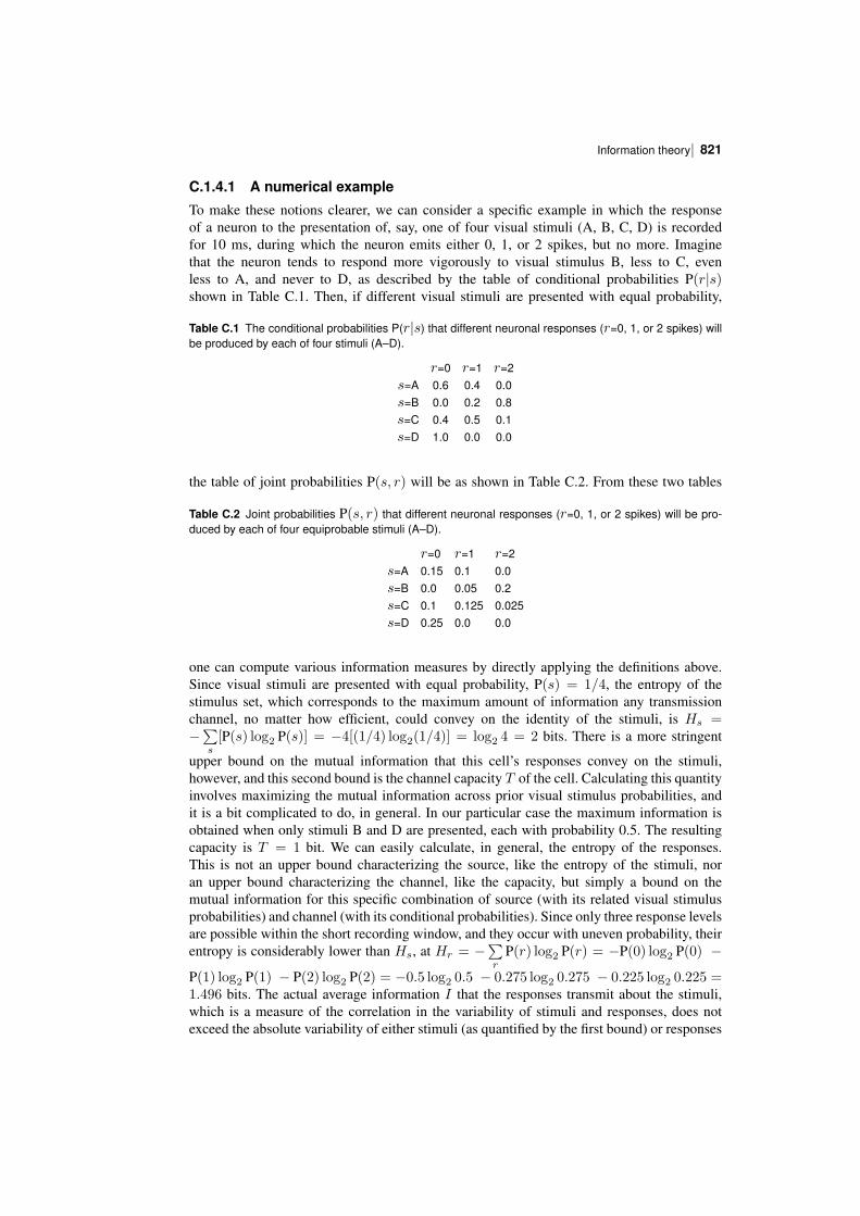
To make these notions clearer, we can consider a specific example in which the responseof a neuron to the presentation of, say, one of four visual stimuli (A, B, C, D) is recordedfor 10 ms, during which the neuron emits either 0, 1, or 2 spikes, but no more. Imaginethat the neuron tends to respond more vigorously to visual stimulus B, less to C, evenless to A, and never to D, as described by the table of conditional probabilities P(r|s)shown in Table C.1. Then, if different visual stimuli are presented with equal probability,
Table C.1 The conditional probabilities P(r|s) that different neuronal responses (r=0, 1, or 2 spikes) willbe produced by each of four stimuli (A–D).
r=0 r=1 r=2s=A 0.6 0.4 0.0s=B 0.0 0.2 0.8s=C 0.4 0.5 0.1s=D 1.0 0.0 0.0
the table of joint probabilities P(s, r) will be as shown in Table C.2. From these two tables
Table C.2 Joint probabilities P(s, r) that different neuronal responses (r=0, 1, or 2 spikes) will be produced by each of four equiprobable stimuli (A–D).
r=0 r=1 r=2s=A 0.15 0.1 0.0s=B 0.0 0.05 0.2s=C 0.1 0.125 0.025s=D 0.25 0.0 0.0
one can compute various information measures by directly applying the definitions above.Since visual stimuli are presented with equal probability, P(s) = 1/4, the entropy of thestimulus set, which corresponds to the maximum amount of information any transmissionchannel, no matter how efficient, could convey on the identity of the stimuli, is Hs =−∑s[P(s) log2 P(s)] = −4[(1/4) log2(1/4)] = log2 4 = 2 bits. There is a more stringent
upper bound on the mutual information that this cell’s responses convey on the stimuli,however, and this second bound is the channel capacity T of the cell. Calculating this quantityinvolves maximizing the mutual information across prior visual stimulus probabilities, andit is a bit complicated to do, in general. In our particular case the maximum information isobtained when only stimuli B and D are presented, each with probability 0.5. The resultingcapacity is T = 1 bit. We can easily calculate, in general, the entropy of the responses.This is not an upper bound characterizing the source, like the entropy of the stimuli, noran upper bound characterizing the channel, like the capacity, but simply a bound on themutual information for this specific combination of source (with its related visual stimulusprobabilities) and channel (with its conditional probabilities). Since only three response levelsare possible within the short recording window, and they occur with uneven probability, theirentropy is considerably lower than Hs, at Hr = −
∑r
P(r) log2 P(r) = −P(0) log2 P(0) −
P(1) log2 P(1) − P(2) log2 P(2) = −0.5 log2 0.5 − 0.275 log2 0.275 − 0.225 log2 0.225 =1.496 bits. The actual average information I that the responses transmit about the stimuli,which is a measure of the correlation in the variability of stimuli and responses, does notexceed the absolute variability of either stimuli (as quantified by the first bound) or responses
Information theory, and neuronal encoding822 |
(as quantified by the last bound), nor the capacity of the channel. An explicit calculation usingthe joint probabilities of the second table in expression C.21 yields I = 0.733 bits. This is ofcourse only the average value, averaged both across stimuli and across responses.
The information conveyed by a particular response can be larger. For example, when thecell emits two spikes it indicates with a relatively large probability stimulus B, and this isreflected in the fact that it then transmits, according to expression C.19, I(r = 2) = 1.497bits, more than double the average value.
Similarly, the amount of information conveyed about each individual visual stimulus varieswith the stimulus, depending on the extent to which it tends to elicit a differential response.Thus, expression C.22 yields that only I(s = C) = 0.185 bits are conveyed on average aboutstimulus C, which tends to elicit responses with similar statistics to the average statisticsacross stimuli, and are therefore not easily interpretable. On the other hand, exactly 1 bit ofinformation is conveyed about stimulus D, since this stimulus never elicits any response, andwhen the neuron emits no spike there is a probability of 1/2 that the stimulus was stimulus D.
C.1.5 The information conveyed by continuous variablesA general feature, relevant also to the case of neuronal information, is that if, among acontinuum of a priori possibilities, only one, or a discrete number, remains a posteriori,the information is strictly infinite. This would be the case if one were told, for example, thatReading is exactly 10′ west, 1′ north of London. The a priori probability of precisely this set ofcoordinates among the continuum of possible ones is zero, and then the information divergesto infinity. The problem is only theoretical, because in fact, with continuous distributions,there are always one or several factors that limit the resolution in the a posteriori knowledge,rendering the information finite. Moreover, when considering the mutual information in theconjoint probability of occurrence of two sets, e.g. stimuli and responses, it suffices thatat least one of the sets is discrete to make matters easy, that is, finite. Nevertheless, theidentification and appropriate consideration of these resolutionlimiting factors in practicalcases may require careful analysis.
C.1.5.1 Example: the information retrieved from an autoassociative memory
One example is the evaluation of the information that can be retrieved from an autoassociativememory. Such a memory stores a number of firing patterns, each one of which can beconsidered, as in Appendix B, as a vector rµ with components the firing rates {rµi }, wherethe subscript i indexes the neuron (and the superscript µ indexes the pattern). In retrievingpattern µ, the network in fact produces a distinct firing pattern, denoted for example simply asr. The quality of retrieval, or the similarity between rµ and r, can be measured by the averagemutual information
< I(rµ, r) > =∑rµ,r
P(rµ, r) log2P(rµ,r)
P(rµ)P(r) (C.23)
≈∑i
∑rµi ,ri
P(rµi , ri) log2P(rµi ,ri)
P(rµi )P(ri).
In this formula the ‘approximately equal’ sign ≈ marks a simplification that is not necessarilya reasonable approximation. If the simplification is valid, it means that in order to extract aninformation measure, one need not compare whole vectors (the entire firing patterns) witheach other, and may instead compare the firing rates of individual cells at storage and retrieval,and sum the resulting singlecell information values. The validity of the simplification is amatter that will be discussed later and that has to be verified, in the end, experimentally, butfor the purposes of the present discussion we can focus on the singlecell terms. If either
| 823Information theory
ri or rµi has a continuous distribution of values, as it will if it represents not the number ofspikes emitted in a fixed window, but more generally the firing rate of neuron i computedby convolving the firing train with a smoothing kernel, then one has to deal with probabilitydensities, which we denote as p(r)dr, rather than the usual probabilities P(r). Substitutingp(r)dr for P(r) and p(rµ, r)drdrµ for P(rµ, r), one can write for each singlecell contribution(omitting the cell index i)
< I(rµ, r) >i =
∫drµdr p(rµ, r) log2
p(rµ, r)
p(rµ)p(r)(C.24)
and we see that the differentials drµdr cancel out between numerator and denominator insidethe logarithm, rendering the quantity well defined and finite. If, however, rµ were to exactlydetermine r, one would have
p(rµ, r)drµdr = p(rµ)δ(r − r(rµ))drµdr = p(rµ)drµ (C.25)
and, by losing one differential on the way, the mutual information would become infinite. It istherefore important to consider what prevents rµ from fully determining r in the case at hand –in other words, to consider the sources of noise in the system. In an autoassociative memorystoring an extensive number of patterns (see Appendix A4 of Rolls and Treves (1998)), onesource of noise always present is the interference effect due to the concurrent storage of allother patterns. Even neglecting other sources of noise, this produces a finite resolution widthρ, which allows one to write an expression of the type p(r|rµ)dr = exp−(r−r(rµ))2/2ρ2drwhich ensures that the information is finite as long as the resolution ρ is larger than zero.
One further point that should be noted, in connection with estimating the informationretrievable from an autoassociative memory, is that the mutual information between thecurrent distribution of firing rates and that of the stored pattern does not coincide with theinformation gain provided by the memory device. Even when firing rates, or spike counts,are all that matter in terms of information carriers, as in the networks considered in this book,one more term should be taken into account in evaluating the information gain. This term,to be subtracted, is the information contained in the external input that elicits the retrieval.This may vary a lot from the retrieval of one particular memory to the next, but of course anefficient memory device is one that is able, when needed, to retrieve much more informationthan it requires to be present in the inputs, that is, a device that produces a large informationgain.
Finally, one should appreciate the conceptual difference between the information a firingpattern carries about another one (that is, about the pattern stored), as considered above, andtwo different notions: (a) the information produced by the network in selecting the correctmemory pattern and (b) the information a firing pattern carries about something in the outsideworld. Quantity (a), the information intrinsic to selecting the memory pattern, is ill definedwhen analysing a real system, but is a welldefined and particularly simple notion whenconsidering a formal model. If p patterns are stored with equal strength, and the selection iserrorless, this amounts to log2 p bits of information, a quantity often, but not always, smallcompared with the information in the pattern itself. Quantity (b), the information conveyedabout some outside correlate, is not defined when considering a formal model that does notinclude an explicit account of what the firing of each cell represents, but is well definedand measurable from the recorded activity of real cells. It is the quantity considered in thenumerical example with the four visual stimuli, and it can be generalized to the informationcarried by the activity of several cells in a network, and specialized to the case that the networkoperates as an associative memory. One may note, in this case, that the capacity to retrievememories with high fidelity, or high information content, is only useful to the extent that the
Information theory, and neuronal encoding824 |
representation to be retrieved carries that amount of information about something relevant –or, in other words, that it is pointless to store and retrieve with great care largely meaninglessmessages. This type of argument has been used to discuss the role of the mossy fibres inthe operation of the CA3 network in the hippocampus (Treves and Rolls 1992b, Rolls andTreves 1998).
C.2 Estimating the information carried by neuronalresponses
C.2.1 The limited sampling problem
We now discuss in more detail the application of these general notions to the informationtransmitted by neurons. Suppose, to be concrete, that an animal has been presented withstimuli drawn from a discrete set, and that the responses of a set ofC cells have been recordedfollowing the presentation of each stimulus. We may choose any quantity or set of quantitiesto characterize the responses; for example let us assume that we consider the firing rate of eachcell, ri, calculated by convolving the spike response with an appropriate smoothing kernel.The response space is then C times the continuous set of all positive real numbers, (R/2)C .We want to evaluate the average information carried by such responses about which stimuluswas shown. In principle, it is straightforward to apply the above formulas, e.g. in the form
< I(s, r) > =∑s
P(s)∫
Πidri p(r|s) log2p(r|s)p(r)
(C.26)
where it is important to note that p(r) and p(r|s) are now probability densities defined over thehighdimensional vector space of multicell responses. The product sign Π signifies that thiswhole vector space has to be integrated over, along all its dimensions. p(r) can be calculatedas∑sp(r|s)P(s), and therefore, in principle, all one has to do is to estimate, from the data,
the conditional probability densities p(r|s) – the distributions of responses following eachstimulus. In practice, however, in contrast to what happens with formal models, in which thereis usually no problem in calculating the exact probability densities, real data come in limitedamounts, and thus sample only sparsely the vast response space. This limits the accuracywith which, from the experimental frequency of each possible response, we can estimate itsprobability, in turn seriously impairing our ability to estimate < I > correctly. We refer tothis as the limited sampling problem. This is a purely technical problem that arises, typicallywhen recording from mammals, because of external constraints on the duration or number ofrepetitions of a given set of stimulus conditions. With computer simulation experiments, andalso with recordings from, for example, insects, sufficient data can usually be obtained thatstraightforward estimates of information are accurate enough (Strong, Koberle, de Ruyter vanSteveninck and Bialek 1998, Golomb, Kleinfeld, Reid, Shapley and Shraiman 1994). Theproblem is, however, so serious in connection with recordings from monkeys and rats inwhich limited numbers of trials are usually available for neuronal data, that it is worthwhileto discuss it, in order to appreciate the scope and limits of applying information theory toneuronal processing.
In particular, if the responses are continuous quantities, the probability of observingexactly the same response twice is infinitesimal. In the absence of further manipulation, thiswould imply that each stimulus generates its own set of unique responses, therefore anyresponse that has actually occurred could be associated unequivocally with one stimulus, andthe mutual information would always equal the entropy of the stimulus set. This absurdity
| 825The information carried by neuronal responses
shows that in order to estimate probability densities from experimental frequencies, one hasto resort to some regularizing manipulation, such as smoothing the pointlike response valuesby convolution with suitable kernels, or binning them into a finite number of discrete bins.
C.2.1.1 Smoothing or binning neuronal response data
The issue is how to estimate the underlying probability distributions of neuronal responses toa set of stimuli from only a limited number of trials of data (e.g. 10–30) for each stimulus.Several strategies are possible. One is to discretize the response space into bins, and estimatethe probability density as the histogram of the fraction of trials falling into each bin. If thebins are too narrow, almost every response is in a different bin, and the estimated informationwill be overestimated. Even if the bin width is increased to match the standard deviation ofeach underlying distribution, the information may still be overestimated. Alternatively, onemay try to ‘smooth’ the data by convolving each response with a Gaussian with a width setto the standard deviation measured for each stimulus. Setting the standard deviation to thisvalue may actually lead to an underestimation of the amount of information available, dueto oversmoothing. Another possibility is to make a bold assumption as to what the generalshape of the underlying densities should be, for example a Gaussian. This may produce closerestimates. Methods for regularizing the data are discussed further by Rolls and Treves (1998)in their Appendix A2, where a numerical example is given.
C.2.1.2 The effects of limited sampling
The crux of the problem is that, whatever procedure one adopts, limited sampling tends toproduce distortions in the estimated probability densities. The resulting mutual informationestimates are intrinsically biased. The bias, or average error of the estimate, is upward if theraw data have not been regularized much, and is downward if the regularization procedurechosen has been heavier. The bias can be, if the available trials are few, much larger thanthe true information values themselves. This is intuitive, as fluctuations due to the finitenumber of trials available would tend, on average, to either produce or emphasize differencesamong the distributions corresponding to different stimuli, differences that are preserved ifthe regularization is ‘light’, and that are interpreted in the calculation as carrying genuineinformation. This is illustrated with a quantitative example by Rolls and Treves (1998) in theirAppendix A2.
Choosing the right amount of regularization, or the best regularizing procedure, is notpossible a priori. Hertz, Kjaer, Eskander and Richmond (1992) have proposed the interestingprocedure of using an artificial neural network to regularize the raw responses. The networkcan be trained on part of the data using backpropagation, and then used on the remainingpart to produce what is in effect a clever datadriven regularization of the responses. Thisprocedure is, however, rather computer intensive and not very safe, as shown by some selfevident inconsistency in the results (Heller, Hertz, Kjaer and Richmond 1995). Obviously, thebest way to deal with the limited sampling problem is to try and use as many trials as possible.The improvement is slow, however, and generating as many trials as would be required for areasonably unbiased estimate is often, in practice, impossible.
C.2.2 Correction procedures for limited sampling
The above point, that data drawn from a single distribution, when artificially paired, at random,to different stimulus labels, results in ‘spurious’ amounts of apparent information, suggestsa simple way of checking the reliability of estimates produced from real data (Optican,Gawne, Richmond and Joseph 1991). One can disregard the true stimulus associated with eachresponse, and generate a randomly reshuffled pairing of stimuli and responses, which should
Information theory, and neuronal encoding826 |
therefore, being not linked by any underlying relationship, carry no mutual information abouteach other. Calculating, with some procedure of choice, the spurious information obtainedin this way, and comparing with the information value estimated with the same procedurefor the real pairing, one can get a feeling for how far the procedure goes into eliminatingthe apparent information due to limited sampling. Although this spurious information, Is, isonly indicative of the amount of bias affecting the original estimate, a simple heuristic trick(called ‘bootstrap’42) is to subtract the spurious from the original value, to obtain a somewhat‘corrected’ estimate. This procedure can result in quite accurate estimates (see Rolls andTreves (1998), Tovee, Rolls, Treves and Bellis (1993))43.
A different correction procedure (called ‘jackknife’) is based on the assumption that thebias is proportional to 1/N , where N is the number of responses (data points) used in theestimation. One computes, beside the original estimate < IN >, N auxiliary estimates< IN−1 >k, by taking out from the data set response k, where k runs across the data set from1 to N . The corrected estimate
< I > = N < IN > −(1/N)∑k
(N − 1) < IN−1 >k (C.27)
is free from bias (to leading order in 1/N ), if the proportionality factor is more or less thesame in the original and auxiliary estimates. This procedure is very timeconsuming, and itsuffers from the same imprecision of any algorithm that tries to determine a quantity as theresult of the subtraction of two large and nearly equal terms; in this case the terms have beenmade large on purpose, by multiplying them by N and N − 1.
A more fundamental approach (Miller 1955) is to derive an analytical expression for thebias (or, more precisely, for its leading terms in an expansion in 1/N , the inverse of the samplesize). This allows the estimation of the bias from the data itself, and its subsequent subtraction,as discussed in Treves and Panzeri (1995) and Panzeri and Treves (1996). Such a procedureproduces satisfactory results, thereby lowering the size of the sample required for a givenaccuracy in the estimate by about an order of magnitude (Golomb, Hertz, Panzeri, Treves andRichmond 1997). However, it does not, in itself, make possible measures of the informationcontained in very complex responses with few trials. As a rule of thumb, the number oftrials per stimulus required for a reasonable estimate of information, once the subtractivecorrection is applied, is of the order of the effectively independent (and utilized) bins in whichthe response space can be partitioned (Panzeri and Treves 1996). This correction procedureis the one that we use standardly (Rolls, Treves, Tovee and Panzeri 1997d, Rolls, Critchleyand Treves 1996a, Rolls, Treves, Robertson, GeorgesFrancois and Panzeri 1998b, Booth andRolls 1998, Rolls, Tovee and Panzeri 1999b, Rolls, Franco, Aggelopoulos and Jerez 2006b).
C.2.3 The information from multiple cells: decoding procedures
The bias of information measures grows with the dimensionality of the response space, andfor all practical purposes the limit on the number of dimensions that can lead to reasonablyaccurate direct measures, even when applying a correction procedure, is quite low, two tothree. This implies, in particular, that it is not possible to apply equation C.26 to extractthe information content in the responses of several cells (more than two to three) recorded
42In technical usage bootstrap procedures utilize random pairings of responses with stimuli with replacement,while shuffling procedures utilize random pairings of responses with stimuli without replacement.
43Subtracting the ‘square’ of the spurious fraction of information estimated by this bootstrap procedure as usedby Optican, Gawne, Richmond and Joseph (1991) is unfounded and does not work correctly (see Rolls and Treves(1998) and Tovee, Rolls, Treves and Bellis (1993)).
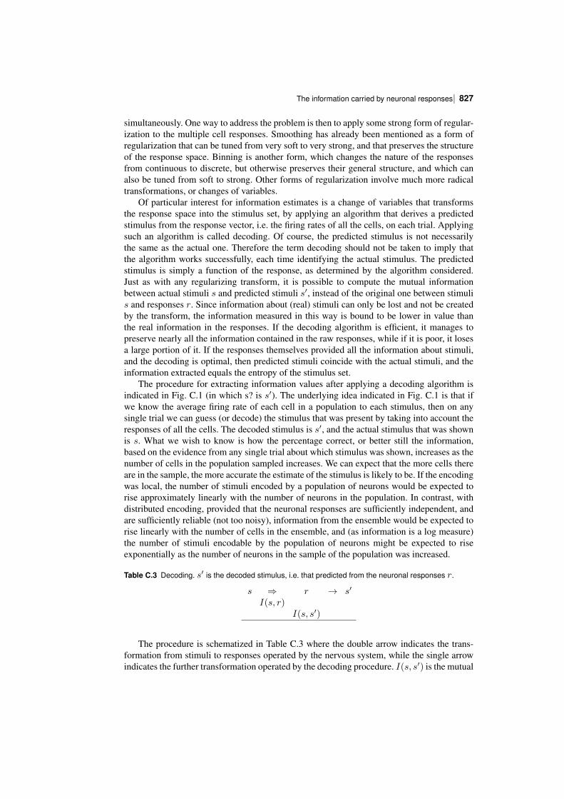
| 827The information carried by neuronal responses
simultaneously. One way to address the problem is then to apply some strong form of regularization to the multiple cell responses. Smoothing has already been mentioned as a form ofregularization that can be tuned from very soft to very strong, and that preserves the structureof the response space. Binning is another form, which changes the nature of the responsesfrom continuous to discrete, but otherwise preserves their general structure, and which canalso be tuned from soft to strong. Other forms of regularization involve much more radicaltransformations, or changes of variables.
Of particular interest for information estimates is a change of variables that transformsthe response space into the stimulus set, by applying an algorithm that derives a predictedstimulus from the response vector, i.e. the firing rates of all the cells, on each trial. Applyingsuch an algorithm is called decoding. Of course, the predicted stimulus is not necessarilythe same as the actual one. Therefore the term decoding should not be taken to imply thatthe algorithm works successfully, each time identifying the actual stimulus. The predictedstimulus is simply a function of the response, as determined by the algorithm considered.Just as with any regularizing transform, it is possible to compute the mutual informationbetween actual stimuli s and predicted stimuli s′, instead of the original one between stimulis and responses r. Since information about (real) stimuli can only be lost and not be createdby the transform, the information measured in this way is bound to be lower in value thanthe real information in the responses. If the decoding algorithm is efficient, it manages topreserve nearly all the information contained in the raw responses, while if it is poor, it losesa large portion of it. If the responses themselves provided all the information about stimuli,and the decoding is optimal, then predicted stimuli coincide with the actual stimuli, and theinformation extracted equals the entropy of the stimulus set.
The procedure for extracting information values after applying a decoding algorithm isindicated in Fig. C.1 (in which s? is s′). The underlying idea indicated in Fig. C.1 is that ifwe know the average firing rate of each cell in a population to each stimulus, then on anysingle trial we can guess (or decode) the stimulus that was present by taking into account theresponses of all the cells. The decoded stimulus is s′, and the actual stimulus that was shownis s. What we wish to know is how the percentage correct, or better still the information,based on the evidence from any single trial about which stimulus was shown, increases as thenumber of cells in the population sampled increases. We can expect that the more cells thereare in the sample, the more accurate the estimate of the stimulus is likely to be. If the encodingwas local, the number of stimuli encoded by a population of neurons would be expected torise approximately linearly with the number of neurons in the population. In contrast, withdistributed encoding, provided that the neuronal responses are sufficiently independent, andare sufficiently reliable (not too noisy), information from the ensemble would be expected torise linearly with the number of cells in the ensemble, and (as information is a log measure)the number of stimuli encodable by the population of neurons might be expected to riseexponentially as the number of neurons in the sample of the population was increased.
Table C.3 Decoding. s′ is the decoded stimulus, i.e. that predicted from the neuronal responses r.
s ⇒ r → s′
I(s, r)I(s, s′)
The procedure is schematized in Table C.3 where the double arrow indicates the transformation from stimuli to responses operated by the nervous system, while the single arrowindicates the further transformation operated by the decoding procedure. I(s, s′) is the mutual
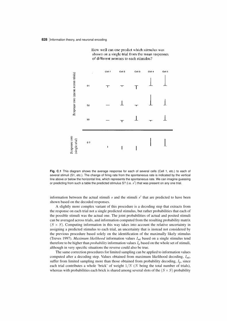
Information theory, and neuronal encoding828 |
Fig. C.1 This diagram shows the average response for each of several cells (Cell 1, etc.) to each ofseveral stimuli (S1, etc.). The change of firing rate from the spontaneous rate is indicated by the verticalline above or below the horizontal line, which represents the spontaneous rate. We can imagine guessingor predicting from such a table the predicted stimulus S? (i.e. s′) that was present on any one trial.
information between the actual stimuli s and the stimuli s′ that are predicted to have beenshown based on the decoded responses.
A slightly more complex variant of this procedure is a decoding step that extracts fromthe response on each trial not a single predicted stimulus, but rather probabilities that each ofthe possible stimuli was the actual one. The joint probabilities of actual and posited stimulican be averaged across trials, and information computed from the resulting probability matrix(S × S). Computing information in this way takes into account the relative uncertainty inassigning a predicted stimulus to each trial, an uncertainty that is instead not considered bythe previous procedure based solely on the identification of the maximally likely stimulus(Treves 1997). Maximum likelihood information values Iml based on a single stimulus tendtherefore to be higher than probability information values Ip based on the whole set of stimuli,although in very specific situations the reverse could also be true.
The same correction procedures for limited sampling can be applied to information valuescomputed after a decoding step. Values obtained from maximum likelihood decoding, Iml,suffer from limited sampling more than those obtained from probability decoding, Ip, sinceeach trial contributes a whole ‘brick’ of weight 1/N (N being the total number of trials),whereas with probabilities each brick is shared among several slots of the (S×S) probability
| 829The information carried by neuronal responses
matrix. The neural network procedure devised by Hertz, Kjaer, Eskander and Richmond(1992) can in fact be thought of as a decoding procedure based on probabilities, which dealswith limited sampling not by applying a correction but rather by strongly regularizing theoriginal responses.
When decoding is used, the rule of thumb becomes that the minimal number of trialsper stimulus required for accurate information measures is roughly equal to the size of thestimulus set, if the subtractive correction is applied (Panzeri and Treves 1996). This correctionprocedure is applied as standard in our multiple cell information analyses that use decoding(Rolls, Treves and Tovee 1997b, Booth and Rolls 1998, Rolls, Treves, Robertson, GeorgesFrancois and Panzeri 1998b, Franco, Rolls, Aggelopoulos and Treves 2004, Aggelopoulos,Franco and Rolls 2005, Rolls, Franco, Aggelopoulos and Jerez 2006b).
C.2.3.1 Decoding algorithms
Any transformation from the response space to the stimulus set could be used in decoding, butof particular interest are the transformations that either approach optimality, so as to minimizeinformation loss and hence the effect of decoding, or else are implementable by mechanismsthat could conceivably be operating in the real system, so as to extract information values thatcould be extracted by the system itself.
The optimal transformation is in theory welldefined: one should estimate from the datathe conditional probabilities P(r|s), and use Bayes’ rule to convert them into the conditionalprobabilities P(s′|r). Having these for any value of r, one could use them to estimate Ip,and, after selecting for each particular real response the stimulus with the highest conditionalprobability, to estimate Iml. To avoid biasing the estimation of conditional probabilities,the responses used in estimating P(r|s) should not include the particular response for whichP(s′|r) is going to be derived (jackknife crossvalidation). In practice, however, the estimationof P(r|s) in usable form involves the fitting of some simple function to the responses. Thisneed for fitting, together with the approximations implied in the estimation of the variousquantities, prevents us from defining the really optimal decoding, and leaves us with variousalgorithms, depending essentially on the fitting function used, which are hopefully close tooptimal in some conditions. We have experimented extensively with two such algorithms, thatboth approximate Bayesian decoding (Rolls, Treves and Tovee 1997b). Both these algorithmsfit the response vectors produced over several trials by the cells being recorded to a productof conditional probabilities for the response of each cell given the stimulus. In one case, thesingle cell conditional probability is assumed to be Gaussian (truncated at zero); in the otherit is assumed to be Poisson (with an additional weight at zero). Details of these algorithmsare given by Rolls, Treves and Tovee (1997b).
Biologically plausible decoding algorithms are those that limit the algebraic operationsused to types that could be easily implemented by neurons, e.g. dot product summations,thresholding and other singlecell nonlinearities, and competition and contrast enhancementamong the outputs of nearby cells. There is then no need for ever fitting functions or othersophisticated approximations, but of course the degree of arbitrariness in selecting a particularalgorithm remains substantial, and a comparison among different choices based on whichyields the higher information values may favour one choice in a given situation and anotherchoice with a different data set.
To summarize, the key idea in decoding, in our context of estimating information values,is that it allows substitution of a possibly very highdimensional response space (which isdifficult to sample and regularize) with a reduced object much easier to handle, that is witha discrete set equivalent to the stimulus set. The mutual information between the new setand the stimulus set is then easier to estimate even with limited data, and if the assumptionsabout population coding, underlying the particular decoding algorithm used, are justified, the
Information theory, and neuronal encoding830 |
value obtained approximates the original target, the mutual information between stimuli andresponses. For each response recorded, one can use all the responses except for that one togenerate estimates of the average response vectors (the average response for each neuron inthe population) to each stimulus. Then one considers how well the selected response vectormatches the average response vectors, and uses the degree of matching to estimate, for allstimuli, the probability that they were the actual stimuli. The form of the matching embodiesthe general notions about population encoding, for example the ‘degree of matching’ mightbe simply the dot product between the current vector and the average vector (rav), suitablynormalized over all average vectors to generate probabilities
P(s′|r(s)) = r(s) · rav(s′)∑s′′
r(s) · rav(s′′)(C.28)
where s′′ is a dummy variable. (This is called dot product decoding in Fig. 25.13.) One endsup, then, with a table of conjoint probabilities P(s, s′), and another table obtained by selectingfor each trial the most likely (or predicted) single stimulus sp, P(s, sp). Both s′ and sp standfor all possible stimuli, and hence belong to the same set S. These can be used to estimatemutual information values based on probability decoding (Ip) and on maximum likelihooddecoding (Iml):
< Ip > =∑s∈S
∑s′∈S
P(s, s′) log2P(s, s′)
P(s)P(s′)(C.29)
and
< Iml > =∑s∈S
∑sp∈S
P(s, sp) log2P(s, sp)
P(s)P(sp). (C.30)
Examples of the use of these procedures are available (Rolls, Treves and Tovee 1997b,Booth and Rolls 1998, Rolls, Treves, Robertson, GeorgesFrancois and Panzeri 1998b, Rolls,Aggelopoulos, Franco and Treves 2004, Franco, Rolls, Aggelopoulos and Treves 2004, Rolls,Franco, Aggelopoulos and Jerez 2006b), and some of the results obtained are described inSection C.3.
C.2.4 Information in the correlations between the spikes of differentcells: a decoding approach
Simultaneously recorded neurons sometimes shows crosscorrelations in their firing, that isthe firing of one is systematically related to the firing of the other cell. One example of thisis neuronal response synchronization. The crosscorrelation, to be defined below, shows thetime difference between the cells at which the systematic relation appears. A significant peakor trough in the crosscorrelation function could reveal a synaptic connection from one cellto the other, or a common input to each of the cells, or any of a considerable number of otherpossibilities. If the synchronization occurred for only some of the stimuli, then the presenceof the significant crosscorrelation for only those stimuli could provide additional evidenceseparate from any information in the firing rate of the neurons about which stimulus had beenshown. Information theory in principle provides a way of quantitatively assessing the relativecontributions from these two types of encoding, by expressing what can be learned from eachtype of encoding in the same units, bits of information.
Figure C.2 illustrates how synchronization occurring only for some of the stimuli couldbe used to encode information about which stimulus was presented. In the Figure the spiketrains of three neurons are shown after the presentation of two different stimuli on one trial.
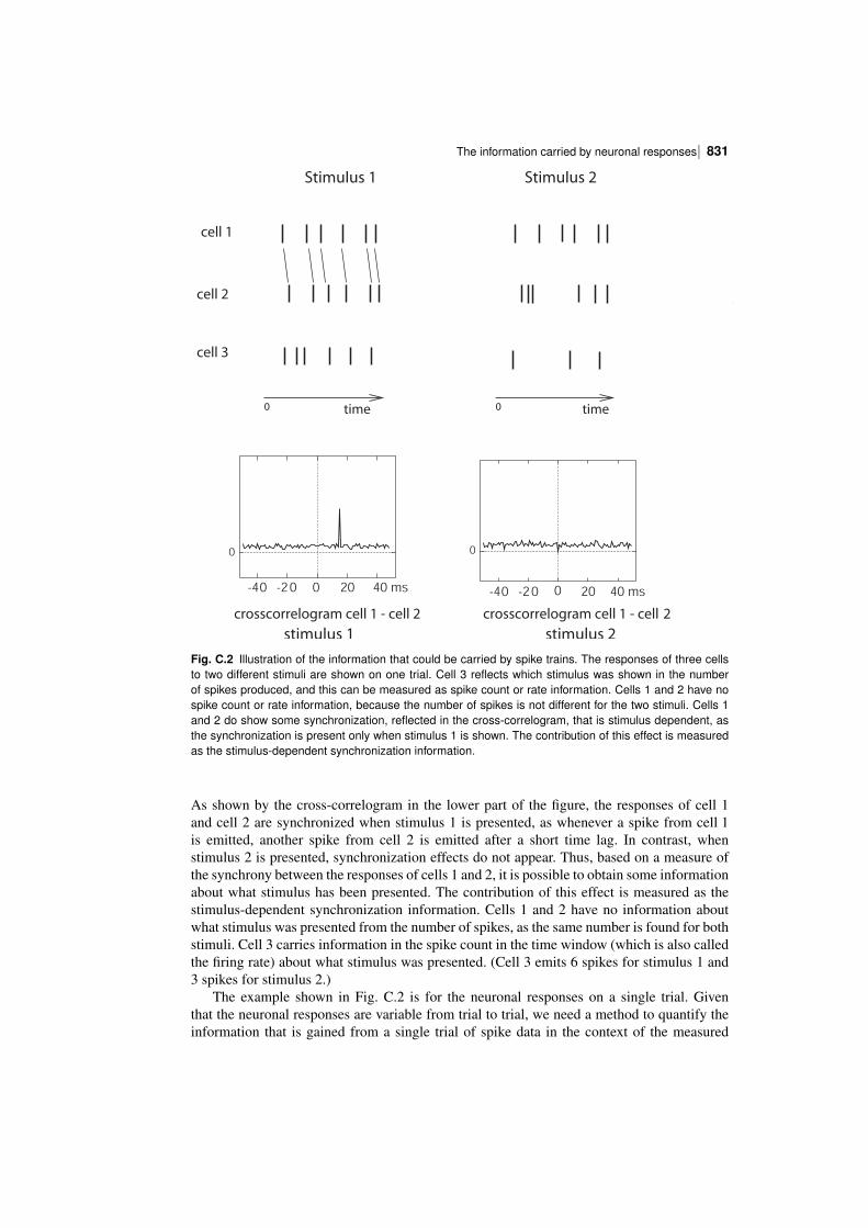
| 831The information carried by neuronal responses
stimulus 1 stimulus 2
Stimulus 1 Stimulus 2
crosscorrelogram cell 1 - cell 2 crosscorrelogram cell 1 - cell 2
0 time 0 time
0
-40 -20 0 20 40 ms
0
-40 -20 0 20 40 ms
t
cell 3
cell 1
cell 2
Fig. C.2 Illustration of the information that could be carried by spike trains. The responses of three cellsto two different stimuli are shown on one trial. Cell 3 reflects which stimulus was shown in the numberof spikes produced, and this can be measured as spike count or rate information. Cells 1 and 2 have nospike count or rate information, because the number of spikes is not different for the two stimuli. Cells 1and 2 do show some synchronization, reflected in the crosscorrelogram, that is stimulus dependent, asthe synchronization is present only when stimulus 1 is shown. The contribution of this effect is measuredas the stimulusdependent synchronization information.
As shown by the crosscorrelogram in the lower part of the figure, the responses of cell 1and cell 2 are synchronized when stimulus 1 is presented, as whenever a spike from cell 1is emitted, another spike from cell 2 is emitted after a short time lag. In contrast, whenstimulus 2 is presented, synchronization effects do not appear. Thus, based on a measure ofthe synchrony between the responses of cells 1 and 2, it is possible to obtain some informationabout what stimulus has been presented. The contribution of this effect is measured as thestimulusdependent synchronization information. Cells 1 and 2 have no information aboutwhat stimulus was presented from the number of spikes, as the same number is found for bothstimuli. Cell 3 carries information in the spike count in the time window (which is also calledthe firing rate) about what stimulus was presented. (Cell 3 emits 6 spikes for stimulus 1 and3 spikes for stimulus 2.)
The example shown in Fig. C.2 is for the neuronal responses on a single trial. Giventhat the neuronal responses are variable from trial to trial, we need a method to quantify theinformation that is gained from a single trial of spike data in the context of the measured
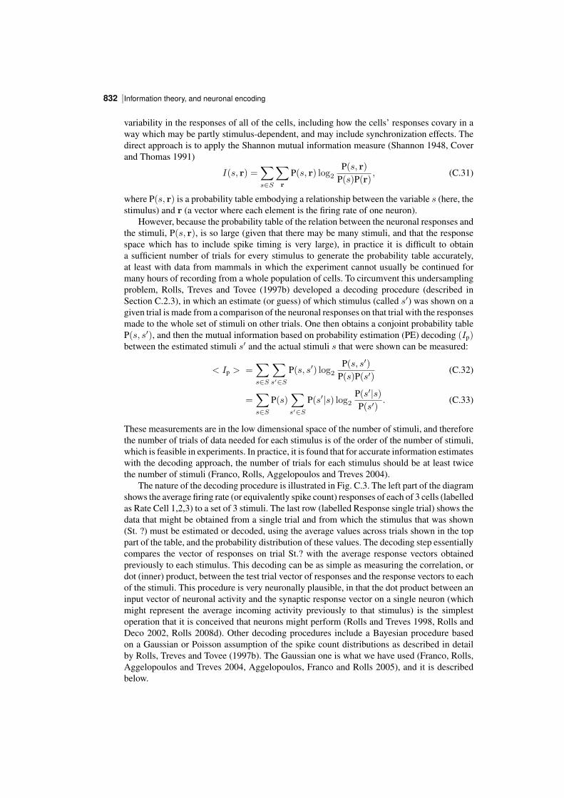
Information theory, and neuronal encoding832 |
variability in the responses of all of the cells, including how the cells’ responses covary in away which may be partly stimulusdependent, and may include synchronization effects. Thedirect approach is to apply the Shannon mutual information measure (Shannon 1948, Coverand Thomas 1991)
I(s, r) =∑s∈S
∑r
P(s, r) log2P(s, r)
P(s)P(r), (C.31)
where P(s, r) is a probability table embodying a relationship between the variable s (here, thestimulus) and r (a vector where each element is the firing rate of one neuron).
However, because the probability table of the relation between the neuronal responses andthe stimuli, P(s, r), is so large (given that there may be many stimuli, and that the responsespace which has to include spike timing is very large), in practice it is difficult to obtaina sufficient number of trials for every stimulus to generate the probability table accurately,at least with data from mammals in which the experiment cannot usually be continued formany hours of recording from a whole population of cells. To circumvent this undersamplingproblem, Rolls, Treves and Tovee (1997b) developed a decoding procedure (described inSection C.2.3), in which an estimate (or guess) of which stimulus (called s′) was shown on agiven trial is made from a comparison of the neuronal responses on that trial with the responsesmade to the whole set of stimuli on other trials. One then obtains a conjoint probability tableP(s, s′), and then the mutual information based on probability estimation (PE) decoding (Ip)between the estimated stimuli s′ and the actual stimuli s that were shown can be measured:
< Ip > =∑s∈S
∑s′∈S
P(s, s′) log2P(s, s′)
P(s)P(s′)(C.32)
=∑s∈S
P(s)∑s′∈S
P(s′|s) log2P(s′|s)P(s′)
. (C.33)
These measurements are in the low dimensional space of the number of stimuli, and thereforethe number of trials of data needed for each stimulus is of the order of the number of stimuli,which is feasible in experiments. In practice, it is found that for accurate information estimateswith the decoding approach, the number of trials for each stimulus should be at least twicethe number of stimuli (Franco, Rolls, Aggelopoulos and Treves 2004).
The nature of the decoding procedure is illustrated in Fig. C.3. The left part of the diagramshows the average firing rate (or equivalently spike count) responses of each of 3 cells (labelledas Rate Cell 1,2,3) to a set of 3 stimuli. The last row (labelled Response single trial) shows thedata that might be obtained from a single trial and from which the stimulus that was shown(St. ?) must be estimated or decoded, using the average values across trials shown in the toppart of the table, and the probability distribution of these values. The decoding step essentiallycompares the vector of responses on trial St.? with the average response vectors obtainedpreviously to each stimulus. This decoding can be as simple as measuring the correlation, ordot (inner) product, between the test trial vector of responses and the response vectors to eachof the stimuli. This procedure is very neuronally plausible, in that the dot product between aninput vector of neuronal activity and the synaptic response vector on a single neuron (whichmight represent the average incoming activity previously to that stimulus) is the simplestoperation that it is conceived that neurons might perform (Rolls and Treves 1998, Rolls andDeco 2002, Rolls 2008d). Other decoding procedures include a Bayesian procedure basedon a Gaussian or Poisson assumption of the spike count distributions as described in detailby Rolls, Treves and Tovee (1997b). The Gaussian one is what we have used (Franco, Rolls,Aggelopoulos and Treves 2004, Aggelopoulos, Franco and Rolls 2005), and it is describedbelow.
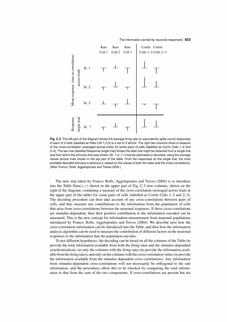
| 833The information carried by neuronal responses
acro
ss t
rial
s
Res
po
nse
St. 1
St. 2
St. 3
St. ?
Mea
n r
esp
on
se
(rat
e o
r co
rrel
atio
n)
sin
gle
tri
al
Rate
Cell 1
Rate
Cell 2 Cell 3
Rate
Cells 1−2 Cells 2−3
CorrlnCorrln
Fig. C.3 The left part of the diagram shows the average firing rate (or equivalently spike count) responsesof each of 3 cells (labelled as Rate Cell 1,2,3) to a set of 3 stimuli. The right two columns show a measureof the crosscorrelation (averaged across trials) for some pairs of cells (labelled as Corrln Cells 1–2 and2–3). The last row (labelled Response single trial) shows the data that might be obtained from a single trialand from which the stimulus that was shown (St. ? or s′) must be estimated or decoded, using the averagevalues across trials shown in the top part of the table. From the responses on the single trial, the mostprobable decoded stimulus is stimulus 2, based on the values of both the rates and the crosscorrelations.(After Franco, Rolls, Aggelopoulos and Treves 2004.)
The new step taken by Franco, Rolls, Aggelopoulos and Treves (2004) is to introduceinto the Table Data(s, r) shown in the upper part of Fig. C.3 new columns, shown on theright of the diagram, containing a measure of the crosscorrelation (averaged across trials inthe upper part of the table) for some pairs of cells (labelled as Corrln Cells 1–2 and 2–3).The decoding procedure can then take account of any crosscorrelations between pairs ofcells, and thus measure any contributions to the information from the population of cellsthat arise from crosscorrelations between the neuronal responses. If these crosscorrelationsare stimulusdependent, then their positive contribution to the information encoded can bemeasured. This is the new concept for information measurement from neuronal populationsintroduced by Franco, Rolls, Aggelopoulos and Treves (2004). We describe next how thecrosscorrelation information can be introduced into the Table, and then how the informationanalysis algorithm can be used to measure the contribution of different factors in the neuronalresponses to the information that the population encodes.
To test different hypotheses, the decoding can be based on all the columns of the Table (toprovide the total information available from both the firing rates and the stimulusdependentsynchronization), on only the columns with the firing rates (to provide the information available from the firing rates), and only on the columns with the crosscorrelation values (to providethe information available from the stimulusdependent crosscorrelations). Any informationfrom stimulusdependent crosscorrelations will not necessarily be orthogonal to the rateinformation, and the procedures allow this to be checked by comparing the total information to that from the sum of the two components. If crosscorrelations are present but are
Information theory, and neuronal encoding834 |
not stimulusdependent, these will not contribute to the information available about whichstimulus was shown.
The measure of the synchronization introduced into the Table Data(s, r) on each trial is,for example, the value of the Pearson crosscorrelation coefficient calculated for that trialat the appropriate lag for cell pairs that have significant crosscorrelations (Franco, Rolls,Aggelopoulos and Treves 2004). This value of this Pearson crosscorrelation coefficient fora single trial can be calculated from pairs of spike trains on a single trial by forming foreach cell a vector of 0s and 1s, the 1s representing the time of occurrence of spikes witha temporal resolution of 1 ms. Resulting values within the range −1 to 1 are shifted toobtain positive values. An advantage of basing the measure of synchronization on the Pearsoncrosscorrelation coefficient is that it measures the amount of synchronization between apair of neurons independently of the firing rate of the neurons. The lag at which the crosscorrelation measure was computed for every single trial, and whether there was a significantcrosscorrelation between neuron pairs, can be identified from the location of the peak in thecrosscorrelogram taken across all trials. The crosscorrelogram is calculated by, for everyspike that occurred in one neuron, incrementing the bins of a histogram that correspond tothe lag times of each of the spikes that occur for the other neuron. The raw crosscorrelogramis corrected by subtracting the “shift predictor” crosscorrelogram (which is produced byrandom repairings of the trials), to produce the corrected crosscorrelogram.
Further details of the decoding procedures are as follows (see Rolls, Treves and Tovee(1997b) and Franco, Rolls, Aggelopoulos and Treves (2004)). The full probability tableestimator (PE) algorithm uses a Bayesian approach to extract P(s′|r) for every single trialfrom an estimate of the probability P(r|s′) of a stimulus–response pair made from all theother trials (as shown in Bayes’ rule shown in equation C.34) in a crossvalidation proceduredescribed by Rolls et al. (1997b).
P(s′|r) = P(r|s′)P(s′)P(r)
. (C.34)
where P(r) (the probability of the vector containing the firing rate of each neuron, where eachelement of the vector is the firing rate of one neuron) is obtained as:
P(r) =∑s′
P(r|s′)P(s′). (C.35)
This requires knowledge of the response probabilities P(r|s′) which can be estimated for thispurpose from P(r, s′), which is equal to P(s′)
∏c P(rc|s′), where rc is the firing rate of cell
c. We note that P(rc|s′) is derived from the responses of cell c from all of the trials except forthe current trial for which the probability estimate is being made. The probabilities P(rc|s′)are fitted with a Gaussian (or Poisson) distribution whose amplitude at rc gives P(rc|s′).By summing over different test trial responses to the same stimulus s, we can extract theprobability that by presenting stimulus s the neuronal response is interpreted as having beenelicited by stimulus s′,
P(s′|s) =∑
r∈test
P(s′|r)P(r|s). (C.36)
After the decoding procedure, the estimated relative probabilities (normalized to 1) wereaveraged over all ‘test’ trials for all stimuli, to generate a (Regularized) table PR
N (s, s′)describing the relative probability of each pair of actual stimulus s and posited stimulus s′
(computed with N trials). From this probability table the mutual information measure (Ip)was calculated as described above in equation C.33.
| 835The information carried by neuronal responses
We also generate a second (Frequency) table PFN (s, sp) from the fraction of times an
actual stimulus s elicited a response that led to a predicted (single most likely) stimulus sp.From this probability Table the mutual information measure based on maximum likelihooddecoding (Iml) was calculated with equation C.37:
< Iml > =∑s∈S
∑sp∈S
P(s, sp) log2P(s, sp)
P(s)P(sp). (C.37)
A detailed comparison of maximum likelihood and probability decoding is provided by Rolls,Treves and Tovee (1997b), but we note here that probability estimate decoding is moreregularized (see below) and therefore may be safer to use when investigating the effect on theinformation of the number of cells. For this reason, the results described by Franco, Rolls,Aggelopoulos and Treves (2004) were obtained with probability estimation (PE) decoding.The maximum likelihood decoding does give an immediate measure of the percentage correct.
Another approach to decoding is the dot product (DP) algorithm which computes thenormalized dot products between the current firing vector r on a “test” (i.e. the current) trialand each of the mean firing rate response vectors in the “training” trials for each stimulus s′ inthe crossvalidation procedure. (The normalized dot product is the dot or inner product of twovectors divided by the product of the length of each vector. The length of each vector is thesquare root of the sum of the squares.) Thus, what is computed are the cosines of the anglesof the test vector of cell rates with, in turn for each stimulus, the mean response vector to thatstimulus. The highest dot product indicates the most likely stimulus that was presented, andthis is taken as the predicted stimulus sp for the probability table P(s, sp). (It can also be usedto provide percentage correct measures.)
We note that any decoding procedure can be used in conjunction with information estimatesboth from the full probability table (to produce Ip), and from the most likely estimated stimulusfor each trial (to produce Iml).
Because the probability tables from which the information is calculated may be unregularized with a small number of trials, a bias correction procedure to correct for the undersamplingis applied, as described in detail by Rolls, Treves and Tovee (1997b) and Panzeri and Treves(1996). In practice, the bias correction that is needed with information estimates using thedecoding procedures described by Franco, Rolls, Aggelopoulos and Treves (2004) and byRolls et al. (1997b) is small, typically less than 10% of the uncorrected estimate of theinformation, provided that the number of trials for each stimulus is in the order of twice thenumber of stimuli. We also note that the distortion in the information estimate from the fullprobability table needs less bias correction than that from the predicted stimulus table (i.e.maximum likelihood) method, as the former is more regularized because every trial makessome contribution through much of the probability table (see Rolls et al. (1997b)). We furthernote that the bias correction term becomes very small when more than 10 cells are includedin the analysis (Rolls et al. 1997b).
Examples of the use of these procedures are available (Franco, Rolls, Aggelopoulos andTreves 2004, Aggelopoulos, Franco and Rolls 2005), and some of the results obtained aredescribed in Section C.3.
C.2.5 Information in the correlations between the spikes of differentcells: a second derivative approach
Another information theorybased approach to stimulusdependent crosscorrelation information has been developed as follows by Panzeri, Schultz, Treves and Rolls (1999a) and Rolls,Franco, Aggelopoulos and Reece (2003b). A problem that must be overcome is the fact that
Information theory, and neuronal encoding836 |
with many simultaneously recorded neurons, each emitting perhaps many spikes at differenttimes, the dimensionality of the response space becomes very large, the information tends tobe overestimated, and even bias corrections cannot save the situation. The approach describedin this Section (C.2.5) limits the problem by taking short time epochs for the informationanalysis, in which low numbers of spikes, in practice typically 0, 1, or 2, spikes are likely tooccur from each neuron.
In a sufficiently short time window, at most two spikes are emitted from a population ofneurons. Taking advantage of this, the response probabilities can be calculated in terms ofpairwise correlations. These response probabilities are inserted into the Shannon informationformula C.38 to obtain expressions quantifying the impact of the pairwise correlations on theinformation I(t) transmitted in a short time t by groups of spiking neurons:
I(t) =∑s∈S
∑r
P(s, r) log2P(s, r)
P(s)P (r)(C.38)
where r is the firing rate response vector comprised by the number of spikes emitted by eachof the cells in the population in the short time t, and P(s, r) refers to the joint probabilitydistribution of stimuli with their respective neuronal response vectors.
The information depends upon the following two types of correlation:
C.2.5.1 The correlations in the neuronal response variability from theaverage to each stimulus (sometimes called “noise” correlations) γ:
γij(s) (for i = j) is the fraction of coincidences above (or below) that expected fromuncorrelated responses, relative to the number of coincidences in the uncorrelated case (whichis ni(s)nj(s), the bar denoting the average across trials belonging to stimulus s, where ni(s)is the number of spikes emitted by cell i to stimulus s on a given trial)
γij(s) =ni(s)nj(s)
(ni(s)nj(s))− 1, (C.39)
and is named the ‘scaled crosscorrelation density’. It can vary from −1 to ∞; negativeγij(s)’s indicate anticorrelation, whereas positive γij(s)’s indicate correlation44. γij(s) canbe thought of as the amount of trial by trial concurrent firing of the cells i and j, comparedto that expected in the uncorrelated case. γij(s) (for i = j) is the ‘scaled crosscorrelationdensity’ (Aertsen, Gerstein, Habib and Palm 1989, Panzeri, Schultz, Treves and Rolls 1999a),and is sometimes called the “noise” correlation (Gawne and Richmond 1993, Shadlen andNewsome 1995, Shadlen and Newsome 1998).
44γij(s) is an alternative, which produces a more compact information analysis, to the neuronal crosscorrelationbased on the Pearson correlation coefficient ρij(s) (equation C.40), which normalizes the number of coincidencesabove independence to the standard deviation of the number of coincidences expected if the cells were independent.The normalization used by the Pearson correlation coefficient has the advantage that it quantifies the strength ofcorrelations between neurons in a rateindependent way. For the information analysis, it is more convenient to usethe scaled correlation density γij(s) than the Pearson correlation coefficient, because of the compactness of theresulting formulation, and because of its scaling properties for small t. γij(s) remains finite as t → 0, thus byusing this measure we can keep the t expansion of the information explicit. Keeping the timedependence of theresulting information components explicit greatly increases the amount of insight obtained from the series expansion.In contrast, the Pearson noisecorrelation measure applied to short timescales approaches zero at short time windows:
ρij(s) ≡ni(s)nj(s)− ni(s)nj(s)
σni(s)σnj(s)
≃ t γij(s) =√
ri(s)rj(s), (C.40)
where σni(s)is the standard deviation of the count of spikes emitted by cell i in response to stimulus s.
| 837The information carried by neuronal responses
C.2.5.2 The correlations in the mean responses of the neurons across theset of stimuli (sometimes called “signal” correlations) ν:
νij =< ni(s)nj(s) >s
< ni(s) >s< nj(s) >s− 1 =
< ri(s)rj(s) >s
< ri(s) >s< rj(s) >s− 1 (C.41)
where ri(s) is the mean rate of response of cell i (amongC cells in total) to stimulus s over allthe trials in which that stimulus was present. νij can be thought of as the degree of similarityin the mean response profiles (averaged across trials) of the cells i and j to different stimuli.νij is sometimes called the “signal” correlation (Gawne and Richmond 1993, Shadlen andNewsome 1995, Shadlen and Newsome 1998).
C.2.5.3 Information in the crosscorrelations in short time periods
In the short timescale limit, the first (It) and second (Itt) information derivatives describe theinformation I(t) available in the short time t
I(t) = t It +t2
2Itt . (C.42)
(The zeroth order, timeindependent term is zero, as no information can be transmitted bythe neurons in a time window of zero length. Higher order terms are also excluded as theybecome negligible.)
The instantaneous information rate It is45
It =
C∑i=1
⟨ri(s) log2
ri(s)
⟨ri(s′)⟩s′
⟩s
. (C.43)
This formula shows that this information rate (the first time derivative) should not be linkedto a high signal to noise ratio, but only reflects the extent to which the mean responses ofeach cell are distributed across stimuli. It does not reflect anything of the variability of thoseresponses, that is of their noisiness, nor anything of the correlations among the mean responsesof different cells.
The effect of (pairwise) correlations between the cells begins to be expressed in thesecond time derivative of the information. The expression for the instantaneous information‘acceleration’ Itt (the second time derivative of the information) breaks up into three terms:
Itt =1
ln 2
C∑i=1
C∑j=1
⟨ri(s)⟩s ⟨rj(s)⟩s
[νij + (1 + νij) ln(
1
1 + νij)
]
+
C∑i=1
C∑j=1
[⟨ri(s)rj(s)γij(s)⟩s
]log2(
1
1 + νij)
+
C∑i=1
C∑j=1
⟨ri(s)rj(s)(1 + γij(s)) log2
[(1 + γij(s)) ⟨ri(s′)rj(s′)⟩s′⟨ri(s′)rj(s′)(1 + γij(s′))⟩s′
]⟩s
.(C.44)
The first of these terms is all that survives if there is no noise correlation at all. Thus therate component of the information is given by the sum of It (which is always greater than orequal to zero) and of the first term of Itt (which is instead always less than or equal to zero).
45Note that s′ is used in equations C.43 and C.44 just as a dummy variable to stand for s, as there are twosummations performed over s.
Information theory, and neuronal encoding838 |
The second term is nonzero if there is some correlation in the variance to a given stimulus,even if it is independent of which stimulus is present; this term thus represents the contributionof stimulusindependent noise correlation to the information.
The third component of Itt represents the contribution of stimulusmodulated noise correlation, as it becomes nonzero only for stimulusdependent noise correlations. These last twoterms of Itt together are referred to as the correlational components of the information.
The application of this approach to measuring the information in the relative time of firingof simultaneously recorded cells, together with further details of the method, are described byPanzeri, Treves, Schultz and Rolls (1999b), Rolls, Franco, Aggelopoulos and Reece (2003b),and Rolls, Aggelopoulos, Franco and Treves (2004), and in Section C.3.7.
C.3 Neuronal encoding: results obtained from applyinginformationtheoretic analyses
How is information encoded in cortical areas such as the inferior temporal visual cortex?Can we read the code being used by the cortex? What are the advantages of the encodingscheme used for the neuronal network computations being performed in different areas ofthe cortex? These are some of the key issues considered in this Section (C.3). Becauseinformation is exchanged between the computing elements of the cortex (the neurons) bytheir spiking activity, which is conveyed by their axon to synapses onto other neurons, theappropriate level of analysis is how single neurons, and populations of single neurons, encodeinformation in their firing. More global measures that reflect the averaged activity of largenumbers of neurons (for example, PET (positron emission tomography) and fMRI (functionalmagnetic resonance imaging), EEG (electroencephalographic recording), and ERPs (eventrelated potentials)) cannot reveal how the information is represented, or how the computationis being performed.
Although information theory provides the natural mathematical framework for analysingthe performance of neuronal systems, its applications in neuroscience have been for manyyears rather sparse and episodic (e.g. MacKay and McCulloch (1952); Eckhorn and Popel(1974); Eckhorn and Popel (1975); Eckhorn, Grusser, Kroller, Pellnitz and Popel (1976)).One reason for this limited application of information theory has been the great effort thatwas apparently required, due essentially to the limited sampling problem, in order to obtainaccurate results. Another reason has been the hesitation in analysing as a single complex‘blackbox’ large neuronal systems all the way from some external, easily controllable inputs,up to neuronal activity in some central cortical area of interest, for example including all visualstations from the periphery to the end of the ventral visual stream in the temporal lobe. In fact,two important bodies of work, that have greatly helped revive interest in applications of thetheory in recent years, both sidestep these two problems. The problem with analyzing a hugeblackbox is avoided by considering systems at the sensory periphery; the limited samplingproblem is avoided either by working with insects, in which sampling can be extensive(Bialek, Rieke, de Ruyter van Steveninck and Warland 1991, de Ruyter van Steveninck andLaughlin 1996, Rieke, Warland, de Ruyter van Steveninck and Bialek 1997), or by utilizinga formal model instead of real data (Atick and Redlich 1990, Atick 1992). Both approacheshave provided insightful quantitative analyses that are in the process of being extended tomore central mammalian systems (see e.g. Atick, Griffin and Relich (1996)).
In the treatment provided here, we focus on applications to the mammalian brain, usingexamples from a whole series of investigations on information representation in visual corticalareas, the original papers on which refer to related publications.
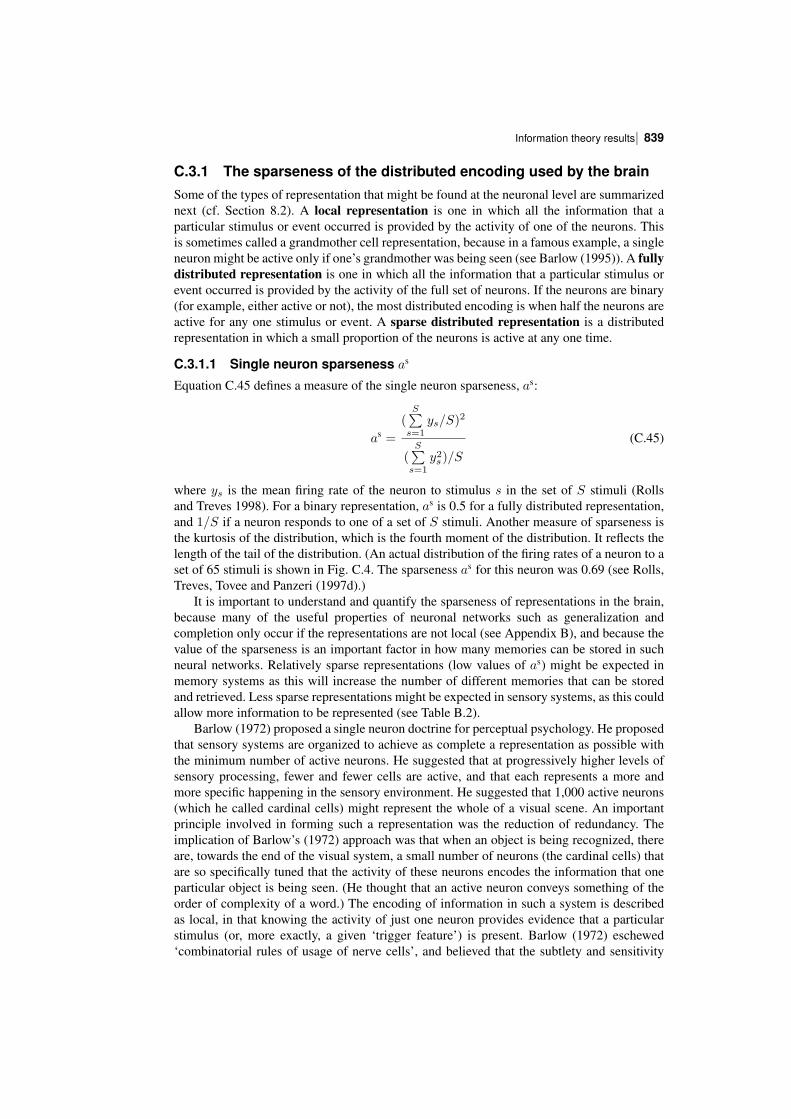
| 839Information theory results
C.3.1 The sparseness of the distributed encoding used by the brainSome of the types of representation that might be found at the neuronal level are summarizednext (cf. Section 8.2). A local representation is one in which all the information that aparticular stimulus or event occurred is provided by the activity of one of the neurons. Thisis sometimes called a grandmother cell representation, because in a famous example, a singleneuron might be active only if one’s grandmother was being seen (see Barlow (1995)). A fullydistributed representation is one in which all the information that a particular stimulus orevent occurred is provided by the activity of the full set of neurons. If the neurons are binary(for example, either active or not), the most distributed encoding is when half the neurons areactive for any one stimulus or event. A sparse distributed representation is a distributedrepresentation in which a small proportion of the neurons is active at any one time.
C.3.1.1 Single neuron sparseness as
Equation C.45 defines a measure of the single neuron sparseness, as:
as =
(S∑
s=1ys/S)
2
(S∑
s=1y2s)/S
(C.45)
where ys is the mean firing rate of the neuron to stimulus s in the set of S stimuli (Rollsand Treves 1998). For a binary representation, as is 0.5 for a fully distributed representation,and 1/S if a neuron responds to one of a set of S stimuli. Another measure of sparseness isthe kurtosis of the distribution, which is the fourth moment of the distribution. It reflects thelength of the tail of the distribution. (An actual distribution of the firing rates of a neuron to aset of 65 stimuli is shown in Fig. C.4. The sparseness as for this neuron was 0.69 (see Rolls,Treves, Tovee and Panzeri (1997d).)
It is important to understand and quantify the sparseness of representations in the brain,because many of the useful properties of neuronal networks such as generalization andcompletion only occur if the representations are not local (see Appendix B), and because thevalue of the sparseness is an important factor in how many memories can be stored in suchneural networks. Relatively sparse representations (low values of as) might be expected inmemory systems as this will increase the number of different memories that can be storedand retrieved. Less sparse representations might be expected in sensory systems, as this couldallow more information to be represented (see Table B.2).
Barlow (1972) proposed a single neuron doctrine for perceptual psychology. He proposedthat sensory systems are organized to achieve as complete a representation as possible withthe minimum number of active neurons. He suggested that at progressively higher levels ofsensory processing, fewer and fewer cells are active, and that each represents a more andmore specific happening in the sensory environment. He suggested that 1,000 active neurons(which he called cardinal cells) might represent the whole of a visual scene. An importantprinciple involved in forming such a representation was the reduction of redundancy. Theimplication of Barlow’s (1972) approach was that when an object is being recognized, thereare, towards the end of the visual system, a small number of neurons (the cardinal cells) thatare so specifically tuned that the activity of these neurons encodes the information that oneparticular object is being seen. (He thought that an active neuron conveys something of theorder of complexity of a word.) The encoding of information in such a system is describedas local, in that knowing the activity of just one neuron provides evidence that a particularstimulus (or, more exactly, a given ‘trigger feature’) is present. Barlow (1972) eschewed‘combinatorial rules of usage of nerve cells’, and believed that the subtlety and sensitivity
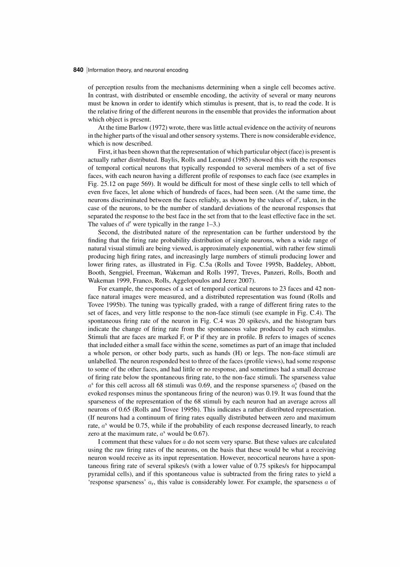
Information theory, and neuronal encoding840 |
of perception results from the mechanisms determining when a single cell becomes active.In contrast, with distributed or ensemble encoding, the activity of several or many neuronsmust be known in order to identify which stimulus is present, that is, to read the code. It isthe relative firing of the different neurons in the ensemble that provides the information aboutwhich object is present.
At the time Barlow (1972) wrote, there was little actual evidence on the activity of neuronsin the higher parts of the visual and other sensory systems. There is now considerable evidence,which is now described.
First, it has been shown that the representation of which particular object (face) is present isactually rather distributed. Baylis, Rolls and Leonard (1985) showed this with the responsesof temporal cortical neurons that typically responded to several members of a set of fivefaces, with each neuron having a different profile of responses to each face (see examples inFig. 25.12 on page 569). It would be difficult for most of these single cells to tell which ofeven five faces, let alone which of hundreds of faces, had been seen. (At the same time, theneurons discriminated between the faces reliably, as shown by the values of d′, taken, in thecase of the neurons, to be the number of standard deviations of the neuronal responses thatseparated the response to the best face in the set from that to the least effective face in the set.The values of d′ were typically in the range 1–3.)
Second, the distributed nature of the representation can be further understood by thefinding that the firing rate probability distribution of single neurons, when a wide range ofnatural visual stimuli are being viewed, is approximately exponential, with rather few stimuliproducing high firing rates, and increasingly large numbers of stimuli producing lower andlower firing rates, as illustrated in Fig. C.5a (Rolls and Tovee 1995b, Baddeley, Abbott,Booth, Sengpiel, Freeman, Wakeman and Rolls 1997, Treves, Panzeri, Rolls, Booth andWakeman 1999, Franco, Rolls, Aggelopoulos and Jerez 2007).
For example, the responses of a set of temporal cortical neurons to 23 faces and 42 nonface natural images were measured, and a distributed representation was found (Rolls andTovee 1995b). The tuning was typically graded, with a range of different firing rates to theset of faces, and very little response to the nonface stimuli (see example in Fig. C.4). Thespontaneous firing rate of the neuron in Fig. C.4 was 20 spikes/s, and the histogram barsindicate the change of firing rate from the spontaneous value produced by each stimulus.Stimuli that are faces are marked F, or P if they are in profile. B refers to images of scenesthat included either a small face within the scene, sometimes as part of an image that includeda whole person, or other body parts, such as hands (H) or legs. The nonface stimuli areunlabelled. The neuron responded best to three of the faces (profile views), had some responseto some of the other faces, and had little or no response, and sometimes had a small decreaseof firing rate below the spontaneous firing rate, to the nonface stimuli. The sparseness valueas for this cell across all 68 stimuli was 0.69, and the response sparseness as
r (based on theevoked responses minus the spontaneous firing of the neuron) was 0.19. It was found that thesparseness of the representation of the 68 stimuli by each neuron had an average across allneurons of 0.65 (Rolls and Tovee 1995b). This indicates a rather distributed representation.(If neurons had a continuum of firing rates equally distributed between zero and maximumrate, as would be 0.75, while if the probability of each response decreased linearly, to reachzero at the maximum rate, as would be 0.67).
I comment that these values for a do not seem very sparse. But these values are calculatedusing the raw firing rates of the neurons, on the basis that these would be what a receivingneuron would receive as its input representation. However, neocortical neurons have a spontaneous firing rate of several spikes/s (with a lower value of 0.75 spikes/s for hippocampalpyramidal cells), and if this spontaneous value is subtracted from the firing rates to yield a‘response sparseness’ ar, this value is considerably lower. For example, the sparseness a of
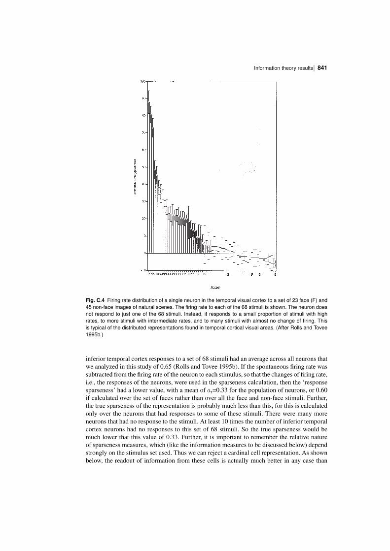
| 841Information theory results
Fig. C.4 Firing rate distribution of a single neuron in the temporal visual cortex to a set of 23 face (F) and45 nonface images of natural scenes. The firing rate to each of the 68 stimuli is shown. The neuron doesnot respond to just one of the 68 stimuli. Instead, it responds to a small proportion of stimuli with highrates, to more stimuli with intermediate rates, and to many stimuli with almost no change of firing. Thisis typical of the distributed representations found in temporal cortical visual areas. (After Rolls and Tovee1995b.)
inferior temporal cortex responses to a set of 68 stimuli had an average across all neurons thatwe analyzed in this study of 0.65 (Rolls and Tovee 1995b). If the spontaneous firing rate wassubtracted from the firing rate of the neuron to each stimulus, so that the changes of firing rate,i.e., the responses of the neurons, were used in the sparseness calculation, then the ‘responsesparseness’ had a lower value, with a mean of ar=0.33 for the population of neurons, or 0.60if calculated over the set of faces rather than over all the face and nonface stimuli. Further,the true sparseness of the representation is probably much less than this, for this is calculatedonly over the neurons that had responses to some of these stimuli. There were many moreneurons that had no response to the stimuli. At least 10 times the number of inferior temporalcortex neurons had no responses to this set of 68 stimuli. So the true sparseness would bemuch lower that this value of 0.33. Further, it is important to remember the relative natureof sparseness measures, which (like the information measures to be discussed below) dependstrongly on the stimulus set used. Thus we can reject a cardinal cell representation. As shownbelow, the readout of information from these cells is actually much better in any case than
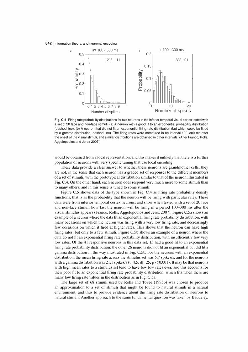
Information theory, and neuronal encoding842 |
0 10 200
0.05
0.1
0.15
0.2
Number of spikes
Pro
ba
bili
ty
int 100 - 300 ms
288 01
0 1 2 3 4 5 6 7 8 90
0.1
0.2
0.3
0.4
0.5
Pro
ba
bili
ty
int 100 - 300 ms
213 11
Number of spikes
a b
Fig. C.5 Firing rate probability distributions for two neurons in the inferior temporal visual cortex tested witha set of 20 face and nonface stimuli. (a) A neuron with a good fit to an exponential probability distribution(dashed line). (b) A neuron that did not fit an exponential firing rate distribution (but which could be fittedby a gamma distribution, dashed line). The firing rates were measured in an interval 100–300 ms afterthe onset of the visual stimuli, and similar distributions are obtained in other intervals. (After Franco, Rolls,Aggelopoulos and Jerez 2007.)
would be obtained from a local representation, and this makes it unlikely that there is a furtherpopulation of neurons with very specific tuning that use local encoding.
These data provide a clear answer to whether these neurons are grandmother cells: theyare not, in the sense that each neuron has a graded set of responses to the different membersof a set of stimuli, with the prototypical distribution similar to that of the neuron illustrated inFig. C.4. On the other hand, each neuron does respond very much more to some stimuli thanto many others, and in this sense is tuned to some stimuli.
Figure C.5 shows data of the type shown in Fig. C.4 as firing rate probability densityfunctions, that is as the probability that the neuron will be firing with particular rates. Thesedata were from inferior temporal cortex neurons, and show when tested with a set of 20 faceand nonface stimuli how fast the neuron will be firing in a period 100–300 ms after thevisual stimulus appears (Franco, Rolls, Aggelopoulos and Jerez 2007). Figure C.5a shows anexample of a neuron where the data fit an exponential firing rate probability distribution, withmany occasions on which the neuron was firing with a very low firing rate, and decreasinglyfew occasions on which it fired at higher rates. This shows that the neuron can have highfiring rates, but only to a few stimuli. Figure C.5b shows an example of a neuron where thedata do not fit an exponential firing rate probability distribution, with insufficiently few verylow rates. Of the 41 responsive neurons in this data set, 15 had a good fit to an exponentialfiring rate probability distribution; the other 26 neurons did not fit an exponential but did fit agamma distribution in the way illustrated in Fig. C.5b. For the neurons with an exponentialdistribution, the mean firing rate across the stimulus set was 5.7 spikes/s, and for the neuronswith a gamma distribution was 21.1 spikes/s (t=4.5, df=25, p< 0.001). It may be that neuronswith high mean rates to a stimulus set tend to have few low rates ever, and this accounts fortheir poor fit to an exponential firing rate probability distribution, which fits when there aremany low firing rate values in the distribution as in Fig. C.5a.
The large set of 68 stimuli used by Rolls and Tovee (1995b) was chosen to producean approximation to a set of stimuli that might be found to natural stimuli in a naturalenvironment, and thus to provide evidence about the firing rate distribution of neurons tonatural stimuli. Another approach to the same fundamental question was taken by Baddeley,
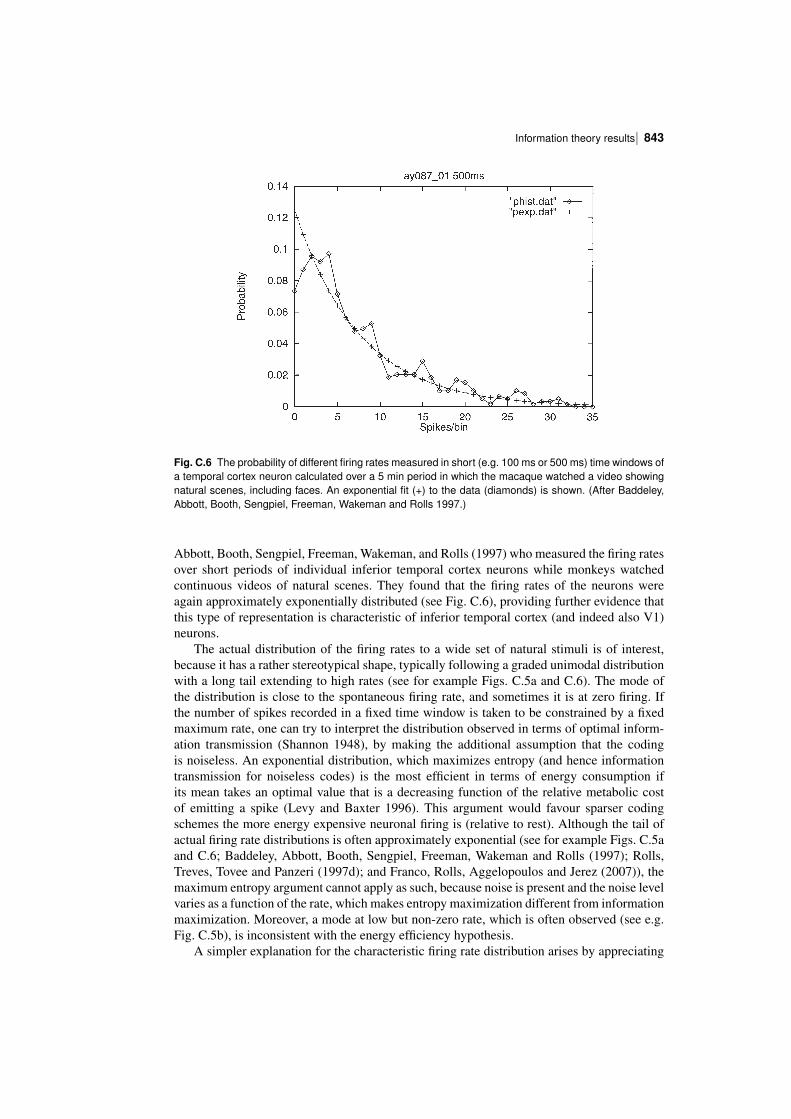
| 843Information theory results
Fig. C.6 The probability of different firing rates measured in short (e.g. 100 ms or 500 ms) time windows ofa temporal cortex neuron calculated over a 5 min period in which the macaque watched a video showingnatural scenes, including faces. An exponential fit (+) to the data (diamonds) is shown. (After Baddeley,Abbott, Booth, Sengpiel, Freeman, Wakeman and Rolls 1997.)
Abbott, Booth, Sengpiel, Freeman, Wakeman, and Rolls (1997) who measured the firing ratesover short periods of individual inferior temporal cortex neurons while monkeys watchedcontinuous videos of natural scenes. They found that the firing rates of the neurons wereagain approximately exponentially distributed (see Fig. C.6), providing further evidence thatthis type of representation is characteristic of inferior temporal cortex (and indeed also V1)neurons.
The actual distribution of the firing rates to a wide set of natural stimuli is of interest,because it has a rather stereotypical shape, typically following a graded unimodal distributionwith a long tail extending to high rates (see for example Figs. C.5a and C.6). The mode ofthe distribution is close to the spontaneous firing rate, and sometimes it is at zero firing. Ifthe number of spikes recorded in a fixed time window is taken to be constrained by a fixedmaximum rate, one can try to interpret the distribution observed in terms of optimal information transmission (Shannon 1948), by making the additional assumption that the codingis noiseless. An exponential distribution, which maximizes entropy (and hence informationtransmission for noiseless codes) is the most efficient in terms of energy consumption ifits mean takes an optimal value that is a decreasing function of the relative metabolic costof emitting a spike (Levy and Baxter 1996). This argument would favour sparser codingschemes the more energy expensive neuronal firing is (relative to rest). Although the tail ofactual firing rate distributions is often approximately exponential (see for example Figs. C.5aand C.6; Baddeley, Abbott, Booth, Sengpiel, Freeman, Wakeman and Rolls (1997); Rolls,Treves, Tovee and Panzeri (1997d); and Franco, Rolls, Aggelopoulos and Jerez (2007)), themaximum entropy argument cannot apply as such, because noise is present and the noise levelvaries as a function of the rate, which makes entropy maximization different from informationmaximization. Moreover, a mode at low but nonzero rate, which is often observed (see e.g.Fig. C.5b), is inconsistent with the energy efficiency hypothesis.
A simpler explanation for the characteristic firing rate distribution arises by appreciating
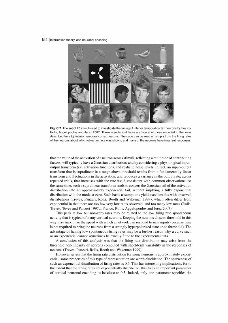
Information theory, and neuronal encoding844 |
Fig. C.7 The set of 20 stimuli used to investigate the tuning of inferior temporal cortex neurons by Franco,Rolls, Aggelopoulos and Jerez 2007. These objects and faces are typical of those encoded in the waysdescribed here by inferior temporal cortex neurons. The code can be read off simply from the firing ratesof the neurons about which object or face was shown, and many of the neurons have invariant responses.
that the value of the activation of a neuron across stimuli, reflecting a multitude of contributingfactors, will typically have a Gaussian distribution; and by considering a physiological input–output transform (i.e. activation function), and realistic noise levels. In fact, an input–outputtransform that is supralinear in a range above threshold results from a fundamentally lineartransform and fluctuations in the activation, and produces a variance in the output rate, acrossrepeated trials, that increases with the rate itself, consistent with common observations. Atthe same time, such a supralinear transform tends to convert the Gaussian tail of the activationdistribution into an approximately exponential tail, without implying a fully exponentialdistribution with the mode at zero. Such basic assumptions yield excellent fits with observeddistributions (Treves, Panzeri, Rolls, Booth and Wakeman 1999), which often differ fromexponential in that there are too few very low rates observed, and too many low rates (Rolls,Treves, Tovee and Panzeri 1997d, Franco, Rolls, Aggelopoulos and Jerez 2007).
This peak at low but nonzero rates may be related to the low firing rate spontaneousactivity that is typical of many cortical neurons. Keeping the neurons close to threshold in thisway may maximize the speed with which a network can respond to new inputs (because timeis not required to bring the neurons from a strongly hyperpolarized state up to threshold). Theadvantage of having low spontaneous firing rates may be a further reason why a curve suchas an exponential cannot sometimes be exactly fitted to the experimental data.
A conclusion of this analysis was that the firing rate distribution may arise from thethreshold nonlinearity of neurons combined with shortterm variability in the responses ofneurons (Treves, Panzeri, Rolls, Booth and Wakeman 1999).
However, given that the firing rate distribution for some neurons is approximately exponential, some properties of this type of representation are worth elucidation. The sparseness ofsuch an exponential distribution of firing rates is 0.5. This has interesting implications, for tothe extent that the firing rates are exponentially distributed, this fixes an important parameterof cortical neuronal encoding to be close to 0.5. Indeed, only one parameter specifies the
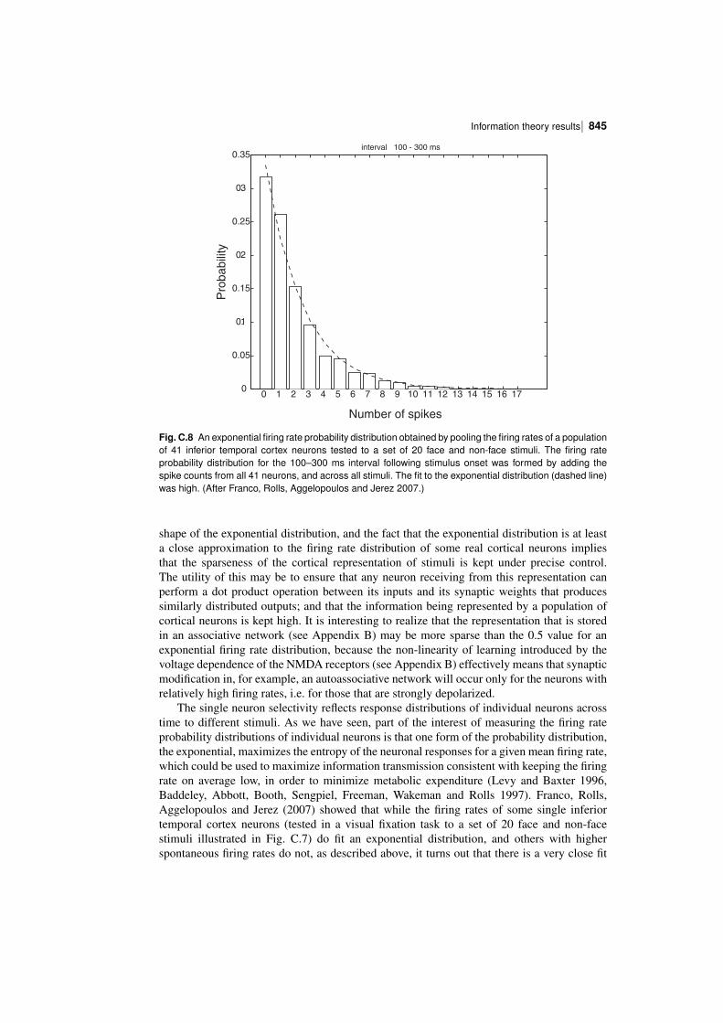
| 845Information theory results
0 1 2 3 4 5 6 7 8 9 10 11 12 13 14 15 16 170
0.05
0.1
0.15
0.2
0.25
0.3
0.35
Number of spikes
Pro
babili
ty
interval 100 - 300 ms
Fig. C.8 An exponential firing rate probability distribution obtained by pooling the firing rates of a populationof 41 inferior temporal cortex neurons tested to a set of 20 face and nonface stimuli. The firing rateprobability distribution for the 100–300 ms interval following stimulus onset was formed by adding thespike counts from all 41 neurons, and across all stimuli. The fit to the exponential distribution (dashed line)was high. (After Franco, Rolls, Aggelopoulos and Jerez 2007.)
shape of the exponential distribution, and the fact that the exponential distribution is at leasta close approximation to the firing rate distribution of some real cortical neurons impliesthat the sparseness of the cortical representation of stimuli is kept under precise control.The utility of this may be to ensure that any neuron receiving from this representation canperform a dot product operation between its inputs and its synaptic weights that producessimilarly distributed outputs; and that the information being represented by a population ofcortical neurons is kept high. It is interesting to realize that the representation that is storedin an associative network (see Appendix B) may be more sparse than the 0.5 value for anexponential firing rate distribution, because the nonlinearity of learning introduced by thevoltage dependence of the NMDA receptors (see Appendix B) effectively means that synapticmodification in, for example, an autoassociative network will occur only for the neurons withrelatively high firing rates, i.e. for those that are strongly depolarized.
The single neuron selectivity reflects response distributions of individual neurons acrosstime to different stimuli. As we have seen, part of the interest of measuring the firing rateprobability distributions of individual neurons is that one form of the probability distribution,the exponential, maximizes the entropy of the neuronal responses for a given mean firing rate,which could be used to maximize information transmission consistent with keeping the firingrate on average low, in order to minimize metabolic expenditure (Levy and Baxter 1996,Baddeley, Abbott, Booth, Sengpiel, Freeman, Wakeman and Rolls 1997). Franco, Rolls,Aggelopoulos and Jerez (2007) showed that while the firing rates of some single inferiortemporal cortex neurons (tested in a visual fixation task to a set of 20 face and nonfacestimuli illustrated in Fig. C.7) do fit an exponential distribution, and others with higherspontaneous firing rates do not, as described above, it turns out that there is a very close fit
Information theory, and neuronal encoding846 |
to an exponential distribution of firing rates if all spikes from all the neurons are consideredtogether. This interesting result is shown in Fig. C.8.
One implication of the result shown in Fig. C.8 is that a neuron with inputs from the inferiortemporal visual cortex will receive an exponential distribution of firing rates on its afferents,and this is therefore the type of input that needs to be considered in theoretical models ofneuronal network function in the brain (see Appendix B). The second implication is that atthe level of single neurons, an exponential probability density function is consistent withminimizing energy utilization, and maximizing information transmission, for a given meanfiring rate (Levy and Baxter 1996, Baddeley, Abbott, Booth, Sengpiel, Freeman, Wakemanand Rolls 1997).
C.3.1.2 Population sparseness ap
If instead we consider the responses of a population of neurons taken at any one time (to onestimulus), we might also expect a sparse graded distribution, with few neurons firing fast toa particular stimulus. It is important to measure the population sparseness, for this is a keyparameter that influences the number of different stimuli that can be stored and retrieved innetworks such as those found in the cortex with recurrent collateral connections between theexcitatory neurons, which can form autoassociation or attractor networks if the synapses areassociatively modifiable (Hopfield 1982, Treves and Rolls 1991, Rolls and Treves 1998, Rollsand Deco 2002, Rolls 2008d) (see Appendix B). Further, in physics, if one can predict thedistribution of the responses of the system at any one time (the population level) from thedistribution of the responses of a component of the system across time, the system is describedas ergodic, and a necessary condition for this is that the components are uncorrelated (Lehky,Sejnowski and Desimone 2005). Considering this in neuronal terms, the average sparsenessof a population of neurons over multiple stimulus inputs must equal the average selectivityto the stimuli of the single neurons within the population provided that the responses of theneurons are uncorrelated (Foldiak 2003).
The sparseness ap of the population code may be quantified (for any one stimulus) as
ap =
(N∑
n=1yn/N)2
(N∑
n=1y2n)/N
(C.46)
where yn is the mean firing rate of neuron n in the set of N neurons.This measure, ap, of the sparseness of the representation of a stimulus by a population
of neurons has a number of advantages. One is that it is the same measure of sparseness thathas proved to be useful and tractable in formal analyses of the capacity of associative neuralnetworks and the interference between stimuli that use an approach derived from theoreticalphysics (Rolls and Treves 1990, Treves 1990, Treves and Rolls 1991, Rolls and Treves 1998)(see Appendix B). We note that high values of ap indicate broad tuning of the population, andthat low values of ap indicate sparse population encoding.
Franco, Rolls, Aggelopoulos and Jerez (2007) measured the population sparseness of aset of 29 inferior temporal cortex neurons to a set of 20 stimuli that included faces and objects(see Fig. C.7). Figure C.9a shows, for any one stimulus picked at random, the normalizedfiring rates of the population of neurons. The rates are ranked with the neuron with the highestrate on the left. For different stimuli, the shape of this distribution is on average the same,though with the neurons in a different order. (The rates of each neuron were normalized to amean of 10 spikes/s before this graph was made, so that the neurons can be combined in thesame graph, and so that the population sparseness has a defined value, as described by Franco,
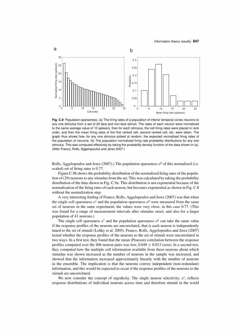
| 847Information theory results
0 5 10 15 20 25 300
5
10
15
20
25
30
Cell index
No
rma
lize
d firin
g r
ate
(sp
ike
s/s
)
0 5 10 15 20 250
0.05
0.1
0.15
0. 2
0.25
0.3
Mean firing rate (spikes/s)
Pro
ba
bili
ty
a b
Fig. C.9 Population sparseness. (a) The firing rates of a population of inferior temporal cortex neurons toany one stimulus from a set of 20 face and nonface stimuli. The rates of each neuron were normalizedto the same average value of 10 spikes/s, then for each stimulus, the cell firing rates were placed in rankorder, and then the mean firing rates of the first ranked cell, second ranked cell, etc. were taken. Thegraph thus shows how, for any one stimulus picked at random, the expected normalized firing rates ofthe population of neurons. (b) The population normalized firing rate probability distributions for any onestimulus. This was computed effectively by taking the probability density function of the data shown in (a).(After Franco, Rolls, Aggelopoulos and Jerez 2007.)
Rolls, Aggelopoulos and Jerez (2007).) The population sparseness ap of this normalized (i.e.scaled) set of firing rates is 0.77.
Figure C.9b shows the probability distribution of the normalized firing rates of the population of (29) neurons to any stimulus from the set. This was calculated by taking the probabilitydistribution of the data shown in Fig. C.9a. This distribution is not exponential because of thenormalization of the firing rates of each neuron, but becomes exponential as shown in Fig. C.8without the normalization step.
A very interesting finding of Franco, Rolls, Aggelopoulos and Jerez (2007) was that whenthe single cell sparseness as and the population sparseness ap were measured from the sameset of neurons in the same experiment, the values were very close, in this case 0.77. (Thiswas found for a range of measurement intervals after stimulus onset, and also for a largerpopulation of 41 neurons.)
The single cell sparseness as and the population sparseness ap can take the same valueif the response profiles of the neurons are uncorrelated, that is each neuron is independentlytuned to the set of stimuli (Lehky et al. 2005). Franco, Rolls, Aggelopoulos and Jerez (2007)tested whether the response profiles of the neurons to the set of stimuli were uncorrelated intwo ways. In a first test, they found that the mean (Pearson) correlation between the responseprofiles computed over the 406 neuron pairs was low, 0.049 ± 0.013 (sem). In a second test,they computed how the multiple cell information available from these neurons about whichstimulus was shown increased as the number of neurons in the sample was increased, andshowed that the information increased approximately linearly with the number of neuronsin the ensemble. The implication is that the neurons convey independent (nonredundant)information, and this would be expected to occur if the response profiles of the neurons to thestimuli are uncorrelated.
We now consider the concept of ergodicity. The single neuron selectivity, as, reflectsresponse distributions of individual neurons across time and therefore stimuli in the world
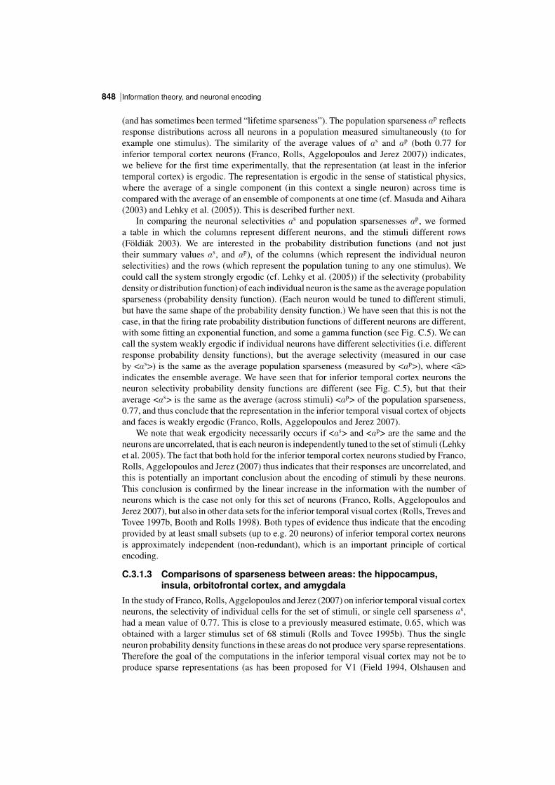
Information theory, and neuronal encoding848 |
(and has sometimes been termed “lifetime sparseness”). The population sparseness ap reflectsresponse distributions across all neurons in a population measured simultaneously (to forexample one stimulus). The similarity of the average values of as and ap (both 0.77 forinferior temporal cortex neurons (Franco, Rolls, Aggelopoulos and Jerez 2007)) indicates,we believe for the first time experimentally, that the representation (at least in the inferiortemporal cortex) is ergodic. The representation is ergodic in the sense of statistical physics,where the average of a single component (in this context a single neuron) across time iscompared with the average of an ensemble of components at one time (cf. Masuda and Aihara(2003) and Lehky et al. (2005)). This is described further next.
In comparing the neuronal selectivities as and population sparsenesses ap, we formeda table in which the columns represent different neurons, and the stimuli different rows(Foldiak 2003). We are interested in the probability distribution functions (and not justtheir summary values as, and ap), of the columns (which represent the individual neuronselectivities) and the rows (which represent the population tuning to any one stimulus). Wecould call the system strongly ergodic (cf. Lehky et al. (2005)) if the selectivity (probabilitydensity or distribution function) of each individual neuron is the same as the average populationsparseness (probability density function). (Each neuron would be tuned to different stimuli,but have the same shape of the probability density function.) We have seen that this is not thecase, in that the firing rate probability distribution functions of different neurons are different,with some fitting an exponential function, and some a gamma function (see Fig. C.5). We cancall the system weakly ergodic if individual neurons have different selectivities (i.e. differentresponse probability density functions), but the average selectivity (measured in our caseby <as>) is the same as the average population sparseness (measured by <ap>), where <ã>indicates the ensemble average. We have seen that for inferior temporal cortex neurons theneuron selectivity probability density functions are different (see Fig. C.5), but that theiraverage <as> is the same as the average (across stimuli) <ap> of the population sparseness,0.77, and thus conclude that the representation in the inferior temporal visual cortex of objectsand faces is weakly ergodic (Franco, Rolls, Aggelopoulos and Jerez 2007).
We note that weak ergodicity necessarily occurs if <as> and <ap> are the same and theneurons are uncorrelated, that is each neuron is independently tuned to the set of stimuli (Lehkyet al. 2005). The fact that both hold for the inferior temporal cortex neurons studied by Franco,Rolls, Aggelopoulos and Jerez (2007) thus indicates that their responses are uncorrelated, andthis is potentially an important conclusion about the encoding of stimuli by these neurons.This conclusion is confirmed by the linear increase in the information with the number ofneurons which is the case not only for this set of neurons (Franco, Rolls, Aggelopoulos andJerez 2007), but also in other data sets for the inferior temporal visual cortex (Rolls, Treves andTovee 1997b, Booth and Rolls 1998). Both types of evidence thus indicate that the encodingprovided by at least small subsets (up to e.g. 20 neurons) of inferior temporal cortex neuronsis approximately independent (nonredundant), which is an important principle of corticalencoding.
C.3.1.3 Comparisons of sparseness between areas: the hippocampus,insula, orbitofrontal cortex, and amygdala
In the study of Franco, Rolls, Aggelopoulos and Jerez (2007) on inferior temporal visual cortexneurons, the selectivity of individual cells for the set of stimuli, or single cell sparseness as,had a mean value of 0.77. This is close to a previously measured estimate, 0.65, which wasobtained with a larger stimulus set of 68 stimuli (Rolls and Tovee 1995b). Thus the singleneuron probability density functions in these areas do not produce very sparse representations.Therefore the goal of the computations in the inferior temporal visual cortex may not be toproduce sparse representations (as has been proposed for V1 (Field 1994, Olshausen and
| 849Information theory results
Field 1997, Vinje and Gallant 2000, Olshausen and Field 2004)). Instead one of the goalsof the computations in the inferior temporal visual cortex may be to compute invariantrepresentations of objects and faces (Rolls 2000a, Rolls and Deco 2002, Rolls 2007e, Rollsand Stringer 2006) (see Chapter 25), and to produce not very sparse distributed representationsin order to maximize the information represented (see Table B.2 on page 722). In this context,it is very interesting that the representations of different stimuli provided by a populationof inferior temporal cortex neurons are decorrelated, as shown by the finding that the mean(Pearson) correlation between the response profiles to a set of 20 stimuli computed over 406neuron pairs was low, 0.049 ± 0.013 (sem) (Franco, Rolls, Aggelopoulos and Jerez 2007).The implication is that decorrelation is being achieved in the inferior temporal visual cortex,but not by forming a sparse code. It will be interesting to investigate the mechanisms for this.
In contrast, the representation in some memory systems may be more sparse. For example,in the hippocampus in which spatial view cells are found in macaques, further analysis of datadescribed by Rolls, Treves, Robertson, GeorgesFrancois and Panzeri (1998b) shows that forthe representation of 64 locations around the walls of the room, the mean single cell sparseness<as> was 0.34 ± 0.13 (sd), and the mean population sparseness ap was 0.33 ± 0.11. Themore sparse representation is consistent with the view that the hippocampus is involved instoring memories, and that for this, more sparse representations than in perceptual areas arerelevant. These sparseness values are for spatial view neurons, but it is possible that whenneurons respond to combinations of spatial view and object (Rolls, Xiang and Franco 2005c),or of spatial view and reward (Rolls and Xiang 2005), the representations are more sparse. Itis of interest that the mean firing rate of these spatial view neurons across all spatial views was1.77 spikes/s (Rolls, Treves, Robertson, GeorgesFrancois and Panzeri 1998b). (The meanspontaneous firing rate of the neurons was 0.1 spikes/s, and the average across neurons of thefiring rate for the most effective spatial view was 13.2 spikes/s.) It is also notable that weakergodicity is implied for this brain region too (given the similar values of <as> and <ap>), andthe underlying basis for this is that the response profiles of the different hippocampal neurons tothe spatial views are uncorrelated. Further support for these conclusions is that the informationabout spatial view increases linearly with the number of hippocampal spatial view neurons(Rolls, Treves, Robertson, GeorgesFrancois and Panzeri 1998b), again providing evidencethat the response profiles of the different neurons are uncorrelated. The representations inthe hippocampus may be more sparse than this, in line with the observation that in rodents,hippocampal neurons may have place fields in only one of several environments.
Further evidence is now available on ergodicity in three further brain areas, the macaqueinsular primary taste cortex, the orbitofrontal cortex, and the amygdala (Rolls, Critchley,Verhagen and Kadohisa 2010a). In all these brain areas sets of neurons were tested with anidentical set of 24 oral taste, temperature, and texture stimuli. (The stimuli were: Taste 0.1M NaCl (salt), 1 M glucose (sweet), 0.01 M HCl (sour), 0.001 M quinine HCl (bitter), 0.1 Mmonosodium glutamate (umami), and water; Temperature 10oC, 37oC and 42oC; flavour blackcurrant juice; viscosity carboxymethylcellulose 10 cPoise, 100 cPoise, 1000 cPoiseand 10000 cPoise; fatty / oily single cream, vegetable oil, mineral oil, silicone oil (100cPoise), coconut oil, and safflower oil; fatty acids linoleic acid and lauric acid; capsaicin;and gritty texture.) Further analysis of data described by Verhagen, Kadohisa and Rolls (2004)showed that in the primary taste cortex the mean value of as across 58 neurons was 0.745and of ap (normalized) was 0.708. Further analysis of data described by Rolls, Verhagenand Kadohisa (2003e), Verhagen, Rolls and Kadohisa (2003), Kadohisa, Rolls and Verhagen(2004) and Kadohisa, Rolls and Verhagen (2005a) showed that in the orbitofrontal cortex themean value of as across 30 neurons was 0.625 and of ap was 0.611. Further analysis of datadescribed by Kadohisa, Rolls and Verhagen (2005b) showed that in the amygdala the meanvalue of as across 38 neurons was 0.811 and of ap was 0.813. Thus in all these cases, the mean
Information theory, and neuronal encoding850 |
value of as is close to that of ap, and weak ergodicity is implied. The values of as and ap arealso relatively high, implying the importance of representing large amounts of information inthese brain areas about this set of stimuli by using a very distributed code, and also perhapsabout the stimulus set, some members of which may be rather similar to each other.
C.3.2 The information from single neurons
Examples of the responses of single neurons (in this case in the inferior temporal visual cortex)to sets of objects and/or faces (of the type illustrated in Fig. C.7) are shown in Figs. 16.15, 25.12and C.4. We now consider how much information these types of neuronal response conveyabout the set of stimuliS, and about each stimulus s in the set. The mutual information I(S,R)that the set of responses R encode about the set of stimuli S is calculated with equation C.21and corrected for the limited sampling using the analytic bias correction procedure describedby Panzeri and Treves (1996) as described in detail by Rolls, Treves, Tovee and Panzeri(1997d). The information I(s,R) about each single stimulus s in the set S, termed thestimulusspecific information (Rolls, Treves, Tovee and Panzeri 1997d) or stimulusspecificsurprise (DeWeese and Meister 1999), obtained from the set of the responses R of the singleneuron is calculated with equation C.22 and corrected for the limited sampling using theanalytic bias correction procedure described by Panzeri and Treves (1996) as described indetail by Rolls, Treves, Tovee and Panzeri (1997d). (The average of I(s,R) across stimuli isthe mutual information I(S,R).)
Figure C.10 shows the stimulusspecific information I(s,R) available in the neuronalresponse about each of 20 face stimuli calculated for the neuron (am242) whose firing rateresponse profile to the set of 65 stimuli is shown in Fig. C.4. Unless otherwise stated, theinformation measures given are for the information available on a single trial from the firingrate of the neuron in a 500 ms period starting 100 ms after the onset of the stimuli. It is shownin Fig. C.10 that 2.2, 2.0, and 1.5 bits of information were present about the three face stimulito which the neuron had the highest firing rate responses. The neuron conveyed some butsmaller amounts of information about the remaining face stimuli. The average informationI(S,R) about this set (S) of 20 faces for this neuron was 0.55 bits. The average firing rateof this neuron to these 20 face stimuli was 54 spikes/s. It is clear from Fig. C.10 that littleinformation was available from the responses of the neuron to a particular face stimulus ifthat response was close to the average response of the neuron across all stimuli. At the sametime, it is clear from Fig. C.10 that information was present depending on how far the firingrate to a particular stimulus was from the average response of the neuron to the stimuli. Ofparticular interest, it is evident that information is present from the neuronal response aboutwhich face was shown if that neuronal response was below the average response, as well aswhen the response was greater than the average response.
One intuitive way to understand the data shown in Fig. C.10 is to appreciate that lowprobability firing rate responses, whether they are greater than or less than the mean responserate, convey much information about which stimulus was seen. This is of course close to thedefinition of information. Given that the firing rates of neurons are always positive, and followan asymmetric distribution about their mean, it is clear that deviations above the mean have adifferent probability to occur than deviations by the same amount below the mean. One mayattempt to capture the relative likelihood of different firing rates above and below the meanby computing a z score obtained by dividing the difference between the mean response toeach stimulus and the overall mean response by the standard deviation of the response to thatstimulus. The greater the number of standard deviations (i.e. the greater the z score) from themean response value, the greater the information might be expected to be. We therefore showin Fig. C.11 the relation between the z score and I(s,R). (The z score was calculated by
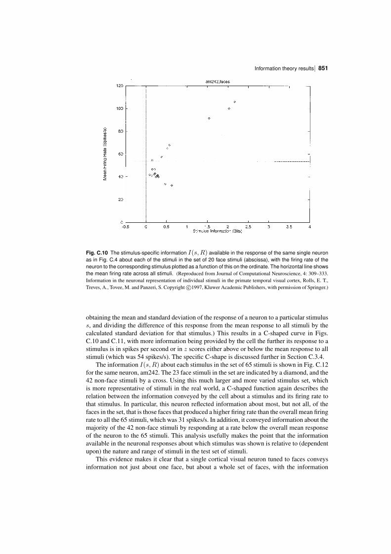
| 851Information theory results
Fig. C.10 The stimulusspecific information I(s,R) available in the response of the same single neuronas in Fig. C.4 about each of the stimuli in the set of 20 face stimuli (abscissa), with the firing rate of theneuron to the corresponding stimulus plotted as a function of this on the ordinate. The horizontal line showsthe mean firing rate across all stimuli. (Reproduced from Journal of Computational Neuroscience, 4: 309–333.Information in the neuronal representation of individual stimuli in the primate temporal visual cortex, Rolls, E. T.,Treves, A., Tovee, M. and Panzeri, S. Copyright c⃝1997, Kluwer Academic Publishers, with permission of Springer.)
obtaining the mean and standard deviation of the response of a neuron to a particular stimuluss, and dividing the difference of this response from the mean response to all stimuli by thecalculated standard deviation for that stimulus.) This results in a Cshaped curve in Figs.C.10 and C.11, with more information being provided by the cell the further its response to astimulus is in spikes per second or in z scores either above or below the mean response to allstimuli (which was 54 spikes/s). The specific Cshape is discussed further in Section C.3.4.
The information I(s,R) about each stimulus in the set of 65 stimuli is shown in Fig. C.12for the same neuron, am242. The 23 face stimuli in the set are indicated by a diamond, and the42 nonface stimuli by a cross. Using this much larger and more varied stimulus set, whichis more representative of stimuli in the real world, a Cshaped function again describes therelation between the information conveyed by the cell about a stimulus and its firing rate tothat stimulus. In particular, this neuron reflected information about most, but not all, of thefaces in the set, that is those faces that produced a higher firing rate than the overall mean firingrate to all the 65 stimuli, which was 31 spikes/s. In addition, it conveyed information about themajority of the 42 nonface stimuli by responding at a rate below the overall mean responseof the neuron to the 65 stimuli. This analysis usefully makes the point that the informationavailable in the neuronal responses about which stimulus was shown is relative to (dependentupon) the nature and range of stimuli in the test set of stimuli.
This evidence makes it clear that a single cortical visual neuron tuned to faces conveysinformation not just about one face, but about a whole set of faces, with the information
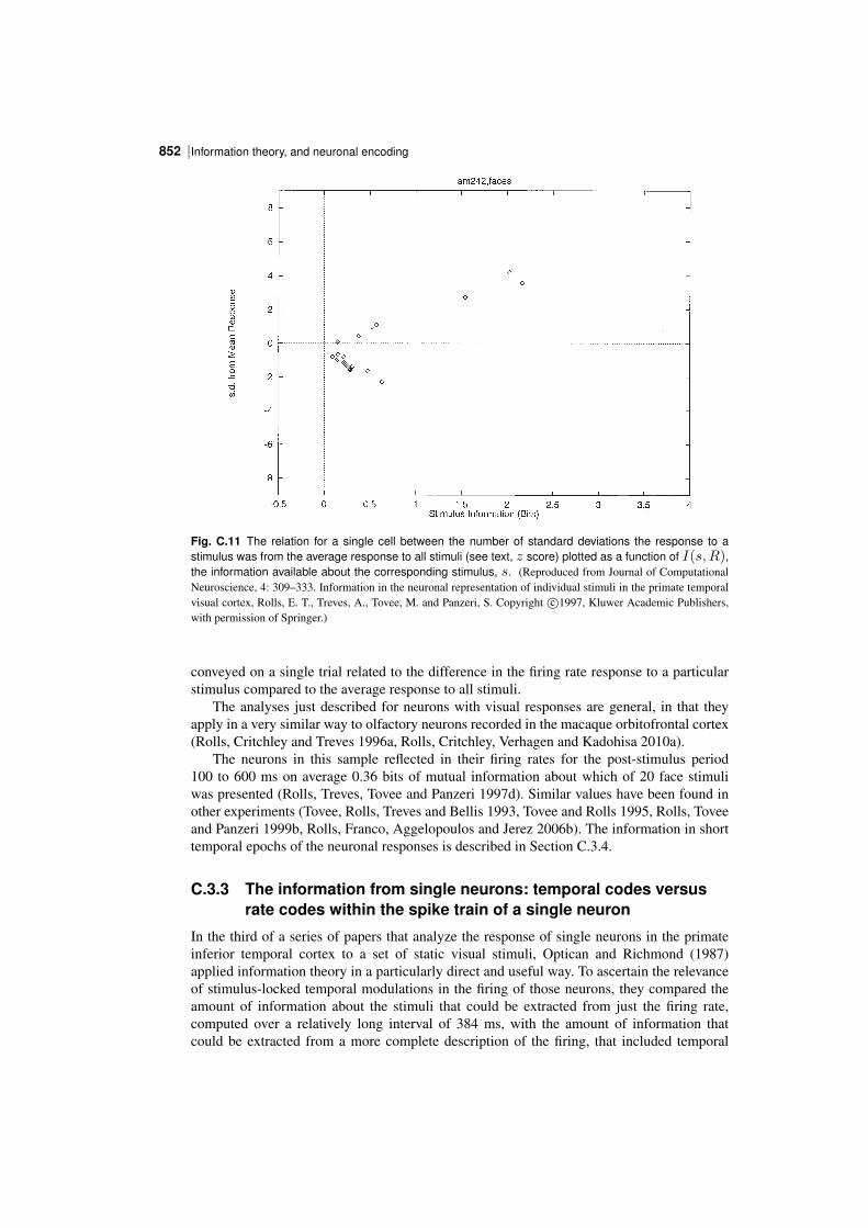
Information theory, and neuronal encoding852 |
Fig. C.11 The relation for a single cell between the number of standard deviations the response to astimulus was from the average response to all stimuli (see text, z score) plotted as a function of I(s,R),the information available about the corresponding stimulus, s. (Reproduced from Journal of ComputationalNeuroscience, 4: 309–333. Information in the neuronal representation of individual stimuli in the primate temporalvisual cortex, Rolls, E. T., Treves, A., Tovee, M. and Panzeri, S. Copyright c⃝1997, Kluwer Academic Publishers,with permission of Springer.)
conveyed on a single trial related to the difference in the firing rate response to a particularstimulus compared to the average response to all stimuli.
The analyses just described for neurons with visual responses are general, in that theyapply in a very similar way to olfactory neurons recorded in the macaque orbitofrontal cortex(Rolls, Critchley and Treves 1996a, Rolls, Critchley, Verhagen and Kadohisa 2010a).
The neurons in this sample reflected in their firing rates for the poststimulus period100 to 600 ms on average 0.36 bits of mutual information about which of 20 face stimuliwas presented (Rolls, Treves, Tovee and Panzeri 1997d). Similar values have been found inother experiments (Tovee, Rolls, Treves and Bellis 1993, Tovee and Rolls 1995, Rolls, Toveeand Panzeri 1999b, Rolls, Franco, Aggelopoulos and Jerez 2006b). The information in shorttemporal epochs of the neuronal responses is described in Section C.3.4.
C.3.3 The information from single neurons: temporal codes versusrate codes within the spike train of a single neuron
In the third of a series of papers that analyze the response of single neurons in the primateinferior temporal cortex to a set of static visual stimuli, Optican and Richmond (1987)applied information theory in a particularly direct and useful way. To ascertain the relevanceof stimuluslocked temporal modulations in the firing of those neurons, they compared theamount of information about the stimuli that could be extracted from just the firing rate,computed over a relatively long interval of 384 ms, with the amount of information thatcould be extracted from a more complete description of the firing, that included temporal
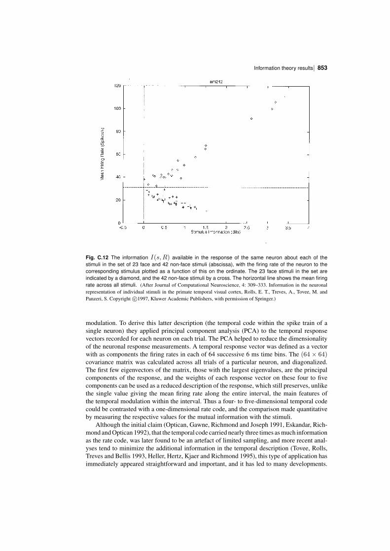
| 853Information theory results
Fig. C.12 The information I(s,R) available in the response of the same neuron about each of thestimuli in the set of 23 face and 42 nonface stimuli (abscissa), with the firing rate of the neuron to thecorresponding stimulus plotted as a function of this on the ordinate. The 23 face stimuli in the set areindicated by a diamond, and the 42 nonface stimuli by a cross. The horizontal line shows the mean firingrate across all stimuli. (After Journal of Computational Neuroscience, 4: 309–333. Information in the neuronalrepresentation of individual stimuli in the primate temporal visual cortex, Rolls, E. T., Treves, A., Tovee, M. andPanzeri, S. Copyright c⃝1997, Kluwer Academic Publishers, with permission of Springer.)
modulation. To derive this latter description (the temporal code within the spike train of asingle neuron) they applied principal component analysis (PCA) to the temporal responsevectors recorded for each neuron on each trial. The PCA helped to reduce the dimensionalityof the neuronal response measurements. A temporal response vector was defined as a vectorwith as components the firing rates in each of 64 successive 6 ms time bins. The (64 × 64)covariance matrix was calculated across all trials of a particular neuron, and diagonalized.The first few eigenvectors of the matrix, those with the largest eigenvalues, are the principalcomponents of the response, and the weights of each response vector on these four to fivecomponents can be used as a reduced description of the response, which still preserves, unlikethe single value giving the mean firing rate along the entire interval, the main features ofthe temporal modulation within the interval. Thus a four to fivedimensional temporal codecould be contrasted with a onedimensional rate code, and the comparison made quantitativeby measuring the respective values for the mutual information with the stimuli.
Although the initial claim (Optican, Gawne, Richmond and Joseph 1991, Eskandar, Richmond and Optican 1992), that the temporal code carried nearly three times as much informationas the rate code, was later found to be an artefact of limited sampling, and more recent analyses tend to minimize the additional information in the temporal description (Tovee, Rolls,Treves and Bellis 1993, Heller, Hertz, Kjaer and Richmond 1995), this type of application hasimmediately appeared straightforward and important, and it has led to many developments.
Information theory, and neuronal encoding854 |
By concentrating on the code expressed in the output rather than on the characterization ofthe neuronal channel itself, this approach is not affected much by the potential complexitiesof the preceding black box. Limited sampling, on the other hand, is a problem, particularlybecause it affects much more codes with a larger number of components, for example thefour to five components of the PCA temporal description, than the onedimensional firingrate code. This is made evident in the paper by Heller, Hertz, Kjaer and Richmond (1995), inwhich the comparison is extended to several more detailed temporal descriptions, includinga binary vector description in which the presence or not of a spike in each 1 ms bin of theresponse constitutes a component of a 320dimensional vector. Obviously, this binary vectormust contain at least all the information present in the reduced descriptions, whereas in theresults of Heller, Hertz, Kjaer and Richmond (1995), despite the use of a sophisticated neuralnetwork procedure to control limited sampling biases, the binary vector appears to be thecode that carries the least information of all. In practice, with the data samples available in theexperiments that have been done, and even when using analytic procedures to control limitedsampling (Panzeri and Treves 1996), reliable comparison can be made only with up to twoto threedimensional codes.
Tovee, Rolls, Treves and Bellis (1993) and Tovee and Rolls (1995) obtained furtherevidence that little information was encoded in the temporal aspects of firing within the spiketrain of a single neuron in the inferior temporal cortex by taking short epochs of the firingof neurons, lasting 20 ms or 50 ms, in which the opportunity for temporal encoding wouldbe limited (because there were few spikes in these short time intervals). They found that aconsiderable proportion (30%) of the information available in a long time period of 400 msutilizing temporal encoding within the spike train was available in time periods as short as 20ms when only the number of spikes was taken into account.
Overall, the main result of these analyses applied to the responses to static stimuli in thetemporal visual cortex of primates is that not much more information (perhaps only up to10% more) can be extracted from temporal codes than from the firing rate measured over ajudiciously chosen interval (Tovee, Rolls, Treves and Bellis 1993, Heller, Hertz, Kjaer andRichmond 1995). Indeed, it turns out that even this small amount of ‘temporal information’ isrelated primarily to the onset latency of the neuronal responses to different stimuli, rather thanto anything more subtle (Tovee, Rolls, Treves and Bellis 1993). Consistent with this point, inearlier visual areas the additional ‘temporally encoded’ fraction of information can be larger,due especially to the increased relevance, earlier on, of precisely locked transient responses(Kjaer, Hertz and Richmond 1994, Golomb, Kleinfeld, Reid, Shapley and Shraiman 1994,Heller, Hertz, Kjaer and Richmond 1995). This is because if the responses to some stimuli aremore transient and to others more sustained, this will result in more information if the temporalmodulation of the response of the neuron is taken into account. However, the relevance ofmore substantial temporal codes for static visual stimuli remains to be demonstrated. Fornonstatic visual stimuli and for other cortical systems, similar analyses have largely yet to becarried out, although clearly one expects to find much more prominent temporal effects e.g. inthe auditory system (Nelken, Prut, Vaadia and Abeles 1994, deCharms and Merzenich 1996),for reasons similar to those just annunciated.
C.3.4 The information from single neurons: the speed of informationtransfer
It is intuitive that if short periods of firing of single cells are considered, there is less timefor temporal modulation effects. The information conveyed about stimuli by the firing rateand that conveyed by more detailed temporal codes become similar in value. When the firingperiods analyzed become shorter than roughly the mean interspike interval, even the statistics
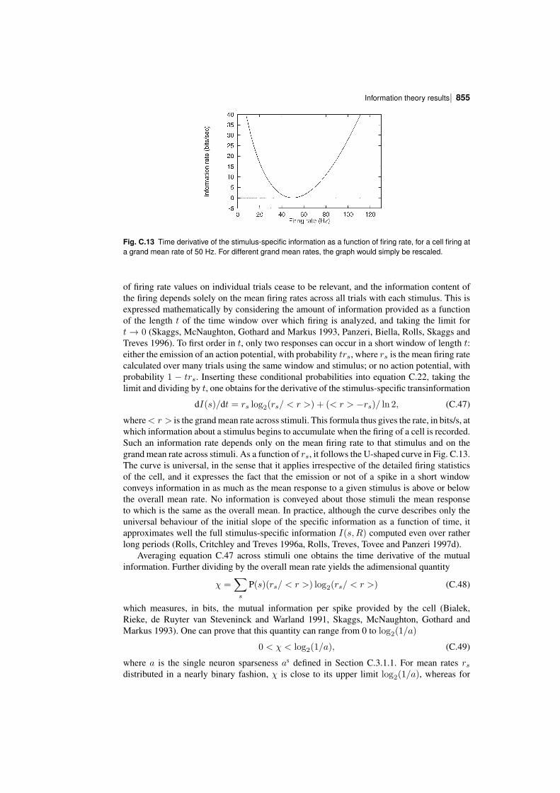
| 855Information theory results
Fig. C.13 Time derivative of the stimulusspecific information as a function of firing rate, for a cell firing ata grand mean rate of 50 Hz. For different grand mean rates, the graph would simply be rescaled.
of firing rate values on individual trials cease to be relevant, and the information content ofthe firing depends solely on the mean firing rates across all trials with each stimulus. This isexpressed mathematically by considering the amount of information provided as a functionof the length t of the time window over which firing is analyzed, and taking the limit fort → 0 (Skaggs, McNaughton, Gothard and Markus 1993, Panzeri, Biella, Rolls, Skaggs andTreves 1996). To first order in t, only two responses can occur in a short window of length t:either the emission of an action potential, with probability trs, where rs is the mean firing ratecalculated over many trials using the same window and stimulus; or no action potential, withprobability 1 − trs. Inserting these conditional probabilities into equation C.22, taking thelimit and dividing by t, one obtains for the derivative of the stimulusspecific transinformation
dI(s)/dt = rs log2(rs/ < r >) + (< r > −rs)/ ln 2, (C.47)
where< r > is the grand mean rate across stimuli. This formula thus gives the rate, in bits/s, atwhich information about a stimulus begins to accumulate when the firing of a cell is recorded.Such an information rate depends only on the mean firing rate to that stimulus and on thegrand mean rate across stimuli. As a function of rs, it follows the Ushaped curve in Fig. C.13.The curve is universal, in the sense that it applies irrespective of the detailed firing statisticsof the cell, and it expresses the fact that the emission or not of a spike in a short windowconveys information in as much as the mean response to a given stimulus is above or belowthe overall mean rate. No information is conveyed about those stimuli the mean responseto which is the same as the overall mean. In practice, although the curve describes only theuniversal behaviour of the initial slope of the specific information as a function of time, itapproximates well the full stimulusspecific information I(s,R) computed even over ratherlong periods (Rolls, Critchley and Treves 1996a, Rolls, Treves, Tovee and Panzeri 1997d).
Averaging equation C.47 across stimuli one obtains the time derivative of the mutualinformation. Further dividing by the overall mean rate yields the adimensional quantity
χ =∑s
P(s)(rs/ < r >) log2(rs/ < r >) (C.48)
which measures, in bits, the mutual information per spike provided by the cell (Bialek,Rieke, de Ruyter van Steveninck and Warland 1991, Skaggs, McNaughton, Gothard andMarkus 1993). One can prove that this quantity can range from 0 to log2(1/a)
0 < χ < log2(1/a), (C.49)
where a is the single neuron sparseness as defined in Section C.3.1.1. For mean rates rsdistributed in a nearly binary fashion, χ is close to its upper limit log2(1/a), whereas for
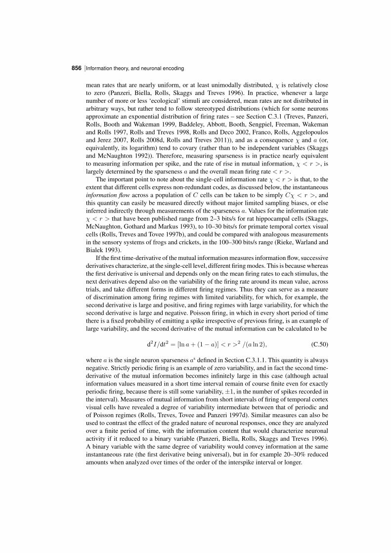
Information theory, and neuronal encoding856 |
mean rates that are nearly uniform, or at least unimodally distributed, χ is relatively closeto zero (Panzeri, Biella, Rolls, Skaggs and Treves 1996). In practice, whenever a largenumber of more or less ‘ecological’ stimuli are considered, mean rates are not distributed inarbitrary ways, but rather tend to follow stereotyped distributions (which for some neuronsapproximate an exponential distribution of firing rates – see Section C.3.1 (Treves, Panzeri,Rolls, Booth and Wakeman 1999, Baddeley, Abbott, Booth, Sengpiel, Freeman, Wakemanand Rolls 1997, Rolls and Treves 1998, Rolls and Deco 2002, Franco, Rolls, Aggelopoulosand Jerez 2007, Rolls 2008d, Rolls and Treves 2011)), and as a consequence χ and a (or,equivalently, its logarithm) tend to covary (rather than to be independent variables (Skaggsand McNaughton 1992)). Therefore, measuring sparseness is in practice nearly equivalentto measuring information per spike, and the rate of rise in mutual information, χ < r >, islargely determined by the sparseness a and the overall mean firing rate < r >.
The important point to note about the singlecell information rate χ < r > is that, to theextent that different cells express nonredundant codes, as discussed below, the instantaneousinformation flow across a population of C cells can be taken to be simply Cχ < r >, andthis quantity can easily be measured directly without major limited sampling biases, or elseinferred indirectly through measurements of the sparseness a. Values for the information rateχ < r > that have been published range from 2–3 bits/s for rat hippocampal cells (Skaggs,McNaughton, Gothard and Markus 1993), to 10–30 bits/s for primate temporal cortex visualcells (Rolls, Treves and Tovee 1997b), and could be compared with analogous measurementsin the sensory systems of frogs and crickets, in the 100–300 bits/s range (Rieke, Warland andBialek 1993).
If the first timederivative of the mutual information measures information flow, successivederivatives characterize, at the singlecell level, different firing modes. This is because whereasthe first derivative is universal and depends only on the mean firing rates to each stimulus, thenext derivatives depend also on the variability of the firing rate around its mean value, acrosstrials, and take different forms in different firing regimes. Thus they can serve as a measureof discrimination among firing regimes with limited variability, for which, for example, thesecond derivative is large and positive, and firing regimes with large variability, for which thesecond derivative is large and negative. Poisson firing, in which in every short period of timethere is a fixed probability of emitting a spike irrespective of previous firing, is an example oflarge variability, and the second derivative of the mutual information can be calculated to be
d2I/dt2 = [ln a+ (1− a)] < r >2 /(a ln 2), (C.50)
where a is the single neuron sparseness as defined in Section C.3.1.1. This quantity is alwaysnegative. Strictly periodic firing is an example of zero variability, and in fact the second timederivative of the mutual information becomes infinitely large in this case (although actualinformation values measured in a short time interval remain of course finite even for exactlyperiodic firing, because there is still some variability, ±1, in the number of spikes recorded inthe interval). Measures of mutual information from short intervals of firing of temporal cortexvisual cells have revealed a degree of variability intermediate between that of periodic andof Poisson regimes (Rolls, Treves, Tovee and Panzeri 1997d). Similar measures can also beused to contrast the effect of the graded nature of neuronal responses, once they are analyzedover a finite period of time, with the information content that would characterize neuronalactivity if it reduced to a binary variable (Panzeri, Biella, Rolls, Skaggs and Treves 1996).A binary variable with the same degree of variability would convey information at the sameinstantaneous rate (the first derivative being universal), but in for example 20–30% reducedamounts when analyzed over times of the order of the interspike interval or longer.
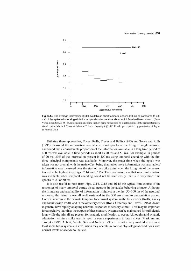
| 857Information theory results
Fig. C.14 The average information I(S,R) available in short temporal epochs (50 ms as compared to 400ms) of the spike trains of single inferior temporal cortex neurons about which face had been shown. (FromVisual Cognition, 2: 35–58, Information encoding in short firing rate epochs by single neurons in the primate temporalvisual cortex. Martin J. Tovee & Edmund T. Rolls. Copyright c⃝1995 Routledge, reprinted by permission of Taylor& Francis Ltd.)
Utilizing these approaches, Tovee, Rolls, Treves and Bellis (1993) and Tovee and Rolls(1995) measured the information available in short epochs of the firing of single neurons,and found that a considerable proportion of the information available in a long time period of400 ms was available in time periods as short as 20 ms and 50 ms. For example, in periodsof 20 ms, 30% of the information present in 400 ms using temporal encoding with the firstthree principal components was available. Moreover, the exact time when the epoch wastaken was not crucial, with the main effect being that rather more information was available ifinformation was measured near the start of the spike train, when the firing rate of the neurontended to be highest (see Figs. C.14 and C.15). The conclusion was that much informationwas available when temporal encoding could not be used easily, that is in very short timeepochs of 20 or 50 ms.
It is also useful to note from Figs. C.14, C.15 and 16.15 the typical time course of theresponses of many temporal cortex visual neurons in the awake behaving primate. Althoughthe firing rate and availability of information is highest in the first 50–100 ms of the neuronalresponse, the firing is overall well sustained in the 500 ms stimulus presentation period.Cortical neurons in the primate temporal lobe visual system, in the taste cortex (Rolls, Yaxleyand Sienkiewicz 1990), and in the olfactory cortex (Rolls, Critchley and Treves 1996a), do notin general have rapidly adapting neuronal responses to sensory stimuli. This may be importantfor associative learning: the outputs of these sensory systems can be maintained for sufficientlylong while the stimuli are present for synaptic modification to occur. Although rapid synapticadaptation within a spike train is seen in some experiments in brain slices (Markram andTsodyks 1996, Abbott, Varela, Sen and Nelson 1997), it is not a very marked effect in atleast some brain systems in vivo, when they operate in normal physiological conditions withnormal levels of acetylcholine, etc.
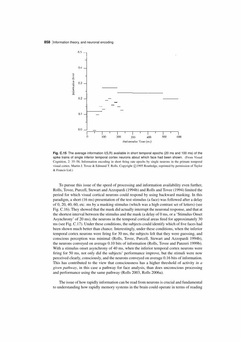
Information theory, and neuronal encoding858 |
Fig. C.15 The average information I(S,R) available in short temporal epochs (20 ms and 100 ms) of thespike trains of single inferior temporal cortex neurons about which face had been shown. (From VisualCognition, 2: 35–58, Information encoding in short firing rate epochs by single neurons in the primate temporalvisual cortex. Martin J. Tovee & Edmund T. Rolls. Copyright c⃝1995 Routledge, reprinted by permission of Taylor& Francis Ltd.)
To pursue this issue of the speed of processing and information availability even further,Rolls, Tovee, Purcell, Stewart and Azzopardi (1994b) and Rolls and Tovee (1994) limited theperiod for which visual cortical neurons could respond by using backward masking. In thisparadigm, a short (16 ms) presentation of the test stimulus (a face) was followed after a delayof 0, 20, 40, 60, etc. ms by a masking stimulus (which was a high contrast set of letters) (seeFig. C.16). They showed that the mask did actually interrupt the neuronal response, and that atthe shortest interval between the stimulus and the mask (a delay of 0 ms, or a ‘Stimulus OnsetAsynchrony’ of 20 ms), the neurons in the temporal cortical areas fired for approximately 30ms (see Fig. C.17). Under these conditions, the subjects could identify which of five faces hadbeen shown much better than chance. Interestingly, under these conditions, when the inferiortemporal cortex neurons were firing for 30 ms, the subjects felt that they were guessing, andconscious perception was minimal (Rolls, Tovee, Purcell, Stewart and Azzopardi 1994b),the neurons conveyed on average 0.10 bits of information (Rolls, Tovee and Panzeri 1999b).With a stimulus onset asynchrony of 40 ms, when the inferior temporal cortex neurons werefiring for 50 ms, not only did the subjects’ performance improve, but the stimuli were nowperceived clearly, consciously, and the neurons conveyed on average 0.16 bits of information.This has contributed to the view that consciousness has a higher threshold of activity in agiven pathway, in this case a pathway for face analysis, than does unconscious processingand performance using the same pathway (Rolls 2003, Rolls 2006a).
The issue of how rapidly information can be read from neurons is crucial and fundamentalto understanding how rapidly memory systems in the brain could operate in terms of reading
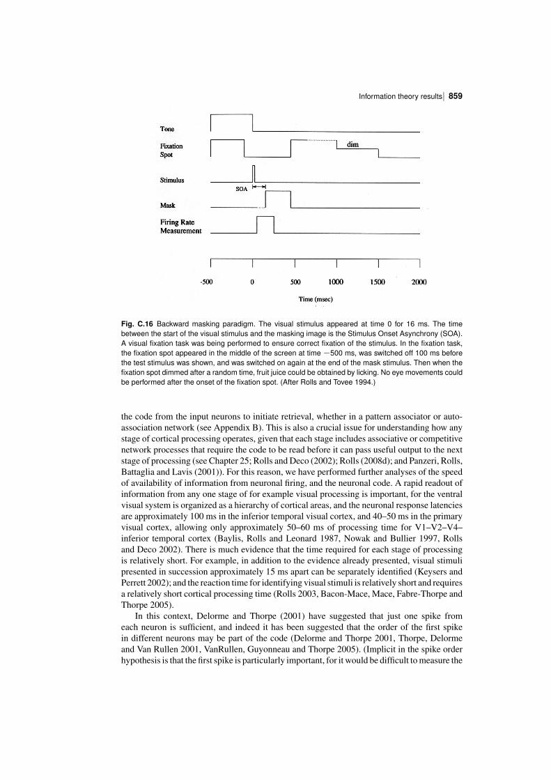
| 859Information theory results
Fig. C.16 Backward masking paradigm. The visual stimulus appeared at time 0 for 16 ms. The timebetween the start of the visual stimulus and the masking image is the Stimulus Onset Asynchrony (SOA).A visual fixation task was being performed to ensure correct fixation of the stimulus. In the fixation task,the fixation spot appeared in the middle of the screen at time −500 ms, was switched off 100 ms beforethe test stimulus was shown, and was switched on again at the end of the mask stimulus. Then when thefixation spot dimmed after a random time, fruit juice could be obtained by licking. No eye movements couldbe performed after the onset of the fixation spot. (After Rolls and Tovee 1994.)
the code from the input neurons to initiate retrieval, whether in a pattern associator or autoassociation network (see Appendix B). This is also a crucial issue for understanding how anystage of cortical processing operates, given that each stage includes associative or competitivenetwork processes that require the code to be read before it can pass useful output to the nextstage of processing (see Chapter 25; Rolls and Deco (2002); Rolls (2008d); and Panzeri, Rolls,Battaglia and Lavis (2001)). For this reason, we have performed further analyses of the speedof availability of information from neuronal firing, and the neuronal code. A rapid readout ofinformation from any one stage of for example visual processing is important, for the ventralvisual system is organized as a hierarchy of cortical areas, and the neuronal response latenciesare approximately 100 ms in the inferior temporal visual cortex, and 40–50 ms in the primaryvisual cortex, allowing only approximately 50–60 ms of processing time for V1–V2–V4–inferior temporal cortex (Baylis, Rolls and Leonard 1987, Nowak and Bullier 1997, Rollsand Deco 2002). There is much evidence that the time required for each stage of processingis relatively short. For example, in addition to the evidence already presented, visual stimulipresented in succession approximately 15 ms apart can be separately identified (Keysers andPerrett 2002); and the reaction time for identifying visual stimuli is relatively short and requiresa relatively short cortical processing time (Rolls 2003, BaconMace, Mace, FabreThorpe andThorpe 2005).
In this context, Delorme and Thorpe (2001) have suggested that just one spike fromeach neuron is sufficient, and indeed it has been suggested that the order of the first spikein different neurons may be part of the code (Delorme and Thorpe 2001, Thorpe, Delormeand Van Rullen 2001, VanRullen, Guyonneau and Thorpe 2005). (Implicit in the spike orderhypothesis is that the first spike is particularly important, for it would be difficult to measure the
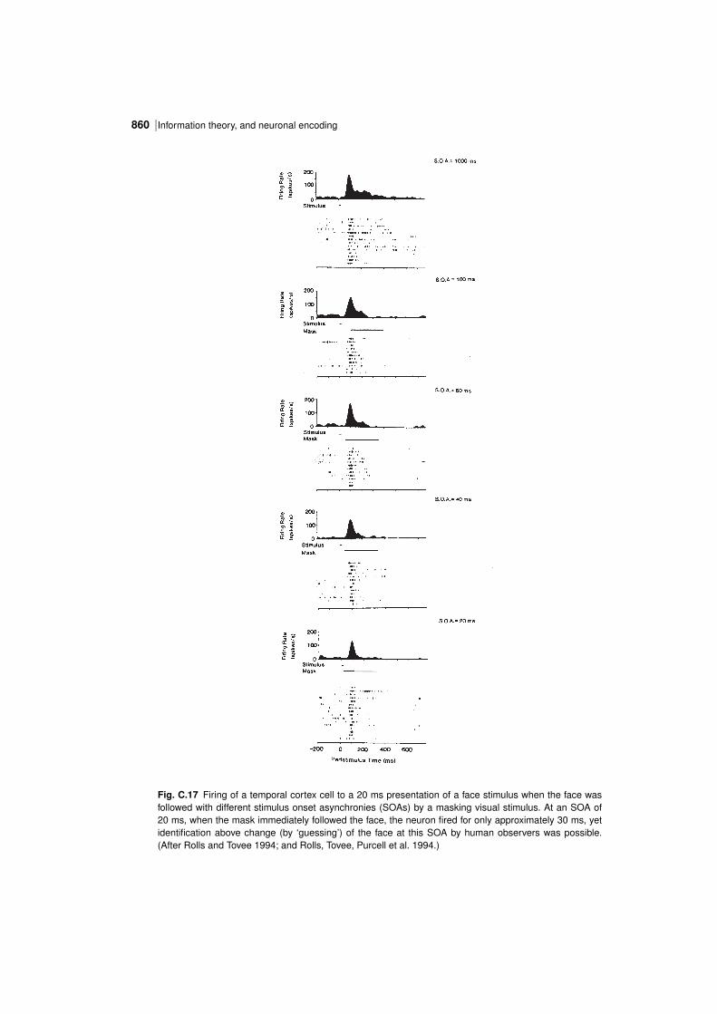
Information theory, and neuronal encoding860 |
Fig. C.17 Firing of a temporal cortex cell to a 20 ms presentation of a face stimulus when the face wasfollowed with different stimulus onset asynchronies (SOAs) by a masking visual stimulus. At an SOA of20 ms, when the mask immediately followed the face, the neuron fired for only approximately 30 ms, yetidentification above change (by ‘guessing’) of the face at this SOA by human observers was possible.(After Rolls and Tovee 1994; and Rolls, Tovee, Purcell et al. 1994.)
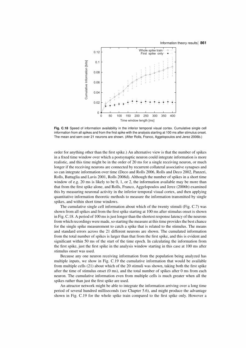
| 861Information theory results
0
0.02
0.04
0.06
0.08
0.1
0.12
0 50 100 150 200 250 300 350 400
Cu
mu
lative
In
form
atio
n [b
its]
Time window length [ms]
Whole spike trainFirst spike only
Fig. C.18 Speed of information availability in the inferior temporal visual cortex. Cumulative single cellinformation from all spikes and from the first spike with the analysis starting at 100 ms after stimulus onset.The mean and sem over 21 neurons are shown. (After Rolls, Franco, Aggelopoulos and Jerez 2006b.)
order for anything other than the first spike.) An alternative view is that the number of spikesin a fixed time window over which a postsynaptic neuron could integrate information is morerealistic, and this time might be in the order of 20 ms for a single receiving neuron, or muchlonger if the receiving neurons are connected by recurrent collateral associative synapses andso can integrate information over time (Deco and Rolls 2006, Rolls and Deco 2002, Panzeri,Rolls, Battaglia and Lavis 2001, Rolls 2008d). Although the number of spikes in a short timewindow of e.g. 20 ms is likely to be 0, 1, or 2, the information available may be more thanthat from the first spike alone, and Rolls, Franco, Aggelopoulos and Jerez (2006b) examinedthis by measuring neuronal activity in the inferior temporal visual cortex, and then applyingquantitative information theoretic methods to measure the information transmitted by singlespikes, and within short time windows.
The cumulative single cell information about which of the twenty stimuli (Fig. C.7) wasshown from all spikes and from the first spike starting at 100 ms after stimulus onset is shownin Fig. C.18. A period of 100 ms is just longer than the shortest response latency of the neuronsfrom which recordings were made, so starting the measure at this time provides the best chancefor the single spike measurement to catch a spike that is related to the stimulus. The meansand standard errors across the 21 different neurons are shown. The cumulated informationfrom the total number of spikes is larger than that from the first spike, and this is evident andsignificant within 50 ms of the start of the time epoch. In calculating the information fromthe first spike, just the first spike in the analysis window starting in this case at 100 ms afterstimulus onset was used.
Because any one neuron receiving information from the population being analyzed hasmultiple inputs, we show in Fig. C.19 the cumulative information that would be availablefrom multiple cells (21) about which of the 20 stimuli was shown, taking both the first spikeafter the time of stimulus onset (0 ms), and the total number of spikes after 0 ms from eachneuron. The cumulative information even from multiple cells is much greater when all thespikes rather than just the first spike are used.
An attractor network might be able to integrate the information arriving over a long timeperiod of several hundred milliseconds (see Chapter 5.6), and might produce the advantageshown in Fig. C.19 for the whole spike train compared to the first spike only. However a
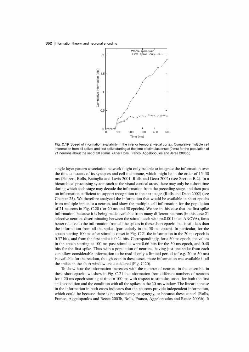
Information theory, and neuronal encoding862 |
0
0.5
1
1.5
2
0 100 200 300 400 500
Cu
mu
lative
in
form
atio
n (
bits)
Time (ms)
Whole spike trainFirst spike only
Fig. C.19 Speed of information availability in the inferior temporal visual cortex. Cumulative multiple cellinformation from all spikes and first spike starting at the time of stimulus onset (0 ms) for the population of21 neurons about the set of 20 stimuli. (After Rolls, Franco, Aggelopoulos and Jerez 2006b.)
single layer pattern association network might only be able to integrate the information overthe time constants of its synapses and cell membrane, which might be in the order of 15–30ms (Panzeri, Rolls, Battaglia and Lavis 2001, Rolls and Deco 2002) (see Section B.2). In ahierarchical processing system such as the visual cortical areas, there may only be a short timeduring which each stage may decode the information from the preceding stage, and then passon information sufficient to support recognition to the next stage (Rolls and Deco 2002) (seeChapter 25). We therefore analyzed the information that would be available in short epochsfrom multiple inputs to a neuron, and show the multiple cell information for the populationof 21 neurons in Fig. C.20 (for 20 ms and 50 epochs). We see in this case that the first spikeinformation, because it is being made available from many different neurons (in this case 21selective neurons discriminating between the stimuli each with p<0.001 in an ANOVA), faresbetter relative to the information from all the spikes in these short epochs, but is still less thanthe information from all the spikes (particularly in the 50 ms epoch). In particular, for theepoch starting 100 ms after stimulus onset in Fig. C.21 the information in the 20 ms epoch is0.37 bits, and from the first spike is 0.24 bits. Correspondingly, for a 50 ms epoch, the valuesin the epoch starting at 100 ms post stimulus were 0.66 bits for the 50 ms epoch, and 0.40bits for the first spike. Thus with a population of neurons, having just one spike from eachcan allow considerable information to be read if only a limited period (of e.g. 20 or 50 ms)is available for the readout, though even in these cases, more information was available if allthe spikes in the short window are considered (Fig. C.20).
To show how the information increases with the number of neurons in the ensemble inthese short epochs, we show in Fig. C.21 the information from different numbers of neuronsfor a 20 ms epoch starting at time = 100 ms with respect to stimulus onset, for both the firstspike condition and the condition with all the spikes in the 20 ms window. The linear increasein the information in both cases indicates that the neurons provide independent information,which could be because there is no redundancy or synergy, or because these cancel (Rolls,Franco, Aggelopoulos and Reece 2003b, Rolls, Franco, Aggelopoulos and Reece 2003b). It
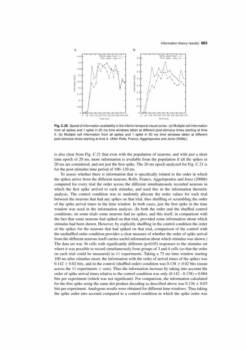
| 863Information theory results
0
0.05
0.1
0.15
0.2
0.25
0.3
0.35
0.4
0.45
0 50 100 150 200 250 300 350 400 450 500
Info
rmatio
n (
bits
)
Time (ms)
Whole spike trainFirst spike only
0
0.1
0.2
0.3
0.4
0.5
0.6
0.7
0.8
0 50 100 150 200 250 300 350 400 450 500
Info
rmatio
n (
bits
)
Time (ms)
Whole spike trainFirst spike only
a b
Fig. C.20 Speed of information availability in the inferior temporal visual cortex. (a) Multiple cell informationfrom all spikes and 1 spike in 20 ms time windows taken at different poststimulus times starting at time0. (b) Multiple cell information from all spikes and 1 spike in 50 ms time windows taken at differentpoststimulus times starting at time 0. (After Rolls, Franco, Aggelopoulos and Jerez 2006b.)
is also clear from Fig. C.21 that even with the population of neurons, and with just a shorttime epoch of 20 ms, more information is available from the population if all the spikes in20 ms are considered, and not just the first spike. The 20 ms epoch analyzed for Fig. C.21 isfor the poststimulus time period of 100–120 ms.
To assess whether there is information that is specifically related to the order in whichthe spikes arrive from the different neurons, Rolls, Franco, Aggelopoulos and Jerez (2006b)computed for every trial the order across the different simultaneously recorded neurons inwhich the first spike arrived to each stimulus, and used this in the information theoreticanalysis. The control condition was to randomly allocate the order values for each trialbetween the neurons that had any spikes on that trial, thus shuffling or scrambling the orderof the spike arrival times in the time window. In both cases, just the first spike in the timewindow was used in the information analysis. (In both the order and the shuffled controlconditions, on some trials some neurons had no spikes, and this itself, in comparison withthe fact that some neurons had spiked on that trial, provided some information about whichstimulus had been shown. However, by explicitly shuffling in the control condition the orderof the spikes for the neurons that had spiked on that trial, comparison of the control withthe unshuffled order condition provides a clear measure of whether the order of spike arrivalfrom the different neurons itself carries useful information about which stimulus was shown.)The data set was 36 cells with significantly different (p<0.05) responses to the stimulus setwhere it was possible to record simultaneously from groups of 3 and 4 cells (so that the orderon each trial could be measured) in 11 experiments. Taking a 75 ms time window starting100 ms after stimulus onset, the information with the order of arrival times of the spikes was0.142 ± 0.02 bits, and in the control (shuffled order) condition was 0.138 ± 0.02 bits (meanacross the 11 experiments ± sem). Thus the information increase by taking into account theorder of spike arrival times relative to the control condition was only (0.142 0.138) = 0.004bits per experiment (which was not significant). For comparison, the information calculatedfor the first spike using the same dot product decoding as described above was 0.136 ± 0.03bits per experiment. Analogous results were obtained for different time windows. Thus takingthe spike order into account compared to a control condition in which the spike order was
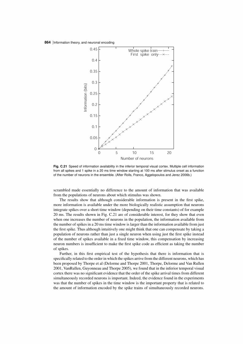
Information theory, and neuronal encoding864 |
0
0.05
0.1
0.15
0.2
0.25
0.3
0.35
0.4
0.45
0 5 10 15 20
Info
rmatio
n (
bits
)
Number of neurons
Whole spike trainFirst spike only
Fig. C.21 Speed of information availability in the inferior temporal visual cortex. Multiple cell informationfrom all spikes and 1 spike in a 20 ms time window starting at 100 ms after stimulus onset as a functionof the number of neurons in the ensemble. (After Rolls, Franco, Aggelopoulos and Jerez 2006b.)
scrambled made essentially no difference to the amount of information that was availablefrom the populations of neurons about which stimulus was shown.
The results show that although considerable information is present in the first spike,more information is available under the more biologically realistic assumption that neuronsintegrate spikes over a short time window (depending on their time constants) of for example20 ms. The results shown in Fig. C.21 are of considerable interest, for they show that evenwhen one increases the number of neurons in the population, the information available fromthe number of spikes in a 20 ms time window is larger than the information available from justthe first spike. Thus although intuitively one might think that one can compensate by taking apopulation of neurons rather than just a single neuron when using just the first spike insteadof the number of spikes available in a fixed time window, this compensation by increasingneuron numbers is insufficient to make the first spike code as efficient as taking the numberof spikes.
Further, in this first empirical test of the hypothesis that there is information that isspecifically related to the order in which the spikes arrive from the different neurons, which hasbeen proposed by Thorpe et al (Delorme and Thorpe 2001, Thorpe, Delorme and Van Rullen2001, VanRullen, Guyonneau and Thorpe 2005), we found that in the inferior temporal visualcortex there was no significant evidence that the order of the spike arrival times from differentsimultaneously recorded neurons is important. Indeed, the evidence found in the experimentswas that the number of spikes in the time window is the important property that is related tothe amount of information encoded by the spike trains of simultaneously recorded neurons.

| 865Information theory results
The fact that there was also more information in the number of spikes in a fixed time windowthan from the first spike only is also evidence that is not consistent with the spike orderhypothesis, for the order between neurons can only be easily read from the first spike, and justusing information from the first spike would discard extra information available from furtherspikes even in short time windows.
The encoding of information that uses the number of spikes in a short time window thatis supported by the analyses described by Rolls, Franco, Aggelopoulos and Jerez (2006b)deserves further elaboration. It could be thought of as a rate code, in that the number ofspikes in a short time window is relevant, but is not a rate code in the rather artificial senseconsidered by Thorpe et al. (Delorme and Thorpe 2001, Thorpe et al. 2001, VanRullen etal. 2005) in which a rate is estimated from the interspike interval. This is not just artificial,but also begs the question of how, once the rate is calculated from the interspike interval, thisdecoded rate is passed on to the receiving neurons, or how, if the receiving neurons calculatethe interspike interval at every synapse, they utilize it. In contrast, the spike count code in ashort time window that is considered here is very biologically plausible, in that each spikewould inject current into the postsynaptic neuron, and the neuron would integrate all suchcurrents in a dendrite over a time period set by the synaptic and membrane time constants,which will result in an integration time constant in the order of 15–20 ms. Explicit models ofexactly this dynamical processing at the integrateandfire neuronal level have been describedto define precisely these operations (Deco and Rolls 2003, Deco and Rolls 2005d, Deco, Rollsand Horwitz 2004, Deco and Rolls 2005b, Rolls and Deco 2002, Rolls 2008d). Even thoughthe number of spikes in a short time window of e.g. 20 ms is likely to be 0, 1, or 2, it can be 3or more for effective stimuli (Rolls, Franco, Aggelopoulos and Jerez 2006b), and this is moreefficient than using the first spike.
To add some detail here, a neuron receiving information from a population of inferiortemporal cortex neurons of the type described here would have a membrane potential thatvaried continuously in time reflecting with a time constant in the order of 15–20 ms (resultingfrom a time constant of order 10 ms for AMPA synapses, 100 ms for NMDA synapses, and20 ms for the cell membrane) a dot (inner) product over all synapses of each spike count andthe synaptic strength. This continuously time varying membrane potential would lead to spikeswhenever the results of this integration process produced a depolarization that exceeded thefiring threshold. The result is that the spike train of the neuron would reflect continuously witha time constant in the order of 15–20 ms the likelihood that the input spikes it was receivingmatched its set of synaptic weights. The spike train would thus indicate in continuous timehow closely the stimulus or input matched its most effective stimulus (for a dot product isessentially a correlation). In this sense, no particular starting time is needed for the analysis,and in this respect it is a much better component of a dynamical system than is a decodingthat utilizes an order in which the order of the spike arrival times is important and a start timefor the analysis must be assumed.
I note that an autoassociation or attractor network implemented by recurrent collateralconnections between the neurons can, using its shortterm memory, integrate its inputs overmuch longer periods, for example over 500 ms in a model of how decisions are made (Decoand Rolls 2006) (see Chapter 5.6), and thus if there is time, the extra information availablein more than the first spike or even the first few spikes that is evident in Figs. C.18 and C.19could be used by the brain.
The conclusions from the single cell information analyses are thus that most of theinformation is encoded in the spike count; that large parts of this information are availablein short temporal epochs of e.g. 20 ms or 50 ms; and that any additional information whichappears to be temporally encoded is related to the latency of the neuronal response, and reflectssudden changes in the visual stimuli. Therefore a neuron in the next cortical area would obtain
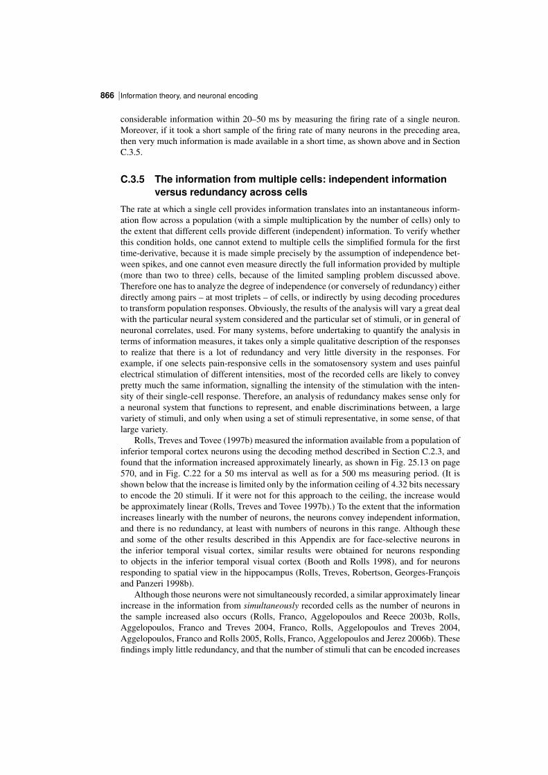
Information theory, and neuronal encoding866 |
considerable information within 20–50 ms by measuring the firing rate of a single neuron.Moreover, if it took a short sample of the firing rate of many neurons in the preceding area,then very much information is made available in a short time, as shown above and in SectionC.3.5.
C.3.5 The information from multiple cells: independent informationversus redundancy across cells
The rate at which a single cell provides information translates into an instantaneous information flow across a population (with a simple multiplication by the number of cells) only tothe extent that different cells provide different (independent) information. To verify whetherthis condition holds, one cannot extend to multiple cells the simplified formula for the firsttimederivative, because it is made simple precisely by the assumption of independence between spikes, and one cannot even measure directly the full information provided by multiple(more than two to three) cells, because of the limited sampling problem discussed above.Therefore one has to analyze the degree of independence (or conversely of redundancy) eitherdirectly among pairs – at most triplets – of cells, or indirectly by using decoding proceduresto transform population responses. Obviously, the results of the analysis will vary a great dealwith the particular neural system considered and the particular set of stimuli, or in general ofneuronal correlates, used. For many systems, before undertaking to quantify the analysis interms of information measures, it takes only a simple qualitative description of the responsesto realize that there is a lot of redundancy and very little diversity in the responses. Forexample, if one selects painresponsive cells in the somatosensory system and uses painfulelectrical stimulation of different intensities, most of the recorded cells are likely to conveypretty much the same information, signalling the intensity of the stimulation with the intensity of their singlecell response. Therefore, an analysis of redundancy makes sense only fora neuronal system that functions to represent, and enable discriminations between, a largevariety of stimuli, and only when using a set of stimuli representative, in some sense, of thatlarge variety.
Rolls, Treves and Tovee (1997b) measured the information available from a population ofinferior temporal cortex neurons using the decoding method described in Section C.2.3, andfound that the information increased approximately linearly, as shown in Fig. 25.13 on page570, and in Fig. C.22 for a 50 ms interval as well as for a 500 ms measuring period. (It isshown below that the increase is limited only by the information ceiling of 4.32 bits necessaryto encode the 20 stimuli. If it were not for this approach to the ceiling, the increase wouldbe approximately linear (Rolls, Treves and Tovee 1997b).) To the extent that the informationincreases linearly with the number of neurons, the neurons convey independent information,and there is no redundancy, at least with numbers of neurons in this range. Although theseand some of the other results described in this Appendix are for faceselective neurons inthe inferior temporal visual cortex, similar results were obtained for neurons respondingto objects in the inferior temporal visual cortex (Booth and Rolls 1998), and for neuronsresponding to spatial view in the hippocampus (Rolls, Treves, Robertson, GeorgesFrancoisand Panzeri 1998b).
Although those neurons were not simultaneously recorded, a similar approximately linearincrease in the information from simultaneously recorded cells as the number of neurons inthe sample increased also occurs (Rolls, Franco, Aggelopoulos and Reece 2003b, Rolls,Aggelopoulos, Franco and Treves 2004, Franco, Rolls, Aggelopoulos and Treves 2004,Aggelopoulos, Franco and Rolls 2005, Rolls, Franco, Aggelopoulos and Jerez 2006b). Thesefindings imply little redundancy, and that the number of stimuli that can be encoded increases
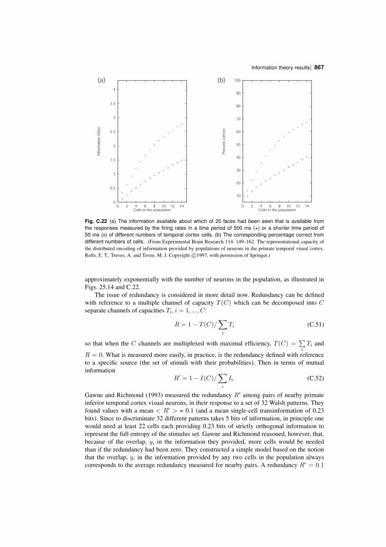
| 867Information theory results
10
20
30
40
50
60
70
80
90
100
0 2 4 6 8 10 12 14
Pe
rce
nt
corr
ect
Cells in the population
0
0.5
1
1.5
2
2.5
3
3.5
4
0 2 4 6 8 10 12 14
Info
rma
tion
(b
its)
Cells in the population
(a) (b)
Fig. C.22 (a) The information available about which of 20 faces had been seen that is available fromthe responses measured by the firing rates in a time period of 500 ms (+) or a shorter time period of50 ms (x) of different numbers of temporal cortex cells. (b) The corresponding percentage correct fromdifferent numbers of cells. (From Experimental Brain Research 114: 149–162. The representational capacity ofthe distributed encoding of information provided by populations of neurons in the primate temporal visual cortex.Rolls, E. T., Treves, A. and Tovee, M. J. Copyright c⃝1997, with permission of Springer.)
approximately exponentially with the number of neurons in the population, as illustrated inFigs. 25.14 and C.22.
The issue of redundancy is considered in more detail now. Redundancy can be definedwith reference to a multiple channel of capacity T (C) which can be decomposed into Cseparate channels of capacities Ti, i = 1, ..., C:
R = 1− T (C)/∑i
Ti (C.51)
so that when the C channels are multiplexed with maximal efficiency, T (C) =∑i
Ti and
R = 0. What is measured more easily, in practice, is the redundancy defined with referenceto a specific source (the set of stimuli with their probabilities). Then in terms of mutualinformation
R′ = 1− I(C)/∑i
Ii. (C.52)
Gawne and Richmond (1993) measured the redundancy R′ among pairs of nearby primateinferior temporal cortex visual neurons, in their response to a set of 32 Walsh patterns. Theyfound values with a mean < R′ > = 0.1 (and a mean singlecell transinformation of 0.23bits). Since to discriminate 32 different patterns takes 5 bits of information, in principle onewould need at least 22 cells each providing 0.23 bits of strictly orthogonal information torepresent the full entropy of the stimulus set. Gawne and Richmond reasoned, however, that,because of the overlap, y, in the information they provided, more cells would be neededthan if the redundancy had been zero. They constructed a simple model based on the notionthat the overlap, y, in the information provided by any two cells in the population alwayscorresponds to the average redundancy measured for nearby pairs. A redundancy R′ = 0.1
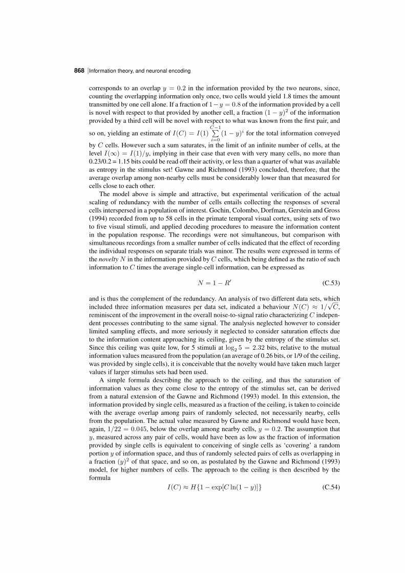
Information theory, and neuronal encoding868 |
corresponds to an overlap y = 0.2 in the information provided by the two neurons, since,counting the overlapping information only once, two cells would yield 1.8 times the amounttransmitted by one cell alone. If a fraction of 1−y = 0.8 of the information provided by a cellis novel with respect to that provided by another cell, a fraction (1 − y)2 of the informationprovided by a third cell will be novel with respect to what was known from the first pair, and
so on, yielding an estimate of I(C) = I(1)C−1∑i=0
(1 − y)i for the total information conveyed
by C cells. However such a sum saturates, in the limit of an infinite number of cells, at thelevel I(∞) = I(1)/y, implying in their case that even with very many cells, no more than0.23/0.2 = 1.15 bits could be read off their activity, or less than a quarter of what was availableas entropy in the stimulus set! Gawne and Richmond (1993) concluded, therefore, that theaverage overlap among nonnearby cells must be considerably lower than that measured forcells close to each other.
The model above is simple and attractive, but experimental verification of the actualscaling of redundancy with the number of cells entails collecting the responses of severalcells interspersed in a population of interest. Gochin, Colombo, Dorfman, Gerstein and Gross(1994) recorded from up to 58 cells in the primate temporal visual cortex, using sets of twoto five visual stimuli, and applied decoding procedures to measure the information contentin the population response. The recordings were not simultaneous, but comparison withsimultaneous recordings from a smaller number of cells indicated that the effect of recordingthe individual responses on separate trials was minor. The results were expressed in terms ofthe noveltyN in the information provided by C cells, which being defined as the ratio of suchinformation to C times the average singlecell information, can be expressed as
N = 1−R′ (C.53)
and is thus the complement of the redundancy. An analysis of two different data sets, whichincluded three information measures per data set, indicated a behaviour N(C) ≈ 1/
√C,
reminiscent of the improvement in the overall noisetosignal ratio characterizing C independent processes contributing to the same signal. The analysis neglected however to considerlimited sampling effects, and more seriously it neglected to consider saturation effects dueto the information content approaching its ceiling, given by the entropy of the stimulus set.Since this ceiling was quite low, for 5 stimuli at log2 5 = 2.32 bits, relative to the mutualinformation values measured from the population (an average of 0.26 bits, or 1/9 of the ceiling,was provided by single cells), it is conceivable that the novelty would have taken much largervalues if larger stimulus sets had been used.
A simple formula describing the approach to the ceiling, and thus the saturation ofinformation values as they come close to the entropy of the stimulus set, can be derivedfrom a natural extension of the Gawne and Richmond (1993) model. In this extension, theinformation provided by single cells, measured as a fraction of the ceiling, is taken to coincidewith the average overlap among pairs of randomly selected, not necessarily nearby, cellsfrom the population. The actual value measured by Gawne and Richmond would have been,again, 1/22 = 0.045, below the overlap among nearby cells, y = 0.2. The assumption thaty, measured across any pair of cells, would have been as low as the fraction of informationprovided by single cells is equivalent to conceiving of single cells as ‘covering’ a randomportion y of information space, and thus of randomly selected pairs of cells as overlapping ina fraction (y)2 of that space, and so on, as postulated by the Gawne and Richmond (1993)model, for higher numbers of cells. The approach to the ceiling is then described by theformula
I(C) ≈ H{1− exp[C ln(1− y)]} (C.54)
| 869Information theory results
that is, a simple exponential saturation to the ceiling. This simple law indeed describesremarkably well the trend in the data analyzed by Rolls, Treves and Tovee (1997b). Althoughthe model has no reason to be exact, and therefore its agreement with the data should not beexpected to be accurate, the crucial point it embodies is that deviations from a purely linearincrease in information with the number of cells analyzed are due solely to the ceiling effect.Aside from the ceiling, due to the sampling of an information space of finite entropy, theinformation contents of different cells’ responses are independent of each other. Thus, in themodel, the observed redundancy (or indeed the overlap) is purely a consequence of the finitesize of the stimulus set. If the population were probed with larger and larger sets of stimuli,or more precisely with sets of increasing entropy, and the amount of information conveyedby single cells were to remain approximately the same, then the fraction of space ‘covered’by each cell, again y, would get smaller and smaller, tending to eliminate redundancy forvery large stimulus entropies (and a fixed number of cells). The actual data were obtainedwith limited numbers of stimuli, and therefore cannot probe directly the conditions in whichredundancy might reduce to zero. The data are consistent, however, with the hypothesisembodied in the simple model, as shown also by the near exponential approach to lowerceilings found for information values calculated with reduced subsets of the original set ofstimuli (Rolls, Treves and Tovee 1997b).
The implication of this set of analyses, some performed towards the end of the ventralvisual stream of the monkey, is that the representation of at least some classes of objects inthose areas is achieved with minimal redundancy by cells that are allocated each to analyse adifferent aspect of the visual stimulus. This minimal redundancy is what would be expected ofa selforganizing system in which different cells acquired their response selectivities througha random process, with or without local competition among nearby cells (see Section B.4). Atthe same time, such low redundancy could also very well result in a system that is organizedunder some strong teaching input, so that the emerging picture is compatible with a simplerandom process, but could be produced in other ways. The finding that, at least with smallnumbers of neurons, redundancy may be effectively minimized, is consistent not only withthe concept of efficient encoding, but also with the general idea that one of the functions of theearly visual system is to progressively minimize redundancy in the representation of visualstimuli (Attneave 1954, Barlow 1961). However, the ventral visual system does much morethan produce a nonredundant representation of an image, for it transforms the representationfrom an image to an invariant representation of objects, as described in Chapter 25. Moreover,what is shown in this section is that the information about objects can be read off from just thespike count of a population of neurons, using decoding as simple as the simplest that could beperformed by a receiving neuron, dot product decoding. In this sense, the information aboutobjects is made explicit in the firing rate of the neurons in the inferior temporal cortex, in thatit can be read off in this way.
We consider in Section C.3.7 whether there is more to it than this. Does the synchronizationof neurons (and it would have to be stimulusdependent synchronization) add significantlyto the information that could be encoded by the number of spikes, as has been suggested bysome?
Before this, we consider why encoding by a population of neurons is more powerfulthan the encoding than is possible by single neurons, adding to previous arguments that adistributed representation is much more computationally useful than a local representation, byallowing properties such as generalization, completion, and graceful degradation in associativeneuronal networks (see Appendix B).
Information theory, and neuronal encoding870 |
C.3.6 Should one neuron be as discriminative as the whole organism,in object encoding systems?
In the analysis of random dot motion with a given level of correlation among the moving dots,single neurons in area MT in the dorsal visual system of the primate can be approximately assensitive or discriminative as the psychophysical performance of the whole animal (Zohary,Shadlen and Newsome 1994). The arguments and evidence presented here (e.g. in SectionC.3.5) suggest that this is not the case for the ventral visual system, concerned with objectidentification. Why should there be this difference?
Rolls and Treves (1998) suggest that the dimensionality of what is being computed mayaccount for the difference. In the case of visual motion (at least in the study referred to), theproblem was effectively onedimensional, in that the direction of motion of the stimulus alonga line in 2D space was extracted from the activity of the neurons. In this lowdimensionalstimulus space, the neurons may each perform one of a few similar computations on a particular(local) portion of 2D space, with the side effect that, by averaging over a larger receptive fieldthan in V1, one can extract a signal of a more global nature. Indeed, in the case of more globalmotion, it is the average of the neuronal activity that can be computed by the larger receptivefields of MT neurons that specifies the average or global direction of motion.
In contrast, in the higher dimensional space of objects, in which there are very manydifferent objects to represent as being different from each other, and in a system that is notconcerned with location in visual space but on the contrary tends to be relatively invariantwith respect to location, the goal of the representation is to reflect the many aspects of theinput information in a way that enables many different objects to be represented, in whatis effectively a very high dimensional space. This is achieved by allocating cells, each withan intrinsically limited discriminative power, to sample as thoroughly as possible the manydimensions of the space. Thus the system is geared to use efficiently the parallel computationsof all its neurons precisely for tasks such as that of face discrimination, which was used asan experimental probe. Moreover, object representation must be kept higher dimensional, inthat it may have to be decoded by dot product decoders in associative memories, in whichthe input patterns must be in a space that is as highdimensional as possible (i.e. the activityon different input axons should not be too highly correlated). In this situation, each neuronshould act somewhat independently of its neighbours, so that each provides its own separatecontribution that adds together with that of the other neurons (in a linear manner, see aboveand Figs. 25.13, C.22 and 25.14) to provide in toto sufficient information to specify which outof perhaps several thousand visual stimuli was seen. The computation involves in this casenot an average of neuronal activity (which would be useful for e.g. head direction (Robertson,Rolls, GeorgesFrancois and Panzeri 1999)), but instead comparing the dot product of theactivity of the population of neurons with a previously learned vector, stored in, for example,associative memories as the weight vector on a receiving neuron or neurons.
Zohary, Shadlen and Newsome (1994) put forward another argument which suggested tothem that the brain could hardly benefit from taking into account the activity of more than avery limited number of neurons. The argument was based on their measurement of a small(0.12) correlation between the activity of simultaneously recorded neurons in area MT. Theysuggested that there would because of this be decreasing signaltonoise ratio advantages asmore neurons were included in the population, and that this would limit the number of neuronsthat it would be useful to decode to approximately 100. However, a measure of correlationsin the activity of different neurons depends entirely on the way the space of neuronal activityis sampled, that is on the task chosen to probe the system. Among face cells in the temporalcortex, for example, much higher correlations would be observed when the task is a simpletwoway discrimination between a face and a nonface, than when the task involves finer
| 871Information theory results
identification of several different faces. (It is also entirely possible that some face cells couldbe found that perform as well in a given particular face / nonface discrimination as thewhole animal.) Moreover, their argument depends on the type of decoding of the activity ofthe population that is envisaged (see further Robertson, Rolls, GeorgesFrancois and Panzeri(1999)). It implies that the average of the neuronal activity must be estimated accurately. Ifa set of neurons uses dot product decoding, and then the activity of the decoding populationis scaled or normalized by some negative feedback through inhibitory interneurons, then theeffect of such correlated firing in the sending population is reduced, for the decoding effectivelymeasures the relative firing of the different neurons in the population to be decoded. This isequivalent to measuring the angle between the current vector formed by the population ofneurons firing, and a previously learned vector, stored in synaptic weights. Thus, with forexample this biologically plausible decoding, it is not clear whether the correlation Zohary,Shadlen and Newsome (1994) describe would place a severe limit on the ability of the brainto utilize the information available in a population of neurons.
The main conclusion from this and the preceding Section is that the information availablefrom a set or ensemble of temporal cortex visual neurons increases approximately linearly asmore neurons are added to the sample. This is powerful evidence that distributed encoding isused by the brain; and the code can be read just by knowing the firing rates in a short timeof the population of neurons. The fact that the code can be read off from the firing rates, andby a principle as simple and neuronlike as dot product decoding, provides strong support forthe general approach taken in this book to brain function.
It is possible that more information would be available in the relative time of occurrenceof the spikes, either within the spike train of a single neuron, or between the spike trains ofdifferent neurons, and it is to this that we now turn.
C.3.7 The information from multiple cells: the effects ofcrosscorrelations between cells
Using the second derivative methods described in Section C.2.5 (see Rolls, Franco, Aggelopoulos and Reece (2003b)), the information available from the number of spikes vs that fromthe crosscorrelations between simultaneously recorded cells has been analyzed for a population of neurons in the inferior temporal visual cortex (Rolls, Aggelopoulos, Franco andTreves 2004). The stimuli were a set of 20 objects, faces, and scenes presented while themonkey performed a visual discrimination task. If synchronization was being used to bind theparts of each object into the correct spatial relationship to other parts, this might be expected tobe revealed by stimulusdependent crosscorrelations in the firing of simultaneously recordedgroups of 2–4 cells using multiple singleneuron microelectrodes.
A typical result from the information analysis described in Section C.2.5 on a set of threesimultaneously recorded cells from this experiment is shown in Fig. C.23. This shows thatmost of the information available in a 100 ms time period was available in the rates, and thatthere was little contribution to the information from stimulusdependent (‘noise’) correlations(which would have shown as positive values if for example there was stimulusdependentsynchronization of the neuronal responses); or from stimulusindependent ‘noise’ correlationeffects, which might if present have reflected common input to the different neurons so thattheir responses tended to be correlated independently of which stimulus was shown.
The results for the 20 experiments with groups of 2–4 simultaneously recorded inferiortemporal cortex neurons are shown in Table C.4. (The total information is the total fromequations C.43 and C.44 in a 100 ms time window, and is not expected to be the sum of thecontributions shown in Table C.4 because only the information from the cross terms (for i = j)is shown in the table for the contributions related to the stimulusdependent contributions and
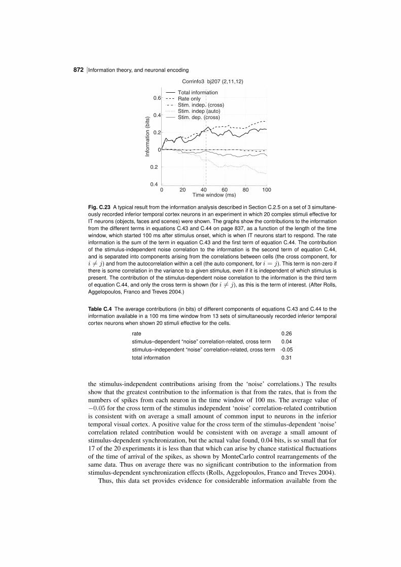
Information theory, and neuronal encoding872 |
0 20 40 60 80 1000.4
0.2
0
0.2
0.4
0.6
Time window (ms)
Info
rma
tio
n (
bits)
Corrinfo3 bj207 (2,11,12)
Total information Rate only Stim. indep. (cross)Stim. indep (auto) Stim. dep. (cross)
Fig. C.23 A typical result from the information analysis described in Section C.2.5 on a set of 3 simultaneously recorded inferior temporal cortex neurons in an experiment in which 20 complex stimuli effective forIT neurons (objects, faces and scenes) were shown. The graphs show the contributions to the informationfrom the different terms in equations C.43 and C.44 on page 837, as a function of the length of the timewindow, which started 100 ms after stimulus onset, which is when IT neurons start to respond. The rateinformation is the sum of the term in equation C.43 and the first term of equation C.44. The contributionof the stimulusindependent noise correlation to the information is the second term of equation C.44,and is separated into components arising from the correlations between cells (the cross component, fori = j) and from the autocorrelation within a cell (the auto component, for i = j). This term is nonzero ifthere is some correlation in the variance to a given stimulus, even if it is independent of which stimulus ispresent. The contribution of the stimulusdependent noise correlation to the information is the third termof equation C.44, and only the cross term is shown (for i = j), as this is the term of interest. (After Rolls,Aggelopoulos, Franco and Treves 2004.)
Table C.4 The average contributions (in bits) of different components of equations C.43 and C.44 to theinformation available in a 100 ms time window from 13 sets of simultaneously recorded inferior temporalcortex neurons when shown 20 stimuli effective for the cells.
rate 0.26stimulus–dependent “noise” correlationrelated, cross term 0.04stimulus–independent “noise” correlationrelated, cross term 0.05total information 0.31
the stimulusindependent contributions arising from the ‘noise’ correlations.) The resultsshow that the greatest contribution to the information is that from the rates, that is from thenumbers of spikes from each neuron in the time window of 100 ms. The average value of−0.05 for the cross term of the stimulus independent ‘noise’ correlationrelated contributionis consistent with on average a small amount of common input to neurons in the inferiortemporal visual cortex. A positive value for the cross term of the stimulusdependent ‘noise’correlation related contribution would be consistent with on average a small amount ofstimulusdependent synchronization, but the actual value found, 0.04 bits, is so small that for17 of the 20 experiments it is less than that which can arise by chance statistical fluctuationsof the time of arrival of the spikes, as shown by MonteCarlo control rearrangements of thesame data. Thus on average there was no significant contribution to the information fromstimulusdependent synchronization effects (Rolls, Aggelopoulos, Franco and Treves 2004).
Thus, this data set provides evidence for considerable information available from the
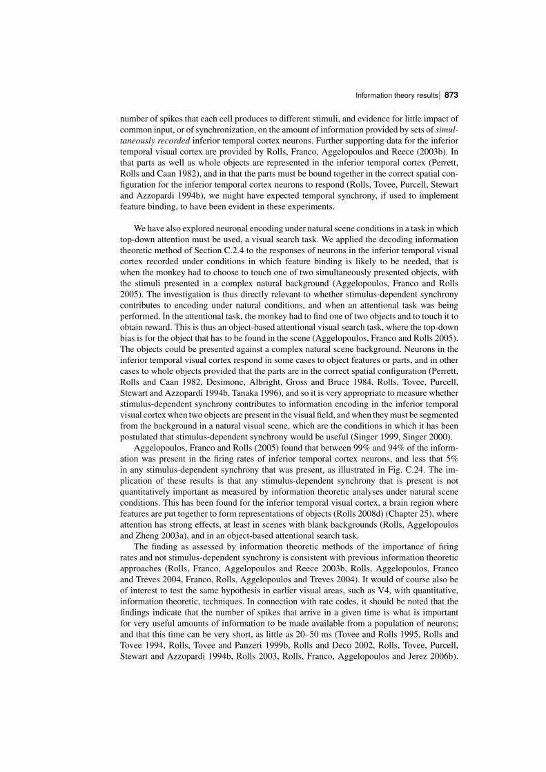
| 873Information theory results
number of spikes that each cell produces to different stimuli, and evidence for little impact ofcommon input, or of synchronization, on the amount of information provided by sets of simultaneously recorded inferior temporal cortex neurons. Further supporting data for the inferiortemporal visual cortex are provided by Rolls, Franco, Aggelopoulos and Reece (2003b). Inthat parts as well as whole objects are represented in the inferior temporal cortex (Perrett,Rolls and Caan 1982), and in that the parts must be bound together in the correct spatial configuration for the inferior temporal cortex neurons to respond (Rolls, Tovee, Purcell, Stewartand Azzopardi 1994b), we might have expected temporal synchrony, if used to implementfeature binding, to have been evident in these experiments.
We have also explored neuronal encoding under natural scene conditions in a task in whichtopdown attention must be used, a visual search task. We applied the decoding informationtheoretic method of Section C.2.4 to the responses of neurons in the inferior temporal visualcortex recorded under conditions in which feature binding is likely to be needed, that iswhen the monkey had to choose to touch one of two simultaneously presented objects, withthe stimuli presented in a complex natural background (Aggelopoulos, Franco and Rolls2005). The investigation is thus directly relevant to whether stimulusdependent synchronycontributes to encoding under natural conditions, and when an attentional task was beingperformed. In the attentional task, the monkey had to find one of two objects and to touch it toobtain reward. This is thus an objectbased attentional visual search task, where the topdownbias is for the object that has to be found in the scene (Aggelopoulos, Franco and Rolls 2005).The objects could be presented against a complex natural scene background. Neurons in theinferior temporal visual cortex respond in some cases to object features or parts, and in othercases to whole objects provided that the parts are in the correct spatial configuration (Perrett,Rolls and Caan 1982, Desimone, Albright, Gross and Bruce 1984, Rolls, Tovee, Purcell,Stewart and Azzopardi 1994b, Tanaka 1996), and so it is very appropriate to measure whetherstimulusdependent synchrony contributes to information encoding in the inferior temporalvisual cortex when two objects are present in the visual field, and when they must be segmentedfrom the background in a natural visual scene, which are the conditions in which it has beenpostulated that stimulusdependent synchrony would be useful (Singer 1999, Singer 2000).
Aggelopoulos, Franco and Rolls (2005) found that between 99% and 94% of the information was present in the firing rates of inferior temporal cortex neurons, and less that 5%in any stimulusdependent synchrony that was present, as illustrated in Fig. C.24. The implication of these results is that any stimulusdependent synchrony that is present is notquantitatively important as measured by information theoretic analyses under natural sceneconditions. This has been found for the inferior temporal visual cortex, a brain region wherefeatures are put together to form representations of objects (Rolls 2008d) (Chapter 25), whereattention has strong effects, at least in scenes with blank backgrounds (Rolls, Aggelopoulosand Zheng 2003a), and in an objectbased attentional search task.
The finding as assessed by information theoretic methods of the importance of firingrates and not stimulusdependent synchrony is consistent with previous information theoreticapproaches (Rolls, Franco, Aggelopoulos and Reece 2003b, Rolls, Aggelopoulos, Francoand Treves 2004, Franco, Rolls, Aggelopoulos and Treves 2004). It would of course also beof interest to test the same hypothesis in earlier visual areas, such as V4, with quantitative,information theoretic, techniques. In connection with rate codes, it should be noted that thefindings indicate that the number of spikes that arrive in a given time is what is importantfor very useful amounts of information to be made available from a population of neurons;and that this time can be very short, as little as 20–50 ms (Tovee and Rolls 1995, Rolls andTovee 1994, Rolls, Tovee and Panzeri 1999b, Rolls and Deco 2002, Rolls, Tovee, Purcell,Stewart and Azzopardi 1994b, Rolls 2003, Rolls, Franco, Aggelopoulos and Jerez 2006b).
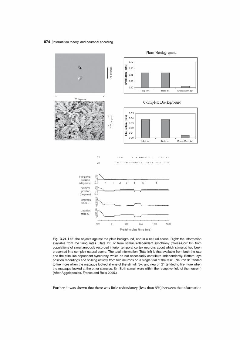
Information theory, and neuronal encoding874 |
70 degrees
17
.5 d
eg
ree
s1
7.5
de
gre
es
Fig. C.24 Left: the objects against the plain background, and in a natural scene. Right: the informationavailable from the firing rates (Rate Inf) or from stimulusdependent synchrony (CrossCorr Inf) frompopulations of simultaneously recorded inferior temporal cortex neurons about which stimulus had beenpresented in a complex natural scene. The total information (Total Inf) is that available from both the rateand the stimulusdependent synchrony, which do not necessarily contribute independently. Bottom: eyeposition recordings and spiking activity from two neurons on a single trial of the task. (Neuron 31 tendedto fire more when the macaque looked at one of the stimuli, S–, and neuron 21 tended to fire more whenthe macaque looked at the other stimulus, S+. Both stimuli were within the receptive field of the neuron.)(After Aggelopoulos, Franco and Rolls 2005.)
Further, it was shown that there was little redundancy (less than 6%) between the information
| 875Information theory results
provided by the spike counts of the simultaneously recorded neurons, making spike counts anefficient population code with a high encoding capacity.
The findings (Aggelopoulos, Franco and Rolls 2005) are consistent with the hypothesis thatfeature binding is implemented by neurons that respond to features in the correct relative spatiallocations (Rolls and Deco 2002, Elliffe, Rolls and Stringer 2002, Rolls 2008d, Rolls 2012c)(Chapter 25), and not by temporal synchrony and attention (Malsburg 1990a, Singer, Gray,Engel, Konig, Artola and Brocher 1990, Abeles 1991, Hummel and Biederman 1992, Singerand Gray 1995, Singer 1999, Singer 2000) (Section 8.3). In any case, the computationalpoint made in Section 25.5.5.1 is that even if stimulusdependent synchrony was useful forgrouping, it would not without much extra machinery be useful for binding the relative spatialpositions of features within an object, or for that matter of the positions of objects in a scenewhich appears to be encoded in a different way (Aggelopoulos and Rolls 2005) (see Section25.5.10).
So far, we know of no analyses that have shown with information theoretic methodsthat considerable amounts of information are available about the stimulus from the stimulusdependent correlations between the responses of neurons in the primate ventral visual system.The use of such methods is needed to test quantitatively the hypothesis that stimulusdependentsynchronization contributes substantially to the encoding of information by neurons.
C.3.8 Conclusions on cortical neuronal encoding
The conclusions emerging from this set of information theoretic analyses, many in corticalareas towards the end of the ventral visual stream of the monkey, and others in the hippocampusfor spatial view cells (Rolls, Treves, Robertson, GeorgesFrancois and Panzeri 1998b), in thepresubiculum for head direction cells (Robertson, Rolls, GeorgesFrancois and Panzeri 1999),and in the orbitofrontal cortex and related areas for olfactory and taste cells (Rolls, Critchleyand Treves 1996a, Rolls, Critchley, Verhagen and Kadohisa 2010a) for which subsequentanalyses have shown a linear increase in information with the number of cells in the population,are as follows (see also Rolls and Treves (2011)).
The representation of at least some classes of objects in those areas is achieved withminimal redundancy by cells that are allocated each to analyze a different aspect of the visualstimulus (Abbott, Rolls and Tovee 1996, Rolls, Treves and Tovee 1997b) (as shown in SectionsC.3.5 and C.3.7). This minimal redundancy is what would be expected of a selforganizingsystem in which different cells acquired their response selectivities through processes thatinclude some randomness in the initial connectivity, and local competition among nearbycells (see Appendix B). Towards the end of the ventral visual stream redundancy may thus beeffectively minimized, a finding consistent with the general idea that one of the functions of theearly visual system is indeed that of progressively minimizing redundancy in the representationof visual stimuli (Attneave 1954, Barlow 1961). Indeed, the evidence described in SectionsC.3.5, C.3.7 and C.3.4 shows that the exponential rise in the number of stimuli that can bedecoded when the firing rates of different numbers of neurons are analyzed indicates that theencoding of information using firing rates (in practice the number of spikes emitted by eachof a large population of neurons in a short time period) is a very powerful coding schemeused by the cerebral cortex, and that the information carried by different neurons is close toindependent provided that the number of stimuli being considered is sufficiently large.
Quantitatively, the encoding of information using firing rates (in practice the number ofspikes emitted by each of a large population of neurons in a short time period) is likely tobe far more important than temporal encoding, in terms of the number of stimuli that canbe encoded. Moreover, the information available from an ensemble of cortical neurons whenonly the firing rates are read, that is with no temporal encoding within or between neurons, is
Information theory, and neuronal encoding876 |
made available very rapidly (see Figs. C.14 and C.15 and Section C.3.4). Further, the neuronalresponses in most ventral or ‘what’ processing streams of behaving monkeys show sustainedfiring rate differences to different stimuli (see for example Fig. 16.15 for visual representations,for the olfactory pathways Rolls, Critchley and Treves (1996a), for spatial view cells in thehippocampus Rolls, Treves, Robertson, GeorgesFrancois and Panzeri (1998b), and for headdirection cells in the presubiculum Robertson, Rolls, GeorgesFrancois and Panzeri (1999)),so that it may not usually be necessary to invoke temporal encoding for the information aboutthe stimulus. Further, as indicated in Section C.3.7, information theoretic approaches haveenabled the information that is available from the firing rate and from the relative time of firing(synchronization) of inferior temporal cortex neurons to be directly compared with the samemetric, and most of the information appears to be encoded in the numbers of spikes emittedby a population of cells in a short time period, rather than by the temporal synchronizationof the responses of different neurons when certain stimuli appear (see Section C.3.7 andAggelopoulos, Franco and Rolls (2005)).
Information theoretic approaches have also enabled different types of readout or decodingthat could be performed by the brain of the information available in the responses of cellpopulations to be compared (Rolls, Treves and Tovee 1997b, Robertson, Rolls, GeorgesFrancois and Panzeri 1999). It has been shown for example that the multiple cell representationof information used by the brain in the inferior temporal visual cortex (Rolls, Treves andTovee 1997b, Aggelopoulos, Franco and Rolls 2005), olfactory cortex (Rolls, Critchley andTreves 1996a), hippocampus (Rolls, Treves, Robertson, GeorgesFrancois and Panzeri 1998b),and presubiculum (Robertson, Rolls, GeorgesFrancois and Panzeri 1999) can be read fairlyefficiently by the neuronally plausible dot product decoding, and that the representation hasall the desirable properties of generalization and graceful degradation, as well as exponentialcoding capacity (see Sections C.3.5 and C.3.7).
Information theoretic approaches have also enabled the information available about different aspects of stimuli to be directly compared. For example, it has been shown that inferiortemporal cortex neurons make explicit much more information about what stimulus has beenshown rather than where the stimulus is in the visual field (Tovee, Rolls and Azzopardi 1994),and this is part of the evidence that inferior temporal cortex neurons provide translation invariant representations. In a similar way, information theoretic analysis has provided clearevidence that view invariant representations of objects and faces are present in the inferiortemporal visual cortex, in that for example much information is available about what objecthas been shown from any single trial on which any view of any object is presented (Boothand Rolls 1998).
Information theory has also helped to elucidate the way in which the inferior temporalvisual cortex provides a representation of objects and faces, in which information about whichobject or face is shown is made explicit in the firing of the neurons in such a way that theinformation can be read off very simply by memory systems such as the orbitofrontal cortex,amygdala, and perirhinal cortex / hippocampal systems. The information can be read offusing dot product decoding, that is by using a synaptically weighted sum of inputs frominferior temporal cortex neurons (see further Section 24.2.6 and Chapter 25). Moreover,information theory has helped to show that for many neurons considerable invariance in therepresentations of objects and faces are shown by inferior temporal cortex neurons (e.g. Boothand Rolls (1998)). Examples of some of the types of objects and faces that are encoded inthis way are shown in Fig. C.7. Information theory has also helped to show that inferiortemporal cortex neurons maintain their object selectivity even when the objects are presentedin complex natural backgrounds (Aggelopoulos, Franco and Rolls 2005) (see further Chapter25 and Section 24.2.6).
Information theory has also enabled the information available in neuronal representations
| 877Information theory results
to be compared with that available to the whole animal in its behaviour (Zohary, Shadlen andNewsome 1994) (but see Section C.3.6).
Finally, information theory also provides a metric for directly comparing the informationavailable from neurons in the brain (see Chapter 25 and this Appendix) with that available fromsingle neurons and populations of neurons in simulations of visual information processing(see Chapter 25).
In summary, the evidence from the application of information theoretic and related approaches to how information is encoded in the visual, hippocampal, and olfactory corticalsystems described during behaviour leads to the following working hypotheses:
1. Much information is available about the stimulus presented in the number of spikes emittedby single neurons in a fixed time period, the firing rate.
2. Much of this firing rate information is available in short periods, with a considerable proportion available in as little as 20 ms. This rapid availability of information enables the nextstage of processing to read the information quickly, and thus for multistage processing tooperate rapidly. This time is the order of time over which a receiving neuron might be ableto utilize the information, given its synaptic and membrane time constants. In this time, asending neuron is most likely to emit 0, 1, or 2 spikes.
3. This rapid availability of information is confirmed by population analyses, which indicatethat across a population on neurons, much information is available in short time periods.
4. More information is available using this rate code in a short period (of e.g. 20 ms) thanfrom just the first spike.
5. Little information is available by time variations within the spike train of individual neuronsfor static visual stimuli (in periods of several hundred milliseconds), apart from a small amountof information from the onset latency of the neuronal response. A static stimulus encompasseswhat might be seen in a single visual fixation, what might be tasted with a stimulus in themouth, what might be smelled in a single sniff, etc. For a timevarying stimulus, clearly thefiring rate will vary as a function of time.
6. Across a population of neurons, the firing rate information provided by each neuron tendsto be independent; that is, the information increases approximately linearly with the numberof neurons. This applies of course only when there is a large amount of information to beencoded, that is with a large number of stimuli. The outcome is that the number of stimuli thatcan be encoded rises exponentially in the number of neurons in the ensemble. (For a smallstimulus set, the information saturates gradually as the amount of information available fromthe neuronal population approaches that required to code for the stimulus set.) This applies upto the number of neurons tested and the stimulus set sizes used, but as the number of neuronsbecomes very large, this is likely to hold less well. An implication of the independence is thatthe response profiles to a set of stimuli of different neurons are uncorrelated.
7. The information in the firing rate across a population of neurons can be read moderatelyefficiently by a decoding procedure as simple as a dot product. This is the simplest type ofprocessing that might be performed by a neuron, as it involves taking a dot product of theincoming firing rates with the receiving synaptic weights to obtain the activation (e.g. depolarization) of the neuron. This type of information encoding ensures that the simple emergentproperties of associative neuronal networks such as generalization, completion, and graceful
Information theory, and neuronal encoding878 |
degradation (see Appendix B) can be realized very naturally and simply.
8. There is little additional information to the great deal available in the firing rates fromany stimulusdependent crosscorrelations or synchronization that may be present. Stimulusdependent synchronization might in any case only be useful for grouping different neuronalpopulations, and would not easily provide a solution to the binding problem in vision. Instead,the binding problem in vision may be solved by the presence of neurons that respond tocombinations of features in a given spatial position with respect to each other.
9. There is little information available in the order of the spike arrival times of differentneurons for different stimuli that is separate or additional to that provided by a rate code.The presence of spontaneous activity in cortical neurons facilitates rapid neuronal responses,because some neurons are close to threshold at any given time, but this also would make aspike order code difficult to implement.
10. Analysis of the responses of single neurons to measure the sparseness of the representationindicates that the representation is distributed, and not grandmother cell like (or local). Moreover, the nature of the distributed representation, that it can be read by dot product decoding,allows simple emergent properties of associative neuronal networks such as generalization,completion, and graceful degradation (see Appendix B) to be realized very naturally and simply. The evidence becoming available from humans is consistent with this summary (Fried,Rutishauser, Cerf and Kreiman 2014, Rolls 2015d, Rolls 2017a).
11. The representation is not very sparse in the perceptual systems studied (as shown forexample by the values of the single cell sparseness as), and this may allow much informationto be represented. At the same time, the responses of different neurons to a set of stimuliare decorrelated, in the sense that the correlations between the response profiles of differentneurons to a set of stimuli are low. Consistent with this, the neurons convey independentinformation, at least up to reasonable numbers of neurons. The representation may be moresparse in memory systems such as the hippocampus, and this may help to maximize thenumber of memories that can be stored in associative networks.
12. The nature of the distributed representation can be understood further by the firing rateprobability distribution, which has a long tail with low probabilities of high firing rates. Thefiring rate probability distributions for some neurons fit an exponential distribution, and forothers there are too few very low rates for a good fit to the exponential distribution. Animplication of an exponential distribution is that this maximizes the entropy of the neuronalresponses for a given mean firing rate under some conditions. It is of interest that in the inferiortemporal visual cortex, the firing rate probability distribution is very close to exponential if alarge number of neurons are included without scaling of the firing rates of each neuron. An implication is that a receiving neuron would see an exponential firing rate probability distribution.
13. The population sparseness ap, that is the sparseness of the firing of a population of neuronsto a given stimulus (or at one time), is the important measure for setting the capacity of associative neuronal networks. In populations of neurons studied in the inferior temporal cortex,hippocampus, and orbitofrontal cortex, it takes the same value as the single cell sparseness as,and this is a situation of weak ergodicity that occurs if the response profiles of the differentneurons to a set of stimuli are uncorrelated.
| 879Information theory terms – a short glossary
Understanding the neuronal code, the subject of this Appendix, is fundamental for understanding how memory and related perceptual systems in the brain operate, as follows:
Understanding the neuronal code helps to clarify what neuronal operations would beuseful in memory and in fact in most mammalian brain systems (e.g. dot product decoding,that is taking a sum in a short time of the incoming firing rates weighted by the synapticweights).
It clarifies how rapidly memory and perceptual systems in the brain could operate, interms of how long it takes a receiving neuron to read the code.
It helps to confirm how the properties of those memory systems in terms of generalization,completion, and graceful degradation occur, in that the representation is in the correct formfor these properties to be realized.
Understanding the neuronal code also provides evidence essential for understanding thestorage capacity of memory systems, and the representational capacity of perceptual systems.
Understanding the neuronal code is also important for interpreting functional neuroimaging, for it shows that functional imaging that reflects incoming firing rates and thus currents injected into neurons, and probably not stimulusdependent synchronization, is likely tolead to useful interpretations of the underlying neuronal activity and processing. Of course,functional neuroimaging cannot address the details of the representation of information in thebrain in the way that is essential for understanding how neuronal networks in the brain couldoperate, for this level of understanding (in terms of all the properties and working hypothesesdescribed above) comes only from an understanding of how single neurons and populationsof neurons encode information.
C.4 Information theory terms – a short glossary1. The amount of information, or surprise, in the occurrence of an event (or symbol) si ofprobability P(si) is
I(si) = log2(1/P(si)) = − log2 P(si). (C.55)
(The measure is in bits if logs to the base 2 are used.) This is also the amount of uncertaintyremoved by the occurrence of the event.
2. The average amount of information per source symbol over the whole alphabet (S) ofsymbols si is the entropy,
H(S) = −∑i
P(si) log2 P(si) (C.56)
(or a priori entropy).
3. The probability of the pair of symbols s and s′ is denoted P(s, s′), and is P(s)P(s′) onlywhen the two symbols are independent.
4. Bayes theorem (given the output s′, what was the input s ?) states that
P(s|s′) = P(s′|s)P(s)P(s′)
(C.57)
where P(s′|s) is the forward conditional probability (given the input s, what will be theoutput s′ ?), and P(s|s′) is the backward (or posterior) conditional probability (given the
Information theory, and neuronal encoding880 |
output s′, what was the input s ?). The prior probability is P(s).
5. Mutual information. Prior to reception of s′, the probability of the input symbol s wasP(s). This is the a priori probability of s. After reception of s′, the probability that the inputsymbol was s becomes P(s|s′), the conditional probability that s was sent given that s′ wasreceived. This is the a posteriori probability of s. The difference between the a priori anda posteriori uncertainties measures the gain of information due to the reception of s′. Onceaveraged across the values of both symbols s and s′, this is the mutual information, ortransinformation
I(S, S′) =∑s,s′
P(s, s′){log2[1/P(s)]− log2[1/P(s|s′)]} (C.58)
=∑s,s′
P(s, s′) log2[P(s|s′)/P(s)].
Alternatively,I(S, S′) = H(S)−H(S|S′). (C.59)
H(S|S′) is sometimes called the equivocation (of S with respect to S′).
C.5 Highlights1. The encoding of information by neuronal responses is described using Shannon inform
ation theory.2. Information is encoded by sparse distributed place coded representations, with almost
independent information conveyed by each neuron, up to reasonable numbers of neurons.
3. Place cell encoding conveys much more information than stimulusdependent synchronicity (‘oscillations’, coherence) of firing of groups of neurons in the awake behaving animal.
References
Abbott, L. F. (1991). Realistic synaptic inputs for model neural networks, Network 2: 245–258.Abbott, L. F. and Blum, K. I. (1996). Functional significance of longterm potentiation for sequence learning and
prediction, Cerebral Cortex 6: 406–416.Abbott, L. F. and Nelson, S. B. (2000). Synaptic plasticity: taming the beast, Nature Neuroscience 3: 1178–1183.Abbott, L. F. and Regehr, W. G. (2004). Synaptic computation, Nature 431: 796–803.Abbott, L. F., Rolls, E. T. and Tovee, M. J. (1996). Representational capacity of face coding in monkeys, Cerebral
Cortex 6: 498–505.Abbott, L. F., Varela, J. A., Sen, K. and Nelson, S. B. (1997). Synaptic depression and cortical gain control, Science
275: 220–224.Abeles, M. (1991). Corticonics: Neural Circuits of the Cerebral Cortex, Cambridge University Press, Cambridge.Abramson, N. (1963). Information Theory and Coding, McGrawHill, New York.Ackley, D. H. (1987). A Connectionist Machine for Genetic HillClimbing, Kluwer Academic Publishers, Dordrecht.Ackley, D. H., Hinton, G. E. and Sejnowski, T. J. (1985). A learning algorithm for Boltzmann machines, Cognitive
Science 9: 147–169.Acsady, L., Kamondi, A., Sik, A., Freund, T. and Buzsaki, G. (1998). Gabaergic cells are the major postsynaptic
targets of mossy fibers in the rat hippocampus, Journal of Neuroscience 18: 3386–403.Adrian, E. D. (1928). The Basis of Sensations, Christophers, London.Aertsen, A. M. H. J., Gerstein, G. L., Habib, M. K. and Palm, G. (1989). Dynamics of neuronal firing correlation:
modulation of ‘effective connectivity’, Journal of Neurophysiology 61: 900–917.Aggelopoulos, N. C. and Rolls, E. T. (2005). Natural scene perception: inferior temporal cortex neurons encode the
positions of different objects in the scene, European Journal of Neuroscience 22: 2903–2916.Aggelopoulos, N. C., Franco, L. and Rolls, E. T. (2005). Object perception in natural scenes: encoding by inferior
temporal cortex simultaneously recorded neurons, Journal of Neurophysiology 93: 1342–1357.Aggleton, J. P. and Nelson, A. J. D. (2015). Why do lesions in the rodent anterior thalamic nuclei cause such severe
spatial deficits?, Neuroscience & Biobehavioral Reviews 54: 108–119.Aigner, T. G., Mitchell, S. J., Aggleton, J. P., DeLong, M. R., Struble, R. G., Price, D. L., Wenk, G. L., Pettigrew,
K. D. and Mishkin, M. (1991). Transient impairment of recognition memory following ibotenic acid lesionsof the basal forebrain in macaques, Experimental Brain Research 86: 18–26.
Aimone, J. B. and Gage, F. H. (2011). Modeling new neuron function: a history of using computational neuroscienceto study adult neurogenesis, European Journal of Neuroscience 33: 1160–9.
Aimone, J. B., Deng, W. and Gage, F. H. (2010). Adult neurogenesis: integrating theories and separating functions,Trends in Cognitive Sciences 14: 325–37.
Akrami, A., Liu, Y., Treves, A. and Jagadeesh, B. (2009). Converging neuronal activity in inferior temporal cortexduring the classification of morphed stimuli, Cerebral Cortex 19: 760–776.
Albantakis, L. and Deco, G. (2009). The encoding of alternatives in multiplechoice decision making, Proceedingsof the National Academy of Sciences USA 106: 10308–10313.
Alberini, C. M. and Ledoux, J. E. (2013). Memory reconsolidation, Current Biology 23: R746–50.Albus, J. S. (1971). A theory of cerebellar function, Mathematical Biosciences 10: 25–61.Aleman, A. and Kahn, R. S. (2005). Strange feelings: do amygdala abnormalities dysregulate the emotional brain in
schizophrenia?, Progress in Neurobiology 77(5): 283–298.Alexander, G. E., Crutcher, M. D. and DeLong, M. R. (1990). Basal ganglia thalamocortical circuits: parallel
substrates for motor, oculomotor, ‘prefrontal’ and ‘limbic’ functions, Progress in Brain Research 85: 119–146.Alexander, R. D. (1975). The search for a general theory of behavior, Behavioral Sciences 20: 77–100.Alexander, R. D. (1979). Darwinism and Human Affairs, University of Washington Press, Seattle.Allport, A. (1988). What concept of consciousness?, in A. J. Marcel and E. Bisiach (eds), Consciousness in
Contemporary Science, Oxford University Press, Oxford, pp. 159–182.Alvarez, P. and Squire, L. R. (1994). Memory consolidation and the medial temporal lobe: a simple network model,
Proceedings of the National Academy of Sciences USA 91: 7041–7045.Amaral, D. G. (1986). Amygdalohippocampal and amygdalocortical projections in the primate brain, in R. Schwarcz
and Y. BenAri (eds), Excitatory Amino Acids and Epilepsy, Plenum Press, New York, pp. 3–18.Amaral, D. G. (1987). Memory: anatomical organization of candidate brain regions, in F. Plum and V. Mountcas
tle (eds), Higher Functions of the Brain. Handbook of Physiology, Part I, American Physiological Society,Washington, DC, pp. 211–294.
Amaral, D. G. (1993). Emerging principles of intrinsic hippocampal organization, Current Opinion in Neurobiology
| 891
3: 225–229.Amaral, D. G. and Price, J. L. (1984). Amygdalocortical projections in the monkey (Macaca fascicularis), Journal
of Comparative Neurology 230: 465–496.Amaral, D. G. and Witter, M. P. (1989). The threedimensional organization of the hippocampal formation: a review
of anatomical data, Neuroscience 31: 571–591.Amaral, D. G. and Witter, M. P. (1995). The hippocampal formation, in G. Paxinos (ed.), The Rat Nervous System,
Academic Press, San Diego, pp. 443–493.Amaral, D. G., Ishizuka, N. and Claiborne, B. (1990). Neurons, numbers and the hippocampal network, Progress in
Brain Research 83: 1–11.Amaral, D. G., Price, J. L., Pitkanen, A. and Carmichael, S. T. (1992). Anatomical organization of the primate
amygdaloid complex, in J. P. Aggleton (ed.), The Amygdala, WileyLiss, New York, chapter 1, pp. 1–66.Amari, S. (1977). Dynamics of pattern formation in lateralinhibition type neural fields, Biological Cybernetics
27: 77–87.Amari, S. (1982). Competitive and cooperative aspects in dynamics of neural excitation and selforganization, in
S. Amari and M. A. Arbib (eds), Competition and Cooperation in Neural Nets, Springer, Berlin, chapter 1,pp. 1–28.
Amari, S., Yoshida, K. and Kanatani, K.I. (1977). A mathematical foundation for statistical neurodynamics, SIAMJournal of Applied Mathematics 33: 95–126.
Amit, D. J. (1989). Modelling Brain Function, Cambridge University Press, New York.Amit, D. J. (1995). The Hebbian paradigm reintegrated: local reverberations as internal representations, Behavioral
and Brain Sciences 18: 617–657.Amit, D. J. and Brunel, N. (1997). Model of global spontaneous activity and local structured activity during delay
periods in the cerebral cortex, Cerebral Cortex 7: 237–252.Amit, D. J. and Tsodyks, M. V. (1991). Quantitative study of attractor neural network retrieving at low spike rates.
I. Substrate – spikes, rates and neuronal gain, Network 2: 259–273.Amit, D. J., Gutfreund, H. and Sompolinsky, H. (1987). Statistical mechanics of neural networks near saturation,
Annals of Physics (New York) 173: 30–67.Anastasio, T. J. (2010). Tutorial on Neural Systems Modelling, Sinauer, Sunderland, MA.Andersen, P., Dingledine, R., Gjerstad, L., Langmoen, I. A. and Laursen, A. M. (1980). Two different responses of
hippocampal pyramidal cells to application of gammaaminobutyric acid, Journal of Physiology 307: 279–296.Andersen, P., Morris, R., Amaral, D., Bliss, T. and O’Keefe, J. (2007). The Hippocampus Book, Oxford University
Press, London.Andersen, R. A. (1995). Coordinate transformations and motor planning in the posterior parietal cortex, in M. S.
Gazzaniga (ed.), The Cognitive Neurosciences, MIT Press, Cambridge, MA, chapter 33, pp. 519–532.Andersen, R. A., Batista, A. P., Snyder, L. H., Buneo, C. A. and Cohen, Y. E. (2000). Programming to look and reach
in the posterior parietal cortex, in M. Gazzaniga (ed.), The New Cognitive Neurosciences, 2 edn, MIT Press,Cambridge, MA, chapter 36, pp. 515–524.
Anderson, J. R. (1996). ACT: a simple theory of complex cognition, American Psychologist 51: 355–365.Arbib, M. A. (1964). Brains, Machines, and Mathematics, McGrawHill, New York (2nd Edn 1987 Springer).Arcizet, F., Mirpour, K. and Bisley, J. W. (2011). A pure salience response in posterior parietal cortex, Cerebral
Cortex 21: 2498–2506.Armstrong, D. M. and Malcolm, M. (1984). Consciousness and Causality, Blackwell, Oxford.Arnold, P. D., Rosenberg, D. R., Mundo, E., Tharmalingam, S., Kennedy, J. L. and Richter, M. A. (2004). Association
of a glutamate (NMDA) subunit receptor gene (GRIN2B) with obsessivecompulsive disorder: a preliminarystudy, Psychopharmacology (Berl) 174: 530–538.
Arnsten, A. F. and Li, B. M. (2005). Neurobiology of executive functions: catecholamine influences on prefrontalcortical functions, Biological Psychiatry 57: 1377–1384.
Aron, A. R., Robbins, T. W. and Poldrack, R. A. (2014). Inhibition and the right inferior frontal cortex: one decadeon, Trends in Cognitive Sciences 18: 177–85.
Artola, A. and Singer, W. (1993). Long term depression: related mechanisms in cerebellum, neocortex and hippocampus, in M. Baudry, R. F. Thompson and J. L. Davis (eds), Synaptic Plasticity: Molecular, Cellular andFunctional Aspects, MIT Press, Cambridge, MA, chapter 7, pp. 129–146.
Atick, J. J. (1992). Could information theory provide an ecological theory of sensory processing?, Network 3: 213–251.
Atick, J. J. and Redlich, A. N. (1990). Towards a theory of early visual processing, Neural Computation 2: 308–320.Atick, J. J., Griffin, P. A. and Relich, A. N. (1996). The vocabulary of shape: principal shapes for probing perception
and neural response, Network 7: 1–5.Attneave, F. (1954). Some informational aspects of visual perception, Psychological Review 61: 183–193.Baars, B. J. (1988). A Cognitive Theory of Consciousness, Cambridge University Press, New York.BaconMace, N., Mace, M. J., FabreThorpe, M. and Thorpe, S. J. (2005). The time course of visual processing:
backward masking and natural scene categorisation, Vision Research 45: 1459–1469.Baddeley, R. J., Abbott, L. F., Booth, M. J. A., Sengpiel, F., Freeman, T., Wakeman, E. A. and Rolls, E. T. (1997).
References892 |
Responses of neurons in primary and inferior temporal visual cortices to natural scenes, Proceedings of theRoyal Society B 264: 1775–1783.
Bagal, A. A., Kao, J. P. Y., Tang, C.M. and Thompson, S. M. (2005). Longterm potentiation of exogenous glutamateresponses at single dendritic spines., Proc Natl Acad Sci U S A 102(40): 14434–9.
Balduzzi, D., Vanchinathan, H. and Buhmann, J. (2014). Kickback cuts backprop’s redtape: Biologically plausiblecredit assignment in neural networks, arXiv preprint arXiv:1411.6191.
Ballard, D. H. (1990). Animate vision uses objectcentred reference frames, in R. Eckmiller (ed.), Advanced NeuralComputers, NorthHolland, Elsevier, Amsterdam, pp. 229–236.
Ballard, D. H. (1993). Subsymbolic modelling of handeye coordination, in D. E. Broadbent (ed.), The Simulationof Human Intelligence, Blackwell, Oxford, chapter 3, pp. 71–102.
Balu, D. T. and Coyle, J. T. (2015). The nmda receptor ’glycine modulatory site’ in schizophrenia: dserine, glycine,and beyond, Current Opinion in Pharmacology 20C: 109–115.
Banks, S. J., Sziklas, V., Sodums, D. J. and JonesGotman, M. (2012). fMRI of verbal and nonverbal memoryprocesses in healthy and epileptogenic medial temporal lobes, Epilepsy and Behavior 25: 42–49.
Banks, W. P. (1978). Encoding and processing of symbolic information in comparative judgements, in G. H.Bower (ed.), The Psychology of Learning and Motivation: Advances in Theory and Research, Academic Press,pp. 101–159.
Bannon, S., Gonsalvez, C. J., Croft, R. J. and Boyce, P. M. (2002). Response inhibition deficits in obsessivecompulsive disorder, Psychiatry Research 110: 165–174.
Bannon, S., Gonsalvez, C. J., Croft, R. J. and Boyce, P. M. (2006). Executive functions in obsessivecompulsivedisorder: state or trait deficits?, Aust N Z Journal of Psychiatry 40: 1031–1038.
Barbas, H. (1993). Organization of cortical afferent input to the orbitofrontal area in the rhesus monkey, Neuroscience56: 841–864.
Barkas, L. J., Henderson, J. L., Hamilton, D. A., Redhead, E. S. and Gray, W. P. (2010). Selective temporalresections and spatial memory impairment: cue dependent lateralization effects, Behavioural Brain Research208: 535–544.
Barlow, H. (1995). The neuron doctrine in perception, in M. S. Gazzaniga (ed.), The Cognitive Neurosciences, MITPress, Cambridge, MA, chapter 26, pp. 415–435.
Barlow, H. (1997). Single neurons, communal goals, and consciousness, in M. Ito, Y. Miyashita and E. T. Rolls(eds), Cognition, Computation, and Consciousness, Oxford University Press, Oxford, chapter 7, pp. 121–136.
Barlow, H. B. (1961). Possible principles underlying the transformation of sensory messages, in W. Rosenblith (ed.),Sensory Communication, MIT Press, Cambridge, MA.
Barlow, H. B. (1972). Single units and sensation: a neuron doctrine for perceptual psychology, Perception 1: 371–394.Barlow, H. B. (1985). Cerebral cortex as model builder, in D. Rose and V. G. Dobson (eds), Models of the Visual
Cortex, Wiley, Chichester, pp. 37–46.Barlow, H. B. (1989). Unsupervised learning, Neural Computation 1: 295–311.Barlow, H. B., Kaushal, T. P. and Mitchison, G. J. (1989). Finding minimum entropy codes, Neural Computation
1: 412–423.Barnes, C. A. (2003). Longterm potentiation and the ageing brain, Philosophical Transactions of the Royal Society
of London B 358: 765–772.Barnes, D. C., Hofacer, R. D., Zaman, A. R., Rennaker, R. L. and Wilson, D. A. (2008). Olfactory perceptual stability
and discrimination, Nature Neuroscience 11: 1378–80.Barrett, H. (2012). A hierarchical model of the evolution of human brain specializations, Proceedings of the National
Academy of Sciences 109(Supplement 1): 10733–10740.Bartlett, M. S. and Sejnowski, T. J. (1997). Viewpoint invariant face recognition using independent component
analysis and attractor networks, in M. Mozer, M. Jordan and T. Petsche (eds), Advances in Neural InformationProcessing Systems 9, MIT Press, Cambridge, MA, pp. 817–823.
Barto, A. G. (1985). Learning by statistical cooperation of selfinterested neuronlike computing elements, HumanNeurobiology 4: 229–256.
Barto, A. G. (1995). Adaptive critics and the basal ganglia, in J. C. Houk, J. L. Davis and D. G. Beiser (eds), Modelsof Information Processing in the Basal Ganglia, MIT Press, Cambridge, MA, chapter 11, pp. 215–232.
Bartol, T. M., Bromer, C., Kinney, J., Chirillo, M. A., Bourne, J. N., Harris, K. M. and Sejnowski, T. J. (2016).Nanoconnectomic upper bound on the variability of synaptic plasticity, eLife 4: e10778.
Bartus, R. T. (2000). On neurodegenerative diseases, models, and treatment strategies: lessons learned and lessonsforgotten a generation following the cholinergic hypothesis, Experimental Neurology 163: 495–529.
Bassett, J. and Taube, J. S. (2005). Head direction signal generation: ascending and descending information streams,in S. I. Wiener and J. S. Taube (eds), Head Direction Cells and the Neural Mechanisms of Spatial Orientation,MIT Press, Cambridge, MA, chapter 5, pp. 83–109.
Battaglia, F. and Treves, A. (1998a). Stable and rapid recurrent processing in realistic autoassociative memories,Neural Computation 10: 431–450.
Battaglia, F. P. and Treves, A. (1998b). Attractor neural networks storing multiple space representations: a model forhippocampal place fields, Physical Review E 58: 7738–7753.
| 893
Baxter, M. G. and Murray, E. A. (2001a). Effects of hippocampal lesions on delayed nonmatchingtosample inmonkeys: a reply to Zola and Squire, Hippocampus 11: 201–203.
Baxter, M. G. and Murray, E. A. (2001b). Opposite relationship of hippocampal and rhinal cortex damage to delayednonmatchingtosample deficits in monkeys, Hippocampus 11: 61–71.
Baxter, R. D. and Liddle, P. F. (1998). Neuropsychological deficits associated with schizophrenic syndromes,Schizophrenia Research 30: 239–249.
Baylis, G. C. and Rolls, E. T. (1987). Responses of neurons in the inferior temporal cortex in short term and serialrecognition memory tasks, Experimental Brain Research 65: 614–622.
Baylis, G. C., Rolls, E. T. and Leonard, C. M. (1985). Selectivity between faces in the responses of a population ofneurons in the cortex in the superior temporal sulcus of the monkey, Brain Research 342: 91–102.
Baylis, G. C., Rolls, E. T. and Leonard, C. M. (1987). Functional subdivisions of temporal lobe neocortex, Journalof Neuroscience 7: 330–342.
Baylis, L. L. and Gaffan, D. (1991). Amygdalectomy and ventromedial prefrontal ablation produce similar deficits infood choice and in simple object discrimination learning for an unseen reward, Experimental Brain Research86: 617–622.
Baylis, L. L. and Rolls, E. T. (1991). Responses of neurons in the primate taste cortex to glutamate, Physiology andBehavior 49: 973–979.
Baylis, L. L., Rolls, E. T. and Baylis, G. C. (1994). Afferent connections of the orbitofrontal cortex taste area of theprimate, Neuroscience 64: 801–812.
Bear, M. F. and Singer, W. (1986). Modulation of visual cortical plasticity by acetylcholine and noradrenaline, Nature320: 172–176.
Beck, A. T. (2008). The evolution of the cognitive model of depression and its neurobiological correlates, AmericanJournal of Psychiatry 165: 969–977.
Beck, D. M. and Kastner, S. (2009). Topdown and bottomup mechanisms in biasing competition in the humanbrain, Vision Research 49: 1154–1165.
Becker, S. and Hinton, G. E. (1992). Selforganizing neural network that discovers surfaces in randomdot stereograms, Nature 355: 161–163.
Beckstead, R. M. and Norgren, R. (1979). An autoradiographic examination of the central distribution of thetrigeminal, facial, glossopharyngeal, and vagal nerves in the monkey, Journal of Comparative Neurology184: 455–472.
Beckstead, R. M., Morse, J. R. and Norgren, R. (1980). The nucleus of the solitary tract in the monkey: projectionsto the thalamus and brainstem nuclei, Journal of Comparative Neurology 190: 259–282.
Bell, A. J. and Sejnowski, T. J. (1995). An informationmaximation approach to blind separation and blind deconvolution, Neural Computation 7: 1129–1159.
Benda, J. and Herz, A. V. M. (2003). A universal model for spikefrequency adaptation, Neural Computation15: 2523–2564.
Bender, W., Albus, M., Moller, H. J. and Tretter, F. (2006). Towards systemic theories in biological psychiatry,Pharmacopsychiatry 39 Suppl 1: S4–S9.
Bengio, Y., Goodfellow, I. J. and Courville, A. (2017). Deep Learning, MIT Press, Cambridge, MA.Bennett, A. (1990). Large competitive networks, Network 1: 449–462.Berg, E. (1948). A simple objective technique for measuring flexibility in thinking, Journal of General Psychology
39: 15–22.Berlin, H., Rolls, E. T. and Kischka, U. (2004). Impulsivity, time perception, emotion, and reinforcement sensitivity
in patients with orbitofrontal cortex lesions, Brain 127: 1108–1126.Berlin, H., Rolls, E. T. and Iversen, S. D. (2005). Borderline Personality Disorder, impulsivity, and the orbitofrontal
cortex, American Journal of Psychiatry 58: 234–245.Bermpohl, F., Kahnt, T., Dalanay, U., Hagele, C., Sajonz, B., Wegner, T., Stoy, M., Adli, M., Kruger, S., Wrase,
J., Strohle, A., Bauer, M. and Heinz, A. (2010). Altered representation of expected value in the orbitofrontalcortex in mania, Human Brain Mapping 31: 958–969.
Bernard, A., Lubbers, L. S., Tanis, K. Q., Luo, R., Podtelezhnikov, A. A., Finney, E. M., McWhorter, M. M., Serikawa,K., Lemon, T., Morgan, R., Copeland, C., Smith, K., Cullen, V., DavisTurak, J., Lee, C. K., Sunkin, S. M.,Loboda, A. P., Levine, D. M., Stone, D. J., Hawrylycz, M. J., Roberts, C. J., Jones, A. R., Geschwind, D. H.and Lein, E. S. (2012). Transcriptional architecture of the primate neocortex, Neuron 73: 1083–99.
Berridge, K. C., Flynn, F. W., Schulkin, J. and Grill, H. J. (1984). Sodium depletion enhances salt palatability in rats,Behavioral Neuroscience 98: 652–660.
Bhattacharyya, S. and Chakraborty, K. (2007). Glutamatergic dysfunction–newer targets for antiobsessional drugs,Recent Patents CNS Drug Discovery 2: 47–55.
Bi, G.Q. and Poo, M.M. (1998). Activityinduced synaptic modifications in hippocampal culture, dependence onspike timing, synaptic strength and cell type, Journal of Neuroscience 18: 10464–10472.
Bi, G.Q. and Poo, M.M. (2001). Synaptic modification by correlated activity: Hebb’s postulate revisited, AnnualReview of Neuroscience 24: 139–166.
Bialek, W., Rieke, F., de Ruyter van Steveninck, R. R. and Warland, D. (1991). Reading a neural code, Science
References894 |
252: 1854–1857.Biederman, I. (1972). Perceiving realworld scenes, Science 177: 77–80.Biederman, I. (1987). Recognitionbycomponents: A theory of human image understanding, Psychological Review
94(2): 115–147.Bienenstock, E. L., Cooper, L. N. and Munro, P. W. (1982). Theory for the development of neuron selectivity:
orientation specificity and binocular interaction in visual cortex, Journal of Neuroscience 2: 32–48.Bierer, L. M., Haroutunian, V., Gabriel, S., Knott, P. J., Carlin, L. S., Purohit, D. P., Perl, D. P., Schmeidler, J., Kanof,
P. and Davis, K. L. (1995). Neurochemical correlates of dementia severity in Alzheimer’s disease: relativeimportance of the cholinergic deficits, Journal of Neurochemistry 64: 749–760.
Binford, T. O. (1981). Inferring surfaces from images, Artificial Intelligence 17: 205–244.Bishop, C. M. (1995). Neural Networks for Pattern Recognition, Clarendon Press, Oxford.Bisley, J. W. and Goldberg, M. E. (2003). Neuronal activity in the lateral intraparietal area and spatial attention,
Science 299: 81–86.Bisley, J. W. and Goldberg, M. E. (2006). Neural correlates of attention and distractibility in the lateral intraparietal
area, Journal of Neurophysiology 95: 1696–1717.Bisley, J. W. and Goldberg, M. E. (2010). Attention, intention, and priority in the parietal lobe, Annual Review of
Neuroscience 33: 1–21.Blair, R. J., Morris, J. S., Frith, C. D., Perrett, D. I. and Dolan, R. J. (1999). Dissociable neural responses to facial
expressions of sadness and anger, Brain 122: 883–893.Blake, R. and Logothetis, N. K. (2002). Visual competition, Nature Reviews Neuroscience 3: 13–21.Blakemore, C. and van Sluyters, R. C. (1974). Reversal of the physiological effects of monocular deprivation in
kittens: Further evidence for a sensitive period, Journal of Physiology 237: 195–216.Blakemore, C., Garey, L. J. and VitalDurand, F. (1978). The physiological effects of monocular deprivation and
their reversal in the monkeys visual cortex, Journal of Physiology 283: 223–262.Bliss, T. V. and Collingridge, G. L. (2013). Expression of NMDA receptordependent LTP in the hippocampus:
bridging the divide, Molecular Brain 6: 5.Block, H. D. (1962). The perceptron: a model for brain functioning, Reviews of Modern Physics 34: 123–135.Block, N. (1995a). On a confusion about a function of consciousness, Behavioral and Brain Sciences 18: 22–47.Block, N. (1995b). Two neural correlates of consciousness, Trends in Cognitive Sciences 9: 46–52.Bloomfield, S. (1974). Arithmetical operations performed by nerve cells, Brain Research 69: 115–124.Bolles, R. C. and Cain, R. A. (1982). Recognizing and locating partially visible objects: The localfeaturefocus
method, International Journal of Robotics Research 1: 57–82.Bonelli, S. B., Powell, R. H., Yogarajah, M., Samson, R. S., Symms, M. R., Thompson, P. J., Koepp, M. J. and
Duncan, J. S. (2010). Imaging memory in temporal lobe epilepsy: predicting the effects of temporal loberesection, Brain 133: 1186–1199.
Booth, D. A. (1985). Foodconditioned eating preferences and aversions with interoceptive elements: learnedappetites and satieties, Annals of the New York Academy of Sciences 443: 22–37.
Booth, M. C. A. and Rolls, E. T. (1998). Viewinvariant representations of familiar objects by neurons in the inferiortemporal visual cortex, Cerebral Cortex 8: 510–523.
BornkesselSchlesewsky, I., Schlesewsky, M., Small, S. L. and Rauschecker, J. P. (2015). Neurobiological roots oflanguage in primate audition: common computational properties, Trends in Cognitive Sciences 19: 142–150.
Borsini, F. and Rolls, E. T. (1984). Role of noradrenaline and serotonin in the basolateral region of the amygdala infood preferences and learned taste aversions in the rat, Physiology and Behavior 33: 37–43.
Boussaoud, D., Desimone, R. and Ungerleider, L. G. (1991). Visual topography of area TEO in the macaque, Journalof Computational Neurology 306: 554–575.
Bovier, A. and Gayrard, V. (1992). Rigorous bounds on the storage capacity of the dilute Hopfield model, Journalof Statistical Physics 69: 597–627.
Brady, M., Ponce, J., Yuille, A. and Asada, H. (1985). Describing surfaces, A. I. Memo 882, The Artificial Intelligence17: 285–349.
Braitenberg, V. and Schutz, A. (1991). Anatomy of the Cortex, SpringerVerlag, Berlin.Braitenberg, V. and Schutz, A. (1998). Cortex: Statistics and Geometry of Neuronal Connectivity, SpringerVerlag,
Berlin.Bressler, S. L. and Seth, A. K. (2011). WienerGranger causality: A well established methodology, Neuroimage
58: 323–329.Bridle, J. S. (1990). Probabilistic interpretation of feedforward classification network outputs, with relationships
to statistical pattern recognition, in F. FogelmanSoulie and J. Herault (eds), Neurocomputing: Algorithms,Architectures and Applications, SpringerVerlag, New York, pp. 227–236.
Brodmann, K. (1925). Vergleichende Localisationslehre der Grosshirnrinde. Translated into English by L.J.Gareyas Localisation in the Cerebral Cortex 1994 London: SmithGordon, 2 edn, Barth, Leipzig.
BrombergMartin, E. S., Matsumoto, M. and Hikosaka, O. (2010a). Dopamine in motivational control: rewarding,aversive, and alerting, Neuron 68: 815–834.
BrombergMartin, E. S., Matsumoto, M., Hong, S. and Hikosaka, O. (2010b). A pallidushabenuladopamine pathway
| 895
signals inferred stimulus values, Journal of Neurophysiology 104: 1068–1076.Brooks, L. R. (1978). Nonanalytic concept formation and memory for instances, in E. Rosch and B. B. Lloyd (eds),
Cognition and Categorization, Erlbaum, Hillsdale, NJ.Brotchie, P., Andersen, R., Snyder, L. and Goodman, S. (1995). Head position signals used by parietal neurons to
encode locations of visual stimuli, Nature London 375: 232–235.Brown, D. A., Gahwiler, B. H., Griffith, W. H. and Halliwell, J. V. (1990a). Membrane currents in hippocampal
neurons, Progress in Brain Research 83: 141–160.Brown, J. M. (2009). Visual streams and shifting attention, Progress in Brain Research 176: 47–63.Brown, M. and Xiang, J. (1998). Recognition memory: neuronal substrates of the judgement of prior occurrence,
Progress in Neurobiology 55: 149–189.Brown, T. H. and Zador, A. (1990). The hippocampus, in G. Shepherd (ed.), The Synaptic Organization of the Brain,
Oxford University Press, New York, pp. 346–388.Brown, T. H., Kairiss, E. W. and Keenan, C. L. (1990b). Hebbian synapses: biophysical mechanisms and algorithms,
Annual Review of Neuroscience 13: 475–511.Brown, T. H., Ganong, A. H., Kairiss, E. W., Keenan, C. L. and Kelso, S. R. (eds) (1989). Longterm Potentiation in
Two Synaptic Systems of the Hippocampal Brain Slice, Academic Press, San Diego.Bruce, V. (1988). Recognising Faces, Erlbaum, Hillsdale, NJ.Brun, V. H., Otnass, M. K., Molden, S., Steffenach, H. A., Witter, M. P., Moser, M. B. and Moser, E. I. (2002). Place
cells and place recognition maintained by direct entorhinal–hippocampal circuitry, Science 296: 2243–2246.Brunel, N. and Wang, X. J. (2001). Effects of neuromodulation in a cortical network model of object working
memory dominated by recurrent inhibition, Journal of Computational Neuroscience 11: 63–85.Buck, L. and Bargmann, C. I. (2013). Smell and taste: the chemical senses, in E. Kandel, J. H. Schwartz, T. H.
Jessell, S. A. Siegelbaum and A. J. Hudspeth (eds), Principles of Neural Science, 5th edn, McGrawHill, NewYork, chapter 32, pp. 712–742.
Buckley, M. J. and Gaffan, D. (2000). The hippocampus, perirhinal cortex, and memory in the monkey, in J. J.Bolhuis (ed.), Brain, Perception, and Memory: Advances in Cognitive Neuroscience, Oxford University Press,Oxford, pp. 279–298.
Buckley, M. J. and Gaffan, D. (2006). Perirhinal contributions to object perception, Trends in Cognitive Sciences10: 100–107.
Buckley, M. J., Booth, M. C. A., Rolls, E. T. and Gaffan, D. (2001). Selective perceptual impairments followingperirhinal cortex ablation, Journal of Neuroscience 21: 9824–9836.
Buhl, E. H., Halasy, K. and Somogyi, P. (1994). Diverse sources of hippocampal unitary inhibitory postsynapticpotentials and the number of synaptic release sites, Nature 368: 823–828.
Buhmann, J., Lange, J., von der Malsburg, C., Vorbruggen, J. C. and Wurtz, R. P. (1991). Object recognition in thedynamic link architecture: Parallel implementation of a transputer network, in B. Kosko (ed.), Neural Networksfor Signal Processing, Prentice Hall, Englewood Cliffs, NJ, pp. 121–159.
Bullier, J. and Nowak, L. (1995). Parallel versus serial processing: new vistas on the distributed organization of thevisual system, Current Opinion in Neurobiology 5: 497–503.
Bunsey, M. and Eichenbaum, H. (1996). Conservation of hippocampal memory function in rats and humans, Nature379: 255–257.
Buot, A. and Yelnik, J. (2012). Functional anatomy of the basal ganglia: limbic aspects, Revue Neurologique (Paris)168: 569–575.
Burgess, N. (2008). Spatial cognition and the brain, Annals of the New York Academy of Sciences 1124: 77–97.Burgess, N., Recce, M. and O’Keefe, J. (1994). A model of hippocampal function, Neural Networks 7: 1065–1081.Burgess, N., Jackson, A., Hartley, T. and O’Keefe, J. (2000). Predictions derived from modelling the hippocampal
role in navigation, Biological Cybernetics 83: 301–312.Burgess, N., Maguire, E. A. and O’Keefe, J. (2002). The human hippocampus and spatial and episodic memory,
Neuron 35: 625–641.Burgess, P. W. (2000). Strategy application disorder: the role of the frontal lobes in human multitasking, Psychological
Research 63: 279–288.Burke, S. N. and Barnes, C. A. (2006). Neural plasticity in the ageing brain, Nature Reviews Neuroscience 7: 30–40.Burton, M. J., Rolls, E. T. and Mora, F. (1976). Effects of hunger on the responses of neurones in the lateral
hypothalamus to the sight and taste of food, Experimental Neurology 51: 668–677.Burwell, R. D., Witter, M. P. and Amaral, D. G. (1995). Perirhinal and postrhinal cortices of the rat: a review of the
neuroanatomical literature and comparison with findings from the monkey brain, Hippocampus 5: 390–408.Bussey, T. J. and Saksida, L. M. (2005). Object memory and perception in the medial temporal lobe: an alternative
approach, Current Opinion in Neurobiology 15: 730–737.Bussey, T. J., Saksida, L. M. and Murray, E. A. (2002). Perirhinal cortex resolves feature ambiguity in complex
visual discriminations, European Journal of Neuroscience 15: 365–374.Bussey, T. J., Saksida, L. M. and Murray, E. A. (2003). Impairments in visual discrimination after perirhinal cortex
lesions: testing “declarative” versus “perceptualmnemonic” views of perirhinal cortex function, EuropeanJournal of Neuroscience 17: 649–660.
References896 |
Bussey, T. J., Saksida, L. M. and Murray, E. A. (2005). The perceptualmnemonic / feature conjunction model ofperirhinal cortex function, Quarterly Journal of Experimental Psychology 58B: 269–282.
Butter, C. M. (1969). Perseveration in extinction and in discrimination reversal tasks following selective prefrontalablations in Macaca mulatta, Physiology and Behavior 4: 163–171.
Butter, C. M., McDonald, J. A. and Snyder, D. R. (1969). Orality, preference behavior, and reinforcement value ofnonfood objects in monkeys with orbital frontal lesions, Science 164: 1306–1307.
Buxton, R. B. and Frank, L. R. (1997). A model for the coupling between cerebral blood flow and oxygen metabolismduring neural stimulation, Journal of Cerebral Blood Flow and Metabolism 17: 64–72.
Buxton, R. B., Wong, E. C. and Frank, L. R. (1998). Dynamics of blood flow and oxygenation changes during brainactivation: the balloon model, Magnetic Resonance in Medicine 39: 855–864.
Buzsáki, G. (2006). Rhythms of the Brain, Oxford University Press, Oxford.Byrne, R. W. and Whiten, A. (1988). Machiavellian Intelligence: Social Expertise and the Evolution of Intellect in
Monkeys, Apes and Humans, Clarendon Press, Oxford.Caan, W., Perrett, D. I. and Rolls, E. T. (1984). Responses of striatal neurons in the behaving monkey. 2. Visual
processing in the caudal neostriatum, Brain Research 290: 53–65.Cadieu, C. F., Hong, H., Yamins, D. L. K., Pinto, N., Ardila, D., Solomon, E. A., Majaj, N. J. and DiCarlo, J. J.
(2014). Deep neural networks rival the representation of primate IT cortex for core visual object recognition,PLoS Computational Biology 10: e1003963.
Cahusac, P. M. B., Miyashita, Y. and Rolls, E. T. (1989). Responses of hippocampal formation neurons in the monkeyrelated to delayed spatial response and objectplace memory tasks, Behavioural Brain Research 33: 229–240.
Cahusac, P. M. B., Rolls, E. T., Miyashita, Y. and Niki, H. (1993). Modification of the responses of hippocampalneurons in the monkey during the learning of a conditional spatial response task, Hippocampus 3: 29–42.
Calvert, G. A., Bullmore, E. T., Brammer, M. J., Campbell, R., Williams, S. C. R., McGuire, P. K., Woodruff, P.W. R., Iversen, S. D. and David, A. S. (1997). Activation of auditory cortex during silent lipreading, Science276: 593–596.
Camille, N., Tsuchida, A. and Fellows, L. K. (2011). Double dissociation of stimulusvalue and actionvalue learningin humans with orbitofrontal or anterior cingulate cortex damage, Journal of Neuroscience 31: 15048–15052.
Capuano, B., Crosby, I. T. and Lloyd, E. J. (2002). Schizophrenia: genesis, receptorology and current therapeutics,Current Medicinal Chemistry 9: 521–548.
Cardinal, R. N., Parkinson, J. A., Hall, J. and Everitt, B. J. (2002). Emotion and motivation: the role of the amygdala,ventral striatum, and prefrontal cortex, Neuroscience and Biobehavioral Reviews 26: 321–352.
Carlson, E. T., Rasquinha, R. J., Zhang, K. and Connor, C. E. (2011). A sparse object coding scheme in area v4,Current Biology 21: 288–93.
Carlson, N. R. (2013). Physiology of Behavior, 11th edn, Pearson, Boston.Carlsson, A. (2006). The neurochemical circuitry of schizophrenia, Pharmacopsychiatry 39 Suppl 1: S10–14.Carmichael, S. T. and Price, J. L. (1994). Architectonic subdivision of the orbital and medial prefrontal cortex in the
macaque monkey, Journal of Comparative Neurology 346: 366–402.Carmichael, S. T. and Price, J. L. (1995a). Limbic connections of the orbital and medial prefrontal cortex in macaque
monkeys, Journal of Comparative Neurology 363: 615–641.Carmichael, S. T. and Price, J. L. (1995b). Sensory and premotor connections of the orbital and medial prefrontal
cortex of macaque monkeys, Journal of Comparative Neurology 363: 642–664.Carmichael, S. T., Clugnet, M.C. and Price, J. L. (1994). Central olfactory connections in the macaque monkey,
Journal of Comparative Neurology 346: 403–434.Carpenter, G. A. (1997). Distributed learning, recognition and prediction by ART and ARTMAP neural networks,
Neural Networks 10(8): 1473–1494.Carpenter, R. H. S. and Williams, M. (1995). Neural computation of log likelihood in control of saccadic eye
movements, Nature 377: 59–62.Carruthers, P. (1996). Language, Thought and Consciousness, Cambridge University Press, Cambridge.Carruthers, P. (2000). Phenomenal Consciousness, Cambridge University Press, Cambridge.Carter, C. S., Perlstein, W., Ganguli, R., Brar, J., Mintun, M. and Cohen, J. D. (1998). Functional hypofrontality and
working memory dysfunction in schizophrenia, American Journal of Psychiatry 155: 1285–1287.Caspers, J., PalomeroGallagher, N., Caspers, S., Schleicher, A., Amunts, K. and Zilles, K. (2015). Receptor
architecture of visual areas in the face and wordform recognition region of the posterior fusiform gyrus, BrainStruct Funct 220: 205–19.
Cassaday, H. J. and Rawlins, J. N. (1997). The hippocampus, objects, and their contexts, Behavioral Neuroscience111: 1228–1244.
Castner, S. A., Williams, G. V. and GoldmanRakic, P. S. (2000). Reversal of antipsychoticinduced working memorydeficits by shortterm dopamine D1 receptor stimulation, Science 287: 2020–2022.
Cavanna, A. E. and Trimble, M. R. (2006). The precuneus: a review of its functional anatomy and behaviouralcorrelates, Brain 129: 564–583.
Celebrini, S., Thorpe, S., Trotter, Y. and Imbert, M. (1993). Dynamics of orientation coding in area V1 of the awakeprimate, Visual Neuroscience 10: 811–825.
| 897
Cerasti, E. and Treves, A. (2010). How informative are spatial CA3 representations established by the dentate gyrus?,PLoS Computational Biology 6: e1000759.
Cerasti, E. and Treves, A. (2013). The spatial representations acquired in CA3 by selforganizing recurrent connections, Frontiers in Cellular Neuroscience 7: 112.
Cerella, J. (1986). Pigeons and perceptrons, Pattern Recognition 19: 431–438.Chadwick, M. J., Mullally, S. L. and Maguire, E. A. (2013). The hippocampus extrapolates beyond the view in
scenes: an fmri study of boundary extension, Cortex 49: 2067–2079.Chakrabarty, K., Bhattacharyya, S., Christopher, R. and Khanna, S. (2005). Glutamatergic dysfunction in OCD,
Neuropsychopharmacology 30: 1735–1740.Chakravarty, I. (1979). A generalized line and junction labeling scheme with applications to scene analysis, IEEE
Transactions PAMI 1: 202–205.Chalmers, D. J. (1996). The Conscious Mind, Oxford University Press, Oxford.Chamberlain, S. R., Fineberg, N. A., Blackwell, A. D., Robbins, T. W. and Sahakian, B. J. (2006). Motor inhibition
and cognitive flexibility in obsessivecompulsive disorder and trichotillomania, American Journal of Psychiatry163: 1282–1284.
Chamberlain, S. R., Fineberg, N. A., Menzies, L. A., Blackwell, A. D., Bullmore, E. T., Robbins, T. W. and Sahakian,B. J. (2007). Impaired cognitive flexibility and motor inhibition in unaffected firstdegree relatives of patientswith obsessivecompulsive disorder, American Journal of Psychiatry 164: 335–338.
Chan, A. M., Dykstra, A. R., Jayaram, V., Leonard, M. K., Travis, K. E., Gygi, B., Baker, J. M., Eskandar, E.,Hochberg, L. R., Halgren, E. and Cash, S. S. (2014). Speechspecific tuning of neurons in human superiortemporal gyrus, Cerebral Cortex 24: 2679–2693.
Chau, B. K., Sallet, J., Papageorgiou, G. K., Noonan, M. P., Bell, A. H., Walton, M. E. and Rushworth, M. F. (2015).Contrasting roles for orbitofrontal cortex and amygdala in credit assignment and learning in macaques, Neuron87: 1106–1118.
Chelazzi, L. (1998). Serial attention mechanisms in visual search: a critical look at the evidence, PsychologicalResearch 62: 195–219.
Chelazzi, L., Miller, E., Duncan, J. and Desimone, R. (1993). A neural basis for visual search in inferior temporalcortex, Nature (London) 363: 345–347.
Cheney, D. L. and Seyfarth, R. M. (1990). How Monkeys See the World, University of Chicago Press, Chicago.Cheng, W., Rolls, E. T., Gu, H., Zhang, J. and Feng, J. (2015). Autism: reduced connectivity between cortical areas
involved in face expression, theory of mind, and the sense of self, Brain 138: 1382–1398.Cheng, W., Rolls, E. T., Qiu, J., Liu, W., Tang, Y., Huang, C.C., Wang, X., Zhang, J., Lin, W., Zheng, L., Pu, J.,
Tsai, S.J., Yang, A. C., Lin, C.P., Wang, F., Xie, P. and Feng, J. (2016). Medial reward and lateral nonrewardorbitofrontal cortex circuits change in opposite directions in depression, Brain p. in revision.
Chiba, A. A., Kesner, R. P. and Reynolds, A. M. (1994). Memory for spatial location as a function of temporal lagin rats: role of hippocampus and medial prefrontal cortex, Behavioral and Neural Biology 61: 123–131.
Cho, Y. H. and Kesner, R. P. (1995). Relational object association learning in rats with hippocampal lesions,Behavioural Brain Research 67: 91–98.
Cho, Y. K., Li, C. S. and Smith, D. V. (2002). Gustatory projections from the nucleus of the solitary tract to theparabrachial nuclei in the hamster, Chemical Senses 27: 81–90.
Chomsky, N. (1965). Aspects of the Theory of Syntax, MIT Press, Cambridge, Massachusetts.Christie, B. R. (1996). Longterm depression in the hippocampus, Hippocampus 6: 1–2.Chu, J. and Anderson, S. A. (2015). Development of cortical interneurons, Neuropsychopharmacology 40: 16–23.Churchland, A. K., Kiani, R. and Shadlen, M. N. (2008). Decisionmaking with multiple alternatives, Nature
Neuroscience 11: 693–702.Cicero, M. T. (55 BC). De Oratore II, Vol. II, Cicero, Rome.Clelland, C. D., Choi, M., Romberg, C., Clemenson, G. D., J., Fragniere, A., Tyers, P., Jessberger, S., Saksida, L. M.,
Barker, R. A., Gage, F. H. and Bussey, T. J. (2009). A functional role for adult hippocampal neurogenesis inspatial pattern separation, Science 325: 210–213.
Colby, C. L., Duhamel, J. R. and Goldberg, M. E. (1993). Ventral intraparietal area of the macaque anatomiclocation and visual response properties, Journal of Neurophysiology 69: 902–914.
Collingridge, G. L. and Bliss, T. V. P. (1987). NMDA receptors: their role in longterm potentiation, Trends inNeurosciences 10: 288–293.
Compte, A., Brunel, N., GoldmanRakic, P. and Wang, X. J. (2000). Synaptic mechanisms and network dynamicsunderlying spatial working memory in a cortical network model, Cerebral Cortex 10: 910–923.
Connor, C. E., Gallant, J. L., Preddie, D. and Van Essen, D. (1996). Responses in area V4 depend on the spatialrelationship between stimulus and attention, Journal of Neurophysiology 75: 1306–1308.
Cooper, J. R., Bloom, F. E. and Roth, R. H. (2003). The Biochemical Basis of Neuropharmacology, 8th edn, OxfordUniversity Press, Oxford.
Corchs, S. and Deco, G. (2002). Largescale neural model for visual attention: integration of experimental singlecell and fMRI data, Cerebral Cortex 12: 339–348.
Corr, P. J. and McNaughton, N. (2012). Neuroscience and approach/avoidance personality traits: a two stage
References898 |
(valuationmotivation) approach, Neuroscience and Biobehavioural Reviews 36: 2339–2254.Corrado, G. S., Sugrue, L. P., Seung, H. S. and Newsome, W. T. (2005). LinearnonlinearPoisson models of primate
choice dynamics, Journal of the Experimental Analysis of Behavior 84: 581–617.Cortes, C., Jaeckel, L. D., Solla, S. A., Vapnik, V. and Denker, J. S. (1996). Learning curves: asymptotic values and
rates of convergence, Neural Information Processing Systems 6: 327–334.Cover, T. M. (1965). Geometrical and statistical properties of systems of linear inequalities with applications in
pattern recognition, IEEEE Transactions on Electronic Computers 14: 326–334.Cover, T. M. and Thomas, J. A. (1991). Elements of Information Theory, Wiley, New York.Cowell, R. A., Bussey, T. J. and Saksida, L. M. (2006). Why does brain damage impair memory? A connectionist
model of object recognition memory in perirhinal cortex, Journal of Neuroscience 26: 12186–12197.Cowey, A. (1979). Cortical maps and visual perception, Quarterly Journal of Experimental Psychology 31: 1–17.Coyle, J. T. (2006). Glutamate and schizophrenia: beyond the dopamine hypothesis, Cellular and Molecular
Neurobiology 26: 365–384.Coyle, J. T. (2012). Nmda receptor and schizophrenia: A brief history, Schizophrenia Bulletin 38: 920–6.Crane, J. and Milner, B. (2005). What went where? Impaired objectlocation learning in patients with right hip
pocampal lesions, Hippocampus 15: 216–231.Creutzfeldt, O. D. (1995). Cortex Cerebri. Performance, Structural and Functional Organisation of the Cortex,
Oxford University Press, Oxford.Crick, F. H. C. (1984). Function of the thalamic reticular complex: the searchlight hypothesis, Proceedings of the
National Academy of Sciences USA 81: 4586–4590.Crick, F. H. C. and Koch, C. (1990). Towards a neurobiological theory of consciousness, Seminars in the Neuro
sciences 2: 263–275.Crick, F. H. C. and Mitchison, G. (1995). REM sleep and neural nets, Behavioural Brain Research 69: 147–155.Critchley, H. D. and Harrison, N. A. (2013). Visceral influences on brain and behavior, Neuron 77: 624–638.Critchley, H. D. and Rolls, E. T. (1996a). Hunger and satiety modify the responses of olfactory and visual neurons
in the primate orbitofrontal cortex, Journal of Neurophysiology 75: 1673–1686.Critchley, H. D. and Rolls, E. T. (1996b). Olfactory neuronal responses in the primate orbitofrontal cortex: analysis
in an olfactory discrimination task, Journal of Neurophysiology 75: 1659–1672.Critchley, H. D. and Rolls, E. T. (1996c). Responses of primate taste cortex neurons to the astringent tastant tannic
acid, Chemical Senses 21: 135–145.da Costa, N. M. and Martin, K. A. (2010). Whose cortical column would that be?, Frontiers in Neuroanatomy 4: 16.da Costa, N. M. and Martin, K. A. C. (2013). Sparse reconstruction of brain circuits: or, how to survive without a
microscopic connectome, Neuroimage 80: 27–36.Damasio, A. R. (1994). Descartes’ Error: Emotion, Reason, and the Human Brain, Grosset/Putnam, New York.Damasio, A. R. (2003). Looking for Spinoza, Heinemann, London.Dan, Y. and Poo, M.M. (2004). Spiketiming dependent plasticity of neural circuits, Neuron 44: 23–30.Dan, Y. and Poo, M.M. (2006). Spiketiming dependent plasticity: from synapse to perception, Physiological
Reviews 86: 1033–1048.Dane, C. and Bajcsy, R. (1982). An objectcentred threedimensional model builder, Proceedings of the 6th Interna
tional Conference on Pattern Recognition, Munich, pp. 348–350.Darwin, C. (1859). The Origin of Species, John Murray [reprinted (1982) by Penguin Books Ltd], London.Daugman, J. (1988). Complete discrete 2DGabor transforms by neural networks for image analysis and compression,
IEEE Transactions on Acoustic, Speech, and Signal Processing 36: 1169–1179.Davies, M. (2008). Consciousness and explanation, in L. Weiskrantz and M. Davies (eds), Frontiers of Consciousness,
Oxford University Press, Oxford, chapter 1, pp. 1–54.Davis, M. (1992). The role of the amygdala in conditioned fear, in J. P. Aggleton (ed.), The Amygdala, WileyLiss,
New York, chapter 9, pp. 255–306.Davis, M. (2000). The role of the amygdala in conditioned and unconditioned fear and anxiety, in J. P. Aggleton (ed.),
The Amygdala: a Functional Analysis, 2nd edn, Oxford University Press, Oxford, chapter 6, pp. 213–287.Davis, M. (2011). NMDA receptors and fear extinction: implications for cognitive behavioral therapy, Dialogues in
Clinical Neuroscience 13: 463–474.Dawkins, M. S. (1986). Unravelling Animal Behaviour, 1st edn, Longman, Harlow.Dawkins, M. S. (1993). Through Our Eyes Only? The Search for Animal Consciousness, Freeman, Oxford.Dawkins, M. S. (1995). Unravelling Animal Behaviour, 2nd edn, Longman, Harlow.Dawkins, R. (1976). The Selfish Gene, Oxford University Press, Oxford.Dawkins, R. (1989). The Selfish Gene, 2nd edn, Oxford University Press, Oxford.Day, M., Langston, R. and Morris, R. G. (2003). Glutamatereceptormediated encoding and retrieval of paired
associate learning, Nature 424: 205–209.Dayan, P. and Abbott, L. F. (2001). Theoretical Neuroscience, MIT Press, Cambridge, MA.Dayan, P. and Sejnowski, T. J. (1994). TD(λ) converges with probability 1, Machine Learning 14: 295–301.De Araujo, I. E. T. and Rolls, E. T. (2004). Representation in the human brain of food texture and oral fat, Journal
of Neuroscience 24: 3086–3093.
| 899
De Araujo, I. E. T., Rolls, E. T. and Stringer, S. M. (2001). A view model which accounts for the response propertiesof hippocampal primate spatial view cells and rat place cells, Hippocampus 11: 699–706.
De Araujo, I. E. T., Kringelbach, M. L., Rolls, E. T. and Hobden, P. (2003a). Representation of umami taste in thehuman brain, Journal of Neurophysiology 90: 313–319.
De Araujo, I. E. T., Kringelbach, M. L., Rolls, E. T. and McGlone, F. (2003b). Human cortical responses to water inthe mouth, and the effects of thirst, Journal of Neurophysiology 90: 1865–1876.
De Araujo, I. E. T., Rolls, E. T., Kringelbach, M. L., McGlone, F. and Phillips, N. (2003c). Tasteolfactoryconvergence, and the representation of the pleasantness of flavour in the human brain, European Journal ofNeuroscience 18: 2059–2068.
De Araujo, I. E. T., Rolls, E. T., Velazco, M. I., Margot, C. and Cayeux, I. (2005). Cognitive modulation of olfactoryprocessing, Neuron 46: 671–679.
De Gelder, B., Vroomen, J., Pourtois, G. and Weiskrantz, L. (1999). Nonconscious recognition of affect in theabsence of striate cortex, NeuroReport 10: 3759–3763.
de Lafuente, V., Jazayeri, M. and Shadlen, M. N. (2015). Representation of accumulating evidence for a decision intwo parietal areas, Journal of Neuroscience 35: 4306–4318.
de Ruyter van Steveninck, R. R. and Laughlin, S. B. (1996). The rates of information transfer at gradedpotentialsynapses, Nature 379: 642–645.
De Sieno, D. (1988). Adding a conscience to competitive learning, IEEE International Conference on NeuralNetworks (San Diego 1988), Vol. 1, IEEE, New York, pp. 117–124.
De Valois, R. L. and De Valois, K. K. (1988). Spatial Vision, Oxford University Press, New York.DeAngelis, G. C., Cumming, B. G. and Newsome, W. T. (2000). A new role for cortical area MT: the perception
of stereoscopic depth, in M. Gazzaniga (ed.), The New Cognitive Neurosciences, Second Edition, MIT Press,Cambridge, MA, chapter 21, pp. 305–314.
Debiec, J., LeDoux, J. E. and Nader, K. (2002). Cellular and systems reconsolidation in the hippocampus, Neuron36: 527–538.
Debiec, J., Doyere, V., Nader, K. and LeDoux, J. E. (2006). Directly reactivated, but not indirectly reactivated,memories undergo reconsolidation in the amygdala, Proceedings of the National Academy of Sciences USA103: 3428–3433.
deCharms, R. C. and Merzenich, M. M. (1996). Primary cortical representation of sounds by the coordination ofactionpotential timing, Nature 381: 610–613.
Deco, G. (2001). Biased competition mechanisms for visual attention, in S. Wermter, J. Austin and D. Willshaw (eds),Emergent Neural Computational Architectures Based on Neuroscience, Springer, Heidelberg, pp. 114–126.
Deco, G. and Lee, T. S. (2002). A unified model of spatial and object attention based on intercortical biasedcompetition, Neurocomputing 44–46: 775–781.
Deco, G. and Lee, T. S. (2004). The role of early visual cortex in visual integration: a neural model of recurrentinteraction, European Journal of Neuroscience 20: 1089–1100.
Deco, G. and Rolls, E. T. (2002). Objectbased visual neglect: a computational hypothesis, European Journal ofNeuroscience 16: 1994–2000.
Deco, G. and Rolls, E. T. (2003). Attention and working memory: a dynamical model of neuronal activity in theprefrontal cortex, European Journal of Neuroscience 18: 2374–2390.
Deco, G. and Rolls, E. T. (2004). A neurodynamical cortical model of visual attention and invariant object recognition,Vision Research 44: 621–644.
Deco, G. and Rolls, E. T. (2005a). Attention, short term memory, and action selection: a unifying theory, Progressin Neurobiology 76: 236–256.
Deco, G. and Rolls, E. T. (2005b). Neurodynamics of biased competition and cooperation for attention: a model withspiking neurons, Journal of Neurophysiology 94: 295–313.
Deco, G. and Rolls, E. T. (2005c). Sequential memory: a putative neural and synaptic dynamical mechanism, Journalof Cognitive Neuroscience 17: 294–307.
Deco, G. and Rolls, E. T. (2005d). Synaptic and spiking dynamics underlying reward reversal in the orbitofrontalcortex, Cerebral Cortex 15: 15–30.
Deco, G. and Rolls, E. T. (2006). A neurophysiological model of decisionmaking and Weber’s law, EuropeanJournal of Neuroscience 24: 901–916.
Deco, G. and Zihl, J. (2001a). A neurodynamical model of visual attention: Feedback enhancement of spatialresolution in a hierarchical system, Journal of Computational Neuroscience 10: 231–253.
Deco, G. and Zihl, J. (2001b). Topdown selective visual attention: a neurodynamical approach, Visual Cognition8: 119–140.
Deco, G. and Zihl, J. (2004). A biased competition based neurodynamical model of visual neglect, MedicalEngineering and Physics 26: 733–743.
Deco, G., Pollatos, O. and Zihl, J. (2002). The time course of selective visual attention: theory and experiments,Vision Research 42: 2925–2945.
Deco, G., Rolls, E. T. and Horwitz, B. (2004). ‘What’ and ‘where’ in visual working memory: a computationalneurodynamical perspective for integrating fMRI and singleneuron data, Journal of Cognitive Neuroscience
References900 |
16: 683–701.Deco, G., Scarano, L. and SotoFaraco, S. (2007). Weber’s law in decision making: integrating behavioral data in
humans with a neurophysiological model, Journal of Neuroscience 27: 11192–11200.Deco, G., Rolls, E. T. and Romo, R. (2009). Stochastic dynamics as a principle of brain function, Progress in
Neurobiology 88: 1–16.Deco, G., Rolls, E. T. and Romo, R. (2010). Synaptic dynamics and decisionmaking, Proceedings of the National
Academy of Sciences 107: 7545–7549.Deco, G., Rolls, E. T., Albantakis, L. and Romo, R. (2013). Brain mechanisms for perceptual and rewardrelated
decisionmaking, Progress in Neurobiology 103: 194–213.Deen, B., Koldewyn, K., Kanwisher, N. and Saxe, R. (2015). Functional organization of social perception and
cognition in the superior temporal sulcus, Cerebral Cortex 25: 4596–609.Dehaene, S. and Naccache, L. (2001). Towards a cognitive neuroscience of consciousness: basic evidence and a
workspace framework, Cognition 79: 1–37.Dehaene, S., Changeux, J. P., Naccache, L., Sackur, J. and Sergent, C. (2006). Conscious, preconscious, and
subliminal processing: a testable taxonomy, Trends in Cognitive Sciences 10: 204–211.Dehaene, S., Charles, L., King, J. R. and Marti, S. (2014). Toward a computational theory of conscious processing,
Current Opinion in Neurobiology 25: 76–84.Delatour, B. and Witter, M. P. (2002). Projections from the parahippocampal region to the prefrontal cortex in the
rat: evidence of multiple pathways, European Journal of Neuroscience 15: 1400–1407.DeLong, M. and Wichmann, T. (2010). Changing views of basal ganglia circuits and circuit disorders, Clinical EEG
and Neuroscience 41: 61–67.DeLong, M. R., Georgopoulos, A. P., Crutcher, M. D., Mitchell, S. J., Richardson, R. T. and Alexander, G. E. (1984).
Functional organization of the basal ganglia: Contributions of singlecell recording studies, Functions of theBasal Ganglia. CIBA Foundation Symposium, Pitman, London, pp. 64–78.
Delorme, A. and Thorpe, S. J. (2001). Face identification using one spike per neuron: resistance to image degradations,Neural Networks 14: 795–803.
Delorme, R., Gousse, V., Roy, I., Trandafir, A., Mathieu, F., MourenSimeoni, M. C., Betancur, C. and Leboyer, M.(2007). Shared executive dysfunctions in unaffected relatives of patients with autism and obsessivecompulsivedisorder, European Psychiatry 22: 32–38.
Deng, W. L., Rolls, E. T., Ji, X., Robbins, T. W., IMAGEN, C. and Feng, J. (2016). Separate neural systems forbehavioral change and for emotional responses to failure during behavioral inhibition.
Dennett, D. C. (1987). The Intentional Stance, MIT Press, Cambridge, MA.Dennett, D. C. (1991). Consciousness Explained, Penguin, London.Dere, E., Easton, A., Nadel, L. and Huston, J. P. (eds) (2008). Handbook of Episodic Memory, Elsevier, Amsterdam.Desimone, R. and Duncan, J. (1995). Neural mechanisms of selective visual attention, Annual Review of Neuroscience
18: 193–222.Desimone, R., Albright, T. D., Gross, C. G. and Bruce, C. (1984). Stimulusselctive responses of inferior temporal
neurons in the macaque, Journal of Neuroscience 4: 2051–2062.DeWeese, M. R. and Meister, M. (1999). How to measure the information gained from one symbol, Network
10: 325–340.Di Lorenzo, P. M. (1990). Corticofugal influence on taste responses in the parabrachial pons of the rat, Brain Research
530: 73–84.Diamond, M. E., Huang, W. and Ebner, F. F. (1994). Laminar comparison of somatosensory cortical plasticity,
Science 265: 1885–1888.DiCarlo, J. J. and Maunsell, J. H. R. (2003). Anterior inferotemporal neurons of monkeys engaged in object
recognition can be highly sensitive to object retinal position, Journal of Neurophysiology 89: 3264–3278.DiCarlo, J. J., Zoccolan, D. and Rust, N. C. (2012). How does the brain solve visual object recognition?, Neuron
73: 415–434.Dillingham, C. M., Frizzati, A., Nelson, A. J. D. and Vann, S. D. (2015). How do mammillary body inputs contribute
to anterior thalamic function?, Neuroscience & Biobehavioral Reviews 54: 108–119.Ding, M., Chen, Y. and Bressler, S. L. (2006). Granger causality: basic theory and application to neuroscience,
in B. Schelter, M. Winterhalder and J. Timmer (eds), Handbook of Time Series Analysis, Wiley, Weinheim,chapter 17, pp. 437–460.
Divac, I. (1975). Magnocellular nuclei of the basal forebrain project to neocortex, brain stem, and olfactory bulb.Review of some functional correlates, Brain Research 93: 385–398.
Dolan, R. J., Fink, G. R., Rolls, E. T., Booth, M., Holmes, A., Frackowiak, R. S. J. and Friston, K. J. (1997). Howthe brain learns to see objects and faces in an impoverished context, Nature 389: 596–599.
Douglas, R. J. and Martin, K. A. (2004). Neuronal circuits of the neocortex, Annual Review of Neuroscience27: 419–51.
Douglas, R. J. and Martin, K. A. (2007). Recurrent neuronal circuits in the neocortex, Current Biology 17: R496–500.Douglas, R. J. and Martin, K. A. C. (1990). Neocortex, in G. M. Shepherd (ed.), The Synaptic Organization of the
Brain, 3rd edn, Oxford University Press, Oxford, chapter 12, pp. 389–438.
| 901
Douglas, R. J., Mahowald, M. A. and Martin, K. A. C. (1996). Microarchitecture of cortical columns, in A. Aertsenand V. Braitenberg (eds), Brain Theory: Biological Basis and Computational Theory of Vision, Elsevier,Amsterdam.
Douglas, R. J., Markram, H. and Martin, K. A. C. (2004). Neocortex, in G. M. Shepherd (ed.), The SynapticOrganization of the Brain, 5th edn, Oxford University Press, Oxford, chapter 12, pp. 499–558.
Dow, B. W., Snyder, A. Z., Vautin, R. G. and Bauer, R. (1981). Magnification factor and receptive field size in fovealstriate cortex of the monkey, Experimental Brain Research 44: 213–218.
Doya, K. (1999). What are the computations of the cerebellum, the basal ganglia and the cerebral cortex?, NeuralNetworks 12: 961–974.
Doyere, V., Debiec, J., Monfils, M. H., Schafe, G. E. and LeDoux, J. E. (2007). Synapsespecific reconsolidation ofdistinct fear memories in the lateral amygdala, Nature Neuroscience 10: 414–416.
Drevets, W. C. (2007). Orbitofrontal cortex function and structure in depression, Annals of the New York Academyof Sciences 1121: 499–527.
Dudai, Y. (2012). The restless engram: consolidations never end, Annual Review of Neuroscience 35: 227–247.Dudchenko, P. A., Wood, E. R. and Eichenbaum, H. (2000). Neurotoxic hippocampal lesions have no effect on
odor span and little effect on odor recognition memory but produce significant impairments on spatial span,recognition, and alternation, Journal of Neuroscience 20: 2964–2977.
Duhamel, J. R., Colby, C. L. and Goldberg, M. E. (1992). The updating of the representation of visual space inparietal cortex by intended eye movements, Science 255: 90–92.
Duncan, J. (1980). The locus of interference in the perception of simultaneous stimuli, Psychological Review87: 272–300.
Duncan, J. (1996). Cooperating brain systems in selective perception and action, in T. Inui and J. L. McClelland(eds), Attention and Performance XVI, MIT Press, Cambridge, MA, pp. 549–578.
Duncan, J. and Humphreys, G. (1989). Visual search and stimulus similarity, Psychological Review 96: 433–458.Durbin, R. and Mitchison, G. (1990). A dimension reduction framework for understanding cortical maps, Nature
343: 644–647.Durstewitz, D. and Seamans, J. K. (2002). The computational role of dopamine D1 receptors in working memory,
Neural Networks 15: 561–572.Durstewitz, D., Kelc, M. and Gunturkun, O. (1999). A neurocomputational theory of the dopaminergic modulation
of working memory functions, Journal of Neuroscience 19: 2807–2822.Durstewitz, D., Seamans, J. K. and Sejnowski, T. J. (2000a). Dopaminemediated stabilization of delayperiod
activity in a network model of prefrontal cortex, Journal of Neurophysiology 83: 1733–1750.Durstewitz, D., Seamans, J. K. and Sejnowski, T. J. (2000b). Neurocomputational models of working memory,
Nature Neuroscience 3 Suppl: 1184–1191.Easton, A. and Gaffan, D. (2000). Amygdala and the memory of reward: the importance of fibres of passage from
the basal forebrain, in J. P. Aggleton (ed.), The Amygdala: a Functional Analysis, 2nd edn, Oxford UniversityPress, Oxford, chapter 17, pp. 569–586.
Easton, A., Ridley, R. M., Baker, H. F. and Gaffan, D. (2002). Unilateral lesions of the cholinergic basal forebrainand fornix in one hemisphere and inferior temporal cortex in the opposite hemisphere produce severe learningimpairments in rhesus monkeys, Cerebral Cortex 12: 729–736.
Eccles, J. C. (1984). The cerebral neocortex: a theory of its operation, in E. G. Jones and A. Peters (eds), CerebralCortex: Functional Properties of Cortical Cells, Vol. 2, Plenum, New York, chapter 1, pp. 1–36.
Eccles, J. C., Ito, M. and Szentagothai, J. (1967). The Cerebellum as a Neuronal Machine, SpringerVerlag, NewYork.
Eckhorn, R. and Popel, B. (1974). Rigorous and extended application of information theory to the afferent visualsystem of the cat. I. Basic concepts, Kybernetik 16: 191–200.
Eckhorn, R. and Popel, B. (1975). Rigorous and extended application of information theory to the afferent visualsystem of the cat. II. Experimental results, Kybernetik 17: 7–17.
Eckhorn, R., Grusser, O. J., Kroller, J., Pellnitz, K. and Popel, B. (1976). Efficiency of different neural codes:information transfer calculations for three different neuronal systems, Biological Cybernetics 22: 49–60.
Edelman, S. (1999). Representation and Recognition in Vision, MIT Press, Cambridge, MA.Eichenbaum, H. (1997). Declarative memory: insights from cognitive neurobiology, Annual Review of Psychology
48: 547–572.Eichenbaum, H. (2014). Time cells in the hippocampus: a new dimension for mapping memories, Nature Reviews
Neuroscience 15: 732–744.Eichenbaum, H. and Cohen, N. J. (2001). From Conditioning to Conscious Recollection: Memory Systems of the
Brain, Oxford University Press, New York.Eichenbaum, H., Otto, T. and Cohen, N. J. (1992). The hippocampus what does it do?, Behavioural and Neural
Biology 57: 2–36.Ekstrom, A. D., Kahana, M. J., Caplan, J. B., Fields, T. A., Isham, E. A., Newman, E. L. and Fried, I. (2003). Cellular
networks underlying human spatial navigation, Nature 425: 184–188.Elliffe, M. C. M., Rolls, E. T., Parga, N. and Renart, A. (2000). A recurrent model of transformation invariance by
References902 |
association, Neural Networks 13: 225–237.Elliffe, M. C. M., Rolls, E. T. and Stringer, S. M. (2002). Invariant recognition of feature combinations in the visual
system, Biological Cybernetics 86: 59–71.Elston, G. N., BenavidesPiccione, R., Elston, A., Zietsch, B., Defelipe, J., Manger, P., Casagrande, V. and Kaas, J. H.
(2006). Specializations of the granular prefrontal cortex of primates: implications for cognitive processing,Anatomical Record A Discov Mol Cell Evol Biol 288: 26–35.
Engel, A. K., Konig, P., Kreiter, A. K., Schillen, T. B. and Singer, W. (1992). Temporal coding in the visual system:new vistas on integration in the nervous system, Trends in Neurosciences 15: 218–226.
Engel, A. K., Roelfsema, P. R., Fries, P., Brecht, M. and Singer, W. (1997). Role of the temporal domain for responseselection and perceptual binding, Cerebral Cortex 7: 571–582.
Enomoto, K., Matsumoto, N., Nakai, S., Satoh, T., Sato, T. K., Ueda, Y., Inokawa, H., Haruno, M. and Kimura, M.(2011). Dopamine neurons learn to encode the longterm value of multiple future rewards, Proceedings of theNational Academy of Sciences U S A 108: 15462–15467.
Epstein, J., Stern, E. and Silbersweig, D. (1999). Mesolimbic activity associated with psychosis in schizophrenia.Symptomspecific PET studies, Annals of the New York Academy of Sciences 877: 562–574.
Epstein, R. and Kanwisher, N. (1998). A cortical representation of the local visual environment, Nature 392: 598–601.Esber, G. R. and Haselgrove, M. (2011). Reconciling the influence of predictiveness and uncertainty on stimulus
salience: a model of attention in associative learning, Proceedings of the Royal Society of London B: BiologicalSciences p. rspb20110836.
Eshel, N. and Roiser, J. P. (2010). Reward and punishment processing in depression, Biological Psychiatry 68: 118–124.
Eskandar, E. N., Richmond, B. J. and Optican, L. M. (1992). Role of inferior temporal neurons in visual memory. I.Temporal encoding of information about visual images, recalled images, and behavioural context, Journal ofNeurophysiology 68: 1277–1295.
Espinosa, J. S. and Stryker, M. P. (2012). Development and plasticity of the primary visual cortex, Neuron 75: 230–249.
Estes, W. K. (1986). Memory for temporal information, in J. A. Michon and J. L. Jackson (eds), Time, Mind andBehavior, SpringerVerlag, New York, pp. 151–168.
Everling, S., Tinsley, C. J., Gaffan, D. and Duncan, J. (2006). Selective representation of taskrelevant objects andlocations in the monkey prefrontal cortex, European Journal of Neuroscience 23: 2197–2214.
Fahy, F. L., Riches, I. P. and Brown, M. W. (1993). Neuronal activity related to visual recognition memory and theencoding of recency and familiarity information in the primate anterior and medial inferior temporal and rhinalcortex, Experimental Brain Research 96: 457–492.
Faisal, A., Selen, L. and Wolpert, D. (2008). Noise in the nervous system, Nature Reviews Neuroscience 9: 292–303.Farah, M. J. (1990). Visual Agnosia, MIT Press, Cambridge, MA.Farah, M. J. (2000). The Cognitive Neuroscience of Vision, Blackwell, Oxford.Farah, M. J. (2004). Visual Agnosia, 2nd edn, MIT Press, Cambridge, MA.Farah, M. J., Meyer, M. M. and McMullen, P. A. (1996). The living/nonliving dissociation is not an artifact: giving
an a priori implausible hypothesis a strong test, Cognitive Neuropsychology 13: 137–154.Fatt, P. and Katz, B. (1950). Some observations on biological noise, Nature 166: 597–598.Faugeras (1993). The Representation, Recognition and Location of 3D Objects, MIT Press, Cambridge, MA.Faugeras, O. D. and Hebert, M. (1986). The representation, recognition and location of 3D objects, International
Journal of Robotics Research 5: 27–52.Fazeli, M. S. and Collingridge, G. L. (eds) (1996). Cortical Plasticity: LTP and LTD, Bios, Oxford.Feigenbaum, J. D. and Rolls, E. T. (1991). Allocentric and egocentric spatial information processing in the hip
pocampal formation of the behaving primate, Psychobiology 19: 21–40.Feldman, D. E. (2009). Synaptic mechanisms for plasticity in neocortex, Annual Reviews of Neuroscience 32: 33–55.Feldman, D. E. (2012). The spiketiming dependence of plasticity, Neuron 75: 556–571.Feldman, J. A. (1985). Four frames suffice: a provisional model of vision and space, Behavioural Brain Sciences
8: 265–289.Felleman, D. J. and Van Essen, D. C. (1991). Distributed hierarchical processing in the primate cerebral cortex,
Cerebral Cortex 1: 1–47.Fellows, L. K. (2007). The role of orbitofrontal cortex in decision making: a component process account, Annalls of
the New York Academy of Sciences 1121: 421–430.Fellows, L. K. (2011). Orbitofrontal contributions to valuebased decision making: evidence from humans with
frontal lobe damage, Annals of the New York Academy of Sciences 1239: 51–58.Fellows, L. K. and Farah, M. J. (2003). Ventromedial frontal cortex mediates affective shifting in humans: evidence
from a reversal learning paradigm, Brain 126: 1830–1837.Ferbinteanu, J., Holsinger, R. M. and McDonald, R. J. (1999). Lesions of the medial or lateral perforant path have
different effects on hippocampal contributions to place learning and on fear conditioning to context, BehavioralBrain Research 101: 65–84.
Field, D. J. (1987). Relations between the statistics of natural images and the response properties of cortical cells,
| 903
Journal of the Optical Society of America, A 4: 2379–2394.Field, D. J. (1994). What is the goal of sensory coding?, Neural Computation 6: 559–601.Finkel, L. H. and Edelman, G. M. (1987). Population rules for synapses in networks, in G. M. Edelman, W. E. Gall
and W. M. Cowan (eds), Synaptic Function, John Wiley & Sons, New York, pp. 711–757.Fiorillo, C. D., Tobler, P. N. and Schultz, W. (2003). Discrete coding of reward probability and uncertainty by
dopamine neurons, Science 299: 1898–1902.Florian, C. and Roullet, P. (2004). Hippocampal CA3region is crucial for acquisition and memory consolidation in
Morris water maze task in mice, Behavioral Brain Research 154: 365–374.Fodor, J. A. (1994). The Elm and the Expert: Mentalese and its Semantics, MIT Press, Cambridge, MA.Foldiak, P. (1991). Learning invariance from transformation sequences, Neural Computation 3: 193–199.Foldiak, P. (1992). Models of sensory coding, Technical Report CUED/F–INFENG/TR 91, University of Cambridge,
Department of Engineering, Cambridge.Foldiak, P. (2003). Sparse coding in the primate cortex, in M. A. Arbib (ed.), Handbook of Brain Theory and Neural
Networks, 2nd edn, MIT Press, Cambridge, MA, pp. 1064–1608.Forgeard, M. J., Haigh, E. A., Beck, A. T., Davidson, R. J., Henn, F. A., Maier, S. F., Mayberg, H. S. and Seligman,
M. E. (2011). Beyond depression: Towards a processbased approach to research, diagnosis, and treatment,Clinical Psychology (New York) 18: 275–299.
Foster, T. C., Castro, C. A. and McNaughton, B. L. (1989). Spatial selectivity of rat hippocampal neurons: dependenceon preparedness for movement, Science 244: 1580–1582.
Francis, S., Rolls, E. T., Bowtell, R., McGlone, F., O’Doherty, J., Browning, A., Clare, S. and Smith, E. (1999). Therepresentation of pleasant touch in the brain and its relationship with taste and olfactory areas, NeuroReport10: 453–459.
Franco, L., Rolls, E. T., Aggelopoulos, N. C. and Treves, A. (2004). The use of decoding to analyze the contributionto the information of the correlations between the firing of simultaneously recorded neurons, ExperimentalBrain Research 155: 370–384.
Franco, L., Rolls, E. T., Aggelopoulos, N. C. and Jerez, J. M. (2007). Neuronal selectivity, population sparseness,and ergodicity in the inferior temporal visual cortex, Biological Cybernetics 96: 547–560.
Frankland, S. M. and Greene, J. D. (2015). An architecture for encoding sentence meaning in left midsuperiortemporal cortex, Proceedings of the National Academy of Sciences of the United States of America 112: 11732–7.
Franks, K. M., Stevens, C. F. and Sejnowski, T. J. (2003). Independent sources of quantal variability at singleglutamatergic synapses, Journal of Neuroscience 23: 3186–3195.
Franzius, M., Sprekeler, H. and Wiskott, L. (2007). Slowness and sparseness lead to place, headdirection, andspatialview cells, PLoS Computational Biology 3(8): e166.
Freedman, D. J. (2015). Learningdependent plasticity of visual encoding in inferior temporal cortex, J Vis 15: 1420.Fregnac, Y. (1996). Dynamics of cortical connectivity in visual cortical networks: an overview, Journal of Physiology,
Paris 90: 113–139.Fregnac, Y., Pananceau, M., Rene, A., Huguet, N., Marre, O., Levy, M. and Shulz, D. E. (2010). A reexamination
of Hebbiancovariance rules and spike timingdependent plasticity in cat visual cortex in vivo, Frontiers inSynaptic Neuroscience 2: 147.
Freiwald, W. A., Tsao, D. Y. and Livingstone, M. S. (2009). A face feature space in the macaque temporal lobe,Nature Neuroscience 12: 1187–96.
Fremaux, N. and Gerstner, W. (2015). Neuromodulated spiketimingdependent plasticity, and theory of threefactorlearning rules, Frontiers in Neural Circuits 9: 85.
Freud, S. (1900). The Interpretation of Dreams.Frey, S. and Petrides, M. (2002). Orbitofrontal cortex and memory formation, Neuron 36: 171–176.Fried, I., MacDonald, K. A. and Wilson, C. (1997). Single neuron activity in human hippocampus and amygdala
during recognition of faces and objects, Neuron 18: 753–765.Fried, I., Rutishauser, U., Cerf, M. and Kreiman, G. (2014). Single Neuron Studies of the Human Brain: Probing
Cognition, MIT Press.Fries, P. (2005). A mechanism for cognitive dynamics: neuronal communication through neuronal coherence, Trends
in Cognitive Sciences 9: 474–480.Fries, P. (2009). Neuronal gammaband synchronization as a fundamental process in cortical computation, Annual
Reviews of Neuroscience 32: 209–224.Fries, P. (2015). Rhythms for cognition: Communication through coherence, Neuron 88: 220–35.Fries, P., Reynolds, J., Rorie, A. and Desimone, R. (2001). Modulation of oscillatory neuronal synchronization by
selective visual attention, Science 291: 1560–1563.Fries, P., Womelsdorf, T., Oostenveld, R. and Desimone, R. (2008). The effects of visual stimulation and selective
visual attention on rhythmic neuronal synchronization in macaque area V4, Journal of Neuroscience 28: 4823–4835.
Friston, K. J., Buechel, C., Fink, G. R., Morris, J., Rolls, E. T. and Dolan, R. J. (1997). Psychophysiological andmodulatory interactions in neuroimaging, Neuroimage 6: 218–229.
References904 |
Frolov, A. A. and Medvedev, A. V. (1986). Substantiation of the “point approximation” for describing the totalelectrical activity of the brain with use of a simulation model, Biophysics 31: 332–337.
Fuhrmann, G., Markram, H. and Tsodyks, M. (2002a). Spike frequency adaptation and neocortical rhythms, Journalof Neurophysiology 88: 761–770.
Fuhrmann, G., Segev, I., Markram, H. and Tsodyks, M. (2002b). Coding of temporal information by activitydependent synapses, Journal of Neurophysiology 87: 140–148.
Fujimichi, R., Naya, Y., Koyano, K. W., Takeda, M., Takeuchi, D. and Miyashita, Y. (2010). Unitized representationof paired objects in area 35 of the macaque perirhinal cortex, European Journal of Neuroscience 32: 659–667.
Fukushima, K. (1975). Cognitron: a selforganizing neural network, Biological Cybernetics 20: 121–136.Fukushima, K. (1980). Neocognitron: a selforganizing neural network model for a mechanism of pattern recognition
unaffected by shift in position, Biological Cybernetics 36: 193–202.Fukushima, K. (1988). Neocognitron: A hierarchical neural network model capable of visual pattern recognition
unaffected by shift in position, Neural Networks 1: 119–130.Fukushima, K. (1989). Analysis of the process of visual pattern recognition by the neocognitron, Neural Networks
2: 413–420.Fukushima, K. (1991). Neural networks for visual pattern recognition, IEEE Transactions E 74: 179–190.Fukushima, K. and Miyake, S. (1982). Neocognitron: A new algorithm for pattern recognition tolerant of deformations
and shifts in position, Pattern Recognition 15(6): 455–469.Funahashi, S., Bruce, C. and GoldmanRakic, P. (1989). Mnemonic coding of visual space in monkey dorsolateral
prefrontal cortex, Journal of Neurophysiology 61: 331–349.Furman, M. and Wang, X.J. (2008). Similarity effect and optimal control of multiplechoice decision making,
Neuron 60: 1153–1168.Fusi, S. and Abbott, L. F. (2007). Limits on the memory storage capacity of bounded synapses, Nature Neuroscience
10: 485–493.Fuster, J. M. (1973). Unit activity in prefrontal cortex during delayedresponse performance: neuronal correlates of
transient memory, Joural of Neurophysiology 36: 61–78.Fuster, J. M. (1989). The Prefrontal Cortex, 2nd edn, Raven Press, New York.Fuster, J. M. (2000). Memory Systems in the Brain, Raven Press, New York.Fuster, J. M. (2008). The Prefrontal Cortex, 4th edn, Academic Press, London.Fuster, J. M. (2014). The prefrontal cortex makes the brain a preadaptive system, Proceedings of the IEEE 102: 417–
426.Fyhn, M., Molden, S., Witter, M. P., Moser, E. I. and Moser, M.B. (2004). Spatial representation in the entorhinal
cortex, Science 2004: 1258–1264.Gabbott, P. and Rolls, E. T. (2013). Increased neuronal firing in resting and sleep in areas of the macaque medial
prefrontal cortex (mPFC) that are part of the default mode network, European Journal of Neuroscience37: 1737–1746.
Gabbott, P. L., Warner, T. A., Jays, P. R. and Bacon, S. J. (2003). Areal and synaptic interconnectivity of prelimbic(area 32), infralimbic (area 25) and insular cortices in the rat, Brain Research 993: 59–71.
Gaffan, D. (1993). Additive effects of forgetting and fornix transection in the temporal gradient of retrograde amnesia,Neuropsychologia 31: 1055–1066.
Gaffan, D. (1994). Scenespecific memory for objects: a model of episodic memory impairment in monkeys withfornix transection, Journal of Cognitive Neuroscience 6: 305–320.
Gaffan, D. and Harrison, S. (1989a). A comparison of the effects of fornix section and sulcus principalis ablationupon spatial learning by monkeys, Behavioural Brain Research 31: 207–220.
Gaffan, D. and Harrison, S. (1989b). Place memory and scene memory: effects of fornix transection in the monkey,Experimental Brain Research 74: 202–212.
Gaffan, D. and Saunders, R. C. (1985). Running recognition of configural stimuli by fornix transected monkeys,Quarterly Journal of Experimental Psychology 37B: 61–71.
Gaffan, D., Saunders, R. C., Gaffan, E. A., Harrison, S., Shields, C. and Owen, M. J. (1984). Effects of fornixsection upon associative memory in monkeys: role of the hippocampus in learned action, Quarterly Journal ofExperimental Psychology 36B: 173–221.
Gallant, J. L., Connor, C. E. and VanEssen, D. C. (1998). Neural activity in areas V1, V2 and V4 during free viewingof natural scenes compared to controlled viewing, NeuroReport 9: 85–90.
Gardner, E. (1987). Maximum storage capacity in neural networks, Europhysics Letters 4: 481–485.Gardner, E. (1988). The space of interactions in neural network models, Journal of Physics A 21: 257–270.GardnerMedwin, A. R. (1976). The recall of events through the learning of associations between their parts,
Proceedings of the Royal Society of London, Series B 194: 375–402.Gattass, R., Sousa, A. P. B. and Covey, E. (1985). Cortical visual areas of the macaque: possible substrates for pattern
recognition mechanisms, Experimental Brain Research, Supplement 11, Pattern Recognition Mechanisms54: 1–20.
Gawne, T. J. and Richmond, B. J. (1993). How independent are the messages carried by adjacent inferior temporalcortical neurons?, Journal of Neuroscience 13: 2758–2771.
| 905
Gaykema, R. P., van der Kuil, J., Hersh, L. B. and Luiten, P. G. (1991). Patterns of direct projections fromthe hippocampus to the medial septumdiagonal band complex: anterograde tracing with phaseolus vulgarisleucoagglutinin combined with immunohistochemistry of choline acetyltransferase, Neuroscience 43: 349–360.
Gazzaniga, M. S. (1988). Brain modularity: towards a philosophy of conscious experience, in A. J. Marcel andE. Bisiach (eds), Consciousness in Contemporary Science, Oxford University Press, Oxford, chapter 10,pp. 218–238.
Gazzaniga, M. S. (1995). Consciousness and the cerebral hemispheres, in M. S. Gazzaniga (ed.), The CognitiveNeurosciences, MIT Press, Cambridge, MA, chapter 92, pp. 1392–1400.
Gazzaniga, M. S. and LeDoux, J. (1978). The Integrated Mind, Plenum, New York.Ge, T., Feng, J., Grabenhorst, F. and Rolls, E. T. (2012). Componential Granger causality, and its application to
identifying the source and mechanisms of the topdown biased activation that controls attention to affective vssensory processing, Neuroimage 59: 1846–1858.
Geesaman, B. J. and Andersen, R. A. (1996). The analysis of complex motion patterns by form/cue invariant MSTdneurons, Journal of Neuroscience 16: 4716–4732.
Gennaro, R. J. (2004). Higher Order Theories of Consciousness, John Benjamins, Amsterdam.GeorgesFrancois, P., Rolls, E. T. and Robertson, R. G. (1999). Spatial view cells in the primate hippocampus:
allocentric view not head direction or eye position or place, Cerebral Cortex 9: 197–212.Georgopoulos, A. P. (1995). Motor cortex and cognitive processing, in M. S. Gazzaniga (ed.), The Cognitive
Neurosciences, MIT Press, Cambridge, MA, chapter 32, pp. 507–517.Gerfen, C. R. and Surmeier, D. J. (2011). Modulation of striatal projection systems by dopamine, Annual Reviews of
Neuroscience 34: 441–466.Gerstner, W. (1995). Time structure of the activity in neural network models, Physical Review E 51: 738–758.Gerstner, W. (2000). Population dynamics of spiking neurons: fast transients, asynchronous states, and locking,
Neural Computation 12: 43–89.Gerstner, W. and Kistler, W. (2002). Spiking Neuron Models: Single Neurons, Populations and Plasticity, Cambridge
University Press, Cambridge.Gerstner, W., Ritz, R. and Van Hemmen, L. (1993). A biologically motivated and analytically solvable model of
collective oscillations in the cortex, Biological Cybernetics 68: 363–374.Gerstner, W., Kreiter, A. K., Markram, H. and Herz, A. V. (1997). Neural codes: firing rates and beyond, Proceedings
of the National Academy of Sciences USA 94: 12740–12741.Gerstner, W., Kistler, W., Naud, R. and Paninski, L. (2014). Neuronal Dynamics – From Single Neurons to Networks
and Models of Cognition, Cambridge University Press, Cambridge.Gibson, J. J. (1950). The Perception of the Visual World, Houghton Mifflin, Boston.Gibson, J. J. (1979). The Ecological Approach to Visual Perception, Houghton Mifflin, Boston.Gil, Z., Connors, B. W. and Amitai, Y. (1997). Differential regulation of neocortical synapses by neuromodulators
and activity, Neuron 19: 679–686.Gilbert, P. E. and Kesner, R. P. (2002). The amygdala but not the hippocampus is involved in pattern separation based
on reward value, Neurobiology of Learning and Memory 77: 338–353.Gilbert, P. E. and Kesner, R. P. (2003). Localization of function within the dorsal hippocampus: the role of the CA3
subregion in pairedassociate learning, Behavioral Neuroscience 117: 1385–1394.Gilbert, P. E. and Kesner, R. P. (2006). The role of dorsal CA3 hippocampal subregion in spatial working memory
and pattern separation, Behavioural Brain Research 169: 142–149.Gilbert, P. E., Kesner, R. P. and DeCoteau, W. E. (1998). Memory for spatial location: role of the hippocampus in
mediating spatial pattern separation, Journal of Neuroscience 18: 804–810.Gilbert, P. E., Kesner, R. P. and Lee, I. (2001). Dissociating hippocampal subregions: double dissociation between
dentate gyrus and CA1, Hippocampus 11: 626–636.Giocomo, L. M. and Hasselmo, M. E. (2007). Neuromodulation by glutamate and acetylcholine can change circuit
dynamics by regulating the relative influence of afferent input and excitatory feedback, Molecular Neurobiology36: 184–200.
Giocomo, L. M., Moser, M. B. and Moser, E. I. (2011). Computational models of grid cells, Neuron 71: 589–603.Giugliano, M., La Camera, G., Rauch, A., Luescher, H.R. and Fusi, S. (2002). Nonmonotonic currenttorate
response function in a novel integrateandfire model neuron, in J. Dorronsoro (ed.), Proceedings of ICANN2002, LNCS 2415, Springer, New York, pp. 141–146.
Giza, B. K. and Scott, T. R. (1983). Blood glucose selectively affects tasteevoked activity in rat nucleus tractussolitarius, Physiology and Behaviour 31: 643–650.
Giza, B. K. and Scott, T. R. (1987a). Blood glucose level affects perceived sweetness intensity in rats, Physiologyand Behaviour 41: 459–464.
Giza, B. K. and Scott, T. R. (1987b). Intravenous insulin infusions in rats decrease gustatoryevoked responses tosugars, American Journal of Physiology 252: R994–R1002.
Giza, B. K., Scott, T. R. and Vanderweele, D. A. (1992). Administration of satiety factors and gustatory responsivenessin the nucleus tractus solitarius of the rat, Brain Research Bulletin 28: 637–639.
Giza, B. K., Deems, R. O., Vanderweele, D. A. and Scott, T. R. (1993). Pancreatic glucagon suppresses gustatory
References906 |
responsiveness to glucose, American Journal of Physiology 265: R1231–7.Glantz, L. A. and Lewis, D. A. (2000). Decreased dendritic spine density on prefrontal cortical pyramidal neurons
in schizophrenia, Archives of General Psychiatry 57: 65–73.Glausier, J. R. and Lewis, D. A. (2013). Dendritic spine pathology in schizophrenia, Neuroscience 251: 90–107.Gleen, J. F. and Erickson, R. P. (1976). Gastric modulation of gustatory afferent activity, Physiology and Behaviour
16: 561–568.Glimcher, P. (2003). The neurobiology of visualsaccadic decision making, Annual Review of Neuroscience 26: 133–
179.Glimcher, P. (2004). Decisions, Uncertainty, and the Brain, MIT Press, Cambridge, MA.Glimcher, P. (2005). Indeterminacy in brain and behavior, Annual Review of Psychology 56: 25–56.Glimcher, P. (2011a). Foundations of Neuroeconomic Analysis, Oxford University Press, Oxford.Glimcher, P. W. (2011b). Understanding dopamine and reinforcement learning: the dopamine reward prediction error
hypothesis, Proceedings of the National Academy of Sciences U S A 108 Suppl 3: 15647–15654.Glimcher, P. W. and Fehr, E. (2013). Neuroeconomics: DecisionMaking and the Brain, 2nd edn, Academic Press,
New York.Glover, G. H. (1999). Deconvolution of impulse response in eventrelated BOLD fMRI, Neuroimage 9: 416–429.Gnadt, J. W. and Andersen, R. A. (1988). Memory related motor planning activity in posterior parietal cortex of
macaque, Experimental Brain Research 70: 216–220.Gochin, P. M., Colombo, M., Dorfman, G. A., Gerstein, G. L. and Gross, C. G. (1994). Neural ensemble encoding
in inferior temporal cortex, Journal of Neurophysiology 71: 2325–2337.Goff, D. C. and Coyle, J. T. (2001). The emerging role of glutamate in the pathophysiology and treatment of
schizophrenia, American Journal of Psychiatry 158: 1367–1377.Gold, A. E. and Kesner, R. P. (2005). The role of the CA3 subregion of the dorsal hippocampus in spatial pattern
completion in the rat, Hippocampus 15: 808–814.Gold, J. I. and Shadlen, M. N. (2007). The neural basis of decision making, Annual Review of Neuroscience
30: 535–574.Gold, P. W. (2015). The organization of the stress system and its dysregulation in depressive illness, Molecular
Psychiatry 20: 32–47.Goldberg, D. E. (1989). Genetic Algorithms in Search, Optimization and Machine Learning, AddisonWesley
Publishing Company, Inc.Goldberg, M. E. and Walker, M. F. (2013). The control of gaze, in E. R. Kandel, J. H. Schwartz, T. M. Jessell,
S. A. Siegelbaum and A. J. Hudspeth (eds), Principles of Neural Science, 5th edn, McGrawHill, New York,chapter 40, pp. 894–916.
Goldberg, M. E., Bisley, J. W., Powell, K. D. and Gottlieb, J. (2006). Saccades, salience and attention: the role of thelateral intraparietal area in visual behavior, Progress in Brain Research 155: 157–175.
GoldmanRakic, P. (1994). Working memory dysfunction in schizophrenia, Journal of Neuropsychology and ClinicalNeuroscience 6: 348–357.
GoldmanRakic, P. S. (1996). The prefrontal landscape: implications of functional architecture for understandinghuman mentation and the central executive, Philosophical Transactions of the Royal Society B 351: 1445–1453.
GoldmanRakic, P. S. (1999). The physiological approach: functional architecture of working memory and disorderedcognition in schizophrenia, Biological Psychiatry 46: 650–661.
GoldmanRakic, P. S., Castner, S. A., Svensson, T. H., Siever, L. J. and Williams, G. V. (2004). Targeting the dopamineD1 receptor in schizophrenia: insights for cognitive dysfunction, Psychopharmacology (Berlin) 174: 3–16.
Golomb, D., Kleinfeld, D., Reid, R. C., Shapley, R. M. and Shraiman, B. (1994). On temporal codes and thespatiotemporal response of neurons in the lateral geniculate nucleus, Journal of Neurophysiology 72: 2990–3003.
Golomb, D., Hertz, J. A., Panzeri, S., Treves, A. and Richmond, B. J. (1997). How well can we estimate theinformation carried in neuronal responses from limited samples?, Neural Computation 9: 649–665.
GonzalezBurgos, G. and Lewis, D. A. (2012). Nmda receptor hypofunction, parvalbuminpositive neurons, andcortical gamma oscillations in schizophrenia, Schizophrenia Bulletin 38: 950–7.
GonzalezBurgos, G., Hashimoto, T. and Lewis, D. A. (2010). Alterations of cortical gaba neurons and networkoscillations in schizophrenia, Curr Psychiatry Rep 12: 335–44.
Goodale, M. A. (2004). Perceiving the world and grasping it: dissociations between conscious and unconsciousvisual processing, in M. S. Gazzaniga (ed.), The Cognitive Neurosciences III, MIT Press, Cambridge, MA,pp. 1159–1172.
GoodrichHunsaker, N. J., Hunsaker, M. R. and Kesner, R. P. (2005). Dissociating the role of the parietal cortex anddorsal hippocampus for spatial information processing, Behavioral Neuroscience 119: 1307–1315.
GoodrichHunsaker, N. J., Hunsaker, M. R. and Kesner, R. P. (2008). The interactions and dissociations of thedorsal hippocampus subregions: how the dentate gyrus, ca3, and ca1 process spatial information, BehavioralNeuroscience 122: 16–26.
Gothard, K. M., Battaglia, F. P., Erickson, C. A., Spitler, K. M. and Amaral, D. G. (2007). Neural responses to facialexpression and face identity in the monkey amygdala, Journal of Neurophysiology 97: 1671–1683.
| 907
Gotlib, I. H. and Hammen, C. L. (2009). Handbook of Depression, Guilford Press, New York.Gottfried, J. A. (2010). Central mechanisms of odour object perception, Nature Reviews Neuroscience 11: 628–641.Gottfried, J. A. (2015). Structural and functional imaging of the human olfactory system, in R. L. Doty (ed.),
Handbook of Olfaction and Gustation, 3rd edn, Wiley Liss, New York, chapter 13, pp. 279–304.Gottfried, J. A., Winston, J. S. and Dolan, R. J. (2006). Dissociable codes of odor quality and odorant structure in
human piriform cortex, Neuron 49: 467–479.Grabenhorst, F. and Rolls, E. T. (2008). Selective attention to affective value alters how the brain processes taste
stimuli, European Journal of Neuroscience 27: 723–729.Grabenhorst, F. and Rolls, E. T. (2010). Attentional modulation of affective vs sensory processing: functional con
nectivity and a top down biased activation theory of selective attention, Journal of Neurophysiology 104: 1649–1660.
Grabenhorst, F. and Rolls, E. T. (2011). Value, pleasure, and choice systems in the ventral prefrontal cortex, Trendsin Cognitive Sciences 15: 56–67.
Grabenhorst, F., Rolls, E. T., Margot, C., da Silva, M. and Velazco, M. I. (2007). How pleasant and unpleasant stimulicombine in the brain: odor combinations, Journal of Neuroscience 27: 13532–13540.
Grabenhorst, F., Rolls, E. T. and Bilderbeck, A. (2008a). How cognition modulates affective responses to taste andflavor: topdown influences on the orbitofrontal and pregenual cingulate cortices, Cerebral Cortex 18: 1549–1559.
Grabenhorst, F., Rolls, E. T. and Parris, B. A. (2008b). From affective value to decisionmaking in the prefrontalcortex, European Journal of Neuroscience 28: 1930–1939.
Grabenhorst, F., D’Souza, A., Parris, B. A., Rolls, E. T. and Passingham, R. E. (2010a). A common neural scale forthe subjective value of different primary rewards, Neuroimage 51: 1265–1274.
Grabenhorst, F., Rolls, E. T., Parris, B. A. and D’Souza, A. (2010b). How the brain represents the reward value offat in the mouth, Cerebral Cortex 20: 1082–1091.
Grady, C. L. (2008). Cognitive neuroscience of aging, Annals of the New York Academy of Sciences 1124: 127–144.Granger, C. W. J. (1969). Investigating causal relations by econometric models and crossspectral methods, Econo
metrica 37: 414–438.Gray, C. M. and Singer, W. (1989). Stimulusspecific neuronal oscillations in orientation columns of cat visual
cortex, Proceedings of National Academy of Sciences USA 86: 1698–1702.Gray, C. M., Konig, P., Engel, A. K. and Singer, W. (1989). Oscillatory responses in cat visual cortex exhibit
intercolumnar synchronization which reflects global stimulus properties, Nature 338: 334–337.Gray, J. A., Young, A. M. J. and Joseph, M. H. (1997). Dopamine’s role, Science 278: 1548–1549.Graybiel, A. M. and Kimura, M. (1995). Adaptive neural networks in the basal ganglia, in J. C. Houk, J. L. Davis
and D. G. Beiser (eds), Models of Information Processing in the Basal Ganglia, MIT Press, Cambridge, MA,chapter 5, pp. 103–116.
Graziano, M. S. A., Andersen, R. A. and Snowden, R. J. (1994). Tuning of MST neurons to spiral motions, Journalof Neuroscience 14: 54–67.
Green, D. and Swets, J. (1966). Signal Detection Theory and Psychophysics, Wiley, New York.Green, M. F. (1996). What are the functional consequences of neurocognitive deficits in schizophrenia?, American
Journal of Psychiatry 153: 321–330.Gregory, R. L. (1970). The Intelligent Eye, McGrawHill, New York.Griffin, D. R. (1992). Animal Minds, University of Chicago Press, Chicago.GrillSpector, K. and Malach, R. (2004). The human visual cortex, Annual Review of Neuroscience 27: 649–677.GrillSpector, K., Sayres, R. and Ress, D. (2006). Highresolution imaging reveals highly selective nonface clusters
in the fusiform face area, Nature Neuroscience 9: 1177–1185.Grimson, W. E. L. (1990). Object Recognition by Computer, MIT Press, Cambridge, MA.Griniasty, M., Tsodyks, M. V. and Amit, D. J. (1993). Conversion of temporal correlations between stimuli to spatial
correlations between attractors, Neural Computation 35: 1–17.Gross, C. G. (2002). Genealogy of the “grandmother cell”, Neuroscientist 8: 512–518.Gross, C. G., RochaMiranda, C. E. and Bender, D. B. (1972). Visual properties of neurons in inferotemporal cortex
of the macaque, Journal of Neurophysiology 35: 96–111.Gross, C. G., Desimone, R., Albright, T. D. and Schwartz, E. L. (1985). Inferior temporal cortex and pattern
recognition, Experimental Brain Research Suppl. 11: 179–201.Grossberg, S. (1976a). Adaptive pattern classification and universal recoding i: parallel development and coding of
neural feature detectors, Biological Cybernetics 23: 121–134.Grossberg, S. (1976b). Adaptive pattern classification and universal recoding ii: feedback, expectation, olfaction,
illusions, Biological Cybernetics 23: 1187–202.Grossberg, S. (1988). Nonlinear neural networks: principles, mechanisms, and architectures, Neural Networks
1: 17–61.Groves, P. M. (1983). A theory of the functional organization of the neostriatum and the neostriatal control of
voluntary movement, Brain Research Reviews 5: 109–132.Groves, P. M., GarciaMunoz, M., Linder, J. C., Manley, M. S., Martone, M. E. and Young, S. J. (1995). Elements of
References908 |
the intrinsic organization and information processing in the neostriatum, in J. C. Houk, J. L. Davis and D. G.Beiser (eds), Models of Information Processing in the Basal Ganglia, MIT Press, Cambridge, MA, chapter 4,pp. 51–96.
Grusser, O.J. and Landis, T. (1991). Visual Agnosias, MacMillan, London.Guest, S., Grabenhorst, F., Essick, G., Chen, Y., Young, M., McGlone, F., de Araujo, I. and Rolls, E. T. (2007).
Human cortical representation of oral temperature, Physiology and Behavior 92: 975–984.Gulyas, A. I., Miles, R., Hajos, N. and Freund, T. F. (1993). Precision and variability in postsynaptic target selection
of inhibitory cells in the hippocampal CA3 region, European Journal of Neuroscience 5: 1729–1751.Gurney, K., Prescott, T. J. and Redgrave, P. (2001a). A computational model of action selection in the basal ganglia
I: A new functional anatomy, Biological Cybernetics 84: 401–410.Gurney, K., Prescott, T. J. and Redgrave, P. (2001b). A computational model of action selection in the basal ganglia
II: Analysis and simulation of behaviour, Biological Cybernetics 84: 411–423.Gutig, R. and Sompolinsky, H. (2006). The tempotron: a neuron that learns spike timingbased decisions, Nature
Neuroscience 9: 420–428.Haber, S. N. and Knutson, B. (2009). The reward circuit: linking primate anatomy and human imaging, Neuropsy
chopharmacology 35: 4–26.Haberly, L. B. (2001). Paralleldistributed processing in olfactory cortex: new insights from morphological and
physiological analysis of neuronal circuitry, Chemical Senses 26: 551–576.Hafner, H., Maurer, K., Loffler, W., an der Heiden, W., Hambrecht, M. and SchultzeLutter, F. (2003). Modeling the
early course of schizophrenia, Schizophrenia Bulletin 29: 325–340.Hafting, T., Fyhn, M., Molden, S., Moser, M. B. and Moser, E. I. (2005). Microstructure of a spatial map in the
entorhinal cortex, Nature 436: 801–806.Hajnal, A., Takenouchi, K. and Norgren, R. (1999). Effect of intraduodenal lipid on parabrachial gustatory coding
in awake rats, Journal of Neuroscience 19: 7182–7190.Hamani, C., Mayberg, H., Snyder, B., Giacobbe, P., Kennedy, S. and Lozano, A. M. (2009). Deep brain stimulation
of the subcallosal cingulate gyrus for depression: anatomical location of active contacts in clinical respondersand a suggested guideline for targeting, Journal of Neurosurgery 111: 1209–1215.
Hamani, C., Mayberg, H., Stone, S., Laxton, A., Haber, S. and Lozano, A. M. (2011). The subcallosal cingulategyrus in the context of major depression, Biological Psychiatry 69: 301–308.
Hamilton, J. P., Chen, M. C. and Gotlib, I. H. (2013). Neural systems approaches to understanding major depressivedisorder: an intrinsic functional organization perspective, Neurobiology of Disease 52: 4–11.
Hamilton, W. D. (1964). The genetical evolution of social behaviour, Journal of Theoretical Biology 7: 1–52.Hamilton, W. D. (1996). Narrow Roads of Gene Land, W. H. Freeman, New York.Hamming, R. W. (1990). Coding and Information Theory, 2nd edn, PrenticeHall, Englewood Cliffs, New Jersey.Hampshire, A. and Owen, A. M. (2006). Fractionating attentional control using eventrelated fMRI, Cerebral Cortex
16: 1679–1689.Hampson, R. E., Hedberg, T. and Deadwyler, S. A. (2000). Differential information processing by hippocampal and
subicular neurons, Annals of the New York Academy of Sciences 911: 151–165.Hampton, A. N. and O’Doherty, J. P. (2007). Decoding the neural substrates of rewardrelated decision making with
functional MRI, Proceedings of the National Academy of Sciences USA 104: 1377–1382.Hampton, R. R. (2001). Rhesus monkeys know when they can remember, Proceedings of the National Academy of
Sciences of the USA 98: 5539–5362.Hampton, R. R. (2005). Monkey perirhinal cortex is critical for visual memory, but not for visual perception:
reexamination of the behavioural evidence from monkeys, Quarterly Journal of Experimental Psychology58B: 283–299.
Handelmann, G. E. and Olton, D. S. (1981). Spatial memory following damage to hippocampal ca3 pyramidal cellswith kainic acid: impairment and recovery with preoperative training, Brain Research 217: 41–58.
Hannesson, D. K., Howland, G. J. and Phillips, A. G. (2004). Interaction between perirhinal and medial prefrontalcortex is required for temporal order but not recognition memory for objects in rats, Journal of Neuroscience24: 4596–4604.
Harel, J., Koch, C. and Perona, P. (2006a). Graphbased visual saliency, Advances in Neural Information ProcessingSystems (NIPS) 19: 545–552.
Harel, J., Koch, C. and Perona, P. (2006b). A saliency implementation in MATLAB,p. http://www.klab.caltech.edu/ harel/share/gbvs.php.
Hargreaves, E. L., Rao, G., Lee, I. and Knierim, J. J. (2005). Major dissociation between medial and lateral entorhinalinput to dorsal hippocampus, Science 308: 1792–1794.
Harmer, C. J. and Cowen, P. J. (2013). ’it’s the way that you look at it’–a cognitive neuropsychological accountof ssri action in depression, Philosophical Transactions of the Royal Society of London B Biological Sciences368: 20120407.
Harris, A. E., Ermentrout, G. B. and Small, S. L. (1997). A model of ocular dominance column development bycompetition for trophic factor, Proceedings of the National Academy of Sciences USA 94: 9944–9949.
Harris, K. D. and MrsicFlogel, T. D. (2013). Cortical connectivity and sensory coding, Nature 503: 51–58.

| 909
Harris, K. D. and Shepherd, G. M. (2015). The neocortical circuit: themes and variations, Nature Neuroscience18: 170–181.
Hartley, T., Lever, C., Burgess, N. and O’Keefe, J. (2014). Space in the brain: how the hippocampal formationsupports spatial cognition, Philosophical Transactions of the Royal Society London B Biological Sciences369: 20120510.
Hartston, H. J. and Swerdlow, N. R. (1999). Visuospatial priming and Stroop performance in patients with obsessivecompulsive disorder, Neuropsychology 13: 447–457.
Hassabis, D., Chu, C., Rees, G., Weiskopf, N., Molyneux, P. D. and Maguire, E. A. (2009). Decoding neuronalensembles in the human hippocampus, Current Biology 19: 546–554.
Hasselmo, M. E. and Bower, J. M. (1993). Acetylcholine and memory, Trends in Neurosciences 16: 218–222.Hasselmo, M. E. and Eichenbaum, H. B. (2005). Hippocampal mechanisms for the contextdependent retrieval of
episodes, Neural Networks 18: 1172–1190.Hasselmo, M. E. and Sarter, M. (2011). Modes and models of forebrain cholinergic neuromodulation of cognition,
Neuropsychopharmacology 36: 52–73.Hasselmo, M. E., Rolls, E. T. and Baylis, G. C. (1989a). The role of expression and identity in the faceselective
responses of neurons in the temporal visual cortex of the monkey, Behavioural Brain Research 32: 203–218.Hasselmo, M. E., Rolls, E. T., Baylis, G. C. and Nalwa, V. (1989b). Objectcentered encoding by faceselective
neurons in the cortex in the superior temporal sulcus of the monkey, Experimental Brain Research 75: 417–429.Hasselmo, M. E., Schnell, E. and Barkai, E. (1995). Learning and recall at excitatory recurrent synapses and
cholinergic modulation in hippocampal region CA3, Journal of Neuroscience 15: 5249–5262.Hawken, M. J. and Parker, A. J. (1987). Spatial properties of the monkey striate cortex, Proceedings of the Royal
Society, London B 231: 251–288.Haxby, J. V. (2006). Fine structure in representations of faces and objects, Nature Neuroscience 9: 1084–1086.Haynes, J. D. and Rees, G. (2005a). Predicting the orientation of invisible stimuli from activity in human primary
visual cortex, Nature Neuroscience 8: 686–691.Haynes, J. D. and Rees, G. (2005b). Predicting the stream of consciousness from activity in human visual cortex,
Current Biology 15: 1301–1307.Haynes, J. D. and Rees, G. (2006). Decoding mental states from brain activity in humans, Nature Reviews Neuro
science 7: 523–534.Haynes, J. D., Sakai, K., Rees, G., Gilbert, S., Frith, C. and Passingham, R. E. (2007). Reading hidden intentions in
the human brain, Current Biology 17: 323–328.He, C., Chen, F., Li, B. and Hu, Z. (2014). Neurophysiology of HCN channels: from cellular functions to multiple
regulations, Progress in Neurobiology 112: 1–23.Hebb, D. O. (1949). The Organization of Behavior: a Neuropsychological Theory, Wiley, New York.Hedges, J. H., Gartshteyn, Y., Kohn, A., Rust, N. C., Shadlen, M. N., Newsome, W. T. and Movshon, J. A. (2011).
Dissociation of neuronal and psychophysical responses to local and global motion, Current Biology 21: 2023–2028.
Hegde, J. and Van Essen, D. C. (2000). Selectivity for complex shapes in primate visual area V2, Journal ofNeuroscience 20: RC61.
Heinke, D., Deco, G., Zihl, J. and Humphreys, G. (2002). A computational neuroscience account of visual neglect,Neurocomputing 44–46: 811–816.
Heinrichs, D. W. (2005). Antidepressants and the chaotic brain: implications for the respectful treatment of selves,Philosophy, Psychiatry, and Psychology 12: 215–227.
Heller, J., Hertz, J. A., Kjaer, T. W. and Richmond, B. J. (1995). Information flow and temporal coding in primatepattern vision, Journal of Computational Neuroscience 2: 175–193.
Helmholtz, H. v. (1867). Handbuch der physiologischen Optik, Voss, Leipzig.Hempel, C. M., Hartman, K. H., Wang, X. J., Turrigiano, G. G. and Nelson, S. B. (2000). Multiple forms of shortterm
plasticity at excitatory synapses in rat medial prefrontal cortex, Journal of Neurophysiology 83: 3031–3041.Henze, D. A., Wittner, L. and Buzsaki, G. (2002). Single granule cells reliably discharge targets in the hippocampal
ca3 network in vivo, Nature Neuroscience 5: 790–5.Herrnstein, R. J. (1984). Objects, categories, and discriminative stimuli, in H. L. Roitblat, T. G. Bever and H. S.
Terrace (eds), Animal Cognition, Lawrence Erlbaum and Associates, Hillsdale, NJ, chapter 14, pp. 233–261.Hertz, J. A., Krogh, A. and Palmer, R. G. (1991). Introduction to the Theory of Neural Computation, AddisonWesley,
Wokingham, UK.Hertz, J. A., Kjaer, T. W., Eskander, E. N. and Richmond, B. J. (1992). Measuring natural neural processing with
artificial neural networks, International Journal of Neural Systems 3 (Suppl.): 91–103.Hestrin, S., Sah, P. and Nicoll, R. (1990). Mechanisms generating the time course of dual component excitatory
synaptic currents recorded in hippocampal slices, Neuron 5: 247–253.Hevner, R. F. (2016). Evolution of the mammalian dentate gyrus, Journal of Comparative Neurology 524: 578–94.Heyes, C. (2008). Beast machines? Questions of animal consciousness, in L. Weiskrantz and M. Davies (eds),
Frontiers of Consciousness, Oxford University Press, Oxford, chapter 9, pp. 259–274.Hikosaka, K. and Watanabe, M. (2000). Delay activity of orbital and lateral prefrontal neurons of the monkey varying
References910 |
with different rewards, Cerebral Cortex 10: 263–271.Hill, S. L., Wang, Y., Riachi, I., Schurmann, F. and Markram, H. (2012). Statistical connectivity provides a sufficient
foundation for specific functional connectivity in neocortical neural microcircuits, Proceedings of the NationalAcademy of Sciences 109: E2885–E2894.
Hinton, G. E. (1989). Deterministic Boltzmann learning performs steepest descent in weightspace, Neural Computation 1: 143–150.
Hinton, G. E. and Anderson, J. A. (1981). Parallel Models of Associative Memory, Erlbaum, Hillsdale, NJ.Hinton, G. E. and Ghahramani, Z. (1997). Generative models for discovering sparse distributed representations,
Philosophical Transactions of the Royal Society of London, B 352: 1177–1190.Hinton, G. E. and Salakhutdinov, R. R. (2006). Reducing the dimensionality of data with neural networks, Science
313: 504–507.Hinton, G. E. and Sejnowski, T. J. (1986). Learning and relearning in Boltzmann machines, in D. Rumelhart and J. L.
McClelland (eds), Parallel Distributed Processing, Vol. 1, MIT Press, Cambridge, MA, chapter 7, pp. 282–317.Hinton, G. E., Dayan, P., Frey, B. J. and Neal, R. M. (1995). The “wakesleep” algorithm for unsupervised neural
networks, Science 268: 1158–1161.Hinton, G. E., Osindero, S. and Teh, Y.W. (2006). A fast learning algorithm for deep belief nets, Neural computation
18: 1527–1554.Hirabayashi, T., Takeuchi, D., Tamura, K. and Miyashita, Y. (2013). Functional microcircuit recruited during retrieval
of object association memory in monkey perirhinal cortex, Neuron 77: 192–203.Hodgkin, A. L. and Huxley, A. F. (1952). A quantitative description of membrane current and its application to
conduction and excitation in nerve, Journal of Physiology 117: 500–544.Hoebel, B. G. (1997). Neuroscience, and appetitive behavior research: 25 years, Appetite 29: 119–133.Hoebel, B. G., Rada, P., Mark, G. P., Parada, M., Puig de Parada, M., Pothos, E. and Hernandez, L. (1996).
Hypothalamic control of accumbens dopamine: a system for feeding reinforcement, in G. Bray and D. Ryan(eds), Molecular and Genetic Aspects of Obesity, Vol. 5, Louisiana State University Press, Baton Rouge, LA,pp. 263–280.
Hoffman, E. A. and Haxby, J. V. (2000). Distinct representations of eye gaze and identity in the distributed neuralsystem for face perception, Nature Neuroscience 3: 80–84.
Hoge, J. and Kesner, R. P. (2007). Role of CA3 and CA1 subregions of the dorsal hippocampus on temporalprocessing of objects, Neurobiology of Learning and Memory 88: 225–231.
Holland, J. H. (1975). Adaptation in Natural and Artificial Systems, The University of Michigan Press, Michigan,USA.
Holland, P. C. and Gallagher, M. (2004). Amygdalafrontal interactions and reward expectancy, Current Opinion inNeurobiology 14: 148–155.
Holscher, C. and Rolls, E. T. (2002). Perirhinal cortex neuronal activity is actively related to working memory in themacaque, Neural Plasticity 9: 41–51.
Holscher, C., Rolls, E. T. and Xiang, J. Z. (2003). Perirhinal cortex neuronal activity related to long term familiaritymemory in the macaque, European Journal of Neuroscience 18: 2037–2046.
Hopfield, J. J. (1982). Neural networks and physical systems with emergent collective computational abilities,Proceedings of the National Academy of Sciences USA 79: 2554–2558.
Hopfield, J. J. (1984). Neurons with graded response have collective computational properties like those of twostateneurons, Proceedings of the National Academy of Sciences USA 81: 3088–3092.
Hopfield, J. J. and Herz, A. V. (1995). Rapid local synchronization of action potentials: toward computation withcoupled integrateandfire neurons, Proceedings of the National Academy of Sciences USA 92: 6655–6662.
Hornak, J., Rolls, E. T. and Wade, D. (1996). Face and voice expression identification in patients with emotional andbehavioural changes following ventral frontal lobe damage, Neuropsychologia 34: 247–261.
Hornak, J., Bramham, J., Rolls, E. T., Morris, R. G., O’Doherty, J., Bullock, P. R. and Polkey, C. E. (2003). Changes inemotion after circumscribed surgical lesions of the orbitofrontal and cingulate cortices, Brain 126: 1691–1712.
Hornak, J., O’Doherty, J., Bramham, J., Rolls, E. T., Morris, R. G., Bullock, P. R. and Polkey, C. E. (2004). Rewardrelated reversal learning after surgical excisions in orbitofrontal and dorsolateral prefrontal cortex in humans,Journal of Cognitive Neuroscience 16: 463–478.
Horne, J. (2006). Sleepfaring: A Journey Through the Science of Sleep, Oxford University Press, Oxford.Horvitz, J. C. (2000). Mesolimbocortical and nigrostriatal dopamine responses to salient nonreward events, Neuro
science 96: 651–656.Horwitz, B. and Tagamets, M.A. (1999). Predicting human functional maps with neural net modeling, Human Brain
Mapping 8: 137–142.Horwitz, B., Tagamets, M.A. and McIntosh, A. R. (1999). Neural modeling, functional brain imaging, and cognition,
Trends in Cognitive Sciences 3: 85–122.Houk, J. C., Adams, J. L. and Barto, A. C. (1995). A model of how the basal ganglia generates and uses neural
signals that predict reinforcement, in J. C. Houk, J. L. Davies and D. G. Beiser (eds), Models of InformationProcessing in the Basal Ganglia, MIT Press, Cambridge, MA, chapter 13, pp. 249–270.
Howard, M. W. and Eichenbaum, H. (2015). Time and space in the hippocampus, Brain Research 1621: 345–354.
| 911
Hubel, D. H. and Wiesel, T. N. (1962). Receptive fields, binocular interaction, and functional architecture in the cat’svisual cortex, Journal of Physiology 160: 106–154.
Hubel, D. H. and Wiesel, T. N. (1968). Receptive fields and functional architecture of monkey striate cortex, Journalof Physiology, London 195: 215–243.
Hubel, D. H. and Wiesel, T. N. (1977). Functional architecture of the macaque monkey visual cortex, Proceedingsof the Royal Society, London [B] 198: 1–59.
Huerta, P. T., Sun, L. D., Wilson, M. A. and Tonegawa, S. (2000). Formation of temporal memory requires NMDAreceptors within CA1 pyramidal neurons, Neuron 25: 473–480.
Hughlings Jackson, J. (1878). Selected writings of John Hughlings Jackson 2, Edited by J. Taylor, 1932, Hodder andStaughton, London.
Human Genome Sequencing, C. I. (2004). Finishing the euchromatic sequence of the human genome, Nature431: 931–945.
Hummel, J. E. and Biederman, I. (1992). Dynamic binding in a neural network for shape recognition, PsychologicalReview 99: 480–517.
Humphrey, N. K. (1980). Nature’s psychologists, in B. D. Josephson and V. S. Ramachandran (eds), Consciousnessand the Physical World, Pergamon, Oxford, pp. 57–80.
Humphrey, N. K. (1986). The Inner Eye, Faber, London.Hunsaker, M. R., Thorup, J. A., Welch, T. and Kesner, R. P. (2006). The role of ca3 and ca1 in the acquisition of an
objecttraceplace pairedassociate task, Behavioral Neuroscience 120: 1252–6.Hunsaker, M. R., Rogers, J. L. and Kesner, R. P. (2007). Behavioral characterization of a transection of dorsal ca3
subcortical efferents: comparison with scopolamine and physostigmine infusions into dorsal ca3, Neurobiologyof Learning and Memory 88: 127–36.
Hunsaker, M. R., Lee, B. and Kesner, R. P. (2008). Evaluating the temporal context of episodic memory: the role ofca3 and ca1, Behavioural Brain Research 188: 310–5.
Huth, A. G., de Heer, W. A., Griffiths, T. L., Theunissen, F. E. and Gallant, J. L. (2016). Natural speech reveals thesemantic maps that tile human cerebral cortex, Nature 532: 453–458.
Huttenlocher, D. P. and Ullman, S. (1990). Recognizing solid objects by alignment with an image, InternationalJournal of Computer Vision 5: 195–212.
Iadarola, N. D., Niciu, M. J., Richards, E. M., Vande Voort, J. L., Ballard, E. D., Lundin, N. B., Nugent, A. C.,MachadoVieira, R. and Zarate, C. A., J. (2015). Ketamine and other nmethyldaspartate receptor antagonistsin the treatment of depression: a perspective review, Therapetic Advances in Chronic Disease 6: 97–114.
Ingvar, D. H. and Franzen, G. (1974). Abnormalities of cerebral blood flow distribution in patients with chronicschizophrenia, Acta Psychiatrica Scandinavica 50: 425–462.
Insabato, A., Pannunzi, M., Rolls, E. T. and Deco, G. (2010). Confidencerelated decisionmaking, Journal ofNeurophysiology 104: 539–547.
Ishai, A., Ungerleider, L. G., Martin, A., Schouten, J. L. and Haxby, J. V. (1999). Distributed representation of objectsin the human ventral visual pathway, Proceedings of the National Academy of Sciences USA 96: 9379–9384.
Ishizuka, N., Weber, J. and Amaral, D. G. (1990). Organization of intrahippocampal projections originating fromCA3 pyramidal cells in the rat, Journal of Comparative Neurology 295: 580–623.
Ison, M. J., Quian Quiroga, R. and Fried, I. (2015). Rapid encoding of new memories by individual neurons in thehuman brain, Neuron 87: 220–230.
Issa, E. B. and DiCarlo, J. J. (2012). Precedence of the eye region in neural processing of faces, The Journal ofNeuroscience 32: 16666–16682.
Ito, M. (1984). The Cerebellum and Neural Control, Raven Press, New York.Ito, M. (1989). Longterm depression, Annual Review of Neuroscience 12: 85–102.Ito, M. (1993a). Cerebellar mechanisms of longterm depression, in M. Baudry, R. F. Thompson and J. L. Davis
(eds), Synaptic Plasticity: Molecular, Cellular and Functional Aspects, MIT Press, Cambridge, MA, chapter 6,pp. 117–128.
Ito, M. (1993b). Synaptic plasticity in the cerebellar cortex and its role in motor learning, Canadian Journal ofNeurological Science Suppl. 3: S70–S74.
Ito, M. (2006). Cerebellar circuitry as a neuronal machine, Progress in Neurobiology 78: 272–303.Ito, M. (2010). Cerebellar cortex, in G. M. Shepherd and S. Grillner (eds), Handbook of Brain Microcircuits, Oxford
University Press, Oxford, chapter 28, pp. 293–300.Ito, M. (2013). Error detection and representation in the olivocerebellar system, Frontiers in Neural Circuits 7: 1.Ito, M. and Komatsu, H. (2004). Representation of angles embedded within contour stimuli in area V2 of macaque
monkeys, Journal of Neuroscience 24: 3313–3324.Ito, M., Yamaguchi, K., Nagao, S. and Yamazaki, T. (2014). Longterm depression as a model of cerebellar plasticity,
Progress in Brain Research 210: 1–30.Itskov, P. M., Vinnik, E. and Diamond, M. E. (2011). Hippocampal representation of touchguided behavior in rats:
persistent and independent traces of stimulus and reward location, PLoS One 6: e16462.Itti, L. and Koch, C. (2000). A saliencybased search mechanism for overt and covert shifts of visual attention, Vision
Research 40: 1489–1506.
References912 |
Itti, L. and Koch, C. (2001). Computational modelling of visual attention, Nature Reviews Neuroscience 2: 194–203.Iversen, S. D. and Mishkin, M. (1970). Perseverative interference in monkey following selective lesions of the
inferior prefrontal convexity, Experimental Brain Research 11: 376–386.Jackendoff, R. (2002). Foundations of Language, Oxford University Press, Oxford.Jackson, B. S. (2004). Including longrange dependence in integrateandfire models of the high interspikeinterval
variability of cortical neurons, Neural Computation 16: 2125–2195.Jackson, M. B. (2013). Recall of spatial patterns stored in a hippocampal slice by longterm potentiation, Journal of
Neurophysiology 110: 2511–2519.Jacobs, J., Weidemann, C. T., Miller, J. F., Solway, A., Burke, J. F., Wei, X. X., Suthana, N., Sperling, M. R., Sharan,
A. D., Fried, I. and Kahana, M. J. (2013). Direct recordings of gridlike neuronal activity in human spatialnavigation, Nature Neuroscience 16: 1188–1190.
Jacoby, L. L. (1983a). Perceptual enhancement: persistent effects of an experience, Journal of Experimental Psychology: Learning, Memory, and Cognition 9: 21–38.
Jacoby, L. L. (1983b). Remembering the data: analyzing interaction processes in reading, Journal of Verbal Learningand Verbal Behavior 22: 485–508.
Jahr, C. and Stevens, C. (1990). Voltage dependence of NMDAactivated macroscopic conductances predicted bysinglechannel kinetics, Journal of Neuroscience 10: 3178–3182.
Jakab, R. L. and Leranth, C. (1995). Septum, in G. Paxinos (ed.), The Rat Nervous System, Academic Press, SanDiego.
James, W. (1884). What is an emotion?, Mind 9: 188–205.Janssen, P., Vogels, R. and Orban, G. A. (1999). Macaque inferior temporal neurons are selective for disparitydefined
threedimensional shapes, Proceedings of the National Academy of Sciences 96: 8217–8222.Janssen, P., Vogels, R. and Orban, G. A. (2000). Selectivity for 3D shape that reveals distinct areas within macaque
inferior temporal cortex, Science 288: 2054–2056.Janssen, P., Vogels, R., Liu, Y. and Orban, G. A. (2003). At least at the level of inferior temporal cortex, the stereo
correspondence problem is solved, Neuron 37: 693–701.Jarrard, L. E. (1993). On the role of the hippocampus in learning and memory in the rat, Behavioral and Neural
Biology 60: 9–26.Jarrard, L. E. and Davidson, T. L. (1990). Acquisition of concurrent conditional discriminations in rats with ibotenate
lesions of hippocampus and subiculum, Psychobiology 18: 68–73.Jarrett, K., Kavukcuoglu, K., Ranzato, M. and LeCun, Y. (2009). What is the best multistage architecture for object
recognition?, 2009 IEEE 12th International Conference on Computer Vision (ICCV) pp. 2146–2153.Jay, T. M. and Witter, M. P. (1991). Distribution of hippocampal CA1 and subicular efferents in the prefrontal
cortex of the rat studied by means of anterograde transport of Phaseolus vulgaris–leucoagglutinin, Journal ofComparative Neurology 313: 574–586.
Jeffery, K. J. and Hayman, R. (2004). Plasticity of the hippocampal place cell representation, Reviews in theNeurosciences 15: 309–331.
Jeffery, K. J., Anderson, M. I., Hayman, R. and Chakraborty, S. (2004). A proposed architecture for the neuralrepresentation of spatial context, Neuroscience and Biobehavioral Reviews 28: 201–218.
Jensen, O. and Lisman, J. E. (1996). Theta/gamma networks with slow NMDA channels learn sequences and encodeepisodic memory: role of NMDA channels in recall, Learning and Memory 3: 264–278.
Jensen, O. and Lisman, J. E. (2005). Hippocampal sequenceencoding driven by a cortical multiitem workingmemory buffer, Trends in the Neurosciences 28: 67–72.
Jensen, O., Idiart, M. A. and Lisman, J. E. (1996). Physiologically realistic formation of autoassociative memory innetworks with theta/gamma oscillations: role of fast NMDA channels, Learning and Memory 3: 243–256.
Jerman, T., Kesner, R. P. and Hunsaker, M. R. (2006). Disconnection analysis of CA3 and DG in mediating encodingbut not retrieval in a spatial maze learning task, Learning and Memory 13: 458–464.
Jezek, K., Henriksen, E. J., Treves, A., Moser, E. I. and Moser, M.B. (2011). Thetapaced flickering betweenplacecell maps in the hippocampus, Nature 278: 246–249.
Jiang, X., Shen, S., Cadwell, C. R., Berens, P., Sinz, F., Ecker, A. S., Patel, S. and Tolias, A. S. (2015). Principles ofconnectivity among morphologically defined cell types in adult neocortex, Science 350: aac9462.
JohansenBerg, H. and Lloyd, D. M. (2000). The physiology and psychology of selective attention to touch, Frontiersin Bioscience 5: D894–904.
JohansenBerg, H., Gutman, D. A., Behrens, T. E., Matthews, P. M., Rushworth, M. F., Katz, E., Lozano, A. M. andMayberg, H. S. (2008). Anatomical connectivity of the subgenual cingulate region targeted with deep brainstimulation for treatmentresistant depression, Cerebral Cortex 18: 1374–1383.
JohnsonLaird, P. N. (1988). The Computer and the Mind: An Introduction to Cognitive Science, Harvard UniversityPress, Cambridge, MA.
Johnston, D. and Amaral, D. (2004). Hippocampus, in G. M. Shepherd (ed.), The Synaptic Organization of the Brain,5th edn, Oxford University Press, Oxford, chapter 11, pp. 455–498.
Johnston, S. T., Shtrahman, M., Parylak, S., Goncalves, J. T. and Gage, F. H. (2015). Paradox of pattern separation andadult neurogenesis: A dual role for new neurons balancing memory resolution and robustness, Neurobiology
| 913
of Learning and Memory.Jonas, E. A. and Kaczmarek, L. K. (1999). The inside story: subcellular mechanisms of neuromodulation, in P. S.
Katz (ed.), Beyond Neurotransmission, Oxford University Press, New York, chapter 3, pp. 83–120.Jones, B. and Mishkin, M. (1972). Limbic lesions and the problem of stimulus–reinforcement associations, Experi
mental Neurology 36: 362–377.Jones, E. G. and Peters, A. (eds) (1984). Cerebral Cortex, Functional Properties of Cortical Cells, Vol. 2, Plenum,
New York.Jones, E. G. and Powell, T. P. S. (1970). An anatomical study of converging sensory pathways within the cerebral
cortex of the monkey, Brain 93: 793–820.Jung, M. W. and McNaughton, B. L. (1993). Spatial selectivity of unit activity in the hippocampal granular layer,
Hippocampus 3: 165–182.Kaas, J. H. and Preuss, T. M. (2014). Human brain evolution, in L. Squire, D. Berg, S. du Lac, A. Ghosh and N. C.
Spitzer (eds), Fundamental Neuroscience, 4th edn, Academic Press, London, chapter 42.Kacelnik, A. and Brito e Abreu, F. (1998). Risky choice and Weber’s Law, Journal of Theoretical Biology 194: 289–
298.Kadohisa, M. and Wilson, D. A. (2006). Separate encoding of identity and similarity of complex familiar odors in
piriform cortex, Proceedings of the National Academy of Sciences U S A 103: 15206–15211.Kadohisa, M., Rolls, E. T. and Verhagen, J. V. (2004). Orbitofrontal cortex neuronal representation of temperature
and capsaicin in the mouth, Neuroscience 127: 207–221.Kadohisa, M., Rolls, E. T. and Verhagen, J. V. (2005a). Neuronal representations of stimuli in the mouth: the primate
insular taste cortex, orbitofrontal cortex, and amygdala, Chemical Senses 30: 401–419.Kadohisa, M., Rolls, E. T. and Verhagen, J. V. (2005b). The primate amygdala: neuronal representations of the
viscosity, fat texture, grittiness and taste of foods, Neuroscience 132: 33–48.Kagel, J. H., Battalio, R. C. and Green, L. (1995). Economic Choice Theory: An Experimental Analysis of Animal
Behaviour, Cambridge University Press, Cambridge.Kamin, L. J. (1969). Predictability, surprise, attention, and conditioning, Punishment and Aversive Behavior pp. 279–
296.Kandel, E. R., Schwartz, J. H. and Jessell, T. H. (eds) (2000). Principles of Neural Science, 4th edn, Elsevier,
Amsterdam.Kandel, E. R., Schwartz, J. H., Jessell, T. M., Siegelbaum, S. A. and Hudspeth, A. J. (eds) (2013). Principles of
Neural Science, 5th edn, McGrawHill, New York.Kanter, I. and Sompolinsky, H. (1987). Associative recall of memories without errors, Physical Review A 35: 380–392.Kanwisher, N., McDermott, J. and Chun, M. M. (1997). The fusiform face area: a module in human extrastriate
cortex specialized for face perception, Journal of Neuroscience 17: 4301–4311.Karno, M., Golding, J. M., Sorenson, S. B. and Burnam, M. A. (1988). The epidemiology of obsessivecompulsive
disorder in five US communities, Archives of General Psychiatry 45: 1094–1099.Kastner, S., De Weerd, P., Desimone, R. and Ungerleider, L. (1998). Mechanisms of directed attention in the human
extrastriate cortex as revealed by functional MRI, Science 282: 108–111.Kastner, S., Pinsk, M., De Weerd, P., Desimone, R. and Ungerleider, L. (1999). Increased activity in human visual
cortex during directed attention in the absence of visual stimulation, Neuron 22: 751–761.Kelly, K. M., Nadon, N. L., Morrison, J. H., Thibault, O., Barnes, C. A. and Blalock, E. M. (2006). The neurobiology
of aging, Epilepsy Research 68, Supplement 1: S5–S20.Kelly, R. M. and Strick, P. L. (2004). Macroarchitecture of basal ganglia loops with the cerebral cortex: use of rabies
virus to reveal multisynaptic circuits, Progress in Brain Research 143: 449–459.Kemp, J. M. and Powell, T. P. S. (1970). The corticostriate projections in the monkey, Brain 93: 525–546.Kennard, C. and Swash, M. (eds) (1989). Hierarchies in Neurology, Springer, London.Kepecs, A., Uchida, N., Zariwala, H. A. and Mainen, Z. F. (2008). Neural correlates, computation and behavioural
impact of decision confidence, Nature 455: 227–231.Kesner, R. P. (1998). Neural mediation of memory for time: role of hippocampus and medial prefrontal cortex,
Psychological Bulletin Reviews 5: 585–596.Kesner, R. P. and Gilbert, P. E. (2006). The role of the medial caudate nucleus, but not the hippocampus, in a
delayedmatchingto sample task for motor response, European Journal of Neuroscience 23: 1888–1894.Kesner, R. P. and Rolls, E. T. (2001). Role of long term synaptic modification in short term memory, Hippocampus
11: 240–250.Kesner, R. P. and Rolls, E. T. (2015). A computational theory of hippocampal function, and tests of the theory: New
developments, Neuroscience and Biobehavioral Reviews 48: 92–147.Kesner, R. P. and Warthen, D. K. (2010). Implications of ca3 nmda and opiate receptors for spatial pattern completion
in rats, Hippocampus 20: 550–7.Kesner, R. P., Gilbert, P. E. and Barua, L. A. (2002). The role of the hippocampus in memory for the temporal order
of a sequence of odors, Behavioral Neuroscience 116: 286–290.Kesner, R. P., Lee, I. and Gilbert, P. (2004). A behavioral assessment of hippocampal function based on a subregional
analysis, Reviews in the Neurosciences 15: 333–351.
References914 |
Kesner, R. P., Hunsaker, M. R. and Gilbert, P. E. (2005). The role of CA1 in the acquisition of an objecttraceodorpaired associate task, Behavioral Neuroscience 119: 781–786.
Kesner, R. P., Hunsaker, M. R. and Warthen, M. W. (2008). The ca3 subregion of the hippocampus is critical forepisodic memory processing by means of relational encoding in rats, Behavioral Neuroscience 122: 1217–1225.
Kesner, R. P., Hunsaker, M. R. and Ziegler, W. (2011). The role of the dorsal and ventral hippocampus in olfactoryworking memory, Neurobiology of Learning and Memory 96: 361–6.
Kesner, R. P., Hui, X., Sommer, T., Wright, C., Barrera, V. R. and Fanselow, M. S. (2014). The role of postnatalneurogenesis in supporting remote memory and spatial metric processing, Hippocampus 24: 1663–71.
Keysers, C. and Perrett, D. I. (2002). Visual masking and RSVP reveal neural competition, Trends in CognitiveSciences 6: 120–125.
Keysers, C., Xiao, D., Foldiak, P. and Perrett, D. (2001). The speed of sight, Journal of Cognitive Neuroscience13: 90–101.
KhalighRazavi, S.M. and Kriegeskorte, N. (2014). Deep supervised, but not unsupervised, models may explain itcortical representation, PLoS Computational Biology 10(11): e1003915.
Khalil, H. (1996). Nonlinear Systems, Prentice Hall, Upper Saddle River, NJ.Kievit, J. and Kuypers, H. G. J. M. (1975). Subcortical afferents to the frontal lobe in the rhesus monkey studied by
means of retrograde horseradish peroxidase transport, Brain Research 85: 261–266.Killian, N. J., Jutras, M. J. and Buffalo, E. A. (2012). A map of visual space in the primate entorhinal cortex, Nature
491: 761–764.Kim, J. G. and Biederman, I. (2012). Greater sensitivity to nonaccidental than metric changes in the relations between
simple shapes in the lateral occipital cortex, Neuroimage 63: 1818–26.Kircher, T. T. and Thienel, R. (2005). Functional brain imaging of symptoms and cognition in schizophrenia, Progress
in Brain Research 150: 299–308.Kirkwood, A., Dudek, S. M., Gold, J. T., Aizenman, C. D. and Bear, M. F. (1993). Common forms of synaptic
plasticity in the hippocampus and neocortex "in vitro", Science 260: 1518–1521.Kjaer, T. W., Hertz, J. A. and Richmond, B. J. (1994). Decoding cortical neuronal signals: network models,
information estimation and spatial tuning, Journal of Computational Neuroscience 1: 109–139.Kleinfeld, D. (1986). Sequential state generation by model neural networks, Proceedings of the National Academy
of Sciences of the USA 83: 9469–9473.Knierim, J. J. and Neunuebel, J. P. (2016). Tracking the flow of hippocampal computation: Pattern separation, pattern
completion, and attractor dynamics, Neurobiology of Learning and Memory 129: 38–49.Knierim, J. J., Neunuebel, J. P. and Deshmukh, S. S. (2014). Functional correlates of the lateral and medial entorhinal
cortex: objects, path integration and localglobal reference frames, Philosophical Transactions of the RoyalSociety London B Biological Sciences 369: 20130369.
Knudsen, E. I. (2011). Control from below: the role of a midbrain network in spatial attention, European Journal ofNeuroscience 33: 1961–1972.
Kobayashi, S. (2012). Organization of neural systems for aversive information processing: pain, error, and punishment, Frontiers in Neuroscience 6: 136.
Koch, C. (1999). Biophysics of Computation, Oxford University Press, Oxford.Koch, C. (2004). The Quest for Consciousness, Roberts, Englewood, CO.Koch, K. W. and Fuster, J. M. (1989). Unit activity in monkey parietal cortex related to haptic perception and
temporary memory, Experimental Brain Research 76: 292–306.Koenderink, J. J. (1990). Solid Shape, MIT Press, Cambridge, MA.Koenderink, J. J. and Van Doorn, A. J. (1979). The internal representation of solid shape with respect to vision,
Biological Cybernetics 32: 211–217.Koenderink, J. J. and van Doorn, A. J. (1991). Affine structure from motion, Journal of the Optical Society of
America, A 8: 377–385.Kohonen, T. (1977). Associative Memory: A System Theoretical Approach, Springer, New York.Kohonen, T. (1982). Clustering, taxonomy, and topological maps of patterns, in M. Lang (ed.), Proceedings of the
Sixth International Conference on Pattern Recognition, IEEE Computer Society Press, Silver Spring, MD,pp. 114–125.
Kohonen, T. (1988). SelfOrganization and Associative Memory, 2nd edn, SpringerVerlag, New York.Kohonen, T. (1989). SelfOrganization and Associative Memory, 3rd (1984, 1st edn; 1988, 2nd edn) edn, Springer
Verlag, Berlin.Kohonen, T. (1995). SelfOrganizing Maps, SpringerVerlag, Berlin.Kohonen, T., Oja, E. and Lehtio, P. (1981). Storage and processing of information in distributed memory systems,
in G. E. Hinton and J. A. Anderson (eds), Parallel Models of Associative Memory, Erlbaum, Hillsdale, NJ,chapter 4, pp. 105–143.
Kolb, B. and Whishaw, I. Q. (2015). Fundamentals of Human Neuropsychology, 7th edn, Macmillan, New York.Kondo, H., Lavenex, P. and Amaral, D. G. (2009). Intrinsic connections of the macaque monkey hippocampal
formation: II. CA3 connections, Journal of Comparative Neurology 515: 349–377.Konopaske, G. T., Lange, N., Coyle, J. T. and Benes, F. M. (2014). Prefrontal cortical dendritic spine pathology in
| 915
schizophrenia and bipolar disorder, JAMA Psychiatry 71: 1323–31.Kosar, E., Grill, H. J. and Norgren, R. (1986). Gustatory cortex in the rat. II. Thalamocortical projections, Brain
Research 379: 342–352.Kosslyn, S. M. (1994). Image and Brain: The Resolution of the Imagery Debate, MIT Press, Cambridge, MA.Kourtzi, Z. and Connor, C. E. (2011). Neural representations for object perception: structure, category, and adaptive
coding, Annual Review of Neuroscience 34: 45–67.Kovacs, G., Vogels, R. and Orban, G. A. (1995). Cortical correlate of pattern backward masking, Proceedings of the
National Academy of Sciences USA 92: 5587–5591.Kraus, B. J., Brandon, M. P., Robinson, R. J., n., Connerney, M. A., Hasselmo, M. E. and Eichenbaum, H. (2015).
During running in place, grid cells integrate elapsed time and distance run, Neuron 88: 578–589.Krauzlis, R. J., Lovejoy, L. P. and Zenon, A. (2013). Superior colliculus and visual spatial attention, Annual Review
of Neuroscience 36: 165–182.Krebs, J. R. and Davies, N. B. (1991). Behavioural Ecology, 3rd edn, Blackwell, Oxford.Krebs, J. R. and Kacelnik, A. (1991). Decision making, in J. R. Krebs and N. B. Davies (eds), Behavioural Ecology,
3rd edn, Blackwell, Oxford, chapter 4, pp. 105–136.Kreiman, G., Koch, C. and Freid, I. (2000). Categoryspecific visual responses of single neurons in the human
temporal lobe, Nature Neuroscience 3: 946–953.Kringelbach, M. L. and Rolls, E. T. (2003). Neural correlates of rapid reversal learning in a simple model of human
social interaction, Neuroimage 20: 1371–1383.Kringelbach, M. L. and Rolls, E. T. (2004). The functional neuroanatomy of the human orbitofrontal cortex: evidence
from neuroimaging and neuropsychology, Progress in Neurobiology 72: 341–372.Kringelbach, M. L., O’Doherty, J., Rolls, E. T. and Andrews, C. (2003). Activation of the human orbitofrontal cortex
to a liquid food stimulus is correlated with its subjective pleasantness, Cerebral Cortex 13: 1064–1071.Krizhevsky, A., Sutskever, I. and Hinton, G. E. (2012). Imagenet classification with deep convolutional neural
networks, in F. Pereira, C. Burges, L. Bottou and K. Weinberger (eds), Advances in Neural InformationProcessing Systems 25, Curran Associates, Inc., pp. 1097–1105.
Kropff, E. and Treves, A. (2005). The storage capacity of Potts models for semantic memory retrieval, Journal ofStatistical Mechanics: Theory and Experiment 2005: P08010.
Kropff, E. and Treves, A. (2008). The emergence of grid cells: Intelligent design or just adaptation?, Hippocampus18: 1256–1269.
Kropff, E., Carmichael, J. E., Moser, M. B. and Moser, E. I. (2015). Speed cells in the medial entorhinal cortex,Nature 523: 419–424.
Kubota, Y. (2014). Untangling gabaergic wiring in the cortical microcircuit, Current Opinion in Neurobiology26: 7–14.
Lally, N., Nugent, A. C., Luckenbaugh, D. A., Niciu, M. J., Roiser, J. P. and Zarate, C. A., J. (2015). Neural correlates ofchange in major depressive disorder anhedonia following openlabel ketamine, Journal of Psychopharmacology29: 596–607.
Lamme, V. A. F. (1995). The neurophysiology of figureground segregation in primary visual cortex, Journal ofNeuroscience 15: 1605–1615.
Land, M. F. (1999). Motion and vision: why animals move their eyes, Journal of Comparative Physiology A185: 341–352.
Land, M. F. and Collett, T. S. (1997). A survey of active vision in invertebrates, in M. V. Srinivasan and S. Venkatesh(eds), From Living Eyes to Seeing Machines, Oxford University Press, Oxford, pp. 16–36.
Lange, C. (1885). The emotions, in E. Dunlap (ed.), The Emotions, 1922 edn, Williams and Wilkins, Baltimore.Lanthorn, T., Storm, J. and Andersen, P. (1984). Currenttofrequency transduction in CA1 hippocampal pyramidal
cells: slow prepotentials dominate the primary range firing, Experimental Brain Research 53: 431–443.Larochelle, H. and Hinton, G. E. (2010). Learning to combine foveal glimpses with a thirdorder Boltzmann machine,
Advances in Neural Information Processing Systems (NIPS) 1: 1243–1251.Lassalle, J. M., Bataille, T. and Halley, H. (2000). Reversible inactivation of the hippocampal mossy fiber synapses
in mice impairs spatial learning, but neither consolidation nor memory retrieval, in the Morris navigation task,Neurobiology of Learning and Memory 73: 243–257.
Lau, H. and Rosenthal, D. (2011). Empirical support for higherorder theories of conscious awareness, Trends inCognitive Sciences 15: 365–373.
Lau, H. C., Rogers, R. D. and Passingham, R. E. (2006). On measuring the perceived onsets of spontaneous actions,Journal of Neuroscience 26: 7265–7271.
Lavenex, P. and Amaral, D. G. (2000). Hippocampalneocortical interaction: a hierarchy of associativity, Hippocampus 10: 420–430.
Lavenex, P., Suzuki, W. A. and Amaral, D. G. (2004). Perirhinal and parahippocampal cortices of the macaquemonkey: Intrinsic projections and interconnections, Journal of Comparative Neurology 472: 371–394.
Laxton, A. W., Neimat, J. S., Davis, K. D., Womelsdorf, T., Hutchison, W. D., Dostrovsky, J. O., Hamani, C.,Mayberg, H. S. and Lozano, A. M. (2013). Neuronal coding of implicit emotion categories in the subcallosalcortex in patients with depression, Biological Psychiatry 74: 714–719.
References916 |
Leak, G. K. and Christopher, S. B. (1982). Freudian psychoanalysis and sociobiology: a synthesis, AmericanPsychologist 37: 313–322.
LeCun, Y., Kavukcuoglu, K. and Farabet, C. (2010). Convolutional networks and applications in vision, 2010 IEEEInternational Symposium on Circuits and Systems pp. 253–256.
LeCun, Y., Bengio, Y. and Hinton, G. (2015). Deep learning, Nature 521: 436–444.LeDoux, J. E. (1992). Emotion and the amygdala, in J. P. Aggleton (ed.), The Amygdala, WileyLiss, New York,
chapter 12, pp. 339–351.LeDoux, J. E. (1995). Emotion: clues from the brain, Annual Review of Psychology 46: 209–235.LeDoux, J. E. (1996). The Emotional Brain, Simon and Schuster, New York.LeDoux, J. E. (2008). Emotional coloration of consciousness: how feelings come about, in L. Weiskrantz and
M. Davies (eds), Frontiers of Consciousness, Oxford University Press, Oxford, pp. 69–130.LeDoux, J. E. (2011). Rethinking the emotional brain, Neuron 73: 653–676.Lee, H., Grosse, R., Ranganath, R. and Ng, A. Y. (2011). Unsupervised learning of hierarchical representations with
convolutional deep belief networks, Communications of the ACM 54: 95–103.Lee, I. and Kesner, R. P. (2002). Differential contribution of NMDA receptors in hippocampal subregions to spatial
working memory, Nature Neuroscience 5: 162–168.Lee, I. and Kesner, R. P. (2003a). Differential roles of dorsal hippocampal subregions in spatial working memory
with short versus intermediate delay, Behavioral Neuroscience 117: 1044–1053.Lee, I. and Kesner, R. P. (2003b). Timedependent relationship between the dorsal hippocampus and the prefrontal
cortex in spatial memory, Journal of Neurosciennce 23: 1517–1523.Lee, I. and Kesner, R. P. (2004a). Differential contributions of dorsal hippocampal subregions to memory acquisition
and retrieval in contextual fearconditioning, Hippocampus 14: 301–310.Lee, I. and Kesner, R. P. (2004b). Encoding versus retrieval of spatial memory: double dissociation between the
dentate gyrus and the perforant path inputs into CA3 in the dorsal hippocampus, Hippocampus 14: 66–76.Lee, I., Rao, G. and Knierim, J. J. (2004). A double dissociation between hippocampal subfields: differential time
course of CA3 and CA1 place cells for processing changed environments, Neuron 42: 803–815.Lee, I., Jerman, T. S. and Kesner, R. P. (2005). Disruption of delayed memory for a sequence of spatial locations
following CA1 or CA3 lesions of the dorsal hippocampus, Neurobiology of Learning and Memory 84: 138–147.Lee, T. S. (1996). Image representation using 2D Gabor wavelets, IEEE Transactions on Pattern Analysis and
Machine Intelligence 18,10: 959–971.Lee, T. S., Mumford, D., Romero, R. D. and Lamme, V. A. F. (1998). The role of primary visual cortex in higher
level vision, Vision Research 38: 2429–2454.Lee, T. S., Yang, C. Y., Romero, R. D. and Mumford, D. (2002). Neural activity in early visual cortex reflects
behavioral experience and higher order perceptual saliency, Nature Neuroscience 5: 589–597.Lehky, S. R., Sejnowski, T. J. and Desimone, R. (2005). Selectivity and sparseness in the responses of striate complex
cells, Vision Research 45: 57–73.Lehn, H., Steffenach, H. A., van Strien, N. M., Veltman, D. J., Witter, M. P. and Haberg, A. K. (2009). A specific
role of the human hippocampus in recall of temporal sequences, Journal of Neuroscience 29: 3475–3484.Leibowitz, S. F. and Hoebel, B. G. (1998). Behavioral neuroscience and obesity, in G. A. Bray, C. Bouchard and
P. T. James (eds), The Handbook of Obesity, Dekker, New York, pp. 313–358.Leonard, C. M., Rolls, E. T., Wilson, F. A. W. and Baylis, G. C. (1985). Neurons in the amygdala of the monkey
with responses selective for faces, Behavioural Brain Research 15: 159–176.Leuner, K. and Muller, W. E. (2006). The complexity of the dopaminergic synapses and their modulation by
antipsychotics, Pharmacopsychiatry 39 Suppl 1: S15–20.Leutgeb, J. K., Leutgeb, S., Moser, M. B. and Moser, E. I. (2007). Pattern separation in the dentate gyrus and CA3
of the hippocampus, Science 315: 961–966.Leutgeb, S. and Leutgeb, J. K. (2007). Pattern separation, pattern completion, and new neuronal codes within a
continuous CA3 map, Learning and Memory 14: 745–757.Leutgeb, S., Leutgeb, J. K., Treves, A., Moser, M. B. and Moser, E. I. (2004). Distinct ensemble codes in hippocampal
areas CA3 and CA1, Science 305: 1295–1298.Leutgeb, S., Leutgeb, J. K., Treves, A., Meyer, R., Barnes, C. A., McNaughton, B. L., Moser, M.B. and Moser,
E. I. (2005). Progressive transformation of hippocampal neuronal representations in “morphed” environments,Neuron 48: 345–358.
LeVay, S., Wiesel, T. N. and Hubel, D. H. (1980). The development of ocular dominance columns in normal andvisually deprived monkeys, Journal of Comparative Neurology 191: 1–51.
Levitt, J. B., Lund, J. S. and Yoshioka, T. (1996). Anatomical substrates for early stages in cortical processing ofvisual information in the macaque monkey, Behavioural Brain Research 76: 5–19.
Levy, W. B. (1985). Associative changes in the synapse: LTP in the hippocampus, in W. B. Levy, J. A. Anderson andS. Lehmkuhle (eds), Synaptic Modification, Neuron Selectivity, and Nervous System Organization, Erlbaum,Hillsdale, NJ, chapter 1, pp. 5–33.
Levy, W. B. (1989). A computational approach to hippocampal function, in R. D. Hawkins and G. H. Bower (eds),Computational Models of Learning in Simple Neural Systems, Academic Press, San Diego, pp. 243–305.
| 917
Levy, W. B. (1996). A sequence predicting ca3 is a flexible associator that learns and uses context to solvehippocampallike tasks, Hippocampus 6: 579–590.
Levy, W. B. and Baxter, R. A. (1996). Energy efficient neural codes, Neural Computation 8: 531–543.Levy, W. B. and Desmond, N. L. (1985). The rules of elemental synaptic plasticity, in W. B. Levy, J. A. Anderson and
S. Lehmkuhle (eds), Synaptic Modification, Neuron Selectivity, and Nervous System Organization, Erlbaum,Hillsdale, NJ, chapter 6, pp. 105–121.
Levy, W. B., Colbert, C. M. and Desmond, N. L. (1990). Elemental adaptive processes of neurons and synapses:a statistical/computational perspective, in M. Gluck and D. Rumelhart (eds), Neuroscience and ConnectionistTheory, Erlbaum, Hillsdale, NJ, chapter 5, pp. 187–235.
Levy, W. B., Wu, X. and Baxter, R. A. (1995). Unification of hippocampal function via computational/encodingconsiderations, International Journal of Neural Systems 6, Suppl.: 71–80.
Lewis, D. A. (2014). Inhibitory neurons in human cortical circuits: substrate for cognitive dysfunction in schizophrenia, Current Opinion in Neurobiology 26: 22–6.
Lewis, M. D. (2005). Bridging emotion theory and neurobiology through dynamic systems modeling, Behavioraland Brain Sciences 28: 169–245.
Li, C. S. and Cho, Y. K. (2006). Efferent projection from the bed nucleus of the stria terminalis suppresses activityof tasteresponsive neurons in the hamster parabrachial nuclei, American Journal of Physiology Regul IntegrComp Physiol 291: R914–R926.
Li, C. S., Cho, Y. K. and Smith, D. V. (2002). Taste responses of neurons in the hamster solitary nucleus are modulatedby the central nucleus of the amygdala, Journal of Neurophysiology 88: 2979–2992.
Li, H., Matsumoto, K. and Watanabe, H. (1999). Different effects of unilateral and bilateral hippocampal lesions inrats on the performance of radial maze and odorpaired associate tasks, Brain Research Bulletin 48: 113–119.
Libet, B. (2002). The timing of mental events: Libet’s experimental findings and their implications, Consciousmessand Cognition 11: 291–299; discussion 304–333.
Liddle, P. F. (1987). The symptoms of chronic schizophrenia: a reexamination of the positivenegative dichotomy,British Journal of Psychiatry 151: 145–151.
Lieberman, J. A., Perkins, D., Belger, A., Chakos, M., Jarskog, F., Boteva, K. and Gilmore, J. (2001). The earlystages of schizophrenia: speculations on pathogenesis, pathophysiology, and therapeutic approaches, BiologicalPsychiatry 50: 884–897.
Lisberger, S. G. and Thach, W. T. (2013). The cerebellum, in E. Kandel, J. H. Schwartz, T. M. Jessell, S. A. Siegelbaumand A. J. Hudspeth (eds), Principles of Neural Science, 5th edn, McGrawHill, New York, chapter 24, pp. 960–981.
Lisman, J. E. and Idiart, M. A. (1995). Storage of 7 ± 2 shortterm memories in oscillatory subcycles, Science267: 1512–1515.
Lisman, J. E. and Idiart, M. A. (1999). Relating hippocampal circuitry to function: recall of memory sequences byreciprocal dentateCA3 interactions, Neuron 22: 233–242.
Lisman, J. E., Fellous, J. M. and Wang, X. J. (1998). A role for NMDAreceptor channels in working memory,Nature Neuroscience 1: 273–275.
Lisman, J. E., Talamini, L. M. and Raffone, A. (2005). Recall of memory sequences by interaction of the dentateand CA3: a revised model of the phase precession, Neural Networks 18: 1191–1201.
Little, W. A. (1974). The existence of persistent states in the brain, Mathematical Bioscience 19: 101–120.Liu, Y. and Wang, X.J. (2001). Spikefrequency adaptation of a generalized leaky integrateandfire model neuron,
Journal of Computational Neuroscience 10: 25–45.Liu, Y. H. and Wang, X.J. (2008). A common cortical circuit mechanism for perceptual categorical discrimination
and veridical judgment, PLoS Computational Biology p. e1000253.Logothetis, N. K. (2008). What we can do and what we cannot do with fMRI, Nature 453: 869–878.Logothetis, N. K. and Sheinberg, D. L. (1996). Visual object recognition, Annual Review of Neuroscience 19: 577–
621.Logothetis, N. K., Pauls, J., Bulthoff, H. H. and Poggio, T. (1994). Viewdependent object recognition by monkeys,
Current Biology 4: 401–414.Logothetis, N. K., Pauls, J. and Poggio, T. (1995). Shape representation in the inferior temporal cortex of monkeys,
Current Biology 5: 552–563.Logothetis, N. K., Pauls, J., Augath, M., Trinath, T. and Oeltermann, A. (2001). Neurophysiological investigation of
the basis of the fMRI signal, Nature 412: 150–157.Loh, M., Rolls, E. T. and Deco, G. (2007a). A dynamical systems hypothesis of schizophrenia, PLoS Computational
Biology 3: e228. doi:10.1371/journal.pcbi.0030228.Loh, M., Rolls, E. T. and Deco, G. (2007b). Statistical fluctuations in attractor networks related to schizophrenia,
Pharmacopsychiatry 40: S78–84.Lowe, D. (1985). Perceptual Organization and Visual Recognition, Kluwer, Boston.Lozano, A. M., Giacobbe, P., Hamani, C., Rizvi, S. J., Kennedy, S. H., Kolivakis, T. T., Debonnel, G., Sadikot, A. F.,
Lam, R. W., Howard, A. K., IlcewiczKlimek, M., Honey, C. R. and Mayberg, H. S. (2012). A multicenterpilot study of subcallosal cingulate area deep brain stimulation for treatmentresistant depression, Journal of
References918 |
Neurosurgery 116: 315–322.Luce, R. D. (1986). Response Times: Their Role in Inferring Elementary Mental Organization, Oxford University
Press, New York.Luck, S. J., Chelazzi, L., Hillyard, S. A. and Desimone, R. (1997). Neural mechanisms of spatial selective attention
in areas V1, V2, and V4 of macaque visual cortex, Journal of Neurophysiology 77: 24–42.Lujan, J. L., Chaturvedi, A., Choi, K. S., Holtzheimer, P. E., Gross, R. E., Mayberg, H. S. and McIntyre, C. C. (2013).
Tractographyactivation models applied to subcallosal cingulate deep brain stimulation, Brain Stimulation6: 737–739.
Luk, C. H. and Wallis, J. D. (2009). Dynamic encoding of responses and outcomes by neurons in medial prefrontalcortex, Journal of Neuroscience 29: 7526–7539.
Lund, J. S. (1984). Spiny stellate neurons, in A. Peters and E. Jones (eds), Cerebral Cortex, Vol. 1, CellularComponents of the Cerebral Cortex, Plenum, New York, chapter 7, pp. 255–308.
Lundy, R. F., J. and Norgren, R. (2004). Activity in the hypothalamus, amygdala, and cortex generates bilateral andconvergent modulation of pontine gustatory neurons, Journal of Neurophysiology 91: 1143–1157.
Luo, Q., Ge, T., Grabenhorst, F., Feng, J. and Rolls, E. T. (2013). Attentiondependent modulation of cortical tastecircuits revealed by Granger causality with signaldependent noise, PLoS Computational Biology 9: e1003265.
Luskin, M. B. and Price, J. L. (1983). The topographic organization of associational fibers of the olfactory system inthe rat, including centrifugal fibers to the olfactory bulb, Journal of Comparative Neurology 216: 264–291.
Lycan, W. G. (1997). Consciousness as internal monitoring, in N. Block, O. Flanagan and G. Guzeldere (eds), TheNature of Consciousness: Philosophical Debates, MIT Press, Cambridge, MA, pp. 755–771.
Lynch, G. and Gall, C. M. (2006). AMPAkines and the threefold path to cognitive enhancement, Trends in Neuroscience 29: 554–562.
Lynch, M. A. (2004). Longterm potentiation and memory, Physiological Reviews 84: 87–136.Ma, Y. (2015). Neuropsychological mechanism underlying antidepressant effect: a systematic metaanalysis, Molec
ular Psychiatry 20: 311–319.Maaswinkel, H., Jarrard, L. E. and Whishaw, I. Q. (1999). Hippocampectomized rats are impaired in homing by path
integration, Hippocampus 9: 553–561.MacDonald, C. J., Lepage, K. Q., Eden, U. T. and Eichenbaum, H. (2011). Hippocampal “time cells” bridge the gap
in memory for discontiguous events, Neuron 71: 737–749.MacGregor, R. J. (1987). Neural and Brain Modelling, Academic Press, San Diego, CA.MacKay, D. M. and McCulloch, W. S. (1952). The limiting information capacity of a neuronal link, Bulletin of
Mathematical Biophysics 14: 127–135.Madsen, J. and Kesner, R. P. (1995). The temporaldistance effect in subjects with dementia of the Alzheimer type,
Alzheimer’s Disease and Associated Disorders 9: 94–100.Maguire, E. A. (2014). Memory consolidation in humans: new evidence and opportunities, Experimental Physiology
99: 471–486.Maier, A., Logothetis, N. K. and Leopold, D. A. (2005). Global competition dictates local suppression in pattern
rivalry, Journal of Vision 5: 668–677.Malhotra, A. K., Pinals, D. A., Weingartner, H., Sirocco, K., Missar, C. D., Pickar, D. and Breier, A. (1996). NMDA
receptor function and human cognition: the effects of ketamine in healthy volunteers, Neuropsychopharmacology 14: 301–307.
Malkova, L. and Mishkin, M. (2003). Onetrial memory for objectplace associations after separate lesions ofhippocampus and posterior parahippocampal region in the monkey, Journal of Neuroscience 23: 1956–1965.
Malkova, L., Bachevalier, J., Mishkin, M. and Saunders, R. C. (2001). Neurotoxic lesions of perirhinal cortex impairvisual recognition memory in rhesus monkeys, Neuroreport 12: 1913–1917.
Malsburg, C. v. d. (1973). Selforganization of orientationsensitive columns in the striate cortex, Kybernetik 14: 85–100.
Malsburg, C. v. d. (1990a). A neural architecture for the representation of scenes, in J. L. McGaugh, N. M. Weinburgerand G. Lynch (eds), Brain Organization and Memory: Cells, Systems and Circuits, Oxford University Press,Oxford, chapter 18, pp. 356–372.
Malsburg, C. v. d. (1990b). A neural architecture for the representation of scenes, in J. L. McGaugh, N. M. Weinbergerand G. Lynch (eds), Brain Organization and Memory: Cells, Systems and Circuits, Oxford University Press,New York, chapter 19, pp. 356–372.
Mangia, S., Giove, F., Tkac, I., Logothetis, N. K., Henry, P. G., Olman, C. A., Maraviglia, B., Di Salle, F. andUgurbil, K. (2009). Metabolic and hemodynamic events after changes in neuronal activity: current hypotheses,theoretical predictions and in vivo NMR experimental findings, Journal of Cerebral Blood Flow and Metabolism29: 441–463.
Manwani, A. and Koch, C. (2001). Detecting and estimating signals over noisy and unreliable synapses: informationtheoretic analysis, Neural Computation 13: 1–33.
Markov, N. T. and Kennedy, H. (2013). The importance of being hierarchical, Current Opinion in Neurobiology23: 187–194.
Markov, N. T., ErcseyRavasz, M., Lamy, C., Ribeiro Gomes, A. R., Magrou, L., Misery, P., Giroud, P., Barone, P.,
| 919
Dehay, C., Toroczkai, Z., Knoblauch, K., Van Essen, D. C. and Kennedy, H. (2013a). The role of longrangeconnections on the specificity of the macaque interareal cortical network, Proceedings of the National Academyof Sciences USA 110: 5187–5192.
Markov, N. T., ErcseyRavasz, M., Van Essen, D. C., Knoblauch, K., Toroczkai, Z. and Kennedy, H. (2013b). Corticalhighdensity counterstream architectures, Science 342: 1238406.
Markov, N. T., ErcseyRavasz, M. M., Ribeiro Gomes, A. R., Lamy, C., Magrou, L., Vezoli, J., Misery, P., Falchier, A.,Quilodran, R., Gariel, M. A., Sallet, J., Gamanut, R., Huissoud, C., Clavagnier, S., Giroud, P., SappeyMarinier,D., Barone, P., Dehay, C., Toroczkai, Z., Knoblauch, K., Van Essen, D. C. and Kennedy, H. (2014a). A weightedand directed interareal connectivity matrix for macaque cerebral cortex, Cerebral Cortex 24: 17–36.
Markov, N. T., Vezoli, J., Chameau, P., Falchier, A., Quilodran, R., Huissoud, C., Lamy, C., Misery, P., Giroud, P.,Ullman, S., Barone, P., Dehay, C., Knoblauch, K. and Kennedy, H. (2014b). Anatomy of hierarchy: Feedforwardand feedback pathways in macaque visual cortex, Journal of Comparative Neurology 522: 225–259.
Markram, H. and Segal, M. (1990). Acetylcholine potentiates responses to NmethylDaspartate in the rat hippocampus, Neurosci Letters 113: 62–65.
Markram, H. and Segal, M. (1992). The inositol 1,4,5triphosphate pathway mediates cholinergic potentiation of rathippocampal neuronal responses to NMDA, Journal of Physiology 447: 513–533.
Markram, H. and Tsodyks, M. (1996). Redistribution of synaptic efficacy between neocortical pyramidal neurons,Nature 382: 807–810.
Markram, H., Lubke, J., Frotscher, M. and Sakmann, B. (1997). Regulation of synaptic efficacy by coincidence ofpostsynaptic APs and EPSPs, Science 275: 213–215.
Markram, H., Pikus, D., Gupta, A. and Tsodyks, M. (1998). Information processing with frequencydependentsynaptic connections, Neuropharmacology 37: 489–500.
Markram, H., Gerstner, W. and Sjostrom, P. J. (2012). Spiketimingdependent plasticity: a comprehensive overview,Frontiers in Synaptic Neuroscience 4: 2.
Markram, H., Muller, E., Ramaswamy, S., Reimann, M. W., Abdellah, M., Sanchez, C. A., Ailamaki, A., AlonsoNanclares, L., Antille, N., Arsever, S., Kahou, G. A., Berger, T. K., Bilgili, A., Buncic, N., Chalimourda, A.,Chindemi, G., Courcol, J. D., Delalondre, F., Delattre, V., Druckmann, S., Dumusc, R., Dynes, J., Eilemann,S., Gal, E., Gevaert, M. E., Ghobril, J. P., Gidon, A., Graham, J. W., Gupta, A., Haenel, V., Hay, E., Heinis,T., Hernando, J. B., Hines, M., Kanari, L., Keller, D., Kenyon, J., Khazen, G., Kim, Y., King, J. G., Kisvarday,Z., Kumbhar, P., Lasserre, S., Le Be, J. V., Magalhaes, B. R., MerchanPerez, A., Meystre, J., Morrice, B. R.,Muller, J., MunozCespedes, A., Muralidhar, S., Muthurasa, K., Nachbaur, D., Newton, T. H., Nolte, M.,Ovcharenko, A., Palacios, J., Pastor, L., Perin, R., Ranjan, R., Riachi, I., Rodriguez, J. R., Riquelme, J. L.,Rossert, C., Sfyrakis, K., Shi, Y., Shillcock, J. C., Silberberg, G., Silva, R., Tauheed, F., Telefont, M., ToledoRodriguez, M., Trankler, T., Van Geit, W., Diaz, J. V., Walker, R., Wang, Y., Zaninetta, S. M., DeFelipe, J.,Hill, S. L., Segev, I. and Schurmann, F. (2015). Reconstruction and simulation of neocortical microcircuitry,Cell 163: 456–92.
Markus, E. J., Qin, Y. L., Leonard, B., Skaggs, W., McNaughton, B. L. and Barnes, C. A. (1995). Interactions betweenlocation and task affect the spatial and directional firing of hippocampal neurons, Journal of Neuroscience15: 7079–7094.
Marr, D. (1969). A theory of cerebellar cortex, Journal of Physiology 202: 437–470.Marr, D. (1970). A theory for cerebral cortex, Proceedings of The Royal Society of London, Series B 176: 161–234.Marr, D. (1971). Simple memory: a theory for archicortex, Philosophical Transactions of The Royal Society of
London, Series B 262: 23–81.Marr, D. (1982). Vision, Freeman, San Francisco.Marr, D. and Nishihara, H. K. (1978). Representation and recognition of the spatial organization of three dimensional
structure, Proceedings of the Royal Society of London B 200: 269–294.Marshuetz, C. (2005). Order information in working memory: an integrative review of evidence from brain and
behavior, Psychological Bulletin 131: 323–339.Marti, D., Deco, G., Del Giudice, P. and Mattia, M. (2006). Rewardbiased probabilistic decisionmaking: meanfield
predictions and spiking simulations, Neurocomputing 39: 1175–1178.Marti, D., Deco, G., Mattia, M., Gigante, G. and Del Giudice, P. (2008). A fluctuationdriven mechanism for slow
decision processes in reverberant networks, PLoS ONE 3: e2534. doi:10.1371/journal.pone.0002534.Martin, K. A. C. (1984). Neuronal circuits in cat striate cortex, in E. Jones and A. Peters (eds), Cerebral Cortex, Vol.
2, Functional Properties of Cortical Cells, Plenum, New York, chapter 9, pp. 241–284.Martin, S. J., Grimwood, P. D. and Morris, R. G. (2000). Synaptic plasticity and memory: an evaluation of the
hypothesis, Annual Review of Neuroscience 23: 649–711.Martinez, C. O., Do, V. H., Martinez, J. L. J. and Derrick, B. E. (2002). Associative longterm potentiation (LTP)
among extrinsic afferents of the hippocampal CA3 region in vivo, Brain Research 940: 86–94.MartinezGarcia, M., Rolls, E. T., Deco, G. and Romo, R. (2011). Neural and computational mechanisms of postponed
decisions, Proceedings of the National Academy of Sciences 108: 11626–11631.MartinezTrujillo, J. and Treue, S. (2002). Attentional modulation strength in cortical area MT depends on stimulus
contrast, Neuron 35: 365–370.
References920 |
Mascaro, M. and Amit, D. J. (1999). Effective neural response function for collective population states, Network10: 351–373.
Mason, A. and Larkman, A. (1990). Correlations between morphology and electrophysiology of pyramidal neuronesin slices of rat visual cortex. I. Electrophysiology, Journal of Neuroscience 10: 1415–1428.
Masuda, N. and Aihara, K. (2003). Ergodicity of spike trains: when does trial averaging make sense?, NeuralComputation 15: 1341–1372.
Matsumoto, M. and Hikosaka, O. (2009). Two types of dopamine neuron distinctly convey positive and negativemotivational signals, Nature 459: 837–841.
Matsumoto, M., Matsumoto, K., Abe, H. and Tanaka, K. (2007). Medial prefrontal selectivity signalling predictionerrors of action values, Nature Neuroscience 10: 647–656.
Matsumura, N., Nishijo, H., Tamura, R., Eifuku, S., Endo, S. and Ono, T. (1999). Spatial and taskdependentneuronal responses during real and virtual translocation in the monkey hippocampal formation, Journal ofNeuroscience 19: 2318–2393.
Mattia, M. and Del Giudice, P. (2002). Population dynamics of interacting spiking neurons, Physical Review E66: 051917.
Mattia, M. and Del Giudice, P. (2004). Finitesize dynamics of inhibitory and excitatory interacting spiking neurons,Physical Review E 70: 052903.
Maunsell, J. H. R. (1995). The brain’s visual world: representation of visual targets in cerebral cortex, Science270: 764–769.
Mayberg, H. S. (2003). Positron emission tomography imaging in depression: a neural systems perspective, Neuroimaging Clinics of North America 13: 805–815.
Maynard Smith, J. (1982). Evolution and the Theory of Games, Cambridge University Press, Cambridge.Maynard Smith, J. (1984). Game theory and the evolution of behaviour, Behavioral and Brain Sciences 7: 95–125.McAdams, C. and Maunsell, J. H. R. (1999). Effects of attention on orientationtuning functions of single neurons
in macaque cortical area V4, Journal of Neuroscience 19: 431–441.McCabe, C. and Rolls, E. T. (2007). Umami: a delicious flavor formed by convergence of taste and olfactory pathways
in the human brain, European Journal of Neuroscience 25: 1855–1864.McCabe, C., Rolls, E. T., Bilderbeck, A. and McGlone, F. (2008). Cognitive influences on the affective representation
of touch and the sight of touch in the human brain, Social, Cognitive and Affective Neuroscience 3: 97–108.McClelland, J. L. and Rumelhart, D. E. (1986). A distributed model of human learning and memory, in J. L.
McClelland and D. E. Rumelhart (eds), Parallel Distributed Processing, Vol. 2, MIT Press, Cambridge, MA,chapter 17, pp. 170–215.
McClelland, J. L. and Rumelhart, D. E. (1988). Explorations in Parallel Distributed Processing, MIT Press,Cambridge, MA.
McClelland, J. L., McNaughton, B. L. and O’Reilly, R. C. (1995). Why there are complementary learning systemsin the hippocampus and neocortex: insights from the successes and failures of connectionist models of learningand memory, Psychological Review 102: 419–457.
McClure, S. M., Laibson, D. I., Loewenstein, G. and Cohen, J. D. (2004). Separate neural systems value immediateand delayed monetary rewards, Science 306: 503–507.
McCormick, D. A. and Westbrook, G. L. (2013). Sleep and dreaming, in E. Kandel, J. H. Schwartz, T. M. Jessell,S. A. Siegelbaum and A. J. Hudspeth (eds), Principles of Neural Science, 5th edn, McGrawHill, New York,chapter 51, pp. 1140–1158.
McGaugh, J. L. (2000). Memory a century of consolidation, Science 287: 248–251.McGinty, D. and Szymusiak, R. (1988). Neuronal unit activity patterns in behaving animals: brainstem and limbic
system, Annual Review of Psychology 39: 135–168.McGurk, H. and MacDonald, J. (1976). Hearing lips and seeing voices, Nature 264: 746–748.McLeod, P., Plunkett, K. and Rolls, E. T. (1998). Introduction to Connectionist Modelling of Cognitive Processes,
Oxford University Press, Oxford.McNaughton, B. L. (1991). Associative pattern completion in hippocampal circuits: new evidence and new questions,
Brain Research Reviews 16: 193–220.McNaughton, B. L. and Morris, R. G. M. (1987). Hippocampal synaptic enhancement and information storage within
a distributed memory system, Trends in Neuroscience 10: 408–415.McNaughton, B. L. and Nadel, L. (1990). HebbMarr networks and the neurobiological representation of action in
space, in M. A. Gluck and D. E. Rumelhart (eds), Neuroscience and Connectionist Theory, Erlbaum, Hillsdale,NJ, pp. 1–64.
McNaughton, B. L., Barnes, C. A. and O’Keefe, J. (1983). The contributions of position, direction, and velocity tosingle unit activity in the hippocampus of freelymoving rats, Experimental Brain Research 52: 41–49.
McNaughton, B. L., Barnes, C. A., Meltzer, J. and Sutherland, R. J. (1989). Hippocampal granule cells are necessaryfor normal spatial learning but not for spatially selective pyramidal cell discharge, Experimental Brain Research76: 485–496.
McNaughton, B. L., Chen, L. L. and Markus, E. J. (1991). “Dead reckoning”, landmark learning, and the sense ofdirection: a neurophysiological and computational hypothesis, Journal of Cognitive Neuroscience 3: 190–202.
| 921
McNaughton, B. L., Battaglia, F. P., Jensen, O., Moser, E. I. and Moser, M.B. (2006). Path integration and the neuralbasis of the hippocampal map, Nature Reviews Neuroscience 7: 663–676.
Mease, R. A., Krieger, P. and Groh, A. (2014). Cortical control of adaptation and sensory relay mode in the thalamus,Proceedings of the National Academy of Sciences 111: 6798–6803.
Medin, D. L. and Schaffer, M. M. (1978). Context theory of classification learning, Psychological Review 85: 207–238.
Mel, B. W. (1997). SEEMORE: Combining color, shape, and texture histogramming in a neurallyinspired approachto visual object recognition, Neural Computation 9: 777–804.
Mel, B. W. and Fiser, J. (2000). Minimizing binding errors using learned conjunctive features, Neural Computation12: 731–762.
Mel, B. W., Ruderman, D. L. and Archie, K. A. (1998). Translationinvariant orientation tuning in visual “complex”cells could derive from intradendritic computations, Journal of Neuroscience 18(11): 4325–4334.
Menzies, L., Chamberlain, S. R., Laird, A. R., Thelen, S. M., Sahakian, B. J. and Bullmore, E. T. (2008). Integrating evidence from neuroimaging and neuropsychological studies of obsessivecompulsive disorder: Theorbitofrontostriatal model revisited, Neuroscience and Biobehavioral Reviews 32: 525–549.
Mesulam, M.M. (1990). Human brain cholinergic pathways, Progress in Brain Research 84: 231–241.Mesulam, M. M. (1998). From sensation to cognition, Brain 121: 1013–1084.Meunier, M., Bachevalier, J. and Mishkin, M. (1997). Effects of orbital frontal and anterior cingulate lesions on
object and spatial memory in rhesus monkeys, Neuropsychologia 35: 999–1015.Middleton, F. A. and Strick, P. L. (1996a). New concepts about the organization of the basal ganglia., in J. A. Obeso
(ed.), Advances in Neurology: The Basal Ganglia and the Surgical Treatment for Parkinson’s Disease, Raven,New York.
Middleton, F. A. and Strick, P. L. (1996b). The temporal lobe is a target of output from the basal ganglia, Proceedingsof the National Academy of Sciences of the USA 93: 8683–8687.
Middleton, F. A. and Strick, P. L. (2000). Basal ganglia output and cognition: evidence from anatomical, behavioral,and clinical studies, Brain and Cognition 42: 183–200.
Mikami, A., Nakamura, K. and Kubota, K. (1994). Neuronal responses to photographs in the superior temporalsulcus of the rhesus monkey, Behavioural Brain Research 60: 1–13.
Miller, E. K. and Buschman, T. J. (2013). Cortical circuits for the control of attention, Current Opinion in Neurobiology23: 216–222.
Miller, E. K. and Desimone, R. (1994). Parallel neuronal mechanisms for shortterm memory, Science 263: 520–522.Miller, E. K., Gochin, P. M. and Gross, C. G. (1993a). Suppression of visual responses of neurons in inferior temporal
cortex of the awake macaque by addition of a second stimulus, Brain Research 616: 25–29.Miller, E. K., Li, L. and Desimone, R. (1993b). Activity of neurons in anterior inferior temporal cortex during a
shortterm memory task, Journal of Neuroscience 13: 1460–1478.Miller, E. K., Nieder, A., Freedman, D. J. and Wallis, J. D. (2003). Neural correlates of categories and concepts,
Current Opinion in Neurobiology 13: 198–203.Miller, G. A. (1955). Note on the bias of information estimates, Information Theory in Psychology; Problems and
Methods IIB pp. 95–100.Miller, J. F., Neufang, M., Solway, A., Brandt, A., Trippel, M., Mader, I., Hefft, S., Merkow, M., Polyn, S. M., Jacobs,
J., Kahana, M. J. and SchulzeBonhage, A. (2013). Neural activity in human hippocampal formation revealsthe spatial context of retrieved memories, Science 342: 1111–1114.
Miller, K. D. (1994). Models of activitydependent neural development, Progress in Brain Research 102: 303–308.Miller, K. D. (2016). Canonical computations of cerebral cortex, Current Opinion in Neurobiology 37: 75–84.Miller, P. and Wang, X.J. (2006). Powerlaw neuronal fluctuations in a recurrent network model of parametric
working memory, Journal of Neurophysiology 95: 1099–1114.Millikan, R. G. (1984). Language, Thought, and Other Biological Categories: New Foundation for Realism, MIT
Press, Cambridge, MA.Milner, A. (2008). Conscious and unconscious visual processing in the human brain, in L. Weiskrantz and M. Davies
(eds), Frontiers of Consciousness, Oxford University Press, Oxford, chapter 5, pp. 169–214.Milner, A. D. and Goodale, M. A. (1995). The Visual Brain in Action, Oxford University Press, Oxford.Milner, P. (1974). A model for visual shape recognition, Psychological Review 81: 521–535.Milton, A. L., Lee, J. L., Butler, V. J., Gardner, R. and Everitt, B. J. (2008). Intraamygdala and systemic antagonism
of NMDA receptors prevents the reconsolidation of drugassociated memory and impairs subsequently bothnovel and previously acquired drugseeking behaviors, Journal of Neuroscience 28: 8230–8237.
Minai, A. A. and Levy, W. B. (1993). Sequence learning in a single trial, International Neural Network Society WorldCongress of Neural Networks 2: 505–508.
Minsky, M. L. and Papert, S. A. (1969). Perceptrons, expanded 1988 edn, MIT Press, Cambridge, MA.Miyamoto, S., Duncan, G. E., Marx, C. E. and Lieberman, J. A. (2005). Treatments for schizophrenia: a critical
review of pharmacology and mechanisms of action of antipsychotic drugs, Molecular Psychiatry 10: 79–104.Miyashita, Y. (1988). Neuronal correlate of visual associative longterm memory in the primate temporal cortex,
Nature 335: 817–820.
References922 |
Miyashita, Y. (1993). Inferior temporal cortex: where visual perception meets memory, Annual Review of Neuroscience 16: 245–263.
Miyashita, Y. and Chang, H. S. (1988). Neuronal correlate of pictorial shortterm memory in the primate temporalcortex, Nature 331: 68–70.
Miyashita, Y., Rolls, E. T., Cahusac, P. M. B., Niki, H. and Feigenbaum, J. D. (1989). Activity of hippocampalneurons in the monkey related to a conditional spatial response task, Journal of Neurophysiology 61: 669–678.
Miyashita, Y., Okuno, H., Tokuyama, W., Ihara, T. and Nakajima, K. (1996). Feedback signal from medial temporallobe mediates visual associative mnemonic codes of inferotemporal neurons, Cognitive Brain Research 5: 81–86.
Miyashita, Y., Kameyama, M., Hasegawa, I. and Fukushima, T. (1998). Consolidation of visual associative longtermmemory in the temporal cortex of primates, Neurobiology of Learning and Memory 1: 197–211.
Mizumori, S. J. and Tryon, V. L. (2015). Integrative hippocampal and decisionmaking neurocircuitry duringgoalrelevant predictions and encoding, Progress in Brain Research 219: 217–242.
Mizumori, S. J., Ragozzino, K. E., Cooper, B. G. and Leutgeb, S. (1999). Hippocampal representational organizationand spatial context, Hippocampus 9: 444–451.
Moller, H. J. (2005). Antipsychotic and antidepressive effects of second generation antipsychotics: two differentpharmacological mechanisms?, European Archives of Psychiatry and Clinical Neuroscience 255: 190–201.
Molnar, Z., Kaas, J. H., de Carlos, J. A., Hevner, R. F., Lein, E. and Nemec, P. (2014). Evolution and developmentof the mammalian cerebral cortex, Brain, Behavior and Evolution 83: 126–39.
Mombaerts, P. (2006). Axonal wiring in the mouse olfactory system, Annual Review of Cell and DevelopmentalBiology 22: 713–737.
Monaghan, D. T. and Cotman, C. W. (1985). Distribution of NmethylDaspartatesensitive L[3H]glutamatebindingsites in the rat brain, Journal of Neuroscience 5: 2909–2919.
Mongillo, G., Barak, O. and Tsodyks, M. (2008). Synaptic theory of working memory, Science 319: 1543–1546.Montagnini, A. and Treves, A. (2003). The evolution of mammalian cortex, from lamination to arealization, Brain
Research Bulletin 60: 387–393.Montague, P. R., Gally, J. A. and Edelman, G. M. (1991). Spatial signalling in the development and function of
neural connections, Cerebral Cortex 1: 199–220.Montague, P. R., KingCasas, B. and Cohen, J. D. (2006). Imaging valuation models in human choice, Annual Review
of Neuroscience 29: 417–448.Moore, D. B., Lee, P., Paiva, M., Walker, D. W. and Heaton, M. B. (1998). Effects of neonatal ethanol exposure on
cholinergic neurons of the rat medial septum, Alcohol 15: 219–226.Mora, F., Rolls, E. T. and Burton, M. J. (1976). Modulation during learning of the responses of neurones in the lateral
hypothalamus to the sight of food, Experimental Neurology 53: 508–519.Mora, F., Avrith, D. B., Phillips, A. G. and Rolls, E. T. (1979). Effects of satiety on selfstimulation of the orbitofrontal
cortex in the monkey, Neuroscience Letters 13: 141–145.Mora, F., Avrith, D. B. and Rolls, E. T. (1980). An electrophysiological and behavioural study of selfstimulation in
the orbitofrontal cortex of the rhesus monkey, Brain Research Bulletin 5: 111–115.Moran, J. and Desimone, R. (1985). Selective attention gates visual processing in the extrastriate cortex, Science
229: 782–784.Morecraft, R. J. and Tanji, J. (2009). Cingulofrontal interactions and the cingulate motor areas, in B. Vogt (ed.),
Cingulate Neurobiology and Disease, Oxford University Press, Oxford, chapter 5, pp. 113–144.Morecraft, R. J., Geula, C. and Mesulam, M.M. (1992). Cytoarchitecture and neural afferents of orbitofrontal cortex
in the brain of the monkey, Journal of Comparative Neurology 323: 341–358.MorenoBote, R., Rinzel, J. and Rubin, N. (2007). Noiseinduced alternations in an attractor network model of
perceptual bistability, Journal of Neurophysiology 98: 1125–1139.Morin, E. L., HadjBouziane, F., Stokes, M., Ungerleider, L. G. and Bell, A. H. (2015). Hierarchical encoding of
social cues in primate inferior temporal cortex, Cerebral Cortex 25: 3036–45.Mormann, F., Dubois, J., Kornblith, S., Milosavljevic, M., Cerf, M., Ison, M., Tsuchiya, N., Kraskov, A., Quiroga,
R. Q., Adolphs, R., Fried, I. and Koch, C. (2011). A categoryspecific response to animals in the right humanamygdala, Nature Neuroscience 14: 1247–1249.
Morris, A. M., Churchwell, J. C., Kesner, R. P. and Gilbert, P. E. (2012). Selective lesions of the dentate gyrusproduce disruptions in place learning for adjacent spatial locations, Neurobiology of Learning and Memory97: 326–31.
Morris, J. S., Fritch, C. D., Perrett, D. I., Rowland, D., Young, A. W., Calder, A. J. and Dolan, R. J. (1996). Adifferential neural response in the human amygdala to fearful and happy face expressions, Nature 383: 812–815.
Morris, R. G., Moser, E. I., Riedel, G., Martin, S. J., Sandin, J., Day, M. and O’Carroll, C. (2003). Elements ofa neurobiological theory of the hippocampus: the role of activitydependent synaptic plasticity in memory,Philosophical Transactions of the Royal Society of London B 358: 773–786.
Morris, R. G. M. (1989). Does synaptic plasticity play a role in information storage in the vertebrate brain?, inR. G. M. Morris (ed.), Parallel Distributed Processing: Implications for Psychology and Neurobiology, Oxford
| 923
University Press, Oxford, chapter 11, pp. 248–285.Morris, R. G. M. (2003). Longterm potentiation and memory, Philosophical Transactions of the Royal Society of
London B 358: 643–647.Moscovitch, M., Rosenbaum, R. S., Gilboa, A., Addis, D. R., Westmacott, R., Grady, C., McAndrews, M. P., Levine,
B., Black, S., Winocur, G. and Nadel, L. (2005). Functional neuroanatomy of remote episodic, semantic andspatial memory: a unified account based on multiple trace theory, Journal of Anatomy 207: 35–66.
Moser, E. I., Moser, M. B. and Roudi, Y. (2014a). Network mechanisms of grid cells, Philosophical Transactions ofthe Royal Society of London B Biological Science 369: 20120511.
Moser, E. I., Roudi, Y., Witter, M. P., Kentros, C., Bonhoeffer, T. and Moser, M. B. (2014b). Grid cells and corticalrepresentation, Nature Reviews Neuroscience 15: 466–481.
Moser, M. B., Rowland, D. C. and Moser, E. I. (2015). Place cells, grid cells, and memory, Cold Spring HarborPerspectives in Biology 7: a021808.
Motter, B. (1994). Neural correlates of attentive selection for colours or luminance in extrastriate area V4, Journalof Neuroscience 14: 2178–2189.
Motter, B. C. (1993). Focal attention produces spatially selective processing in visual cortical areas V1, V2, and V4in the presence of competing stimuli, Journal of Neurophysiology 70: 909–919.
Mountcastle, V. B. (1957). Modality and topographic properties of single neurons of cat’s somatosensory cortex,Journal of Neurophysiology 20: 408–434.
Mountcastle, V. B. (1984). Central nervous mechanisms in mechanoreceptive sensibility, in I. DarianSmith (ed.),Handbook of Physiology, Section 1: The Nervous System, Vol III, Sensory Processes, Part 2, American Physiological Society, Bethesda, MD, pp. 789–878.
Movshon, J. A., Adelson, E. H., Gizzi, M. S. and Newsome, W. T. (1985). The analysis of moving visual patterns,in C. Chagas, R. Gattass and C. G. Gross (eds), Pattern Recognition Mechanisms, SpringerVerlag, New York,pp. 117–151.
Moyer, J. R. J., Deyo, R. A. and Disterhoft, J. F. (1990). Hippocampectomy disrupts trace eyeblink conditioning inrabbits, Behavioral Neuroscience 104: 243–252.
Mozer, M. C. (1991). The Perception of Multiple Objects: A Connectionist Approach, MIT Press, Cambridge, MA.Mueser, K. T. and McGurk, S. R. (2004). Schizophrenia, Lancet 363: 2063–2072.Muir, J. L., Everitt, B. J. and Robbins, T. W. (1994). AMPAinduced excitotoxic lesions of the basal forebrain: a
significant role for the cortical cholinergic system in attentional function, Journal of Neuroscience 14: 2313–2326.
Muller, R. U., Kubie, J. L., Bostock, E. M., Taube, J. S. and Quirk, G. J. (1991). Spatial firing correlates of neuronsin the hippocampal formation of freely moving rats, in J. Paillard (ed.), Brain and Space, Oxford UniversityPress, Oxford, pp. 296–333.
Muller, R. U., Ranck, J. B. and Taube, J. S. (1996). Head direction cells: properties and functional significance,Current Opinion in Neurobiology 6: 196–206.
Mundy, J. and Zisserman, A. (1992). Introduction – towards a new framework for vision, in J. Mundy and A. Zisserman(eds), Geometric Invariance in Computer Vision, MIT Press, Cambridge, MA, pp. 1–39.
Murray, E. A. and Izquierdo, A. (2007). Orbitofrontal cortex and amygdala contributions to affect and action inprimates, Annals of the New York Academy of Sciences 1121: 273–296.
Murray, E. A., Gaffan, D. and Mishkin, M. (1993). Neural substrates of visual stimulus–stimulus association inrhesus monkeys, Journal of Neuroscience 13: 4549–4561.
Murray, E. A., Baxter, M. G. and Gaffan, D. (1998). Monkeys with rhinal cortex damage or neurotoxic hippocampallesions are impaired on spatial scene learning and object reversals, Behavioral Neuroscience 112: 1291–1303.
Murray, E. A., Moylan, E. J., Saleem, K. S., Basile, B. M. and Turchi, J. (2015). Specialized areas for value updatingand goal selection in the primate orbitofrontal cortex, Elife.
Mutch, J. and Lowe, D. G. (2008). Object class recognition and localization using sparse features with limitedreceptive fields, International Journal of Computer Vision 80: 45–57.
Naber, P. A., Lopes da Silva, F. H. and Witter, M. P. (2001). Reciprocal connections between the entorhinal cortexand hippocampal fields CA1 and the subiculum are in register with the projections from CA1 to the subiculum,Hippocampus 11: 99–104.
Nadal, J. P., Toulouse, G., Changeux, J. P. and Dehaene, S. (1986). Networks of formal neurons and memorypalimpsests, Europhysics Letters 1: 535–542.
Nagahama, Y., Okada, T., Katsumi, Y., Hayashi, T., Yamauchi, H., Oyanagi, C., Konishi, J., Fukuyama, H. andShibasaki, H. (2001). Dissociable mechanisms of attentional control within the human prefrontal cortex,Cerebral Cortex 11: 85–92.
Nakazawa, K., Quirk, M. C., Chitwood, R. A., Watanabe, M., Yeckel, M. F., Sun, L. D., Kato, A., Carr, C. A.,Johnston, D., Wilson, M. A. and Tonegawa, S. (2002). Requirement for hippocampal CA3 NMDA receptorsin associative memory recall, Science 297: 211–218.
Nakazawa, K., Sun, L. D., Quirk, M. C., RondiReig, L., Wilson, M. A. and Tonegawa, S. (2003). HippocampalCA3 NMDA receptors are crucial for memory acquisition of onetime experience, Neuron 38: 305–315.
Nakazawa, K., McHugh, T. J., Wilson, M. A. and Tonegawa, S. (2004). NMDA receptors, place cells and hippocampal
References924 |
spatial memory, Nature Reviews Neuroscience 5: 361–372.Nanry, K. P., Mundy, W. R. and Tilson, H. A. (1989). Colchicineinduced alterations of reference memory in rats:
role of spatial versus nonspatial task components, Behavioral Brain Research 35: 45–53.Nasr, S., Liu, N., Devaney, K. J., Yue, X., Rajimehr, R., Ungerleider, L. G. and Tootell, R. B. (2011). Sceneselective
cortical regions in human and nonhuman primates, Journal of Neuroscience 31: 13771–85.Nelken, I., Prut, Y., Vaadia, E. and Abeles, M. (1994). Population responses to multifrequency sounds in the cat
auditory cortex: one and twoparameter families of sounds, Hearing Research 72: 206–222.Nesse, R. M. and Lloyd, A. T. (1992). The evolution of psychodynamic mechanisms, in J. H. Barkow, L. Cosmides
and J. Tooby (eds), The Adapted Mind, Oxford University Press, New York, pp. 601–624.Newcomer, J. W., Farber, N. B., JevtovicTodorovic, V., Selke, G., Melson, A. K., Hershey, T., Craft, S. and
Olney, J. W. (1999). Ketamineinduced NMDA receptor hypofunction as a model of memory impairment andpsychosis, Neuropsychopharmacology 20: 106–118.
Newman, E. L., Gupta, K., Climer, J. R., Monaghan, C. K. and Hasselmo, M. E. (2012). Cholinergic modulation ofcognitive processing: insights drawn from computational models, Frontiers in Behavioural Neuroscience 6: 24.
Newsome, W. T., Britten, K. H. and Movshon, J. A. (1989). Neuronal correlates of a perceptual decision, Nature341: 52–54.
Nicoll, R. A. (1988). The coupling of neurotransmitter receptors to ion channels in the brain, Science 241: 545–551.Niv, Y., Duff, M. O. and Dayan, P. (2005). Dopamine, uncertainty, and TD learning, Behavioral and Brain Functions
1: 6.Norgren, R. (1974). Gustatory afferents to ventral forebrain, Brain Research 81: 285–295.Norgren, R. (1976). Taste pathways to hypothalamus and amygdala, Journal of Comparative Neurology 166: 17–30.Norgren, R. (1990). Gustatory system, in G. Paxinos (ed.), The Human Nervous System, Academic Press, San Diego,
pp. 845–861.Norgren, R. and Leonard, C. M. (1971). Taste pathways in rat brainstem, Science 173: 1136–1139.Norgren, R. and Leonard, C. M. (1973). Ascending central gustatory pathways, Journal of Comparative Neurology
150: 217–238.Nowak, L. and Bullier, J. (1997). The timing of information transfer in the visual system, in K. Rockland, J. Kaas
and A. Peters (eds), Cerebral Cortex: Extrastriate Cortex in Primate, Plenum, New York, p. 870.Nugent, A. C., Milham, M. P., Bain, E. E., Mah, L., Cannon, D. M., Marrett, S., Zarate, C. A., Pine, D. S., Price, J. L.
and Drevets, W. C. (2006). Cortical abnormalities in bipolar disorder investigated with mri and voxelbasedmorphometry, Neuroimage 30: 485–497.
Nusslock, R., Young, C. B. and Damme, K. S. (2014). Elevated rewardrelated neural activation as a unique biologicalmarker of bipolar disorder: assessment and treatment implications, Behaviour Research and Therapy 62: 74–87.
O’Doherty, J., Kringelbach, M. L., Rolls, E. T., Hornak, J. and Andrews, C. (2001). Abstract reward and punishmentrepresentations in the human orbitofrontal cortex, Nature Neuroscience 4: 95–102.
Oja, E. (1982). A simplified neuron model as a principal component analyzer, Journal of Mathematical Biology15: 267–273.
O’Kane, D. and Treves, A. (1992). Why the simplest notion of neocortex as an autoassociative memory would notwork, Network 3: 379–384.
O’Keefe, J. (1979). A review of the hippocampal place cells, Progress in Neurobiology 13: 419–439.O’Keefe, J. (1984). Spatial memory within and without the hippocampal system, in W. Seifert (ed.), Neurobiology
of the Hippocampus, Academic Press, London, pp. 375–403.O’Keefe, J. (1990). A computational theory of the hippocampal cognitive map, Progress in Brain Research 83: 301–
312.O’Keefe, J. and Dostrovsky, J. (1971). The hippocampus as a spatial map: preliminary evidence from unit activity in
the freely moving rat, Brain Research 34: 171–175.O’Keefe, J. and Nadel, L. (1978). The Hippocampus as a Cognitive Map, Clarendon Press, Oxford.O’Keefe, J. and Speakman, A. (1987). Single unit activity in the rat hippocampus during a spatial memory task,
Experimental Brain Research 68(1): 1–27.Okubo, Y., Suhara, T., Sudo, Y. and Toru, M. (1997a). Possible role of dopamine D1 receptors in schizophrenia,
Molecular Psychiatry 2: 291–292.Okubo, Y., Suhara, T., Suzuki, K., Kobayashi, K., Inoue, O., Terasaki, O., Someya, Y., Sassa, T., Sudo, Y., Matsushima,
E., Iyo, M., Tateno, Y. and Toru, M. (1997b). Decreased prefrontal dopamine D1 receptors in schizophreniarevealed by PET, Nature 385: 634–636.
Olshausen, B. A. and Field, D. J. (1997). Sparse coding with an incomplete basis set: a strategy employed by V1,Vision Research 37: 3311–3325.
Olshausen, B. A. and Field, D. J. (2004). Sparse coding of sensory inputs, Current Opinion in Neurobiology14: 481–487.
Olshausen, B. A., Anderson, C. H. and Van Essen, D. C. (1993). A neurobiological model of visual attention andinvariant pattern recognition based on dynamic routing of information, Journal of Neuroscience 13: 4700–4719.
Olshausen, B. A., Anderson, C. H. and Van Essen, D. C. (1995). A multiscale dynamic routing circuit for formingsize and positioninvariant object representations, Journal of Computational Neuroscience 2: 45–62.
| 925
O’Mara, S. M., Rolls, E. T., Berthoz, A. and Kesner, R. P. (1994). Neurons responding to wholebody motion in theprimate hippocampus, Journal of Neuroscience 14: 6511–6523.
O’Neill, M. J. and Dix, S. (2007). AMPA receptor potentiators as cognitive enhancers, IDrugs 10: 185–192.Ongur, D. and Price, J. L. (2000). The organisation of networks within the orbital and medial prefrontal cortex of
rats, monkeys and humans, Cerebral Cortex 10: 206–219.Ongur, D., Ferry, A. T. and Price, J. L. (2003). Architectonic subdivision of the human orbital and medial prefrontal
cortex, Journal of Comparative Neurology 460: 425–449.Ono, T., Nakamura, K., Nishijo, H. and Eifuku, S. (1993). Monkey hippocampal neurons related to spatial and
nonspatial functions, Journal of Neurophysiology 70: 1516–1529.Optican, L. M. and Richmond, B. J. (1987). Temporal encoding of twodimensional patterns by single units in
primate inferior temporal cortex: III. Information theoretic analysis, Journal of Neurophysiology 57: 162–178.Optican, L. M., Gawne, T. J., Richmond, B. J. and Joseph, P. J. (1991). Unbiased measures of transmitted information
and channel capacity from multivariate neuronal data, Biological Cybernetics 65: 305–310.Oram, M. W. and Perrett, D. I. (1994). Modeling visual recognition from neurophysiological constraints, Neural
Networks 7: 945–972.O’Regan, J. K., Rensink, R. A. and Clark, J. J. (1999). Changeblindness as a result of ‘mudsplashes’, Nature
398: 1736–1753.O’Reilly, J. and Munakata, Y. (2000). Computational Explorations in Cognitive Neuroscience, MIT Press, Cambridge,
MA.O’Reilly, R. C. (2006). Biologically based computational models of highlevel cognition, Science 314: 91–94.O’Reilly, R. C. and Rudy, J. W. (2001). Conjunctive representations in learning and memory: principles of cortical
and hippocampal function, Psychological Review 108: 311–345.Otto, T. and Eichenbaum, H. (1992). Neuronal activity in the hippocampus during delayed nonmatch to sample
performance in rats: evidence for hippocampal processing in recognition memory, Hippocampus 2: 323–334.Owen, M. J., Sawa, A. and Mortensen, P. B. (2016). Schizophrenia, Lancet.PadoaSchioppa, C. (2011). Neurobiology of economic choice: a goodbased model, Annual Review of Neuroscience
34: 333–359.PadoaSchioppa, C. and Assad, J. A. (2006). Neurons in the orbitofrontal cortex encode economic value, Nature
441: 223–226.PalomeroGallagher, N. and Zilles, K. (2004). Isocortex, in G. Paxinos (ed.), The Rat Nervous System, Elsevier
Academic Press, San Diego, pp. 729–757.Pandya, D. N., Seltzer, B., Petrides, M. and Cipolloni, P. B. (2015). Cerebral Cortex: Architecture, Connections, and
the Dual Origin Concept, Oxford University Press, Oxford.Panksepp, J. (1998). Affective Neuroscience: The Foundations of Human and Animal Emotions, Oxford University
Press, New York.Pantelis, C., Barber, F. Z., Barnes, T. R., Nelson, H. E., Owen, A. M. and Robbins, T. W. (1999). Comparison of
setshifting ability in patients with chronic schizophrenia and frontal lobe damage, Schizophrenia Research37: 251–270.
Panzeri, S. and Treves, A. (1996). Analytical estimates of limited sampling biases in different information measures,Network 7: 87–107.
Panzeri, S., Biella, G., Rolls, E. T., Skaggs, W. E. and Treves, A. (1996). Speed, noise, information and the gradednature of neuronal responses, Network 7: 365–370.
Panzeri, S., Schultz, S. R., Treves, A. and Rolls, E. T. (1999a). Correlations and the encoding of information in thenervous system, Proceedings of the Royal Society B 266: 1001–1012.
Panzeri, S., Treves, A., Schultz, S. and Rolls, E. T. (1999b). On decoding the responses of a population of neuronsfrom short time windows, Neural Computation 11: 1553–1577.
Panzeri, S., Rolls, E. T., Battaglia, F. and Lavis, R. (2001). Speed of feedforward and recurrent processing inmultilayer networks of integrateandfire neurons, Network: Computation in Neural Systems 12: 423–440.
Papp, G. and Treves, A. (2008). Network analysis of the significance of hippocampal subfields, in S. J. Y. Mizumori(ed.), Hippocampal Place Fields: Relevance to Learning and Memory., Oxford University Press, New York,chapter 20, pp. 328–342.
Parga, N. and Rolls, E. T. (1998). Transform invariant recognition by association in a recurrent network, NeuralComputation 10: 1507–1525.
Parisi, G. (1986). A memory which forgets, Journal of Physics A 19: L617–L619.Park, H. and Poo, M.M. (2013). Neurotrophin regulation of neural circuit development and function, Nature Reviews
Neuroscience 14: 7–23.Parker, A. J. (2007). Binocular depth perception and the cerebral cortex, Nature Reviews Neuroscience 8: 379–391.Parker, A. J., Cumming, B. G. and Dodd, J. V. (2000). Binocular neurons and the perception of depth, in M. Gazzaniga
(ed.), The New Cognitive Neurosciences, Second Edition, MIT Press, Cambridge, MA, chapter 18, pp. 263–277.Parkinson, J. K., Murray, E. A. and Mishkin, M. (1988). A selective mnemonic role for the hippocampus in monkeys:
memory for the location of objects, Journal of Neuroscience 8: 4059–4167.Passingham, R. E. P. and Wise, S. P. (2012). The Neurobiology of the Prefrontal Cortex, Oxford University Press,
References926 |
Oxford.Pauls, D. L., Abramovitch, A., Rauch, S. L. and Geller, D. A. (2014). Obsessivecompulsive disorder: an integrative
genetic and neurobiological perspective, Nature Reviews: Neuroscience 15: 410–24.Pearce, J. M. and Hall, G. (1980). A model for Pavlovian learning: variations in the effectiveness of conditioned but
not of unconditioned stimuli., Psychological Review 87: 532.Peitgen, H.O., Jürgens, H. and Saupe, D. (2004). Chaos and Fractals: New Frontiers of Science, Springer, New
York.Peled, A. (2004). From plasticity to complexity: a new diagnostic method for psychiatry, Medical Hypotheses
63: 110–114.Penades, R., Catalan, R., Andres, S., Salamero, M. and Gasto, C. (2005). Executive function and nonverbal memory
in obsessivecompulsive disorder, Psychiatry Research 133: 81–90.Penades, R., Catalan, R., Rubia, K., Andres, S., Salamero, M. and Gasto, C. (2007). Impaired response inhibition in
obsessive compulsive disorder, European Psychiatry 22: 404–410.Peng, H. C., Sha, L. F., Gan, Q. and Wei, Y. (1998). Energy function for learning invariance in multilayer perceptron,
Electronics Letters 34(3): 292–294.Percheron, G., Yelnik, J. and Francois, C. (1984a). The primate striatopallidonigral system: an integrative system
for cortical information, in J. S. McKenzie, R. E. Kemm and L. N. Wilcox (eds), The Basal Ganglia: Structureand Function, Plenum, New York, pp. 87–105.
Percheron, G., Yelnik, J. and Francois, C. (1984b). A Golgi analysis of the primate globus pallidus. III. Spatialorganization of the striatopallidal complex, Journal of Comparative Neurology 227: 214–227.
Percheron, G., Yelnik, J., Francois, C., Fenelon, G. and Talbi, B. (1994). Informational neurology of the basal gangliarelated system, Revue Neurologique (Paris) 150: 614–626.
Perez Castillo, I. and Skantzos, N. S. (2004). The LittleHopfield model on a sparse random graph, Journal of PhysicsA: Math Gen 37: 9087–9099.
Perin, R., Berger, T. K. and Markram, H. (2011). A synaptic organizing principle for cortical neuronal groups, ProcNatl Acad Sci U S A 108: 5419–5424.
Perin, R., Telefont, M. and Markram, H. (2013). Computing the size and number of neuronal clusters in local circuits,Frontiers in Neuroanatomy 7: 1.
Perrett, D. I., Rolls, E. T. and Caan, W. (1982). Visual neurons responsive to faces in the monkey temporal cortex,Experimental Brain Research 47: 329–342.
Perrett, D. I., Smith, P. A. J., Mistlin, A. J., Chitty, A. J., Head, A. S., Potter, D. D., Broennimann, R., Milner,A. D. and Jeeves, M. A. (1985a). Visual analysis of body movements by neurons in the temporal cortex of themacaque monkey: a preliminary report, Behavioural Brain Research 16: 153–170.
Perrett, D. I., Smith, P. A. J., Potter, D. D., Mistlin, A. J., Head, A. S., Milner, D. and Jeeves, M. A. (1985b). Visualcells in temporal cortex sensitive to face view and gaze direction, Proceedings of the Royal Society of London,Series B 223: 293–317.
Perry, C. J. and Fallah, M. (2014). Feature integration and object representations along the dorsal stream visualhierarchy, Frontiers in Computational Neuroscience.
Perry, G., Rolls, E. T. and Stringer, S. M. (2006). Spatial vs temporal continuity in view invariant visual objectrecognition learning, Vision Research 46: 3994–4006.
Perry, G., Rolls, E. T. and Stringer, S. M. (2010). Continuous transformation learning of translation invariantrepresentations, Experimental Brain Research 204: 255–270.
Personnaz, L., Guyon, I. and Dreyfus, G. (1985). Information storage and retrieval in spinglasslike neural networks,Journal de Physique Lettres (Paris) 46: 359–365.
Pessoa, L. (2009). How do emotion and motivation direct executive control?, Trends in Cognitive Sciences 13: 160–166.
Pessoa, L. and Adolphs, R. (2010). Emotion processing and the amygdala: from a ‘low road’ to ‘many roads’ ofevaluating biological significance, Nature Reviews Neuroscience 11: 773–783.
Pessoa, L. and Padmala, S. (2005). Quantitative prediction of perceptual decisions during nearthreshold feardetection, Proceedings of the National Academy of Sciences USA 102: 5612–5617.
Peters, A. (1984a). Bipolar cells, in A. Peters and E. G. Jones (eds), Cerebral Cortex, Vol. 1, Cellular Componentsof the Cerebral Cortex, Plenum, New York, chapter 11, pp. 381–407.
Peters, A. (1984b). Chandelier cells, in A. Peters and E. G. Jones (eds), Cerebral Cortex, Vol. 1, Cellular Componentsof the Cerebral Cortex, Plenum, New York, chapter 10, pp. 361–380.
Peters, A. and Jones, E. G. (eds) (1984). Cerebral Cortex, Vol. 1, Cellular Components of the Cerebral Cortex,Plenum, New York.
Peters, A. and Regidor, J. (1981). A reassessment of the forms of nonpyramidal neurons in area 17 of the cat visualcortex, Journal of Comparative Neurology 203: 685–716.
Peters, A. and Saint Marie, R. L. (1984). Smooth and sparsely spinous nonpyramidal cells forming local axonalplexuses, in A. Peters and E. G. Jones (eds), Cerebral Cortex, Vol. 1, Cellular Components of the CerebralCortex, New York, Plenum, chapter 13, pp. 419–445.
Peterson, C. and Anderson, J. R. (1987). A mean field theory learning algorithm for neural networks, Complex
| 927
Systems 1: 995–1015.Petrides, M. (1985). Deficits on conditional associativelearning tasks after frontal and temporallobe lesions in
man, Neuropsychologia 23: 601–614.Petrides, M. (1996). Specialized systems for the processing of mnemonic information within the primate frontal
cortex, Philosophical Transactions of the Royal Society of London B 351: 1455–1462.Phelps, E. A. and LeDoux, J. E. (2005). Contributions of the amygdala to emotion processing: from animal models
to human behavior, Neuron 48: 175–187.Phillips, W. A., Kay, J. and Smyth, D. (1995). The discovery of structure by multistream networks of local processors
with contextual guidance, Network 6: 225–246.Pickens, C. L., Saddoris, M. P., Setlow, B., Gallagher, M., Holland, P. C. and Schoenbaum, G. (2003). Different roles
for orbitofrontal cortex and basolateral amygdala in a reinforcer devaluation task, Journal of Neuroscience23: 11078–11084.
Pinker, S. and Bloom, P. (1992). Natural language and natural selection, in J. H. Barkow, L. Cosmides and J. Tooby(eds), The Adapted Mind, Oxford University Press, New York, chapter 12, pp. 451–493.
Pinto, N., Cox, D. D. and DiCarlo, J. J. (2008). Why is realworld visual object recognition hard?, PLoS ComputationalBiology 4: e27.
Pinto, N., Doukhan, D., DiCarlo, J. J. and Cox, D. D. (2009). A highthroughput screening approach to discoveringgood forms of biologically inspired visual representation, PLoS Computational Biology 5: e1000579.
Pirmoradian, S. and Treves, A. (2013). Encoding words into a Potts attractor network, in J. Mayor and P. Gomez (eds),Computational Models of Cognitive Processes: Proceedings of the 13th Neural Computation and PsychologyWorkshop (NCPW13), World Scientific Press, Singapore, pp. 29–42.
Pitkanen, A., Kelly, J. L. and Amaral, D. G. (2002). Projections from the lateral, basal, and accessory basal nucleiof the amygdala to the entorhinal cortex in the macaque monkey, Hippocampus 12: 186–205.
Pittenger, C. (2015). Glutamatergic agents for ocd and related disorders, Curr Treat Options Psychiatry 2: 271–283.Pittenger, C., Krystal, J. H. and Coric, V. (2006). Glutamatemodulating drugs as novel pharmacotherapeutic agents
in the treatment of obsessivecompulsive disorder, NeuroRx 3(1): 69–81.Pittenger, C., Bloch, M. H. and Williams, K. (2011). Glutamate abnormalities in obsessive compulsive disorder:
Neurobiology, pathophysiology, and treatment, Pharmacology and Therapeutics 132: 314–332.Poggio, T. and Edelman, S. (1990). A network that learns to recognize threedimensional objects, Nature 343: 263–
266.Poggio, T. and Girosi, F. (1990a). Networks for approximation and learning, Proceedings of the IEEE 78: 1481–1497.Poggio, T. and Girosi, F. (1990b). Regularization algorithms for learning that are equivalent to multilayer networks,
Science 247: 978–982.Pollen, D. and Ronner, S. (1981). Phase relationship between adjacent simple cells in the visual cortex, Science
212: 1409–1411.Posner, M. I. and Keele, S. W. (1968). On the genesis of abstract ideas, Journal of Experimental Psychology
77: 353–363.Poucet, B. (1989). Object exploration, habituation, and response to a spatial change in rats following septal or medial
frontal cortical damage, Behavioral Neuroscience 103: 1009–1016.Powell, T. P. S. (1981). Certain aspects of the intrinsic organisation of the cerebral cortex, in O. Pompeiano and
C. Ajmone Marsan (eds), Brain Mechanisms and Perceptual Awareness, Raven Press, New York, pp. 1–19.Power, J. M. and Sah, P. (2008). Competition between calciumactivated K+ channels determines cholinergic action
on firing properties of basolateral amygdala projection neurons, Journal of Neuroscience 28: 3209–3220.Preston, A. R. and Eichenbaum, H. (2013). Interplay of hippocampus and prefrontal cortex in memory, Current
Biology 23: R764–73.Preuss, T. M. (1995). Do rats have prefrontal cortex? The RoseWoolseyAkert program reconsidered, Journal of
Cognitive Neuroscience 7: 1–24.Price, J. (2006). Connections of orbital cortex, in D. H. Zald and S. L. Rauch (eds), The Orbitofrontal Cortex, Oxford
University Press, Oxford, chapter 3, pp. 39–55.Price, J. L. and Drevets, W. C. (2012). Neural circuits underlying the pathophysiology of mood disorders, Trends in
Cognitive Science 16: 61–71.Price, J. L., Carmichael, S. T., Carnes, K. M., Clugnet, M.C. and Kuroda, M. (1991). Olfactory input to the prefrontal
cortex, in J. L. Davis and H. Eichenbaum (eds), Olfaction: A Model System for Computational Neuroscience,MIT Press, Cambridge, MA, pp. 101–120.
Pritchard, T. C., Hamilton, R. B., Morse, J. R. and Norgren, R. (1986). Projections of thalamic gustatory and lingualareas in the monkey, Journal of Comparative Neurology 244: 213–228.
Pritchard, T. C., Hamilton, R. B. and Norgren, R. (1989). Neural coding of gustatory information in the thalamus ofmacaca mulatta, Journal of Neurophysiology 61: 1–14.
Pryce, C. R., Azzinnari, D., Spinelli, S., Seifritz, E., Tegethoff, M. and Meinlschmidt, G. (2011). Helplessness: asystematic translational review of theory and evidence for its relevance to understanding and treating depression,Pharmacol Ther 132: 242–267.
Quinlan, P. T. and Humphreys, G. W. (1987). Visual search for targets defined by combinations of color, shape, and
References928 |
size: An examination of the task constraints on feature and conjunction searches, Perception and Psychophysics41: 455–472.
Quiroga, R. Q. (2012). Concept cells: the building blocks of declarative memory functions, Nature Reviews Neuroscience 13: 587–597.
Quiroga, R. Q. (2013). Gnostic cells in the 21st century, Acta Neurobiol Exp (Warsaw) 73: 463–471.Quiroga, R. Q., Reddy, L., Kreiman, G., Koch, C. and Fried, I. (2005). Invariant visual representation by single
neurons in the human brain, Nature 453: 1102–1107.Quiroga, R. Q., Kreiman, G., Koch, C. and Fried, I. (2008). Sparse but not ‘grandmothercell’ coding in the medial
temporal lobe, Trends in Cognitive Sciences 12: 87–91.Rabinovich, M., Huerta, R. and Laurent, G. (2008). Transient dynamics for neural processing, Science 321: 48–50.Rabinovich, M. I., Varona, P., Tristan, I. and Afraimovich, V. S. (2014). Chunking dynamics: heteroclinics in mind,
Frontiers in Computational Neuroscience 8: 22.Rachlin, H. (1989). Judgement, Decision, and Choice: A Cognitive/Behavioural Synthesis, Freeman, New York.Rada, P., Mark, G. P. and Hoebel, B. G. (1998). Dopamine in the nucleus accumbens released by hypothalamic
stimulationescape behavior, Brain Research 782: 228–234.Ranck, Jr., J. B. (1985). Head direction cells in the deep cell layer of dorsolateral presubiculum in freely moving rats,
in G. Buzsaki and C. H. Vanderwolf (eds), Electrical Activity of the Archicortex, Akademiai Kiado, Budapest.Rao, S. C., Rainer, G. and Miller, E. K. (1997). Integration of what and where in the primate prefrontal cortex,
Science 276: 821–824.Ratcliff, R. and Rouder, J. F. (1998). Modeling response times for twochoice decisions, Psychological Science
9: 347–356.Ratcliff, R., Zandt, T. V. and McKoon, G. (1999). Connectionist and diffusion models of reaction time, Psychological
Reviews 106: 261–300.Rauch, A., La Camera, G., Luescher, H.R., Senn, W. and Fusi, S. (2003). Neocortical pyramidal cells respond as
integrateandfire neurons to in vivolike input currents, Journal of Neurophysiology 90: 1598–1612.Rauschecker, J. P. (2012). Ventral and dorsal streams in the evolution of speech and language, Frontiers in Evolu
tionary Neuroscience 4: 7.Rauschecker, J. P. and Scott, S. K. (2009). Maps and streams in the auditory cortex: nonhuman primates illuminate
human speech processing, Nature Neuroscience 12: 718–724.Rawlins, J. N. P. (1985). Associations across time: the hippocampus as a temporary memory store, Behavioral Brain
Science 8: 479–496.Redgrave, P., Prescott, T. J. and Gurney, K. (1999). Is the shortlatency dopamine response too short to signal reward
error?, Trends in Neuroscience 22: 146–151.Reisenzein, R. (1983). The Schachter theory of emotion: two decades later, Psychological Bulletin 94: 239–264.Renart, A., Parga, N. and Rolls, E. T. (1999a). Associative memory properties of multiple cortical modules, Network
10: 237–255.Renart, A., Parga, N. and Rolls, E. T. (1999b). Backprojections in the cerebral cortex: implications for memory
storage, Neural Computation 11: 1349–1388.Renart, A., Parga, N. and Rolls, E. T. (2000). A recurrent model of the interaction between the prefrontal cortex
and inferior temporal cortex in delay memory tasks, in S. Solla, T. Leen and K.R. Mueller (eds), Advances inNeural Information Processing Systems, Vol. 12, MIT Press, Cambridge, MA, pp. 171–177.
Renart, A., Moreno, R., Rocha, J., Parga, N. and Rolls, E. T. (2001). A model of the IT–PF network in object workingmemory which includes balanced persistent activity and tuned inhibition, Neurocomputing 38–40: 1525–1531.
Rensink, R. A. (2000). Seeing, sensing, and scrutinizing, Vision Research 40: 1469–1487.Rensink, R. A. (2014). Limits to the usability of iconic memory, Frontiers in Psychology 5: 971.Rescorla, R. A. and Wagner, A. R. (1972). A theory of Pavlovian conditioning: the effectiveness of reinforcement
and nonreinforcement, Classical Conditioning II: Current Research and Theory, AppletonCenturyCrofts,New York, pp. 64–69.
Reynolds, J. and Desimone, R. (1999). The role of neural mechanisms of attention in solving the binding problem,Neuron 24: 19–29.
Reynolds, J. and Desimone, R. (2003). Interacting roles of attention and visual saliency in V4, Neuron 37: 853–863.Reynolds, J. H., Chelazzi, L. and Desimone, R. (1999). Competitive mechanisms subserve attention in macaque
areas V2 and V4, Journal of Neuroscience 19: 1736–1753.Reynolds, J. H., Pastemak, T. and Desimone, R. (2000). Attention increases sensitivity of V4 neurons, Neuron
26: 703–714.Rhodes, P. (1992). The open time of the NMDA channel facilitates the selforganisation of invariant object responses
in cortex, Society for Neuroscience Abstracts 18: 740.Ridley, M. (1993). The Red Queen: Sex and the Evolution of Human Nature, Penguin, London.Rieke, F., Warland, D. and Bialek, W. (1993). Coding efficiency and information rates in sensory neurons, Europhysics
Letters 22: 151–156.Rieke, F., Warland, D., de Ruyter van Steveninck, R. R. and Bialek, W. (1997). Spikes: Exploring the Neural Code,
MIT Press, Cambridge, MA.
| 929
Riesenhuber, M. and Poggio, T. (1998). Just one view: Invariances in inferotemporal cell tuning, in M. I. Jordan,M. J. Kearns and S. A. Solla (eds), Advances in Neural Information Processing Systems, Vol. 10, MIT Press,Cambridge, MA, pp. 215–221.
Riesenhuber, M. and Poggio, T. (1999a). Are cortical models really bound by the “binding problem”?, Neuron24: 87–93.
Riesenhuber, M. and Poggio, T. (1999b). Hierarchical models of object recognition in cortex, Nature Neuroscience2: 1019–1025.
Riesenhuber, M. and Poggio, T. (2000). Models of object recognition, Nature Neuroscience Supplement 3: 1199–1204.
Riley, M. A. and Turvey, M. T. (2001). The selforganizing dynamics of intentions and actions, American Journal ofPsychology 114: 160–169.
Risold, P. Y. and Swanson, L. W. (1997). Connections of the rat lateral septal complex, Brain Research Reviews24: 115–195.
Robertson, R. G., Rolls, E. T. and GeorgesFrancois, P. (1998). Spatial view cells in the primate hippocampus:Effects of removal of view details, Journal of Neurophysiology 79: 1145–1156.
Robertson, R. G., Rolls, E. T., GeorgesFrancois, P. and Panzeri, S. (1999). Head direction cells in the primatepresubiculum, Hippocampus 9: 206–219.
Robins, L. N., Helzer, J. E., Weissman, M. M., Orvaschel, H., Gruenberg, E., Burke, J. D. J. and Regier, D. A. (1984).Lifetime prevalence of specific psychiatric disorders in three sites, Archives of General Psychiatry 41: 949–958.
Robinson, L. and Rolls, E. T. (2015). Invariant visual object recognition: biologically plausibile approaches,Biological Cybernetics 109: 505–535.
Roelfsema, P. R., Lamme, V. A. and Spekreijse, H. (1998). Objectbased attention in the primary visual cortex of themacaque monkey, Nature 395: 376–381.
Roesch, M. R., Esber, G. R., Li, J., Daw, N. D. and Schoenbaum, G. (2012). Surprise! Neural correlates of Pearce–Halland Rescorla–Wagner coexist within the brain, European Journal of Neuroscience 35: 1190–1200.
Rogers, J. L. and Kesner, R. P. (2003). Cholinergic modulation of the hippocampus during encoding and retrieval,Neurobiology of Learning and Memory 80: 332–342.
Rogers, J. L. and Kesner, R. P. (2004). Cholinergic modulation of the hippocampus during encoding and retrieval oftone/shockinduced fear conditioning, Learning and Memory 11: 102–107.
Rogers, J. L., Hunsaker, M. R. and Kesner, R. P. (2006). Effects of ventral and dorsal CA1 subregional lesions ontrace fear conditioning, Neurobiology of Learning and Memory 86: 72–81.
Rogers, R. D., Andrews, T. C., Grasby, P. M., Brooks, D. J. and Robbins, T. W. (2000). Contrasting corticaland subcortical activations produced by attentionalset shifting and reversal learning in humans, Journal ofCognitive Neuroscience 12: 142–162.
Roitman, J. D. and Shadlen, M. N. (2002). Response of neurons in the lateral intraparietal area during a combinedvisual discrimination reaction time task, Journal of Neuroscience 22: 9475–9489.
Roland, P. E. and Friberg, L. (1985). Localization of cortical areas activated by thinking, Journal of Neurophysiology53: 1219–1243.
Rolls, E. T. (1975). The Brain and Reward, Pergamon Press, Oxford.Rolls, E. T. (1981a). Central nervous mechanisms related to feeding and appetite, British Medical Bulletin 37: 131–
134.Rolls, E. T. (1981b). Processing beyond the inferior temporal visual cortex related to feeding, learning, and striatal
function, in Y. Katsuki, R. Norgren and M. Sato (eds), Brain Mechanisms of Sensation, Wiley, New York,chapter 16, pp. 241–269.
Rolls, E. T. (1982). Neuronal mechanisms underlying the formation and disconnection of associations between visualstimuli and reinforcement in primates, in C. D. Woody (ed.), Conditioning: Representation of Involved NeuralFunctions, Plenum, New York, pp. 363–373.
Rolls, E. T. (1986a). Neural systems involved in emotion in primates, in R. Plutchik and H. Kellerman (eds), Emotion:Theory, Research, and Experience, Vol. 3: Biological Foundations of Emotion, Academic Press, New York,chapter 5, pp. 125–143.
Rolls, E. T. (1986b). Neuronal activity related to the control of feeding, in R. Ritter, S. Ritter and C. Barnes (eds),Feeding Behavior: Neural and Humoral Controls, Academic Press, New York, chapter 6, pp. 163–190.
Rolls, E. T. (1986c). A theory of emotion, and its application to understanding the neural basis of emotion, inY. Oomura (ed.), Emotions. Neural and Chemical Control, Japan Scientific Societies Press; and Karger, Tokyo;and Basel, pp. 325–344.
Rolls, E. T. (1987a). Information representation, processing and storage in the brain: analysis at the single neuronlevel, in J.P. Changeux and M. Konishi (eds), The Neural and Molecular Bases of Learning, Wiley, Chichester,pp. 503–540.
Rolls, E. T. (1987b). Information representation, processing and storage in the brain: analysis at the single neuronlevel, in J. P. Changeux and M. Konishi (eds), The Neural and Molecular Bases of Learning, Wiley, Chichester,pp. 503–540.
Rolls, E. T. (1989a). Functions of neuronal networks in the hippocampus and cerebral cortex in memory, in R. Cotterill
References930 |
(ed.), Models of Brain Function, Cambridge University Press, Cambridge, pp. 15–33.Rolls, E. T. (1989b). Functions of neuronal networks in the hippocampus and neocortex in memory, in J. H. Byrne
and W. O. Berry (eds), Neural Models of Plasticity: Experimental and Theoretical Approaches, AcademicPress, San Diego, CA, chapter 13, pp. 240–265.
Rolls, E. T. (1989c). Information processing and basal ganglia function, in C. Kennard and M. Swash (eds),Hierarchies in Neurology, SpringerVerlag, London, chapter 15, pp. 123–142.
Rolls, E. T. (1989d). Information processing in the taste system of primates, Journal of Experimental Biology146: 141–164.
Rolls, E. T. (1989e). Parallel distributed processing in the brain: implications of the functional architecture ofneuronal networks in the hippocampus, in R. G. M. Morris (ed.), Parallel Distributed Processing: Implicationsfor Psychology and Neurobiology, Oxford University Press, Oxford, chapter 12, pp. 286–308.
Rolls, E. T. (1989f). The representation and storage of information in neuronal networks in the primate cerebral cortexand hippocampus, in R. Durbin, C. Miall and G. Mitchison (eds), The Computing Neuron, AddisonWesley,Wokingham, England, chapter 8, pp. 125–159.
Rolls, E. T. (1990a). Functions of the primate hippocampus in spatial processing and memory, in D. S. Olton and R. P.Kesner (eds), Neurobiology of Comparative Cognition, L. Erlbaum, Hillsdale, NJ, chapter 12, pp. 339–362.
Rolls, E. T. (1990b). Theoretical and neurophysiological analysis of the functions of the primate hippocampus inmemory, Cold Spring Harbor Symposia in Quantitative Biology 55: 995–1006.
Rolls, E. T. (1990c). A theory of emotion, and its application to understanding the neural basis of emotion, Cognitionand Emotion 4: 161–190.
Rolls, E. T. (1992a). Neurophysiological mechanisms underlying face processing within and beyond the temporalcortical visual areas, Philosophical Transactions of the Royal Society 335: 11–21.
Rolls, E. T. (1992b). Neurophysiology and functions of the primate amygdala, in J. P. Aggleton (ed.), The Amygdala,WileyLiss, New York, chapter 5, pp. 143–165.
Rolls, E. T. (1993). The neural control of feeding in primates, in D. Booth (ed.), Neurophysiology of Ingestion,Pergamon, Oxford, chapter 9, pp. 137–169.
Rolls, E. T. (1994a). Brain mechanisms for invariant visual recognition and learning, Behavioural Processes 33: 113–138.
Rolls, E. T. (1994b). Neurophysiological and neuronal network analysis of how the primate hippocampus functionsin memory, in J. Delacour (ed.), The Memory System of the Brain, World Scientific, London, pp. 713–744.
Rolls, E. T. (1995a). Learning mechanisms in the temporal lobe visual cortex, Behavioural Brain Research 66: 177–185.
Rolls, E. T. (1995b). A model of the operation of the hippocampus and entorhinal cortex in memory, InternationalJournal of Neural Systems 6, Supplement: 51–70.
Rolls, E. T. (1995c). A theory of emotion and consciousness, and its application to understanding the neural basisof emotion, in M. S. Gazzaniga (ed.), The Cognitive Neurosciences, MIT Press, Cambridge, MA, chapter 72,pp. 1091–1106.
Rolls, E. T. (1996a). The orbitofrontal cortex, Philosophical Transactions of the Royal Society B 351: 1433–1444.Rolls, E. T. (1996b). Roles of long term potentiation and long term depression in neuronal network operations in the
brain, in M. S. Fazeli and G. L. Collingridge (eds), Cortical Plasticity: LTP and LTD, Bios, Oxford, chapter 11,pp. 223–250.
Rolls, E. T. (1996c). A theory of hippocampal function in memory, Hippocampus 6: 601–620.Rolls, E. T. (1997a). Brain mechanisms of vision, memory, and consciousness, in M. Ito, Y. Miyashita and E. Rolls
(eds), Cognition, Computation, and Consciousness, Oxford University Press, Oxford, chapter 6, pp. 81–120.Rolls, E. T. (1997b). Consciousness in neural networks?, Neural Networks 10: 1227–1240.Rolls, E. T. (1997c). A neurophysiological and computational approach to the functions of the temporal lobe cortical
visual areas in invariant object recognition, in M. Jenkin and L. Harris (eds), Computational and PsychophysicalMechanisms of Visual Coding, Cambridge University Press, Cambridge, chapter 9, pp. 184–220.
Rolls, E. T. (1997d). Taste and olfactory processing in the brain and its relation to the control of eating, CriticalReviews in Neurobiology 11: 263–287.
Rolls, E. T. (1999a). The Brain and Emotion, Oxford University Press, Oxford.Rolls, E. T. (1999b). The functions of the orbitofrontal cortex, Neurocase 5: 301–312.Rolls, E. T. (1999c). Spatial view cells and the representation of place in the primate hippocampus, Hippocampus
9: 467–480.Rolls, E. T. (2000a). Functions of the primate temporal lobe cortical visual areas in invariant visual object and face
recognition, Neuron 27: 205–218.Rolls, E. T. (2000b). Memory systems in the brain, Annual Review of Psychology 51: 599–630.Rolls, E. T. (2000c). Neurophysiology and functions of the primate amygdala, and the neural basis of emotion, in
J. P. Aggleton (ed.), The Amygdala: Second Edition. A Functional Analysis, Oxford University Press, Oxford,chapter 13, pp. 447–478.
Rolls, E. T. (2000d). The orbitofrontal cortex and reward, Cerebral Cortex 10: 284–294.Rolls, E. T. (2001). The rules of formation of the olfactory representations found in the orbitofrontal cortex olfactory
| 931
areas in primates, Chemical Senses 26: 595–604.Rolls, E. T. (2003). Consciousness absent and present: a neurophysiological exploration, Progress in Brain Research
144: 95–106.Rolls, E. T. (2004a). The functions of the orbitofrontal cortex, Brain and Cognition 55: 11–29.Rolls, E. T. (2004b). A higher order syntactic thought (HOST) theory of consciousness, in R. J. Gennaro (ed.),
Higher Order Theories of Consciousness, John Benjamins, Amsterdam, chapter 7, pp. 137–172.Rolls, E. T. (2005). Emotion Explained, Oxford University Press, Oxford.Rolls, E. T. (2006a). Consciousness absent and present: a neurophysiological exploration of masking, in H. Ogmen
and B. G. Breitmeyer (eds), The First Half Second, MIT Press, Cambridge, MA, chapter 6, pp. 89–108.Rolls, E. T. (2006b). The neurophysiology and functions of the orbitofrontal cortex, in D. H. Zald and S. L. Rauch
(eds), The Orbitofrontal Cortex, Oxford University Press, Oxford, chapter 5, pp. 95–124.Rolls, E. T. (2007a). The affective neuroscience of consciousness: higher order syntactic thoughts, dual routes to
emotion and action, and consciousness, in P. D. Zelazo, M. Moscovitch and E. Thompson (eds), CambridgeHandbook of Consciousness, Cambridge University Press, New York, chapter 29, pp. 831–859.
Rolls, E. T. (2007b). A computational neuroscience approach to consciousness, Neural Networks 20: 962–982.Rolls, E. T. (2007c). Invariant representations of objects in natural scenes in the temporal cortex visual areas, in
S. Funahashi (ed.), Representation and Brain, Springer, Tokyo, chapter 3, pp. 47–102.Rolls, E. T. (2007d). Memory systems: multiple systems in the brain and their interactions, in H. L. Roediger,
Y. Dudai and S. M. Fitzpatrick (eds), Science of Memory: Concepts, Oxford University Press, New York,chapter 59, pp. 345–351.
Rolls, E. T. (2007e). The representation of information about faces in the temporal and frontal lobes of primatesincluding humans, Neuropsychologia 45: 124–143.
Rolls, E. T. (2007f). Sensory processing in the brain related to the control of food intake, Proceedings of the NutritionSociety 66: 96–112.
Rolls, E. T. (2008a). Emotion, higher order syntactic thoughts, and consciousness, in L. Weiskrantz and M. Davies(eds), Frontiers of Consciousness, Oxford University Press, Oxford, chapter 4, pp. 131–167.
Rolls, E. T. (2008b). Face representations in different brain areas, and critical band masking, Journal of Neuropsychology 2: 325–360.
Rolls, E. T. (2008c). Functions of the orbitofrontal and pregenual cingulate cortex in taste, olfaction, appetite andemotion, Acta Physiologica Hungarica 95: 131–164.
Rolls, E. T. (2008d). Memory, Attention, and DecisionMaking. A Unifying Computational Neuroscience Approach,Oxford University Press, Oxford.
Rolls, E. T. (2008e). The primate hippocampus and episodic memory, in E. Dere, A. Easton, L. Nadel and J. P.Huston (eds), Handbook of Episodic Memory, Elsevier, Amsterdam, chapter 4.2, pp. 417–438.
Rolls, E. T. (2008f). Topdown control of visual perception: attention in natural vision, Perception 37: 333–354.Rolls, E. T. (2009a). The anterior and midcingulate cortices and reward, in B. Vogt (ed.), Cingulate Neurobiology
and Disease, Oxford University Press, Oxford, chapter 8, pp. 191–206.Rolls, E. T. (2009b). Functional neuroimaging of umami taste: what makes umami pleasant, American Journal of
Clinical Nutrition 90: 803S–814S.Rolls, E. T. (2009c). The neurophysiology and computational mechanisms of object representation, in S. Dickinson,
M. Tarr, A. Leonardis and B. Schiele (eds), Object Categorization: Computer and Human Vision Perspectives,Cambridge University Press, Cambridge, chapter 14, pp. 257–287.
Rolls, E. T. (2010a). The affective and cognitive processing of touch, oral texture, and temperature in the brain,Neuroscience and Biobehavioral Reviews 34: 237–245.
Rolls, E. T. (2010b). A computational theory of episodic memory formation in the hippocampus, Behavioural BrainResearch 215: 180–196.
Rolls, E. T. (2010c). Noise in the brain, decisionmaking, determinism, free will, and consciousness, in E. Perry,D. Collerton, F. LeBeau and H. Ashton (eds), New Horizons in the Neuroscience of Consciousness, JohnBenjamins, Amsterdam, pp. 113–120.
Rolls, E. T. (2011a). Chemosensory learning in the cortex, Frontiers in Systems Neuroscience 5: 78 (1–13).Rolls, E. T. (2011b). Consciousness, decisionmaking, and neural computation, in V. Cutsuridis, A. Hussain and
J. G. Taylor (eds), PerceptionAction Cycle: Models, architecture, and hardware, Springer, Berlin, chapter 9,pp. 287–333.
Rolls, E. T. (2011c). David Marr’s Vision: floreat computational neuroscience, Brain 134: 913–916.Rolls, E. T. (2011d). Face neurons, in A. J. Calder, G. Rhodes, M. H. Johnson and J. V. Haxby (eds), The Oxford
Handbook of Face Perception, Oxford University Press, Oxford, chapter 4, pp. 51–75.Rolls, E. T. (2011e). Taste, olfactory, and food texture reward processing in the brain and obesity, International
Journal of Obesity 35: 550–561.Rolls, E. T. (2012a). Advantages of dilution in the connectivity of attractor networks in the brain, Biologically
Inspired Cognitive Architectures 1: 44–54.Rolls, E. T. (2012b). Glutamate, obsessivecompulsive disorder, schizophrenia, and the stability of cortical attractor
neuronal networks, Pharmacology, Biochemistry and Behavior 100: 736–751.
References932 |
Rolls, E. T. (2012c). Invariant visual object and face recognition: neural and computational bases, and a model,VisNet, Frontiers in Computational Neuroscience 6(35): 1–70.
Rolls, E. T. (2012d). Neuroculture: On the Implications of Brain Science, Oxford University Press, Oxford.Rolls, E. T. (2012e). Taste, olfactory, and food texture reward processing in the brain and the control of appetite,
Proceedings of the Nutrition Society 71: 488–501.Rolls, E. T. (2012f). Willed action, free will, and the stochastic neurodynamics of decisionmaking, Frontiers in
Integrative Neuroscience 6: 68.Rolls, E. T. (2013a). A biased activation theory of the cognitive and attentional modulation of emotion, Frontiers in
Human Neuroscience 7: 74.Rolls, E. T. (2013b). The mechanisms for pattern completion and pattern separation in the hippocampus, Frontiers
in Systems Neuroscience 7: 74.Rolls, E. T. (2013c). On the relation between the mind and the brain: a neuroscience perspective, Philosophia
Scientiae 17: 31–70.Rolls, E. T. (2013d). A quantitative theory of the functions of the hippocampal CA3 network in memory, Frontiers
in Cellular Neuroscience 7: 98.Rolls, E. T. (2013e). What are emotional states, and why do we have them?, Emotion Review 5: 241–247.Rolls, E. T. (2014a). Emotion and DecisionMaking Explained, Oxford University Press, Oxford.Rolls, E. T. (2014b). Emotion and decisionmaking explained: Précis, Cortex 59: 185–193.Rolls, E. T. (2014c). Neuroculture: art, aesthetics, and the brain, Rendiconti Lincei Scienze Fisiche e Naturali
25: 291–307.Rolls, E. T. (2015a). Central neural integration of taste, smell and other sensory modalities, in R. L. Doty (ed.),
Handbook of Olfaction and Gustation, third edn, Wiley, New York, chapter 44, pp. 1027–1048.Rolls, E. T. (2015b). Diluted connectivity in pattern association networks facilitates the recall of information from
the hippocampus to the neocortex, Progress in Brain Research 219: 21–43.Rolls, E. T. (2015c). Functions of the anterior insula in taste, autonomic, and related functions, Brain and Cognition
p. doi: 10.1016./j.bandc.2015.07.002.Rolls, E. T. (2015d). Limbic systems for emotion and for memory, but no single limbic system, Cortex 62: 119–157.Rolls, E. T. (2015e). The neuronal representation of information in the human brain. Review, Brain 138: 3459–3462.Rolls, E. T. (2015f). Taste, olfactory, and food reward value processing in the brain, Progress in Neurobiology
127–128: 64–90.Rolls, E. T. (2016a). Cerebral Cortex: Principles of Operation, Oxford University Press, Oxford.Rolls, E. T. (2016b). Cortical coding, Language, Cognition and Neuroscience p. doi:
10.1080/23273798.2016.1203443.Rolls, E. T. (2016c). Motivation Explained: Ultimate and proximate accounts of hunger and appetite, Advances in
Motivation Science 3: 187–249.Rolls, E. T. (2016d). Neurobiological foundations of aesthetics and art, New Ideas in Psychology p. in press.Rolls, E. T. (2016e). A nonreward attractor theory of depression, Neuroscience and Biobehavioral Reviews 68: 47–58.Rolls, E. T. (2016f). Pattern separation, completion, and categorisation in the hippocampus and neocortex, Neurobi
ology of Learning and Memory 129: 4–28.Rolls, E. T. (2016g). Reward systems in the brain and nutrition, Annual Review of Nutrition 36: 14.1–14.36.Rolls, E. T. (2016h). A scientific theory of ars memoriae: spatial view cells in a continuous attractor network with
linked items.Rolls, E. T. and Baylis, G. C. (1986). Size and contrast have only small effects on the responses to faces of neurons
in the cortex of the superior temporal sulcus of the monkey, Experimental Brain Research 65: 38–48.Rolls, E. T. and Baylis, L. L. (1994). Gustatory, olfactory and visual convergence within the primate orbitofrontal
cortex, Journal of Neuroscience 14: 5437–5452.Rolls, E. T. and Deco, G. (2002). Computational Neuroscience of Vision, Oxford University Press, Oxford.Rolls, E. T. and Deco, G. (2006). Attention in natural scenes: neurophysiological and computational bases, Neural
Networks 19: 1383–1394.Rolls, E. T. and Deco, G. (2010). The Noisy Brain: Stochastic Dynamics as a Principle of Brain Function, Oxford
University Press, Oxford.Rolls, E. T. and Deco, G. (2011a). A computational neuroscience approach to schizophrenia and its onset, Neuro
science and Biobehavioral Reviews 35: 1644–1653.Rolls, E. T. and Deco, G. (2011b). Prediction of decisions from noise in the brain before the evidence is provided,
Frontiers in Neuroscience 5: 33.Rolls, E. T. and Deco, G. (2015a). Networks for memory, perception, and decisionmaking, and beyond to how the
syntax for language might be implemented in the brain, Brain Research 1621: 316–334.Rolls, E. T. and Deco, G. (2015b). A stochastic neurodynamics approach to the changes in cognition and memory in
aging, Neurobiology of Learning and Memory 118: 150–161.Rolls, E. T. and Deco, G. (2016). Nonreward neural mechanisms in the orbitofrontal cortex, Cortex
p. doi:10.1016/j.cortex.2016.06.023.Rolls, E. T. and Grabenhorst, F. (2008). The orbitofrontal cortex and beyond: from affect to decisionmaking,
| 933
Progress in Neurobiology 86: 216–244.Rolls, E. T. and Johnstone, S. (1992). Neurophysiological analysis of striatal function, in G. Vallar, S. Cappa
and C. Wallesch (eds), Neuropsychological Disorders Associated with Subcortical Lesions, Oxford UniversityPress, Oxford, chapter 3, pp. 61–97.
Rolls, E. T. and Kesner, R. P. (2006). A theory of hippocampal function, and tests of the theory, Progress inNeurobiology 79: 1–48.
Rolls, E. T. and Kesner, R. P. (2016). Pattern separation and pattern completion in the hippocampal system,Neurobiology of Learning and Memory 129: 1–3.
Rolls, E. T. and McCabe, C. (2007). Enhanced affective brain representations of chocolate in cravers vs noncravers,European Journal of Neuroscience 26: 1067–1076.
Rolls, E. T. and Milward, T. (2000). A model of invariant object recognition in the visual system: learning rules,activation functions, lateral inhibition, and informationbased performance measures, Neural Computation12: 2547–2572.
Rolls, E. T. and O’Mara, S. (1993). Neurophysiological and theoretical analysis of how the hippocampus functionsin memory, in T. Ono, L. Squire, M. Raichle, D. Perrett and M. Fukuda (eds), Brain Mechanisms of Perceptionand Memory: From Neuron to Behavior, Oxford University Press, New York, chapter 17, pp. 276–300.
Rolls, E. T. and O’Mara, S. M. (1995). Viewresponsive neurons in the primate hippocampal complex, Hippocampus5: 409–424.
Rolls, E. T. and Rolls, B. J. (1973). Altered food preferences after lesions in the basolateral region of the amygdalain the rat, Journal of Comparative and Physiological Psychology 83: 248–259.
Rolls, E. T. and Rolls, J. H. (1997). Olfactory sensoryspecific satiety in humans, Physiology and Behavior 61: 461–473.
Rolls, E. T. and Scott, T. R. (2003). Central taste anatomy and neurophysiology, in R. Doty (ed.), Handbook ofOlfaction and Gustation, 2nd edn, Dekker, New York, chapter 33, pp. 679–705.
Rolls, E. T. and Stringer, S. M. (2000). On the design of neural networks in the brain by genetic evolution, Progressin Neurobiology 61: 557–579.
Rolls, E. T. and Stringer, S. M. (2001a). Invariant object recognition in the visual system with error correction andtemporal difference learning, Network: Computation in Neural Systems 12: 111–129.
Rolls, E. T. and Stringer, S. M. (2001b). A model of the interaction between mood and memory, Network: Computationin Neural Systems 12: 89–109.
Rolls, E. T. and Stringer, S. M. (2005). Spatial view cells in the hippocampus, and their idiothetic update based onplace and head direction, Neural Networks 18: 1229–1241.
Rolls, E. T. and Stringer, S. M. (2006). Invariant visual object recognition: a model, with lighting invariance, Journalof Physiology – Paris 100: 43–62.
Rolls, E. T. and Stringer, S. M. (2007). Invariant global motion recognition in the dorsal visual system: a unifyingtheory, Neural Computation 19: 139–169.
Rolls, E. T. and Tovee, M. J. (1994). Processing speed in the cerebral cortex and the neurophysiology of visualmasking, Proceedings of the Royal Society, B 257: 9–15.
Rolls, E. T. and Tovee, M. J. (1995a). The responses of single neurons in the temporal visual cortical areas of themacaque when more than one stimulus is present in the visual field, Experimental Brain Research 103: 409–420.
Rolls, E. T. and Tovee, M. J. (1995b). Sparseness of the neuronal representation of stimuli in the primate temporalvisual cortex, Journal of Neurophysiology 73: 713–726.
Rolls, E. T. and Treves, A. (1990). The relative advantages of sparse versus distributed encoding for associativeneuronal networks in the brain, Network 1: 407–421.
Rolls, E. T. and Treves, A. (1998). Neural Networks and Brain Function, Oxford University Press, Oxford.Rolls, E. T. and Treves, A. (2011). The neuronal encoding of information in the brain, Progress in Neurobiology
95: 448–490.Rolls, E. T. and Webb, T. J. (2012). Cortical attractor network dynamics with diluted connectivity, Brain Research
1434: 212–225.Rolls, E. T. and Webb, T. J. (2014). Finding and recognising objects in natural scenes: complementary computations
in the dorsal and ventral visual systems, Frontiers in Computational Neuroscience 8: 85.Rolls, E. T. and Williams, G. V. (1987a). Neuronal activity in the ventral striatum of the primate, in M. B. Carpenter
and A. Jayamaran (eds), The Basal Ganglia II – Structure and Function – Current Concepts, Plenum, NewYork, pp. 349–356.
Rolls, E. T. and Williams, G. V. (1987b). Sensory and movementrelated neuronal activity in different regions of theprimate striatum, in J. S. Schneider and T. I. Lidsky (eds), Basal Ganglia and Behavior: Sensory Aspects andMotor Functioning, Hans Huber, Bern, pp. 37–59.
Rolls, E. T. and Xiang, J.Z. (2005). Reward–spatial view representations and learning in the primate hippocampus,Journal of Neuroscience 25: 6167–6174.
Rolls, E. T. and Xiang, J.Z. (2006). Spatial view cells in the primate hippocampus, and memory recall, Reviews inthe Neurosciences 17: 175–200.
Rolls, E. T., Burton, M. J. and Mora, F. (1976). Hypothalamic neuronal responses associated with the sight of food,
References934 |
Brain Research 111: 53–66.Rolls, E. T., Judge, S. J. and Sanghera, M. (1977). Activity of neurones in the inferotemporal cortex of the alert
monkey, Brain Research 130: 229–238.Rolls, E. T., Sanghera, M. K. and RoperHall, A. (1979). The latency of activation of neurons in the lateral
hypothalamus and substantia innominata during feeding in the monkey, Brain Research 164: 121–135.Rolls, E. T., Burton, M. J. and Mora, F. (1980). Neurophysiological analysis of brainstimulation reward in the
monkey, Brain Research 194: 339–357.Rolls, E. T., Perrett, D. I., Caan, A. W. and Wilson, F. A. W. (1982). Neuronal responses related to visual recognition,
Brain 105: 611–646.Rolls, E. T., Rolls, B. J. and Rowe, E. A. (1983a). Sensoryspecific and motivationspecific satiety for the sight and
taste of food and water in man, Physiology and Behavior 30: 185–192.Rolls, E. T., Thorpe, S. J. and Maddison, S. P. (1983b). Responses of striatal neurons in the behaving monkey. 1.
Head of the caudate nucleus, Behavioural Brain Research 7: 179–210.Rolls, E. T., Thorpe, S. J., Boytim, M., Szabo, I. and Perrett, D. I. (1984). Responses of striatal neurons in the
behaving monkey. 3. Effects of iontophoretically applied dopamine on normal responsiveness, Neuroscience12: 1201–1212.
Rolls, E. T., Baylis, G. C. and Leonard, C. M. (1985). Role of low and high spatial frequencies in the faceselectiveresponses of neurons in the cortex in the superior temporal sulcus, Vision Research 25: 1021–1035.
Rolls, E. T., Baylis, G. C. and Hasselmo, M. E. (1987). The responses of neurons in the cortex in the superiortemporal sulcus of the monkey to bandpass spatial frequency filtered faces, Vision Research 27: 311–326.
Rolls, E. T., Scott, T. R., Sienkiewicz, Z. J. and Yaxley, S. (1988). The responsiveness of neurones in the frontalopercular gustatory cortex of the macaque monkey is independent of hunger, Journal of Physiology 397: 1–12.
Rolls, E. T., Baylis, G. C., Hasselmo, M. and Nalwa, V. (1989a). The representation of information in the temporallobe visual cortical areas of macaque monkeys, in J. Kulikowski, C. Dickinson and I. Murray (eds), SeeingContour and Colour, Pergamon, Oxford.
Rolls, E. T., Miyashita, Y., Cahusac, P. M. B., Kesner, R. P., Niki, H., Feigenbaum, J. and Bach, L. (1989b).Hippocampal neurons in the monkey with activity related to the place in which a stimulus is shown, Journal ofNeuroscience 9: 1835–1845.
Rolls, E. T., Sienkiewicz, Z. J. and Yaxley, S. (1989c). Hunger modulates the responses to gustatory stimuli of singleneurons in the caudolateral orbitofrontal cortex of the macaque monkey, European Journal of Neuroscience1: 53–60.
Rolls, E. T., Yaxley, S. and Sienkiewicz, Z. J. (1990). Gustatory responses of single neurons in the orbitofrontalcortex of the macaque monkey, Journal of Neurophysiology 64: 1055–1066.
Rolls, E. T., Cahusac, P. M. B., Feigenbaum, J. D. and Miyashita, Y. (1993). Responses of single neurons in thehippocampus of the macaque related to recognition memory, Experimental Brain Research 93: 299–306.
Rolls, E. T., Hornak, J., Wade, D. and McGrath, J. (1994a). Emotionrelated learning in patients with social andemotional changes associated with frontal lobe damage, Journal of Neurology, Neurosurgery and Psychiatry57: 1518–1524.
Rolls, E. T., Tovee, M. J., Purcell, D. G., Stewart, A. L. and Azzopardi, P. (1994b). The responses of neurons in thetemporal cortex of primates, and face identification and detection, Experimental Brain Research 101: 474–484.
Rolls, E. T., Critchley, H. D. and Treves, A. (1996a). The representation of olfactory information in the primateorbitofrontal cortex, Journal of Neurophysiology 75: 1982–1996.
Rolls, E. T., Critchley, H. D., Mason, R. and Wakeman, E. A. (1996b). Orbitofrontal cortex neurons: role in olfactoryand visual association learning, Journal of Neurophysiology 75: 1970–1981.
Rolls, E. T., Critchley, H. D., Wakeman, E. A. and Mason, R. (1996c). Responses of neurons in the primate tastecortex to the glutamate ion and to inosine 5′monophosphate, Physiology and Behavior 59: 991–1000.
Rolls, E. T., Robertson, R. G. and GeorgesFrancois, P. (1997a). Spatial view cells in the primate hippocampus,European Journal of Neuroscience 9: 1789–1794.
Rolls, E. T., Treves, A. and Tovee, M. J. (1997b). The representational capacity of the distributed encoding ofinformation provided by populations of neurons in the primate temporal visual cortex, Experimental BrainResearch 114: 149–162.
Rolls, E. T., Treves, A., Foster, D. and PerezVicente, C. (1997c). Simulation studies of the CA3 hippocampalsubfield modelled as an attractor neural network, Neural Networks 10: 1559–1569.
Rolls, E. T., Treves, A., Tovee, M. and Panzeri, S. (1997d). Information in the neuronal representation of individualstimuli in the primate temporal visual cortex, Journal of Computational Neuroscience 4: 309–333.
Rolls, E. T., Critchley, H. D., Browning, A. and Hernadi, I. (1998a). The neurophysiology of taste and olfaction inprimates, and umami flavor, Annals of the New York Academy of Sciences 855: 426–437.
Rolls, E. T., Treves, A., Robertson, R. G., GeorgesFrancois, P. and Panzeri, S. (1998b). Information about spatialview in an ensemble of primate hippocampal cells, Journal of Neurophysiology 79: 1797–1813.
Rolls, E. T., Critchley, H. D., Browning, A. S., Hernadi, A. and Lenard, L. (1999a). Responses to the sensoryproperties of fat of neurons in the primate orbitofrontal cortex, Journal of Neuroscience 19: 1532–1540.
Rolls, E. T., Tovee, M. J. and Panzeri, S. (1999b). The neurophysiology of backward visual masking: information
| 935
analysis, Journal of Cognitive Neuroscience 11: 335–346.Rolls, E. T., Stringer, S. M. and Trappenberg, T. P. (2002). A unified model of spatial and episodic memory,
Proceedings of The Royal Society B 269: 1087–1093.Rolls, E. T., Aggelopoulos, N. C. and Zheng, F. (2003a). The receptive fields of inferior temporal cortex neurons in
natural scenes, Journal of Neuroscience 23: 339–348.Rolls, E. T., Franco, L., Aggelopoulos, N. C. and Reece, S. (2003b). An information theoretic approach to the
contributions of the firing rates and the correlations between the firing of neurons, Journal of Neurophysiology89: 2810–2822.
Rolls, E. T., Kringelbach, M. L. and De Araujo, I. E. T. (2003c). Different representations of pleasant and unpleasantodours in the human brain, European Journal of Neuroscience 18: 695–703.
Rolls, E. T., O’Doherty, J., Kringelbach, M. L., Francis, S., Bowtell, R. and McGlone, F. (2003d). Representationsof pleasant and painful touch in the human orbitofrontal and cingulate cortices, Cerebral Cortex 13: 308–317.
Rolls, E. T., Verhagen, J. V. and Kadohisa, M. (2003e). Representations of the texture of food in the primateorbitofrontal cortex: neurons responding to viscosity, grittiness, and capsaicin, Journal of Neurophysiology90: 3711–3724.
Rolls, E. T., Aggelopoulos, N. C., Franco, L. and Treves, A. (2004). Information encoding in the inferior temporalvisual cortex: contributions of the firing rates and the correlations between the firing of neurons, BiologicalCybernetics 90: 19–32.
Rolls, E. T., Browning, A. S., Inoue, K. and Hernadi, S. (2005a). Novel visual stimuli activate a population of neuronsin the primate orbitofrontal cortex, Neurobiology of Learning and Memory 84: 111–123.
Rolls, E. T., Franco, L. and Stringer, S. M. (2005b). The perirhinal cortex and longterm familiarity memory,Quarterly Journal of Experimental Psychology B 58: 234–245.
Rolls, E. T., Xiang, J.Z. and Franco, L. (2005c). Object, space and objectspace representations in the primatehippocampus, Journal of Neurophysiology 94: 833–844.
Rolls, E. T., Critchley, H. D., Browning, A. S. and Inoue, K. (2006a). Faceselective and auditory neurons in theprimate orbitofrontal cortex, Experimental Brain Research 170: 74–87.
Rolls, E. T., Franco, L., Aggelopoulos, N. C. and Jerez, J. M. (2006b). Information in the first spike, the order ofspikes, and the number of spikes provided by neurons in the inferior temporal visual cortex, Vision Research46: 4193–4205.
Rolls, E. T., Stringer, S. M. and Elliot, T. (2006c). Entorhinal cortex grid cells can map to hippocampal place cellsby competitive learning, Network: Computation in Neural Systems 17: 447–465.
Rolls, E. T., Grabenhorst, F. and Parris, B. (2008a). Warm pleasant feelings in the brain, Neuroimage 41: 1504–1513.Rolls, E. T., Grabenhorst, F., Margot, C., da Silva, M. and Velazco, M. I. (2008b). Selective attention to affective
value alters how the brain processes olfactory stimuli, Journal of Cognitive Neuroscience 20: 1815–1826.Rolls, E. T., Loh, M. and Deco, G. (2008c). An attractor hypothesis of obsessivecompulsive disorder, European
Journal of Neuroscience 28: 782–793.Rolls, E. T., Loh, M., Deco, G. and Winterer, G. (2008d). Computational models of schizophrenia and dopamine
modulation in the prefrontal cortex, Nature Reviews Neuroscience 9: 696–709.Rolls, E. T., McCabe, C. and Redoute, J. (2008e). Expected value, reward outcome, and temporal difference error
representations in a probabilistic decision task, Cerebral Cortex 18: 652–663.Rolls, E. T., Tromans, J. M. and Stringer, S. M. (2008f). Spatial scene representations formed by selforganizing
learning in a hippocampal extension of the ventral visual system, European Journal of Neuroscience 28: 2116–2127.
Rolls, E. T., Grabenhorst, F. and Franco, L. (2009). Prediction of subjective affective state from brain activations,Journal of Neurophysiology 101: 1294–1308.
Rolls, E. T., Critchley, H., Verhagen, J. V. and Kadohisa, M. (2010a). The representation of information about tasteand odor in the primate orbitofrontal cortex, Chemosensory Perception 3: 16–33.
Rolls, E. T., Grabenhorst, F. and Deco, G. (2010b). Choice, difficulty, and confidence in the brain, Neuroimage53: 694–706.
Rolls, E. T., Grabenhorst, F. and Deco, G. (2010c). Decisionmaking, errors, and confidence in the brain, Journal ofNeurophysiology 104: 2359–2374.
Rolls, E. T., Grabenhorst, F. and Parris, B. A. (2010d). Neural systems underlying decisions about affective odors,Journal of Cognitive Neuroscience 10: 1068–1082.
Rolls, E. T., Webb, T. J. and Deco, G. (2012). Communication before coherence, European Journal of Neuroscience36: 2689–2709.
Rolls, E. T., DempereMarco, L. and Deco, G. (2013). Holding multiple items in short term memory: a neuralmechanism, PLoS One 8: e61078.
Rolls, E. T., Cheng, W., Qiu, J., Liu, W., Tang, Y., Huang, C.C., Wang, X., Zhang, J., Lin, W., Zheng, L., Pu, J., Tsai,S.J., Yang, A. C., Lin, C.P., Wang, F., Xie, P. and Feng, J. (2016a). Medial reward and lateral nonrewardorbitofrontal cortex circuits change in opposite directions in depression, Federation of European NeuroscienceSocieties, Abstracts 2016 p. 3147.
Rolls, E. T., Lu, W., Wan, L., Liddle, P., Palaniyappan, L., Li, M., Guo, S., Zhou, Q., Wang, Q., Ma, X., Deng, W.,
References936 |
Zhao, L., Guo, W., Deng, W., Li, T. and Feng, J. (2016b). Individual differences in schizophrenia.Romanski, L. M., Averbeck, B. B. and Diltz, M. (2005). Neural representation of vocalizations in the primate
ventrolateral prefrontal cortex, Journal of Neurophysiology 93: 734–747.Romo, R. and Salinas, E. (2003). Flutter discrimination: Neural codes, perception, memory and decision making,
Nature Reviews Neuroscience 4: 203–218.Romo, R., Hernandez, A. and Zainos, A. (2004). Neuronal correlates of a perceptual decision in ventral premotor
cortex, Neuron 41: 165–173.RondiReig, L., Libbey, M., Eichenbaum, H. and Tonegawa, S. (2001). Ca1specific nmethyldaspartate receptor
knockout mice are deficient in solving a nonspatial transverse patterning task, Proceedings of the NationalAcademy of Sciences of the United States of America 98: 3543–8.
Rosch, E. (1975). Cognitive representations of semantic categories, Journal of Experimental Psychology: General104: 192–233.
Rosenberg, D. R., MacMaster, F. P., Keshavan, M. S., Fitzgerald, K. D., Stewart, C. M. and Moore, G. J. (2000).Decrease in caudate glutamatergic concentrations in pediatric obsessivecompulsive disorder patients takingparoxetine, Journal of the American Academy of Child and Adolescent Psychiatry 39: 1096–1103.
Rosenberg, D. R., MacMillan, S. N. and Moore, G. J. (2001). Brain anatomy and chemistry may predict treatmentresponse in paediatric obsessive–compulsive disorder, International Journal of Neuropsychopharmacology4: 179–190.
Rosenberg, D. R., Mirza, Y., Russell, A., Tang, J., Smith, J. M., Banerjee, S. P., Bhandari, R., Rose, M., Ivey, J.,Boyd, C. and Moore, G. J. (2004). Reduced anterior cingulate glutamatergic concentrations in childhood OCDand major depression versus healthy controls, Journal of the American Academy of Child and AdolescentPsychiatry 43: 1146–1153.
Rosenblatt, F. (1961). Principles of Neurodynamics: Perceptrons and the Theory of Brain Mechanisms, Spartan,Washington, DC.
Rosenkilde, C. E., Bauer, R. H. and Fuster, J. M. (1981). Single unit activity in ventral prefrontal cortex in behavingmonkeys, Brain Research 209: 375–394.
Rosenthal, D. (1990). A theory of consciousness, ZIF Report 40/1990. Zentrum für Interdisziplinaire Forschung,Bielefeld. Reprinted in Block, N., Flanagan, O. and Guzeldere, G. (eds.) (1997) The Nature of Consciousness:Philosophical Debates. MIT Press, Cambridge MA, pp. 729–853.
Rosenthal, D. M. (1986). Two concepts of consciousness, Philosophical Studies 49: 329–359.Rosenthal, D. M. (1993). Thinking that one thinks, in M. Davies and G. W. Humphreys (eds), Consciousness,
Blackwell, Oxford, chapter 10, pp. 197–223.Rosenthal, D. M. (2004). Varieties of higher order theory, in R. J. Gennaro (ed.), Higher Order Theories of
Consciousness, John Benjamins, Amsterdam, pp. 17–44.Rosenthal, D. M. (2005). Consciousness and Mind, Oxford University Press, Oxford.Rosenthal, D. M. (2012). Higherorder awareness, misrepresentation, and function, Philosophical Transactions of
the Royal Society B: Biological Sciences 367: 1424–1438.Rossi, A. F., Pessoa, L., Desimone, R. and Ungerleider, L. G. (2009). The prefrontal cortex and the executive control
of attention, Experimental Brain Research 192: 489–497.Rottschy, C., Langner, R., Dogan, I., Reetz, K., Laird, A. R., Schulz, J. B., Fox, P. T. and Eickhoff, S. B. (2012).
Modelling neural correlates of working memory: a coordinatebased metaanalysis, Neuroimage 60: 830–846.Roudi, Y. and Treves, A. (2006). Localized activity profiles and storage capacity of ratebased autoassociative
networks, Physical Review E 73: 061904.Roudi, Y. and Treves, A. (2008). Representing where along with what information in a model of a cortical patch,
PLoS Computational Biology 4(3): e1000012.Roy, R. D., Stefan, M. I. and Rosenmund, C. (2014). Biophysical properties of presynaptic shortterm plasticity in
hippocampal neurons: insights from electrophysiology, imaging and mechanistic models, Frontiers in CellularNeuroscience 8: 141.
Rudebeck, P. H. and Murray, E. A. (2011). Dissociable effects of subtotal lesions within the macaque orbital prefrontalcortex on rewardguided behavior, Journal of Neuroscience 31: 10569–10578.
Rudebeck, P. H., Behrens, T. E., Kennerley, S. W., Baxter, M. G., Buckley, M. J., Walton, M. E. and Rushworth,M. F. (2008). Frontal cortex subregions play distinct roles in choices between actions and stimuli, Journal ofNeuroscience 28: 13775–13785.
Rumelhart, D. E. and McClelland, J. L. (1986). Parallel Distributed Processing, Vol. 1: Foundations, MIT Press,Cambridge, MA.
Rumelhart, D. E. and Zipser, D. (1985). Feature discovery by competitive learning, Cognitive Science 9: 75–112.Rumelhart, D. E., Hinton, G. E. and Williams, R. J. (1986a). Learning internal representations by error propagation,
in D. E. Rumelhart, J. L. McClelland and the PDP Research Group (eds), Parallel Distributed Processing:Explorations in the Microstructure of Cognition, Vol. 1, MIT Press, Cambridge, MA, chapter 8, pp. 318–362.
Rumelhart, D. E., Hinton, G. E. and Williams, R. J. (1986b). Learning representations by backpropagating errors,Nature 323: 533–536.
Rupniak, N. M. J. and Gaffan, D. (1987). Monkey hippocampus and learning about spatially directed movements,
| 937
Journal of Neuroscience 7: 2331–2337.Rushworth, M. F., Noonan, M. P., Boorman, E. D., Walton, M. E. and Behrens, T. E. (2011). Frontal cortex and
rewardguided learning and decisionmaking, Neuron 70: 1054–1069.Rushworth, M. F., Kolling, N., Sallet, J. and Mars, R. B. (2012). Valuation and decisionmaking in frontal cortex:
one or many serial or parallel systems?, Current Opinion in Neurobiology 22: 946–955.Rushworth, M. F. S., Walton, M. E., Kennerley, S. W. and Bannerman, D. M. (2004). Action sets and decisions in
the medial frontal cortex, Trends in Cognitive Sciences 8: 410–417.Rushworth, M. F. S., Behrens, T. E., Rudebeck, P. H. and Walton, M. E. (2007a). Contrasting roles for cingulate and
orbitofrontal cortex in decisions and social behaviour, Trends in Cognitive Sciences 11: 168–176.Rushworth, M. F. S., Buckley, M. J., Behrens, T. E., Walton, M. E. and Bannerman, D. M. (2007b). Functional
organization of the medial frontal cortex, Current Opinion in Neurobiology 17: 220–227.Russchen, F. T., Amaral, D. G. and Price, J. L. (1985). The afferent connections of the substantia innominata in the
monkey, Macaca fascicularis, Journal of Comparative Neurology 242: 1–27.Rust, N. C., Mante, V., Simoncelli, E. P. and Movshon, J. A. (2006). How mt cells analyze the motion of visual
patterns, Nature Neuroscience 9: 1421–31.Sah, P. (1996). Ca2+activated K+ currents in neurones: types, physiological roles and modulation, Trends in
Neuroscience 19: 150–154.Sah, P. and Faber, E. S. (2002). Channels underlying neuronal calciumactivated potassium currents, Progress in
Neurobiology 66: 345–353.SaintCyr, J. A., Ungerleider, L. G. and Desimone, R. (1990). Organization of visual cortical inputs to the striatum
and subsequent outputs to the pallidonigral complex in the monkey, Journal of Comparative Neurology298: 129–156.
Sakai, K. and Miyashita, Y. (1991). Neural organisation for the longterm memory of paired associates, Nature354(6349): 152–155.
Salakhutdinov, R. and Larochelle, H. (2010). Efficient learning of deep boltzmann machines, International Conferenceon Artificial Intelligence and Statistics, pp. 693–700.
Salinas, E. and Abbott, L. F. (1996). A model of multiplicative neural responses in parietal cortex, Proceedings ofthe National Academy of Sciences USA 93: 11956–11961.
Salinas, E. and Abbott, L. F. (1997). Invariant visual responses from attentional gain fields, Journal of Neurophysiology77: 3267–3272.
Salinas, E. and Sejnowski, T. J. (2001). Correlated neuronal activity and the flow of neural information, Nat RevNeurosci 2: 539–550.
Samsonovich, A. and McNaughton, B. (1997). Path integration and cognitive mapping in a continuous attractorneural network model, Journal of Neuroscience 17: 5900–5920.
Sanghera, M. K., Rolls, E. T. and RoperHall, A. (1979). Visual responses of neurons in the dorsolateral amygdalaof the alert monkey, Experimental Neurology 63: 610–626.
Saper, C. B., Swanson, L. W. and Cowan, W. M. (1979). An autoradiographic study of the efferent connections ofthe lateral hypothalamic area in the rat, Journal of Comparative Neurology 183: 689–706.
Sargolini, F., Fyhn, M., Hafting, T., McNaughton, B. L., Witter, M. P., Moser, M. B. and Moser, E. I. (2006).Conjunctive representation of position, direction, and velocity in entorhinal cortex, Science 312: 758–762.
Sato, T. (1989). Interactions of visual stimuli in the receptive fields of inferior temporal neurons in macaque,Experimental Brain Research 77: 23–30.
Save, E., Guazzelli, A. and Poucet, B. (2001). Dissociation of the effects of bilateral lesions of the dorsal hippocampusand parietal cortex on path integration in the rat, Behavavioral Neuroscience 115: 1212–1223.
Sawaguchi, T. and GoldmanRakic, P. S. (1991). D1 dopamine receptors in prefrontal cortex: Involvement in workingmemory, Science 251: 947–950.
Sawaguchi, T. and GoldmanRakic, P. S. (1994). The role of D1dopamine receptor in working memory: localinjections of dopamine antagonists into the prefrontal cortex of rhesus monkeys performing an oculomotordelayedresponse task, Journal of Neurophysiology 71: 515–528.
Schachter, S. and Singer, J. (1962). Cognitive, social and physiological determinants of emotional state, PsychologicalReview 69: 378–399.
Scheuerecker, J., Ufer, S., Zipse, M., Frodl, T., Koutsouleris, N., Zetzsche, T., Wiesmann, M., Albrecht, J., Bruckmann,H., Schmitt, G., Moller, H. J. and Meisenzahl, E. M. (2008). Cerebral changes and cognitive dysfunctions inmedicationfree schizophrenia – An fMRI study, Journal of Psychiatric Research 42: 469–476.
Schiller, D., Monfils, M. H., Raio, C. M., Johnson, D. C., LeDoux, J. E. and Phelps, E. A. (2010). Preventing thereturn of fear in humans using reconsolidation update mechanisms, Nature 463: 49–53.
Schliebs, R. and Arendt, T. (2006). The significance of the cholinergic system in the brain during aging and inAlzheimer’s disease, Journal of Neural Transmission 113: 1625–1644.
Schmolesky, M., Wang, Y., Hanes, D., Thompson, K., Leutgeb, S., Schall, J. and Leventhal, A. (1998). Signal timingacross the macaque visual system, Journal of Neurophysiology 79: 3272–3277.
Schnupp, J., Nelken, I. and King, A. (2011). Auditory Neuroscience: Making Sense of Sound, MIT Press.Schoenbaum, G., Roesch, M. R., Stalnaker, T. A. and Takahashi, Y. K. (2009). A new perspective on the role of the
References938 |
orbitofrontal cortex in adaptive behaviour, Nature Reviews Neuroscience 10: 885–892.Schultz, S. and Rolls, E. T. (1999). Analysis of information transmission in the schaffer collaterals, Hippocampus
9: 582–598.Schultz, W. (2004). Neural coding of basic reward terms of animal learning theory, game theory, microeconomics
and behavioural ecology, Current Opinion in Neurobiology 14: 139–147.Schultz, W. (2006). Behavioral theories and the neurophysiology of reward, Annual Review of Psychology 57: 87–115.Schultz, W. (2013). Updating dopamine reward signals, Current Opinion in Neurobiology 23: 229–238.Schultz, W., Romo, R., Ljunberg, T., Mirenowicz, J., Hollerman, J. R. and Dickinson, A. (1995). Rewardrelated
signals carried by dopamine neurons, in J. C. Houk, J. L. Davis and D. G. Beiser (eds), Models of InformationProcessing in the Basal Ganglia, MIT Press, Cambridge, MA, chapter 12, pp. 233–248.
Schultz, W., Dayan, P. and Montague, P. R. (1997). A neural substrate of prediction and reward, Science 275: 1593–1599.
Schwindel, C. D. and McNaughton, B. L. (2011). Hippocampalcortical interactions and the dynamics of memorytrace reactivation, Progress in Brain Research 193: 163–177.
Scott, T. R. and Giza, B. K. (1987). A measure of taste intensity discrimination in the rat through conditioned tasteaversions, Physiology and Behaviour 41: 315–320.
Scott, T. R. and PlataSalaman, C. R. (1999). Taste in the monkey cortex, Physiology and Behavior 67: 489–511.Scott, T. R. and Small, D. M. (2009). The role of the parabrachial nucleus in taste processing and feeding, Annals of
the New York Academy of Sciences 1170: 372–377.Scott, T. R., Yaxley, S., Sienkiewicz, Z. J. and Rolls, E. T. (1986a). Gustatory responses in the frontal opercular
cortex of the alert cynomolgus monkey, Journal of Neurophysiology 56: 876–890.Scott, T. R., Yaxley, S., Sienkiewicz, Z. J. and Rolls, E. T. (1986b). Taste responses in the nucleus tractus solitarius
of the behaving monkey, Journal of Neurophysiology 55: 182–200.Scott, T. R., Yan, J. and Rolls, E. T. (1995). Brain mechanisms of satiety and taste in macaques, Neurobiology
3: 281–292.Seamans, J. K. and Yang, C. R. (2004). The principal features and mechanisms of dopamine modulation in the
prefrontal cortex, Progress in Neurobiology 74: 1–58.Seamans, J. K., Gorelova, N., Durstewitz, D. and Yang, C. R. (2001). Bidirectional dopamine modulation of
GABAergic inhibition in prefrontal cortical pyramidal neurons, Journal of Neuroscience 21: 3628–3638.Seeman, P. and Kapur, S. (2000). Schizophrenia: more dopamine, more D2 receptors, Proceeedings of the National
Academy of Sciences USA 97: 7673–7675.Seeman, P., Weinshenker, D., Quirion, R., Srivastava, L. K., Bhardwaj, S. K., Grandy, D. K., Premont, R. T., Sotnikova,
T. D., Boksa, P., ElGhundi, M., O’Dowd, B. F., George, S.R., Perreault, M. L., Mannisto, P. T., Robinson, S.,Palmiter, R. D. and Tallerico, T. (2005). Dopamine supersensitivity correlates with D2 High states, implyingmany paths to psychosis, Proceedings of the National Academy of Sciences USA 102: 3513–3518.
Seeman, P., Schwarz, J., Chen, J. F., Szechtman, H., Perreault, M., McKnight, G. S., Roder, J. C., Quirion, R., Boksa,P., Srivastava, L. K., Yanai, K., Weinshenker, D. and Sumiyoshi, T. (2006). Psychosis pathways converge viaD2 high dopamine receptors, Synapse 60: 319–346.
Seigel, M. and Auerbach, J. M. (1996). Neuromodulators of synaptic strength, in M. S. Fazeli and G. L. Collingridge(eds), Cortical Plasticity, Bios, Oxford, chapter 7, pp. 137–148.
Seleman, L. D. and GoldmanRakic, P. S. (1985). Longitudinal topography and interdigitation of corticostriatalprojections in the rhesus monkey, Journal of Neuroscience 5: 776–794.
Selfridge, O. G. (1959). Pandemonium: A paradigm for learning, in D. Blake and A.Uttley (eds), The Mechanizationof Thought Processes, H. M. Stationery Office, London, pp. 511–529.
Seltzer, B. and Pandya, D. N. (1978). Afferent cortical connections and architectonics of the superior temporal sulcusand surrounding cortex in the rhesus monkey, Brain Research 149: 1–24.
Senn, W., Markram, H. and Tsodyks, M. (2001). An algorithm for modifying neurotransmitter release probabilitybased on pre and postsynaptic spike timing, Neural Computation 13: 35–67.
Serre, T., Kreiman, G., Kouh, M., Cadieu, C., Knoblich, U. and Poggio, T. (2007a). A quantitative theory of immediatevisual recognition, Progress in Brain Research 165: 33–56.
Serre, T., Oliva, A. and Poggio, T. (2007b). A feedforward architecture accounts for rapid categorization, Proceedingsof the National Academy of Sciences 104: 6424–6429.
Serre, T., Wolf, L., Bileschi, S., Riesenhuber, M. and Poggio, T. (2007c). Robust object recognition with cortexlikemechanisms, IEEE Transactions on Pattern Analysis and Machine Intelligence 29: 411–426.
Shackman, A. J., Salomons, T. V., Slagter, H. A., Fox, A. S., Winter, J. J. and Davidson, R. J. (2011). The integrationof negative affect, pain and cognitive control in the cingulate cortex, Nature Reviews Neuroscience 12: 154–167.
Shadlen, M. and Newsome, W. (1995). Is there a signal in the noise?, Current Opinion in Neurobiology 5: 248–250.Shadlen, M. and Newsome, W. (1998). The variable discharge of cortical neurons: implications for connectivity,
computation and coding, Journal of Neuroscience 18: 3870–3896.Shadlen, M. N. and Kiani, R. (2013). Decision making as a window on cognition, Neuron 80: 791–806.Shadlen, M. N. and Movshon, J. A. (1999). Synchrony unbound: A critical evaluation of the temporal binding
hypothesis, Neuron 24: 67–77.
| 939
Shallice, T. and Burgess, P. (1996). The domain of supervisory processes and temporal organization of behaviour,Philosophical Transactions of the Royal Society B, 351: 1405–1411.
Shannon, C. E. (1948). A mathematical theory of communication, AT&T Bell Laboratories Technical Journal27: 379–423.
Sharp, P. E. (1996). Multiple spatial–behavioral correlates for cells in the rat postsubiculum: multiple regressionanalysis and comparison to other hippocampal areas, Cerebral Cortex 6: 238–259.
Sharp, P. E. (ed.) (2002). The Neural Basis of Navigation: Evidence From Single Cell Recording, Kluwer, Boston.Shashua, A. (1995). Algebraic functions for recognition, IEEE Transactions on Pattern Analysis and Machine
Intelligence 17: 779–789.Sheinberg, D. L. and Logothetis, N. K. (2001). Noticing familiar objects in real world scenes: The role of temporal
cortical neurons in natural vision, Journal of Neuroscience 21: 1340–1350.Shepherd, G. M. (2004). The Synaptic Organisation of the Brain, 5th edn, Oxford University Press, Oxford.Shepherd, G. M. and Grillner, S. (eds) (2010). Handbook of Brain Microcircuits, Oxford University Press, Oxford.Shergill, S. S., Brammer, M. J., Williams, S. C., Murray, R. M. and McGuire, P. K. (2000). Mapping auditory
hallucinations in schizophrenia using functional magnetic resonance imaging, Archives of General Psychiatry57: 1033–1038.
Sherman, S. M. and Guillery, R. W. (2013). Functional Connections of Cortical Areas: A New View from theThalamus, MIT Press, Cambridge, MA.
Shevelev, I. A., Novikova, R. V., Lazareva, N. A., Tikhomirov, A. S. and Sharaev, G. A. (1995). Sensitivity tocrosslike figures in cat striate neurons, Neuroscience 69: 51–57.
Shizgal, P. and Arvanitogiannis, A. (2003). Gambling on dopamine, Science 299: 1856–1858.Si, B. and Treves, A. (2009). The role of competitive learning in the generation of dg fields from ec inputs, Cognitive
Neurodynamics 3: 177–87.Sidhu, M. K., Stretton, J., Winston, G. P., Bonelli, S., Centeno, M., Vollmar, C., Symms, M., Thompson, P. J., Koepp,
M. J. and Duncan, J. S. (2013). A functional magnetic resonance imaging study mapping the episodic memoryencoding network in temporal lobe epilepsy, Brain 136: 1868–1888.
Sigala, N. and Logothetis, N. K. (2002). Visual categorisation shapes feature selectivity in the primate temporalcortex, Nature 415: 318–320.
Sikstrom, S. (2007). Computational perspectives on neuromodulation of aging, Acta Neurochirurgica, Supplement97: 513–518.
Silberberg, G., Wu, C. and Markram, H. (2004). Synaptic dynamics control the timing of neuronal excitation in theactivated neocortical microcircuit, Journal of Physiology 556: 19–27.
Silberberg, G., Grillner, S., LeBeau, F. E. N., Maex, R. and Markram, H. (2005). Synaptic pathways in neuralmicrocircuits, Trends in neurosciences 28: 541–551.
Sillito, A. M. (1984). Functional considerations of the operation of GABAergic inhibitory processes in the visualcortex, in E. G. Jones and A. Peters (eds), Cerebral Cortex, Vol. 2, Functional Properties of Cortical Cells,Plenum, New York, chapter 4, pp. 91–117.
Sillito, A. M., Grieve, K. L., Jones, H. E., Cudeiro, J. and Davis, J. (1995). Visual cortical mechanisms detectingfocal orientation discontinuities, Nature 378: 492–496.
Simmen, M. W., Rolls, E. T. and Treves, A. (1996a). On the dynamics of a network of spiking neurons, in F. Eekmanand J. Bower (eds), Computations and Neuronal Systems: Proceedings of CNS95, Kluwer, Boston.
Simmen, M. W., Treves, A. and Rolls, E. T. (1996b). Pattern retrieval in thresholdlinear associative nets, Network7: 109–122.
Singer, W. (1987). Activitydependent selforganization of synaptic connections as a substrate for learning, in J.P.Changeux and M. Konishi (eds), The Neural and Molecular Bases of Learning, Wiley, Chichester, pp. 301–335.
Singer, W. (1995). Development and plasticity of cortical processing architectures, Science 270: 758–764.Singer, W. (1999). Neuronal synchrony: A versatile code for the definition of relations?, Neuron 24: 49–65.Singer, W. (2000). Response synchronisation: A universal coding strategy for the definition of relations, in M. Gaz
zaniga (ed.), The New Cognitive Neurosciences, 2nd edn, MIT Press, Cambridge, MA, chapter 23, pp. 325–338.Singer, W. and Gray, C. M. (1995). Visual feature integration and the temporal correlation hypothesis, Annual Review
of Neuroscience 18: 555–586.Singer, W., Gray, C., Engel, A., Konig, P., Artola, A. and Brocher, S. (1990). Formation of cortical cell assemblies,
Cold Spring Harbor Symposium on Quantitative Biology 55: 939–952.Sjostrom, P. J., Turrigiano, G. G. and Nelson, S. B. (2001). Rate, timing, and cooperativity jointly determine cortical
synaptic plasticity, Neuron 32: 1149–1164.Skaggs, W. E. and McNaughton, B. L. (1992). Quantification of what it is that hippocampal cell firing encodes,
Society for Neuroscience Abstracts 18: 1216.Skaggs, W. E., McNaughton, B. L., Gothard, K. and Markus, E. (1993). An information theoretic approach to
deciphering the hippocampal code, in S. Hanson, J. D. Cowan and C. L. Giles (eds), Advances in NeuralInformation Processing Systems, Vol. 5, Morgan Kaufmann, San Mateo, CA, pp. 1030–1037.
Skaggs, W. E., Knierim, J. J., Kudrimoti, H. S. and McNaughton, B. L. (1995). A model of the neural basis of therat’s sense of direction, in G. Tesauro, D. S. Touretzky and T. K. Leen (eds), Advances in Neural Information
References940 |
Processing Systems, Vol. 7, MIT Press, Cambridge, MA, pp. 173–180.Sloper, J. J. and Powell, T. P. S. (1979a). An experimental electron microscopic study of afferent connections to the
primate motor and somatic sensory cortices, Philosophical Transactions of the Royal Society of London, SeriesB 285: 199–226.
Sloper, J. J. and Powell, T. P. S. (1979b). A study of the axon initial segment and proximal axon of neurons in theprimate motor and somatic sensory cortices, Philosophical Transactions of the Royal Society of London, SeriesB 285: 173–197.
Small, D. M. (2010). Taste representation in the human insula, Brain Structre and Function 214: 551–561.Small, D. M. and Scott, T. R. (2009). Symposium overview: What happens to the pontine processing? Repercussions
of interspecies differences in pontine taste representation for tasting and feeding, Annals of the New YorkAcademy of Science 1170: 343–346.
Small, D. M., Zald, D. H., JonesGotman, M., Zatorre, R. J., Petrides, M. and Evans, A. C. (1999). Human corticalgustatory areas: a review of functional neuroimaing data, NeuroReport 8: 3913–3917.
Smerieri, A., Rolls, E. T. and Feng, J. (2010). Decision time, slow inhibition, and theta rhythm, Journal ofNeuroscience 30: 14173–14181.
Smith, M. L. and Milner, B. (1981). The role of the right hippocampus in the recall of spatial location, Neuropsychologia 19: 781–793.
SmithSwintosky, V. L., PlataSalaman, C. R. and Scott, T. R. (1991). Gustatory neural coding in the monkey cortex:stimulus quality, Journal of Neurophysiology 66: 1156–1165.
Snyder, A. Z. and Raichle, M. E. (2012). A brief history of the resting state: The washington university perspective,Neuroimage 62: 902–910.
Soltani, A. and Koch, C. (2010). Visual saliency computations: mechanisms, constraints, and the effect of feedback,Journal of Neuroscience 30: 12831–12843.
Soltani, A. and Wang, X.J. (2006). A biophysically based neural model of matching law behavior: melioration bystochastic synapses, Journal of Neuroscience 26: 3731–3744.
Somogyi, P. (2010). Hippocampus: intrinsic organisation, in G. M. Shepherd and S. Grillner (eds), Handbook ofBrain Microcircuits, Oxford University Press, Oxford, chapter 15, pp. 148–164.
Somogyi, P. and Cowey, A. C. (1984). Double bouquet cells, in A. Peters and E. G. Jones (eds), Cerebral Cortex,Vol. 1, Cellular Components of the Cerebral Cortex, Plenum, New York, chapter 9, pp. 337–360.
Somogyi, P., Kisvarday, Z. F., Martin, K. A. C. and Whitteridge, D. (1983). Synaptic connections of morphologicallyidentified and physiologically characterized large basket cells in the striate cortex of the cat, Neuroscience10: 261–294.
Somogyi, P., Tamas, G., Lujan, R. and Buhl, E. H. (1998). Salient features of synaptic organisation in the cerebralcortex, Brain Research Reviews 26: 113–135.
Sompolinsky, H. (1987). The theory of neural networks: the Hebb rule and beyond, in L. van Hemmen andI. Morgenstern (eds), Heidelberg Colloquium on Glassy Dynamics, Vol. 275, Springer, New York, pp. 485–527.
Sompolinsky, H. and Kanter, I. (1986). Temporal association in asymmetric neural networks, Physical Review Letters57: 2861–2864.
Song, S., Yao, H. and Treves, A. (2014). A modular latching chain, Cognitive neurodynamics 8: 37–46.Spiridon, M., Fischl, B. and Kanwisher, N. (2006). Location and spatial profile of categoryspecific regions in human
extrastriate cortex, Human Brain Mapping 27: 77–89.Spitzer, H., Desimone, R. and Moran, J. (1988). Increased attention enhances both behavioral and neuronal perfor
mance, Science 240: 338–340.Spoerer, C. J., Eguchi, A. and Stringer, S. M. (2016). A computational exploration of complementary learning
mechanisms in the primate ventral visual pathway, Vision Research 119: 16–28.Spruston, N., Jonas, P. and Sakmann, B. (1995). Dendritic glutamate receptor channel in rat hippocampal CA3 and
CA1 pyramidal neurons, Journal of Physiology 482: 325–352.Squire, L. R. (1992). Memory and the hippocampus: A synthesis from findings with rats, monkeys and humans,
Psychological Review 99: 195–231.Squire, L. R., Stark, C. E. L. and Clark, R. E. (2004). The medial temporal lobe, Annual Review of Neuroscience
27: 279–306.Stankiewicz, B. and Hummel, J. (1994). Metricat: A representation for basic and subordinatelevel classification, in
G. W. Cottrell (ed.), Proceedings of the 18th Annual Conference of the Cognitive Science Society, Erlbaum,San Diego, pp. 254–259.
Stefanacci, L., Suzuki, W. A. and Amaral, D. G. (1996). Organization of connections between the amygdaloidcomplex and the perirhinal and parahippocampal cortices in macaque monkeys, Journal of ComparativeNeurology 375: 552–582.
Stein, D. J., Kogan, C. S., Atmaca, M., Fineberg, N. A., Fontenelle, L. F., Grant, J. E., Matsunaga, H., Reddy, Y. C.,Simpson, H. B., Thomsen, P. H., van den Heuvel, O. A., Veale, D., Woods, D. W. and Reed, G. M. (2016).The classification of obsessivecompulsive and related disorders in the icd11, Journal of Affective Disorders190: 663–74.
| 941
Stella, F., Cerasti, E. and Treves, A. (2013). Unveiling the metric structure of internal representations of space,Frontiers in Neural Circuits 7: 81.
Stent, G. S. (1973). A psychological mechanism for Hebb’s postulate of learning, Proceedings of the NationalAcademy of Sciences USA 70: 997–1001.
Stephan, K. E. and Roebroeck, A. (2012). A short history of causal modeling of fmri data, Neuroimage 62: 856–863.Stephan, K. E., Baldeweg, T. and Friston, K. J. (2006). Synaptic plasticity and dysconnection in schizophrenia,
Biological Psychiatry 59: 929–939.Stewart, S. E., Fagerness, J. A., Platko, J., Smoller, J. W., Scharf, J. M., Illmann, C., Jenike, E., Chabane, N., Leboyer,
M., Delorme, R., Jenike, M. A. and Pauls, D. L. (2007). Association of the SLC1A1 glutamate transportergene and obsessivecompulsive disorder, American Journal of Medical Genetics B: Neuropsychiatric Genetics144: 1027–1033.
StormMathiesen, J., Zimmer, J. and Ottersen, O. P. (1990). Understanding the brain through the hippocampus,Progress in Brain Research 83: 1–.
Strick, P. L., Dum, R. P. and Picard, N. (1995). Macroorganization of the circuits connecting the basal ganglia withthe cortical motor areas, in J. C. Houk, J. L. Davis and D. G. Beiser (eds), Models of Information Processingin the Basal Ganglia, MIT Press, Cambridge, MA, chapter 6, pp. 117–130.
Stringer, S. M. and Rolls, E. T. (2000). Position invariant recognition in the visual system with cluttered environments,Neural Networks 13: 305–315.
Stringer, S. M. and Rolls, E. T. (2002). Invariant object recognition in the visual system with novel views of 3Dobjects, Neural Computation 14: 2585–2596.
Stringer, S. M. and Rolls, E. T. (2006). Selforganizing path integration using a linked continuous attractor andcompetitive network: path integration of head direction, Network: Computation in Neural Systems 17: 419–445.
Stringer, S. M. and Rolls, E. T. (2007). Hierarchical dynamical models of motor function, Neurocomputing 70: 975–990.
Stringer, S. M. and Rolls, E. T. (2008). Learning transform invariant object recognition in the visual system withmultiple stimuli present during training, Neural Networks 21: 888–903.
Stringer, S. M., Rolls, E. T., Trappenberg, T. P. and De Araujo, I. E. T. (2002a). Selforganizing continuous attractornetworks and path integration: Twodimensional models of place cells, Network: Computation in NeuralSystems 13: 429–446.
Stringer, S. M., Trappenberg, T. P., Rolls, E. T. and De Araujo, I. E. T. (2002b). Selforganizing continuous attractornetworks and path integration: Onedimensional models of head direction cells, Network: Computation inNeural Systems 13: 217–242.
Stringer, S. M., Rolls, E. T. and Trappenberg, T. P. (2004). Selforganising continuous attractor networks withmultiple activity packets, and the representation of space, Neural Networks 17: 5–27.
Stringer, S. M., Rolls, E. T. and Trappenberg, T. P. (2005). Selforganizing continuous attractor network models ofhippocampal spatial view cells, Neurobiology of Learning and Memory 83: 79–92.
Stringer, S. M., Perry, G., Rolls, E. T. and Proske, J. H. (2006). Learning invariant object recognition in the visualsystem with continuous transformations, Biological Cybernetics 94: 128–142.
Stringer, S. M., Rolls, E. T. and Taylor, P. (2007a). Learning movement sequences with a delayed reward signal in ahierarchical model of motor function, Neural Networks 29: 172–181.
Stringer, S. M., Rolls, E. T. and Tromans, J. M. (2007b). Invariant object recognition with trace learning and multiplestimuli present during training, Network: Computation in Neural Systems 18: 161–187.
Strong, S. P., Koberle, R., de Ruyter van Steveninck, R. R. and Bialek, W. (1998). Entropy and information in neuralspike trains, Physical Review Letters 80: 197–200.
Sugase, Y., Yamane, S., Ueno, S. and Kawano, K. (1999). Global and fine information coded by single neurons inthe temporal visual cortex, Nature 400: 869–873.
Sugrue, L. P., Corrado, G. S. and Newsome, W. T. (2005). Choosing the greater of two goods: neural currencies forvaluation and decision making, Nature Reviews Neuroscience 6: 363–375.
Suri, R. E. and Schultz, W. (1998). Learning of sequential movements by neural network model with dopaminelikereinforcement signal, Experimental Brain Research 121: 350–354.
Sutherland, N. S. (1968). Outline of a theory of visual pattern recognition in animal and man, Proceedings of theRoyal Society, B 171: 297–317.
Sutherland, R. J. and Rudy, J. W. (1991). Exceptions to the rule of space, Hippocampus 1: 250–252.Sutherland, R. J., Whishaw, I. Q. and Kolb, B. (1983). A behavioural analysis of spatial localization following
electrolytic, kainate or colchicineinduced damage to the hippocampal formation in the rat, Behavioral BrainResearch 7: 133–153.
Sutton, R. S. (1988). Learning to predict by the methods of temporal differences, Machine Learning 3: 9–44.Sutton, R. S. and Barto, A. G. (1981). Towards a modern theory of adaptive networks: expectation and prediction,
Psychological Review 88: 135–170.Sutton, R. S. and Barto, A. G. (1990). Timederivative models of Pavlovian reinforcement, in M. Gabriel and J. Moore
(eds), Learning and Computational Neuroscience, MIT Press, Cambridge, MA, pp. 497–537.
References942 |
Sutton, R. S. and Barto, A. G. (1998). Reinforcement Learning, MIT Press, Cambridge, MA.Suzuki, W. A. and Amaral, D. G. (1994a). Perirhinal and parahippocampal cortices of the macaque monkey – cortical
afferents, Journal of Comparative Neurology 350: 497–533.Suzuki, W. A. and Amaral, D. G. (1994b). Topographic organization of the reciprocal connections between the
monkey entorhinal cortex and the perirhinal and parahippocampal cortices, Journal of Neuroscience 14: 1856–1877.
Swann, A. C. (2009). Impulsivity in mania, Current Psychiatry Reports 11: 481–487.Swanson, L. W. and Cowan, W. M. (1977). An autoradiographic study of the organization of the efferent connections
of the hippocampal formation in the rat, Journal of Comparative Neurology 172: 49–84.Swash, M. (1989). John Hughlings Jackson: a historical introduction, in C. Kennard and M. Swash (eds), Hierarchies
in Neurology, Springer, London, chapter 1, pp. 3–10.Szabo, M., Almeida, R., Deco, G. and Stetter, M. (2004). Cooperation and biased competition model can explain
attentional filtering in the prefrontal cortex, European Journal of Neuroscience 19: 1969–1977.Szabo, M., Deco, G., Fusi, S., Del Giudice, P., Mattia, M. and Stetter, M. (2006). Learning to attend: Modeling the
shaping of selectivity in inferotemporal cortex in a categorization task, Biological Cybernetics 94: 351–365.Szentagothai, J. (1978). The neuron network model of the cerebral cortex: a functional interpretation, Proceedings
of the Royal Society of London, Series B 201: 219–248.Szymanski, F. D., GarciaLazaro, J. A. and Schnupp, J. W. (2009). Current source density profiles of stimulusspecific
adaptation in rat auditory cortex, Journal of Neurophysiology 102: 1483–90.Tabuchi, E., Mulder, A. B. and Wiener, S. I. (2003). Reward value invariant place responses and reward site associated
activity in hippocampal neurons of behaving rats, Hippocampus 13: 117–132.Tagamets, M. and Horwitz, B. (1998). Integrating electrophysical and anatomical experimental data to create a
largescale model that simulates a delayed matchtosample human brain study, Cerebral Cortex 8: 310–320.Takeuchi, T., Duszkiewicz, A. J. and Morris, R. G. (2014). The synaptic plasticity and memory hypothesis: encoding,
storage and persistence, Philosophical Transactions of the Royal Society of London. Series B: BiologicalSciences 369: 20130288.
Tanaka, K. (1993). Neuronal mechanisms of object recognition, Science 262: 685–688.Tanaka, K. (1996). Inferotemporal cortex and object vision, Annual Review of Neuroscience 19: 109–139.Tanaka, K., Saito, C., Fukada, Y. and Moriya, M. (1990). Integration of form, texture, and color information in the
inferotemporal cortex of the macaque, in E. Iwai and M. Mishkin (eds), Vision, Memory and the TemporalLobe, Elsevier, New York, chapter 10, pp. 101–109.
Tanaka, K., Saito, H., Fukada, Y. and Moriya, M. (1991). Coding visual images of objects in the inferotemporalcortex of the macaque monkey, Journal of Neurophysiology 66: 170–189.
Tanaka, S. C., Doya, K., Okada, G., Ueda, K., Okamoto, Y. and Yamawaki, S. (2004). Prediction of immediate andfuture rewards differentially recruits corticobasal ganglia loops, Nature Neuroscience 7: 887–893.
Tanila, H. (1999). Hippocampal place cells can develop distinct representations of two visually identical environments,Hippocampus 9: 235–246.
Taube, J. S., Muller, R. U. and Ranck, Jr., J. B. (1990a). Headdirection cells recorded from the postsubiculum infreely moving rats. I. Description and quantitative analysis, Journal of Neuroscience 10: 420–435.
Taube, J. S., Muller, R. U. and Ranck, Jr., J. B. (1990b). Headdirection cells recorded from the postsubiculum infreely moving rats. II. Effects of environmental manipulations, Journal of Neuroscience 10: 436–447.
Taube, J. S., Goodridge, J. P., Golob, E. G., Dudchenko, P. A. and Stackman, R. W. (1996). Processing the headdirection signal: a review and commentary, Brain Research Bulletin 40: 477–486.
Taylor, J. G. (1999). Neural ‘bubble’ dynamics in two dimensions: foundations, Biological Cybernetics 80: 393–409.Teyler, T. J. and DiScenna, P. (1986). The hippocampal memory indexing theory, Behavioral Neuroscience 100: 147–
154.Thibault, O., Porter, N. M., Chen, K. C., Blalock, E. M., Kaminker, P. G., Clodfelter, G. V., Brewer, L. D. and
Landfield, P. W. (1998). Calcium dysregulation in neuronal aging and AlzheimerŠs disease: history and newdirections, Cell Calcium 24: 417–433.
Thomson, A. M. and Deuchars, J. (1994). Temporal and spatial properties of local circuits in neocortex, Trends inNeurosciences 17: 119–126.
Thomson, A. M. and Lamy, C. (2007). Functional maps of neocortical local circuitry, Frontiers in Neuroscience1: 19–42.
Thorpe, S. J. and Imbert, M. (1989). Biological constraints on connectionist models, in R. Pfeifer, Z. Schreter andF. FogelmanSoulie (eds), Connectionism in Perspective, Elsevier, Amsterdam, pp. 63–92.
Thorpe, S. J., Maddison, S. and Rolls, E. T. (1979). Single unit activity in the orbitofrontal cortex of the behavingmonkey, Neuroscience Letters S3: S77.
Thorpe, S. J., Rolls, E. T. and Maddison, S. (1983). Neuronal activity in the orbitofrontal cortex of the behavingmonkey, Experimental Brain Research 49: 93–115.
Thorpe, S. J., O’Regan, J. K. and Pouget, A. (1989). Humans fail on XOR pattern classification problems, inL. Personnaz and G. Dreyfus (eds), Neural Networks: From Models to Applications, I.D.S.E.T., Paris, pp. 12–25.
| 943
Thorpe, S. J., Fize, D. and Marlot, C. (1996). Speed of processing in the human visual system, Nature 381: 520–522.Thorpe, S. J., Delorme, A. and Van Rullen, R. (2001). Spikebased strategies for rapid processing, Neural Networks
14: 715–725.Tompa, T. and Sary, G. (2010). A review on the inferior temporal cortex of the macaque, Brain Res Rev 62: 165–82.Tonegawa, S., Nakazawa, K. and Wilson, M. A. (2003). Genetic neuroscience of mammalian learning and memory,
Philosophical Transactions of the Royal Society of London B Biological Sciences 358: 787–795.Tononi, G. and Cirelli, C. (2014). Sleep and the price of plasticity: from synaptic and cellular homeostasis to memory
consolidation and integration, Neuron 81: 12–34.Tou, J. T. and Gonzalez, A. G. (1974). Pattern Recognition Principles, AddisonWesley, Reading, MA.Tovee, M. J. and Rolls, E. T. (1992). Oscillatory activity is not evident in the primate temporal visual cortex with
static stimuli, Neuroreport 3: 369–372.Tovee, M. J. and Rolls, E. T. (1995). Information encoding in short firing rate epochs by single neurons in the primate
temporal visual cortex, Visual Cognition 2: 35–58.Tovee, M. J., Rolls, E. T., Treves, A. and Bellis, R. P. (1993). Information encoding and the responses of single
neurons in the primate temporal visual cortex, Journal of Neurophysiology 70: 640–654.Tovee, M. J., Rolls, E. T. and Azzopardi, P. (1994). Translation invariance and the responses of neurons in the
temporal visual cortical areas of primates, Journal of Neurophysiology 72: 1049–1060.Tovee, M. J., Rolls, E. T. and Ramachandran, V. S. (1996). Rapid visual learning in neurones of the primate temporal
visual cortex, NeuroReport 7: 2757–2760.TranthamDavidson, H., Neely, L. C., Lavin, A. and Seamans, J. K. (2004). Mechanisms underlying differential D1
versus D2 dopamine receptor regulation of inhibition in prefrontal cortex, Journal of Neuroscience 24: 10652–10659.
Trappenberg, T. P., Rolls, E. T. and Stringer, S. M. (2002). Effective size of receptive fields of inferior temporalvisual cortex neurons in natural scenes, in T. G. Dietterich, S. Becker and Z. Gharamani (eds), Advances inNeural Information Processing Systems, Vol. 14, MIT Press, Cambridge, MA, pp. 293–300.
Treisman, A. (1982). Perceptual grouping and attention in visual search for features and for objects, Journal ofExperimental Psychology: Human Perception and Performance 8: 194–214.
Treisman, A. (1988). Features and objects: The fourteenth Barlett memorial lecture, The Quarterly Journal ofExperimental Psychology 40A: 201–237.
Treisman, A. and Gelade, G. (1980). A featureintegration theory of attention, Cognitive Psychology 12: 97–136.Treisman, A. and Sato, S. (1990). Conjunction search revisited, Journal of Experimental Psychology: Human
Perception and Performance, 16: 459–478.Tremblay, L. and Schultz, W. (1999). Relative reward preference in primate orbitofrontal cortex, Nature 398: 704–708.Tremblay, L. and Schultz, W. (2000). Modifications of reward expectationrelated neuronal activity during learning
in primate orbitofrontal cortex, Journal of Neurophysiology 83: 1877–1885.Treves, A. (1990). Gradedresponse neurons and information encodings in autoassociative memories, Physical
Review A 42: 2418–2430.Treves, A. (1991a). Are spinglass effects relevant to understanding realistic autoassociative networks?, Journal of
Physics A 24: 2645–2654.Treves, A. (1991b). Dilution and sparse encoding in thresholdlinear nets, Journal of Physics A: Mathematical and
General 24: 327–335.Treves, A. (1993). Meanfield analysis of neuronal spike dynamics, Network 4: 259–284.Treves, A. (1995). Quantitative estimate of the information relayed by the Schaffer collaterals, Journal of Computa
tional Neuroscience 2: 259–272.Treves, A. (1997). On the perceptual structure of face space, Biosystems 40: 189–196.Treves, A. (2003). Computational constraints that may have favoured the lamination of sensory cortex, Journal of
computational neuroscience 14: 271–282.Treves, A. (2005). Frontal latching networks: a possible neural basis for infinite recursion, Cognitive Neuropsychology
22: 276–291.Treves, A. (2016). The dentate gyrus, defining a new memory of David Marr, in L. M. Vaina and R. E. Passingham
(eds), Computational Theories and their Implementation in the Brain: The Legacy of David Marr, OxfordUniversity Press, Oxford.
Treves, A. and Panzeri, S. (1995). The upward bias in measures of information derived from limited data samples,Neural Computation 7: 399–407.
Treves, A. and Rolls, E. T. (1991). What determines the capacity of autoassociative memories in the brain?, Network2: 371–397.
Treves, A. and Rolls, E. T. (1992a). Computational analysis of the operation of a real neuronal network in the brain:the role of the hippocampus in memory, in I. Aleksander and J. Taylor (eds), Artificial Neural Networks, Vol. 2,NorthHolland, Amsterdam, pp. 891–898.
Treves, A. and Rolls, E. T. (1992b). Computational constraints suggest the need for two distinct input systems to thehippocampal CA3 network, Hippocampus 2: 189–199.
Treves, A. and Rolls, E. T. (1994). A computational analysis of the role of the hippocampus in memory, Hippocampus
References944 |
4: 374–391.Treves, A., Rolls, E. T. and Simmen, M. (1997). Time for retrieval in recurrent associative memories, Physica D
107: 392–400.Treves, A., Panzeri, S., Rolls, E. T., Booth, M. and Wakeman, E. A. (1999). Firing rate distributions and efficiency
of information transmission of inferior temporal cortex neurons to natural visual stimuli, Neural Computation11: 601–631.
Treves, A., Tashiro, A., Witter, M. P. and Moser, E. I. (2008). What is the mammalian dentate gyrus good for?,Neuroscience 154: 1155–72.
Trivers, R. L. (1976). Foreword, The Selfish Gene by R. Dawkins, Oxford University Press, Oxford.Trivers, R. L. (1985). Social Evolution, Benjamin, Cummings, CA.Tsao, D. (2014). The macaque face patch system: A window into object representation, Cold Spring Harbor Symposia
on Quantitative Biology 79: 109–14.Tsao, D. Y., Freiwald, W. A., Tootell, R. B. and Livingstone, M. S. (2006). A cortical region consisting entirely of
faceselective cells, Science 311: 617–618.Tsodyks, M. V. and Feigel’man, M. V. (1988). The enhanced storage capacity in neural networks with low activity
level, Europhysics Letters 6: 101–105.Tuckwell, H. (1988). Introduction to Theoretical Neurobiology, Cambridge University Press, Cambridge.Turner, B. H. (1981). The cortical sequence and terminal distribution of sensory related afferents to the amygdaloid
complex of the rat and monkey, in Y. BenAri (ed.), The Amygdaloid Complex, Elsevier, Amsterdam, pp. 51–62.Turova, T. (2012). The emergence of connectivity in neuronal networks: From bootstrap percolation to auto
associative memory, Brain Research 1434: 277–284.Ullman, S. (1996). HighLevel Vision. Object Recognition and Visual Cognition, Bradford/MIT Press, Cambridge,
MA.Ullman, S., Assif, L., Fetaya, E. and Harari, D. (2016). Atoms of recognition in human and computer vision,
Proceedings of the National Academy of Sciences of the United States of America 113: 2744–9.Ungerleider, L. G. (1995). Functional brain imaging studies of cortical mechanisms for memory, Science 270: 769–
775.Ungerleider, L. G. and Haxby, J. V. (1994). ‘What’ and ‘Where’ in the human brain, Current Opinion in Neurobiology
4: 157–165.Ungerleider, L. G. and Mishkin, M. (1982). Two cortical visual systems, in D. Ingle, M. A. Goodale and R. J. W.
Mansfield (eds), Analysis of Visual Behaviour, MIT Press, Cambridge, MA.Usher, M. and McClelland, J. (2001). On the time course of perceptual choice: the leaky competing accumulator
model, Psychological Reviews 108: 550–592.Usher, M. and Niebur, E. (1996). Modelling the temporal dynamics of IT neurons in visual search: A mechanism for
topdown selective attention, Journal of Cognitive Neuroscience 8: 311–327.van den Heuvel, O. A., Veltman, D. J., Groenewegen, H. J., Cath, D. C., van Balkom, A. J., van Hartskamp, J.,
Barkhof, F. and van Dyck, R. (2005). Frontalstriatal dysfunction during planning in obsessivecompulsivedisorder, Archives of General Psychiatry 62: 301–309.
Van Essen, D., Anderson, C. H. and Felleman, D. J. (1992). Information processing in the primate visual system: anintegrated systems perspective, Science 255: 419–423.
van Haeften, T., Bakste Bulte, L., Goede, P. H., Wouterlood, F. G. and Witter, M. P. (2003). Morphological andnumerical analysis of synaptic interactions between neurons in deep and superficial layers of the entorhinalcortex of the rat, Hippocampus 13: 943–952.
Van Hoesen, G. W. (1981). The differential distribution, diversity and sprouting of cortical projections to the amygdalain the rhesus monkey, in Y. BenAri (ed.), The Amygdaloid Complex, Elsevier, Amsterdam, pp. 77–90.
Van Hoesen, G. W. (1982). The parahippocampal gyrus. New observations regarding its cortical connections in themonkey, Trends in Neurosciences 5: 345–350.
Van Hoesen, G. W., Yeterian, E. H. and LavizzoMourey, R. (1981). Widespread corticostriate projections fromtemporal cortex of the rhesus monkey, Journal of Comparative Neurology 199: 205–219.
Van Ooyen, A. (2011). Using theoretical models to analyse neural development, Nature Reviews Neuroscience12: 311–326.
van Strien, N. M., Cappaert, N. L. and Witter, M. P. (2009). The anatomy of memory: an interactive overview of theparahippocampalhippocampal network, Nature Reviews Neuroscience 10: 272–282.
Vann, S. D. and Aggleton, J. P. (2004). The mammillary bodies: two memory systems in one?, Nature ReviewsNeuroscience 5: 35–44.
VanRullen, R., Guyonneau, R. and Thorpe, S. J. (2005). Spike times make sense, Trends in Neuroscience 28: 1–4.Varela, F., Lachaux, J., Rodriguez, E. and Martinerie, J. (2001). The brainweb: phase synchronization and largescale
integration, Nature Reviews Neuroscience 2: 229–239.Veale, D. M., Sahakian, B. J., Owen, A. M. and Marks, I. M. (1996). Specific cognitive deficits in tests sensitive to
frontal lobe dysfunction in obsessivecompulsive disorder, Psychological Medicine 26: 1261–1269.Verhagen, J. V., Rolls, E. T. and Kadohisa, M. (2003). Neurons in the primate orbitofrontal cortex respond to fat
texture independently of viscosity, Journal of Neurophysiology 90: 1514–1525.
| 945
Verhagen, J. V., Kadohisa, M. and Rolls, E. T. (2004). The primate insular taste cortex: neuronal representations of theviscosity, fat texture, grittiness, and the taste of foods in the mouth, Journal of Neurophysiology 92: 1685–1699.
Vigliocco, G., Vinson, D. P., Druks, J., Barber, H. and Cappa, S. F. (2011). Nouns and verbs in the brain: a review ofbehavioural, electrophysiological, neuropsychological and imaging studies, Neuroscience and BiobehavioralReviews 35: 407–426.
Vinje, W. E. and Gallant, J. L. (2000). Sparse coding and decorrelation in primary visual cortex during natural vision,Science 287: 1273–1276.
Vogels, R. and Biederman, I. (2002). Effects of illumination intensity and direction on object coding in macaqueinferior temporal cortex, Cerebral Cortex 12: 756–766.
Vogt, B. A. (ed.) (2009). Cingulate Neurobiology and Disease, Oxford University Press, Oxford.Von Bonin, G. and Bailey, P. (1947). The Neocortex of Macaca mulatta, University of Illinois Press, Urbana.Von der Malsburg, C. and Bienenstock, E. (1986). Statistical coding and shortterm synaptic plasticity: a scheme
for knowledge representation in the brain, in E. Bienenstock, F. FogelmanSoulie and G. Weisbach (eds),Disordered Systems and Biological Organization, NATO ASI Series, Vol. F20, Springer, Berlin, pp. 247–272.
Waelti, P., Dickinson, A. and Schultz, W. (2001). Dopamine responses comply with basic assumptions of formallearning theory, Nature 412: 43–48.
Walker, M. P. and Stickgold, R. (2006). Sleep, memory, and plasticity, Annual Review of Psychology 57: 139–166.Wallace, D. G. and Whishaw, I. Q. (2003). NMDA lesions of Ammon’s horn and the dentate gyrus disrupt the
direct and temporally paced homing displayed by rats exploring a novel environment: evidence for a role ofthe hippocampus in dead reckoning, European Journal of Neuroscience 18: 513–523.
Wallenstein, G. V. and Hasselmo, M. E. (1997). GABAergic modulation of hippocampal population activity: sequencelearning, place field development, and the phase precession effect, Journal of Neurophysiology 78: 393–408.
Wallis, G. and Baddeley, R. (1997). Optimal unsupervised learning in invariant object recognition, Neural Computation 9: 883–894.
Wallis, G. and Bulthoff, H. (1999). Learning to recognize objects, Trends in Cognitive Sciences 3: 22–31.Wallis, G. and Rolls, E. T. (1997). Invariant face and object recognition in the visual system, Progress in Neurobiology
51: 167–194.Wallis, G., Rolls, E. T. and Foldiak, P. (1993). Learning invariant responses to the natural transformations of objects,
International Joint Conference on Neural Networks 2: 1087–1090.Wallis, J. D. and Miller, E. K. (2003). Neuronal activity in primate dorsolateral and orbital prefrontal cortex during
performance of a reward preference task, European Journal of Neuroscience 18: 2069–2081.Wallis, J. D., Anderson, K. C. and Miller, E. K. (2001). Single neurons in prefrontal cortex encode abstract rules,
Nature 411: 953–956.Wallisch, P., Lusignan, M., Benayoun, M., Baker, T. L., Dickey, A. S. and Hatsoupoulos, N. G. (2009). MATLAB for
Neuroscientists, Academic Press, Burlington, MA.Wang, M., Ramos, B. P., Paspalas, C. D., Shu, Y., Simen, A., Duque, A., Vijayraghavan, S., Brennan, A., Dudley,
A., Nou, E., Mazer, J. A., McCormick, D. A. and Arnsten, A. F. (2007). Alpha2aadrenoceptors strengthenworking memory networks by inhibiting camphcn channel signaling in prefrontal cortex, Cell 129: 397–410.
Wang, S.H. and Morris, R. G. M. (2010). Hippocampalneocortical interactions in memory formation, consolidation,and reconsolidation, Annual Review of Psychology 61: 49–79.
Wang, X. J. (1999). Synaptic basis of cortical persistent activity: the importance of NMDA receptors to workingmemory, Journal of Neuroscience 19: 9587–9603.
Wang, X. J. (2001). Synaptic reverberation underlying mnemonic persistent activity, Trends in Neurosciences24: 455–463.
Wang, X. J. (2002). Probabilistic decision making by slow reverberation in cortical circuits, Neuron 36: 955–968.Wang, X.J. (2008). Decision making in recurrent neuronal circuits, Neuron 60: 215–234.Wang, X. J. and Kennedy, H. (2016). Brain structure and dynamics across scales: in search of rules, Current Opinion
in Neurobiology 37: 92–98.Wang, Y., Markram, H., Goodman, P. H., Berger, T. K., Ma, J. and GoldmanRakic, P. S. (2006). Heterogeneity in
the pyramidal network of the medial prefrontal cortex, Nature Neuroscience 9: 534–542.Warrington, E. K. and Weiskrantz, L. (1973). An analysis of shortterm and longterm memory defects in man, in
J. A. Deutsch (ed.), The Physiological Basis of Memory, Academic Press, New York, chapter 10, pp. 365–395.Wasserman, E., KirkpatrickSteger, A. and Biederman, I. (1998). Effects of geon deletion, scrambling, and movement
on picture identification in pigeons, Journal of Experimental Psychology – Animal Behavior Processes 24: 34–46.
Watanabe, S., Lea, S. E. G. and Dittrich, W. H. (1993). What can we learn from experiments on pigeon discrimination?,in H. P. Zeigler and H.J. Bischof (eds), Vision, Brain, and Behavior in Birds, MIT Press, Cambridge, MA,pp. 351–376.
Watkins, L. H., Sahakian, B. J., Robertson, M. M., Veale, D. M., Rogers, R. D., Pickard, K. M., Aitken, M. R.and Robbins, T. W. (2005). Executive function in Tourette’s syndrome and obsessivecompulsive disorder,Psychological Medicine 35: 571–582.
Webb, T. J. and Rolls, E. T. (2014). Deformationspecific and deformationinvariant visual object recognition: pose
References946 |
vs identity recognition of people and deforming objects, Frontiers in Computational Neuroscience 8: 37.Webb, T. J., Rolls, E. T., Deco, G. and Feng, J. (2011). Noise in attractor networks in the brain produced by graded
firing rate representations, PLoS One 6: e23620.Weiner, K. S. and GrillSpector, K. (2015). The evolution of face processing networks, Trends in Cognitive Sciences
19: 240–1.Weiskrantz, L. (1997). Consciousness Lost and Found, Oxford University Press, Oxford.Weiskrantz, L. (1998). Blindsight, 2nd edn, Oxford University Press, Oxford.Weiskrantz, L. (2009). Is blindsight just degraded normal vision?, Experimental Brain Research 192: 413–416.Weiss, A. P. and Heckers, S. (1999). Neuroimaging of hallucinations: a review of the literature, Psychiatry Research
92: 61–74.Weiss, C., Bouwmeester, H., Power, J. M. and Disterhoft, J. F. (1999). Hippocampal lesions prevent trace eyeblink
conditioning in the freely moving rat, Behavioural Brain Research 99: 123–132.Weissman, M. M., Bland, R. C., Canino, G. J., Greenwald, S., Hwu, H. G., Lee, C. K., Newman, S. C., Oakley
Browne, M. A., RubioStipec, M., Wickramaratne, P. J. et al. (1994). The cross national epidemiology ofobsessive compulsive disorder. The Cross National Collaborative Group, Journal of Clinical Psychiatry 55Suppl: 5–10.
Welford, A. T. (ed.) (1980). Reaction Times, Academic Press, London.Wessa, M., Kanske, P. and Linke, J. (2014). Bipolar disorder: a neural network perspective on a disorder of emotion
and motivation, Restorative Neurology and Neuroscience 32: 51–62.Whalen, P. J. and Phelps, E. A. (2009). The Human Amygdala, Guilford, New York.Wheeler, E. Z. and Fellows, L. K. (2008). The human ventromedial frontal lobe is critical for learning from negative
feedback, Brain 131: 1323–1331.Whelan, R., Conrod, P. J., Poline, J.B., Lourdusamy, A., Banaschewski, T., Barker, G. J., Bellgrove, M. A., Buchel,
C., Byrne, M., Cummins, T. D. R. et al. (2012). Adolescent impulsivity phenotypes characterized by distinctbrain networks, Nature Neuroscience 15: 920–925.
Whishaw, I. Q., Hines, D. J. and Wallace, D. G. (2001). Dead reckoning (path integration) requires the hippocampalformation: evidence from spontaneous exploration and spatial learning tasks in light (allothetic) and dark(idiothetic) tests, Behavioral Brain Research 127: 49–69.
Whitehouse, P., Price, D., Struble, R., Clarke, A., Coyle, J. and Delong, M. (1982). Alzheimer’s disease in seniledementia: loss of neurones in the basal forebrain, Science 215: 1237–1239.
Whiten, A. and Byrne, R. W. (1997). Machiavellian Intelligence II: Extensions and Evaluations, CambridgeUniversity Press, Cambridge.
Whittlesea, B. W. A. (1983). Representation and Generalization of Concepts: the Abstractive and Episodic Perspectives Evaluated, Unpublished doctoral dissertation, MacMaster University.
Widrow, B. and Hoff, M. E. (1960). Adaptive switching circuits, 1960 IRE WESCON Convention Record, Part 4(Reprinted in Anderson and Rosenfeld, 1988), IRE, New York, pp. 96–104.
Widrow, B. and Stearns, S. D. (1985). Adaptive Signal Processing, PrenticeHall, Englewood Cliffs, NJ.Wiener, N. (1956). The theory of prediction, in E. Beckenbach (ed.), Modern Mathematics for Engineers, McGraw
Hill, New York, chapter 8, pp. 165–190.Wiener, S. I. and Taube, J. (eds) (2005). Head Direction Cells and the Neural Mechanisms Underlying Directional
Orientation, MIT Press, Cambridge, MA.Williams, G. V., Rolls, E. T., Leonard, C. M. and Stern, C. (1993). Neuronal responses in the ventral striatum of the
behaving macaque, Behavioural Brain Research 55: 243–252.Wills, T. J., Lever, C., Cacucci, F., Burgess, N. and O’Keefe, J. (2005). Attractor dynamics in the hippocampal
representation of the local environment, Science 308: 873–876.Willshaw, D. J. (1981). Holography, associative memory, and inductive generalization, in G. E. Hinton and J. A.
Anderson (eds), Parallel Models of Associative Memory, Erlbaum, Hillsdale, NJ, chapter 3, pp. 83–104.Willshaw, D. J. and Buckingham, J. T. (1990). An assessment of Marr’s theory of the hippocampus as a temporary
memory store, Philosophical Transactions of The Royal Society of London, Series B 329: 205–215.Willshaw, D. J. and LonguetHiggins, H. C. (1969). The holophone – recent developments, in D. Mitchie (ed.),
Machine Intelligence, Vol. 4, Edinburgh University Press, Edinburgh.Willshaw, D. J. and von der Malsburg, C. (1976). How patterned neural connections can be set up by selforganization,
Proceedings of The Royal Society of London, Series B 194: 431–445.Willshaw, D. J., Dayan, P. and Morris, R. G. (2015). Memory, modelling and marr: a commentary on marr (1971)
’simple memory: a theory of archicortex’, Philosophical Transactions of the Royal Society of London. SeriesB: Biological Sciences.
Wilson, C. J. (1995). The contribution of cortical neurons to the firing pattern of striatal spiny neurons, in J. C.Houk, J. L. Davis and D. G. Beiser (eds), Models of Information Processing in the Basal Ganglia, MIT Press,Cambridge, MA, chapter 3, pp. 29–50.
Wilson, D. A. and Sullivan, R. M. (2011). Cortical processing of odor objects, Neuron 72: 506–519.Wilson, D. A., Xu, W., Sadrian, B., Courtiol, E., Cohen, Y. and Barnes, D. C. (2014). Cortical odor processing in
health and disease, Progress in Brain Research 208: 275–305.
| 947
Wilson, F. A., O’Scalaidhe, S. P. and GoldmanRakic, P. S. (1994a). Functional synergism between putative gammaaminobutyratecontaining neurons and pyramidal neurons in prefrontal cortex, Proceedings of the NatlionalAcademy of Sciences U S A 91: 4009–4013.
Wilson, F. A. W. and Rolls, E. T. (1990a). Learning and memory are reflected in the responses of reinforcementrelatedneurons in the primate basal forebrain, Journal of Neuroscience 10: 1254–1267.
Wilson, F. A. W. and Rolls, E. T. (1990b). Neuronal responses related to reinforcement in the primate basal forebrain,Brain Research 509: 213–231.
Wilson, F. A. W. and Rolls, E. T. (1990c). Neuronal responses related to the novelty and familiarity of visual stimuliin the substantia innominata, diagonal band of broca and periventricular region of the primate, ExperimentalBrain Research 80: 104–120.
Wilson, F. A. W. and Rolls, E. T. (1993). The effects of stimulus novelty and familiarity on neuronal activity in theamygdala of monkeys performing recognition memory tasks, Experimental Brain Research 93: 367–382.
Wilson, F. A. W. and Rolls, E. T. (2005). The primate amygdala and reinforcement: a dissociation between rulebasedand associativelymediated memory revealed in amygdala neuronal activity, Neuroscience 133: 1061–1072.
Wilson, F. A. W., Riches, I. P. and Brown, M. W. (1990). Hippocampus and medial temporal cortex: neuronalresponses related to behavioural responses during the performance of memory tasks by primates, BehavioralBrain Research 40: 7–28.
Wilson, F. A. W., O’Sclaidhe, S. P. and GoldmanRakic, P. S. (1993). Dissociation of object and spatial processingdomains in primate prefrontal cortex, Science 260: 1955–1958.
Wilson, F. A. W., O’Scalaidhe, S. P. and GoldmanRakic, P. (1994b). Functional synergism between putativegammaaminobutyratecontaining neurons and pyramidal neurons in prefrontal cortex, Proceedings of theNational Academy of Science 91: 4009–4013.
Wilson, H. R. (1999). Spikes, Decisions and Actions: Dynamical Foundations of Neuroscience, Oxford UniversityPress, Oxford.
Wilson, H. R. and Cowan, J. D. (1972). Excitatory and inhibitory interactions in localised populations of modelneurons, Biophysics Journal 12: 1–24.
Wilson, M. A. (2002). Hippocampal memory formation, plasticity, and the role of sleep, Neurobiology of Learningand Memory 78: 565–569.
Wilson, M. A. and McNaughton, B. L. (1994). Reactivation of hippocampal ensemble memories during sleep,Science 265: 603–604.
Winkielman, P. and Berridge, K. C. (2003). What is an unconscious emotion?, Cognition and Emotion 17: 181–211.Winkielman, P. and Berridge, K. C. (2005). Unconscious affective reactions to masked happy versus angry faces
influence consumption behavior and judgments of value, Personality and Social Psychology Bulletin 31: 111–135.
Winocur, G. and Moscovitch, M. (2011). Memory transformation and systems consolidation, Journal of the International Neuropsychological Society 17: 766–80.
Winston, P. H. (1975). Learning structural descriptions from examples, in P. H. Winston (ed.), The Psychology ofComputer Vision, McGrawHill, New York, pp. 157–210.
Winterer, G. and Weinberger, D. R. (2004). Genes, dopamine and cortical signaltonoise ratio in schizophrenia,Trends in Neurosciences 27: 683–690.
Winterer, G., Ziller, M., Dorn, H., Frick, K., Mulert, C., Wuebben, Y., Herrmann, W. M. and Coppola, R. (2000).Schizophrenia: reduced signaltonoise ratio and impaired phaselocking during information processing, Clinical Neurophysiology 111: 837–849.
Winterer, G., Coppola, R., Goldberg, T. E., Egan, M. F., Jones, D. W., Sanchez, C. E. and Weinberger, D. R.(2004). Prefrontal broadband noise, working memory, and genetic risk for schizophrenia, American Journalof Psychiatry 161: 490–500.
Winterer, G., Musso, F., Beckmann, C., Mattay, V., Egan, M. F., Jones, D. W., Callicott, J. H., Coppola, R. andWeinberger, D. R. (2006). Instability of prefrontal signal processing in schizophrenia, American Journal ofPsychiatry 163: 1960–1968.
Wirth, S., Yanike, M., Frank, L. M., Smith, A. C., Brown, E. N. and Suzuki, W. A. (2003). Single neurons in themonkeys hippocampus and learning of new associations, Science 300: 1578–1581.
Wise, S. P. (2008). Forward frontal fields: phylogeny and fundamental function, Trends in Neuroscience 31: 599–608.Wiskott, L. (2003). Slow feature analysis: A theoretical analysis of optimal free responses, Neural Computation
15: 2147–2177.Wiskott, L. and Sejnowski, T. J. (2002). Slow feature analysis: unsupervised learning of invariances, Neural
Computation 14: 715–770.Witter, M. P. (1993). Organization of the entorhinal–hippocampal system: a review of current anatomical data,
Hippocampus 3: 33–44.Witter, M. P. (2007). Intrinsic and extrinsic wiring of ca3: indications for connectional heterogeneity, Learning and
Memory 14: 705–713.Witter, M. P., Groenewegen, H. J., Lopes da Silva, F. H. and Lohman, A. H. M. (1989a). Functional organization of
the extrinsic and intrinsic circuitry of the parahippocampal region, Progress in Neurobiology 33: 161–254.
References948 |
Witter, M. P., Van Hoesen, G. W. and Amaral, D. G. (1989b). Topographical organisation of the entorhinal projectionto the dentate gyrus of the monkey, Journal of Neuroscience 9: 216–228.
Witter, M. P., Naber, P. A., van Haeften, T., Machielsen, W. C., Rombouts, S. A., Barkhof, F., Scheltens, P. andLopes da Silva, F. H. (2000a). Corticohippocampal communication by way of parallel parahippocampalsubicular pathways, Hippocampus 10: 398–410.
Witter, M. P., Wouterlood, F. G., Naber, P. A. and Van Haeften, T. (2000b). Anatomical organization of theparahippocampalhippocampal network, Annals of the New York Academcy of Sciences 911: 1–24.
Wolfe, J. M., Cave, K. R. and Franzel, S. L. (1989). Guided search: An alternative to the feature integration modelfor visual search, Journal of Experimental Psychology: Human Perception and Performance 15: 419–433.
Wolkin, A., Sanfilipo, M., Wolf, A. P., Angrist, B., Brodie, J. D. and Rotrosen, J. (1992). Negative symptoms andhypofrontality in chronic schizophrenia, Archives of General Psychiatry 49: 959–965.
Womelsdorf, T., Fries, P., Mitra, P. and Desimone, R. (2006). Gammaband synchronization in visual cortex predictsspeed of change detection, Nature 439: 733–736.
Womelsdorf, T., Schoffelen, J. M., Oostenveld, R., Singer, W., Desimone, R., Engel, A. K. and Fries, P. (2007).Modulation of neuronal interactions through neuronal synchronization, Science 316: 1609–1612.
Wong, K. and Wang, X.J. (2006). A recurrent network mechanism of time integration in perceptual decisions,Journal of Neuroscience 26: 1314–1328.
Wong, K. F., Huk, A. C., Shadlen, M. N. and Wang, X.J. (2007). Neural circuit dynamics underlying accumulationof timevarying evidence during perceptual decision making, Frontiers in Computational Neuroscience 1: 6.
Wood, E. R., Dudchenko, P. A. and Eichenbaum, H. (1999). The global record of memory in hippocampal neuronalactivity, Nature 397: 613–616.
Wood, E. R., Agster, K. M. and Eichenbaum, H. (2004). Onetrial odor–reward association: a form of event memorynot dependent on hippocampal function, Behavioral Neuroscience 118: 526–539.
Wu, X., Baxter, R. A. and Levy, W. B. (1996). Context codes and the effect of noisy learning on a simplifiedhippocampal CA3 model, Biological Cybernetics 74: 159–165.
Wyss, R., Konig, P. and Verschure, P. F. (2006). A model of the ventral visual system based on temporal stability andlocal memory, PLoS Biology 4: e120.
Xavier, G. F., OliveiraFilho, F. J. and Santos, A. M. (1999). Dentate gyrusselective colchicine lesion and disruptionof performance in spatial tasks: difficulties in “place strategy” because of a lack of flexibility in the use ofenvironmental cues?, Hippocampus 9: 668–681.
Xiang, J. Z. and Brown, M. W. (1998). Differential neuronal encoding of novelty, familiarity and recency in regionsof the anterior temporal lobe, Neuropharmacology 37: 657–676.
Xiang, J. Z. and Brown, M. W. (2004). Neuronal responses related to longterm recognition memory processes inprefrontal cortex, Neuron 42: 817–829.
Yamane, S., Kaji, S. and Kawano, K. (1988). What facial features activate face neurons in the inferotemporal cortexof the monkey?, Experimental Brain Research 73: 209–214.
Yamins, D. L., Hong, H., Cadieu, C. F., Solomon, E. A., Seibert, D. and DiCarlo, J. J. (2014). Performanceoptimizedhierarchical models predict neural responses in higher visual cortex, Proceedings of the National Academy ofSciences of the U S A 111: 8619–8624.
Yates, F. A. (1992). The Art of Memory, University of Chicago Press, Chicago, Ill.Yaxley, S., Rolls, E. T., Sienkiewicz, Z. J. and Scott, T. R. (1985). Satiety does not affect gustatory activity in the
nucleus of the solitary tract of the alert monkey, Brain Research 347: 85–93.Yaxley, S., Rolls, E. T. and Sienkiewicz, Z. J. (1988). The responsiveness of neurones in the insular gustatory cortex
of the macaque monkey is independent of hunger, Physiology and Behavior 42: 223–229.Yaxley, S., Rolls, E. T. and Sienkiewicz, Z. J. (1990). Gustatory responses of single neurons in the insula of the
macaque monkey, Journal of Neurophysiology 63: 689–700.Yelnik, J. (2002). Functional anatomy of the basal ganglia, Movement Disorders 17 Suppl 3: S15–S21.Yoganarasimha, D., Yu, X. and Knierim, J. J. (2006). Head direction cell representations maintain internal coherence
during conflicting proximal and distal cue rotations: comparison with hippocampal place cells, Journal ofNeuroscience 26: 622–631.
Zanos, P., Moaddel, R., Morris, P. J., Georgiou, P., Fischell, J., Elmer, G. I., Alkondon, M., Yuan, P., Pribut, H. J.,Singh, N. S., Dossou, K. S., Fang, Y., Huang, X. P., Mayo, C. L., Wainer, I. W., Albuquerque, E. X., Thompson,S. M., Thomas, C. J., Zarate, C. A., J. and Gould, T. D. (2016). Nmdar inhibitionindependent antidepressantactions of ketamine metabolites, Nature 533: 481–6.
Zeidman, P. and Maguire, E. A. (2016). Anterior hippocampus: the anatomy of perception, imagination and episodicmemory, Nature Reviews Neuroscience p. 10.1038/nrn.2015.24.
Zhang, K. (1996). Representation of spatial orientation by the intrinsic dynamics of the headdirection cell ensemble:A theory, Journal of Neuroscience 16: 2112–2126.
Zilli, E. A. (2012). Models of grid cell spatial firing published 2005–2011, Frontiers in Neural Circuits 6: 16.Zink, C. F., Pagnoni, G., Martin, M. E., Dhamala, M. and Berns, G. S. (2003). Human striatal responses to salient
nonrewarding stimuli, Journal of Neuroscience 23: 8092–8097.Zink, C. F., Pagnoni, G., MartinSkurski, M. E., Chappelow, J. C. and Berns, G. S. (2004). Human striatal responses

| 949
to monetary reward depend on saliency, Neuron 42: 509–517.Zipser, K., Lamme, V. and Schiller, P. (1996). Contextual modulation in primary visual cortex, Journal of Neuroscience
16: 7376–7389.Zohary, E., Shadlen, M. N. and Newsome, W. T. (1994). Correlated neuronal discharge rate and its implications for
psychophysical performance, Nature 370: 140–143.ZolaMorgan, S., Squire, L. R., Amaral, D. G. and Suzuki, W. A. (1989). Lesions of perirhinal and parahippocampal
cortex that spare the amygdala and hippocampal formation produce severe memory impairment, Journal ofNeuroscience 9: 4355–4370.
ZolaMorgan, S., Squire, L. R. and Ramus, S. J. (1994). Severity of memory impairment in monkeys as a functionof locus and extent of damage within the medial temporal lobe memory system, Hippocampus 4: 483–494.
Zucker, R. S. and Regehr, W. G. (2002). Shortterm synaptic plasticity, Annual Review of Physiology 64: 355–405.Zucker, S. W., Dobbins, A. and Iverson, L. (1989). Two stages of curve detection suggest two styles of visual
computation, Neural Computation 1: 68–81.
Index
∆I , 119–1313D model, 578–5803D object recognition, 631–636
accumulation of evidence, 98accumulator model of decisionmaking, 132–134acetylcholine, 230, 247–248, 339–344, 481, 508, 857acetylcholine and memory, 542action, 281–304, 432–439
multiple routes to, 420–454activation, 6activation function, 6, 188, 725active vision, 581adaptation, 7, 229, 241–254, 339, 341, 355, 481, 502,
686, 771–772, 791, 857adaptation model, 242addiction, 286ADHD, 338advantages of cortical architecture, 413aesthetics, 692ageing, 230, 727aging, 335–344alignment approach to object recognition, 580–581Alzheimer’s disease, 339–344, 481amnesia, 479–486, 493, 550
retrograde, 470, 493amygdala, 289, 848
and reversal, 300AND, 700ars memoriae, 531art, 692art of memory, 531associative learning, 653, 706–734associative reward–penalty algorithm, 808–810asymmetric receptive fields, 651attention, 141–185, 311, 642–652, 685
biased activation theory of, 171–181biased competition, 145–147, 149–185binding, 143bottomup, 141computational models, 145–185feature integration theory, 143inferior temporal cortex, 147neuroimaging, 149, 183neurophysiology, 145–146neuropsychology, 171–184nonspatial, 147–149object, 158–163saliency, 141serial vs parallel, 167–171spatial, 158–163V4, V2 and V1, 146–147
attention deficit hyperactivity disorder, ADHD, 338attentional bias, 147attractor basins
flow analysis, 308attractor dynamics, 684attractor network, 86, 107–140, 636–642, 723–767,
791attractor network dynamics, 91–140, 773–780attractor network simulations, 881–884attractor networks, 684
diluted connectivity, 187–198, 207discrete and continuous, 767obsessivecompulsive disorder, 317–325storage capacity, 80
auditory processing, 64–66autism, 286autoassociation network, 636–642, 723–734autoassociation network simulations, 881–884autobiographical memory, 442autocorrelation memory, 726autonomic responses, 420, 438, 451
backprojections, 85, 255–261, 297, 370, 421, 518,520–526, 586, 686, 734, 743–745
quantitative analysis, 523–526backprojections and attention, 259–260backprojections and constraint satisfaction, 261backprojections and learning, 257–258backprojections and memory recall, 258–259backprojections and recall, 266backpropagation of error network, 16, 799–804backward masking, 450basal forebrain, 339–344basal ganglia, 406–415basal ganglia computation, 408–415basal magnocellular cholinergic neurons, 340–344basal magnocellular forebrain nuclei of Meynert, 339Bayes’ theorem, 879biased activation hypothesis of attention, 171–185biased activation theory of attention, 171–181biased competition, 141–185, 642–652, 788–789biased competition hypothesis of attention, 143–171binding, 143, 221, 345–363, 448, 449, 559, 579–580,
584, 612–624, 687, 875binocular rivalry, 134biologically plausible models of object recognition,
663–668biologically plausible networks, 16, 803–804bipolar disorder, 333–335blindsight, 421, 434body motion cells, 477BOLD signal, 122–131Boltzmann machine, 806–807brain vs digital computers, 676–680Brodmann areas, 24–26
CA1 neurons, 515–520CA3 neurons, 490–515

| 951
capacity, 700autoassociator, 730–731competitive network, 738pattern associator, 718–720, 722–723
catastrophic changes in the qualitative shape descriptors, 631
categorization, 734, 737caudate nucleus
head, 407causality, 674central tendency, 716, 728cerebellar cortex, 455–462cerebellar Purkinje cell, 460cerebral cortex
principles of operation, 674–693change blindness, 669–670cholinergic neurons, 339–344cingulate cortex, 55, 289clamped inputs, 727classical conditioning, 810classification, 734climbing fibres in the cerebellum, 457cluster analysis, 741cluttered environment, 624–631coarse coding, 14coding, 15, 209–226, 568–572, 677, 685, 715, 722,
814–880distributed, 214–221labelleled line, 211place, 211
cognition and attention, 176–178Cognitron, 582coherence, 221columnar organization, 575combinatorial explosion, 586, 624common scale of reward, 287communication through coherence, 221competition, 269competitive network, 509, 583, 734–756competitive network simulations, 886–889competitive networks
diluted connectivity, 201–207completion, 537, 542, 725, 727computers, 676–693conditional expected value neurons, 63conditional reward neurons, 63, 298conditioning, 706–723conductances, 769–773confabulation, 137, 421–441confidence, 115, 126–131
decisions about, 129–131configural learning, 513conflict, 437conjunction search, 171connectionism, 16connectome, 255consciousness, 136, 420–454, 857
access, 420causal role, 445phenomenal, 420process, 420threshold, 450
consolidation, 417, 520, 545constraint satisfaction, 261content, 443–445content addressability, 676, 723context, 731continuous attractor, 131continuous attractor networks, 479, 495–499, 530,
540, 659, 669, 756–767continuous attractors, 495continuous spatial transformation learning, 653–654contrastive Hebbian learning, 581, 806–807convergence, 583convolution networks, 668, 804–806correlation, 696cortex, 107
decisionmaking, 305–344noisy operation, 305–344object processing before reward processing, 284–
286principles of operation, 674–693rodents, 380–381stability, 305–344stochastic dynamics, 91–140
corticalbackprojections, 520–526
cortical architectureadvantages, 413disadvantages, 413quantitative aspects, 32–39
cortical attractor networks, 75–90cortical circuits, 34–39, 364–384, 689–693cortical computations, 455–462cortical design, 27–39, 364–379, 385–405, 575, 689–
693cortical dynamics, 684cortical evolution, 364–384cortical processing
Tiers 1–3, 284–286cortical structure, 27–39, 364–379, 689–693cost–benefit analysis, 433–436coupled attractor networks, 86–90creative thought, 137credit assignment, 426, 447, 802critical band masking, 561critical period, 230, 269crosscorrelation, 871–875crosscorrelations
between neuronal spike trains, 830–838cytoarchitecture, 377
Damasio’s theory of emotion, 451Darwin, 445deception, 436, 437decision confidence
decisions about, 129–131decision prediction, 138decision time, 98–101decisionmaking
accumulator or race models, 132–134probabilistic, 106–131synaptic facilitation, 253–254
decisionmaking between rewards, 291–292
Index952 |
decisions, 106–131about decision confidence, 129–131
decoding, 568, 826–830Deep Belief Networks, 807deep learning, 668, 804–806default mode network, 416deformationinvariant object recognition, 656–657delayed match to sample, 300, 339delta rule, 793delta rule learning, 810–814dentate granule cells, 265, 269, 506–515, 531depression, 325–335, 428
ketamine, 329theory of, 325–335
determinism, 136, 432, 441deterministic behaviour, 136difference of Gaussian (DOG) filtering, 592–594difficult vs easy decisions, 112–131diffusion process, 132–134, 291digital computers vs brain computation, 676–680diluted connectivity, 83, 142, 186–208, 277, 502–
505, 526, 685discoveries
cortical systems for emotion, 281–304face neurons, 211hippocampal spatial view neurons, 213neural encoding in the primate brain, 210–221properties of inferior temporal visual cortex neur
ons, 553–673reward systems in the orbitofrontal cortex, 212secondary taste cortex in the orbitofrontal cortex,
211sensoryspecific satiety, 212theories of schizophrenia, obsessivecompulsive
disorder, depression, and normal aging,305–344
theory of hippocampal operation, 463–553theory of invariant visual object recognition, 553–
673distractibility, 311distributed computation, 678distributed encoding, 586distributed representation, 14–16, 209–221, 568–572,
715, 722, 839–850, 870–871advantages, 15–16, 209–226
divisive inhibition, 30dopamine, 301, 338
and reward prediction error, 813dopamine and schizophrenia, 313–316dopamine neurons
error signal, 234–240reward, 234–240
dorsal visual pathway, 18–23dorsal visual system, 46–48, 73, 275, 656, 659, 749dot product, 576, 694–697, 703
neuronal operation, 712dot product decoding, 209–226, 829–830dotproduct decoding, 279dreaming, 416–419dreams, 418driftdiffusion model of decisionmaking, 132–134dual origin concept of cortex, 73
dual routes to action, 432–439Duns Scotus, 135dynamical systems properties, 680dynamics, 767–791
economic value, 288–289economics, 692emotion, 281–304, 688
action vs affect, 438and memory, 339–344definition, 281feelings, 420–454
emotional feelings, 420–454encoding, 209–221, 568, 814–880encoding in humans, 223–226energy
in an autoassociator, 726energy landscape, 95–101, 116, 193–198, 307–344,
495, 690, 726entorhinal cortex, 477, 488, 497–498, 510–513, 517epilepsy, 680episodic memory, 262, 463–553, 733, 767ergodicity, 682, 783, 846–848error correction, 791–804error correction learning, 604–612, 810–814error learning, 809–814error neurons, 234–240, 292–297, 326, 328, 813error signal, 809escaping time, 98–101ethics, 692evolution, 107, 364–405, 425, 447, 687evolution of cortex, 73, 364–405, 689–693evolution of emotion, 440evolutionary utility, 135excitatory cortical neurons, 27–29executive function, 435expansion recoding, 457, 721, 742expected reward value, 810–814expected utility, 234–240expected value, 61, 62, 810explicit processing, 136explicit systems, 421–454exponential firing rate distribution, 205, 842–852expression, 572–576
face, 572extinction, 171–184, 810
face expression, 286, 434, 572–576face identity, 286, 555–576face processing
neuroimaging, 575facilitation, 686false binding errors, 623–624Fano factor, 92fat, 52fault tolerance, 676, 714, 727fear, 451feature
binding, 584combinations, 584
feature analysis, 736feature analyzer, 741
| 953
feature binding, 612–624, 875feature combinations, 272, 561, 582, 597, 612–624feature hierarchies, 582–673
introduction, 582feature hierarchy, 582, 586–673feature integration theory, 143feature spaces, 577–578feature subsets and supersets, 613–617features, 582feedforward cortical processing, 277feedback inhibition, 32feedback processing, 773–780feedforward processing, 777–780feelings
emotional, 420–454finite size noise, 93finitesize effects, 117–119firing rate distribution, 209–226, 731, 839–850
exponential, 205, 839–850firing rate encoding, 504fixation, 275flow, 95–101fMRI, 122–131, 218food texture, 52foraging, 136forgetting, 228–233, 493forgetting as a principle of operation of the cortex,
264free will, 136, 432, 441–443frequency encoding, 209Freud, 418frontal lobes, 23, 382functional magnetic resonance neuroimaging, 122–
131
GABA, 410GABA, gammaaminobutyric acid, 29–32, 725Gabor filters, 592, 593gamma oscillations, 449–450genedefined goal, 281–283, 444generalization, 621–624, 713, 727generic view, 631genes, 385
number, 40genetic algorithm, 390, 749genetic algorithms, 385genetic specification
of brain connectivity, 385–405, 679genetic specification of cortical connectivity, 687global attractor network, 81global motion, 46–48, 656global workspace, 431, 452Go/NoGo task, 298goal for action, 281–283, 285, 444, 445graceful degradation, 676, 714, 727gradient of retrograde amnesia, 494grammar, 345–363grandmother cell, 14, 214, 568, 583Granger causality, 174, 675granule cells, 456–462, 487–515, 689–692granule cells in the cerebellum, 457grid cells, 477, 497–498, 510–513
habit learning, 428head direction cells, 478–479, 497, 549, 758–764Hebb rule, 8–13, 227–240, 707, 708, 717, 724, 729heteroclinic channel, 684Hick’s law, 131hidden units, 803hierarchical feature analysis networks for object recog
nition, 582–673hierarchical organisation in the visual system, 272hierarchical organization, 683hierarchical processing, 40–71, 680, 745, 777–780hierarchy, 68, 440, 583
feature, 586–673higher order thought theories of consciousness, 447–
449higherorder thoughts, 420–454hippocampal CA3 as a global attractor network, 263–
264hippocampal cortex
functional architecture, 374–376, 691–692hippocampal simulation, 526hippocampal theory
evaluation, 546–553hippocampus, 23, 81, 269, 340, 421, 422, 461, 463–
553, 742, 848anatomy, 465–467backprojections, 518, 520–526CA1, 542–546CA3, 535–542CA3 neurons, 490–509completion, 537lesions, 467–468mossy fibres, 206neurophysiology, 470–477object–spatial view neurons, 473recall, 518reward–spatial view neurons, 475Rolls’ theory of, 463–553speed cells, 477subcortical connections, 267tests of the theory, 531–546theory, 486–528
hippocampus and memory, 262–268, 463–553HMAX, 663–668HodgkinHuxley equations, 769Hopfield, 726human cortical coding, 878human neuroimaging, 218hypothalamus
lateral, 339–344hypotheses, invariant object recognition, 582–586
idiothetic (selfmotion) inputs, 761–764imaging, 575implicit processing, 136implicit responses, 432–439incentive motivation, 432inferior temporal cortex
attention, 147models, 553–673subdivisions, 572–576topology, 278, 746

Index954 |
inferior temporal visual cortex, 443, 450learning, 565
informationcontinuous variables, 822–824in the crosscorrelation, 871–875in the rate, 871–875limited sampling problem, 824–830multiple cell, 826–838, 866–879mutual, 819, 880neuronal, 824–879neuronal synchronization, 830–838, 871–875single neuron, 819–822speed of transfer, 854–866temporal encoding within a spike train, 838–854
information encoding, 209, 814–879information from multiple neurons, 866–875information theoretic analysis of neuronal responses,
838–879information theory, 568, 814–880inhibitory cortical neurons, 29–32inhibitory neurons, 297, 413, 725inner product, 694, 712instrumental reinforcers, 296, 444insula, 48–51insular taste cortex, 48–51, 848integrateandfire model, 149integrateandfire neuronal networks, 93–106, 767–
780interacting attractor networks, 86–90interests, 439
genedefined, 439of the individual defined by the reasoning system,
439interference, 720invariance, 272–280, 554–673
dendritic computation, 668rotation, 584scale, 584size, 584spatial frequency, 584translation, 584, 621–624view, 585, 631–636
invariance learning, 749–751invariant object recognition, 680
approaches, 576–582hypotheses, 582–586models, 553–673
invariant representations, 555–565, 576invariant visual object recognition, 689invertebrate neural systems, 401invertible networks, 581–582
James–Lange theory, 438, 451just noticeable difference, 119
ketamine and depression, 329Kohonen map, 745–749
labelled line, 211labelled line encoding, 209lamination, 689language, 345–363, 421–454, 679
lateral hypothalamus, 339–344lateral inhibition, 29, 30, 735, 745lateral intraparietal cortex, LIP, 141, 275, 659lateralization of function, 72, 467learning, 227–240, 338
associative, 296–301, 706–723in a cluttered environment, 629instrumental, 296
learning in IT, 565learning rule
local, 717, 729learning set, 296–301least mean squares rule, 793LeDoux’s theory of emotion, 451levels of explanation, 674lighting invariance, 654–655linear algebra, 694–705linear separability, 698–704, 739linearity, 698linearly independent vectors, 698–704local attractor network, 81local field potential (LFP), 218local learning rule, 9, 16, 717, 729local representation, 14, 214, 722localization of function, 72–74, 684logical operations, 679longterm depression, 460longterm depression (LTD), 9–14, 228, 296, 300longterm memory, 80, 106, 479–486longterm potentiation (LTP), 9–14, 227–240, 708LTD, 9–14, 227–240, 460LTP, 9–14, 227–240
Machiavellian intelligence, 437machine learning, 804magnetoencephalography, 218mania, 333–335map, 575, 745–749masking, 857–858
backward, 434, 450matching law, 132McGurk effect, 87–89, 745mean field neurodynamics, 781–791meanfield analysis of decisionmaking, 110–112memory, 107, 262–268, 300, 339–344, 425, 435, 442,
463–553, 706for sequences, 500hippocampus, 463–553object recognition, 553–673perirhinal cortex, 479–486recognition, 479–486sequential order, 250
memory and sleep, 231, 416–419memory capacity, 576memory consolidation, 417memory recall, 106memory reconsolidation, 230–233memory systems, 688mind–body problem, 445–447, 674mind–brain problem, 674mind–brain relationship, 445–447, 674–676mixture states, 732
| 955
model of adaptation, 242model of reversal learning, 296–301modular organization, 747modularity, 32–39, 72–74, 680, 681monitoring, 129–131, 352, 447–449, 452mossy fibres, 506–508motivation, 281–304motor control, 406–415, 455–462, 766MT, 749multicompartment neurons, 777multilayer perceptron, 799–804multimodal representations, 744multimodular dynamics, 789–791multiple cell information, 866–879multiple decisionmaking systems, 432–441multiple memory systems, 560multiple objects, 623, 642–652multiple objects in a scene, 651multiplechoice decisions, 131multiplicative interactions, 790multistability, 98–101mutual information, 880myeloarchitecture, 377
natural scene, 624–631natural scene object recognition, 659–662natural scenes, 557–560, 614, 642–652Necker cube, 134negative reward prediction error, 292neglect, 171
objectbased, 171Neocognitron, 582neocortex, 18–39, 365–370, 461, 689–692
evolution, 376net value, 288–289neural encoding, 209–221, 568–572, 715neuroeconomics, 288–289neurogenesis, 206, 269–271, 514, 533, 681, 687neuroimaging, 122–131, 149, 183, 567, 575
task difficulty, 126–129neuronal adaptation, 241–254neuronal encoding, 814–880neuronal encoding in humans, 223–226neuronal networks, 17–18, 706–814neuronal recordings in animals, 218neurons, 4–16, 27–39neuropsychology of attention, 171–184nightmares, 418NMDA receptors, 10–14, 164, 296, 300, 335, 720,
753, 765noise, 98–119, 678
definition, 93finite size, 93measurement, 93
noise and diluted connectivity, 504noise in the brain
source, 91–93noise reduction, 716, 728noisy cortex, 91–140, 685nonlinear networks, 703–705nonlinearity, 698
in the learning rule, 753
nonreward, 282–301nonreward and depression, 328–333nonreward computation, 251–252nonreward neurons, 292–297, 325–326noradrenaline, 248, 338noradrenergic neurons, 248norepinephrine, 338normalization
of neuronal activity, 30of synaptic weights, 752
object identification, 575, 670object motion, 46–48, 656object recognition, 272–280, 553–673
biologically plausible models, 663–668Rolls’ theory of, 553–673
object recognition in natural scenes, 659–662object representation, 689object representations, 484, 571, 838–879objectbased encoding, 562–565obsessivecompulsive disorder, 316–325occlusion, 564, 629–631Oja learning rule, 753olfactory processing, 57–64olfactory receptor genes, 270orbitofrontal cortex, 51–64, 251–252, 377–381, 408,
433, 848evolution, 378–381rodent, 377–384
orthogonalization, 509, 737, 742oscillations, 221, 449outcome, 810output systems for emotion, 422–424, 432–439
palimpsest, 228Panksepp’s theory of emotion, 452parallel computation, 678parallel distributed computation, 677, 678parallel fibres in the cerebellum, 456parallel processing, 167–171Parkinson’s disease, 338path integration, 479, 497–498, 541, 548, 761–766pattern association, 526pattern association memory, 706–723pattern association network simulations, 884–886pattern association networks
diluted connectivity, 198–201pattern rivalry, 134pattern separation, 265, 461, 505–515, 532–534, 739Pavlovian conditioning, 810percent correct, 117perceptron, 460, 700, 791–804perceptual learning, 565perforant path, 508perirhinal cortex, 479–486personality, 302place cells, 265, 470–478, 529place cells vs spatial view cells, 528–529place code, 211, 346place coding, 209planning, 422–443plasticity, 269
Index956 |
pleasure, 281–304, 420–430Poisson firing, 678Poisson process, 92population encoding, 214–218population sparseness, 846–850positive reinforcement, 429predicted reward value, 810–814predicting a decision, 138prediction error, 292prediction error hypothesis, 234–240, 813preference
relative, 62prefrontal cortex, 23, 68, 150–163, 377primary reinforcers, 282, 444, 445priming, 259principal component analysis (PCA), 741probabilistic emotionrelated decisionmaking, 292prospective memory, 414prototype extraction, 716, 728psychiatric states, 305–344
qualia, 420, 428, 429, 451
radial basis function (RBF) networks, 751–752rate coding, 209rational choice, 435–437rationality, 423raw feels, 420, 428reaction time, 98–101, 167–171reaction times, 116reasoning, 423recall, 258, 266, 425, 442, 447, 468, 520–526, 707,
709, 725in autoassociation memories, 725semantic, 520
recall of semantic representation, 520receptive field size, 276, 583receptive fields
asymmetric, 651recognition
of objects, 553–673recognition memory, 479–486reconsolidation, 230–233recurrent collateral connections, 107recurrent collaterals, 75–90, 684redundancy, 737, 741, 866–871reinforcement learning, 804, 807–814reinforcers, 282–304, 436, 444, 445relative preference, 62religion, 443representation
semantic, 520representations, 443–445
of objects, 484, 571, 838–879resting state, 416retrograde amnesia, 468–470, 493, 494retrospective memory, 414reversal learning, 326reversal learning set, 296–301reversal learning, model of, 296–301reward, 234–240
common scale, 287
reward devaluation, 287reward magnitude, 810reward outcome, 810reward outcome value, 52reward predicting neuron, 62reward prediction error, 234–240, 297, 810–811reward processing, 67–68reward value, 52, 234–240, 288–289Riesenhuber and Poggio model of invariant recogni
tion, 663–668rivalry, 134Rolls’
biased activation theory of attention, 171–181conjectures on free will, 432–443conjectures on symbol grounding, 426–428, 443–
445discoveries on neuronal encoding, 209–226, 814–
880discovery of conditional reward neurons, 297–301discovery of error neurons, 292–301discovery of face cells, 42, 211, 225, 286, 556discovery of fat texture neurons, 52–58discovery of hippocampal spatial view cells, 470–
478discovery of olfactory reward neurons, 57–62discovery of sensoryspecific satiety, 52–58discovery of taste reward neurons, 48–64discovery of visual reward neurons, 62–64hypotheses about the implementation of syntax in
the cortex, 345–363hypotheses of how the cortex is specified and evolves,
385–405theory of consciousness, 420–454theory of depression, 325–335theory of dreams, 418–419theory of emotion, 281–304theory of hippocampal function, 463–553theory of invariant global motion recognition in
the dorsal visual system, 46–48, 656theory of normal aging, 335–343theory of obsessivecompulsive disorder, 316–325theory of schizophrenia, 305–316theory of visual object recognition, 553–673
Rolls’ discoveries on neuronal encoding, 210–221Rolls’ discovery of face neurons, 211Rolls’ discovery of hippocampal spatial view neur
ons, 213Rolls’ discovery of sensoryspecific satiety, 212Rolls’ model of invariant recognition, 582–673Rolls’ theory of invariant object recognition, 680
saccade, 275salience, 240saliency, 275saliency attention, 141satiety, 52–58, 379–381scene representation, 657–659schizophrenia, 305–316search
conjunction, 167feature, 167neuropsychology, 171–184
| 957
segregation of processing streams, 749segregation of visual pathways
principles, 749selection of action, 410selfidentity, 442selfmonitoring, 129–131selforganization, 736selforganization of cortical processing without a teacher,
273selforganizing map, 575selfish gene, 439–440selfish individual, 439–440selfish phene, 439–440selfish phenotype, 439–440semantic memory, 470semantic representation, 520sensoryspecific satiety, 52–58, 249, 287sequence memory, 500, 516, 517, 732, 791serial vs parallel processing, 167–171shift invariance, 584shortterm memory, 68, 77–80, 86, 143–185, 250,
253, 297, 339, 414, 567, 706, 723, 733for visual stimuli, 567stability, 101–106visual, 669
shunting inhibition, 30SigmaPi neurons, 9, 615, 668, 704–705sigmoid activation function, 205signaldetection theory, 92signaltonoise ratio, 93simulations: attractor networks, 881–884simulations: autoassociation networks, 881–884simulations: competitive networks, 886–889simulations: pattern association networks, 884–886simultanagnosia, 669–670size constancy, 561size invariance, 560sleep, 416–419, 469sleep and memory, 231, 416–419slow learning, 663soft competition, 753–754soft max, 754sparse distributed coding, 685sparse distributed representation, 214sparse distributed representations, 273sparse representations, 209sparseness, 14, 214, 568, 715, 720, 722, 738, 839–
850sparseness, population, 846–850sparsification, 742spatial continuity, 653–654spatial frequency, 560spatial view cells, 265, 470–478, 529speed cells, 477speed of operation, 83speed of operation of memory systems, 499speed of processing, 32, 716, 728, 767–780spike response model, 780–781spike timingdependent plasticity (STDP), 13, 233spin glass, 726spontaneous firing
principles, 91–94, 729
spontaneous firing of neurons, 91spontaneous firing rate, 32stability
shortterm memory, 101–106stability of attractor networks in the cortex, 305–344statistical fluctuations, 117–119statistical mechanics, 726STDP, 13, 233stimulus–reinforcer association learning, 706–723stimulus–reinforcer reversal learning, model of, 296–
301stochastic dynamics, 678, 685striatum, 406–415structural description, 578structural shape descriptions, 578–580subgenual cingulate cortex, 289subliminal processing, 450superficial vs deep cortical layers, 689symbol grounding, 426, 443, 445symbols, 421–454symmetric synaptic weights, 724symmetrybreaking, 135synaptic adaptation, 241–254synaptic facilitation, 241–254synaptic learning, 227–240synaptic modification, 8–14, 296–297, 300, 338, 686synaptic weight vector, 707–723synchronization, 613–615, 871–875synchrony, 221, 345syntactic binding, 613–615syntactic pattern recognition, 578–580syntax, 345–363, 421–454, 679, 684, 687synthesis, 674–693
taste processing, 48–64taste reward, 48–64, 379–381template matching, 580–581temporal difference (TD) error, 811–814temporal difference (TD) learning, 604–612, 811–
814temporal difference learning, 607–612, 811–814temporal encoding within a spike train, 838–854temporal lobes, 381temporal synchronization, 613, 871–875texture, 52thalamocortical adaptation, 244thalamus, 374
functions of, 366, 374theories of hippocampal function, 548–553theory of emotion
Damasio, 451James–Lange, 451LeDoux, 451Panksepp, 452Rolls, 281–304
theory of mind, 423time cells, 517–518topdown processing, 255–261topographic map, 745–749, 886–889topographic maps, 85topographical map, 42topological maps, 278

Index958 |
trace learning rule, 584, 587–589, 604–612trace rule value η, 600, 603–604trajectory through a state space, 684translation invariance, 584, 621–624
umami taste, 48–52uncertainty, 234–240, 813unclamped inputs, 727unconditioned stimulus, 706–715unconscious processing, 136unifying principles, 48, 158, 159, 163, 182, 529, 552,
657, 671, 674–693unpredictable behaviour, 136, 138unsupervised learning, 586unsupervised networks, 734–756
V1, 146V2, 146V4, 145–147, 749value, 288–289vector, 694, 707–723
angle, 696correlation, 696dot product, 694length, 695linear combination, 698–704linear independence, 698–704linear separability, 698–704normalization, 696normalized dot product, 696outer product, 697
vector quantization, 737vector similarity computation, 676–680ventral cortical stream, 73ventral striatum, 409, 572ventral visual pathway, 18–23, 553–673
principles, 749ventral visual stream, 749ventral visual system, 41–46, 272–280, 285, 659vestibular inputs, 477view invariance, 585, 601–603, 631–636viewbased object recognition, 582–673viewinvariant representation, 562–565viscosity, 52vision, 553–673VisNet, 529, 586–673, 680
3D transforms, 631–636and spatial view cells, 529, 657architecture, 587–594attention, 642–652attractor version, 636–642capacity, 636–642cluttered environment, 624–631continuous spatial transformation learning, 653feature binding, 612–624generalization, 621–624lighting invariance, 654motion, 46, 656multiple objects, 642–652natural scenes, 642–652object motion, 46, 656occlusion, 629–631
performance measures, 594–596receptive field size, 642–652trace learning rule, 587–589trace rule, 604–612trace rule value η, 600, 603–604translation invariance, 601view invariance, 601–603
visual object recognition, 272–280visual system learning, 274visuospatial scratchpad, 669–670
wakesleep algorithm, 806Weber’s law, 106–122weight normalization, 752what visual stream, 18–23, 553–673where visual stream, 18–23Widrow–Hoff rule, 793winnertakeall, 583, 736wiring length, 575, 747word, 520
XOR, 700