Switchgear & Duct Bank Infrastructure - Houston Community ...
Cell Proliferation and Oncogene Expression After Bile DuctLigation in the Rat: Evidence of a Specific Growth Effect on BileDuct Cells
Lorenzo Polimeno1,2, Alessandro Azzarone1,2, Qui Hua Zeng2, Carmine Panella1, VladimirSubbotin2, Brian Carr2, Boumediene Bouzahzah2, Antonio Francavilla1,2, and Thomas E.Starzl21 Department of Gastroenterology, University of Bari, Bari, Italy2 Department of Surgery and the Transplantation Institute, University of Pittsburgh Medical Center,Pittsburgh, PA
AbstractThe proliferative response of the rat liver was measured after temporary or permanent total biliaryobstruction (BDO) and in different regions after selective ligation of the lobar ducts draining theright 60% of the hepatic mass. The results were compared with those after 70% partial hepatectomy(PH). Cell proliferation was assessed globally by measuring DNA synthesis and stratified to theseparate cell populations with cytostaining techniques that allowed distinction of hepatocytes, ductcells, and nonparenchymal cells (NPCs). In selected experimental groups, gene expression wasdetermined of transforming growth factor-β1 (TGFβ-1), prothrom-bin, c-erb-B2, transforminggrowth factor alpha (TGFα), human Cyclophilin (CyP), and 28S ribosomal RNA. The stimulationof a proliferative response to total BDO required obstruction for longer than 24 hours, but after thisdeligation did not switch off regeneration. In the first week after permanent BDO, there wasprogressive infiltration of NPCs, fibrous linkage of some portal areas, and a crescendo of DNAsynthesis that was obvious at 24 hours, maximal at 48 hours, and back nearly to baseline at 6 days.At the 2-day mark. the bile duct cells had a 17-fold increase in proliferation, accompanied by athreefold to fourfold increase in hepatocyte renewal Little or no increase in expression of TGFα orthe hepatocyte-specific prothrombin gene was detectable in the first 48 hours, whereas levels of theoncogene c-erb-B2 that is associated with cholangiocarcinoma were expressed from 48 to 96 hours.Livers subjected to regional BDO with or without immunosuppressive treatment with FK 506 andcyclosporine had an inflammatory reaction only on the side with ligated ducts. DNA synthesisincreased in both the obstructed and freely draining lobes to approximately half the level that occurredafter total BDO. The proliferation of the obstructed side was similar to the mixed duct cell/hepatocyteresponse after total BDO, but this almost exclusively involved duct cells on the freely draining side.In contrast to the findings after BDO, livers after PH regenerated maximally at 24 hours rather than48 hours, had a predominantly noninflammatory hepatocyte as opposed to duct cell response, andhad marked expression of the prothrombin and TGFα genes but only weakly and late of c-erb-B2messenger RNA. The results show that the liver responds as a whole and in a biologically intelligentway to the nature of the injury inflicted on any part of it. It further implies the presence of humoralcommunications and control networks that assure organ homeostasis and relate this to total bodyhomeostasis.
Address requests to: Antonio Francavilla, MD, VA Hospital, University Dr C, Building 6, Room 110, Pittsburgh, PA 15240.
NIH Public AccessAuthor ManuscriptHepatology. Author manuscript; available in PMC 2010 October 25.
Published in final edited form as:Hepatology. 1995 April ; 21(4): 1070–1078.
NIH
-PA Author Manuscript
NIH
-PA Author Manuscript
NIH
-PA Author Manuscript
The kinetic and morphological features of the proliferative response to bile duct ligation inrats1–5 as well as those to 70% hepatectomy6–8 have been extensively studied. With thehypothesis that the two response patterns reflect different mechanisms, we have compared thechanges including those of oncogene expression in both experimental models. The results haveconfirmed our hypothesis and have shown the uncanny coordination of the whole liver responseto injury to any portion.
MATERIAL AND METHODSChemical and Biological Reagents
Fraction V Bovine Albumin, sodium phosphate, 5 bromo-2′-deoxyuridine (BrdU),ethylenediaminetetra-acetic acid di-sodium salt, lauryl sulfate sodium salt, and ethidiumbromide were purchased from Sigma Chemical Company (St. Louis, MO). Vectastain ABCkit PK 4002, for cell proliferation determination, was purchased from the Vectors Laboratories(Burlingame, CA); [3H]-thymidine (50 to 80 Ci/mmol), from Du Pont-New England Nuclear(Boston, MA); and Aquasol (scintillant solution), from Amersham Corp. (Arlington Heights,IL). Cyclosporine and FK 506 were gifts from Sandoz Pharmaceuticals Inc. (East Hanover,NJ), and the Fujisawa Pharmaceutical Company Ltd. (Osaka, Japan), respectively. RNAzolwas purchased from Biotecx, Houston, TX; labeling kits for cDNA probes, from BoehringerMannheim Co. (Indianapolis. IN); and cytokeratin 19 specific. monoclonal antibody (CK19)for immunohistochemistry studies and antl-5-bromo-2′-deoxyuridine monoclonal antibody.from Dako Corporation (Carpinteria, CA). Silicone tubes for surgical technique werepurchased from Portex Ltd. (Hythe. Kent. UK)
AnimalsMale Fischer rats (F-344) weighing 180 to 220 g were purchased from Zivic MillerLaboratories (Zelienople, PA). All the animals were maintained in a temperature- and light-controlled room (light from 6:30 AM to 6:30 PM) for at least 1 week before being used. Theyreceived food and water adlibitum.
Surgical ProceduresSurgical procedures were performed between 8 and 10 AM, under 40 mg/kg nembutalanesthesia administered intraperitoneally.
Temporary Bile Duct Obstruction—The common bile duct was isolated and transected1 cm below the lowest tributary, taking care not to damage the pancreatic ducts. The proximaland distal duct ends were then cannulated and connected through a silicone tube loop (innerdiameter 0.28 mm; outer diameter 0.61 mm). The loop was externalized through the abdominalwall and secured with two 6–0 silk threads. The central part of the externalized tube was tiedoff with a 6–0 silk, causing total obstruction, which was relieved by deligation from 0 to 48hours later, restoring free bile flow through the tube and into the distal duct. The animals werekilled at 48 hours for tissue collections.
Permanent Total Bile Duct Obstruction—After the same dissection, the common ductwas ligated and transected, with the identical technique used by Accatino et al.2
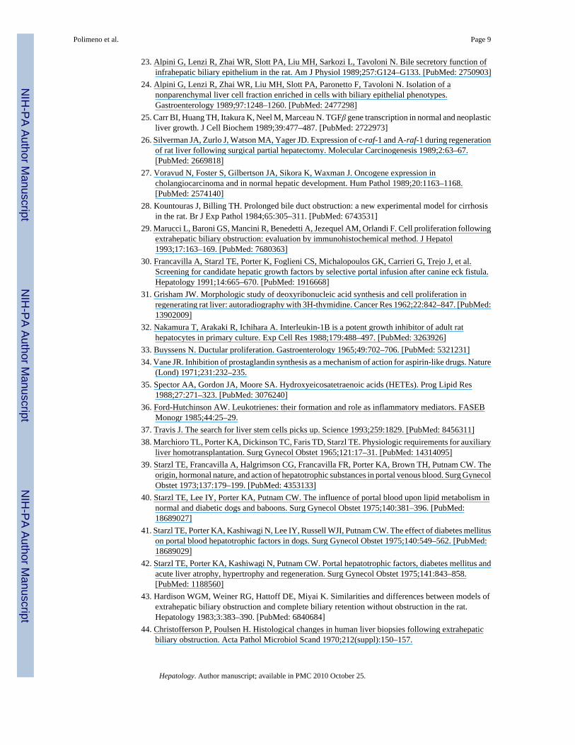
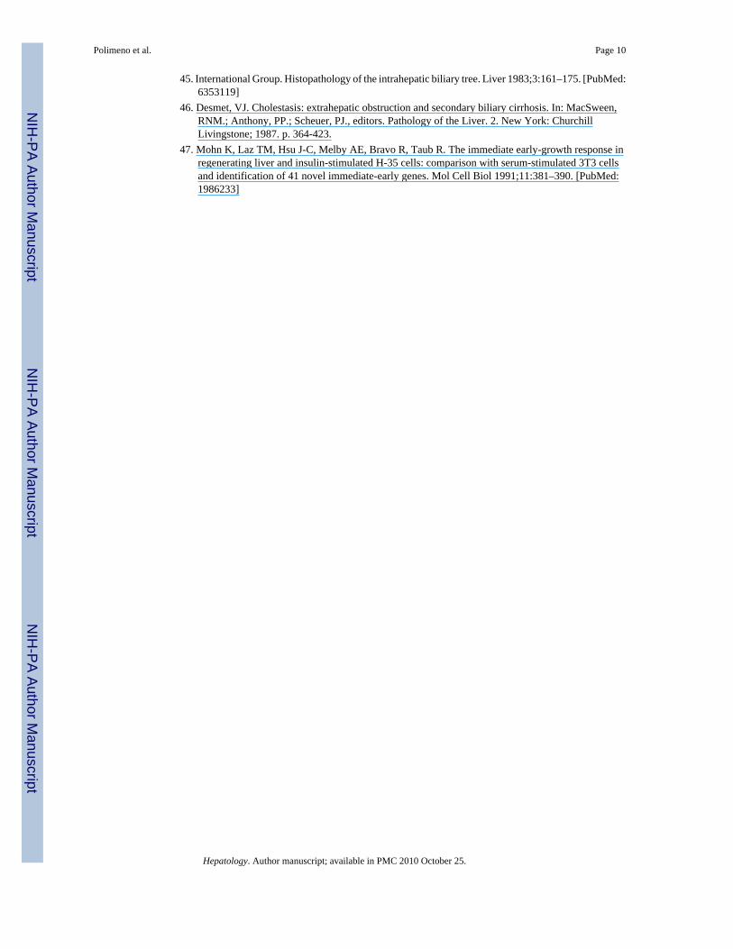
Regional Bile Duct Obstruction—The bile duct branches of the right lateral, right and leftmedial, and caudate lobes were doubly ligated and divided, leaving intact the biliary drainageof the left lobe (Fig. 1), which constitutes approximately 40% of the rat liver mass. The absenceof ductal cross-communication between the obstructed and nonobstructed lobes was provedwith methylene blue injections (Fig. 2). The animals were killed 2 days later for tissuecollections.
Polimeno et al. Page 2
Hepatology. Author manuscript; available in PMC 2010 October 25.
NIH
-PA Author Manuscript
NIH
-PA Author Manuscript
NIH
-PA Author Manuscript
Regional Bile Duct Obstruction Plus Immunosuppression—Cyclosporine and FK506 have been shown to augment regeneration after partial hepatectomy9–11 and portacavalshunt. 11–13 The mechanisms of these hepatotrophic effects are not known. However, they maybe by immune modulation of the nonparenchymal cells (NPC). Thus, exactly the sameexperiment as that just described was performed except that the rats were pretreated for 4 daysbefore operation and the first 2 days afterward with cyclosporine (10 mg/kg/day orally) or FK506 (1 mg/kg/day intramuscularly).
Partial Hepatectomy—Seventy percent partial hepatectomy (PH) was performed asdescribed by Higgins and Anderson.14 Thirty animals (three for each group) underwent 70%PH and were killed after 12, 24, 36, 48 hours, and 3, 4, 5, 6, 7, and 8 days.
Sham Operation—Twenty control rats underwent laparotomy only. Ten animals were killedat 0 hours and 10 at 48 hours after the surgery.
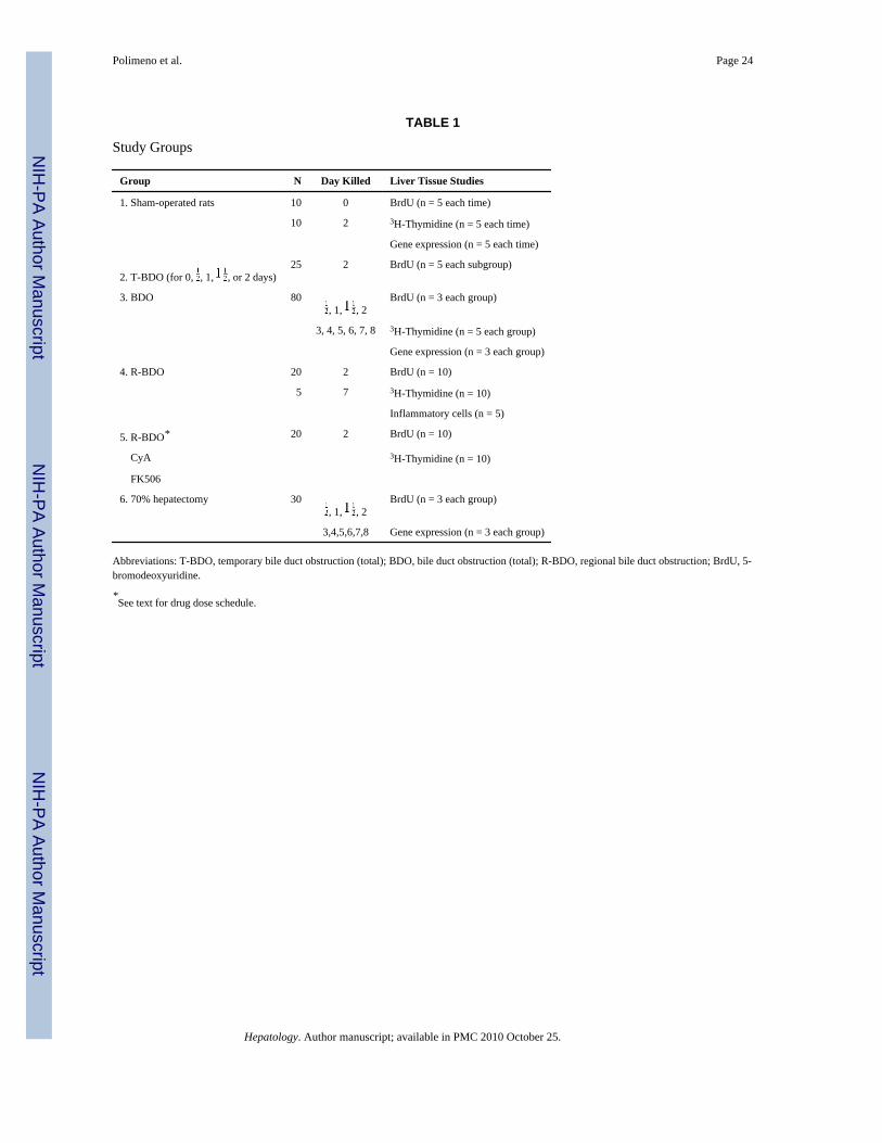
EXPERIMENTAL DESIGNThe groups are summarized in Table 1. In essence, the proliferative and gene expressionresponse to total biliary obstruction (group 3) and the duration of obstruction necessary toevoke these responses (group 2) were defined. In addition, the effect of lobar biliary obstructionon the obstructed versus the drained portion of the liver was determined in untreated (group 4)and immunosuppressed animals (group 5). The results were compared with those in rats aftersham operations (group 1 and specific group controls) and with those after 70% hepatectomy(group 6).
Experimental End PointsAll analyses were of liver fragments obtained at killing from multiple lobes. The specimensfor studies of gene expression or DNA synthesis were snap frozen and stored at −80°C.
Assessment of Cell Proliferation—The animals whose DNA synthesis was measuredchemically by thymidine incorporation were given an intraperitoneal injection of 200 μCi/kg 3H-thymidine 2 hours before killing. Frozen samples were analyzed as previously reported.15
Animals studied histopathologically for localization of the proliferating cells were injectedintraperitoneally with 120 mg/kg BrdU 2 hours before killing. Liver samples, 5 mm thick, fromthe different lobes were fixed in buffered formalin, imbedded in paraffin, sectioned at 4 μm,and stained with hematoxylin-eosin and trichrome.16 BrdU incorporation was detected with amonoclonal anti-BrdU antibody at a 1:20 dilution.17–19 The reaction product was developedby 3-amino-9-ethylcarbazole (AEC).20 Bile duct epithelial cell proliferation was determinedin 10 randomly selected portal areas in which the number of nuclear labeled and unlabeledepithelial cells was counted At least 500 cell counts were obtained from each liver lobe. Tomeasure hepatocyte proliferation, 25 parenchymal fields of 1,000 hepatocytes were counted,from which the percent of BrdU-positive cells was determined.
The number of inflammatory cells was counted in the regional bile duct obstruction experiment7 days after surgery. In addition, cytokeratin 19 expression (C-K 19; mol wt 40.000 in thecatalogue of Moll et al21) was used as a specific marker of bile duct cells.22–24
Gene Expression Determination—These studies were performed with previouslydescribed methods.25 Briefly, total cellular RNA from frozen hepatic tissue samples wasextracted by RNAzol using the manufacturer’s procedure. Total RNA (20 μg) was subjectedto electrophoresis in a 1% agarose gel and then transferred to Zitabind nylon membrane
Polimeno et al. Page 3
Hepatology. Author manuscript; available in PMC 2010 October 25.
NIH
-PA Author Manuscript
NIH
-PA Author Manuscript
NIH
-PA Author Manuscript
(Whatman Co., Maidstone, UK) overnight in 20× standard saline citrate. After transfer, theblot was fixed by ultraviolet light (short wave, 254 nm). Complementary DNA probes werelabeled with 32P with a random primed labeling kit. Prehybridization and hybridization wereperformed at 70°C with Church buffer (1% bovine serum albumin 7% sulfate sodium salt, 0.5mol/L sodium phosphate, 1 mmol/L ethylenediaminetetra-acetic acid). The membranes thenwere washed, air-dried, and autoradiographed at −70°C. To check for equivalent transfer ofRNAs from agarose gel to membrane, the gel was stained with ethidium bromide after capillarytransfer and exammed; little RNA was found to remain in any lane. The probes used were c-raf (purchased from American Type Culture Collection, Rockville, MD); transforming growthfactor-β1 (TGF-β1) (gift from R. Derynk; Genetech, San Francisco, CA); pro-thrombin (giftfrom S. J. F. Degen, university of Pittsburgh, PA); c-erb-B2 (gift from Wataru Yasui,Hiroshima university, Japan), rodent TGFα (gift from G. Lee, University of North Carolina).The TGF-alpha and β and c-raf probes were selected because they have been frequently usedto study post-hepatectomy regeneration.6,26 The c-erb-B2 probe was selected because thisgene, like c-raf, has been specifically associated with cholangiocarcinoma.27 For internalcontrols, we used 28S ribosomal RNA (Oncogene Science, Inc., Union-dale, NY), and humancyclophilin G (gift from C. T. Walsh, Harvard University, Cambridge, MA).
STATISTICAL ANALYSISStatistical analysis was performed using a two-way ANOVA (Epistat software) available onIBM (Danbury, CT) PC and assuming statistical significance only with a P < .05. Data wereexpressed as (M ± SD).
RESULTSTotal Bile Duct Obstruction
Standard Histopathology—The livers of sham-operated rats were normal. Livers withbiliary obstruction had a light infiltration of inflammatory cells in the portal areas by 2 days.By day 7, there were signs of intensive proliferation and formation of new bile ducts (Fig. 3),which were surrounded by thin fibrous connective tissues linking some portal areas, as hasbeen reported. 1–5,28.29 The hyperplastic ducts as well as the normal ducts and ductules in thesham-operated livers were positive for cytokeratin 19, whereas hepatocytes were alwaysnegative (data not shown) as reported by Alpini et al.23,24
DNA Synthesis—A crescendo of DNA synthesis began at 24 hours, was maximal at 2 days,and receded almost to baseline by day 6 (Fig. 4).
BrdU Incorporation—The increased DNA synthesis could be seen in the nuclear labeledBrdU-positive cells to be mainly a reflection of duct cell proliferation rather than of hepatocytes(Fig. 5). In the animals of group 3, 17% of bile duct cells were labeled at the peak of the responseat 2 days compared with 1% at the outset. A much smaller response, reaching 4.8% at 2 days,was seen in the hepatocytes of these livers (Fig. 6, top panels).
These results were strikingly different than those following 70% PH (Fig. 6, lower panels).With PH, a bile duct response also promptly occurred, but it was short-lived compared withbile duct obstruction (BDO). In addition, the percentage of BrdU-positive hepatocytes wasmore than 5 times greater than after BDO (Fig. 6).
Required Time of Obstruction—In the animals of group 2, biliary obstruction for 12 hoursor even 24 hours did not cause significant increases in BrdU-positive cells in the livers obtainedat killing at 2 days. However, with obstruction for 36 hours that was then relieved or with
Polimeno et al. Page 4
Hepatology. Author manuscript; available in PMC 2010 October 25.
NIH
-PA Author Manuscript
NIH
-PA Author Manuscript
NIH
-PA Author Manuscript
continuous obstruction throughout the 48-hour duration of the experiment, the duct cellproliferation (>15% of total) was similar to that found in the animals of group 3 (Fig. 7).
Gene Expression—Hepatic gene expression was different after BDO than after 70% PH,and the differences had a physiologic correlation. The features that appeared to be most typicalof BDO during the first 48 hours were lack of expression of TGFα, weak and delayedprothrombin gene expression, but with prompt appearance of c-erb-B2 messenger RNA. Incontrast, the typical response to PH was strong expression of TGFα and prothrombin by 12hours but without the appearance of c-erb-B2 messenger RNA.
Essentially no increase of TGFα, a potent hepatocyte mitogen, was detectable during the first48 hours after BDO when DNA synthesis was maximum, whereas TGFα was markedlyincreased after PH (Fig. 8). Elevated levels of c-erb-B2, an oncogene associated withcholangiocarcinoma,27 were clearly expressed from 48 to 96 hours after BDO, but appearedonly at 72 hours after PH (Fig. 9). c-raf, a proto-oncogene associated with hepatocytesproliferation after PH,26 and also with cholangiocarcinoma,27 increased significantly 48 to 96hours after BDO, whereas it was slightly detectable after PH (Fig. 9). Interestingly, theexpression of the hepatocyte-specific prothrombin gene was enhanced during the increasedDNA synthesis and mitoses after PH, but not after BDO (Fig. 10). TGF-β1 messenger RNAexpression was similar both after PH and BDO (Fig. 11) and was similar to that previouslyreported for PH.6
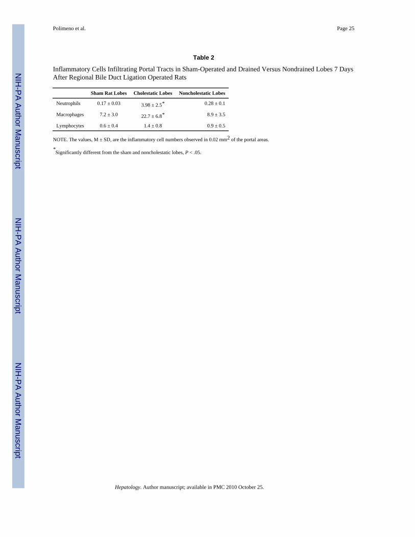
Regional Bile Duct ObstructionStandard Histopathology—The obstructed liver lobes in the rats of group 4 at 7 days hadearly changes similar to those in the totally obstructed livers of group 3, including theappearance of inflammatory cells (Table 2). The freely draining left lobes had a normal numberof inflammatory cells, but there was striking proliferation and formation of new ducts.
DNA Synthesis—At 48 hours, which was the time of the maximal proliferative response inthe total bilary obstruction experiments previously described, DNA synthesis was significantlyand almost equally elevated, in the obstructed as opposed to the freely draining lobes (Fig. 12).The magnitude of DNA response in both locations was less than half that caused by total biliaryobstruction.
BrdU Incorporation—As in the total biliary obstruction experiments of group 3, thepredominant proliferative response was that of the duct cells. This was true both in theobstructed and freely draining lobes (Fig. 13). Interestingly, increased hepatocyte proliferationoccurred only in the obstructed lobes. In the drained lobes, hepatocyte proliferation wasnegligible although significantly more than in the sham-operated controls (Fig. 13).
Addition of Immunosuppression—Preoperative and postoperative treatment withcyclosporine and FK 506 did not significantly affect the results (data not shown).
DISCUSSIONThe literature on an increasing number of hepatic growth factors has accumulated rapidly inthe last two decades.6–8,30 However, this information has not been synthesized into a generallyaccepted explanation of hepatic regeneration. Because hepatocytes, biliary duct cells and NPCshave long been known to have different kinetic orders of proliferation,31 a reasonableimplication is that they are variably cross-regulated. NPCs have been particularly attractivecandidates for such a role.32–36
Polimeno et al. Page 5
Hepatology. Author manuscript; available in PMC 2010 October 25.
NIH
-PA Author Manuscript
NIH
-PA Author Manuscript
NIH
-PA Author Manuscript
The biliary obstruction models have lent themselves well to proliferation studies of the ductcell component of the liver. These cells constitute approximately 30% of the total liver mass.Previous investigations have defined the morphologic consequences of duct obstruction, whichinclude pronounced ductular hyperplasia, a modest hepatocyte proliferative response, andinfiltration of inflammatory cells.1–5,23,24 These findings were confirmed in rat livers aftercommon duct ligation and in liver lobes that were selectively obstructed. The duct cell andhepatocyte proliferative responses were not simultaneous but they were closely relatedtemporally. These were compared with the changes after 70% partial hepatectomy.
With PH, the duct cells and hepatocytes both participated vigorously in the regeneration, thepeak hepatocyte response being at 24 hours, with the duct cells only slightly behind. The globalproliferative response to BDO or regional BDO as defined by DNA synthesis was similar toPH except that its peak was at 2 days instead of 1, with a slower return to baseline. However,the differences between the PH and BDO models were more than temporal. Cell labelingstudies demonstrated that most of the heightened DNA synthesis after biliary obstruction wasoccurring in the duct cells, with only a minor contribution from the hepatocytes. The requiredstimulus in the BDO model was continuation of the obstruction for more than a day. Blockagefor 24 hours or less was without significant effect at the time of killing at 2 days.
Hypotheses have been proposed to explain the hyperplastic duct cell response to biliaryobstruction in humans and animals. Excluding the possibility that there are pluripotent (oval)stem cells,37 these fall into two general categories. In one of these generic alternatives, Slottet al4 suggested that proliferating hepatocytes were able to invade the basal membrane of theobstructed duct, taking up residence in the epithelium, where they differentiated to thephenotypic characteristics of biliary duct cells. Our experiments provided no support for thisconcept. Instead, the primary proliferative response of the totally obstructed liver was of theduct cells, the hepatocytes being relatively minor participants. The observations in the regionalbiliary obstruction experiments were considerably more damaging to the ectopic hepatocytehypothesis. Here, the duct cell proliferation in the ostensibly healthy freely draining lobes wasof the same magnitude as in the lobes with ligated ducts. In both the draining and cholestaticlobes, the muted hepatocyte response (which was slightly greater in the obstructed lobes)presumably was in response to partial obstruction to the injury caused unilaterally by the ductligation. This response to partial obstruction was only about half of that caused when the entirebiliary system was occluded, as would be expected with the proportionately less completedamage to the parenchyma.
In an alternative general hypothesis explaining the ductular hyperplasia after biliaryobstruction, Buyssens33 has proposed a contributory or regulatory role of NPC by-products ofthe lipooxygenase pathway,34 which are chemotactic for and activate polymorphonuclearleukocytes30,31 as well as multiple cytokines. A histopathologic basis for such a mechanismwas evident in that there was a striking leukocyte inflammatory reaction that appeared toinclude multiple NPC lineages in the obstructed liver tissue, whether this obstruction was ofthe whole organ or only part of it. However, in the partial obstruction experiments the vigorousduct cell proliferation in the freely draining lobes that did not have a significant inflammatoryreaction showed that the local presence of NPCs was not a direct paracrine requirement forstimulation of duct cell hyperplasia, while not ruling out a humorally transmitted effect ofcytokines from leukocytes on the contralateral side. The role of these immunologically capableNPCs in the events after duct ligation remains to be determined. Although the T-cell–directeddrugs, cyclosporine and FK 506, both augment the proliferation associated withhepatectomy9–11 and Eck’s fistula,11-13 these immunosuppressive agents did not affect theoutcome of the regeneration after regional biliary obstruction. If a stem cell actually exists,37
it could be accommodated by this paradigm.
Polimeno et al. Page 6
Hepatology. Author manuscript; available in PMC 2010 October 25.
NIH
-PA Author Manuscript
NIH
-PA Author Manuscript
NIH
-PA Author Manuscript
Whether or not an immune component was involved, the regional biliary obstructionexperiments showed the intelligence with which one part of the liver communicates withanother. This has been noted before in auxiliary liver transplant models38 and in “double liver”preparations in which one liver fragment is provided a physiologic advantage such assplanchnic venous blood that is rich in hepatotrophic factors.39 Under such circumstances,there is increased proliferation by the “disadvantaged” liver or fragment, but this is minor inthe long run compared with the compensatory hyperplasia and hypertrophy in the advantagedhepatic tissue.40–42 The coregulation between the coexisting livers or liver fragments resultsin the eventual atrophy of the damaged liver fragment, regeneration of the healthy one, andmaintenance of the original total hepatic mass. Although slower in evolution than with regionalportal blood deprivation, this also is the well-known outcome weeks or months after lobar ductligation in animals28,43 and humans.44–46
The specificity of the cross-talk between liver fragments was evident in the regional ductligation experiments. Here, the acute primary insult to the duct cells in one part of the liverwas promptly sensed and responded to by duct cell hyperplasia in the affected as well as theunaffected liver. This consequence could reflect a unique humorally transmitted signal from astimulus such as but not limited to increased intraductal hydrostatic pressure,3–5 in whichinflammatory cells could play an intermediary role. Alternatively, the possibility cannot beruled out that there are separate and highly specific growth factors other than cytokinesgoverning duct cell regeneration.
The dissociation of messenger RNA expression of the four growth factors studied was strikingand correlated with the physiologic end points. The complete absence of TGFα expression wasstriking in animals whose global DNA response to BDO was essentially equivalent to that ofanimals with 70% PH, but with a different temporal and cell target profile. The early expressionof c-erb-B2 with BDO only was of particular interest because of the association of thisoncogene with obstructing cholangiocarcinoma.27 It will be worthwhile to do systematicstudies of this oncogene in biopsy specimens of other duct-specific diseases. In addition, it willbe important to determine the more complete range of gene activation caused by BDO, whichmay prove to be multiple, as has already been shown after PH.47
AcknowledgmentsSupported by research grants from the Department of Veterans Affairs. Washington, DC (Project grant DK 299611and from the National Institute of Health. Bethesda, MD (grant CA 44602) and by Consiglio Nazionale delle Ricerche,ACRO program (grant no. 94.01132.PF29), Rome, Italv.
Abbreviations
BrdU 5 bromo·2′·deoxyuridine
NPC nonparencymal cells
PH partial hepatectomy
TGF transfonning growth factor
BDO bile duct obstruction
References1. Tramas EG, Symeonidis A. Morphologic and functional changes in the livers of rats after ligation or
excision of the common bile duct. Am J Pathol 1957;33:13–27. [PubMed: 13394688]
Polimeno et al. Page 7
Hepatology. Author manuscript; available in PMC 2010 October 25.
NIH
-PA Author Manuscript
NIH
-PA Author Manuscript
NIH
-PA Author Manuscript
2. Accatino L, Contreras A, Fernandez S, Quintana C. The effect of complete biliary obstruction on bileflow and bile acid excretion: postcholestatic choleresis in the rat. J Lab Clin Med 1979;93:706–717.[PubMed: 429869]
3. Alpini G, Lenzi R, Sarkozi L, Tavoloni N. Biliary physiology in rats with bile ductular cell hyperplasia:evidence for a secretory function of proliferated bile ductules. J Clin Invest 1988;81:569–578.[PubMed: 2448343]
4. Slott PA, Liu MH, Tavoloni N. Origin, pattern and mechanism of bile duct proliferation followingbiliary obstruction in the rat. Gastroenterology 1990;99:466–477. [PubMed: 1694804]
5. Shibayama Y. Factors producing bile infarction and bile duct proliferation in biliary obstruction. JPathol 1990;160:57–62. [PubMed: 2313481]
6. Fausto, N. Liver regeneration. In: Zakim, D.; Boyer, TD., editors. Hepatology: a textbook of liverdisease. 2. Philadelphia: Saunders; 1990. p. 49-65.
7. Michalopoulos GK. Liver regeneration: molecular mechanism of growth control. FASEB J1990;4:176–87. [PubMed: 2404819]
8. Francavilla, A.; Starzl, TE.; Van Thiel, DH.; Barone, M.; Polimeno, L. Hepatic regeneration. In:Lebouton, AV., editor. Molecular and cell biology of the liver. Boca Raton, FL: CRC Press; 1993. p.309-346.
9. Makowka L, Scanas G, Esquivel C, Venkataramanan R, Todo S, Iwatsuki S, Van Thiel DH, et al. Theeffect of cyclosporine on hepatic regeneration. Surg Forum 1986;37:352.
10. Francavilla A, Barone M, Todo S, Zeng DH, Porter KA, Starzl TE. Augmentation of rat liverregeneration by FK 506 compared with cyclosporine. Lancet 1989;2:1248–1249. [PubMed:2479802]
11. Francavilla A, Starzl TE, Carr B, Azzarone A, Carrieri G, Zeng QH, Porter KA. The effects of FK506, cyclosporine and rapamycin on liver growth in vitro and in vivo. Transplant Proc 1991;23:2817–2820. [PubMed: 1721287]
12. Mazzaferro V, Porter KA, Scotti-Foglieni CL, Venkataramanan R, Makowka L, Rossaro L,Francavilla A, et al. The hepatotrophic influence of cyclosporine. Surgery 1990;107:533–539.[PubMed: 2185568]
13. Starzl TE, Porter KA, Mazzaferro V, Todo S, Fung J, Francavilla A. Hepatotrophic effects of FK506in dogs. Transplantation 1991;51:67–70. [PubMed: 1702912]
14. Higgins GM, Anderson RM. Experimental pathology of the liver. I. Restoration of the liver of thewhite rat following partial surgical removal. Arch Pathol 1931;12:186–202.
15. Francavilla A, Eagon PK, Di Leo A, Polimeno L, Panella C, Aquilino AM, Ingrosso M, et al. Sexhormone related functions in regenerating male rat liver. Gastroenterology 1986;91:1263–1270.[PubMed: 3758617]
16. Brandbury, P.; Gordon, KC. Connective tissue and stain. In: Bancroft, JD.; Stevens, A., editors.Theory and Practice of Histological Techniques. New York: Churchill Livingstone; 1990. p. 119-142.
17. Morstyn AG, Hsu SM, Kinsella T, Russo A, Gratzner H, Mitchell J. Bromodeoxyuridine in tumorsand chromosomes detected with a monoclonal antibody. J Clin Invest 1983;73:1844–1850. [PubMed:6355188]
18. Wilson GD. Assessment of human tumor proliferation using bromodeoxyuridine-current status. ActaOncol 1991;30:903–910. [PubMed: 1777243]
19. Sasaki A, Naganuma H, Kimura R, Isoe S, Nakano S, Nukui H, Suzuki K, et al. Proliferating cellnuclear antigen (PCNA) immunostaining as an alternative to bromodeoxyuridine (BrdU)immunostaining for brain tumours in paraffin embedded sections. Acta Neurochir 1992;117:178–181.
20. Graham RC, Lindholm V, Karnovsky MJ. Cytochemical demonstration of peroxidase activity with3-amino-9-ethylcarbazole. J Histochem Cytochem 1965;13:150–156. [PubMed: 14293042]
21. Moll R, Franke WW, Schiller DL. The catalog of human cytokeratins: patterns of expression in normalepithelia, tumors, and cultured cells. Cell 1982;31:11–24. [PubMed: 6186379]
22. Van Eyken P, Sciot R, Von Damme B, DeWolf-Peeters C, Desmet VJ. Keratin immunohistochemistryin normal human liver: cytokeratin pattern of hepatocytes, bile ducts and acinar gradient. VirchowsArch [A] 1987;412:63–72.
Polimeno et al. Page 8
Hepatology. Author manuscript; available in PMC 2010 October 25.
NIH
-PA Author Manuscript
NIH
-PA Author Manuscript
NIH
-PA Author Manuscript
23. Alpini G, Lenzi R, Zhai WR, Slott PA, Liu MH, Sarkozi L, Tavoloni N. Bile secretory function ofinfrahepatic biliary epithelium in the rat. Am J Physiol 1989;257:G124–G133. [PubMed: 2750903]
24. Alpini G, Lenzi R, Zhai WR, Liu MH, Slott PA, Paronetto F, Tavoloni N. Isolation of anonparenchymal liver cell fraction enriched in cells with biliary epithelial phenotypes.Gastroenterology 1989;97:1248–1260. [PubMed: 2477298]
25. Carr BI, Huang TH, Itakura K, Neel M, Marceau N. TGFβ gene transcription in normal and neoplasticliver growth. J Cell Biochem 1989;39:477–487. [PubMed: 2722973]
26. Silverman JA, Zurlo J, Watson MA, Yager JD. Expression of c-raf-1 and A-raf-1 during regenerationof rat liver following surgical partial hepatectomy. Molecular Carcinogenesis 1989;2:63–67.[PubMed: 2669818]
27. Voravud N, Foster S, Gilbertson JA, Sikora K, Waxman J. Oncogene expression incholangiocarcinoma and in normal hepatic development. Hum Pathol 1989;20:1163–1168.[PubMed: 2574140]
28. Kountouras J, Billing TH. Prolonged bile duct obstruction: a new experimental model for cirrhosisin the rat. Br J Exp Pathol 1984;65:305–311. [PubMed: 6743531]
29. Marucci L, Baroni GS, Mancini R, Benedetti A, Jezequel AM, Orlandi F. Cell proliferation followingextrahepatic biliary obstruction: evaluation by immunohistochemical method. J Hepatol1993;17:163–169. [PubMed: 7680363]
30. Francavilla A, Starzl TE, Porter K, Foglieni CS, Michalopoulos GK, Carrieri G, Trejo J, et al.Screening for candidate hepatic growth factors by selective portal infusion after canine eck fistula.Hepatology 1991;14:665–670. [PubMed: 1916668]
31. Grisham JW. Morphologic study of deoxyribonucleic acid synthesis and cell proliferation inregenerating rat liver: autoradiography with 3H-thymidine. Cancer Res 1962;22:842–847. [PubMed:13902009]
32. Nakamura T, Arakaki R, Ichihara A. Interleukin-1B is a potent growth inhibitor of adult rathepatocytes in primary culture. Exp Cell Res 1988;179:488–497. [PubMed: 3263926]
33. Buyssens N. Ductular proliferation. Gastroenterology 1965;49:702–706. [PubMed: 5321231]34. Vane JR. Inhibition of prostaglandin synthesis as a mechanism of action for aspirin-like drugs. Nature
(Lond) 1971;231:232–235.35. Spector AA, Gordon JA, Moore SA. Hydroxyeicosatetraenoic acids (HETEs). Prog Lipid Res
1988;27:271–323. [PubMed: 3076240]36. Ford-Hutchinson AW. Leukotrienes: their formation and role as inflammatory mediators. FASEB
Monogr 1985;44:25–29.37. Travis J. The search for liver stem cells picks up. Science 1993;259:1829. [PubMed: 8456311]38. Marchioro TL, Porter KA, Dickinson TC, Faris TD, Starzl TE. Physiologic requirements for auxiliary
liver homotransplantation. Surg Gynecol Obstet 1965;121:17–31. [PubMed: 14314095]39. Starzl TE, Francavilla A, Halgrimson CG, Francavilla FR, Porter KA, Brown TH, Putnam CW. The
origin, hormonal nature, and action of hepatotrophic substances in portal venous blood. Surg GynecolObstet 1973;137:179–199. [PubMed: 4353133]
40. Starzl TE, Lee IY, Porter KA, Putnam CW. The influence of portal blood upon lipid metabolism innormal and diabetic dogs and baboons. Surg Gynecol Obstet 1975;140:381–396. [PubMed:18689027]
41. Starzl TE, Porter KA, Kashiwagi N, Lee IY, Russell WJI, Putnam CW. The effect of diabetes mellituson portal blood hepatotrophic factors in dogs. Surg Gynecol Obstet 1975;140:549–562. [PubMed:18689029]
42. Starzl TE, Porter KA, Kashiwagi N, Putnam CW. Portal hepatotrophic factors, diabetes mellitus andacute liver atrophy, hypertrophy and regeneration. Surg Gynecol Obstet 1975;141:843–858.[PubMed: 1188560]
43. Hardison WGM, Weiner RG, Hattoff DE, Miyai K. Similarities and differences between models ofextrahepatic biliary obstruction and complete biliary retention without obstruction in the rat.Hepatology 1983;3:383–390. [PubMed: 6840684]
44. Christofferson P, Poulsen H. Histological changes in human liver biopsies following extrahepaticbiliary obstruction. Acta Pathol Microbiol Scand 1970;212(suppl):150–157.
Polimeno et al. Page 9
Hepatology. Author manuscript; available in PMC 2010 October 25.
NIH
-PA Author Manuscript
NIH
-PA Author Manuscript
NIH
-PA Author Manuscript
45. International Group. Histopathology of the intrahepatic biliary tree. Liver 1983;3:161–175. [PubMed:6353119]
46. Desmet, VJ. Cholestasis: extrahepatic obstruction and secondary biliary cirrhosis. In: MacSween,RNM.; Anthony, PP.; Scheuer, PJ., editors. Pathology of the Liver. 2. New York: ChurchillLivingstone; 1987. p. 364-423.
47. Mohn K, Laz TM, Hsu J-C, Melby AE, Bravo R, Taub R. The immediate early-growth response inregenerating liver and insulin-stimulated H-35 cells: comparison with serum-stimulated 3T3 cellsand identification of 41 novel immediate-early genes. Mol Cell Biol 1991;11:381–390. [PubMed:1986233]
Polimeno et al. Page 10
Hepatology. Author manuscript; available in PMC 2010 October 25.
NIH
-PA Author Manuscript
NIH
-PA Author Manuscript
NIH
-PA Author Manuscript
Fig. 1.Regional biliary duct obstruction procedure that permits the study of obstructed and non-obstructed liver in the rat. The drained non-cholestatic portion is approximately 40% of theliver mass.
Polimeno et al. Page 11
Hepatology. Author manuscript; available in PMC 2010 October 25.
NIH
-PA Author Manuscript
NIH
-PA Author Manuscript
NIH
-PA Author Manuscript
Fig. 2.Evidence of lack of ductal cross-communication between obstructed and non-obstructed lobesproved with methylene-blue injection into the common bile duct. Right lobe obstructed andleft lobe nonobstructed.
Polimeno et al. Page 12
Hepatology. Author manuscript; available in PMC 2010 October 25.
NIH
-PA Author Manuscript
NIH
-PA Author Manuscript
NIH
-PA Author Manuscript
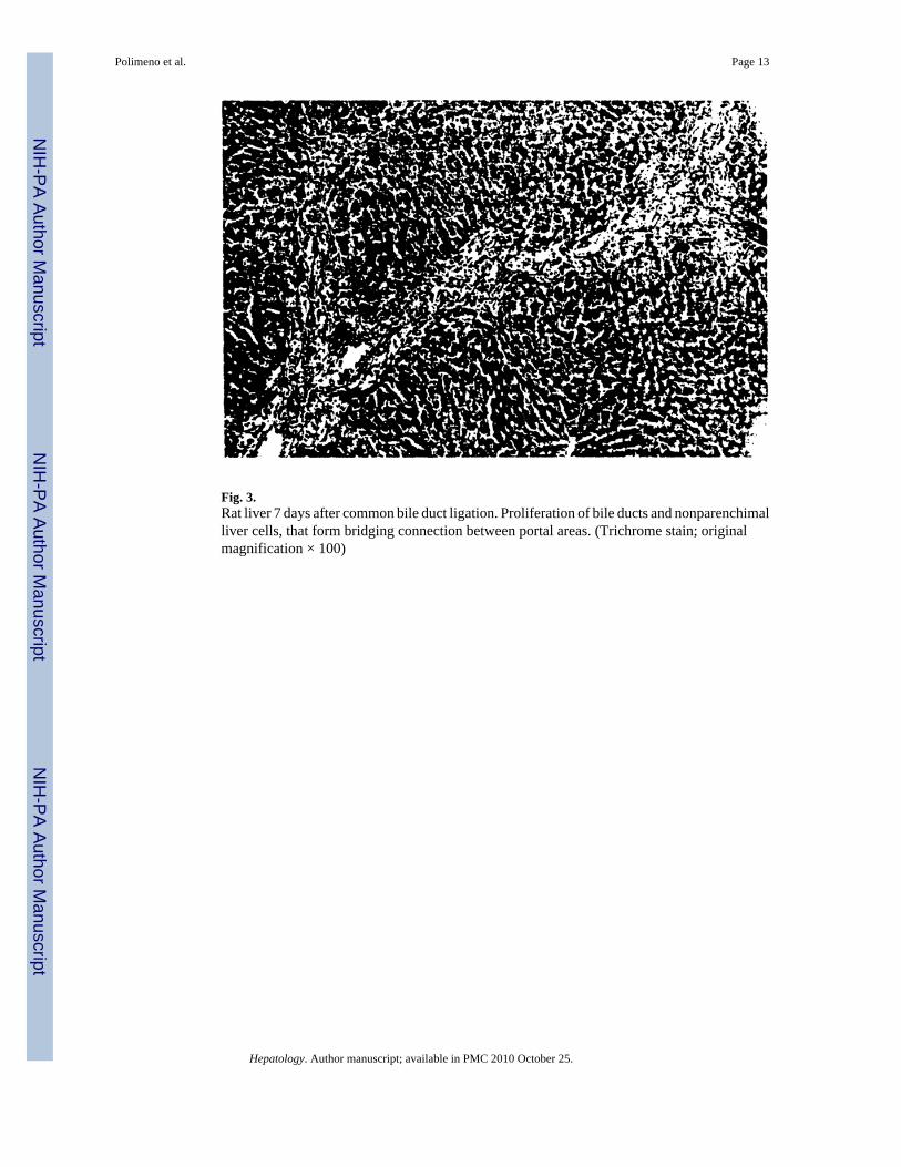
Fig. 3.Rat liver 7 days after common bile duct ligation. Proliferation of bile ducts and nonparenchimalliver cells, that form bridging connection between portal areas. (Trichrome stain; originalmagnification × 100)
Polimeno et al. Page 13
Hepatology. Author manuscript; available in PMC 2010 October 25.
NIH
-PA Author Manuscript
NIH
-PA Author Manuscript
NIH
-PA Author Manuscript
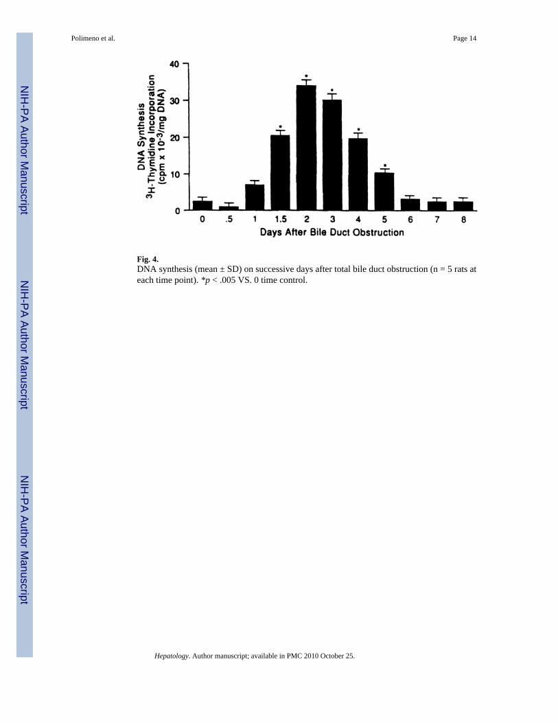
Fig. 4.DNA synthesis (mean ± SD) on successive days after total bile duct obstruction (n = 5 rats ateach time point). *p < .005 VS. 0 time control.
Polimeno et al. Page 14
Hepatology. Author manuscript; available in PMC 2010 October 25.
NIH
-PA Author Manuscript
NIH
-PA Author Manuscript
NIH
-PA Author Manuscript
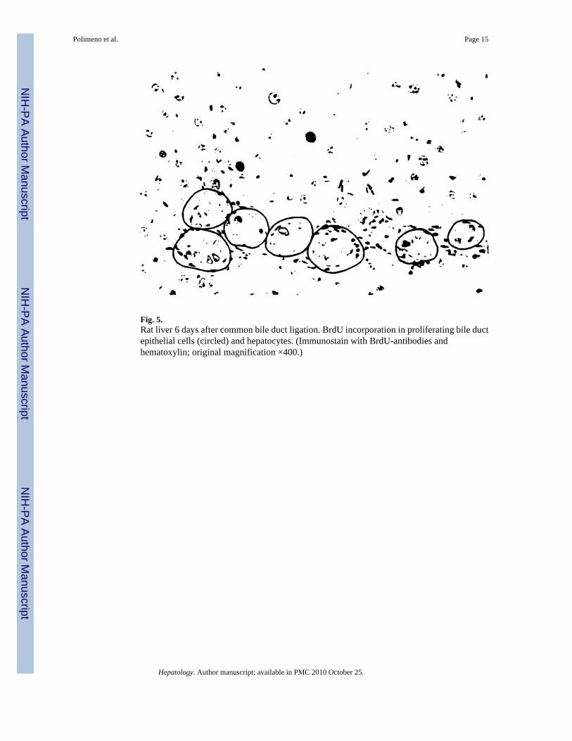
Fig. 5.Rat liver 6 days after common bile duct ligation. BrdU incorporation in proliferating bile ductepithelial cells (circled) and hepatocytes. (Immunostain with BrdU-antibodies andhematoxylin; original magnification ×400.)
Polimeno et al. Page 15
Hepatology. Author manuscript; available in PMC 2010 October 25.
NIH
-PA Author Manuscript
NIH
-PA Author Manuscript
NIH
-PA Author Manuscript
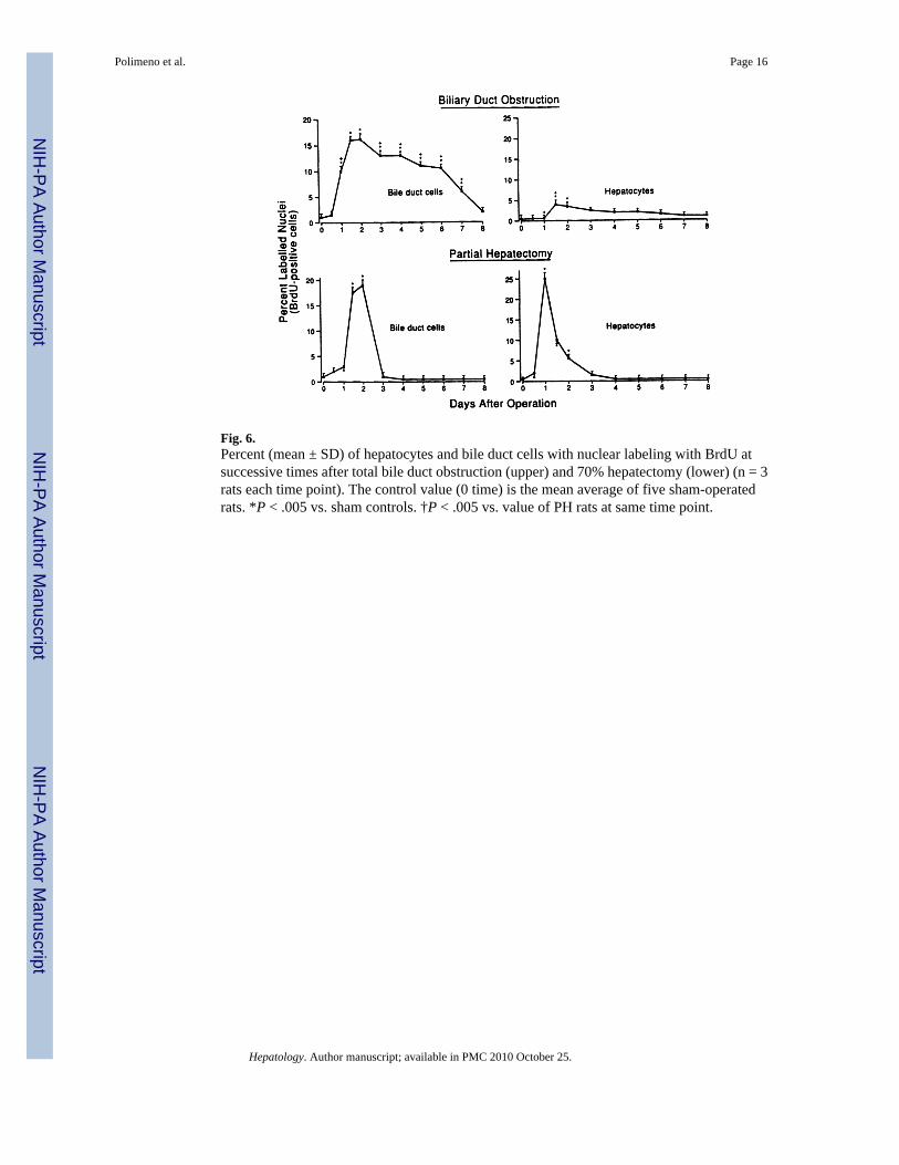
Fig. 6.Percent (mean ± SD) of hepatocytes and bile duct cells with nuclear labeling with BrdU atsuccessive times after total bile duct obstruction (upper) and 70% hepatectomy (lower) (n = 3rats each time point). The control value (0 time) is the mean average of five sham-operatedrats. *P < .005 vs. sham controls. †P < .005 vs. value of PH rats at same time point.
Polimeno et al. Page 16
Hepatology. Author manuscript; available in PMC 2010 October 25.
NIH
-PA Author Manuscript
NIH
-PA Author Manuscript
NIH
-PA Author Manuscript
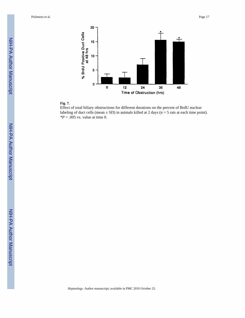
Fig. 7.Effect of total biliary obstructions for different durations on the percent of BrdU nuclearlabeling of duct cells (mean ± SD) in animals killed at 2 days (n = 5 rats at each time point).*P < .005 vs. value at time 0.
Polimeno et al. Page 17
Hepatology. Author manuscript; available in PMC 2010 October 25.
NIH
-PA Author Manuscript
NIH
-PA Author Manuscript
NIH
-PA Author Manuscript
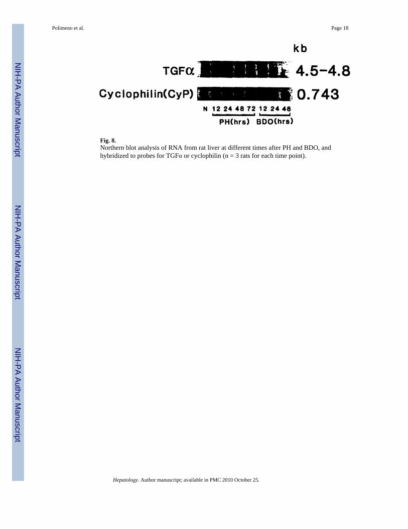
Fig. 8.Northern blot analysis of RNA from rat liver at different times after PH and BDO, andhybridized to probes for TGFα or cyclophilin (n = 3 rats for each time point).
Polimeno et al. Page 18
Hepatology. Author manuscript; available in PMC 2010 October 25.
NIH
-PA Author Manuscript
NIH
-PA Author Manuscript
NIH
-PA Author Manuscript
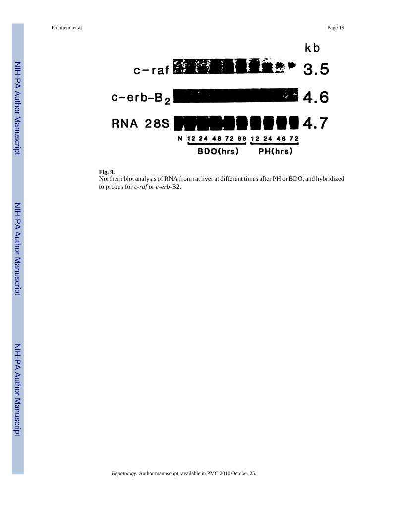
Fig. 9.Northern blot analysis of RNA from rat liver at different times after PH or BDO, and hybridizedto probes for c-raf or c-erb-B2.
Polimeno et al. Page 19
Hepatology. Author manuscript; available in PMC 2010 October 25.
NIH
-PA Author Manuscript
NIH
-PA Author Manuscript
NIH
-PA Author Manuscript
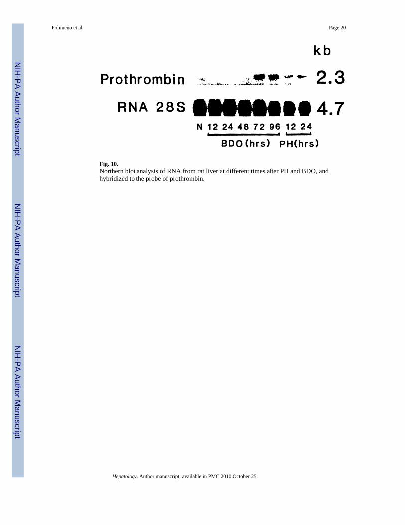
Fig. 10.Northern blot analysis of RNA from rat liver at different times after PH and BDO, andhybridized to the probe of prothrombin.
Polimeno et al. Page 20
Hepatology. Author manuscript; available in PMC 2010 October 25.
NIH
-PA Author Manuscript
NIH
-PA Author Manuscript
NIH
-PA Author Manuscript
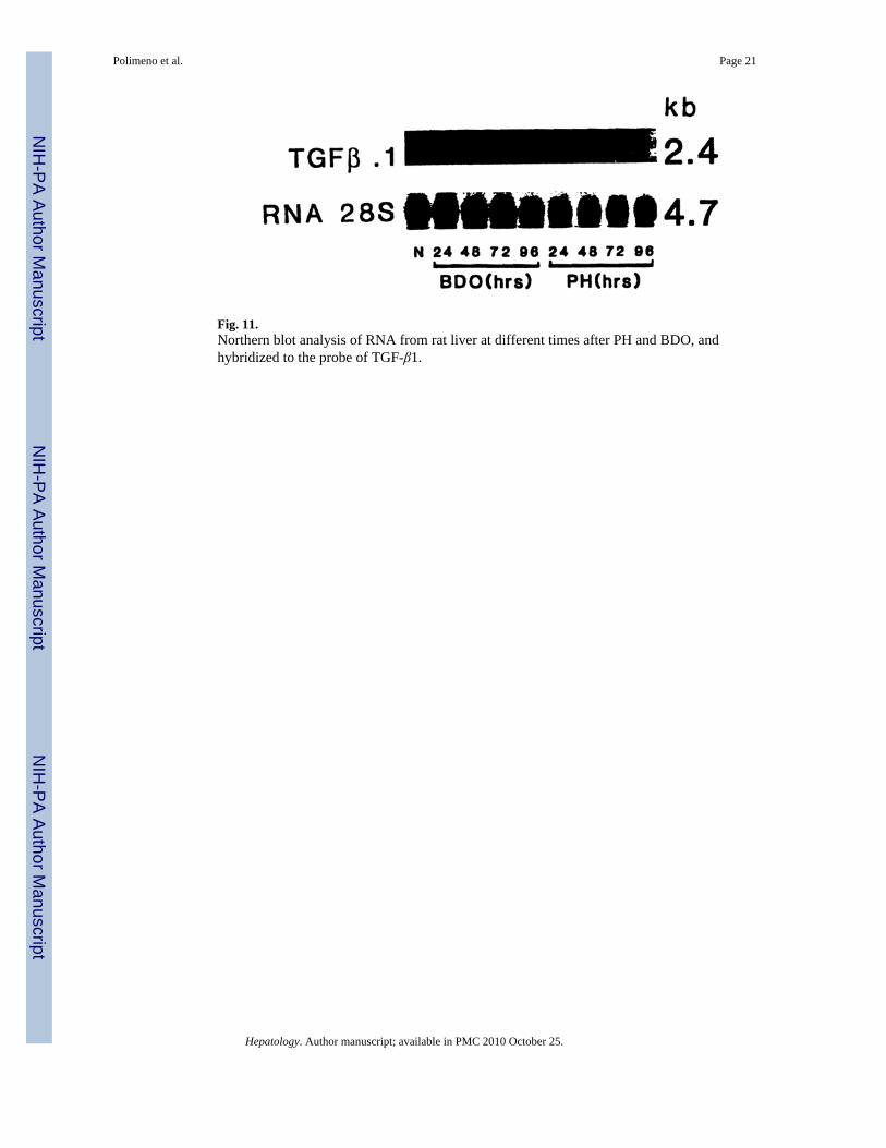
Fig. 11.Northern blot analysis of RNA from rat liver at different times after PH and BDO, andhybridized to the probe of TGF-β1.
Polimeno et al. Page 21
Hepatology. Author manuscript; available in PMC 2010 October 25.
NIH
-PA Author Manuscript
NIH
-PA Author Manuscript
NIH
-PA Author Manuscript
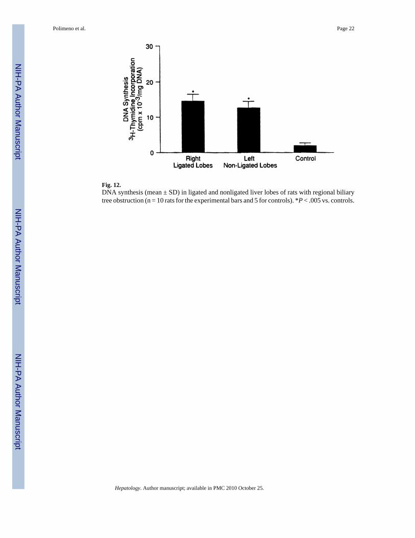
Fig. 12.DNA synthesis (mean ± SD) in ligated and nonligated liver lobes of rats with regional biliarytree obstruction (n = 10 rats for the experimental bars and 5 for controls). *P < .005 vs. controls.
Polimeno et al. Page 22
Hepatology. Author manuscript; available in PMC 2010 October 25.
NIH
-PA Author Manuscript
NIH
-PA Author Manuscript
NIH
-PA Author Manuscript
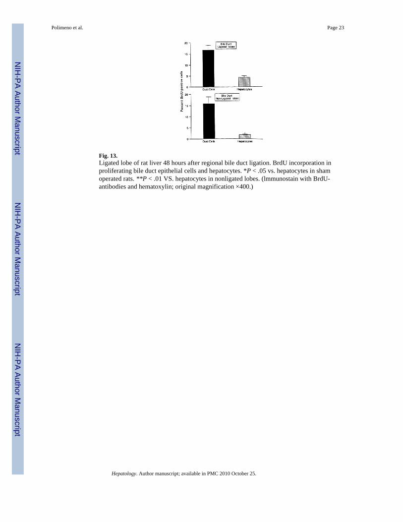
Fig. 13.Ligated lobe of rat liver 48 hours after regional bile duct ligation. BrdU incorporation inproliferating bile duct epithelial cells and hepatocytes. *P < .05 vs. hepatocytes in shamoperated rats. **P < .01 VS. hepatocytes in nonligated lobes. (lmmunostain with BrdU-antibodies and hematoxylin; original magnification ×400.)
Polimeno et al. Page 23
Hepatology. Author manuscript; available in PMC 2010 October 25.
NIH
-PA Author Manuscript
NIH
-PA Author Manuscript
NIH
-PA Author Manuscript
NIH
-PA Author Manuscript
NIH
-PA Author Manuscript
NIH
-PA Author Manuscript
Polimeno et al. Page 24
TABLE 1
Study Groups
Group N Day Killed Liver Tissue Studies
1. Sham-operated rats 10 0 BrdU (n = 5 each time)
10 2 3H-Thymidine (n = 5 each time)
Gene expression (n = 5 each time)
2. T-BDO (for 0, , 1, , or 2 days)25 2 BrdU (n = 5 each subgroup)
3. BDO 80, 1, , 2
BrdU (n = 3 each group)
3, 4, 5, 6, 7, 8 3H-Thymidine (n = 5 each group)
Gene expression (n = 3 each group)
4. R-BDO 20 2 BrdU (n = 10)
5 7 3H-Thymidine (n = 10)
Inflammatory cells (n = 5)
5. R-BDO* 20 2 BrdU (n = 10)
CyA 3H-Thymidine (n = 10)
FK506
6. 70% hepatectomy 30, 1, , 2
BrdU (n = 3 each group)
3,4,5,6,7,8 Gene expression (n = 3 each group)
Abbreviations: T-BDO, temporary bile duct obstruction (total); BDO, bile duct obstruction (total); R-BDO, regional bile duct obstruction; BrdU, 5-bromodeoxyuridine.
*See text for drug dose schedule.
Hepatology. Author manuscript; available in PMC 2010 October 25.
NIH
-PA Author Manuscript
NIH
-PA Author Manuscript
NIH
-PA Author Manuscript
Polimeno et al. Page 25
Table 2
Inflammatory Cells Infiltrating Portal Tracts in Sham-Operated and Drained Versus Nondrained Lobes 7 DaysAfter Regional Bile Duct Ligation Operated Rats
Sham Rat Lobes Cholestatic Lobes Noncholestatic Lobes
Neutrophils 0.17 ± 0.03 3.98 ± 2.5* 0.28 ± 0.1
Macrophages 7.2 ± 3.0 22.7 ± 6.8* 8.9 ± 3.5
Lymphocytes 0.6 ± 0.4 1.4 ± 0.8 0.9 ± 0.5
NOTE. The values, M ± SD, are the inflammatory cell numbers observed in 0.02 mm2 of the portal areas.
*Significantly different from the sham and noncholestatic lobes, P < .05.
Hepatology. Author manuscript; available in PMC 2010 October 25.
Copyright © 2022 FDOKUMEN