CD73 Is a Major Regulator of Adenosinergic Signalling in Mouse Brain
12
CD73 Is a Major Regulator of Adenosinergic Signalling in Mouse Brain Natalia Kulesskaya 1,2 , Vootele Vo ˜ ikar 1,2 *, Marjaana Peltola 1 , Gennady G. Yegutkin 3 , Marko Salmi 3,4 , Sirpa Jalkanen 3 , Heikki Rauvala 1 * 1 Neuroscience Center, University of Helsinki, Helsinki, Finland, 2 Department of Biosciences, University of Helsinki, Helsinki, Finland, 3 MediCity and Department of Medical Microbiology and Immunology, University of Turku and National Institute of Health and Welfare, Turku, Finland, 4 Department of Medical Biochemistry and Genetics, University of Turku, Turku, Finland Abstract CD73 (ecto-5’-nucleotidase) is a cell surface enzyme that regulates purinergic signalling by desphosphorylating extracellular AMP to adenosine. 59-nucleotidases are known to be expressed in brain, but the expression of CD73 and its putative physiological functions at this location remain elusive. Here we found, using immunohistochemistry of wild-type and CD73 deficient mice, that CD73 is prominently expressed in the basal ganglia core comprised of striatum (caudate nucleus and putamen) and globus pallidus. Furthermore, meninges and the olfactory tubercle were found to specifically express CD73. Analysis of wild type (wt) and CD73 deficient mice revealed that CD73 confers the majority of 5’-nucleotidase activity in several areas of the brain. In a battery of behavioural tests and in IntelliCage studies, the CD73 deficient mice demonstrated significantly enhanced exploratory locomotor activity, which probably reflects the prominent expression of CD73 in striatum and globus pallidus that are known to control locomotion. Furthermore, the CD73 deficient mice displayed altered social behaviour. Overall, our data provide a novel mechanistic insight into adenosinergic signalling in brain, which is implicated in the regulation of normal and pathological behaviour. Citation: Kulesskaya N, Vo ˜ ikar V, Peltola M, Yegutkin GG, Salmi M, et al. (2013) CD73 Is a Major Regulator of Adenosinergic Signalling in Mouse Brain. PLoS ONE 8(6): e66896. doi:10.1371/journal.pone.0066896 Editor: Tobias Eckle, University of Colorado Denver, United States of America Received April 1, 2013; Accepted May 13, 2013; Published June 12, 2013 Copyright: ß 2013 Kulesskaya et al. This is an open-access article distributed under the terms of the Creative Commons Attribution License, which permits unrestricted use, distribution, and reproduction in any medium, provided the original author and source are credited. Funding: This work was supported by the Finnish Academy, Biocenter Finland and the Sigrid JusA ˜ ßlius Foundation. The funders had no role in study design, data collection and analysis, decision to publish, or preparation of the manuscript. Competing Interests: The authors have declared that no competing interests exist. * E-mail: [email protected] (VV); [email protected] (HR) Introduction CD73 (also known as ecto-5’-nucleotidase) is a key regulator of the extracellular nucleotide breakdown. Extracellular AMP can be hydrolyzed to adenosine by CD73, or converted to ADP by adenylate kinase. Adenosine has multiple signalling functions in many different tissues, since it can bind to four different receptors, A1, A2A, A2B and A3 [1]. In the central nervous system (CNS), adenosine plays a critical role in controlling a multitude of neural functions [2–4]. Through the activation of its G-protein coupled receptors, adenosine is involved in diverse physiological and pathological processes such as in the regulation of sleep, general arousal state and activity, local neuronal excitability, and coupling of the cerebral blood flow to the energy demand. Moreover, manipulation of adenosine signalling may have therapeutic potential in neurodegenerative diseases such as Alzheimer’s disease, Parkinson’s disease and Huntington’s disease, and in psychiatric diseases such as schizo- phrenia and autism [5]. Therefore, alterations in the adenosine concentrations could have dramatic effects on functions and behavior of the whole organism [6,7]. In contrast to the wealth of information on the role of adenosine in the central nervous system, almost nothing is known about the functions of CD73 at that location. Importantly, there are altogether seven different 5’-nucleotidases in man [8], and therefore the contribution of CD73 to the adenosine production can basically only be dissected using gene-deficient mice. CD73 has been shown to regulate the vascular permeability and inflammatory balance in multiple organs, including inflamed brain [9]. Adenosine, in contrast to ADP (and ATP), is an anti- inflammatory and a leakiness inhibiting molecule, and CD73 has been shown to mediate its cell migratory and vascular barrier functions mainly via its enzymatic activity, although other mechanisms may be involved as well [10–12]. The role of CD73 in controlling behavioural aspects has remained unknown. Here we used CD73 deficient mice to dissect the contribution of CD73 to enzymatic activity of the purinergic signalling cascades in brain. Moreover, we performed compre- hensive behavioural phenotyping of these mice using both traditional tests and automated monitoring in natural home-cage environment. The results reveal new functions for CD73 in controlling exploratory activities and social interactions. Materials and Methods All experiments have been carried out in accordance with the Guidelines laid down with the European Communities Council Directive of 24 November 1986 (86/609/EEC) and were approved by the County Administrative Board of Southern Finland (license number ESLH-2007-09104/Ym-23). PLOS ONE | www.plosone.org 1 June 2013 | Volume 8 | Issue 6 | e66896
-
Upload
independent -
Category
Documents
-
view
1 -
download
0
Transcript of CD73 Is a Major Regulator of Adenosinergic Signalling in Mouse Brain
CD73 Is a Major Regulator of Adenosinergic Signalling inMouse BrainNatalia Kulesskaya1,2, Vootele Voikar1,2*, Marjaana Peltola1, Gennady G. Yegutkin3, Marko Salmi3,4,
Sirpa Jalkanen3, Heikki Rauvala1*
1 Neuroscience Center, University of Helsinki, Helsinki, Finland, 2 Department of Biosciences, University of Helsinki, Helsinki, Finland, 3 MediCity and Department of
Medical Microbiology and Immunology, University of Turku and National Institute of Health and Welfare, Turku, Finland, 4 Department of Medical Biochemistry and
Genetics, University of Turku, Turku, Finland
Abstract
CD73 (ecto-5’-nucleotidase) is a cell surface enzyme that regulates purinergic signalling by desphosphorylating extracellularAMP to adenosine. 59-nucleotidases are known to be expressed in brain, but the expression of CD73 and its putativephysiological functions at this location remain elusive. Here we found, using immunohistochemistry of wild-type and CD73deficient mice, that CD73 is prominently expressed in the basal ganglia core comprised of striatum (caudate nucleus andputamen) and globus pallidus. Furthermore, meninges and the olfactory tubercle were found to specifically express CD73.Analysis of wild type (wt) and CD73 deficient mice revealed that CD73 confers the majority of 5’-nucleotidase activity inseveral areas of the brain. In a battery of behavioural tests and in IntelliCage studies, the CD73 deficient mice demonstratedsignificantly enhanced exploratory locomotor activity, which probably reflects the prominent expression of CD73 in striatumand globus pallidus that are known to control locomotion. Furthermore, the CD73 deficient mice displayed altered socialbehaviour. Overall, our data provide a novel mechanistic insight into adenosinergic signalling in brain, which is implicated inthe regulation of normal and pathological behaviour.
Citation: Kulesskaya N, Voikar V, Peltola M, Yegutkin GG, Salmi M, et al. (2013) CD73 Is a Major Regulator of Adenosinergic Signalling in Mouse Brain. PLoSONE 8(6): e66896. doi:10.1371/journal.pone.0066896
Editor: Tobias Eckle, University of Colorado Denver, United States of America
Received April 1, 2013; Accepted May 13, 2013; Published June 12, 2013
Copyright: � 2013 Kulesskaya et al. This is an open-access article distributed under the terms of the Creative Commons Attribution License, which permitsunrestricted use, distribution, and reproduction in any medium, provided the original author and source are credited.
Funding: This work was supported by the Finnish Academy, Biocenter Finland and the Sigrid JusA�lius Foundation. The funders had no role in study design,data collection and analysis, decision to publish, or preparation of the manuscript.
Competing Interests: The authors have declared that no competing interests exist.
* E-mail: [email protected] (VV); [email protected] (HR)
Introduction
CD73 (also known as ecto-5’-nucleotidase) is a key regulator of
the extracellular nucleotide breakdown. Extracellular AMP can be
hydrolyzed to adenosine by CD73, or converted to ADP by
adenylate kinase. Adenosine has multiple signalling functions in
many different tissues, since it can bind to four different receptors,
A1, A2A, A2B and A3 [1].
In the central nervous system (CNS), adenosine plays a critical
role in controlling a multitude of neural functions [2–4]. Through
the activation of its G-protein coupled receptors, adenosine is
involved in diverse physiological and pathological processes such
as in the regulation of sleep, general arousal state and activity,
local neuronal excitability, and coupling of the cerebral blood flow
to the energy demand. Moreover, manipulation of adenosine
signalling may have therapeutic potential in neurodegenerative
diseases such as Alzheimer’s disease, Parkinson’s disease and
Huntington’s disease, and in psychiatric diseases such as schizo-
phrenia and autism [5]. Therefore, alterations in the adenosine
concentrations could have dramatic effects on functions and
behavior of the whole organism [6,7].
In contrast to the wealth of information on the role of adenosine
in the central nervous system, almost nothing is known about the
functions of CD73 at that location. Importantly, there are
altogether seven different 5’-nucleotidases in man [8], and
therefore the contribution of CD73 to the adenosine production
can basically only be dissected using gene-deficient mice. CD73
has been shown to regulate the vascular permeability and
inflammatory balance in multiple organs, including inflamed
brain [9]. Adenosine, in contrast to ADP (and ATP), is an anti-
inflammatory and a leakiness inhibiting molecule, and CD73 has
been shown to mediate its cell migratory and vascular barrier
functions mainly via its enzymatic activity, although other
mechanisms may be involved as well [10–12].
The role of CD73 in controlling behavioural aspects has
remained unknown. Here we used CD73 deficient mice to dissect
the contribution of CD73 to enzymatic activity of the purinergic
signalling cascades in brain. Moreover, we performed compre-
hensive behavioural phenotyping of these mice using both
traditional tests and automated monitoring in natural home-cage
environment. The results reveal new functions for CD73 in
controlling exploratory activities and social interactions.
Materials and Methods
All experiments have been carried out in accordance with the
Guidelines laid down with the European Communities Council
Directive of 24 November 1986 (86/609/EEC) and were
approved by the County Administrative Board of Southern
Finland (license number ESLH-2007-09104/Ym-23).
PLOS ONE | www.plosone.org 1 June 2013 | Volume 8 | Issue 6 | e66896
AnimalsCD73 deficient mice were generated and back-crossed for 9
generations to C57/B16/J strain as described [12]. The CD73
deficient and the wild type B6 background strain (wt) mice were
identified using one PCR reaction for the wt allele, and one for the
recombined allele, as described.
Twenty-nine CD73 deficient mice (16 females, 13 males) and 38
wt (23 females, 15 males) were used for the primary behavioural
phenotyping essentially as described [13,14]. Additional 17 mice (9
CD73 deficient and 8 wt; all females) were used for the IntelliCage
assays. Animals were kept under standard conditions (group-
housed 2–5 mice per cage in a mixed sex colony) with a 12 h/12 h
lights on-off time (lights on at 6 p.m.), relative humidity 50–60%,
and room temperature 21+/2 1uC. Food and water were
available ad libitum. The mice were group-housed (2–5 animals
per cage) Behavioural testing began at the age of 8 weeks. All
experiments (with exception of IntelliCage experiments) were
carried out between 9 a.m and 2 p.m.
ImmunohistochemistryMice were anesthetized with pentobarbital and perfusion fixed
with 4% paraformaldehyde (PFA). Brains were immersion fixed
overnight, cryoprotected in 30% sucrose and freezed on dry ice.
Floating sections (40 mm) were blocked with 2% bovine serum
albumin (BSA) and 0.5% Triton X-100 in PBS, and incubated
with rat antibody to CD73 (clone TY/23, BD Biosciences) for 48 h
(5 mg/ml), followed by Alexa Fluor 488 conjugated goat anti-rat
antibody (2 mg/ml, Invitrogen) and Alexa Fluor 488 conjugated
donkey anti-goat antibody (2 mg/ml, Invitrogen). Antibody
dilutions were made in 2% bovine serum albumin (BSA) and
0.1% Triton X-100 in PBS. Thorough washings with 0.1% Triton
X-100 in PBS were performed after each antibody incubation.
Enzymatic assaysBrains were rapidly dissected from the wt and CD73 deficient
mice, divided to forebrain, middle brain and cerebellum fractions,
excised and incubated with a lysis buffer (0.2% Triton X-100 in
PBS). After a 2-hour incubation at 4uC, the lysates were
centrifuged for 10 min at 15000 g and the supernatants stored
at –70uC. Total protein concentrations in the lysates were
determined by Protein Assay Kit (Pierce, Rockford, IL) according
to manufacturer’s instructions.
The standard enzyme assay was performed at 37uC in a final
volume of 80 ml RPMI-1640 medium containing 4–6 mg brain
lysate, 4 mmol/L b-glycerophosphate, various unlabelled nucleo-
tides and tracer [2,8-3H]ATP (Perkin Elmer), [2,8-3H]ADP
(Perkin Elmer) or [2-3H]AMP (Amersham Biosciences) as appro-
priate substrates. Purinergic activities were determined in the
following ways: (i) for ATPase, ADPase and 59-nucleotidase
(AMPase) assays, brain lysates were incubated for 60 min with
400 mmol/L [3H]ATP, [3H]ADP or 300 mmol/L [3H]AMP,
respectively; (ii) for detecting adenylate kinase, samples were
incubated for 45 min with 400 mmol/L [3H]AMP in the presence
of 750 mmol/L c-phosphate-donating ATP. Catalytic reactions
were terminated by applying aliquots of the mixture onto Alugram
SIL G/UV254 sheets (Macherey-Nagel, Duren, Germany).
Radiolabelled nucleotides and nucleosides were separated by
TLC and quantified by scintillation b-counting, as described [15].
Individual behavioural testsThe tests in elevated plus maze, open field, light-dark
exploration, Y-maze, hot plate, rota-rod, pre-pulse inhibition,
fear conditioning, water maze and forced swim test [13,14] and in
olfaction [16] were carried out essentially according to the
previously described procedures. The previously described order
of individual tests was followed in the behavioural screen [14].
Video tracking. During the elevated plus-maze, Y-maze,
water maze, forced swim test and sociability test the paths of the
mice were video-tracked by using a Noldus EthoVision 3.0 system
(Noldus Information Technology, Wageningen, The Netherlands).
The system recorded the distance travelled by the subjects, the
time spent in pre-defined zones and the status of specified event
recorder keys on the keyboard. The raw data were analysed by the
same software.
Elevated plus maze. This test was used for assessment of
unconditioned anxiety-like behaviour. The maze consisted of
central platform (565 cm), two open (3065 cm) and two closed
arms (3065615 cm) with transparent side- and end-walls, and it
was raised to 38.5 cm above the floor. The mouse was placed in
the center of the maze facing one of the enclosed arms and
observed for 5 min. The following parameters were measured: the
time spent in the closed and open arms, number of entries to the
open and closed arms, latency to the first entry to the open arm,
the distance travelled and the number of rearings.
Open field activity. Activity was measured in the Activity
Monitor system (MedAssiociates, St. Albans, VT). The mice were
released in the corner of the open field arena (30630 cm) facing
the wall. Horizontal and vertical activities were recorded during
30 min. For the analyses, the arena was divided to a central
(18618 cm square) and a peripheral zone, and the following
parameters were calculated: the distance travelled, the distance
and the percent of the distance travelled in the zones, the time
spent in both zones, the number of entries to the centre, latency to
the fist entry to the centre, and the time spent in vertical activity.
Light-dark exploration. The experiments were performed
in the Activity Monitor system modified with a dark box insert that
is opaque to visible light and designed to cover half the area of the
open field arena (an opening 5.567 cm allowed free movement
between the compartments). Animals were placed to the open part
of the arena and monitored for 10 min. The total distance
travelled, the distance and percent of the distance travelled in light
and dark zones, the time spent in zones, latency to entries to the
dark zone and the time spent in vertical activity were calculated.
Y-maze. Spontaneous alternation performance was assessed
in a symmetrical Y-maze under reduced light conditions (,100 lx).
Each arm was 30 cm long and 7 cm wide with transparent walls.
Mice were allowed to explore the maze for 5 min, and the number
and sequence of the arm entries were recorded. The percent of
alternations was calculated as the number of alternations (entries
into free different arms consecutively) divided by the total possible
alternations (the number of total arm entries minus 2) and
multiplied by 100 (e.g. visit sequence ABCCBAC contains 6 visits
and 3 complete alternations, alternation 75%). In addition, the
number of rearings, grooming behaviour and fecal boli were
counted.
Hot plate. Standard hot plate (TSE Systems, Bad Homburg,
Germany) was used for the assessment of nociceptive sensitivity.
The plate was preheated to 52uC and the mouse was confined
there by Plexiglas cylinder. Latency to show a hind paw response
(licking or shaking) was taken as the pain threshold.
Rota-rod. The rota-rod test for motor coordination and
motor learning was performed during 2 days and consisted of 3
subsequent trials per day with 1 h inter-trial intervals. Mice were
placed on a slowly rotating drum (Ugo Basile, Comerio, Italy) that
accelerated from 4 to 40 r.p.m. over a 5- min period. The latency
to fall off was the measure of motor coordination, and
improvement across the trials was the measure of motor learning.
CD73 and Purinergic Signalling
PLOS ONE | www.plosone.org 2 June 2013 | Volume 8 | Issue 6 | e66896
Pre-pulse inhibition of acoustic startle reflex
(PPI). Experiments for loco-motor gaiting assessment were
performed in Acoustic Startle Reflex System (Med Associates,
St. Albans, VT). Animals were restrained in round acrylic holders
and placed on a piezoelectric platform inside an isolated chamber.
The background white noise was 65 dB. After a 5 min
acclimatisation the test was performed in three blocks. The first
block consisted of 5 white noise acoustic stimuli (SS, 105 dB, 40
ms) presented alone with an inter-trial interval of 8–15 s. The
second block protocol included 50 trials of 5 different types. One
of them was a startle stimulus (SS) alone as in block 1. In the four
other trial types the startle stimulus was preceded by an acoustic
pre-pulse (PPS, 20 ms) of 68, 72, 76 or 80 dB. The delay interval
between PPS and SS was 10 ms. The third block was exactly the
same as the first one. The startle response was averaged over 10
trials from block 2 for each trial type. The pre-pulse inhibition for
each pre-pulse stimulus intensity was calculated using the formula:
PPI = 100–[(PPS +SS startle response/SS alone startle re-
sponse)6100].
Fear conditioning. A computer-controlled fear conditioning
system (TSE Systems, Bad Homburg, Germany) was used for
assessment of contextual and cue-dependent memory. The
protocol consisted of 3 sessions: fear conditioning on the first
day, the contextual memory test 24 h after the training followed by
a cue-dependent memory test under new conditions after 2 h. The
training was performed in a clear acrylic chamber with grid flow
(4 mm diameter, 9 mm distance) within a fear conditioned box.
Illumination was about 550 lx and loudspeakers provided a
constant, white background noise (68dB). After a 2 min
acclimatisation, a conditioned stimulus was applied for 30 s (CS,
10 kHz tone, pulsed 5 Hz, 75dB). The tone was terminated by a
footstock (US, 0.7 mA, 2 s). The second CS-US pairing was
applied after 30 s. After each test the animal chambers were
cleaned by ethanol. Contextual memory was tested 24 h after the
training in the same chamber and same conditions without any
stimulation of tone or shock. The distance travelled and the total
time of freezing were measured by infrared beams during 180 s.
Cue-dependent memory was tested 2 h later in a novel context.
The novel context was a similar size black Plexiglas chamber with
a flat floor. Light was reduced to 50 Lx. Before each trial the
chamber was cleaned with propanol. After 120 s of free
exploration in the novel context, the CS was applied for another
120 s. The distance travelled and the freezing time was measured
over the whole test period.
Water maze. The test was used for analyzing spatial learning
and memory. Water maze consisted of a black circular swimming
pool (120 cm diameter) and a black escape platform (10 cm
diameter) submerged 0.5 cm under the water surface in the center
of one of four imaginary quadrants. Mice were released to swim
from random positions facing the wall, and the time to reach the
escape platform was measured in each trial (maximum time
allowed to swim was 60 s). Two training sessions consisting of three
trials each were conducted daily. The interval between trials was
4–5 min and between the sessions about 3h. The platform was in
the same place during the three training days (6 sessions), and was
moved thereafter to an opposite quadrant for the next 2 days (4
sessions). The transfer tests were conducted on the next day after
completing the training to the hidden platform and the moved
hidden platform (after the 6th and 10th sessions). The animals
were allowed to swim for 1 min in the maze without available
platform. The spatial memory was estimated by the time spent in
the training zone around the platform (30 cm diameter) and in the
corresponding zones on three other quadrants, and by the time in
the training quadrant and the number of entries to the training
zones. Additionally, the swimming distance and the time spent
within 10 cm from the wall (thigmotaxis) were measured. After the
water maze with the hidden platform, one session was conducted
with a visible platform. Time to reach the platform was measured.
Forced swim test. This method is used to estimate behav-
ioural despair in a stressful and inescapable situation. The mice
were placed for 6 min in the glass cylinder (18 cm diameter, 25 cm
high) filled with 15-cm-deep water (2361uC). The time of
immobility (passive floating with only slight movements of the tail
or one hind limb) was measured during the last 4 min of the test.
Olfaction. Olfaction was tested based on latency to sniffing
and time spend in olfactory investigation of capsules with two
different odors during 1 min trial. Cinnamon and cocoa were used
as odors to be discerned in the test. Trials with cinnamon were
repeated 4 times with 10 min interval. During the 5th
dishabituation trial new odor (cocoa) was applied. The test was
performed in a cage where the test mouse was acclimatizing for 20
min.
Assessment of barbering behaviour. Barbering behaviour
was assessed by examining the hair and whisker loss in group-
housed males. Barbering was scored as 0 ( = intact hair), or 1 ( =
loss of hair).
Tube test. The tube test was chosen to measure social
dominance in mice [17]. Two mice of the same sex and different
genotype were placed in the opposite ends of a 3063.5 cm
transparent plastic tube and released simultaneously. The match
ended, when one mouse completely retreated from the tube. The
mouse remaining in the tube was the winner. Each animal was
tested against all animals from the opposed group. The percent of
lost matches as well as aggressive postures were scored for each
animal. The matches, which lasted more than 2 min or in which
the animals crossed over each other were not scored.
Resident-intruder test. The resident-intruder test was used
to measure social activity and aggression in mice of both sexes. An
intruder mouse (C57Bl/6J, same sex) was put in the cage, where
the test mouse was acclimatizing for 30 min. The time spent in
social activity (sniffing, hetero-grooming) and non-social activity
(attack behaviour, digging, grooming and rearing) were recorded
for 5 min.
Social novelty test. The social novelty test apparatus
consisted of three rectangular compartments (18635618 cm)
divided by Plexiglas walls with small openings allowing the animal
to move between the compartments [18]. The test mouse was first
allowed to habituate to the apparatus for 10 min. Sociability test: an
unfamiliar mouse of the same sex (stranger 1) that had no prior
contact with the test animal was placed in one of the side
compartments. The location of the stranger 1 in either of the side
compartments varied systematically between the trials. The
stranger 1 mouse was enclosed in a small grid cage (7.5 cm
diameter, 10 cm high) that allowed a snout contact between the
bars but not biting or other fighting behaviour. The test mouse was
then allowed to explore the whole apparatus for the next 10 min.
The time spent in and entries into each compartment as well as the
time spent sniffing near the unfamiliar mouse were recorded.
Social novelty preference. A second unfamiliar mouse
(stranger 2) was placed in a cage in the chamber that was
previously empty. The test mouse had a choice between an
already-investigated mouse (stranger 1) and non-investigated
unfamiliar mouse (stranger 2). Again, entries into the compart-
ments as well as the time spent in each compartment and the time
spent sniffing were recorded. The mice serving as unfamiliar ones
had been earlier habituated to the test conditions. Strangers for the
sociability and the social novelty tests were taken from separate
cages.
CD73 and Purinergic Signalling
PLOS ONE | www.plosone.org 3 June 2013 | Volume 8 | Issue 6 | e66896
Circadian activity. Using a Comprehensive Lab Animal
Monitoring System (CLAMS; Columbus Instruments, USA), the
animals were individually screened for their activity, food and
water consumption for 72 h with the 12:12 light : dark cycle.
Animals were placed to special individual cages with water and
food available ad libitum. Data were automatically recorded every
30 min. Body weights were determined just before and after
testing.
Behavioural assessments in IntelliCage (IC)IntelliCage (NewBehavior AG, Zurich, Switzerland) allows fully
automated monitoring of spontaneous and learned behaviour of
mice in the home cage environment without stress of social
isolation and handling [19]. Female mice were placed into 2 cages
(Tecniplast 2000, 37.5655620.5 cm) in a genotype-mixed order.
Bedding material and plastic shelters were used as environmental
enrichment. Usual food was available ad libitum. Each cage
contained 4 operant corners with 2 openings allowing access to the
nozzles of the drinking tubes. The openings could be blocked by
motorized doors. Access into the corners was provided via a
tubular antenna reading the transponder codes. Subcutaneously
(in the dorso-cervical area) implanted RFID transponders (T-IS
8010 FDX-B, DATAMARS, Switzerland) were used for the
subject identification in the operant corners of IntelliCage. The
implantation of the transponders was done one week before the
experiment started. The system measures the number and
duration of the visits to every corner, nose-pokes to the door
areas and licking of the drinking tubes. The following test schedule
was applied: free adaptation (7 days), nosepoke adaptation (4 days),
adaptation to drinking sessions (3 days), corner preference learning
in sessions (4 days), reversal learning in sessions (3 days). All tests
started between 11.00 –12.00 am.
Free adaptation. the mice were allowed to explore new
environment with all gates open and free access to drinking tubes.
Exploratory activity (corner visits for the first 1 h and before
darkness), locomotor activity (corner visits) as well as circadian
activity, anxiety parameters (latency to the first corner visit, nose-
poke and licking), drinking behaviour (number of lickings, % of
visits with licking) and spontaneous alternations were analysed.
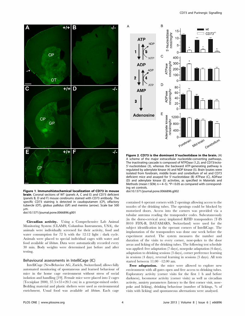
Figure 1. Immunohistochemical localization of CD73 in mousebrain. Coronal sections of WT (panels A, C and E) and CD73 deficient(panels B, D and F) mouse cerebrums stained with CD73 antibody. Thespecific CD73 staining is detected in caudoputamen (CP), olfactorytubercle (OT), globus pallidus (GP) and meninx (arrow). Scale bar 500mm.doi:10.1371/journal.pone.0066896.g001
Figure 2. CD73 is the dominant 5’nucleotidase in the brain. (A)A scheme of the major extracellular nucleotide-converting pathways.The inactivating cascade is composed of NTPDase (1,2), and CD73/ecto-5’-nucleotidase (3), whereas the backward ATP-generating pathway isregulated by adenylate kinase (4) and NDP kinase (5). Brain lysates wereisolated from forebrain, middle brain and cerebellum of wt and CD73deficient mice and assayed for 5’-nucleotidase (B) ATPase (C), ADPase(D) and adenylate kinase (E) activities, as specified in Materials andMethods (mean6SEM; n = 4–5). *P,0.05 as compared with correspond-ing wt controls.doi:10.1371/journal.pone.0066896.g002
CD73 and Purinergic Signalling
PLOS ONE | www.plosone.org 4 June 2013 | Volume 8 | Issue 6 | e66896
Adaptation to nose-poke. All gates were closed at the
beginning of module and mice were trained to poke into closed
gates to reach drinking tubes. Only the first nosepoke of the visit
opened the door for 7 s.
Adaptation to drinking session. gates were programmed to
open after the first nose-poke only during two 1-hour periods, from
21:00 to 22:00 and from 02:00 to 03:00. Drinking sessions were
applied for increasing the motivation to visit the corners and
thereby providing defined time windows for testing learning.
Corner preference learning. during 4 days (8 sessions) the
mice were trained to drink from only one corner. For each mouse
a certain corner was programmed as the ‘‘correct’’ one where the
first nosepoke of the visit was rewarded by opening the door and
access to water during drinking sessions. One pair of mice (one wt
and one CD73 deficient mouse) was assigned to each corner of the
IntelliCage. Learning was measured as a percentage of visits to the
‘‘correct’’ corner.
Reversal learning. this test was used to assess the ability to
relearn and extinct previously learned association. The mice had
again access to water in one ‘‘correct’’ corner that was diagonally
opposite to the previously used corner. Percentage of visits to the
new ‘‘correct’’ corner and percentage of visits to the corner that
was ‘‘correct’’ in the previous learning test were analysed.
Social behaviour. The last two nights of Corner preference
learning and Reversal learning phases were chosen for assessment of
social competition in the IntelliCage. The duration of visits in the
correct corner during the first drinking session of the night was
compared between wt and ko mice (8 pairs). In each mouse pair
the animal that spent more time in the correct corner was
designated as dominant.
Real time qPCRRNA was isolated from mouse brain using Trizol (Invitrogen)
and cDNA was synthesized using a Reverse-iT kit (ABGene).
Primers specific for A1R (forward) 59-GTTTGGCTGGAA-
CAACCTGA-39, (reverse) 59-ACACTTGATCACGGGCTCC-3
were used to determine gene expression levels and standardized to
the GAPDH (forward) 59-CCCCAATGTGTCCGTCGTG-39,
(reverse) 59- GCCTGCTTCACCACCTTCT-39 housekeeping
gene using a SYBR-Green kit (ABGene) run on an ABI 7500
real time PCR system. To determine relative fold change (RFC),
mRNA levels were normalized to the gene expression levels found
in naıve wt mice, with 0.0 representing baseline values. Melt curve
analyses were performed to measure the specificity for each qPCR
product.
StatisticsData were analyzed with two-way analysis of variance
(ANOVA) with genotype and sex as independent variables. Pre-
pulse inhibition, learning tests, CLAMS, rota-rod and activity data
were analyzed with two-way ANOVA for repeated measures with
amplitude, trial number or time as the repeated measure.
Multivariate ANOVA was applied when measuring different
parameters derived from the same test. In simple tests with one
variable only, the t-test was used. Barbering data and data from
dominance test in IntelliCage were analyzed with a Chi-square
test. Results are given as means 6 SEM. Results were considered
significant at P , 0.05.
Table 1. Summary of individual behavioural tests carried outin wt and CD73.
Test and parametersmeasured wt CD73 -/- p-value
Elevated plus maze
distance, cm 768.7650.4 1009.2670 p,0.01
open arm latency, s 104.2619 117.5622.9 ns
open entries, % 21.363.1 17.763.1 ns
open arm time, s 40.768 34.467.3 ns
rearings, n 16.561.3 15.861.5 ns
Open field
distance, cm 4580.66160.5 55686213.6 p,0.01
distance in center, % 24.160.7 25.660.9 ns
latency to center, s 114.3620.6 65.6616.5 ns
time in center,s 320.4618.9 323.7624.9 ns
center entries, n 470.2612.6 550.6619.7 ns
rearing, s 380.4624 429.9633.1 ns
Light-dark
distance, cm 1243.2646.9 1258.3655.4 ns
distance in light, % 38.661.4 39.261.4 ns
time in light, s 114.267.2 121.366 ns
rearing, s 4062.2 34.362.9 ns
Y-maze
spontaneous alternation, % 53.562.1 57.662.1 ns
Hot plate
latency to reaction, s 13.760.8 12.860.6 ns
Rota-Rod
latency to fall, first trial, s 216.6617.4 193.6620.6 ns
latency to fall, last trial, s 283612.9 250.9618.8 ns
Fear-conditioning
contextual freezing, % 3164.1 28.563.8 ns
cued freezing, % 38.263.4 38.464.3 ns
Forced swim test
immobility time, s 161.2611.3 134.9616.2 ns
deficient mice.doi:10.1371/journal.pone.0066896.t001
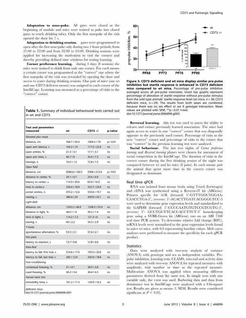
Figure 3. CD73 deficient and wt mice display similar pre-pulseinhibition but startle response is enhanced in CD73 deficientmice compared to wt mice. Percentage of pre-pulse inhibitionaveraged across all pre-pulse intensities. Insert bar graphs representpercentage of alteration of startle response without pre-pulse stimulusfrom the wild type animals’ startle response level (wt mice, n = 38; CD73deficient mice, n = 29). The results from both sexes are combined,because there was no sex effect or sex X genotype interaction. Meanvalues are plotted with SEM, **p,0.01 t-test.doi:10.1371/journal.pone.0066896.g003
CD73 and Purinergic Signalling
PLOS ONE | www.plosone.org 5 June 2013 | Volume 8 | Issue 6 | e66896
Results
CD73 is expressed in subcortical structures of theforebrain and in meninges
We studied the expression of CD73 using immunohistochem-
istry of wild-type and CD73 deficient mice. Specific and intense
immunostaining was found in caudate and putamen that form the
dorsal part of striatum (Fig. 1). The olfactory tubercle that belongs
to ventral striatum was also found to clearly express CD73. Globus
pallidus, a component of the basal ganglia core together with
striatum, displayed intense expression. Furthermore, choroid
plexus and meninges were found to be positive in CD73
immunohistochemistry.
CD73 is the major 5’-nucleotidase hydrolyzing AMP inbrain
The duration and magnitude of purinergic signalling is known
to be governed by a network of purine-converting ectoenzymes
(Fig. 2A) abundantly expressed in various cells and tissues,
including the brain [8,20]. Since six other 5’nucleotidases in
addition to CD73 have been described, we first analyzed the
contribution of CD73 to the total AMP hydrolysis in the brain.
Measurement of the rate of [3H]AMP breakdown revealed
markedly decreased activity in all brain regions (forebrain,
midbrain and cerebellum) in the absence of CD73 (Fig. 2B).
Our data revealed that ecto-5’-nucleotidase/CD73 accounts for
,85–95% of all AMP-hydrolyzing capability in the murine brain.
We then determined the activities of other key nucleotide
converting enzymes in the different parts of brain in the wt and
CD73 deficient mice (Figure 2C–E). To that end, the lysates from
the forebrain, midbrain and cerebellum were incubated with
saturating concentrations of [3H]ATP (Figure 2C) or [3H]ADP
(Figure 2D). We found that all samples from both genotypes
displayed significant nucleoside triphosphate diphosphohydrolase
(NTPDase) activity with ATPase/ADPase ratio of ,5. Interest-
ingly, ATPase activity was slightly but significantly upregulated in
the forebrain of the CD73 deficient mice. No other genotype-
specific differences in the ATPase or ADPase activity were found
in any part of the brain. The counteracting adenylate kinase
activity was also studied by determining the phosphoryl transfer
from ATP into [3H]AMP. We found that the adenylate kinase
activities were similar in wt and CD73 deficient mice in all regions
of the brain (Figure 2E). Together, these data suggest that a minor
increase in the ATPase activity, probably as a compensatory
mechanism, takes place in the forebrain of the CD73 deficient
mice. However, despite of the dramatic decrease in the AMP
hydrolysis, the activities of several other nucleotide-converting
ecto-enzymes remain almost intact in the brain in the absence of
CD73.
CD73 deficient mice do not differ from wt mice in mostbehavioural tests
Since enzymatically active CD73 was present in several brain
regions, we speculated that the induction of the purinergic
signalling through CD73-derived adenosine might be involved in
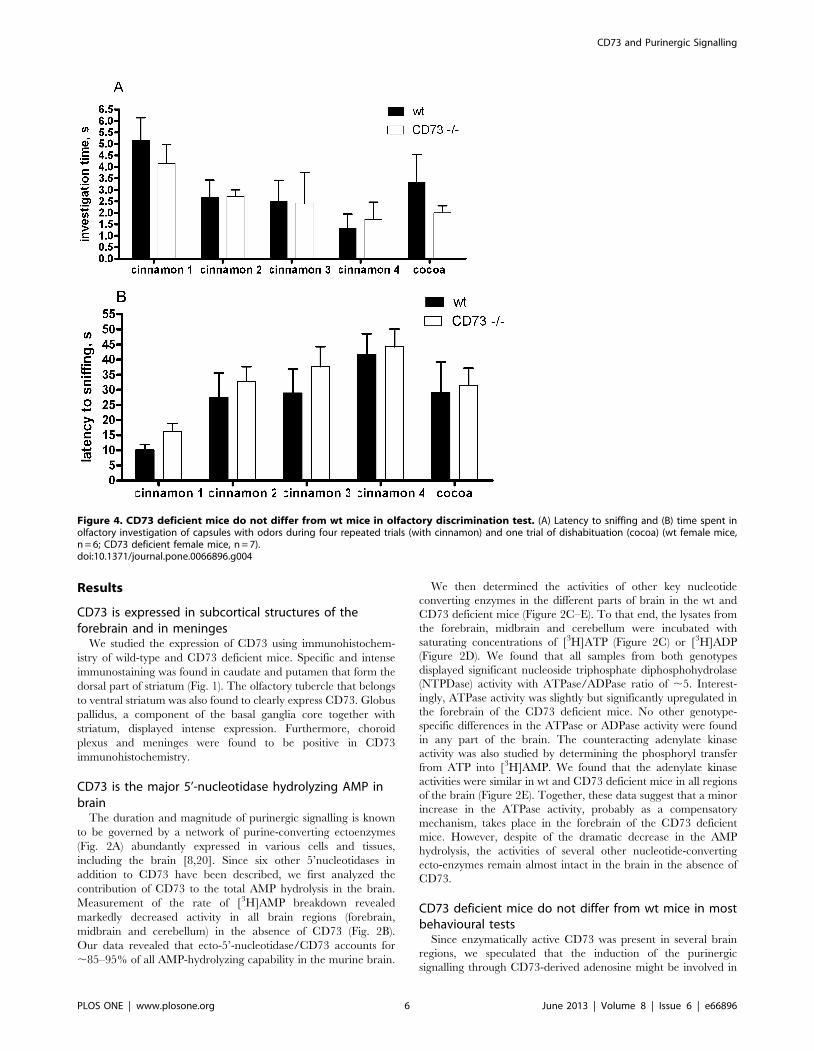
Figure 4. CD73 deficient mice do not differ from wt mice in olfactory discrimination test. (A) Latency to sniffing and (B) time spent inolfactory investigation of capsules with odors during four repeated trials (with cinnamon) and one trial of dishabituation (cocoa) (wt female mice,n = 6; CD73 deficient female mice, n = 7).doi:10.1371/journal.pone.0066896.g004
CD73 and Purinergic Signalling
PLOS ONE | www.plosone.org 6 June 2013 | Volume 8 | Issue 6 | e66896
controlling behaviour. To address this, we compared the wt and
CD73 deficient mice in multiple behavioral tests performed both
individually (summarized in Table 1) and also in the IntelliCage
environment. For the assessment of anxiety level, three tests were
performed: elevated plus-maze, open field and light-dark explo-
ration. In these tests, the parameters reflecting anxiety-like
behaviour were comparable in both genotypes (Table 1).
Furthermore, in the forced swim test for the assessment of
depression-like behaviour the wt and CD73 deficient mice
demonstrated the same level of immobility in an inescapable
situation (Table I). We found no significant genotype-specific
differences in spontaneous alternation (Y maze), nociception (hot
plate), motor coordination and motor learning, or in fear-
conditioning. Pre-pulse inhibition did not differ either from that
observed in the wt mice although the startle response to acoustic
stimulus without pre-stimulus was increased in the CD73 deficient
mice (Fig. 3), agreeing with novelty-induced hyperactivity in these
mice (see below). Olfactory discrimination also appeared intact in
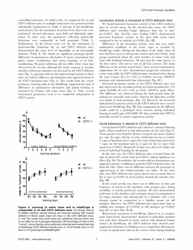
the CD73 knockout mice (Fig. 4). The results from the corner
preference learning tasks in the IntelliCage supported the lack of
differences in spontaneous alternation and spatial learning as
measured by Y-maze and water maze (Fig. 5). Thus, several
behavioural parameters seem to be completely independent of
CD73 activity.
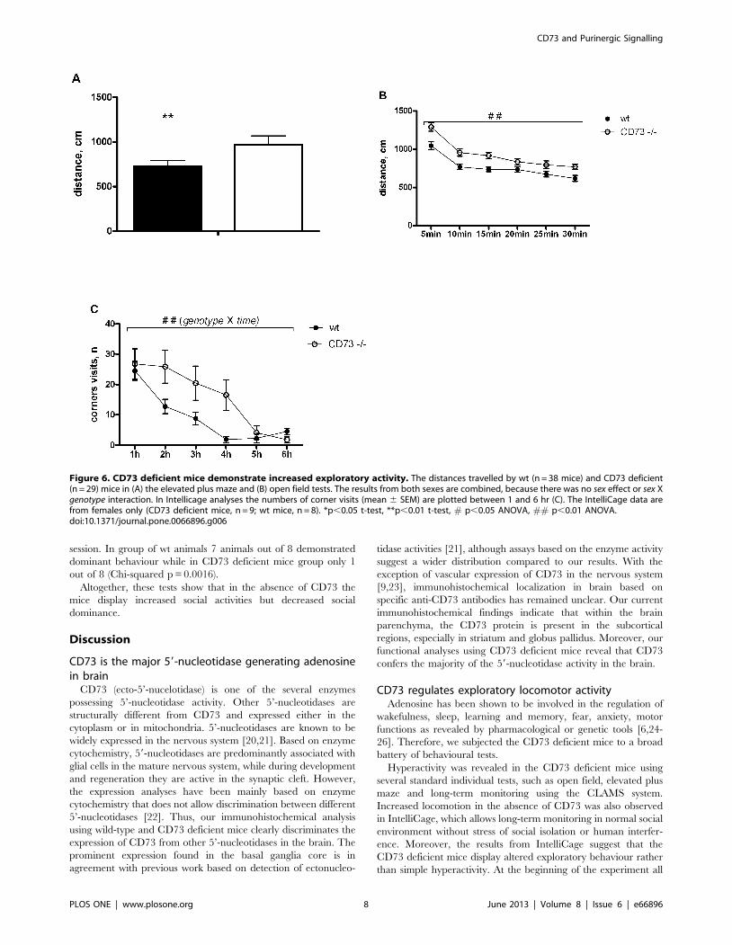
Locomotor activity is increased in CD73 deficient miceWe found increased locomotor activity of the CD73 deficient
mice in several assays. In the elevated plus maze, the CD73
deficient mice travelled longer distances than the wt mice
(p = 0.0057; Fig. 6A).The mice lacking CD73 demonstrated
increased locomotor activity in the open field arena when
compared to the wt animals (p = 0.004; Fig. 6B).
Differences in the locomotor activity were also seen under
undisturbed conditions in the home cages as revealed by
IntelliCage studies. During the first phase of the study, when all
mice had free access to all operant corners and water bottles, both
groups performed about the same number of corner visits and
visits with drinking behaviour. All mice had the same latency to
the first corner visit and to visit of all four corners. The main
difference in the activity started after 1 hour of novel environment
exploration: the wt mice demonstrated progressive reduction in
corner visits while the CD73 deficient mice remained active during
the next 3 hours (F(5,75) = 3.87, p = 0.0036, two-way ANOVA,
genotype and experimental time interaction) (Fig. 6C).
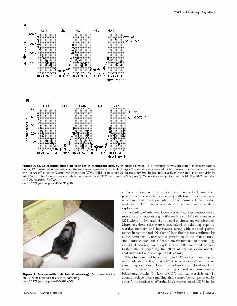
Increased locomotor activity in the CD73 deficient mice was
also observed in the circadian activity test when measured for 72 h
using CLAMS (F(1,53) = 5.95, p = 0.02, ANOVA, main effect).
The difference was observed during the dark period when the
animals are naturally more active, whereas the light time activity
was similar in both genotypes (Fig. 7A). However, the increased
dark-period locomotor activity in the CD73 deficient mice was not
observed in IntelliCage (Fig. 7B). One explanation for the different
results could be a prolonged stress reaction during individual
housing in small cages of CLAMS. We thus conclude that CD73 is
normally needed to restrict exploratory activity.
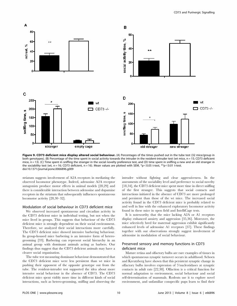
Social behaviour is altered in CD73 deficient miceGroup-housed CD73 deficient mice showed a strong barbering
habit, which manifested as big bald patches on the back (Fig. 8).
Those patches were found in all mice except in one mouse (barber)
per cage. In cages with the wt mice, barbering was not so evident
(7 cages of knockout and 7 cages of wt mice; barbering observed in
7 cages for the knockout and in 2 cages for the wt mice; Chi-
squared p = 0.0053). Regrowth of hair was observed within one
week of individual housing for all animals.
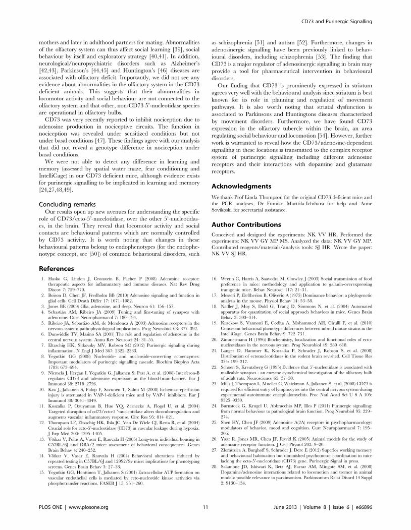
In the tube test, the CD73 deficient mice retreated from the
tube in about 60% of the trials (p,0.0001) without significant sex
effect (Fig. 9A). Nevertheless, the wt mice did not demonstrate any
aggressive posture or fighting to push the CD73 deficient mice out
from the tube. There was no difference between the genotypes in
aggressiveness in the resident-intruder test either. At the same
time, the CD73 deficient mice spent almost twice as much time as
the wt mice (p,0.001) in social activity towards the intruder mice
(Fig. 9B).
In the social novelty test, there was no difference in time and
frequency of entries to the chambers with stranger mice during
sociability or novelty preference sessions. All mice demonstrated
preference to the chamber with a stranger mouse in comparison to
an empty chamber, and subsequently to a chamber with a new
stranger mouse in comparison to a familiar mouse (an old
stranger). However, the CD73 deficient mice spent more time in
sniffing new strangers (p = 0.0426) or old strangers (p = 0.0262)
than the wt mice (Fig. 9C,D).
Behavioural assessment in IntelliCage allowed us to compare
some behavioural characteristics obtained in individual standard
tests with data obtained in social context with minimal human
contact. In IntelliCage, the CD73 deficient mice showed more
suppressive behaviour in drinking access competition allowing the
wt mice to spend more time in the correct corner during drinking
Figure 5. Learning in water maze and in IntelliCage iscomparable in wt and CD73 deficient mice. (A) Escape latencyto hidden platform during training and reversed training with movedplatform in Morris water maze (wt mice, n = 38; CD73 deficient mice,n = 29). The results from both sexes are combined, because there wasno sex effect or sex X genotype interaction. (B) Percentage of visits to the‘‘correct’’ corner during corner preference learning and reversal learningin IntelliCage (CD73 deficient female mice, n = 9; wt female mice, n = 8).doi:10.1371/journal.pone.0066896.g005
CD73 and Purinergic Signalling
PLOS ONE | www.plosone.org 7 June 2013 | Volume 8 | Issue 6 | e66896
session. In group of wt animals 7 animals out of 8 demonstrated
dominant behaviour while in CD73 deficient mice group only 1
out of 8 (Chi-squared p = 0.0016).
Altogether, these tests show that in the absence of CD73 the
mice display increased social activities but decreased social
dominance.
Discussion
CD73 is the major 59-nucleotidase generating adenosinein brain
CD73 (ecto-5’-nucelotidase) is one of the several enzymes
possessing 5’-nucleotidase activity. Other 5’-nucleotidases are
structurally different from CD73 and expressed either in the
cytoplasm or in mitochondria. 5’-nucleotidases are known to be
widely expressed in the nervous system [20,21]. Based on enzyme
cytochemistry, 59-nucleotidases are predominantly associated with
glial cells in the mature nervous system, while during development
and regeneration they are active in the synaptic cleft. However,
the expression analyses have been mainly based on enzyme
cytochemistry that does not allow discrimination between different
5’-nucleotidases [22]. Thus, our immunohistochemical analysis
using wild-type and CD73 deficient mice clearly discriminates the
expression of CD73 from other 5’-nucleotidases in the brain. The
prominent expression found in the basal ganglia core is in
agreement with previous work based on detection of ectonucleo-
tidase activities [21], although assays based on the enzyme activity
suggest a wider distribution compared to our results. With the
exception of vascular expression of CD73 in the nervous system
[9,23], immunohistochemical localization in brain based on
specific anti-CD73 antibodies has remained unclear. Our current
immunohistochemical findings indicate that within the brain
parenchyma, the CD73 protein is present in the subcortical
regions, especially in striatum and globus pallidus. Moreover, our
functional analyses using CD73 deficient mice reveal that CD73
confers the majority of the 59-nucleotidase activity in the brain.
CD73 regulates exploratory locomotor activityAdenosine has been shown to be involved in the regulation of
wakefulness, sleep, learning and memory, fear, anxiety, motor
functions as revealed by pharmacological or genetic tools [6,24-
26]. Therefore, we subjected the CD73 deficient mice to a broad
battery of behavioural tests.
Hyperactivity was revealed in the CD73 deficient mice using
several standard individual tests, such as open field, elevated plus
maze and long-term monitoring using the CLAMS system.
Increased locomotion in the absence of CD73 was also observed
in IntelliCage, which allows long-term monitoring in normal social
environment without stress of social isolation or human interfer-
ence. Moreover, the results from IntelliCage suggest that the
CD73 deficient mice display altered exploratory behaviour rather
than simple hyperactivity. At the beginning of the experiment all
Figure 6. CD73 deficient mice demonstrate increased exploratory activity. The distances travelled by wt (n = 38 mice) and CD73 deficient(n = 29) mice in (A) the elevated plus maze and (B) open field tests. The results from both sexes are combined, because there was no sex effect or sex Xgenotype interaction. In Intellicage analyses the numbers of corner visits (mean 6 SEM) are plotted between 1 and 6 hr (C). The IntelliCage data arefrom females only (CD73 deficient mice, n = 9; wt mice, n = 8). *p,0.05 t-test, **p,0.01 t-test, # p,0.05 ANOVA, ## p,0.01 ANOVA.doi:10.1371/journal.pone.0066896.g006
CD73 and Purinergic Signalling
PLOS ONE | www.plosone.org 8 June 2013 | Volume 8 | Issue 6 | e66896
animals explored a novel environment quite actively and then
progressively decreased their activity with time. Four hours in a
novel environment was enough for the wt mouse to become calm,
while the CD73 deficient animals were still very active in their
exploration.
Our finding of enhanced locomotor activity is in contrast with a
recent study characterizing a different line of CD73 deficient mice
[27], where no hyperactivity in novel environment was observed.
Moreover, those mice were characterized as exhibiting superior
working memory and habituation along with reduced perfor-
mance in rota-rod task. Neither of these findings was confirmed by
our experiments. Differences in generation of the mutant mice,
small sample size and different environmental conditions (e.g.
individual housing) could explain these differences and warrant
further studies regarding the effect of various environmental
challenges on the phenotype of CD73 mice.
The observation of hyperactivity in CD73 deficient mice agrees
well with the finding that CD73 is a major 59-nucleotidase
generating adenosine in brain since adenosine is a global regulator
of neuronal activity in brain, causing a basal inhibitory tone of
behavioural activity [6]. Lack of CD73 thus causes a deficiency in
adenosine-dependent signalling that cannot be compensated by
other 59-nucleotidases of brain. High expression of CD73 in the
Figure 7. CD73 controls circadian changes in locomotor activity in isolated mice. (A) Locomotor activity presented as activity countsduring 72 hr observation period, when the mice were separated in individual cages. These data are presented for both sexes together, because therewas no sex effect or sex X genotype interaction (CD73 deficient mice, n = 25; wt mice, n = 30). (B) Locomotor activity measured as corner visits inIntelliCage. In IntelliCage analyses only females were used (CD73 deficient, n = 9; wt, n = 8). Mean values are plotted with SEM, # p,0.05 and ##p,0.01, repeated ANOVA.doi:10.1371/journal.pone.0066896.g007
Figure 8. Mouse with hair loss (barbering). An example of amouse with bald patches due to barbering.doi:10.1371/journal.pone.0066896.g008
CD73 and Purinergic Signalling
PLOS ONE | www.plosone.org 9 June 2013 | Volume 8 | Issue 6 | e66896
striatum suggests involvement of A2A receptors in mediating the
observed locomotor phenotype. Indeed, adenosine A2A receptor
antagonists produce motor effects in animal models [28,29] and
there is considerable interaction between adenosine and dopamine
receptors in the striatum that subsequently influences spontaneous
locomotor activity [28,30–32].
Modulation of social behaviour in CD73 deficient miceWe observed increased spontaneous and circadian activity in
the CD73 deficient mice in individual testing, but not when the
mice lived in groups. This suggests that behaviour of the CD73
deficient mice is strongly dependent on their social environment.
Therefore, we analyzed their social interactions more carefully.
The CD73 deficient mice showed intensive barbering behaviour.
In group-housed mice barbering is an intensive form of hetero-
grooming [33]. Barbering can represent social hierarchy in an
animal group with dominant animals acting as barbers. Our
findings thus suggest that the CD73 deficient animals may have a
clearer social organisation.
The tube test measuring dominant behaviour demonstrated that
the CD73 deficient mice were less persistent than wt mice in
pushing their opponent of the opposite genotype out from the
tube. The resident-intruder test supported the idea about more
intensive social behaviour in the absence of CD73. The CD73
deficient mice spent visibly more time in different kinds of social
interactions, such as hetero-grooming, sniffing and observing the
intruder without fighting and clear aggressiveness. In the
assessments of the sociability level and preference to social novelty
[18,34], the CD73 deficient mice spent more time in direct sniffing
of the first stranger. This suggests that social contacts and
interactions initiated in the absence of CD73 are more prolonged
and persistent than those of the wt mice. The increased social
activity found in the CD73 deficient mice is probably related to
and well in line with the enhanced exploratory locomotor activity
found in these mice in open field and IntelliCage tests.
It is noteworthy that the mice lacking A2A or A1 receptors
display enhanced anxiety and aggression [35,36]. Moreover, the
mice selectively bred for maternal aggression exhibit significantly
enhanced levels of adenosine A1 receptors [37]. These findings
together with our observations strongly suggest involvement of
adenosine in modulation of social behaviour.
Preserved sensory and memory functions in CD73deficient mice
Mature retina and olfactory bulbs are rare examples of tissues in
which spontaneous synaptic turnover occurs in adulthood. Schoen
and Kreutzberg have shown that this persistent synaptic change in
olfactory bulbs involves expression of 5’nucleotidases at synaptic
contacts in adult rats [22,38]. Olfaction is a critical function for
normal adaptation to environment, social behaviour and social
self-determination of mammals. Rodents use it to explore novel
environment, and unfamiliar conspecific pups learn to find their
Figure 9. CD73 deficient mice display altered social behaviour. (A) Percentages of the times pushed out in the tube test (32 mice/group inboth genotypes). (B) Percentage of the time spent in social activity towards the intruder in the resident-intruder test (wt mice, n = 15; CD73 deficientmice, n = 13). (C) Time spent in sniffing the stranger in the social novelty preference test, and (D) time spent in sniffing a new and an old stranger inthe sociability test (wt, n = 16; CD73 deficient, n = 16). Mean values are plotted with SEM, *p,0.05 t-test, **p,0.01 t-test.doi:10.1371/journal.pone.0066896.g009
CD73 and Purinergic Signalling
PLOS ONE | www.plosone.org 10 June 2013 | Volume 8 | Issue 6 | e66896
mothers and later in adulthood partners for mating. Abnormalities
of the olfactory system can thus affect social learning [39], social
behaviour by itself and exploratory strategy [40,41]. In addition,
neurological/neuropsychiatric disorders such as Alzheimer’s
[42,43], Parkinson’s [44,45] and Huntington’s [46] diseases are
associated with olfactory deficit. Importantly, we did not see any
evidence about abnormalities in the olfactory system in the CD73
deficient animals. This suggests that their abnormalities in
locomotor activity and social behaviour are not connected to the
olfactory system and that other, non-CD73 5’-nucleotidase species
are operational in olfactory bulbs.
CD73 was very recently reported to inhibit nociception due to
adenosine production in nociceptive circuits. The function in
nociception was revealed under sensitized conditions but not
under basal conditions [47]. These findings agree with our analysis
that did not reveal a genotype difference in nociception under
basal conditions.
We were not able to detect any difference in learning and
memory (assessed by spatial water maze, fear conditioning and
IntelliCage) in our CD73 deficient mice, although evidence exists
for purinergic signalling to be implicated in learning and memory
[24,27,48,49].
Concluding remarksOur results open up new avenues for understanding the specific
role of CD73/ecto-5’-nucelotidase, over the other 5’-nucleotidas-
es, in the brain. They reveal that locomotor activity and social
contacts are behavioural patterns which are normally controlled
by CD73 activity. It is worth noting that changes in these
behavioural patterns belong to endophenotypes (for the endophe-
notype concept, see [50]) of common behavioural disorders, such
as schizophrenia [51] and autism [52]. Furthermore, changes in
adenosinergic signalling have been previously linked to behav-
ioural disorders, including schizophrenia [53]. The finding that
CD73 is a major regulator of adenosinergic signalling in brain may
provide a tool for pharmaceutical intervention in behavioural
disorders.
Our finding that CD73 is prominently expressed in striatum
agrees very well with the behavioural analysis since striatum is best
known for its role in planning and regulation of movement
pathways. It is also worth noting that striatal dysfunction is
associated to Parkinsons and Huntingtons diseases characterized
by movement disorders. Furthermore, we have found CD73
expression in the olfactory tubercle within the brain, an area
regulating social behaviour and locomotion [54]. However, further
work is warranted to reveal how the CD73/adenosine-dependent
signalling in these locations is transmitted to the complex receptor
system of purinergic signalling including different adenosine
receptors and their interactions with dopamine and glutamate
receptors.
Acknowledgments
We thank Prof Linda Thompson for the original CD73 deficient mice and
the PCR analyses, Dr Fumiko Marttila-Ichihara for help and Anne
Sovikoski for secretarial assistance.
Author Contributions
Conceived and designed the experiments: NK VV HR. Performed the
experiments: NK VV GY MP MS. Analyzed the data: NK VV GY MP.
Contributed reagents/materials/analysis tools: SJ HR. Wrote the paper:
NK VV SJ HR.
References
1. Hasko G, Linden J, Cronstein B, Pacher P (2008) Adenosine receptor:
therapeutic aspects for inflammatory and immune diseases. Nat Rev Drug
Discov 7: 759–770.
2. Boison D, Chen JF, Fredholm BB (2010) Adenosine signaling and function in
glial cells. Cell Death Differ 17: 1071–1082.
3. Jones BE (2009) Glia, adenosine, and sleep. Neuron 61: 156–157.
4. Sebastiao AM, Ribeiro JA (2009) Tuning and fine-tuning of synapses with
adenosine. Curr Neuropharmacol 7: 180–194.
5. Ribeiro JA, Sebastiao AM, de Mendonca A (2003) Adenosine receptors in the
nervous system: pathophysiological implications. Prog Neurobiol 68: 377–392.
6. Dunwiddie TV, Masino SA (2001) The role and regulation of adenosine in the
central nervous system. Annu Rev Neurosci 24: 31–55.
7. Eltzschig HK, Sitkovsky MV, Robson SC (2012) Purinergic signaling during
inflammation. N Engl J Med 367: 2322–2333.
8. Yegutkin GG (2008) Nucleotide- and nucleoside-converting ectoenzymes:
Important modulators of purinergic signalling cascade. Biochim Biophys Acta
1783: 673–694.
9. Niemela J, Ifergan I, Yegutkin G, Jalkanen S, Prat A, et al. (2008) Interferon-B
regulates CD73 and adenosine expression at the blood-brain-barrier. Eur J
Immunol 38: 2718–2726.
10. Kiss J, Jalkanen S, Fulop F, Savunen T, Salmi M (2008) Ischemia-reperfusion
injury is attenuated in VAP-1-deficient mice and by VAP-1 inhibitors. Eur J
Immunol 38: 3041–3049.
11. Koszalka P, Ozuyaman B, Huo YQ, Zernecke A, Flogel U, et al. (2004)
Targeted disruption of cd73/ecto-5 ’-nucleotidase alters thromboregulation and
augments vascular inflammatory response. Circ Res 95: 814–821.
12. Thompson LF, Eltzschig HK, Ibla JC, Van De Wiele CJ, Resta R, et al. (2004)
Crucial role for ecto-5’-nucleotidase (CD73) in vascular leakage during hypoxia.
J Exp Med 200: 1395–1405.
13. Voikar V, Polus A, Vasar E, Rauvala H (2005) Long-term individual housing in
C57BL/6J and DBA/2 mice: assessment of behavioral consequences. Genes
Brain Behav 4: 240–252.
14. Voikar V, Vasar E, Rauvala H (2004) Behavioral alterations induced by
repeated testing in C57BL/6J and 129S2/Sv mice: implications for phenotyping
screens. Genes Brain Behav 3: 27–38.
15. Yegutkin GG, Henttinen T, Jalkanen S (2001) Extracellular ATP formation on
vascular endothelial cells is mediated by ecto-nucleotide kinase activities via
phosphotransfer reactions. FASEB J 15: 251–260.
16. Wrenn C, Harris A, Saavedra M, Crawley J (2003) Social transmission of food
preference in mice: methodology and application to galanin-overexpressing
transgenic mice. Behav Neurosci 117: 21–31.
17. Messeri P, Eleftheriou B, Oliverio A (1975) Dominance behavior: a phylogenetic
analysis in the mouse. Physiol Behav 14: 53–58.
18. Nadler J, Moy S, Dold G, Trang D, Simmons N, et al. (2004) Automated
apparatus for quantitation of social approach behaviors in mice. Genes Brain
Behav 3: 303–314.
19. Krackow S, Vannoni E, Codita A, Mohammed AH, Cirulli F, et al. (2010)
Consistent behavioral phenotype differences between inbred mouse strains in the
IntelliCage. Genes Brain Behav 9: 722–731.
20. Zimmermann H (1996) Biochemistry, localization and functional roles of ecto-
nucleotidases in the nervous system. Prog Neurobiol 49: 589–618.
21. Langer D, Hammer K, Koszalka P, Schrader J, Robson S, et al. (2008)
Distribution of ectonucleotidases in the rodent brain revisited. Cell Tissue Res
334: 199–217.
22. Schoen S, Kreutzberg G (1995) Evidence that 5’-nucleotidase is associated with
malleable synapses - an enzyme cytochemical investigation of the olfactory bulb
of adult rats. Neuroscience 65: 37–50.
23. Mills J, Thompson L, Mueller C, Waickman A, Jalkanen S, et al. (2008) CD73 is
required for efficient entry of lymphocytes into the central nervous system during
experimental autoimmune encephalomyelitis. Proc Natl Acad Sci U S A 105:
9325–9330.
24. Burnstock G, Krugel U, Abbracchio MP, Illes P (2011) Purinergic signalling:
from normal behaviour to pathological brain function. Prog Neurobiol 95: 229–
274.
25. Shen HY, Chen JF (2009) Adenosine A(2A) receptors in psychopharmacology:
modulators of behavior, mood and cognition. Curr Neuropharmacol 7: 195–
206.
26. Yaar R, Jones MR, Chen JF, Ravid K (2005) Animal models for the study of
adenosine receptor function. J Cell Physiol 202: 9–20.
27. Zlomuzica A, Burghoff S, Schrader J, Dere E (2012) Superior working memory
and behavioural habituation but diminished psychomotor coordination in mice
lacking the ecto-5’-nucleotidase (CD73) gene. Purinergic Signal in press.
28. Salamone JD, Ishiwari K, Betz AJ, Farrar AM, Mingote SM, et al. (2008)
Dopamine/adenosine interactions related to locomotion and tremor in animal
models: possible relevance to parkinsonism. Parkinsonism Relat Disord 14 Suppl
2: S130–134.
CD73 and Purinergic Signalling
PLOS ONE | www.plosone.org 11 June 2013 | Volume 8 | Issue 6 | e66896
29. Florio C, Rosati AM, Traversa U, Vertua R (1997) Inhibitory and excitatory
effects of adenosine antagonists on spontaneous locomotor activity in mice. LifeSci 60: 1477–1486.
30. Xie X, Ramkumar V, Toth LA (2007) Adenosine and dopamine receptor
interactions in striatum and caffeine-induced behavioral activation. Comp Med57: 538–545.
31. Ferre S, Fredholm BB, Morelli M, Popoli P, Fuxe K (1997) Adenosine-dopamine receptor-receptor interactions as an integrative mechanism in the
basal ganglia. Trends Neurosci 20: 482–487.
32. Salmi P, Chergui K, Fredholm BB (2005) Adenosine-dopamine interactionsrevealed in knockout mice. J Mol Neurosci 26: 239–244.
33. Kalueff A, Minasyan A, Keisala T, Shah Z, P T (2006) Hair barbering in mice:implications for neurobehavioural research. Behav Processes 71: 8–15.
34. Moy S, Nadle rJ, Young N, Nonneman R, Grossman A, et al. (2009) Socialapproach in genetically engineered mouse lines relevant to autism. Genes Brain
Behav 8: 129–142.
35. Ledent C, Vaugeois JM, Schiffmann SN, Pedrazzini T, El Yacoubi M, et al.(1997) Aggressiveness, hypoalgesia and high blood pressure in mice lacking the
adenosine A2a receptor. Nature 388: 674–678.36. Gimenez-Llort L, Fernandez-Teruel A, Escorihuela RM, Fredholm BB, Tobena
A, et al. (2002) Mice lacking the adenosine A1 receptor are anxious and
aggressive, but are normal learners with reduced muscle strength and survivalrate. Eur J Neurosci 16: 547–550.
37. Gammie SC, Auger AP, Jessen HM, Vanzo RJ, Awad TA, et al. (2007) Alteredgene expression in mice selected for high maternal aggression. Genes Brain
Behav 6: 432–443.38. Schoen S, Kreutzberg G (1997) 5’-nucleotidase enzyme cytochemistry as a tool
for revealing activated glial cells and malleable synapses in CNS development
and regeneration. Brain Res Brain Res Protoc 1: 33–43.39. Sanchez-Andrade G, Kendrick K (2009) The main olfactory system and social
learning in mammals. Behav Brain Res 200: 323–335.40. Stork O, Welzl H, Cremer H, Schachner M (1997) Increased intermale
aggression and neuroendocrine response in mice deficient for the neural cell
adhesion molecule (NCAM). Eur J Neurosci 9: 1117–1125.41. Vinkers CH, Breuer ME, Westphal KG, Korte SM, Oosting RS, et al. (2009)
Olfactory bulbectomy induces rapid and stable changes in basal and stress-induced locomotor activity, heart rate and body temperature responses in the
home cage. Neuroscience 159: 39–46.
42. Murphy C, Solomon E, Haase L, Wang M, Morgan C (2009) Olfaction in aging
and Alzheimer’s disease: event-related potentials to a cross-modal odor-
recognition memory task discriminate ApoE epsilon4+ and ApoE epsilon 4-
individuals. Ann N Y Acad Sci 1170: 647–657.
43. Wesson D, Levy E, Nixon R, Wilson D (2010) Olfactory dysfunction correlates
with amyloid-beta burden in an Alzheimer’s disease mouse model. J Neurosci
30: 505–514.
44. Karpa M, Gopinath B, Rochtchina E, Wang JJ, Cumming R, et al. (2010)
Prevalence and neurodegenerative or other associations with olfactory
impairment in an older community. J Aging Health 22: 154–168.
45. Oka H, Toyoda C, Yogo M, Mochio S (2010) Olfactory dysfunction and
cardiovascular dysautonomia in Parkinson’s disease. J Neurol 257: 969–976.
46. Lazic S, Goodman A, Grote H, Blakemore C, Morton A, et al. (2007) Olfactory
abnormalities in Huntington’s disease: decreased plasticity in the primary
olfactory cortex of R6/1 transgenic mice and reduced olfactory discrimination in
patients. Brain Res 1151: 219–226.
47. Sowa N, Taylor-Blake B, Zylka M (2010) Ecto-59-Nucleotidase (CD73) inhibits
nociception by hydrolyzing AMP to adenosine in nociceptive circuits. J Neurosci
30: 2235–2244.
48. Bonan CD, Roesler R, Pereira GS, Battastini AM, Izquierdo I, et al. (2000)
Learning-specific decrease in synaptosomal ATP diphosphohydrolase activity
from hippocampus and entorhinal cortex of adult rats. Brain Res 854: 253–256.
49. Bonan CD, Dias MM, Battastini AM, Dias RD, Sarkis JJ (1998) Inhibitory
avoidance learning inhibits ectonucleotidases activities in hippocampal synap-
tosomes of adult rats. Neurochem Res 23: 977–982.
50. Gould TD, Gottesman II (2006) Psychiatric endophenotypes and the
development of valid animal models. Genes Brain Behav 5: 113–119.
51. Powell CM, Miyakawa T (2006) Schizophrenia-relevant behavioral testing in
rodent models: a uniquely human disorder? Biol Psychiatry 59: 1198–1207.
52. Crawley JN (2007) Mouse behavioral assays relevant to the symptoms of autism.
Brain Pathol 17: 448–459.
53. Gotoh L, Mitsuyasu H, Kobayashi Y, Oribe N, Takata A, et al. (2009)
Association analysis of adenosine A1 receptor gene (ADORA1) polymorphisms
with schizophrenia in a Japanese population. Psychiatr Genet 19: 328–335.
54. Ikemoto S (2007) Dopamine reward circuitry: two projection systems from the
ventral midbrain to the nucleus accumbens-olfactory tubercle complex. Brain
Res Rev 56: 27–78.
CD73 and Purinergic Signalling
PLOS ONE | www.plosone.org 12 June 2013 | Volume 8 | Issue 6 | e66896