Cryopreservation of adenovirus-transfected dendritic cells (DCs) for clinical use
Upload
independentCategory
view
3download
0
CD34-derived dendritic cells transfected ex vivo with HIV-GagmRNA induce polyfunctional T-cell responses in nonhumanprimates
Gabrielle Romain1,2, Ellen van Gulck3, Olivier Epaulard1,2, SangKon Oh4, Dapeng Li4,Gerard Zurawski4, Sandra Zurawski4, Antonio Cosma1,2, Lucille Adam1,2, CatherineChapon1,2, Biliana Todorova1,2, Jacques Banchereau4, Nathalie Dereuddre-Bosquet1,2,Guido Vanham3,5, Roger Le Grand1,2, and Frédéric Martinon1,2,6
1CEA, Division of Immuno-Virology, Institute for Emerging Diseases and Innovative Therapies(iMETI), DSV, Fontenay-aux-Roses, France2Université Paris-Sud, UMR E1, Orsay, France3Virology Unit, Division of Microbiology, Department of Biomedical Sciences (HIV andRetrovirology Research Unit, Department of Microbiology), Department of Microbiology, Instituteof Tropical Medicine of Antwerp (ITMA), Belgium4Baylor Institute for Immunology Research and INSERM U899, Dallas, TX 75204, USA5Faculty of Medicine and Pharmacy, Free University of Brussels (VUB), Brussels, Belgium6Inserm, Paris, France
AbstractThe pivotal role of DCs in initiating the immune responses led to their use as vaccine vectors.However, the relationship between DC subsets involved in antigen presentation and the type ofelicited immune responses underlined the need for the characterization of the DCs generated invitro. The phenotypes of tissue-derived APCs from a cynomolgus macaque model for humanvaccine development were compared with ex vivo-derived DCs. Monocyte/macrophagespredominated in bone marrow (BM) and blood. Myeloid DCs (mDCs) were present in all testedtissues and were more highly represented than plasmacytoid DCs (pDCs). As in human skin,Langerhans cells (LCs) resided exclusively in the macaque epidermis, expressing CD11c, highlevels of CD1a and Langerin (CD207). Most DC subsets were endowed with tissue-specificcombinations of PRRs. DCs generated from CD34+ BM cells (CD34-DCs) were heterogeneous inphenotype. CD34-DCs shared properties (differentiation and PRR) of dermal and epidermal DCs.After injection into macaques, CD34-DCs expressing HIV-Gag induced Gag-specific CD4+ andCD8+ T cells producing IFN-γ, TNF-α, MIP-1β or IL-2. In high responding animals, the numbersof polyfunctional CD8+ T cells increased with the number of booster injections. This DC-basedvaccine strategy elicited immune responses relevant to the DC subsets generated in vitro.
© 2012 WILEY-VCH Verlag GmbH & Co. KGaA, Weinheim
Corresponding author: Dr Frédéric Martinon, Service d’Immuno-Virologie, iMETI, CEA, 18 route du Panorama, 92265 Fontenay-aux-Roses, France; [email protected]; Phone: +33 1 4654 8757; Fax: +33 1 4654 7726.
Conflict of interestThe authors declare no financial or commercial conflict of interest.
NIH Public AccessAuthor ManuscriptEur J Immunol. Author manuscript; available in PMC 2013 August 01.
Published in final edited form as:Eur J Immunol. 2012 August ; 42(8): 2019–2030. doi:10.1002/eji.201242478.
NIH
-PA Author Manuscript
NIH
-PA Author Manuscript
NIH
-PA Author Manuscript

KeywordsDendritic cells; Vaccination; Immune responses; Antigen presenting cells
1. IntroductionDCs are a heterogeneous population of migratory and tissue-resident immune cells with thecapacity to prime specific immune responses. As part of their function, DCs are able todetect PAMPs through their PRRs [1]. Several types of tissue-resident DCs can bedistinguished by the expression of particular PRRs, such as C-type lectin receptors (CLRs),which act as anchors for a large number of microbes. CLRs at the surface of DCs allowantigen internalization and initiate the process of antigen presentation to T and Blymphocytes [2]. The signaling motifs in the cytoplasmic domains of some CLRs lead totheir activation or suppression upon triggering, and thereby modulate both innate andadaptive immune responses [1, 2]. Altogether, these important properties of DCs can beutilized for the design of new vaccine strategies.
DC-based therapeutic vaccines have been considered for the treatment of cancers [3, 4] andthe control of chronic infections, such as those caused by HCV and HIV [5] because of theirunique ability to prime naïve T cells against specific antigens. Patients have been infusedwith autologous DCs derived ex vivo from blood monocytes or from CD34+ hematopoieticprogenitor cells (HPCs) [6–8] loaded with various antigens, including proteins, peptides,inactivated viruses, tumor lysates and recombinant viral vectors. The use of antigen-encoding mRNA to transfect DCs ex vivo may have multiple advantages [9]. Specifically,the cost of antigen production may be significantly reduced and nucleic acids offer multiplepossibilities for immunogen engineering and design. It is also possible to develop“personalized” immunogens adapted to each immunized individual. These personalizedimmunogens, used to transfect the DCs, can be derived directly from patient tumor cells orfrom viral variants replicating in the host [10, 11]. The use of mRNA offers the additionaladvantage over DNA and retroviral vectors that it cannot possibly be integrated into thehuman DNA and hence avoids the potential risk of insertional genetic damage [12].
In the present study, we extensively characterized the phenotype of different DC populationsof the macaques used as animal models for immunogenicity and safety tests of humanvaccines. We demonstrate that DCs derived from macaque BM CD34+ cells and transfectedex vivo with HIV Gag-encoding mRNA can efficiently prime a specific and polyfunctionalT-cell response.
2. Results2.1. Characterization of DC subsets in blood and lymphoid tissues of cynomolgusmacaques
DCs represent a heterogeneous APC population expressing diverse differentiation andmaturation markers according to their localization and contribution to the host immuneresponse [3]. We first analyzed the different DC subsets in the BM, blood, spleen, LNs andskin of cynomolgus macaques with the aim of comparing their phenotypes with thephenotype of the DCs we generated ex vivo for our vaccine purposes. HLA-DR+ cells thatwere negative for the lymphocyte lineage (CD3−, CD8−, CD20−) were analyzed forexpression of CD1a, CD14, CD11c and CD123 (Fig. 1 and Table 1). As reported forhumans, BM contained the highest proportion of HLA-DR+Lin− cells in all tissuesexamined, the majority of which were CD1a−CD14+ monocytes/macrophages (subset 1 inFig. 1). These cells were also represented in blood. In contrast, LNs contained mainly
Romain et al. Page 2
Eur J Immunol. Author manuscript; available in PMC 2013 August 01.
NIH
-PA Author Manuscript
NIH
-PA Author Manuscript
NIH
-PA Author Manuscript
CD1a+CD14− APCs whereas spleen contained a mixed population of CD1a+CD14− APCsand monocytes/macrophages. In all tissues, CD11c+CD123− mDCs (subset 2 in Fig. 1) weremore highly represented than CD11c−CD123+ pDCs (subset 3 in Fig. 1). In accordance withpreviously published results, including those of our group for macaque cells [13–16],CD11c−CD123+ pDCs were in fewer proportions in blood than in the spleen and LNs (Table1). The skin is populated with a wide variety of APCs [17], providing a very attractive routefor vaccine delivery. Similar to human skin, the macaque epidermis contains a single DCsubset composed of CD14− cells expressing CD11c and high levels of CD1a. These cellsalso express langerin (Fig. 2) and thus correspond to LCs [17]. Altogether, these results andour previously published data indicate that cynomolgus macaque tissues exhibited APCsubset distributions that were very similar to those reported for the same tissues in humans.
In BM, blood and spleen, the monocytes/macrophages were mainly activated based on thelevel of expression of CD40 and CD86 (Fig. 1). In LNs and at steady state, this subset wasalmost undetectable by flow cytometry. Moderately activated mDCs (HLA-DR+Lin−CD14−CD11c+CD123−) were observed in BM and blood whereas in spleen andLNs this subset was highly activated, as previously reported [13]. In spleen, two populationsof mDCs could be distinguished based on CD40 expression levels. However, as illustratedby epidermal DCs, CD1a+ DCs also expressed CD11c. In the spleen, these CD1a+ CD11c+
DCs formed a subpopulation within subset 2, which could be relevant with the CD40expression profile. This indicated that subset 2, designated as mDC, was peculiar to eachtissue. As expected from previous studies in NHPs [13, 18], a similar activation profile forpDCs (HLA-DR+Lin−CD14−CD11c−CD123+) was observed in these same four tissues andwas characterized by slightly higher CD40 and CD86 expression levels and low-level ofCD83 expression. CD83 expression on mDCs was low in BM, blood and LNs, but higher inspleen, whereas CD1d expression seemed negative. In all four tissues, pDCs expressedneither CD83 nor CD1d and low-level of CD86. With regard to skin DCs, both epidermaland dermal DCs expressed relatively high levels of CD86 and CD1d, whereas CD40 andCD83 were restricted to epidermal DCs.
To further evaluate the capacity of these APC subsets to react to PAMPs, we analyzed theexpression of 12 APC-specific markers, including CLRs (Fig. 2). Monocytes/macrophagesexpressed most of the markers studied with the exception of langerin (Lang), which, asexpected, was expressed only on LCs. These epidermal cells also expressed moderate levelsof DEC205, in accordance with previous studies [19], DC-ASGPR, and DC-SIGN. Incontrast to what was reported for human LCs obtained after migration ex vivo from skinbiopsies [20], macaque LCs expressed low levels of DC-SIGN. As expected, the CD163scavenger receptor was expressed at high levels only on monocyte/macrophages [21]. InBM, blood and spleen, moderate expression levels of DC-ASGPR, CLEC6, MARCO,CD163 and CD1d and high levels of DEC205 expression characterized mDCs. Interestingly,DEC-205 was the only lectin expressed by all APC subsets in all tested tissues. In spleen,DEC-205 expression levels, like those of CD40, allowed to distinguish two cell populations(Fig. 1 & 2). Expression levels for all these markers, except CD163, were high in LN-derived cells, probably in relation to the high level of mDC activation in this tissue (Fig. 1).Plasmacytoid DCs exhibited similar phenotypes in all tissues and were characterized by highlevels of DEC-205 expression and moderate levels of MR, DC-ASGPR, CLEC-6 and CD1dexpression.
2.2. Characterization of cynomolgus macaque CD34-DCsWe derived DCs from CD34+ HPC from BM to generate sufficient numbers of DCs forvaccine purposes. Culture conditions for proliferation, differentiation and maturation, wereadapted from human studies [22, 23] and allowed us to obtain a mean of 67.5 × 10⁶ ± 10.5 ×10⁶ DCs from a 14-day culture starting from a mean of 2.5 × 10⁶ ± 0.6 × 10⁶ CD34+ HPCs
Romain et al. Page 3
Eur J Immunol. Author manuscript; available in PMC 2013 August 01.
NIH
-PA Author Manuscript
NIH
-PA Author Manuscript
NIH
-PA Author Manuscript
(n=19 experiments). Cell numbers increased by a factor of 21.3 ± 3.7 (n=22) betweenculture days 0 and 7 in presence of SCF, Flt3-L, IL-3 and IL-6, and by a factor of 3.08 ±0.49 (n=20) between days 7 and 14 (Supporting information Fig. 2A) in presence of GM-CSF and IL-4. These increases are consistent with previous studies of macaque [24] andhuman HPCs. Maturation was induced by a 24 h incubation with TNF-α, IFN-γ, TLR7/8-L(R848) and PGE2 and resulted in a mean loss of 31% ± 6% of the cells (n=13). Overall, themacaque CD34-DC cultures displayed typical irregular round shaped cells emitting longdendrites (Supporting information Fig. 2B), and were similar in appearance to human CD34-DCs.
As expected, most of the cells were HLA-DR+Lin− (84.7% ± 2.2%, n=34) and had downregulated CD34 expression (4.0 ± 0.8%) after 15 days of culture (Fig. 3A). Few cellsexpressed the PMN markers CD66abce (17.3% ± 4.2%, n=11), which was probably due tothe presence of GM-CSF during the culture. However, CD66abce expression has also beenreported for immature DCs. Very few CD41a+ megakaryocytes were generated (0.6% ±0.2%, n=9) in our culture system.
At day 15, most of the HLA-DR+ cells (Fig. 3B) were predominantly of a mDC-likeCD11c+CD123− phenotype. As previously reported [25], in vitro-generated CD34-DCsmatured towards either the CD14+ or the CD1a+ pathway of differentiation, in differentproportions (Fig. 3B). After 24 h in the presence of maturation cocktail, the cell populationbecame heterogeneous with more immature DCs (CD83−CD86low, 60.4% ± 4.3%, n=17)than mature DCs (CD83+CD86high, 29.5 ± 3.5%, n = 17) (Fig. 4). Mature DCs werecharacterized by increased expression levels of HLA-DR, CD1a, CCR7, CD40, DEC205and, to a lesser extent, langerin. In contrast, high levels of CD14, CD1d, DC-SIGN, DCIR,Dectin-1 and Lox-1 expression characterized immature DCs (Fig. 4). Similar to the in vivoDC subsets, the level of DEC-205 expression matched the level of CD40 expression. MatureDCs expressing high levels of CD1a also expressed moderate levels of langerin (Fig. 4), as ithas been previously reported for human CD34-DCs [20]. Altogether, these results indicatedthat mature DCs resembled in vivo mDCs whereas immature DCs shared similarities withCD14+ APCs.
In order to assess the ability of macaque BM-derived DCs to stimulate T lymphocytes,allogeneic PBMCs were mixed in vitro with various ratios of CD34-DCs (Fig. 5A).Maximum CD4+ and CD8+ T cell proliferation was induced by a PBMC:CD34-DC ratio of1:0.1 (59.9% ± 6.2% of CD4+ T cells and 40.1% ± 9.3% of CD8+ T cells). In addition,IL-12/IL-23p40, IL-6, IL-8, IL-5 and IL-15, as well as MIP-1β and IL-1β chemokines wereinduced within the 24 h maturation step (Fig. 5B). Therefore, the macaque CD34-DCs thatwe generated in vitro were harnessed with lymphocyte antigen presentation and co-stimulation functions. To prepare DCs for the purpose of vaccination, CD34-DCs weretransfected with codon-optimized HIV-gag encoding mRNA after the maturation step. Gagprotein production was analyzed by flow cytometry (Fig. 5C) and was significantly higher(p=0.014, n=4) in mature DCs (78.0±2.7%) than in immature DCs (44.1±6.0%).Interestingly, and according to previously reported studies using ex vivo-derived DCs forimmunization in primates [26], Q-dot loaded CD34-DCs injected into the skin (s.c. andintradermaly (i.d.)) of cynomolgus macaques did not appear to massively migrate from theskin to LNs. High levels of fluorescent signal persisted at the injection site as evidenced by anear infrared in vivo imaging (Fig. 5D). By contrast, fluorescent signal due the free Q-dotsinjected as controls rapidly diminished at the injection site over the 44 h of follow up,suggesting rapid diffusion from the injection site and draining of the particles by regionalLNs.
Romain et al. Page 4
Eur J Immunol. Author manuscript; available in PMC 2013 August 01.
NIH
-PA Author Manuscript
NIH
-PA Author Manuscript
NIH
-PA Author Manuscript

2.3. Immune response induced in macaques by Gag-expressing CD34-DCsAutologous CD34-DCs expressing Gag were injected into the four cynomolgus macaquesfrom which DCs were derived. The T-cell responses induced in the macaques immunizedwith the transfected CD34-DCs were compared with those obtained in other macaquesimmunized with recombinant soluble Gag protein (Fig. 6A). As expected, repeatedinjections of Gag protein alone (63 μg) without any adjuvant, were insufficient to induceany detectable specific IFN-γ secreting T cells upon ex vivo stimulation with a pool of 15-mer Gag peptides (Fig. 6A). This confirms our previous observations and reported results[27]. In contrast, a single injection of the ex vivo-transfected CD34-DCs induced significantIFN-γ (Fig. 6B) and IL-2 (Fig. 6C) T cell responses in four out of four animals. One animal(#OBBT5) showed a very high IFN-γ T cell response, which was boosted by successiveinjections of autologous Gag-expressing CD34-DC. One other animal (#OBEA5) respondedmoderately and booster injections had little effect on the T cell responses. Interestingly, gag-mRNA transfected CD34-DCs inoculated into the high responder include both,CD1a+CD14− (16.0%) and CD1a−CD14+ (64.1%) cells whereas almost no CD1a+CD14−
(1.2%) cells have been injected into the low responder macaque (Fig. 6D and Supportinginformation Table 1). Although observation in only two animals is too limited to reach clearconclusions, it is worth noting that this is in accordance with studies demonstrating thecapability of LCs to generate antigen-specific T cell responses.
A high frequency of Gag-specific T cells producing IFN-γ in the high responder (#OBBT5)was confirmed by flow cytometry (Fig. 7). CD4+ and CD8+ T cells were both involved. Thefrequencies of polyfunctional CD8+ T cells that produced IFN-γ and at least one of the twocytokines MIP-1β and TNF-α were clearly increased by booster injections (Fig. 7A). Thesecond Gag-DC injection generated CD8+ T cells endowed with 3 or 4 functions. After thefourth vaccine injection, the main increase concerned CD8+ T cells that produced IFN-γ,MIP-1β and TNF-α. In contrast, the frequency of Gag-specific, IL-2-producing CD8+ Tcells remained unchanged after the third and fourth Gag-DC injections.
Specific CD4+ T cells, as identified by the appearance of the activation marker CD154(CD40L) following ex vivo stimulation with Gag peptide pools, mainly produced IL-2 andvery few of them produced MIP-1β (Fig. 7B). The frequencies of polyfunctional Gag-specific CD4+ T cells showing three or four functions also increased as the number ofvaccine injections increased. There was also an increase in the frequency of cells expressingonly the CD154 marker, indicating that the vaccine may elicit other CD4+ T cell functions.Interestingly, no anti-Gag antibody response was induced in any of the CD34-DC injectedmacaques (not shown).
3. DiscussionTo our knowledge, the present study is the first to extensively characterize the DC subsets inthe various tissues of macaques. We observe a distribution of CD14 (macrophage-like),CD1a (LCs), CD11c (mDCs) and CD123 (pDCs) in macaque tissues, which is similar tohumans. In addition, we also provide evidence that mixed DCs can be generated fromCD34+ HPCs and that these CD34-DCs can successfully be applied for vaccination purposesagainst HIV Gag, resulting in the induction of polyfunctional CD4 and CD8 T cells. Theseobservations provide a strong basis for the use of cynomolgous macaques as a reliablemodel for immunization studies.
CD11c has been used to discriminate mDCs from pDCs amongst HLA-DR+Lin− cells inblood [28, 29]. CD14 is a classical marker of monocytes/macrophages in blood [28, 30],which is also expressed by some immature DCs in other tissues [29, 31, 32] such as the skin.We can therefore hypothesize that the CD14+ APCs that we have identified in vivo in
Romain et al. Page 5
Eur J Immunol. Author manuscript; available in PMC 2013 August 01.
NIH
-PA Author Manuscript
NIH
-PA Author Manuscript
NIH
-PA Author Manuscript

macaques include both monocytes/macrophages and CD14+ DCs. Overall, CD14+ APCs aredistinct from APCs expressing CD11c, and the respective proportions of both subsets differin different tissues. These CD14+ APCs express higher levels of C-type lectin receptors andscavenger molecules able to recognize and/or capture pathogens through their PAMPs thando CD11c+ APCs. Since we used the skin as the vaccine injection site, we also characterizedAPCs from this tissue [33–35]. In the steady state, the only mDCs present in the skinepidermis are the LCs, expressing Langerin and high levels of CD1a. Human LCs alsoexpresses intermediate levels of DEC205, as observed in macaque skin.
Skin DCs migrate to LNs upon activation by contact with antigen and/or after immuno-stimulation [33–35]. However very little information is available for these DCs in humanLNs, thus studies in NHPs may provide insight into these populations. For vaccinedevelopment, identification of these cells is critical. As observed with the free Q-dotparticles, some vaccines may be drained directly into the proximal LNs through thelymphatics, and thus have the potential to directly interact with LN-resident DCs andmacrophages. In the steady state, specific DC subsets present in the skin and lymphoidtissues have unique CLR and TLR signatures. Targeting multiple CLRs of DCs residing indifferent locations may trigger distinct types of immune responses [36].
The CD34-DCs generated ex vivo gave rise to a CD1a+ population known to bephenotypically and functionally linked to LCs (LC-like DCs). In addition, CD34-DCs alsogenerated CD14+ cells, which may be phenotypically interstitial-dermal-like DCs [25]. Forthese reasons, we wanted to determine whether these two distinct phenotype patterns couldbe found in vivo in macaques. We demonstrated that CD1a and CD14 expression weremutually exclusive and that the proportions of the corresponding cell populations variedaccording to the compartment considered. Macaque epidermal LCs are characterized bystrong expression of Langerin and CD1a, and as expected, CD1a+ APCs fell into theCD11c+ subset [26]. In mesenteric and axillary LNs, CD1a+ APCs significantlyoutnumbered CD14+ APCs, which were scarcely present in these tissues.
Plasmacytoid DCs are key actors of innate immunity and may contribute to shape thesubsequent adaptive T and B-cell antigen-specific responses. We showed that macaquepDCs express significant levels of several lectin receptors, such as DEC-205 and CLEC-6.Other CLRs were expressed more weakly (MR, DC-ASGPR, DC-SIGN, MARCO andCD163). Expression of these PRRs is consistent with the capacity of pDCs to capture andsubsequently present antigens to T cells, and such an extensive comparison has not beenpreviously reported [37].
For over 15 years, due to their unique role in antigen presentation and in directing immuneresponse, DCs have been considered for immunotherapeutic protocols aiming at enhancingpatients’ responses against cancer and several infectious diseases, such as HIV infection [5].In line with this type of approach, the second goal of this study was to demonstrate theimmunogenicity and feasibility of preparing macaque CD34-DCs loaded with Gag mRNA.Generating NHP DCs in vitro either from CD34+ HPCs or monocytes [24, 26, 38], has beenpreviously described and given the limitations of monocyte-DC culture, we favoredgenerating DCs from BM CD34+ HPCs. Our culture system allowed variable proportions ofcells to differentiate into CD1a+ DCs and CD14+ DCs, with the former displaying a moremature phenotype (higher levels of HLA-DR and costimulatory molecules, together withlower levels of CLRs and scavenger receptors). These distinct patterns of expression for theCD1a+ and CD14+ DC subsets are consistent with other studies focusing on TLR expressionand cytokine secretion patterns [39, 40], and are in accordance with their respectivefunctions. In addition, ex vivo-generated CD34-DCs may possess similar [7], if not better,immunostimulatory capacities than monocyte-derived DCs [8, 41]. Matured CD34-DCs are
Romain et al. Page 6
Eur J Immunol. Author manuscript; available in PMC 2013 August 01.
NIH
-PA Author Manuscript
NIH
-PA Author Manuscript
NIH
-PA Author Manuscript
strong stimulators in mixed lymphocyte reactions and have been observed to secrete a largespectrum of proinflammatory cytokines [23]. We surmise that the CD34-DC heterogeneitywe observed can be beneficial, for instance by extending the range of cytokines secreted bythe mixed population, including IL-6, IL-8, IL-13, IL-12 and IL-15 [20, 39, 41, 42].
Our study demonstrated that in cynomolgus macaques, CD34-DCs, loaded with mRNAencoding a viral antigen (HIV Gag), induced virus-specific T cell immunity although wecould not detect their migration to LNs by using in vivo imaging, probably due to the lownumber of migrating injected DCs. A single CD34-DC vaccine injection produced potentHIV Gag-specific CD8+ T cell responses in naïve animals with a high frequency of T cellswith functional effector phenotype. Boosting injections were also able to increase thepolyfunctionality of CD4+ and CD8+ Gag-specific T cells, while using viral mRNA-loadedDCs to generate strong antiviral T cell responses. It is interesting to note that the humoralresponse remained undetectable in our vaccinated macaques, which may be related to thelow frequency of specific CD4+ T cells induced and to the immune properties of injectedCD34-DCs [20, 25]. Our work also confirms that mRNA transfection is an efficient mean ofloading DCs with a specific viral antigen [9, 43, 44]. This strategy allows the use of HIVsequences derived from the patient’s own virus quasispecies [11, 43, 44], thus the patient’simmune responses can be theoretically focused and stimulated through the preparation oftailored vaccines [5, 45, 46]. Further studies have been considered to address the use of DCstransfected ex vivo with viral antigen-encoding mRNA and to assess efficacy of thesevaccines at protecting against high viral loads and disease progression.
4. Materials and methods4.1. Animals
Adult male cynomolgus macaques (Macaca fascicularis) imported from Mauritius andweighing 4–8 kg, were housed in CEA facilities (accreditation no.: B 92-032-02) andhandled (investigator accreditation no.: NDB, 92-171; RLG, B 92-073; FM, C 92-241) inaccordance with European guidelines for NHP care (EEC Directive N 86-609). This studywas approved by the regional animal care and use committee (Comité Régional d’EthiqueIle de France Sud).
4.2. Phenotypic characterization of cynomolgus macaque tissue specific DCsDCs were obtained from blood, BM, axillary and mesenteric LNs, spleen, and skin punchescollected after euthanasia. LNs were dilacerated and incubated in Roswell Park MemorialInstitute (RPMI) 1640 medium (Invitrogen, Carlsbad, CA, USA) containing 1.5 μg/mLcollagenase D (Roche Diagnostics, Basel, Switzerland) for one hour at 37°C. Skin puncheswere prepared as previously reported [47]. Cells were prepared for flow cytometry by usingmAbs (Supporting information Table 2) after incubation with LIVE/DEAD Fixable BlueDead Cell Stain Kit (Invitrogen) according to manufacturer’s instructions, acquired on aLSR II (BD Bioscience) and analyzed with FlowJo software 7 (Tree Star, Ashland, OR,USA).
4.3. In vitro generation of CD34-DCsBM samples were collected in 10% sterile acid citrate dextrose (ACD-A, Baxter, Deerflied,IL, USA), and mononuclear cells were isolated on Lymphocytes Separation Medium(Eurobio, Les Ulis, France). CD34+ cells were sorted on magnetic beads coated withantibodies to CD34 (Dynal Progenitor Cell Selection, Invitrogen), following themanufacturer’s instructions. CD34+ HPCs at 1 × 10⁶ cells/mL were cultured in IMDMGlutamax (Invitrogen) supplemented with 1% penicillin and streptomycin (Invitrogen) and5% heat-inactivated macaque serum for seven days in the presence of 100 ng/mL of
Romain et al. Page 7
Eur J Immunol. Author manuscript; available in PMC 2013 August 01.
NIH
-PA Author Manuscript
NIH
-PA Author Manuscript
NIH
-PA Author Manuscript
recombinant human SCF, 100 ng/mL Flt3-L, 20 ng/mL IL-3 and 20 ng/mL IL-6 (all fromPeproTech, Rocky Hill, NJ, USA). After seven days of culture in the presence of 50 ng/mLof recombinant human GM-CSF and 50 ng/mL rhesus IL-4 (R&D Systems, Minneapolis,MN, USA) and a 24-hour maturation in 2.5 ng/mL TNF-α, 35 ng/mL PGE2 (Sigma-Aldrich), 25 ng/mL IFN-γ (R&D Systems) and 2 μg/mL TLR7/8-L (CL097, InvivoGen SanDiego, CA, USA), the CD34-DC were gently harvested for using as vaccine vectors andstained for phenotyping.
4.4. Transfection of CD34–DCs by electroporationThe codon-optimized HIV-1 HxB-2 gag mRNA, prepared as previously described [48] wasused to transfect CD34-DCs [49]. Electroporation was performed with the Gene Pulser II(Bio-Rad, Marnes-la-Coquette, France), set at 300 μF and 300V. Cells were cryopreservedin a serum-free freezing medium (ProFreeze-CDM Freezing Medium, Lonza, Italy)supplemented with 7.5% DMSO and stored at −135°C.
4.5. Cytokine assay in CD34-DC supernatantsAt day 14, immature CD34-DCs were harvested, washed twice in RPMI-1640, andincubated at a density 1 × 10⁶ cells/mL in fresh culture medium in the presence or absenceof the maturation cocktail. After 24 h of incubation, supernatants were harvested and titratedfor the concentrations of cytokines by using the Milliplex MAP Non-Human PrimateImmunoassay kit (Millipore, Billerica, MA, USA).
4.6. Allogeneic mixed lymphocyte reactionThe allogeneic mixed lymphocyte reaction was performed by mixing 1 × 10⁵ PBMCs thathad been previously stained with the CFSE Celltrace kit (Invitrogen) following themanufacturer’s instructions, with graded doses of allogeneic CD34-DCs in order to obtainthe indicated PBMC:CD34-DC ratios. At day seven, cells were harvested and prepared forflow cytometry.
4.7. ImmunizationsGag-transfected autologous CD34-DCs were administered 24 h after thawing to fourcynomolgus macaques at weeks 0 and 4. Two animals received two additional injections atweeks 9 and 13. Each immunization consisted in an injection of a total number of 1 × 10⁷
live Gag mRNA-transfected CD34-DCs. Cells were injected in the proximal region of theinguinal LNs: one-half was injected s.c. in a volume of 300 μL and the other half wasinjected i.d. at 3 other sites (100 μL each). Consensus clade B HIV-1 Gag protein was usedto immunize control animals. The amount of protein we used (63 μg) for each immunizationtime is highly immunogenic when injected in presence of Montanide and R848 [27]. Here,HIV-Gag was injected i.d. without adjuvant.
4.8. ImagingGag-transfected autologous CD34-DCs were labeled with Qtracker® 800 (QD800) celllabeling kit (Invitrogen). One macaque received 1 × 10⁷ labeled DCs in the proximal regionof the left inguinal LN and free QD800 non-targeted Q-Dots (20 pmol in PBS solution) inthe proximal region of the right inguinal LN. In vivo fluorescence imaging was performedby using a Fluobeam® (Fluoptics, Grenoble, France) near-infrared imaging system. Allimages were acquired under the same conditions and were comparable from day to day.
4.9. IFN-γ and IL-2 ELISPOTELISPOT assays were performed on fresh PBMCs as described previously [50] by usinganti-monkey IFN-γ mAb (Mabtech AB, Sophia Antipolis, France) or anti-monkey IL-2
Romain et al. Page 8
Eur J Immunol. Author manuscript; available in PMC 2013 August 01.
NIH
-PA Author Manuscript
NIH
-PA Author Manuscript
NIH
-PA Author Manuscript

mAb (U-CyTech, Utrecht, The Netherlands). PBMCs (2 × 10⁵ cells/well) were stimulatedwith a pool of 15-mer overlapping peptides (85 peptides, 11 amino acid overlap, 1 μM perpeptide), corresponding to the HIV-1 clade B Gag p17 and p24 consensus sequence. Spotswere counted with the automated ELISPOT Reader System (AID, Strassberg, Germany).The results were expressed as the mean numbers of IFN-γ or IL-2 spot-forming cells per106 PBMCs (IFN-γ or IL-2 SFC/million PBMCs) from triplicate wells. Backgroundmeasured in negative control was deducted.
4.10. Intracellular cytokine stainingPBMCs from immunized animals were stimulated in vitro in the presence of Gag peptidepool, medium alone (negative control) or PMA/ionomycin (positive control). After onehour, brefeldin A (5 μg/mL, Sigma-Aldrich) and monensin (6:10,000 dilution, BD GolgiStop) were added. Cells were incubated for an additional five hours, then washed twicebefore staining with LIVE/DEAD Fixable Blue Dead Cell Stain Kit. Cells were thenincubated in BD Cytofix/Cytoperm buffer for 20 min, washed, and stained for an additional30 min at 4°C in BD Perm/Wash buffer containing mAbs specific for the following markersand cytokines: CD45RA, CD3, CD4, CD8, CD154, IFN-γ, IL-2, MIP-1β, TNF-α (allpurchased from BD Biosciences) to detect surface and intracellular immunoreactivity(Supporting information Table 2). After washes, cells were suspended in PBS and analyzedwith an LSR II cytometer. A minimum of 1 × 10⁵ living CD3+ T cells was acquired for eachsample. Data were analyzed with FlowJo (Tree Star) using a previously described gatingstrategy [51] (Supporting Information Fig. 5). Analysis and presentation of distributions wasperformed using SPICE version 5.1, downloaded from <http://exon.niaid.nih.gov/spice>[52].
4.11. Statistical analysisData are depicted as the mean ± standard error of the mean (SEM). Statistical analyses wereperformed with Prism 5.0 (GraphPad Software Inc, La Jolla, CA, USA) software by usingnon-parametric Wilcoxon paired or Mann-Whitney unpaired tests.
Supplementary MaterialRefer to Web version on PubMed Central for supplementary material.
AcknowledgmentsThe work benefits from the strong technical support of the core laboratory (TIPIV) of the division of immune-virology and the ANRS NHP working group. We thank C. Joubert and J.M. Helies, veterinarians, for thesupervision of the animal care and help for animal studies. This work was supported by the Agence Nationale deRecherche sur le SIDA et les Hépatites Virales (ANRS, Paris, France) and by the National Institutes of Health(NIH) prime award N° 2U19AI057234-06. It was also part of the ANRS HIV vaccine network (AHVN) andprograms of the Vaccine Research Institute (VRI, Créteil, France) directed by Pr Y. Lévy. G.R. receivedfellowships from the ANRS and Sidaction (Paris, France). O.E. received a fellowship from the ANRS. EVG andGV were also supported by grants from SOFI (Secundaire Onderzoeksfinanciering ITG) and IAP (Inter-UniversityAttraction Poles).
List of abbreviations
BM bone marrow
mDC myeloid DC
pDC plasmacytoid DC
LC Langerhans cell
Romain et al. Page 9
Eur J Immunol. Author manuscript; available in PMC 2013 August 01.
NIH
-PA Author Manuscript
NIH
-PA Author Manuscript
NIH
-PA Author Manuscript
CLR C-type lectin receptor
HPC hematopoietic progenitor cell
i.d intradermally
References1. van Vliet SJ, den Dunnen J, Gringhuis SI, Geijtenbeek TB, van Kooyk Y. Innate signaling and
regulation of Dendritic cell immunity. Curr Opin Immunol. 2007; 19:435–440. [PubMed:17629469]
2. Geijtenbeek TB, Gringhuis SI. Signalling through C-type lectin receptors: shaping immuneresponses. Nat Rev Immunol. 2009; 9:465–479. [PubMed: 19521399]
3. Steinman RM, Banchereau J. Taking dendritic cells into medicine. Nature. 2007; 449:419–426.[PubMed: 17898760]
4. Schuler G. Dendritic cells in cancer immunotherapy. Eur J Immunol. 2010; 40:2123–2130.[PubMed: 20853498]
5. Rinaldo CR. Dendritic cell-based human immunodeficiency virus vaccine. J Intern Med. 2009;265:138–158. [PubMed: 19093966]
6. Sallusto F, Lanzavecchia A. Efficient presentation of soluble antigen by cultured human dendriticcells is maintained by granulocyte/macrophage colony-stimulating factor plus interleukin 4 anddownregulated by tumor necrosis factor alpha. J Exp Med. 1994; 179:1109–1118. [PubMed:8145033]
7. Syme R, Bajwa R, Robertson L, Stewart D, Gluck S. Comparison of CD34 and monocyte-deriveddendritic cells from mobilized peripheral blood from cancer patients. Stem Cells. 2005; 23:74–81.[PubMed: 15625124]
8. Bai L, Feuerer M, Beckhove P, Umansky V, Schirrmacher V. Generation of dendritic cells fromhuman bone marrow mononuclear cells: advantages for clinical application in comparison toperipheral blood monocyte derived cells. Int J Oncol. 2002; 20:247–253. [PubMed: 11788884]
9. Gilboa E, Vieweg J. Cancer immunotherapy with mRNA-transfected dendritic cells. Immunol Rev.2004; 199:251–263. [PubMed: 15233739]
10. Boczkowski D, Nair SK, Nam JH, Lyerly HK, Gilboa E. Induction of tumor immunity andcytotoxic T lymphocyte responses using dendritic cells transfected with messenger RNA amplifiedfrom tumor cells. Cancer Res. 2000; 60:1028–1034. [PubMed: 10706120]
11. Tcherepanova I, Harris J, Starr A, Cleveland J, Ketteringham H, Calderhead D, Horvatinovich J, etal. Multiplex RT-PCR amplification of HIV genes to create a completely autologous DC-basedimmunotherapy for the treatment of HIV infection. PLoS One. 2008; 3:e1489. [PubMed:18231576]
12. Kustikova O, Brugman M, Baum C. The genomic risk of somatic gene therapy. Semin CancerBiol. 2010; 20:269–278. [PubMed: 20600920]
13. Brown KN, Trichel A, Barratt-Boyes SM. Parallel loss of myeloid and plasmacytoid dendritic cellsfrom blood and lymphoid tissue in simian AIDS. J Immunol. 2007; 178:6958–6967. [PubMed:17513745]
14. Malleret B, Karlsson I, Maneglier B, Brochard P, Delache B, Andrieu T, Muller-Trutwin M, et al.Effect of SIVmac infection on plasmacytoid and CD1c+ myeloid dendritic cells in cynomolgusmacaques. Immunology. 2008; 124:223–233. [PubMed: 18217951]
15. Szabolcs P, Park KD, Reese M, Marti L, Broadwater G, Kurtzberg J. Absolute values of dendriticcell subsets in bone marrow, cord blood, and peripheral blood enumerated by a novel method.Stem Cells. 2003; 21:296–303. [PubMed: 12743324]
16. Malleret B, Maneglier B, Karlsson I, Lebon P, Nascimbeni M, Perie L, Brochard P, et al. Primaryinfection with simian immunodeficiency virus: plasmacytoid dendritic cell homing to lymphnodes, type I interferon, and immune suppression. Blood. 2008; 112:4598–4608. [PubMed:18787223]
Romain et al. Page 10
Eur J Immunol. Author manuscript; available in PMC 2013 August 01.
NIH
-PA Author Manuscript
NIH
-PA Author Manuscript
NIH
-PA Author Manuscript

17. Valladeau J, Saeland S. Cutaneous dendritic cells. Semin Immunol. 2005; 17:273–283. [PubMed:15953735]
18. Reeves RK, Fultz PN. Characterization of plasmacytoid dendritic cells in bone marrow of pig-tailed macaques. Clin Vaccine Immunol. 2008; 15:35–41. [PubMed: 17989338]
19. Ebner S, Ehammer Z, Holzmann S, Schwingshackl P, Forstner M, Stoitzner P, Huemer GM, et al.Expression of C-type lectin receptors by subsets of dendritic cells in human skin. Int Immunol.2004; 16:877–887. [PubMed: 15113774]
20. Klechevsky E, Morita R, Liu M, Cao Y, Coquery S, Thompson-Snipes L, Briere F, et al.Functional specializations of human epidermal Langerhans cells and CD14+ dermal dendriticcells. Immunity. 2008; 29:497–510. [PubMed: 18789730]
21. Van Gorp H, Delputte PL, Nauwynck HJ. Scavenger receptor CD163, a Jack-of-all-trades andpotential target for cell-directed therapy. Mol Immunol. 2010; 47:1650–1660. [PubMed:20299103]
22. Bontkes HJ, De Gruijl TD, Schuurhuis GJ, Scheper RJ, Meijer CJ, Hooijberg E. Expansion ofdendritic cell precursors from human CD34(+) progenitor cells isolated from healthy donor blood;growth factor combination determines proliferation rate and functional outcome. J Leukoc Biol.2002; 72:321–329. [PubMed: 12149423]
23. Jonuleit H, Kuhn U, Muller G, Steinbrink K, Paragnik L, Schmitt E, Knop J, et al. Pro-inflammatory cytokines and prostaglandins induce maturation of potent immunostimulatorydendritic cells under fetal calf serum-free conditions. Eur J Immunol. 1997; 27:3135–3142.[PubMed: 9464798]
24. Pinchuk LM, Grouard-Vogel G, Magaletti DM, Doty RT, Andrews RG, Clark EA. Isolation andcharacterization of macaque dendritic cells from CD34(+) bone marrow progenitors. CellImmunol. 1999; 196:34–40. [PubMed: 10486153]
25. Caux C, Massacrier C, Vanbervliet B, Dubois B, Durand I, Cella M, Lanzavecchia A, et al. CD34+hematopoietic progenitors from human cord blood differentiate along two independent dendriticcell pathways in response to granulocyte-macrophage colony-stimulating factor plus tumornecrosis factor alpha: II. Functional analysis. Blood. 1997; 90:1458–1470. [PubMed: 9269763]
26. Barratt-Boyes SM, Henderson RA, Finn OJ. Chimpanzee dendritic cells with potentimmunostimulatory function can be propagated from peripheral blood. Immunology. 1996;87:528–534. [PubMed: 8675205]
27. Wille-Reece U, Flynn BJ, Lore K, Koup RA, Kedl RM, Mattapallil JJ, Weiss WR, et al. HIV Gagprotein conjugated to a Toll-like receptor 7/8 agonist improves the magnitude and quality of Th1and CD8+ T cell responses in nonhuman primates. Proc Natl Acad Sci U S A. 2005; 102:15190–15194. [PubMed: 16219698]
28. Haniffa M, Ginhoux F, Wang XN, Bigley V, Abel M, Dimmick I, Bullock S, et al. Differentialrates of replacement of human dermal dendritic cells and macrophages during hematopoietic stemcell transplantation. J Exp Med. 2009; 206:371–385. [PubMed: 19171766]
29. MacDonald KP, Munster DJ, Clark GJ, Dzionek A, Schmitz J, Hart DN. Characterization ofhuman blood dendritic cell subsets. Blood. 2002; 100:4512–4520. [PubMed: 12393628]
30. Wang K, Nishimoto KP, Mehta RS, Nelson EL. An alternative flow cytometry strategy forperipheral blood dendritic cell enumeration in the setting of repetitive GM-CSF dosing. J TranslMed. 2006; 4:18. [PubMed: 16635268]
31. Geissmann F, Gordon S, Hume DA, Mowat AM, Randolph GJ. Unravelling mononuclearphagocyte heterogeneity. Nat Rev Immunol. 2010; 10:453–460. [PubMed: 20467425]
32. Angel CE, Lala A, Chen CJ, Edgar SG, Ostrovsky LL, Dunbar PR. CD14+ antigen-presentingcells in human dermis are less mature than their CD1a+ counterparts. Int Immunol. 2007;19:1271–1279. [PubMed: 17804688]
33. Sparber F, Tripp CH, Hermann M, Romani N, Stoitzner P. Langerhans cells and dermal dendriticcells capture protein antigens in the skin: possible targets for vaccination through the skin.Immunobiology. 2010; 215:770–779. [PubMed: 20599290]
34. Unger WW, van Kooyk Y. ‘Dressed for success’ C-type lectin receptors for the delivery of glyco-vaccines to dendritic cells. Curr Opin Immunol. 2011; 23:131–137. [PubMed: 21169001]
Romain et al. Page 11
Eur J Immunol. Author manuscript; available in PMC 2013 August 01.
NIH
-PA Author Manuscript
NIH
-PA Author Manuscript
NIH
-PA Author Manuscript

35. Romani N, Thurnher M, Idoyaga J, Steinman RM, Flacher V. Targeting of antigens to skindendritic cells: possibilities to enhance vaccine efficacy. Immunol Cell Biol. 2010; 88:424–430.[PubMed: 20368713]
36. Flacher V, Tripp CH, Stoitzner P, Haid B, Ebner S, Del Frari B, Koch F, et al. EpidermalLangerhans cells rapidly capture and present antigens from C-type lectin-targeting antibodiesdeposited in the dermis. J Invest Dermatol. 2010; 130:755–762. [PubMed: 19890348]
37. Villadangos JA, Young L. Antigen-presentation properties of plasmacytoid dendritic cells.Immunity. 2008; 29:352–361. [PubMed: 18799143]
38. O’Doherty U, Ignatius R, Bhardwaj N, Pope M. Generation of monocyte-derived dendritic cellsfrom precursors in rhesus macaque blood. J Immunol Methods. 1997; 207:185–194. [PubMed:9368645]
39. Rozis G, Benlahrech A, Duraisingham S, Gotch F, Patterson S. Human Langerhans’ cells anddermal-type dendritic cells generated from CD34 stem cells express different toll-like receptorsand secrete different cytokines in response to toll-like receptor ligands. Immunology. 2008;124:329–338. [PubMed: 18194273]
40. Ketloy C, Engering A, Srichairatanakul U, Limsalakpetch A, Yongvanitchit K, Pichyangkul S,Ruxrungtham K. Expression and function of Toll-like receptors on dendritic cells and otherantigen presenting cells from non-human primates. Vet Immunol Immunopathol. 2008; 125:18–30. [PubMed: 18571243]
41. Ratzinger G, Baggers J, de Cos MA, Yuan J, Dao T, Reagan JL, Munz C, et al. Mature humanLangerhans cells derived from CD34+ hematopoietic progenitors stimulate greater cytolytic Tlymphocyte activity in the absence of bioactive IL-12p70, by either single peptide presentation orcross-priming, than do dermal-interstitial or monocyte-derived dendritic cells. J Immunol. 2004;173:2780–2791. [PubMed: 15294997]
42. de Saint-Vis B, Fugier-Vivier I, Massacrier C, Gaillard C, Vanbervliet B, Ait-Yahia S, BanchereauJ, et al. The cytokine profile expressed by human dendritic cells is dependent on cell subtype andmode of activation. J Immunol. 1998; 160:1666–1676. [PubMed: 9469423]
43. Melhem NM, Liu XD, Boczkowski D, Gilboa E, Barratt-Boyes SM. Robust CD4+ and CD8+ Tcell responses to SIV using mRNA-transfected DC expressing autologous viral Ag. Eur JImmunol. 2007; 37:2164–2173. [PubMed: 17615585]
44. Van Gulck ER, Vanham G, Heyndrickx L, Coppens S, Vereecken K, Atkinson D, Florence E, et al.Efficient in vitro expansion of human immunodeficiency virus (HIV)-specific T-cell responses bygag mRNA-electroporated dendritic cells from treated and untreated HIV type 1-infectedindividuals. J Virol. 2008; 82:3561–3573. [PubMed: 18234800]
45. Figdor CG, de Vries IJ, Lesterhuis WJ, Melief CJ. Dendritic cell immunotherapy: mapping theway. Nat Med. 2004; 10:475–480. [PubMed: 15122249]
46. Routy JP, Nicolette C. Arcelis AGS-004 dendritic cell-based immunotherapy for HIV infection.Immunotherapy. 2010; 2:467–476. [PubMed: 20636001]
47. McLellan AD, Heiser A, Sorg RV, Fearnley DB, Hart DN. Dermal dendritic cells associated withT lymphocytes in normal human skin display an activated phenotype. J Invest Dermatol. 1998;111:841–849. [PubMed: 9804348]
48. Van Gulck ER, Ponsaerts P, Heyndrickx L, Vereecken K, Moerman F, De Roo A, Colebunders R,et al. Efficient stimulation of HIV-1-specific T cells using dendritic cells electroporated withmRNA encoding autologous HIV-1 Gag and Env proteins. Blood. 2006; 107:1818–1827.[PubMed: 16263796]
49. Ponsaerts P, Van Tendeloo VF, Cools N, Van Driessche A, Lardon F, Nijs G, Lenjou M, et al.mRNA-electroporated mature dendritic cells retain transgene expression, phenotypical propertiesand stimulatory capacity after cryopreservation. Leukemia. 2002; 16:1324–1330. [PubMed:12094257]
50. Martinon F, Kaldma K, Sikut R, Culina S, Romain G, Tuomela M, Adojaan M, et al. Persistentimmune responses induced by a human immunodeficiency virus DNA vaccine delivered inassociation with electroporation in the skin of nonhuman primates. Hum Gene Ther. 2009;20:1291–1307. [PubMed: 19627235]
Romain et al. Page 12
Eur J Immunol. Author manuscript; available in PMC 2013 August 01.
NIH
-PA Author Manuscript
NIH
-PA Author Manuscript
NIH
-PA Author Manuscript

51. Kutscher S, Dembek CJ, Allgayer S, Heltai S, Stadlbauer B, Biswas P, Nozza S, et al. Theintracellular detection of MIP-1beta enhances the capacity to detect IFN-gamma mediated HIV-1-specific CD8 T-cell responses in a flow cytometric setting providing a sensitive alternative to theELISPOT. AIDS research and therapy. 2008; 5:22. [PubMed: 18837993]
52. Roederer M, Nozzi JL, Nason MC. SPICE: exploration and analysis of post-cytometric complexmultivariate datasets. Cytometry Part A: the journal of the International Society for AnalyticalCytology. 2011; 79:167–174. [PubMed: 21265010]
Romain et al. Page 13
Eur J Immunol. Author manuscript; available in PMC 2013 August 01.
NIH
-PA Author Manuscript
NIH
-PA Author Manuscript
NIH
-PA Author Manuscript

Figure 1.Phenotype of APC subsets in different tissues of cynomolgus macaques. Six compartmentswere studied by flow cytometry: BM, peripheral blood, spleen (Spl), LN, epidermis (Epi),and dermis (Der). Cell suspensions were first gated for their morphology and viability(Supporting Information Fig. 1). Dot plots on the left column show Lineage (Lin: CD3, CD8and CD20) and HLA-DR expression. APCs are gated as Lin−/HLA-DR+. Dot plots in thesecond and third columns show CD14/CD1a and CD123/CD11c expression in Lin−HLA-DR+ cells respectively. This gating strategy allows three populations of interest to bedistinguished, referred to hereafter as subset 1 (CD14+CD11c−CD123−), subset 2(CD14−CD11c+CD123−) and subset 3 (CD14−CD11c−CD123+). Histogram plots on theright show the expression of the maturation markers CD40, CD83 and CD86 (black linehistograms) in the three populations defined in the dot plots on the left. Isotype-matchedstaining overlays are shown as gray-filled histograms. Data shown are from one macaquerepresentative of four analyzed.
Romain et al. Page 14
Eur J Immunol. Author manuscript; available in PMC 2013 August 01.
NIH
-PA Author Manuscript
NIH
-PA Author Manuscript
NIH
-PA Author Manuscript

Figure 2.Profiles of PRR expression in APC subsets from different tissues of cynomolgus macaques.Histogram plots display the expression of different receptors as analyzed by flow cytometryin subsets 1, 2 and 3 (defined in Figure 1). Cells stained with antibodies targeting APC-specific receptors are represented by black line histograms whereas those stained withisotype-matched controls are represented by gray-filled histograms. BM: Bone marrow; Spl:Spleen; LN: Lymph node; Epi: Epidermis; Der: Dermis. Data shown are from one macaquerepresentative of four analyzed.
Romain et al. Page 15
Eur J Immunol. Author manuscript; available in PMC 2013 August 01.
NIH
-PA Author Manuscript
NIH
-PA Author Manuscript
NIH
-PA Author Manuscript

Figure 3.Phenotype of CD34-DCs. BM CD34+ HPCs were cultured in vitro as described in theMaterials and methods. Flow cytometry analyses were performed in order to identify themain cell subtypes derived under our culture conditions. Cells were first gated for theirmorphology and viability (Supporting Information Fig. 3). (A) Analysis of the CD34+
fraction at day 0 (top) and after 15 days of culture (bottom): DCs (Lin−HLA-DR+),granulocytes (HLA-DR−CD66abce+) and megakaryocytes/proplatelets (HLA-DR−CD41a+).Quadrant gates are positioned after non-specific isotype-matched staining analysis. (B)Analysis of CD34-DC HLA-DR+ cells after 15 days of in vitro culture. One representativeexperiment out of 13 performed is presented.
Romain et al. Page 16
Eur J Immunol. Author manuscript; available in PMC 2013 August 01.
NIH
-PA Author Manuscript
NIH
-PA Author Manuscript
NIH
-PA Author Manuscript
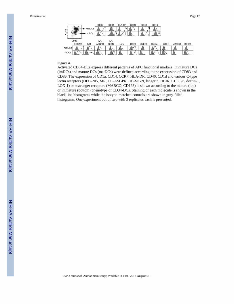
Figure 4.Activated CD34-DCs express different patterns of APC functional markers. Immature DCs(imDCs) and mature DCs (matDCs) were defined according to the expression of CD83 andCD86. The expression of CD1a, CD14, CCR7, HLA-DR, CD40, CD1d and various C-typelectin receptors (DEC-205, MR, DC-ASGPR, DC-SIGN, langerin, DCIR, CLEC-6, dectin-1,LOX-1) or scavenger receptors (MARCO, CD163) is shown according to the mature (top)or immature (bottom) phenotype of CD34-DCs. Staining of each molecule is shown in theblack line histograms while the isotype-matched controls are shown in gray-filledhistograms. One experiment out of two with 3 replicates each is presented.
Romain et al. Page 17
Eur J Immunol. Author manuscript; available in PMC 2013 August 01.
NIH
-PA Author Manuscript
NIH
-PA Author Manuscript
NIH
-PA Author Manuscript

Figure 5.Functional characterization of mature CD34-DCs. (A) Increasing ratios of matured CD34-DCs were cocultured with allogeneic CFSE-stained PBMCs for 7 days and analyzed by flowcytometry. Histogram plots illustrate the gating performed to measure the percentage ofproliferating cells among live CD3+/CD4+ (top) or CD3+/CD8+ T cells (bottom) (seeSupporting Information Fig. 4 for T lymphocyte gating strategy), with CD34-DCs (left) orwithout CD34-DCs (middle) as a negative control. Stimulation with Con A (right) was usedas a positive control. The graph represents mean values ± SEM of data obtained withPBMCs from four macaques cocultured with CD34-DC from four other animals. Data arerepresentative of 4 experiments. (B) The cytokines in the culture supernatants fromunstimulated CD34-DCs (−; grey bars) and from CD34-DCs stimulated with the maturationcocktail for 24 h (+; black bars) were titrated by Multiplex. The data shown are the mean +SEM of four macaques; each assay was performed in duplicate. (C) CD34-DCs were
Romain et al. Page 18
Eur J Immunol. Author manuscript; available in PMC 2013 August 01.
NIH
-PA Author Manuscript
NIH
-PA Author Manuscript
NIH
-PA Author Manuscript
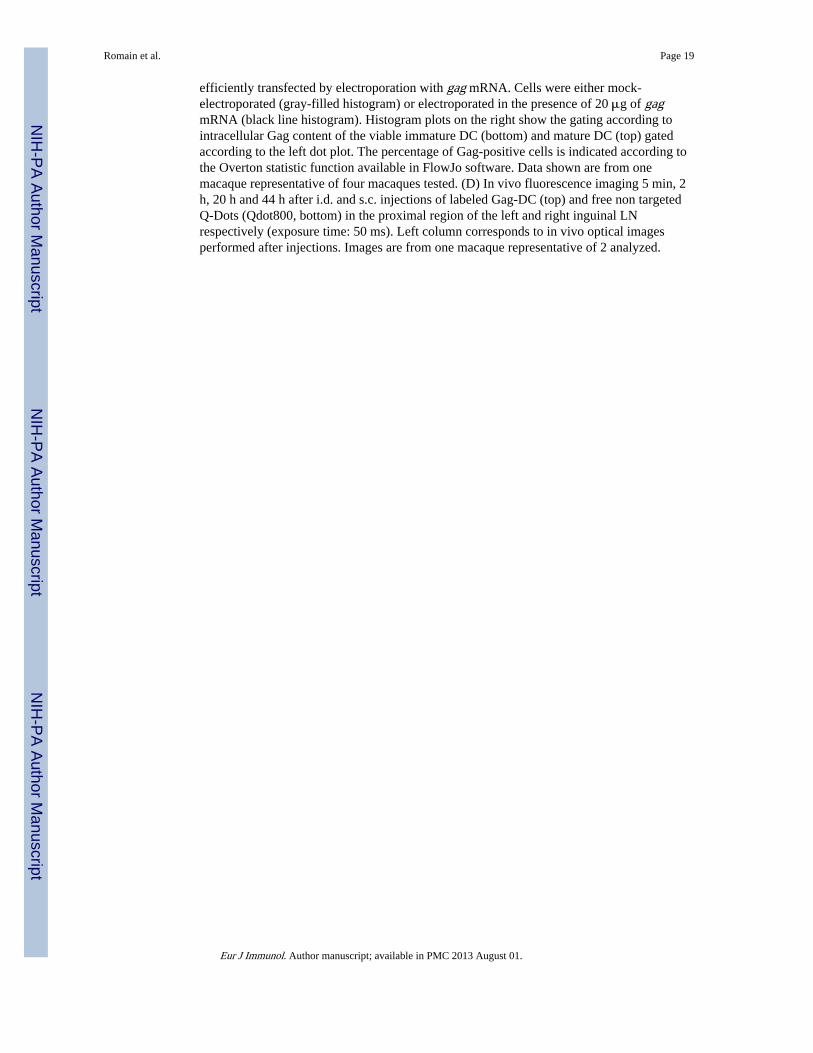
efficiently transfected by electroporation with gag mRNA. Cells were either mock-electroporated (gray-filled histogram) or electroporated in the presence of 20 μg of gagmRNA (black line histogram). Histogram plots on the right show the gating according tointracellular Gag content of the viable immature DC (bottom) and mature DC (top) gatedaccording to the left dot plot. The percentage of Gag-positive cells is indicated according tothe Overton statistic function available in FlowJo software. Data shown are from onemacaque representative of four macaques tested. (D) In vivo fluorescence imaging 5 min, 2h, 20 h and 44 h after i.d. and s.c. injections of labeled Gag-DC (top) and free non targetedQ-Dots (Qdot800, bottom) in the proximal region of the left and right inguinal LNrespectively (exposure time: 50 ms). Left column corresponds to in vivo optical imagesperformed after injections. Images are from one macaque representative of 2 analyzed.
Romain et al. Page 19
Eur J Immunol. Author manuscript; available in PMC 2013 August 01.
NIH
-PA Author Manuscript
NIH
-PA Author Manuscript
NIH
-PA Author Manuscript

Figure 6.gag mRNA-loaded autologous CD34-DCs generate Gag-specific T-cell responses afterinjection into macaques. (A) Peak of the Gag-specific IFN-γ response, measured byELISPOT, in PBMCs of macaques injected with gag mRNA-loaded CD34-DCs and or Gagprotein alone as control after one (V1) or two immunizations (V2). *p<0.05, Mann-Whitneynon-parametric test. Four naïve macaques received two immunizations (week 0 and 4) ofgag mRNA-loaded autologous CD34-DC (5 –15 × 106 cells) injected in the proximal zoneof the inguinal draining lymph node, (half i.d., half s.c.). Two macaques (OBBT5 andOBEA5) received two additional injections of the same batches of gag mRNA-loadedautologous CD34-DCs. The kinetics of Gag-specific (B) IFN-γ responses and (C) IL-2responses were assessed by ELISPOT in freshly isolated PBMCs. Means of triplicates wereplotted for each animal. (D) Phenotype of Gag-mRNA transfected CD34-DC injected forvaccination. Comparison of the cells injected to the high responder animal OBBT5 and tothe low responder animal OBEA5.
Romain et al. Page 20
Eur J Immunol. Author manuscript; available in PMC 2013 August 01.
NIH
-PA Author Manuscript
NIH
-PA Author Manuscript
NIH
-PA Author Manuscript
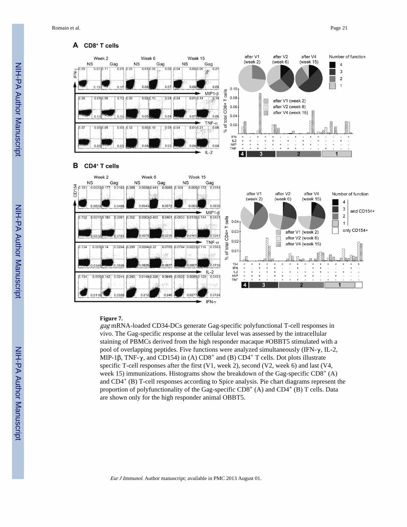
Figure 7.gag mRNA-loaded CD34-DCs generate Gag-specific polyfunctional T-cell responses invivo. The Gag-specific response at the cellular level was assessed by the intracellularstaining of PBMCs derived from the high responder macaque #OBBT5 stimulated with apool of overlapping peptides. Five functions were analyzed simultaneously (IFN-γ, IL-2,MIP-1β, TNF-γ, and CD154) in (A) CD8+ and (B) CD4+ T cells. Dot plots illustratespecific T-cell responses after the first (V1, week 2), second (V2, week 6) and last (V4,week 15) immunizations. Histograms show the breakdown of the Gag-specific CD8+ (A)and CD4+ (B) T-cell responses according to Spice analysis. Pie chart diagrams represent theproportion of polyfunctionality of the Gag-specific CD8+ (A) and CD4+ (B) T cells. Dataare shown only for the high responder animal OBBT5.
Romain et al. Page 21
Eur J Immunol. Author manuscript; available in PMC 2013 August 01.
NIH
-PA Author Manuscript
NIH
-PA Author Manuscript
NIH
-PA Author Manuscript
Copyright © 2022 FDOKUMEN