The Yellow Fever Virus Vaccine Induces a Broad and Polyfunctional Human Memory CD8+ T Cell Response
12
The Yellow Fever Virus Vaccine Induces a Broad and Polyfunctional Human Memory CD8 T Cell Response 1 Rama S. Akondy,* Nathan D. Monson,* Joseph D. Miller,* Srilatha Edupuganti,* Dirk Teuwen, ¶ Hong Wu,* Farah Quyyumi,* Seema Garg,* John D. Altman,* Carlos Del Rio,* Harry L. Keyserling, ‡ Alexander Ploss, § Charles M. Rice, § Walter A. Orenstein,* Mark J. Mulligan,* and Rafi Ahmed 2 * † The live yellow fever vaccine (YF-17D) offers a unique opportunity to study memory CD8 T cell differentiation in humans following an acute viral infection. We have performed a comprehensive analysis of the virus-specific CD8 T cell response using overlapping peptides spanning the entire viral genome. Our results showed that the YF-17D vaccine induces a broad CD8 T cell response targeting several epitopes within each viral protein. We identified a dominant HLA-A2-restricted epitope in the NS4B protein and used tetramers specific for this epitope to track the CD8 T cell response over a 2 year period. This longitudinal analysis showed the following. 1) Memory CD8 T cells appear to pass through an effector phase and then gradually down- regulate expression of activation markers and effector molecules. 2) This effector phase was characterized by down-regulation of CD127, Bcl-2, CCR7, and CD45RA and was followed by a substantial contraction resulting in a pool of memory T cells that re-expressed CD127, Bcl-2, and CD45RA. 3) These memory cells were polyfunctional in terms of degranulation and production of the cytokines IFN-, TNF-, IL-2, and MIP-1. 4) The YF-17D-specific memory CD8 T cells had a phenotype (CCR7 CD45RA ) that is typically associated with terminally differentiated cells with limited proliferative capacity (T EMRA ). However, these cells exhibited robust proliferative potential showing that expression of CD45RA may not always associate with terminal differentiation and, in fact, may be an indicator of highly functional memory CD8 T cells generated after acute viral infections. The Journal of Immunology, 2009, 183: 7919 –7930. I mmune memory forms the basis of vaccine-induced protec- tion, and memory CD8 T cells form an important cellular component of this immunity (1). Our understanding of CD8 T cell memory has advanced a great deal through exhaustive stud- ies in acute viral infections that cause protective, long-lasting memory (2–5). We know from these studies that Ag-driven clonal expansion results in a dynamic antiviral CD8 T cell response initially manifested as pathogen clearance via cytotoxic molecules and effector cytokines and later by the presence of a small popu- lation of memory cells that can be rapidly recruited to blunt sub- sequent infections. We have also begun to understand the differ- ences between the properties of poor and high quality memory CD8 T cells based on their cytotoxicity, cytokine production, homeostatic turnover, and proliferative potential (6 –17). The yellow fever vaccine strain 17D (YF-17D), 3 a live attenuated form of the wild-type virus, has not only been extremely effective in controlling yellow fever but is also one of the safest vaccines in young adults (18, 19). A single immunization confers protection, and virus- neutralizing Abs can be detected for up to 30 years post-vaccination (20). The live viral nature of the vaccine combined with its efficacy is useful for studying how humans generate functional immunity in the context of an acute viral infection and also as a potential expression vector for recombinant vaccines (21, 22). Some of the studies that address the complex interactions between the virus and the immune system suggest that the ability of YF-17D to infect dendritic cells and activate multiple TLRs may be crucial for generating the robust adaptive immune response seen after vaccination (23–25). The im- mune memory developed as a consequence comprises the ability to rapidly produce neutralizing Abs as well as the CD8 T cell ef- fectors (26 –28) that are likely required for killing cells infected with the virus that escape the humoral response. This cellular memory has been of interest to our group because it presents a rare opportunity to study primary CD8 T cell responses in humans. CD8 T cells that recognize HLA-B35-restricted epitopes in E, NS1, NS2B, and NS3 proteins have been reported in vaccinees (26). In addition, we demonstrated recently that the magnitude of the total effector CD8 T cells response against YF-17D (or the smallpox live viral vaccine) could be measured using the transient Ki67 Bcl-2 low HLA-DR CD38 phenotype of effector T cells (28, 29). However, there are gaps in our knowledge regarding the breadth of the response, the differentiation of virus-specific effector *Emory Vaccine Center and the Hope Clinic, † Department of Microbiology and Im- munology, and ‡ Department of Pediatrics, Emory University School of Medicine, Atlanta, GA 30022; § Center for the Study of Hepatitis C, Laboratory of Virology and Infectious Disease, Rockefeller University, New York, NY 10065; and ¶ Sanofi Pas- teur, Lyon, France Received for publication December 1, 2008. Accepted for publication October 20, 2009. The costs of publication of this article were defrayed in part by the payment of page charges. This article must therefore be hereby marked advertisement in accordance with 18 U.S.C. Section 1734 solely to indicate this fact. 1 This work was supported by National Institutes of Health (NIH) U19 Grant AI057266 (to R.A.) and in part by Sanofi-Pasteur, Lyon, France. C.M.R. receives support from the Greenberg Medical Research Institute, the Starr Foundation, the Foundation for the National Institutes of Health through the Grand Challenges in Global Health initiative (grant identification nos. 334 and 574), General Clinical Re- search Center Grant M01-RR00102 (to Rockefeller University Hospital), and Center for Translational Science Award Grant 1UL1 RR024143-01 (to Rockefeller Univer- sity Hospital) from the NIH National Center for Research Resources. 2 Address correspondence and reprint requests to Dr. Rafi Ahmed, 1510 Clifton Road., G211, Atlanta, GA 30322. E-mail address: [email protected] 3 Abbreviations used in this paper: YF-17D, yellow fever virus strain 17D; ICC, intra- cellular cytokine; PD-1, programmed death-1; T EMRA , effector memory RA T cell. Copyright © 2009 by The American Association of Immunologists, Inc. 0022-1767/09/$2.00 The Journal of Immunology www.jimmunol.org/cgi/doi/10.4049/jimmunol.0803903
-
Upload
independent -
Category
Documents
-
view
1 -
download
0
Transcript of The Yellow Fever Virus Vaccine Induces a Broad and Polyfunctional Human Memory CD8+ T Cell Response

The Yellow Fever Virus Vaccine Induces a Broad andPolyfunctional Human Memory CD8� T Cell Response1
Rama S. Akondy,* Nathan D. Monson,* Joseph D. Miller,* Srilatha Edupuganti,*Dirk Teuwen,¶ Hong Wu,* Farah Quyyumi,* Seema Garg,* John D. Altman,* Carlos Del Rio,*Harry L. Keyserling,‡ Alexander Ploss,§ Charles M. Rice,§ Walter A. Orenstein,*Mark J. Mulligan,* and Rafi Ahmed2*†
The live yellow fever vaccine (YF-17D) offers a unique opportunity to study memory CD8� T cell differentiation in humansfollowing an acute viral infection. We have performed a comprehensive analysis of the virus-specific CD8� T cell response usingoverlapping peptides spanning the entire viral genome. Our results showed that the YF-17D vaccine induces a broad CD8� T cellresponse targeting several epitopes within each viral protein. We identified a dominant HLA-A2-restricted epitope in the NS4Bprotein and used tetramers specific for this epitope to track the CD8� T cell response over a 2 year period. This longitudinalanalysis showed the following. 1) Memory CD8� T cells appear to pass through an effector phase and then gradually down-regulate expression of activation markers and effector molecules. 2) This effector phase was characterized by down-regulation ofCD127, Bcl-2, CCR7, and CD45RA and was followed by a substantial contraction resulting in a pool of memory T cells thatre-expressed CD127, Bcl-2, and CD45RA. 3) These memory cells were polyfunctional in terms of degranulation and productionof the cytokines IFN-�, TNF-�, IL-2, and MIP-1�. 4) The YF-17D-specific memory CD8� T cells had a phenotype(CCR7�CD45RA�) that is typically associated with terminally differentiated cells with limited proliferative capacity (TEMRA).However, these cells exhibited robust proliferative potential showing that expression of CD45RA may not always associate withterminal differentiation and, in fact, may be an indicator of highly functional memory CD8� T cells generated after acute viralinfections. The Journal of Immunology, 2009, 183: 7919–7930.
I mmune memory forms the basis of vaccine-induced protec-tion, and memory CD8� T cells form an important cellularcomponent of this immunity (1). Our understanding of CD8�
T cell memory has advanced a great deal through exhaustive stud-ies in acute viral infections that cause protective, long-lastingmemory (2–5). We know from these studies that Ag-driven clonalexpansion results in a dynamic antiviral CD8� T cell responseinitially manifested as pathogen clearance via cytotoxic moleculesand effector cytokines and later by the presence of a small popu-lation of memory cells that can be rapidly recruited to blunt sub-sequent infections. We have also begun to understand the differ-ences between the properties of poor and high quality memoryCD8� T cells based on their cytotoxicity, cytokine production,homeostatic turnover, and proliferative potential (6–17).
The yellow fever vaccine strain 17D (YF-17D),3 a live attenuatedform of the wild-type virus, has not only been extremely effective incontrolling yellow fever but is also one of the safest vaccines in youngadults (18, 19). A single immunization confers protection, and virus-neutralizing Abs can be detected for up to 30 years post-vaccination(20). The live viral nature of the vaccine combined with its efficacy isuseful for studying how humans generate functional immunity in thecontext of an acute viral infection and also as a potential expressionvector for recombinant vaccines (21, 22). Some of the studies thataddress the complex interactions between the virus and the immunesystem suggest that the ability of YF-17D to infect dendritic cells andactivate multiple TLRs may be crucial for generating the robustadaptive immune response seen after vaccination (23–25). The im-mune memory developed as a consequence comprises the ability torapidly produce neutralizing Abs as well as the CD8� T cell ef-fectors (26–28) that are likely required for killing cells infectedwith the virus that escape the humoral response. This cellularmemory has been of interest to our group because it presents a rareopportunity to study primary CD8� T cell responses in humans.CD8� T cells that recognize HLA-B35-restricted epitopes in E,NS1, NS2B, and NS3 proteins have been reported in vaccinees(26). In addition, we demonstrated recently that the magnitude ofthe total effector CD8� T cells response against YF-17D (or thesmallpox live viral vaccine) could be measured using the transientKi67�Bcl-2lowHLA-DR�CD38� phenotype of effector T cells(28, 29). However, there are gaps in our knowledge regarding thebreadth of the response, the differentiation of virus-specific effector
*Emory Vaccine Center and the Hope Clinic, †Department of Microbiology and Im-munology, and ‡Department of Pediatrics, Emory University School of Medicine,Atlanta, GA 30022; §Center for the Study of Hepatitis C, Laboratory of Virology andInfectious Disease, Rockefeller University, New York, NY 10065; and ¶Sanofi Pas-teur, Lyon, France
Received for publication December 1, 2008. Accepted for publication October20, 2009.
The costs of publication of this article were defrayed in part by the payment of pagecharges. This article must therefore be hereby marked advertisement in accordancewith 18 U.S.C. Section 1734 solely to indicate this fact.1 This work was supported by National Institutes of Health (NIH) U19 GrantAI057266 (to R.A.) and in part by Sanofi-Pasteur, Lyon, France. C.M.R. receivessupport from the Greenberg Medical Research Institute, the Starr Foundation, theFoundation for the National Institutes of Health through the Grand Challenges inGlobal Health initiative (grant identification nos. 334 and 574), General Clinical Re-search Center Grant M01-RR00102 (to Rockefeller University Hospital), and Centerfor Translational Science Award Grant 1UL1 RR024143-01 (to Rockefeller Univer-sity Hospital) from the NIH National Center for Research Resources.2 Address correspondence and reprint requests to Dr. Rafi Ahmed, 1510 CliftonRoad., G211, Atlanta, GA 30322. E-mail address: [email protected]
3 Abbreviations used in this paper: YF-17D, yellow fever virus strain 17D; ICC, intra-cellular cytokine; PD-1, programmed death-1; TEMRA, effector memory RA T cell.
Copyright © 2009 by The American Association of Immunologists, Inc. 0022-1767/09/$2.00
The Journal of Immunology
www.jimmunol.org/cgi/doi/10.4049/jimmunol.0803903

and memory CD8� T cells, and the functional qualities of thesecells.
To address the above questions, we used a peptide library en-compassing the entire YF-17D genome for estimating the magni-tude of the T cells targeting each viral protein and the range ofepitopes recognized. Using a strategy involving direct ex vivostimulation of cells with overlapping peptides and detection ofcytokine production (30), we identified an immunodominant classI-restricted epitope that permitted longitudinal tracking of YF-17D-specific CD8� T cells in individual vaccinees using MHCclass I tetramers. This analysis offers novel perspectives for tworeasons. First, our study has the advantage that the subjects wereprimary vaccinees from a geographical area that is not endemic foryellow fever, i.e., the United States (19). Thus, an accurate deter-mination of the breadth and magnitude of the primary YF-17D-specific response could be made while avoiding the possible cross-reactivity introduced by prior exposure to either the yellow fevervirus or other closely related flaviviruses. Second, we performed alongitudinal tetramer-based analysis and followed YF-17D-spe-cific CD8� T cells from the time they first appeared in circulationuntil 2 years later, in the same group of vaccinees. Hence, we wereable to study various stages of memory CD8� T cell differentiationlongitudinally without the confounding effects introduced by across-sectional analysis. This analysis will be valuable in under-standing the basic tenets of human memory T cell differentiationafter acute viral infections.
Materials and MethodsStudy subjects and blood samples
Healthy volunteers (18–40 years of age) were recruited in the study afterinformed consent. Approval for all procedures was obtained from theEmory University Institutional Review Board (Atlanta, GA). A single dose(0.5 ml containing at least 105 PFU) of 17D live-attenuated yellow fevervaccine strain was administered subcutaneously. The recommendations es-tablished by the Advisory Committee on Immunization Practices (Depart-ment of Health and Services, Centers for Disease Control and Prevention,Atlanta, GA) were followed for selection and vaccination of individuals inthe study. In addition, individuals with a previous history of vaccinationwith YF-17D or exposure to flaviviruses as evidenced by serology or ahistory of travel to endemic areas were excluded from the study. Bloodsamples were analyzed before and at various times postvaccination as in-dicated in the text. Seroconversion after vaccination was confirmed byassaying the neutralizing Ab titers for YF-17D (data not shown). PBMCwere purified from cell preparation tubes (BD Biosciences), and EDTAblood samples were used to quantify viral RNA by real-time PCR as de-scribed elsewhere (28).
YF-17D peptide library
To map T cell epitopes, a library of overlapping peptides spanning the entireYF-17D polyprotein was made. All 851 peptides comprising the library were15-aa long (Synpep) and nonamidylated, with neighboring peptides overlap-ping by 11 aa. The peptides were organized into 60 pools based on a matrixsuch that each pool had 24–30 peptides, each at a concentration of 10 �g/ml,and any two pools had no more than one peptide in common (30, 31).
Cell preparation, stimulation, and intracellular cytokine (ICC)staining
PBMC were purified from cell preparation tubes (BD Biosciences) accord-ing to standard protocols and cryopreserved in 90% FCS plus 10% DMSO.For mapping T cell epitopes, freshly isolated PBMC were stimulated witheach of 60 peptide pools. Assays for confirming epitopes in the singlepeptides identified using the pools were done using cryopreserved PBMCthat were revived, rested overnight at 37°C, and stimulated with 10 �g/mlpeptides. Fresh or revived PBMC were stimulated for 6 h in 96-well round-bottom plates in the presence of brefeldin A (1 �l/ml) and anti-CD28/CD49d (10 �l/ml) and then stained for relevant T cell markers and cyto-kines. Culture medium for all cellular assays was RPMI 1640 containing10% FCS, 2 mM glutamine, 100 IU/ml penicillin, and 100 �g/ml strepto-mycin (RPMI 1640 plus 10% FCS).
Staining and flow cytometry analysis
All mAbs except anti-granzyme B (Caltag), programmed death (PD)-1(provided by Dr. G. J. Freeman, Dana Farber Cancer Institute, Boston,MA), and CCR7 (R&D Systems) were obtained from BD Biosciences.A2-NS4B 214–222 tetramers were made in-house. For ICC after in vitrostimulation, cells were stained for T cell markers by incubation with therelevant Abs at room temperature for 30 min, washed with PBS, and thenstained for cytokines using anti-IFN-�, TNF-�, IL-2 and Mip-1� Abs aftercell permeabilization with the Cytofix/Cytoperm kit (BD Biosciences). De-granulation in stimulated cells was measured by including anti-CD107a-PE(10 �l/well) in the initial culture medium. For phenotypic analysis of A2-NS4B� CD8� T cells, 100–200 �l of unprocessed whole blood was in-cubated at room temperature first with tetramer for 10 min and then for afurther 30 min with Abs for surface markers. This was followed by a10-min lysis of RBC using FACS lysing solution (BD Biosciences), wash-ing with PBS, and fixing in 1% p-formaldehyde. For staining intracellularproteins like Bcl-2, Ki-67, granzyme B, and perforin, cells were permeabilizedand stained using the Cytofix/Cytoperm kit (BD Biosciences) according to themanufacturer’s instructions. Data were acquired on a FACSCalibur (BD Bio-sciences) or LSR-II (BD Biosciences) flow cytometer and analyzed using FlowJo (Tree Star) software. In addition, the programs SPICE (version 4.1.5) andPESTLE (version 1.5.2) (both from M. Roederer, National Institutes of Health,Vaccine Research Center, Bethesda, MD) were used to quantify CD8� T cellspositive for the various combinations of functions assayed. Statistical analysisand graphical representation of data was performed using GraphPad Prismsoftware.
CFSE labeling and in vitro expansion of CD8� T cells
PBMC from vaccinated subjects were labeled for 5 min with 1 �M CFSE(Molecular Probes) in PBS at room temperature; cold FCS was then added andcells were washed extensively with RPMI 1640 plus 10% FCS. CFSE-labeledcells were incubated with or without the NS4B 214–222 peptide (10 �g/ml)for 6 days, at the end of which flow cytometry and analysis were performed asdescribed above. Responding CD8� T cells were identified either by tetramerstaining or by ICC staining after a 6-h recall with peptide.
ResultsYF-17D elicits a broad diversity of memory CD8� T cells
To define the breadth of the primary antiviral CD8� T cell re-sponse, we created a library of overlapping peptides encompassingthe entire viral polyprotein. This library was organized into 60pools of multiple peptides based on a 24 � 36 matrix such thateach peptide was present in precisely two pools (30). Responses toall of these pools were studied in nine healthy subjects vaccinated2 mo previously with a single s.c. injection of YF-17D. PBMCisolated from vaccinees were stimulated with individual pools fol-lowed by ICC staining assays, and responding virus-specific CD8�
T cells were identified by their ability to produce IFN-�. Of 60peptide pools tested with this assay, between eight and 25 poolswere capable of eliciting IFN-� production in different vaccinees.Many pools contained peptides from adjacent sequences in theviral polyprotein and thus had epitopes primarily from one protein.IFN-� production stimulated by these pools was used to estimatethe contribution of individual viral proteins toward eliciting aCD8� T cell response (Fig. 1). Overall, each vaccinee had CD8�
T cells targeting multiple proteins and several epitopes within eachprotein, resulting in broad cellular immunity. Despite variation intheir magnitude, CD8� T cells specific for each of the 10 YF-17Dproteins were detected, indicating that every viral protein was im-munogenic. IFN-�� CD8� T cells specific for E, NS3, and NS5were elicited in all vaccinees with an average of 10.7% (SD 8.0),16.7% (SD 7.0), and 27.7% (SD 14.0) of the total respondingCD8� T cells specific for E, NS3, and NS5 respectively. NS1 alsoelicited frequent (in 8/9 vaccinees) but lower numbers (mean7.2%; SD 3.7) of CD8� T cells, whereas the magnitude of C-, M-,NS2A-, and NS2B-reactive CD8� T cells was low (�3%) whendetected. Strikingly, in five vaccinees the NS4B component wasdominant and accounted for �42.1% (SD 13.5) of the totalIFN-�� CD8� T cells (Fig. 1).
7920 YFV-17D INDUCES POLYFUNCTIONAL HUMAN CD8� T CELL MEMORY
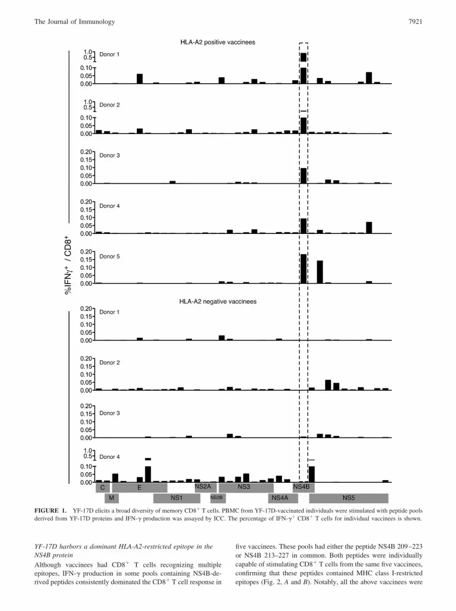
YF-17D harbors a dominant HLA-A2-restricted epitope in theNS4B protein
Although vaccinees had CD8� T cells recognizing multipleepitopes, IFN-� production in some pools containing NS4B-de-rived peptides consistently dominated the CD8� T cell response in
five vaccinees. These pools had either the peptide NS4B 209–223or NS4B 213–227 in common. Both peptides were individuallycapable of stimulating CD8� T cells from the same five vaccinees,confirming that these peptides contained MHC class I-restrictedepitopes (Fig. 2, A and B). Notably, all the above vaccinees were
FIGURE 1. YF-17D elicits a broad diversity of memory CD8� T cells. PBMC from YF-17D-vaccinated individuals were stimulated with peptide poolsderived from YF-17D proteins and IFN-� production was assayed by ICC. The percentage of IFN-�� CD8� T cells for individual vaccinees is shown.
7921The Journal of Immunology

positive for the HLA-A2 serotype (data not shown), suggestingthat the response was HLA-A2 restricted. An epitope predictionalgorithm (32) was used to identify putative HLA-A0201-re-stricted epitopes in the 15-mer NS4B 209–223 and NS4B 213–227peptides. Of the several nonamer epitopes predicted, three epitopeswith the highest scores (and hence the best binding motifs), NS4B
213–221, NS4B 214–222, and NS4B 215–223 were tested by ICCstaining for their ability to stimulate IFN-� production. Only onepeptide, NS4B 214–222 (amino acid sequence LLWNGPMAV)generated IFN-�-producing CD8� T cells in HLA-A2� vaccinees,identifying it as the HLA-A2-restricted CD8� T cell epitope (Fig.2, A and B). In addition, control peptides that had a single aminoacid substitution (either L214 to V214 or L215 to E215) did notstimulate IFN-� production, thus validating the specificity of theNS4B 214–222 epitope. MHC-peptide tetramers containing HLA-A0201 and the NS4B 214–222 peptide were prepared and tested inindividuals. Tetramer-stained CD8� cells were seen in vaccinatedHLA-A2� but not HLA-A2� individuals, confirming the specific-ity of the epitope (Fig. 2C). This tool enabled us to perform alongitudinal analysis of YF-17D-specific CD8� T cells.
Kinetics of the YF-17D-specific primary CD8� T cell responsetracked by MHC class I tetramers
We analyzed the dynamics of viral replication after the vaccinationof HLA-A2� individuals who did not have a history of exposureto flaviviruses (Fig. 3). Most vaccinees did not have detectableviral RNA in the first 2 days following vaccination; on day 3,several vaccinees tested positive for viral RNA (mean 114 copies/ml; SD 50), and the number of positives increased further such thatall vaccinees had YF-17D genomes in the plasma by day 5. Thehighest level of viral RNA in circulation was seen on day 5 formost vaccinees (mean 3598 copies/ml; SD 1813) and on day 7 forothers (mean 1443 copies/ml; SD 613), after which it declinedquickly. By day 11 all except two vaccinees had no viral RNA inthe plasma, and by day 14 viral genomes were absent.
Ag dictates the size of the effector CD8� T cell response and thekinetics of initial expansion (33). We used A2-NS4B 214–222tetramers to quantify the size and examine the kinetics of the YF-17D-driven primary CD8� T cell response in vaccinees. Strik-ingly, A2-NS4B tetramer� CD8� T cells were seen in nearly allHLA-A2� vaccinees (19 of 21). The A2-NS4B� CD8� cells wereoften detectable as early as 11 days after vaccination and definitelyafter 14 days in all responding individuals, coincident with thedecline in plasma viral RNA. Tetramer� cells continued to expandtill 30 days postvaccination, when the peak tetramer frequencyranging from 0.5 to 17% of CD8� T cells was seen in the vacci-nees; it diminished by �3.6-fold by day 90. The size of the A2-NS4B� pool at this time was roughly proportional to that seen onday 30; vaccinees with high peak frequencies retained high num-bers of memory cells. We were able to track these cells for anadditional time in five vaccinees from the same cohort and foundthat tetramer� cells were readily detectable even 1 year after vac-cination (Fig. 3B). In conclusion, HLA-A2� vaccinees have a ro-bust and long-lived response to the NS4B 214–222 epitope, show-ing expansion, contraction, and memory kinetics typical of CD8�
T cells elicited in a primary acute viral infection.
Effector and memory differentiation of YF-17D-specific CD8�
T cells
To understand the differentiation of effector and memory CD8� Tcells and to gain an insight into the generation of YF-17D-specificCD8� memory, we performed a detailed longitudinal analysis oftetramer-stained CD8� T cells in 15 HLA-2� vaccinees (Fig. 4).Because rapid clonal expansion is the hallmark of a virus-drivenCD8� T response, we assessed proliferation of YF-17D-specificCD8� T cells using Ki-67, a marker that is tightly associated withcycling CD8� T cells (34). Compared with naive CD8� T cellsthat were Ki-67 negative, �95% of the A2-NS4B� cells detectedearly on (days 11 to 14) were Ki-67 positive. The Ki-67 stainingdeclined significantly (present in �5% of the cells) by day 30 and
FIGURE 2. YF-17D harbors a dominant HLA-A2 restricted epitope inthe NS4B protein. A, Sequences of the peptides used to confirm and iden-tify the immunogenic nonamer epitope in NS4B. B, PBMC from HLA-A2� vaccinees were cultured in the presence or absence of 15-mer peptides(NS4B 209–223 or NS4B213–227) and ICC staining was performed (up-per panels). To confirm the nonamer epitope in these peptides, PBMC werestimulated with either the NS4B 214–222 peptide or with control peptidesthat differed by one amino acid (lower panels). Plots gated on CD8� T cellsfor one representative donor are shown. C, Identification of YF-17D-spe-cific CD8� T cells using MHC class I tetramers. Peripheral blood fromHLA-A2� or HLA-A2� individuals vaccinated with YF-17D 2 wk previ-ously was stained with the A2-NS4B tetramer. Plots are gated on all lym-phocytes and numbers indicate the percentage of tetramer� cells from totallymphocytes.
7922 YFV-17D INDUCES POLYFUNCTIONAL HUMAN CD8� T CELL MEMORY

decreased to baseline by day 90. The proliferation seen early onwas coupled with decreased expression of the antiapoptotic proteinBcl-2, suggesting that a majority of these cells were destined toundergo apoptosis. Progressive reexpression of Bcl-2 occurredconcomitantly with loss of Ki-67 and differentiation to a memoryphenotype. Expression of the activation markers HLA-DR andCD38 mirrored the kinetics seen with Ki-67. Activated but notnaive CD8� T cells also expressed the chemokine receptor CCR5.Although we observed that the CCR5 expression kinetics was sim-ilar to those of the proliferation and activation markers, unlike theother markers CCR5 was retained in 40–50% of A2-NS4B� mem-ory cells. Potent effector properties were evidenced by high levelsof granzyme B (Fig. 4), granzyme A, and perforin (data not shown)expression until as late as 30 days after YF-17D vaccination, andtetramer� cells continued to express granzyme B later, although atlower levels.
We analyzed the expression of markers associated with otherrelevant functions of CD8� T cells (Fig. 4). CD127 (IL-7R�)has been shown earlier to be an important marker that is down-regulated on effectors and selectively reexpressed on a subsetdestined to form precursors of the memory pool (10, 35). Weobserved substantial but not complete down-regulation ofCD127 on A2-NS4B� CD8� T cells over the 30 days followingvaccination, and a gradual reexpression thereafter. Coexpres-sion of CCR7, a homing marker, and CD45RA, a transmem-
brane tyrosine phosphatase, is characteristic of naive cells (36).Both markers were down-regulated in the YF-17D-specificCD8� T cells during effector differentiation. However in con-trast to the lack of CCR7 on a majority of memory cells,CD45RA was reexpressed over time. We next examined theprofiles of signaling molecules that regulate T cell activation.PD-1 is a regulatory receptor of the CD28 family that is tran-siently expressed on activated T cells during acute viral infec-tions (37). Its up-regulation by CD8� T cells (transient in acuteand long-term in chronic viral infections) inhibits the activa-tion, expansion, and acquisition of effector CD8� T cell func-tions (38). A2-NS4B� cells transiently up-regulated PD-1 be-tween 11 and 14 days after YF-17D vaccination, similar toreports with other acute viral infections in mice (38). To assessthe ability to respond to costimulatory signals at the differentstages of differentiation, we evaluated the expression of twocritical signaling proteins, CD27 and CD28. CD27 was uni-formly expressed at high levels during all stages of the differ-entiating virus specific CD8� T cells, whereas CD28 expressionwas uniform in effectors (day 11–14) and heterogeneous onmemory cells. CD11a, a component of the adhesion moleculeLFA-1 (39), was expressed by YF-17D-experienced but not na-ive CD8� T cells as described in studies with other infections(40).
FIGURE 3. Kinetics of the YF-17D-specific primary CD8� T cell response tracked by MHC class I tetramers. A, Tetramer kinetics in onerepresentative vaccinee is shown. Plots show all lymphocytes but the numbers indicate the percentage of A2-NS4B� cells from total CD8� T cells.B, YF-17D genomes in the plasma (red line; mean � SD) and the percentage of A2-NS4B� (black lines; each for one vaccinee) in YF-17D vaccineesover 1 year is shown.
7923The Journal of Immunology

To summarize, the primary antiviral response was characterizedby a pronounced activation and expansion phase that led to in-creasing frequencies of A2-NS4B� cells by day 30. At this stage,YF-17D-specific cells were at the end of the expansion phase andshowed a phenotype intermediate to that of effector and memoryCD8� T cells. Substantial contraction resulted in a pool of mem-ory cells that had lost activation and proliferation markers, uni-
formly expressed CD45RA and CD27, and had heterogeneous ex-pression with respect to proteins such as CCR5, CD28, andCD127.
The YF-17D-specific CD8� T cell memory is fine tuned over time
Memory differentiation is a gradual and continuous process result-ing in subtle changes in the memory pool that may not be obvious
FIGURE 4. Effector and memorydifferentiation of YF-17D-specificCD8� T cells. A, Results from the lon-gitudinal phenotypic analysis of A2-NS4B� cells present in blood of 10 to15 vaccinees are summarized. The day11, 14, 30, and 90 data are from thesame group of vaccinees. The day 0data are from a separate group and rep-resent expression of the relevantmarker on naive (CD45RA�CCR7�)CD8� T cells. The percentage of A2-NS4B� CD8� T cells expressing therelevant marker for each individual(open circles) and the group mean(horizontal line) are shown. MFI,Mean fluorescence intensity. B, Flowplots representing the phenotype ofYF-17D-specific CD8� T cells. Plotsare gated on total CD8� T cells (blackbackground) or YF-17D-specific (A2-NS4B�) CD8� T cells (red dots).Numbers show the percentage of A2-NS4B� CD8� T cells in the gate.
7924 YFV-17D INDUCES POLYFUNCTIONAL HUMAN CD8� T CELL MEMORY

FIGURE 4. (continued)
7925The Journal of Immunology

over a short period of time. We followed A2-NS4B� memory cellsat 3 mo and 2 years after vaccination in the same three individualsto determine any such gradual changes (Fig. 5). After 2 years, thefrequency of A2-NS4B� memory cells in the blood of these indi-viduals had decreased from 1.3, 0.43, and 0.36% at 3 mo to 0.13,0.12, and 0.06%, respectively (data not shown). Phenotypic anal-ysis revealed that strikingly, �95% of the A2-NS4B� cells con-tinued to express CD45RA even 2 years later, verifying that reex-pression of this molecule was not a transient phase of memorydifferentiation (Fig. 5A). In addition, uniform CD27 expressionand heterogeneous CD28 expression were observed at both early(3 mo) and late (2 years) memory stages. However, we did observea subtle increase over time with respect to CCR7 and CD127, twomarkers associated with high-quality immune memory (Fig. 5A).The expression of CD45RA has been associated with senescencein several studies (41), whereas CD127 confers the ability to re-spond to IL-7 mediated survival signals (4). To further dissect theissue of whether these memory cells were still capable of differ-entiation, we examined two markers, CD56 and CD57, associatedwith terminal effectors (Fig. 6). A2-NS4B� CD8� T cells did notexpress either CD56, which is thought to identify CD8� T cellswith direct cytolytic activity (42), or CD57, which is associatedwith replicative senescence (43), suggesting that YF-17D-specificmemory cells were not terminally differentiated even though theyexpressed CD45RA and were mostly CCR7 negative, a phenotypeoften associated with terminal effectors.
YF-17D elicits polyfunctional, long-lived memory CD8� T cells
CD8� T cells have a spectrum of functions to achieve viralcontrol. We evaluated five functions (degranulation and secre-tion of IFN-�, TNF-�, Mip1�, and IL-2) of CD8� T cells rec-ognizing the A2-NS4B epitope by stimulation of PBMC in vitrowith the NS4B 214 –222 peptide followed by ICC staining andmulticolor flow cytometry. The kinetics of IFN-�-producingCD8� T cells (Fig. 6A) matched that seen using tetramer-basedanalysis (Fig. 3), with highest frequencies seen 30 days post-vaccination. Upon assessing the composition of the cytokine-producing population, we found that at every time point ana-lyzed the cells that simultaneously produced IFN-� and TNF-�dominated the response, and a prominent fraction of these cellsadditionally produced IL-2 (Fig. 6A). Surface mobilization ofCD107a (lysosome-associated membrane protein (LAMP)-1)after peptide stimulation indicates the ability to release cyto-lytic granules (44). Independent and simultaneous measurementof four functions showed that the majority of virus-specificCD8� T cells from days 11 through 90 were CD107a� as wellas capable of IFN-�, TNF-�, and Mip-1� production at thesame time (Fig. 6B). Notably, this polyfunctional CD8� T cellmemory was retained even 2 years after vaccination (Fig. 6C).Successful pathogen clearance depends on both quantity andquality of CD8� T cells, and our findings show that highlypolyfunctional memory CD8� T cells are elicited and main-tained after YF-17D vaccination.
A cardinal property of memory CD8� T cells is their ability toundergo rapid proliferation upon reencountering Ag (5). This is animportant component of protective immunity; a higher prolifera-tive potential implies a larger pool of secondary effectors. To ex-amine the proliferative potential of A2-NS4B-specific CD8� Tcells, CFSE-labeled PBMC from vaccinees were stimulated invitro with the relevant peptide and the dilution of CFSE was as-sayed as a measure of cell division (Fig. 7A). Expansion of A2-NS4B� cells at day 90 was much higher (7.2-fold) compared withday 14 (2.3-fold). This proliferative potential was preserved evenwhen �95% of tetramer� cells were CD45RA� (supplemental
FIGURE 5. The YF-17D-specific CD8� T cell memory is fine-tunedover 2 years. A, Comparison of the phenotype YF-17D-specific memoryCD8� T cells 3 mo and 2 years after vaccination. Representative flow plots(gated on total CD8� T cells) from one of three vaccinees are shown. B,Representative flow plots (gated on total CD8� T cells) show expression ofthe late differentiation markers CD56 and CD57 on YF-17D-specific mem-ory CD8� T cells in an individual vaccinated 2 years earlier.
7926 YFV-17D INDUCES POLYFUNCTIONAL HUMAN CD8� T CELL MEMORY

FIGURE 6. YF-17D elicits polyfunctional, long-lived memory CD8� T cells. A, Kinetics of cytokine-producing CD8� T cells in individuals immunizedwith YF-17D. PBMC were isolated from vaccinees and responses to the NS4B 214–222 peptide were measured by ICC staining. Representative flow plots(gated on total CD8� T cells) from one vaccinee are shown. B, Functional profile of the YF-17D-specific CD8� T cell response. All possible combinationsof four functions (degranulation by CD107a staining and secretion of IFN-�, TNF-�, and Mip-1�) are shown on the x-axis. Bars indicate the percentageof the total response contributed by CD8� T cells with the combination of responses on the x-axis. Responses are grouped according to the number offunctions and the data summarized by the pie charts. Each slice of the pie represents the fraction of the total response that consists of CD8� T cells positivefor a given number of functions. Mean data from four YF-17D-immunized individuals are shown. C, Functional profile of the YF-17D-specific memoryCD8� T cells 2 years after vaccination. Plots gated on CD8� T cells show the cytokines produced in response to the stimulation of PBMC with the NS4B214–222 peptide. Data from one representative vaccinee are shown.
7927The Journal of Immunology

Fig. 1).4 Thus, YF-17D-specific CD8� T cells progressively ac-quire proliferative potential over the course of time. The quality ofmemory CD8� T cells generated in chronic viral infections is in-ferior to that in response to acute viral infections (9). Ag-drivenproliferation in particular is reported to be lower (45, 46). We wereable to make a direct comparison in this regard between YF-17D,an acute virus, and CMV, a chronic virus, in an individual vacci-nated 1 year previously with YF-17D. This vaccinee had CD8� Tcells specific for A2-NS4B as well as for a CMV pp65-derivedepitope (A2-NLV). A2-NS4B� CD8� T cells expanded to higherlevels (8.6-fold) than A2-NLV-specific CD8� T cells (1.45-fold)upon stimulation with the corresponding peptides, indicative of thesuperior proliferation capability in YF-17D memory CD8� T cells(Fig. 7B). Strikingly, A2-NS4B� cells that diluted CFSE could bedetected in an individual immunized 10 years previously (Fig. 7C).This vaccinee had no history of recent travel to a yellow feverendemic area, strongly suggesting that long-term persistence ofYF-17D-specific CD8� T cell memory does not require antigenicstimulation.
To summarize, YF-17D generates a CD8� T cell responsethat is high in magnitude, broadly diverse, and polyfunctional.This results in memory CD8� T cells that progressively acquirepotent proliferative potential and retain it long after the virus iscleared.
DiscussionLive viral vaccines like YF-17D offer a rare opportunity to studythe progressive differentiation of human Ag-specific CD8� T cells
that are generated as part of a protective antiviral immune re-sponse. We have studied the magnitude, breadth, and dynamics ofthe YF-17D-specific CD8� T cell response as a model to study thegeneration and maintenance of CD8� T cell memory in the contextof acute viral infections. We used a set of overlapping peptidesspanning the entire virus to analyze the total antiviral CD8� T cellsand report several interesting findings. Firstly, each vaccinee re-sponded to multiple proteins, and although some proteins such asE, NS3, and NS5 were targeted more commonly, most viral pro-teins contained CD8� T cell epitopes. Secondly, the magnitude ofthe NS4B-specific response was particularly high in HLA-A2�
and not HLA-A2� vaccinees. We identified an immunodominant,HLA-A2-restricted epitope (NS4B 214–222) responsible for this.MHC class I tetramers specific for this epitope were used forlongitudinal analysis of A2-NS4B� CD8� T cells in a cohort ofHLA-A2� vaccinees. A2-NS4B� CD8� T cells had an activatedKi67�Bcl-2lowHLA-DR� CD38� phenotype on day 14 that wasprogressively lost by day 90. This differentiation was also accom-panied by the acquisition of proliferation potential. Notably,although the earliest detectable A2-NS4B� cells were heteroge-neous for many markers, nearly all had the activated Ki67�Bcl-2lowHLA-DR� CD38� phenotype, indicating a prominent phaseof activation and expansion. Lastly, A2-NS4B� memory CD8� Tcells were detected in circulation 2 years after vaccination. Inter-estingly, a majority of these cells were CD45RA�CCR7�, a phe-notype that has been associated with terminal differentiation andlack of proliferative potential in many studies (6, 41, 47). How-ever, they retained high proliferation potential 5–10 years aftervaccination and produced multiple cytokines upon antigenic re-call in vitro. Overall, these characteristics may be used to un-derstand the qualities of antiviral CD8� T cell memory inhealthy individuals.
Using an immunodominant epitope identified in YF-17D toquantify virus-specific CD8� T cells over time, we observed ex-pansion, contraction, and long-term maintenance typical of a pri-mary CD8� T cell response. Tetramer� CD8� T cells were firstdetected 11 days after vaccination, reached highest frequency at 30days, diminished to �30% of the peak by day 90, and only 5–10%of the peak cell numbers were seen 1 year later. However, it isevident that YF-17D-specific memory persists even longer, be-cause A2-NS4B� cells were detectable in subjects vaccinated5–10 years previously. Significantly, these individuals had no his-tory of recent exposure to the virus, strongly suggesting that thepersistence of YF-17D CD8� memory is Ag independent.
Based on an estimated 1–3 � 1011 total CD8� T cells in humans(48) and the percentage of tetramer� cells observed in the currentstudy (0.5–17% at the peak), the approximate number of A2-NS4B� CD8� T cells is between 5 � 108 and 2 � 109, compa-rable to the number of total YF-17D-specific CD8� T cells (1.5–9 � 109) approximated on the basis of IFN-� production in ourearlier study (28). We found that at day 14 A2-NS4B� cellsformed a subset of total responding CD8� T cells; both popula-tions had the Ki67�Bcl-2lowHLA-DR� CD38� activated pheno-type (28). However, we saw discordance between the times atwhich these two populations peaked. The highest frequency oftotal activated cells was seen 2 wk after vaccination, whereas thefrequency of A2-NS4B� cells continued to increase for an addi-tional 2 wk. This suggested that the YF-17D-specific CD8� T cellscontinued to expand beyond day 14. In keeping with this, we coulddetect Ki-67� CD8� T cells at day 21 with a frequency that was70–75% lower than that seen at day 14 (data not shown). Becausenaive CD8� T cell expansion is Ag driven, this indicated thatYF-17D could persist for slightly longer (possibly in tissues other4 The online version of this article contains supplemental material.
FIGURE 7. YF-17D elicits memory CD8� T cells with a robust prolif-erative potential. A, Proliferative potential of YF-17D-specific effector (day14) and memory (day 90) CD8� T cells. PBMC from vaccinated individ-uals were labeled with CFSE and stimulated (Stim) or not (Unstim) withthe A2-NS4B peptide for 6 days. CFSE dilution and staining with theA2-NS4B tetramer was used to identify divided cells. The percentage ofCD8� T cells that are A2-NS4B� cells is shown as the fold increase abovethe percentage in blood. B, Expansion of YF-17D-specific memory CD8�
T cells compared with the expansion of CMV-specific CD8� T cells fromthe same individual. CFSE-labeled PBMC were stimulated with either A2-NS4B or the CMV-NLV peptide for 6 days. The percentage of CD8� Tcells that are tetramer� is shown as the fold increase above the percentagein blood. C, Retention of proliferation potential by YF-17D-specific mem-ory CD8� T cells. PBMC from an individual vaccinated with YF-17D 10years previously were labeled and stimulated as described above and A2-NS4B specific responses were measured. Plots are gated on CD8� T cells.
7928 YFV-17D INDUCES POLYFUNCTIONAL HUMAN CD8� T CELL MEMORY

than blood) than indicated by the viral genomes in circulation thatare undetectable by day 14.
Elaboration of the breadth of YF-17D epitopes recognizedshowed that vaccinees had CD8� T cells specific for multiple pro-teins with variation among donors in the size of the response toeach protein. However, the hierarchy of CD8� T cell reactivityestablished early on was retained through the memory stage asevidenced in stimulations with recombinant vesicular stomatitisvirus expressing YF-17D proteins (data not shown). Interestingly,specificity of CD8� T cells in HLA-A2� vaccinees was highlyskewed toward NS4B, where we later identified an epitope. Insome vaccinees up to 86.6% of the proliferating (Ki-67�) CD8� Tcells recognized the NS4B 214–222 epitope and the frequency wasas high as 17% of total CD8� T cells at the peak of expansion,suggesting that the NS4B 214–222 epitope was highly immuno-genic. Binding affinity of the peptide for MHC class I is an im-portant factor contributing to the immunogenicity of CD8� T cellepitopes, with most known peptide epitopes having high (IC50 �50 nM) or intermediate (IC50 of 50–500 nM) binding affinities. Wespeculate that the high binding affinity of the NSB 214–222 pep-tide predicted for HLA-A2 alleles (IC50 � 10 nM; Ref. 49) isinstrumental for the immunogenicity of the NS4B 214–222 pep-tide. Also, the response to NS4B was much more vigorous com-pared with that directed toward the more frequently recognized E,NS3, and NS5 proteins. A similar observation has been reportedrecently in a murine vaccinia infection where Oseroff and col-leagues find that the most frequently recognized epitopes differfrom the immunodominant ones (50).
Studies of total CD8� T cell populations in healthy individ-uals have provided useful T cell differentiation models by cat-egorizing CD8� T cells into subsets based on their distinct phe-notypes and finding functions unique to each subset. In onewidely accepted model, Ag-experienced CD8� T cells are clas-sified into central memory cells (CD45RA�CCR7�), effectormemory cells (CD45RA�CCR7�), or CD45RA� effector mem-ory cells (TEMRA; CD45RA�CCR7�) (13, 36). Unexpectedly,YF-17D-specific memory cells shared the CD45RA�CCR7� phe-notype of TEMRA cells. Cells with this phenotype are reported tohave very little proliferative capacity and to be sensitive to apo-ptosis and associated with senescence. However, in contrast to the“bulk” TEMRA population, A2-NS4B� cells had a high prolifera-tive potential and did not express other markers of terminal dif-ferentiation (CD56 and CD57). Recent reports showed that mem-ory CD8� T cells for two acute viruses, vaccinia (7, 28) and B19(51), were CD45RA�. Together with our findings, this stronglysuggests that CD45RA can be reexpressed in the absence of anti-genic stimulation. Thus, the presence of CD45RA in Ag-experi-enced cells may not always be associated with the lack of prolif-erative potential and in fact may be an indicator of highlyfunctional memory CD8� T cells generated after acute viralinfections.
The smallpox vaccine (Dryvax) is another highly efficacious liveviral vaccine that offers some interesting comparisons with theyellow fever vaccine. Possibly due to factors like the route ofvaccination used and the complexity and tropism of each virus,there are some differences between the CD8� T cell responseselicited by them. Firstly, Dryvax elicits a CD8� T cell response 4-to 10-fold larger than that seen for YF-17D (28). Secondly, thecontraction of YF-17D-specific CD8� T cells seems more pro-tracted (4-fold contraction by day 90) than that of Dryvax-specificCD8� T cells (10- to 20-fold contraction by day 90) (28). Lastly,in Dryvax vaccinees the peak of tetramer� cells occurs at the sametime as the effector (Ki-67� Bcl-2low) peak, unlike the discordanceseen for YF-17D vaccinees. Overall however, both vaccines elicit
a brisk, broadly targeting, polyfunctional CD8� T cell response (7,28, 52) with similarities in the phenotype of virus specific-CD8�
T cells, such as the reexpression of CD45RA on memory cells.Given the remarkable safety and efficacy of YF-17D, it has re-
cently been exploited to develop vaccine candidates against otherflaviviral diseases by substitution of the YF-17D M and E geneswith those of the target virus. YF-17D-based West Nile, Dengue,and Japanese encephalitis chimeric vaccines have already beenevaluated in several phase I–III human trials (53–55). This strategyis also being used to test vaccines against some pathogens unre-lated to YF-17D, such as the malaria parasites of the plasmodiumgenus (56–58). The current study may provide a useful benchmarkfor evaluation of the responses to these vaccines that use YFV-17Das a vector. In addition, the YF-17D epitope identified may beuseful in a simultaneous comparison of CD8� responses to vectorand recombinant proteins in chimeric vaccines, which may be use-ful for ruling out vector-dominant responses as reported in a studywith an modified vaccinia Ankara recombinant vaccine (59).
We define the attributes of a human CD8� T cell response thatgenerates high-quality immune memory by performing a compre-hensive analysis of the CD8� T cells elicited after vaccination withthe efficacious yellow fever live virus vaccine. The YF-17D-spe-cific CD8� T cells displayed broad specificity, high magnitude,multiple functions, robust proliferative potential, and long-termpersistence, all characteristics of protective cellular immunity. Wesaw an interesting discordance between the “terminal effector-like”phenotype of yellow fever memory CD8� T cells and their robustrecall potential. This observation restates the importance of mul-tiparameter analysis for evaluating an immune response. Recentlyavailable systems biology approaches to analyzing changes inglobal gene expression profiles induced by the vaccine haveproved useful for examining the YF-17D-specific innate immuneresponse in depth (29). The ability to track YF-17D-specific CD8�
T cells using tetramers raises the possibility of defining the signa-ture of high quality CD8� T cell memory in humans.
AcknowledgmentsWe thank K. Araki and R. Aubert for input and critical reading of themanuscript and A. Popkowski for technical assistance. We also thankMario Roederer, National Institutes of Health, Vaccine Research Center,for providing the PESTLE and SPICE software.
DisclosuresThe authors have no financial conflict of interest.
References1. Ahmed, R., and D. Gray. 1996. Immunological memory and protective immunity:
understanding their relation. Science 272: 54–60.2. Kaech, S. M., E. J. Wherry, and R. Ahmed. 2002. Effector and memory T-cell
differentiation: implications for vaccine development. Nat. Rev. Immunol. 2:251–262.
3. Kalia, V., S. Sarkar, T. S. Gourley, B. T. Rouse, and R. Ahmed. 2006. Differ-entiation of memory B and T cells. Curr. Opin. Immunol. 18: 255–264.
4. Williams, M. A., B. J. Holmes, J. C. Sun, and M. J. Bevan. 2006. Developing andmaintaining protective CD8� memory T cells. Immunol. Rev. 211: 146–153.
5. Wherry, E. J., and R. Ahmed. 2004. Memory CD8 T-cell differentiation duringviral infection. J. Virol. 78: 5535–5545.
6. Champagne, P., G. S. Ogg, A. S. King, C. Knabenhans, K. Ellefsen, M. Nobile,V. Appay, G. P. Rizzardi, S. Fleury, M. Lipp, et al. 2001. Skewed maturation ofmemory HIV-specific CD8 T lymphocytes. Nature 410: 106–111.
7. Precopio, M. L., M. R. Betts, J. Parrino, D. A. Price, E. Gostick, D. R. Ambrozak,T. E. Asher, D. C. Douek, A. Harari, G. Pantaleo, et al. 2007. Immunization withvaccinia virus induces polyfunctional and phenotypically distinctive CD8� T cellresponses. J. Exp. Med. 204: 1405–1416.
8. Huster, K. M., V. Busch, M. Schiemann, K. Linkemann, K. M. Kerksiek,H. Wagner, and D. H. Busch. 2004. Selective expression of IL-7 receptor onmemory T cells identifies early CD40L-dependent generation of distinct CD8�
memory T cell subsets. Proc. Natl. Acad. Sci. USA 101: 5610–5615.9. Wherry, E. J., D. L. Barber, S. M. Kaech, J. N. Blattman, and R. Ahmed. 2004.
Antigen-independent memory CD8 T cells do not develop during chronic viralinfection. Proc. Natl. Acad. Sci. USA 101: 16004–16009.
7929The Journal of Immunology

10. Sarkar, S., V. Kalia, W. N. Haining, B. T. Konieczny, S. Subramaniam, andR. Ahmed. 2008. Functional and genomic profiling of effector CD8 T cell subsetswith distinct memory fates. J. Exp. Med. 205: 625–640.
11. Sylwester, A. W., B. L. Mitchell, J. B. Edgar, C. Taormina, C. Pelte, F. Ruchti,P. R. Sleath, K. H. Grabstein, N. A. Hosken, F. Kern, et al. 2005. Broadly targetedhuman cytomegalovirus-specific CD4� and CD8� T cells dominate the memorycompartments of exposed subjects. J. Exp. Med. 202: 673–685.
12. Appay, V., and S. L. Rowland-Jones. 2004. Lessons from the study of T-celldifferentiation in persistent human virus infection. Semin. Immunol. 16: 205–212.
13. Sallusto, F., D. Lenig, R. Forster, M. Lipp, and A. Lanzavecchia. 1999. Twosubsets of memory T lymphocytes with distinct homing potentials and effectorfunctions. Nature 401: 708–712.
14. de Bree, G. J., J. Heidema, E. M. van Leeuwen, G. M. van Bleek, R. E. Jonkers,H. M. Jansen, R. A. van Lier, and T. A. Out. 2005. Respiratory syncytial virus-specific CD8� memory T cell responses in elderly persons. J. Infect. Dis. 191:1710–1718.
15. He, X. S., K. Mahmood, H. T. Maecker, T. H. Holmes, G. W. Kemble,A. M. Arvin, and H. B. Greenberg. 2003. Analysis of the frequencies and of thememory T cell phenotypes of human CD8� T cells specific for influenza Aviruses. J. Infect. Dis. 187: 1075–1084.
16. Appay, V., P. R. Dunbar, M. Callan, P. Klenerman, G. M. Gillespie, L. Papagno,G. S. Ogg, A. King, F. Lechner, C. A. Spina, et al. 2002. Memory CD8� T cellsvary in differentiation phenotype in different persistent virus infections. Nat. Med.8: 379–385.
17. Hamann, D., P. A. Baars, M. H. Rep, B. Hooibrink, S. R. Kerkhof-Garde,M. R. Klein, and R. A. van Lier. 1997. Phenotypic and functional separation ofmemory and effector human CD8� T cells. J. Exp. Med. 186: 1407–1418.
18. Lang, J., J. Zuckerman, P. Clarke, P. Barrett, C. Kirkpatrick, and C. Blondeau.1999. Comparison of the immunogenicity and safety of two 17D yellow fevervaccines. Am J Trop Med Hyg. 60: 1045–1050.
19. Barnett, E. D. 2007. Yellow fever: epidemiology and prevention. Clin. Infect.Dis. 44: 850–856.
20. Poland, J. D., C. H. Calisher, T. P. Monath, W. G. Downs, and K. Murphy. 1981.Persistence of neutralizing antibody 30–35 years after immunization with 17Dyellow fever vaccine. Bull. World Health Organ. 59: 895–900.
21. McAllister, A., A. E. Arbetman, S. Mandl, C. Pena-Rossi, and R. Andino. 2000.Recombinant yellow fever viruses are effective therapeutic vaccines for treatmentof murine experimental solid tumors and pulmonary metastases. J. Virol. 74:9197–9205.
22. Gaucher, D., R. Therrien, N. Kettaf, B. R. Angermann, G. Boucher,A. filai-Mouhim, J. M. Moser, R. S. Mehta, D. R Drake III, E. Castro, et al. 2008.Yellow fever vaccine induces integrated multilineage and polyfunctional immuneresponses. J. Exp. Med. 205: 3119–3131.
23. Barba-Spaeth, G., R. S. Longman, M. L. Albert, and C. M. Rice. 2005. Liveattenuated yellow fever 17D infects human DCs and allows for presentation ofendogenous and recombinant T cell epitopes. J. Exp. Med. 202: 1179–1184.
24. Querec, T., S. Bennouna, S. Alkan, Y. Laouar, K. Gorden, R. Flavell, S. Akira,R. Ahmed, and B. Pulendran. 2006. Yellow fever vaccine YF-17D activatesmultiple dendritic cell subsets via TLR2, 7, 8, and 9 to stimulate polyvalentimmunity. J. Exp. Med. 203: 413–424.
25. Martins, M. A., M. L. Silva, A. P. Marciano, V. Peruhype-Magalhaes,S. M. Eloi-Santos, G. L. Ribeiro, R. Correa-Oliveira, A. Homma, et al. 2007.Activation/modulation of adaptive immunity emerges simultaneously after 17DDyellow fever first-time vaccination: is this the key to prevent severe adversereactions following immunization? Clin. Exp. Immunol. 148: 90–100.
26. Co, M. D., M. Terajima, J. Cruz, F. A. Ennis, and A. L. Rothman. 2002. Humancytotoxic T lymphocyte responses to live attenuated 17D yellow fever vaccine:identification of HLA-B35-restricted CTL epitopes on nonstructural proteinsNS1, NS2b, NS3, and the structural protein E. Virology 293: 151–163.
27. Santos, A. P., D. C. Matos, A. L. Bertho, S. C. Mendonca, and R. Marcovistz.2008. Detection of TH1/TH2 cytokine signatures in yellow fever 17DD first-timevaccinees through ELISpot assay. Cytokine 42: 152–155.
28. Miller, J. D., R. G. van der Most, R. S. Akondy, J. T. Glidewell, S. Albott,D. Masopust, K. Murali-Krishna, P. L. Mahar, S. Edupuganti, S. Lalor, et al.2008. Human effector and memory CD8� T cell responses to smallpox and yel-low fever vaccines. Immunity 28: 710–722.
29. Querec, T. D., R. S. Akondy, E. K. Lee, W. Cao, H. I. Nakaya, D. Teuwen,A. Pirani, K. Gernert, J. Deng, B. Marzolf, et al. 2009. Systems biology approachpredicts immunogenicity of the yellow fever vaccine in humans. Nat. Immunol.10: 116–125.
30. Kern, F., I. P. Surel, C. Brock, B. Freistedt, H. Radtke, A. Scheffold, R. Blasczyk,P. Reinke, J. Schneider-Mergener, A. Radbruch, et al. 1998. T-cell epitope map-ping by flow cytometry. Nat. Med. 4: 975–978.
31. Hoffmeister, B., F. Kiecker, L. Tesfa, H. D. Volk, L. J. Picker, and F. Kern. 2003.Mapping T cell epitopes by flow cytometry. Methods 29: 270–281.
32. Rammensee, H., J. Bachmann, N. P. Emmerich, O. A. Bachor, and S. Stevanovic.1999. SYFPEITHI: database for MHC ligands and peptide motifs. Immunoge-netics 50: 213–219.
33. Harty, J. T., A. R. Tvinnereim, and D. W. White. 2000. CD8� T cell effectormechanisms in resistance to infection. Annu. Rev. Immunol. 18: 275–308.
34. Callan, M. F., L. Tan, N. Annels, G. S. Ogg, J. D. Wilson, C. A. O’Callaghan,N. Steven, A. J. McMichael, and A. B. Rickinson. 1998. Direct visualization ofantigen-specific CD8� T cells during the primary immune response to Epstein-Barr virus in vivo. J. Exp. Med. 187: 1395–1402.
35. Kaech, S. M., J. T. Tan, E. J. Wherry, B. T. Konieczny, C. D. Surh, andR. Ahmed. 2003. Selective expression of the interleukin 7 receptor identifies
effector CD8 T cells that give rise to long-lived memory cells. Nat. Immunol. 4:1191–1198.
36. Appay, V., R. A. van Lier, F. Sallusto, and M. Roederer. 2008. Phenotype andfunction of human T lymphocyte subsets: consensus and issues. Cytometry A 73:975–983.
37. Keir, M. E., L. M. Francisco, and A. H. Sharpe. 2007. PD-1 and its ligands inT-cell immunity. Curr. Opin. Immunol. 19: 309–314.
38. Barber, D. L., E. J. Wherry, D. Masopust, B. Zhu, J. P. Allison, A. H. Sharpe,G. J. Freeman, and R. Ahmed. 2006. Restoring function in exhausted CD8 T cellsduring chronic viral infection. Nature 439: 682–687.
39. Springer, T. A. 1990. Adhesion receptors of the immune system. Nature 346:425–434.
40. Wills, M. R., G. Okecha, M. P. Weekes, M. K. Gandhi, P. J. Sissons, andA. J. Carmichael. 2002. Identification of naive or antigen-experienced humanCD8� T cells by expression of costimulation and chemokine receptors: analysisof the human cytomegalovirus-specific CD8� T cell response. J. Immunol. 168:5455–5464.
41. Hamann, D., S. Kostense, K. C. Wolthers, S. A. Otto, P. A. Baars, F. Miedema,and R. A. van Lier. 1999. Evidence that human CD8�CD45RA�CD27� cells areinduced by antigen and evolve through extensive rounds of division. Int. Immu-nol. 11: 1027–1033.
42. Pittet, M. J., D. E. Speiser, D. Valmori, J. C. Cerottini, and P. Romero. 2000.Cutting edge: cytolytic effector function in human circulating CD8� T cellsclosely correlates with CD56 surface expression. J. Immunol. 164: 1148–1152.
43. Brenchley, J. M., N. J. Karandikar, M. R. Betts, D. R. Ambrozak, B. J. Hill,L. E. Crotty, J. P. Casazza, J. Kuruppu, S. A. Migueles, M. Connors, et al. 2003.Expression of CD57 defines replicative senescence and antigen-induced apoptoticdeath of CD8� T cells. Blood 101: 2711–2720.
44. Betts, M. R., J. M. Brenchley, D. A. Price, S. C. De Rosa, D. C. Douek,M. Roederer, and R. A. Koup. 2003. Sensitive and viable identification of anti-gen-specific CD8� T cells by a flow cytometric assay for degranulation. J. Im-munol. Methods 281: 65–78.
45. Migueles, S. A., A. C. Laborico, W. L. Shupert, M. S. Sabbaghian, R. Rabin,C. W. Hallahan, D. Van Baarle, S. Kostense, F. Miedema, M. McLaughlin, et al.2002. HIV-specific CD8� T cell proliferation is coupled to perforin expressionand is maintained in nonprogressors. Nat. Immunol. 3: 1061–1068.
46. Wherry, E. J., J. N. Blattman, and R. Ahmed. 2005. Low CD8 T-cell proliferativepotential and high viral load limit the effectiveness of therapeutic vaccination.J. Virol. 79: 8960–8968.
47. Geginat, J., A. Lanzavecchia, and F. Sallusto. 2003. Proliferation and differen-tiation potential of human CD8� memory T-cell subsets in response to antigen orhomeostatic cytokines. Blood 101: 4260–4266.
48. Ganusov, V. V., and R. J. De Boer. 2007. Do most lymphocytes in humans reallyreside in the gut? Trends Immunol. 28: 514–518.
49. Peters, B., J. Sidney, P. Bourne, H. H. Bui, S. Buus, G. Doh, W. Fleri,M. Kronenberg, R. Kubo, O. Lund, et al. 2005. The immune epitope database andanalysis resource: from vision to blueprint. PLoS Biol. 3: e91.
50. Oseroff, C., B. Peters, V. Pasquetto, M. Moutaftsi, J. Sidney, V. Panchanathan,D. C. Tscharke, B. Maillere, H. Grey, and A. Sette. 2008. Dissociation betweenepitope hierarchy and immunoprevalence in CD8 responses to vaccinia virusWestern Reserve. J. Immunol. 180: 7193–7202.
51. Isa, A., V. Kasprowicz, O. Norbeck, A. Loughry, K. Jeffery, K. Broliden,P. Klenerman, T. Tolfvenstam, and P. Bowness. 2005. Prolonged activation ofvirus-specific CD8� T cells after acute B19 infection. PLoS Med. 2: e343.
52. Jing, L., T. M. Chong, C. L. McClurkan, J. Huang, B. T. Story, and D. M. Koelle.2005. Diversity in the acute CD8 T cell response to vaccinia virus in humans.J. Immunol. 175: 7550–7559.
53. Guirakhoo, F., S. Kitchener, D. Morrison, R. Forrat, K. McCarthy, R. Nichols,S. Yoksan, X. Duan, T. H. Ermak, N. Kanesa-Thasan, et al. 2006. Live attenuatedchimeric yellow fever dengue type 2 (ChimeriVax-DEN2) vaccine: phase I clin-ical trial for safety and immunogenicity: effect of yellow fever pre-immunity ininduction of cross neutralizing antibody responses to all 4 dengue serotypes.Hum. Vaccin. 2: 60–67.
54. Monath, T. P., F. Guirakhoo, R. Nichols, S. Yoksan, R. Schrader, C. Murphy,P. Blum, S. Woodward, K. McCarthy, D. Mathis, et al. 2003. Chimeric live,attenuated vaccine against Japanese encephalitis (ChimeriVax-JE): phase 2 clin-ical trials for safety and immunogenicity, effect of vaccine dose and schedule, andmemory response to challenge with inactivated Japanese encephalitis antigen.J. Infect. Dis. 188: 1213–1230.
55. Monath, T. P., J. Liu, N. Kanesa-Thasan, G. A. Myers, R. Nichols, A. Deary,K. McCarthy, C. Johnson, T. Ermak, S. Shin, et al. 2006. A live, attenuatedrecombinant West Nile virus vaccine. Proc. Natl. Acad. Sci. USA 103:6694–6699.
56. Li, S., E. Locke, J. Bruder, D. Clarke, D. L. Doolan, M. J. Havenga, A. V. Hill,P. Liljestrom, T. P. Monath, H. Y. Naim, et al. 2007. Viral vectors for malariavaccine development. Vaccine 25: 2567–2574.
57. Tao, D., G. Barba-Spaeth, U. Rai, V. Nussenzweig, C. M. Rice, andR. S. Nussenzweig. 2005. Yellow fever 17D as a vaccine vector for microbialCTL epitopes: protection in a rodent malaria model. J. Exp. Med. 201: 201–209.
58. Van Epps, H. L. 2005. Broadening the horizons for yellow fever: new uses for anold vaccine. J. Exp. Med. 201: 165–168.
59. Smith, C. L., F. Mirza, V. Pasquetto, D. C. Tscharke, M. J. Palmowski,P. R. Dunbar, A. Sette, A. L. Harris, and V. Cerundolo. 2005. Immunodominanceof poxviral-specific CTL in a human trial of recombinant-modified vaccinia An-kara. J. Immunol. 175: 8431–8437.
7930 YFV-17D INDUCES POLYFUNCTIONAL HUMAN CD8� T CELL MEMORY


![[Guidelines] | Rheumatic Fever New Zealand - RHD Action |](https://static.fdokumen.com/doc/165x107/6328f1eb2dd4b030ca0c5afa/guidelines-rheumatic-fever-new-zealand-rhd-action-.jpg)