
Carbon nanopipettes characterize calcium release pathways in breast cancer cells
17
University of Pennsylvania ScholarlyCommons Departmental Papers (MEAM) Department of Mechanical Engineering & Applied Mechanics 7-3-2008 Carbon Nanopipettes Characterize Calcium Release Pathways in Breast Cancer Cells Michael G. Schrlau University of Pennsylvania Eugen Brailiou Temple University Sandip Patel University College London Yury Gogotsi Drexel University Nae J. Dun Temple University See next page for additional authors Postprint version. Published in Nanotechnology, Volume 19, Number 32, Article 325102, July 2008, 5 pages. Publisher URL: http://dx.doi.org/http://dx.doi.org/10.1088/0957-4484/19/32/325102 This paper is posted at ScholarlyCommons. http://repository.upenn.edu/meam_papers/154 For more information, please contact [email protected].
Transcript of Carbon nanopipettes characterize calcium release pathways in breast cancer cells

University of PennsylvaniaScholarlyCommons
Departmental Papers (MEAM) Department of Mechanical Engineering &Applied Mechanics
7-3-2008
Carbon Nanopipettes Characterize CalciumRelease Pathways in Breast Cancer CellsMichael G. SchrlauUniversity of Pennsylvania
Eugen BrailiouTemple University
Sandip PatelUniversity College London
Yury GogotsiDrexel University
Nae J. DunTemple University
See next page for additional authors
Postprint version. Published in Nanotechnology, Volume 19, Number 32, Article 325102, July 2008, 5 pages.Publisher URL: http://dx.doi.org/http://dx.doi.org/10.1088/0957-4484/19/32/325102
This paper is posted at ScholarlyCommons. http://repository.upenn.edu/meam_papers/154For more information, please contact [email protected].

Author(s)Michael G. Schrlau, Eugen Brailiou, Sandip Patel, Yury Gogotsi, Nae J. Dun, and Haim H. Bau
This journal article is available at ScholarlyCommons: http://repository.upenn.edu/meam_papers/154

Schrlau M, Brailoiu, E., Patel, S., Gogotsi, Y., Dun, N., and Bau, H. H., 2008, Carbon Nanopipettes Characterize Calcium Release Pathways in Breast Cancer Cells, Nanotechnology 19, 325102
* Corresponding Author, Haim H. Bau, (215) 898-6368, [email protected] Pg. 1
Carbon Nanopipettes Characterize Calcium Release
Pathways in Breast Cancer Cells
Michael G. Schrlau
Department of Mechanical Engineering and Applied Mechanics, University of Pennsylvania,
Philadelphia, Pennsylvania 19104
Eugen Brailoiu
Department of Pharmacology, Temple University, Philadelphia, Pennsylvania 19104
Sandip Patel
Department of Physiology, University College London, London WC1E 6BT, UK
Yury Gogotsi

Pg. 2
Department of Materials Science and Engineering, Drexel University, Philadelphia,
Pennsylvania 19104
Nae J. Dun
Department of Pharmacology, Temple University, Philadelphia, Pennsylvania 19104
Haim H. Bau*
Department of Mechanical Engineering and Applied Mechanics, University of Pennsylvania,
229 Towne Building, 220 S. 33rd St.
Philadelphia, Pennsylvania 19104
Fax: (215) 573-6334, Phone: (215) 898-8363
* All correspondence should be addressed to this author: [email protected]

Pg. 3
ABSTRACT
Carbon-based nanoprobes are attractive for minimally-invasive cell interrogation but their
application in cell physiology has thus far been limited. We have developed carbon nanopipettes
(CNPs) with nanoscopic tips and used them to inject calcium-mobilizing messengers into cells
without compromising cell viability. We identify pathways sensitive to cyclic adenosine
diphosphate ribose (cADPr) and nicotinic acid adenine dinucleotide phosphate (NAADP) in
breast carcinoma cells. Our findings demonstrate the superior utility of CNPs for intracellular
delivery of impermeant molecules and, more generally, for cell physiology studies. The CNPs
do not appear to cause any lasting damage to cells. Their advantages over the commonly used
glass pipettes include smaller size, breakage and clogging resistance, and potential for
multifunctionality such as concurrent injection and electrical measurements.
1. INTRODUCTION
Since the discovery of carbon nanotubes [1] and the fabrication of nanoscale carbon pipes
[2], we and others have sought to create carbon-based, one-dimensional cellular probes [3-9] for,
among other uses, intracellular delivery, probing, sensing, and nanosurgery. Carbon nanotube-
based probes are minimally invasive to cell membranes and organelles and offer superior
electrical, thermal, and mechanical properties [10, 11] compared with their glass counterparts.
Several one-dimensional, nanotube-based probes developed recently [5-8] have been used for
cell interrogation, but difficulties in probe fabrication and concerns about nanotube toxicity have
hindered the use of carbon-based nanoprobes in cell physiology studies. To address these
shortcomings, we have developed carbon nanopipettes (CNPs) capable of probing cells and
injecting fluids without compromising cell viability [9]. In this report, we show that CNPs are
superior to their glass counterparts for fluid delivery. We demonstrate the utility of CNPs in cell

Pg. 4
physiology studies by injecting calcium-mobilizing messengers into cells to discover signaling
pathways in breast cancer cells.
2. BACKGROUND
Intracellular, calcium-mobilizing messengers are charged, cell-impermeant molecules,
produced in response to a variety of external cues, which couple cell surface stimulation to the
release of calcium from intracellular calcium stores [12]. The endoplasmic reticulum is an
established calcium store housing calcium channels sensitive to the messenger inositol
trisphosphate (IP3) and, in some cells, to cADPr. More recent studies have identified lysosomes
as calcium stores which appear to express calcium channels for the messenger NAADP [13],
although this is still controversial [14]. Unregulated release of stored calcium has been
implicated in several diseases, including cancer [15]. However, to date, only IP3 signalling has
been investigated in cancerous cells. Here, we use CNPs developed and fabricated in-house to
inject calcium-mobilizing messengers into SKBR3 cells derived from a human breast carcinoma.
By monitoring the cytosolic calcium concentration changes during messenger injection, we study
the calcium release mechanisms of these cells.
3. EXPERIMENT
CNP Fabrication
CNPs suitable for microinjection (Fig. 1) were fabricated using processes described
previously [9]. Quartz capillaries (Q100-70-7.5, Sutter Co.) were filled with 18mg Iron (III)
Nitrate (Fisher) dissolved in 25ml isopropyl alcohol and allowed to air dry for 24 hrs at room
temperature. The catalyst-laden capillaries were pulled with a Sutter P-2000 micropipette puller
into very fine-tipped, blunt taper micropipettes (Fig. 1a). A carbon film was deposited on the

Pg. 5
inner catalyzed surface by chemical vapor deposition (900°C) in an Argon/Methane environment
(300/200sccm, resp.). The tips were then wet-etched with 6:1 buffered hydrofluoric acid (BHF,
Transene) at room temperature for 4 min to remove the quartz exterior and expose a short length
of the interior carbon nanopipe (5-15 µm lengthm length, 30 nm wall thickness). Hundreds of CNPs with
tip outer diameters ranging from 200 to 400 nm (Fig. 1b) were fabricated concurrently without
any assembly. Although their tip diameters can be decreased to tens of nanometers, we used
relatively larger CNPs in this study to facilitate fluid transport and manipulation under light
microscopy.
Cell Culture
SKBR3 (GPR30-positive breast cancer cells) were purchased from ATCC. Cells were grown in
McCoy’s 5A modified media supplemented with 10% fetal serum. The cells were plated on 12
mm cover slips 48 h prior to the calcium measurements.
Cytosolic Ca2+ Measurement
Cytosolic Ca2+ measurements were performed as described previously [16]. Cells were
incubated with 5 µ M Fura-2 AM (Molecular Probes, Eugene, OR, USA) in Hanks’ balanced salt
solution (HBSS) at room temperature for 45 min in the dark, washed thrice with dye-free buffer,
and then incubated for another 45 min to allow for complete de-esterification of the dye. The
cover slips were subsequently mounted in a custom-designed bath, which was positioned on the
stage of an Eclipse TE 2000-U Nikon inverted microscope equipped with a Roper Scientific
CCD camera (Optical Apparatus Co., Ardmore, PA, USA). Cells were routinely superfused with
HBSS at a flow rate of 0.5 ml/min. Fura-2 fluorescence (emission = 510 nm), following alternate
excitation at 340 and 380 nm with a Sutter LB10W32 filter wheel, was acquired at a frequency

Pg. 6
of 0.33 Hz. The ratio of the fluorescence signals (340 nm/380 nm) was converted to Ca2+
concentrations [17]. Images were acquired and analyzed using Metafluor software.
Microinjection
Control injections were performed with intracellular solution containing 20mM HEPES,
110mM KCl, 10mM NaCl, pH 7.2 and was filtered (0.2um) before use. In the experiments,
messenger molecules were diluted in intracellular solution to a final concentration of 10 µM. For
glass micropipette injection, IP3 and NAADP were injected with Eppendorf Femtotips I (tip
dimensions, 1.0/0.5 µm OD/ID) m OD/ID) and cADPr with Eppendorf Femtotips II (tip dimensions, 0.7/0.5
µm OD/ID). m OD/ID). For CNPs and glass micropipettes, injection settings were adjusted (compensation
pressures 30-60 hPa, injection pressures 60-120 hPa, 0.4 sec duration) to inject ~1% of cell
volume as determined by visualizing fluorescent dye injection. Resulting injections were
estimated to produce 100 nM messengers inside cells. Concurrent calcium imaging and injection
were performed on a Nikon Eclipse TE2000-U inverted microscope as described above.
Eppendorf Transferman NK2 and Femtojet were used, respectively, for micromanipulation and
microinjection.
4. RESULTS AND DISCUSSION
We studied the response of SKBR3 cells when injected with intracellular solution and with
the calcium-mobilizing messenger IP3 (Fig. 2). Figures 2a and b depict the cytosolic calcium
concentrations as functions of time in response to injections of intracellular solution and IP3 with
CNPs and glass micropipettes, respectively. First, CNPs and conventional glass micropipettes
(Eppendorf, 0.7-1.0 µm tip outer diameters) m tip outer diameters) were used to inject intracellular solution into
SKBR3 cells. Basal intracellular calcium concentration was not significantly affected either by

Pg. 7
the penetration of the probes through the cell membrane or by the injection of intracellular
solution alone (Fig. 2a and b, blue circles).
Next, CNPs were used to inject IP3. The IP
3 injection (resulting in approximately 100 nM IP
3
inside the cell) evoked a prompt increase in intracellular calcium concentrations (Fig. 2a, black
squares) above basal levels followed by decay back to the basal levels. The signals were highly
reproducible (Fig. 2a, the black squares feature modest deviations from the mean). The return to
basal calcium concentrations 3 min after injection indicates the IP3-mediated calcium increases
are transient. These observations are significant since sustained increases in cytosolic calcium
concentration are a sensitive indicator of cytotoxicity [18]. Thus, our data illustrates that CNP
use does not compromise cell viability or unexpectedly alter basal cytosolic calcium.
The IP3-evoked responses following CNP injection (Fig. 2a, black squares) were similar in
kinetics and magnitude to those evoked by the injection of IP3 with glass micropipettes (Fig. 2b,
black squares). As expected, IP3 responses were forestalled by cell pre-treatment with the IP
3
receptor antagonist, Xestospongin C (XeC, 10 µM for 15 min) (Fig. 2b, red triangles).
Additionally, IP3-evoked responses were only observed in injected cells. Figure 2c shows a CNP
positioned tens of microns above a Fura-2-loaded SKBR3 cell. Figures 2d and e show,
respectively, the ratiometric image of Fura-2 fluorescent signals immediately before and after the
CNP injected IP3 into the cell on the right. A cell directly injected with the messenger stimulated
calcium release (Fig. 2e, white color) while cells that were not injected (left cell) remained at
basal calcium levels (Fig. 2d and e, blue-green color).
To determine whether additional calcium release pathways are present in SKBR3 cells, we
used CNPs to inject cADPr and NAADP. Figure 3 depicts the cytosolic calcium concentrations
as functions of time following the injection of the two messengers. As shown in Figures 3a and
c, injection by CNPs of either messenger evoked clear calcium signals, providing the first direct

Pg. 8
evidence for IP3-independent calcium release pathways in SKBR3 cells. As with IP
3, the cADPr-
and NAADP-evoked calcium signals following injections with CNPs (Fig. 3a and c, black
squares) were indistinguishable from those elicited by glass micropipettes (Fig. 3b and d, black
squares).
To identify the mechanisms of messenger-evoked calcium release, cells were pre-treated with
the V type ATPase inhibitor, bafilomycin-A1 (BAF, 1 µM for 60 min). As expected, depleting
acidic calcium stores with BAF had no effect on cADPr-mediated calcium signals (Figure 3b,
red triangles). In marked contrast, the prevention of NAADP-mediated calcium signals in BAF
pre-treated cells (Figure 3d, red triangles) suggests that NAADP targets acidic calcium stores in
breast cancer cells, as is shown in several non-transformed cells [19]. The effects of cADPr were
blocked by cell pre-treatment with the ryanodine receptor antagonist, Ryanodine (Ry, 10 µM for
15 min). This confirms cADPr’s involvement in releasing calcium via the ryanodine receptor
(Figure 3b, blue circles). The partially inhibited response of Ry-treated cells to NAADP (Figure
3d, blue circles) confirms that, in breast cancer cells, acidic store Ca2+
release by NAADP triggers
ryanodine receptors in the ER to produce global Ca2+
signals [20].
In the process of discovering calcium signaling pathways in breast cancer cells with CNPs,
we recognized significant advantages for using CNPs for microinjection instead of glass. First,
unlike their glass counterparts, the carbon tips of CNPs can bend or buckle without fracture [9].
During injection experiments, we observed less instances of tip breakage with CNPs than with
commercial glass injectors. Second, CNPs were less prone to blockage than the commercial
glass injectors despite their smaller tip diameters. In practice, a single CNP was used to inject
roughly 4 times the number of cells than injected with a single glass injector. Third, compared to
glass, CNPs were more readily visible under light microscopy due to their dark color. This
feature substantially facilitated microinjection by providing more accurate control of the location

Pg. 9
and depth of the tip for penetrating the cell membrane. Better control resulted in more efficient
microinjection by causing less cell damage and death (~75% success rate with CNPs as opposed
to ~40% success rate with glass). We also expect CNPs to cause less damage to cell membranes
and organelles than their glass counterparts because of their smaller diameters. Although not
studied here, CNPs’ electrical conductivity [9] allows concurrent fluid transport and electrical
signal measurement.
In our studies, we did not observe any adverse impact resulting from short duration
penetration of carbon nanopipes into cells. Recently, Poland et al. [21] have reported, however,
that prolonged exposure to long carbon nanotubes introduced into the abdominal cavity of mice
caused asbestos-like pathogenicity.
5. SUMMARY
We have used carbon nanopipettes (CNPs) to effectively deliver calcium-mobilizing
messengers into live cells without compromising viability, thereby highlighting the utility of
these probes for cell physiology and pharmacology studies. We have shown, for the first time,
the existence of functional calcium channels sensitive to cADPr and NAADP in breast cancer
cells, both of which might therefore contribute to altered calcium signaling during tumor
progression. We have found that NAADP acts on a ryanodine-insensitive channel located on an
acidic calcium store, the activation of which is intimately coupled to the opening of ryanodine
receptors on the endoplasmic reticulum. We identified several advantages for using CNPs for
microinjection over commercial glass injectors such as smaller size, higher durability, easier
spatial control, and resistance to clogging. In the future, we will take advantage of the
electrically conductive hollow carbon tip to simultaneously inject fluids and measure
electrophysiological signals, such as membrane potentials.

Pg. 10
ACKNOWLEDGEMENTS
This work was supported in part by the Nanotechnology Institute, Ben Franklin Technology
Partners of Southeastern Pennsylvania. S.P. was supported by the BBSRC, Alzheimer’s
Research Trust and Research in to Aging (UK). N.J.D. and E.B. acknowledge partial support
from NIH Grants NS18710 and HL51314. Y.G. and H.H.B. acknowledge partial support from
NSF NIRT grant CTS-0609062. Y.G. acknowledges partial support from the W. Keck
Foundation.
REFERENCES
[1] Iijima, S. Nature 1991, 354, 56-58.
[2] Kyotani, T.; Tsai, L. F.; Tomita, A. Chem. Mater. 1995, 7, 1427-1428.
[3] McKnight, T. E.; Melechko, A. V.; Griffin, G. D.; Guillorn, M. A.; Merkulov, V. I.; Serna,
F.; Hensley, D. K.; Doktycz, M. J.; Lowndes, D., H.; Simpson, M. L. Nanotechnology 2003, 14,
551-556.
[4] Kim, B. M.; Murray, T.; Bau, H. H. Nanotechnology 2005, 16, 1317-1320.
[5] Kouklin, N. A.; Kim, W. E.; Lazareck, A. D.; Xu, J. M. Appl. Phys. Lett. 2005, 87, 173901.
[6] Freedman, J. R.; Mattia, D.; Korneva, G.; Gogotsi, Y.; Friedman, G.; Fontecchio, A. K.
Appl. Phys. Lett. 2007, 90, 103108.

Pg. 11
[7] Vakarelski, I.U.; Brown, S.C.; Higashitani, K.; Moudgil, B.M. Langmuir 2007, 23, 10893-
10896.
[8] Chen, X.; Kis, A.; Zettl, A.; Bertozzi, C. R. Proc. Natl. Acad. Sci. 2007, 104, 8218-8222.
[9] Schrlau, M. G.; Falls, E. R.; Ziober, B. L.; Bau, H. H. Nanotechnology 2008, 19, 015101.
[10] Ajayan, P.M. Chem. Rev. 1999, 99, 1787-1799.
[11] Gogotsi, Y, Ed.; Nanotubes and Nanofibers; CRC Press: Boca Raton, FL, pp. 1-36, 2006.
[12] Berridge, M. J. Nature 1993, 361, 315-325.
[13] Lee, H. C. J. Biol. Chem. 2005, 280, 33693-33696.
[14] Galione, A.; Petersen, O. H., Mol. Interv. 2005, 5, 73-79.
[15] Monteith, G. R.; McAndrew, D.; Faddy, H. M.; Roberts-Thomson, S. J., Nat. Rev. Cancer
2007, 7, 519-530.
[16] Brailoiu, E.; Churamani, D.; Pandey, V.; Brailoiu, G. C.; Tuluc, F.; Patel, S.; Dun, N. J., J.
Biol. Chem., 2006, 281, 15923-15928.
[17] Grynkiewicz, G.; Poenie, M.; Tsien, R.Y., J. Biol. Chem., 1985, 260, 3440–3450.
[18] Dong, Z.; Saikumar, P.; Weinberg, J.; Venkatachalam, M. A., Annu. Rev. Pathol. 2006, 1,
405-434.
[19] Lee, H. C. Mol. Med. 2006, 12, 317-323.
[20] Galione, A., Biochem. Soc. Trans. 2006, 34, 922-926.

Pg. 12
[21] Poland, C. A.; Duffin, R.; Kinloch, I.; Maynard, A.; Wallace, W. A. H.; Seaton, A.; Stone,
V.; Brown, S.; MacNee, W.; Donaldson, K. Nature Nanotechnology, 2008, published online
doi:10.1038/nnano.2008.111

Pg. 13
Figure 1: CNP for microinjection. (a) Image of a very fine-tipped, blunt taper CNP.
Scale bar, 1mm. (b) SEM micrograph (10KV) of the very fine tip of a blunt taper CNP
showing the nanoscopic carbon tip (i), the end of the exposed carbon tip (ii), and the
quartz micropipette exterior (iii). Scale bar, 10 µm.m.
Pg. 14
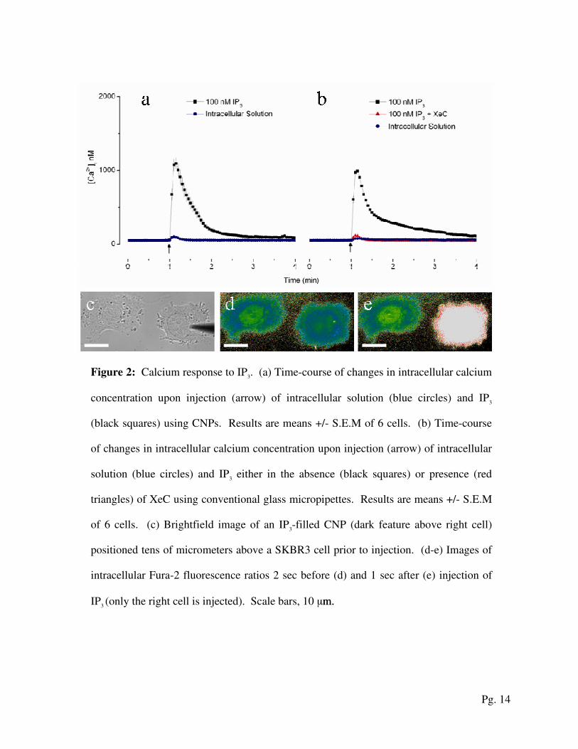
Figure 2: Calcium response to IP3. (a) Time-course of changes in intracellular calcium
concentration upon injection (arrow) of intracellular solution (blue circles) and IP3
(black squares) using CNPs. Results are means +/- S.E.M of 6 cells. (b) Time-course
of changes in intracellular calcium concentration upon injection (arrow) of intracellular
solution (blue circles) and IP3 either in the absence (black squares) or presence (red
triangles) of XeC using conventional glass micropipettes. Results are means +/- S.E.M
of 6 cells. (c) Brightfield image of an IP3-filled CNP (dark feature above right cell)
positioned tens of micrometers above a SKBR3 cell prior to injection. (d-e) Images of
intracellular Fura-2 fluorescence ratios 2 sec before (d) and 1 sec after (e) injection of
IP3 (only the right cell is injected). Scale bars, 10 µm. m.
Pg. 15
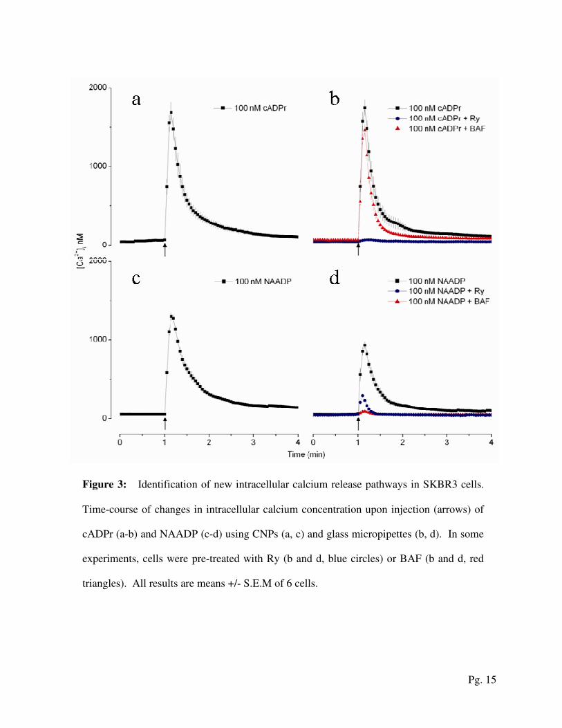
Figure 3: Identification of new intracellular calcium release pathways in SKBR3 cells.
Time-course of changes in intracellular calcium concentration upon injection (arrows) of
cADPr (a-b) and NAADP (c-d) using CNPs (a, c) and glass micropipettes (b, d). In some
experiments, cells were pre-treated with Ry (b and d, blue circles) or BAF (b and d, red
triangles). All results are means +/- S.E.M of 6 cells.