Biofilms of non-Candida albicans Candida species: quantification, structure and matrix composition
Upload
khangminh22Category
view
1download
0
antibiotics
Perspective
Candida Periprosthetic Joint Infection: Is It Curable?
Laura Escolà-Vergé 1,2,3,* , Dolors Rodríguez-Pardo 1,2,3 , Pablo S. Corona 2,3,4 and Carles Pigrau 1,2,3
�����������������
Citation: Escolà-Vergé, L.;
Rodríguez-Pardo, D.; Corona, P.S.;
Pigrau, C. Candida Periprosthetic
Joint Infection: Is It Curable?.
Antibiotics 2021, 10, 458. https://
doi.org/10.3390/antibiotics10040458
Academic Editor:
Wolf-Rainer Abraham
Received: 15 March 2021
Accepted: 13 April 2021
Published: 17 April 2021
Publisher’s Note: MDPI stays neutral
with regard to jurisdictional claims in
published maps and institutional affil-
iations.
Copyright: © 2021 by the authors.
Licensee MDPI, Basel, Switzerland.
This article is an open access article
distributed under the terms and
conditions of the Creative Commons
Attribution (CC BY) license (https://
creativecommons.org/licenses/by/
4.0/).
1 Infectious Diseases Department, Hospital Universitari Vall d’Hebron, Universitat Autònoma de Barcelona,Passeig Vall d’Hebron 119-129, 08035 Barcelona, Spain; [email protected] (D.R.-P.);[email protected] (C.P.)
2 Spanish Network for Research in Infectious Diseases (REIPI RD16/0016/0003), Instituto de Salud Carlos III,28029 Madrid, Spain; [email protected]
3 Study Group on Osteoarticular Infections of the Spanish Society of Clinical Microbiology and InfectiousDiseases (GEIO-SEIMC), 28003 Madrid, Spain
4 Septic and Reconstructive Surgery Unit (UCSO), Orthopaedic Surgery Department, Vall d’Hebron UniversityHospital, Passeig Vall d’Hebron 119-129, 08035 Barcelona, Spain
* Correspondence: [email protected]; Tel.: +34-932-746-090; Fax: +34-934-894-091
Abstract: Candida periprosthetic joint infection (CPJI) is a rare and very difficult to treat infection,and high-quality evidence regarding the best management is scarce. Candida spp. adhere to medicaldevices and grow forming biofilms, which contribute to the persistence and relapse of this infection.Typically, CPJI presents as a chronic infection in a patient with multiple previous surgeries and longcourses of antibiotic therapy. In a retrospective series of cases, the surgical approach with higher ratesof success consists of a two-stage exchange surgery, but the best antifungal treatment and duration ofantifungal treatment are still unclear, and the efficacy of using an antifungal agent-loaded cementspacer is still controversial. Until more evidence is available, focusing on prevention and identifyingpatients at risk of CPJI seems more than reasonable.
Keywords: Candida spp.; periprosthetic joint infection; fungus; biofilm; antifungal-loaded cementspacer; two-stage exchange surgery
1. Introduction
Periprosthetic joint infection (PJI), which occurs in approximately 1–2% of all pro-cedures, is one of the most feared complications after arthroplasty due to its associatedcomorbidities and the possible need for implant removal [1]. Candida periprosthetic jointinfection (CPJI) represents a rare etiology among all PJIs; sometimes it is very difficultto diagnose, and it is especially difficult to treat when the prosthetic material cannotbe removed [2]. In addition, we have no clear guidelines regarding the best antifungalmanagement in these cases [3–7], and evidence is based on small retrospective series.
2. Epidemiology
There have been a few recent studies analyzing the prevalence of these infections,and most of them are retrospective in nature [8–11]. A Spanish retrospective multicenterstudy that analyzed the etiology of PJIs from 2003 to 2012 found that a fungal etiologyrepresented 1.3% of all culture-positive PJIs (n = 2288), and Candida spp. were responsiblefor 90% of all fungal infections [9]. A smaller retrospective multicenter study performedin Australia from 2006 to 2008 found that CPJI accounted for 0.7% (1/152) of all culture-positive infections [10], and another study that compared the etiology of PJIs between tworeferral centers in Europe and in the United States between 2000 and 2011 found that fungalPJIs were responsible for 2.3% of 772 cases and 0.3% of 898 cases, respectively [11].
The species of Candida depends on the local epidemiology of the geographical area.In two multicenter studies in Spain [2,9] and one in the United States [12], C. albicanswas the most frequently isolated fungus (55–65%), followed by C. parapsilosis (13–33%).
Antibiotics 2021, 10, 458. https://doi.org/10.3390/antibiotics10040458 https://www.mdpi.com/journal/antibiotics
Antibiotics 2021, 10, 458 2 of 10
Other species, such as C. glabrata and C. tropicalis, were more anecdotic (3–7% and 2–4%,respectively). Smaller series have found similar results [13,14], and in a recent reviewof the literature, C. albicans (47.3%) was the most frequent strain isolated, followed by C.parapsilosis (22.3%) [15], but epidemiology may still vary among regions.
3. Pathogenesis and Risk Factors
Colonization by Candida spp. is regarded as the first step for subsequent infection [16],and Candida spp. are common commensals of the human skin and gut microbiota in healthyindividuals [17–19]. Invasive disease, which encompasses both candidemia and deep-seated infections, usually results from an abnormal or increased number of fungi combinedwith alterations in the cutaneous and mucosal barriers due to weakening of host immu-nity [16,17], which permits the transition from Candida sp. commensalism to opportunism.Three possible routes of CPJI development have been described: (1) the hematogenousroute from an infected catheter or a urinary or intraabdominal source; (2) direct inoculationduring prosthesis implantation, revision surgery, or even after arthrocentesis, especially incolonized patients; and (3) extension into synovial fluid from contiguous infected tissues.
Candida spp. have specific properties allowing them to adhere to surfaces and formbiofilms, especially on prosthetic devices, which permits the development of persister cells,facilitating antifungal resistance, and explains treatment failure when the implant is notremoved. In vitro experiments have shown that C. albicans biofilm formation begins withthe adherence of yeast to a substrate and thereafter yeast cells proliferate across the surfaceand produce filamentous forms, including hyphae and pseudohyphae. As the biofilmmatures, an extracellular matrix accumulates, facilitating antifungal resistance, notablyto azoles and polyenes, through different mechanisms [20], which may explain the highfailure rates in CPJI when the implant is not removed. Finally, non-adherent yeast cells arereleased from the biofilm into the surrounding medium (the dispersal step). C. albicans, themost frequent causative agent of CPJI, has been reported to form larger and more complexbiofilms than other Candida species [21].
All parts of the immune system are involved in the response to this infection. Forexample, deficiencies in the T-helper 17 lymphocyte cell line impair the mucosal immuneresponse to Candida spp. and facilitate Candida infections. Neutrophil dysfunction orleukopenia also predisposes patients to suffer invasive candidiasis, and complement orimmunoglobulin deficiency or alteration is associated with complicated disease as well [17].The regulatory pathways and mechanisms that govern Candida biofilm developmentare very complex [20]; gene expression of C. albicans is regulated by both a continuoushost–pathogen interplay and by distinct genetic mechanisms [19], but this is not the scopeof this review.
However, there are other factors that are not only easier to identify than alterations inhost immunity but also probably more prevalent in patients with CPJIs and may play amajor role in the pathogenesis of invasive candidiasis. The most reported factors are asfollows [17]: (1) the long-term or repeated use of broad-spectrum antibiotics, especiallyin the previous 3 months, which depletes commensal gut bacteria, enabling Candida sp.overgrowth. Many antibiotics are known to promote fungal growth and pathogenicitybecause they disrupt the microbiota and eliminate anaerobic bacteria in the gut whichcould have otherwise inhibited the fungi, and studies show that the introduction of smallamounts of C. albicans to mice after antibiotic treatment caused significant changes inthe gut microbiota, which may persist in the long term [22]. (2) Breach of the cutaneousand gastrointestinal barriers by chemotherapy, surgery, gastrointestinal perforation, orinstrumentation, such as central venous catheters, which may facilitate Candida sp. translo-cation into the bloodstream. (3) Immunosuppression secondary to malignant diseases,immunodeficiencies, or immunosuppressive therapy. Other risk factors reported in pa-tients with CPJIs have been older age [18], diabetes, rheumatoid arthritis, malnutrition,and tuberculosis, which probably also reflect alterations in host immunity [2,12–14,23].Other series have also identified that multiple previous surgeries at the site of the CPJI
Antibiotics 2021, 10, 458 3 of 10
are also a risk factor [2,13,23,24]. A recent retrospective case–control study that comparedfungal PJIs with bacterial PJIs found that recent antibiotic consumption (OR: 3.4; 95% CI:1.2–9.3) and prolonged wound drainage (OR: 7.3; 95% CI: 2.02–26.95) were significantlyassociated with CPJI [13]. In our experience, patients treated with long courses of linezolidfor multidrug-resistant chronic bacterial PJIs tend to present mucocutaneous candidiasis,and their colonization may persist for an unknown duration, which could also be anotherrisk factor for hip CPJI.
Although it has not been deeply studied, considering the pathogenesis of the disease,previous Candida spp. colonization in the urine or Candida intertrigo may also be riskfactors in patients undergoing hip arthroplasty [2,13]. In a multicenter retrospective studyof patients with CPJIs, we found 14% of patients with Candida intertrigo and 9% of patientswith a previous urinary tract infection (three with positive blood cultures) caused by thesame Candida spp. before the diagnosis of CPJI [2].
4. Clinical Manifestations and Diagnosis
CPJIs are usually chronic infections characterized by pain, swelling, and sinus tracts.Implant loosening may be observed on radiography in nearly 50% of cases, as previouslyreported in some studies [2,25]. In fact, the median duration from the index surgeryand the diagnosis of CPJI averaged 17–25 months [12,13]. Blood tests could show noleukocytosis, and the C-reactive protein (CRP) level and erythrocyte sedimentation rate areusually normal or mildly elevated [2,12]. The same recently published study comparingpatients with CPJIs with those with bacterial PJIs showed that patients with CPJIs hadlower median CRP values (2.95 mg/dL vs. 5.99 mg/dL) and lower synovial fluid leukocytelevels (13,953 cells/mm3 vs. 33,198 cell/mm3) [13].
The criteria to diagnose CPJI are not well established, and the same criteria used indiagnosing bacterial PJIs may not be reliable in some cases. The Infectious Diseases Societyof America (IDSA) guidelines [3], a previous International Consensus on PJIs [6], and arecent European Bone and Joint Infection Society (EBJIS) consensus [26] consider two ormore intraoperative cultures or the combination of preoperative aspiration and intraop-erative cultures yielding the same organism definitive evidence of a PJI [3]. However,when reviewing published series of CPJI cases, the microbiological criteria changed fromone study to another. Some authors consider that one positive preoperative aspirationculture and/or a positive intraoperative culture is sufficient [9], while others require twopositive cultures [2,13] or one positive culture with additional criteria for PJIs [2,24]. In ouropinion, when Candida spp. are found in only one intraoperative culture, the case shouldbe evaluated carefully, and treating the Candida etiology should be considered, especiallyin patients with other risk factors for CPJI such as previous antibiotic therapy or multipleprevious surgeries (Figure 1). In fact, even if another microorganism is isolated in two ormore cultures, polymicrobial infection is not infrequent, particularly in the hip location,being found in 16% to 26% of cases, depending on the series [2,12], and this should not bea criterion for discarding the value of one positive culture for Candida spp.

Antibiotics 2021, 10, 458 4 of 10Antibiotics 2021, 10, x FOR PEER REVIEW 4 of 10
Figure 1. Diagnosis of Candida periprosthetic joint infection.
5. Medical and Surgical Treatment
International guidelines on candidiasis and PJIs [5,7] recommend, with limited evi-
dence, the combination of prosthesis removal and reimplantation in two stages. They rec-
ommend a prolonged period of antifungal therapy for at least 12 weeks after resection
arthroplasty and at least 6 weeks after prosthesis implantation, without specifying the best
antifungal option [5]. They state that the use of antifungal agent-loaded cement spacers is
controversial.
The fact that Candida spp. grow and form biofilms on medical devices makes these
microorganisms highly resistant to antifungal agents and the host immune system [27–
30]. Therefore, the best surgical approach is to remove the prosthetic material to avoid the
problem of antifungals penetrating and acting within the biofilm. In this sense, a two-
stage exchange arthroplasty strategy is probably the best option when feasible to eradicate
the infection and to preserve joint function [15], with variable success rates from 14% to
almost 100% depending on the series and on the definition of success [2,12,14,23–25,31–
35]. In patients with reduced mobility, particularly old patients with multiple previous
surgeries in the same location, a resection arthroplasty may be the best alternative. There
is less evidence of success with a one-stage exchange arthroplasty strategy, which has
been reported in only a few cases [15,36,37]. In a recent review of the literature of 76 epi-
sodes of CPJI, one-stage exchange arthroplasty was performed only in three patients with
a favorable outcome [15], but in another series of 11 CPJI episodes, it was performed in
four with success in two [14]. However, due to the publication bias, the small amount of
experience and the difficulty of curing this type of infection, with a high rate of relapses,
in our opinion, this procedure should be used only in very selected cases. Irrigation and
debridement with prosthesis retention usually fails to cure the infection (cure rates from
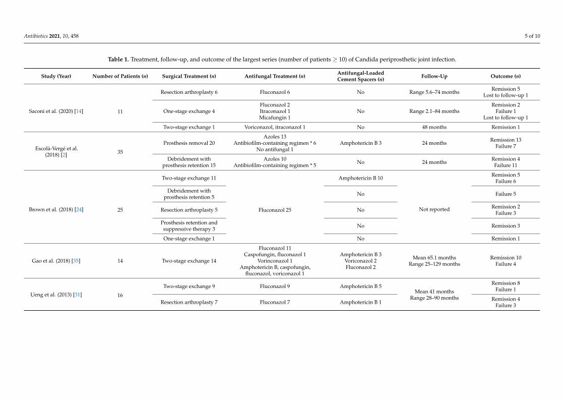
0% to 20%), especially in cases of chronic infection [2,12,23,32,35]. Table 1 summarizes the
type of treatment, the duration of follow-up and the outcome of the larger case series
(number of patients ≥ 10) of CPJI.
Figure 1. Diagnosis of Candida periprosthetic joint infection.
5. Medical and Surgical Treatment
International guidelines on candidiasis and PJIs [5,7] recommend, with limited ev-idence, the combination of prosthesis removal and reimplantation in two stages. Theyrecommend a prolonged period of antifungal therapy for at least 12 weeks after resectionarthroplasty and at least 6 weeks after prosthesis implantation, without specifying the bestantifungal option [5]. They state that the use of antifungal agent-loaded cement spacersis controversial.
The fact that Candida spp. grow and form biofilms on medical devices makes thesemicroorganisms highly resistant to antifungal agents and the host immune system [27–30].Therefore, the best surgical approach is to remove the prosthetic material to avoid theproblem of antifungals penetrating and acting within the biofilm. In this sense, a two-stageexchange arthroplasty strategy is probably the best option when feasible to eradicate theinfection and to preserve joint function [15], with variable success rates from 14% to almost100% depending on the series and on the definition of success [2,12,14,23–25,31–35]. Inpatients with reduced mobility, particularly old patients with multiple previous surgeriesin the same location, a resection arthroplasty may be the best alternative. There is lessevidence of success with a one-stage exchange arthroplasty strategy, which has beenreported in only a few cases [15,36,37]. In a recent review of the literature of 76 episodesof CPJI, one-stage exchange arthroplasty was performed only in three patients with afavorable outcome [15], but in another series of 11 CPJI episodes, it was performed infour with success in two [14]. However, due to the publication bias, the small amount ofexperience and the difficulty of curing this type of infection, with a high rate of relapses,in our opinion, this procedure should be used only in very selected cases. Irrigation anddebridement with prosthesis retention usually fails to cure the infection (cure rates from 0%to 20%), especially in cases of chronic infection [2,12,23,32,35]. Table 1 summarizes the typeof treatment, the duration of follow-up and the outcome of the larger case series (numberof patients ≥ 10) of CPJI.
Antibiotics 2021, 10, 458 5 of 10
Table 1. Treatment, follow-up, and outcome of the largest series (number of patients ≥ 10) of Candida periprosthetic joint infection.
Study (Year) Number of Patients (n) Surgical Treatment (n) Antifungal Treatment (n) Antifungal-LoadedCement Spacers (n) Follow-Up Outcome (n)
Saconi et al. (2020) [14] 11
Resection arthroplasty 6 Fluconazol 6 No Range 5.6–74 months Remission 5Lost to follow-up 1
One-stage exchange 4Fluconazol 2Itraconazol 1Micafungin 1
No Range 2.1–84 monthsRemission 2
Failure 1Lost to follow-up 1
Two-stage exchange 1 Voriconazol, itraconazol 1 No 48 months Remission 1
Escolà-Vergé et al.(2018) [2] 35
Prosthesis removal 20Azoles 13
Antibiofilm-containing regimen * 6No antifungal 1
Amphotericin B 3 24 months Remission 13Failure 7
Debridement withprosthesis retention 15
Azoles 10Antibiofilm-containing regimen * 5 No 24 months Remission 4
Failure 11
Brown et al. (2018) [24] 25
Two-stage exchange 11
Fluconazol 25
Amphotericin B 10
Not reported
Remission 5Failure 6
Debridement withprosthesis retention 5 No Failure 5
Resection arthroplasty 5 No Remission 2Failure 3
Prosthesis retention andsuppressive therapy 3 No Remission 3
One-stage exchange 1 No Remission 1
Gao et al. (2018) [35] 14 Two-stage exchange 14
Fluconazol 11Caspofungin, fluconazol 1
Vorinconazol 1Amphotericin B, caspofungin,
fluconazol, voriconazol 1
Amphotericin B 3Voriconazol 2Fluconazol 2
Mean 65.1 monthsRange 25–129 months
Remission 10Failure 4
Ueng et al. (2013) [31] 16
Two-stage exchange 9 Fluconazol 9 Amphotericin B 5Mean 41 months
Range 28–90 months
Remission 8Failure 1
Resection arthroplasty 7 Fluconazol 7 Amphotericin B 1 Remission 4Failure 3
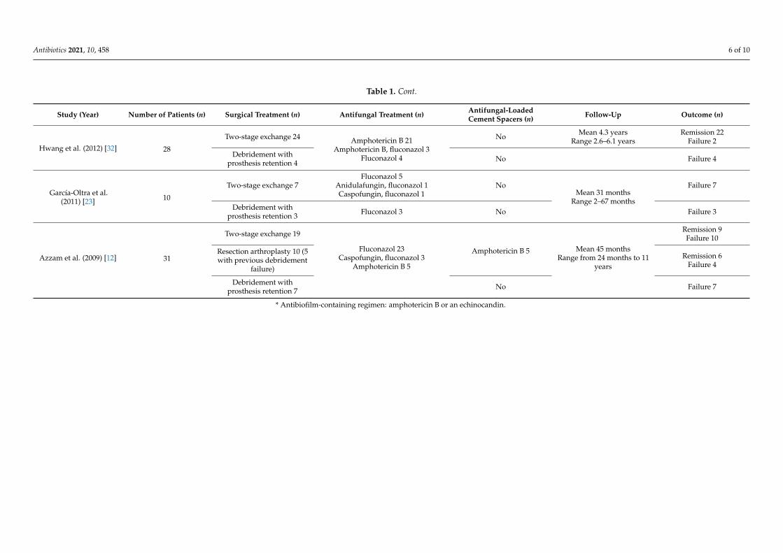
Antibiotics 2021, 10, 458 6 of 10
Table 1. Cont.
Study (Year) Number of Patients (n) Surgical Treatment (n) Antifungal Treatment (n) Antifungal-LoadedCement Spacers (n) Follow-Up Outcome (n)
Hwang et al. (2012) [32] 28
Two-stage exchange 24 Amphotericin B 21Amphotericin B, fluconazol 3
Fluconazol 4
No Mean 4.3 yearsRange 2.6–6.1 years
Remission 22Failure 2
Debridement withprosthesis retention 4 No Failure 4
García-Oltra et al.(2011) [23] 10
Two-stage exchange 7Fluconazol 5
Anidulafungin, fluconazol 1Caspofungin, fluconazol 1
NoMean 31 months
Range 2–67 months
Failure 7
Debridement withprosthesis retention 3 Fluconazol 3 No Failure 3
Azzam et al. (2009) [12] 31
Two-stage exchange 19
Fluconazol 23Caspofungin, fluconazol 3
Amphotericin B 5
Amphotericin B 5 Mean 45 monthsRange from 24 months to 11
years
Remission 9Failure 10
Resection arthroplasty 10 (5with previous debridement
failure)
Remission 6Failure 4
Debridement withprosthesis retention 7 No Failure 7
* Antibiofilm-containing regimen: amphotericin B or an echinocandin.
Antibiotics 2021, 10, 458 7 of 10
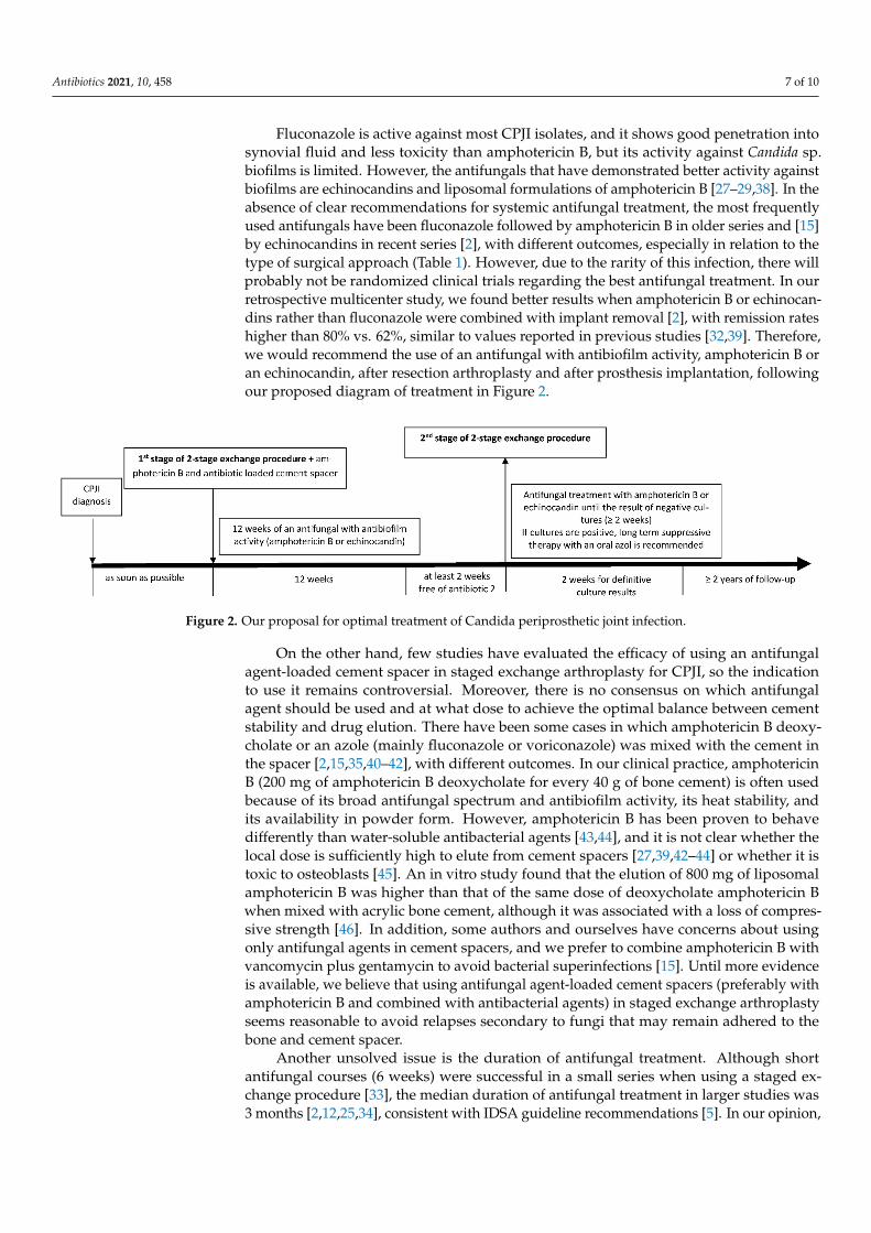
Fluconazole is active against most CPJI isolates, and it shows good penetration intosynovial fluid and less toxicity than amphotericin B, but its activity against Candida sp.biofilms is limited. However, the antifungals that have demonstrated better activity againstbiofilms are echinocandins and liposomal formulations of amphotericin B [27–29,38]. In theabsence of clear recommendations for systemic antifungal treatment, the most frequentlyused antifungals have been fluconazole followed by amphotericin B in older series and [15]by echinocandins in recent series [2], with different outcomes, especially in relation to thetype of surgical approach (Table 1). However, due to the rarity of this infection, there willprobably not be randomized clinical trials regarding the best antifungal treatment. In ourretrospective multicenter study, we found better results when amphotericin B or echinocan-dins rather than fluconazole were combined with implant removal [2], with remission rateshigher than 80% vs. 62%, similar to values reported in previous studies [32,39]. Therefore,we would recommend the use of an antifungal with antibiofilm activity, amphotericin B oran echinocandin, after resection arthroplasty and after prosthesis implantation, followingour proposed diagram of treatment in Figure 2.
Antibiotics 2021, 10, x FOR PEER REVIEW 7 of 10
7
Fluconazole is active against most CPJI isolates, and it shows good penetration into
synovial fluid and less toxicity than amphotericin B, but its activity against Candida sp.
biofilms is limited. However, the antifungals that have demonstrated better activity
against biofilms are echinocandins and liposomal formulations of amphotericin B [27–
29,38]. In the absence of clear recommendations for systemic antifungal treatment, the
most frequently used antifungals have been fluconazole followed by amphotericin B in
older series and [15] by echinocandins in recent series [2], with different outcomes, espe-
cially in relation to the type of surgical approach (Table 1). However, due to the rarity of
this infection, there will probably not be randomized clinical trials regarding the best an-
tifungal treatment. In our retrospective multicenter study, we found better results when
amphotericin B or echinocandins rather than fluconazole were combined with implant
removal [2], with remission rates higher than 80% vs. 62%, similar to values reported in
previous studies [32,39]. Therefore, we would recommend the use of an antifungal with
antibiofilm activity, amphotericin B or an echinocandin, after resection arthroplasty and
after prosthesis implantation, following our proposed diagram of treatment in Figure 2.
Figure 2. Our proposal for optimal treatment of Candida periprosthetic joint infection.
On the other hand, few studies have evaluated the efficacy of using an antifungal
agent-loaded cement spacer in staged exchange arthroplasty for CPJI, so the indication to
use it remains controversial. Moreover, there is no consensus on which antifungal agent
should be used and at what dose to achieve the optimal balance between cement stability
and drug elution. There have been some cases in which amphotericin B deoxycholate or
an azole (mainly fluconazole or voriconazole) was mixed with the cement in the spacer
[2,15,35,40–42], with different outcomes. In our clinical practice, amphotericin B (200 mg
of amphotericin B deoxycholate for every 40 g of bone cement) is often used because of its
broad antifungal spectrum and antibiofilm activity, its heat stability, and its availability
in powder form. However, amphotericin B has been proven to behave differently than
water-soluble antibacterial agents [43,44], and it is not clear whether the local dose is suf-
ficiently high to elute from cement spacers [27,39,42–44] or whether it is toxic to osteo-
blasts [45]. An in vitro study found that the elution of 800 mg of liposomal amphotericin
B was higher than that of the same dose of deoxycholate amphotericin B when mixed with
acrylic bone cement, although it was associated with a loss of compressive strength [46].
In addition, some authors and ourselves have concerns about using only antifungal agents
in cement spacers, and we prefer to combine amphotericin B with vancomycin plus gen-
tamycin to avoid bacterial superinfections [15]. Until more evidence is available, we be-
lieve that using antifungal agent-loaded cement spacers (preferably with amphotericin B
and combined with antibacterial agents) in staged exchange arthroplasty seems reasona-
ble to avoid relapses secondary to fungi that may remain adhered to the bone and cement
spacer.
Another unsolved issue is the duration of antifungal treatment. Although short anti-
fungal courses (6 weeks) were successful in a small series when using a staged exchange
procedure [33], the median duration of antifungal treatment in larger studies was 3
Figure 2. Our proposal for optimal treatment of Candida periprosthetic joint infection.
On the other hand, few studies have evaluated the efficacy of using an antifungalagent-loaded cement spacer in staged exchange arthroplasty for CPJI, so the indicationto use it remains controversial. Moreover, there is no consensus on which antifungalagent should be used and at what dose to achieve the optimal balance between cementstability and drug elution. There have been some cases in which amphotericin B deoxy-cholate or an azole (mainly fluconazole or voriconazole) was mixed with the cement inthe spacer [2,15,35,40–42], with different outcomes. In our clinical practice, amphotericinB (200 mg of amphotericin B deoxycholate for every 40 g of bone cement) is often usedbecause of its broad antifungal spectrum and antibiofilm activity, its heat stability, andits availability in powder form. However, amphotericin B has been proven to behavedifferently than water-soluble antibacterial agents [43,44], and it is not clear whether thelocal dose is sufficiently high to elute from cement spacers [27,39,42–44] or whether it istoxic to osteoblasts [45]. An in vitro study found that the elution of 800 mg of liposomalamphotericin B was higher than that of the same dose of deoxycholate amphotericin Bwhen mixed with acrylic bone cement, although it was associated with a loss of compres-sive strength [46]. In addition, some authors and ourselves have concerns about usingonly antifungal agents in cement spacers, and we prefer to combine amphotericin B withvancomycin plus gentamycin to avoid bacterial superinfections [15]. Until more evidenceis available, we believe that using antifungal agent-loaded cement spacers (preferably withamphotericin B and combined with antibacterial agents) in staged exchange arthroplastyseems reasonable to avoid relapses secondary to fungi that may remain adhered to thebone and cement spacer.
Another unsolved issue is the duration of antifungal treatment. Although shortantifungal courses (6 weeks) were successful in a small series when using a staged ex-change procedure [33], the median duration of antifungal treatment in larger studies was3 months [2,12,25,34], consistent with IDSA guideline recommendations [5]. In our opinion,

Antibiotics 2021, 10, 458 8 of 10
at least 3 months of antifungal treatment are necessary, especially with a drug with an-tibiofilm activity (an echinocandin or amphotericin B) and combined with implant removalwhenever possible, preferably in the form of a two-stage exchange procedure to maintainjoint functionality (Figure 2). In patients with high surgical risk for whom prosthesescannot be removed, suppressive therapy with azoles may be an alternative treatment tomaintain joint functionality [2].
6. Prognosis and Prevention
The prognosis of patients with CPJIs varies depending on the medical and surgical ap-proach. Often, aggressive surgical treatment is dismissed due to the patient’s comorbidities,and resection arthroplasty or amputation is performed, resulting in poor patient functional-ity, but at least curing the infection. On the other hand, even if performing the best strategy(a two-stage exchange), some patients may persist with the infection or relapse. A recentstudy found that the main risk factors for two-stage exchange failure are hemodialysis,obesity, multiple previous procedures, diabetes, corticosteroid therapy, hypoalbuminemia,immunosuppression, rheumatological diseases, coagulation disorders, and infection due tomultidrug-resistant bacteria or fungal species [47]. Therefore, if some of these risk factorscoexist in a patient with CPJI, a resection arthroplasty, agreed with the patient, may be thebest alternative to cure the infection even if it implies loosing functionality. Unfortunately,we have no score of risk that helps us in making the best decision. In addition, due tothe formation of biofilms by Candida spp., CPJIs, when treated, may take several monthsor even years to relapse. Patient follow-up varies among some studies, and this makes itdifficult to establish when CPJI can be considered cured. In our personal experience, dueto the chronic nature of CPJI, follow-up periods shorter than 2 years may not be able todetect some relapses.
As histories of previous antibiotic therapy or surgery are not modifiable, we believethat searching for and treating Candida intertrigo in patients with risk factors for CPJIwould be a reasonable, cost-effective measure [2,13]. Therefore, although there is nostrong evidence to support this hypothesis, we believe that patients with previous Candidainfection or clinical Candida colonization may benefit from the addition of fluconazole tostandard prophylaxis before hip arthroplasty. Another more debatable measure would beincluding fluconazole in surgical prophylaxis for patients with an advanced age, diabetes, along course of antibiotic therapy in the previous months (especially if it was with linezolid)and multiple previous orthopedic surgeries. As these factors may be difficult to evaluateretrospectively, prospective multicenter studies are needed.
Given the poor prognosis of this type of infection, until more evidence is availableregarding the best antifungal treatment, the duration of treatment, and the efficacy of usingantifungal agent-loaded cement spacers, focusing on CPJI prevention remains essential.
Author Contributions: L.E.-V. and C.P. contributed to the conception, methodology, writing of theoriginal draft, and writing of the review and editing with the assistance of a medical writer. D.R.-P.and P.S.C. contributed to the review and editing of the manuscript. All authors have read and agreedto the published version of the manuscript.
Funding: This research did not receive any specific grant from funding agencies in the public,commercial, or not-for-profit sectors.
Conflicts of Interest: The authors declare no conflict of interest.
Abbreviations
PJI periprosthetic joint infectionCPJI Candida periprosthetic joint infection
Antibiotics 2021, 10, 458 9 of 10
References1. Tande, A.J.; Patel, R. Prosthetic Joint Infection. Clin. Microbiol. Rev. 2014, 27, 302–345. [CrossRef] [PubMed]2. Escolà-Vergé, L.; Rodríguez-Pardo, D.; Lora-Tamayo, J.; Morata, L.; Murillo, O.; Vilchez, H.; Sorli, L.; Carrión, L.G.; Barbero, J.M.;
Palomino-Nicás, J.; et al. Candida Periprosthetic Joint Infection: A Rare and Difficult-to-Treat Infection. J. Infect. 2018, 77, 151–157.[CrossRef]
3. Osmon, D.R.; Berbari, E.F.; Berendt, A.R.; Lew, D.; Zimmerli, W.; Steckelberg, J.M.; Rao, N.; Hanssen, A.; Wilson, W.R. Diagnosisand Management of Prosthetic Joint Infection: Clinical Practice Guidelines by the Infectious Diseases Society of America. Clin.Infect. Dis. 2013, 56, e1–e25. [CrossRef] [PubMed]
4. Ariza, J.; Cobo, J.; Baraia-Etxaburu, J.; Benito, N.; Bori, G.; Cabo, J.; Corona, P.; Esteban, J.; Horcajada, J.P.; Lora-Tamayo, J.;et al. Executive Summary of Management of Prosthetic Joint Infections. Clinical Practice Guidelines by the Spanish Society ofInfectious Diseases and Clinical Microbiology (SEIMC). Enferm. Infect. Microbiol. Clin. 2017, 35, 189–195. [CrossRef]
5. Pappas, P.G.; Kauffman, C.A.; Andes, D.R.; Clancy, C.J.; Marr, K.A.; Ostrosky-Zeichner, L.; Reboli, A.C.; Schuster, M.G.; Vazquez,J.A.; Walsh, T.J.; et al. Clinical Practice Guideline for the Management of Candidiasis: 2016 Update by the Infectious DiseasesSociety of America. Clin. Infect. Dis. 2015, civ933. [CrossRef]
6. Parvizi, J.; Gehrke, T.; Chen, A.F. Proceedings of the International Consensus on Periprosthetic Joint Infection. Bone Jt. J. 2013, 95,1450–1452. [CrossRef]
7. Belden, K.; Cao, L.; Chen, J.; Deng, T.; Fu, J.; Guan, H.; Jia, C.; Kong, X.; Kuo, F.-C.; Li, R.; et al. Hip and Knee Section, FungalPeriprosthetic Joint Infection, Diagnosis and Treatment: Proceedings of International Consensus on Orthopedic Infections.J. Arthroplast. 2019, 34, S387–S391. [CrossRef]
8. Benito, N.; Mur, I.; Ribera, A.; Soriano, A.; Rodríguez-Pardo, D.; Sorlí, L.; Cobo, J.; Fernández-Sampedro, M.; del Toro, M.; Guío, L.;et al. The Different Microbial Etiology of Prosthetic Joint Infections According to Route of Acquisition and Time after ProsthesisImplantation, Including the Role of Multidrug-Resistant Organisms. J. Clin. Med. 2019, 8, 673. [CrossRef]
9. Benito, N.; Franco, M.; Ribera, A.; Soriano, A.; Rodriguez-Pardo, D.; Sorlí, L.; Fresco, G.; Fernández-Sampedro, M.; Do-lores Del Toro, M.; Guío, L.; et al. Time Trends in the Aetiology of Prosthetic Joint Infections: A Multicentre Cohort Study.Clin. Microbiol. Infect. 2016, 22, 732.e1–732.e8. [CrossRef]
10. Peel, T.N.; Cheng, A.C.; Buising, K.L.; Choong, P.F.M. Microbiological Aetiology, Epidemiology, and Clinical Profile of ProstheticJoint Infections: Are Current Antibiotic Prophylaxis Guidelines Effective? Antimicrob. Agents Chemother. 2012, 56, 2386–2391.[CrossRef] [PubMed]
11. Aggarwal, V.K.; Bakhshi, H.; Ecker, N.U.; Parvizi, J.; Gehrke, T.; Kendoff, D. Organism Profile in Periprosthetic Joint Infection:Pathogens Differ at Two Arthroplasty Infection Referral Centers in Europe and in the United States. J. Knee Surg. 2014, 27, 399–406.[CrossRef]
12. Azzam, K.; Parvizi, J.; Jungkind, D.; Hanssen, A.; Fehring, T.; Springer, B.; Bozic, K.; Della Valle, C.; Pulido, L.; Barrack, R.Microbiological, Clinical, and Surgical Features of Fungal Prosthetic Joint Infections: A Multi-Institutional Experience. J. Bone Jt.Surg. Am. Vol. 2009, 91, 142–149. [CrossRef] [PubMed]
13. Riaz, T.; Tande, A.J.; Steed, L.L.; Demos, H.A.; Salgado, C.D.; Osmon, D.R.; Marculescu, C.E. Risk Factors for Fungal ProstheticJoint Infection. J. Bone Jt. Infect. 2020, 5, 76–81. [CrossRef] [PubMed]
14. Saconi, E.S.; de Carvalho, V.C. Prosthetic Joint Infection Due to Candida Species. Medicine 2020, 99, e19735. [CrossRef] [PubMed]15. Cobo, F.; Rodríguez-Granger, J.; Sampedro, A.; Aliaga-Martínez, L.; Navarro-Marí, J.M. Candida Prosthetic Joint Infection.
A Review of Treatment Methods. J. Bone Jt. Infect. 2017, 2, 114–121. [CrossRef]16. Pappas, P.G.; Lionakis, M.S.; Arendrup, M.C.; Ostrosky-Zeichner, L.; Kullberg, B.J. Invasive Candidiasis. Nat. Rev. Dis. Primers
2018, 4, 18026. [CrossRef] [PubMed]17. McCarty, T.P.; Pappas, P.G. Invasive Candidiasis. Infect. Dis. Clin. N. Am. 2016, 30, 103–124. [CrossRef] [PubMed]18. Kullberg, B.J.; Arendrup, M.C. Invasive Candidiasis. N. Engl. J. Med. 2015, 373, 1445–1456. [CrossRef]19. Hube, B. From Commensal to Pathogen: Stage- and Tissue-Specific Gene Expression of Candida albicans. Curr. Opin. Microbiol.
2004, 7, 336–341. [CrossRef]20. Finkel, J.S.; Mitchell, A.P. Genetic Control of Candida albicans Biofilm Development. Nat. Rev. Microbiol. 2011, 9, 109–118.
[CrossRef]21. Kuhn, D.M. Comparison of Biofilms Formed by Candidaalbicans and Candidaparapsilosis on Bioprosthetic Surfaces.
Infect. Immun. 2002, 70, 878–888. [CrossRef] [PubMed]22. Sam, Q.; Chang, M.; Chai, L. The Fungal Mycobiome and Its Interaction with Gut Bacteria in the Host. Int. J. Mol. Sci. 2017, 18,
330. [CrossRef] [PubMed]23. García-Oltra, E.; García-Ramiro, S.; Martínez, J.C.; Tibau, R.; Bori, G.; Bosch, J.; Mensa, J.; Soriano, A. Prosthetic joint infection by
Candida spp. Rev. Esp. Quimioter. 2011, 24, 37–41. [PubMed]24. Brown, T.S.; Petis, S.M.; Osmon, D.R.; Mabry, T.M.; Berry, D.J.; Hanssen, A.D.; Abdel, M.P. Periprosthetic Joint Infection With
Fungal Pathogens. J. Arthroplast. 2018, 33, 2605–2612. [CrossRef]25. Kuiper, J.W.; van den Bekerom, M.P.; van der Stappen, J.; Nolte, P.A.; Colen, S. 2-Stage Revision Recommended for Treatment
of Fungal Hip and Knee Prosthetic Joint Infections: An Analysis of 164 Patients, 156 from the Literature and 8 Own Cases.Acta Orthop. 2013, 84, 517–523. [CrossRef]
Antibiotics 2021, 10, 458 10 of 10
26. McNally, M.; Sousa, R.; Wouthuyzen-Bakker, M.; Chen, A.F.; Soriano, A.; Vogely, H.C.; Clauss, M.; Higuera, C.A.; Trebše, R. TheEBJIS Definition of Periprosthetic Joint Infection: A Practical Guide for Clinicians. Bone Jt. J. 2021, 103, 18–25. [CrossRef]
27. Kuhn, D.M.; George, T.; Chandra, J.; Mukherjee, P.K.; Ghannoum, M.A. Antifungal Susceptibility of Candida Biofilms: UniqueEfficacy of Amphotericin B Lipid Formulations and Echinocandins. Antimicrob. Agents Chemother. 2002, 46, 1773–1780. [CrossRef]
28. Nett, J.E. Future Directions for Anti-Biofilm Therapeutics Targeting Candida. Expert Rev. Anti Infect. Ther. 2014, 12, 375–382.[CrossRef]
29. Iñigo, M.; Pemán, J.; Del Pozo, J.L. Antifungal Activity against Candida Biofilms. Int. J. Artif. Organs 2012, 35, 780–791. [CrossRef]30. Mukherjee, P.K.; Chandra, J. Candida Biofilms: Development, Architecture, and Resistance. Microbiol. Spectrum. 2015, 3.
[CrossRef]31. Ueng, S.W.N.; Lee, C.-Y.; Hu, C.; Hsieh, P.-H.; Chang, Y. What Is the Success of Treatment of Hip and Knee Candidal Periprosthetic
Joint Infection? Clin. Orthop. Relat. Res. 2013, 471, 3002–3009. [CrossRef]32. Hwang, B.H.; Yoon, J.Y.; Nam, C.H.; Jung, K.A.; Lee, S.C.; Han, C.D.; Moon, S.H. Fungal Peri-Prosthetic Joint Infection after
Primary Total Knee Replacement. J. Bone Jt. Surg. Br. 2012, 94, 656–659. [CrossRef] [PubMed]33. Anagnostakos, K.; Kelm, J.; Schmitt, E.; Jung, J. Fungal Periprosthetic Hip and Knee Joint Infections. J. Arthroplast. 2012, 27,
293–298. [CrossRef] [PubMed]34. Phelan, D.M.; Osmon, D.R.; Keating, M.R.; Hanssen, A.D. Delayed Reimplantation Arthroplasty for Candidal Prosthetic Joint
Infection: A Report of 4 Cases and Review of the Literature. Clin. Infect. Dis. 2002, 34, 930–938. [CrossRef] [PubMed]35. Gao, Z.; Li, X.; Du, Y.; Peng, Y.; Wu, W.; Zhou, Y. Success Rate of Fungal Peri-Prosthetic Joint Infection Treated by 2-Stage Revision
and Potential Risk Factors of Treatment Failure: A Retrospective Study. Med. Sci. Monit. 2018, 24, 5549–5557. [CrossRef] [PubMed]36. Selmon, G.P.F.; Slater, R.N.S.; Shepperd, J.N.; Wright, E.P. Successful 1-Stage Exchange Total Knee Arthroplasty for Fungal
Infection. J. Arthroplast. 1998, 13, 114–115. [CrossRef]37. Klatte, T.O.; Kendoff, D.; Kamath, A.F.; Jonen, V.; Rueger, J.M.; Frommelt, L.; Gebauer, M.; Gehrke, T. Single-Stage Revision for
Fungal Peri-Prosthetic Joint Infection: A Single-Centre Experience. Bone Jt. J. 2014, 96-B, 492–496. [CrossRef]38. Katragkou, A.; Chatzimoschou, A.; Simitsopoulou, M.; Dalakiouridou, M.; Diza-Mataftsi, E.; Tsantali, C.; Roilides, E. Differential
Activities of Newer Antifungal Agents against Candida Albicans and Candida Parapsilosis Biofilms. Antimicrob. Agents Chemother.2008, 52, 357–360. [CrossRef]
39. Schoof, B.; Jakobs, O.; Schmidl, S.; Klatte, T.O.; Frommelt, L.; Gehrke, T.; Gebauer, M. Fungal Periprosthetic Joint Infection of theHip: A Systematic Review. Orthop. Rev. 2015, 7. [CrossRef]
40. Bruce, A.S.W.; Kerry, R.M.; Norman, P.; Stockley, I. Fluconazole-Impregnated Beads in the Management of Fungal Infection ofProsthetic Joints. J. Bone Jt. Surg. Br. Vol. 2001, 83, 183–184. [CrossRef]
41. Deelstra, J.J.; Neut, D.; Jutte, P.C. Successful Treatment of Candida Albicans–Infected Total Hip Prosthesis with Staged ProcedureUsing an Antifungal-Loaded Cement Spacer. J. Arthroplast. 2013, 28, 374.e5–374.e8. [CrossRef] [PubMed]
42. Marra, F.; Robbins, G.M.; Masri, B.A.; Duncan, C.; Wasan, K.M.; Kwong, E.H.; Jewesson, P.J. Amphotericin B-Loaded BoneCement to Treat Osteomyelitis Caused by Candida albicans. Can. J. Surg. 2001, 44, 383.
43. Goss, B.; Lutton, C.; Weinrauch, P.; Jabur, M.; Gillett, G.; Crawford, R. Elution and Mechanical Properties of Antifungal BoneCement. J. Arthroplast. 2007, 22, 902–908. [CrossRef] [PubMed]
44. Kweon, C.; McLaren, A.C.; Leon, C.; McLemore, R. Amphotericin B Delivery From Bone Cement Increases With Porosity butStrength Decreases. Clin. Orthop. Relat. Res. 2011, 469, 3002–3007. [CrossRef] [PubMed]
45. Harmsen, S.; McLaren, A.C.; Pauken, C.; McLemore, R. Amphotericin B Is Cytotoxic at Locally Delivered Concentrations.Clin. Orthop. Relat. Res. 2011, 469, 3016–3021. [CrossRef] [PubMed]
46. Cunningham, B.; McLaren, A.C.; Pauken, C.; McLemore, R. Liposomal Formulation Increases Local Delivery of Amphotericinfrom Bone Cement: A Pilot Study. Clin. Orthop. Relat. Res. 2012, 470, 2671–2676. [CrossRef]
47. Fagotti, L.; Tatka, J.; Salles, M.J.C.; Queiroz, M.C. Risk Factors and Treatment Options for Failure of a Two-Stage Exchange.Curr. Rev. Musculoskelet. Med. 2018, 11, 420–427. [CrossRef] [PubMed]
Copyright © 2022 FDOKUMEN


![[Diagnostic strategies in cases of suspected periprosthetic infection of the knee. A review of the literature and current recommendations]](https://static.fdokumen.com/doc/165x107/6343c4dfbd0b0d0a6b08818b/diagnostic-strategies-in-cases-of-suspected-periprosthetic-infection-of-the-knee.jpg)