Cancer Res NuBCP-9 Cargos: Antitumor Efficacy of the BCL-2 Conversion Peptide Novel Polymeric...
12
2014;74:3271-3281. Published OnlineFirst April 16, 2014. Cancer Res Manoj Kumar, Dikshi Gupta, Gurpal Singh, et al. NuBCP-9 Cargos: Antitumor Efficacy of the BCL-2 Conversion Peptide Novel Polymeric Nanoparticles for Intracellular Delivery of Peptide Updated version 10.1158/0008-5472.CAN-13-2015 doi: Access the most recent version of this article at: Cited Articles http://cancerres.aacrjournals.org/content/74/12/3271.full.html#ref-list-1 This article cites by 50 articles, 15 of which you can access for free at: E-mail alerts related to this article or journal. Sign up to receive free email-alerts Subscriptions Reprints and . [email protected] To order reprints of this article or to subscribe to the journal, contact the AACR Publications Department at Permissions . [email protected] To request permission to re-use all or part of this article, contact the AACR Publications Department at on June 20, 2014. © 2014 American Association for Cancer Research. cancerres.aacrjournals.org Downloaded from Published OnlineFirst April 16, 2014; DOI: 10.1158/0008-5472.CAN-13-2015 on June 20, 2014. © 2014 American Association for Cancer Research. cancerres.aacrjournals.org Downloaded from Published OnlineFirst April 16, 2014; DOI: 10.1158/0008-5472.CAN-13-2015
Transcript of Cancer Res NuBCP-9 Cargos: Antitumor Efficacy of the BCL-2 Conversion Peptide Novel Polymeric...
2014;74:3271-3281. Published OnlineFirst April 16, 2014.Cancer Res Manoj Kumar, Dikshi Gupta, Gurpal Singh, et al. NuBCP-9Cargos: Antitumor Efficacy of the BCL-2 Conversion Peptide Novel Polymeric Nanoparticles for Intracellular Delivery of Peptide
Updated version
10.1158/0008-5472.CAN-13-2015doi:
Access the most recent version of this article at:
Cited Articles
http://cancerres.aacrjournals.org/content/74/12/3271.full.html#ref-list-1
This article cites by 50 articles, 15 of which you can access for free at:
E-mail alerts related to this article or journal.Sign up to receive free email-alerts
Subscriptions
Reprints and
To order reprints of this article or to subscribe to the journal, contact the AACR Publications Department at
Permissions
To request permission to re-use all or part of this article, contact the AACR Publications Department at
on June 20, 2014. © 2014 American Association for Cancer Research. cancerres.aacrjournals.org Downloaded from
Published OnlineFirst April 16, 2014; DOI: 10.1158/0008-5472.CAN-13-2015
on June 20, 2014. © 2014 American Association for Cancer Research. cancerres.aacrjournals.org Downloaded from
Published OnlineFirst April 16, 2014; DOI: 10.1158/0008-5472.CAN-13-2015

Therapeutics, Targets, and Chemical Biology
Novel Polymeric Nanoparticles for Intracellular Delivery ofPeptide Cargos: Antitumor Efficacy of the BCL-2 ConversionPeptide NuBCP-9
Manoj Kumar1, Dikshi Gupta1, Gurpal Singh1, Sapna Sharma1, Madhusudan Bhat2, C.K. Prashant2,A.K. Dinda2, Surender Kharbanda3, Donald Kufe3, and Harpal Singh1
AbstractThe preclinical development of peptidyl drugs for cancer treatment is hampered by their poor pharmacologic
properties and cell penetrative capabilities in vivo. In this study, we report a nanoparticle-based formulation thatovercomes these limitations, illustrating their utility in studies of the anticancer peptideNuBCP-9, which convertsBCL-2 from a cell protector to a cell killer. NuBCP-9 was encapsulated in polymeric nanoparticles composed of apolyethylene glycol (PEG)–modified polylactic acid (PLA) diblock copolymer (NuBCP-9/PLA-PEG) or PEG-polypropylene glycol-PEG-modified PLA—tetrablock copolymer (NuBCP-9/PLA-PEG-PPG-PEG). We found thatpeptide encapsulation was enhanced by increasing the PEG chain length in the block copolymers. NuBCP-9release from the nanoparticleswas controlled by both PEGchain length and thePLAmolecularweight, permittingtime-release over sustained periods. Treatment of human cancer cells with these nanoparticles in vitro triggeredapoptosis by NuBCP-9–mediated mechanism, with a potency similar to NuBCP-9 linked to a cell-penetratingpoly-Arg peptide. Strikingly, in vivo administration of NuBCP-9/nanoparticles triggered complete regressions inthe Ehrlich syngeneicmousemodel of solid tumor. Our results illustrate an effectivemethod for sustained deliveryof anticancer peptides, highlighting the superior qualities of the novel PLA-PEG-PPG-PEG tetrablock copolymerformulation as a tool to target intracellular proteins. Cancer Res; 74(12); 3271–81. �2014 AACR.
IntroductionBCL-2 family proteins are important regulators of the mito-
chondrial outer membrane potential (MOMP) and thereby theinduction of apoptosis (1, 2). The BCL-2 family is divided intoproapoptotic and antiapoptotic members that share BCL-2homology (BH) domains. The proapoptotic BCL-2 familymembers BAX and BAK contain multiple BH domains andcontrol the MOMP. Other proapoptotic members that possessonly the BH3 domain (BAD, BIM, BID, PUMA, and NOXA)either directly activate BAX/BAK, or inhibit the antiapoptoticactivity of BCL-2 and BCL-xL to promote BAX/BAK activation.The antiapoptotic activities of the BCL-2 and BCL-xL proteinshave been targetedwith peptides derived from theBH3domainand with small molecule inhibitors, such as ABT-737 and
navitoclax, that mimic the BH3 a-helix (3–5). BCL-2 has alsobeen targeted with a Nur77-derived peptide of 9 amino acids(NuBCP-9; ref. 6). Nur77 is an orphan nuclear receptor thatinteracts with the BCL-2 N-terminal loop region and induces aBCL-2 conformational change (7). Binding of Nur77 exposesthe BCL-2 BH3 domain and converts BCL-2 from an antia-poptotic protein to an inducer of apoptosis (7). Based on thesefindings, the D-amino acid NuBCP-9 peptide corresponding tothe Nur77 region that interacts with BCL-2 was conjugated tothe cell-penetrating D-Arg octamer (r8). Significantly, NuBCP-9-r8 was shown to induce apoptosis of cancer, but not normal,cells by a BCL-2–dependent mechanism (6). As predicted fromNur77 studies, NuBCP-9 binding was associated with a BCL-2conformational change and thereby neutralization of BCL-2–mediated inhibition of BAX (6). NuBCP-9 binding to BCL-2 alsoexposes the BCL-2 BH3 domain and inhibits the survivalfunction of BCL-xL (6). These findings and the demonstrationthat NuBCP-9-r8 induces apoptosis of cancer cells in vitro andin animalmodels supported the development ofNuBCP-9-r8 asa selective anticancer agent.
Intracellular cancer targets that are devoid of an ATP-binding pocket are often undruggable with small chemicalinhibitors. Cell-penetrating therapeutic peptides have thusemerged as promising agents because of their potential fortargeting intracellular proteins in cancer cells with high spec-ificity and limited off-target toxicity (8, 9). However, delivery ofanticancer peptides presents a challenge as a result of thepotential degradation and immunogenicity of these agents. In
Authors' Affiliations: 1Center for Biomedical Engineering, Indian Instituteof Technology, Hauz Khas; 2Department of Pathology, All India Institute ofMedical Sciences, Ansari Nagar, New Delhi; and 3Department of MedicalOncology, Dana-Farber Cancer Institute, HarvardMedical School, Boston,Massachusetts
Note: Supplementary data for this article are available at Cancer ResearchOnline (http://cancerres.aacrjournals.org/).
Corresponding Authors: Donald Kufe, Dana-Farber Cancer Institute, 450Brookline Avenue, Dana 830, Boston, MA 02215. Phone: 617-632-3141;Fax: 617-632-2934; E-mail: [email protected]; and HarpalSingh, [email protected]
doi: 10.1158/0008-5472.CAN-13-2015
�2014 American Association for Cancer Research.
CancerResearch
www.aacrjournals.org 3271
on June 20, 2014. © 2014 American Association for Cancer Research. cancerres.aacrjournals.org Downloaded from
Published OnlineFirst April 16, 2014; DOI: 10.1158/0008-5472.CAN-13-2015
addition, small therapeutic peptides generally have shortcirculating half-lives and require frequent administration forsustained inhibition of their target proteins (10). Therapeuticpeptides also frequently require a protein transduction domain(PTD) for cell membrane penetration and intracellular local-ization (11–13). Cell-penetrating peptides (CPP) that have beenwidely used for cargo delivery include, among others, the TATpeptide, penetratin, and oligoarginines (11). The selection of acertain CPP is of importance in that the CPP can in a cargo-dependent manner contribute to decreased serum stability,inefficient transit from the endosome to the cytosol, andunanticipated toxicities (14). Indeed, for NuBCP-9, the cell-penetrating r8 used for intracellular delivery acts in synergywith the N-terminal phenylalanine of NuBCP-9 to cause mem-brane blebbing and cell necrosis that are independent of BCL-2expression (15). These findings have supported the study ofalternative CPPs for NuBCP-9 or other formulations thatpreclude the use of a CPP for intracellular delivery.
Nanoparticle delivery systems for anticancer agents, partic-ularly smallmolecules such as doxorubicin and paclitaxel, havebeen developed to improve pharmacokinetic parameters andtherapeutic index (16). In this context, tumor microenviron-ments that are subject to hypoxia and acidosis can limit theeffectiveness of small molecule anticancer agents (16). Byextension and through what is referred to as the enhancedpermeation retention (EPR) effect, nanoparticle delivery ofanticancer agents can overcome limitations imposed by thetumor microenvironment and sustain drug exposure (16, 17).Nanoparticles can also be decorated with ligands to selectivelytarget the surface of tumor cells (18). Polymeric nanoparticlesrepresent one class that has been widely studied and shown tobe nontoxic, biocompatible, and biodegradable (19, 20). Spe-cifically, PLA-PEG block copolymer nanoparticles have beenused as carriers for anticancer drugs in sustained/controlledrelease and targeted delivery systems to enhance efficacy andcircumvent drug resistance (19, 20). For example, Genexol-PMis a paclitaxel-loaded PLA-PEG polymeric nanoparticle that isapproved for use in Korea and is undergoing phase II evalu-ation in the United States for metastatic cancers (19, 21). BINDBiosciences also has under development docetaxel-encapsu-lated PLA-PEG nanoparticles for the treatment of solid tumors(19). Curiously, polymeric nanoparticles have not been fullyevaluated for the delivery of anticancer peptide drugs.
The present studies have focused on the encapsulation ofNuBCP-9 into PLA-PEG nanoparticles or a novel PLA-PEG-PPG-PEG tetrablock nanoparticle system to assess delivery ofthis anticancer peptide to malignant cells in vitro and in vivo.The results obtained provide the experimental basis for thefurther development of NuBCP-9/nanoparticles and for thepotential delivery of other anticancer peptide drugs.
Materials and MethodsSynthesis and characterization of PLA-PEG copolymers
PLA-PEG diblock copolymers were synthesized using 72-kDa PLA (NatureWorks) or �12-kDa PLA (Purac Chemicals).PEGwas used at 1, 2, or 4 kDa (CDH, India) for coupling to PLA.PEG-PPG-PEG (12.5 kDa; Poloxamer-F127, Sigma-Aldrich) wasalso used for the synthesis of PLA block copolymers. In a
standard experiment, 0.014 mmol PLA and PEG or PEG-PPG-PEG were dissolved in 100 mL dichloromethane (CH2Cl2) andstirred at 0�C to 2�C. To these solutions, 5 mL of 1% N,N-dicyclohexylcarbodimide (DCC) was added slowly, followed bythe addition of 2mL of 0.1% 4-dimethylaminopyridine (DMAP)and stirring for 16 hours. The resulting PLA-PEGandPLA-PEG-PPG-PEG block copolymers were precipitated with a 1:1 mix-ture of diethyl ether and methanol to remove unreacted PEGand PEG-PPG-PEG. Gel permeation chromatography (GPC)analysis was performed at room temperature using a ViscoteckGPC system with tetrahydrofuran as the mobile phase. Thesynthesized PLA block copolymers were dried under vacuumand stored at �20�C until use. 1HNMR of PLA-PEG or PLA-PEG-PPG-PEG was performed in CDCl3 at 300 Hz (Bruker).
Preparation of rhodamine B- and NuBCP-9–loadednanoparticles
Loading of the PLA-PEG and PLA-PEG-PPG-PEG nanoparti-cleswas performedusingadouble emulsion solvent evaporationmethod. PLA-PEG or PLA-PEG-PPG-PEG copolymers (100 mg)were dissolved in 5 mL acetonitrile. Rhodamine B (1 mg) or L-amino acid NuBCP-9 (10 mg; Bioconcept, India) was added tothe solution with brief sonication. The resulting primary emul-sion was then added dropwise into a 20 mL aqueous phasecomposed of Poloxamer-F127 (PEG-PPG-PEG) in distilledwaterand stirred at room temperature for 6 to 8 hours to facilitatesolvent evaporation and nanoparticle stabilization. Nanoparti-cles were loaded with NuBCP-9 (FSRSLHSLL) peptide (7) andfiltered through an Amikon 10-kDa ultrafilter (Millipore). Thenanoparticles were lyophilized and stored at �20�C until use.Thefiltratewas collected and analyzed for freeNuBCP-9 peptideusing a Micro-BCA Kit (Pierce Chemicals). Encapsulation effi-ciency of NuBCP-9 peptide was determined using the followingformula:
EE %ð Þ ¼ Peptide� �
Total� Peptide� �
Filtrate
Peptide� �
Total�100
Morphology and particle size of the nanoparticles wasdetermined using a Zeiss EVO 50 Series scanning electronmicroscope (SEM). z Potential of the nanoparticles wasassessed by nanoparticle tracking analysis (NanoSight NS500).
Assessment of NuBCP-9 release from nanoparticlesThe in vitro release kinetics of NuBCP-9 from nanoparticles
were determined by the ultrafiltrationmethod. Briefly, samplesof freeze-dried nanoparticles (10 mg) were suspended in PBSand incubated at 37�Cwith gentle shaking at 150 to 160 rpm. Atpredetermined time points of up to 60 days, the samples wereremoved from the incubator and ultrafiltered through 10-kDaAmikon filters (Millipore). The filtrates were collected foranalysis and fresh buffer was added to the respective tubes.Peptide concentration in the filtrates was determined bymicro-BCA assay.
Cell cultureHuman MCF-7 breast cancer and HepG2 hepatocellular
carcinoma cells were grown in DMEM containing 10% FBS,100 units/mL penicillin, and 100 g/mL streptomycin. Human
Kumar et al.
Cancer Res; 74(12) June 15, 2014 Cancer Research3272
on June 20, 2014. © 2014 American Association for Cancer Research. cancerres.aacrjournals.org Downloaded from
Published OnlineFirst April 16, 2014; DOI: 10.1158/0008-5472.CAN-13-2015
umbilical vein endothelial cells (HUVEC) were cultured inEBM-2 medium with endothelial cell growth supplement(Lonza). Proliferation was assessed by the XTT-Based In VitroAssay Kit (Cayman). To assess uptake of nanoparticles, MCF-7cells were seeded on coverslips and grown for 24 hours. Afterincubation with rhodamine B loaded nanoparticles, the cover-slips were removed, washed with PBS, and fixed with 4%paraformaldehyde. The cells were then stained with 40,6-dia-midino-2-phenylindole (DAPI) and visualized under a confocallaser scanning microscope (CLSM; Olympus, Fluoview FV1000Microscope).
Assessment of apoptosisCells were stained using the Annexin V-Alexa Fluor 488/PI
Apoptosis Assay Kit (Invitrogen). Quantification of apopto-sis/necrosis was performed using Cellometer Vision (Nex-celom Bioscience LLC). Cells were also imaged using theCLSM microsocope.
Immunoblot analysisCell lysates were prepared with M-PER reagent (Pierce
Chemicals) and analyzed by immunoblotting with anti-BCL-2, anti-caspase-3 (Santa Cruz Biotechnology) and anti-b-actin(Sigma).
Analysis of antitumor activityMouse Ehrlich tumor cells were injected subcutaneously
in the hind limb of syngeneic Balb/c mice (17–22 g). Mice-bearing tumors (�400 mm3) were divided into 9 groups(10 mice/group) and treated intraperitoneally (i.p.) or intra-tumorally (i.t.) with different agents using two schedules for 21days (Supplementary Table S1). Tumor volume was deter-mined by calipers and calculated using the formula (A � B2)� 0.5, where A and B are the longest and shortest tumordiameters, respectively. From each group, 1 mouse was sacri-ficed on day 7, 14, and 21 for harvesting of tumor for histo-pathologic examination. Statistical analysis of tumor volumeswas performed by one-way ANOVA and the Dunnett test usingOrigin 8.0 (Origin Lab). Survival of the mice was determined bythe Kaplan–Meier method using Prism 4.0 software (GraphPad Software). Onemouse fromeachof the control andNuBCP-9/nanoparticle-treated (i.p. once/week) groups was sacrificedon day 14 for tumor excision. The tumors were fixed in 10%formalin/saline and embedded in paraffin. Five-micrometersections were stained with hematoxylin and eosin.
ResultsPreparation and characterization of NuBCP-9–loadedpolymeric nanoparticlesPLA-PEG nanoparticles have been commonly synthesized
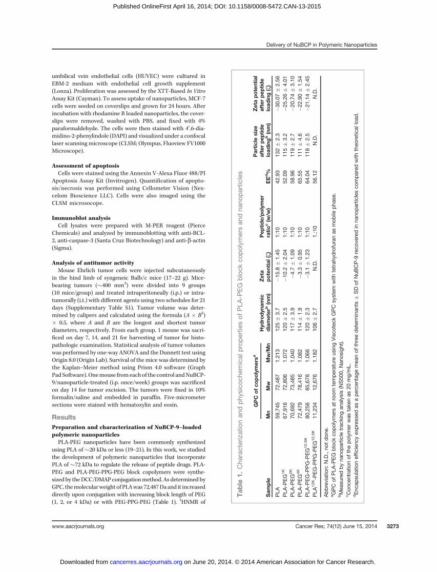
using PLA of �20 kDa or less (19–21). In this work, we studiedthe development of polymeric nanoparticles that incorporatePLA of �72 kDa to regulate the release of peptide drugs. PLA-PEG and PLA-PEG-PPG-PEG block copolymers were synthe-sized by theDCC/DMAPconjugationmethod. As determined byGPC, themolecularweight of PLAwas 72,487Daand it increaseddirectly upon conjugation with increasing block length of PEG(1, 2, or 4 kDa) or with PEG-PPG-PEG (Table 1). 1HNMR of
Tab
le1.
Cha
racterizationan
dphy
sico
chem
ical
propertie
sof
PLA
-PEG
block
copolym
ersan
dna
nopartic
les
GPC
ofco
polymersa
Sam
ple
Mn
Mw
Mw/M
nHyd
rodyn
amic
diameter
b(nm)
Zeta
potential(z)
Pep
tide/polymer
ratio
c(w
/w)
EEd%
Particlesize
afterpep
tide
load
ingb(nm)
Zetapotential
afterpep
tide
load
ing(z)
PLA
59,745
72,487
1.21
312
5�
3.7
�15.8�
1.45
1:10
42.93
132�
2.3
�30.07
�2.56
PLA
-PEG1K
67,916
72,806
1.07
212
0�
2.5
�10.2�
2.04
1:10
52.09
115�
3.2
�25.26
�4.01
PLA
-PEG2K
70,692
73,485
1.04
011
7�
3.9
�4.7
�1.09
1:10
58.96
119�
2.7
�20.74
�3.10
PLA
-PEG4K
72,479
78,416
1.08
211
4�
1.9
�3.3
�0.95
1:10
65.55
111�
4.6
�22.90
�1.54
PLA
-PEG-P
PG-P
EG12.5K
80,256
85,678
1.06
612
0�
2.3
�3.1
�1.23
1:10
64.04
118�
2.5
�21.14
�2.45
PLA
12K-P
EG-P
PG-P
EG12.5K
11,234
12,676
1.18
210
6�
2.7
N.D.
1.:10
56.12
N.D.
N.D.
Abbreviation:
N.D.,no
tdo
ne.
aGPC
ofPLA
-PEG
block
copolym
ersat
room
temperatureus
ingVisco
teck
GPC
system
with
tetrah
ydrofuranas
mob
ilepha
se.
bMea
suredby
nano
partic
letrac
king
analysis
(NS50
0,Nan
osight).
cCon
centratio
nof
thepo
lymer
was
take
nas
20mg/mL.
dEnc
apsu
latio
nefficien
cyex
pres
sedas
aperce
ntag
emea
nof
threedeterminan
ts�
SDof
NuB
CP-9
reco
veredin
nano
particlesco
mpared
with
theo
retic
alload
.
Delivery of NuBCP in Polymeric Nanoparticles
www.aacrjournals.org Cancer Res; 74(12) June 15, 2014 3273
on June 20, 2014. © 2014 American Association for Cancer Research. cancerres.aacrjournals.org Downloaded from
Published OnlineFirst April 16, 2014; DOI: 10.1158/0008-5472.CAN-13-2015

PLA72K-PEG showed peaks at 5.2 ppm [OCH(CH3)C(O)n-O-(CH2CH2O)m], 3.7 ppm [OCH(CH3)C(O)n-O-(CH2CH2O)m], and1.6 ppm [OCH(CH3)C(O)n-O-(CH2CH2O)m]. An additional peakat 2.1 ppm was observed for PLA72K-PEG-PPG-PEG because ofthe –OCH3 proton of PPG. These results confirmed coupling ofPLA72K with PEG or PEG-PPG-PEG.
The PLA-PEG block copolymer consists of a bilayer struc-ture with the PLA hydrophobic core and the PEG hydrophilicshell interfacing with the aqueous medium. By contrast, con-jugation of PEG-PPG-PEG with PLA results in multilayerednanoparticles as a result of PPG extending the hydrophobicstructure (22). SEM studies of both the PLA72K-PEG4K andPLA72K-PEG-PPG-PEG nanoparticles showed that the particlesare spherical with uniform sizes ranging from 40 to 50 nm indiameter (Fig. 1A, left and right). The nanoparticles were thenloaded with rhodamine B to assess their uptake in cells.Incubation of the rhodamine B/PLA72K-PEG4K nanoparticleswith MCF-7 breast cancer cells demonstrated uptake over 3 to12 hours (Fig. 1B). The results further showed intracellularfluorescence of the rhodamine B-nanoparticles diffuselythroughout the cytosol (Fig. 1B). Similar results were obtainedwith the rhodamine B/PLA72K-PEG-PPG-PEG nanoparticles(data not shown). Uptake of PLA-based nanoparticles isthrough endocytosis and is associated with surface chargereversal (anionic to cationic) in the acidic pH of the endo-lysosomes. This charge reversal facilitates interaction of thenanoparticles with vesicular membranes, leading to transientand localizedmembrane destabilization, and thereby escape ofthe nanoparticles into the cytosol (23). Alternatively, rhoda-mine B may have been released from the nanoparticles.However, extensive washing of the nanoparticles after rhoda-mine B loading and the short 12-hour duration of the exper-iment support a mechanism other than release.
Our results further demonstrate that the hydrophilic PEGsegment of the block copolymers facilitated the encapsulationof the L-amino acid NuBCP-9 peptide into the core of thePLA72K-PEG and PLA72K-PEG-PPG-PEG nanoparticles. For
example, when the feeding ratio of NuBCP-9/block copolymers(w/w) was 1:10, the encapsulation efficiency of the differentpolymeric nanoparticles ranged from43% to 66% (Table 1). Theencapsulation efficiency of NuBCP-9 in PLA72K-PEG nanopar-ticles was higher than that obtained with PLA72K nanoparticles(Table 1). In concert with those findings, increases in PEGchain length resulted in improved encapsulation of the hydro-philic NuBCP-9 peptide (Table 1) andmaximum encapsulationwas obtainedwith PLA72K-PEG4K.Moreover, the encapsulationefficiency of the PLA72K-PEG-PPG-PEG nanoparticles (64%)was similar to that obtained with the PLA72K-PEG4K nanopar-ticles (66%; Table 1). Final loading of NuBCP-9 into these PLA-based nanoparticles was approximately 0.065 mg peptide/mgpolymer and this concentration was used throughout thefollowing in vitro and in vivo studies.
Nanoparticle tracking analysis measurements of thenanoparticles documented a hydrodynamic diameter rang-ing from 114 to 125 nm, which did not change significantlyafter NuBCP-9 loading (Table 1). In addition, the hydrody-namic diameter of the nanoparticles was not significantlyaltered by coupling to the different PEG molecular weights(1, 2, and 4 kDa), consistent with the greater mass of PLA ascompared with PEG (Table 1). By contrast, the z potential ofthe nanoparticles increased with increasing PEG blocklength (Table 1), a finding in concert with the demonstrationthat the z potential of nanoparticles approaches neutral withincreases in hydrophilicity because of PEG coupling (24).Notably, the z potential of the PLA72K-PEG4K nanoparticleswas similar to that for PLA72K-PEG-PPG-PEG nanoparticlesbefore and after NuBCP-9 loading (Table 1). We also foundthat NuBCP-9 loading is associated with a marked decreasein z potential as compared with that obtained with theunloaded nanoparticles (Table 1). The precise reason forthis decrease in z potential is not clear; however, it isplausible that because of the interaction of the positivelycharged adsorbed peptide with the negatively charged PLA,the peptide carboxyl groups, which have a negative charge,
Figure 1. Cellular uptake ofthe PLA72K-PEG nanoparticles.A, SEM of the indicatedPLA72K-PEG4K and PLA72K-PEG-PPG-PEG nanoparticles. Scalebars, 100 nm. B, PLA72K-PEG4K
nanoparticles were loaded withrhodamine B and then incubatedwith MCF-7 cells for the indicatedtimes. CLSM images are shownwith detection of intracellularrhodamine B (red) and DAPIstaining of nuclei (blue). Scale bars,60 mm.
Kumar et al.
Cancer Res; 74(12) June 15, 2014 Cancer Research3274
on June 20, 2014. © 2014 American Association for Cancer Research. cancerres.aacrjournals.org Downloaded from
Published OnlineFirst April 16, 2014; DOI: 10.1158/0008-5472.CAN-13-2015

are exposed on the surface of the nanoparticles. Studies haveshown that negatively charged nanoparticles are taken upless efficiently by cells in vitro as compared with positivelycharged nanoparticles (25). However, negatively chargednanoparticles are not cleared as rapidly from the circulationas positively charges nanoparticles (25). Thus, the decreasein z potential of the NuBCP-9–loaded nanoparticles couldaffect their behavior in vitro and in vivo.
Release of NuBCP-9 from nanoparticles in vitroThe release profiles (percent cumulative release and percent
release/day) of NuBCP-9 from the nanoparticles was assessedat pH 7.4 for up to 60 days (Fig. 2A–D and Supplementary Fig.S1A–S1D). PLA72K and PLA72K-PEG nanoparticles with PEGchain lengths of 1, 2, and 4 kDa exhibited cumulative releases of62%, 78%, 85%, and 92%, respectively (Fig. 2A and B andSupplementary Fig. S1A and S1B). Maximum NuBCP-9 releaseat pH 7.4 was thus observed with the PLA72K-PEG4K nanopar-ticles, consistent with an increase in flexibility and hydrophi-licity, and thereby hydration of the nanoparticles withincreases in PEG chain length. At pH 5.0, the initial releaseofNuBCP-9 fromPLA72K-PEGnanoparticleswas similar to thatfound at pH 7.4 (Supplementary Fig. S2A and S2B). However,NuBCP-9 release at pH 5.0 was slower after day 7 and was only68% as compared with 92% at pH 7.4 (Supplementary Fig. S2Aand S2B). At pH 5.0, PLA chains remain in a coiled state and arethereby less susceptible to hydrolytic degradation (26, 27). Bycontrast, at pH 7.4, the PLA chains uncoil because of ionization,which is associated with an increase in nanoparticle degrada-tion and release of NuBCP-9. The slower release at pH 5.0 couldbe a favorable characteristic in that therewould be lessNuBCP-9 release in the lysosome, a compartment with acidic pH, and
thereby less degradation of the peptide. In addition, as com-pared with PLA72K-PEG4K nanoparticles, release of NuBCP-9fromPLA72K-PEG-PPG-PEGnanoparticleswas slower at pH7.4(Fig. 2A–D), which is likely because of the hydrophobic natureof the PPG in the PEG-PPG-PEGblock. In this regard, the recentsynthesis of PLA-PEG-PPG-PEG copolymers by ring-openingpolymerization of L-lactide has demonstrated that the hydro-philic–hydrophobic balance is not sufficient for the formationof bilayer vesicles and that the assembly of an onion-like vesicleis responsible for sustained insulin release (22).
Lower molecular weight PLA (12 kDa) was also used tosynthesize PLA-PEG block copolymers to assess the effect onNuBCP-9 loading and release. The encapsulation efficiency ofNuBCP-9 into PLA12K-PEG-PPG-PEG was similar to thatobtained with PLA72K-PEG-PPG-PEG (Table 1). However,release of NuBCP-9 from PLA12K-PEG-PPG-PEG was sustainedfor only 10 days as compared with 60 days for PLA72K-PEG-PPG-PEG. After 10 days, peptide was no longer detectable inthe filtrate, a finding probably due in part to the degradationand bio-solubilization of low molecular weight PLA, whichinterferes with peptide detection in the assays (Fig. 2A–D).Based on these results, the NuBCP-9–encapsulated PLA72K
-PEG4K and PLA72K-PEG-PPG-PEG nanoparticles were furtherstudied for biologic activity in vitro and in vivo.
Effects of NuBCP-9–encapsulated nanoparticles oncancer cell growth and survival in vitro
To assess activity of the NuBCP-9–loaded nanoparticles, westudied their effects on growth of BCL-2–expressing MCF-7(28) and HepG2 (29) cells and, as a control, primary HUVECcells. Notably, NuBCP-9 is ineffective in inhibiting cancer cellproliferation in the absence of a CPP, such as r8 (6), and these
Figure 2. Release of NuBCP-9 fromnanoparticles. A andB, percentagecumulative release studies ofNuBCP-9 from PLA72K (triangles),PLA72K-PEG4K (diamonds),PLA12K-PEG-PPG-PEG (squares),and PLA72K-PEG-PPG-PEG(circles) nanoparticles in PBS atpH 7.4. Results are expressed asthe mean � SD of three replicates.C and D, percentage release ofpeptide/day studies of NuBCP-9from PLA72K (triangles),PLA72K-PEG4K (diamonds),PLA12K-PEG-PPG-PEG (squares),and PLA72K-PEG-PPG-PEG(circles) nanoparticles in PBS at pH7.4. Results are expressed as themean � SD of three replicates.
Delivery of NuBCP in Polymeric Nanoparticles
www.aacrjournals.org Cancer Res; 74(12) June 15, 2014 3275
on June 20, 2014. © 2014 American Association for Cancer Research. cancerres.aacrjournals.org Downloaded from
Published OnlineFirst April 16, 2014; DOI: 10.1158/0008-5472.CAN-13-2015

observations were confirmed when NuBCP-9 was testedagainst MCF-7 and HepG2 cells (Fig. 3A and B). However, asexpected from previous studies (6), the L-amino acid NuBCP-9-R8 (R denotes L-amino acid; r denotes D-amino) completelyblocked growth of these cells after treatment at 15 mmol/L for48 hours (Fig. 3A and B), confirming that R8 is necessary for cellpenetration and BCL-2 targeting. Notably, the NuBCP-9 thathas been encapsulated in nanoparticles in this studies is devoidof R8 and therefore would be expected to be inactive unlesseffectively delivered by the nanoparticles. Indeed, the NuBCP-9–encapsulated nanoparticles were highly effective in inhibit-
ing growth of MCF-7 and HepG2 cells (Fig. 3A and B). Bycontrast, empty nanoparticles not encapsulated with NuBCP-9had little if any effect on growth (data not shown). Otherstudies were performed to assess the effects of using differentconcentrations of NuBCP-9 nanoparticles on cell viability. Asexpected, NuBCP-9-R8 induced death of MCF-7 cells in aconcentration-dependent manner (Fig. 3C). In addition, theNuBCP-9 nanoparticles (PLA72K-PEG4K and PLA72K-PEG-PPG-PEG) were effective in killing MCF-7 cells (Fig. 3C). Similarresults were obtained when HepG2 cells were treated withdifferent concentrations of PLA-PEG nanoparticles (Fig. 3D).
Figure 3. Effects of NuBCP-9 and NuBCP-9 nanoparticles on carcinoma cell proliferation. A, MCF-7 breast carcinoma cells were treated with15 mmol/L NuBCP-9 (closed squares), 15 mmol/L NuBCP-9-R8 (closed circles), NuBCP-9 PLA72K-PEG4K nanoparticles (15 mmol/L NuBCP-9 peptide; closedtriangles), or NuBCP-9/PLA72K-PEG-PPG-PEG nanoparticles (15 mmol/L NuBCP-9 peptide; open squares) for the indicated times. Cell proliferationwas assessed by XTT assays. Results are expressed asmean�SE of three independent experiments. B, HepG2 hepatocellular carcinoma cells were treatedwith 15 mmol/L NuBCP-9 (closed squares), 15 mmol/L NuBCP-9-R8 (closed circles) or NuBCP-9 PLA72K-PEG4K nanoparticles (15 mmol/L NuBCP-9 peptide;closed triangles) for the indicated times. Cell proliferation was assessed by XTT assays. Results are expressed as mean � SE of three independentexperiments. C, MCF-7 cells were treated with different NuBCP-9 concentrations [mmol/L] of NuBCP-9-R8 (closed circles), NuBCP-9 PLA72K-PEG4K
nanoparticles (closed triangles), or NuBCP-9 PLA72K-PEG-PPG-PEG nanoparticles (open squares) for 72 hours. Cell proliferation was assessed by XTTassays. Results are expressed asmean�SE of three independent experiments. D, HepG2 cells were treatedwith different NuBCP-9 concentrations [mmol/L]of NuBCP-9-R8 (closed circles) or NuBCP-9 PLA72K-PEG4K nanoparticles (closed triangles) for 72 hours. Cell proliferation was assessed by XTT assays.Results are expressed as mean� SE of three independent experiments. E, HUVECs were treated with 15 mmol/L NuBCP-9-R8 (closed circles) and NuBCP-9PLA72K-PEG4K nanoparticles (15 mmol/L NuBCP-9 peptide; closed triangles) for the indicated times. Cell proliferation was assessed by XTT assays. Resultsare expressed as mean � SE of three independent experiments.
Kumar et al.
Cancer Res; 74(12) June 15, 2014 Cancer Research3276
on June 20, 2014. © 2014 American Association for Cancer Research. cancerres.aacrjournals.org Downloaded from
Published OnlineFirst April 16, 2014; DOI: 10.1158/0008-5472.CAN-13-2015

The IC50 values for the NuBCP-9 nanoparticles ranged from 1.9to 4.2 mmol/L, as compared with 7.1 to 9.1 mmol/L for NuBCP-9R8 (Table 2). In addition and in concert with the lack of NuBCP-9 activity against normal cells (6), NuBCP-9–encapsulatednanoparticles had no apparent effect on HUVEC cell growth(Fig. 3E). These results indicate that NuBCP-9 can be deliveredintracellularly in an active form by polymeric nanoparticles.
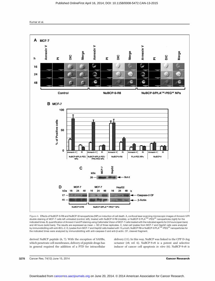
NuBCP-9/nanoparticles induce apoptosis of cancer cellsNuBCP-9-r8 is a selective inducer of cancer cell apoptosis by
targeting Bcl-2 (6). To assess the effects of NuBCP-9 nanopar-ticles on the apoptotic response, MCF-7 cells were treated withNuBCP-9/PLA72K-PEG4K nanoparticles and monitored forexternalization of phosphatidylserine at the cell membrane.Confocal images of MCF-7 cells stained with Annexin V-Alexaflour 488/PI demonstrated that treatment with NuBCP-9nanoparticles is associated with the induction of an apoptoticresponse (Fig. 4A). By contrast, treatment with empty nano-particles had no apparent effect (Fig. 4A). Quantitation ofAnnexin V and PI staining by Cellometer Vision confirmedthat NuBCP-9 PLA72K-PEG4K nanoparticles and NuBCP-9PLA72K-PEG-PPG-PEG nanoparticles are as effective asNuBCP-9-R8 in inducing apoptosis of MCF-7 cells at 48 hours(Fig. 4B). As controls, empty PLA72K-PEG4K nanoparticles andNuBCP-9 devoid of R8 had little, if any effect on MCF-7 cellapoptosis (Fig. 4B). As reported (28, 29), immunoblot analysisof MCF-7 and HepG2 cell lysates confirmed the expression ofBCL-2 (Fig. 4C). Moreover and in concert with the effects ofNuBCP-9 on BCL-2 (6), treatment of MCF-7 and HepG2 cellswith NuBCP-9 nanoparticles was associated with activation ofcaspase-3 (Fig. 4D). These findings thus demonstrate that, likeNuBCP-9-R8, encapsulation of NuBCP-9 in nanoparticlesresults in the induction of apoptosis (Fig. 4D).
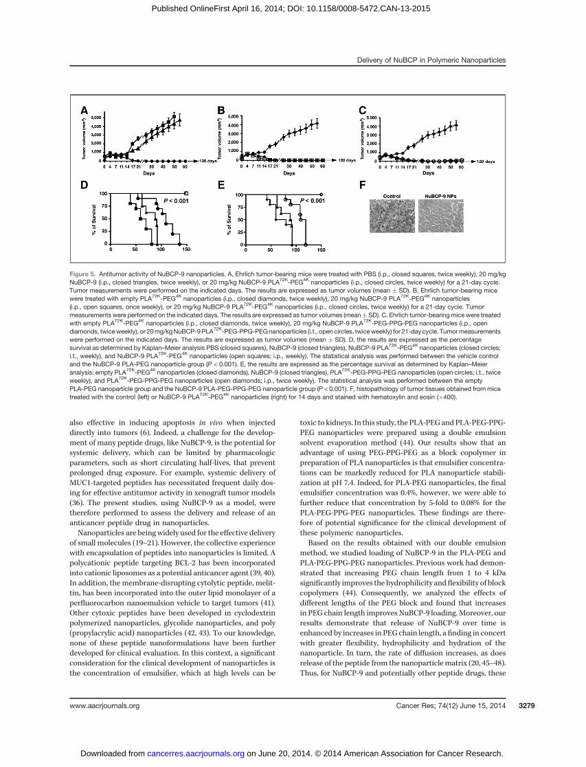
NuBCP-9 nanoparticles are effective in inducingcomplete tumor regressionsTo assess the antitumor effects of NuBCP-9 nanoparticles in
vivo, we treated Balb/c mice bearing established (�400 mm3)subcutaneous BCL-2–positive Ehrlich syngeneic tumors (30).In this widely used model to assess the effects of anticanceragents, accurate administration of the viscous suspensions of
nanoparticles was problematic in the narrow tail vein. As such,we used the i.p. route of administration, which allows nano-particles to enter the systemic circulation through mesentericvessels and the portal vein (31). Moreover, these studies wereperformed with NuBCP-9 (devoid of R8) because (i) NuBCP-9-R8 is active in vivo when administered i.t. (6), and (ii) theNuBCP-9 should be inactive in the absence of a CPP. Intra-peritoneal injection of NuBCP-9 on a twice weekly schedulefor 21 days had no significant effect on tumor growth as com-pared with that obtained with the saline control (PBS; Fig. 5Aand Supplementary Table S1). Significantly, i.p. treatment withNuBCP-9 PLA72K-PEG4K nanoparticles at a dose of 20 mg/kg(NuBCP-9 peptide dose) on the same schedule was associatedwith complete and prolonged tumor regressions (Fig. 5A).Moreover, in contrast to the empty PLA72K-PEG4K nanoparti-cles, i.p. injection of NuBCP-9 PLA72K-PEG4K nanoparticleson a weekly schedule for 21 days resulted in similar anti-tumor responses (Fig. 5B). For assessment of direct delivery ofNuBCP-9 nanoparticles into the tumors, we also administeredNuBCP-9 PLA72K-PEG-PPG-PEG nanoparticles i.t. on a twiceweekly schedule for 21 days and compared the effects ofintraperitoneal administration of PLA72K-PEG-PPG-PEG ontumor growth. The results demonstrate that regression of thetumors was similar when the NuBCP-9 nanoparticles weredelivered i.t. as compared with i.p. (Fig. 5C). However, i.p.treatment was more effective than i.t. in maintaining theseantitumor responses after completing treatment (Fig. 5C).Similar results were obtained when NuBCP-9 PLA72K-PEG4K
nanoparticles were administered i.t. weekly or twice weekly(Supplementary Fig. S3). Analysis of survival as determined byKaplan–Meier plots further demonstrated that mice treatedwith NuBCP-9 nanoparticles (PLA72K-PEG4K or PLA72K-PEG-PPG-PEG) weekly or twice weekly survived significantly longerthan the saline, empty nanoparticles, or NuBCP-9 controls (Fig.5D and E).Moreover, the NuBCP-9 nanoparticles–treatedmicesurvived longerwhen administrationwas i.p. as comparedwiththe i.t. route (Fig. 5D and E). Significantly, there was no weightloss or other overt toxicities observed in the NuBCP-9/nano-particle-treated mice (data not shown). Analysis of tissuesections demonstrated that tumor cells from the control miceexhibitedmitosis and little, if any, evidence of necrosis (Fig. 5F,left). By contrast, tumor cells from NuBCP-9 PLA72K-PEG4K
nanoparticles-treated mice had decreased mitotic activity andconsiderably larger regions of necrosis (Fig. 5F, right), consis-tent with the antitumor activity of NuBCP-9.
DiscussionPeptide drugs have considerable potential for targeting
intracellular oncoproteins that lack hydrophobic pockets andare thereby not eminently druggable with small molecules (32).In this context, there are increasing reports of peptide drugsunder development that target nonkinase proteins such assurvivin (33), HDM2 (34), NOTCH (35), MUC1 (36), and b-cate-nin (37), among others. The BCL-2 family proteins have alsobeen the focus of peptide inhibitor development, specifically inone strategy using stabilized a-helices of BCL-2 domains(SAHB; ref. 38). BCL-2 has also been converted from an anti-apoptotic protein to an inducer of cell death by the Nur77-
Table 2. IC50 of NuBCP-9 encapsulated PLA-PEG nanoparticles
IC50 (mmol/L)
MCF-7 HepG2
NuBCP-9-PLA 2.58 4.18NuBCP-9-PLA-PEG1K 2.31 3.90NuBCP-9-PLA-PEG2K 1.90 2.39NuBCP-9-PLA-PEG4K 2.15 2.03NuBCP-9-PLA-PEG-PPG-PEG12.5K
2.00 ND
NuBCP-9-R8 7.11 9.10
Abbreviation: ND, not done.
Delivery of NuBCP in Polymeric Nanoparticles
www.aacrjournals.org Cancer Res; 74(12) June 15, 2014 3277
on June 20, 2014. © 2014 American Association for Cancer Research. cancerres.aacrjournals.org Downloaded from
Published OnlineFirst April 16, 2014; DOI: 10.1158/0008-5472.CAN-13-2015
derived NuBCP peptide (6, 7). With the exception of SAHBs,which penetrate cell membranes, delivery of peptide drugs hasin general required the addition of a PTD for intracellular
delivery (11). In this way, NuBCP was linked to the CPP D-Argoctamer (r8; ref. 6). NuBCP-9-r8 is a potent and selectiveinducer of cancer cell apoptosis in vitro (6). NuBCP-9-r8 is
Figure 4. Effects of NuBCP-9-R8 and NuBCP-9/nanoparticles (NP) on induction of cell death. A, confocal laser scanning microscopic images of Annexin V/PIdouble staining of MCF-7 cells left untreated (control; left), treated with NuBCP-9 R8 (middle), or NuBCP-9 PLA72K-PEG4K nanoparticles (right) for theindicated times.B, quantification of Annexin V andPI staining usingCellometer Vision ofMCF-7 cells treatedwith the indicated agents for 24 hours (openbars)and 48 hours (solid bars). The results are expressed as mean � SD of three replicates. C, total cell lysates from MCF-7 and HepG2 cells were analyzedby immunoblotting with anti-BCL-2. D, lysates fromMCF-7 and HepG2 cells treated with 15 mmol/L NuBCP R8 or NuBCP-9 PLA72K-PEG4K nanoparticles forthe indicated times were analyzed by immunoblotting with anti-caspase-3 and anti-b-actin. CF, cleaved fragment.
Kumar et al.
Cancer Res; 74(12) June 15, 2014 Cancer Research3278
on June 20, 2014. © 2014 American Association for Cancer Research. cancerres.aacrjournals.org Downloaded from
Published OnlineFirst April 16, 2014; DOI: 10.1158/0008-5472.CAN-13-2015
also effective in inducing apoptosis in vivo when injecteddirectly into tumors (6). Indeed, a challenge for the develop-ment of many peptide drugs, like NuBCP-9, is the potential forsystemic delivery, which can be limited by pharmacologicparameters, such as short circulating half-lives, that preventprolonged drug exposure. For example, systemic delivery ofMUC1-targeted peptides has necessitated frequent daily dos-ing for effective antitumor activity in xenograft tumor models(36). The present studies, using NuBCP-9 as a model, weretherefore performed to assess the delivery and release of ananticancer peptide drug in nanoparticles.Nanoparticles are beingwidely used for the effective delivery
of small molecules (19–21). However, the collective experiencewith encapsulation of peptides into nanoparticles is limited. Apolycationic peptide targeting BCL-2 has been incorporatedinto cationic liposomes as a potential anticancer agent (39, 40).In addition, the membrane-disrupting cytolytic peptide, melit-tin, has been incorporated into the outer lipid monolayer of aperfluorocarbon nanoemulsion vehicle to target tumors (41).Other cytoxic peptides have been developed in cyclodextrinpolymerized nanoparticles, glycolide nanoparticles, and poly(propylacrylic acid) nanoparticles (42, 43). To our knowledge,none of these peptide nanoformulations have been furtherdeveloped for clinical evaluation. In this context, a significantconsideration for the clinical development of nanoparticles isthe concentration of emulsifier, which at high levels can be
toxic to kidneys. In this study, the PLA-PEGandPLA-PEG-PPG-PEG nanoparticles were prepared using a double emulsionsolvent evaporation method (44). Our results show that anadvantage of using PEG-PPG-PEG as a block copolymer inpreparation of PLA nanoparticles is that emulsifier concentra-tions can be markedly reduced for PLA nanoparticle stabili-zation at pH 7.4. Indeed, for PLA-PEG nanoparticles, the finalemulsifier concentration was 0.4%, however, we were able tofurther reduce that concentration by 5-fold to 0.08% for thePLA-PEG-PPG-PEG nanoparticles. These findings are there-fore of potential significance for the clinical development ofthese polymeric nanoparticles.
Based on the results obtained with our double emulsionmethod, we studied loading of NuBCP-9 in the PLA-PEG andPLA-PEG-PPG-PEG nanoparticles. Previous work had demon-strated that increasing PEG chain length from 1 to 4 kDasignificantly improves the hydrophilicity andflexibility of blockcopolymers (44). Consequently, we analyzed the effects ofdifferent lengths of the PEG block and found that increasesin PEG chain length improves NuBCP-9 loading. Moreover, ourresults demonstrate that release of NuBCP-9 over time isenhanced by increases in PEG chain length, afinding in concertwith greater flexibility, hydrophilicity and hydration of thenanoparticle. In turn, the rate of diffusion increases, as doesrelease of the peptide from the nanoparticle matrix (20, 45–48).Thus, for NuBCP-9 and potentially other peptide drugs, these
Figure 5. Antitumor activity of NuBCP-9 nanoparticles. A, Ehrlich tumor-bearing mice were treated with PBS (i.p., closed squares, twice weekly), 20 mg/kgNuBCP-9 (i.p., closed triangles, twice weekly), or 20 mg/kg NuBCP-9 PLA72K-PEG4K nanoparticles (i.p., closed circles, twice weekly) for a 21-day cycle.Tumor measurements were performed on the indicated days. The results are expressed as tumor volumes (mean � SD). B, Ehrlich tumor-bearing micewere treated with empty PLA72K-PEG4K nanoparticles (i.p., closed diamonds, twice weekly), 20 mg/kg NuBCP-9 PLA72K-PEG4K nanoparticles(i.p., open squares, once weekly), or 20 mg/kg NuBCP-9 PLA72K-PEG4K nanoparticles (i.p., closed circles, twice weekly) for a 21-day cycle. Tumormeasurements were performed on the indicated days. The results are expressed as tumor volumes (mean� SD). C, Ehrlich tumor-bearing mice were treatedwith empty PLA72K-PEG4K nanoparticles (i.p., closed diamonds, twice weekly), 20 mg/kg NuBCP-9 PLA72K-PEG-PPG-PEG nanoparticles (i.p., opendiamonds, twiceweekly), or 20mg/kgNuBCP-9PLA72K-PEG-PPG-PEGnanoparticles (i.t., open circles, twiceweekly) for 21-daycycle. Tumormeasurementswere performed on the indicated days. The results are expressed as tumor volumes (mean � SD). D, the results are expressed as the percentagesurvival as determined by Kaplan–Meier analysis PBS (closed squares), NuBCP-9 (closed triangles), NuBCP-9 PLA72K-PEG4K nanoparticles (closed circles;i.t., weekly), and NuBCP-9 PLA72K-PEG4K nanoparticles (open squares; i.p., weekly). The statistical analysis was performed between the vehicle controland the NuBCP-9 PLA-PEG nanoparticle group (P < 0.001). E, the results are expressed as the percentage survival as determined by Kaplan–Meieranalysis: empty PLA72K-PEG4K nanoparticles (closed diamonds), NuBCP-9 (closed triangles), PLA72K-PEG-PPG-PEG nanoparticles (open circles; i.t., twiceweekly), and PLA72K-PEG-PPG-PEG nanoparticles (open diamonds; i.p., twice weekly). The statistical analysis was performed between the emptyPLA-PEG nanoparticle group and the NuBCP-9 PLA-PEG-PPG-PEG nanoparticle group (P < 0.001). F, histopathology of tumor tissues obtained from micetreated with the control (left) or NuBCP-9 PLA72K-PEG4K nanoparticles (right) for 14 days and stained with hematoxylin and eosin (�400).
Delivery of NuBCP in Polymeric Nanoparticles
www.aacrjournals.org Cancer Res; 74(12) June 15, 2014 3279
on June 20, 2014. © 2014 American Association for Cancer Research. cancerres.aacrjournals.org Downloaded from
Published OnlineFirst April 16, 2014; DOI: 10.1158/0008-5472.CAN-13-2015
findings indicate that PEG chain length is of importance forboth loading in and release from polymeric nanoparticles.Another aspect of our studies worth highlighting is the useof a high molecular weight PLA (72 kDa) in the NuBCP-9encapsulated nanoparticles. Previouswork onpolymeric nano-particles has generally involved lower molecular weight PLA,for example, using 20 kDa or less (49, 50). Our results indicatethat in vitro release of NuBCP-9 from our nanoparticles wassustained to more than 60 days when using �72-kDa PLA ascompared with release of the peptide more than 10 days with�12-kDa PLA. These results can be explained, at least in part,by the hydrolysis of PLA and thereby NuBCP-9 diffusion. At pH5.0, PLA remains coiled and is less susceptible to hydrolysis;whereas at pH 7.4, the PLA chains open with an increasedpropensity for hydrolysis. The uncoiling of PLA is thus asso-ciated with an increase in its degradation and release ofNuBCP-9 (26, 27). These findings collectively indicate thatpeptide release from polymeric nanoparticles can be con-trolled by varying the sizes of the PEG and PLA blocks.
NuBCP-9 is a highly promising anticancer peptide thatselectively induces apoptosis of cancer cells by exposing theBCL-2 BH3 domain and blocking the BCL-xL survival function(6). NuBCP-9 was linked to the D-Arg octamer r8 for intracel-lular delivery, a modification that has been reported todecrease selectivity by inducing BCL-2–independent cell kill-ing involving membrane disruption (15). In this work, deliveryof NuBCP-9 into cancer cells by the polymeric nanoparticleswas achieved without the need for the R8 PTD. In addition, theNuBCP-9–encapsulated nanoparticles were by comparisonmore potent in inducing apoptosis than NuBCP-9-R8. TheNuBCP-9/nanoparticles also maintained the reported selec-tivity of NuBCP-9 for cancer cells as evidenced by their absenceof HUVEC killing (6). Previous studies of the antitumor effectsof NuBCP-9 in vivo were performed by direct injection ofNuBCP-9-r8 into MDA-MB-435 breast cancer xenografts grow-ing in SCID mice (6). Under these experimental conditions,NuBCP-9-r8 treatment was associated with partial MDA-MB-435 tumor regressions (6). In this studies using the subcuta-neous Ehrlich tumor model in syngeneic mice, we comparedthe effectiveness of NuBCP-9/nanoparticles administered i.p.and i.t.. Significantly, both routes of NuBCP-9/nanoparticleadministrationwere effective in inducing complete regressionsof the Ehrlich tumors. Moreover and interestingly, i.p. admin-
istration was more effective than i.t. delivery in maintainingprolonged tumor regressions. The basis for this distinction ispresently not clear, but is likely related to pharmacokineticsand pharmacodynamics of the NuBCP-9/nanoparticles, whichwill be the focus of subsequent studies. Of further importance,administration of the NuBCP-9/nanoparticles was well toler-ated with no evidence of weight loss or overt toxicities. Theseresults thus provide support for the effective delivery of L-amino acid NuBCP-9 by PLA-PEG-PPG-PEG nanoparticles andmay be applicable to other anticancer peptides. In summary,our findings describe a novel nanoparticle-based approach fordelivery of L-amino acid peptides without a cell-penetratingdomain that (i) requires reduced levels of emulsifier, (ii)incorporates higher molecular weights of PLA for sustainedand prolonged peptide release, (iii) induces effective antican-cer activity in vitro and in vivo, and (iv) requires less frequentdosing (once/week) compared with the daily injections thatwere necessary for partial antitumor activity of the D-aminoacid NuBCP-9-r8 peptide in vivo (6).
Disclosure of Potential Conflicts of InterestNo potential conflicts of interest were disclosed.
Authors' ContributionsConception and design:M. Kumar, A.K. Dinda, S. Kharbanda, D. Kufe, H. SinghDevelopment of methodology: M. Kumar, A.K. Dinda, H. SinghAcquisition of data (provided animals, acquired and managed patients,provided facilities, etc.): M. Kumar, D. Gupta, G. Singh, S. Sharma, M. Bhat,C.K. Prashant, A.K. Dinda, H. SinghAnalysis and interpretation of data (e.g., statistical analysis, biostatistics,computational analysis): M. Kumar, S. Sharma, A.K. Dinda, S. Kharbanda,H. SinghWriting, review, and/or revision of the manuscript:M. Kumar, A.K. Dinda,S. Kharbanda, D. Kufe, H. SinghAdministrative, technical, or material support (i.e., reporting or orga-nizing data, constructing databases): M. Kumar, A.K. DindaStudy supervision: A.K. Dinda, S. Kharbanda, H. Singh
Grant SupportThis work was supported by the National Cancer Institute of the NIH under
award number CA97098, by the Lung Cancer Research Foundation, and by agrant from the Department of Science and Technology, Government of India.
The costs of publication of this article were defrayed in part by the payment ofpage charges. This article must therefore be hereby marked advertisement inaccordance with 18 U.S.C. Section 1734 solely to indicate this fact.
Received July 18, 2013; revised March 12, 2014; accepted March 19, 2014;published OnlineFirst April 16, 2014.
References1. Cory S, Huang DC, Adams JM. The Bcl-2 family: roles in cell survival
and oncogenesis. Oncogene 2003;22:8590–607.2. Green DR, Kroemer G. The pathophysiology of mitochondrial cell
death. Science 2004;305:626–9.3. Pitter K, Bernal F, Labelle J, Walensky LD. Dissection of the BCL-2
family signaling network with stabilized a-helices of BCL-2 domains.Methods Enzymol 2008;446:387–408.
4. Oltersdorf T, Elmore SW, Shoemaker AR, Armstrong RC, Augeri DJ,Belli BA, et al. An inhibitor ofBcl-2 family proteins induces regressionofsolid tumours. Nature 2005;435:677–81.
5. Tse C, Shoemaker AR, Adickes J, Anderson MG, Chen J, Jin S, et al.ABT-263: a potent andorally bioavailable Bcl-2 family inhibitor. CancerRes 2008;68:3421–8.
6. Kolluri SK, Zhu X, Zhou X, Lin B, Chen Y, Sun K, et al. A short Nur77-derived peptide converts Bcl-2 from a protector to a killer. Cancer Cell2008;14:285–98.
7. Lin B, Kolluri SK, Lin F, LiuW, HanYH,Cao X, et al. Conversion of Bcl-2from protector to killer by interaction with nuclear orphan receptorNur77/TR3. Cell 2004;116:527–40.
8. Bidwell GL 3rd, Raucher D. Therapeutic peptides for cancer therapy.Part I—peptide inhibitors of signal transduction cascades. Expert OpinDrug Deliv 2009;6:1033–47.
9. Raucher D, Moktan S, Massodi I, Bidwell GL 3rd. Therapeuticpeptides for cancer therapy. Part II—cell cycle inhibitory peptidesand apoptosis-inducing peptides. Expert Opin Drug Deliv 2009;6:1049–64.
Kumar et al.
Cancer Res; 74(12) June 15, 2014 Cancer Research3280
on June 20, 2014. © 2014 American Association for Cancer Research. cancerres.aacrjournals.org Downloaded from
Published OnlineFirst April 16, 2014; DOI: 10.1158/0008-5472.CAN-13-2015
10. Talmadge JE. Pharmacodynamic aspects of peptide administra-tion biological response modifiers. Adv Drug Deliv Rev 1998;33:241–52.
11. Fischer PM. Cellular uptake mechanisms and potential therapeuticutility of peptidic cell delivery vectors: progress 2001–2006. Med ResRev 2007;27:755–95.
12. Grdisa M. The delivery of biologically active (therapeutic) peptides andproteins into cells. Curr Med Chem 2011;18:1373–9.
13. Li H, Nelson CE, Evans BC, Duvall CL. Delivery of intracellular-actingbiologics in pro-apoptotic therapies. Curr Pharm Des 2011;17:293–319.
14. GuZ, Biswas A, ZhaoM, Tang Y. Tailoring nanocarriers for intracellularprotein delivery. Chem Soc Rev 2011;40:3638–55.
15. Watkins CL, Sayers EJ, Allender C, Barrow D, Fegan C, BrennanP, et al. Co-operative membrane disruption between cell-pene-trating peptide and cargo: implications for the therapeutic use ofthe Bcl-2 converter peptide D-NuBCP-9-r8. Mol Ther 2011;19:2124–32.
16. Kratz F, Warnecke A. Finding the optimal balance: challenges ofimproving conventional cancer chemotherapy using suitable combi-nations with nano-sized drug delivery systems. J Control Release2012;164:221–35.
17. Manzoor AA, Lindner LH, LandonCD, Park JY, Simnick AJ, DreherMR,et al. Overcoming limitations in nanoparticle drug delivery: triggered,intravascular release to improve drug penetration into tumors. CancerRes 2012;72:5566–75.
18. Zhang XY, Chen J, Zheng YF, Gao XL, Kang Y, Liu JC, et al.Follicle-stimulating hormone peptide can facilitate paclitaxelnanoparticles to target ovarian carcinoma in vivo. Cancer Res2009;69:6506–14.
19. Kamaly N, Xiao Z, Valencia PM, Radovic-Moreno AF, Farokhzad OC.Targeted polymeric therapeutic nanoparticles: design, developmentand clinical translation. Chem Soc Rev 2012;41:2971–3010.
20. Xiao RZ, Zeng ZW, Zhou GL, Wang JJ, Li FZ, Wang AM. Recentadvances in PEG-PLA block copolymer nanoparticles. Int J Nanome-dicine 2010;5:1057–65.
21. Kim TY, Kim DW, Chung JY, Shin SG, Kim SC, Heo DS, et al. Phase Iand pharmacokinetic study of Genexol-PM, a cremophor-free, poly-meric micelle-formulated paclitaxel, in patients with advanced malig-nancies. Clin Cancer Res 2004;10:3708–16.
22. Xiong XY, Li QH, Li YP, Guo L, Li ZL, Gong YC. Pluronic P85/poly(lacticacid) vesicles as novel carrier for oral insulin delivery. Colloids Surf BBiointerfaces 2013;111C:282–8.
23. Panyam J, Zhou WZ, Prabha S, Sahoo SK, Labhasetwar V. Rapidendo-lysosomal escape of poly(DL-lactide-co-glycolide) nanoparti-cles: implications for drug and gene delivery. FASEB J 2002;16:1217–26.
24. Doiron AL, Chu K, Ali A, Brannon-Peppas L. Preparation and initialcharacterization of biodegradable particles containing gadolinium-DTPA contrast agent for enhanced MRI. Proc Natl Acad Sci U S A2008;105:17232–7.
25. Albanese A, TangPS,ChanWC. The effect of nanoparticle size, shape,and surface chemistry on biological systems. Annu Rev Biomed Eng2012;14:1–16.
26. Andersson SR, Hakkarainen M, Inkinen S, Sodergard A, AlbertssonAC. Customizing the hydrolytic degradation rate of stereocomplexPLA through different PDLA architectures. Biomacromolecules 2012;13:1212–22.
27. Hoglund A, Odelius K, Albertsson AC. Crucial differences in thehydrolytic degradation between industrial polylactide and labora-tory-scale poly(L-lactide). ACS Appl Mater Interfaces 2012;4:2788–93.
28. Nathan B, Gusterson B, Jadayel D, O'Hare M, Anbazhagan R, Jaya-tilake H, et al. Expression of BCL-2 in primary breast cancer and itscorrelation with tumour phenotype. For the International (Ludwig)Breast Cancer Study Group. Ann Oncol 1994;5:409–14.
29. Tan J, Wang B, Zhu L. Regulation of survivin and Bcl-2 in HepG2 cellapoptosis induced by quercetin. Chem Biodivers 2009;6:1101–10.
30. Bhattacharyya A, Choudhuri T, Pal S, Chattopadhyay S, K Datta G, SaG, et al. Apoptogenic effects of black tea on Ehrlich's ascites carci-noma cell. Carcinogenesis 2003;24:75–80.
31. Turner PV, Brabb T, Pekow C, Vasbinder MA. Administration of sub-stances to laboratory animals: routes of administration and factors toconsider. J Am Assoc Lab Anim Sci 2011;50:600–13.
32. Verdine GL, Walensky LD. The challenge of drugging undruggabletargets in cancer: lessons learned from targeting BCL-2 family mem-bers. Clin Cancer Res 2007;13:7264–70.
33. Plescia J, Salz W, Xia F, Pennati M, Zaffaroni N, Daidone MG, et al.Rational design of shepherdin, a novel anticancer agent. Cancer Cell2005;7:457–68.
34. Sarafraz-Yazdi E, Bowne WB, Adler V, Sookraj KA, Wu V, Shteyler V,et al. Anticancer peptide PNC-27 adopts an HDM-2-binding confor-mation and kills cancer cells by binding to HDM-2 in their membranes.Proc Natl Acad Sci U S A 2010;107:1918–23.
35. Moellering RE, Cornejo M, Davis TN, Del Bianco C, Aster JC, BlacklowSC, et al. Direct inhibition of the NOTCH transcription factor complex.Nature 2009;462:182–8.
36. Raina D, Kosugi M, Ahmad R, Panchamoorthy G, Rajabi H, Alam M,et al. Dependence on the MUC1-C oncoprotein in non-small cell lungcancer cells. Mol Cancer Ther 2011;10:806–16.
37. Grossmann TN, Yeh JT, Bowman BR, Chu Q, Moellering RE, VerdineGL. Inhibition of oncogenic Wnt signaling through direct targeting ofbeta-catenin. Proc Natl Acad Sci U S A 2012;109:17942–7.
38. Stewart ML, Fire E, Keating AE, Walensky LD. The MCL-1 BH3 helix isan exclusive MCL-1 inhibitor and apoptosis sensitizer. Nat Chem Biol2010;6:595–601.
39. Ko YT, Falcao C, Torchilin VP. Cationic liposomes loaded with proa-poptotic peptide D-(KLAKLAK)(2) and Bcl-2 antisense oligodeoxynu-cleotide G3139 for enhanced anticancer therapy. Mol Pharm 2009;6:971–7.
40. Adar L, Shamay Y, Journo G, A D. Pro-apoptic peptide-polymerconjugates to induce mitochondrial-dependent cell death. Polym AdvTechnol 2010;22:199–208.
41. Soman NR, Baldwin SL, Hu G, Marsh JN, Lanza GM, Heuser J, et al.Molecularly targeted nanocarriers deliver the cytolytic peptide melittinspecifically to tumor cells in mice, reducing cell growth. J Clin Invest2009;119:2830–42.
42. Schluep T, Gunawan P, Ma L, Jensen GS, Duringer J, Hinton S, et al.Polymeric tubulysin-peptide nanoparticles with potent antitumoractivity. Clin Cancer Res 2009;15:181–9.
43. Kim SK, Huang L. Nanoparticle delivery of a peptide targeting EGFRsignaling. J Control Release 2012;157:279–86.
44. Tomar L, Tyagi C, Kumar M, Kumar P, Singh H, Choonara YE, et al. Invivo evaluation of a conjugated poly(lactide-ethylene glycol) nanopar-ticle depot formulation for prolonged insulin delivery in the diabeticrabbit model. Int J Nanomedicine 2013;8:505–20.
45. Essa S, Rabanel JM, Hildgen P. Effect of polyethylene glycol (PEG)chain organization on the physicochemical properties of poly(D, L-lactide) (PLA) based nanoparticles. Eur J Pharm Biopharm 2010;75:96–106.
46. Feng R, Song Z, Zhai G. Preparation and in vivo pharmacokinetics ofcurcumin-loaded PCL-PEG-PCL triblock copolymeric nanoparticles.Int J Nanomedicine 2012;7:4089–98.
47. HansM, LowmanA.Biodegradable nanoparticles for drugdelivery andtargeting. Curr Opin Solid State Mater Sci 2002;6:319–27.
48. Wang B, Jiang W, Yan H, Zhang X, Yang L, Deng L, et al. Novel PEG-graft-PLA nanoparticles with the potential for encapsulation and con-trolled release of hydrophobic and hydrophilic medications in aqueousmedium. Int J Nanomedicine 2011;6:1443–51.
49. Hrkach J, VonHoff D,MukkaramAliM, AndrianovaE, Auer J,CampbellT, et al. Preclinical development and clinical translation of a PSMA-targeted docetaxel nanoparticle with a differentiated pharmacologicalprofile. Sci Transl Med 2012;4:128ra39.
50. Farokhzad OC, Cheng J, Teply BA, Sherifi I, Jon S, Kantoff PW, et al.Targeted nanoparticle-aptamer bioconjugates for cancer chemother-apy in vivo. Proc Natl Acad Sci U S A 2006;103:6315–20.
www.aacrjournals.org Cancer Res; 74(12) June 15, 2014 3281
Delivery of NuBCP in Polymeric Nanoparticles
on June 20, 2014. © 2014 American Association for Cancer Research. cancerres.aacrjournals.org Downloaded from
Published OnlineFirst April 16, 2014; DOI: 10.1158/0008-5472.CAN-13-2015