Astrocytes Give Rise to New Neurons in the Adult Mammalian Hippocampus
Upload
independentCategory
view
2download
0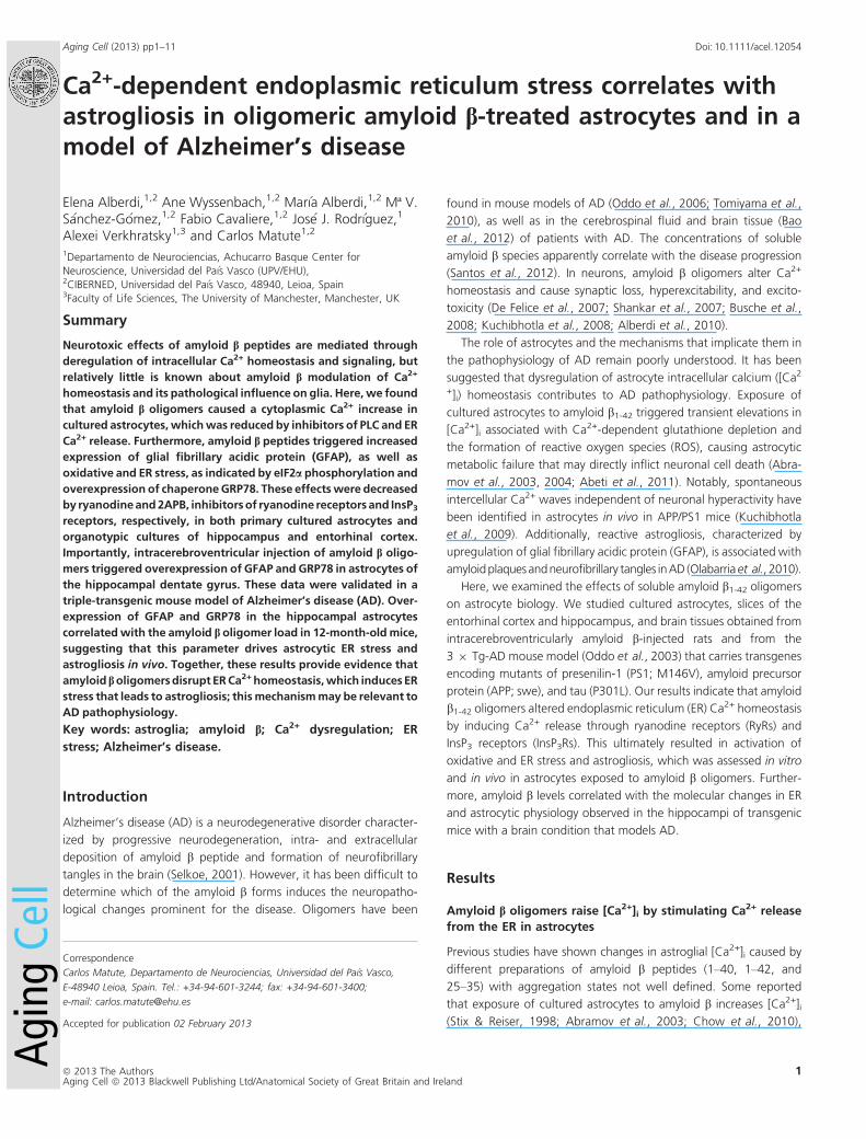
Ca2+-dependent endoplasmic reticulum stress correlates withastrogliosis in oligomeric amyloid b-treated astrocytes and in amodel of Alzheimer’s disease
Elena Alberdi,1,2 Ane Wyssenbach,1,2 Mar�ıa Alberdi,1,2 Mª V.S�anchez-G�omez,1,2 Fabio Cavaliere,1,2 Jos�e J. Rodr�ıguez,1
Alexei Verkhratsky1,3 and Carlos Matute1,2
1Departamento de Neurociencias, Achucarro Basque Center for
Neuroscience, Universidad del Pa�ıs Vasco (UPV/EHU),2CIBERNED, Universidad del Pa�ıs Vasco, 48940, Leioa, Spain3Faculty of Life Sciences, The University of Manchester, Manchester, UK
Summary
Neurotoxic effects of amyloid b peptides are mediated through
deregulation of intracellular Ca2+ homeostasis and signaling, but
relatively little is known about amyloid b modulation of Ca2+
homeostasis and its pathological influence on glia. Here,we found
that amyloid b oligomers caused a cytoplasmic Ca2+ increase in
cultured astrocytes,whichwas reduced by inhibitors of PLC and ER
Ca2+ release. Furthermore, amyloid b peptides triggered increased
expression of glial fibrillary acidic protein (GFAP), as well as
oxidative and ER stress, as indicated by eIF2a phosphorylation and
overexpression of chaperoneGRP78. These effectswere decreased
by ryanodineand2APB, inhibitorsof ryanodine receptorsand InsP3receptors, respectively, in both primary cultured astrocytes and
organotypic cultures of hippocampus and entorhinal cortex.
Importantly, intracerebroventricular injection of amyloid b oligo-
mers triggered overexpression of GFAP and GRP78 in astrocytes of
the hippocampal dentate gyrus. These data were validated in a
triple-transgenic mouse model of Alzheimer’s disease (AD). Over-
expression of GFAP and GRP78 in the hippocampal astrocytes
correlatedwith the amyloid b oligomer load in 12-month-oldmice,
suggesting that this parameter drives astrocytic ER stress and
astrogliosis in vivo. Together, these results provide evidence that
amyloid boligomersdisrupt ERCa2+homeostasis,which inducesER
stress that leads to astrogliosis; thismechanismmay be relevant to
AD pathophysiology.
Key words: astroglia; amyloid b; Ca2+ dysregulation; ER
stress; Alzheimer’s disease.
Introduction
Alzheimer’s disease (AD) is a neurodegenerative disorder character-
ized by progressive neurodegeneration, intra- and extracellular
deposition of amyloid b peptide and formation of neurofibrillary
tangles in the brain (Selkoe, 2001). However, it has been difficult to
determine which of the amyloid b forms induces the neuropatho-
logical changes prominent for the disease. Oligomers have been
found in mouse models of AD (Oddo et al., 2006; Tomiyama et al.,
2010), as well as in the cerebrospinal fluid and brain tissue (Bao
et al., 2012) of patients with AD. The concentrations of soluble
amyloid b species apparently correlate with the disease progression
(Santos et al., 2012). In neurons, amyloid b oligomers alter Ca2+
homeostasis and cause synaptic loss, hyperexcitability, and excito-
toxicity (De Felice et al., 2007; Shankar et al., 2007; Busche et al.,
2008; Kuchibhotla et al., 2008; Alberdi et al., 2010).
The role of astrocytes and the mechanisms that implicate them in
the pathophysiology of AD remain poorly understood. It has been
suggested that dysregulation of astrocyte intracellular calcium ([Ca2
+]i) homeostasis contributes to AD pathophysiology. Exposure of
cultured astrocytes to amyloid b1-42 triggered transient elevations in
[Ca2+]i associated with Ca2+-dependent glutathione depletion and
the formation of reactive oxygen species (ROS), causing astrocytic
metabolic failure that may directly inflict neuronal cell death (Abra-
mov et al., 2003, 2004; Abeti et al., 2011). Notably, spontaneous
intercellular Ca2+ waves independent of neuronal hyperactivity have
been identified in astrocytes in vivo in APP/PS1 mice (Kuchibhotla
et al., 2009). Additionally, reactive astrogliosis, characterized by
upregulation of glial fibrillary acidic protein (GFAP), is associated with
amyloidplaquesandneurofibrillary tangles inAD (Olabarriaet al., 2010).
Here, we examined the effects of soluble amyloid b1-42 oligomers
on astrocyte biology. We studied cultured astrocytes, slices of the
entorhinal cortex and hippocampus, and brain tissues obtained from
intracerebroventricularly amyloid b-injected rats and from the
3 9 Tg-AD mouse model (Oddo et al., 2003) that carries transgenes
encoding mutants of presenilin-1 (PS1; M146V), amyloid precursor
protein (APP; swe), and tau (P301L). Our results indicate that amyloid
b1-42 oligomers altered endoplasmic reticulum (ER) Ca2+ homeostasis
by inducing Ca2+ release through ryanodine receptors (RyRs) and
InsP3 receptors (InsP3Rs). This ultimately resulted in activation of
oxidative and ER stress and astrogliosis, which was assessed in vitro
and in vivo in astrocytes exposed to amyloid b oligomers. Further-
more, amyloid b levels correlated with the molecular changes in ER
and astrocytic physiology observed in the hippocampi of transgenic
mice with a brain condition that models AD.
Results
Amyloid b oligomers raise [Ca2+]i by stimulating Ca2+ release
from the ER in astrocytes
Previous studies have shown changes in astroglial [Ca2+]i caused by
different preparations of amyloid b peptides (1–40, 1–42, and
25–35) with aggregation states not well defined. Some reported
that exposure of cultured astrocytes to amyloid b increases [Ca2+]i(Stix & Reiser, 1998; Abramov et al., 2003; Chow et al., 2010),
Correspondence
Carlos Matute, Departamento de Neurociencias, Universidad del Pa�ıs Vasco,
E-48940 Leioa, Spain. Tel.: +34-94-601-3244; fax: +34-94-601-3400;
e-mail: [email protected]
Accepted for publication 02 February 2013
ª 2013 The AuthorsAging Cell ª 2013 Blackwell Publishing Ltd/Anatomical Society of Great Britain and Ireland
1
Aging Cell (2013) pp1–11 Doi: 10.1111/acel.12054Ag
ing
Cell
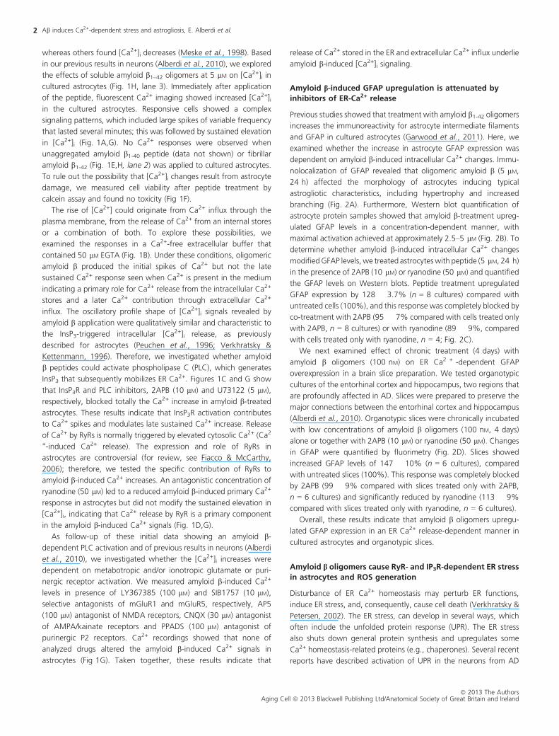
whereas others found [Ca2+]i decreases (Meske et al., 1998). Based
in our previous results in neurons (Alberdi et al., 2010), we explored
the effects of soluble amyloid b1–42 oligomers at 5 lM on [Ca2+]i in
cultured astrocytes (Fig. 1H, lane 3). Immediately after application
of the peptide, fluorescent Ca2+ imaging showed increased [Ca2+]iin the cultured astrocytes. Responsive cells showed a complex
signaling patterns, which included large spikes of variable frequency
that lasted several minutes; this was followed by sustained elevation
in [Ca2+]i (Fig. 1A,G). No Ca2+ responses were observed when
unaggregated amyloid b1-40 peptide (data not shown) or fibrillar
amyloid b1-42 (Fig. 1E,H, lane 2) was applied to cultured astrocytes.
To rule out the possibility that [Ca2+]i changes result from astrocyte
damage, we measured cell viability after peptide treatment by
calcein assay and found no toxicity (Fig 1F).
The rise of [Ca2+] could originate from Ca2+ influx through the
plasma membrane, from the release of Ca2+ from an internal stores
or a combination of both. To explore these possibilities, we
examined the responses in a Ca2+-free extracellular buffer that
contained 50 lM EGTA (Fig. 1B). Under these conditions, oligomeric
amyloid b produced the initial spikes of Ca2+ but not the late
sustained Ca2+ response seen when Ca2+ is present in the medium
indicating a primary role for Ca2+ release from the intracellular Ca2+
stores and a later Ca2+ contribution through extracellular Ca2+
influx. The oscillatory profile shape of [Ca2+]i signals revealed by
amyloid b application were qualitatively similar and characteristic to
the InsP3-triggered intracellular [Ca2+]i release, as previously
described for astrocytes (Peuchen et al., 1996; Verkhratsky &
Kettenmann, 1996). Therefore, we investigated whether amyloid
b peptides could activate phospholipase C (PLC), which generates
InsP3 that subsequently mobilizes ER Ca2+. Figures 1C and G show
that InsP3R and PLC inhibitors, 2APB (10 lM) and U73122 (5 lM),
respectively, blocked totally the Ca2+ increase in amyloid b-treated
astrocytes. These results indicate that InsP3R activation contributes
to Ca2+ spikes and modulates late sustained Ca2+ increase. Release
of Ca2+ by RyRs is normally triggered by elevated cytosolic Ca2+ (Ca2
+-induced Ca2+ release). The expression and role of RyRs in
astrocytes are controversial (for review, see Fiacco & McCarthy,
2006); therefore, we tested the specific contribution of RyRs to
amyloid b-induced Ca2+ increases. An antagonistic concentration of
ryanodine (50 lM) led to a reduced amyloid b-induced primary Ca2+
response in astrocytes but did not modify the sustained elevation in
[Ca2+]i, indicating that Ca2+ release by RyR is a primary component
in the amyloid b-induced Ca2+ signals (Fig. 1D,G).
As follow-up of these initial data showing an amyloid b-
dependent PLC activation and of previous results in neurons (Alberdi
et al., 2010), we investigated whether the [Ca2+]i increases were
dependent on metabotropic and/or ionotropic glutamate or puri-
nergic receptor activation. We measured amyloid b-induced Ca2+
levels in presence of LY367385 (100 lM) and SIB1757 (10 lM),
selective antagonists of mGluR1 and mGluR5, respectively, AP5
(100 lM) antagonist of NMDA receptors, CNQX (30 lM) antagonist
of AMPA/kainate receptors and PPADS (100 lM) antagonist of
purinergic P2 receptors. Ca2+ recordings showed that none of
analyzed drugs altered the amyloid b-induced Ca2+ signals in
astrocytes (Fig 1G). Taken together, these results indicate that
release of Ca2+ stored in the ER and extracellular Ca2+ influx underlie
amyloid b-induced [Ca2+]i signaling.
Amyloid b-induced GFAP upregulation is attenuated by
inhibitors of ER-Ca2+ release
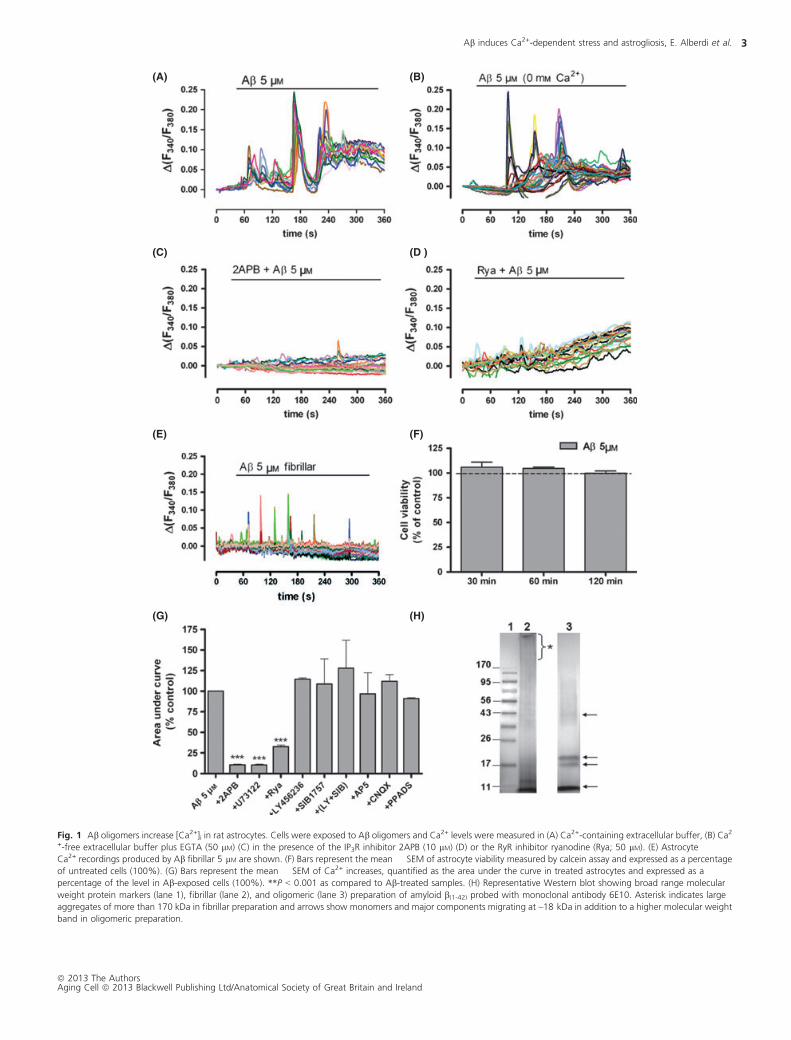
Previous studies showed that treatment with amyloid b1-42 oligomers
increases the immunoreactivity for astrocyte intermediate filaments
and GFAP in cultured astrocytes (Garwood et al., 2011). Here, we
examined whether the increase in astrocyte GFAP expression was
dependent on amyloid b-induced intracellular Ca2+ changes. Immu-
nolocalization of GFAP revealed that oligomeric amyloid b (5 lM,
24 h) affected the morphology of astrocytes inducing typical
astrogliotic characteristics, including hypertrophy and increased
branching (Fig. 2A). Furthermore, Western blot quantification of
astrocyte protein samples showed that amyloid b-treatment upreg-
ulated GFAP levels in a concentration-dependent manner, with
maximal activation achieved at approximately 2.5–5 lM (Fig. 2B). To
determine whether amyloid b-induced intracellular Ca2+ changes
modifiedGFAP levels, we treated astrocyteswith peptide (5 lM, 24 h)
in the presence of 2APB (10 lM) or ryanodine (50 lM) and quantified
the GFAP levels on Western blots. Peptide treatment upregulated
GFAP expression by 128 � 3.7% (n = 8 cultures) compared with
untreated cells (100%), and this response was completely blocked by
co-treatment with 2APB (95 � 7% compared with cells treated only
with 2APB, n = 8 cultures) or with ryanodine (89 � 9%, compared
with cells treated only with ryanodine, n = 4; Fig. 2C).
We next examined effect of chronic treatment (4 days) with
amyloid b oligomers (100 nM) on ER Ca2 + -dependent GFAP
overexpression in a brain slice preparation. We tested organotypic
cultures of the entorhinal cortex and hippocampus, two regions that
are profoundly affected in AD. Slices were prepared to preserve the
major connections between the entorhinal cortex and hippocampus
(Alberdi et al., 2010). Organotypic slices were chronically incubated
with low concentrations of amyloid b oligomers (100 nM, 4 days)
alone or together with 2APB (10 lM) or ryanodine (50 lM). Changes
in GFAP were quantified by fluorimetry (Fig. 2D). Slices showed
increased GFAP levels of 147 � 10% (n = 6 cultures), compared
with untreated slices (100%). This response was completely blocked
by 2APB (99 � 9% compared with slices treated only with 2APB,
n = 6 cultures) and significantly reduced by ryanodine (113 � 9%
compared with slices treated only with ryanodine, n = 6 cultures).
Overall, these results indicate that amyloid b oligomers upregu-
lated GFAP expression in an ER Ca2+ release-dependent manner in
cultured astrocytes and organotypic slices.
Amyloid b oligomers cause RyR- and IP3R-dependent ER stress
in astrocytes and ROS generation
Disturbance of ER Ca2+ homeostasis may perturb ER functions,
induce ER stress, and, consequently, cause cell death (Verkhratsky &
Petersen, 2002). The ER stress, can develop in several ways, which
often include the unfolded protein response (UPR). The ER stress
also shuts down general protein synthesis and upregulates some
Ca2+ homeostasis-related proteins (e.g., chaperones). Several recent
reports have described activation of UPR in the neurons from AD
Ab induces Ca2+-dependent stress and astrogliosis, E. Alberdi et al.2
ª 2013 The AuthorsAging Cell ª 2013 Blackwell Publishing Ltd/Anatomical Society of Great Britain and Ireland
(A) (B)
(C) (D )
(E) (F)
(G) (H)
Fig. 1 Ab oligomers increase [Ca2+]i in rat astrocytes. Cells were exposed to Ab oligomers and Ca2+ levels were measured in (A) Ca2+-containing extracellular buffer, (B) Ca2
+-free extracellular buffer plus EGTA (50 lM) (C) in the presence of the IP3R inhibitor 2APB (10 lM) (D) or the RyR inhibitor ryanodine (Rya; 50 lM). (E) AstrocyteCa2+ recordings produced by Ab fibrillar 5 lM are shown. (F) Bars represent the mean � SEM of astrocyte viability measured by calcein assay and expressed as a percentage
of untreated cells (100%). (G) Bars represent the mean � SEM of Ca2+ increases, quantified as the area under the curve in treated astrocytes and expressed as a
percentage of the level in Ab-exposed cells (100%). **P < 0.001 as compared to Ab-treated samples. (H) Representative Western blot showing broad range molecular
weight protein markers (lane 1), fibrillar (lane 2), and oligomeric (lane 3) preparation of amyloid b(1-42) probed with monoclonal antibody 6E10. Asterisk indicates large
aggregates of more than 170 kDa in fibrillar preparation and arrows show monomers and major components migrating at ~18 kDa in addition to a higher molecular weight
band in oligomeric preparation.
Ab induces Ca2+-dependent stress and astrogliosis, E. Alberdi et al. 3
ª 2013 The AuthorsAging Cell ª 2013 Blackwell Publishing Ltd/Anatomical Society of Great Britain and Ireland
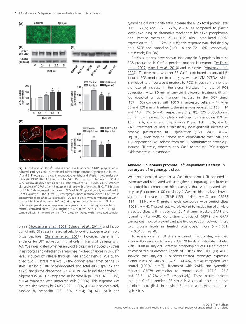
brains (Hoozemans et al., 2009; Scheper et al., 2011), and induc-
tion of mild ER stress in neuronal cells following exposure to amyloid
b1–42 peptides (Chafekar et al., 2007). However, there is no
evidence for UPR activation in glial cells in brains of patients with
AD. We investigated whether amyloid b oligomers induced ER stress
in astrocytes and whether this response involved changes in ER Ca2+
levels induced by release through RyRs and/or InsP3Rs. We quan-
tified two ER stress markers: (i) the downstream target of the ER
stress sensor pPERK phosphorylated and total eIF2a (peIF2a and
eIF2a) and (ii) the chaperone GRP78 (BIP). We found that amyloid b
oligomers (5 lM, 1 h) triggered an increase in peIF2a (132 � 13%,
n = 4) compared with untreated cells (100%). This response was
reduced significantly by 2APB (122 � 10%, n = 4), and completely
blocked by ryanodine (93 � 3%, n = 4; Fig. 3A). 2APB and
ryanodine did not significantly increase the eIF2a total protein level
(115 � 24%; and 107 � 22%, n = 4, as compared to b-actin
levels) excluding an alternative mechanism for eIF2a phosphoryla-
tion. Peptide treatment (5 lM, 6 h) also upregulated GRP78
expression to 151 � 12% (n = 8); this response was abolished by
both 2APB and ryanodine (100 � 8 and 72 � 6%, respectively,
n = 8 each; Fig. 3A).
Previous reports have shown that amyloid b peptides increase
ROS production in Ca2+-dependent manner in neurons (De Felice
et al., 2007; Alberdi et al., 2010) and astrocytes (Abramov et al.,
2004). To determine whether ER Ca2+ contributed to amyloid b-
induced ROS production in astrocytes, we used CM-DCFDA, which
is oxidized to a fluorescent product by ROS, in such a manner that
the rate of increase in the signal indicates the rate of ROS
generation. After 30 min of amyloid b oligomer treatment (5 lM),
we detected a rapid transient increase in the DCF signal
(137 � 6% compared with 100% in untreated cells, n = 4). After
60 and 120 min of treatment, the signal was reduced to 125 � 14
and 113 � 7% (n = 4), respectively (Fig. 3B). ROS production at
30 min was almost completely inhibited by ryanodine (50 lM;
106 � 2%, n = 4) and thapsigargin (1 lM; 108 � 3%, n = 4).
2APB treatment caused a statistically nonsignificant increase of
amyloid b-stimulated ROS generation (153 � 24%, n = 4;
Fig. 3C). Taken together, these data demonstrate that RyR- and
IP3R-dependent Ca2+ release from the ER contributes to amyloid b-
induced ER stress, whereas only Ca2+ release via RyRs triggers
oxidative stress in astrocytes.
Amyloid b oligomers promote Ca2+-dependent ER stress in
astrocytes of organotypic slices
We next examined whether a Ca2+-dependent UPR occurred in
astrocytes and correlated with astrogliosis in organotypic cultures of
the entorhinal cortex and hippocampus that were treated with
amyloid b oligomers (100 nM; 4 days). Western blot analysis showed
significant increases in GFAP (139 � 14%, n = 4) and GRP78
(184 � 38%, n = 4) protein levels compared with control slices
(100%, n = 4). These effects were blocked by incubation of amyloid
b-treated slices with intracellular Ca2+ channel blockers 2APB and
ryanodine (Fig. 4A,B). Correlation analysis of GRP78 and GFAP
expressions showed a significant positive correlation between these
two protein levels in treated organotypic slices (r = 0.631;
P = 0.0138; Fig. 4C).
To assess whether ER stress occurred in astrocytes, we used
immunofluorescence to analyze GRP78 levels in astrocytes labeled
with S100B in amyloid b-treated organotypic slices. Quantification
of colocalized fluorescent signals of GRP78 and S100 (Fig. 4D,E)
showed that amyloid b oligomer-treated astrocytes expressed
higher levels of GRP78 (304.7 � 41.4%, n = 4) compared with
control (100%, n = 7). Treatment with 2APB and ryanodine
reduced GRP78 expression to control levels (107.8 � 25.8
and 98.5 � 49.7% n = 7, respectively). These results indicate
that the Ca2+-dependent ER stress is a critical mechanism that
mediates astrogliosis in amyloid b-treated astrocytes in organo-
typic slices.
(A)
(B)
(D)
(C)
Fig. 2 Inhibitors of ER Ca2+ release attenuate Ab-induced GFAP upregulation in
cultured astrocytes and in entorhinal cortex-hippocampus organotypic cultures.
(A and B) Photographs show immunocytochemistry and Western blot analysis of
astrocytic GFAP after Ab treatment for 24 h. Data represent the mean � SEM of
GFAP optical density normalized to b-actin values for n = 4 cultures. (C) Western
blot analysis of GFAP after Ab-treatment (5 lM) with or without ER Ca2+ inhibitors
for 24 h. Data represent the mean � SEM of GFAP optical density normalized to
b-actin values; n = 8 cultures. (D) Photographs show immunolabeled GFAP (red) in
organotypic slices after Ab treatment (100 nM, 4 days) with or without ER Ca2+
release inhibitors (left; bar = 100 lm). Histogram shows the mean � SEM of
GFAP signal per slice area, expressed as a percentage of the signal detected in
control, untreated slices (100%) (right: n = 6 cultures). *P < 0.05, **P < 0.01
compared with untreated control; #P < 0.05, compared with Ab-treated samples.
Ab induces Ca2+-dependent stress and astrogliosis, E. Alberdi et al.4
ª 2013 The AuthorsAging Cell ª 2013 Blackwell Publishing Ltd/Anatomical Society of Great Britain and Ireland

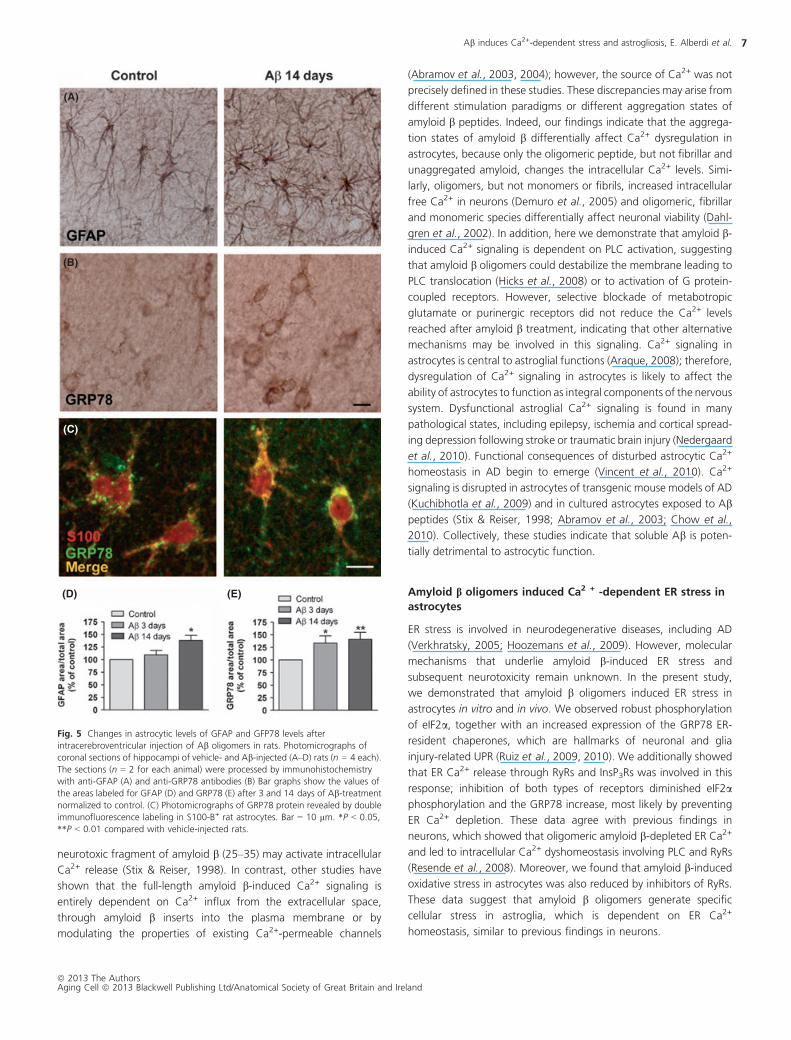
Acute intracerebroventricular injection of amyloid boligomers induced GFAP and GRP78 overexpression in
astrocytes of dentate gyrus of rat brains
To determine whether the amyloid b-dependent ER stress and
astrogliosis found in cell and organotypic culture experiments also
occur in vivo, we investigated effects of amyloid b oligomers on
GFAP and GRP78 expressions in the brains of adult rats. Rats
received a single intracerebroventricular (i.c.v.) injection of amyloid b
oligomers (40 lg/rat) or vehicle and were sacrificed 3 and 14 days
later. To quantify GFAP and GRP78 expressions in hippocampal
astrocytes, we used immunolabeling and measured the area positive
for GFAP and GRP78 antibodies versus total analyzed area. No
significant changes were found in 3 days after injection (n = 4;
Fig. 5D); however, in 2 weeks after injection, we detected a
significant increase of astrocytic GFAP levels (138 � 10%, n = 4
animals; Fig. 5A–D) in the dentate gyrus compared with vehicle-
injected rats (control 100%, n = 4). At the same time, levels of
GRP78 in astrocytes (Fig. 5B,E) were significantly elevated at both 3
and 14 days after injection (133 � 14 and 141 � 14%, respec-
tively, n = 4 animals per group) compared with the control-injected
rats (100%, n = 4). Amyloid b injection decreased nonsignificantly
the cell density in dentate gyrus as compared with controls
(681 � 30 and 617 � 30 cells/mm2, respectively). Thus, there is
no apparent contribution of cell number to changes of GFAP and
GRP78 expression in the studied areas. We confirmed higher GRP78
expression in astrocytes in amyloid b-treated rat brains by double
immunofluorescence labeling with S100B (Fig. 5C). Overall, these
histological findings were consistent with those described in
cultured astrocytes, demonstrating that amyloid b oligomers initiate
ER stress in astrocytes and astrogliosis in the brains of rats,
establishing the in vivo relevance of our findings in the in vitro
system.
Oligomeric amyloid b levels correlate with GFAP and GRP78
levels in 33Tg mice
Previous reports have demonstrated changes in astroglial mor-
phology in the hippocampi of 39Tg-AD mice at different ages
(Olabarria et al., 2010). By Western blot analysis, we examined
whether GFAP upregulation was associated with changes in
GRP78 and amyloid b oligomer levels in 39Tg-AD animals
(Fig. 6A,B). In protein extract from hippocampi of 12-month-old
39Tg-AD mice, GFAP and GRP78 levels were increased to
(A)
(B) (C)
Fig. 3 2APB and Rya decrease Ab-induced ER and oxidative stress in astrocytes. (A) Cells were treated with Ab oligomers for 1 h (peIF2a) or 6 h (GRP78) in the presence or
absence of 2APB (10 lM) or Rya (50 lM). Data represent optical density values normalized to total eIF2a or b-actin values and expressed as a percentage of the control
(untreated or 2APB-Rya-treated signal, 100%) (n = 4). (B) Astrocytes were treated with Ab for 30, 60, or 120 min, and ROS generation was monitored with CM-H2DCFDA
(30 lM). (C) Rya (50 lM), but not 2APB (10 lM), reduced Ab-induced ROS generation in astrocytes. Data represent mean � SEM of the CM-H2DCFDA/calcein signal in n = 8
cultures, expressed as a percentage of the control untreated levels (100%). *P < 0.05 compared with untreated cells; #P < 0.05, ##P < 0.01 compared with Ab-treated cells.
Ab induces Ca2+-dependent stress and astrogliosis, E. Alberdi et al. 5
ª 2013 The AuthorsAging Cell ª 2013 Blackwell Publishing Ltd/Anatomical Society of Great Britain and Ireland

127 � 8 and 131 � 7%, respectively, compared with age-
matched non-Tg controls. With the 6E10 antibody, we detected
detergent-resistant amyloid b oligomers (pentamers of ~20 kDa)
in hippocampal samples of 39Tg-AD mice (Fig. 6A). Correlation
analysis showed a significant positive correlations of GFAP
(r = 0.9756, P = 0.0046) and GRP78 (r = 0.91, P = 0.032) with
hippocampal levels of amyloid b oligomer (Fig. 6C,D). Data
analysis also showed a significant positive correlation between
GRP78 and GFAP in this AD animal model (Fig. 6E). Overall, these
results indicate that overexpression of GFAP and GRP78 is
associated with increased levels of amyloid b oligomer in 12-
month-old 39Tg-AD mice. These observations were further
corroborated by immunohistochemistry of GFAP and GRP78 in
astrocytes. Analysis of the areas of astrocytes labeled with GFAP
(Fig. 6F) and GRP78 (Fig. 6G) antibodies versus total area showed
increased protein levels in 39Tg-AD mice (137 � 18 and
156 � 15%, respectively) compared with age-matched non-
Tg mice (100%). There was no significant changes in astrocyte
cell density in 39Tg-AD as compared with control mice
(801 � 32 and 781 � 28 cells/mm2, respectively). Double immu-
nofluorescence labeling with the specific astrocytic marker S100B
revealed that astrocytes in 39Tg-AD mice expressed higher
GRP78 levels compared with non-Tg mice (Fig. 6H). Overall,
these data show that astrogliosis and astrocytic ER stress
correlated with increase levels of amyloid b oligomers in the
39Tg mouse model of AD.
Discussion
In the present study, we demonstrated that astrocytes responded to
the oligomeric form of amyloid b peptide by mobilizing ER Ca2+,
which triggered ER stress response that initiated astrogliosis.
Amyloid b oligomers injected in vivo caused a similar response in
hippocampal astrocytes, and comparable changes were found in the
39Tg-AD mice. These results suggest that astrocytic ER stress
initiated by amyloid b-dependent Ca2+ dysregulation may be directly
linked to reactive astrogliosis in AD.
Oligomeric amyloid b affects astroglial Ca2+ homeostasis
Pathogenic interactions between altered cellular Ca2+ signaling and
amyloid b in its different aggregation states have been extensively
described in neurons (for review, see Demuro et al., 2010); however,
the effects in astrocytes remain controversial. Here, we found that
the [Ca2+]i rise caused by amyloid b1-42 oligomers was due to Ca2+
release from intracellular stores, because (i) it occurred in nominally
Ca2+-free medium and (ii) [Ca2+]i increases were reduced in the
presence of inhibitors of PLC, InsP3Rs, and RyRs. We also show that
Ca2+ influx contributes to a late sustained Ca2+ response that is
modulated by IP3 signaling but not by Ca2+ release through RyRs.
Further experiments will be necessary to define the extracellular Ca2+
routes of amyloid b-induced Ca2+ response in astrocytes. Consistent
with these results, previous observations have also suggested that the
(A) (B)
(D) (E)
(C)
Fig. 4 Ca2+-dependent ER stress in astrocytes in Ab-treated organotypic slices. (A) Slices were treated with oligomeric Ab (100 nM, 4 days) in the presence or absence
of 2APB (10 lM) or Rya (50 lM). GFAP and GRP78 levels were quantified by Western blot analysis. (B) Data represent optical density values normalized to b-actin values and
expressed as a percentage of the control (untreated or 2-APB or Rya-treated slices, 100%). (C) Correlation analyses between GFAP and GRP78 levels in the Ab-treatedslices (r = 0.631, P = 0.0138, n = 12) are shown. (D) Photomicrographs of organotypic slices showing colocalization colormap of intensities of fluorescent signals of GRP78
and S-100-B immunolabeling. Hot colors represent colocalization, whereas cold colors represent exclusion. (E) Colocalization of two markers is expressed as correlation
of intensities between pairs of pixel of two different channels. Data represent the normalized mean deviation products >0 of eight photomicrographs of each treatment
(n = 4 experiments); *P < 0.05 compared with untreated cells; #P < 0.05, ##P < 0.01 compared with Ab-treated cells.
Ab induces Ca2+-dependent stress and astrogliosis, E. Alberdi et al.6
ª 2013 The AuthorsAging Cell ª 2013 Blackwell Publishing Ltd/Anatomical Society of Great Britain and Ireland
neurotoxic fragment of amyloid b (25–35) may activate intracellular
Ca2+ release (Stix & Reiser, 1998). In contrast, other studies have
shown that the full-length amyloid b-induced Ca2+ signaling is
entirely dependent on Ca2+ influx from the extracellular space,
through amyloid b inserts into the plasma membrane or by
modulating the properties of existing Ca2+-permeable channels
(Abramov et al., 2003, 2004); however, the source of Ca2+ was not
precisely defined in these studies. These discrepancies may arise from
different stimulation paradigms or different aggregation states of
amyloid b peptides. Indeed, our findings indicate that the aggrega-
tion states of amyloid b differentially affect Ca2+ dysregulation in
astrocytes, because only the oligomeric peptide, but not fibrillar and
unaggregated amyloid, changes the intracellular Ca2+ levels. Simi-
larly, oligomers, but not monomers or fibrils, increased intracellular
free Ca2+ in neurons (Demuro et al., 2005) and oligomeric, fibrillar
and monomeric species differentially affect neuronal viability (Dahl-
gren et al., 2002). In addition, here we demonstrate that amyloid b-
induced Ca2+ signaling is dependent on PLC activation, suggesting
that amyloid b oligomers could destabilize the membrane leading to
PLC translocation (Hicks et al., 2008) or to activation of G protein-
coupled receptors. However, selective blockade of metabotropic
glutamate or purinergic receptors did not reduce the Ca2+ levels
reached after amyloid b treatment, indicating that other alternative
mechanisms may be involved in this signaling. Ca2+ signaling in
astrocytes is central to astroglial functions (Araque, 2008); therefore,
dysregulation of Ca2+ signaling in astrocytes is likely to affect the
ability of astrocytes to function as integral components of the nervous
system. Dysfunctional astroglial Ca2+ signaling is found in many
pathological states, including epilepsy, ischemia and cortical spread-
ing depression following stroke or traumatic brain injury (Nedergaard
et al., 2010). Functional consequences of disturbed astrocytic Ca2+
homeostasis in AD begin to emerge (Vincent et al., 2010). Ca2+
signaling is disrupted in astrocytes of transgenic mouse models of AD
(Kuchibhotla et al., 2009) and in cultured astrocytes exposed to Ab
peptides (Stix & Reiser, 1998; Abramov et al., 2003; Chow et al.,
2010). Collectively, these studies indicate that soluble Ab is poten-
tially detrimental to astrocytic function.
Amyloid b oligomers induced Ca2 + -dependent ER stress in
astrocytes
ER stress is involved in neurodegenerative diseases, including AD
(Verkhratsky, 2005; Hoozemans et al., 2009). However, molecular
mechanisms that underlie amyloid b-induced ER stress and
subsequent neurotoxicity remain unknown. In the present study,
we demonstrated that amyloid b oligomers induced ER stress in
astrocytes in vitro and in vivo. We observed robust phosphorylation
of eIF2a, together with an increased expression of the GRP78 ER-
resident chaperones, which are hallmarks of neuronal and glia
injury-related UPR (Ruiz et al., 2009, 2010). We additionally showed
that ER Ca2+ release through RyRs and InsP3Rs was involved in this
response; inhibition of both types of receptors diminished eIF2a
phosphorylation and the GRP78 increase, most likely by preventing
ER Ca2+ depletion. These data agree with previous findings in
neurons, which showed that oligomeric amyloid b-depleted ER Ca2+
and led to intracellular Ca2+ dyshomeostasis involving PLC and RyRs
(Resende et al., 2008). Moreover, we found that amyloid b-induced
oxidative stress in astrocytes was also reduced by inhibitors of RyRs.
These data suggest that amyloid b oligomers generate specific
cellular stress in astroglia, which is dependent on ER Ca2+
homeostasis, similar to previous findings in neurons.
(A)
(B)
(C)
(D) (E)
Fig. 5 Changes in astrocytic levels of GFAP and GFP78 levels after
intracerebroventricular injection of Ab oligomers in rats. Photomicrographs of
coronal sections of hippocampi of vehicle- and Ab-injected (A–D) rats (n = 4 each).
The sections (n = 2 for each animal) were processed by immunohistochemistry
with anti-GFAP (A) and anti-GRP78 antibodies (B) Bar graphs show the values of
the areas labeled for GFAP (D) and GRP78 (E) after 3 and 14 days of Ab-treatment
normalized to control. (C) Photomicrographs of GRP78 protein revealed by double
immunofluorescence labeling in S100-B+ rat astrocytes. Bar = 10 lm. *P < 0.05,
**P < 0.01 compared with vehicle-injected rats.
Ab induces Ca2+-dependent stress and astrogliosis, E. Alberdi et al. 7
ª 2013 The AuthorsAging Cell ª 2013 Blackwell Publishing Ltd/Anatomical Society of Great Britain and Ireland

ER Ca2+ release and GFAP expression in amyloid b-treatedastrocytes
Pathological insults to the CNS trigger reactive astrogliosis (Verkh-
ratsky et al., 2012). Astrogliosis by itself alters synaptic transmission
and network excitability in the absence of neuronal pathology and
microglial reactivity (Ortinski et al., 2010). Therefore, pathological
remodeling of astroglia associated with AD progression may
directly contribute to neuronal signaling deficits (Olabarria et al.,
2010), and it is important to understand the mechanisms that
stimulate astrocytes to acquire the reactive phenotype in AD. In the
present study, we found that amyloid b directly triggered astrogl-
iosis in an ER Ca2+-dependent manner and that these changes
correlated with ER stress in vitro and in vivo. These effects may
impair a variety of astrocytic functions, including Ca2+ signaling,
expression of membrane transporters, secretion of neuroactive
compounds, and morphological plasticity, which are central to glial
modulation of neuronal signaling. However, further in vivo
experiments are necessary to understand the link between dysreg-
ulated Ca2+ homeostasis, ER stress, and astrocytic reactivity in
amyloid b-treated cells.
GFAP and GRP78 upregulation correlates with amyloid boligomer levels in 33Tg-AD mice
Accumulation of amyloid b is one of the earliest known molecular
events in AD. Oligomers appear to be relatively stable structures;
SDS-resistant species have been isolated from postmortem human
brains, cell lines, and animal models of AD (Oddo et al., 2006).
Brains from 39Tg-AD mice accumulate amyloid b oligomers in an
age-dependent manner. In the current study, we observed increased
levels of GFAP and GRP78 in hippocampal astrocytes of 39Tg-AD
mice at 12 months of age. These GFAP and GRP78 levels correlated
with the levels of oligomeric amyloid b peptide, suggesting that
oligomeric species may play a causal role in astrogliosis in this AD
mouse model.
Mutations in APP, presenilin 1 and presenilin 2 genes change
amyloid b metabolism leading to an increase in total amyloid b,
alteration of the amyloid b 42/40 ratio or its propensity to aggregate
(Selkoe, 2001). Furthermore, amyloid b accumulation plays a role in
tau pathology in the 39Tg-AD mice (Oddo et al., 2006). Therefore,
the involvement of tau and presenilin as inducers of astrogliosis
cannot be excluded in this AD mouse model, and further experi-
ments will be necessary to assess the link between astrogliosis and
mutations in these three genes in the 39Tg-AD. Our current
observations indicate that astrocytes are a target for oligomeric
amyloid b and support the hypothesis that the astrocyte response
may contribute to brain damage in AD.
In summary, our results indicate that, in astrocytes, the ER Ca2+
release through RyRs and InsP3Rs contributes to amyloid b-induced
Ca2+ dysregulation, generation of ER and oxidative stress, and
astroglial reactivity. Markers of altered astrocytic physiology were
consistently overexpressed in the hippocampal formation in amyloid
b-injected brains. In 39Tg-AD mice, the astrogliotic and cellular
stress response correlated with an increase in oligomeric amyloid b
species in the hippocampus, which is in agreement with amyloid b
plaque-associated astrocytic hypertrophy observed in the CA1 area
of the hippocampus (Olabarria et al., 2010). Understanding astro-
cyte dysfunction in AD may help to define the links between amyloid
b and progressive decline in cognitive function. Based on our data,
we propose that molecules that stabilize ER Ca2+ homeostasis in
(A) (B)
(C) (D) (E)
(F)
(G)
(H)
Fig. 6 Oligomeric Ab levels correlated with GFAP and GRP78 in the hippocampi of 39Tg-AD mice. (A) Protein levels of GRP78, GFAP, and oligomeric Ab in 12-month-old
wild-type (WT; n = 3), and 39Tg-AD mice (n = 5) were analyzed by Western blot. (B) Data represent optical density values of proteins normalized to b-actin values,
expressed as a percentage of control values (100%). (C–E) Correlation analyses show significant positive correlations of Ab oligomer levels with GFAP (C) or GRP78 (D) and
between GRP78 and GFAP (E). (F–H) Photomicrographs of coronal sections of hippocampi of 12-month-old wild-type and 39Tg-AD mice (n = 6 each group) show increased
levels of GFAP (F) and GRP78 (G) in astrocytes of 39Tg-AD mice compared with those of wild-type mice. (H) Photomicrographs of GRP78 revealed by double
immunofluorescence labeling in S100-B+ mouse astrocytes. Bar = 10 lm. *P < 0.05 compared with wild-type mice.
Ab induces Ca2+-dependent stress and astrogliosis, E. Alberdi et al.8
ª 2013 The AuthorsAging Cell ª 2013 Blackwell Publishing Ltd/Anatomical Society of Great Britain and Ireland
astrocytes could represent potential targets in developing thera-
peutic applications for AD.
Experimental procedures
Preparation of amyloid b peptides
Oligomeric and fibrillar amyloid b (Ab1–42) was prepared as
reported previously (Dahlgren et al., 2002). Briefly, Ab1–42 (ABX,
Radeberg, Germany) was initially dissolved in hexafluoroisopropanol
(HFIP, Sigma, St. Louis, MO, USA) to a concentration of 1 mM. For
the aggregation protocol, the peptide was resuspended in dry
dimethylsulfoxide (5 mM; Sigma). Hams F-12 (PromoCell, LabClinics,
Barcelona, Spain) or HCl (10 mM) were added to adjust the final
peptide concentration to 100 lM to obtain oligomers (4 °C for
24 h) or fibrils (37ºC for 24 h), respectively.
Astrocyte-rich cell culture
Primary cultures of cerebral cortical astrocytes were prepared from
newborn (P0–P2) Sprague-Dawley rats as described previously
(McCarthy & de Vellis, 1980; supporting information). After
2 weeks, cells were trypsinized and astrocytes were plated onto
PDL-coated, 12-mm glass coverslips (for Ca2+ imaging and immu-
nocytochemistry), or six-well plates (for Western blotting).
Preparation of organotypic cultures
Organotypic slices were prepared according to the previously
described method (Alberdi et al., 2010). Slices of entorhinal cortex
and hippocampus were treated with Ab 100 nM for 4 days and were
immunostained with polyclonal anti-GFAP antibody (1:100; Dako,
Glostrup, Denmark), rabbit polyclonal S100 (1:100; Dako), and
monoclonal anti-ER retention signal KDEL (Grp78, Grp 94) (1:100;
Stressgen Bioreagents, Ann Arbor, MI, USA). Labeling was detected
with fluorescent goat anti-rabbit or sheep anti-mouse secondary
antibodies (Alexa Fluor 488 and 546, Invitrogen, Barcelona, Spain).
GFAP levels were measured by fluorimetry with a Synergy-HT
fluorimeter (Bio-Tek Instruments Inc., Beverly, MA, USA; excitation
at 540, emission at 570 nm). The colocalization colormap application
of Image J (National Institutes of Health, Bethesda, MD, USA) was
used to calculate GRP78 and S100 protein colocalization. Distribution
of normalized mean deviation product (nMDP) values (ranging from
�1 to 1) was visualized with a color scale, with indexes below 0
represented by cold colors (exclusion) and indexes above 0 repre-
sented by hot colors (colocalization).
Ca2+ imaging
Intracellular Ca2+ levels were determined according to the previously
described method (Alberdi et al., 2010; Data S1).
Protein preparation and Western blot analysis
Astrocytes (2.5 9 105) were exposed to Ab oligomers for 1, 6, or
24 h. Antagonists or inhibitors were added 15 min before addition
of the Ab oligomers. Cells were pelleted by centrifugation and lysed
in gel-loading buffer for GRP78 and peIF2a analysis and in RIPA with
protease inhibitor cocktail (Complete, Mini EDTA-free tablets,
Roche, Mannheim, Germany) for GFAP analysis. Tissues from mice
and organotypic slices were homogenized with a douncer, and
sonicated in 200 lL or 100 lL, respectively, of RIPA with a protease
inhibitor cocktail. Homogenates were centrifuged at 4°C for 15 min
at 12 0009g. Proteins (10–20 lg) were analyzed by Western blot
with the following specific primary antibodies: monoclonal anti-
GFAP (0.1 lg/ml; Chemicon, Millipore Ib�erica, Madrid, Spain),
polyclonal antibodies against the phosphorylated and unphosphor-
ylated forms of eukaryotic initiation factor 2a (peIF2a and eIF2a,
respectively) (1:1000; Cell Signaling Technology, Beverly, MA, USA);
monoclonal antibody against the ER retention signal KDEL (Grp 78)
(1:1000; Stressgen Bioreagents); monoclonal anti-Ab antibody
(6E10, 1:1000; Covance, Emeryville, CA, USA); and polyclonal
anti-b-actin antibody (1:5000; Sigma). Membranes were incubated
with horseradish peroxidase-conjugated secondary antibodies
(1:5000; Cell Signaling Technology) and were developed with Super
Signal West Dura or Super Signal West Femto (Pierce, Rockford,
IL, USA). The protein bands were detected with a ChemiDocTM XRS
Imaging System (Bio-Rad, Hercules, CA, USA), and the band inten-
sities were quantified by density using Quantity One� (Bio-Rad,
Hercules, CA, USA) software.
Immunocytochemistry
Cells on coverslips were incubated with Ab for 24 h, washed in ice-
cold PBS, and fixed for 15 min in 4%PFA. Cells were permeabilized in
methanol at�20 °C,washed in PBS and then saturated for 1 h in 4%
NGS in PBS with 0.1% Triton-X 100 (blocking solution). Cells were
then incubated for 2 h with polyclonal anti-GFAP antibody (dilution
1:100; Dako) in blocking solution. After washing, the cultures were
incubated in PBS plus 4% NGS with goat anti-rabbit secondary
antibody conjugated to Alexa Fluor� 488 (Invitrogen) for 1 h at room
temperature. Immunofluorescence was visualized with an Observer
Z1, Zeiss inverted microscope (Oberkochen, Germany). Photographs
were acquired with a high-resolution, digital, AxioCam HRc camera
(Zeiss, Oberkochen, Germany), and AxioVision software.
Experimental animals, tissue preparation, and
immunohistochemistry
Rats were divided into two groups: One group (n = 8) received a
10-lL injection of vehicle (DMSO 17%, Ham’s F-12 83%) and a
second group (n = 8) received an injection of Ab peptide oligomers
(40 lg/rat i.c.v.; more details in supporting information). Tissues
from rats or 12-month-old 3 9 Tg-AD animals and the non-Tg
controls; n = 6 each group were fixed and processed as described
previously (Gottlieb et al., 2006). Coronal vibratome sections (40–
50 lm) of the dorsal hippocampus were collected and processed for
immunoperoxidase and immunofluorescence histochemistry. The
mouse monoclonal antibody against GFAP (1:1000; Chemicon,
Millipore Ib�erica, Madrid, Spain), rabbit polyclonal antibody against
S100 (1:100 Dako), and monoclonal antibody against the ER
Ab induces Ca2+-dependent stress and astrogliosis, E. Alberdi et al. 9
ª 2013 The AuthorsAging Cell ª 2013 Blackwell Publishing Ltd/Anatomical Society of Great Britain and Ireland

retention signal KDEL (Grp78) (1:100, Stressgen Bioreagents) were
used to quantify protein levels in 3–6 hippocampal sections per
animal, from the control and Ab-injected rats and wild-type and
3 9 Tg-AD mice. Stained sections were observed and photographed
with a digital camera (AxioCam MRc5, Zeiss) and Olympus Optical
Fluoview FV500 confocal microscopy (409 magnification). Quanti-
fication of GFAP and GRP78 levels in astrocytes was performed using
the threshold application of ImageJ software, and values are
expressed as the percentage of astrocytic labeled area versus total
area analyzed.
Measurement of intracellular reactive oxygen species
Astrocytes were exposed to Ab oligomers alone or with antagonists
or inhibitors, as indicated. Cells were loaded with 30 lM CM-
H2DCFDA (Invitrogen, Barcelona Spain) to assay ROS levels. Calcein-
AM (1 lM) was used to quantify the number of cells within the
reading field. Fluorescence was measured with a Synergy-HT
fluorimeter (Bio-Tek Instruments Inc.).
Statistical Analysis
Data are presented as mean � SEM. Statistical analysis was carried
out with the Student’s t-test. In all instances, a value of P < 0.05
was considered to indicate significance.
Acknowledgments
We thank Hazel G�omez, Silvia Mart�ın and Saioa Marcos for technical
assistance. This study was supported by CIBERNED and by grants
from the Ministerio de Ciencia e Innovaci�on, the Departamento de
Sanidad del Gobierno Vasco, Ikerbasque, and the Universidad del
Pa�ıs Vasco.
References
Abeti R, Abramov AY, Duchen MR (2011) Beta-amyloid activates PARP causing
astrocytic metabolic failure and neuronal death. Brain 134, 1658–1672.Abramov AY, Canevari L, Duchen MR (2003) Changes in intracellular calcium and
glutathione in astrocytes as the primary mechanism of amyloid neurotoxicity. J.
Neurosci. 23, 5088–5095.Abramov AY, Canevari L, Duchen MR (2004) b-amyloid peptides induce
mitochondrial dysfunction and oxidative stress in astrocytes and death of
neurons through activation of NADPH oxidase. J. Neurosci. 24, 565–575.Alberdi E, S�anchez-G�omez MV, Cavaliere F, P�erez-Samart�ın A, Zugaza JL, Trullas R,
Domercq M, Matute C (2010) Amyloid b oligomers induce Ca2+ dysregulation
and neuronal death through activation of ionotropic glutamate receptors. Cell
Calcium 47, 264–272.Araque A (2008) Astrocytes process synaptic information. Neuron Glia Biol. 4, 3–10.Bao F, Wicklund L, Lacor PN, Klein WL, Nordberg A, Marutle A (2012) Different b-amyloid oligomer assemblies in Alzheimer brains correlate with age of disease
onset and impaired cholinergic activity. Neurobiol. Aging 33, e1–e13.Busche MA, Eichhoff G, Adelsberger H, Abramowski D, Wiederhold KH, Haass C,
Staufenbiel M, Konnerth A, Garaschuk O (2008) Clusters of hyperactive neurons
near amyloid plaques in a mouse model of Alzheimer’s disease. Science 321,1686–1689.
Chafekar SM, Hoozemans JJ, Zwart R, Baas F, Scheper W (2007) Ab 1–42 induces
mild endoplasmic reticulum stress in an aggregation state-dependent manner.
Antioxid. Redox Signal. 9, 2245–2254.Chow SK, Yu D, Macdonald CL, Buibas M, Silva GA (2010) Amyloid b-peptidedirectly induces spontaneous calcium transients, delayed intercellular calcium
waves and gliosis in rat cortical astrocytes. ASN Neuro 2, e00026.
Dahlgren KN, Manelli AM, Stine WB Jr, Baker LK, Krafft GA, LaDu MJ (2002)
Oligomeric and fibrillar species of amyloid-beta peptides differentially affect
neuronal viability. J. Biol. Chem. 277, 32046–32053.De Felice FG, Velasco PT, Lambert MP, Viola K, Fernandez SJ, Ferreira ST, Klein WL
(2007) Ab oligomers induce neuronal oxidative stress through an N-methyl-D-
aspartate receptor-dependent mechanism that is blocked by the Alzheimer drug
memantine. J. Biol. Chem. 282, 11590–11601.Demuro A, Mina E, Kayed R, Milton SC, Parker I, Glabe CG (2005) Calcium
dysregulation and membrane disruption as a ubiquitous neurotoxic mechanism
of soluble amyloid oligomers. J. Biol. Chem. 280, 17294–17300.Demuro A, Parker I, Stutzmann GE (2010) Calcium signaling and amyloid toxicity in
Alzheimer disease. J. Biol. Chem. 285, 12463–12468.Fiacco TA, McCarthy KD (2006) Astrocyte calcium elevations: properties, propa-
gation, and effects on brain signaling. Glia 54, 676–690.Garwood CJ, Pooler AM, Atherton J, Hanger DP, Noble W (2011) Astrocytes are
important mediators of Ab-induced neurotoxicity and tau phosphorylation in
primary cultures. Cell Death Dis. 2, e167.Gottlieb M, Leal-Campanario R, Campos-Esparza MR, S�anchez-G�omez MV,
Alberdi E, Arranz A, Delgado-Garc�ıa JM, Gruart A, Matute C (2006) Neuropro-
tection by two polyphenols following excitotoxicity and experimental ischemia.
Neurobiol. Dis. 23, 374–386.Hicks JB, Lai Y, Sheng W, Yang X, Zhu D, Sun GY, Lee JC (2008) Amyloid-beta
peptide induces temporal membrane biphasic changes in astrocytes through
cytosolic phospholipase A2. Biochim. Biophys. Acta 1778, 2512–2519.Hoozemans JJ, van Haastert ES, Nijholt DA, Rozemuller AJ, Eikelenboom P, Scheper
W (2009) The unfolded protein response is activated in pretangle neurons in
Alzheimer’s disease hippocampus. Am. J. Pathol. 174, 1241–1251.Kuchibhotla KV, Goldman ST, Lattarulo CR, Wu HY, Hyman BT, Bacskai BJ (2008)
Ab plaques lead to aberrant regulation of calcium homeostasis in vivo resulting
in structural and functional disruption of neuronal networks. Neuron 59, 214–225.
Kuchibhotla KV, Lattarulo CR, Hyman BT, Bacskai BJ (2009) Synchronous
hyperactivity and intercellular calcium waves in astrocytes in Alzheimer mice.
Science 323, 1211–1215.McCarthy KD, de Vellis J (1980) Preparation of separate astroglial and oligoden-
droglial cell cultures from rat cerebral tissue. J. Cell Biol. 85, 890–902.Meske V, Hamker U, Albert F, Ohm TG (1998) The effects of b/A4-amyloid and its
fragments on calcium homeostasis, glial fibrillary acidic protein and S100bstaining, morphology and survival of cultured hippocampal astrocytes. Neuro-
science 85, 1151–1160.Nedergaard M, Rodriguez JJ, Verkhratsky A (2010) Glial calcium and diseases of
the nervous system. Cell Calcium 47, 140–149.Oddo S, Caccamo A, Shepherd JD, Murphy MP, Golde TE, Kayed R, Metherate R,
Mattson MP, Akbari Y, LaFerla FM (2003) Triple-transgenic model of Alzheimer’s
disease with plaques and tangles: intracellular Abeta and synaptic dysfunction.
Neuron 39, 409–421.Oddo S, Caccamo A, Tran L, Lambert MP, Glabe CG, Klein WL, LaFerla FM (2006)
Temporal profile of amyloid-b (Ab) oligomerization in an in vivo model of
Alzheimer disease: a link between Ab and tau pathology. J. Biol. Chem. 281,1599–1604.
Olabarria M, Noristani HN, Verkhratsky A, Rodr�ıguez JJ (2010) Concomitant
astroglial atrophy and astrogliosis in a triple transgenic animal model of
Alzheimer’s disease. Glia 58, 831–838.Ortinski PI, Dong J, Mungenast A, Yue C, Takano H, Watson DJ, Haydon PG,
Coulter DA (2010) Selective induction of astrocytic gliosis generates deficits in
neuronal inhibition. Nat. Neurosci. 13, 584–591.Peuchen S, Clark JB, Duchen MR (1996) Mechanisms of intracellular calcium
regulation in adult astrocytes. Neuroscience 71, 871–883.Resende R, Ferreiro E, Pereira C, Resende de Oliveira C (2008) Neurotoxic effect of
oligomeric and fibrillar species of amyloid-b peptide 1–42: involvement of
endoplasmic reticulum calcium release in oligomer-induced cell death. Neuro-
science 155, 725–737.Ruiz A, Matute C, Alberdi E (2009) Endoplasmic reticulum calcium release through
ryanodine and IP3 receptors contributes to neuronal excitotoxicity. Cell Calcium
46, 273–281.Ruiz A, Matute C, Alberdi E (2010) Intracellular Ca2+ release through ryanodine
receptors contributes to AMPA receptor-mediated mitochondrial dysfunction
and ER stress in oligodendrocytes. Cell Death Dis. 1, e54.Santos AN, Ewers M, Minthon L, Simm A, Silber RE, Blennow K, Prvulovic D,
Hansson O, Hampel H (2012) Amyloid-b oligomers in cerebrospinal fluid are
associated with cognitive decline in patients with Alzheimer’s disease. J.
Alzheimers Dis. 29, 171–176.
Ab induces Ca2+-dependent stress and astrogliosis, E. Alberdi et al.10
ª 2013 The AuthorsAging Cell ª 2013 Blackwell Publishing Ltd/Anatomical Society of Great Britain and Ireland

Scheper W, Nijholt DA, Hoozemans JJ (2011) The unfolded protein response and
proteostasis in Alzheimer disease: preferential activation of autophagy by
endoplasmic reticulum stress. Autophagy 7, 910–911.Selkoe DJ (2001) Alzheimer’s disease: genes, proteins and therapy. Physiol. Rev.
81, 741–766.Shankar GM, Bloodgood BL, Townsend M, Walsh DM, Selkoe DJ, Sabatini BL
(2007) Natural oligomers of the Alzheimer amyloid-b protein induce reversible
synapse loss by modulating an NMDA-type glutamate receptor-dependent
signalling pathway. J. Neurosci. 27, 2866–2875.Stix B, Reiser G (1998) b-amyloid peptide 25–35 regulates basal and hormone-
stimulated Ca2+ levels in cultured rat astrocytes. Neurosci. Lett. 243, 121–124.Tomiyama T, Matsuyama S, Iso H, Umeda T, Takuma H, Ohnishi K, Ishibashi K,
Teraoka R, Sakama N, Yamashita T, Nishitsuji K, Ito K, Shimada H, Lambert MP,
Klein WL, Mori HA (2010) A mouse model of amyloid b oligomers: their
contribution to synaptic alteration, abnormal tau phosphorylation, glial activa-
tion, and neuronal loss in vivo. J. Neurosci. 30, 4845–4856.Verkhratsky A (2005) Physiology and pathophysiology of the calcium store in the
endoplasmic reticulum of neurons. Physiol. Rev. 85, 201–279.Verkhratsky A, Kettenmann H (1996) Calcium signalling in glial cells. Trends
Neurosci. 19, 346–352.
Verkhratsky A, Petersen OH (2002) The endoplasmic reticulum as an integrating
signalling organelle: from neuronal signalling to neuronal death. Eur. J.
Pharmacol. 447, 141–154.Verkhratsky A, Sofroniew MV, Messing A, deLanerolle NC, Rempe D, Rodriguez JJ,
Nedergaard M (2012) Neurological diseases as primary gliopathies: a reassess-
ment of neurocentrism. ASN Neuro 4, e00082.Vincent AJ, Gasperini R, Foa L, Small DH (2010) Astrocytes in Alzheimer disease:
emerging roles in calcium dysregulation and synaptic plasticity. J. Alzheimers Dis.
22, 699–714.
Supporting Information
Additional supporting information may be found in the online version of this
article at the publisher’s web-site.
Data S1. Material and methods.
Ab induces Ca2+-dependent stress and astrogliosis, E. Alberdi et al. 11
ª 2013 The AuthorsAging Cell ª 2013 Blackwell Publishing Ltd/Anatomical Society of Great Britain and Ireland
Copyright © 2022 FDOKUMEN