Concomitant astroglial atrophy and astrogliosis in a triple transgenic animal model of Alzheimer's...
8
Concomitant Astroglial Atrophy and Astrogliosis in a Triple Transgenic Animal Model of Alzheimer’s Disease MARKEL OLABARRIA, 1 HARUN N. NORISTANI, 1 ALEXEI VERKHRATSKY, 1,2 * AND JOS E J. RODR IGUEZ 1,2,3,4 * 1 Faculty of Life Sciences, The University of Manchester, Manchester, United Kingdom 2 Institute of Experimental Medicine, ASCR, Videnska 1083, Prague 4, Czech Republic 3 IKERBASQUE, Basque Foundation for Science, Bilbao, Spain 4 Department of Neurosciences, University of the Basque Country UPV/EHU, Leioa, Spain KEY WORDS astroglia; Alzheimer’s disease; hippocampus; plasticity; b-amyloid ABSTRACT Astrocytes are fundamental for brain homeostasis and are at the fulcrum of neurological diseases including Alzhei- mer’s disease (AD). Here, we monitored changes in astro- glia morphology throughout the age-dependent progression of AD. We used an immunohistochemical approach that allows us to determine the domain of glial cytoskeleton, by measuring the surface, volume, and the relationship between astrocytes and neuritic plaques. We investigated astroglia in the hippocampus of a triple transgenic mouse model of AD (3xTg-AD) that mimics the progression of the human disease. The numerical density of astrocytes is affected neither by AD nor by age. We found reduction of surface and volume of GFAP profiles from early ages (6 months; 43.84 and 52.76%, respectively), persisting at 12 (40.73 and 45.39%) and 18 months (64.80 and 71.95%) in the dentate gyrus (DG) of 3xTg-AD, whereas in CA1 it appears at 18 months (29.42 and 32.74%). This cytoskeleton atrophy is accompanied by a significant reduction of glial somata vol- ume in DG at 12 and 18 months (40.46 and 75.55%, respec- tively), whereas in CA1 it is significant at 18 months (42.81%). However, while astroglial atrophy appears as a generalized process, astrocytes surrounding plaques are clearly hypertrophic as revealed by increased surface (48.06%; 66.66%), and volume (57.10%; 71.06%) of GFAP pro- files in DG and CA1, respectively, at 18 months. We suggest differential effects of AD on astroglial populations depending on their association with plaques accounting for the progres- sive disruption of neural networks connectivity and neuro- transmitters imbalance which underlie mnesic and cognitive impairments observed in AD. V V C 2010 Wiley-Liss, Inc. INTRODUCTION Astrocytes are the main homeostatic cellular elements of the central nervous system (CNS), which define its structure and provide for numerous aspects of CNS function (Nedergaard et al., 2003; Oberheim et al., 2006, 2009; Verkhratsky, 2006, 2009; Wang and Bordey, 2008). Astrocytes generate the blood–brain barrier, control extracellular ion homeostasis and neurotransmitter bal- ance, and maintain synaptic connectivity, strength and plasticity (Abbott et al., 2006; Bundgaard and Abbott, 2008; Halassa et al., 2007; Haydon and Carmignoto, 2006; Perea et al., 2009; Volterra and Meldolesi, 2005). Neurological diseases result from disruption of a fine equilibrium among neural cell damage, neuroprotection, and regeneration. Astrocytes, being homeostatic cells in the CNS are invariably involved in every kind of neuro- pathology (Giaume et al., 2007; Nedergaard and Dir- nagl, 2005; Seifert et al., 2006). Astroglia forms the first line of brain defense through its wide-ranged homeo- static activity. Astrocyte regulates the volume and com- position of extracellular space (Kofuji and Newman, 2004), contain extracellular levels of glutamate (Danbolt, 2001; Kirischuk et al., 2007), thereby limiting the intrin- sic excitotoxicity of the latter, regulate fluid movements, and provide the main antioxidant system (Dringen, 2000; Simard and Nedergaard, 2004). However, there is an intrinsic dichotomy in the function of astroglia, which can contribute to neuronal damage through failure or re- versal of various homeostatic molecular cascades (Giaume et al., 2007; Nedergaard and Dirnagl, 2005). Pathological insults to the CNS trigger a specific astro- cytic reaction known as reactive astrogliosis, which aims at protecting the brain parenchyma, isolating the dam- aged area, reconstructing the blood–brain barrier, and promoting the remodeling of the neural circuitry through isomorphic gliosis (Wilhelmsson et al., 2006). Alzheimer disease (AD, Alzheimer, 1907; Kraepelin, 1919) is one of the most common form of dementia (Braak et al., 1999), clinically manifested by cognitive deficits such as memory and learning impairment, and histopathologically by substantial neuronal and synaptic loss associated with extracellular b-amyloid (Ab) accu- mulation in the form of plaques and appearance of intra- neuronal hyperphosphorilated tau protein fibrillary tangles (Selkoe, 2001). The first and most affected areas in AD are those directly responsible for cognitive tasks; Jose J. Rodriguez is currently at IKERBASQUE, Department of Neuroscience, The University of the Basque Country UPV/EHU, Technological Park, Bldg. 205, Floor 1, Laida Bidea, 48170 Zamudio, Spain. Grant sponsor: Alzheimer’s Research Trust Programme; Grant number: ART/PG2004A/1; Grant sponsor: Grant Agency of the Czech Republic; Grant numbers: GACR 309/09/1696, GACR 305/08/1384. *Correspondence to: Alexei Verkhratsky or Jos e J. Rodr ıguez, Faculty of Life Sci- ences, The University of Manchester, A. V. Hill Building, Room 2.002, Oxford Road, Manchester M13 9PT, UK. E-mail: [email protected] or [email protected] Received 22 September 2009; Accepted 17 December 2009 DOI 10.1002/glia.20967 Published online 5 January 2010 in Wiley InterScience (www.interscience. wiley.com). GLIA 58:831–838 (2010) V V C 2010 Wiley-Liss, Inc.
-
Upload
manchester -
Category
Documents
-
view
3 -
download
0
Transcript of Concomitant astroglial atrophy and astrogliosis in a triple transgenic animal model of Alzheimer's...

Concomitant Astroglial Atrophy and Astrogliosis in aTriple Transgenic Animal Model of Alzheimer’s Disease
MARKEL OLABARRIA,1 HARUN N. NORISTANI,1 ALEXEI VERKHRATSKY,1,2* AND JOS�E J. RODR�IGUEZ1,2,3,4*1Faculty of Life Sciences, The University of Manchester, Manchester, United Kingdom2Institute of Experimental Medicine, ASCR, Videnska 1083, Prague 4, Czech Republic3IKERBASQUE, Basque Foundation for Science, Bilbao, Spain4Department of Neurosciences, University of the Basque Country UPV/EHU, Leioa, Spain
KEY WORDSastroglia; Alzheimer’s disease; hippocampus; plasticity;b-amyloid
ABSTRACTAstrocytes are fundamental for brain homeostasis and areat the fulcrum of neurological diseases including Alzhei-mer’s disease (AD). Here, we monitored changes in astro-glia morphology throughout the age-dependent progressionof AD. We used an immunohistochemical approach thatallows us to determine the domain of glial cytoskeleton, bymeasuring the surface, volume, and the relationshipbetween astrocytes and neuritic plaques. We investigatedastroglia in the hippocampus of a triple transgenic mousemodel of AD (3xTg-AD) that mimics the progression of thehuman disease. The numerical density of astrocytes isaffected neither by AD nor by age. We found reduction ofsurface and volume of GFAP profiles from early ages (6months; 43.84 and 52.76%, respectively), persisting at 12(40.73 and 45.39%) and 18 months (64.80 and 71.95%) in thedentate gyrus (DG) of 3xTg-AD, whereas in CA1 it appearsat 18 months (29.42 and 32.74%). This cytoskeleton atrophyis accompanied by a significant reduction of glial somata vol-ume in DG at 12 and 18 months (40.46 and 75.55%, respec-tively), whereas in CA1 it is significant at 18 months(42.81%). However, while astroglial atrophy appears as ageneralized process, astrocytes surrounding plaques areclearly hypertrophic as revealed by increased surface(48.06%; 66.66%), and volume (57.10%; 71.06%) of GFAP pro-files in DG and CA1, respectively, at 18 months. We suggestdifferential effects of AD on astroglial populations dependingon their association with plaques accounting for the progres-sive disruption of neural networks connectivity and neuro-transmitters imbalance which underlie mnesic and cognitiveimpairments observed in AD. VVC 2010 Wiley-Liss, Inc.
INTRODUCTION
Astrocytes are the main homeostatic cellular elementsof the central nervous system (CNS), which define itsstructure and provide for numerous aspects of CNSfunction (Nedergaard et al., 2003; Oberheim et al., 2006,2009; Verkhratsky, 2006, 2009; Wang and Bordey, 2008).Astrocytes generate the blood–brain barrier, controlextracellular ion homeostasis and neurotransmitter bal-ance, and maintain synaptic connectivity, strength andplasticity (Abbott et al., 2006; Bundgaard and Abbott,2008; Halassa et al., 2007; Haydon and Carmignoto,2006; Perea et al., 2009; Volterra and Meldolesi, 2005).
Neurological diseases result from disruption of a fineequilibrium among neural cell damage, neuroprotection,and regeneration. Astrocytes, being homeostatic cells inthe CNS are invariably involved in every kind of neuro-pathology (Giaume et al., 2007; Nedergaard and Dir-nagl, 2005; Seifert et al., 2006). Astroglia forms the firstline of brain defense through its wide-ranged homeo-static activity. Astrocyte regulates the volume and com-position of extracellular space (Kofuji and Newman,2004), contain extracellular levels of glutamate (Danbolt,2001; Kirischuk et al., 2007), thereby limiting the intrin-sic excitotoxicity of the latter, regulate fluid movements,and provide the main antioxidant system (Dringen,2000; Simard and Nedergaard, 2004). However, there isan intrinsic dichotomy in the function of astroglia, whichcan contribute to neuronal damage through failure or re-versal of various homeostatic molecular cascades(Giaume et al., 2007; Nedergaard and Dirnagl, 2005).Pathological insults to the CNS trigger a specific astro-cytic reaction known as reactive astrogliosis, which aimsat protecting the brain parenchyma, isolating the dam-aged area, reconstructing the blood–brain barrier, andpromoting the remodeling of the neural circuitrythrough isomorphic gliosis (Wilhelmsson et al., 2006).
Alzheimer disease (AD, Alzheimer, 1907; Kraepelin,1919) is one of the most common form of dementia(Braak et al., 1999), clinically manifested by cognitivedeficits such as memory and learning impairment, andhistopathologically by substantial neuronal and synapticloss associated with extracellular b-amyloid (Ab) accu-mulation in the form of plaques and appearance of intra-neuronal hyperphosphorilated tau protein fibrillarytangles (Selkoe, 2001). The first and most affected areasin AD are those directly responsible for cognitive tasks;
Jose J. Rodriguez is currently at IKERBASQUE, Department of Neuroscience,The University of the Basque Country UPV/EHU, Technological Park, Bldg. 205,Floor 1, Laida Bidea, 48170 Zamudio, Spain.
Grant sponsor: Alzheimer’s Research Trust Programme; Grant number:ART/PG2004A/1; Grant sponsor: Grant Agency of the Czech Republic; Grantnumbers: GACR 309/09/1696, GACR 305/08/1384.
*Correspondence to: Alexei Verkhratsky or Jos�e J. Rodr�ıguez, Faculty of Life Sci-ences, The University of Manchester, A. V. Hill Building, Room 2.002, OxfordRoad, Manchester M13 9PT, UK. E-mail: [email protected] [email protected]
Received 22 September 2009; Accepted 17 December 2009
DOI 10.1002/glia.20967
Published online 5 January 2010 in Wiley InterScience (www.interscience.wiley.com).
GLIA 58:831–838 (2010)
VVC 2010 Wiley-Liss, Inc.

these areas include entorhinal cortex, hippocampus,basal forebrain, and nucleus basalis of Meynert(Yankner, 1996).
The pathological potential of glia in dementia wasoriginally suggested by Alois Alzheimer in 1910 (Alzhei-mer, 1910). Several studies have shown that both aggre-gated Ab protein and the intact cores of Ab plaques iso-lated from human AD brain tissue, as well as Ab frag-ments can stimulate astrogliosis, which is also observedin human AD brains (Nagele et al., 2004). Experimentsin vitro, have demonstrated that Ab can trigger astro-glial Ca21 signaling, mitochondrial depolarization, andoxidative stress (Abramov et al., 2003, 2004); the Ab wasalso reported to trigger apoptosis in cultured embryonicastrocytes (Assis-Nascimento et al., 2007) and in humanAD brains (Kobayashi et al., 2004). Reactive astrocyteswere postulated to accumulate large amounts of Ab
(Nagele et al., 2003, 2004) forming astroglial Ab depos-its, although this ability has not been conclusively con-firmed. Here, we report the results of detailed analysisof astroglial morphology in the hippocampus of trans-genic AD animal model of different ages.
MATERIALS AND METHODS
All animal procedures were carried out in accordancewith the United Kingdom Animals (Scientific Proce-dures) Act of 1986 under the license from the HomeOffice. All efforts were made to reduce the number ofanimals by following the 3R’s.
Mice
Experiments were performed on male 3xTg-AD miceand their background-matching controls as described indetail previously (Oddo et al., 2003a,b; Rodriguez et al.2008, 2009a).
Fixation and Tissue Processing
3xTg-AD animals of different age groups (3, 6, 9, 12,and 18 months; n 5 6, 6, 7, 7, 3; respectively) and theirequivalent (3, 6, 9, 12, and 18 months; n 5 6, 7, 7, 4, 3;respectively) non-Tg controls were anesthetized with in-traperitoneal injection of sodium pentobarbital (50 mg/kg). Mice were perfused through the aortic arch with3.75% acrolien (25 mL, TAAB, UK) in a solution of 2%paraformaldehyde (Sigma, UK) and 0.1 M phosphatebuffer (PB) pH 7.4, followed by 2% paraformaldehyde(75 mL). Brains were then removed and cut into 4–5mm coronal slabs of tissue consisting of the entire ros-trocaudal extent of the hippocampus, as described previ-ously (Rodriguez et al., 2008). The brain sections werepostfixed in 2% paraformaldehyde for 24 h and kept in0.1 M PB, pH 7.4. Coronal sections of the brain were cutinto 40–50 lm thickness using a vibrating microtome(VT1000S, Leica, Milton Keynes, UK). Free floating
brain sections in 0.1 M PB, pH 7.4 were collected andstored in cryoprotectant solution containing 25% sucroseand 3.5% glycerol in 0.05 M PB at pH 7.4. Coronalvibratome sections at levels 21.58 mm/–2.46 mm (hippo-campus) posterior to Bregma were selected for immuno-histochemistry according to the mouse brain atlas ofPaxinos and Franklin (2004).
Antibodies
A monoclonal mouse antiserum generated againstGFAP from pig spinal cord (anti-GFAP; Sigma-Aldrich,UK; #G3893) was used for the determination of glial cy-toskeleton properties and change and rabbit anti-GFAPIgG fraction of antiserum (Sigma-Aldrich, UK; #G9269)for the analysis of glial cytoskeleton in relationship withneuritic plaques. For the identification of beta amyloid(Ab) plaques, we used a monoclonal mouse antiserumthat reacts with abnormally processed isoforms, as wellas precursor forms of Ab, recognizing an epitope withinamino acids 3–8 (EFRHDS; anti-Ab 6E10 [SIG-39320];Signet Laboratories, Dedham, MA). The immunolabelingpattern we obtained with this antibody is equivalent tothat obtained previously in different brain regions (Oddoet al., 2003a). The specificity of the antibodies has beenpreviously reported previously using immunohistochem-istry and western blot (Eng et al., 2000; Halliday et al.,1996; Rodriguez et al., 2008). To assess for nonspecificbackground labeling or cross-reactivity between antibod-ies derived from different host species, a series of controlexperiments were performed. Omission of primary andsecondary antibodies from the incubation solutionsresulted in a total absence of target labeling (data notshown).
Immunohistochemistry
For the detection and determination of GFAP cellsand its relationship with Ab senile plaques we usedboth, single, and dual indirect immunofluorescencelabeling. To minimize methodological variability, sectionsthrough the dorsal hippocampus containing both hemi-spheres of all animals were processed at the same timeusing precisely the same experimental conditions. How-ever, since the same animals have been used for bothexperiments we could only use sequential sections thatdid not allow an exact matching in anatomical levels forboth single and dual labeling. Nevertheless, this wasminimized by collecting sections that ranged between 40and 100 lm apart therefore guaranteeing the maximumpossible anatomical proximity. For this procedure, thevibratome sections were first incubated in 1% sodiumborohydride (Aldrich, UK) for 30 min. The sections werethen washed with PB profusely before rinsing in 0.1 MTS for 10 min. Brain sections were then incubated with0.5% albumin bovine serum BSA (Sigma, UK) in 0.1 MTS and 0.25% Triton X-100 (Sigma, UK) for 30 min. Forthe single labeling, sections were incubated for 48 h at
832 OLABARRIA ET AL.
GLIA

room temperature in primary antibody (mouse anti-GFAP, 1:30,000, SIGMA, Saint Louise, MI). The sectionswere rinsed in 0.1 M TS for 30 min and incubated in1:200 dilution of fluorescein (FITC)-conjugated goat anti-mouse IgG (Jackson Immunoresearch, Baltimore Pike,PA) for 1 h at room temperature and finally rinsed in0.1M TS for 30 min.
For dual labeling, the sections were incubated for48 h at room temperature in primary antibody cocktailcontaining: (1) mouse anti-beta amyloid monoclonalantibody (Ab; 1:2,000; Covance, Emeryville, CA) and (2)rabbit anti-GFAP (1:30,000; SIGMA, Saint Louise, MI)simultaneously. Subsequently, Ab and GFAP weredetected in a sequential manner on the same sections byincubation with Rhodamine (TRITC)-conjugated goatanti-mouse and (FITC)-conjugated goat anti-rabbit (Invi-trogen, Paisley, UK) IgG, respectively. Finally, sectionswere rinsed with 0.1M TS for 30 min and permanentlymounted in an aqueous medium (Vectashield; Vector lab-oratories, Peterborough, UK).
Morphological Analysis of Astrocyte Cytoskeleton
Astrocytes (n 5 30–35, in both single and dual label-ing experiments) were imaged using confocal scanningmicroscopy (Leica SP5 upright), recording layers at ev-ery 0.2 lm. Parallel confocal planes were superimposedand morphological analysis was carried out by cell ana-lyst (Chvatal et al., 2007) using digital filters (average 33 3, convolution, gauss 5 3 5, despeckle, simple objectsremoval) to determine the surface (S) and the volume(V) of the GFAP-stained cytoskeleton of astrocytes.When analyzing astroglial morphology in relation to Ab
plaques, we considered all cells with somata within 50lm from the plaque border as plaque-associated, andcell with somata positioned more distantly as cells notassociated with plaques.
GFAP-Positive Cell Count in Hippocampus
To determine whether the changes in hippocampalGFAP cytoskeleton surface and volume are linked withchanges in the number of astrocytes expressing GFAP,we determined the total number of GFAP-positive astro-cytes in the dentate gyrus (DG) and CA1 subfields infive representative nonconsecutive sections throughoutthe dorsal hippocampus (analyzed in an area of 152,000lm2 for DG and 235,000 lm2 for CA1 in coronal sectionsof 40 lm thickness). The specific analyzed areas werethe molecular layer in the DG and stratum radiatumand stratum lacunosum-moleculare in the CA1. Confocalstack images were used to this propose. GFAP-positiveastrocytes were intensely labeled against dark back-ground that made them easy to identify with equalchance of being counted. The number of GFAP-positiveastrocytes was determined blindly by a single observer;therefore, counting bias was kept to a minimum.
Statistical Analysis
Data were expressed as mean 6 SE. Paired orunpaired t-test were used to examine differences in thenumber, surface, and volume of GFAP labeled cellsbetween the 3xTg-AD and non-Tg animals, as well aswas to analyze astrocytic cytoskeleton differences inrelationship with Ab senile plaques.
RESULTSAgeing and AD Conditions Are Not Associated
with Changes in Numbers of GFAP-PositiveAstrocytes
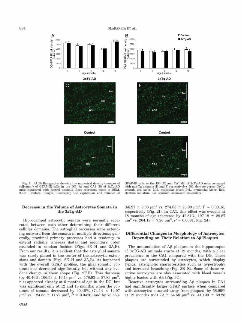
We analyzed the astroglial morphology in the hippo-campus, where the majority (�80%) of astrocytes expressGFAP (Kimelberg, 2004). Numerous GFAP immunoreac-tive cells (GFAP-IR), showing stellate shape and multiplebranched processes that are typical of astrocytes (Figs. 1–3), were widely distributed throughout the hippocampusin both non-Tg and 3xTg-AD mice; some of them havingthe typical morphology of perivascular astrocytes (Fig.3C). When we calculated the density of astrocytes withinthe DG and CA1 of the 3xTg-AD mice and compared itwith the non-Tg, we found no changes in the density ofGFAP-IR astrocytes at all ages (Fig. 1).
Astroglial Cytoskeletal Modificationsin the 3xTg-AD
From 6 months of age, astrocytes in the 3xTg-AD miceshowed a clear tendency to reduce their GFAP surfaceby 43.84% (781.11 6 87.44 lm2 vs. 1390.89 6 340.01lm2; n.s) and volume by 52.76% (254.61 6 36.62 lm3 vs.559.08 6 152.39 lm3; n.s), when compared with theirnon-TG controls in the DG (Fig. 2A,B, respectively) butnot in the CA1 subfield where similar changes onlyappears at later age (18 months; Figs. 2C and 3D). How-ever, these changes in the DG become significant only at12 and 18 months of age in 3xTg-AD mice when com-pared with controls. The surface decreases by 40.73%(575.04 6 89.65 lm2 vs. 970.33 6 54.84 lm2, P <0.0001) and 64.79% (631.25 6 78.17 lm2 vs. 1793.84 6
138.53 lm2, P 5 0.0019), respectively (Fig. 2A). Like-wise, there was a significant decrease in volume by45.39% (165.18 6 32.32 lm3 vs. 302.48 6 21.72 lm3, P5 0.0162) and 71.98% (186.76 6 22.19 lm3 vs. 665.95 6
64.18 lm3, P 5 0.0103), respectively (Fig. 2B). Thesechanges in GFAP surface and volume were accompaniedby a reduction in glial branching, indicative of the pro-cess of atrophy (Fig. 2F,H). In the CA1 subfield, thedecrease in surface and volume of GFAP profiles in3xTg-AD animals was significant only at 18 months(Fig. 2C,D). The surface decrease by 29.42% (1586.00 6
106.97 lm2 vs. 2247.19 6 120.76 lm2, P 5 0.0095)whereas the volume decreased by 32.74% (615.02 6
66.92 lm3 vs. 914.44 6 58.88 lm3, P 5 0.0237; Fig.2C,D).
833ASTROGLIA IN ALZHEIMER’S DISEASE
GLIA
Decrease in the Volume of Astrocytes Somata inthe 3xTg-AD
Hippocampal astrocytic somata were normally sepa-rated between each other determining their differentcellular domains. The astroglial processes were extend-ing outward from the somata in multiple directions; gen-erally, proximal primary processes had a tendency toextend radially whereas distal and secondary orderextended in random fashion (Figs. 2E–H and 3A,B).From our results, it is evident that the astroglial somatawas rarely placed in the center of the astrocytic exten-sions and domain (Figs. 2E–H and 3A,B). As happenedwith the overall GFAP profiles, the glial somatic vol-umes also decreased significantly, but without any evi-dent change in their shape (Fig. 2F,H). This decrease(by 40.46%, 106.53 6 16.14 lm3 vs. 178.93 6 57.83 lm3,n.s) appeared already at 6 months of age in the DG, butwas significant only at 12 and 18 months; when the vol-umes of somata decreased by 40.46%, (74.14 6 14.97lm3 vs. 124.53 6 11.72 lm3, P 5 0.0476) and by 75.55%
(66.97 6 8.88 lm3 vs. 274.03 6 25.90 lm3, P 5 0.0016),respectively (Fig. 2I). In CA1, this effect was evident at18 months of age (decrease by 42.81%, 197.19 6 28.87lm3 vs. 264.18 6 7.26 lm3, P 5 0.0081; Fig. 2J).
Differential Changes in Morphology of AstrocytesDepending on Their Relation to Ab Plaques
The accumulation of Ab plaques in the hippocampusof 3xTG-AD animals starts at 12 months, with a clearprevalence in the CA1 compared with the DG. Theseplaques are surrounded by astrocytes, which displaytypical astrogliotic characteristics such as hypertrophyand increased branching (Fig. 3B–E). Some of these re-active astrocytes are also associated with blood vesselshighly loaded with Ab (Fig. 3C).
Reactive astrocytes surrounding Ab plaques in CA1had significantly larger GFAP surface when comparedwith astrocytes situated away from plaques (by 58.95%at 12 months (651.72 6 54.58 lm2 vs. 410.00 6 69.28
Fig. 1. (A,B) Bar graphs showing the numerical density (number ofcells/mm3) of GFAP-IR cells in the DG (A) and CA1 (B) of 3xTg-ADmice compared with control animals. Bars represent mean 6 SEM.(C–F) Confocal images illustrating the expression and number of
GFAP-IR cells in the DG (C) and CA1 (E) of 3xTg-AD mice comparedwith non-Tg animals (D and F, respectively). DG, dentate gyrus; GrCL,granule cell layer; Mol, molecular layer; PyL, pyramidal layer; Rad,stratum radiatum; Lac, stratum lacunosum moleculare.
834 OLABARRIA ET AL.
GLIA

lm2, P 5 0.0030) and by 66.65% at 18 months (702.16 6
44.61 lm2 vs. 421.30 6 40.91 lm2, P 5 0.0044; Fig. 3F).This increase in surface was paralleled by an increase involume by 65.18% (183.33 6 20.75 lm3 vs. 110.98 6
23.27 lm3, P 5 0.0037) and 71.06% (196.32 6 17.07 lm3
vs. 114.76 6 11.50 lm3, P 5 0.0034) at 12 and 18months of age respectively when compared with astro-cytes situated away from plaques (Fig. 3G). Similarly,we observed significant increase in the volume of somataof astrocytes associated with plaques when comparedwith astrocytes distant to the plaques (by 46.95% at 12months (89.32 6 7.79 lm3 vs. 60.78 6 10.32 lm3, P 5
0.0046) and by 21.59% at 18 months (84.84 6 7.49 lm3
vs. 69.77 6 6.55 lm3, P 5 0.0137; Fig. 3H).Astrocytes surrounding plaques in the DG showed
similar structural changes at 18 months of age when theburden of Ab plaques becomes evident within this
region. These reactive astrocytes associated with Ab pla-ques in DG also increased significantly their GFAP sur-face (by 48.06%, 327.82 6 24.19 lm2 vs. 221.40 6 15.66lm2, P 5 0.0013; Fig 3I), volume (by 57.10%, 72.27 6
6.77 lm3 vs. 46.00 6 4.85 lm3, P 5 0.0032; Fig 3J), aswell as the somatas volume by 110.70% (31.10 6 3.25lm3 vs. 14.76 6 1.36 lm3, P 5 0.0005; Fig. 3K) whencompared with control animals, At the same time, theastrocytes positioned away from the plaques kept show-ing atrophic characteristics in both the CA1 and DGareas (Fig. 3A).
DISCUSSION
The astroglial cells, which form the ultimate homeo-static system in the CNS, are intimately involved in
Fig. 2. Bar graphs showing a decreased GFAP surface, volume, andsoma volume in both the DG (A, B, I) and the CA1 (C, D, J) of the hip-pocampus of the 3xTg-AD mice when compared with control animals.
Bars represent mean 6 SEM (P < 0.05. (G–J) Confocal micrographsillustrating the astrocytic atrophy in 3xTg-AD mice in the DG (F) andCA1 (H) compared with control animals (E and G).
835ASTROGLIA IN ALZHEIMER’S DISEASE
GLIA

many forms of neurological diseases. At the same time,the reaction of astrocytes during the early stages ofchronic neurological disorders and their pathogenetic
role remain virtually unknown. The reactive astrogliosisis a characteristic feature of terminal stages of neurode-generative processes, including the AD (Nagele et al.,2004; Rodriguez et al., 2009b; Rossi and Volterra, 2009;Simpson et al., 2008).
Here, we analyzed the GFAP-positive astroglial pro-files in the hippocampus of transgenic AD animals of dif-ferent age. To the best of our knowledge, this is the firstattempt to characterize the age-dependent changes inthe morphology of GFAP-positive astrocytes in the rele-vant model of family AD. For our analysis, we have cho-sen hippocampus because the majority of astroglial cellsin this brain region are GFAP positive (Kimelberg,2004). First, we found that overall density of GFAP-posi-tive astroglial cells is affected neither by age nor by theAD conditions. This shows that neither ageing nor ADpathology are associated with cell loss; similarly, itargues against the prominent astroglial proliferativeresponse in the AD brain.
Second, contrary to the general assumption that theAD is associated with reactive astrogliosis with overallincrease in GFAP expression and astroglial proliferation(Nagele et al., 2004), we found that in both DG and theCA1 areas there is a generalized atrophy of GFAP-posi-tive astrocytes. This atrophy is manifested by decreasedsurface and volume of GFAP-positive profiles andappears at the early stages of the disease. Indeed, al-ready at 6 months (when the hippocampus is virtuallyfree from the neuritic plaques) both volume and surfacearea of astrocytes in DG were decreased. This overallhypotrophy of GFAP-positive profiles in DG remains at12 and 18 months. The similar hypotrophy of GFAP-pos-itive astrocytes in CA1 region becomes apparent only at18 months of age.
During the later stages of AD, the changes in astro-glial morphology is complex. The appearance of the neu-ritic plaques in AD, similarly to other brain injuries(Wilhelmsson et al., 2006), results in astrogliosis in bothhippocampal areas. Importantly, the astrocytes demon-strating clear signs of reactivity (hypertrophy, thickprocesses, enlarged cell bodies, and overall increase inthe volume and surface area of GFAP-positive profiles)were exclusively associated with the Ab plaques. Astro-cytes localized distantly (>50 lm) from the borders ofthe plaques were atrophic as reflected by decreased vol-ume of cell bodies, reduced number of main processes,their arborization, and overall GFAP surface and vol-ume. The existence of two distinct populations of astro-cytes in Ab plaques-infested brains of transgenic ADanimals was also demonstrated in the in vivo Ca21
imaging experiments, which showed increased Ca21
excitability of the astrocytes contacting neuritic plaques(Kuchibhotla et al., 2009).
The early atrophy of astroglial cells in the progressionof family-like AD (and indeed the transgenic AD modelsreproduce the FAD type pathology) can be pathologicallyrelevant. The atrophic changes were not only associatedwith the thick primary proximal processes but also withsecondary medial and occasionally distal thin processes,which may indicate the reduction in overall astroglial
Fig. 3. (A,B) Confocal images of hippocampal preparations duallylabeled by GFAP and by anti-b amyloid monoclonal antibody illustratingdifferential changes in GFAP profiles in astrocytes distant to the plaques(A) and associated with the Ab plaques (B). (C–E) Confocal dual labellingimages (GFAP in green and Ab in red) in 3xTg-AD mice showing the accu-mulation of astrocytes around the Ab plaques and vascular Ab deposits.Astrocytes surrounding Ab plaques (D, E) and Ab loaded blood vessel (C),undergo astrogliosis. (F–K) Bar graphs showing GFAP-positive astrocyticsurface (F), volume (G), and soma volume (H) differences between astro-cytes located around the amyloid plaques (Ab) and those distant to the pla-ques in the CA1 of 3xTg-AD animals. (I–K): Similar astrocytic surface (I),volume (J), and soma volume (K) differences are observed in the DG at 18months of age. Bars represent mean 6 SEM (P < 0.05).
836 OLABARRIA ET AL.
GLIA

arborisation and hence possible decrease in glial synap-tic coverage (Rodriguez et al., 2009b). The reduction ofsynaptic coverage by astroglial membranes togetherwith overall atrophic changes in astroglia can be instru-mental in the early imbalance of neurotransmission,reduced glial homeostatic function, and in disruptedsynaptic connectivity. The atrophic or degenerativechanges in astroglia have been recently observed in sev-eral types of chronic neurodegeneration, including amyo-trophic lateral sclerosis (Rossi et al., 2008; Rossi andVolterra, 2009), in Wernicke encephalopathy (Hazell,2009), in frontotemporal dementia (Broe et al., 2004), inschizophrenia and major depression (Gosselin et al.,2009; Rajkowska et al., 2002; Si et al., 2004). The degen-erative/atrophic changes in astroglia may represent thecommon mechanism in early disruptions of synaptic con-nectivity in all forms of neurodegeneration. In AD, theearly astroglial asthenia may result in inadequate sup-port of synapses, leading to the extinction of the latterand disruption of connectivity within neural circuitry.Indeed, the synaptic loss at the early stages of AD iswell documented (Scheff et al., 2007; Terry, 2000), andthe underlying mechanisms are unclear. The results ofour morphological analysis may indicate that it is theastroglial failure which produces early synaptic disor-ders, and hence early cognitive deficits, in the Alzheimertype neuropathology.
ACKNOWLEDGMENTS
We would like to thank Mrs. Mathilde Menoret forher technical help.
REFERENCES
Abbott NJ, Ronnback L, Hansson E. 2006. Astrocyte-endothelial inter-actions at the blood-brain barrier. Nat Rev Neurosci 7:41–53.
Abramov AY, Canevari L, Duchen MR. 2003. Changes in intracellularcalcium and glutathione in astrocytes as the primary mechanism ofamyloid neurotoxicity. J Neurosci 23:5088–5095.
Abramov AY, Canevari L, Duchen MR. 2004. Beta-amyloid peptidesinduce mitochondrial dysfunction and oxidative stress in astrocytesand death of neurons through activation of NADPH oxidase. J Neuro-sci 24:565–575.
Alzheimer A. 1907. €Uber eine eigenartige Erkrankung der Hirnrinde.Allg Z Psychiat Psych-Gericht Med 64:146–148.
Alzheimer A. 1910. Beitr€age zur Kenntnis der pathologischen Neurogliaund ihrer Beziehungen zu den Abbauvorg€angen im Nervengewebe.In: Nissl F, Alzheimer A, editors. Histologische und histopathologi-sche Arbeiten €uber die Grosshirnrinde mit besonderer Ber€ucksichti-gung der pathologischen Anatomie der Geisteskrankheiten. Jena:Gustav Fischer. pp 401–562.
Assis-Nascimento P, Jarvis KM, Montague JR, Mudd LM. 2007. Beta-amyloid toxicity in embryonic rat astrocytes. Neurochem Res 32:1476–1482.
Braak E, Griffing K, Arai K, Bohl J, Bratzke H, Braak H. 1999. Neuro-pathology of Alzheimer’s disease: What is new since A. Alzheimer?Eur Arch Psychiatry Clin Neurosci 249 (Suppl 3):14–22.
Broe M, Kril J, Halliday GM. 2004. Astrocytic degeneration relates tothe severity of disease in frontotemporal dementia. Brain 127:2214–2220.
Bundgaard M, Abbott NJ. 2008. All vertebrates started out with a glialblood-brain barrier 4–500 million years ago. Glia 56:699–708.
Chvatal A, Anderova M, Hock M, Prajerova I, Neprasova H, Chvatal V,Kirchhoff F, Sykova E. 2007. Three-dimensional confocal morphometry
reveals structural changes in astrocyte morphology in situ. J NeurosciRes 85:260–271.
Danbolt NC. 2001. Glutamate uptake. Progr Neurobiol 65:1–105.Dringen R. 2000. Metabolism and functions of glutathione in brain.
Prog Neurobiol 62:649–671.Eng LF, Ghirnikar RS, Lee YL. 2000. Glial fibrillary acidic protein:
GFAP-thirty-one years (1969–2000). Neurochem Res 25:1439–1451.Giaume C, Kirchhoff F, Matute C, Reichenbach A, Verkhratsky A.
2007. Glia: The fulcrum of brain diseases. Cell Death Differ 14:1324–1335.
Gosselin RD, Gibney S, O’Malley D, Dinan TG, Cryan JF. 2009. Regionspecific decrease in glial fibrillary acidic protein immunoreactivity inthe brain of a rat model of depression. Neuroscience 159:915–925.
Halassa MM, Fellin T, Haydon PG. 2007. The tripartite synapse: Rolesfor gliotransmission in health and disease. Trends Mol Med 13:54–63.
Halliday GM, Cullen KM, Kril JJ, Harding AJ, Harasty J. 1996. Glialfibrillary acidic protein (GFAP) immunohistochemistry in human cor-tex: A quantitative study using different antisera. Neurosci Lett 209:29–32.
Haydon PG, Carmignoto G. 2006. Astrocyte control of synaptic trans-mission and neurovascular coupling. Physiol Rev 86:1009–1031.
Hazell AS. 2009. Astrocytes are a major target in thiamine deficiencyand Wernicke’s encephalopathy. Neurochem Int 55:129–135.
Kimelberg HK. 2004. The problem of astrocyte identity. Neurochem Int45:191–202.
Kirischuk S, Kettenmann H, Verkhratsky A. 2007. Membrane currentsand cytoplasmic sodium transients generated by glutamate transportin Bergmann glial cells. Pflugers Arch 454:245–252.
Kobayashi K, Hayashi M, Nakano H, Shimazaki M, Sugimori K, Kosh-ino Y. 2004. Correlation between astrocyte apoptosis and Alzheimerchanges in gray matter lesions in Alzheimer’s disease. J AlzheimersDis 6:623–632.
Kofuji P, Newman EA. 2004. Potassium buffering in the central nervoussystem. Neuroscience 129:1045–1056.
Kraepelin E. 1919. Dementia praecox and paraphrenia (translated byR. Mary Barclay). Edinburgh: E. & S. Livingstone. 328 p.
Kuchibhotla KV, Lattarulo CR, Hyman BT, Bacskai BJ. 2009. Synchro-nous hyperactivity and intercellular calcium waves in astrocytes inAlzheimer mice. Science 323:1211–1215.
Nagele RG, D’Andrea MR, Lee H, Venkataraman V, Wang HY. 2003.Astrocytes accumulate A beta 42 and give rise to astrocytic amyloidplaques in Alzheimer disease brains. Brain Res 971:197–209.
Nagele RG, Wegiel J, Venkataraman V, Imaki H, Wang KC. 2004. Con-tribution of glial cells to the development of amyloid plaques in Alz-heimer’s disease. Neurobiol Aging 25:663–674.
Nedergaard M, Dirnagl U. 2005. Role of glial cells in cerebral ischemia.Glia 50:281–286.
Nedergaard M, Ransom B, Goldman SA. 2003. New roles for astrocytes:Redefining the functional architecture of the brain. Trends Neurosci26:523–530.
Oberheim NA, Takano T, Han X, He W, Lin JH, Wang F, Xu Q, WyattJD, Pilcher W, Ojemann JG, Ransom BR, Goldman SA, NedergaardM. 2009. Uniquely hominid features of adult human astrocytes.J Neurosci 29:3276–3287.
Oberheim NA, Wang X, Goldman S, Nedergaard M. 2006. Astrocyticcomplexity distinguishes the human brain. Trends Neurosci 29:547–553.
Oddo S, Caccamo A, Kitazawa M, Tseng BP, LaFerla FM. 2003a. Amy-loid deposition precedes tangle formation in a triple transgenic modelof Alzheimer’s disease. Neurobiol Aging 24:1063–1070.
Oddo S, Caccamo A, Shepherd JD, Murphy MP, Golde TE, Kayed R,Metherate R, Mattson MP, Akbari Y, LaFerla FM. 2003b. Triple-transgenic model of Alzheimer’s disease with plaques and tangles: In-tracellular Abeta and synaptic dysfunction. Neuron 39:409–421.
Paxinos G, Franklin KBJ. 2004. The mouse Brain in Stereotaxic Coor-dinates. Compact 2nd Edn. Academic Press: San Diego, CA.
Perea G, Navarrete M, Araque A. 2009. Tripartite synapses: Astrocytesprocess and control synaptic information. Trends Neurosci 32:421–431.
Rajkowska G, Miguel-Hidalgo JJ, Makkos Z, Meltzer H, Overholser J,Stockmeier C. 2002. Layer-specific reductions in GFAP-reactive astro-glia in the dorsolateral prefrontal cortex in schizophrenia. SchizophrRes 57:127–138.
Rodriguez JJ, Jones VC, Tabuchi M, Allan SM, Knight EM, LaFerlaFM, Oddo S, Verkhratsky A. 2008. Impaired adult neurogenesis inthe dentate gyrus of a triple transgenic mouse model of Alzheimer’sdisease. PLoS One 3:e2935
Rodriguez JJ, Jones VC, Verkhratsky A. 2009a. Impaired cell prolifera-tion in the subventricular zone in an Alzheimer’s disease model. Neu-roreport 20:907–912.
Rodriguez JJ, Olabarria M, Chvatal A, Verkhratsky A. 2009b. Astrogliain dementia and Alzheimer’s disease. Cell Death Differ 16:378–385.
837ASTROGLIA IN ALZHEIMER’S DISEASE
GLIA

Rossi D, Brambilla L, Valori CF, Roncoroni C, Crugnola A, Yokota T,Bredesen DE, Volterra A. 2008. Focal degeneration of astrocytes inamyotrophic lateral sclerosis. Cell Death Differ 15:1691–1700.
Rossi D, Volterra A. 2009. Astrocytic dysfunction: Insights on the rolein neurodegeneration. Brain Res Bull 80:224–232.
Scheff SW, Price DA, Schmitt FA, DeKosky ST, Mufson EJ. 2007. Syn-aptic alterations in CA1 in mild Alzheimer disease and mild cognitiveimpairment. Neurology 68:1501–1508.
Seifert G, Schilling K, Steinhauser C. 2006. Astrocyte dysfunction inneurological disorders: A molecular perspective. Nat Rev Neurosci 7:194–206.
Selkoe DJ. 2001. Alzheimer’s disease: Genes, proteins, and therapy.Physiol Rev 81:741–766.
Si X, Miguel-Hidalgo JJ, O’Dwyer G, Stockmeier CA, Rajkowska G.2004. Age-dependent reductions in the level of glial fibrillary acidicprotein in the prefrontal cortex in major depression. Neuropsycho-pharmacology 29:2088–2096.
Simard M, Nedergaard M. 2004. The neurobiology of glia in the contextof water and ion homeostasis. Neuroscience 129:877–896.
Simpson JE, Ince PG, Lace G, Forster G, Shaw PJ, Matthews F, SavvaG, Brayne C, Wharton SB. 2008. Astrocyte phenotype in relation to
Alzheimer-type pathology in the ageing brain. Neurobiol Aging doi:10.1016/jneurobioaging.2008.05.015.
Terry RD. 2000. Cell death or synaptic loss in Alzheimer disease. JNeuropathol Exp Neurol 59:1118–1119.
Verkhratsky A. 2006. Patching the glia reveals the functional organisa-tion of the brain. Pflugers Arch 453:411–420.
Verkhratsky A. 2009. Neuronismo y reticulismo: Neuronal-glial circuitsunify the reticular and neuronal theories of brain organization. ActaPhysiol (Oxf) 195:111–122.
Volterra A, Meldolesi J. 2005. Astrocytes, from brain glue to communi-cation elements: The revolution continues. Nat Rev Neurosci 6:626–640.
Wang DD, Bordey A. 2008. The astrocyte odyssey. Prog Neurobiol 86:342–367.
Wilhelmsson U, Bushong EA, Price DL, Smarr BL, Phung V, Terada M,Ellisman MH, Pekny M. 2006. Redefining the concept of reactiveastrocytes as cells that remain within their unique domains uponreaction to injury. Proc Natl Acad Sci USA 103:17513–17518.
Yankner BA. 1996. Mechanisms of neuronal degeneration in Alzhei-mer’s disease. Neuron 16:921–932.
838 OLABARRIA ET AL.
GLIA


![Acute ethanol exposure induces [Ca 2+ ] i transients, cell swelling and transformation of actin cytoskeleton in astroglial primary cultures](https://static.fdokumen.com/doc/165x107/63221b7a61d7e169b00c78d0/acute-ethanol-exposure-induces-ca-2-i-transients-cell-swelling-and-transformation.jpg)