PROSENSOR D O C U M E N TAT I O N T E C H N I Q U E Régulateurs & Indicateurs
Upload
khangminh22Category
view
0download
0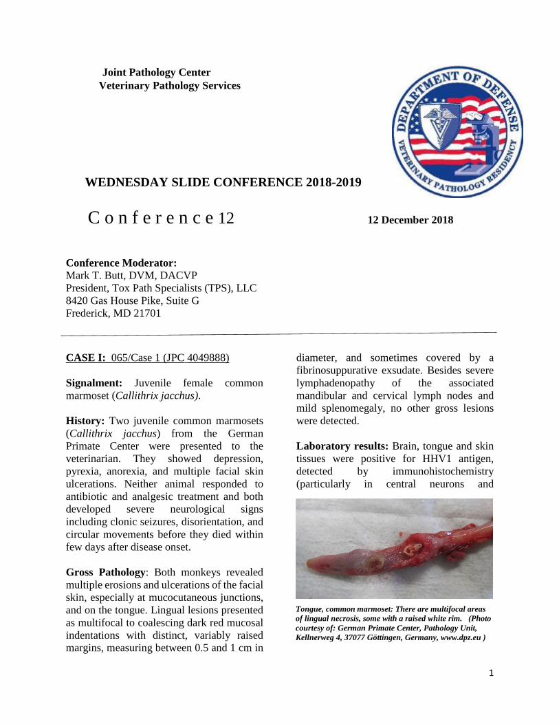
1
Joint Pathology Center Veterinary Pathology Services
WEDNESDAY SLIDE CONFERENCE 2018-2019
C o n f e r e n c e 12 12 December 2018
Conference Moderator: Mark T. Butt, DVM, DACVP President, Tox Path Specialists (TPS), LLC 8420 Gas House Pike, Suite G Frederick, MD 21701
CASE I: 065/Case 1 (JPC 4049888) Signalment: Juvenile female common marmoset (Callithrix jacchus). History: Two juvenile common marmosets (Callithrix jacchus) from the German Primate Center were presented to the veterinarian. They showed depression, pyrexia, anorexia, and multiple facial skin ulcerations. Neither animal responded to antibiotic and analgesic treatment and both developed severe neurological signs including clonic seizures, disorientation, and circular movements before they died within few days after disease onset. Gross Pathology: Both monkeys revealed multiple erosions and ulcerations of the facial skin, especially at mucocutaneous junctions, and on the tongue. Lingual lesions presented as multifocal to coalescing dark red mucosal indentations with distinct, variably raised margins, measuring between 0.5 and 1 cm in
diameter, and sometimes covered by a fibrinosuppurative exsudate. Besides severe lymphadenopathy of the associated mandibular and cervical lymph nodes and mild splenomegaly, no other gross lesions were detected. Laboratory results: Brain, tongue and skin tissues were positive for HHV1 antigen, detected by immunohistochemistry (particularly in central neurons and
Tongue, common marmoset: There are multifocal areas of lingual necrosis, some with a raised white rim. (Photo courtesy of: German Primate Center, Pathology Unit, Kellnerweg 4, 37077 Göttingen, Germany, www.dpz.eu )
2
keratinocytes).Transmission electron microscopy revealed numerous l herpes virions with characteristic electron-dense core, capsid and envelope in cutaneous keratinocytes. Microscopic Description: Brain: Multifocally throughout the grey and to a lesser extend in the white matter of cerebrum, brainstem and cerebellum, there are mild to moderate inflammatory foci, either concentrated around blood vessels (perivascular cuffing) or scattered within the neuropil. The inflammatory infiltrate consists of lymphocytes, histiocytes, neutrophils and fewer plasma cells, often admixed with mild karyorrhexis, karyolysis and pyknosis (necrotic neutrophils). The affected neuropil in addition reveals mild to moderate scattered to nodular gliosis and several shrunken, angular and hypereosinophilic neurons with hyperchromatic nuclei (neuronal necrosis).Within very few glia cells and degenerate neurons, there are conspicuous large basophilic intranuclear inclusions, which marginate the chromatin. Perivascular cuffing is often associated with mild endothelial hypertrophy. Meningeal blood vessels are often engorged with erythrocytes (hyperemia) together with moderate to marked intravascular stasis of leucocytes, mainly neutrophils, and multifocal mild to moderate perivascular inflammatory infiltration as previously described. Multifocally within the cerebral white matter, there is also mild to moderate dilation and vacuolation of Virchow-Robin spaces (edema). Tongue: There is multifocal to coalescing ulceration of the lingual surface with the extensively lost mucosal epithelium being replaced by a thick serocellular crust composed of fibrin, varying amounts of viable and degenerate neutrophils and multifocal coccoid bacterial colonies. In
areas with less extensive ulceration, circumvallate papillae are multifocally expanded by intraepithelial clefts filled with variable amounts of viable and degenerate neutrophils, fibrin, edema, and few acantholytic keratinocytes (vesicopustules). Ulcerations and pustules are multifocally flanked by groups of degenerate or necrotic keratinocytes, often bearing large eosinophilic to amphophilic, homogenous intranuclear inclusions with marked margination of chromatin. The adjacent epithelium is mild to moderately hyperplastic, characterized by acanthosis and intracellular edema. Fibrin, mild hemorrhage and edema together with varying numbers of neutrophils, histiocytes, fewer lymphocytes and plasma cells moderately expand the adjacent subepithelial connective tissue and separate superficial skeletal muscle fibers and individual myocytes. Contributor’s Morphologic Diagnoses: Central nervous system: Cerebrum, cerebellum and brainstem: Meningoencephalitis, lymphohistiocytic and neutrophilic, multifocal, moderate, subacute
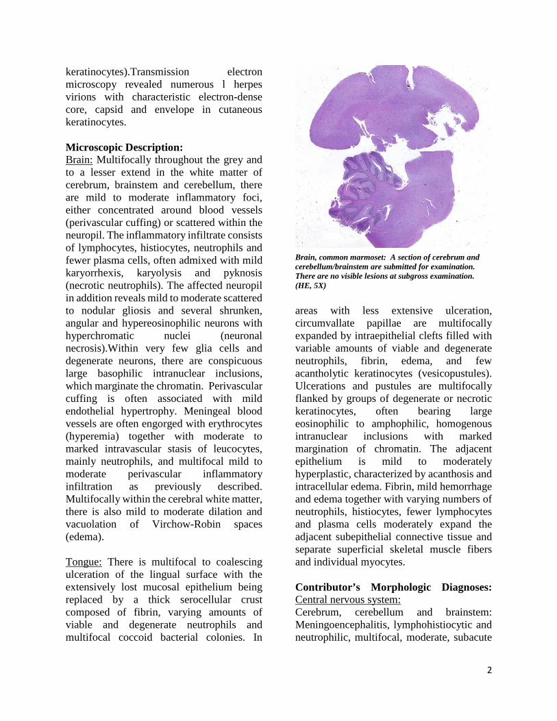
Brain, common marmoset: A section of cerebrum and cerebellum/brainstem are submitted for examination. There are no visible lesions at subgross examination. (HE, 5X)
3
with neuronal necrosis, gliosis and very few glial and neuronal homogenous basophilic intranuclear inclusion bodies; common marmoset (Callithrix jacchus), nonhuman primate. Tongue: Glossitis, vesicopustular to ulcerative, multifocal to coalescing, subacute, marked with fibrinous to serocellular crusting and epithelial homogenous eosinophilic to amphophilic intranuclear inclusion bodies; common marmoset (Callithrix jacchus), nonhuman primate. Contributor’s Comment: Herpesviridae are global pathogens of almost every vertebrate species. In their natural hosts, they do not cause fatal disease, but cross-species infection can cause acute death 1,10. Infection with Human Herpesvirus 1 (HHV1), an alphaherpesvirus, leads to mild mucosal lesions or an inapparent course in the natural host (human).9,13 Fatal encephalitis and/or systemic disease can occur in neonates or immune compromised individuals.7,14 In humans, infection with
HHV1 persists lifelong because of viral latency in sensory neurons. When immunity is compromised through stress or disease, it becomes activated through suppression of the latency-associated transcript gene.11 It can be shed even in the absence of visible lesions. Several nonhuman primate species are susceptible to infection with human herpesviruses. Old World Primates like gorillas (Gorilla gorilla), bonobos (Pan paniscus), white handed gibbons (Hylobates lar) and chimpanzees (Pan troglotydes), can be naturally infected with HHV1. Although single fatal disease outcomes have been described,2,6 their virus-host relationship seems to be similar to that in humans, as they usually show only mild symptoms (lesions in mucocutaneous tissue, skin, conjunctiva).10 However, New World monkey species and prosimians are highly susceptible to HHV 1 infections. 1,4,5,9,10 Typical lesions in these species are erosions and ulcers of oral mucous membranes, especially the tongue, and mucocutaneous junctions of the lips. Severe necrotizing hepatitis and multifocal nonsuppurative meningoencephalitis often occur in these species.5,10 Thus, in cases of ulcerative skin lesions in nonhuman primates, HHV1 has to be considered as a possible cause and callitrichids represent a highly susceptible species for HHV1 infections. The exact route of transmission is unknown, but likely occurs via direct mucosal contact, wounds, saliva, maternal milk, contaminated fomites and through airborne and waterborne routes.6 Additionally, direct transmission from latently infected humans to their monkey pets is described. In the present case, possible sources of virulent HHV1 are animal keepers or visitors. HHV1 specific PCR can be used for
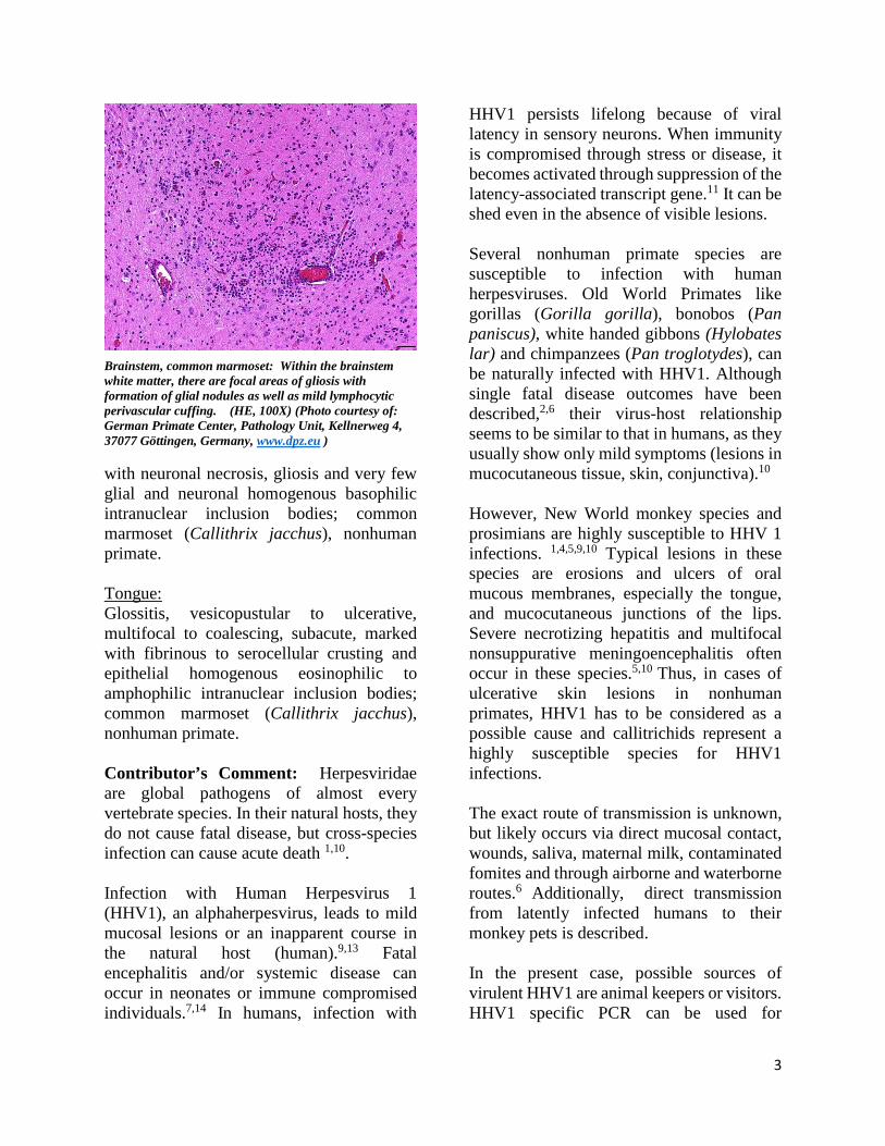
Brainstem, common marmoset: Within the brainstem white matter, there are focal areas of gliosis with formation of glial nodules as well as mild lymphocytic perivascular cuffing. (HE, 100X) (Photo courtesy of: German Primate Center, Pathology Unit, Kellnerweg 4, 37077 Göttingen, Germany, www.dpz.eu )
4
antemortem diagnostics to allow appropriate treatment with antiviral drugs. Viral antigen can be detected in histological sections of lesions by immunohistochemistry and Transmission electron microscopy can serve as a useful tool to seek for viral particles. Contributing Institution: German Primate Center, Pathology Unit, Kellnerweg 4, 37077 Göttingen, Germany www.dpz.eu JPC Diagnosis: 1. Tongue: Glossitis, necrotizing, focally extensive, moderate, with ulceration, epithelial syncytia and intranuclear viral inclusions. 2. Cerebrum and brainstem: Encephalitis, necrotizing, multifocal, mild to moderate with gliosis, perivascular cuffing, and rare intranuclear viral inclusions. JPC Comment: The contributor has provided an excellent overview of cross-species herpesviral infection, in this case human herpesvirus 1 (human simplex virus 1) and its devastating effects on New World primates.
Cross species infection by alphaherpesviruses has been well known to result in tragic circumstances, via zoonotic and reverse-zoonotic (as seen here) means, as well as between diverse animal species. A review of the literature is replete with interesting cases, some of which are recounted here. Perhaps the most famous case of interspecies transmission of herpesvirus was described in a case report by Albert Sabin, MD (who would go on to develop the oral polio vaccine) in 1932, who described an ascending paralysis in an accomplished young physician and research colleague following a “monkey bite”. For many years, this researcher was simply known as “W.B”, while the agent isolated from his neural tissue gained great renown as the “B virus” (now known as macacine herpesvirus-1) which has claimed 70% of its known victims over the years due to fatal encephalitis. In 1985, that young researcher’s name was finally published, Dr. William Barlett Brebner (1903-1932).12 Equine herpesvirus-1 (EHV-1) is a well-known pathogen of horses that is known to cause a wide range of disease in its natural host, but may also result in disease in widely disparate mammalian species as well. A 2011 article15 details fatal encephalitides characterized by non-suppurative meningo-encephalitis, which were shown to be caused equine herpesvirus 1 in four black bears (Ursus americanus), two Thompson gazelles (Eudorcas thomsoni) and eighteen guinea pigs (Cavia porcellus). In addition, a closely-related herpesvirus, equine herpesvirus-9 (EHV-9), thought to have originated in the plains zebra (Equus burchelli), has been known to cause fatal encephalitis in polar bears.3 Pseudorabies virus (Suid herpesvirus-1) is another alphaherpesvirus with a wide host range. Primarily infecting swine (who are the natural reservoir),
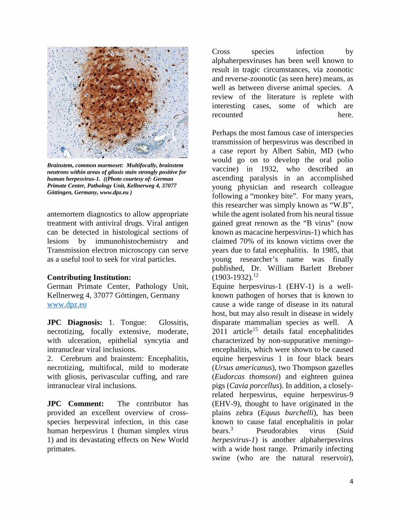
Brainstem, common marmoset: Multifocally, brainstem neutrons within areas of gliosis stain strongly positive for human herpesvirus-1. ((Photo courtesy of: German Primate Center, Pathology Unit, Kellnerweg 4, 37077 Göttingen, Germany, www.dpz.eu )
5
secondary mammalian hosts abound, including cattle, horses, dogs, mink, and cats. Human infection, although possible in immunocompetent humans has been limited to non-fatal conditions. One final tragic cross-species herpesviral infection has resulted in the depth of over 100 young captive and wild Asian elephants from a rapid-onset, acute hemorrhagic disease caused primarily by multiple distinct strains of two closely related chimeric variants of a novel herpesvirus species designated elephant endotheliotropic herpesvirus (EEHV1A and EEHV1B). These and two other species of Probosciviruses (EEHV4 and EEHV5) are evidently ancient and also result in commonly asymptomatic infections of adult Asian. Although only a few similar cases have been observed in African elephants, they harbor similar viruses—EEHV2, EEHV3, EEHV6, and EEHV7—have been isolated in African elephants in lung and skin nodules or saliva. For reasons not yet understood, approximately 20% of Asian elephant calves appear to be susceptible to the disease when primary infections are not controlled by normal innate cellular and humoral immune responses.8
References: 1. Costa EA, Luppi MM, Malta Mde C et al.
Outbreak of human herpesvirus type 1 infection in nonhuman primates (Callithrix penincillata). J Wildl Dis. 2011;47:690-693.
2. Emmons RW, Lennette EH: Natural herpesvirus hominis infection of a gibbon (Hylobates lar). Arch Gesamte Virusforsch. 1970; 31:215-218.
3. Greenwood AD, Tsangaras K, Ho SYW, Szentiks CA, Nikolin VM, Ma G, Daminani A, East ML, Lawrenz A, Hofer h, Oserrieder N. A potentially fatal mix of herpes in zoos. Curr Biol 2012; 22:1727-1732.
4. Huemer HP, Larcher C, Czedik-Eysenberg T, Nowotny N, Reifinger M. Fatal infection of a pet monkey with Human herpesvirus. Emerg Infect Dis. 2002;8:639-642.
5. Juan-Salles C, Ramos-Vara JA, Prats N, Sole-Nicolas J, Segales J, Marco AJ. Spontaneous herpes simplex virus infection in common marmosets (Callithrix jacchus). J Vet Diagn Invest. 1997;9:341-345.
6. Kik MJ, Bos JH, Groen J, Dorrestein GM. Herpes simplex infection in a juvenile orangutan (Pongo pygmaeus pygmaeus). J Zoo Wildl Med. 2005;36:131-134.
7. Kleinschmidt-DeMasters BK, Gilden DH. The expanding spectrum of herpesvirus infections of the nervous system. Brain Pathol. 2001;11:440-451.
8. Long SY, Latimer EM, Hayward GS. Review of elephant endotheliotropic herpesviruses and acute hemorrhagic disease. ILAR J; 2016:56(3):283-296.
9. Longa CS, Bruno SF, Pires AR, et al. Human herpesvirus 1 in wild marmosets, Brazil, 2008. Emerg Infect Dis. 2011;17:1308-1310.
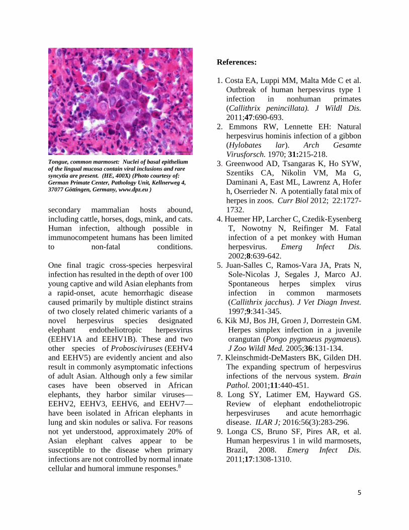
Tongue, common marmoset: Nuclei of basal epithelium of the lingual mucosa contain viral inclusions and rare syncytia are present. (HE, 400X) (Photo courtesy of: German Primate Center, Pathology Unit, Kellnerweg 4, 37077 Göttingen, Germany, www.dpz.eu )
6
10. Mätz-Rensing K, Jentsch KD, Rensing S, et al. Fatal herpes simplex infection in a group of common marmosets (Callithrix jacchus). Vet Pathol. 2003;40:405-411.
11. Perng GC, Jones C, Ciacci-Zanella J, et al. Virus-induced neuronal apoptosis blocked by the herpes simplex virus latency-associated transcript. Science. 2000;287:1500-1503.
12. Pimmentel JD. Past professive: Herpes B virus – “B” is for Brebner: Dr. William Barlet Brebner (1903-1932), Can Med Assoc J 2018: 178(6):726.
13.Schrenzel MD, Osborn KG, Shima A, Klieforth RB, Maalouf GA. Naturally occurring fatal herpes simplex virus 1 infection in a family of white-faced saki monkeys (Pithecia pithecia pithecia). J Med Primatol. 2003;32:7-14.
14.Whitley RJ, Roizman B. Herpes simplex virus infections. Lancet. 2001;357:1513-1518.
15. Wohlsein P, Lehmbecker A, Spitzbarth I, Algermissen D, Baumgartner W, Boer M, Kummrow M, aas L, Grummer B. Fatal epizootic equine herpesvirus 1 infections in new and unnatural hosts. Vet Microbiol 2011 149(3-4):456-460.
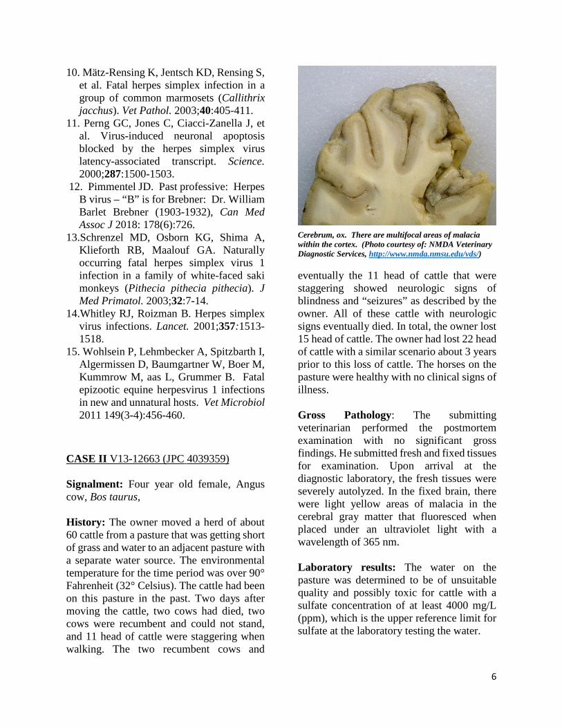
CASE II V13-12663 (JPC 4039359) Signalment: Four year old female, Angus cow, Bos taurus, History: The owner moved a herd of about 60 cattle from a pasture that was getting short of grass and water to an adjacent pasture with a separate water source. The environmental temperature for the time period was over 90° Fahrenheit (32° Celsius). The cattle had been on this pasture in the past. Two days after moving the cattle, two cows had died, two cows were recumbent and could not stand, and 11 head of cattle were staggering when walking. The two recumbent cows and
eventually the 11 head of cattle that were staggering showed neurologic signs of blindness and “seizures” as described by the owner. All of these cattle with neurologic signs eventually died. In total, the owner lost 15 head of cattle. The owner had lost 22 head of cattle with a similar scenario about 3 years prior to this loss of cattle. The horses on the pasture were healthy with no clinical signs of illness. Gross Pathology: The submitting veterinarian performed the postmortem examination with no significant gross findings. He submitted fresh and fixed tissues for examination. Upon arrival at the diagnostic laboratory, the fresh tissues were severely autolyzed. In the fixed brain, there were light yellow areas of malacia in the cerebral gray matter that fluoresced when placed under an ultraviolet light with a wavelength of 365 nm. Laboratory results: The water on the pasture was determined to be of unsuitable quality and possibly toxic for cattle with a sulfate concentration of at least 4000 mg/L (ppm), which is the upper reference limit for sulfate at the laboratory testing the water.
Cerebrum, ox. There are multifocal areas of malacia within the cortex. (Photo courtesy of: NMDA Veterinary Diagnostic Services, http://www.nmda.nmsu.edu/vds/)
7
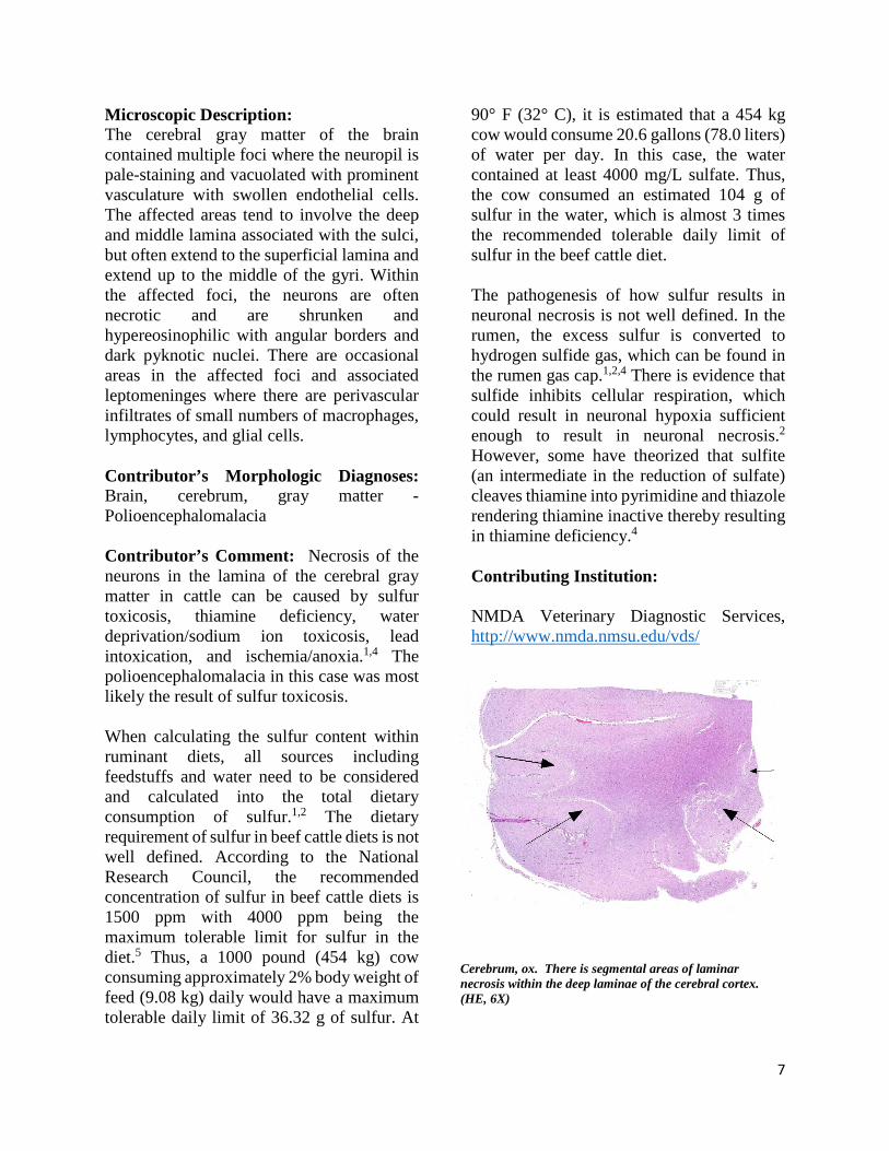
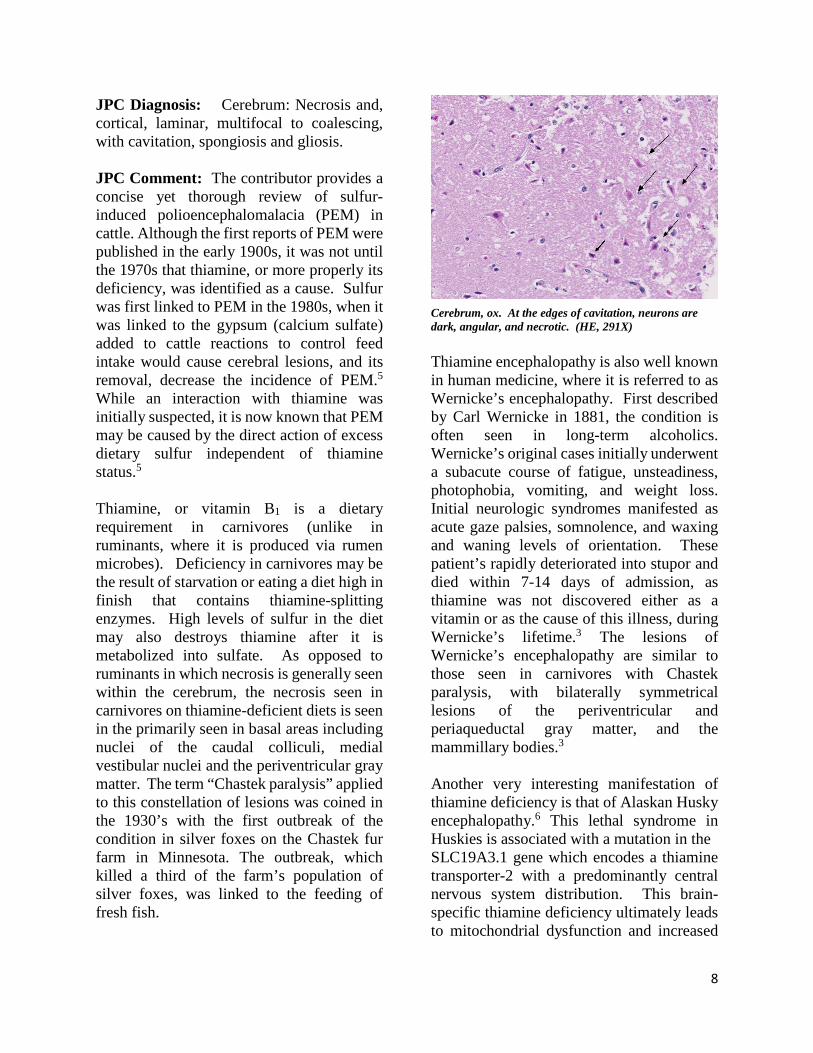
Microscopic Description: The cerebral gray matter of the brain contained multiple foci where the neuropil is pale-staining and vacuolated with prominent vasculature with swollen endothelial cells. The affected areas tend to involve the deep and middle lamina associated with the sulci, but often extend to the superficial lamina and extend up to the middle of the gyri. Within the affected foci, the neurons are often necrotic and are shrunken and hypereosinophilic with angular borders and dark pyknotic nuclei. There are occasional areas in the affected foci and associated leptomeninges where there are perivascular infiltrates of small numbers of macrophages, lymphocytes, and glial cells. Contributor’s Morphologic Diagnoses: Brain, cerebrum, gray matter - Polioencephalomalacia Contributor’s Comment: Necrosis of the neurons in the lamina of the cerebral gray matter in cattle can be caused by sulfur toxicosis, thiamine deficiency, water deprivation/sodium ion toxicosis, lead intoxication, and ischemia/anoxia.1,4 The polioencephalomalacia in this case was most likely the result of sulfur toxicosis. When calculating the sulfur content within ruminant diets, all sources including feedstuffs and water need to be considered and calculated into the total dietary consumption of sulfur.1,2 The dietary requirement of sulfur in beef cattle diets is not well defined. According to the National Research Council, the recommended concentration of sulfur in beef cattle diets is 1500 ppm with 4000 ppm being the maximum tolerable limit for sulfur in the diet.5 Thus, a 1000 pound (454 kg) cow consuming approximately 2% body weight of feed (9.08 kg) daily would have a maximum tolerable daily limit of 36.32 g of sulfur. At
90° F (32° C), it is estimated that a 454 kg cow would consume 20.6 gallons (78.0 liters) of water per day. In this case, the water contained at least 4000 mg/L sulfate. Thus, the cow consumed an estimated 104 g of sulfur in the water, which is almost 3 times the recommended tolerable daily limit of sulfur in the beef cattle diet. The pathogenesis of how sulfur results in neuronal necrosis is not well defined. In the rumen, the excess sulfur is converted to hydrogen sulfide gas, which can be found in the rumen gas cap.1,2,4 There is evidence that sulfide inhibits cellular respiration, which could result in neuronal hypoxia sufficient enough to result in neuronal necrosis.2 However, some have theorized that sulfite (an intermediate in the reduction of sulfate) cleaves thiamine into pyrimidine and thiazole rendering thiamine inactive thereby resulting in thiamine deficiency.4 Contributing Institution: NMDA Veterinary Diagnostic Services, http://www.nmda.nmsu.edu/vds/
Cerebrum, ox. There is segmental areas of laminar necrosis within the deep laminae of the cerebral cortex. (HE, 6X)
8
JPC Diagnosis: Cerebrum: Necrosis and, cortical, laminar, multifocal to coalescing, with cavitation, spongiosis and gliosis. JPC Comment: The contributor provides a concise yet thorough review of sulfur-induced polioencephalomalacia (PEM) in cattle. Although the first reports of PEM were published in the early 1900s, it was not until the 1970s that thiamine, or more properly its deficiency, was identified as a cause. Sulfur was first linked to PEM in the 1980s, when it was linked to the gypsum (calcium sulfate) added to cattle reactions to control feed intake would cause cerebral lesions, and its removal, decrease the incidence of PEM.5 While an interaction with thiamine was initially suspected, it is now known that PEM may be caused by the direct action of excess dietary sulfur independent of thiamine status.5
Thiamine, or vitamin B1 is a dietary requirement in carnivores (unlike in ruminants, where it is produced via rumen microbes). Deficiency in carnivores may be the result of starvation or eating a diet high in finish that contains thiamine-splitting enzymes. High levels of sulfur in the diet may also destroys thiamine after it is metabolized into sulfate. As opposed to ruminants in which necrosis is generally seen within the cerebrum, the necrosis seen in carnivores on thiamine-deficient diets is seen in the primarily seen in basal areas including nuclei of the caudal colliculi, medial vestibular nuclei and the periventricular gray matter. The term “Chastek paralysis” applied to this constellation of lesions was coined in the 1930’s with the first outbreak of the condition in silver foxes on the Chastek fur farm in Minnesota. The outbreak, which killed a third of the farm’s population of silver foxes, was linked to the feeding of fresh fish.
Thiamine encephalopathy is also well known in human medicine, where it is referred to as Wernicke’s encephalopathy. First described by Carl Wernicke in 1881, the condition is often seen in long-term alcoholics. Wernicke’s original cases initially underwent a subacute course of fatigue, unsteadiness, photophobia, vomiting, and weight loss. Initial neurologic syndromes manifested as acute gaze palsies, somnolence, and waxing and waning levels of orientation. These patient’s rapidly deteriorated into stupor and died within 7-14 days of admission, as thiamine was not discovered either as a vitamin or as the cause of this illness, during Wernicke’s lifetime.3 The lesions of Wernicke’s encephalopathy are similar to those seen in carnivores with Chastek paralysis, with bilaterally symmetrical lesions of the periventricular and periaqueductal gray matter, and the mammillary bodies.3 Another very interesting manifestation of thiamine deficiency is that of Alaskan Husky encephalopathy.6 This lethal syndrome in Huskies is associated with a mutation in the SLC19A3.1 gene which encodes a thiamine transporter-2 with a predominantly central nervous system distribution. This brain-specific thiamine deficiency ultimately leads to mitochondrial dysfunction and increased
Cerebrum, ox. At the edges of cavitation, neurons are dark, angular, and necrotic. (HE, 291X)
9
oxidative stress in certain areas of the brain. Affected Huskies present with seizures, altered mentation, dysphagia, central blindness, hypermetria, and proprioception positioning deficits. Cavitary lesions are present in the thalamus, and smaller lesions are often present in the putamen, claustrum, and that the junction of the gray and white matter in the cortex, brainstem, and midline cerebellar vermis.6
From a terminology point of view, the moderator reminded the group that the term “cortex” should be applied to the cerebral gray matter and does not include the underlying white matter. There are six levels to the cortex (which are best seen in man and non-human primates). References:
1. Gould DH. Polioencephalomalacia. J Anim Sci. 1998; 76: 309-314
2. Gould DH, Dargatz DA, Garry FG, et. al. J Am Vet Med Assoc. 2002; 221: 673-677
3. Isenberg-Grzeda E, Hsu AJ, Hatzoglou V, Nelso C, Breitbard W. Palliative treatment of thiamine-related encephalopathy (Wernicke’s encephalopathy) in cancer: a case series and review of the literature. Palliat Support Care 2015: 12(5): 1241-1249.
4. Maxie MG, Youssef S. Nervous system. In: Maxie MG, ed. Pathology of Domestic Animals. 5th ed. Philadelphia, PA: Elsevier Saunders; 2007: 281-457
5. Niles GA, Morgan SE, Edwards WC, The relationship between sulfur, thiamine and polioencephalomalacia – a review. Bovine Practice. 2002; 36: 93-99.
6. Vernau K, Napoli E, Wong S, Ross-Inta C, Cameron J, Bannasch D, Boilen, Dickenson P, Giulivi C. thiamine deficiency-mediated brain mitochondrial pathology in Alaskan Huskies with mutation in SLC19A3.1. Brain Pathol 2015; 25(4):441-453.
CASE III 111474 (JPC 4084217) Signalment: 7-year-old, male castrated, Shih Tzu dog (Canis familiaris) History: A 7-year-old, male castrated, Shih Tzu dog was referred to the Cornell University Hospital for Animals for acute neurologic signs after 4 episodes of vomiting. The dog had a 4-5 year history of polyuria and polydipsia (PU/PD). After vomiting overnight, the following day the dog showed signs of lethargy and ataxia which progressed to disorientation and lateral recumbency. The dog was brought to the referring veterinarian where blood work showed > 180 mmol/μl of sodium [144-160 mmol/μl]. Initial fluid therapy consisting of lactated Ringer's solution (LRS) was administered (unknown dose); however, the dog began head pressing. Furosemide (5 mg/kg) was administered. Soon after the patient had a focal seizure and received an additional 4 mg/kg of furosemide. The dog was referred to Cornell University Hospital for Animals’ Emergency Service. He had an additional seizure during transit. On examination, the patient was stuporous, hyperthermic (104.6 F), and tachycardic (200 bpm) with a weak femoral pulse. The patient was approximately 9% dehydrated and had tacky, injected mucous membranes with a capillary refill time of 1 second. The initial blood pressure was 97/24 with a mean of 43.
10
Anisocoria was present (meiosis OD). A neurologic consultation revealed a forebrain neurolocalization. The patient was recumbent with an absent menace response and nasal sensation, bilaterally. Pro-prioception was absent in all four limbs with intact spinal reflexes. No head or neck pain was noted. Point of care blood work (PCV, total solids and venous blood gas) revealed severe hypernatremia (182 mEq/L; 140–154 mEq/L) and hyperchloridemia (134 mEq/L; 104–119 mEq/L) with normokalemia (3.04 mEq/L; 3.8–5.4 mEq/L).
The patient was started on a custom fluid containing 175 mEq/L of sodium. A recheck of venous blood gas analysis revealed sodium of 191mEq/L. On the bases of the history, physical examination findings, and biochemical abnormalities, central diabetes insipidus was suspected. Fluid therapy was changed to a hyponatremic solution and with continued fluid adjustments and subcutaneous administration of 1mcg/kg of desmopressin, the serum sodium decreased to 177.7 mEq/L over the course of 12 hours. The patient’s mental status remained unchanged with an additional seizure. MRI revealed bilateral grey matter swelling characterized by a reduced size of the subarachnoid space, effacement of the sulci and compression of the adjacent white matter tracts consistent with moderate, diffuse, cerebral edema, which was primarily cytotoxic. The sodium level improved steadily over the 48 hours of hospitalization and fluid therapy and there was a transient improvement of mentation, but the patient became bradycardic and nonresponsive to atropine and experienced cardiac arrest. No cardiopulmonary resuscitation efforts were made at the request of the owner. Gross Pathology: The submitting veterinarian performed the postmortem examination with no significant gross findings. He submitted fresh and fixed tissues for examination. Upon arrival at the diagnostic laboratory, the fresh tissues were severely autolyzed. In the fixed brain, there were light yellow areas of malacia in the cerebral gray matter that fluoresced when placed under an ultraviolet light with a wavelength of 365 nm. Laboratory results: The sodium level of the brain tissue was 13,850 ppm (reference
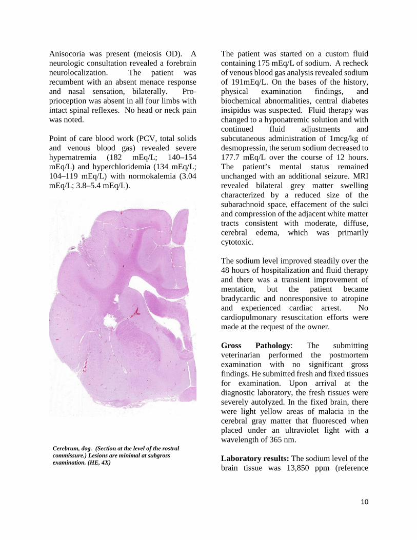
Cerebrum, dog. (Section at the level of the rostral commissure.) Lesions are minimal at subgross examination. (HE, 4X)
11
interval, calculated on a dry matter basis, 6000-8000 ppm). Microscopic Description: Brain, cerebral cortex: Diffusely, the blood vessels throughout all the sections are prominent, severely congested, lined by plump, reactive endothelial cells, and occasionally surrounded by small numbers of macrophages and fewer lymphocytes, hemorrhage, and fibrin within the Virchow-Robbin space. In the neuropil are scattered pyknotic and karyorrhectic cellular debris, most frequent in the mid to deep cerebral gray matter. In this area, there are frequent necrotic neurons: 1) angular with hypereosinophilic, variably microvacuolated cytoplasm that lack Nissl substance, with a smudgy nucleus without a nucleolus, or 2) appear as faint angular eosinophilic shadows of ghost neurons that lack a nucleus. The necrosis is most prominent in the mid to deep cerebral gray matter, while the perivascular
cuffing, vascular branching, and perivascular fibrin and hemorrhage are most prominent in the caudate nucleus. Pituitary gland (image not included): The pituitary gland is severely distorted by multifocal to coalescing cysts replacing and expanding 60% of the pars distalis, most of the pars intermedia, and 10% of the pars nervosa. The cysts are filled with variably eosinophilic, fibrillar to homogenous material, and lined by attenuated to low columnar, pseudostratified ciliated epithelium. A variety of immunohistochemical and histochemical stains were applied to the brain sections. All positive and negative controls were adequate. Microtubule associated protein 2 for neurons (MAP2) demonstrated strong but infrequent cytoplasmic immunoreactivity of the soma
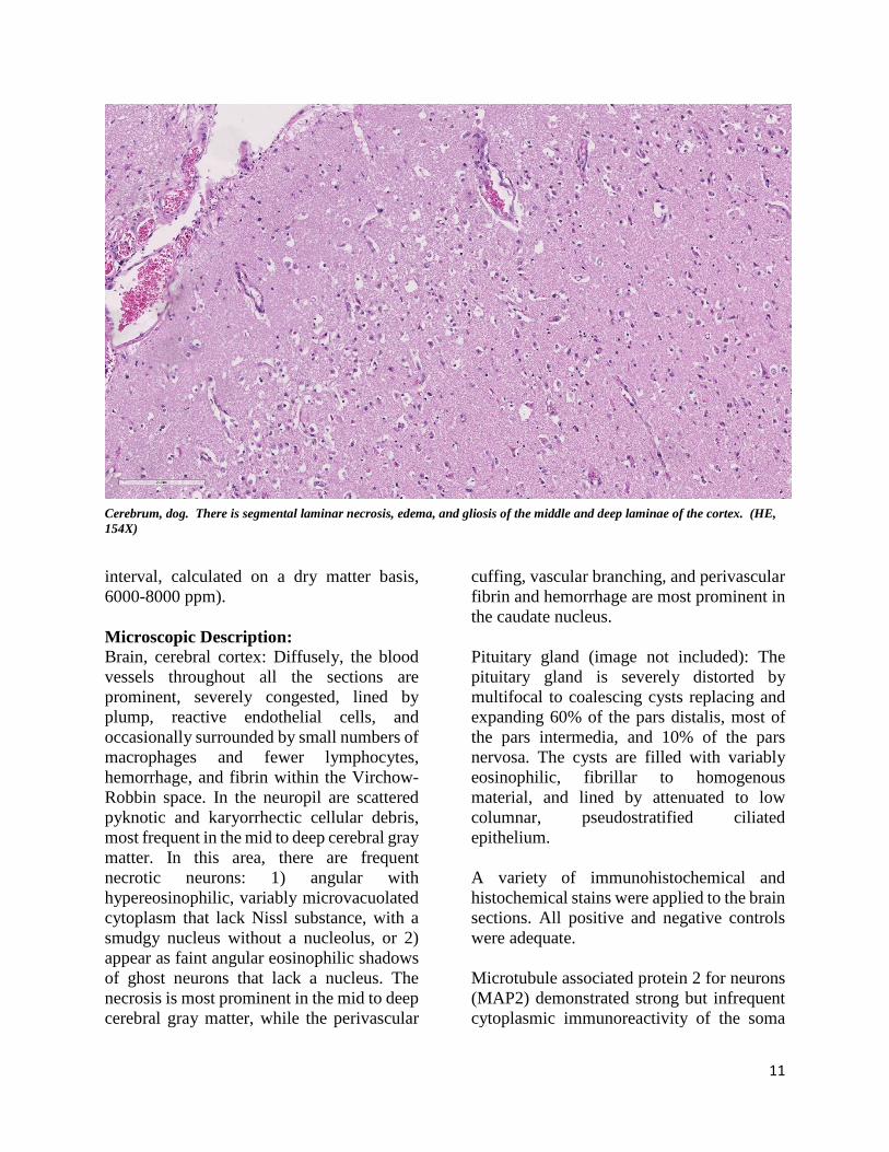
Cerebrum, dog. There is segmental laminar necrosis, edema, and gliosis of the middle and deep laminae of the cortex. (HE, 154X)
12
and moderate immunoreactivity of cytoplasmic processes in neurons of the mid to deep cerebral gray matter indicative of neuronal necrosis. An image of a positive control from a normal dog is included as an inset. In this image, the neuropil exhibits widespread moderate immunoreactivity with moderate to strong immunoreactivity of the neuronal soma and processes. The internal negative control is adequate highlighted by the negative glial cells and blood vessels. Glial fibrillary acidic protein (GFAP) for astrocytes highlights the well-demarcated cortical laminar necrosis affecting the mid to deep gray matter with very few remaining astrocytes in this area.
Olig2 for oligodendrocytes maps the GFAP in the mid to deep gray matter. Ionized calcium binding adaptor molecule 1 (Iba1) for microglia and histiocytes in the affected mid to deep cerebral gray matter revealed three different populations of microglia . The first population is round, ameboid, or nonramified with strong cytoplasmic immunoreactivity interpreted as reactive microglia. The second population is stellate or ramified with strong cytoplasmic immunoreactivity interpreted as quiescent microglia. The third is a background population of small and round microglia and exhibits weak cytoplasmic immunoreactivity and were considered degenerate. Note perivascular mononuclear cells are predominantly macrophages. Contributor’s Morphologic Diagnoses: Brain: Severe, acute, cortical laminar necrosis with reactive microgliosis, reactive endothelium, histiocytic perivascular cuffing, perivascular hemorrhage and fibrin exudation Contributor’s Comment: Hypernatremia is an infrequently encountered condition in dogs caused by decreased body fluid or
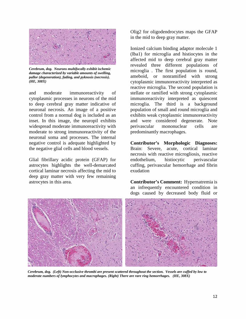
Cerebrum, dog. Neurons multifocally exhibit ischemic damage characterized by variable amounts of swelling, pallor (degeneration), fading, and pyknosis (necrosis). (HE, 308X)
Cerebrum, dog. (Left) Non-occlusive thrombi are present scattered throughout the section. Vessels are cuffed by low to moderate numbers of lymphocytes and macrophages. (Right) There are rare ring hemorrhages. (HE, 308X)
13
sodium gain.14 Causes of hypernatremia include diabetes insipidus, insensible loss such as hyperventilation or fever14, lack of water intake due to hypothalamic defect resulting in a defective thirst response,13 iatrogenic administration of hypertonic saline or sodium bicarbonate or consumption of high salt content material,9 and decreased renal excretion of sodium due to hyperaldosteronism.3
In a recent retrospective study, the three most common causes of severe hypernatremia in dogs were central diabetes insipidus, alimentary loss, and fever/hyperthermia.14 Central diabetes insipidus (CDI) is characterized by diminished activity or lack of the antidiuretic hormone (ADH) due to damage of neuroanatomic sites involved in ADH secretion including the neurohypophysis, hypothalamic osmo-receptors, the supraoptic or para-ventricular nuclei, or the superior portion of the supraopticohypophyseal tract.12 Underlying causes include vascular, autoimmune, infection, drugs, surgery, trauma, benign or
metastatic pituitary-hypothalamic tumors. Patients with a normal thirst mechanism and free access to water generally do not present clinically; however, when water intake is restricted, hypernatremia occurs.1 In this case, the central diabetes insipidus was attributed to craniopharyngeal duct cysts and predisposed this dog to severe hypernatremia. However, given the widespread, severe lesions of the brain, the aggressive fluid therapy likely contributed to the severe histologic lesions.
Clinically, hypernatremia primarily affects the central nervous system and rapid changes in plasma sodium concentrations can cause severe, permanent, and lethal brain injury.2 The pathogenesis of these lesions is due to the failure to retain brain osmolarity. As the plasma osmolarity decreases with fluid therapy when these cases are recognized, the brain osmolarity fails to decrease at a similar rate due to failure of idiogenic osmoles to dissipate leading to cerebral edema.
Salt toxicity and water deprivation and its histologic features have been widely documented in other domestic animals including pigs, poultry, and ruminants.9 Most cases are caused by indirect salt poisoning which occurs when consumption of high levels of salt, followed by water deprivation, and then excessive water consumption occurs.9 Direct salt poisoning is in general rare, but primarily has been reported in cattle.15 The histopathologic lesions shared across these species include cerebral cortical laminar necrosis, perivascular mononuclear cuffing, and cerebral edema.8 Indirect salt poisoning in pigs is a well-known pathologic characterized by eosinophilic meningitis and perivascular eosinophilic cuffing.13
Neuropathologic alterations of hypernatremia in the dog have mostly been extrapolated from human and rodent studies
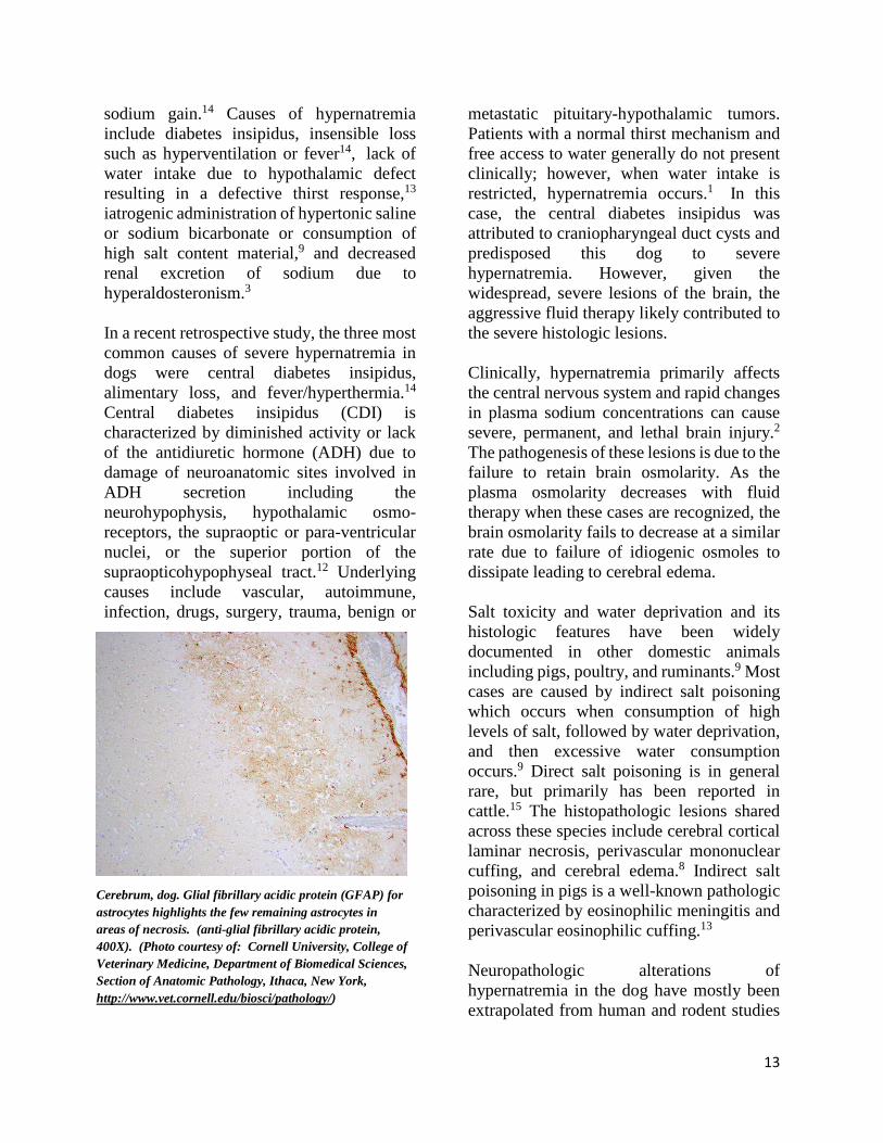
Cerebrum, dog. Glial fibrillary acidic protein (GFAP) for astrocytes highlights the few remaining astrocytes in areas of necrosis. (anti-glial fibrillary acidic protein, 400X). (Photo courtesy of: Cornell University, College of Veterinary Medicine, Department of Biomedical Sciences, Section of Anatomic Pathology, Ithaca, New York, http://www.vet.cornell.edu/biosci/pathology/)
14
with only brief descriptions in the literature. Most of the histologic description in canine cases has been quite variable. Cerebrocortical neuronal necrosis with symmetrical encephalomalacia of the brain stem has been reported in a dog diagnosed with pituitary-dependent hyperadrenocorticism that was euthanized due to hypernatremia 4 weeks post hypophysectomy.9 Neuronal degeneration and astrocytosis of the thalamus and hypothalamus has been reported in a young dog with hypodipsic hypernatremia.4 In a case of acute salt toxicity by ingestion of salt-flour mixture used as clay, diffuse white matter vacuolation with occasional perivascular cuffing by necrotic hyperchromatic mononuclear leukocytes with a thin ring of hemorrhage was observed with no evidence of axonal or neuronal necrosis.6
This case described the range of
histologic changes that can be seen in dogs
with hypernatremic
encephalopathy which includes 1) cortical laminar necrosis, 2) reactive and
necrotic endothelium with
perivascular histiocytic
cuffing, edema, fibrin, and hemorrhage, 3)
reactive microgliosis, 4) white matter
vacuolization, and 5) Purkinje
cell necrosis. In the absence of relevant history, toxic-metabolic encephalopathies or global ischemic encephalopathy where widespread bilateral ischemic damage can occur in anesthetic deaths, cardiac arrest, severe anemia, hypotension, hypovolemic shock, and neonatal maladjustment syndrome may be considered. In this case, the sudden and aggressive ion alterations likely account for the brain lesions and the neurologic signs. In conclusion, severe hypernatremia should be considered as a differential in dogs with cortical laminar necrosis. Contributing Institution: Cornell University, College of Veterinary Medicine, Department of Biomedical Sciences, Section of Anatomic Pathology, Ithaca, New York, USA http://www.vet.cornell.edu/biosci/pathology/
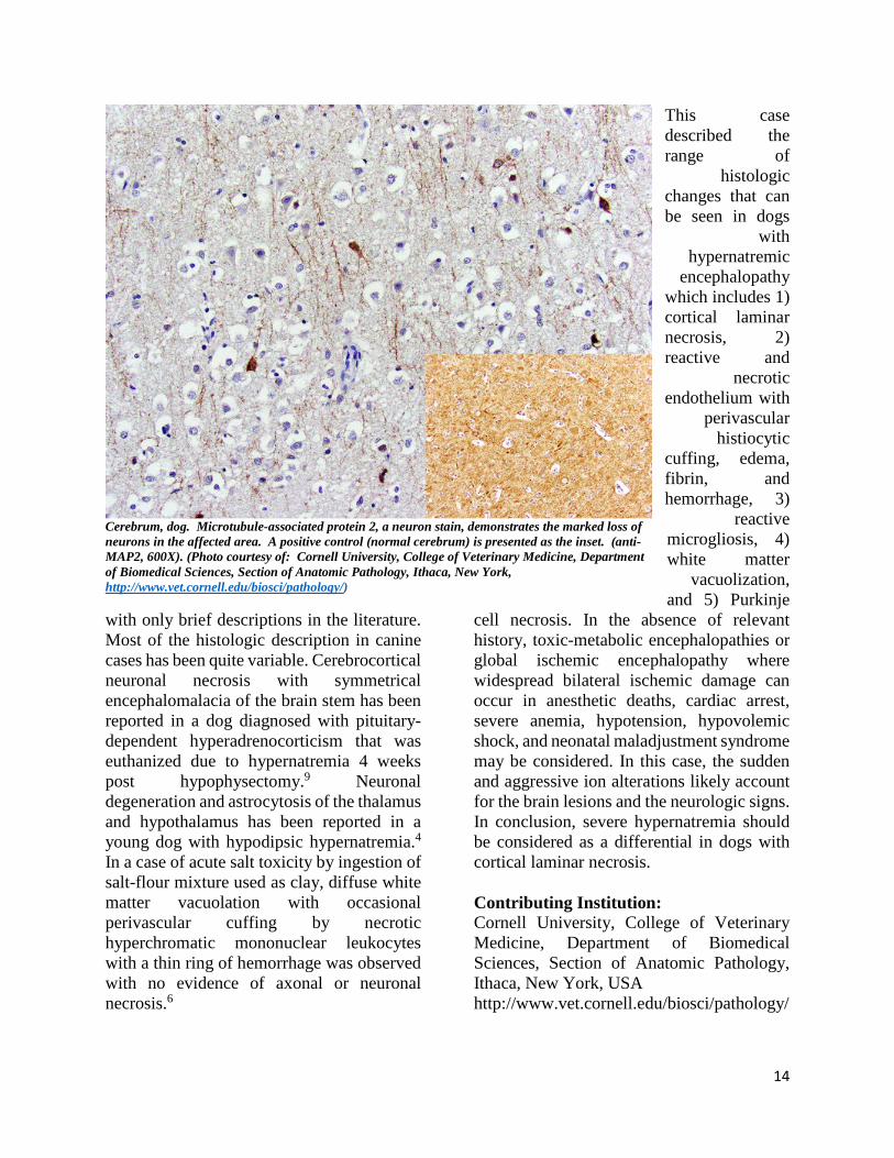
Cerebrum, dog. Microtubule-associated protein 2, a neuron stain, demonstrates the marked loss of neurons in the affected area. A positive control (normal cerebrum) is presented as the inset. (anti-MAP2, 600X). (Photo courtesy of: Cornell University, College of Veterinary Medicine, Department of Biomedical Sciences, Section of Anatomic Pathology, Ithaca, New York, http://www.vet.cornell.edu/biosci/pathology/)
15
JPC Diagnosis: Cerebrum: Necrosis, cortical, laminar and segmental, multifocal to coalescing, with gliosis. JPC Comment: The contributor provides an excellent review of hypernatremia in the dog. To add to the extensive list of differentials for polioencephalomalacia in the dog, other potential causes to be considered in the dog include lead, cyanide,thiamine deficiency, hypoglycemia, canine distemper infection, head trauma, cardiac arrest, and following portosystemic shunt ligation.8 Seizure activity has been documented to result in cortical damage in man and domestic and experimental animals, and the moderator discussed the possibility of this being the root cause of the laminar cortical necrosis in this animal.8
One of the classic symptoms of salt toxicity in affected animal species is blindness. In a previous WSC case of salt toxicity in a Vietnamese potbellied pig (WSC 2012-2013, Conference 21, Case 1), resulting from eating potato chips (this has been reported several times in the literature!), blindness was a long-term sequela following hospitalization and treatment and ultimately resulted in his euthanasia. While a number of changes occur in the brain following the development of hypernatremia and hyperosmolarity and may result in dysfunction of the optic pathway, equally severe and irreparable changes also result in the globe. Following ingestion of diet with 3% salt concentration (the minimum concentration associated with changes in the globe) for 3-4 days, the following ocular changes were noted in study
pigs, rats, and rabbits – conjunctival injection, corneal and lens opacities, swelling of the optic disc, blindness, vitreous collapse, and ultimately progress to phthisis bulbi. Due to
hyperosmolality, fluid loss from the vitreous resulted in vitreous shrinkage resulting in vascular damage, retinal and
vitreous hemorrhage,
posterior vitreal detachment, and retinal detachment.6 This suggests that while many of the CNS signs in salt-intoxicated animals may resolve with successful therapy to
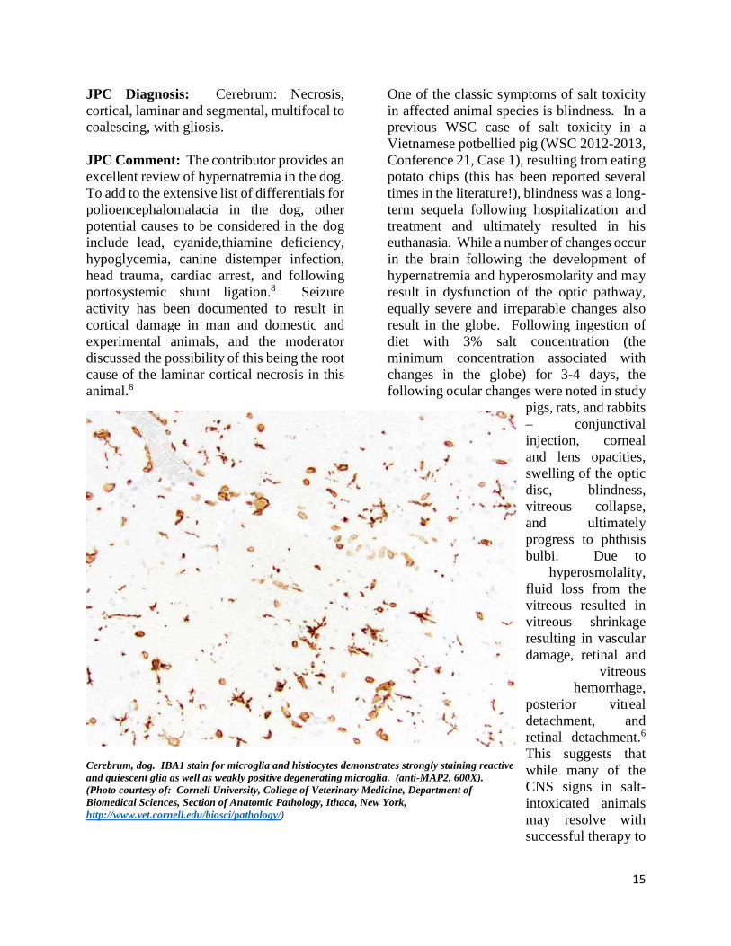
Cerebrum, dog. IBA1 stain for microglia and histiocytes demonstrates strongly staining reactive and quiescent glia as well as weakly positive degenerating microglia. (anti-MAP2, 600X). (Photo courtesy of: Cornell University, College of Veterinary Medicine, Department of Biomedical Sciences, Section of Anatomic Pathology, Ithaca, New York, http://www.vet.cornell.edu/biosci/pathology/)
16
combat hyperosmolality, the changes in the eye may be irreversible and blindness, if present, permanent. In the hospitalized dog and cat, hypernatremia is associated with a significant decrease in prognosis.13 Case fatality rates of dogs and cats with hypernatremia was 20.6 and 28.1% respectively compared to 4.4 and 4.5% with a normal blood or serum sodium concentration. In a review case histories in this study, 50% of the dogs and 38.5% of cats presented with hypernatremia, and the remaining animals developed hospital acquired hypernatremia. References:
1. Adrogue, H.J., and N.E. Madias. 2000. Hypernatremia. N Engl J Med 342: 1493-1499. 2. Ayus, J.C., D.L. Armstrong, and A.I. Arieff. 1996. Effects of hypernatraemia in the central nervous system and its therapy in rats and rabbits. J Physiol 492 ( Pt 1): 243-255. 3. Breitschwerdt, E.B., D.J. Meuten, C.L. Greenfield, L.W. Anson, C.S. Cook, and R.E. Fulghum. 1985. Idiopathic hyperaldosteronism in a dog. J Am Vet Med Assoc 187: 841-845. 4. Crawford, M.A., M.D. Kittleson, and G.D. Fink. 1984. Hypernatremia and adipsia in a dog. J Am Vet Med Assoc 184: 818-821. 5. Greco, D.S. 2012. Pituitary deficiencies. Top Companion Anim Med 27: 2-7. 6. Heydarpour F, Derakhshandeh J, Fekn A, Heydarpour P. Salt poisoning blindness in Wistar rat, rabbit, and pig. Toxicol Environ Chem 2008; 90(5):1035-1042. 7. Khanna, C., H.J. Boermans, and B. Wilcock. 1997. Fatal hypernatremia in a dog from salt ingestion. J Am Anim Hosp Assoc 33: 113-117. 8. Mariana, CL, Platt SR, Newell SM, Terrell SP, Chrisman CL, Clemmons RM. Magnetic resonance imaging of cerebral cortical
encrosis (polioencedcephalomalacia) in a dog. 9. Maxie, M.G., editor. 2015. Jubb, Kennedy, and Palmer's Pathology of Domestic Animals. Elsevier, St. Louis, MO. 314-315 pp. 10. Meij, B.P., G. Voorhout, T.S. van den Ingh, H.A. Hazewinkel, E. Teske, and A. Rijnberk.. Results of transsphenoidal hypophysectomy in 52 dogs with pituitary-dependent hyperadrenocorticism. Vet Surg 1998; 27: 246-261. 11. Qureshi, S., S. Galiveeti, D.G. Bichet, and J. Roth. 2014. Diabetes insipidus: celebrating a century of vasopressin therapy. Endocrinology 155: 4605-4621. 12. Shimokawa Miyama, T., E. Iwamoto, S. Umeki, M. Nakaichi, M. Okuda, and T. Mizuno. 2009. Magnetic resonance imaging and clinical findings in a miniature Schnauzer with hypodipsic hypernatremia. J Vet Med Sci 71: 1387-1391. 13. Smith, D.L. 1957. Poisoning by sodium salt; a cause of eosinophilic meningoencephalitis in swine. Am J Vet Res 18: 825-850. 14. Ueda, Y., K. Hopper, and S.E. Epstein. 2015. Incidence, severity and prognosis associated with hypernatremia in dogs and cats. J Vet Intern Med 29: 794-800. 15. Van Leeuwen, J.A. 1999. Salt poisoning in beef cattle on coastal pasture on Prince Edward Island. Can Vet J 40: 347-348.
CASE IV 16H94455 or blank (JPC 4100730) Signalment: Canine, Golden Retriever, 0.5 years old, male History: This dog was purchased through a breeder at 7 weeks of age. At around 4 months of age, the dog started having uncontrolled watery−mucoid diarrhea that was large volume and frequent. He was placed on Metronidazole and Proviable and
17
needed to stay on the medication as diarrhea would return when the drugs were not used. The dog was diagnosed with carpal flexure deformity at 3 months of age. Radiographs of the cervical spine and hips revealed mild hip dysplasia and shortened distal cervical vertebral bodies. The dog developed urinary accidents (normal volume) at home. On presentation to Iowa State University Teaching Hospital, the dog had lateral strabismus, vertical nystagmus, ataxia that was worse in hindlimbs with normal reflexes and placement and hopping, head tremor, and rectal mucosa felt very irregular on rectal palpation. The owner did not allow many additional tests; only creatine kinase, which was mildly increased at 378, and blood smear which showed possible granules in lymphocytes. The dog’s condition deteriorated over a week and was euthanized. Gross Pathology: Small intestine: A small number (approximately 10) of 2−5 cm in length and 2 mm in diameter, white, elongate nematodes with tapered ends (roundworms)
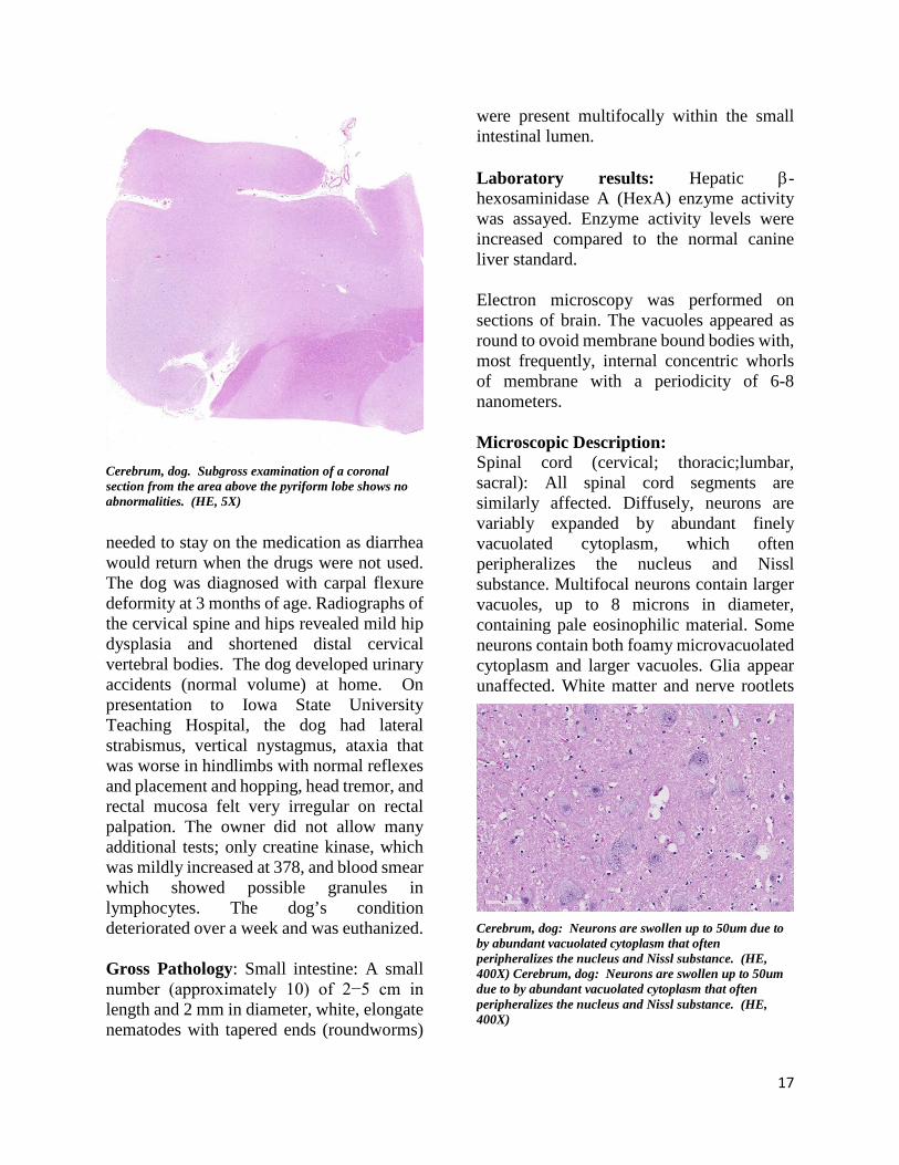
were present multifocally within the small intestinal lumen. Laboratory results: Hepatic β-hexosaminidase A (HexA) enzyme activity was assayed. Enzyme activity levels were increased compared to the normal canine liver standard. Electron microscopy was performed on sections of brain. The vacuoles appeared as round to ovoid membrane bound bodies with, most frequently, internal concentric whorls of membrane with a periodicity of 6-8 nanometers. Microscopic Description: Spinal cord (cervical; thoracic;lumbar, sacral): All spinal cord segments are similarly affected. Diffusely, neurons are variably expanded by abundant finely vacuolated cytoplasm, which often peripheralizes the nucleus and Nissl substance. Multifocal neurons contain larger vacuoles, up to 8 microns in diameter, containing pale eosinophilic material. Some neurons contain both foamy microvacuolated cytoplasm and larger vacuoles. Glia appear unaffected. White matter and nerve rootlets
Cerebrum, dog. Subgross examination of a coronal section from the area above the pyriform lobe shows no abnormalities. (HE, 5X)
Cerebrum, dog: Neurons are swollen up to 50um due to by abundant vacuolated cytoplasm that often peripheralizes the nucleus and Nissl substance. (HE, 400X) Cerebrum, dog: Neurons are swollen up to 50um due to by abundant vacuolated cytoplasm that often peripheralizes the nucleus and Nissl substance. (HE, 400X)
18
contain scattered dilated and often empty axon sheaths. Meninges, blood vessels − no specific pathologic findings. Brain: thalamus, hippocampus; level of caudate nucleus; cerebrum; midbrain; cerebellum; rostral cerebral cortex; pons: diffusely, neurons are moderately to markedly expanded by similar micro-vacuolated cytoplasm, which often peripheralizes the nucleus and occasionally extends into and expands proximal processes. Infrequently, neurons also contain larger vacuoles up to 8 microns in diameter. Microglia are subjectively increased in number. There is rare neuronal necrosis, and rare (two) blood vessels are cuffed by few lymphocytes. White matter is mildly vacuolated with occasional spheroids and few vacuolated cells (small neuron or astrocyte morphology). Neuronal
vacuolation in diencephalon and brainstem nuclei is especially pronounced. The caudate nucleus is regionally strikingly hypercellular with vascular proliferation and gliosis − extant neurons are similarly vacuolated. Purkinje cells are similarly vacuolated; PC density appears normal. Cerebellar nuclei are hypercellular with marked gliosis, similarly vacuolated neurons, and moderate numbers of spheroids in white matter tracts. Meninges, blood vessels − no specific pathologic findings. Eye: Retinal ganglion cells are similarly enlarged and vacuolated with margination of the nucleus. Intestine (duodenum, colon): Myenteric and submucosal plexus neurons occasionally appear enlarged with microvacuolated clear
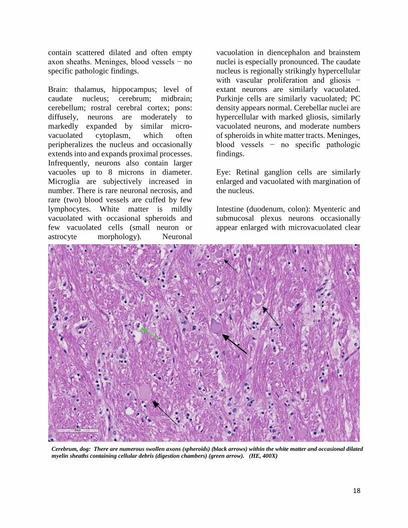
Cerebrum, dog: There are numerous swollen axons (spheroids) (black arrows) within the white matter and occasional dilated myelin sheaths containing cellular debris (digestion chambers) (green arrow). (HE, 400X)
19
to eosinophilic cytoplasm with marginated nuclei. Urinary bladder: Ganglion cells within the bladder wall are similarly enlarged and vacuolated. Liver: Diffusely, hepatocytes contain coarse, coalescing vacuoles with ragged margins. Mesenteric ganglion: Ganglion cells are markedly distended by similar microvaculated cytoplasm as described above. No significant microscopic changes were identified in the following tissues: kidney, colon, skeletal muscle (rare protozoal cysts), ileum, cecum (advanced mucosal autolysis), trachea, sciatic nerve, prostate, testis, tongue, heart, lung (marked congestion with RBC extravasation), spleen (moderate to advanced autolysis), thymus, thyroid gland, adrenal gland, pituitary (pars distalis), mandibular salivary gland. Contributor’s Morphologic Diagnoses: Brain, spinal cord, mesenteric ganglia: Neuronal cytoplasmic vacuolation, diffuse, moderate to severe. Retina: Retinal ganglion cell vacuolation, cytoplasmic, diffuse, moderate. Small intestine: Mild endoparasitism (roundworms) Contributor’s Comment: Lysosomal storage diseases (LSD) are inherited (often autosomal recessive) or acquired (from ingested plant or other toxins) disorders that result in accumulation of macromolecules in lysosomes due to defective catabolism.1,2 Most are due to deficient or inactive hydrolytic enzymes or defects such as inactive proteins, impaired posttranslational
processing, lack of enzyme activator, lack of substrate and lack of transport proteins.3 Types of LSDs of veterinary importance include: Sphingomyelin lipidosis (e.g., GM1 and GM2 gangliosidosis and globoid cell leukodystrophy), glycoproteinoses (alpha and beta mannosidosis, l-fucosidosis), mucopolysaccharidoses (MPSI, MPSII, MPS III, VI, and VII), glycogenoses (alpha 1, 4 glucosidase), and neuronal ceroid lipofuscinoses (infantile, late infantile, juvenile, and adult variants). Microscopic changes in this dog were consistent with a lysosomal storage disease. Vacuolation was predominantly, if not solely, intraneuronal affecting the central and autonomic nervous systems, and retina. Hepatocellular vacuolation was also present, but vacuole morphology appeared more consistent with glycogen than a storage disease enzyme product. Biochemical and/or genetic testing is required for diagnosis of the specific type of LSD. By electron microscopy, vacuoles appeared most commonly as concentric lamellated inclusions, most typical of the gangliosidoses. Restriction of disease to the nervous system in this case makes a GM2-gangliosidosis more likely. GM2 gangliosidoses reported in animals include Tay-Sachs/variant B (caused by HEXA mutations), Sandhoff/variant 0 (HEXB mutations), and GM2 activator deficiency/Variant AB (GM2A mutations). The finding of increased HexA activity in this case is inconsistent with Tay-Sachs and Sandhoff. In the AB variant, hexosaminidase activity is retained, but there is deficiency of an activator protein (GM2A) that is required for degradation of GM2 ganglioside by HexA. The AB variant has been reported in a Japanese Spaniel.5 Additionally, a novel HEXA missense mutation has been recently identified in Japanese Chin dogs that have a
20
phenotype similar to the AB variant.6 DNA was isolated from this dog and is being tested for known LSD mutations. Contributing Institution: https://vetmed.iastate.edu/vpath JPC Diagnosis: Cerebrum: Neuronal and glial vacuolation, diffuse, marked, with gliosis. JPC Comment: GM2 ganglioside is a cell membrane glycolipid which is catabolized by the action of N-acetylyhexosaminidase and GM2 activator (GM2A) protein. N-acetylyhexosaminidase exists as a dimer in two forms, hexosaminidase A and hexosaminidase B. Improper functioning of either one of these subunits, or GM2A, results in accumulation of GM2 gangliosides in the lysosomes of neurons leading to a progressive deterioration of the central
nervous system (GM2 gangliosidosis, monosialoganglioside gangliosidosis). Tay-Sachs disease of humans is a GM2 gangliosidosis caused by a mutation in the HexA gene and deficienct production of the hexosaminidase A dimer, while Sandhoff disease is caused by mutation of the Hex B gene and deficiency of both hexosaminidase A and B dimers.4 GM2 gangliosidosis has been previously described in toy poodles, German shorthaired pointers and Japanese Spaniel dogs, domestic short haired and Korat cats, Jacob sheep, Yorkshire pigs, and Muntjak deer. Three forms of GM2 gangliosidosis are defined based on the age of onset and subsequent disease severity a) infantile (classical), b) juvenile, and c) adult onset (a more heterogeneous progression. The most frequent initial signs are developmental arrest, abnormal startling responses, and low
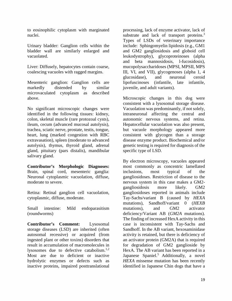
Cerebrum, dog: Microglia also are also expanded by abundant vacuolated cytoplasm (arrows). (HE, 400X)
21
muscle tone, ultimately progressing to seizures, blindness, and coma. Gross lesions in affected animals may include macrocephaly, cortical atrophy, broadening of gyri and shallow sulci. 4 Histologic lesions include neuronal vacuolation with displacement of Nissl substance. As gangliosides figure prominently in synaptic transmission, spheroids may be numerous in white matter, as seen in this case. Cytoplasmic vacuolation may be seen in other cells throughout the body including hepatocytes, leukocytes, renal tubular epithelium, pancreatic acinar cells, and corneal stromal cells (corneal clouding has been identified grossly in Portuguese water dogs and cats). Recent therapeutic studies in treatment of feline GM2-gangliosidosis cats resembling Sandhoff’s disease have shown promise in ameliorating the signs of disease and extending life span. Treatment requires intrathalamic or intraventricular injection of an adenovirus associated virus vehicle encoding both the human HEX A and HEX B genes. Sandhoff-type cats were treated between 4 and 6 weeks of age prior to the onset of clinical signs. Treated cats in this study demonstrated increase Hex activity up to 4-fold, had twice the life-span of untreated cats, and demonstrated up to 90% decreased GM2 ganglioside storage in all CNS regions analyzed.1
The attendees noted that in the submitted sections of slides, it appears that microglia cells are also swollen by vacuolated cytoplasm, which is not inconsistent with a clinical diagnosis of GM2-gangliosidosis, in which vacuolation of perivascular macrophages and glial cells has been described.
References:
1. Bradbury AM, Gurda BL, Casal ML, Ponder KP, Vite CH, Haskins ME: A review of gene therapy in canine and
feline models of lysosomal storage disorders. H Gene Ther Clin Devel 2015; 26:27-37.
2. Cummings JF, Wood PA, Walkley SU, de Lahunta A, DeForest ME: GM2 gangliosidosis in a Japanese Spaniel. Acta Neuropathol, 1985; 67(3):247-253.
3. Haskins ME, Giger U, Patterson DF. Animal models of lysosomal storage diseases: their development and clinical relevance. In: Mehta A, Beck M, Sunder-Plassmann G, editors. Fabry Disease: Perspectives from 5 years of FOS. Oxford: Oxford PharmaGenesis;, 2006
4. Jolly RD, Walkley SU. Lysosomal storage diseases of animals: an essay in comparative pathology. Vet Pathol 1997; 34:527-548
5. Miller AD, Zachary JF. Nervous
system. In: Zachary JF, ed. Pathologic basis of veterinary disease, Elsevier, 2017, pp. 858-861.
6. Sanders DN, Zeng R, Wenger DA, Johnson GS, Johnson GC, Decker JE, Katz ML, Platte SR, O'Brien DP: GM2 gangliosidosis associated with a HEXA missense mutation in Japanese Chin dogs: A potential model for Tay Sachs disease. Molecular Genetics and Metabolism 2013; 108(1):70-75.
Copyright © 2022 FDOKUMEN