D e p e n d e n t N u cle o so m e P o sitio n in g and Chromatin ...
213
Roser Zaurin Quer PhD Thesis - 2009 Sequence-Dependent Nucleosome Positioning and Chromatin Remodelling of Hormone Responsive Genes Roser Zaurin Quer
-
Upload
khangminh22 -
Category
Documents
-
view
3 -
download
0
Transcript of D e p e n d e n t N u cle o so m e P o sitio n in g and Chromatin ...
Ros
er Z
aurin
Que
rP
hD T
hesi
s -
2009
Seq
uenc
e-D
epen
dent
Nuc
leos
ome
Pos
ition
ing
and Chromatin Remodelling of Hormone Responsive Genes
Roser Zaurin Quer

Foto de portada: “L’Americano” Foto de contraportada: “Ceagabeau”
d'Ennio Francavilla


Sequence-Dependent Nucleosome Positioning and Chromatin Remodelling of Hormone-
Responsive Genes
Roser Zaurin Quer


Sequence-Dependent Nucleosome Positioning and Chromatin Remodelling of Hormone-Responsive Genes
Roser Zaurin Quer
Memòria presentada per optar al Grau de Doctora per la Universitat Pompeu Fabra.
Aquesta tesi ha estat realitzada al laboratori de Cromatina i Expressió Gènica del
programa de Regulació Gènica, al Centre de Regulació Genòmica (CRG) de
Barcelona, sota la direcció del Dr. Miguel Beato del Rosal i el Dr. Guillermo Pablo
Vicent, des de setembre de 2004 a juliol de 2009.
Dr. Miguel Beato Dr. Guillermo P. Vicent
Roser Zaurin Quer
Barcelona, juliol de 2009


Table Of Contents
Summary………………………………………………………………………………….11
Resum………………………………………………………………………………….….13
Abbreviations…………………………………………………………………………….15
Introduction
Section 1: The bases of chromatin organization…………………………………...17 1. The nucleosome..……………….…………………………………..…..…......17
1.1 Linker histones
2. The chromatin fiber…………………………………..………………………...20
Section 2: Chromatin modification and gene regulation……………………….….21
1. Histone modifications……………………………………………………...…....21
1.1 Enzymes that covalently modify the core histones
1.1.1 Histone acetyltransferases and Histone deacetylases
1.1.2 Histone methyltransferases and Histone demethylases
2. Incorporation of canonical histone variants and associated functions…....26
2.1 Core histone variants
2.2 Linker histone variants
3. ATP-dependent chromatin remodelling complexes……………………..…..27
3.1 The four classes of remodelling complexes
3.2 Mechanisms of chromatin remodellling
3.3 SWI/SNF-related functions
Section 3: Nucleosome positioning and gene regulation…………………....……34 1. The concept……………………………………………………………………...34
2. A brief historical overview: searching DNA sequence patterns………….…34

3. Recent advances in nucleosome cartography: genome-wide studies.......35
4. Yeast: the best characterized nucleosome occupancy map…………........37
5. Methods to predict nucleosome positions…………………………………...38
Section 4: Progesterone-responsive gene regulation…………………………….39 1. Nuclear receptor……………………………………………………………..…39
2. Steroid hormones (SH) and steroid hormone receptors (SHR)……….…..39
3. Progesterone receptor……………………………………………………...….40
3.1 Structure
3.2 PR effects on gene regulation
3.3 Co-activators
3.4 Co-represors
4. Hormone Responsive Elements (HREs)………………………………..……43
Section 5: MMTV promoter…………………………………….………………..………44
1. Chromatin organization of the MMTV promoter……………………………..44
2. Hormonal induction of the MMTV promoter………………………………….46
2.1 Nuclear factor-1 (NF1)
2.2 MMTV promoter activation process
Materials and Methods............................................................................................51
Objectives…………………………………………………………………….……………77
Results and Discussion
Chapter 1: “Studies on Sequence-Dependent Nucleosome Positioning
and Predictability”
1.1 Results………………………………………………………………….83 1.2 Discussion………………………………………………..……………109
Chapter 2: “Two chromatin remodelling activities cooperate during activation of
hormone responsive promoters”……………..…………...................................115

Chapter 3: “Characterization of the H3/H4 histone tetramer as the output
of the SWI/SNF-dependent remodelling of the nucleosome B within the
MMTV promoter”
3.1 Results…………………………………………………..……….…157
3.2 Discussion……………………………………………………….…179
Conclusions……………………………………………………………………………185
Supplementary articles…………………..……………………………….…….……187
Yang X, Zaurin R, Beato M and Peterson CL. Swi3p controls SWI/SNF assembly and ATP-dependent H2A-H2B displacement. Nat Struct Mol Biol. 2007
Vicent GP, Ballaré C, Zaurin R, Saragüeta P and Beato M. Chromatin remodelling and control of cell proliferation by progestins via cross talk of progesterone receptor with the estrogen receptor and kinase signalling pathways. Ann NY Academic of Science. Review. 2006
References…………………………………………………………………………..…211
Agraïments…………………………………………………………………………..…225


Summary
Evidence has been accumulating over the last few years pointing to the importance of
chromatin structure and nucleosome positioning in cellular processes such as transcriptional
regulation. Recent technological advances in the field have allowed the construction of detailed
genome-scale maps of nucleosome positions, and there have been several attempts to define the
sequence characteristics that guide the positioning of nucleosomes, the so-called “nucleosome code”.
In this thesis I give experimental evidence for the existence of a subset of very well-positioned
nucleosomes in the human genome based on nucleosome-resolution tilling arrays and on deep
sequencing of MNase-digested chromatin using the Solexa-Illumina platform. I show that these
nucleosomes, which we have named “key nucleosomes”, tend to occupy genomic locations of specific
function, indicative of their special role. The DNA of these “key nucleosomes” exhibits a high symmetry
of curvature, allowing their precise position on the human genome to be predicted in silico based on
the structural attributes of the primary DNA sequence.
The second part of this thesis provides new insights into the importance of chromatin
organization and dynamics in the context of gene regulation. Steroid hormones induce transcription of
their target genes by a complex mechanism requiring binding of the hormone receptors to hormone
responsive elements (HREs) and the recruitment of a variety of coregulators. The Mouse Mammary
Tumor Virus (MMTV) promoter has long been used as a model for the study of hormone receptor-
mediated gene activation. It is known that progesterone receptor (PR) binds the exposed HRE1 of the
MMTV promoter chromatin and recruits chromatin remodellers that catalyse ATP-dependent histone
H2A/H2B displacement. I show that the ATP-dependent chromatin remodelling complex BAF, but not
PBAF, is recruited after hormone treatment and is necessary for MMTV promoter activation. Along
with the previously reported phosphorylation of H3S10 by Msk, I show that an early PCAF-mediated
acetylation of H3K14 is essential for the activation of the promoter by anchoring the BAF complex.
Following transient displacement of H2A/H2B dimers, binding of NF1 is required for stabilizing the
11

12
remodelled conformation of the MMTVnucleosome. To further study the activation process I have used
MMTVminichromosomes, mononucleosomes and H3/H4 tetramer particles reconstituted on wild type
MMTV and MMTV promoter fragments with point mutations disrupting binding of PR and NF1. I show
that only when MMTV sequences are assembled on H3/H4 tetramer particles can PR bind to all five
HREs while allowing NF1 access to its cognate site. Furthermore, I found that binding of NF1
facilitates access of PR to the central HREs 2 and 3, thus contributing to the reciprocal synergism
between PR and NF1.

Resum
Evidències recents han remarcat la importància del paper de l’estructura de la
cromatina i el posicionament de nucleosomes en processos cel·lulars bàsics com és la
regulació de la transcripció gènica. L’avenç de noves tecnologies en el camp ha
permès l’estudi detallat de la disposició de nucleosomes en genomes sencers. Hi ha
hagut també molts intents per definir les característiques de la seqüència de l’ADN que
podrien arribar a guiar el posicionament dels nucleosomes; sent el conjunt d’aquestes
característiques l’anomenat “codi nucleosòmic”. En la present tesis doctoral, s’aporten
evidències experimentals sobre l’existència d’un grup de nucleosomes molt ben
posicionats en el genoma humà. Això ha estat possible mitjançant tècniques com els
microarray i la seqüenciació massiva en paral·lel. Aquest treball mostra com aquests
nucleosomes, que anomenem “nucleosomes clau”, tenen tendència a ocupar regions
del genoma amb funcions específiques, la qual cosa indica el seu paper especial.
L’ADN dels “nucleosomes clau” resulta tenir una alta simetria de curvatura. Aquesta
característica inherent a la seqüència fa que sigui possible la predicció in silico de les
posicions de nucleosomes d’aquest tipus.
En la segona part de la present tesi doctoral, aporto noves evidències
experimentals que fan avançar el camp de la organització de la cromatina i la seva
dinàmica en el context de la regulació gènica. Les hormones esteroidees indueixen la
transcripció dels seus gens diana a través de la unió dels receptors hormonals amb els
seu corresponent motiu de reconeixement a l’ADN (HREs), així com el reclutament
d’una gran varietat de co-reguladors. El promotor del Virus de Tumor Mamari de Ratolí
(MMTV) ha estat un model molt usat per l’estudi dels efectes en l’activació gènica dels
receptors hormonals. És conegut que el receptor de progesterona (PR) s’uniex a
l’HRE1, accessible, i recluta maquinàries de remodelament de la cromatina que
utilitzen l’ATP com a font energètica per expulsar els dimers d’histones H2A/H2B del
13

14
nucleosoma B del promotor de l’MMTV. Aquí demostro que la màquinaria de
remodelament reclutada és específicament BAF. Treballs anteriors van demostrar que
la kinasa Msk1 és la responsable de la fosforilació de la serina 10 de la histona H3. En
aquesta tesi es demostra que l’acetilació de la Lysina 14 de la histona H3 és essencial
per l’activació del promotor, així com per l’anclatge de BAF. Després de l’expulsió dels
dimers d’H2A/H2B, la unió d’NF1 al promotor és indispensable per estabilitzar la forma
remodelada del nucleosoma. Per l’estudi en més detall de l’activació del promotor de
l’MMTV he utilitzat el sistema de minicromosomes, mononucleosomes i tetràmers
d’histones H3/H4 reconstituïts en seqüències salvatges i mutants del promotor de
l’MMTV. He demostrat que, només quan un fragment de la seqüencia de l’MMTV està
reconstituïda en tetràmers, el PR i l’NF1 poden estar units simultàniament a la seva
seqüència de reconeixement en el promotor. També aporto evidències on es demostra
que la unió d’NF1 al promotor facilita la posterior unió de més mol·lècules de PR als
HREs interns (llocs 2 i 3) caracterizant en més detall el sinergisme funcional que
existeix entre el PR i l’NF1 en aquestes condicions.

Abbreviations
ACF : ATP-utilizing chromatin assembly and remodelling factor AD: activation Domain ATP : adenosine triphosphate BAF: BRG1/hBRM-Associated Factors BAP: Brahma-associated Protein BP: base pair BRG1 : brahma related gene1 CARM1: Coactivator-associated arginine methyltransferase CTD : C terminal domain CBP : CREB binding protein CD: Chromodomain CHRAC: Chromatin Accessibility Complex Co-Rest: RE-1 silencing transcription factor (REST) co-repressor CENP-A : centromeric protein A Chd1 : chromodomain helicase DNA binding 1 ChIP : chromatin immunoprecipitation DNA : Deoxyribonucleic acid DBD: Dna binding Domain DREX: Drosophila embryo extract ERK: Extracellular-signal regulated kinase Gcn5 : general control nonderepressible 5 GNAT : Gcn5 N-acetyltransferase related GST : glutathione S transferase GR: glucocorticoid receptor HAT : histone acetyltransferase Hat1 : histone acetyltransferase 1 HDAC : histone désacétylase HMT : histone methyltransferase HP1 : heterochromatin protein 1 HMG: High Mobility Group HRE: Hormone Resposnsive Element HRR: Hormone Responsive Region ISWI : imitation SWI/SNF
15
16
LTR: Long Terminal Repeat LXXLL: L, Leu; X, any amino acid MAPK: Mitogen-Activated Protein Kinase MYST : MOZ, Ybf2/Sas3, Sas2 et TIP60 Mi-2: Dermatomyositis-specific autoantigen MMTV: Mouse Mammary Tumor Virus MSK1: Mitogen-and Stress-Activated Specific Kinase NcoR: Nuclear co-repressor NCP: nucleosome core particle NAD : nicotinamide adenine dinucleotide NuA4 : nucleosome acetyltransferase of H4 NuRD : nucleosome remodeling histone deacetylase complex NURF: Nucleosome Remodelling Factor ORF : open reading frame Oct-1: Octamer binding transcription factor PCAF : p300/CBP associated factor PCR : polymerase chain reaction PHD : plant homeodomain PI3K : phosphatidylinositol 3 kinase PIC : pre initiation complex pRb : protein retinoblastoma PRMT1 : proteine arginine methyltransferase 1 PBAF: Polybromo associated-BAF Pol: polymerase PR: progesterone receptor RAR: retinoc aced receptor RNA : Ribonucleic acid RSC : remodel the structure of chromatin SAGA : Spt, Ada, Gcn5 Acetyltransferase SAM : S adenosyl methionine SANT : Swi3, Ada2, N-Cor et TFIIIB SET : Su(var3-9), enhancer of zeste, trithorax Sin3 : swi-independent 3 Sir : silent information regulator protein SNF : sucrose non fermentation SWI : mating type switching Swr1 : Swi2/Snf2-related ATPase 1 TAF : TBP associated factor TBP : TATA box binding protein TFTC : TATA-binding-protein-free TAFII containing complex TIP60 : Tat interacting protein (60kDa) TSA : trichostatin A WT : wild type

Introduction
Section 1: The bases of chromatin organization
1. The nucleosome
Mammals package 3 billion base pairs of DNA, a total of 2 meters of human
DNA, that encode about 30,000 genes into a cell nucleus which average diameter is
approximately 6 micrometers. DNA is packaged using a protein scaffold, forming a
complex tertiary structure that is referred as chromatin. The fundamental structure of
chromatin is the nucleosome, which consists of 146 base pairs of DNA wrapped
around an octamer of two molecules each of the histones H2A, H2B, H3 and H4
(Figure 1). These cells must therefore accomplish the difficult task of folding the
nucleosomes in a highly compact manner while still allowing access to various nuclear
factors. A fifth histone protein, the linker histone or histone H1, has an important
function in the higher structure organization and its stability. The location of H1 in the
nucleosome has been mapped by protein-DNA photo-crosslinking showing that the H1
globular domain forms interaction with the DNA at either the entry or exit strand of the
nucleosomal DNA, but not with the core histones. The unit consisting of a nucleosome
particle plus one H1 molecule was first observed by Simpson et al. (1978). Adjacent
nucleosomes are connected by linker DNA and progressive coiling of nucleosomes
leads to compact, higher-order chromatin structures.
17
INTRODUCTION
18
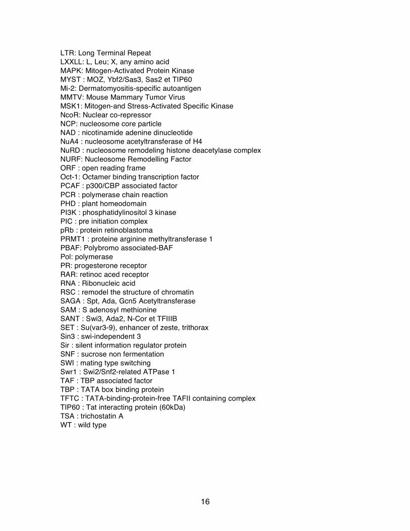
Figure 1. The atomic structure of the nucleosome core particle. Each strand of DNA is shown in differenr shade of blue. The DNA makes 1.7 turns around the histone octamer to form an overall particle with disk-like structure. Histones are colored as Figures 2 and 3. (from Khorasanizadeh, 2004)
Each core histone uses a protein fold, the histone fold, consisting of a three-
helix core domain: a long central α-helix flanked on either side by a loop and a short α-
helix. These domains form handshake arrangements to give rise to the heterodimer
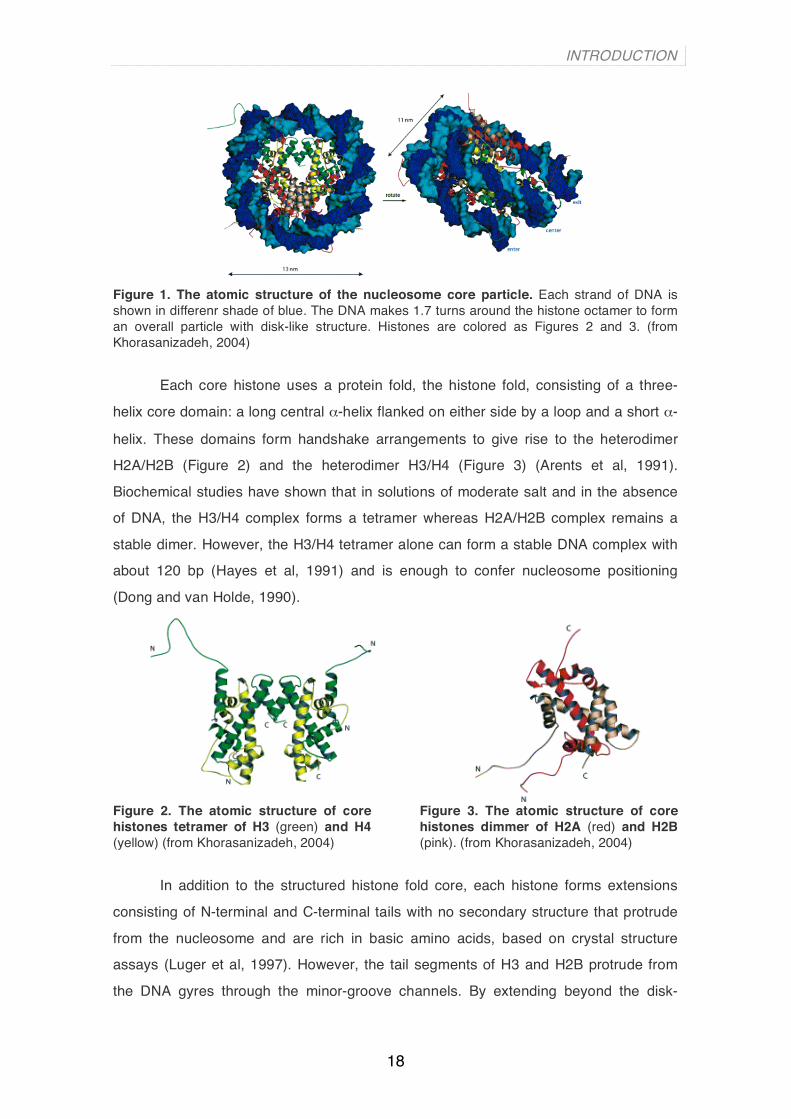
H2A/H2B (Figure 2) and the heterodimer H3/H4 (Figure 3) (Arents et al, 1991).
Biochemical studies have shown that in solutions of moderate salt and in the absence
of DNA, the H3/H4 complex forms a tetramer whereas H2A/H2B complex remains a
stable dimer. However, the H3/H4 tetramer alone can form a stable DNA complex with
about 120 bp (Hayes et al, 1991) and is enough to confer nucleosome positioning
(Dong and van Holde, 1990).
Figure 2. The atomic structure of core histones tetramer of H3 (green) and H4 (yellow) (from Khorasanizadeh, 2004)
Figure 3. The atomic structure of core histones dimmer of H2A (red) and H2B (pink). (from Khorasanizadeh, 2004)
In addition to the structured histone fold core, each histone forms extensions
consisting of N-terminal and C-terminal tails with no secondary structure that protrude
from the nucleosome and are rich in basic amino acids, based on crystal structure
assays (Luger et al, 1997). However, the tail segments of H3 and H2B protrude from
the DNA gyres through the minor-groove channels. By extending beyond the disk-

INTRODUCTION
19
shaped nucleosome surface, the tails form ideal surfaces for covalent modifications by
enzyme machineries.
The DNA is wrapped around the histone octamer so that it forms 1.65 turns of a
left-handed superhelix within the nucleosome core particle (see Figure 1, blue chain is
DNA with different shades of blue for each strand). The helical periodicity around the
nucleosome core is 10.2 base pair as compared to 10.6 base pair for the helical
periodicity of a free B-DNA. This small adjustment between free and nucleosomal DNA
is largely a result of the torsion during wrapping into a superhelix, which also allows the
minor grooves to bind to histones. Approximately 50% of the 120 direct protein-DNA
interactions are formed by hydrogen bonds between the protein main chain amide
groups and the oxygen atoms of the phosphodiester backbone, each time the minor
groove faces the histone octamer (Muthurajan et al, 2003).
1.1 Linker histones
Linker histone does not have a histone fold. The canonical metazoan linker
histone molecule contains three domains: two highly basic and unstructured tails in
solution and a non-polar central globular domain. Whereas the sequence of the central
globular domain is relatively well conserved through evolution in animals, plants and
funghi, the N and C terminal domains are extremely heterogenous, both in length and
amino acid composition. An interesting work has mapped the intereaction surface of
linker histone H1 with the nucleosome of native chromatin in vivo (Brown et al, 2005)
(Figure 4).
Figure 4. Molecular model of the location of H1 within the nucleosome. Blue, chromatosomal DNA; yellow, nucleosome dyad; red, the globular domain of H1. (from Brown et al, 2006)
INTRODUCTION
20
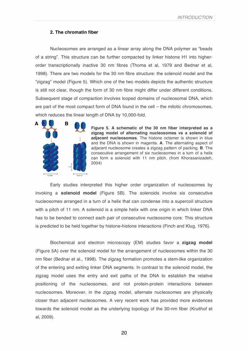
2. The chromatin fiber
Nucleosomes are arranged as a linear array along the DNA polymer as “beads
of a string”. This structure can be further compacted by linker histone H1 into higher-
order transcriptionally inactive 30 nm fibres (Thoma et al, 1979 and Bednar et al,
1998). There are two models for the 30 nm fibre structure: the solenoid model and the
“zigzag” model (Figure 5). Which one of the two models depicts the authentic structure
is still not clear, though the form of 30 nm fibre might differ under different conditions.
Subsequent stage of compaction involves looped domains of nucleosomal DNA, which
are part of the most compact form of DNA found in the cell – the mitotic chromosomes,
which reduces the linear length of DNA by 10,000-fold. Figure 5. A schematic of the 30 nm fiber interpreted as a zigzag model of alternating nucleosomes vs a solenoid of adjacent nucleosomes. The histone octamer is shown in blue and the DNA is shown in magenta. A. The alternating aspect of adjacent nucleosome creates a zigzag pattern of packing. B. The consecutive arrangement of six nucleosomes in a turn of a helix can form a solenoid with 11 nm pitch. (from Khorasanizadeh, 2004)
Early studies interpreted this higher order organization of nucleosomes by
invoking a solenoid model (Figure 5B). The solenoids involve six consecutive
nucleosomes arranged in a turn of a helix that can condense into a supercoil structure
with a pitch of 11 nm. A solenoid is a simple helix with one origin in which linker DNA
has to be bended to connect each pair of consecutive nucleosome core. This structure
is predicted to be held together by histone-histone interactions (Finch and Klug, 1976).
Biochemical and electron microscopy (EM) studies favor a zigzag model
(Figure 5A) over the solenoid model for the arrangement of nucleosomes within the 30
nm fiber (Bednar et al., 1998). The zigzag formation promotes a stem-like organization
of the entering and exiting linker DNA segments. In contrast to the solenoid model, the
zigzag model uses the entry and exit paths of the DNA to establish the relative
positioning of the nucleosomes, and not protein-protein interactions between
nucleosomes. Moreover, in the zigzag model, alternate nucleosomes are physically
closer than adjacent nucleosomes. A very recent work has provided more evidences
towards the solenoid model as the underlying topology of the 30-nm fiber (Kruithof et
al, 2009).
A B

INTRODUCTION
21
Section 2: Chromatin modification and gene regulation The cell has developed mechanisms to modify in a temporal/spacial manner the
chromatin organization and to ensure the maintenance of such an organization through
mitotic and meiotic cell division:
a) Posttranslational covalent modification of histones within a nucleosome
can either facilitate of hinder the accessibility of other transcriptional co-
activators or co-repressors to chromatin.
b) Canonical histones in a nucleosome can be replaced by histone variants
through a DNA-replication independent deposition mechanism.
c) Multisubunit complexes that use the energy of adenosine triphosphate (ATP),
the so-called ATP-dependent remodelling complexes, can twist or slide
nucleosomes exposing or occluding areas to interaction with regulator factors.
d) Methylation at the C-5 position of cytosine residues present in CpG
dinucleotides by DNA methyltransferases (DNMTs) facilitates static long-term
gene silencing. This is achieved through recognition of methyl-cytosine by
specific methyl-DNA binding proteins that recruit transcriptional repressor
complexes and histone modifying activities.
1. Histone modifications
Histone modifications have been associated with regulation of transcription, with
some modifications seemingly correlating with a repressive state of chromatin
(heterochromatin), while others seem to indicate transcriptionally active chromatin
(euchromatin). These modifications can have two mechanistic functions. Certain
histone modifications, such as incorporation of an acetyl group, will change
electrostatic charge of histones’ tails and consequently weaken DNA:histone
interactions. On the other hand, epigenetic marks can function as anchoring sites for
effectors proteins that can regulate transcription. There are at least eight distinct types
of modifications of histones: acetylation, methylation, ribosylation, phosphorylation,
ubiquitylation sumoylation, ADP ribosylation, deimination and proline isomerization

INTRODUCTION
22
(Kouzarides, 2007; Lehnertz et al, 2003). All modifications with target residues as well
as their related functions are shown in Table 1. Most modifications affect the N-terminal
and C-terminal histone tails and few of them the globular domain.
Chromatin Modifications
Residues Modified Functions Regulated
Acetylation K-ac Transcription, Repair, Replication, Condensation
Methylation (lysines) K-me1 K-me2 K-me3 Transcription, Repair Methylation (arginines) R-me1 R-me2a R-me2s Transcription Phosphorylation S-ph T-ph Transcription, Repair Condensation Ubiquitylation K-ub Transcription, Repair Sumoylation K-su Transcription ADP ribosylation E-ar Transcription Deimination R > Cit Transcription Proline Isomerization P-cis > P-trans Transcription Table 1. Covalent modifications of histones. Table shows all known modifications and its related function (From Kouzarides, 2007)
The abundance of modifications on the histone tails makes “crosstalk” between
modifications very likely (Figure 6). Firstly, many different types of modification occur
on lysine residues (see Table 1 second column). This implies that distinct types of
modifications on lysines are mutually exclusive. Secondly, the binding of a protein
could be disrupted by an adjacent modification. The best example of this is the
phosphorylation of H3S10 that affects binding of HP1 to methylated H3K9 (Fischle et
al., 2005). Thirdly, an enzyme could recognize its substrate more effectively in the
context of a second modification; for instance, GCN5 acetyltransferase, recognize H3
more effectively when it is phosphorylated at H3S10 (Clements et al, 2003). This
crosstalk between histone modifications, named the “histone code” hypothesis, was
first postulated by Jenuwein and Allis (2001).
Figure 6. Crosstalk between Histone Modifications: the “histone code”. Some examples of crosstalks between histone posttranslational modifications between residues from the same of different histone tail. The positive influence of one modification over another is shown by an arrow and the negative effect by a dish-line (from Kouzarides, 2007).

INTRODUCTION
23
1.1 Enzymes that covalently modify the core histones
In addition to histones, transcription factors and other chromatin-associated
proteins are themselve substrates of histone-modifying enzymes and are regulated by
these posttranslational modifications in their binding affinity to DNA or interaction
partners, transactivation capacity, protein stability and nuclear localisation (Berger et al,
2001 and Kouzarides et al, 2000).
From all possible histone posttranslational modifications we will focus on
incorporation or removal of acetyl and methyl groups, as well as the histone modifier
enzymes that catalyse these reactions.
1.1.1 Histone acetyltransferases and Histone deacetylases
The best characterized of the histone modifications is acetylation (Marmorstein
et al, 2001 and Kouzarides et al, 1999), which is carried out by two families of
enzymes, the histone acetyltransferases (HATs) and the histone deacetylases
(HDACs). Acetylated histones always associate with transcriptionally active genes,
whereas deacetylation is involved in transcriptional repression and heterochromatin
formation. Consistent with this, many coactivators possess an intrinsic HAT activity.
Histone acetyltransferase are classified into two different classes (Figure 7), based on
their functional localization:
1) Type A-HATs, which are nuclear
2) Type B-HATs, which are cytoplasmic and modify the newly synthesized
histones before the assembly. HAT1 and HAT2 are the members of this
family.

INTRODUCTION
24
Figure 7. Classification of Histone acetyltransferases (HATs) depending on their localization. Type A is nuclear while type B is cytoplasmic.
The other five different classes of type A-HATs are classified based on
structural and functional differences (Grant et al, 1999) (Figure 7). The GNAT family
protein members are structured with a HAT domain of around 160 residues and a
conserved bromodomain, which has been shown to recognize and bind acetyl-lysine
residues. Gcn5 was the first histone acetyltransferase identified. This enzyme acts
predominantly on histone substrates, but also on transcription activator factors (TAFs).
Another member of this family that has a wide array of substrates is p300/CREB-
binding protein (CBP)-associated factor (PCAF). It acetylates histones, nonhistone
proteins like p53, E1A, and various other substrates with diverse physiological effects.
This enzyme is an important transcriptional coactivator (Yang XJ et al, 1996). In both
Gcn5 and PCAF, the N- and C-terminal regions play a direct role in histone substrate
binding. ELP3 is another enzyme belonging to this class and acts only on histone
substrates.
The MYST family of HAT proteins differ from the other groups from the
catalytical mechanism involved in acetylation reaction. Members of the MYST family
are involved in a wide range of regulatory functions including transcriptional activation,
transcriptional silencing, dosage compensation and cell cycle progression.
INTRODUCTION
25
The p300/CBP family is another major group of nuclear HATs that has been
extensively characterized. Members of this family are more global regulators of
transcription; contain a considerably larger HAT domain of about 500 residues, and
other protein domains, including a bromodomain.
Some transcription factors have also been shown to possess
acetyltransferase activity, thereby having a direct effect on transcriptional activation.
The well-known members of this class are ATF2, TAF1, and TFIIIC90. The nuclear
hormone-related HATs are relatively few; representative members are SRC-1, -3 and
-4 and ACTR that act on the histone substrate. These enzymes were identified as
nuclear receptor coactivators and then found to be endowed with histone
acetyltransferase activity.
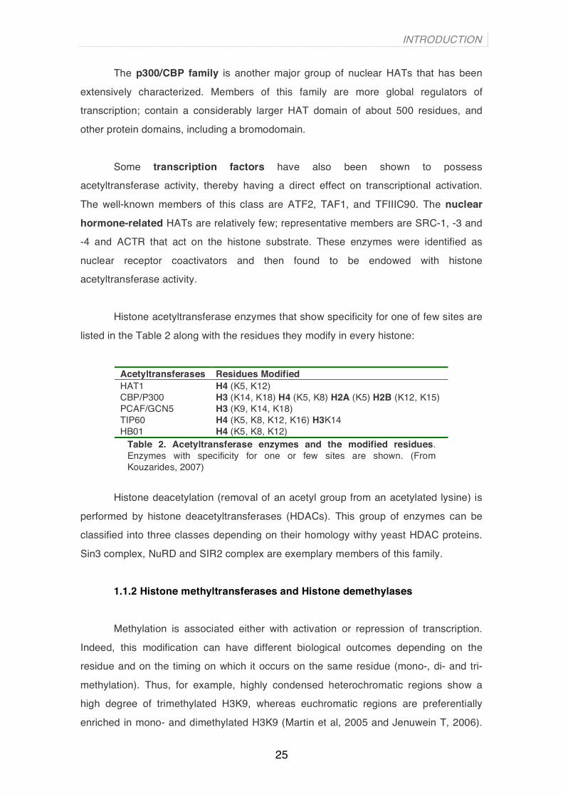
Histone acetyltransferase enzymes that show specificity for one of few sites are
listed in the Table 2 along with the residues they modify in every histone:
Acetyltransferases Residues Modified HAT1 H4 (K5, K12) CBP/P300 H3 (K14, K18) H4 (K5, K8) H2A (K5) H2B (K12, K15) PCAF/GCN5 H3 (K9, K14, K18) TIP60 H4 (K5, K8, K12, K16) H3K14 HB01 H4 (K5, K8, K12)
Table 2. Acetyltransferase enzymes and the modified residues. Enzymes with specificity for one or few sites are shown. (From Kouzarides, 2007)
Histone deacetylation (removal of an acetyl group from an acetylated lysine) is
performed by histone deacetyltransferases (HDACs). This group of enzymes can be
classified into three classes depending on their homology withy yeast HDAC proteins.
Sin3 complex, NuRD and SIR2 complex are exemplary members of this family.
1.1.2 Histone methyltransferases and Histone demethylases
Methylation is associated either with activation or repression of transcription.
Indeed, this modification can have different biological outcomes depending on the
residue and on the timing on which it occurs on the same residue (mono-, di- and tri-
methylation). Thus, for example, highly condensed heterochromatic regions show a
high degree of trimethylated H3K9, whereas euchromatic regions are preferentially
enriched in mono- and dimethylated H3K9 (Martin et al, 2005 and Jenuwein T, 2006).
INTRODUCTION
26
Histones are methylated at arginine and lysine residues. Conversely to HATs, histone
methyltransferases (HMTs) are quite specific enzymes that usually modify one single
residue on a specific histone. Unlike histone acetylation, histone methylation does not
change nucleosome charge, but usually it can be recognized by effector proteins
displaying chromo-like domains.
Histone lysine methylation is mediated by HMTs, many of which contain a
conserved SET [Su(var)3-9, Enhancer-of-zeste, Trithorax] domain, such as Suv39h1
(Suppressor of variegation 39h1) and G9a (Kouzarides T, 2007; Lachner et al, 2004;
and Schneider et al, 2002).
Histone demethylases catalyze the removal of methyl groups from histone
lysines and arginines. Since the half-life of histones and of their methylated lysines is
identical, lysine methylation has been considered for years to be an irreversible mark
(Byvoet et al, 1972, Duerre and Lee, 1974). However, recent progress in the field led to
the identification of two enzyme-families capable of catalysing this reaction, the LSD-1
family and the jumonji-family.
2. Incorporation of canonical histone variants and associated functions
2.1 Core histone variants
In higher organisms, each histone is represented by a family of genes encoding
multiple non-allelic primary-sequence variants. It is thought that histone variants serve
two main purposes in the cell. First, the histone-exchange removes epigenetic marks
on core histones and facilitates reprogramming of the particular gene. Second, their
incorporation permits effectuation of various functions. Figure 8 shows all histone
variants and the functions to which they are associated to that exist in mammalian
cells. The most studied histone variants are the ones of histones H3 and H2A. These
include H3.3, which functions in transcriptional activation (Ahmad and Henikoff, 2002)
and CENPA - centromeric histone H3 (Sullivan et al., 1994), H2A.Z, which plays a role
in gene expression and chromosome segregation (Allis et al., 1986; Rangasamy et al.,
2004), H2AX, which acts in DNA repair and recombination (Rogakou et al., 1998; de la
Barre et al., 2001), macro H2A – transcriptional repression and X chromosome

INTRODUCTION
27
inactivation (Costanzi and Pehrson, 1998; Angelov et al., 2003), and H2ABBD possibly
functioning in transcriptional activation (Bao et al., 2004; Gautier et al., 2004).
Figure 8. Synopsis of core histone and linker histone variants in human cells (From Lindner, 2008)
2.2 Linker histone variants
In superior eukaryote there are multiple isoforms or subtypes of linker histone
H1 that are important for cell growth and proliferation (for review see, Khochbin, 2001).
In mammals, six somatic variants (designated H1.0, H1.1, H1.2, H1.3, H1.4 and H1.5),
a male germ line-specific subtype (H1t) and an oocyte-specific subtype (H1oo) have
been identified (Panyim and Chakley, 1969; Bucci et al, 1982; Lennox et al, 1984;
Tanaka et al, 2001) (Figure 8). The variants differ in timing of expression (Khochbin et
al, 2001), extent of phosphorylation (Lennox et al, 1982), turnover rate (Hall et al, 1985;
Dominguez et al, 1992), binding affinity (Th’ng et al, 2005), and evolutionary stability
(Ponte et al, 1998). Recently, our lab has published an extensive work on H1 variants
in T47D breast cancer cells in which H1.2 and H1.4 depletion specifically caused arrest
of cell proliferation (Sancho et al. 2008) In particular, H1.2 caused decreased in global
nucleosome spacing and repressed expression of a number of cell cycle genes,
supporting the idea that distinct roles exist for the linker histone variants (Sancho et al,
2008).
3. ATP-dependent chromatin remodelling complexes
This type of chromatin remodelling complexes uses the energy of ATP to
perform nucleosome alterations. The ATP-dependent chromatin remodelling
complexes comprise a central helicase-related, ATP-consuming subunit, with various

INTRODUCTION
28
numbers of associated subunits that modulate and target its activity. In humans, these
complexes are divided into four classes depending on the structural domains contained
in the ATPase protein subunit. The four family proteins are (Figure 9):
a) SWI2/SNF2 family, containing a bromodomain.
b) ISWI family, containing a SANT domain.
c) CHD family, containing a cromodomain and a DNA-binding motif.
d) INO80 and SWR1 families, without any additional domain.
Figure 9. ATPases of the four main families of ATP-dependent Swi2/Snf2-related chromatin remodelling complexes: SWI/SNF, ISWI, CHD (or Mi-2) and INO80. Each family is defined by the presence of a distinct ATPase containing signature structural domains and a unique subunit composition. In addition, an extended split in their ATPase domain characterizes the INO80 and SWR1 class.
(From Mohrmann and Verrijzer, 2005)
These ATPases that define subfamilies are present in multicomponent
complexes, having each of them numerous subunits (typical of SWI2/SNF2 subfamily)
or few (two or four in ISWI subfamily).
3.1 The four classes of remodelling complexes
SWI2/SNF2 family The components of the SWI/SNF chromatin-remodelling complex were initially
identified in yeast in screens for genes that regulate mating-type switching (SWI) and
sucrose non-fermenting (SNF) phenotypes (Carlson et al., 1981; Neigeborn and
Carlson, 1984, 1987; Stern et al., 1984; Abrams et al., 1986; Nasmyth and Shore, 1987
and Carlson and Laurent, 1994). It was recognized that a subset of the SWI genes are
identical to those identified in the SNF screen, and those genes that are involved both
in mating-type switching and sucrose fermentation have come to be known as
SWI/SNF genes (Peterson et al., 1994 and Wolffe, 1994). The ySWI/SNF remodelling
complex is a multiprotein complex with a central ATPase subunit, known as Swi2/Snf2.
Homologs of SWI/SNF complexes were found in Drosophila and humans.

INTRODUCTION
29
The mammalian SWI/SNF complexes have two mutually exclusive ATPase
subunits, named BRM and BRG1 (these are ortholog subunit of the yeast Swi2/Snf2),
and 8–10 subunits, which are referred to as BRM- or BRG1-associated factors or BAFs
(Wang et al., 1996). Table 3 lists all of the well-characterized mammalian subunits of
the SWI/SNF complex and shows their non-vertebrate orthologs, the Drosophila BAPs
and yeast SWI/SNF gene family members. The yeast SWI/SNF complex exhibits an
apparent molecular mass of 1.14 MDa (Smith et al., 2003), whereas the mammalian
SWI/SNF complex has an apparent molecular mass of approximately 2 MDa.
Mammalian BAF proteins are conventionally identified by their molecular size; hence
BAF47 refers to a BRG1-associated protein with an apparent molecular mass of 47
kDa. Human BRG1 is approximately 74% identical to human BRM (Khavari et al.,
1993), 52% identical to Drosophila BRM and 33% identical to yeast Swi2/Snf2 (Fry and
Peterson, 2001).
Table 3. Relationship between SWI2/SNF2-subfamily chromatin remodelling complexes from yeast, Drosophila and mammals. Conserved subunit are listed horizontally with the ATPase subunit listed first. SWI/SNF (Switching defective/Sucrose nonfermenting); RSC (Remodel the Structure of Chromatin); BAP (BRM-Associated Proteins); PBAP (Polybromo-associated BAP); BAF (BRG1/hBRM-Associated Factors; PBAF (Polybromo-associated BAF). Distinction between ySWI/SNF and RSC has been conserved throughout eukaryotic evolution. ySWI/SNF class remdodelers are referred as BAP and BAF, whereas PBAP and PBAF are RSC orthologues (From Mohrmann and Verrijzer, 2005)
INTRODUCTION
30
Individual SWI/SNF complex contains either BRM or BRG1, but not both (Wang
et al., 1996), such that BRM/BAF complexes are structurally distinct from BRG1/BAF
complexes (see Table 3). The extent to which these complexes are functionally distinct
is a topic of active investigation. The BRG1-containing complexes are further divided
into those that contain the BAF250 (or OSA protein) or the BAF180 protein (Wang,
2003). BAF180 is the mammalian ortholog of Drosophila polybromo, and the BAF180-
containing SWI/SNF complex has been designated as PBAF, to distinguish it from the
BAF250-containing BRM/BAF and BRG1/BAF complexes (Table 3).
PBAF complex is thus defined by the presence of BAF180 and BAF200
whereas BAF complex is exclusively containing the BAF 250 subunit. BAF180
(polybromo, PB1, PBRM1)-containing SWI/SNF complexes have different properties
from those that contain BAF250 subunits (Nie et al., 2000; Lemon et al., 2001 and
Wang et al., 2004). BAF180 harbors a distinctive set of structural motifs, characteristic
of three components of RSC (Xue et al., 2000 and Table). It also contains a Bromo
domain that binds to acetylated histones. BAF200 is also believed to function as part of
the PBAF complex, similar to BAF180 (Yan et al., 2005). BAF200, like BAF250, is a
member of the ARID gene family and is encoded by ARID2.
ISWI subfamily
The ISWI (Imitation of SWI) based complexes were initially purified from
Drosophila. Genetic studies in this organism suggest that ISWI based complexes can
activate gene expression in both in vitro and in vivo (Dirscherl and Krebs, 2004). In vitro, most of the ISWI-based complexes have nucleosome spacing activity that results
in the formation of regulatory ordered nucleosome arrays. These properties have been
used to assemble chromatin in vitro using an ATP-utilizing chromatin assembly and
remodelling factor (ACF) and a histone chaperone (such as NAP-1 or CAF-1) (Ito et al,
1997). In vivo it is thought that this ability is related with the chromatin assembly
following DNA replication and chromosome organization (Corona and Tamkun, 2004).
CHD subfamily
Mi-2/NuRD complexes are highly heterogeneous in subunit composition but
they all contain Mi-2 as the core ATPase. The complexes also contain HDACs,
methylated DNA binding proteins (MBDs), histone H4-interacting proteins
INTRODUCTION
31
(Retinoblastoma associated proteins PbAp 46/48) and members of metastasis-
associated protein gene family (MTAs). Because both HDACs and methylated DNA are
usually associated with gene repression and silencing, these complexes have been
involved in transcriptional repression (Bowen et al, 2004).
INO80 and SRW1 They are the ATPase of distinct multisubunit complexes firstly found in yeast.
INO80 complexes are involved in transcriptional activation and DNA damage repair
(Shen et al, 2004). They also catalyze ATP-dependent sliding of nucleosomes along
DNA. Recently it has been also characterized the human complex (Jin et al, 2005). The
SWR1-based complex is responsible for the incorporation of the histone H2A.Z variant
(Htz1 in yeast) into nucleosomes in a replication-independent manner (Mizuguchi et al,
2004). A human complex with many similarities has being discovered (Cai et al, 2005).
3.2 Mechanisms of chromatin remodelling
The energy for SWI/SNF-mediated chromatin remodelling is transduced by the
catalytic subunit, BRM or BRG1, both of which have DNA-dependent ATPase activity
(Muchardt and Yaniv, 1999 and Hassan et al., 2001). The function of SWI/SNF
complex has been thoroughly studied in vitro. It was shown to be capable of several
biochemical activities that lead to chromatin disruption events (Figure 10). It causes
mobilization of histone core in cis (on the same DNA molecule), called nucleosome
sliding (Schnitzler et al, 1998, Whitehouse et al, 1999 and Schnitzler et al, 2001).
Sliding is nucleosome translocation in which DNA sequences previously occluded by
core histones are exposed in internucleosomal regions. SWI/SNF complex also
transfer histone octamer in trans to other DNA molecules (Lorch et al, 1999), cause
formation of altered dinucleosome particles (Lorch et al, 1998 and Peterson, 2000),
generation of negative superhelical torsion on linear DNA and nucleosomal templates
(Havas et al, 2000) and partial or total displacement of core histones (Boeger et al,
2003; Bruno et al, 2003; Reinke and Horz, 2003; Vicent et al, 2004). Biochemical
evidence and recent atomic force microscopy studies indicate that SWI/SNF
remodelling involves both sliding and disruption of histone-DNA interactions. Despite
extensive study, the mechanism by which SWI/SNF complexes remodel nucleosomes
is not well understood.

INTRODUCTION
32
Figure 10. Schematic model depicting different modes of ATP-dependent chromatin remodelling. ATP-dependent chromatin remodelling complexes use the energy derived from ATP hydrolysis to alter histone-DNA contacts in such a way that (A) nucleosomes slide to another position, (B) a remodelled state is created in which the DNA becomes more accessible but histones remain bound, (C) DNA and histones dissociate completely, or (D) histone (variant) replacement. (From Mohrmann and Verrijzer, 2005).
These perturbations increase susceptibility of nucleosomal DNA to DNaseI and
restriction endonucleases (Coté et al, 1998; Imbalzano et al, 1994 and 1996), change
the crosslinking pattern between histones and nucleosomal DNA (Lee et al, 1999 and
Sengupta et al, 2001) and alter the number of DNA super coils constrained by
nucleosomes (Kwon et al, 1994; Bazett-Jones et al, 1999 and Jaskelioff et al, 2000).
3.3 SWI/SNF-related functions
SWI/SNF is a regulator of gene expression. In mammalian cells, SWI/SNF has
been linked to a large number of transcription factors. The oncogenic transcription
heterodimer activated protein-1 (AP-1) is known to be SWI/SNF dependent (Ito et al.,
2001); similarly, EKLF, which regulates beta-hemogloblin synthesis, also requires this
complex for its function (Armstrong et al., 1998 and Lee et al., 1999). All known steroid
receptors are functionally linked to SWI/SNF (Yoshinaga et al., 1992; Sumi-Ichinose et
al., 1997; Fryer and Archer, 1998; Belandia et al., 2002 and Vicent et al, 2004). Hence,
SWI/SNF does not regulate an exclusive signalling pathway; instead, it serves as a
fundamental component of various essential, and often unrelated, pathways.
INTRODUCTION
33
SWI/SNF activity is essential for differentiation in yeasts, flies and mammals.
Moreover, SWI/SNF factors are also involved in malignant transformation, an
association most clearly demonstrated with the SWI/SNF subunit BAF47. The
strongest evidence for the role of BAF47 in cancer development comes from studies on
rhabdoid tumors showing that one BAF47 allele is consistently deleted, and the other
allele is either mutated or silenced by methylation (Versteege et al., 1998; Rousseau-
Merck et al., 1999; Sevenet et al., 1999; Biegel et al., 2000; Biegel and Pollack, 2004).
Other than transcription regulation, SWI/SNF has been linked to different cellular
mechanisms such as alternative splicing, with a complex that associates Brm with
Sam68 (Batsché et al, 2006) and repression of transcription interacting with the methyl-
CpG binding protein MeCP2 (Harikrishnan et al, 2005).

INTRODUCTION
34
Section 3: Nucleosome positioning and gene regulation
1. The concept
Condensation of the genomic material into chromatin is carried out in an
effective and precise way that allows all major cellular processes to take place.
Furthermore, it has been shown that the effective packaging of DNA into nucleosomes
can actively take part into the regulation of transcription, allowing the access of
transcription factors at specific sites, while protecting others from being transcribed.
Thus, the positioning of nucleosomes, their translocation or disruption may play a
crucial role in gene expression and regulation.
2. A brief historical overview: searching DNA sequence patterns
Nucleosome positioning was first addressed as a structural more than a
functional issue. First works done on the field were based on the alignment of 147 bp
DNA sequence of hundreds of well-positioned nucleosomes and the identification of
particular base pair combination commons in all of them. Early in the 90’s, a number of
works defined sequence prerequisites for nucleosome positioning (or exclusion),
although a definite “nucleosomal signature” needs still to be described. Early
characterisation of nucleosomal DNA revealed several nucleosome positioning signals:
an abundant of AA/TT dinucleotides separated by roughly 10 bp (Drew HR et al, 1985),
five repeated (A/T)3NN(G/C)3NN motifs embedded in a DNA fragment, the so-called TG
pentamer, and fragments having repeated TATAAACGCC motifs, referred to as TATA
tetrads (Widlund HR et al, 1997). More extended periodicities, have also been noted.
The trinucleotide pattern VWG (V = non-T, W = A or T) was observed to follow a ~11bp
period in nucleosomal sequences (Baldi et al., 1996; Stein and Bina, 1999). As
dinucleotide periodicity studies became more detailed due to the increase of available
sequences (Widom et al, 1996) a more consistent periodical pattern was revealed,
according to which two consecutive purines (RR) were to be observed with a period of
~10.5 counter-phase with a corresponding occurrence of two consecutive pyrimidines
(YY) with the same period (Kogan and Trifonov, 2005). Finally, periodically occurring
triplets were correlated to gene expression of yeast mini-chromosomes early in 2002
by Tomita and colleagues suggesting a link between chromatin structure and
transcription.
INTRODUCTION
35
3. Recent advances in nucleosome cartography: genome-wide studies
The first genome-scale identification of nucleosome positions was done for the
whole chromosome 3 of Saccharomyces cerevisiae using the tilling microarray
technique (Yuan et al, 2005). Based on statistical calculations, this study revealed that
between 65 and 69% of the yeast DNA may be organized in well-positioned
nucleosomes. Recently, the densities of printed probes on microarrays have increased
dramatically, allowing millions of genomic loci to be interrogated by ChIP-chip analysis
in a single experiment. The first two complete genomic maps of positioned
nucleosomes were published for Lee et al (2007) and Mavrich et al (2008, Nature) for
yeast and Drosophila genomes, respectively. However, a new genome-scale
techniques based on massively parallel sequencing was developed to study more
precisely nucleosome positioning in complete genomes. The first such high-resolution
genome-wide ChIP-Seq nucleosome map was achieved for nucleosomes containing
H2A.Z in S.cerevisiae (Albert et al, 2007). Nucleosome maps of a similar resolution in
yeast (Whitehouse et al, 2007 and Shivaswamy et al, 2008), worm (Valouev et al,
2008) and humans (Ozsolak et al, 2007, Schones et al, 2008 and Barski et al, 2007)
have now been published, helping to understand how nucleosome positions are exactly
determined in vivo.
From this and other works on the field, we have learned some points that help to clarify
the difficult goal of understanding nucleosome positioning:
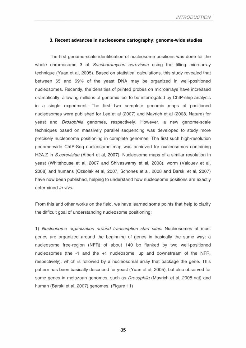
1) Nucleosome organization around transcription start sites. Nucleosomes at most
genes are organized around the beginning of genes in basically the same way: a
nucleosome free-region (NFR) of about 140 bp flanked by two well-positioned
nucleosomes (the -1 and the +1 nucleosome, up and downstream of the NFR,
respectively), which is followed by a nucleosomal array that package the gene. This
pattern has been basically described for yeast (Yuan et al, 2005), but also observed for
some genes in metazoan genomes, such as Drosophila (Mavrich et al, 2008-nat) and
human (Barski et al, 2007) genomes. (Figure 11)
INTRODUCTION
36
Figure 11. Nucleosome-free region around the transcription start sites in yeast genes. Nuclesomes -1 and +1 flancking the Nucleosome-Free Region (NFR) are coloured in blue. The arrow represents the TSS. (From Jiang et al, 2009).
2) Nucleosome spacing thoughout the genome. There have been observed differences
among the species in distance between adjacent nucleosome midpoints. S. cerevisiae
has approximately 165 bp distance between adjacent nucleosomes (that is a linker
DNA of ∼18 bp) (Lee et al, 2007, Mavrich et al 2008-gen research and Shivaswamy et
al, 2008); for D. melanogaster (Mavrich et al, 2007) and C. elegans (Valouev et al,
2008) there is a 175 bp distance in average (that is linkers of ∼28 bp); and for humans
(Barski et al, 2007 and Schones et al, 2008) it has been described a 185 bp distance
between neighbour nucleosomes (and it means a linker DNA of ∼38 bp). This
differences in linker length may be explain by absence and presence of linker histone
H1 and other remodelling factors that have species-specific DNA length requirements
for binding, as well as increasing complexity of genomes through evolution.
And some other general conclusions such as:
1) Nucleosomes positions are highly dynamic. (Mellor et al, 2005). Hogan et al. (2006)
have reported cell cycle-specific fluctuation of nucleosome occupancy at gene
promoters and Shivaswamy et al. (2008) have identified changes in individual
nucleosome positions before and after subjecting cells to heat shock.
2) There exist other factors that in specific locations can contribute to nucleosome
positioning (Saha et al, 2006, Whitehouse et al, 2007, Pechham et al, 2007 and
Whitehouse et al, 2006), indicating that no only the DNA sequence is acting as a
positional constraint in the organization of nucleosomes. A well example is the work
done by Whitehouse and colleagues (2007). They have shown that the ATP-dependent
chromatin remodelling complex Isw2 repositions nucleosomes onto promoter regions
that are intrinsically designed to exclude them. This Isw2-dependent nucleosome
repositioning enforces directionality on transcription by preventing transcription
initiation from cryptic sites.
INTRODUCTION
37
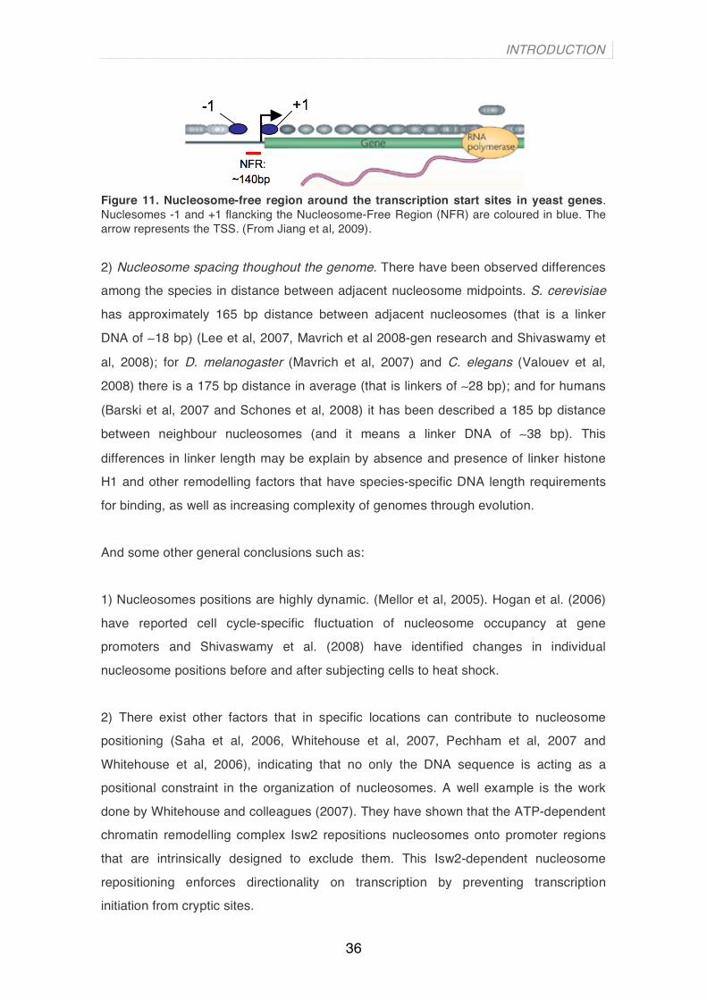
4. Yeast: the best characterized nucleosome occupancy map
Probably due to its low genomic complexity and reduced size, yeast has been
the best well-characterized genome in terms of nucleosome organization. What we
have learned from yeast works is summarized in Figure 12. Firstly, nucleosomes are
well organized around the transcription start sites (TSS) and transcription termination
sites (TTS) of genes. Secondly, there is a nucleosome free region (NFR) right
upstream of the TSS and downstream of TTS. Nucleosome -1 is located approximately
between nucleotide position -150 and -300 and it is very dynamic and usually suffers
one or more of the following modifications: histone replacement, acetylation,
methylation, translational repositioning and finally completely eviction. All these
modifications facilitate the pre-initiation complex (PIC) formation and allow transcription
to take place. After the NFR we found the nucleosome +1. It is usually defined by the
presence of histone variants such as histone H2A.Z and H3.3 (Malik et al, 2003), as
well as by methylation and acetylation of residues in its histone tails (Kouzarides, 2007
and Li et al, 2007). This nucleosome is most likely evicted when transcription takes
place but rapidly return to its location after the Pol II has passed. Downstream of the
nucleosome +1, we find nucleosome +2 and +3 that mostly share the same
characteristics of the nucleosome +1 (content histone variants and histone acetylation
and/or methylation) but in a less consistent way (Li et al, 2007 and Lieb et al, 2005).
Constraints for nucleosome positioning disappear as we go inside the coding region of
the gene. In contrast, nucleosome organization around the NFR of the TTS is not yet
as well known as it is in the TSS.
Figure 12. General roles for nucleosome organization in yeast genes. Nucleosomes are represented as yellow ovals. Numbers inside nucleosomes correspond to the consensus nucleosome numbering respect the NFR (Nucleosome-Free Region). Transcription start site (TTS) is marked with a blue arrow, while transcription termination site (TTS) is represented with a line and a small circle in red. The probability to find acetylation and H3K4 methylation as well as some histone variants is shown in different intensities of blue and dark red, respectively. Less intensity of these colours means less probability to find these histone marks and variants on consecutives nucleosomes (for instance, nucleosome+3 displays less histone variant H2A.Z than nucleosome +2).

INTRODUCTION
38
5. Methods to predict nucleosome positions
Many studies have attempted to predict computationally nucleosome positioning
de novo in different organisms based on properties of the DNA, since Segal et al.
published in 2006 a nucleosome-DNA interaction model that highlight that ∼50% of the
nucleosomal positioning is governed by the sequence. Briefly, they used the AA/TT/TA
dinucleotide frequency patterns obtained from a set of 199 positioned
mononucleosomes to construct a probabilistic model, representative of the DNA
sequence preferences of nucleosomes in Saccharomyces cerevisiae. A following study
(Ioshikhes et al, 2006) used comparative analysis of six Saccharomyces species
combined with the frequencies of AA/TT dinucleotides to observe conserved patterns
of nucleosome positioning among yeast species, pointing out the existence of
sequence constraints in nucleosomal positions. Both of the above studies identified
positions of NFRs in the vicinity of promoter and gene-upstream regions, in agreement
with experimentally defined ones (Yuan et al, 2005). A more recent method (Peckham
et al, 2007) made use of the original raw experimental data of Yuan et al. (2005) to
develop a method that distinguish nucleosome occupancy from depletion based on the
GC content of the sequence, which constitutes a very general attribute to be assigned
as a nucleosome-formation pattern. All these studies that have used DNA sequence
features to predict genome-wide nucleosome positions (Yuan et al, 2008, Ioshikhes et
al, 2006, Segal et al, 2006, Miele et al, 2008, Field et al, 2008, Pechham et al, 2007),
confirm that nucleosome positioning is partially encoded in the genomic DNA
sequence. The most accurate method that makes de novo predictions of nucleosome
positions till now is probably Gupta et al. (2008).
INTRODUCTION
39
Section 4: Progesterone-responsive gene regulation
1. Nuclear receptors
Nuclear receptors (NR) act as ligand inducible transcription factors by directly
interacting as monomers, homodimers or heterodimers with DNA response elements of
target genes, as well as by cross-talking to other signalling pathways. The effects of
NR on transcription are mediated through interaction with other transcription factors
and through recruitment of coregulators. The nuclear receptor superfamily is typically
subdivided into three families:
1) The steroid receptor family,
2) the thyroid/retinoid family, that includes thyroid receptor (TR), vitamin D
receptor (VDR), retinoic acid receptor (RAR), and peroxisime proliferators-
activated receptor (PPAR) and,
3) the orphan receptor family, defined by a set of proteins identified by
comparative sequence analysis as belonging to the nuclear receptor
superfamily, but which ligands are unknown.
NR can be also grouped into a large superfamily depending on the way in which they
bind to DNA, as homodimers (ER, GR, PR, AR and MR), as heterodimers (RAR, RXR,
TR, VDR and EcR) or monomers (SF1) (Dilworth and Chambon, 2001).
2. Steroid hormones (SH) and steroid hormone receptors (SHR)
Hormones are chemical signalling molecules produced by endocrine glands of
vertebrates and other organisms. Their functions vary depending on the tissue or cell
type. Several hormones and their receptors have been shown to play an important role
in numerous cellular functions, including metabolic regulation, immune system
responses, cell growth and development. One of the four classes of hormones is
steroid hormones, which are small cholesterol-derived hydrophobic molecules that can
traverse the cell membrane and activate their specific steroid hormone receptors.
Mammalian steroid hormones, produced by gonads and adrenal glands, can be
grouped into five groups by the receptors to which they bind: glucocorticoids,
mineralocorticoids, androgens, estrogens, and progestagens. It is through these
receptors that the steroid hormones exert their regulatory effects in a wide variety of
biological processes including reproduction, differentiation, development, cell
INTRODUCTION
40
proliferation, apoptosis, inflammation, metabolism, homeostasis and brain function
(Mangelsdorf et al, 1995). Unliganted SHRs are assossiated with a large multiprotein
complex of chaperons, including heat shock protein 90 (Hsp 90) and immunophilin
Hsp56.
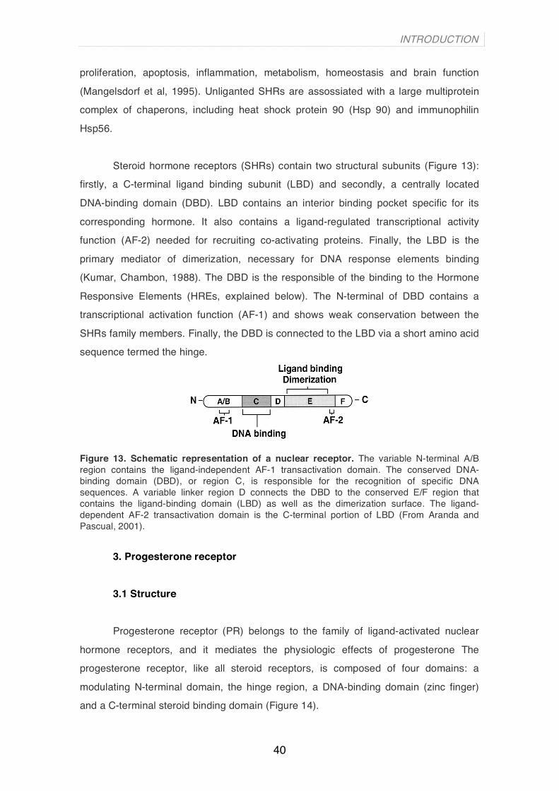
Steroid hormone receptors (SHRs) contain two structural subunits (Figure 13):
firstly, a C-terminal ligand binding subunit (LBD) and secondly, a centrally located
DNA-binding domain (DBD). LBD contains an interior binding pocket specific for its
corresponding hormone. It also contains a ligand-regulated transcriptional activity
function (AF-2) needed for recruiting co-activating proteins. Finally, the LBD is the
primary mediator of dimerization, necessary for DNA response elements binding
(Kumar, Chambon, 1988). The DBD is the responsible of the binding to the Hormone
Responsive Elements (HREs, explained below). The N-terminal of DBD contains a
transcriptional activation function (AF-1) and shows weak conservation between the
SHRs family members. Finally, the DBD is connected to the LBD via a short amino acid
sequence termed the hinge.
Figure 13. Schematic representation of a nuclear receptor. The variable N-terminal A/B region contains the ligand-independent AF-1 transactivation domain. The conserved DNA-binding domain (DBD), or region C, is responsible for the recognition of specific DNA sequences. A variable linker region D connects the DBD to the conserved E/F region that contains the ligand-binding domain (LBD) as well as the dimerization surface. The ligand-dependent AF-2 transactivation domain is the C-terminal portion of LBD (From Aranda and Pascual, 2001).
3. Progesterone receptor 3.1 Structure
Progesterone receptor (PR) belongs to the family of ligand-activated nuclear
hormone receptors, and it mediates the physiologic effects of progesterone The
progesterone receptor, like all steroid receptors, is composed of four domains: a
modulating N-terminal domain, the hinge region, a DNA-binding domain (zinc finger)
and a C-terminal steroid binding domain (Figure 14).

INTRODUCTION
41
Figure 14. Structure of the progesterone receptor (PRA and PRB are the two isoforms). The numbers refer to amino acid positions. Arrows indicate the amico acid initation of PR isoforms. AF-3: activation function 3; IF: inhibition function; AF-1Activation Function1; DBD: DNA binding domain; H: hinge, short amino acid sequence that connects DBD with LBD; LBD AF-2: comprise ligand binding domain and activation function2.
The gene encoding the PR protein contains separate promoters and
translational start sites to produce two isoforms – PRA and PRB, that differ in their
molecular weight (additional 164 amino acids present only in the N terminus of PRB)
(Figure 14). Although PRA and PRB isoforms show high degree of sequence identity,
they display significant different functional properties on the regulation of target
promoter. PRB is a much stronger transcriptional activator than PRA (Sartorius et al,
1994). PRA gene knock-out mice develop uterine dysplasia and abnormal ovaries,
whereas PRB gene knock-out have affected the mammary glands, causing incomplete
lobular-alveolar differentiation (Mulac-Jericevic et al, 2000). Moreover, microarray
studies showed that the two isoforms regulate different subset of genes (Richer et al,
2002).
3.2 PR effects on gene regulation
Progesterone receptor is a sequence-specific transcription factor. Upon binding
of hormone to its carboxy terminal domain it undergoes homodimerization. The
activated receptor plays a role in control of gene activation by either 1. direct binding to
hormone responsive elements (HREs) in regulated genes (pathway known as
genomic or direct effects) or 2. induction of kinase cascades activated by
cytoplasmic events (pathway known as non-genomic or signalling-mediated effects).
The genomic pathway implicates direct binding to HREs in regulatory regions,
where it recruits modifiers which remodel the nucleosomes and these in turn recruit
other co-regulators. The critical interactions with chromatin and its complement of
associated proteins are therefore crucial for progesterone receptor action. The non-

INTRODUCTION
42
genomic pathway often occurs via second messenger cascades, which in turn originate
from signalling complexes located at membranes. For instance, estrogens activate the
Src/Ras/Erk and PI3K/Akt pathways via direct interaction of ER with c-Src and the
regulatory subunit of PI3K, respectively (Castoria et al, 2001 and Migliaccio et al,
1996). Progesterone receptor also cross-talk to kinases cascades through an
interaction of PR with SH3 domain of c-Src (Ballaré et al, 2003 and
Boonyaratanakornkit et al, 2001). The targets of the activated kinases cascades might
be transcription factors and co-regulators involved in DNA synthesis and cell
proliferation.
3.3 Co-activators
Co-activators recruited by ligand-bound nuclear receptors include chromatin
remodelling complexes, such as SWI/SNF, as well as members of the steroid receptor
coactivators family (SRC) (Beato, Klug 2000), which serve as adaptors to mediate
interactions with histone acetyltransferases (HATs) (Torchia et al, 1997 and Voegel et
al, 1998). SRC family consists of three members: SRC-1 (or NCoA-1), SRC-2 (or
NCoA-2, GRIP-1 or TIF-2) and SRC-3. C-terminal domains of SRC-1 and -3 contain
histone acetyltransferase (HAT) activity although it is much weaker than those in CBP,
p300 or PCAF enzymes (Spencer et al, 1997). SRC pre-existing complex with CBP,
p300, PCAF, CARM-1 and PRMT-1 are recruited to chromatin by ligand-triggered
interaction between SHRs and SRCs (Li et al, 2003).
In addition to co-activators that interact directly with transcriptional activation
domains of SHRs, proteins that modulate sequence specific DNA binding SHRs have
been identify. The best characterized of these factors are the nuclear high mobility
group of proteins HMG-1 and related HMG-2, that enhance the affinity of the SHRs for
their specific target HREs. HMG-1 and -2 in mammalian cells enhance the hormone-
dependent transcriptional activity of several different SHRs including PR, AR, GR and
ER, contributing to the stability of the receptor-DNA complex (Onate et al, 1994).
3.4 Co-repressors
Several groups have discovered that antagonists of ER and PR promote
receptor association with the co-repressor NCoR (Nuclear receptor co-repressor) and
INTRODUCTION
43
SMRT (silencer mediator or retinoc and thyroid receptor) (Wagner et al, 1998). NCoR
and SMRT are multiprotein complexes that exhibit deacetylase activity (HDAC),
indicating that targeted deacetylation of core histones is necessary for the silencing
activity of these proteins. Thus, NCoR and SMRT have opposite effect on chromatin
structure to HATs and repress access of general transcription factors to DNA.
4. Hormone Responsive Elements (HREs)
Hormone Responsive Elements were initially described as glucocorticoid
responsive elements (GREs) (Karin et al, 1984 and Scheidereit et al 1983). They have
been classically described in the 5’ regulatory region of a given gene but recent
evidences show HREs can be located in coding regions (exons and introns) and
intergenic regions. HREs are generally composed of two hexameric core motifs. Two
such consensus motifs have been identified (AGAACA and AGG/TTCA), which are
recognized by different members of the nuclear receptors superfamily, though usually
HREs can show significant variations from the consensus. Steroid hormone receptors
including glucocorticoids, mineralocorticoids, progesterone and androgens typically
bind to palindromes of AGAACA sequences separated by three non-conserved base
pairs (Beato, 1989), with the exception of estrogen receptor, which recognizes
AGGTCA motif. Modification of location of the half-sites relative to one another
contributes to diversity of HREs. For dimeric HREs, the half-sites can be configured as
palindromes, inverted palindromes or direct repeats. HREs additionally vary in a
number of neutral base pairs separating the half-site repeats (Zhao et al, 2000). It is
the separating sequence that contributes to the binding specificity of different
heterodimer molecules. It also provides the geometry that is needed for two subunits to
interact specifically. The DNA nucleotide sequence and its specific packaging into
chromatin determine the interaction of hormone regulatory sites with their
correspondent SHRs (Beato and Klug, 2000). A number of studied promoters contain
HREs in their DNA sequences. One of the most studied ones are TAT promoter
(Rahman et al, 1995), 11βHSD promoter, Gluco/P4, metallothionein promoter, genital
HPV promoter, E2, PS2 and MMTV promoter. All of the mentioned promoters are
organized into positioned nucleosomes.

INTRODUCTION
44
Section 5: MMTV promoter The promoter of the mouse mammary tumor virus (MMTV) provirus is probably the
best characterized example of transcriptional control by steroid hormones in which the
chromatin organization plays an important role (Richard-Foy and Hager, 1987).
1. Chromatin organization of the MMTV promoter
The mouse mammary tumour virus (MMTV) is a B-type retrovirus belonging to
the genus beta-retroviruses. Discovered in 1936, it was found to induce
adenocarcinomas and T-cell lymphomas in mice through a process called insertional
mutagenesis, in which a part of the viral genome is inserted within or near an
oncogene. There are two routes of transmission of the virus - exogenous MMTV is
transmitted vertically via milk, whereas endogenous is inherited through the germ line.
The genome of the MMTV is 8805bp in size and contains the retroviral structural genes
and an additional gene sag coding for a superantigen. The MMTV long terminal repeat
(LTR) is responsive to steroid hormones, thus it can infect a variety of cells but it is
tumourigenic only in mammary epithelial cells. Some studies have shown that following
integration of the exogenous MMTV, the mouse int, wnt and fgf genes are activated
(Clausse et al., 1993; Durgam and Tekmal, 1994; Uyttendaele et al., 1996).
The MMTV promoter, located in the long terminal repeat (LTR) of the provirus,
is induced by glucocorticoids and progestins. The hormone responsive region
comprises five binding sites for the hormone receptors (Hormone Responsive
Elements, HREs), a binding site for NF1, and two binding site for the Octamer
transcription factor 1, Oct-1 (Truss and Beato, 1993). The MMTV promoter is organized
into six positioned nucleosomes (Richard-Foy and Hager, 1987), named from A to F,
with a nucleosome (nucleosome B) covering all the HREs, the binding site for NF1 and
for Oct-1 (Figure 15).

INTRODUCTION
45
Figure 15. Chromatin organization of the LTR where the MMTV is located. HRE: Hormone Resposnive Element; NF1: Nuclear Factor-1; Oct-1: Octamer binding factor 1. (From Vicent et al, 2006)
In vivo setting of the nucleosomes in MMTV promoter regulatory region is
similar to that obtained in vitro (Piña et al., 1990). Through in vitro nucleosome
reconstitution experiments precise translational and rotational positioning of the MMTV
promoter sequence on the surface of histone octamers was revealed. In the
experiments for analysis of the chromatin structure of the non-induced MMTV promoter
in vivo it was shown the nucleosome B covering the regulatory region extends from –
210 to –32 in agreement with the reconstitution experiments in vitro (Piña et al., 1990).
Glucocorticoid or progesterone treatment renders the nucleosome B region of MMTV
hypersensitive to endonucleases, which is characteristic of remodelled, or “active”
chromatin regions (Lee and Archer, 1994).

INTRODUCTION
46
2. Hormonal induction of the MMTV promoter
2.1 Nuclear factor-1 (NF1)
Nuclear factor 1 (NF1) protein family is a group of sequence-specific
transcription factors, required for the expression of many cellular and viral genes.
Human NF1 was first isolated from HeLa cells, where it was found to function as a
replication factor for adenovirus replication (Nagata et al, 1983). The family is derived
from alternative splicing of transcripts of four genes – NF1-A, NF1-B, NF1-C and NF1-
X, which are highly conserved from chickens to humans. NF1 proteins share a highly
conserved N-terminal domain, which is a DNA binding domain. They can bind to DNA
either as homodimers or heterodimers, recognizing a partially palindromic consensus
DNA sequence TGGA/C(N5)GCCA (de Vries et al, 1987). NF1 binding sites occur in a
variety of tissue-specific and development-related genes, as well as in the regulatory
regions of several viruses. The carboxy terminal is highly variable within the family and
this implicates that NF1 proteins have diverse functions.
2.2 MMTV promoter activation process
The provirus integrated in the host cell chromatin is virtually silent in the
absence of hormones but responds with rapid transcriptional activation to the addition
of either glucocorticoids or progestins. The receptors for these hormones bind to a
cluster of HREs and facilitate the interaction of other transcription factors with their
target sites locate between the HREs and the TATA box. This results in a synergistic
activation of transcription by the hormone receptors and Nuclear Factor 1 (NF1) (Di
Croce et al., 1999) as well as the octamer transcription factor, Oct1/OTF1
(Bruggemeier et al., 1991) (for a review see (Beato et al., 1995)). How these factors
synergize is a question that has attracted considerable attention, but the mechanism is
not simply cooperative binding of the various proteins to the MMTV promoter
(Bruggemeier et al., 1990).
When introduced in Saccharomyces cerevisiae engineered to express GR or
PR and NF1, the MMTV promoter is silent in the absence of hormone, is organized into
positioned nucleosomes, responds poorly to NF1 or to a NF1-VP16 fusion, but can be
induced by hormone treatment (Chavez et al., 1995). Deletion of the HREs disturbs

INTRODUCTION
47
nucleosome positioning and makes the promoter responsive to NF1-VP16, as if the
HREs would repress access to the NFI site by positioning a nucleosome (Candau et
al., 1996). Hormone induction was believed to cause a displacement of the nucleosome
over the promoter, thus allowing free access of NF1 to its binding site and
transcriptional activation (Richard-Foy and Hager, 1987). Moreover, in a cell-free
system synergistic binding of receptors and NF1 to the MMTV promoters depends on
its previous assembly into minichromosomes with positioned nucleosomes and on
preincubation in the presence of ATP (Di Croce et al., 1999). The nature of the
hormone induced nucleosomal change that permits simultaneous binding of
transcription factors to the MMTV promoter remains obscure. Depletion of histone H1
from the MMTV promoter could be a possibility (Bresnick et al., 1992) and a role for
histone H1 phosphorylation in modulating MMTV activation has been postulated (Lee
and Archer, 1998). However, the MMTV promoter is regulated in budding yeast which
lacks linker histones (Chavez et al., 1995) and also in minicromosomes assembled in
the absence of histone H1 (Di Croce et al., 1999). Alternative possibilities are
recruitment by the receptors of chromatin remodelling activities, either ATP-dependent,
such as the SWI/SNF complex (Cote et al., 1994), or ATP-independent, such histone
acetyl transferases (HATs) (Lee et al., 1993). This later possibility has received
considerable attention following the discovery that several steroid receptors
coactivators exhibit HAT activity, including members of the SRC-1 family, P/CAF and
p300/CBP (Brown et al., 2000).
The role of histone acetylation on hormone induction of the MMTV promoter is
not yet clear. High doses of histone deacetylases inhibitors, butyrate or TSA, lead to
intense hiperacetylation of core histones and inhibit hormone induction (Bartsch et al.,
1996; Bresnick et al., 1990) without altering nucleosome positioning (Bresnick et al.,
1991). However, low dosis of the inhibitors activate the MMTV promoter both in the
absence and in the presence of hormone (Bartsch et al., 1996). The extend of the
hormonal induction is not affected by derepressing concentrations of the inhibitors of
histone deacetylases (Bartsch et al., 1996). Similar results are obtained with these
inhibitors in the cell-free transcription assay with MMTV minichromosomes, suggesting
that inhibitor-sensitive acetylation is not involved in mediating the synergism between
receptors and NF1.

INTRODUCTION
48
There is evidence for a requirement of the SWI/SNF complex in glucocorticoid
gene regulation in yeast (Yoshinaga et al., 1992) and in animal cells (Muchardt and
Yaniv, 1993), and hSWI/SNF seems to be required for chromatin remodelling initiated
by glucocorticoids (Fryer and Archer, 1998). The situation appears to be different for
progestins, which act through the same HREs of the MMTV promoter (Fryer and
Archer, 1998). We have reproduced the synergism between SHR and NF1 in a cell-free
system driven by MMTV minichromosomes assembled in extracts from Drosophila
embryos (Di Croce et al., 1999). These minichromosomes exhibit nucleosomes
positioned in the same way as in cultured cells and the NF1 site in the promoter is not
accessible (Venditti et al., 1998). Addition of hormone activated GR or PR leads to an
ATP-dependent remodeling of these minichromosomes, as demonstrated by
topoisomer analysis (Di Croce et al., 1999), and to simultaneous binding of receptors
and NF1. This activity can be partly reproduced with purified MMTV minichromosomes
with recombinant dISWI, but not with purified dCHRAC or ySWI/SNF complex (Di
Croce et al., 1999). Moreover, incubation of the minichromosomes in the Drosophila
extract in the presence of receptors and NF1 leads to recruitment from dISWI, and
dNURF30, two subunits of the NURF complex (Gdula et al., 1998). Thus, in the
Drosophila embryo extract, PR recruits NURF to the MMTV promoter chromatin, and
NURF remodels the minichromosomes in an ATP-dependent fashion.
However, in mammalian cells with a single MMTV promoter integrated in
chromatin we have recently found the hormone induced recruitment to the promoter of
both Brg1 and ISWI containing complexes (Vicent et al., 2004). Moreover in
experiments with MMTV mononucleosomes assembled in vitro, PR is able to recruit
purified yeast Swi/Snf to the promoter and it catalyzes the ATP-dependent
displacement of H2A/H2B dimmers (Vicent et al., 2004). Though we can not formally
exclude a replacement of the H2A/H2B dimers by dimers containing histone variants,
our results strongly suggest that the activated MMTV nucleosome containing the bound
SHRs and NF1 is a an H3/H4 tetramer particle.

INTRODUCTION
49
Figure16. Model of the crosstalk between Erk and Msk kinases and PR molecules upon hormone induction. (from Vicent et al, 2006)
We have investigated the function of the activation of Erk by progesterone,
which is occurs via an interaction between PR and ERα (Migliaccio et al., 1998) (Figure
16). Recently we observed that progestin activation of the MMTV promoter can be
blocked by inhibiting Erk activation either with antiestrogens, with small molecular
kinase inhibitors (PD 98059), with dominant negative kinase mutant, or with RNAi
against Erk (Vicent et al., 2006). As PR is phosphorylated by Erk at ser293 in response
to progesterone, it is possible that the transcriptional inhibition of MMTV induction was
due to the lack of PR phosphrylation. However, a similar inhibition of MMTV induction
was observed when the activation of Msk1 was prevented by small molecular inhibitors
(H89), a dominant negative mutant of Msk, or down-regulation of Msk1 with an specific
RNAi (Vicent et al., 2006). Unexpectely we detect a ternary complex of phosphorylated
PR and activated Erk and Msk1 already 5 minutes after hormone treatment and this
complex is selectively recruited to the MMTV promoter nucleosome containing the
HREs (Vicent et al., 2006). Concomitant with the recruitment of the ternary complex,
histone H3 becomes phosphorylated at serine 10 and acetylated at lysine 14, only over

INTRODUCTION
50
the nucleosome containing the HREs and not in adjacent nucleosomes (Vicent et al.,
2006). The results with the MMTV promoter underline the significance of incorporating
a precise knowledge of the structure of the native DNA sequences in chromatin into
any attempt to understand the interactions of various transcription factors with their
target sequences and with each other responsible for the physiological regulation of
individual genes.

Materials and Methods General Molecular Biology Protocols
Small-scale and Large-scale preparation of plasmid DNA
To isolate plasmid DNA from bacterial culture the large number of methods, including
treatment of nonionic and ionic detergents, organic solvents, alkali, or heat can be
employed. Both for mini- and maxi-scale DNA preparation a NucleoBond kit (BD
Biosciences) were used. It employs a modified alkaline/SDS lysis procedure, potassium
acetate (KAc) for precipitate formation and NucleoBond column which contains silica-
based anion exchange resin. The procedure is as follows. First the bacterial culture is
spinned down in a 4 °C centrifuge (4500 rpm for 4 min. for mini-prep, 6000g for 15 min. for
maxi.prep). Then, the cells are resuspended in Buffer S1 (50 mM Tris-HCl, 10 mM EDTA,
100 µg/mL RNase A) and lysed when Buffer S2 is added (200 mM NaOH, 1% SDS). The
lysate is neutralized upon addition of Buffer S3 (2.8 M Kac, pH 5.1) and incubation for 20
min. on ice. In the case of mini-prep after this step the phenol/chloroform extraction and
ethanol precipitation was performed as already described before. For maxi-prep the
protocol from NucleoBond was followed, using the AX 500 column to bind, wash and elute
51

MATERIALS AND METHODS
52
the plasmid DNA. Precipitation was performed with isopropanol and subsequent
centrifugation at 16000g for 30 min. The DNA pellet was washed with 70% cold ethanol,
air-dried and resuspended in 400 µL of ddH2O.
Transformation of DH5α competent cells
Bacterial transformation is the process by which bacterial cells take up naked DNA
molecules. If the foreign DNA has an origin of replication recognized by the host cell DNA
polymerases, the bacteria will replicate the foreign DNA along with their own DNA.
Bacteria can be induced to uptake certain DNA molecules and the antibiotic selection
techniques coupled with transformation are the way to select the correct transformants.
The E.Coli DH5α competent cells (100 µL) were transformed with 7 µL of the ligation mix.
The transformation method involved incubation on ice for 30 seconds, the heat shock step
at 42°C for 40 sec. and a 5 min. incubation on ice. Subsequently 900 µL of Luria - Bertani
(LB) medium was added and the transformed cells were grown at 37 °C for 45 min. in a
shaker. After that the cells were spinned down for 4 min. at 8000 rpm and 900 µL of the
supernatant was discarded. One hundred microlitres of resuspended cells were plated on
LB plates with Ampicillin (Cf=100 µg/mL) (for selection of positive clones) and incubated at
37 °C overnight. A single colony was picked the next day and incubated overnight with
agitation in 4 mL of LB medium with Ampicillin.
Phenol/chloroform extraction and ethanol precipitation
This method is a way to remove proteins from DNA solutions using equilibrated
phenol:chloroform:isoamyl (25:24:1) (v/v/v) mixture. This method was used after each PCR
reaction, restriction enzyme digestion experiment, alkaline phosphatase treatment or gel
purification. Before phenol/chloroform extraction proteinase K to the final concentration of
0.075 μg/μL was added and incubated 1 hour at 45 ˚C. The volume in each sample was
increased to 200 μL with H2O and 1 volume of equilibrated phenol:chloroform:isoamyl
alcohol mix was added. Following 15 seconds vortexing and centrifugation for 5 min.
13000 rpm the aqueous phase was transferred into a fresh tube containing 20 μg of
glycogen. 3M NaAc was added to the final concentration of 300mM and 3 volumes of
100% ethanol. After short vortexing DNA precipitation was done at -80°C for 30 min.

MATERIALS AND METHODS
53
Following centrifugation for 20 min. at 4 °C the pellet was washed with 900 µl of 70%
ethanol and centrifuged at RT for 5 min. at max. speed. The pellet was shortly air-dried
and resuspended in 20 μL of ddH2O or TE buffer.
Sequencing reactions
The reaction was done using a ready reaction mix, which contained the dye terminators,
deoxynucleoside triphosphates, AmpliTaq DNA Polymerase, FS, magnesium chloride, and
buffer. The reaction components were as follows:
premix 4 µL
DMSO 50X 0.7 µL
dsDNA 300 ng-500 ng
RV3primer (Ci=1 pmol/µL) 3.2 µL
ddH2O until 10 µL
Total volume 10 µL
The following programme was used in GeneAmp® PCR System 9700:
94°C 3 min.
96°C 10 sec.
50°C 5 sec.
60°C 4 min.
4°C ∞
The extension products were purified by ethanol/sodium acetate precipitation. Eighty µL of
the mix of 62.5 µL of ethanol 95% + 3 µL 3M NaAc (pH 4.6) + 24.5 µL of ddH2O was
added to each sample and incubated for 15 min. at RT. Following the centrifugation for 20
min. at 14000 rpm the pellet was washed with 250 µL of 70% ethanol, centrifuge for 5 min.
at 14000 rpm. The pellet was air-dried and sent for sequencing.
DNA purification from agarose gel
The quick and efficient way to separate and extract DNA fragments is a silica-gel-based
purification. There are many commercially available purification kits. In general the

MATERIALS AND METHODS
54
procedure involves first solubilization of agarose gel slice (with DNA fragment in it). The
DNA is bound to the silica-gel material in the presence of high concentration of chaotropic
salts. A quick wash step removes impurities, and DNA is then eluted in low-salt buffer.
GFX PCR DNA and Gel Band Purification Kit (GE Healthcare) was used to perform
purifications. The procedure was followed according to manufacturer’s instructions.
Shortly, the digested mutated fragments and the pGAW222 plasmid were run on 1%
agarose gel and the slices of agarose containing the DNA to be purified were excised. 10
μL of capture buffer was added for each 10 mg of gel slice, and the samples were
incubated for around 15 min. at 60 °C. The samples was transferred to the GFX columns
(MiscoSpinTM columns pre-packed with a glass fibre matrix), and centrifuged for 30 sec.
at 13000 rpm. The columns were washed with 500 μL of wash buffer (Tris-EDTA buffer in
80% ethanol) and subsequently 15 μL of ddH2O was added and the columns were
centrifuged at full speed to recover the purified DNA.
Phosphatase treatment
Before ligation reaction calf intestinal alkaline phosphatase (CIAP) was used to remove 5’
phosphate groups to suppress self-ligation and recircularization of the pGAW222 plasmid
DNA. The reaction was performed in CIAP buffer using 2 μL (10U/μL) of CIAP
(InvitrogenTM) for 1.5 hours at 37 °C. The reaction was stopped with 0.5 M EDTA and
dephosphorylated DNA was then purified and extracted with phenol/chloroform method
described earlier.
Quantification of the protein concentration (Bradford)
The Bradford solution (Bio-Rad Laboratories GmbH) was first diluted and the standard
curve was prepared with bovine serum albumin (BSA). The octamer solution was diluted
1:50 in a separate tube and 900 µL of the prepared Bradford reagent was added to all the
samples. The standard curve samples and the octamer were incubated at RT for 5 min.
and the absorbance was measured at 595 nm. The concentration of the octamer in the
solution was calculated based on the obtained graph. The concentration of the octamer
obtained in two reconstitutions was on average 0.76 µg/µL.

MATERIALS AND METHODS
55
End-labelling of DNA fragments (at 5’ end) with 32P
In order to analyse the 3’ end of nucleosome B DNA the 5’ end of the DNA was labelled.
This is because in the DNase I digestion experiments the 3’ end digestion products of
nucleosome B DNA (where the NF1 site is located) could be observed at a higher
resolution (running slower or “higher”) than the 5’ end digestion products, located at the
lower parts of the gel (running faster), where the resolution was not as high.
50 µg of wt and HRE2-/3- plasmid DNA (pGAW222 and pGAW222 ΔHRE2/3) were
digested with EcoRI restriction enzyme for 2 hours at 37°C to create 5’ overhang. Large
fragment of E.coli DNA Polymerase I - Klenow fragment (2U/µL) (Roche diagnostics) was
used to fill the recessed 5’ terminus. The reaction contained a mix of dGTP, dCTP and
dTTP, and α[32P]-ATP (GE Healthcare). The sample was incubated for 30 minutes at 30°C
and then the reaction was stopped for 15 min. at 75°C. The second restriction enzyme
BamH I was used to separate the 222 bp fragments from the plasmid. After that
phenol/chloroform extraction and ethanol precipitation was performed and the pellet was
resuspended in approximately 15 µL of ddH2O.
5% polyacrylamide gel
Component Quantity
H2O 77.6 mL
10X TBE buffer 5 mL
30% acrylamide:Bisacrylamide 16.6 mL
10% APS 0.7 mL
Temed 70 µL
After setting up the gel in the support, the gel was pre-run for 30 min. at 90V in 0.5X TBE
buffer. After addition of glycerol the samples were loaded and the gel was run at 170 V.
Purification of DNA fragments from agarose gel using “trap” method
Following the digestion of 800 µg of plasmids pGAW222 ΔHRE 1, pGAW222 ΔHRE 2/3
and pGAW222 ΔNF1 or others with EcoR I and BamH I, the digestion products were run

MATERIALS AND METHODS
56
on 1% agarose gel. To obtain the 232 bp fragments (222 bp of nucleosome B DNA + 10
bp remaining from restriction sites), the gel was stopped and the gel slice downstream the
running 232 bp band was excised and the space was filled with 15% polyethylene glycol
(PEG) in 1X TAE. The voltage was applied again and after several minutes the PEG
solution containing the trapped DNA was collected into eppendorf tubes. The DNA was
purified using phenol/chloroform extraction method. In order to acquire DNA of high purity
the procedure was repeated. At least 20 µg of each mutated fragment was obtained.

MATERIALS AND METHODS
57
Specific Molecular Biology Protocols
Generation of pGAW222 ΔHRE 1, pGAW222 ΔHRE 2/3 and pGAW222 ΔNF1.
The pMMTVCAT B-B plasmids containing MMTV promoter sequence from –640 to +126
with point mutations in HRE 1, HRE 2/3 and NF1 site were used to obtain 222bp long DNA
sequences, which were used for subcloning into pGAW222 plasmid. The vector pGAW is a
derivative plasmid from pGL3-basic (Promega).This plasmid carries a 222bp insert which
corresponds to the wt DNA sequence of nucleosome B of MMTV promoter (kindly provided
by Dr. Guntram Suske, IMT, Philipps Universität, Marburg, Germany). The advantage of
using pGAW222 plasmid over pMMTV plasmids was the presence of EcoRI and BamHI
sites present in the pGAW222 vector. These two restriction sites, flanking the 222bp
fragments after insertion, enabled to easily obtain the 222bp mutated sequences for further
experiments. The maps of pMMTVCAT B-B and pGAW222 plasmids are shown below:
MATERIALS AND METHODS
58
The 222 bp DNA fragments of mutated HRE 1, HRE 2/3 and NF1 were amplified by
polymerase chain reaction from pMMTVCAT B-B plasmids containing point mutations in:
1) HRE 1 – two point mutations in both hexameric core motifs (CG, CG)
2) HRE 2/3 – point mutation in HRE 2 (GC) and HRE 3 (CG) core consensus
3) NF1 – two point mutations (GA, CT)
50 ng of each plasmid was amplified using primers NucEco and NucBam (see Appendix
for primer sequences) in the following reaction.
adequate plasmid template (Ci=10ng/μL) 5 μL
primer NucEco (10 μM) 10 μL
primer NucBam (10 μM) 10 μL
Taq polymerase (InvitrogenTM) (5U/μL) 0.5 μL
10X PCR buffer 10 μL
dNTPs (20 mM) 1 μL
MgCl2 (50 mM) 6 μL
ddH2O 57.5 μL
Total 100 μL The PCR was performed using GeneAmp® PCR System 9700 with the following programme. 94°C 5 min.
95°C 30 sec. (denaturing step)
56°C 30 sec. (annealing)
72°C 30 sec. (elongation)
72°C 7 min.
Expression and purification of histones in E.Coli
BL21(DE3)pLysS Escherichia coli strain was used for the expression of histone proteins
H2A, H2B, H3 and H4. Cells grown in 2X TY medium containing 16 g/l Bacto Tryptone,
10g/l yeast extract, 5g/l NaCl, 100ug/l ampicillin and 25µg/l chloramphenicol. Expression

MATERIALS AND METHODS
59
was induced at an A600 of 0.8 by addition of isopropyl-b,D-thiogalactopyranoside (IPTG)
to a final concentration of 0.2mM and the culture was incubated for another 2 hours for all
histone proteins. Cells were harvested by centrifugation for fifteen minutes at 4000rpm,
room temperature. The total cell mass was ressuspended in 33ml of wash buffer
containing 50mM Tris-HCl (pH 7.5), 100mM NaCl, 1mM benzamidine and 1mM Na-EDTA,
and store at –20ºC.
The cell suspension was thawed at 37ºC in a water bath, adjusted to 40ml with wash
buffer, and reduced the viscosity by shearing with an Ultraturrax homogeniser. The crude
extract was spun for twenty minutes at 4ºC and the supernatant was discarded, because
the pellets contained inclusion bodies of the corresponding histone protein. The pellet was
then washed by the ressuspension and centrifugation three times in 35ml of wash buffer
containing 1% (v/v) Triton X-100. The detergent was removed by repeating the washing
two times with the wash buffer. The remaining pellet containing the inclusion bodies was
stored at –80ºC.
Next day, the pellet was thaw with 333.5ul of dimethyl-sulfoxide (DMSO) for 30 minutes at
room temperature. A 5.5 ml volume of a 7M guanidinium hydrochloride solution, containing
20mM Tris-HCl (pH 7.5), 10mM DTT, was added slowly and unfolding was allowed to
proceed for one hour at room temperature under gentle mixing. Solubilized inclusion
bodies were applied to a 26/60 Sephacryl S200 gel filtration column equilibrated with 1.5
columns volumes of S200-buffer containing 7M urea, 20mM sodium acetate (pH 5.2),
200mM NaCl and 5mM b-mercaptoethanol. The column was run at 22ºC at a flow rate of
1ml/min; the elution of protein was monitored by its absorbance at 276nm. Pooled protein
fractions were dialyzed extensively at 4ºC against water containing 5mM b-
mercaptoethanol, and stored in lyophilized form at -20ºC.
Octamer and tetramer reconstitution
Plasmids used The pMMTVCAT B-B plasmids containing MMTV promoter sequence from –640 to +126
with point mutations in HRE 1, HRE 2/3 and NF1 site were used to obtain 222bp long DNA
sequences, which were used for subcloning into pGAW222 plasmid. The vector pGAW is a

MATERIALS AND METHODS
60
derivative plasmid from pGL3-basic (Promega).This plasmid carries a 222bp insert which
corresponds to the wt DNA sequence of nucleosome B of MMTV promoter (kindly provided
by Dr. Guntram Suske, IMT, Philipps Universität, Marburg, Germany). The advantage of
using pGAW222 plasmid over pMMTV plasmids was the presence of EcoRI and BamHI
sites present in the pGAW222 vector. These two restriction sites, flanking the 222bp
fragments after insertion, enabled to easily obtain the 222bp mutated sequences for further
experiments. The maps of pMMTVCAT B-B and pGAW222 plasmids are shown below.
The constructs were point mutated in the following sites: buscar número exacted dels
residus) HRE 1 (two point mutations in both hexameric core motifs CG, CG); HRE 2/3
(point mutation in HRE 2 GC and HRE 3 CG); core consensus NF1 (two point
mutations GA, CT). 50 ng of each plasmid was amplified using primers NucEco (5’-
ATCTGGGAATTCCCAACCTTGCGGTTCCCAGGGTTTAAA-3’) and NucBam (5’-
ATACCGGGATTCAGTTTACTCAAAAAATCAGCACTCTTTT-3’).
HRE -1 5’-ATCTGGGAATTCCCAACCTTGCGGTTCCCAGGGTTT-3’
Octamer/tetramer particles reconstitution by salt dyalisis In order to avoid an interference of storage buffer (50% glycerol), prior to the reconstitution
the octamer were dialysed against the refolding buffer (see below) in a Slide-A-Lyzer MINI
dialysis unit (Pierce). The histone octamer solution (20 µg per each reconstitution sample)
was placed in the dialysis vial and dialyzed against 2L of refolding buffer overnight at 4°C.
The next day the protein concentration was determined using Bradford quantification
assay (Bio-Rad Laboratories GmbH).
Refolding buffer:
NaCl (Cf 2M) 116.8 g
Tris- HCl 1M pH 7.5 10 mL
*βmercapto 14M (Cf 5M) 392 µL
EDTA 100mM 10 mL
* Added just before using
Mononucleosome templates and tetramer particles were generated with a 222 bp EcoRI-
BamHI fragment containing the MMTV promoter sequence (from –221 to +1). The
fragment was radiolabeled at the 5’ ends with the Klenow fragment of DNA polymerase

MATERIALS AND METHODS
61
and α[32P]-CTP. The mouse rDNA region from –232 to +16 relative to the transcription
start site was synthesized by PCR with the plasmid pMR974 (Langst et al, 1999), as
template and radiolabeled at the 5’ end with polynucleotide kinase and γ-[32P]ATP. All
labelled fragments were purified on a native 5% polyacrylamide gel. Mononucleosomes
and tetramers were reconstituted by the salt dialysis method as described (Vicent et al.,
2002), using recombinant X.laevis histones expressed in E.Coli (Luger K et al. 1999).
Four solutions of different salt concentrations were prepared for various steps of
reconstitution. Solutions composition can be consulted below.
* Added just before using
DNA (20 µg) together with the histone octamer (20 µg) and different components required
for the reconstitution were placed in an eppendorf tube in the total volume of 100 µL (the
components are listed in the order of addition).
H2O until 100 µL
NaCl 5 M 40 µL
Tris 1 M pH 8.0 1 µL
DTT 1 M 1 µL
EDTA 100 mM 1 µL
BSA (C 1.6 µg/µL) 2 µL
*βmercapto 14 M 1.66 µL
DNA (20 µg) 15 µL
Octamer (20 µg) depends on conc.
TOTAL 100 µL
Sol A (2M) Sol B (0M) Sol C (250mM) Sol D (0M)
H2O
NaCl 5 M
Tris 1% (1 M) pH 8.0
EDTA (100 mM)
*βmercapto 14 M
145 mL
100 mL
2.5 mL
2.5 mL
39 µL
734 mL
0 mL
7.5 mL
7.5 mL
117 µL
232 mL
12.5 mL
2.5 mL
2.5 mL
39 µL
490 mL
0 mL
5 mL
0 mL
78 µL
TOTAL 250 mL 750 mL 250 mL 500 mL

MATERIALS AND METHODS
62
The mix was incubated for 30 min. at 4 °C. The incubated radioactive solution was then
transferred into a dialysis vial and placed in a beaker with solution A (salt concentration 2
M). The dialysis lasted for two hours with steering. To decrease the salt concentration from
2 M to 250 mM using solution B (0 M) the pump was set overnight with flow rate 0.8
mL/min. The next day the vial was transferred into solution C (250 mM) for 2h at 4 °C with
steering, and subsequently to solution D for another 2h. Finally the reconstituted
nucleosome solution was collected in an eppendorf tube and stored overnight at 4 °C.
Purification of the reconstituted material Purification of the reconstituted material was done by glycerol gradient ultracentrifugation
using a linear gradient from 10-30% (v/v) glycerol in 50mM Tris-Hcl (pH 8.0), 5mM EDTA,
1mM DTT, 0.1mg/ml BSA. Solutions recipes:
10% glycerol 30% glycerol
Quantity Component Final concentration Quantity Component Final
concentration 1 mL 0.1 mL 0.1 mL 10 µL 0.5 mL 8.25 mL
glycerol 100% EDTA 0.5M BSA 10 mg/mL DTT 1 M Tris 1 M pH 8.0 H2O
5 mM 0.1 mg/mL 1 mM 50 mM
3 mL 0.1 mL 0.1 mL 10 µL 0.5 mL 6.25 mL
glycerol 100% EDTA 0.5 M BSA 10 mg/mL DTT 1 M Tris 1 M pH 8.0 H2O
5 mM 0.1 mg/mL 1 mM 50 mM
Gradient master was used to create the gradient. The tube needs to be at least one hour
at 4ºC before loading the sample. Centrifugation was performed in an SW60 rotor for 9h at
55.000rpm and 4ºC. Fractions of 100µl were manually collected from the bottom of the
gradient and stored at 4ºC.
The reconstituted mononucleosomes samples were slowly added on the top of the tubes
with the glycerol gradient. The tubes were centrifuged in SW60Ti Beckman rotor in the
OptimaTM XL-100K Ultracentrifuge (Beckamn Coulter) at 55000 rpm for 9 hours at 4°C.
The fractions were collected using manual sample collector (around 5 drops in each tube).
The ones presumed to contain the mononucleosome populations and the free DNA were
analyzed by 5% polyacrylamide gel (preparation as described before). Afterwards the gel
was dried in the gel dryer model 583 (Bio-Rad) for at least 1 hour at 85 °C. The storage
phosphor screen was placed on top of the dried gel for overnight exposure. The
MATERIALS AND METHODS
63
radioactivity was read in phosphorimager and image processing was done in Image Gauge
v 4.0 (Fujifilm). The fractions containing the mononucleosomes, as well as the ones with
free DNA were used in further experiments.
Nucleosome Remodelling reaction: sliding
Reactions (10µl) were done in 10mM HEPES (pH 7.9), 60mM KCl, 6mM MgCl2, 60uM
EDTA, 2mM DTT, 13% glycerol containing 20nM of MMTV mononucleosomes and either
1.5 or 6 nM ySWI/SNF in the presence of 1mM ATP. Nucleosomes were incubated for 30
min at 30ºC followed by additional 30 min incubation with 250ng of poly-dIdC as competitor
in order to remove SWI/SNF from the nucleosome. Nucleosomes remodelled were
analyzed by electrophoresis in a 5% acrylamide/0,5X TBE gel at 4ºC, 5-6h at 170V. The
gel was dried and autoradiographed.
Nucleosome Remodelling reaction: Sac I restriction enzyme accessibility
Reconstituted mononucleosomes were remodelled as previously described and digested
at 37ºC, for 40 minutes, with 80U of SacI in a total volume of 160µl. At the indicated time,
40ul of STOP buffer and 40ug of proteinase K were added and incubated at 50ºC for 1h.
The samples were then extracted with phenol, phenol/chloroform and chloroform/isoamyl
alcohol, and precipitated with three volumes of ethanol. After washing with 70% (v/v)
ethanol and drying, the samples were analyzed on 5% acrylamide gel.
The Sac I restriction digestion enzyme was used as a determinant for SWI/SNF
remodelling activity on mononucleosomes. Therefore first the SWI/SNF remodelling of
reconstituted mononucleosomes was performed in a SWI/SNF buffer (see below), in the
presence of ATP for 30 min. at 30 ºC. After that the poly dIdC was added as a competitor
to withdraw the remodelling complex from the nucleosomes, and the reaction was
incubated for 30 min at 30 ºC. Following that Sac I enzyme was added (80 U/reaction) to
the final volume of 160 µL. The reaction was stopped the EDTA and DNA was purified
using phenol/chloroform extraction and ethanol precipitation method. The DNA fragments
were analysed on 5 % polyacrylamide gel.

MATERIALS AND METHODS
64
SWI/SNF buffer composition HEPES 1M pH 8.0 200 µL
KCl 1M 1.2 mL
MgCl2 1M 120 µL
EDTA 100 mM 12 µL
DTT 1M 40 µL
Glycerol 50 % 5.2 mL
H2O 3.228 mL
Chromatin assembly with pre-blastoderm Drosophila Melanogaster embryo extract
Mix:
95 µl Extraction buffer 50 (Exb 50)
1.2 µl DNA at 0.5mg/ml (supercoiled plasmid)
14 µl McNAP
30 µl DREX postblastodermic
total = 140µl
Incubate for 4-6h at 26ºC in a regulated bath
Exb 50: (50 refers to the mM concentration of KCl); prepare and store at -20ºC
838.5 µl H2O
10 µl HEPES 1M pH 7.6
25 µl KCl 2M
1.5 µl MgCl2 1M
5 µl EGTA 0.1M pH8
20 µl β-Glycerophosphate 0.5 M
100 µl Glycerol
1ml
McNAP: (prepare each time)
16.5 µl H2O
1.5 µl MgCl2 1M
1 µl 0.1M DTT
15 µl 100mM ATP

MATERIALS AND METHODS
65
15 µl Creatine Phosphate 1M
1 µl Creatine Kinase 1ug/ul (it can be stored with Exb 50 with 50% glycerol)
total= 50 µl Assembled minichromosomes are analyzed by Microccocacl nuclease digestion. Reconstitutions
can be checked along time by a supercolinig assay or performing a MNase ladder.
In vitro transcription of Chromatin templates or naked DNA
Mix: 20 µl reaction volume
Rxn mix:
220 µl H2O
8 µl Tris-HCl pH 7.8
40 µl Calf Thymus DNA (200ng/µl)
2 µl MgCl2 (1M)
12 µl DTT (100mM)
24 µl rNTP mix (25mM each)
100 µl PEG 8000 (25%)
10 µl transcription factors + C90 (e.g. 3 µl PR + 1 µl NF1 + 6 µl C90)
C90 buffer: 734 µl H2O
20 µl Tris-HCl
2 µl EDTA 0.5M
10 µl DTE 100mM
115 µl glycerol 87%
18 µl NaCl 5M TF must be titrated in a wide range that can vary between dilutions until direct addition of several µl
with the less concentrated preparation of factors. A proper synergism between factors is the
objective of the titrations, as well, as a good signal for total transcription.
12 µl chromatin assembly reaction (25ng pMCBB) that has proceed during 4h at 26ºC for
transcription of naked DNA and assembly reaction without DNA has been incubated in
parallel with the chromatin reconstitution. At the moment before the addition to the
transcription, the corresponding amount of DNA is added, without leaving enough time to

MATERIALS AND METHODS
66
organize in chromatin. The control of naked DNA is used to know which are the chromatin
specific effects.
- Incubate for 30min at 30ºC
- Add 12 µl HeLa nuclear extract (the extract is depleted of NF1 by incubation with a DNA
affinity matrix)
- Incubation at 30ºC for 1h
- Add 100ul of 2xStop mix (before use add 12 µl 20mg/ml tRNA and 24 µl 10mg/ml protein
K per 1ml of 2xStop mix)
2xstop mix: 200mM NaCl, 20mM EDTA, 1%SDS
- Incubate for 15 min at 39ºC
- Extract once with 1x vol (165 µl) Phenol/Chloroform/Isoamyl Alcohol (25:24:1)
- Precipitate by adding 550 µl cold ethanol
- 30min at -80ºC and spin 25 min at 4ºC
- Discard supernatant and dry pellet for 5min in speed-vac
- Add 24 µl annealing mix. Leave 5 min at RT. Resuspend.
Annealing mix: 365 µl H2O
25µl KCl 2M
10µl Tris-HCl 1M pH8.3
2µl 32P-labelled CAT probes
- 10 min at -80ºC
- 30 min at 39ºC
- Spin down briefly and put back at 39ºC
- Add 66 µl of Primer Extension mix
PE mix: 830 µl H2O
57 µl Tris-HCl 1M pH 8.3
8 µl KCl 2M
14 µl DTT 100mM
24 µl dNTP mis (25mM each)
12 µl M-MLV Rev.Transcriptase 200U/µl
- Incubate at 39ºC for 25 min
- Add 165ul of (1ml 2xStop mix+300 µl H2O)
MATERIALS AND METHODS
67
- Extract once with 260 µl pf Phenol/Chl/IA
- Ethanol precipitation (820 µl cold ethanol). 30min at -80ºC and spin 25 min at 4ºC
- Pipet off supernmatant and dry pellet for 2min speed-vac
- Add 8 µl formamide loading Dye: 100% Formamide + 0,025% Xylene cyanol FF +
0,025% Bromophenol blue
- Leave for 3min at RT to dissolve better
- Incubate for 2 min at 100ºC
- Resuspend (by pipeting up and down)
- Incubate 3min at 100ºC
- Load on a denaturing 10% PA gel:
10% poluacrilamide
8.3M Urea
1xTBE
Tip 30 ml mix, add 12,5 µl TEMED and 225 µl 10% APS
Pour in a 26x18 cm gel, 0,25 mm thick
Pre-run at 32 Watts for 15-30 min
Run at 25-30 Watt in 1xTBE for aprox. 1h
- Transfer the gel to whatmann filter paper
- Dry under vacuum at 85ºC for 35 min
- Autoradiograph over night (up to 5 days)
In vitro ChIPs in mononucleosomes and minichromosomes
Nucleosome remodelling reactions (10 µl) were done in 10mM HEPES (pH 7.9), 60mM
KCl, 6mM MgCl2, 60µM EDTA, 2mM DTT, 13% glycerol containing 20nM of MMTV
nucleosomes and 6 or 12 nM of WT-SWI/SNF or Minimal SWI/SNF complex, in the
presence of 1mM ATP. Nucleosomes were incubated for 30 minutes at 30 ºC followed by
additional 30 minutes incubation with 250ng of poly-dIdC as competitor. Remodelled
nucleosomes were cross-linked with 2.5% HCHO for 10 minutes at 37ºC, and the reaction
was stopped by 0.1M glycine (pH 7.5) for 5 minutes at room temperature. 1ml of ChIP
Immunoprecipitation (IP) buffer (0.01% SDS, 1.1% Triton X-100, 1.2mM EDTA, 16.7mM
Tris-pH8.1, 167mM NaCl, 1mM PMSF, 1µg/ml aprotinin and 1µg/ml pepstatinA) was
added and, after removal of an aliquot for input control (10% total volume),

MATERIALS AND METHODS
68
mononucleosomes were subjected to immunoprecipitation with antibodies against histones
H2A and H4 (Angelov et al., 2000). Before extraction with phenol/chloroform and ethanol
precipitation, the samples were decross-linked at 65ºC. The PCRs were carried out with
Taq DNA polymerase under standard conditions. The specific primers generate a 232 bp
fragment of the nucleosome B of the MMTV promoter. PCR products were resolved on 1%
agarose gels and stained with ethidium bromide.
Chromatin immunoprecipitation was performed in order to analyse NF1 and PR binding to
both free DNA and mononucleosomes. The protocol used was a modified protocol from
Upstate Biotechnologies. Four different rabbit polyclonal antibodies against NF1 were used
– N20, B1, C1 (Santa Cruz) and self-made rabbit polyclonal antibody from Naoko Tanese’s
laboratory (here called TNF1). After addition of desired factors to the mononucleosomes
the reactants were crosslinked with 2 µL of diluted (11%) HCHO and incubated for 10 min.
at 37 °C. The reaction was stopped with 0.12 M glycine and incubated for 5 min. at RT.
One mL of immunoprecipitation buffer containing protease inhibitors (PMSF 1 mM,
aprotinin 1 µg/mL, pepstatin A 1 µg/mL) was added to each sample. Aliquots of 100 µL
were taken from each sample as input material. The specific antibody was added and the
samples were incubated overnight at 4 °C on a nutator. The next day 8 µL of Dynabeads®
Protein A polystyrene magnetizable beads (Dynal® biotech) were added and incubated
nutating for 2 hours at 4 °C. After that three washes of beads with Wash buffer I, II and III
and two washes with 1X TE buffer were performed. Each washing step involved 5 min.
nutating at 4 °C. Elution was performed with elution buffer (250 µL) for 30 min. at RT. The
supernatant was transferred into a new eppendorf tube. The elution step was repeated and
the supernatants pooled. Uncrosslinking was carried out overnight at 65 °C by addition of
20 µL of 5 M NaCl both to the samples and the inputs. The mix of:
EDTA 0.5 M - 10 µL
Tris 1 M (pH 6.5) - 20 µL
Proteinase K (10 µg/µL) - 2 µL
was added to each sample (incubation for 1h at 45 °C). Phenol/chloroform extraction and
ethanol precipitation was used to purify the DNA. For each ChIP experiment, PCR
reactions were performed with a different number of cycles to determine the linear range of

MATERIALS AND METHODS
69
the amplification. The specific primers generated a 222 bp fragment of the nucleosome B
of the MMTV promoter sequence.
DNA 1 µL
PCR buffer 10x 2.5 µL
Mg2+ (50 mM) 1.5 µL
dNTPs (25 mM) 0.4 µL
NucEco up (10 µM) 0.5 µL
NucBam down (10 µM) 0.5 µL
Taq polymerase (5U/µL) 0.25 µL
ddH2O 18.35 µL
Total 25µL
The following programme was utilized:
94 °C 3min.
95 °C 1min.
59 °C 1min.
72 °C 1min.
72 °C 7min.
4 °C ∝
The PCR products were resolved on 1% agarose gels stained with ethidium bromide
Electrophoretic Mobility Shift Assay (EMSA)
Electrophoretic mobility shift assay (EMSA), also called gel mobility shift assay, is a
method for studying DNA-protein interactions in vitro. The method is based on the
observation, that the DNA-protein complexes run slower on a gel than the free DNA when
subjected to non-denaturing polyacrylamide or agarose gel electrophoresis.
Recombinant human PR, isoform B (PRB), and Nuclear factor 1 were expressed in
baculovirus. Nucleosomes and naked DNA were incubated with different amounts of PR or
NF1 in TGA-buffer (10 mM Tris-HCl [pH 8.0], 0.5 mM EDTA, 5% glycerol, 0.5 mM 2-

MATERIALS AND METHODS
70
mercaptoethanol, 90 mM NaCl, 1 μg poly-dI-dC, 100 ng of calf thymus DNA, and 60 μg
bovine serum albumin) in a 20 μl final volume. Binding reactions were incubated for 20 min
at room temperature and analyzed by electrophoresis at 4°C in a 3.5% acrylamide/20%
glycerol/0.5% agarose/0.3x TBE gel. For PR and NF1 binding to tetramer particles a 3.5%
acrylamide gel was used.
Electrophoretic mobility shift assay was used to determine NF1-C isoform (from pig liver,
purified using baculovirus expression system) and PRB [human recombinant isoform B
expressed in baculovirus (Di Croce et al., 1999)] binding both to free DNA and to
mononucleosomes in TGA buffer (10 mM Tris-HCl pH 8.0, 0.5 mM EDTA, 5% glycerol, 0.5
mM 2-mercaptoethanol, 90 mM NaCl). Some reactions with PR contained 250 ng of poly-
dI-dC and 60 µg of bovine serum albumin (BSA) in a final volume of 20 µL. Binding
reactions were incubated at 30 min. at RT and analysed by electrophoresis at 4 ºC in a 3.5
% acrylamide/20 % glycerol/0.5 % agarose/0.3X TBE gel, or alternatively in a 5 %
acrylamide gel. The gel was then dried and radioactivity was detected as described before.
Purification of recombinant SWI/SNF from yeast cells using TAP purification
procedure
The purification procedure was adopted from Smith et al., and the yeast strain CY944
Tandem Tap-tagged SWI2 was a gift from C.L. Peterson’s laboratory. The strain contains
the Tandem Purification Affinity (TAP) cassette, which encodes a four tandem repeats of
protein A and a calmodulin binding domain (CBD) separated from the repeats by a TEV
protease cleavage site. The cassette is located downstream the SWI2/SNF2 gene which
codes for the ATPase subunit of yeast SWI/SNF. The cassette is expressed at the C
terminus of the Swi2p/Snf2p. It consists of four
tandem repeats of protein A, calmodulin binding
domain (CBD) and a TEV site located in between.
The purification was performed as follows. A colony was picked from a YPD (Yeast
extreact/Peptone/Dextrose) plate with the yeast strain incubated overnight at 30ºC, and
grown in 100 mL of YPD medium overnight at 30 ºC at 180 rpm. The next day 10 ml of the
starter culture was transferred into three 5 L Erlenmeyer flasks containing 2 L of YPD
MATERIALS AND METHODS
71
medium, and grown at 30 ºC at 180 rpm until it reached O.D = 2.5-3 (~16h). After the
incubation, the cultures were centrifuged at 4550g for 10 minutes at 4ºC. The pellet was
transferred into a 20 mL syringe and emptied into liquid nitrogen. The frozen pellet was
mixed with dry ice 1:1 weight ratio and ruptured in a home coffee mill for 3 minutes at
maximum speed. The mix was transferred into a plastic tube and 1 volume of Elution
buffer (E buffer) was added (at this step CO2 evaporation takes place). After subsequent
centrifugation for 45 min. at 40.000 rpm at 4ºC the supernatant was collected in a 50 mL
tube and pre-washed IgG beads (600 µL) were added to the lysate and incubated for 2h at
4ºC rotating in an Econocolumn (Bio-Rad). The flow through was discarded, and after
washing with 15 mL of E buffer and 10 mL of TEV cleavage buffer, 4 mL of TEV cleavage
buffer with previously added 300 units of TEV protease (30 µL) was added and incubated
rocking o/n at 4 ºC (or at least 3h). The next day the flow through was collected, together
with additional 1 mL of TEV cleavage buffer. To the 5 mL 20 µL of CaCl2 (Cf=2 mM) was
added and transferred to a previously prepared Econocolumn with Calmodulin affinity resin
and the column was rotated for 2.5 hours at 4 ºC. The unbound material was discarded
and the resin was washed with 10 mL of calmodulin binding buffer (CBB). SWI/SNF was
eluted from the calmodulin resin with 1.2 mL of calmodulin elution buffer (CEB). The
protein solution was collected using the Vivaspin 2 mL tube and concentrated to 100 µL in
a 4 ºC centrifuge (8000 rpm, around 30 min.). After that the protein solution was dialysed
for 3 hours in a dialysis vial (Pierce) against 400 mL of E buffer containing 50 µM ZnCl2.
SWI/SNF was again concentrated in 4 ºC centrifuge to around 30 µL using 500 µL
Vivaspin tube. The solution was then aliquoted and rapidly frozen in liquid N2 and stored at
-80ºC. Fractions collected at several steps and the obtained protein were analyzed by SDS
Tris-HCl 5-20 % gradient gel.
Solutions used E buffer (total 1L)
Hepes (pH 7.4) 1 M 20 mL
NaCl 5 M 70 mL
Glycerol 100% 100 mL
Tween 100% 1 mL
PMSF 0.1 M 3 mL
PIC 20 tablets
MATERIALS AND METHODS
72
TEV cleavage buffer - E buffer + DTT (Cf=1 mM)
Calmodulin binding buffer (CBB) - E buffer + CaCl2 (Cf=2 mM)
Calmodulin Elution buffer (CEB) - E buffer + EGTA (Cf=10 mM)
SDS 5-20 % gradient gel and silver staining The gradient gel for analysis of the purified yeast SWI/SNF subunits was prepared as
follows:
5 % 20 %
Quantity Component Quantity Component
6.65 mL
0.4 mL
10 mL
22.65 mL
0.2 mL
16 µL
30 % acrylamide
10 % SDS
Tris 1.5 M pH 8.8
H2O
APS 10 %
TEMED
26.6 mL
0.4 mL
10 mL
2.75 mL
0.15 mL
16 µL
30 % acrylamide
10 % SDS
Tris 1.5 M pH 8.8
H2O
APS 10 %
TEMED
The gradient was prepared with a gradient maker with steering. The gel was poured using
a pump at flow rate of 4 mL/min. After loading of the samples, the gel was run at 120V,
and then silver stained to analyse the purified yeast SWI/SNF subunits. The procedure
involved first fixing for 20 min. fixing solution (see below for solution contents), then
washing with 50 % methanol in H2O and another washing with H2O alone, both for 10 min.
Following sensitisation for 1 minute, the gel was washed twice with water. The silver
reaction was performed for 20 min. at 4 ºC. After the washing step (2x) developing was
done in developing solution for around 40 min. Subsequently washing was performed for 1
min. in 5 % acetic acid.
Solutions used Sensitization solution – 0.02 % sodium thiosulfate in H2O
Silver reaction solution – 0.1 % silver nitrate in H2O
Developing solution – 0.04 % formalin (35 % formaldehyde) 2 % sodium carbonate

MATERIALS AND METHODS
73
Specific Protocols for the study of nucleosome positioning
Purification and Sequencing of Nucleosomal DNA
T47D-MTVL breast cancer cells carrying one stably integrated copy of the luciferase
reporter gene driven by the MMTV promoter (Truss et al., 1995) were routinely grown in
RPMI 1640 medium supplemented with 10% FBS, 2 mM L-glutamine, 100 U/ml penicillin,
and 100 μg/ml streptomycin. For DNA preparation, cells were plated in RPMI medium
without phenol red supplemented with 10% dextran-coated charcoal-treated FBS
(DCC/FBS), and 48 hr later medium was replaced by fresh medium without serum. After 1
day in serum-free conditions, cells were treated for 5 minutes at 37ºC with 2ml of buffer A
(150mM sucrose, 15mM tris pH 7.5, 60mM KCl, 15mM NaCl, 2mM CaCl2) containing 0,5
mg/ml of lysolecithin. Micrococcal nuclease digestion was performed by adding 2ml of
buffer A, supplemented with 720U of MNase (Worthington) for 3 minutes at 37ºC. The
reaction was stopped by adding 40mM EDTA. Cells were scrapped and lysated with 1%
SDS, 10mM EDTA and 50mM Tris pH 8.1 buffer, during 10 minutes on ice. Smaples were
then treated with 0,1mg/ml RNAse A for 30 minutes at 37ºC and 0,1 mg/ml of proteinase K
for 1 hour at 45ºC. DNA was purified by phenol/chloroform extractions and etanol
precipitation. The digested DNA was run on 1% agarose gel, and band corresponding to
mononucleosomes was gel-extracted.
Short linker sequences (5’-GATCCGCGGCCGCCCGAT-3’ and 5’-ATCGGGCGGCCGCG-
3’) were ligated to both ends of the mononucleosomal DNA and fragments were clonned
into the pBluescript SK- vector using the NotI restriction enzyme site. Sequenciation was
carried out by using either T7promoter or T3promoter universal primers.
Microarray hybridization

MATERIALS AND METHODS
74
Labeling was done using Bioprime genomic labeling kit (Invitrogen, cat.no. 18095-011 and
18095-012) following manufacturer’s instructions. Briefly, 500 ng isolated nucleosomal
DNA or total genomic DNA partially digested with MNase was labeled with either dUTP-
Cy5 or dUTP-Cy3 fluorescent mononucleotides (Amersham GE PA55322), respectively. 2
mg of the obtained labeled sample were co-hybridized to the Agilent customized oligo
microarray in the presence of 50 mg Human Cot-1 DNA and Agilent oligo aCGH
hybridization kit (cat.no. 5188-5220) with includes blocking agents for unspecific
hybridization following manufacturer’s instructions. Hybridization was performed in a
rotating oven at 65ºC for 40 hours. Arrays were washed and fluorescent images were
acquire using G2565BA Microarray Scanner System (Agilent) with 100% laser power and
100% PMT settings. 16-bit TIFF images, one for each channel, were quantified using
GenePix Pro 6.0 microarray analysis software (Axon Instruments Inc.). Microarray data
was normalized applying a global loess algorithm using a smoothing factor of 0.2 (Yang et al. 2002) obtaining normalized log2[intensity Cy5]/[Intensity Cy3] mean values for each
spot on the array.
Inferring nucleosome positions from tiling arrays
We use the derivative of the signal (d(Signal)n) to filter out such fluctuations and therefore
to assign nucleosome positions depending on local differences.
d(Signal)n = Signaln+1 - Signaln Since the singnal pattern of a well-positioned nucleosome would be a peak of a given
width (147nts), we search for how expressed this pattern is at every position and score its
intensity. For every position n in the sequence we calculate the following score:
Pnucn = - (d(Signal)n-70 • d(Signal)n+70)
The Pnuc is higher when a local rise (positive derivative) in the signal values is followed by
a local decrease (negative derivative) at a distance close to the nucleosomal length, thus
corresponding to the assumed nucleosomal pattern. (We use a window of 140 nucleotides
since the resolution of the chip does not allow a more detailed calculation). This sort of
detrending allows us to capture the local fluctuations in the assumed nucleosome forming
potential regardless of variable features of the sequence such as GC content, repeat
sequences etc. Moreover, we assign a nucleosome formation score to all predicted

MATERIALS AND METHODS
75
positions depending on these local differences, meaning steep changes in the values of
symmetry of curvature would result into high scores and vice versa. A set of all possible
nucleosome positions, without excluding overlapping ones, each one with a specific score
is the final output of this process.
In the next step we apply dynamic programming (DP) algorithm to parse the query
sequence in bound and unbound segments. Bound segments are of fixed 147 bp length,
and unbound (spacer) segments are at least 20 bp long. The DP algorithm finds the
partition Nuc* in successive non-overlaping nucleosome bound and unbound segments
which maximizes the overall nucleosome-formation score Pnuc. This is explained in the
following:
Let S be a DNA sequence s1,…,sl of length l, and let P be a distribution of scores along the
sequence:
( ) , ,..., ( 25)P i i j l j j! = " =�
Let Wx be a 147bp sequence window starting at position x within S, that is, Wx = sx, sx+1,… , sx+146. Let, the SC score of the window Wx be:
146
( )
0
( )x x i
i
SC W SC+
=
=!
A nucleosome parsing of S is a series of 147bp windows, WI(1), · · · ,WI(k) (k < l) such that
for all
i = 1,…, k − 1, I(i + 1) > I(i) + 147 + 20. That is, a nucleosome parsing is a series of 147bp
windows separated by at least 20bp from each other.
Given a nucleosome parsing N = WI(1) ,…,WI(k) the SC score of N is:
( )
1
( ) ( )k
I i
i
P N SC W
=
=!
The optimal nucleosome parsing of the sequence S, N*, is the parsing that maximizes the
P score.
That is, from all possible parsings, the optimal one is the one that has the maximum P
score. Dynamic Programming can be used to find N* very efficiently. Indeed, let *( )k
P N the
score of the optimal nucleosome parse that can be constructed in the subsequence s1,…, sk+146. It is easy to see that

MATERIALS AND METHODS
76
*
*
1*
*
167 1
( )max for k=1,...,167
( )( )
( )max for k=168,...,l-167
( ) ( )
k
k
k
k
k k
P N
P WP N
P N
P N P W
+
! +
" "# $# %
= $"#$# +%%
with *
0( ) 0P N = . Then *
147( )l
P N! is *( )P N . The core of the optimal nucleosome parsing
can thus be obtained recursively, by computing the score of the optimal parsing within
each subsequence s1,…,sk, k = 1, l−146. It is easy to see, therefore, that because the
score of the optimal parsing at each position depends only on the score of one optimal
parsing previously computed, the running time of the Dynamic Programming algorithm
grows linearly with the length of the sequence. To recover the optimal nucleosome
parsing, at each step k during the computation, an additional pointer variable I is updated.
I(k) simply points to the terminal nuclesome in the optimal parsing within s1,…, sk. That is,
for k = 1,…, 167, I(k + 1) = I(k) if *( ) ( )k k
P N P W> , otherwise I(k + 1) = 0; For k = 168,… , l
−147, I(k +1) = I(k) if * *
169 1( ) ( ) ( )k k k
P A P N P W! +
> + otherwise I(k +1) = k −167;
We used the chaining program GenAmic (Guigo, 1998), as implemented within the
program geneid (Parra et al., 2006) to obtain the optimal nucleosome parsing. The input to
GenAmic is a file in General Feature Format (GFF, refs) with the coordinates of all 147bp
windows in the query sequence S, and their P score. The final output is a GFF file
representing the non-overlapping regions of a fixed length of 147nts with a minimum 20nts
spacer between them, each one still assigned with its initial nucleosome-formation score.
This allows us at the same time to have an overall view of the nucleosomal landscape for
the sequence under examination while maintaining a means of filtering of the strength of
each one of calls.
77
Objectives
1. Identification of special features on the nucleosomal DNA sequence that
determine nucleosome positioning and studies on the potential use of these
findings for in silico nucleosome positioning predictions.
2. Characterization of the early steps of hormone-dependent activation
process of the MMTV promoter.

78

79
Results and
Discussion

80

81
Chapter 1
“Studies on Sequence-Dependent Nucleosome Positioning and
Predictability” Part of the results presented on this chapter has been send for publication to PloS Computational Biology in the following format:
Nikolaou C., Zaurin R., Althammer S., Rué P., Beato M. and Guigó R. “A novel ab initio method for predicting nucleosome positioning gives
insights into the chromatin organization of the human genome”.

82
Results
1. Nucleosome positioning and the DNA sequence
As shown in the Introduction section, most of the work done on nucleosome
positioning till 2004, focused on finding DNA motifs or nucleotide periodicity that
defined nucleosomal DNA sequences. Despite all new findings in the field, a clear
sequence-dependent positional code for nucleosomes still needed to be described.
Moreover, our lab had discovered evidences that pointed towards the existence of
features in the nucleotide sequence essential to decide the outcome of SWI/SNF-
mediated nucleosome remodelling (Vicent et al, 2004). Early on 2006, we started to
collaborate with C. Nikolaou and R. Guigó from the bioinformatic program of the CRG
in order to further investigate the contribution of the nucleotide sequence to the
positioning of nucleosomes, as well as to remodelling. The importance of curvature in
the formation of nucleosomes has been pointed out by a number of studies before
(Drew and Travers, 1985; Belikov et al, 1997; Richmond and Davey, 2003 and
Edayathumangalam et al, 2004). Moreover, the nucleosomal structure is highly
symmetrical, since the histone octamer comprises a symmetrical core tetramer
(H3/H4)2 flanked by two identical H2A/H2B heterodimers (Arents et al, 1991). It is also
known that 147bp long nucleosomal double helix is wrapped around the octamer in a
symmetrical manner with a central distorted part, which is associated with the
octamer’s dyad axis. Based on the nucleosome structural symmetry and the need for
the DNA sequence spooling the histone octamer to be curved, it is plausible to assume
that a tendency for curvature will be advantageous for a nucleosome-forming
sequence. These features are more likely to be structural as the core histone octamer
does not recognize specific nucleotide sequences via direct interaction with the DNA
bases but rather their conformation in the three dimensional space. For this reason, the
symmetric constraints are likely to be detected in the structure rather than the primary
sequence itself.
83
Chapter I RESULTS
84
The basic assumption of C. Nikolaou and R. Guigó’s work is that sequences
selected for nucleosome binding will tend to accumulate special features facilitating
nucleosome information through evolution, being this signal a combination of curvature
tendencies and symmetry. Thus, they postulated that a well-positioned nucleosome
may present symmetry of curvature on both sides of the dyad axis and that the
stronger the position, the higher the scores of these symmetry. On these bases, an
algorithm hat calculates curvature of a given sequence and its intrinsic symmetry has
been delineated. The so-called SymCurv method uses the BENDS algorithm (Goodsell
and Dickerson, 1994) as extended by Munteaunu et al. (1998) to compute the
curvature at each nucleotide in the nucleosome sequences and results in a SymCurv
scores per each DNA sequence (Nikolaou et al., submitted).
The results presented in the following section are fruit of a tight collaboration
with C.Nikolaou and R.Guigó. My contribution on this collaboration was in the design
and perfoming of the experimental experiments and the discussion and experimental
design of the rest of the work.
1.1 Symmetry of Curvature in yeast nucleosomal sequences
The concept of Symmetry of Curvature was first tested against the already
published dataset of nucleosomal DNA from yeast, described by Segal et al (2006).
The 199 S. cerevisiae mononucleosome DNA sequences of length between 142 and
152 bp were obtained through MNase digestion by Segal et al. (2006). The yeast
dataset was pre-aligned at the center of the sequences, whose reverse complement
was also included in the dataset. Then we computed the SymCurv value at each
position along the complete length of each sequence, using a window of 50 nucleotides
and computed the average values across the entire dataset. This produced a strong
symmetrical pattern, which is reflected on the average SymCurv values (Figure 17)
with a clear maximum at the center of the sequence alignment, coinciding with the
dyad axis of the studied sequences. Thus these set of nucleosomal sequences
presented symmetry of curvature confirming our previous hypothesis.

Chapter I RESULTS
85
Figure 17. Symmetry of Curvature in yeast nucleosomal sequences. Observed curvature pattern obtained from 199 aligned yeast nucleosomes (Segal et al, 2006).
1.2 Preparation of a human nucleosomal dataset
We next wanted to test whether this feature was also present in the human
nucleosomal sequences. Since there were no available data of human nucleosome
sequences, I used a genome-wide assay to isolate DNA regions that were stably
wrapped in nucleosomes. My experimental method maps nucleosomes on the human
genome with great accuracy using in vivo Micrococcal nuclease (MNase) digestion
followed by pyrosequencing (Figure 18A). Microcococcal nuclease was the enzyme
that first biochemically defined the repetitive subunit structure of chromatin (Van Holde
KE, 1988) and has increasingly been employed in mapping positioned nucleosomes
due to the susceptibility of linker DNA to this nuclease (Simpson RT, 1998 and
Simpson RT, 1999). MNase preferentially cuts chromatin in the linker region between
nucleosome core particles, creating a ladder of oligonucleosomes in partial digestions.
The principle of using MNase for studying nucleosome positioning is the following
(Figure 18B). In regions where nucleosomes are randomly placed, nucleosomal DNA
fragments obtained after MNase digestion mapped the entire region, as shown in
Figure 18B (top panel). In contrast, when there exist constrains for specific nucleosome
location, the MNase digestion results in an enrichment of some DNA fragments that
correspond to the well-positioned nucleosomes protecting the DNA against nuclease
attack while the nucleosome-free regions are absent (Figure 18B, bottom panel).

Chapter I RESULTS
86
Figure 18. Preparation of nucleosomal sequences. A. Schematic representation of the chromatin digestion with MNase. B. Principle of the MNase digestion as a good method to identify well-positioned nucleosomes. Orange bar represents nucleosomal DNA.
Human nucleosome sequences were obtained as following. Briefly, T47D-MTVL
breast cancer cells carrying one stably integrated copy of the luciferase reporter gene
driven by the MMTV promoter were treated in vivo with limited amounts of MNase.
Prior to the nuclease digestion, cells are permeabilized to allow access of the enzyme
to the nucleus. After DNA extraction, samples were loaded in a 1% agarose gel. Figure
19A shows a well-defined micrococcal nuclease ladder on ethidium-stained agarose
gel with individual nucleosome bands visible up to heptamer. Mononucleosome length
DNA was extracted from the gel and short adaptor sequences were ligated to both
ends. I used single-stranded gels to verify that there were no nicks on the DNA (data
not shown). DNA sequences were cloned into the pBluescrit SK- vector using the NotI
restriction enzyme site to create the first human nucleosomal DNA library for T47D
breast cancer cell line. Sequencing was carried out using either T7 promoter or T3
promoter universal primers followed by pyrosequencing technique (Figure 19B, left
column). I first mapped the obtained sequences on the human genome and retained
those having at least BLAST hit with greater than 95% identity and a length range
between 100 and 200 nucleotides (∼85% of the total). From an initial collection of 250
human nucleosomal sequences, 213 were retained after imposing the above
restrictions of size and quality (Table 4, collection A).
B A

Chapter I RESULTS
87
Figure 19. Preparation of nucleosomal sequences. A. In vivo permealized T47D cells were treated with different amounts of MNase for 5 minutes. After digestion cells samples were analyzed in a 5% acrylamide gel and stained by Ethidium Bromide. B. Scheme of the construction of the nucleosomal DNA libraries from T47D .
To obtained higher-quality nucleosomal sequences I added an additional in vitro
reconstitution step (see scheme on Figure 19B, right column). Purified nucleosomal
DNA from the library was reconstituted in nucleosomes with recombinant and purified
X. laevis histones by salt-dialysis (see Materials and Methods). The in vitro
reconstituted mononucleosomes were separated from the remaining free DNA by a
10%-30% glycerol gradient and then, sequenced as above. By this method, I obtained
561 sequences, from which 429 were retained after imposing the restriction of size and
quality previously explained. This new library was called “two-steps” dataset or
collection B (Table 4, collection B).
Table 4. Nucleosomal bound sequences
A B

Chapter I RESULTS
88
1.3 Symmetry of Curvature in human nucleosomal sequences
We then applied the curvature calculation and symmetry to the new human
nucleosomal dataset considering the central nucleotide as the dyad axis. As observed
for the yeast nucleosome sequences, a periodical SymCurv pattern is present for the
213 human nucleosomal sequences of the collection A (Figure 20A) although lower
scores are obtained compared to the yeast nucleosomes (Figure 17). We already
expected that the performance of SymCurv in human sequences was lower than the
one obtained from yeast due to the greater complexity of the former (presence of linker
histones, chromatin remodelling complexes among other factors). However, differences
could be explained as well by the insufficient quality of the human nucleosomal
sequences. For this reason, I worked to produce a higher-quality dataset by introducing
an in vitro mononucleosome reconstitution step before the final sequencing, as
explained in Figure 19B. The 429 sequences of our “two-steps” H. sapiens
nucleosomal DNA dataset were used in constructing artificial nucleosomal arrays
(Figure 20B). The artificial arrays were done by juxtaposing the nucleosomal
sequences with the use of random sequence spacers, whose length was selected each
time so that a dyad-to-dyad distance of 300 nucleotides is maintained between
consecutive sequences (see scheme on Figure 20B). That means that these artificial
nucleosomal arrays were expected to have canonically spaced nucleosomes with a
period of 300 nucleotides. In Figure 20B (red line) the SymCurv pattern for the
alignment of all these artificial arrays is observed, which is in fact shown to be
periodical with the expected periodicity, compared to the flat pattern obtained with
random sequences (black line). The pattern obtained if one uses the reverse
complementary sequence instead of the original one is practically identical (Figure 20
blue line).

Chapter I RESULTS
89
Figure 20. Symmetry of Curvature in human nucleosomal sequences. A. Collection A of nucleosomal DNA (213 sequences) were aligned by the central nucleotides and SymCurv values were calculated. B. 429 human sequences obtained through MNase digestion followed by in vitro histone reconstitution (“two-steps” collection) were juxtaposed in an artificial array interrupted by random sequence linkers. Each nucleosomal sequence was placed in the centre of a 300 nucleotide segment with the flanking remaining space being covered by random sequence spacers obtained through a random number generator using average nucleotide composition of the human genome. The resulting artificial sequence was scanned and its SymCurv values calculated.
A
B

Chapter I RESULTS
90
1.4 Other features of the human nucleosomal sequences: specific sequence
tendencies
After confirming the presence of symmetry tendencies on curvature features, we
wanted to further characterize the nucleotide sequence of human nucleosomes. We
first tested the percentage of repetitive sequences in the two collections of nucleosomal
DNA. The repeated sequences content in both databases (in terms of percentage of
total length) only slightly exceed the corresponding average for the human genome
(46,3% against 45%, respectively). The same was observed for specific Alu elements,
LINE-1 and 2 and LTR retrotransposons when comparing the average from both
datasets against the expected value for the whole human genome (Table 5). Mapping
of the sequences on the human genome suggested no significant preference for
specific genomic regions, while the distribution of the positions throughout the human
genome does not reveal any clustering tendencies among them (data not shown). This
may be expected given the small number of examined sequences that does not permit
sufficient coverage to reveal nucleosome positional preferences.
Table 5. Distribution of nucleosome bound sequences
We then tried to characterize in depth the human nucleosomal DNA focusing on the
frequencies of occurrence of oligonucleotide groups. Frequencies of occurrence for di-,
tri-, tetra- and penta-nucleotides were calculated and compared with the corresponding
ratios obtained from the complete human genome. The results suggested considerable
over-representation of CG-containing oligonucleotides. The CG dinucleotide is 3-fold
overrepresented in nucleosomal sequences when compared to the complete genome.
The 8 CG-containing tri-nucleotides are the most overrepresented triplets with ratios
that go from 2-fold (CGC) to 6-fold (CGT). These results motivated a more careful look
into the distribution of length-nucleosome dataset in the genome. The human
nucleosomal sequences overlap with a CpG island with a frequency an order of
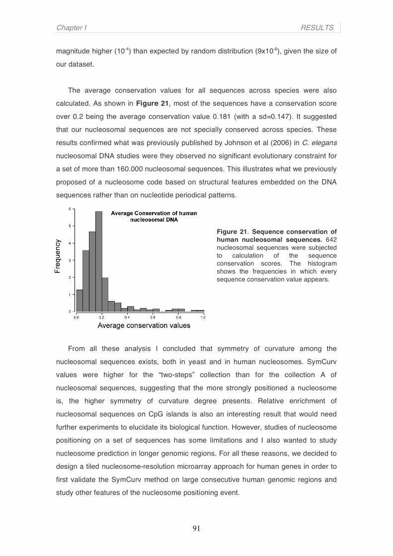
Chapter I RESULTS
91
magnitude higher (10-4) than expected by random distribution (9x10⁻⁶), given the size of
our dataset.
The average conservation values for all sequences across species were also
calculated. As shown in Figure 21, most of the sequences have a conservation score
over 0.2 being the average conservation value 0.181 (with a sd=0.147). It suggested
that our nucleosomal sequences are not specially conserved across species. These
results confirmed what was previously published by Johnson et al (2006) in C. elegans
nucleosomal DNA studies were they observed no significant evolutionary constraint for
a set of more than 160.000 nucleosomal sequences. This illustrates what we previously
proposed of a nucleosome code based on structural features embedded on the DNA
sequences rather than on nucleotide periodical patterns.
Figure 21. Sequence conservation of human nucleosomal sequences. 642 nucleosomal sequences were subjected to calculation of the sequence conservation scores. The histogram shows the frequencies in which every sequence conservation value appears.
From all these analysis I concluded that symmetry of curvature among the
nucleosomal sequences exists, both in yeast and in human nucleosomes. SymCurv
values were higher for the “two-steps” collection than for the collection A of
nucleosomal sequences, suggesting that the more strongly positioned a nucleosome
is, the higher symmetry of curvature degree presents. Relative enrichment of
nucleosomal sequences on CpG islands is also an interesting result that would need
further experiments to elucidate its biological function. However, studies of nucleosome
positioning on a set of sequences has some limitations and I also wanted to study
nucleosome prediction in longer genomic regions. For all these reasons, we decided to
design a tiled nucleosome-resolution microarray approach for human genes in order to
first validate the SymCurv method on large consecutive human genomic regions and
study other features of the nucleosome positioning event.
Chapter I RESULTS
92
2. Studying the role of nucleosome disposition in promoters and coding regions of hormone-regulated genes
2.1 Description of the nucleosome-resolution tilling array platform
I used a DNA microarray method to identify nucleosomal and linker DNA
sequences on the basis of susceptibility of linker DNA to Micrococcal nuclease (Rando
et al, 2005, and Figure 22A). Our lab has designed the platform according to previous
results we obtained from expression arrays performed in T47D breast cancer cells
treated with progesterone or estradiol (Dr. Cecilia Ballaré, unpublished work). The most
interesting cases (a total of 40 genes) were included on the nucleosome-resolution
tilling array (Figure 22B) as well as two hormone-independent genes as negative
controls (IFNB1 and HBE1 genes).
Nucleosomal DNA was isolated from T47D cells, as explained before, labeled
with Cy3 fluorescent dye (green), and mixed with Cy5-labeled partially MNase-digested
genomic DNA (red). MNase has sequence preference in digestion of protein-free DNA
and therefore control digestions of naked nucleic acid are required when assessing the
susceptibility of cutting sites in chromatin. In our case, the DNA control was total DNA
isolated from T47D cells subjected to limited MNase digestion. This mixture was
hybridized to the microarray platform made without removing repeated sequences and
with oligonucleotide probes of around 50 nt, and similar melting profiles, spaced every
20 bp. In average each base pair was covered by three oligonucleotide probes (Figure
22A). The probes encompassed the immediate gene upstream region (from -8000 to
+1nt) and part of the coding region (from +1 to +2000nts) of these 42 human genes. A
graph of green:red ratio values for spots along the represented regions is expected to
show nucleosomes as 120-160 base pairs broad peaks, or six to eight microarray
spots surrounded by lower ratio values corresponding to linker regions (Figure 22A).
The results of all experiments done on this self-designed microarray platform
and next detailed were submitted to Array Express (http://www.ebi.ac.uk/microarray-
as/ae/), a public archive for microarray experiments, with the accession number: E-
MEXP-1606 and name: “Nucleosome Mapping MNase”. Release date: the 1st of July
2009.
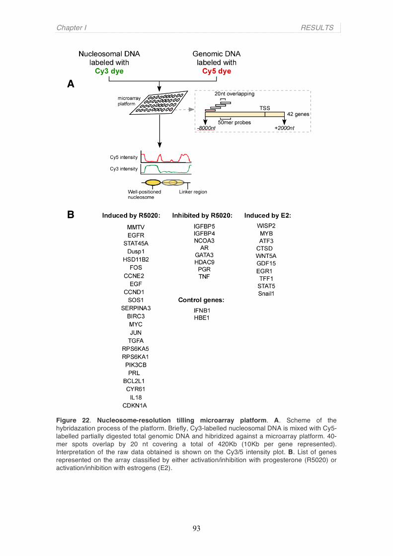
Chapter I RESULTS
93
Figure 22. Nucleosome-resolution tilling microarray platform. A. Scheme of the hybridazation process of the platform. Briefly, Cy3-labelled nucleosomal DNA is mixed with Cy5-labelled partially digested total genomic DNA and hibridized against a microarray platform. 40-mer spots overlap by 20 nt covering a total of 420Kb (10Kb per gene represented). Interpretation of the raw data obtained is shown on the Cy3/5 intensity plot. B. List of genes represented on the array classified by either activation/inhibition with progesterone (R5020) or activation/inhibition with estrogens (E2).
B
A

Chapter I RESULTS
94
2.2 Characterization of nucleosome positioning in promoter and coding
regions
Three biological and three technical replicas of the MNase treatment and
mononucleosomal DNA isolation were mixed with partially-digested genomic DNA and
hybridized against the platform. As observed in Table 6, Pearson correlation
coefficients for all replicas are over 0.9 (except for the case of MNase_RepF vs
MNase_RepD). This coefficient is used to determine the degree to which two or more
variables vary together. Table 6 shows high coefficient numbers meaning that there is
a high degree of parallelism between all replicas and thus, reliable data for the
following analysis.
Table 6. Pearson correlation coefficients of replicas
As an example, the raw signal, corresponding to the average fold-change
green:red ratio obtained from all replicas, for the AR, SOS1 and PRL gene regions is
plotted against gene coordinates in Figure 23 (blue line). The blue line plot shows the
ratio between Cy3 and Cy5 intensity dyes, that is, nucleosomal and genomic DNA,
respectively. Experimental nucleosome calls were obtained after application of the
derivative-dynamic programming approach on this raw data (see Methods and Materials). Peaks of about 120-160 base pair long surrounded by lower ratios values
indicated positions of nucleosomes that we represented as brown boxes of 147-base
pairs size. Different intensities of brown correspond to different experimental
nucleosomes calls scores, related to stronger or weaker nucleosome positions
(explained in more detail in section 2.3).
We have experimentally identified a total of 1653 nucleosomes by the tilling
array methodology over 420 Kb of genomic human DNA, that is a 57,85% coverage of
all genomic region represented. If these 1653 nucleosomes were periodically
distributes, we would find a nucleosome every a hundred nucleotides in average.
Inferring nucleosome positions from the microarray raw signal, allowed us to describe
the first level of chromatin structure of the regions represented at the platform.

Chapter I RESULTS
95
Figure 23. Nucleosome positioning in uninduced hormone-dependent genes. A. Nucleosome occupancy in the human DUSP1 gene region (dual specificity phosphatase 1, MKP1) (A), SOS1 gene region (Son of sevenless homolog 1 (Drosophila)) (B) and PRL gene region (Prolactin) (C). Grey bars: Median fold-change value from 6 replicate experiments, Blue line: Smoothed data. Brown bars represent nucleosomes and color intensity corresponds to relative scores (darker bars indicating higher scores).
B
A
C
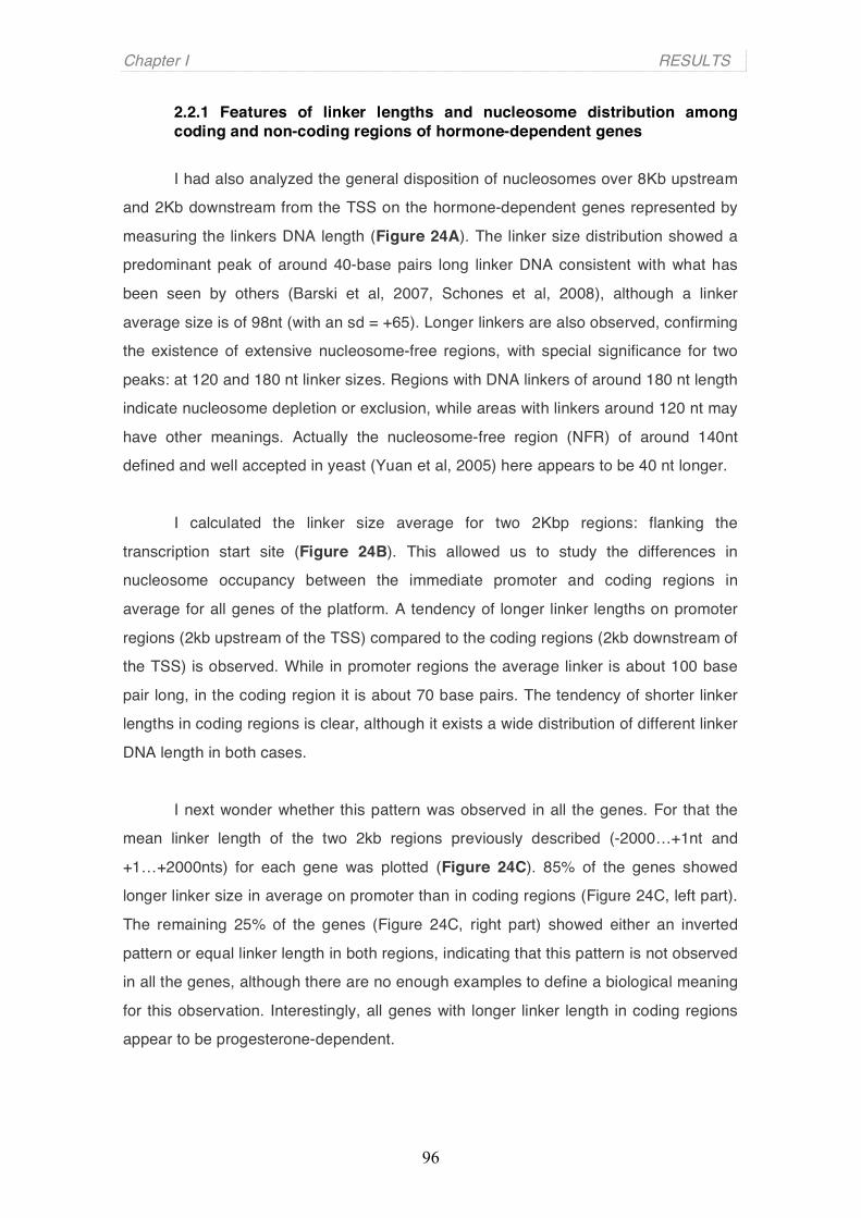
Chapter I RESULTS
96
2.2.1 Features of linker lengths and nucleosome distribution among coding and non-coding regions of hormone-dependent genes
I had also analyzed the general disposition of nucleosomes over 8Kb upstream
and 2Kb downstream from the TSS on the hormone-dependent genes represented by
measuring the linkers DNA length (Figure 24A). The linker size distribution showed a
predominant peak of around 40-base pairs long linker DNA consistent with what has
been seen by others (Barski et al, 2007, Schones et al, 2008), although a linker
average size is of 98nt (with an sd = +65). Longer linkers are also observed, confirming
the existence of extensive nucleosome-free regions, with special significance for two
peaks: at 120 and 180 nt linker sizes. Regions with DNA linkers of around 180 nt length
indicate nucleosome depletion or exclusion, while areas with linkers around 120 nt may
have other meanings. Actually the nucleosome-free region (NFR) of around 140nt
defined and well accepted in yeast (Yuan et al, 2005) here appears to be 40 nt longer.
I calculated the linker size average for two 2Kbp regions: flanking the
transcription start site (Figure 24B). This allowed us to study the differences in
nucleosome occupancy between the immediate promoter and coding regions in
average for all genes of the platform. A tendency of longer linker lengths on promoter
regions (2kb upstream of the TSS) compared to the coding regions (2kb downstream of
the TSS) is observed. While in promoter regions the average linker is about 100 base
pair long, in the coding region it is about 70 base pairs. The tendency of shorter linker
lengths in coding regions is clear, although it exists a wide distribution of different linker
DNA length in both cases.
I next wonder whether this pattern was observed in all the genes. For that the
mean linker length of the two 2kb regions previously described (-2000…+1nt and
+1…+2000nts) for each gene was plotted (Figure 24C). 85% of the genes showed
longer linker size in average on promoter than in coding regions (Figure 24C, left part).
The remaining 25% of the genes (Figure 24C, right part) showed either an inverted
pattern or equal linker length in both regions, indicating that this pattern is not observed
in all the genes, although there are no enough examples to define a biological meaning
for this observation. Interestingly, all genes with longer linker length in coding regions
appear to be progesterone-dependent.
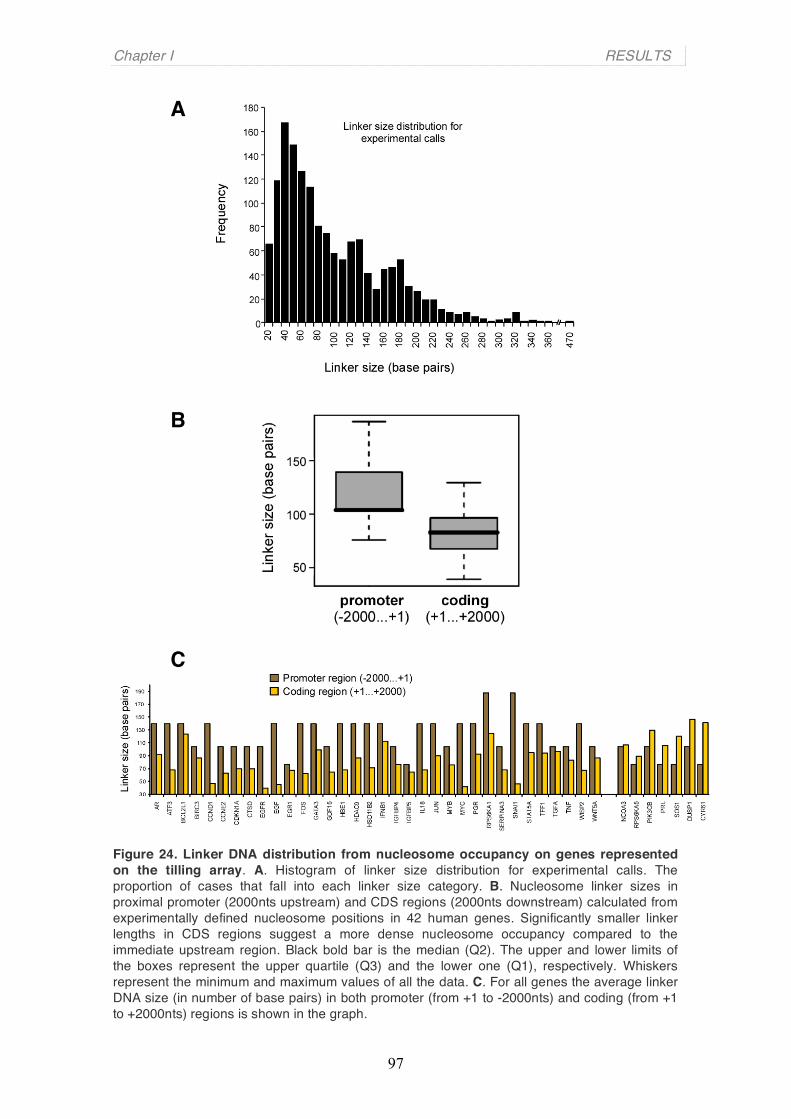
Chapter I RESULTS
97
Figure 24. Linker DNA distribution from nucleosome occupancy on genes represented on the tilling array. A. Histogram of linker size distribution for experimental calls. The proportion of cases that fall into each linker size category. B. Nucleosome linker sizes in proximal promoter (2000nts upstream) and CDS regions (2000nts downstream) calculated from experimentally defined nucleosome positions in 42 human genes. Significantly smaller linker lengths in CDS regions suggest a more dense nucleosome occupancy compared to the immediate upstream region. Black bold bar is the median (Q2). The upper and lower limits of the boxes represent the upper quartile (Q3) and the lower one (Q1), respectively. Whiskers represent the minimum and maximum values of all the data. C. For all genes the average linker DNA size (in number of base pairs) in both promoter (from +1 to -2000nts) and coding (from +1 to +2000nts) regions is shown in the graph.
B
C
A
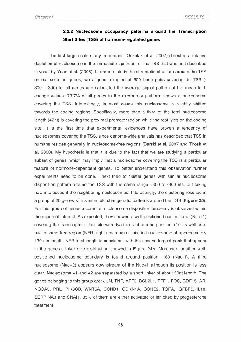
Chapter I RESULTS
98
2.2.2 Nucleosome occupancy patterns around the Transcription
Start Sites (TSS) of hormone-regulated genes
The first large-scale study in humans (Oszolak et al, 2007) detected a relative
depletion of nucleosome in the immediate upstream of the TSS that was first described
in yeast by Yuan et al. (2005). In order to study the chromatin structure around the TSS
on our selected genes, we aligned a region of 600 base pairs covering de TSS (-
300...+300) for all genes and calculated the average signal pattern of the mean fold-
change values. 73,7% of all genes in the microarray platform shows a nucleosome
covering the TSS. Interestingly, in most cases this nucleosome is slightly shifted
towards the coding regions. Specifically, more than a third of the total nucleosome
length (42nt) is covering the proximal promoter region while the rest lyies on the coding
site. It is the first time that experimental evidences have proven a tendency of
nucleosomes covering the TSS, since genome-wide analysis has described that TSS in
humans resides generally in nucleosome-free regions (Barski et al, 2007 and Tirosh et
al, 2008). My hypothesis is that it is due to the fact that we are studying a particular
subset of genes, which may imply that a nucleosome covering the TSS is a particular
feature of hormone-dependent genes. To better understand this observation further
experiments need to be done. I next tried to cluster genes with similar nucleosome
disposition pattern around the TSS with the same range +300 to -300 nts, but taking
now into account the neighboring nucleosomes. Interestingly, the clustering resulted in
a group of 20 genes with similar fold change ratio patterns around the TSS (Figure 25).
For this group of genes a common nucleosome disposition tendency is observed within
the region of interest. As expected, they showed a well-positioned nucleosome (Nuc+1)
covering the transcription start site with dyad axis at around position +10 as well as a
nucleosome-free region (NFR) right upstream of this first nucleosome of approximately
130 nts length. NFR total length is consistent with the second largest peak that appear
in the general linker size distribution showed in Figure 24A. Moreover, another well-
positioned nucleosome boundary is found around position -180 (Nuc-1). A third
nucleosome (Nuc+2) appears downstream of the Nuc+1 although its position is less
clear. Nucleosome +1 and +2 are separated by a short linker of about 30nt length. The
genes belonging to this group are: JUN, TNF, ATF3, BCL2L1, TFF1, FOS, GDF15, AR,
NCOA3, PRL, PIK3CB, WNT5A, CCND1, CDKN1A, CCNE2, TGFA, IGFBP5, IL18,
SERPINA3 and SNAI1. 85% of them are either activated or inhibited by progesterone
treatment.

Chapter I RESULTS
99
Figure 25. Nucleosome occupancy at the TSS. The mean of the log2 ratio values for a subset of genes for every nucleotide in a 600 base pair-region around the TSS (from -300 to +300) was calculated. Red line corresponds to the mean values for every nucleotide position. Grey lines on the back refers to the log2 ratio mean values for each of the genes of the subset separately. Yellow ovals represent the estimated position of nucleosomes inferred from the red-line curve.
2.3 The strongest positioned nucleosomes coincide with the highest SymCurv score
In order to be able to differentiate strong-positioned nucleosomes from weaker
ones, we assigned an intensity score to each experimental call (Figure 23, different
intensities of brown-coloured boxes). A high nucleosome call score is a direct indication
of a fold-change pattern that fits well with our notion of a nucleosome peak (peaks of
about 140 base pair long surrounded by lower ratios values). Along all regions
represented on the array we have observed the existence of well-positioned
nucleosomes alternating with less ones. Since I was particularly interested in
understanding the biological function of the existence of a subset of strong
nucleosomes, I looked in more detail at their position in the six replicas separately.
Surprisingly, I have found that some nucleosomes were almost exactly reproduced
(more than 90% overlap) in all replicas and that it coincided with the highest call scores
values. Our analysis consisted basically on calculating the average call scores of
nucleosome divided into six groups depending on their reproducibility among all six
replicate experiments (Figure 26). As shown in the figure, this subpopulation of

Chapter I RESULTS
100
reproducible positioned nucleosomes (class 6) exhibited also the highest average
scores in the SymCurv algorithm. In particular, SymCurv scores for nucleosomes
appearing in all 6 tiling array replicas were on average four-fold higher than the
corresponding ones for nucleosomes appearing in a single replicate (p<e-4). This
subset of nucleosomal positions that were consistent among all six experiments
constituted a ∼15% over the total number of nucleosomes. These parameters,
reproducibility and high score, identify a subset of “well-positioned nucleosomes”,
pointing to a determining role of the underlying nucleotide sequence for defining its
positions. We will refer to these nucleosomes as “key nucleosomes”.
Figure 26. Experimentally well-defined nucleosomes with high call scores. Experimentally defined nucleosome positions are classified in six distinct classes depending on the number of experiments where they occur. The graph shows boxplots for all 6 classes on nucleosome against the experimental call scores assigned by a derivative-dynamic programming algorithm. Horizontal black bar for every box represent the mean of score values.
2.3.1 The concept of “key nucleosomes”
Higher experimental call score implies higher probability for a nucleosome to be
always positioned in the same place. Sequence tendencies, such as symmetry of
curvature, are clearer in this subset of consistent and reproducible nucleosome
positions. These observations point to the existence of very well-defined nucleosome
positions that are mainly governed by the sequence features, including the symmetry of
curvature. Due to its dependent on the DNA sequence, it is feasible to hypothesis that
the “key nucleosomes” will be consistent among cell types and different conditions.
Whether they play relevant biological functions on the organization of chromatin needs
to be further investigated. Moreover, given the number of unsuccessful attempts to
define a universal sequence-based nucleosome positional code it is not surprising that

Chapter I RESULTS
101
the nucleosomes which position is based on sequence features are merely a small
subset of the total number of nucleosomes.
2.3.2 Symmetry of Curvature as a nucleosome positioning predictive tool
Based on the observation of high degrees of symmetry of curvature in “key
nucleosomes”, we proposed the SymCurv algorithm as a predictive tool method for a
potential nucleosomal DNA sequence. Since “key nucleosomes” are well defined by
their strong degree of symmetry of curvature, this parameter could be enough to
identify their positions.
We used the genomic regions represented in the array to validate the SymCurv
as a predictive method for nucleosome positioning. We first calculated the average
SymCurv scores, and from the symmetry of curvature values the positions of predicted
nucleosomes were inferred. Next, we compare both experimental and predicted
nucleosome calls and calculate the distance of each predicted position to its proximal
experimentally defined one. The highest the overlapping between nucleosome calls,
the better performance of SymCurv algorithm to predict nucleosome positions.
Overlapping nucleosomes were classified into different groups depending on the
number of nucleotides that overlaps (within a range from 146 to 112nts) (Figure 27).
The relative scores (yellow bars for the experimental one, magenta for the SymCurv
scores) increased with increase overlapping between predictions and experimental
calls (grey curve). In addition there is a coincidence of high scores for both
experimental calls and SymCurv-based predictions as the accuracy of the prediction
increases, in other words, when there are 146nts of overlapping the highest SymCurv
and experimental call scores are observed (Figure 27, firsts bars from the left). This
observation implies that regions with high symmetry of curvature are also likely to be
strongly occupied by nucleosomes and that this relation is quantitative. Thus, my
conclusion is that stronger predictions tend to coincide better with strongly defined
experimental positions and because of that, SymCurv calculation seems to be a useful
tool to precisely identify “key nucleosomes”.
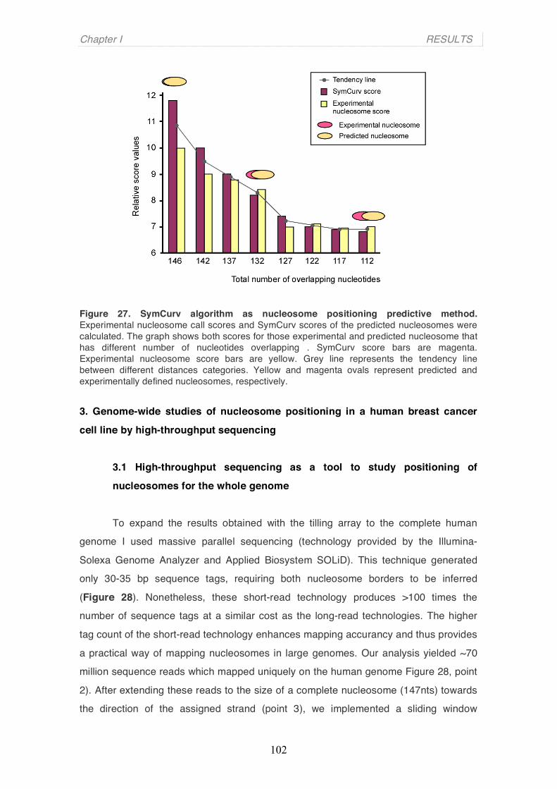
Chapter I RESULTS
102
Figure 27. SymCurv algorithm as nucleosome positioning predictive method. Experimental nucleosome call scores and SymCurv scores of the predicted nucleosomes were calculated. The graph shows both scores for those experimental and predicted nucleosome that has different number of nucleotides overlapping . SymCurv score bars are magenta. Experimental nucleosome score bars are yellow. Grey line represents the tendency line between different distances categories. Yellow and magenta ovals represent predicted and experimentally defined nucleosomes, respectively. 3. Genome-wide studies of nucleosome positioning in a human breast cancer
cell line by high-throughput sequencing
3.1 High-throughput sequencing as a tool to study positioning of
nucleosomes for the whole genome
To expand the results obtained with the tilling array to the complete human
genome I used massive parallel sequencing (technology provided by the Illumina-
Solexa Genome Analyzer and Applied Biosystem SOLiD). This technique generated
only 30-35 bp sequence tags, requiring both nucleosome borders to be inferred
(Figure 28). Nonetheless, these short-read technology produces >100 times the
number of sequence tags at a similar cost as the long-read technologies. The higher
tag count of the short-read technology enhances mapping accurancy and thus provides
a practical way of mapping nucleosomes in large genomes. Our analysis yielded ~70
million sequence reads which mapped uniquely on the human genome Figure 28, point
2). After extending these reads to the size of a complete nucleosome (147nts) towards
the direction of the assigned strand (point 3), we implemented a sliding window

Chapter I RESULTS
103
strategy at maximum resolution (1nt) to obtain a read-coverage score per nucleotide
(point 4). Thus every nucleotide was attributed a score which was equivalent to the
reads mapping on the corresponding genomic position. We then used these scores to
assign non-overlapping nucleosomal positions along the human genome (Figure 28
point 5 and see Material and Methods for more detail). Finally, every nucleosome
inferred has a score relative to the number of reads that defined it (from now, called the
read-count score).The resulting dataset consisted of 8.89 million of non-overlapping
nucleosomes. The distribution of nucleosome scores is highly non-random when
compared to an exponential distribution with the same mean (p<e-15), suggesting a
greater number of nucleosomes with high score than expected by random distribution.
Figure 28. ChIP-Seq nucleosome mapping technology.(1) From 30 to 35 base pairs of each edge of nucleosomal sequences are sequenced (2) and mapped against the human genome. Some reads are from the positive strand (Watson) and others from the negative (Crick). (3) The reads are extended till a size of 147nt fragment in the direction of the strand. (4) We calculate a read-coverage score per nucleotide, counting reads from both strands. Positions of nucleosomes are deduced depending on the wide of the peak. Peaks of more than 147nt wide are considered fuzzy nucleosomes, as represented by overlapping of different ovals. (5) For those that have a well-defined position, we attribute a score depending on the number of reads that defines each deduced nucleosome. For instance, first nucleosome to the left is defined by 3 reads from positive and 3 reads from negative strand, while middle nucleosome position is defined by just 3 reads from one strand. Score of the first nucleosome will be twice the one for the second.
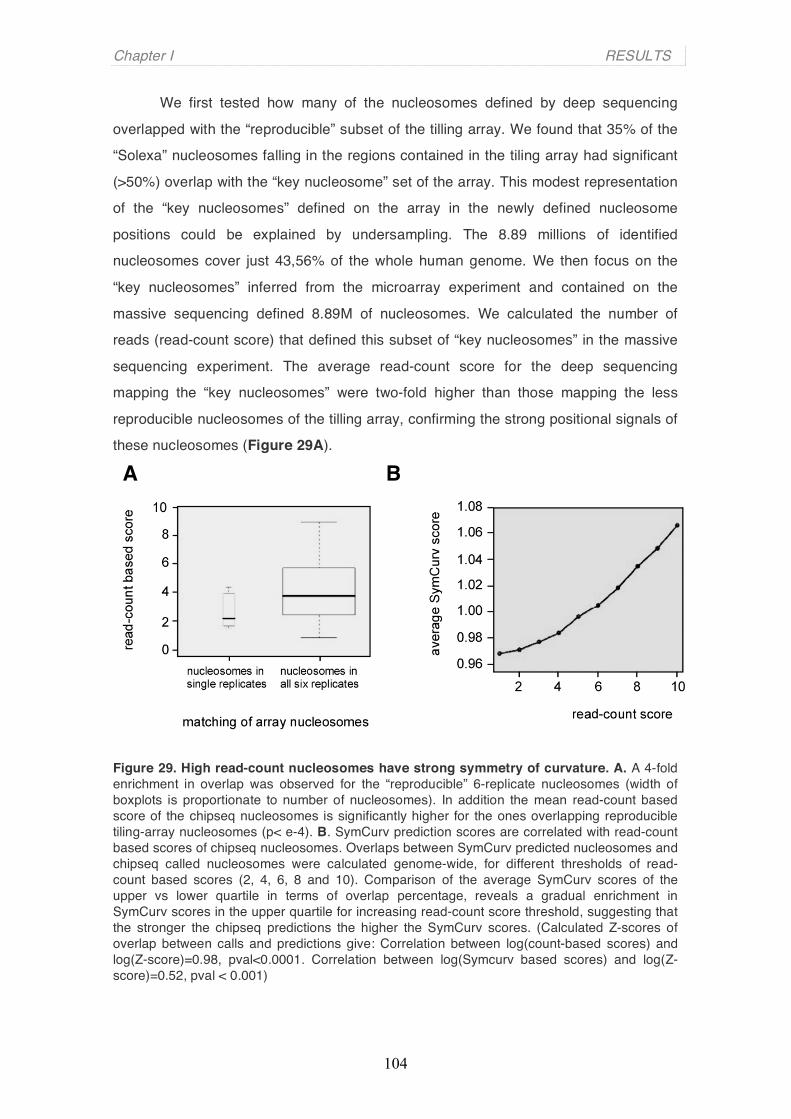
Chapter I RESULTS
104
We first tested how many of the nucleosomes defined by deep sequencing
overlapped with the “reproducible” subset of the tilling array. We found that 35% of the
“Solexa” nucleosomes falling in the regions contained in the tiling array had significant
(>50%) overlap with the “key nucleosome” set of the array. This modest representation
of the “key nucleosomes” defined on the array in the newly defined nucleosome
positions could be explained by undersampling. The 8.89 millions of identified
nucleosomes cover just 43,56% of the whole human genome. We then focus on the
“key nucleosomes” inferred from the microarray experiment and contained on the
massive sequencing defined 8.89M of nucleosomes. We calculated the number of
reads (read-count score) that defined this subset of “key nucleosomes” in the massive
sequencing experiment. The average read-count score for the deep sequencing
mapping the “key nucleosomes” were two-fold higher than those mapping the less
reproducible nucleosomes of the tilling array, confirming the strong positional signals of
these nucleosomes (Figure 29A).
Figure 29. High read-count nucleosomes have strong symmetry of curvature. A. A 4-fold enrichment in overlap was observed for the “reproducible” 6-replicate nucleosomes (width of boxplots is proportionate to number of nucleosomes). In addition the mean read-count based score of the chipseq nucleosomes is significantly higher for the ones overlapping reproducible tiling-array nucleosomes (p< e-4). B. SymCurv prediction scores are correlated with read-count based scores of chipseq nucleosomes. Overlaps between SymCurv predicted nucleosomes and chipseq called nucleosomes were calculated genome-wide, for different thresholds of read-count based scores (2, 4, 6, 8 and 10). Comparison of the average SymCurv scores of the upper vs lower quartile in terms of overlap percentage, reveals a gradual enrichment in SymCurv scores in the upper quartile for increasing read-count score threshold, suggesting that the stronger the chipseq predictions the higher the SymCurv scores. (Calculated Z-scores of overlap between calls and predictions give: Correlation between log(count-based scores) and log(Z-score)=0.98, pval<0.0001. Correlation between log(Symcurv based scores) and log(Z-score)=0.52, pval < 0.001)
B A

Chapter I RESULTS
105
3.2 High read-count nucleosomes have strong symmetry of curvature
We next asked whether the highest read-count nucleosomes showed also high
degree of symmetry of curvature. Indeed, a correlation between SymCurv scores and
read-count scores nucleosome was observed (Figure 29B). Results presented in
Figure 29B, show that high read-count nucleosomes also have high SymCurv scores
as we previously observed with the subset of “key nucleosomes” on the tilling array.
Thus, we concluded that deep sequencing identifies a particular set of well-positioned
“key nucleosomes” now defined by high number of reads that also show high degrees
of symmetry of curvature.
4. Properties of key nucleosomes
4.1 Key nucleosomes in biologically relevant locations: TSS, TTS and
splicing sites
If “key nucleosomes” are important for chromatin organization, and function
presumably they will be located in biologically relevant locations for gene expressions.
To test this hypothesis we studied the distribution of “key nucleosomes” in transcription
start and termination sites (TSS and TTS, respectively) as well as alternative splicing
junctions. We used the complete set of transcription start sites from the ENSEMBL
human genes to analyze the nucleosome occupancy. We observed an increasing
enrichment in occupancy immediately downstream of the transcription initiation (Figure
30A, red and brown lines), indicating that “key” nucleosomes tend to be constitutively
positioned at the beginning of transcripts. It also shows a tendency for a nucleosome-
free region immediately upstream of the TSS. Interestingly, the pattern observed when
nucleosomes are defined by a lower number of reads (less than 7) is the opposed
(Figure 30A green and yellow lines). These lower read-counts nucleosomes present a
nucleosome covering the TSS and two clear nucleosme-free regions at both sites.
These results suggest that key nucleosomes are preferentially positioned in the vicinity
of TSS region, more precisely right downstream of the TSS. A possible explanation for
which these “organizing” nucleosomes are positioned at the initiation sites of
transcription would be to facilitate transcription, imposing a physical “barrier” on the

Chapter I RESULTS
106
correct position close to which the transcriptional machinery is assembled.
Alternatively, they may serve to preclude “uncontrolled” transcription, being the
silencing of genes the main function of chromatin.
An analysis identical to the one of the initiation sites was performed for the transcription
termination sites (TTSs) (Figure 30B). Here the situation is inverted, as a nucleosome-
free region is observed exactly at the end of the transcripts. In this case the pattern
becomes clearer for high reads-count nucleosomes key nucleosomes (brown and red
lines).
A similar sort of analysis performed on the splice sites of genes encompassing
the ENCODE regions reveals strong nucleosomal patterns at the splice junctions
(Figure 30C). In this case, just nucleosomes defined by 10 or more reads were plotted.
The analysis revealed key nucleosome positioning at both exon-intron and intron-exon
boundaries with a tendency for a positioned nucleosome at the side of the intron
(Figure 30D). It is also worth to emphasize the valley of nucleosome occupancy
located right at the beginning and end of exons. Recent works have pointed out the
temporal-spatial connection between transcription and splicing. As in the case of
transcription initiation nucleosome-free regions followed by a strong nucleosome
patterns at the splice sites would act as physical barriers that might delay the RNA
polymerase, giving time for the splicing machinery to be recruited. Thus, key
nucleosomes seem to be functioning as chromatin barriers in genomic positions of
interest to the cell's proper functioning.
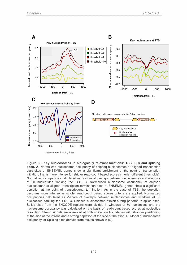
Chapter I RESULTS
107
Figure 30. Key nucleosomes in biologically relevant locations: TSS, TTS and splicing sites. A. Normalized nucleosome occupancy of chipseq nucleosomes at aligned transcription start sites of ENSEMBL genes show a significant enrichment at the point of transcription initiation, that is more intense for stricter read-count based scores criteria (different thresholds). Normalized occupancies calculated as Z-score of overlaps between nucleosomes and windows of 50 nucleotides flanking the TSS. B. Normalized nucleosome occupancy of chipseq nucleosomes at aligned transcription termination sites of ENSEMBL genes show a significant depletion at the point of transcriptional termination. As in the case of TSS, the depletion becomes more intense as stricter read-count based scores criteria are applied. Normalized occupancies calculated as Z-score of overlaps between nucleosomes and windows of 50 nucleotides flanking the TTS. C. Chipseq nucleosomes exhibit strong patterns in splice sites. Splice sites from the ENCODE regions were divided in windows of 50 nucleotides and the nucleosome occupancy was calculated on the basis of read-count based scores at nucleotide resolution. Strong signals are observed at both splice site boundaries with stronger positioning at the side of the introns and a strong depletion at the side of the exon. D. Model of nucleosome occupancy for Splicing sites derived from results shown in (C).
A B
D C

Chapter I RESULTS
108
4.2 Similar patterns on TSS, TTS and splice junctions are obtained with
SymCurv values-based predictions
We tested transcription initiation and termination sites, as well as splice-sites
from the ENCODE regions with the use of the SymCurv algorithm (Figure 31). For both
TSS and TTS, SymCurv predictions yield a very similar pattern, predicting high
nucleosome occupancy downstream of the start site and a nucleosome-free region on
the termination site (Figure 31A red and blue lines, respectively).
Similarly, for the splice sites, strong nucleosomal patterns are predicted at the
vicinity of both splice junctions with stronger nucleosome occupancy at the side of the
introns (Figures 31B, intron-exon represented in blue and exon-intron in red).
Similarities between the predicted nucleosomal patterns and the ones obtained with
strict read-count score thresholds aiming at key nucleosomes, are indicative of the fact
that chromatin structure of functional regions of the genome is reflected upon structural
properties of the DNA primary sequence.
Figure 31. SymCurv-based predictions in TSS, TTS and Splicing Sites. A. SymCurv-predicted nucleosomes exhibit strong patterns at the starts and ends of genes. TSS and TTS sites obtained from the ENCODE regions were divided in windows of 50 nucleotides and the nucleosome occupancy was calculated on the basis of SymCurv scores at nucleotide resolution. Strong positioning is observed at the initiation and strong nucleosome-free regions are predicted for the termination sites in accordance with experimental results. B. SymCurv-predicted nucleosomes exhibit strong patterns around splice sites. Splice sites from the ENCODE regions were divided in windows of 50 nucleotides and nucleosome occupancy was calculated on the basis of SymCurv scores at nucleotide resolution.

Discussion
We introduce a new theoretical concept, SymCurv, that allows precise position
of a subset of nucleosomes on the human genome to be predicted in silico based on
the structural attributes of the primary DNA sequence. Nucleosomal DNA of these
subset of nucleosomes exhibited the highest symmetry of curvature, suggesting that
their positions are strongly determined by the conformation of the underlying DNA
sequence. In this work I give experimental evidence for the existence of a subset of
very well-positioned nucleosomes in the human genome. Using a tilling array
encompassing 420 kb around the 5´-end of 42 human genes and a novel dynamic
programming based algorithm, we found that ∼15 % of the nucleosome calls mapped
reproducibly to the same positions (more than 90 % overlapping) in six experimental
replica, while the positions of the other nucleosomes were less consistent. Analysis of
the whole human genome by massive nucleosome sequencing confirmed the existence
of a subset of well-positioned nucleosomes, named “key nucleosomes”, which exhibit
interesting sequence properties and a peculiar distribution along the genome, tending to
occupy genomic space of specific functionalities.
The Symmetry of Curvature on nucleosomal DNA
The approach described to identify well-positioned nucleosomes, is based on
the hypothesis for the existence of a genomic signature for nucleosome positioning. The
basic assumption is that sequences selected for nucleosome binding will tend to
accumulate special features facilitating nucleosome formation through evolution. These
features are more likely to be structural and expected to be more extended, meaning
covering a longer sequence string, and more degenerate than a usual protein binding
sequence. Nucleosome formation involves wrapping of a ~146bp sequence around a
protein octamer, thus DNA curvature is expected to facilitate this process. In addition,
the symmetric structure of this octamer is likely to impose corresponding symmetry
constraints on the sequence, as in a manner similar to the majority of symmetric DNA-
109

Chapter I DISCUSSION
110
binding proteins. Combining the above prerequisites we have proposed the symmetry of
curvature as a possible signal facilitating nucleosome formation. The results presented
in this thesis show that there exists a correlation between this property and the
tendency for nucleosome positioning. Moreover, symmetry of curvature as a property of
nucleosomal DNA was a found in nucleosomal sequences analyzed by very different
experimental techniques (collections of nucleosomal DNA, tilling microarray
hybridization and massive parallel sequencing), proving the reliability of the concept.
SymCurv algorithm as a nucleosome predictive tool
Around 15% of the total number of experimental nucleosome calls showed a
high tendency to occupy the same place among different experimental replicas in
microarrays’ experiments. In addition, SymCurv performed with increased efficiency on
this same subset. Larger scale nucleosome mapping on human genome showed
nucleosomes defined by a high number of reads that appear to have also the highest
SymCurv scores. Consistent with our results, other works on the field have also
suggested that only a subset of the predicted nucleosomes is actually guided by the
sequence (Yuan et al., 2005; Segal et al., 2006; Kaplan et al., 2008), but no attempt
was done on defining this subset of nucleosomes. Moreover, when testing various
datasets of nucleosomal sequences, we observed a gradient increase of the SymCurv
pattern intensity in the datasets obtained through more rigorous and step-filtering
experimental processes, suggesting that SymCurv algorithm is also a quantitative
method to predict nucleosomes (high scores correlate with strong signal of nucleosome
positioning for a given sequence).
The first genome-wide nucleosomal sequence set published, originating from C. elegans (Johnson et al., 2006) also suggests that only a small part of the sequences is
actually under constraint of nucleosome positioning. Later works by Valouev et al.
(2008) and Schones et al. (2007) further supported this finding, pointing towards a
model of statistical positioning of nucleosomes, according to which, a small set of well-
positioned nucleosomes (or alternatively, a set of regions that strongly exclude them)
would guide the subsequent binding of the large mass of nucleosome particles, which
would occupy the remaining space. The presented results indicate that the SymCurv
method could help in identifying regions of high nuclesome forming potential in many
different species.
Chapter I DISCUSSION
111
Nucleosome organization around hormone-dependent genes
Here I have used a tilling array to study the general patterns of nucleosome
organization at promoter and coding regions, and specially around the TSS, of a subset
of hormone responsive genes. One of the limitations of the method used to infer
nucleosomes from the raw microarray data is that regions defined by fuzzy
nucleosomes are, instead, occupied by experimental nucleosomes with low scores.
Dynamic programming will always position nucleosomes even if it is a wide region with
many different possible overlapping positions. However, our analysis on linker length
and nucleosome occupancy of the TSS will not be modified by this point. Different linker
lengths observed all over the regions represented on the array might reflect the
implication of extra factors other than the histone core that modulate spacing and
general positioning of the nucleosomes, such as presence of different linker histone
variants and remodelling complex among others (Whitehouse et al, 2007). Alternatively,
in some cases linker DNA would contain nucleosome exclusion signals that avoid
positioning of core histone particles in specific locations. In yeast the meaning of
nucleosome-free regions (NFR) around TSS has been widely studied (Petesch et al,
2008 and Venters et al, 2009). It has been described that although a NFR is permissive
for transcription, it is not sufficient to activate genes. Thus, NFRs might allow low basal
levels of leaky transcription, which could be interpreted as meaningless biological noise,
particularly if the transcripts are rapidly degraded. NFRs in the 5’ and the 3’ of yeast
genes has opposed functions: 5’NFRs are likely to be sites for the assembly of the
transcription machinery, whereas 3’NFRs are likely to be sites for the disassembly of
the transcription machinery. The meaning of NFRs in human genes remains still an
open question.
Till now we have characterized the nucleosome occupancy on 40 hormone-
responsive genes. Next step will be to study how nucleosome positioning is changed
after induction/inhibition of transcription upon treating the cells with hormone. Our
system is limited to 40 genes but it allows us to presicely study nucleosome-resolution
changes after promoter regulations. It is an interesting assay, since in human genes it is
still unclear the pattern of nucleosome occupancy around inducible genes, since most
of the studies focus on constitutively activated genes.
Chapter I DISCUSSION
112
Genomic-scale studies on nucleosome position
Recent technological advances in highly parallel sequencing allow the
conduction of large-scale nucleosome positioning studies. In a recent study, Dennis et
al. (2007) discuss experimental methods for large-scale structural analysis of
mammalian chromatin, which, although elaborate, do not precisely replicate the
nucleosomal pattern of well-studied cases. This is likely due to both the complexity of
the positioning problem, (alternative positioning, flexibility of binding, influence of
sequence-specific DNA-binding proteins, etc) and the number of information-processing
steps needed from the raw experimental data to actually call a nucleosomal
position. Thus it is not surprise that the concordance of computationally predicted
positions with experimentally defined ones is relatively small in all published methods to
date, whether they are based on probabilistic likelihood (Segal et al., 2006 and Kaplan
et al, 2008), comparative genomics (Ioshikhes et al., 2006) or supervised learning
(Peckham et al., 2007). Our aim was to focus on the subgroup of nucleosomes which
position signal was strong and based on the DNA sequence features.
The meaning for a key nucleosome to be in specific functional locations
We have shown the significant enrichment of “key nucleosomes” upstream of
the TSS and depletion right on the TTS, as well as specific positioning around
alternative splicing sites. The biological meaning of these observations can be
explained generally by the need to control of basic cellular mechanisms. “Key
nucleosomes” at splice sites would act as physical barriers that might delay the RNA
polymerase, giving time for the splicing machinery to be recruited, while the meaning for
the TSS locations could be either a physical barrier or a platform for both specific
histone posttranslational modifications and histone variant incorporations that may
modulate the active state of the promoter.
However, future analysis is needed in order to further elucidate the functional
role of “key nucleosomes” in specific locations. Future experiments could go, for
instance, towards the study of the correlation of gene expression and “key nucleosome”
occupancy or sequence conservation of these subset of nucleosomes compared to
neigbouring ones. To test whether “key nucleosomes” are also playing an important role
on higher-order chromatin organization, it would be interesting to study whether they are

Chapter I DISCUSSION
113
periodically spaced on the human genome. One hypothesis could be that “key
nucleosomes” are multi-functional entities that may at the same time regulate cellular
processes and organize chromatin compaction. In this way, nucleosomal sequences
may simultaneously achieve a number of different tasks.

Chapter I DISCUSSION
114
115
Chapter 2
“Two chromatin remodeling activities cooperate during activation of
hormone responsive promoters”
This manuscript has been send for publication to PloS Genetics (No. 08-PLGE-RA-1702R3). It is currently under the third revision.

116

117
Two chromatin remodeling activities cooperate during activation of hormone
responsive promoters
Guillermo Pablo Vicent1,3, Roser Zaurin1,3, A. Silvina Nacht1, Ang Li1, Jofre Font-Mateu1,
Francois Le Dily1, Michiel Vermeulen2, Matthias Mann2 and Miguel Beato1
1Centre de Regulació Genòmica (CRG), Universitat Pompeu Fabra, Parc de Recerca
Biomèdica (PRBB), Dr Aiguader 88, E-08003, Barcelona, Spain.
2Max Planck Institute for Biochemistry, Department of Proteomics and Signal
transduction, Am Klopferspitz 18, D-82152 Martinsried, Germany.
3 both authors contribute equally to this work
Correspondence should be addressed to M.B.: e-mail: [email protected]
Running Title: BAF and PCAF cooperate in MMTV induction

118
Abstract
Steroid hormones regulate gene expression by interaction of their receptors with
hormone responsive elements (HREs) and recruitment of kinases, chromatin
remodeling complexes and coregulators to their target promoters. Here we show that in
breast cancer cells the BAF, but not the closely related PBAF complex is required for
progesterone induction of several target genes including MMTV, where it catalyzes
localized displacement of histones H2A and H2B and subsequent NF1 binding. PCAF
is also needed for induction of progesterone target genes and acetylates histone H3 at
K14, an epigenetic mark that interacts with the BAF subunits anchoring the complex to
chromatin. In the absence of PCAF, full loading of target promoters with hormone
receptors and BAF is precluded and induction is compromised. Thus activation of
hormone-responsive promoters requires cooperation of at least two chromatin
remodeling activities, BAF and PCAF.
Author Summary
In order to adapt its gene expression program to the needs of the environment the cell
must access the information stored in the DNA sequence that is tightly packaged into
chromatin in the cell nucleus. How the cell manages to do it in a selective maner is still
unclear. Here we show that in breast cancer cells treated with the ovarian hormone
progesterone the hormone receptor recruits to the regulated genes two chromatin
remodeling complexes that cooperate in opening the chromatin structure. One of the
complexes puts a mark in a chromatin protein that anchors the other complex enabling
full gene activation. The present discovery highlights the importance of the concerted
order of events for access to genomic information during activation of gene expression
and reveals the intricacies of hormonal gene regulation.
119
Introduction
Regulation of eukaryotic gene expression implies mechanisms that permit transcription
factors to gain access to chromatin packaged DNA sequences. The basic unit of
chromatin, the nucleosome, consists of an octamer formed of two copies of each of the
four core histones (H2A, H2B, H3, and H4) around which 147 bp DNA is wrapped in
1.65 left-handed superhelical turns [1]. Modulation of the structure and dynamics of
nucleosomes is an important regulatory mechanism in all DNA-based processes and is
catalyzed by chromatin remodeling complexes. Such complexes can either modify
histone residues or use the energy of ATP hydrolysis to alter the relationship between
histones and DNA [2, 3].
The yeast SWI/SNF complex, the first ATP-dependent chromatin remodeling
complex to be identified, is a 2-MDa complex of 11 subunits that regulates gene
expression by catalyzing octamer transfer, nucleosome sliding, dinucleosome
formation, and H2A/H2B displacement [4-6]. RSC, (Remodel the Structure of
Chromatin) [7], is a closely related yeast chromatin remodeling complex of 15 subunits,
that shares two identical and at least four homologous subunits with the ySWI/SNF
complex [8]. There are two human SWI/SNF-like complexes both containing ATPase
subunits similar to yeast Swi2/Snf2, hBRM (human Brahma) or BRG1 (Brahma-Related
Gene 1), as well as a series of other subunits, some of which differ in various cell types
[8]. The hSWI/SNF-α complexes, also called BAF for BRG1/hBRM-Associated Factor,
contain either hBRM or BRG1 as ATPase and is orthologue to yeast SWI/SNF. The
hSWI/SNF-β complex, also called PBAF (Polybromo-associated BAF) contains only
hBRG1 and is orthologue to yeast RSC complex [4, 9]. The BAF and PBAF complexes
share many subunits but have also subtype specific subunits: BAF250 and hBRM are
only found in BAF, whrereas BAF180 and BAF 200 are only found in PBAF [4, 10].
BAF57 has been reported as a common subunit for BAF and PBAF complexes [4, 9].
Histone acetyltransferases (HATs) and histone deacetylases (HDACs)
represent other group of chromatin remodeling complexes that regulate the level of
acetylation on the N-terminal tails of core histone proteins and other protein substrates
[11, 12]. The HATs are divided into five families, including the GCN5-related N-
acetyltransferases (GNATs) with GCN5 and PCAF as the best characterized members;
the MYST ('MOZ, Ybf2/Sas3, Sas2 and Tip60')-related HATs; p300/CREB-binding
protein (CBP) HATs; the general transcription factor HATs including the TFIID subunit
TBP-associated factor-1 (TAF1); and the nuclear hormone-related HATs SRC1 and
ACTR (SRC3) [13, 14]. Recombinant PCAF, like full-lenght GCN5, acetylates either

120
free histones or nucleosomes [15], primarily on lysine -14 of histone H3 [16]. The role
of PCAF in transcription has been investigated in multiple studies, and its requirement
as a HAT and coactivator has been described in several processes including nuclear
receptor mediated events [17, 18], but the precise mechanism of action has not yet
been elucidated .
The functional relationship between different chromatin remodeling enzymatic
activities is of great interest. A remarkable interdependence has been described during
transcriptional activation in S. cerevisiae between the SWI/SNF complex and the
histone acetylation complex SAGA [19]. Bromodomains of the RSC complex have been
shown to recognize acetylated H3K14 [20]. HAT activity stabilizes SWI/SNF binding to
promoter nucleosomes providing a mechanistic basis for the ordered recruitment of
these complexes [21]. In mammalian cells, transcriptional activation by nuclear
receptors requires multiple cofactors including CBP/p300, SWI/SNF and Mediator. The
ordered recruitment of these cofactors to the promoters depends not only on the direct
interactions between nuclear receptors and cofactors but also on cofactor-cofactor
interaction and on histone modifications [22].
Gene regulation by steroid hormones is mediated by intracellular receptors,
which upon hormone binding can activate signal transduction cascades and interact in
the nucleus with other transcription factors and/or with dedicated DNA sequences,
called hormone responsive elements (HREs) [23]. When ligand-receptor complexes
interact with HREs they alter the transcriptional state of the associated genes via the
recruitment of chromatin remodeling complexes and other coregulators to the target
promoters [24, 25]. The mouse mammary tumor virus (MMTV) long terminal-repeat
region encompasses a promoter including within 140 bp five degenerated hormone
responsive elements (HREs) and a binding site for nuclear factor 1 (NF1). In chromatin
the MMTV promoter is organized into positioned nucleosomes [26], with a nucleosome
located over the five HREs and the NF1 binding site [27]. On this promoter
nucleosome, the binding site for NF1 is not accessible and only two of the five HREs,
the strong palindromic HRE1 and the weak half palindrome HRE4, can be bound by
hormone receptors, while the central HREs, in particular the palindromic HRE2 and the
half palindrome HRE3, are not accessible for receptor binding [28]. Following hormone
induction in vivo all HREs and the binding site for NF1 are occupied simultaneously on
the surface of a nucleosome-like structure and a functional synergism is observed
between progesterone receptor (PR) and NF1 [27].

121
Already 5 minutes after addition of hormone to breast cancer cells with an
integrated copy of an MMTV-luc reporter, the cytoplasmic signaling cascade
Src/Ras/Erk is activated via an interaction of PR with the estrogen receptor, which
activates Src [29]. As a consequence of Erk activation, PR is phosphorylated, Msk1 is
activated, and a ternary complex PR-Erk-Msk1 is recruited to nucleosome B [30]. Msk1
phosphorylates H3 at serine 10, a modification which is accompanied by acetylation at
H3 lysine 14, displacement of HP1γ, and recruitment of RNA polymerase II [30].
Inhibition of Erk or Msk activation blocks H3S10 phosphorylation, H3K14 acetylation,
and hormonal induction of MMTV and other progesterone target genes [30].
There have been several reports indicating a role for SWI/SNF, Brg1 and Brm in
glucocorticoid regulation of MMTV transcription [31-33], but the situation with
progesterone is less clear. The MMTV promoter assembled in minichromosomes with
positioned nucleosomes [34] is activated by PR in a process involving a NURF-
dependent mutual synergism between PR and NF1 [35]. Progesterone treatment of a
breast cancer cell line carrying a single integrated copy of a MMTV transgene leads to
recruitment of PR, SWI/SNF, and SNF2h-related complexes to the MMTV promoter,
accompanied by selective displacement of histones H2A and H2B from nucleosome B
[6, 25]. Recently, the acidic N terminus of the Swi3p subunit of yeast SWI/SNF was
identified as a novel H2A-H2B-binding domain required for ATP-dependent H2A/H2B
dimer displacement [36]. Moreover, a characteristic and sharp DNase I hypersensitive
site appears near the symmetry axis of the nucleosome encompassing the HREs [27].
The same hypersensitive site can be induced in the absence of hormone by treatment
with low concentrations of histone deacetylases inhibitors, such as sodium butyrate or
trichostatin A [37], suggesting that it can be initiated by a moderate increased in
histone acetylation. However, the relationship between histone acetylation and ATP-
dependent nucleosome remodeling following hormone treatment is not clear. To
approach this question we have performed knockdown and ChIP experiments targeting
human SWI/SNF complexes and histone acetyl transferases (HATs) in breast cancer
cells.
Results BAF is required for progesterone gene activation in T47D-MTVL cells and is
recruited to the MMTV promoter upon induction The requirement of Brm and Brg1 for MMTV promoter activation was assessed in
T47D-MTVL cells transfected with specific siRNAs. When siRNA against Brg1 was

122
used, the levels of Brm protein increased (Figure 1 A, left panels) and viceversa
(Figure 1 A, middle panels). A significant reduction (80% and 55% for Brm and Brg1,
respectively) of both ATPases was only observed when the two siRNAs were
combined (Figure 1 A, right panels). The levels of the two PR isoforms, PRB and PRA,
were unaffected by Brg1 or Brm siRNAs (Figure 1 A, third row). In cells transfected with
control siRNA an eight-fold increase in MMTV-luc transcription was observed 8 hours
after hormone treatment (Figure 1 B). In cells transfected with siRNAs against either
Brg1 or Brm, the induction was reduced by 30% and 20% respectively, whereas
transfection of both siRNA simultaneously reduced the induction of the MMTV promoter
by 60%, confirming the essential role of hSWI/SNF on MMTV promoter activation
(Figure 1 B). Progestin induction of MYC and FOS, two other progesterone target
genes, was also impaired by the combined siRNAs to a level similar to that observed
with MMTV-luc (Figure 1C). However, not all hormone-dependent genes required
SWI/SNF, as the combined siRNAs did not inhibit the progestin induction of the cyclin
D1 gene (Figure 1 C).
To identify the nature of the Brm/Brg1-containing complex in T47D-MTVL cells,
we performed western blotting against subunits of BAF and PBAF, the two distinctive
hSWI/SNF complexes. Subunits of both complexes exist in T47D-MTVL cells
(Supplementary Fig.1), indicating that our cell line is capable of forming both BAF and
PBAF. The specific subunits that distinguish the two complexes are Brm and BAF250
for BAF, and BAF180 and BAF200 for PBAF. Knocking down BAF57 and BAF250
levels by approximately 50% resulted in a similar reduction in the hormonal induction of
the MMTV promoter without changes in Brg1 and Brm protein levels (Figure 1 D and E
and data not shown). Co-immunoprecipitation experiments demonstrated that in T47D-
MTVL cells BAF57 forms a complex with the core subunits BAF155, BAF170, BAF 180
and the ATPases Brg1 and Brm (Figure 1 F, lane 2 and data not shown). We conclude
that BAF and PBAF complexes in the breast cancer cell lines used for these studies
contain BAF57.
To further identify the specific complex recruited to the MMTV promoter, we
performed ChIP experiments using antibodies recognizing specific components of the
BAF and PBAF complexes. Simultaneously with the recruitment of PR to the MMTV
promoter after hormone treatment, we observed recruitment of Brg1, Brm, BAF250 and
BAF57 (Figure 1 G). In contrast, BAF180 and BAF200, two of the specific subunits of
PBAF, were not recruited to the MMTV promoter after hormone treatment (Figure 1 G,
third and fourth rows from the bottom), while they were recruited to the RARb2
123
promoter after retinoic acid treatment in U937 cells (Supplementary Fig.2, A).
Moreover, siRNA mediated downregulation of BAF180 in T47D-MTVL cells did no
influenced hormonal induction of the MMTV promoter (Supplementary Fig.2, B). Thus,
BAF complexes containing either one of the two related ATPases together with
BAF250 and BAF57 are recruited to the MMTV promoter along with PR and likely
mediate promoter transactivation.
BAF57 and BAF250 interact with progesterone receptor in T47D-MTVL cells
We used co-immunoprecipitation experiments to test whether BAF57 forms a complex
with PR in cultured cells. In the absence of hormone, a certain proportion of BAF57
already coprecipitated with PR probably due to the large proportion of PR molecules
already present in the nucleus in the uninduced state; however 30 minutes after
hormone addition the extent of coprecipitation was increased (Figure 2 A, lanes 4 and
5). In contrast, no complex of PR with the PBAF specific subunit, BAF180 was
observed independently of the addition of the hormone (Figure 2 B). As a positive
control for this experiment we used BAF250, a known BAF specific subunit [38].
BAF250 as BAF57 also showed a hormone-dependent interaction with PR (Figure 2 B,
lanes 2 and 3).
The PR-BAF57 interaction is likely direct, as activated ligand-bound PR
interacts with GST-BAF57 in pull-down studies (Figure 2 D, lane 2). Deletion mutants
lacking either the C-terminal domain or the N-terminal domain, or containing only the
central NHRLI domain (Figure 2 C, left panel), could not form a complex with PR in
GST-pulldown experiments (Figure 2 D top row, lanes 3-5).
The fact that BAF57 is present in both BAF and PBAF complexes but only BAF
is recruited to the target promoters indicate that either the PR-BAF57 interaction is not
sufficient for recruitment of the complex or BAF57 can only interact with PR in the
context of the BAF complex but not when integrated in the PBAF complex.
Acetylation on histone H3K14 by PCAF is required for hormonal induction As the central ATPases of the BAF complex, Brg1 and Brm, both contain
bromodomains that recognize acetylated lysines [39, 40] and have been shown to bind
acetylated histone H3 and H4 tails [41, 42], we wondered about possible roles of
histone acetyltransferases (HATs) in hormone induction. Using siRNAs we knocked
down PCAF and/or SRC1, which are known to be recruited to the MMTV promoter
upon induction [6, 30] (Figure 3 A). We do not have an explanation for the
124
enhancement of the unspecific band (marked with an asterisk in Figure 3A, first row,
lanes 2 and 4). When siPCAF was used, there was a 50% reduction of the MMTV
induction (Figure 3 A, right panel) paralleling the decrease in PCAF protein (Figure 3 A,
left panel), whereas siSRC1 caused only a 15% decrease (Figure 3 A, right panel). The
combination of the two siRNAs decreased induction by 62% (Figure 3 A, right panel),
indicating that PCAF is the more relevant HAT in hormonal activation of MMTV. PCAF
depletion also decreased induction of other progesterone target genes such as MYC
and FOS (Figure 3 B), which were shown to depend on BAF.
The need for the catalytic activity of PCAF in regulating MMTV promoter was
studied in T47D-MTVL cells transfected with wild type PCAF or an enzymatic deficient
mutant (PCAFΔHAT). While transfection of wild type PCAF further increased the level
of activity obtained after hormone addition, transfection of PCAFΔHAT did not further
stimulate the MMTV promoter (Figure 3 C). Although PCAF and PCAFΔHAT � are
expressed at similar levels (data not shown), PCAFΔHAT does not act as a dominant
negative mutant possibly because its levels are not sufficient to compete with
endogenous PCAF in transient transfection. Similar results were obtained when MYC
and FOS genes were analysed (data not shown). Thus, the catalytic activity of PCAF is
required for transcriptional activation of those progestin target genes, which induction
depends on BAF. Regarding other acetyl transferases, no decrease in hormone-
dependent activation of the MMTV was observed after transfection with specific
siRNAs against GCN5 (data not shown).
Acetylated H3 lysine 14 anchors the BAF complex on the promoter We next tested the influence of histone acetylation on BAF recruitment to the MMTV
promoter. In control cells hormone treatment induced increased acetylation of H3K14
accompanied by recruitment of BAF57 and BAF250, whereas knockdown of PCAF and
SRC1 abrogated K14 acetylation and markedly reduced BAF57 and BAF250 loading
on the promoter (Figure 3 D, compare lanes 1-2 vs 3-4; quantification by real time PCR
is shown in the right panel). To test whether H3K14 acetylation and BAF binding take
place on the same promoter, we performed sequential ChIP (re-ChIP) experiments in
the MMTV, FOS and the MYC promoters. Hormone treatment induced increased
acetylation of H3K14 accompanied by recruitment of BAF250 to the corresponding
promoters (Figure 3 E, lanes 5 and 6), indicating that the H3K14 acetylation and BAF
recruitment occur in the same genomic region. In contrast, control re-ChIPs performed

125
with irrelevant IgG as first antibody showed no amplification product (Figure 3 E, lanes
3 and 4).
To test whether the association of H3K14 acetylation and BAF is a general
phenomenon, we performed co-immunoprecipitation of total hormone-treated
chromatin followed by western blot. This seems to be the case, as immunoprecipitation
with antibody against acetylated H3K14 co-precipitated BAF155 (Figure 3 F) and Brg1
(data not shown).
We next used pull-down experiments with T47D-MTVL nuclear extracts and
biotinylated peptides of H3 and H4 tails containing various modifications to test whether
H3K14 acetylation influences the binding affinity of the BAF complex for histone tails.
Histone H1 was used as loading control (Figure 4 A, lower row). We detected binding
to histone tails containing H3K14ac of nearly all subunits of the BAF and PBAF
complexes, namely Brg1, Brm, BAF170, BAF155, BAF57, BAF180 and SNF5 (Figure
4 A lanes 4, 5, 7 and 8). Similar results were also obtained using HeLa nuclear extracts
(data not shown). Moreover, binding of BAF and PBAF complexes to H3 peptides
depends solely on H3K14ac and it is not affected by acetylation, phosphorylation or
methylation of adjacent residues. Peptides carrying additional modifications, such as
K4me3, K9ac, K9me3, or S10p, exhibited similar affinity for BAF subunits as those
carrying only K14ac (Figure 4 A lanes 5, 7, 8 and data not shown). H3 peptides
acetylated at K9 and pan-acetylated (K5, 8, 12, 16) H4 peptides did not exhibit
preferential BAF affinity (Figure 4 A lanes 3, 10 and 11). Although H4K8 acetylation
takes place very early after progesterone addition to T47D-MTVL cells (Figure 4 B,
upper panel), we found here no binding of the BAF complex to acetylated H4 peptides.
The PBAF specific subunit, BAF180 showed the same behaviour as the BAF subunits
when incubated with the modified peptides (Figure 4 A, third row from the top). It is
important to note, that we detected a faint and consistent binding of PR to all tested H3
and H4 peptides without any preference for particular epigenetic modification,
indicating that PR is not acting as a bridging factor for the binding of BAF complex to
the K14 acetylated peptides (Figure 4 A, second row from the bottom). A similar
behaviour was observed for PCAF without any preference for the tested modifications
(data not shown).
Next, we quantify the interaction of the BAF subunits to H3 acetylated peptides
by stable isotope labeling by amino acids in cell culture (SILAC) [43]. Nuclear extracts
derived from human HeLaS3 cells grown in light or heavy medium were incubated with
immobilized histone peptides in the nonacetylated (H3) and acetylated form,
126
respectively (Supplementary Fig.3, A). We identified BAF180, BAF200, Brg1 and Brm
as selective interactors for H3K9ac, H3K14ac and H3K9acK14ac. The SILAC ratio
(Heavy/Light, that is modified/unmodified) for Brm was 1.8 for H3K9ac and 7.9 for
H3K14ac (Figure 4 C and Supplementary Fig.3), indicating that BAF complex is
attached preferentially to K14 acetylated peptides and confirming the results obtained
with peptide pull downs (Figure 4 A). Higher SILAC ratios with H3K14ac peptide were
also obtained for Brg1 and for the PBAF specific subunits, BAF180 and BAF200
(Figure 4 C). An additive effect on binding was observed when the double modification
H3K9acK14ac was used (Figure 4 C). However, we did not find an increase in H3K9ac
on the MMTV promoter following hormone induction (Figure 4 B, lower panel). These
results highlight the role of K14 acetylation as the main modification responsible for
BAF and PBAF binding.
Knocking down Brg1 and Brm reduces the interaction of BAF57 and BAF155
with H3K14ac peptides without affecting the intracellular levels of their complex with
BAF170 (Supplementary Fig.4 and data not shown), indicating that the bromodomains
of the ATPases contribute to the anchoring of the BAF complex at H3Kac containing
sites.
The BAF complex facilitates NF1 binding by mediating histone H2A/H2B
displacement Next we used knockdown experiments to test whether BAF is involved in NF1 binding
to the MMTV promoter, a critical step in hormonal induction [35]. The NF1 family of
transcription factors in vertebrates is composed of four different genes: NF1A, NF1B,
NF1C and NF1X [44], of which NF1C most abundantly expressed in mammary gland
and is involved in reciprocal and sequential synergism with hormone receptors [45, 46].
In control cells, NF1C bound to the MMTV promoter in response to hormone, while
binding was diminished upon Brg1 and Brm knockdown (Figure 4 D, first row, compare
lanes 1-2 vs 3-4; quantification by real time PCR is shown in the right panel),
suggesting that BAF complexes are necessary for NF-1 to access the promoter in
response to hormone.
To investigate how BAF action facilitates NF1 binding we investigated its
involvement in the localized displacement of histones H2A and H2B dimers, which is
observed after hormone induction [6]. In cells transfected with control siRNA,
recruitment of PR 30 minutes after hormone addition (Figure 4 D, second row, lanes 1-
2) is accompanied by displacement of histone H2A (Figure 4 D, third row, lanes 1-2).

127
Depletion of Brm and Brg1 diminished PR binding and abolished H2A displacement
(Figure 4 D, lanes 3-4; quantification by real time PCR is shown in the right panel).
Thus, BAF complexes are required for PR binding and H2A/H2B histone displacement,
a requisite for NF1 binding. It is likely that the residual PR bound in the absence of BAF
corresponds to receptor interacting with the exposed HRE1 [27].
Discussion
The results of this study contribute to a better understanding of the molecular
mechanisms of promoter activation by progesterone. We have shown previously that
PR interacts with an exposed HRE on the surface of a nucleosome on the MMTV
promoter [27] and recruits Brg1/Brm-containing complexes [6]. Here we demonstrate
the need of the hSWI/SNFa complex, known as BAF, for activation of MMTV and other
progesterone target genes. We show that BAF is recruited at least in part via an
interaction between PR and the BAF57 subunit of the complex. In a previous report we
have described that acetylation of H3K14 in response to progestins [30]. Now we show
that H3K14ac, likely generated by PCAF, anchors the BAF complex, suggesting a
mechanism for the cooperation between two types of chromatin remodeling activities.
The recruited BAF catalyzes the ATP dependent displacement of histones H2A/H2B
needed for NF1 to gain access to the promoter site. Thus synergism between the two
transcription factors PR and NF1 is mediated by a cooperation between two chromatin
remodeling machines, BAF and PCAF.
BAF interacts with PR and is necessary for induction of progesterone target
genes in breast cancer cells Activation of several hormone sensitive promoters exhibits SWI/SNF-dependence
reflecting the requirement of chromatin remodeling [38, 47-49]. There is evidence that
glucocorticoid receptor recruits a Brg1-containing complex to promoters via protein-
protein interaction with BAF250 [38], whereas androgen receptor (AR) and ERa can
directly bind BAF57 [49, 50]. We have described a progesterone dependent
recruitment of Brg1 and Brm to the MMTV promoter in breast cancer cells and have
shown that yeast SWI/SNF can displace H2A/H2B dimers from MMTV recombinant
nucleosomes [6], but the nature of the complex recruited in intact cells and its function
in gene activation was not known.
The human SWI/SNF complexes can have either Brm or Brg1 as ATPase
subunits [51]. Although the two ATPases exhibit certain differences in their biological
128
activities [52, 53], they can partly compensate for each other in mouse cells [54]. We
found that in T47D-MTVL cells depletion of Brm increases Brg1 levels and viceversa.
This could be due to the existence of an excess of both free ATPases in equilibrium
with the complex bound forms. If one of the ATPase is depleted the other will be
incorporated to a higher extent in the SWI/SNF complex thus becoming more resistant
to proteolytic degradation. A similar finding has been reported in mice lacking Brm [55].
The BAF complex is recruited to the MMTV promoter within minutes after
progestin treatment, likely through a direct interaction with the activated PR. The fact
that PR was unable to form a complex with PBAF indicates that the receptor can
discriminate between these two related machineries and promotes BAF recruitment to
progestin target promoters. Although BAF57 is present in BAF and PBAF complexes it
is possible that hormone-dependent interaction with PR is only possible in the context
of the BAF complex. Similar results have been previously reported for AR and ERa [49,
50, 56]. Depletion of BAF250 has a similar effect on MMTV induction as depletion of
both Brg1 and Brm, confirming the importance of the BAF complex. BAF dependence
is observed with other progesterone responsive genes, such as FOS and MYC,
indicating that chromatin remodeling by BAF plays a more general role in progesterone
gene regulation. However, as exemplified by the cyclin D1 gene, not all hormone
responsive genes required BAF for induction, indicating the existence of alternative
pathways for hormonal gene regulation. It is likely that other ATP-dependent
remodeling complexes participate in regulation of MMTV and other progesterone
regulated genes. In minichromosomes reconstituted in Drosophila embryo extracts
NURF mediates synergism between recombinant PR and NF1 in a SWI/SNF
independent manner [35]. The difference with our present results may reflect the
different nature of chromatin in breast cancer cells and Drosophila embryo, such as
lack of linker histones and transcriptional activity. We know that hSnf2H is recruited to
the MMTV promoter 30 min after progesterone treatment of T47D-MTVL cells but its
role in hormonal induction remains to be established [30].
Acetylation on histone H3K14 by PCAF is essential for hormonal induction by
anchoring BAF to the promoter The role of histone acetylation on MMTV activity has been controversial. We have
observed that low concentration of HDAC inhibitors leading to moderate increase in
acetylation, enhance MMTV transcription [37], whereas high concentrations completely
abolish transcription [37, 57]. In previous reports, we have demonstrated the co-
129
recruitment of PCAF and SRC1 to the MMTV promoter following progestin treatment [6,
30]. The siRNA knockdown results presented here demonstrate that PCAF is one of
the main HATs acting on the MMTV promoter, that its levels correlated with the extent
of H3K14 acetylation, and that acetylation is necessary and important for progesterone
activation of the MMTV promoter.
There have also been controversies over the nature of the histone modifications
influencing binding of the SWI/SNF complex [58]. It was first claimed that acetylation of
H4K8 recruits the SWI/SNF complex via Brg1, whereas acetylation of H3K9 and
H3K14 is only important for the recruitment of TFIID general transcription factors [41].
Two independent recent reports demonstrated that the bromodomain-containing
ATPase Brg1 has the highest binding affinity to acetylated H3K14 peptide or doubly
acetylated H3K9/K14 peptide, whereas binding to acetylated H4K8 peptide is
insignificant [59, 60]. Although H4 acetylation takes place very early after progesterone
addition to T47D-MTVL cells (Figure 4 B), we found here no binding of the BAF
complex to acetylated H4 peptides (Figure 4 A). In contrast, we show that components
of the BAF complex bind to peptides containing acetylated H3K14, alone or in
combination with other histone modifications. Furthermore, we quantify the interaction
of components of BAF and PBAF to H3 acetylated peptides by SILAC. Our results
indicate that compared to H3K9ac, H3K14ac is the major contributor to anchoring of
these chromatin remodeling complexes to nucleosomes. Several subunits of BAF and
PBAF contain bromodomains that can bind acetylated residues. Obvious candidates
are Brg1 and Brm, but other subunits, such as BAF180, could cooperate on the
recognition of H3K14ac by PBAF. Despite the fact that both BAF and PBAF can bind
acetylated K14, only BAF is recruited to the MMTV promoter, indicating that H3K14ac
participates in anchoring or retention of the recruited complex but is not sufficient for
recruitment.
The colocalization of BAF and H3K14ac (Figure 3 F) could reflect a general role
of this histone mark to BAF anchoring in chromatin, but other explanations are
possible. For instance, a common transcription factor could recruit BAF and HATs to
the same chromatin regions. Alternatively, components of the BAF complex could be
important for HATs recruitment. A combination of these hypotheses is also possible.

130
BAF mediates nucleosome remodeling leading to H2A/H2B displacement that
facilitates NF1 binding Following hormone treatment, PR is phosphorylated, forms a complex with activated
Erk and Msk1 and this ternary complex binds the exposed HRE1 on the surface of the
MMTV promoter nucleosome leading to modification of the H3 tail and displacement of
a repressive complex [30]. PCAF is also recruited to the promoter and acetylation of
H3K14 is observed 5 min after hormone treatment [30]. The activated ternary complex
pPR-pErk-pMsk1 recruits BAF that initiates nucleosome remodeling, since inhibiting
Erk or Msk1 activation blocks BAF recruitment and the subsequent steps. We have not
addressed the initial kinetics of acetylation and BAF recruitment in sufficient detail and
cannot propose a precise order of events. Moreover, we cannot exclude that PCAF
interacts with BAF and that both complexes are recruited together.
In T47D-MTVL cells the BAF mediated chromatin remodeling event is a
localized displacement of dimers of H2A/H2B from promoter nucleosome B, a catalytic
activity exhibited by the purified yeast SWI/SNF complex on recombinant MMTV
nucleosomes [6]. An earlier study with recombinant nucleosomes and SWI/SNF
complexes from yeast suggest that both BAF and PBAF complexes would have the
capacity to displace histones H2A/H2B as they share the relevant subunit, Swi3p [36].
This remodeling event is a prerequisite for enabling access of NF1 to its binding site in
the MMTV promoter following hormone treatment, as demonstrated by the lack of H2A
displacement and NF1 binding in BAF-depleted cells. We have previously shown that
NF1 binding stabilizes the open conformation of the nucleosome allowing binding of
further PR molecules to the internal HREs and subsequent activation of transcription
[35]. A model representing our present view of the initial steps in progesterone
activation of the MMTV promoter is shown in Figure 5. The model underlines the feed-
forward cyclical nature of the activation process and explains why interfering with any
of the initial steps has consequences for binding of all factors involved, PR, NF1, BAF
and PCAF.
Finally, it is worth noting that we have not mentioned in these studies the
possible role of linker histones. Our previous results [61] and those of other groups [62-
64], suggest that changes histone H1 stoichiometry and phosphorylation by
CyclinA/Cdk2 take place at different time points during the hormonal induction and are
important for transcriptional activation. Future studies will be required to clarify the
relationship of these changes to those reported in this study.
131
Materials and methods
Cell Culture and hormone treatments
T47D-MTVL breast cancer cells carrying one stably integrated copy of the luciferase
reporter gene driven by the MMTV promoter [27] were routinely grown in RPMI 1640
medium supplemented with 10% FBS, 2 mM L-glutamine, 100 U/ml penicillin and 100
µg/ml streptomycin. For the experiments, cells were plated in RPMI medium without
phenol red supplemented with 10% dextran-coated charcoal treated FBS (DCC/FBS)
and 48 h later medium was replaced by fresh medium without serum. After 24h in
serum-free conditions, cells were incubated with R5020 (10 nM) or vehicle (ethanol) for
different times at 37ºC.
Immunofluorescence Assays
Cells were grown on coverslips and treated as described above. Cells were then fixed
with 4% paraformaldehyde in PBS and permeabilized with PBS 0.2% Triton X-100 at
room temperature. Coverslips were blocked with 5% BSA at room temperature and
incubated with anti-BAF57 (Bethyl) and pPR (Abcam) antibodies. After washes with
PBS containing 0.05% Tween 20, samples were incubated with appropriate secondary
antibodies. After washes with PBS Tween 0.05% and DNA staining with DAPI, samples
were mounted with mowiol.
Transient transfections
Cells were cultured into 6-well plates at a density of 4 X 10⁵ cells/well and treated as
indicated above. Transient transfections were performed by using Lipofectamine 2000
(Invitrogen). cDNAs expressing PCAF and its deacetylase defective mutant (PCAF
ΔHAT) were kindly provided by Tony Kouzarides (Cambridge, UK).
Chromatin Immunoprecipitation (ChIP) and re-ChIP assays in cultured cells
ChIP assays were performed as described [65] using the NF1 specific antibody (gift
from Dr Naoko Tanese), the H2A antibody (gift from Stefan Dimitrov), anti-BAF180 (gift
from Dr Weidong Wang), anti-PR (H190) and anti-Brg1 (H88), both from Santa Cruz,
anti-SMARCA2/BRM (ab15597) from Abcam, anti-BAF57/SMARCE1 from Bethyl and
anti-acetyl(Lys14)-Histone H3 and anti-BAF250 from Upstate. Quantification of
chromatin immunoprecipitation was performed by real time PCR using Roche
Lightcycler (Roche). The fold enrichment of target sequence in the immunoprecipitated
(IP) compared to input (Ref) fractions was calculated using the comparative Ct (the
number of cycles required to reach a threshold concentration) method with the equation

132
2Ct(IP)-Ct(Ref). Each of these values were corrected by the human b-globin gene and
referred as relative abundance over time zero. Primers sequences are available on
request. For re-ChIP assays, immunoprecipitations were sequentially washed as
previously described [66]. Complexes were eluted with 10 mM DTT at 37 °C for 30 min,
diluted 50 times with dilution buffer, and immunoprecipitated with the indicated
antibodies. The antibodies used for ChIPs assays are listed in Supplementary Fig.5.
RNA interference experiments
All siRNAs were transfected into the T47D-MTVL cells using Lipofectamine 2000
(Invitrogen). After 48 h the medium was replaced by fresh medium without serum. After
one day in serum-free conditions, cells were incubated with R5020 (10 nM) or vehicle
(ethanol) for different times at 37ºC. The down-regulation of Brg1, Brm, NF1, BAF57,
BAF250, PCAF and SRC1 expression was determined by Western blotting. The
siRNAs used are listed in Supplementary Fig.5.
RNA extraction and RT-PCR
Total RNA was prepared and cDNA generated as previously described [30].
Quantification of LUC and GAPDH gene products was performed by real time PCR.
Each value calculated using the standard curve method was corrected by the human
GAPDH and expressed as relative RNA abundance over time zero. Primer sequences
are available on request.
Coimmunoprecipitation assay
Cells were lysed and cell extracts (2 mg) were incubated with protein G/A agarose
beads previously coupled with 6µg of the corresponding antibodies or an unspecific
control antibody. The immunoprecipitated proteins (IP) were eluted by boiling in SDS
sample buffer. Inputs and IPs were analyzed by western blot using Brg1, Brm, BAF155,
BAF170 and H3K14ac specific antibodies.
Peptide Pull Down Assays
Nuclear extracts from T47DMTVL breast cancer cells were prepared as described [67].
Peptide pull down assays were performed as described previously [68], with the
exception of using 100 µg of nuclear extract during incubation of peptide-bound beads.
Synthetic biotinylated H3 and H4 peptides were either purchased from Upstate or were
kind gifts from M. Vermeulen. For Western immunoblotting, antibodies against BRG1,
133
BAF170, BAF155, PR (Santa Cruz), BRM (Abcam), BAF57, SNF5 (Bethyl) and H1
(Upstate 05-457) were used.
Mass spectrometry of proteins
After trypsin digestion of gel slices peptides were extracted, desalted using stage tips
[69] and analyzed using a nano-HPLC Agilent 1100 nanoflow system connected online
to an LTQ-Orbitrap mass spectrometer (Thermo Fisher, Bremen). The mass
spectrometer was operated in the data-dependent mode to automatically switch
between MS and MS2. The instrument was operated with ‘lock mass option’ as recently
described [70]. Survey spectra were acquired with 60,000 resolution in the orbitrap,
while acquiring up to five tandem mass spectra in the LTQ part of the instrument. The
raw data files were analyzed with an in-house developed quantitative proteomics
software MaxQuant, version 1.0.12.5 [71], in combination with the Mascot search
engine (Matrix Science). The data was searched against a decoy human IPI database
3.37 including common contaminants. False discovery rates, both at the peptide and
protein level were set to 1%. Minimal peptide length was set to 6 amino acids. False
positive rates for peptides are calculated as described in [72].
Acknowledgements
We wish to thank C. Peterson, Worcester, USA, for purified SWI/SNF; Naoko Tanese,
NYU, New York, USA, for anti NF1 antibody, Stefan Dimitrov, CNR Grenoble for
histone H2A antibodies and Borja Belandia, IIB, Madrid, Spain, for BAF57 constructs.
G.P.V. was a recipient of a fellowship of the Ramón y Cajal Programme. M.V. was
supported by the Dutch Cancer Society (KWF/NKB). The experimental work was
supported by grants from the Departament d´Innovació Universitat i Empresa (DIUiE),
Ministerio de Educación y Ciencia (MEC) BMC 2003-02902, Consolider (CSD2006-
00049) and Fondo de Investigación Sanitaria (FIS) PI0411605 and CP04/00087, and
EU IP HEROIC.
References 1. Luger K, Mader AW, Richmond RK, Sargent DF and Richmond TJ (1997) Crystal structure of the nucleosome core particle at 2.8 A resolution. Nature 389: 251-260. 2. Aalfs JD and Kingston RE (2000) What does 'chromatin remodeling' mean? Trends Biochem Sci 25: 548-555.

134
3. Whitehouse I, Rando OJ, Delrow J and Tsukiyama T (2007) Chromatin remodelling at promoters suppresses antisense transcription. Nature 450: 1031-1035. 4. Mohrmann L and Verrijzer CP (2005) Composition and functional specificity of SWI2/SNF2 class chromatin remodeling complexes. Biochim Biophys Acta 1681: 59-73. 5. Bruno M, Flaus A, Stockdale C, Rencurel C, Ferreira H, et al. (2003) Histone H2A/H2B dimer exchange by ATP-dependent chromatin remodeling activities. Mol Cell 12: 1599-1606. 6. Vicent GP, Nacht AS, Smith CL, Peterson CL, Dimitrov S, et al. (2004) DNA instructed displacement of histones H2A and H2B at an inducible promoter. Mol Cell 16: 439-452. 7. Cairns BR, Schlichter A, Erdjument-Bromage H, Tempst P, Kornberg RD, et al. (1999) Two functionally distinct forms of the RSC nucleosome-remodeling complex, containing essential AT hook, BAH, and bromodomains. Mol Cell 4: 715-723. 8. Wang W (2003) The SWI/SNF family of ATP-dependent chromatin remodelers: similar mechanisms for diverse functions. Curr Top Microbiol Immunol 274: 143-169. 9. Xue Y, Canman JC, Lee CS, Nie Z, Yang D, et al. (2000) The human SWI/SNF-B chromatin-remodeling complex is related to yeast rsc and localizes at kinetochores of mitotic chromosomes. Proc Natl Acad Sci U S A 97: 13015-13020. 10. Yan Z, Cui K, Murray DM, Ling C, Xue Y, et al. (2005) PBAF chromatin-remodeling complex requires a novel specificity subunit, BAF200, to regulate expression of selective interferon-responsive genes. Genes Dev 19: 1662-1667. 11. Cheung P, Allis CD and Sassone-Corsi P (2000) Signaling to chromatin through histone modifications. Cell 103: 263-271. 12. Narlikar GJ, Fan HY and Kingston RE (2002) Cooperation between complexes that regulate chromatin structure and transcription. Cell 108: 475-487. 13. Carrozza MJ, Utley RT, Workman JL and Cote J (2003) The diverse functions of histone acetyltransferase complexes. Trends Genet 19: 321-329. 14. Roth SY, Denu JM and Allis CD (2001) Histone acetyltransferases. Annu Rev Biochem 70: 81-120. 15. Yang XJ, Ogryzko VV, Nishikawa J, Howard BH and Nakatani Y (1996) A p300/CBP-associated factor that competes with the adenoviral oncoprotein E1A. Nature 382: 319-324. 16. Schiltz RL, Mizzen CA, Vassilev A, Cook RG, Allis CD, et al. (1999) Overlapping but distinct patterns of histone acetylation by the human coactivators p300 and PCAF within nucleosomal substrates. J Biol Chem 274: 1189-1192. 17. Blanco JC, Minucci S, Lu J, Yang XJ, Walker KK, et al. (1998) The histone acetylase PCAF is a nuclear receptor coactivator. Genes Dev 12: 1638-1651.
135
18. Korzus E, Torchia J, Rose DW, Xu L, Kurokawa R, et al. (1998) Transcription factor-specific requirements for coactivators and their acetyltransferase functions. Science 279: 703-707. 19. Roberts SM and Winston F (1997) Essential functional interactions of SAGA, a Saccharomyces cerevisiae complex of Spt, Ada, and Gcn5 proteins, with the Snf/Swi and Srb/mediator complexes. Genetics 147: 451-465. 20. Kasten M, Szerlong H, Erdjument-Bromage H, Tempst P, Werner M, et al. (2004) Tandem bromodomains in the chromatin remodeler RSC recognize acetylated histone H3 Lys14. Embo J 23: 1348-1359. 21. Hassan AH, Neely KE and Workman JL (2001) Histone acetyltransferase complexes stabilize swi/snf binding to promoter nucleosomes. Cell 104: 817-827. 22. Huang ZQ, Li J, Sachs LM, Cole PA and Wong J (2003) A role for cofactor-cofactor and cofactor-histone interactions in targeting p300, SWI/SNF and Mediator for transcription. Embo J 22: 2146-2155. 23. Beato M, Herrlich P and Schutz G (1995) Steroid hormone receptors: many actors in search of a plot. Cell 83: 851-857. 24. Metivier R, Reid G and Gannon F (2006) Transcription in four dimensions: nuclear receptor-directed initiation of gene expression. EMBO Rep 7: 161-167. 25. Vicent GP, Ballare C, Zaurin R, Saragueta P and Beato M (2006) Chromatin remodeling and control of cell proliferation by progestins via cross talk of progesterone receptor with the estrogen receptors and kinase signaling pathways. Ann N Y Acad Sci 1089: 59-72. 26. Richard-Foy H and Hager GL (1987) Sequence-specific positioning of nucleosomes over the steroid-inducible MMTV promoter. Embo J 6: 2321-2328. 27. Truss M, Bartsch J, Schelbert A, Hache RJ and Beato M (1995) Hormone induces binding of receptors and transcription factors to a rearranged nucleosome on the MMTV promoter in vivo. Embo J 14: 1737-1751. 28. Pina B, Bruggemeier U and Beato M (1990) Nucleosome positioning modulates accessibility of regulatory proteins to the mouse mammary tumor virus promoter. Cell 60: 719-731. 29. Migliaccio A, Piccolo D, Castoria G, Di Domenico M, Bilancio A, et al. (1998) Activation of the Src/p21ras/Erk pathway by progesterone receptor via cross-talk with estrogen receptor. Embo J 17: 2008-2018. 30. Vicent GP, Ballare C, Nacht AS, Clausell J, Subtil-Rodriguez A, et al. (2006) Induction of progesterone target genes requires activation of Erk and Msk kinases and phosphorylation of histone H3. Mol Cell 24: 367-381. 31. Bhattacharjee RN and Archer TK (2006) Transcriptional silencing of the mouse mammary tumor virus promoter through chromatin remodeling is concomitant with histone H1 phosphorylation and histone H3 hyperphosphorylation at M phase. Virology 346: 1-6.

136
32. Hebbar PB and Archer TK (2003) Nuclear factor 1 is required for both hormone-dependent chromatin remodeling and transcriptional activation of the mouse mammary tumor virus promoter. Mol Cell Biol 23: 887-898. 33. Muchardt C and Yaniv M (1993) A human homologue of Saccharomyces cerevisiae SNF2/SWI2 and Drosophila brm genes potentiates transcriptional activation by the glucocorticoid receptor. Embo J 12: 4279-4290. 34. Venditti P, Di Croce L, Kauer M, Blank T, Becker PB, et al. (1998) Assembly of MMTV promoter minichromosomes with positioned nucleosomes precludes NF1 access but not restriction enzyme cleavage. Nucleic Acids Res 26: 3657-3666. 35. Di Croce L, Koop R, Venditti P, Westphal HM, Nightingale KP, et al. (1999) Two-step synergism between the progesterone receptor and the DNA-binding domain of nuclear factor 1 on MMTV minichromosomes. Mol Cell 4: 45-54. 36. Yang X, Zaurin R, Beato M and Peterson CL (2007) Swi3p controls SWI/SNF assembly and ATP-dependent H2A-H2B displacement. Nat Struct Mol Biol 14: 540-547. 37. Bartsch J, Truss M, Bode J and Beato M (1996) Moderate increase in histone acetylation activates the mouse mammary tumor virus promoter and remodels its nucleosome structure. Proc Natl Acad Sci U S A 93: 10741-10746. 38. Nie Z, Xue Y, Yang D, Zhou S, Deroo BJ, et al. (2000) A specificity and targeting subunit of a human SWI/SNF family-related chromatin-remodeling complex. Mol Cell Biol 20: 8879-8888. 39. Sif S (2004) ATP-dependent nucleosome remodeling complexes: enzymes tailored to deal with chromatin. J Cell Biochem 91: 1087-1098. 40. Hassan AH, Prochasson P, Neely KE, Galasinski SC, Chandy M, et al. (2002) Function and selectivity of bromodomains in anchoring chromatin-modifying complexes to promoter nucleosomes. Cell 111: 369-379. 41. Agalioti T, Chen G and Thanos D (2002) Deciphering the transcriptional histone acetylation code for a human gene. Cell 111: 381-392. 42. Kouskouti A and Talianidis I (2005) Histone modifications defining active genes persist after transcriptional and mitotic inactivation. Embo J 24: 347-357. 43. Schulze WX and Mann M (2004) A novel proteomic screen for peptide-protein interactions. J Biol Chem 279: 10756-10764. 44. Gronostajski RM (2000) Roles of the NFI/CTF gene family in transcription and development. Gene 249: 31-45. 45. Kane R, Finlay D, Lamb T and Martin F (2000) Transcription factor NF 1 expression in involuting mammary gland. Adv Exp Med Biol 480: 117-122. 46. Hebbar PB and Archer TK (2007) Chromatin-dependent cooperativity between site-specific transcription factors in vivo. J Biol Chem 282: 8284-8291.

137
47. Marshall TW, Link KA, Petre-Draviam CE and Knudsen KE (2003) Differential requirement of SWI/SNF for androgen receptor activity. J Biol Chem 278: 30605-30613. 48. Fryer CJ and Archer TK (1998) Chromatin remodelling by the glucocorticoid receptor requires the BRG1 complex. Nature 393: 88-91. 49. Belandia B, Orford RL, Hurst HC and Parker MG (2002) Targeting of SWI/SNF chromatin remodelling complexes to estrogen-responsive genes. Embo J 21: 4094-4103. 50. Link KA, Burd CJ, Williams E, Marshall T, Rosson G, et al. (2005) BAF57 governs androgen receptor action and androgen-dependent proliferation through SWI/SNF. Mol Cell Biol 25: 2200-2215. 51. Wang W, Xue Y, Zhou S, Kuo A, Cairns BR, et al. (1996) Diversity and specialization of mammalian SWI/SNF complexes. Genes Dev 10: 2117-2130. 52. Mizutani T, Ito T, Nishina M, Yamamichi N, Watanabe A, et al. (2002) Maintenance of integrated proviral gene expression requires Brm, a catalytic subunit of SWI/SNF complex. J Biol Chem 277: 15859-15864. 53. Wang F, Zhang R, Beischlag TV, Muchardt C, Yaniv M, et al. (2004) Roles of Brahma and Brahma/SWI2-related gene 1 in hypoxic induction of the erythropoietin gene. J Biol Chem 279: 46733-46741. 54. Reyes JC, Barra J, Muchardt C, Camus A, Babinet C, et al. (1998) Altered control of cellular proliferation in the absence of mammalian brahma (SNF2alpha). Embo J 17: 6979-6991. 55. Peterson CL and Herskowitz I (1992) Characterization of the yeast SWI1, SWI2, and SWI3 genes, which encode a global activator of transcription. Cell 68: 573-583. 56. Garcia-Pedrero JM, Kiskinis E, Parker MG and Belandia B (2006) The SWI/SNF chromatin remodeling subunit BAF57 is a critical regulator of estrogen receptor function in breast cancer cells. J Biol Chem 281: 22656-22664. 57. Mulholland NM, Soeth E and Smith CL (2003) Inhibition of MMTV transcription by HDAC inhibitors occurs independent of changes in chromatin remodeling and increased histone acetylation. Oncogene 22: 4807-4818. 58. Strahl BD and Allis CD (2000) The language of covalent histone modifications. Nature 403: 41-45. 59. Shen W, Xu C, Huang W, Zhang J, Carlson JE, et al. (2007) Solution structure of human Brg1 bromodomain and its specific binding to acetylated histone tails. Biochemistry 46: 2100-2110. 60. Singh M, Popowicz GM, Krajewski M and Holak TA (2007) Structural ramification for acetyl-lysine recognition by the bromodomain of human BRG1 protein, a central ATPase of the SWI/SNF remodeling complex. Chembiochem 8: 1308-1316.
138
61. Koop R, Di Croce L and Beato M (2003) Histone H1 enhances synergistic activation of the MMTV promoter in chromatin. Embo J 22: 588-599. 62. Bhattacharjee RN, Banks GC, Trotter KW, Lee HL and Archer TK (2001) Histone H1 phosphorylation by Cdk2 selectively modulates mouse mammary tumor virus transcription through chromatin remodeling. Mol Cell Biol 21: 5417-5425. 63. Narayanan R, Adigun AA, Edwards DP and Weigel NL (2005) Cyclin-dependent kinase activity is required for progesterone receptor function: novel role for cyclin A/Cdk2 as a progesterone receptor coactivator. Mol Cell Biol 25: 264-277. 64. Bresnick EH, Bustin M, Marsaud V, Richard-Foy H and Hager GL (1992) The transcriptionally-active MMTV promoter is depleted of histone H1. Nucleic Acids Res 20: 273-278. 65. Strutt H and Paro R (1999) Mapping DNA target sites of chromatin proteins in vivo by formaldehyde crosslinking. Methods Mol Biol 119: 455-467. 66. Shang Y, Myers M and Brown M (2002) Formation of the androgen receptor transcription complex. Mol Cell 9: 601-610. 67. Dignam JD, Lebovitz RM and Roeder RG (1983) Accurate transcription initiation by RNA polymerase II in a soluble extract from isolated mammalian nuclei. Nucleic Acids Res 11: 1475-1489. 68. Vermeulen M, Mulder KW, Denissov S, Pijnappel WW, van Schaik FM, et al. (2007) Selective anchoring of TFIID to nucleosomes by trimethylation of histone H3 lysine 4. Cell 131: 58-69. 69. Rappsilber J, Ishihama Y and Mann M (2003) Stop and go extraction tips for matrix-assisted laser desorption/ionization, nanoelectrospray, and LC/MS sample pretreatment in proteomics. Anal Chem 75: 663-670. 70. Olsen JV, de Godoy LM, Li G, Macek B, Mortensen P, et al. (2005) Parts per million mass accuracy on an Orbitrap mass spectrometer via lock mass injection into a C-trap. Mol Cell Proteomics 4: 2010-2021. 71. Cox J and Mann M (2007) Is proteomics the new genomics? Cell 130: 395-398. 72. Nesvizhskii AI, Vitek O and Aebersold R (2007) Analysis and validation of proteomic data generated by tandem mass spectrometry. Nat Methods 4: 787-797. Figure Legends Figure 1. BAF is essential for MMTV promoter activity in T47D-MTVL cells
Panel a. Cells were transfected either with siRNA against Brg1, Brm , or both siRNAs
combined as indicated. After 48 h the medium was replaced by fresh medium without
serum. After one day in serum-free conditions, cells were lysed and the levels of Brm,
Brg1, PR and tubulin were determined by Western blotting. C, control siRNA.
139
Panel b. Cells were transfected with Control, Brg1 and Brm siRNAs as described in (A).
After one day in serum-free conditions, cells were incubated with 10 nM R5020 for 8 hs
and total RNA was prepared, cDNA was generated and used as template for real time
PCR with luciferase oligonucleotides. The values represent the mean and standard
deviation from 3 experiments performed in duplicate.
Panel c. Cells were transfected with Control, Brg1 and Brm siRNAs as described in (A)
and treated with 10 nM R5020. cDNA was generated and used as template for real
time PCR using specific c-Fos, c-Myc, Cyclin D1 and GAPDH primers. The values
represent the mean ± SD of 3 experiments performed in duplicate.
Panel d. Cells were transfected with Control and BAF57 siRNAs as described in (A)
and treated with 10 nM R5020. Left: BAF57 levels were analyzed by Western blotting.
Right: RNA was extracted, cDNA was generated and used as template for real time
PCR with luciferase oligonucleotides. The values represent the mean ± SD of 2
experiments performed in duplicate.
Panel e. Experiments similar to those shown in (d), but cells were transfected with
Control and BAF250 siRNAs as described in (a) and (d). The values represent the
mean ± SD of 2 experiments performed in duplicate.
Panel f. Cells were lysed and immunoprecipitated either with a-BAF57 antibody or with
normal rabbit IgG as a negative control (IgG). The immunoprecipitates (IP) were
analyzed by western blotting with non-discriminating Brg1/Brm antibody, α-BAF155 or
α-BAF170 specific antibodies.
Panel g. Cells were untreated (0) or treated for 5, 30 or 60 min with 10 nM R-5020 and
subjected to ChIP assays with α-PR, α-Brg1, α-Brm, α-BAF57, α-BAF250, α-BAF180
and α-BAF200 specific antibodies. The precipitated DNA fragments were subjected to
PCR analysis to test for the presence of sequences corresponding to the MMTV
nucleosome B. Input material (1%) is shown for comparison. A representative of three
independent experiments is shown.
Figure 2. PR forms a complex with BAF in T47D-MTVL cells and binds BAF57 in vitro Panel a. Cells were untreated (-) or treated for 30 min with R5020, lysed and
immunoprecipitated either with a-PR antibody or with normal rabbit IgG as a negative
control (IgG). Inputs and IPs were analyzed by western blot using α-BAF57 and α-PR,
as indicated.
140
Panel b. Cells were treated as in A. Inputs and IPs were analyzed by western blot
using α-BAF180, α-BAF250 and α-PR, as indicated.
Panel c. Left: Scheme of wild type GST-BAF57 or GST-BAF57 deletion mutants used
for pulldown assays. Right: Coomassie-stained gel with the level of expression of the
BAF57-GST fusion proteins.
Panel d. Recombinant PR and either wild type GST-BAF57 or GST-BAF57 deletion
mutants were used for pulldown assays. Binding reactions were incubated with
glutathione sepharose beads and eluted by boiling in SDS sample buffer. Input and
bound material were analyzed by western blot with α-PR.
Figure 3. Acetylation on histone H3K14 by PCAF is essential for hormonal
transactivation in T47D-MTVL cells Panel a. Left: Cells were transfected with the indicated siRNAs and the levels of PCAF,
SRC1, and tubulin were determined by Western blotting. The asterisks indicate
inespecific bands. Right: Cells were transfected with the indicated siRNAs and treated
with 10 nM R5020 for 6 h. RNA was extracted, cDNA was generated and used as
template for real time PCR with luciferase oligonucleotides. The values represent the
mean and standard deviation from 3 experiments performed in duplicate.
Panel b. Cells were transfected with siRNAs, treated with 10 nM R5020 as indicated
and the levels of c-Fos and c-Myc mRNAs were analyzed by RT-PCR. The values
represent the mean and standard deviation from 2 experiments performed in duplicate. Panel c. Cells were transfected with PCAF or PCAF HAT mutant (PCAFΔHAT), treated
with hormone as indicated and transcription from the MMTV promoter was determined
as in (B). An empty vector was used as control. The values represent the mean and
standard deviation from 2 experiments performed in duplicate. Panel d. Left: Cells transfected with the siRNAs and treated with hormone as indicated
were subjected to ChIP assays with α-BAF57, α-BAF250 and α-K14acH3. The
precipitated DNA fragments were subjected to PCR analysis to test for the presence of
sequences corresponding to the MMTV nucleosome B. A representative of three
independent experiments is shown Right: quantification of the results by real time PCR
from two experiments performed induplicate.
Panel e. Cells were treated with R5020 as indicated and subjected to re-ChIP assays
with α-H3K14ac and IgG (first IP) followed by α-BAF250 for the second IP. Precipiated
DNA was analysed by PCR for the presence of sequences corresponding to the
MMTV, c-Fos and c-Myc PR binding regions.
141
Panel f. Cells were treated for 30 min with R5020, lysed and chromatin was
immunoprecipitated either with a-H3K14ac or with normal rabbit IgG as a negative
control (IgG). IPs were analyzed by western blot using a-BAF155 and a-H3K14ac.
Figure 4. The BAF complex binds preferentially to histone H3K14ac and
promotes H2A displacement. Panel a. Nuclear extracts derived from T47D-MTVL cells were used for pulldown
experiments with the indicated H3 and H4 tail peptides coupled to beads.
Immunoblotting was performed for the presence of components of the BAF and PBAF
complex, histone H1 and PR.
Panel b. Cells were untreated (0) or treated for 5 and 30 min with 10 nM R-5020 and
subjected to ChIP assays with α-H4K8ac and α-H3K9ac specific antibodies. The
precipitated DNA fragments were subjected to PCR analysis to test for the presence of
sequences corresponding to the MMTV nucleosome B. Input material (1%) is shown
for comparison. A representative of three independent experiments is shown.
Panel c. Quantification of the interaction of selected human SWI/SNF subunits with the
indicated H3 tail acetylated peptides. SILAC ratios represent the relative abundance of
the ‘heavy’ (modified) to the ‘light’ (unmodified) peptide. The variation (in %) and the
number of quantified peptides are indicated.
Panel d. Left: T47D-MTVL cells were transfected with siRNAs and treated with
hormone as indicated, subjected to ChIP assays using α-NF1, α-PR and α-H2A and
primers for MMTV nucleosome B. Right: quantification of the results by real time PCR.
A representative of three independent experiments is shown.
Figure 5. Model for the initial steps of MMTV promoter activation
Before hormone addition the MMTV promoter is silent and associated with a repressive
complex that includes HP1g (step 1). After hormone addition the activated complex of
pPR-pErk-pMsk, as well as PCAF and BAF, are recruited to the MMTV promoter. For
simplicity PR is shown as a monomer, though the active form is a homodimer. Msk and
PCAF phosphoacetylates H3 leading to H3S10phK14ac (step 2). This modification
displaces the repressive complex and anchors the BAF complex, enabling ATP-
dependent H2A/H2B displacement (step 3). The nucleosome opening facilitates NF1
binding generating a stable platform that exposes previously hidden HREs for the
recruitment of additional PR and BAF complexes, coactivator and eventually promoter
activation (step 4). Depletion of BAF prevents progression of the activation process to
142
step 2: no histone displacement is observed, NF1 cannot bind and consequently less
PR/BAF complexes are bound to the promoter. Depletion of PCAF has a similar effect,
most likely by labilization of BAF binding, blocking the activation process at step 2.

143
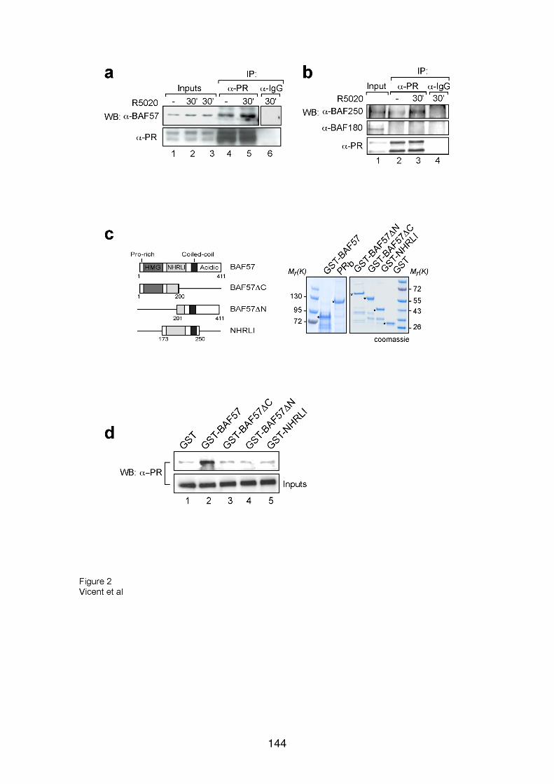
144

145

146

147
148

149
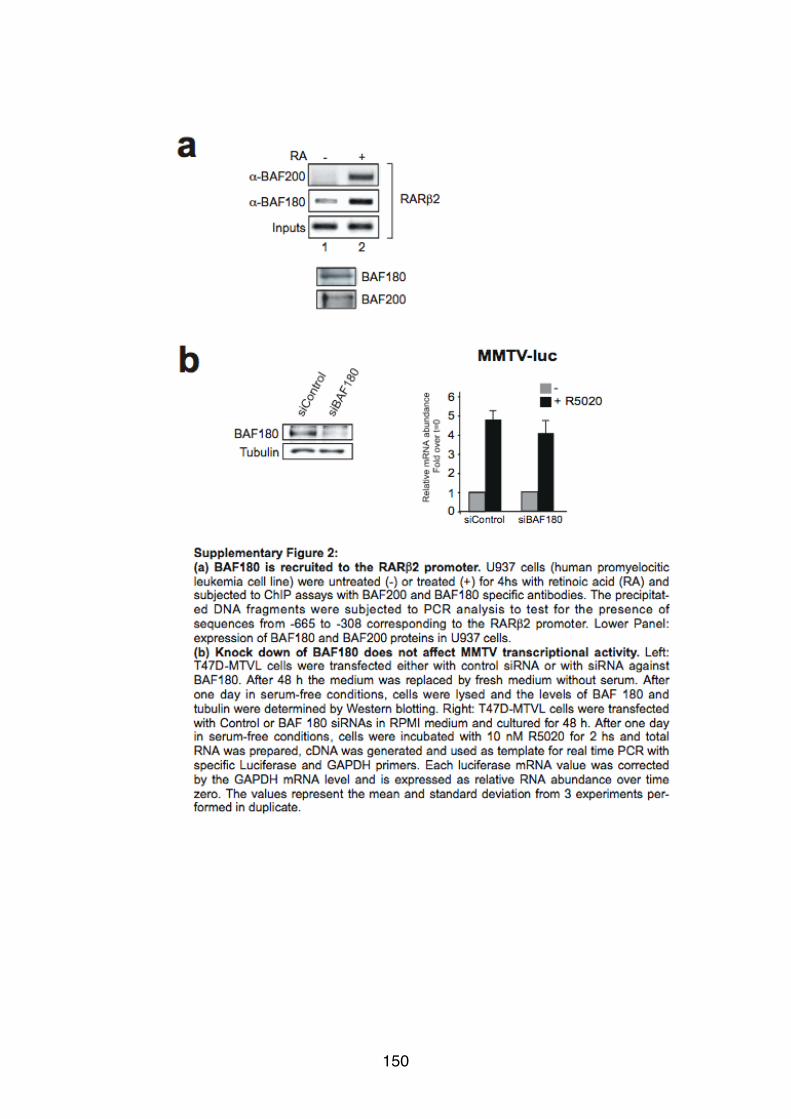
150
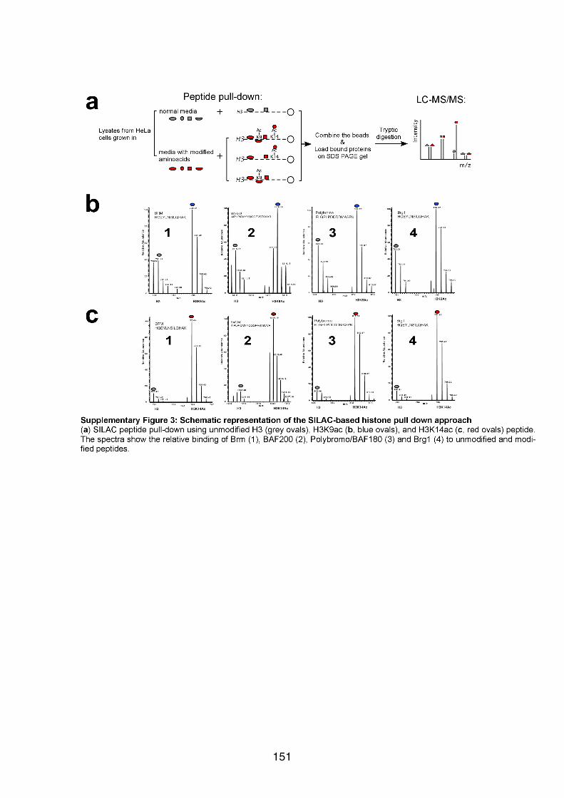
151

152
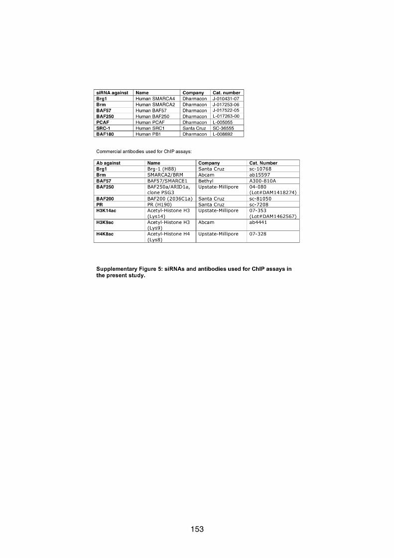
153

154

155
Chapter 3
“Characterization of H3/H4 histone tetramer as the output of SWI/SNF-
dependent remodelling of the nucleosome B within the MMTV
promoter”

156
Results 1. H3/H4 histone tetramer as the output of the SWI/SNF-dependent remodelling
of the nucleosome B within the MMTV promoter
The chromatin structure of the MMTV promoter is remodelled during hormone
induction as indicated by the appearance of a DNAase I hypersensitive site at the dyad
axis of nucleosome B (Truss et al, 1995). The remodelling mechanism following
progesterone treatment was first described by our laboratory in 2004 (Vicent et al,
2004). In that study, we analyzed the recruitment of transcription factors and ATP-
dependent remodelling complexes at the MMTV promoter of T47D-MTVL breast cancer
cells, which carry a single copy of this promoter integrated in their nuclear genome
(Truss et al, 1995). In a search for the molecular mechanism of chromatin remodelling,
the stoichiometry of core histones in the promoter nucleosome was analysed by ChIP
experiments using histone-specific antibodies and showed that H2A and H2B are
selectively displaced from the nucleosome B (Vicent et al, 2004).
In Figure 32, I reproduced the previously published results showing that
SWI/SNF-mediated remodelling of the nucleosome B results in ATP-dependent
histones H2A and H2B displacement. In the study presented here, I used an in vitro
system in which purified nucleosomes carrying the sequence of the nucleosome B
were incubated with purified SWI/SNF complex and ATP. Remodelled products were
subjected to ChIP assays using polyclonal antibodies against H2A and H4 followed by
PCR amplification of the nucleosome B sequence. As expected, histone H4 content
remained unchanged while the amount of histone H2A was 60% decreased (Figure
32A, lane 4 and 2, respectively and quantification in Figure 32B). These results
reproduced and confirmed that dimmers of histone H2A and H2B are displaced from
the promoter nucleosome B upon hormone induction, suggesting that the remodelled
nucleosome would be an H3/H4 tetramer structure. To provide further evidence for this
hypothesis, I used an in vitro system to reconstitute H3/H4 tetramer particles and
SWI/SNF-remodelled nucleosomes in order to compare their structures and functional
characteristics.
157

Chapter III RESULTS
158
Figure 32. SWI/SNF-dependent histone dimmer H2A/H2B displacement in the MMTV promoter. A. In vitro ChIP assay done in monocleosomes treated and non-treated with ySWI/SNF and ATP. Polyclonal antibodies against histones H2A and H4 were used. The immunoprecipitated material was subjected to PCR amplification using specific primers for nucleosome B sequence. B. RealTime-PCR quantifications of experiment explained in (A).
1.1 Preparation of octamers and H3/H4 tetramer particles from recombinant histones
For the reconstitution of octamers and H3/H4 tetramers, I used a radioactively
labelled DNA fragment comprising the sequence between -221 and +1 of the MMTV
promoter, encompassing the region corresponding to nucleosome B (Figure 33). The
positioned nucleosome B is located over the promoter region covering the five hormone
responsive elements (HREs) and the binding site for nuclear factor 1 (NF1) (Figure
33A). Our laboratory has described the existence of two main translational positions of
the nucleosome B shifted by 20 base pairs (Figure 33A, grey and yellow ovals). The
dyad axis of these two nucleosome populations are located either at position -107 or at
-127, relative to the transcription start site (+1) (Spangenberg et al, 1998). The
predominant position after an in vitro reconstitution with the nucleosome B sequence is
the one with dyad axis at -107 (Figure 33A, grey oval). The external HREs (HRE1 and
A
B
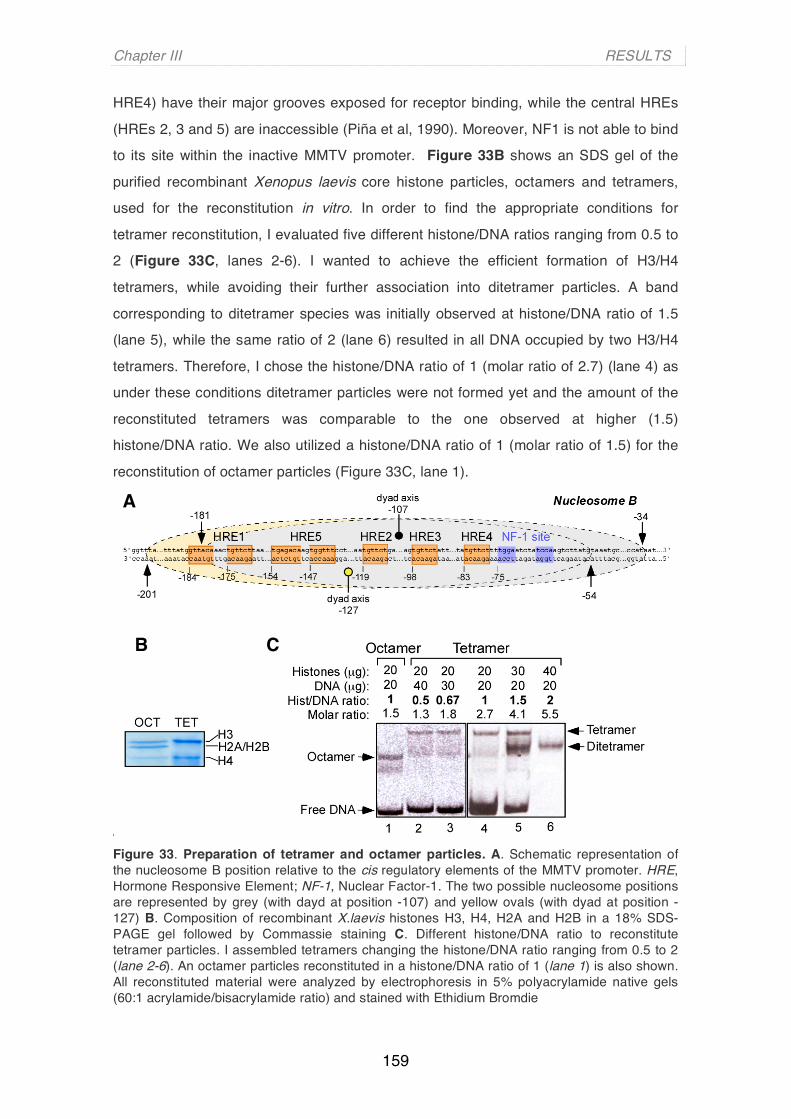
Chapter III RESULTS
159
HRE4) have their major grooves exposed for receptor binding, while the central HREs (HREs 2, 3 and 5) are inaccessible (Piña et al, 1990). Moreover, NF1 is not able to bind
to its site within the inactive MMTV promoter. Figure 33B shows an SDS gel of the
purified recombinant Xenopus laevis core histone particles, octamers and tetramers,
used for the reconstitution in vitro. In order to find the appropriate conditions for
tetramer reconstitution, I evaluated five different histone/DNA ratios ranging from 0.5 to
2 (Figure 33C, lanes 2-6). I wanted to achieve the efficient formation of H3/H4
tetramers, while avoiding their further association into ditetramer particles. A band
corresponding to ditetramer species was initially observed at histone/DNA ratio of 1.5
(lane 5), while the same ratio of 2 (lane 6) resulted in all DNA occupied by two H3/H4
tetramers. Therefore, I chose the histone/DNA ratio of 1 (molar ratio of 2.7) (lane 4) as
under these conditions ditetramer particles were not formed yet and the amount of the
reconstituted tetramers was comparable to the one observed at higher (1.5)
histone/DNA ratio. We also utilized a histone/DNA ratio of 1 (molar ratio of 1.5) for the
reconstitution of octamer particles (Figure 33C, lane 1).
Figure 33. Preparation of tetramer and octamer particles. A. Schematic representation of the nucleosome B position relative to the cis regulatory elements of the MMTV promoter. HRE, Hormone Responsive Element; NF-1, Nuclear Factor-1. The two possible nucleosome positions are represented by grey (with dayd at position -107) and yellow ovals (with dyad at position -127) B. Composition of recombinant X.laevis histones H3, H4, H2A and H2B in a 18% SDS-PAGE gel followed by Commassie staining C. Different histone/DNA ratio to reconstitute tetramer particles. I assembled tetramers changing the histone/DNA ratio ranging from 0.5 to 2 (lane 2-6). An octamer particles reconstituted in a histone/DNA ratio of 1 (lane 1) is also shown. All reconstituted material were analyzed by electrophoresis in 5% polyacrylamide native gels (60:1 acrylamide/bisacrylamide ratio) and stained with Ethidium Bromdie
A
B C
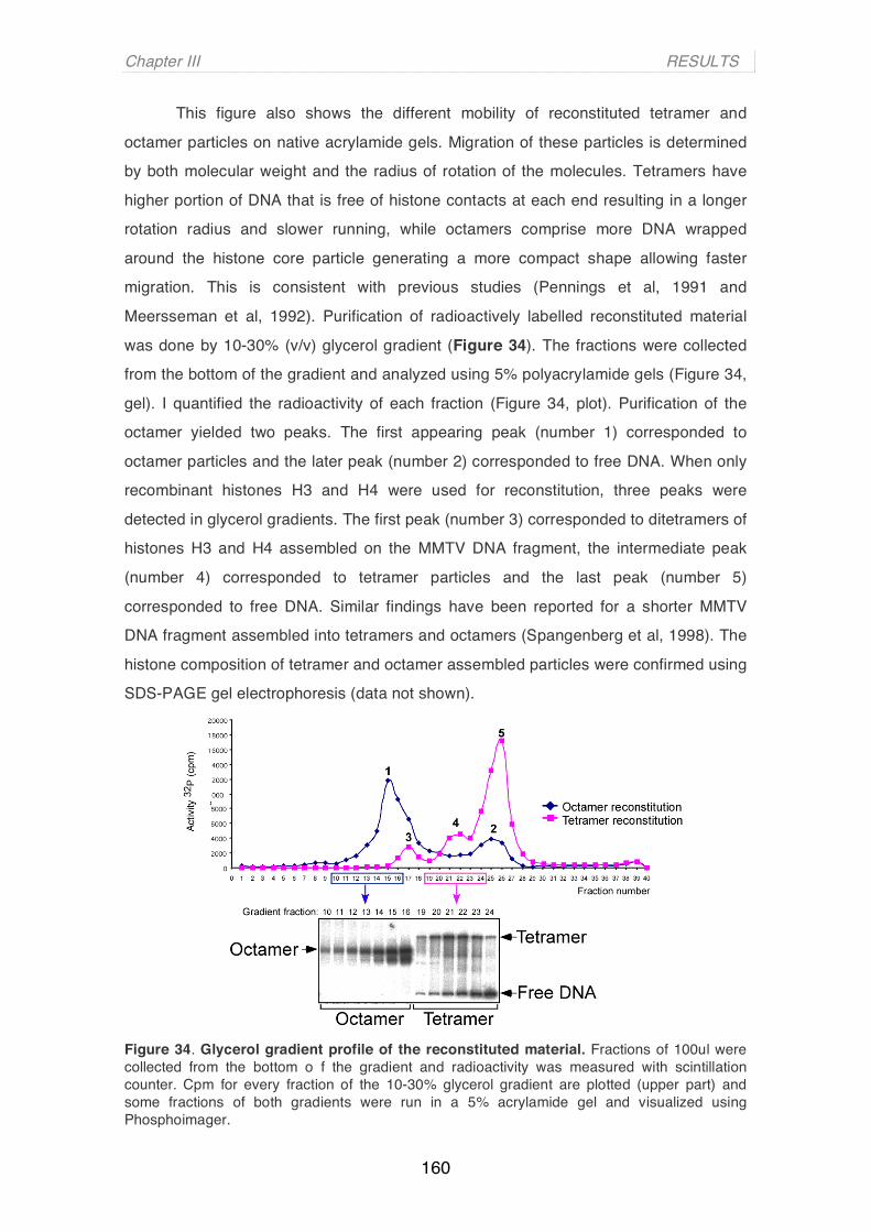
Chapter III RESULTS
160
This figure also shows the different mobility of reconstituted tetramer and
octamer particles on native acrylamide gels. Migration of these particles is determined
by both molecular weight and the radius of rotation of the molecules. Tetramers have
higher portion of DNA that is free of histone contacts at each end resulting in a longer
rotation radius and slower running, while octamers comprise more DNA wrapped
around the histone core particle generating a more compact shape allowing faster
migration. This is consistent with previous studies (Pennings et al, 1991 and
Meersseman et al, 1992). Purification of radioactively labelled reconstituted material
was done by 10-30% (v/v) glycerol gradient (Figure 34). The fractions were collected
from the bottom of the gradient and analyzed using 5% polyacrylamide gels (Figure 34,
gel). I quantified the radioactivity of each fraction (Figure 34, plot). Purification of the
octamer yielded two peaks. The first appearing peak (number 1) corresponded to
octamer particles and the later peak (number 2) corresponded to free DNA. When only
recombinant histones H3 and H4 were used for reconstitution, three peaks were
detected in glycerol gradients. The first peak (number 3) corresponded to ditetramers of
histones H3 and H4 assembled on the MMTV DNA fragment, the intermediate peak
(number 4) corresponded to tetramer particles and the last peak (number 5)
corresponded to free DNA. Similar findings have been reported for a shorter MMTV
DNA fragment assembled into tetramers and octamers (Spangenberg et al, 1998). The
histone composition of tetramer and octamer assembled particles were confirmed using
SDS-PAGE gel electrophoresis (data not shown).
Figure 34. Glycerol gradient profile of the reconstituted material. Fractions of 100ul were collected from the bottom o f the gradient and radioactivity was measured with scintillation counter. Cpm for every fraction of the 10-30% glycerol gradient are plotted (upper part) and some fractions of both gradients were run in a 5% acrylamide gel and visualized using Phosphoimager.

Chapter III RESULTS
161
2. The structure and binding properties of the reconstituted tetramer particles
resemble the SWI/SNF-remodelled nucleosomes
2.1 SWI/SNF remodelling makes nucleosomal DNA more accessible for MNase digestion
I examined the structural organization of the SWI/SNF-remodelled MMTV
nucleosomes and the reconstituted tetramer particles using digestion with Micrococcal
nuclease (MNase). This method has been widely employed to study specific
nucleosome positions due to the protection of DNA in contact with the histone octamer
against cleavage by this nuclease (Simpson RT, 1998 and Simpson RT, 1999). The
DNA products of MNase-digestions are the fragments that are protected by the histone
core particle, since MNase is able to digest all remaining linker DNA. Figure 35 shows
the DNA digestion patterns for octamer (OCT), SWI/SNF-treated octamer particles
(OCT+SWI/SNF) and tetramer (TET). Protected DNA fragments were extracted,
labelled and separated on 6% polyacrylamide gels. MNase digestion of MMTV
nucleosomes yielded a single 146 base pair fragment (Figure 35, lane 3), which
corresponded to the core particle DNA. The 146-bp fragment was also observed
following the MNase digestion of the SWI/SNF-remodelled nucleosomes, but was
accompanied by shorter fragments with a main band of 96 base pairs (Figure 35, lanes
4-8). The 96-bp band was the dominant species detected when H3/H4 tetramers were
subjected to MNase treatment (Figure 35, lane10).
Figure 35. Comparison of MNase-digestion of untreated ot SWI/SNF treated octamers and tetramers. Reconstituted octamers (OCT), SWI/SNF-treated nucleosomes (OCT+SWI/SNF) and tetramers (TET) were digested with increasing concentrations of MNase (0.6, 1.2 and 2.4 unit/reaction in lanes 1-3; 0.6, 1.2, 2.4, 5 and 10 unit/reaction in lanes 4-8; 0.15 and 0.2 unit/reaction in lanes 9 and 10) for 5 minutes at 22°C. The digested particles were analyzed by electrophoresis in a 6% polyacrylamide native gel.

Chapter III RESULTS
162
The remaining 146-bp band in SWI/SNF-treated octamers indicates that the
used concentration of SWI/SNF was not sufficient to remodel all nucleosome particles
(Figure 35, lanes 4-8). It seems likely that the reduction of total DNA observed after
MNase digestion with SWI/SNF-remodelled octamers and with tetramers is due to the
lack of H2A and H2B histones, that makes the DNA more accessible to the nuclease.
The fact that SWI/SNF remodelled octamers generate an MNase product of the same
size as H3/H4 tetramers suggests that a H3/H4 tetramer is the structure that results
from SWI/SNF-mediated remodelling of the nucleosome B.
2.2 Simultaneous binding of PR and NF1 observed at in vivo activated
MMTV promoters can be reproduced using reconstituted tetramers
The functional consequence of the observed changes in the nucleosome B
structure during hormone induction is to allow the simultaneous binding of more
molecules of PR to HREs 2, 3 and 4, together with the binding of NF1 to its recognition
site, facilitating full activation of the MMTV promoter. As said above, only two of the five
HREs can be bound by progesterone receptor in the inactive state of the MMTV
promoter, in which the binding site of NF1 is also inaccessible. However, following
hormone induction in vivo all HREs and the binding site for NF1 are occupied
simultaneously on the surface of a nucleosome-like structure (Truss et al, 1995) and a
functional synergism is observed between progesterone receptor (PR) and NF1
(Chalepakis et al, 1988).
2.2.1 NF1 and PR binding to reconstituted octamers and tetramers
Therefore, we next compared non-remodelled and remodelled nucleosome core
particles, H3/H4 tetramer particles and free DNA in terms of NF1 binding. Tetramers
and octamers were reconstituted in vitro on MMTV DNA as explained in Figure 33 and
34 (and Materials and Methods). The reconstituted particles and free DNA were
incubated with increasing amounts of recombinant NF1 transcription factor (Figure
36A). Free DNA was used to determine the appropriate amount of NF1 required for the
band shift experiment (Figure 36A lanes 2-4). As expected (Spangenberg et al, 1998),
regardless of the protein concentration used, I have not detected binding of NF1 to
DNA organized into the octamer core particle (Figure 36A lanes 6-8). However, NF1
was able to bind both the tetramer particle (Figure 36A lane 10-12) and the remodelled

Chapter III RESULTS
163
octamer (Vicent et al, 2004). Based on these results, I concluded that the NF1 site of
the MMTV promoter assembled into tetramer particles is accessible as in hormone-
activated MMTV promoter chromatin in vivo (Truss et al, 1995).
Figure 36. Differential binding of NF1 and PR to assembled octamer and tetramer particles. A. Binding of NF 1 to tetramer and octamer particles. Free DNA, octamer (OCT) and H3/H4 tetramer (TET) particles were incubated with increasing amounts of recombinant NF1 (1 ul, 2 ul and 4 ul). After allowing the binding reaction to proceed for 20 minutes at room temperature, the samples were analyzed on a 5% acrylamide gel. B. Binding of PR to tetramer and octamer particle. Free DNA, octamer (OCT) and tetramer (TET) particles were incubated for 20 minutes at room temperature with different amounts of PR (DNA: 1 ul, 2 ul and 5 ul; OCT: 8 ul; TET: 1 ul, 2 ul and 5 ul). The samples were analyzed in 3.5% acrylamide / 20%glycerol / 0.5% agarose / 0.3xTBE gel.
I next studied the progesterone receptor (PR) binding properties of an H3/H4
tetramer particle compared to an octamer particle and free DNA (Figure 36B). In the
absence of remodelling only the exposed HRE 1 is occupied by PR (Figure 36B lane 6
and Vicent et al, 2002). The half palindromic HRE 4 has low affinity for PR and is not
occupied under these conditions. In tetramers (Figure 36B lanes 7-10) and in
remodelled nucleosomes (Vicent et al, 2004) access to the hidden HREs is visualized
as two differentially migrating bands: one band corresponds to the occupancy of only
A
B

Chapter III RESULTS
164
HRE 1 by PR (TET-PR(1)), while a slower band corresponds to the situation where all
HRE sites are occupied by PR molecules (TET-PR(5)). The occupancy pattern of the
HREs in free DNA is similar to that observed in reconstituted tetramer particles (Figure
36B, compare lanes 2-4 with 8-10). This band shift assay was visualized using an
agarose-acrylamide gel since the TET-PR(5) complex is too large to be resolved in an
acrylamide gel. The lower resolution of the gel, compared to the products of the NF1
band shift assay (Figure 36A), is due to the addition of agarose to the gel composition.
In conclusion, all HRE can be occupied by PR in MMTV DNA assemble on H3/H4
tetramers.
2.2.2 Simultaneous binding of NF1 and PR to tetramers
In order to understand the properties of the nucleosome B in activated promoters,
I decided to test if H3/H4 tetramer particles (TET) are able to simultaneously bind NF1
and PR. The binding of these two proteins to the MMTV sequences organized around an
H3/H4 tetramer was studied using a band shift assay that detects very large complexes
(Figure 37A). Incubation of tetramer particles with NF1 generated a single retarded
complex (Figure 37A lane 2). Incubation of TET with PR generated a complex pattern of
bands, reflecting occupancy of HRE 1 alone or in various combinations with other HREs
(Figure 37A lane 3). Incubation of tetramer particles with NF1 and increasing amounts of
PR produced two main slow-migrating complexes containing both proteins (Figure 37A
lanes 4 and 5) and some intermediate complexes. The faster complex corresponds to
tetramers carrying NF1 and one molecule of PR (TET-NF1-PR(1)), while the slower
migrating complex, dominant at higher concentrations of PR (lane 5), correspond to
tetramers associated with NF1 and multiple PRs (TET-NF1-PR(5)). The same slow-
migrating complex was detected when either PR (Figure 37A lane 7) or NF1 (Figure 37A
lane 8) were first bound to the tetramer and the second transcription factor (NF1 or PR,
respectively) was added afterwards.
In order to confirm the nature of the observed complexes, I performed the same
band shift experiments with the addition of excess of oligonucleotides carrying either
the NF1 site sequence or the PRE element (Figure 37B). An excess of NF1
oligonucleotide competed with the TET-NF1 complex (Figure 37B, lane 3) leaving
almost all tetramers in a free state (Figure 37B, compare lanes 2 and 3). The addition
of NF1 oligo also abolished the appearance of the very slow migrating complex of TET-
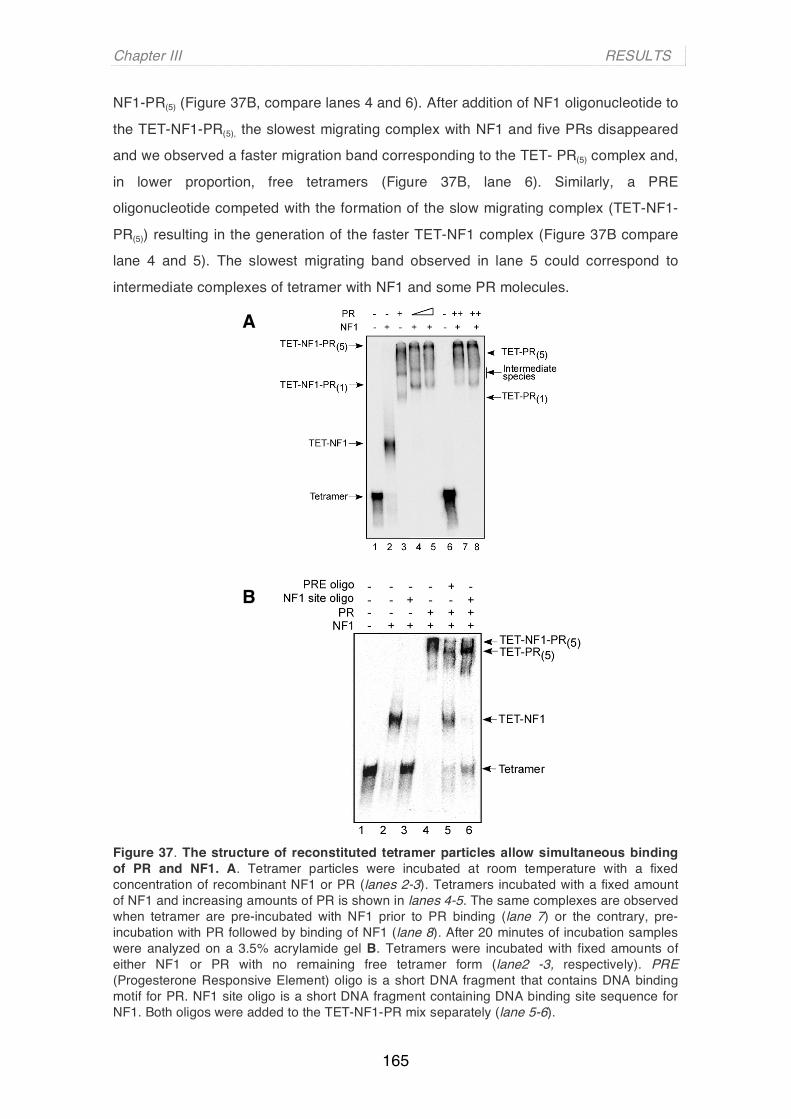
Chapter III RESULTS
165
NF1-PR(5) (Figure 37B, compare lanes 4 and 6). After addition of NF1 oligonucleotide to
the TET-NF1-PR(5), the slowest migrating complex with NF1 and five PRs disappeared
and we observed a faster migration band corresponding to the TET- PR(5) complex and,
in lower proportion, free tetramers (Figure 37B, lane 6). Similarly, a PRE
oligonucleotide competed with the formation of the slow migrating complex (TET-NF1-
PR(5)) resulting in the generation of the faster TET-NF1 complex (Figure 37B compare
lane 4 and 5). The slowest migrating band observed in lane 5 could correspond to
intermediate complexes of tetramer with NF1 and some PR molecules.
Figure 37. The structure of reconstituted tetramer particles allow simultaneous binding of PR and NF1. A. Tetramer particles were incubated at room temperature with a fixed concentration of recombinant NF1 or PR (lanes 2-3). Tetramers incubated with a fixed amount of NF1 and increasing amounts of PR is shown in lanes 4-5. The same complexes are observed when tetramer are pre-incubated with NF1 prior to PR binding (lane 7) or the contrary, pre-incubation with PR followed by binding of NF1 (lane 8). After 20 minutes of incubation samples were analyzed on a 3.5% acrylamide gel B. Tetramers were incubated with fixed amounts of either NF1 or PR with no remaining free tetramer form (lane2 -3, respectively). PRE (Progesterone Responsive Element) oligo is a short DNA fragment that contains DNA binding motif for PR. NF1 site oligo is a short DNA fragment containing DNA binding site sequence for NF1. Both oligos were added to the TET-NF1-PR mix separately (lane 5-6).
A
B

Chapter III RESULTS
166
These results indicate that the tetramer particle, mimicking the product of the
MMTV nucleosome remodelling generated by SWI/SNF in vivo, can accommodate the
full loading of the promoter with PR and NF1 as observed in living cells after hormone
induction (Truss et al, 1995).
3. H3/H4 tetramer is not a suitable substrate for SWI/SNF-dependent remodelling
Previous reports have claimed that SWI/SNF can remodel tetramers of histones
H3 and H4, although these tetramers are poorer substrates for SWI/SNF-dependent
remodelling compared to nucleosomal arrays (Boyer et al, 2000). To investigate this
further with MMTV promoter sequences, I incubated MMTV octamer and tetramer
particles with SWI/SNF and ATP and visualized the remodelled products in a 5%
acrylamide gel (Figure 38A). SWI/SNF treatment resulted in faster migrating bands
(Figure 38A lanes 2 and 3) that correspond to nucleosomes located to the ends of the
sequence (as indicated in the scheme and lanes 2 and 3). In contrast, SWI/SNF-treated
tetramer particles did not show any change in their migration pattern, even when the
highest concentration of the remodelling complex was used (Figure 38A lane 5).
To further explore whether tetramers could be substrates for the remodelling
activities of SWI/SNF, tetramers treated and non-treated with purified SWI/SNF were
analyzed by centrifugation in 10-30% glycerol gradient and characterized by
electrophoresis on 5% native polyacrylamide gels (Figure 38B). I found exactly the
same distribution of fractions for treated (lanes 9-16) and non-treated (lanes 1-8)
tetramer particles. The appearance of tetramer particles and free DNA occur exactly at
the same fraction numbers (compare lanes 4-5 with 12-13 for the tetramer; and lanes 8
and 16 for free DNA) suggesting no change in either structure or histone composition in
treated as compared to non-treated tetramers. Taken together, these results suggest
that the tetramer particle is not a suitable substrate for purified SWI/SNF remodelling
complex.
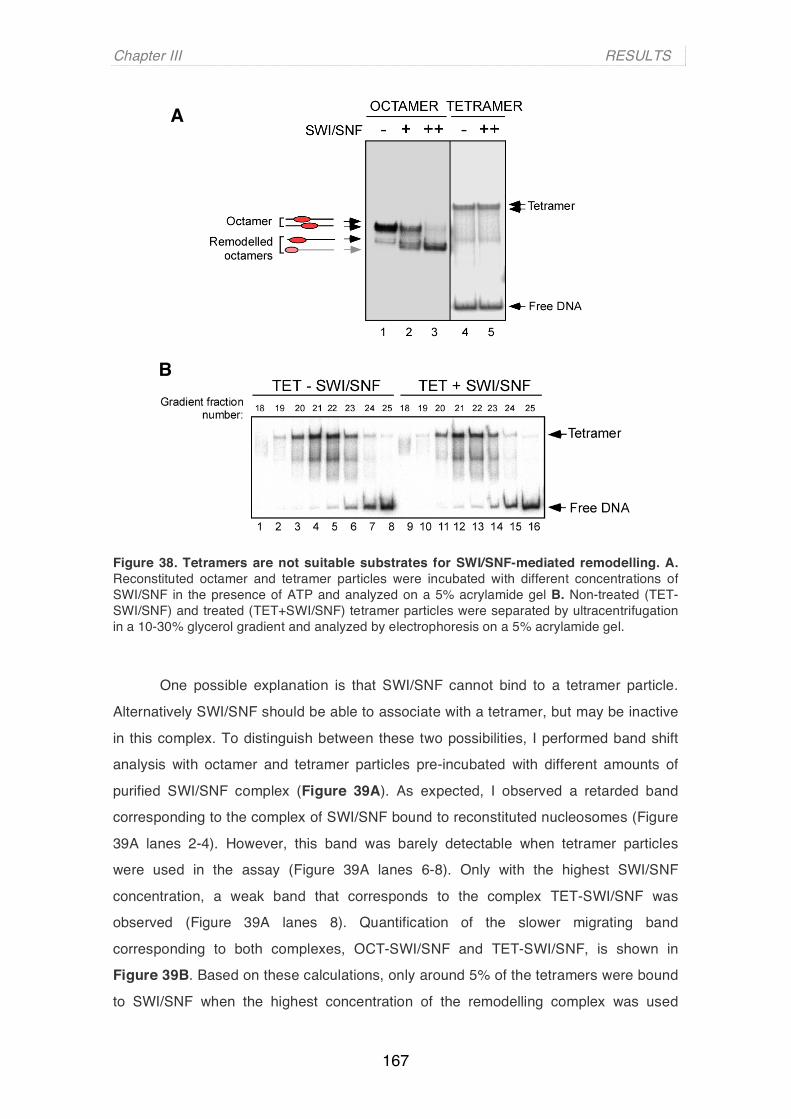
Chapter III RESULTS
167
Figure 38. Tetramers are not suitable substrates for SWI/SNF-mediated remodelling. A. Reconstituted octamer and tetramer particles were incubated with different concentrations of SWI/SNF in the presence of ATP and analyzed on a 5% acrylamide gel B. Non-treated (TET-SWI/SNF) and treated (TET+SWI/SNF) tetramer particles were separated by ultracentrifugation in a 10-30% glycerol gradient and analyzed by electrophoresis on a 5% acrylamide gel.
One possible explanation is that SWI/SNF cannot bind to a tetramer particle.
Alternatively SWI/SNF should be able to associate with a tetramer, but may be inactive
in this complex. To distinguish between these two possibilities, I performed band shift
analysis with octamer and tetramer particles pre-incubated with different amounts of
purified SWI/SNF complex (Figure 39A). As expected, I observed a retarded band
corresponding to the complex of SWI/SNF bound to reconstituted nucleosomes (Figure
39A lanes 2-4). However, this band was barely detectable when tetramer particles
were used in the assay (Figure 39A lanes 6-8). Only with the highest SWI/SNF
concentration, a weak band that corresponds to the complex TET-SWI/SNF was
observed (Figure 39A lanes 8). Quantification of the slower migrating band
corresponding to both complexes, OCT-SWI/SNF and TET-SWI/SNF, is shown in
Figure 39B. Based on these calculations, only around 5% of the tetramers were bound
to SWI/SNF when the highest concentration of the remodelling complex was used
A
BA

Chapter III RESULTS
168
(Figure 39A lane 8). On the other hand, at the same concentration, 80% of octamers
form a complex with SWI/SNF (Figure 39A lane 4). These data, together with the
results showing that SWI/SNF actively displaces H2A/H2B dimers from the nucleosome
B upon hormone induction (Vicent et al, 2004), imply that H2A/H2B dimers are needed
for the recognition and binding of the SWI/SNF to the nucleosome.
Figure 39. SWI/SNF binding to tetramers and octamers. A. Octamer and tetramer particles were incubated with increasing amounts of SWI/SNF in presence of ATP for 20 minutes at 30 ºC. After the incubation complexes were crosslinked and analysed in a 5% acrylamide gel B.Quantification of the appearing of a slow migrating band corresponding to either tetramer bound to SWI/SNF (TET-SWI/SNF complex and red line) or octamer bound to SWI/SNF (OCT-SWI/SNF complex and blue line).
4. Role of the internal HRE 2 and 3 in the activation of the MMTV promoter
Till now, I have shown that reconstituted tetramer particles resemble the
SWI/SNF-remodelled nucleosomes in histone content (Figure 32 and Vicent et al,
2004), histone-DNA interactions (Figure 35) and PR and NF1 binding properties
(Figures 36 and 37). We next examined the functional role of the inner HRE sites and
the NF1 binding site, all of which become accessible in the MMTV nucleosome B
during remodelling events.
A BA
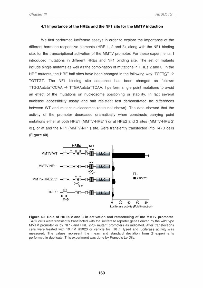
Chapter III RESULTS
169
4.1 Importance of the HREs and the NF1 site for the MMTV induction
We first performed luciferase assays in order to explore the importance of the
different hormone responsive elements (HRE 1, 2 and 3), along with the NF1 binding
site, for the transcriptional activation of the MMTV promoter. For these experiments, I
introduced mutations in different HREs and NF1 binding site. The set of mutants
include single mutants as well as the combination of mutations in HREs 2 and 3. In the
HRE mutants, the HRE half sites have been changed in the following way: TGTTCT
TGTTGT. The NF1 binding site sequence has been changed as follows:
TTGGAatctaTCCAA TTGAAatctaTTCAA. I perform single point mutations to avoid
an effect of the mutations on nucleosome positioning or stability. In fact several
nuclease accessibility assay and salt resistant test demonstrated no differences
between WT and mutant nucleosomes (data not shown). The data showed that the
activity of the promoter decreased dramatically when constructs carrying point
mutations either at both HRE1 (MMTV-HRE1-) or at HRE2 and 3 sites (MMTV-HRE 2-
/3-), or at and the NF1 (MMTV-NF1-) site, were transiently transfected into T47D cells
(Figure 40).
Figure 40. Role of HREs 2 and 3 in activation and remodelling of the MMTV promoter. T47D cells were transiently transfected with the luciferase reporter genes driven by the wild type MMTV promoter or by NF1- and HRE 2-/3- mutant promoters as indicated. After transfections cells were treated with 10 nM R5020 or vehicle for 16 h, lysed and luciferase activity was measured. The values represent the mean and standard deviation from 2 experiments performed in duplicate. This experiment was done by François Le Dily.
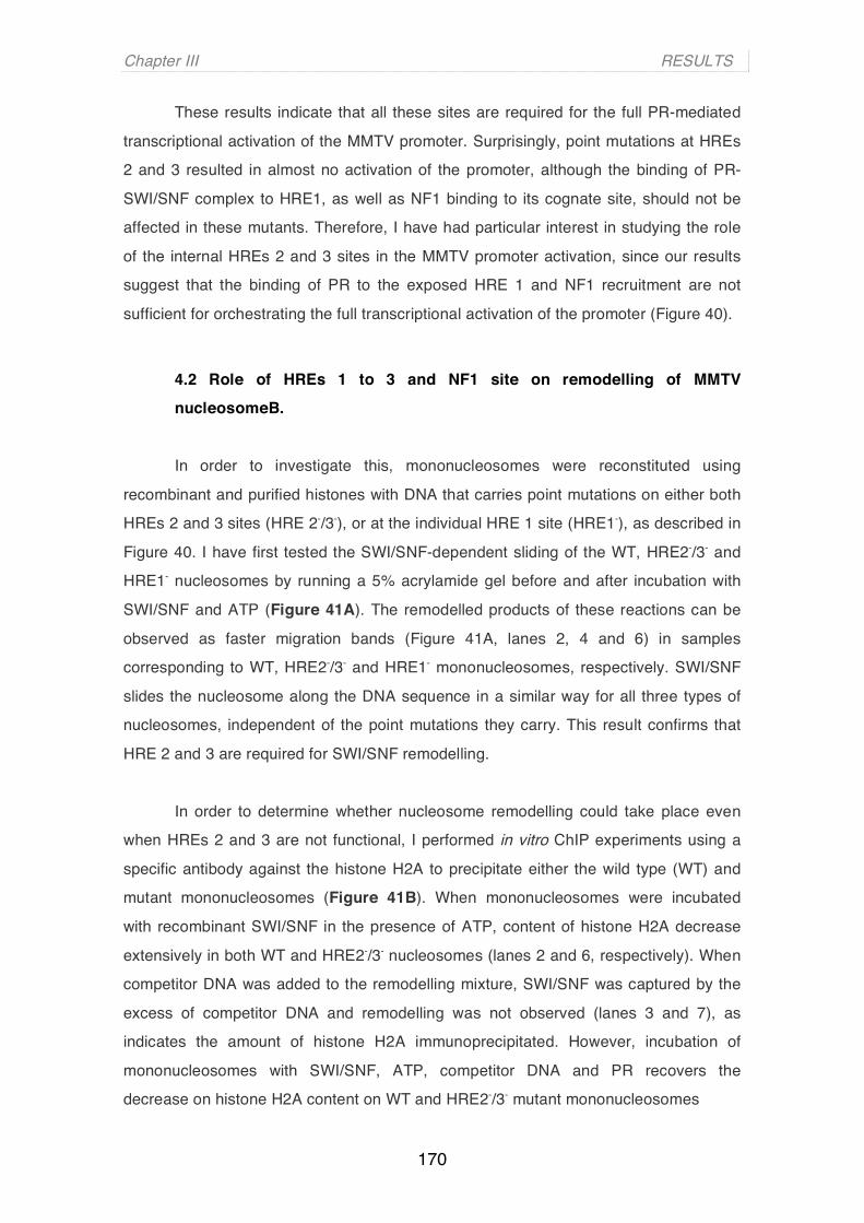
Chapter III RESULTS
170
These results indicate that all these sites are required for the full PR-mediated
transcriptional activation of the MMTV promoter. Surprisingly, point mutations at HREs
2 and 3 resulted in almost no activation of the promoter, although the binding of PR-
SWI/SNF complex to HRE1, as well as NF1 binding to its cognate site, should not be
affected in these mutants. Therefore, I have had particular interest in studying the role
of the internal HREs 2 and 3 sites in the MMTV promoter activation, since our results
suggest that the binding of PR to the exposed HRE 1 and NF1 recruitment are not
sufficient for orchestrating the full transcriptional activation of the promoter (Figure 40).
4.2 Role of HREs 1 to 3 and NF1 site on remodelling of MMTV nucleosomeB.
In order to investigate this, mononucleosomes were reconstituted using
recombinant and purified histones with DNA that carries point mutations on either both
HREs 2 and 3 sites (HRE 2-/3-), or at the individual HRE 1 site (HRE1-), as described in
Figure 40. I have first tested the SWI/SNF-dependent sliding of the WT, HRE2-/3- and
HRE1- nucleosomes by running a 5% acrylamide gel before and after incubation with
SWI/SNF and ATP (Figure 41A). The remodelled products of these reactions can be
observed as faster migration bands (Figure 41A, lanes 2, 4 and 6) in samples
corresponding to WT, HRE2-/3- and HRE1- mononucleosomes, respectively. SWI/SNF
slides the nucleosome along the DNA sequence in a similar way for all three types of
nucleosomes, independent of the point mutations they carry. This result confirms that
HRE 2 and 3 are required for SWI/SNF remodelling.
In order to determine whether nucleosome remodelling could take place even
when HREs 2 and 3 are not functional, I performed in vitro ChIP experiments using a
specific antibody against the histone H2A to precipitate either the wild type (WT) and
mutant mononucleosomes (Figure 41B). When mononucleosomes were incubated
with recombinant SWI/SNF in the presence of ATP, content of histone H2A decrease
extensively in both WT and HRE2-/3- nucleosomes (lanes 2 and 6, respectively). When
competitor DNA was added to the remodelling mixture, SWI/SNF was captured by the
excess of competitor DNA and remodelling was not observed (lanes 3 and 7), as
indicates the amount of histone H2A immunoprecipitated. However, incubation of
mononucleosomes with SWI/SNF, ATP, competitor DNA and PR recovers the
decrease on histone H2A content on WT and HRE2-/3- mutant mononucleosomes

Chapter III RESULTS
171
Figure 41. Role of HREs 2 and 3 in activation and remodelling of the MMTV promoter. A. Wild type, HRE 2-/3- and HRE1- mononucleosomes were treated with recombinant SWI/SNF as explained. Following incubation at 30ºC for 30 minutes excess of competitor DNA was added to the reaction for another 30 minutes at 30ºC. Remodelled products were analyzed by running a 5% acrylamide gel B. Nucleosomes assembled on wild type (WT) MMTV promoter DNA and on MMTV promoter mutated in HREs 2 and 3 (HRE 2-/3-) and HRE1 (HRE1-) were treated with SWI/SNF in the presence of ATP, competitor DNA and PR as indicated. Following incubation at 30ºC for 30 minutes, the nucleosomes were crosslinked in 0.25 % formaldehyde and immunoprecipitated with a polyclonal antibody against histone H2A. The precipitated DNA fragments were subjected to PCR analysis (20 cycles) with oligonucleotides corresponding to the MMTV promoter nucleosome B. 5% of the input DNA was used.
A
B
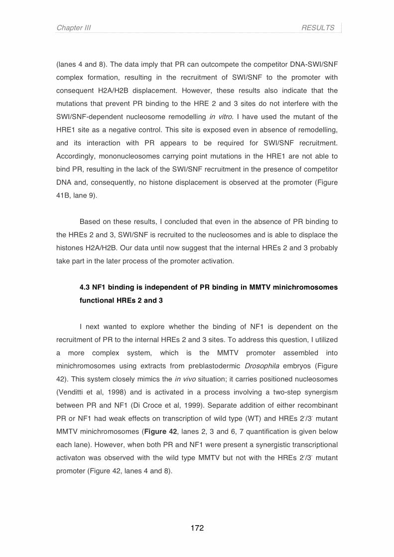
Chapter III RESULTS
172
(lanes 4 and 8). The data imply that PR can outcompete the competitor DNA-SWI/SNF
complex formation, resulting in the recruitment of SWI/SNF to the promoter with
consequent H2A/H2B displacement. However, these results also indicate that the
mutations that prevent PR binding to the HRE 2 and 3 sites do not interfere with the
SWI/SNF-dependent nucleosome remodelling in vitro. I have used the mutant of the
HRE1 site as a negative control. This site is exposed even in absence of remodelling,
and its interaction with PR appears to be required for SWI/SNF recruitment.
Accordingly, mononucleosomes carrying point mutations in the HRE1 are not able to
bind PR, resulting in the lack of the SWI/SNF recruitment in the presence of competitor
DNA and, consequently, no histone displacement is observed at the promoter (Figure
41B, lane 9).
Based on these results, I concluded that even in the absence of PR binding to
the HREs 2 and 3, SWI/SNF is recruited to the nucleosomes and is able to displace the
histones H2A/H2B. Our data until now suggest that the internal HREs 2 and 3 probably
take part in the later process of the promoter activation.
4.3 NF1 binding is independent of PR binding in MMTV minichromosomes
functional HREs 2 and 3
I next wanted to explore whether the binding of NF1 is dependent on the
recruitment of PR to the internal HREs 2 and 3 sites. To address this question, I utilized
a more complex system, which is the MMTV promoter assembled into
minichromosomes using extracts from preblastodermic Drosophila embryos (Figure
42). This system closely mimics the in vivo situation; it carries positioned nucleosomes
(Venditti et al, 1998) and is activated in a process involving a two-step synergism
between PR and NF1 (Di Croce et al, 1999). Separate addition of either recombinant
PR or NF1 had weak effects on transcription of wild type (WT) and HREs 2-/3- mutant
MMTV minichromosomes (Figure 42, lanes 2, 3 and 6, 7 quantification is given below
each lane). However, when both PR and NF1 were present a synergistic transcriptional
activaton was observed with the wild type MMTV but not with the HREs 2-/3- mutant
promoter (Figure 42, lanes 4 and 8).

Chapter III RESULTS
173
Figure 42.Central HREs are critical for transcriptional induction of the MMTV promoter. Wild type and HRE2-/3- mutant MMTV minichromosomes (25 ng of DNA in each reaction) assembled on post-blastodermic Drosophila embryo extracts were incubated with purified recombinant activated PR and NF1 and transcribed in vitro with HeLa nuclear extract. Products were visualized by primer extension analysis and sequencing gel electrophoresis. Quantification of intensities of the bands is shown below the gel. This experiment was performed by Jofre Font Mateu.
I next performed ChIP assays with the wild type and HRE 2-/3- mutant MMTV
reconstituted minichromosomes. In the presence of PR and NF1, when the MMTV
promoter is actively transcribed (Figure 42, lane 4), we detected the binding of PR
(Figure 43A, lanes 7-8) and NF1 (Figure 43B, lanes7-8) to the wild type (WT)
minichromosomes under the same conditions. I observed an eleven-fold decrease in
PR binding to the HRE 2-/3- mutant minichromosomes, (Figure 43A lanes 11-12), which
was in a good correlation with the low transcriptional rate displayed by this mutant
(Figure 42, lane 8). Nevertheless, the HRE 2-/3- mutant binds NF1 to a similar extent as
the wild type minichromosomes (Figure 43B, lanes 11-12). This HRE 2 and 3-
independent binding of NF1 was also observed by in vitro ChIP assay with the MMTV
promoter assembled in mononucleosomes (data not shown). These data lend further
support to the hypothesis that SWI/SNF-mediated recruitment of NF1 is dependent on
the binding of PR to the exposed HRE 1, but is independent of the PR binding to the
central HREs.
Chapter III RESULTS
174
Figure 43. PR and NF1 binding on minichromosomes. Wild type and HRE2-/3- mutant MMTV minichromosomes were incubated with purified recombinant activated PR and NF1 and subjected to ChIP assays as described previously using specific antibodies against PR (A) and NF1 (B). The precipitated DNA fragments were subjected to PCR analysis (20 and 25 cycles) to test for the presence of the MMTV nucleosome B sequence. Input material (5%) is shown for comparison. Quantification was done by real time PCR as previously described (Vicent et al., 2006).
5. NF1 cannot recruit SWI/SNF through direct interaction with the MMTV promoter
I further wanted to more precisely define the role of NF1 binding in the activation
of the MMTV promoter. One possibility is that NF1 could recruit more SWI/SNF
complex to the promoter facilitating the final remodelling of the nucleosome B. To test
this, I used a mutant MMTV promoter containing an insertion of 50bp downstream of
the HREs. The 50-bp insertion (+50) maintains the rotational setting and moves the
NF1 binding site away from the dyad axis and into the linker DNA (Eisfeld et al, 1997). I
performed binding assays using both the reconstituted mutant nucleosomes (Nuc +50)
and free DNA (DNA +50), which were incubated with increasing amounts of
recombinant NF1 (Figure 44A). In contrast to wild type nucleosomes (Figure 36A lanes
6-8), NF1 binds very efficiently to +50 mutant nucleosomes and a single retarded
complex is formed (Figure 44A lanes 6-8). The residual band includes a subpopulation
of nucleosomes in which the histone octamer is located at the end of the DNA fragment
partially covering the NF1 binding site (Eisfeld et al, 1997; see scheme on the right
site). The control band shift experiments with free DNA (DNA +50) are depicted in
BA
AA

Chapter III RESULTS
175
parallel (Figure 44A lanes 2-4). These data confirm that the mutant nucleosomes (Nuc
+50) have an accessible NF1 binding site in reconstituted ocatmer core particles.
Figure 44. NF1 cannot recruit SWI/SNF to MMTV nucleosome B. A. Nucleosome B sequence carrying as insertion of 50 nucleotides (DNA +50) and reconstituted MMTV nucleosomes on this sequences of DNA (Nuc +50) were incubated at room temperature for 20 minutes with increasing amounts of recombinant NF1 in 20 ul reactions. The samples were electrophoresed on a 3.5% acrylamide/0.5% agarose/20% glycerol/0.3 x TBE gel. The interpretation of the bands is shown on the left for free DNA (DNA +50) and on the right for nucleosomes (Nuc +50). B. Reconstituted nucleosomes on the wild type nucleosome B sequence (WT) and nucleosomes reconstituted on the DNA+50 (Nuc +50) were treated with SWI/SNF in the presence of ATP, competitor DNA and NF1 as indicated. Following incubation at room temperature for 20 min, the nucleosomes were digested with SacI restriction enzyme. The DNA fragments were electrophoresed in 8% denaturing polyacrylamide gels. The % of cut DNA is shown on top of each lane in bold numbers.
To investigate whether NF1 is able to recruit SWI/SNF complex to the
nucleosome B and promote histone displacement, I used SacI restriction enzyme
accessibility assays with +50 nucleosomes compared to the wild type (WT)
nucleosomes (Figure 44B). SacI target site is near the dyad axis (-104/-103; see
scheme on Figure 44A) and is not accessible in the absence of SWI/SNF-dependent
remodelling. Restriction endonuclease assay is used to determine structural changes
BA
ABA

Chapter III RESULTS
176
on nucleosome particles after specific remodelling reaction. Incubations with SWI/SNF
and ATP (in the absence of competitor DNA and NF1) made the SacI restriction
enzyme sites more accessible for cleavage similarly in WT and +50 nucleosomes
(Figure 44B; lane 5 compared to lane 1 and lane 7 compared to lane 3, respectively),
while no remodelling was observed in the presence of an excess of competitor DNA
(Figure 44B lanes 2 and 4, respectively). When nucleosomes were preincubated with
NF1, and in the presence of competitor DNA, no increase in SacI digestion was
observed (Figure 44B lanes 6 and 8, respectively). Quantifications are shown in Figure
44B, line seven from the top. These results indicate that, in contrast to PR, NF1
transcription factor is not able to recruit SWI/SNF to MMTV nucleosomes in vitro, even
when it is efficiently bound to the DNA.
Alternatively, NF1 could be bound too far from the nucleosome, since its site is
now placed 50 nt more downstream in the linker DNA region. In order to role out this
possibility I did the same experiment with a DNA carrying a shorter insertion: 30nt (data
not shown). The addition of 30nt locates now the NF1 binding site right at the end of
the nucleosomal DNA (while the edge of the nucleosome is in position -34, the new
location for the NF1 binding site is between -44 and -31). Restriction enzyme
accessibility assays performed in Nuc+30 gave similar results to those obtained with
Nuc+50 (data not shown). Moreover, in the absence of competitor DNA, SWI/SNF was
able to remodel the nucleosome even when NF1 was pre-bound to its binding site 30nt
far from the original site, indicating that the presence of NF1 do not physically prevents
SWI/SNF to remodel the nucleosome.
6. NF1 facilitates binding of PR to the HREs 2 and 3 after SWI/SNF
remodelling
The reciprocal synergism between PR and NF1 has been interpreted in terms of
NF1 stabilizing the open H2A/H2B depleted form of nucleosome B, but a more active
role of NF1 cannot be excluded. I next sought to determine whether NF1 could have a
role in PR binding to the inner HREs following SWI/SNF-mediated H2A/H2B
displacement. I performed band shift assays in which we tested whether PR
preferentially binds complexes of tetramer particles with NF1 rather than free tetramer
particles (Figure 45). Reconstituted tetramer particles were pre-incubated with a fixed
amount of NF1 that resulted in around 70% of the tetramers being in a free state and

Chapter III RESULTS
177
roughly 30% in a complex with NF1 (TET-NF1) (Figure 45A, lane 2). Increasing
amounts of PR were added in order to test its binding preference, which was followed
as the disappearance of the bands corresponding to the complex of tetramer with NF1
and the tetramer alone (Figure 45A lanes 3-7). At low concentrations of PR, the TET-
NF1 band decreased up to 70%, while the intensity of the free tetramer band was
reduced only about 20% (Figure 45A, lanes 3-4, and quantification in Figure 45B). At
high amounts of PR, both bands disappeared completely. These results imply that PR
has higher binding affinity for an NF1-bound tetramer than for a free tetramer particle.
Figure 45. NF1 facilitates PR binding to the HREs 2 and 3. A. Reconstituted MMTV tetramer particles were incubated with a fixed amount of NF1 (lane 2) and increasing amounts of PR (lane 3-7). Samples were analyzed in a 5% acrylamide gel. B. Quantification of the bands corresponding to the free tetramers (TET) and the complex of tetramer and NF1 (TET-NF1), as percentage of the value in the absence of added PR, is plotted against the amount of added PR. C. Reconstituted HRE 2-/3- MMTV tetramer particles were subjected to the same experimenatl conditions explained in (A and B).
To test if the observed higher affinity of PR for TET-NF1 was due to PR binding
to the internal HREs 2 and 3, we performed the same assay using tetramers
reconstituted from a sequence carrying mutations in HREs 2 and 3 (Figure 45C). In
this case, no preference of PR for TET-NF1 complex was found, the disappearance of
the free tetramer and TET-NF1 complex bands followed the same trend starting from
BA
AA
CA

Chapter III RESULTS
178
the lowest to the highest concentration of PR. Thus, I reasoned that the previously
detected difference in the affinity (Figure 45A and B) is due to the binding of PR to
HREs 2 and 3 and not to HRE 1. Taken together, these results demonstrate that NF1
binding to its recognition site facilitates recruitment of PR to the inner HREs. This
probably occurs through an NF1 dependent destabilization of the interaction of DNA
with the H3/H4 tetramer that makes HREs 2 and 3 more accessible for PR binding.
Accordingly, it is likely that NF1 plays a double structural role in the activation process
of the MMTV promoter. First by preventing reforming of the octamer particle and
second by labilizing histone-DNA interaction in the H3/H4 tetramer. Although I have
observed that NF1 binding occurs prior to PR recruitment to HREs 2 and 3, I also
tested if there is a significant difference in the affinity with which NF1 binds free
tetramer particles and TET-PR complexes (Figure 46A). I incubated wild type
tetramers with a PR concentration that results in 40% of the tetramer particles forming
a complex with PR in which HRE 1 is occupied (Figure 46A lane 2). When increasing
amounts of NF1 were added to the incubation mixture, I detected no significant
difference between the binding affinity of NF1 for a TET-PR(1) complex and for a free
tetramer particle (Figure 46A, lanes 3-6, and quantification in Figure 46B). These
results imply that PR mediates NF1 binding through the induction of chromatin
remodelling and histone displacement in the promoter region, but PR is not directly
involved in the NF1 recruitment to its recognition site. Contrary to this, bound NF1
facilitates in a remodelling-independent manner an open conformation of the
remodelled nucleosome optimal to PR recruitment and binding to HREs 2 and 3 (Figure
45).
Figure 46. NF1 binds TET-PR complexes and free tetramers with similar affinity. A. Reconstituted MMTV tetramer particles were incubated with a fixed amount of PR (lane 2) and increasing amounts of NF1 (lanes 3-6) and the samples were analyzed in a 5% acrylamide gel. B. Quantification of the bands corresponding to free tetramer (TET) and the complex of tetramer with PR (TET-PR), as percentage of the value in the absence of added NF1, is plotted against the amount of added NF1.
AA
BA
Discussion The results presented in this thesis contribute to a better understanding of the
molecular mechanisms involved in activation of the MMTV promoter by progesterone.
We have shown previously that PR interacts with an exposed HRE on the surface of a
positioned nucleosome and initiates chromatin remodelling as a prerequisite for the
subsequent steps of gene activation (Vicent et al, 2004). Here, I demonstrate the need
of the hSWI/SNFα complex, known as BAF, for the initial chromatin remodelling and
explore its mechanism of action. BAF is recruited at least in part via an interaction
between PR and the BAF57 subunit of the complex and is anchored via an interaction
with H3K14ac, likely generated by PCAF (Results Chapter 2). This suggests a
mechanism for the cooperation between two types of chromatin remodelling activitites.
The recruited BAF catalyzes the ATP dependent displacement of histones H2A/H2B
needed for NF1 to gain access to its cognate site in the promoter. NF1 binding in turn
stabilizes the remodelled nucleosome and facilitates recruitment of PR and associated
BAF to the central HREs 2 and 3. Receptor binding to the central HREs 2 and 3 is
necessary for full induction, but not for H2A/H2B displacement or NF1 binding. Finally, I
show that PR and NF1 can bind synergistically to MMTV promoter sequence
assembled on a tetramer of histones H3 and H4, a finding compatible with the idea that
the hormone induced remodelling of MMTV chromatin generates a tetramer of H3/H4
that acts as a platform on which the activating complex of PR and associated kinases
can orchestrate the assembly of co-activators and the basic transcriptional machinery.
The activated form of nucleosome B could be an H3/H4 tetramer particle
A reduction in the size of DNA protected against digestion with Micrococcal
nuclease in mononucleosomes treated with recombinant SWI/SNF complex reflects
structural changes on the histone composition of the core particle due to the
remodelling (Truss et al, 1995). The appearance of a predominant subnucleosomal
size fragment of 96 bp, that coincided with the one observed after MNase-treated
tetramers, suggests similarities between the tetramer structure and the remodelled
product of SWI/SNF-treated mononucleosomes. This observation is consistent with a
179

Chapter III DISCUSSION
180
previous publication (Spangenberg et al, 1998), in which they studied tetramer
positioning on the MMTV nucleosome B sequence. This work concluded, from DNA
cleavage analysis, that the curvature of DNA is preserved between octamer and
tetramer core particles except for the most proximal and distal two or three turns of the
nucleosomal double helix, which are more accessible to nuclease in the tetramer than
in the octamer. Consistent also with another previous reports where it was observed
that assembly of an H3/H4 tetramer is sufficient to position at least 90 bp of DNA
(Luger et al, 1997), I found that tetramers and SWI/SNF-treated octamers generate a
96 bp band in the MNase digestion. So, SWI/SNF-treated octamer particles protect the
same length of DNA than H3/H4 tetramers and this is another indicative that the
remodelled product could be a tetramer of histones H3 and H4.
Atomic Force Microscopy (AFM) studies of human SWI/SNF interacting with
reconstituted MMTV promoter arrays have shown a decrease of 25-50% in nucleosome
heights of some nucleosomes of the array after ATP-dependent remodelling, indicating
formation of smaller structures probably due to histone H2A/H2B loss (Wang et al,
2005). Interestingly, they also showed hSWI/SNF complexes partially overlapping
potentially histone-depleted nucleosomes (Wang et al, 2005). This result contradicts
my observation of tetramer as weak substrates for SWI/SNF binding, although
differences could be explained by the use of different systems and conditions. Some
recent works has contributed to a better understanding on the SWI/SNF-nucleosome
complex architecture and further remodelling actions (Sengupta et al, 2001, Wang et al,
2005, Dechassa et al, 2008) and specially a biochemical study on the yeast SWI/SNF
complex has shown the acidic N terminus domain of the Swi3p subunit as a novel
H2A/H2B-binding domain required for ATP-dependent H2A/H2B dimer loss (Yang et al,
2007). The direct interaction between Swi3p subunit and histones H2A and H2B may
serve as well to anchor SWI/SNF to the nucleosome, thus, lack of both histone dimmer
could results in weaker binding of the complex to the H3/H4 tetramer particle, as
observed.
Whether active displacement of H2A/H2B dimmers is an exclusive phenomenon
for the nucleosome B of the MMTV promoter or is a general effect of the SWI/SNF
complex remained still to be further investigated. It will be very interesting to extent this
study to other promoter or regulatory regions where SWI/SNF is acting and try to
explain the biological function for which some nucleosomes suffer histone H2A/H2B

Chapter III DISCUSSION
181
displacement. Our lab has proven that the contribution of the DNA sequence on the
definition of the outcome of the remodelling process is essential (Vicent et al, 2006 and
Sims et al, 2007). We have worked towards the finding of sequence features that lead
SWI/SNF to one or other remodelling process, although results are still not conclusive.
There are already some examples in the literature where SWI/SNF-mediated
remodelling seems to be similar to the nucleosome B. The human ε-globin promoter
region that is organized in well-positioned nucleosomes, with a nucleosome called N1
(the TATA-proximal nucleosome), is an interesting case. Transcriptional activation of
the ε-globin gene is accompanied by chromatin structural changes limited to the N1
nucleosome. The altered structure is reflected in the increased sensitivity of N1 to
nucleases and the generation of a DNase I Hypersensitive site (Gong et al, 1996).
MNase digestion revealed that a subnucleosome-size N1 DNA fragment was released
from actively transcribed promoters, while digestion of inactive promoters produced
DNA fragments of nucleosomal length (Gui et al, 2001). Structural changes are
accompanied by both H3 hyperacetylation and partially depletion of linker histone H1,
as shown by in vitro ChIPs with reconstituted minichromosomes. It has been shown
that recruitment of SWI/SNF to the nucleosome N1 is indispensable for remodelling
and ε-globin promoter full activation (Gui et al, 2003). Thus, it would be interesting to
study similar cases and understand the biological meaning of SWI/SNF-mediated
displacement of histones H2A/H2B targeting specific nucleosome.
Synergistic binding of PR and NF1 on a H3/H4 tetramer structure
A tetramer of histones H3 and H4 positions MMTV promoter sequences in a
similar way to histone octamers but NF1 can bind to an H3/H4 tetramer particle with
relatively high affinity (Spangenberg et al 1998), although both octamer and tetramer
populations cover the NF1 binding site (located at –75, respect to +1). Here I show that
PR can acces all HREs in MMTV sequences positioned around an H3/H4 tetramer.
More important, such particles can bind PR and NF1 simultaneously, reminiscent of
what is observed in cells carrying a single copy of the MMTV promoter integrated in
chromatin (Truss et al, 1995). Since NF1 recognition site and the HREs 3 are very
closed in the DNA sequence (-75/-63 are the NF1 binding site boundaries while -98/-93
are the HRE 3 ones), it is plausible to believe that NF1 physically impedes PR binding
on the HRE 2-4 cluster. Actually, it has been observed by DNAse I footprinting assays
that no simultaneous binding of NF1 and PR in free DNA takes place (Di Croce et al,

Chapter III DISCUSSION
182
1999). A physical impediment between the NF1 binding site and the HRE 2-4 cluster is
the more feasible explanation. However, I observed that not only both factors bound
simultaneously in reconstituted tetramer particles, but also pre-bound NF1 to the
tetramer favours PR binding to inner HREs 2 and 3. The nature of this synergism is
unknown but it does not result from a direct interaction between NF1 and PR (data not
shown). Our interpretation is that the tridimentional structure of the DNA wrapped
around an H3/H4 tetramer generates a new disposition of the NF1 binding site and the
HRE 2-4, allowing the binding of both factors to its cognate sites on the DNA.
Moreover, the presence of NF1 may cause a conformational change that favours the
binding of more PR heterodimmers to the inner HRE 2 and 3, most likely as a
prerequisite for full activation of the MMTV promoter.
Knocking down NF1c by siRNA in T47D-MTVL cells causes a drastic inhibition
of the MMTV promoter activity, indicating that binding of NF1 is essential for promoter
activation (Vicent et al, manuscript in preparation). The structural role of NF1 in the
binding to the nucleosome B was first emphasized by Prado et al. (2002). They
observed that the DNA binding domain of NF1, which is devoid of significant
transactivation activity, still synergizes with glucocorticoid receptor (GR) in
transactivation and chromatin remodelling, using both yeast system (with addition of
recombinant GR and NF1) and in vitro transcription (with assembled minichromosomes
in Drosophila embryo extracts and HeLa cell nuclear extract) (Prado et al, 2002). Thus,
they showed that NF1 does not act like a conventional transcription factor in the context
of the activated MMTV promoter, since removal of its proline-rich transactivation
domain did not interact with the activation process of the MMTV promoter. As this
structural effect is maintained when only the NF1 DNA binding domain is used with
MMTV minichromosomes (Di Croce et al, 1999), it can be accounted for by at least two
possible mechanism: direct competition between NF1 and histones H2A/H2B dimmers
for DNA binding, or a deformation of the double helix upon NF1 binding favourable for
PR access to HREs 2-4. Though we know that NF1 does not recruit BAF to the
promoter, we cannot exclude that NF1 is being recruited to the promoter forming a
complex with other co-activator factors that will help on the activation process.
For all these reasons, one of the main contributions of this work to the model we
propose for MMTV activation is the essential role of NF1 binding prior to the second
wave of PR recruitment to the internal HREs 2 and 3. Our hypothesis is that NF1
Chapter III DISCUSSION
183
binding will act as a “switch” on the activation process. Once NF1 is bound, the
transcriptional activation becomes irreversible. On the contrary, in cells lacking NF1,
the nucleosome B undergoes the first steps of the activation process but, the open
conformation is not stabilized by the binding of NF1 and thus neither PR binding to
HREs 2 and 3 is possible nor full activation of the promoter. Thus, in the absence of
NF1, the nucleosome B returns to the inactive state by deposition of H2A/H2B
dimmers, binding of the HP1-γ repressive complex and displacement of BAF and other
coactivators.
MMTV promoter activation: a model to better understand hormone-dependent
promoters
Our present model for activation of the MMTV promoter is shown in Figure 47.
Before hormone addition the MMTV promoter is silent due to its interaction with a
repressive complex that includes HP1γ (1). The nature of this repressive complex is a
current matter of investigation in the lab. Very rapidly after hormone addition (5-10
min), PR is phosphorylated by Erk at Ser293. Activated receptors bind to the exposed
HRE1 as part of a ternary complex with activated Erk, activated Msk1, which has also
been phosphorylated by Erk (Vicent et al, 2006). PR is also recruiting BAF complex to
the nucleosome B through direct interaction of BAF57 subunit, as we demonstrated by
GST pull down experiments. We cannot exclude an interaction between PR and other
BAF complex. PCAF is present at short times after hormone induction on the promoter,
but we still don’t know if its recruitment is via a direct interaction with the progesterone
receptor (2). The PR-Erk-Msk and PCAF complex phosphoacetylates H3, leading to
H3S10PhosK14Ac, modifications that displace the repressive complex (3) and anchors
the BAF complex which is stabilized specially by H3K14Ac (4). BAF catalyzes the ATP-
dependent H2A/H2B displacement that facilitates NF1 binding (5). In the absence of
NF1 the histone octamer particle is reformed and the HP1γ-containing repressive
complex brings the promoter to the initial silenced state, thus preventing efficient
activation. In this case, BAF is presumably displaced from the promoter as well as the
other co-activators. How the nucleosome returns to a silent state is still unknown.
Contrary, in the presence of NF1 bound to the H3/H4 tetramer particles (6) the
reassociation of H2A/H2B dimmers is prevented and binding of further PR molecules
and BAF complexes to the recently exposed HREs 2 and 3 is facilitated (7), leading to
full promoter activation (8). It is worth to mention the key role on NF1 in cooperate with

Chapter III DISCUSSION
184
the open conformation of the promoter to maintain accessible the inner HREs 2 and 3
for further PR binding. Whether this new PR is coming in a complex with more BAF and
kinases or other factors it is still unclear. Actually, an open question that remains from
this work is why the promoter still need BAF complex on the nucleosome after
remodelling take place and is it anchor if the nucleosome has became an H3/H4
tetramer particle. How this full loaded promoter on an H3/H4 tetramer is further
converted into a preinitiation complex remains to be established as well. We also do
not know under which conditions the activated tetramer particle reverts to the inactive
octamer state (9).
Figure 47. Model of the activation process of the MMTV promoter.
Conclusions
1. Both yeast and human nucleosomal DNA sequences exhibit symmetric patterns
of curvature, while for human nucleosome sequences other features have been
described as well: enrichment with CpG island regions, no contrains for
sequence conservation among species and overrepresentation of certain triplet
nucleotide patterns containing CG.
2. High SymCurv scores tend to coincide with well-positioned nucleosomes,
suggesting the ability of the method to predict strong nucleosome positions with
high accurancy.
3. “Key nucleosomes” are a subset of the whole nucleosome map with strong
signal for positioning which is basically defined by the symmetry of curvature of
its nucleosomal sequence.
4. “Key nucleosomes” are located in functional genomic regions such as
Transcription Start Site and Splice junctions, suggesting its special role in
cellular processes regulation.
5. BAF is required for progesterone gene activation in T47D-MTVL cells and is
recruited to the MMTV promoter upon induction.
6. BAF57 and BAF250 interact with progesterone receptor in T47D-MTVL cells.
7. Acetylation on histone H3K14 by PCAF is required for hormonal induction and it
anchors the BAF complex on the promoter.
185

CONCLUSIONS
186
8. The BAF complex facilitates NF1 binding by mediating histone H2A/H2B
displacement.
9. H3/H4 histone tetramer is most likely the output of the SWI/SNF-dependent
remodelling of the nucleosome B within the MMTV promoter.
10. The structure and binding properties of the reconstituted tetramer particles
resemble the SWI/SNF-remodelled nucleosomes.
11. H3/H4 tetramer is not a suitable substrate for SWI/SNF-dependent remodelling.
12. NF1 binding is independent of PR binding in MMTV minichromosomes
functional HREs 2 and 3.
13. NF1 cannot recruit SWI/SNF through direct interaction within the MMTV
promoter.
14. NF1 facilitates binding of PR to the HREs 2 and 3 after SWI/SNF remodelling.

187
Supplementary articles

188

Yang X, Zaurin R, Beato M, Peterson CL. Swi3p controls SWI/SNF assembly and ATP-dependent H2A-H2B displacement. Nat Struct Mol Biol. 2007 Jun;14(6):540-7.
Vicent GP, Ballaré C, Zaurin R, Saragüeta P, Beato M. Chromatin remodeling and control of cell proliferation by progestins via cross talk of progesterone receptor with the estrogen receptors and kinase signaling pathways. Ann N Y Acad Sci. 2006 Nov;1089:59-72.

211
References
A Abrams, E., Neigeborn, L. and Carlson, M. (1986). Molecular analysis of SNF2 and SNF5, genes required for expression of glucose-repressible genes in Saccharomyces cerevisiae. Mol Cell Biol 6, 3643-51. Ahmad, K. and Henikoff, S. (2002). The histone variant H3.3 marks active chromatin by replication-independent nucleosome assembly. Mol Cell 9, 1191-200. Albert, I., Mavrich, T. N., Tomsho, L. P., Qi, J., Zanton, S. J., Schuster, S. C. and Pugh, B. F. (2007). Translational and rotational settings of H2A.Z nucleosomes across the Saccharomyces cerevisiae genome. Nature 446, 572-6. Allis, C. D., Richman, R., Gorovsky, M. A., Ziegler, Y. S., Touchstone, B., Bradley, W. A. and Cook, R. G. (1986). hv1 is an evolutionarily conserved H2A variant that is preferentially associated with active genes. J Biol Chem 261, 1941-8. Angelov, D., Molla, A., Perche, P. Y., Hans, F., Cote, J., Khochbin, S., Bouvet, P. and Dimitrov, S. (2003). The histone variant macroH2A interferes with transcription factor binding and SWI/SNF nucleosome remodeling. Mol Cell 11, 1033-41. Aranda, A. and Pascual, A. (2001). Nuclear hormone receptors and gene expression. Physiol Rev 81, 1269-304. Arents, G., Burlingame, R. W., Wang, B. C., Love, W. E. and Moudrianakis, E. N. (1991). The nucleosomal core histone octamer at 3.1 A resolution: a tripartite protein assembly and a left-handed superhelix. Proc Natl Acad Sci U S A 88, 10148-52. Armstrong, J. A., Bieker, J. J. and Emerson, B. M. (1998). A SWI/SNF-related chromatin remodeling complex, E-RC1, is required for tissue-specific transcriptional regulation by EKLF in vitro. Cell 95, 93-104.
B Baldi, P., Brunak, S., Chauvin, Y. and Krogh, A. (1996). Naturally occurring nucleosome positioning signals in human exons and introns. J Mol Biol 263, 503-10. Ballare, C., Uhrig, M., Bechtold, T., Sancho, E., Di Domenico, M., Migliaccio, A., Auricchio, F. and Beato, M. (2003). Two domains of the progesterone receptor interact with the estrogen receptor and are required for progesterone activation of the c-Src/Erk pathway in mammalian cells. Mol Cell Biol 23, 1994-2008. Bao, Y., Konesky, K., Park, Y. J., Rosu, S., Dyer, P. N., Rangasamy, D., Tremethick, D. J., Laybourn, P. J. and Luger, K. (2004). Nucleosomes containing the histone variant H2A.Bbd organize only 118 base pairs of DNA. Embo J 23, 3314-24. Barski, A., Cuddapah, S., Cui, K., Roh, T. Y., Schones, D. E., Wang, Z., Wei, G., Chepelev, I. and Zhao, K. (2007). High-resolution profiling of histone methylations in the human genome. Cell 129, 823-37.

212
Bartsch, J., Truss, M., Bode, J. and Beato, M. (1996). Moderate increase in histone acetylation activates the mouse mammary tumor virus promoter and remodels its nucleosome structure. Proc Natl Acad Sci U S A 93, 10741-6. Batsche, E., Yaniv, M. and Muchardt, C. (2006). The human SWI/SNF subunit Brm is a regulator of alternative splicing. Nat Struct Mol Biol 13, 22-9. Bazett-Jones, D. P., Cote, J., Landel, C. C., Peterson, C. L. and Workman, J. L. (1999). The SWI/SNF complex creates loop domains in DNA and polynucleosome arrays and can disrupt DNA-histone contacts within these domains. Mol Cell Biol 19, 1470-8. Beato, M. (1989). Gene regulation by steroid hormones. Cell 56, 335-44. Beato, M., Herrlich, P. and Schutz, G. (1995). Steroid hormone receptors: many actors in search of a plot. Cell 83, 851-7. Beato, M. and Klug, J. (2000). Steroid hormone receptors: an update. Hum Reprod Update 6, 225-36. Becker, P. B., Gloss, B., Schmid, W., Strahle, U. and Schutz, G. (1986). In vivo protein-DNA interactions in a glucocorticoid response element require the presence of the hormone. Nature 324, 686-8. Bednar, J., Horowitz, R. A., Grigoryev, S. A., Carruthers, L. M., Hansen, J. C., Koster, A. J. and Woodcock, C. L. (1998). Nucleosomes, linker DNA, and linker histone form a unique structural motif that directs the higher-order folding and compaction of chromatin. Proc Natl Acad Sci U S A 95, 14173-8. Belandia, B., Orford, R. L., Hurst, H. C. and Parker, M. G. (2002). Targeting of SWI/SNF chromatin remodelling complexes to estrogen-responsive genes. Embo J 21, 4094-103. Belikov, S. V. and Karpov, V. L. (1996). Mapping protein-DNA interactions with CIS-DDP: chromatin structure of promoter region of D. Melanogaster hsp 70 gene. Biochem Mol Biol Int 38, 997-1002. Biegel, J. A. and Pollack, I. F. (2004). Molecular analysis of pediatric brain tumors. Curr Oncol Rep 6, 445-52. Boeger, H., Griesenbeck, J., Strattan, J. S. and Kornberg, R. D. (2003). Nucleosomes unfold completely at a transcriptionally active promoter. Mol Cell 11, 1587-98. Boonyaratanakornkit, V., Scott, M. P., Ribon, V., Sherman, L., Anderson, S. M., Maller, J. L., Miller, W. T. and Edwards, D. P. (2001). Progesterone receptor contains a proline-rich motif that directly interacts with SH3 domains and activates c-Src family tyrosine kinases. Mol Cell 8, 269-80. Bowen, N. J., Fujita, N., Kajita, M. and Wade, P. A. (2004). Mi-2/NuRD: multiple complexes for many purposes. Biochim Biophys Acta 1677, 52-7. Bresnick, E. H., Bustin, M., Marsaud, V., Richard-Foy, H. and Hager, G. L. (1992). The transcriptionally-active MMTV promoter is depleted of histone H1. Nucleic Acids Res 20, 273-8. Bresnick, E. H., John, S., Berard, D. S., LeFebvre, P. and Hager, G. L. (1990). Glucocorticoid receptor-dependent disruption of a specific nucleosome on the mouse mammary tumor virus promoter is prevented by sodium butyrate. Proc Natl Acad Sci U S A 87, 3977-81. Bresnick, E. H., John, S. and Hager, G. L. (1991). Histone hyperacetylation does not alter the positioning or stability of phased nucleosomes on the mouse mammary tumor virus long terminal repeat. Biochemistry 30, 3490-7. Brown, C. E., Lechner, T., Howe, L. and Workman, J. L. (2000). The many HATs of transcription coactivators. Trends Biochem Sci 25, 15-9. Brown, D. T., Izard, T. and Misteli, T. (2006). Mapping the interaction surface of linker histone H1(0) with the nucleosome of native chromatin in vivo. Nat Struct Mol Biol 13, 250-5. Bruggemeier, U., Kalff, M., Franke, S., Scheidereit, C. and Beato, M. (1991). Ubiquitous transcription factor OTF-1 mediates induction of the MMTV promoter through synergistic interaction with hormone receptors. Cell 64, 565-72. Bruggemeier, U., Rogge, L., Winnacker, E. L. and Beato, M. (1990). Nuclear factor I acts as a transcription factor on the MMTV promoter but competes with steroid hormone receptors for DNA binding. Embo J 9, 2233-9. Bruno, M., Flaus, A., Stockdale, C., Rencurel, C., Ferreira, H. and Owen-Hughes, T. (2003). Histone H2A/H2B dimer exchange by ATP-dependent chromatin remodeling activities. Mol Cell 12, 1599-606. Bucci, L. R., Brock, W. A. and Meistrich, M. L. (1982). Distribution and synthesis of histone 1 subfractions during spermatogenesis in the rat. Exp Cell Res 140, 111-8.

213
Byvoet, P., Shepherd, G. R., Hardin, J. M. and Noland, B. J. (1972). The distribution and turnover of labeled methyl groups in histone fractions of cultured mammalian cells. Arch Biochem Biophys 148, 558-67.
C Cai, Y., Jin, J., Florens, L., Swanson, S. K., Kusch, T., Li, B., Workman, J. L., Washburn, M. P., Conaway, R. C. and Conaway, J. W. (2005). The mammalian YL1 protein is a shared subunit of the TRRAP/TIP60 histone acetyltransferase and SRCAP complexes. J Biol Chem 280, 13665-70. Candau, R., Chavez, S. and Beato, M. (1996). The hormone responsive region of mouse mammary tumor virus positions a nucleosome and precludes access of nuclear factor I to the promoter. J Steroid Biochem Mol Biol 57, 19-31. Carlson, M. and Laurent, B. C. (1994). The SNF/SWI family of global transcriptional activators. Curr Opin Cell Biol 6, 396-402. Carlson, M., Osmond, B. C. and Botstein, D. (1981). Mutants of yeast defective in sucrose utilization. Genetics 98, 25-40. Castoria, G., Lombardi, M., Barone, M. V., Bilancio, A., Di Domenico, M., De Falco, A., Varricchio, L., Bottero, D., Nanayakkara, M., Migliaccio, A. et al. (2004). Rapid signalling pathway activation by androgens in epithelial and stromal cells. Steroids 69, 517-22. Chavez, S. and Beato, M. (1997). Nucleosome-mediated synergism between transcription factors on the mouse mammary tumor virus promoter. Proc Natl Acad Sci U S A 94, 2885-90. Chavez, S., Candau, R., Truss, M. and Beato, M. (1995). Constitutive repression and nuclear factor I-dependent hormone activation of the mouse mammary tumor virus promoter in Saccharomyces cerevisiae. Mol Cell Biol 15, 6987-98. Clausse, N., Baines, D., Moore, R., Brookes, S., Dickson, C. and Peters, G. (1993). Activation of both Wnt-1 and Fgf-3 by insertion of mouse mammary tumor virus downstream in the reverse orientation: a reappraisal of the enhancer insertion model. Virology 194, 157-65. Clements, A., Poux, A. N., Lo, W. S., Pillus, L., Berger, S. L. and Marmorstein, R. (2003). Structural basis for histone and phosphohistone binding by the GCN5 histone acetyltransferase. Mol Cell 12, 461-73. Corona, D. F. and Tamkun, J. W. (2004). Multiple roles for ISWI in transcription, chromosome organization and DNA replication. Biochim Biophys Acta 1677, 113-9. Costanzi, C. and Pehrson, J. R. (1998). Histone macroH2A1 is concentrated in the inactive X chromosome of female mammals. Nature 393, 599-601. Cote, J., Peterson, C. L. and Workman, J. L. (1998). Perturbation of nucleosome core structure by the SWI/SNF complex persists after its detachment, enhancing subsequent transcription factor binding. Proc Natl Acad Sci U S A 95, 4947-52. Cote, J., Quinn, J., Workman, J. L. and Peterson, C. L. (1994). Stimulation of GAL4 derivative binding to nucleosomal DNA by the yeast SWI/SNF complex. Science 265, 53-60.
D de la Barre, A. E., Angelov, D., Molla, A. and Dimitrov, S. (2001). The N-terminus of histone H2B, but not that of histone H3 or its phosphorylation, is essential for chromosome condensation. Embo J 20, 6383-93.

214
de Vries, E., van Driel, W., van den Heuvel, S. J. and van der Vliet, P. C. (1987). Contactpoint analysis of the HeLa nuclear factor I recognition site reveals symmetrical binding at one side of the DNA helix. Embo J 6, 161-8. Dechassa, M. L., Zhang, B., Horowitz-Scherer, R., Persinger, J., Woodcock, C. L., Peterson, C. L. and Bartholomew, B. (2008). Architecture of the SWI/SNF-nucleosome complex. Mol Cell Biol 28, 6010-21. Dennis, J. H., Fan, H. Y., Reynolds, S. M., Yuan, G., Meldrim, J. C., Richter, D. J., Peterson, D. G., Rando, O. J., Noble, W. S. and Kingston, R. E. (2007). Independent and complementary methods for large-scale structural analysis of mammalian chromatin. Genome Res 17, 928-39. Di Croce, L., Koop, R., Venditti, P., Westphal, H. M., Nightingale, K. P., Corona, D. F., Becker, P. B. and Beato, M. (1999). Two-step synergism between the progesterone receptor and the DNA-binding domain of nuclear factor 1 on MMTV minichromosomes. Mol Cell 4, 45-54. Dilworth, F. J. and Chambon, P. (2001). Nuclear receptors coordinate the activities of chromatin remodeling complexes and coactivators to facilitate initiation of transcription. Oncogene 20, 3047-54. Dirscherl, S. S. and Krebs, J. E. (2004). Functional diversity of ISWI complexes. Biochem Cell Biol 82, 482-9. Dominguez, V., Pina, B. and Suau, P. (1992). Histone H1 subtype synthesis in neurons and neuroblasts. Development 115, 181-5. Dong, F., Hansen, J. C. and van Holde, K. E. (1990). DNA and protein determinants of nucleosome positioning on sea urchin 5S rRNA gene sequences in vitro. Proc Natl Acad Sci U S A 87, 5724-8. Drew, H. R. and Travers, A. A. (1985). DNA bending and its relation to nucleosome positioning. J Mol Biol 186, 773-90. Duerre, J. A. and Lee, C. T. (1974). In vivo methylation and turnover of rat brain histones. J Neurochem 23, 541-7. Durgam, V. R. and Tekmal, R. R. (1994). The nature and expression of int-5, a novel MMTV integration locus gene in carcinogen-induced mammary tumors. Cancer Lett 87, 179-86.
E Edayathumangalam, R. S., Weyermann, P., Gottesfeld, J. M., Dervan, P. B. and Luger, K. (2004). Molecular recognition of the nucleosomal "supergroove". Proc Natl Acad Sci U S A 101, 6864-9. Espinas, M. L., Roux, J., Ghysdael, J., Pictet, R. and Grange, T. (1994). Participation of Ets transcription factors in the glucocorticoid response of the rat tyrosine aminotransferase gene. Mol Cell Biol 14, 4116-25.
F Field, Y., Kaplan, N., Fondufe-Mittendorf, Y., Moore, I. K., Sharon, E., Lubling, Y., Widom, J. and Segal, E. (2008). Distinct modes of regulation by chromatin encoded through nucleosome positioning signals. PLoS Comput Biol 4, e1000216. Finch, J. T. and Klug, A. (1976). Solenoidal model for superstructure in chromatin. Proc Natl Acad Sci U S A 73, 1897-901. Fry, C. J. and Peterson, C. L. (2001). Chromatin remodeling enzymes: who's on first? Curr Biol 11, R185-97. Fryer, C. J. and Archer, T. K. (1998). Chromatin remodelling by the glucocorticoid receptor requires the BRG1 complex. Nature 393, 88-91.

215
G Gautier, T., Abbott, D. W., Molla, A., Verdel, A., Ausio, J. and Dimitrov, S. (2004). Histone variant H2ABbd confers lower stability to the nucleosome. EMBO Rep 5, 715-20. Gdula, D. A., Sandaltzopoulos, R., Tsukiyama, T., Ossipow, V. and Wu, C. (1998). Inorganic pyrophosphatase is a component of the Drosophila nucleosome remodeling factor complex. Genes Dev 12, 3206-16. Gong, Q. H., McDowell, J. C. and Dean, A. (1996). Essential role of NF-E2 in remodeling of chromatin structure and transcriptional activation of the epsilon-globin gene in vivo by 5' hypersensitive site 2 of the beta-globin locus control region. Mol Cell Biol 16, 6055-64. Goodsell, D. S. and Dickerson, R. E. (1994). Bending and curvature calculations in B-DNA. Nucleic Acids Res 22, 5497-503. Gui, C. Y. and Dean, A. (2001). Acetylation of a specific promoter nucleosome accompanies activation of the epsilon-globin gene by beta-globin locus control region HS2. Mol Cell Biol 21, 1155-63. Gui, C. Y. and Dean, A. (2003). A major role for the TATA box in recruitment of chromatin modifying complexes to a globin gene promoter. Proc Natl Acad Sci U S A 100, 7009-14. Gupta, S., Dennis, J., Thurman, R. E., Kingston, R., Stamatoyannopoulos, J. A. and Noble, W. S. (2008). Predicting human nucleosome occupancy from primary sequence. PLoS Comput Biol 4, e1000134.
H Hall, J. M. and Cole, R. D. (1985). Modulation in proportions of H1 histone subfractions by differential changes in synthesis and turnover during butyrate treatment of neuroblastoma cells. Biochemistry 24, 7765-71. Hassan, A. H., Neely, K. E. and Workman, J. L. (2001). Histone acetyltransferase complexes stabilize swi/snf binding to promoter nucleosomes. Cell 104, 817-27. Havas, K., Flaus, A., Phelan, M., Kingston, R., Wade, P. A., Lilley, D. M. and Owen-Hughes, T. (2000). Generation of superhelical torsion by ATP-dependent chromatin remodeling activities. Cell 103, 1133-42. Hayes, J. J., Clark, D. J. and Wolffe, A. P. (1991). Histone contributions to the structure of DNA in the nucleosome. Proc Natl Acad Sci U S A 88, 6829-33.
I Ioshikhes, I. P., Albert, I., Zanton, S. J. and Pugh, B. F. (2006). Nucleosome positions predicted through comparative genomics. Nat Genet 38, 1210-5.

216
J Jaskelioff, M., Gavin, I. M., Peterson, C. L. and Logie, C. (2000). SWI-SNF-mediated nucleosome remodeling: role of histone octamer mobility in the persistence of the remodeled state. Mol Cell Biol 20, 3058-68. Jenuwein, T. (2006). The epigenetic magic of histone lysine methylation. Febs J 273, 3121-35. Jenuwein, T. and Allis, C. D. (2001). Translating the histone code. Science 293, 1074-80. Jiang, C. and Pugh, B. F. (2009). Nucleosome positioning and gene regulation: advances through genomics. Nat Rev Genet 10, 161-72. Jin, J., Cai, Y., Yao, T., Gottschalk, A. J., Florens, L., Swanson, S. K., Gutierrez, J. L., Coleman, M. K., Workman, J. L., Mushegian, A. et al. (2005). A mammalian chromatin remodeling complex with similarities to the yeast INO80 complex. J Biol Chem 280, 41207-12.
K Kaplan, N., Moore, I. K., Fondufe-Mittendorf, Y., Gossett, A. J., Tillo, D., Field, Y., LeProust, E. M., Hughes, T. R., Lieb, J. D., Widom, J. et al. (2009). The DNA-encoded nucleosome organization of a eukaryotic genome. Nature 458, 362-6. Karin, M., Haslinger, A., Holtgreve, H., Richards, R. I., Krauter, P., Westphal, H. M. and Beato, M. (1984). Characterization of DNA sequences through which cadmium and glucocorticoid hormones induce human metallothionein-IIA gene. Nature 308, 513-9. Katoh, M. (2002). WNT and FGF gene clusters (review). Int J Oncol 21, 1269-73. Khavari, P. A., Peterson, C. L., Tamkun, J. W., Mendel, D. B. and Crabtree, G. R. (1993). BRG1 contains a conserved domain of the SWI2/SNF2 family necessary for normal mitotic growth and transcription. Nature 366, 170-4. Khochbin, S. and Kao, H. Y. (2001). Histone deacetylase complexes: functional entities or molecular reservoirs. FEBS Lett 494, 141-4. Khorasanizadeh, S. (2004). The nucleosome: from genomic organization to genomic regulation. Cell 116, 259-72. Kogan, S. and Trifonov, E. N. (2005). Gene splice sites correlate with nucleosome positions. Gene 352, 57-62. Kouzarides, T. (1999). Histone acetylases and deacetylases in cell proliferation. Curr Opin Genet Dev 9, 40-8. Kouzarides, T. (2000). Acetylation: a regulatory modification to rival phosphorylation? Embo J 19, 1176-9. Kouzarides, T. (2007). Chromatin modifications and their function. Cell 128, 693-705. Kruithof, M., Chien, F. T., Routh, A., Logie, C., Rhodes, D. and van Noort, J. (2009). Single-molecule force spectroscopy reveals a highly compliant helical folding for the 30-nm chromatin fiber. Nat Struct Mol Biol 16, 534-40. Kumar, V., Green, S., Stack, G., Berry, M., Jin, J. R. and Chambon, P. (1987). Functional domains of the human estrogen receptor. Cell 51, 941-51. Kwon, H., Imbalzano, A. N., Khavari, P. A., Kingston, R. E. and Green, M. R. (1994). Nucleosome disruption and enhancement of activator binding by a human SW1/SNF complex. Nature 370, 477-81.
217
L Lachner, M., Sengupta, R., Schotta, G. and Jenuwein, T. (2004). Trilogies of histone lysine methylation as epigenetic landmarks of the eukaryotic genome. Cold Spring Harb Symp Quant Biol 69, 209-18. Lee, C. H., Murphy, M. R., Lee, J. S. and Chung, J. H. (1999). Targeting a SWI/SNF-related chromatin remodeling complex to the beta-globin promoter in erythroid cells. Proc Natl Acad Sci U S A 96, 12311-5. Lee, D. Y., Hayes, J. J., Pruss, D. and Wolffe, A. P. (1993). A positive role for histone acetylation in transcription factor access to nucleosomal DNA. Cell 72, 73-84. Lee, H. L. and Archer, T. K. (1994). Nucleosome-mediated disruption of transcription factor-chromatin initiation complexes at the mouse mammary tumor virus long terminal repeat in vivo. Mol Cell Biol 14, 32-41. Lee, H. L. and Archer, T. K. (1998). Prolonged glucocorticoid exposure dephosphorylates histone H1 and inactivates the MMTV promoter. Embo J 17, 1454-66. Lee, W., Tillo, D., Bray, N., Morse, R. H., Davis, R. W., Hughes, T. R. and Nislow, C. (2007). A high-resolution atlas of nucleosome occupancy in yeast. Nat Genet 39, 1235-44. Lehnertz, B., Ueda, Y., Derijck, A. A., Braunschweig, U., Perez-Burgos, L., Kubicek, S., Chen, T., Li, E., Jenuwein, T. and Peters, A. H. (2003). Suv39h-mediated histone H3 lysine 9 methylation directs DNA methylation to major satellite repeats at pericentric heterochromatin. Curr Biol 13, 1192-200. Lennox, R. W. and Cohen, L. H. (1984). The alterations in H1 histone complement during mouse spermatogenesis and their significance for H1 subtype function. Dev Biol 103, 80-4. Lennox, R. W., Oshima, R. G. and Cohen, L. H. (1982). The H1 histones and their interphase phosphorylated states in differentiated and undifferentiated cell lines derived from murine teratocarcinomas. J Biol Chem 257, 5183-9. Li, B., Carey, M. and Workman, J. L. (2007). The role of chromatin during transcription. Cell 128, 707-19. Li, X., Wong, J., Tsai, S. Y., Tsai, M. J. and O'Malley, B. W. (2003). Progesterone and glucocorticoid receptors recruit distinct coactivator complexes and promote distinct patterns of local chromatin modification. Mol Cell Biol 23, 3763-73. Lieb, J. D. and Clarke, N. D. (2005). Control of transcription through intragenic patterns of nucleosome composition. Cell 123, 1187-90. Lindner, H. H. (2008). Analysis of histones, histone variants, and their post-translationally modified forms. Electrophoresis 29, 2516-32. Lorch, Y., Cairns, B. R., Zhang, M. and Kornberg, R. D. (1998). Activated RSC-nucleosome complex and persistently altered form of the nucleosome. Cell 94, 29-34. Lorch, Y., Zhang, M. and Kornberg, R. D. (1999). Histone octamer transfer by a chromatin-remodeling complex. Cell 96, 389-92. Luger, K., Mader, A. W., Richmond, R. K., Sargent, D. F. and Richmond, T. J. (1997). Crystal structure of the nucleosome core particle at 2.8 A resolution. Nature 389, 251-60.
M Malik, H. S. and Henikoff, S. (2003). Phylogenomics of the nucleosome. Nat Struct Biol 10, 882-91. Mangelsdorf, D. J., Thummel, C., Beato, M., Herrlich, P., Schutz, G., Umesono, K., Blumberg, B., Kastner, P., Mark, M., Chambon, P. et al. (1995). The nuclear receptor superfamily: the second decade. Cell 83, 835-9. Marmorstein, R. and Roth, S. Y. (2001). Histone acetyltransferases: function, structure, and catalysis. Curr Opin Genet Dev 11, 155-61.
218
Mavrich, T. N., Jiang, C., Ioshikhes, I. P., Li, X., Venters, B. J., Zanton, S. J., Tomsho, L. P., Qi, J., Glaser, R. L., Schuster, S. C. et al. (2008). Nucleosome organization in the Drosophila genome. Nature 453, 358-62. McNally, J. G., Muller, W. G., Walker, D., Wolford, R. and Hager, G. L. (2000). The glucocorticoid receptor: rapid exchange with regulatory sites in living cells. Science 287, 1262-5. Mellor, J. (2005). The dynamics of chromatin remodeling at promoters. Mol Cell 19, 147-57. Miele, V., Vaillant, C., d'Aubenton-Carafa, Y., Thermes, C. and Grange, T. (2008). DNA physical properties determine nucleosome occupancy from yeast to fly. Nucleic Acids Res 36, 3746-56. Migliaccio, A., Di Domenico, M., Castoria, G., de Falco, A., Bontempo, P., Nola, E. and Auricchio, F. (1996). Tyrosine kinase/p21ras/MAP-kinase pathway activation by estradiol-receptor complex in MCF-7 cells. Embo J 15, 1292-300. Migliaccio, A., Piccolo, D., Castoria, G., Di Domenico, M., Bilancio, A., Lombardi, M., Gong, W., Beato, M. and Auricchio, F. (1998). Activation of the Src/p21ras/Erk pathway by progesterone receptor via cross-talk with estrogen receptor. Embo J 17, 2008-18. Mizuguchi, G., Shen, X., Landry, J., Wu, W. H., Sen, S. and Wu, C. (2004). ATP-driven exchange of histone H2AZ variant catalyzed by SWR1 chromatin remodeling complex. Science 303, 343-8. Mohrmann, L. and Verrijzer, C. P. (2005). Composition and functional specificity of SWI2/SNF2 class chromatin remodeling complexes. Biochim Biophys Acta 1681, 59-73. Muchardt, C. and Yaniv, M. (1993). A human homologue of Saccharomyces cerevisiae SNF2/SWI2 and Drosophila brm genes potentiates transcriptional activation by the glucocorticoid receptor. Embo J 12, 4279-90. Muchardt, C. and Yaniv, M. (1999). The mammalian SWI/SNF complex and the control of cell growth. Semin Cell Dev Biol 10, 189-95. Mulac-Jericevic, B., Mullinax, R. A., DeMayo, F. J., Lydon, J. P. and Conneely, O. M. (2000). Subgroup of reproductive functions of progesterone mediated by progesterone receptor-B isoform. Science 289, 1751-4. Munteanu, M. G., Vlahovicek, K., Parthasarathy, S., Simon, I. and Pongor, S. (1998). Rod models of DNA: sequence-dependent anisotropic elastic modelling of local bending phenomena. Trends Biochem Sci 23, 341-7. Muthurajan, U. M., Park, Y. J., Edayathumangalam, R. S., Suto, R. K., Chakravarthy, S., Dyer, P. N. and Luger, K. (2003). Structure and dynamics of nucleosomal DNA. Biopolymers 68, 547-56.
N Nagata, K., Guggenheimer, R. A. and Hurwitz, J. (1983). Adenovirus DNA replication in vitro: synthesis of full-length DNA with purified proteins. Proc Natl Acad Sci U S A 80, 4266-70. Nasmyth, K. and Shore, D. (1987). Transcriptional regulation in the yeast life cycle. Science 237, 1162-70. Nie, Z., Xue, Y., Yang, D., Zhou, S., Deroo, B. J., Archer, T. K. and Wang, W. (2000). A specificity and targeting subunit of a human SWI/SNF family-related chromatin-remodeling complex. Mol Cell Biol 20, 8879-88.
O Onate, S. A., Prendergast, P., Wagner, J. P., Nissen, M., Reeves, R., Pettijohn, D. E. and Edwards, D. P. (1994). The DNA-bending protein HMG-1 enhances progesterone receptor binding to its target DNA sequences. Mol Cell Biol 14, 3376-91.

219
Ozsolak, F., Song, J. S., Liu, X. S. and Fisher, D. E. (2007). High-throughput mapping of the chromatin structure of human promoters. Nat Biotechnol 25, 244-8.
P Panyim, S. and Chalkley, R. (1969). A new histone found only in mammalian tissues with little cell division. Biochem Biophys Res Commun 37, 1042-9. Peckham, H. E., Thurman, R. E., Fu, Y., Stamatoyannopoulos, J. A., Noble, W. S., Struhl, K. and Weng, Z. (2007). Nucleosome positioning signals in genomic DNA. Genome Res 17, 1170-7. Peterson, C. L. (2000). ATP-dependent chromatin remodeling: going mobile. FEBS Lett 476, 68-72. Peterson, C. L., Dingwall, A. and Scott, M. P. (1994). Five SWI/SNF gene products are components of a large multisubunit complex required for transcriptional enhancement. Proc Natl Acad Sci U S A 91, 2905-8. Pina, B., Bruggemeier, U. and Beato, M. (1990). Nucleosome positioning modulates accessibility of regulatory proteins to the mouse mammary tumor virus promoter. Cell 60, 719-31. Ponte, I., Vidal-Taboada, J. M. and Suau, P. (1998). Evolution of the vertebrate H1 histone class: evidence for the functional differentiation of the subtypes. Mol Biol Evol 15, 702-8. Prado, F., Koop, R. and Beato, M. (2002a). Accurate chromatin organization of the mouse mammary tumor virus promoter determines the nature of the synergism between transcription factors. J Biol Chem 277, 4911-7. Prado, F., Vicent, G., Cardalda, C. and Beato, M. (2002b). Differential role of the proline-rich domain of nuclear factor 1-C splice variants in DNA binding and transactivation. J Biol Chem 277, 16383-90.
R Rahman, A., Esmaili, A. and Saatcioglu, F. (1995). A unique thyroid hormone response element in the human immunodeficiency virus type 1 long terminal repeat that overlaps the Sp1 binding sites. J Biol Chem 270, 31059-64. Rangasamy, D., Greaves, I. and Tremethick, D. J. (2004). RNA interference demonstrates a novel role for H2A.Z in chromosome segregation. Nat Struct Mol Biol 11, 650-5. Reinke, H. and Horz, W. (2003). Histones are first hyperacetylated and then lose contact with the activated PHO5 promoter. Mol Cell 11, 1599-607. Richard-Foy, H. and Hager, G. L. (1987). Sequence-specific positioning of nucleosomes over the steroid-inducible MMTV promoter. Embo J 6, 2321-8. Richer, J. K., Jacobsen, B. M., Manning, N. G., Abel, M. G., Wolf, D. M. and Horwitz, K. B. (2002). Differential gene regulation by the two progesterone receptor isoforms in human breast cancer cells. J Biol Chem 277, 5209-18. Richmond, T. J. and Davey, C. A. (2003). The structure of DNA in the nucleosome core. Nature 423, 145-50. Rigaud, G., Roux, J., Pictet, R. and Grange, T. (1991). In vivo footprinting of rat TAT gene: dynamic interplay between the glucocorticoid receptor and a liver-specific factor. Cell 67, 977-86. Rogakou, E. P., Pilch, D. R., Orr, A. H., Ivanova, V. S. and Bonner, W. M. (1998). DNA double-stranded breaks induce histone H2AX phosphorylation on serine 139. J Biol Chem 273, 5858-68.

220
Rousseau-Merck, M. F., Versteege, I., Legrand, I., Couturier, J., Mairal, A., Delattre, O. and Aurias, A. (1999). hSNF5/INI1 inactivation is mainly associated with homozygous deletions and mitotic recombinations in rhabdoid tumors. Cancer Res 59, 3152-6.
S Saha, A., Wittmeyer, J. and Cairns, B. R. (2006). Chromatin remodelling: the industrial revolution of DNA around histones. Nat Rev Mol Cell Biol 7, 437-47. Sancho, M., Diani, E., Beato, M. and Jordan, A. (2008). Depletion of human histone H1 variants uncovers specific roles in gene expression and cell growth. PLoS Genet 4, e1000227. Sartorius, C. A., Melville, M. Y., Hovland, A. R., Tung, L., Takimoto, G. S. and Horwitz, K. B. (1994). A third transactivation function (AF3) of human progesterone receptors located in the unique N-terminal segment of the B-isoform. Mol Endocrinol 8, 1347-60. Scheidereit, C., Geisse, S., Westphal, H. M. and Beato, M. (1983). The glucocorticoid receptor binds to defined nucleotide sequences near the promoter of mouse mammary tumour virus. Nature 304, 749-52. Schneider, R., Bannister, A. J. and Kouzarides, T. (2002). Unsafe SETs: histone lysine methyltransferases and cancer. Trends Biochem Sci 27, 396-402. Schnitzler, G., Sif, S. and Kingston, R. E. (1998). Human SWI/SNF interconverts a nucleosome between its base state and a stable remodeled state. Cell 94, 17-27. Schnitzler, G. R., Cheung, C. L., Hafner, J. H., Saurin, A. J., Kingston, R. E. and Lieber, C. M. (2001). Direct imaging of human SWI/SNF-remodeled mono- and polynucleosomes by atomic force microscopy employing carbon nanotube tips. Mol Cell Biol 21, 8504-11. Schones, D. E., Cui, K., Cuddapah, S., Roh, T. Y., Barski, A., Wang, Z., Wei, G. and Zhao, K. (2008). Dynamic regulation of nucleosome positioning in the human genome. Cell 132, 887-98. Sengupta, S. M., VanKanegan, M., Persinger, J., Logie, C., Cairns, B. R., Peterson, C. L. and Bartholomew, B. (2001). The interactions of yeast SWI/SNF and RSC with the nucleosome before and after chromatin remodeling. J Biol Chem 276, 12636-44. Sevenet, N., Sheridan, E., Amram, D., Schneider, P., Handgretinger, R. and Delattre, O. (1999). Constitutional mutations of the hSNF5/INI1 gene predispose to a variety of cancers. Am J Hum Genet 65, 1342-8. Shen, X., Ranallo, R., Choi, E. and Wu, C. (2003). Involvement of actin-related proteins in ATP-dependent chromatin remodeling. Mol Cell 12, 147-55. Shivaswamy, S. and Iyer, V. R. (2008). Stress-dependent dynamics of global chromatin remodeling in yeast: dual role for SWI/SNF in the heat shock stress response. Mol Cell Biol 28, 2221-34. Simpson, R. T. (1978). Structure of the chromatosome, a chromatin particle containing 160 base pairs of DNA and all the histones. Biochemistry 17, 5524-31. Simpson, R. T. (1998). Chromatin structure and analysis of mechanisms of activators and repressors. Methods 15, 283-94. Sims, H. I., Lane, J. M., Ulyanova, N. P. and Schnitzler, G. R. (2007). Human SWI/SNF drives sequence-directed repositioning of nucleosomes on C-myc promoter DNA minicircles. Biochemistry 46, 11377-88. Smith, C. L., Archer, T. K., Hamlin-Green, G. and Hager, G. L. (1993). Newly expressed progesterone receptor cannot activate stable, replicated mouse mammary tumor virus templates but acquires transactivation potential upon continuous expression. Proc Natl Acad Sci U S A 90, 11202-6. Spangenberg, C., Eisfeld, K., Stunkel, W., Luger, K., Flaus, A., Richmond, T. J., Truss, M. and Beato, M. (1998). The mouse mammary tumour virus promoter positioned on a tetramer of histones H3 and H4 binds nuclear factor 1 and OTF1. J Mol Biol 278, 725-39. Spencer, T. E., Jenster, G., Burcin, M. M., Allis, C. D., Zhou, J., Mizzen, C. A., McKenna, N. J., Onate, S. A., Tsai, S. Y., Tsai, M. J. et al. (1997). Steroid receptor coactivator-1 is a histone acetyltransferase. Nature 389, 194-8.

221
Stein, A. and Bina, M. (1999). A signal encoded in vertebrate DNA that influences nucleosome positioning and alignment. Nucleic Acids Res 27, 848-53. Stenoien, D. L., Nye, A. C., Mancini, M. G., Patel, K., Dutertre, M., O'Malley, B. W., Smith, C. L., Belmont, A. S. and Mancini, M. A. (2001a). Ligand-mediated assembly and real-time cellular dynamics of estrogen receptor alpha-coactivator complexes in living cells. Mol Cell Biol 21, 4404-12. Stenoien, D. L., Patel, K., Mancini, M. G., Dutertre, M., Smith, C. L., O'Malley, B. W. and Mancini, M. A. (2001b). FRAP reveals that mobility of oestrogen receptor-alpha is ligand- and proteasome-dependent. Nat Cell Biol 3, 15-23. Stern, M., Jensen, R. and Herskowitz, I. (1984). Five SWI genes are required for expression of the HO gene in yeast. J Mol Biol 178, 853-68.
T Tanaka, M., Hennebold, J. D., Macfarlane, J. and Adashi, E. Y. (2001). A mammalian oocyte-specific linker histone gene H1oo: homology with the genes for the oocyte-specific cleavage stage histone (cs-H1) of sea urchin and the B4/H1M histone of the frog. Development 128, 655-64. Th'ng, J. P., Sung, R., Ye, M. and Hendzel, M. J. (2005). H1 family histones in the nucleus. Control of binding and localization by the C-terminal domain. J Biol Chem 280, 27809-14. Thoma, F., Koller, T. and Klug, A. (1979). Involvement of histone H1 in the organization of the nucleosome and of the salt-dependent superstructures of chromatin. J Cell Biol 83, 403-27. Torchia, J., Rose, D. W., Inostroza, J., Kamei, Y., Westin, S., Glass, C. K. and Rosenfeld, M. G. (1997). The transcriptional co-activator p/CIP binds CBP and mediates nuclear-receptor function. Nature 387, 677-84. Truss, M., Bartsch, J., Hache, R. S. and Beato, M. (1993). Chromatin structure modulates transcription factor binding to the mouse mammary tumor virus (MMTV) promoter. J Steroid Biochem Mol Biol 47, 1-10. Truss, M., Bartsch, J., Schelbert, A., Hache, R. J. and Beato, M. (1995). Hormone induces binding of receptors and transcription factors to a rearranged nucleosome on the MMTV promoter in vivo. Embo J 14, 1737-51.
U Uyttendaele, H., Marazzi, G., Wu, G., Yan, Q., Sassoon, D. and Kitajewski, J. (1996). Notch4/int-3, a mammary proto-oncogene, is an endothelial cell-specific mammalian Notch gene. Development 122, 2251-9.
V Valouev, A., Ichikawa, J., Tonthat, T., Stuart, J., Ranade, S., Peckham, H., Zeng, K., Malek, J. A., Costa, G., McKernan, K. et al. (2008). A high-resolution, nucleosome position map of C. elegans reveals a lack of universal sequence-dictated positioning. Genome Res 18, 1051-63. Venditti, P., Di Croce, L., Kauer, M., Blank, T., Becker, P. B. and Beato, M. (1998). Assembly of MMTV promoter minichromosomes with positioned nucleosomes precludes NF1 access but not restriction enzyme cleavage. Nucleic Acids Res 26, 3657-66.

222
Venters, B. J. and Pugh, B. F. (2009). A canonical promoter organization of the transcription machinery and its regulators in the Saccharomyces genome. Genome Res 19, 360-71. Versteege, I., Sevenet, N., Lange, J., Rousseau-Merck, M. F., Ambros, P., Handgretinger, R., Aurias, A. and Delattre, O. (1998). Truncating mutations of hSNF5/INI1 in aggressive paediatric cancer. Nature 394, 203-6. Vicent, G. P., Ballare, C., Nacht, A. S., Clausell, J., Subtil-Rodriguez, A., Quiles, I., Jordan, A. and Beato, M. (2006a). Induction of progesterone target genes requires activation of Erk and Msk kinases and phosphorylation of histone H3. Mol Cell 24, 367-81. Vicent, G. P., Ballare, C., Nacht, A. S., Clausell, J., Subtil-Rodriguez, A., Quiles, I., Jordan, A. and Beato, M. (2008). Convergence on chromatin of non-genomic and genomic pathways of hormone signaling. J Steroid Biochem Mol Biol 109, 344-9. Vicent, G. P., Ballare, C., Zaurin, R., Saragueta, P. and Beato, M. (2006b). Chromatin remodeling and control of cell proliferation by progestins via cross talk of progesterone receptor with the estrogen receptors and kinase signaling pathways. Ann N Y Acad Sci 1089, 59-72. Vicent, G. P., Koop, R. and Beato, M. (2002a). Complex role of histone H1 in transactivation of MMTV promoter chromatin by progesterone receptor. J Steroid Biochem Mol Biol 83, 15-23. Vicent, G. P., Melia, M. J. and Beato, M. (2002b). Asymmetric binding of histone H1 stabilizes MMTV nucleosomes and the interaction of progesterone receptor with the exposed HRE. J Mol Biol 324, 501-17. Vicent, G. P., Nacht, A. S., Smith, C. L., Peterson, C. L., Dimitrov, S. and Beato, M. (2004). DNA instructed displacement of histones H2A and H2B at an inducible promoter. Mol Cell 16, 439-52. Voegel, J. J., Heine, M. J., Tini, M., Vivat, V., Chambon, P. and Gronemeyer, H. (1998). The coactivator TIF2 contains three nuclear receptor-binding motifs and mediates transactivation through CBP binding-dependent and -independent pathways. Embo J 17, 507-19.
W Wagner, B. L., Norris, J. D., Knotts, T. A., Weigel, N. L. and McDonnell, D. P. (1998). The nuclear corepressors NCoR and SMRT are key regulators of both ligand- and 8-bromo-cyclic AMP-dependent transcriptional activity of the human progesterone receptor. Mol Cell Biol 18, 1369-78. Wang, F., Zhang, R., Beischlag, T. V., Muchardt, C., Yaniv, M. and Hankinson, O. (2004). Roles of Brahma and Brahma/SWI2-related gene 1 in hypoxic induction of the erythropoietin gene. J Biol Chem 279, 46733-41. Wang, H., Bash, R., Lindsay, S. M. and Lohr, D. (2005). Solution AFM studies of human Swi-Snf and its interactions with MMTV DNA and chromatin. Biophys J 89, 3386-98. Wang, W. (2003). The SWI/SNF family of ATP-dependent chromatin remodelers: similar mechanisms for diverse functions. Curr Top Microbiol Immunol 274, 143-69. Wang, W., Cote, J., Xue, Y., Zhou, S., Khavari, P. A., Biggar, S. R., Muchardt, C., Kalpana, G. V., Goff, S. P., Yaniv, M. et al. (1996a). Purification and biochemical heterogeneity of the mammalian SWI-SNF complex. Embo J 15, 5370-82. Wang, W., Xue, Y., Zhou, S., Kuo, A., Cairns, B. R. and Crabtree, G. R. (1996b). Diversity and specialization of mammalian SWI/SNF complexes. Genes Dev 10, 2117-30. Whitehouse, I., Flaus, A., Cairns, B. R., White, M. F., Workman, J. L. and Owen-Hughes, T. (1999). Nucleosome mobilization catalysed by the yeast SWI/SNF complex. Nature 400, 784-7. Whitehouse, I., Rando, O. J., Delrow, J. and Tsukiyama, T. (2007). Chromatin remodelling at promoters suppresses antisense transcription. Nature 450, 1031-5. Whitehouse, I. and Tsukiyama, T. (2006). Antagonistic forces that position nucleosomes in vivo. Nat Struct Mol Biol 13, 633-40. Widlund, H. R., Cao, H., Simonsson, S., Magnusson, E., Simonsson, T., Nielsen, P. E., Kahn, J. D., Crothers, D. M. and Kubista, M. (1997). Identification and characterization of genomic nucleosome-positioning sequences. J Mol Biol 267, 807-17. Wolffe, A. P. (1994). Transcriptional activation. Switched-on chromatin. Curr Biol 4, 525-8.
223
X Xue, Y., Canman, J. C., Lee, C. S., Nie, Z., Yang, D., Moreno, G. T., Young, M. K., Salmon, E. D. and Wang, W. (2000). The human SWI/SNF-B chromatin-remodeling complex is related to yeast rsc and localizes at kinetochores of mitotic chromosomes. Proc Natl Acad Sci U S A 97, 13015-20.
Y Yan, Z., Cui, K., Murray, D. M., Ling, C., Xue, Y., Gerstein, A., Parsons, R., Zhao, K. and Wang, W. (2005). PBAF chromatin-remodeling complex requires a novel specificity subunit, BAF200, to regulate expression of selective interferon-responsive genes. Genes Dev 19, 1662-7. Yang, X., Zaurin, R., Beato, M. and Peterson, C. L. (2007). Swi3p controls SWI/SNF assembly and ATP-dependent H2A-H2B displacement. Nat Struct Mol Biol 14, 540-7. Yang, X. J., Ogryzko, V. V., Nishikawa, J., Howard, B. H. and Nakatani, Y. (1996). A p300/CBP-associated factor that competes with the adenoviral oncoprotein E1A. Nature 382, 319-24. Yoshinaga, S. K., Peterson, C. L., Herskowitz, I. and Yamamoto, K. R. (1992). Roles of SWI1, SWI2, and SWI3 proteins for transcriptional enhancement by steroid receptors. Science 258, 1598-604. Yuan, G. C., Liu, Y. J., Dion, M. F., Slack, M. D., Wu, L. F., Altschuler, S. J. and Rando, O. J. (2005). Genome-scale identification of nucleosome positions in S. cerevisiae. Science 309, 626-30.
Z Zhao, Q., Chasse, S. A., Devarakonda, S., Sierk, M. L., Ahvazi, B. and Rastinejad, F. (2000a). Structural basis of RXR-DNA interactions. J Mol Biol 296, 509-20. Zhao, X. Y., Malloy, P. J., Krishnan, A. V., Swami, S., Navone, N. M., Peehl, D. M. and Feldman, D. (2000b). Glucocorticoids can promote androgen-independent growth of prostate cancer cells through a mutated androgen receptor. Nat Med 6, 703-6. Zhou, Y. B., Gerchman, S. E., Ramakrishnan, V., Travers, A. and Muyldermans, S. (1998). Position and orientation of the globular domain of linker histone H5 on the nucleosome. Nature 395, 402-5.

224

225
Agraïments
Gracias Miguel. La primera vez que entré al CRG era para tener una entrevista corta contigo, tenías que aceptarme como estudiante de prácticas por seis meses. Glòria me
dijo que esperara en una sala en aquel pasillo de administración cuando el CRG era todavía de miniatura. Llegaste tarde y corriendo, vestido con camisa pero con calzado
de deporte (!); uno de los cristales de tus gafas redonditas estaba partido en dos. La imagen que siempre había tenido del típico científico loco se hizo real de repente! Y
después de cinco años puedo decirte que esa primera impresión no estaba muy lejos de la realidad…Y que siga siendo así por muchos años! Gracias por tu confianza des
de muy al principio, de creer que podía hacer una buena tesis. Gracias también por creer tanto en mi proyecto, a veces más que yo misma incluso; por darle prioridad y
mostrar mis resultados. Por darme la oportunidad de asistir a tantos congresos y meetings, que han ayudado tanto a mantener mi motivación, como a tener nuevas
ideas, a conocer a gente interesante y también a tener una visión cada vez más clara de lo que es el mundo de la ciencia.
Pero sobretodo, quería darte las gracias por tu apoyo durante este último año, en que he dedicado un poco más de tiempo a otras cosas que no al doctorado y siempre he encontrado de tu parte una sonrisa y un “bienvenida otra vez, vamos a hablar de ciencia”. Guille…me siento un poco ridícula escribiéndote… después de cinco años de verte (y escucharte), de tenerte al lado cada día. Escribirte es como si ya no estuvieras o estuvieras lejos o lo estuviera yo, que es lo que va a pasar. Porque va a pasar… qué raro imaginarlo, no. ¿Cómo se deshace una simbiosis? Pero ya que te escribo, por una vez que sean cosas serias. Que quede por escrito que estos años hubieran sido peores sin: las medias rosas y las minifaldas cortas, sin la Bersuit y los Piojos, sin las peleas y las reconciliaciones, sin los “das”, las agas y los angs, sin el boomerang que no vuelve, sin el muere lentamente que no es de Pablo, o la cabra que tenías y querías más que a tus hijos (Silvina, ¿sabías?), sin tantos que pasaron y fuimos superando (…), sin colarnos a los meetings des de Heidelberg al de Meritxell, sin el río de la plata que es en realidad un trozo de mar, sin tantas cosas que han ido pasando y quedarán…pero sobretodo sin ti. Esta tesis hubiera tenido otro color sin ti. Cuantas cosas aprendidas y desaprendidas… pero eso me lo quedo para mi. Gracias. Y que nunca se acaben estos tuyos microlitros para los (¿mejor las?) que van a seguir viniendo con ganas de conocer lo que es la cromatina...
Silvina, me prometí no decir nada de la pastafrola en los agradecimiento…pero no puedo resistir. Todos tiene que saber que haces la mejor pastafrola del mundo. Y
Cecita lo sabe bien….(Jofre menos porque casi nunca llega a tiempo a pillar el trocito ¡qué pena!). Qué bien haberte tenido cerca durante estos años, Silvina. Aguantándome,
aguantándole, aguantándonos. Con tu aparente tranquilidad y buen humor, siempre allí. Un equipo… que incluye Cecita. Entre las dos, ¿cuántas preguntas de
experimentos tontos habréis contestado con paciencia a estudiantes incapaces como

226
nosotros? Un monumento, Cecita. Te lo dije y ahora te lo escribo, a ver si Miguel lo lee y convierte tu poyata en un altar. Qué dúo hemos formado al final, eh! compartiendo la
pasión por los bioinformáticos, esa pasión masoquista pero que crea dependencia... Con tablas que colapsan el ordenador, el Excel y el mundo entero. Pero nosotras
seguimos allí con más y más reads. Gracias también por estar y que nos vaya bien en todo lo que emprendamos.
I tampoc em puc imaginar com hagués estat tot sense el Jofre, el Jaume i la seva radio. Quins temps els d’aquell estiu amb el Sinatra, Jaume… dies i nits. I amb el Vlad! Van ser uns bons inicis…érem un laboratori acollidor, a la nostra manera. I qué Jofre? …ara ja només quedaràs tu d’allò. Un plaer haver estat també amb tu aquests anys. Després de tot, casi com germans….Quina pena que s’acabi… Espavila que et vull veure defensar la TEVA tesis. Gracias a tres investigadores que por suerte o por desgracia les tocó perder mucho tiempo con mi proyecto… Gracias a Elena, Pura y Juan... Y a Juan, especialmente!
A los que han llegado más tarde, hacia el final de este camino, quiero también dar las gracias por momentos bonitos, por querer conocer y compartir, por darme cada día un
poquito más de vuestra amistad. A: François (mi François), Elisa, Oscar, Gari, Mariana, Susana , los de inglés y Maños
y demás… Adonis i Josep, por momentos bonitos, Roni, Marija, Michael, Diana, los del lab, Antoine, un momento fugaz, Marija, Laure, Ale, Imma, la “la mujé má bonita”, i
totes les secres guapes que ens fan la vida més fácil, Vincent y tu bioética, i a tots els SMSs amb els que he compartit reunions, cafès científics i opens days…
Iris. Sempre ens hem definit per no haver de posar paraules a res. I no és perquè no
sàpiga el que vull dir, però trobo tant absurd donar-li una definició…Estem. Ens tenim. I no cal més. Gràcies, per un ser incondicional, atemporal.
Indefinible.
I l’Ana… on estàs últimament? O més ben dit on no estic jo… Bé, gràcies també per moure’t aprop meu, per les converses interminables de passadís. Per una tesi que ens ha unit, i que duri! Aquests anys han estat també els de més canvi personal, (i el que falta encara)…mai oblidaré els inicis amb festes de Sant Joan al pati de casa, amb les llokes, el Faren, el Tange, i després més tranquil·les ja, a Minyons i als Amics. Molta activitat i també molt suport encara que no entenieu res. I veu començar a entendre menys i menys quan ja no pujava cada cap de setmana, quan no arribava als 2x1 i marxava sense avisar dels locals (recordeu?). Jo si. I recordo també com em deieu “quan tornaràs a la ser la Rousy d’abans?” i fa poc encara t’ho vaig sentir dir, Txe... Però els processos es fan per avançar i no tornar o per avançar guardant trocets. Però no parar d’avançar. La tesi i les meves ganes de dedicar-li la vida han fet que el camí de 45 minuts en tren es fes més i més llarg, però també la distància dóna noves perspectives. I el que s’ha compartit no t’ho treu ningú. Ja sabrem retrobar el que, potser algun dia, es va quedar en un tren perdut entre Terrassa i Barcelona. Gràcies per haver entès tant bé, sense entendre res, que l’única manera de fer front a un tesi, és sent pacient. També el Paral·lel ha estat part d’aquest primers anys… amb la Neus i la Trip. I… la Núria, l’Anna, el Ricard i l’Ana. Amb qui més he compartit perquè heu pogut i volgut entendre tant del que ha anat passant. Hem passat junts aquest procés de saltar sense xarxa i no ens ha anat gens malament. L’èxit més gran: haver conservat
227
l’amistat durant deu anys, que es diu ràpid. Quin orgull. Gràcies pel trocet que cada un de vosaltres ha donat per seguir, seguir aquí. I la colla es va fer gran i qué bé! Mateu. Martí, Maria, Edu, fins i tot la més petita estarà en aquesta tesi, la Mar… Ferran, tu véns ara. Et toca, sentir-te dir que si vols caldo, aquí en tens 200 pàgines. Quants Iposes, i demés que han calgut per pujar els ànims dels moments més difícils. Però veus, poc a poc, treball de formigueta, i l’amistat es consolido (jo diria que és ja un totxo…), com s’han consolidat els dimecres. T’enrecordes quan ho vas propasar per primera vegada. Quina bona idea! Som de dimecres nosaltres…. Aquesta tesi té molt de tu, encara que no ho sembli. Qui sap quants viatges a Laos, Brasil, Buenos Aires hauré fet mentalment mentre corria un gel, esperava que acabés una PCR o feia temps per assecar el pellet de DNA! Regals d’imaginació. Mil i una gràcies. Pai. pai.
…El Raval, els Pescadors, Alaska, l’Americano… i caminant cap a la Barceloneta, carrer del Mar, la Soli, la Lancetta, il Botel, i els pieroghi de Sopot (con gocetto), la
Voivodina (nyema problema), la senyora Rosa i moviments de cap, Danubi, i la Daisy de Camagüey, i més, molt més que m’has donat per fer que aquests anys no fossin
només hores i hores de córrer gels, mirar bandes i preparar meetings. La tesi de vida que he fet, en paral·lel a aquesta, te la dec, i dedico, a tu, ho saps però t’ho torno a dir.
Azzie…
Y llega el final y apareces tú. Qué mal momento! Pero que bueno…Nos encontramos en la Antártica, como dos pingüinos perdidos y qué bien estar perdido en la Antártica
cuando allí fuera pasan cosas tan malas… ¿Te di las gracias? Seguro que no lo suficiente. Gracias por estar cerquita en los peores momentos, para seguir luchando y
por entenderme tan bien en estos últimos días. Intensida. Qué ganas de volver a la Antártica! ¿Nos vamos?
Si durant cinc anys de tesis, hi ha moments en que es té poc temps pels que t’envolten, sense cap mena de dubte, els que sortiu més malparats sou vosaltres, família! Qué difícil és combinar un diumenge per dinar plegats o un divedres per un café, o pujar a Terrassa a veure la iaia, o estar aquí per nadal… quants poquets moments compartits, si miro enrera, però cap retret. Quina paciència…. Quan d’amor perquè si. Moltíssimes gràcies, de veritat. Especialment a mama, papa i Mar. Solenn, que estarás ahora con tus abejorros, también hay un rinconcito para ti. Volando se ven mejor las cosa, no? Fue al final pero apareciste, y contigo tu pandilla: Ale, Karolos, Philipos, Davina, Judit,… otro mundo para mi. Y de Sol a…
Isita, como tú, yo igual. Nos define, verdad? Los últimos días sí. Qué placer haberte encontrado de paso y así poquito a poquito juntándonos para darnos soporte en
momentos difíciles, para disfrutar de otros bonitos… poquet a poquet com es fan les coses ben fetes. Y qué experiencia la de escribir en paralelo. Para sufrir el doble pero
cuando haya pasado todo, para disfrutar el doble! Seguimos…
El final, el final serà per les que acaben d’arribar… poc temps però intens, íntim i profund. A les binet filastin: Ari i Pili. Que també m’heu acompanyat….
….
Gràcies a tots per estar a prop en aquest camí que encara que no hagi resultat fàcil,
em sento orgullosa d’haver-lo fet, i haver-lo fet a prop de vosaltres. I ara, a seguir nadant…

228

I si després de tot, no queda res? Entonces……… ¿Qué?