E D I Z I O N I M I N E R V A M E D I C A
39
Volume 21 - No. 3 - December 2020 PUBBLICAZIONE PERIODICA QUADRIMESTRALE - POSTE ITALIANE S.P.A. - SPED. IN A. P. D.L. 353/2003 (CONV. IN L. 27/02/2004 N° 46) ART. 1, COMMA 1, DCB/CN - ISSN 1593-232X TAXE PERÇUE THE ELOQUENCE OF SYMBOLS From Medicine a call to the Community of the Phlebological World Agus GB ORIGINAL ARTICLES Outpatient treatment and prevention of acute hemorrhoids with sulodexide Katorkin SE, Andreev PS, Sotnikov VM Effect of four-layer dressing on venous ulcer Tiwary SK, Choubey KK, Khanna S, Kumar P, Khanna AK Study of quality of life in patients with varicose vein after radiofrequency ablation and ultrasound guided foam sclerotherapy Tiwary SK, Alam S, Sureka P, Kumar P, Khanna AK CASE REPORTS Takotsubo Syndrome induced by sclerotherapy with polidocanol Cifuentes JS, Ulloa JH, Pinto P, Bravo JA, Montenegro AC Recurrent thrombosis: a case report of young patient JAK2+ without myeloproliferative disease and other risk factors. The role of sport activity Sica A, Sagnelli C, Sagnelli E, Fiorelli A, Casale B Foot acrosyndromes in patients with COVID-19: the podiatrist’s approach Secolo IS, Toscano RE, Risso D, Secolo G EDITORIAL COVID-19, congresses and the dissemination of scientific information Allegra C In this issue: E D I Z I O N I M I N E R V A M E D I C A
-
Upload
khangminh22 -
Category
Documents
-
view
8 -
download
0
Transcript of E D I Z I O N I M I N E R V A M E D I C A

Volume 21 - No. 3 - December 2020
P U B B L I C A Z I O N E P E R I O D I C A Q UA D R I M E ST R A L E - P O ST E I TA L I A N E S . P. A . - S P E D . I N A . P. D . L . 3 5 3 / 2 0 0 3 ( C O N V. I N L . 2 7/ 0 2 / 2 0 0 4 N ° 4 6 ) A RT. 1, C O M M A 1, D C B / C N - I S S N 15 9 3 - 2 3 2 X TA X E P E R Ç U E
THE ELOQUENCE OF SYMBOLSFrom Medicine a call to the Community of the Phlebological WorldAgus GB
ORIGINAL ARTICLESOutpatient treatment and prevention of acute hemorrhoids with sulodexideKatorkin SE, Andreev PS, Sotnikov VM
Effect of four-layer dressing on venous ulcerTiwary SK, Choubey KK, Khanna S, Kumar P, Khanna AK
Study of quality of life in patients with varicose vein after radiofrequency ablation and ultrasound guided foam sclerotherapyTiwary SK, Alam S, Sureka P, Kumar P, Khanna AK
CASE REPORTSTakotsubo Syndrome induced by sclerotherapy with polidocanolCifuentes JS, Ulloa JH, Pinto P, Bravo JA, Montenegro AC
Recurrent thrombosis: a case report of young patient JAK2+ without myeloproliferative disease and other risk factors. The role of sport activitySica A, Sagnelli C, Sagnelli E, Fiorelli A, Casale B
Foot acrosyndromes in patients with COVID-19: the podiatrist’s approachSecolo IS, Toscano RE, Risso D, Secolo G
EDITORIALCOVID-19, congresses and the dissemination of scientific informationAllegra C
In this issue:
E D I Z I O N I M I N E R V A M E D I C A

Association of Vascular Surgeons, Phlebologists and Angiologists of UkraineBangladesh Vascular Society
Canadian Society of PhlebologyChilean Phlebology and Lymphology Foundation
College of Surgeons J. Raymond TournayEcuadorean Society of Phlebolymphology
and Microcirculation
Egyptian Venous ForumGeorgian Society of Phlebology
Hungarian Venous ForumMexican Academy of Phlebology and Lymphology
Panamerican Society of Phlebology and LymphologyRomanian Society of Phlebology
Venous Association of India
ACTAPHLEBOLOGICA
AFFILIATED SOCIETIES
Founder Editor and Chief Editor Emeritus
Pier Luigi Antignani Vascular Centre, Nuova Villa Claudia, Rome, Italy
Claudio Allegra International Union of Phlebology, Rome, Italy
Chief Editor
Imre Bihari Semmelweis University College, Budapest, HungaryLarisa Chernukha Shalimov’s National Institute of Surgery and Transplantation, Kiev, Ukraine Ayman M. Fakhry Military Academy, Alexandria, Egypt Shantonu K. Ghosh Shaheed Suhrawardy Medical College and Hospital, Dhaka, BangladeshEugenio Jiménez Gorena AVE Medical Center, Monterrey, MéxicoErnesto Intriago Universidad de Especialidades Espíritu Santo, Guayaquil, Ecuador
Zaza Lazarashvili Chapidze Emergency Cardiovascular Center, Tbilisi, Georgia Sorin Olariu Victor Babes University of Medicine and Pharmacy, Timisoara, Romania Alvaro Orrego San Sebastian University, Santiago del Chile, ChileShoaib Padaria Saifee Hospital, Mumbai, India Pauline Raymond-Martimbeau Dallas Noninvasive Vascular Laboratory, Dallas, Texas, USAJavier A. Serralde GallegosNational Autonomous University of Mexico (UNAM), Mexico City, Mexico
Associate Editors
Giovanni B. AgusUniversity of Milan, Milan, ItalyLeonardo AluigiPrivate Villalba Hospital (GVM), Bologna, ItalyAlbert C. BenamouPitié Salpêtrière Hospital, Unoversity Pierre et Marie Curie, Paris, FranceJanna BentleyKelowna General Hospital, Kelowna, British Columbia, CanadaOscar BottiniUniversidad de Buenos Aires, Buenos Aires, Argentina Victor CanataNational University of Asuncion, Asuncion, ParaguayJoseph A. CapriniNorthshore University HealthSystem, Evanston, Illinois, USAPatrick CarpentierJoseph Fourier University, Grenoble, FranceAndré Cornu-ThenardSaint Antoine Hospital, Paris, France
Mohamed Omar ElfarokGOTHI General Organization of Teaching Hospitals and Institutes, Cairo, EgyptJawied FareedHemostasis & Thrombosis Research, Laboratories at Loyola, University Medical Center, Maywood, Illinois, USA Giacomo FaillaUniversity of Catania, Catania, ItalyBahare FazeliMashhad University of Medical Sciences, Mashhad, IranJohn P. FletcherWestmead Hospital, University of Sydney, Westmead, AustraliaEmad A. HusseinAin Shams University, Cairo, EgyptArkadiusz JawienCollegium Medicum University of Nicolai Copernicus, Bydgoszcz, PolandChristopher R. LattimerEaling Hospital and Imperial College London, London, UK
Editorial Board
ACTAPHLEBOLOGICA
This Journal is PEER REVIEWED and is quoted in: EMBASE, Emerging Sources Citation Index, Scopus.Published by Edizioni Minerva Medica - Corso Bramante 83-85 - 10126 Torino (Italy) - Tel. +39 011 678282 - Fax +39 011 674502 - Web Site: www.minervamedica.it - Editorial office: [email protected] - Subscriptions: [email protected] - Advertising: [email protected] - Chief Editor address: Prof. Pier Luigi Antignani, Nuova Villa Claudia Vascular Center, Rome, Italy. E-mail: [email protected] subscriptions:Italy - Individual: Online € 90,00, Print € 112,00, Print+Online € 130,00; Institutional: Online € 450,00, Print € 190,00, Print+Online € 472,00.European Union - Individual: Online € 90,00, Print € 146,00, Print+Online € 169,00; Institutional: Online € 450,00, Print € 277,00, Print+Online € 486,00.Outside European Union - Individual: Online € 90,00, Print € 162,00, Print+Online € 188,00; Institutional: Online € 450,00, Print € 308,00, Print+Online € 504,00.Subscribers: Payment to be made in Italy: a) by check; b) by bank transfer to: Edizioni Minerva Medica, INTESA SANPAOLO Branch no. 18 Torino. IBAN: IT45 K030 6909 2191 0000 0002 917 c) through postal account no. 00279109 in the name of Edizioni Minerva Medica, Corso Bramante 83-85, 10126 Torino; d) by credit card Diners Club International, Master Card, VISA, American Express. Foreign countries: a) by check; b) by bank transfer to: Edizioni Minerva Medica, INTESA SANPAOLO Branch no. 18 Torino. IBAN: IT45 K030 6909 2191 0000 0002 917; BIC: BCITITMM c) by credit card Diners Club International, Master Card, VISA, American Express.Notification of changes to mailing addresses, e-mail addresses or any other subscription information must be received in good time. Notification can be made by sending the new and old information by mail, fax or e-mail or directly through the website www.minervamedica.it at the section “Your subscriptions - Contact subscriptions department”. Complaints regarding missing issues must be made within six months of the issue’s publication date. Prices for back issues and years are available upon request.© Edizioni Minerva Medica - Torino 2020. All rights reserved. No part of this publication may be reproduced, stored or transmitted in any form or by any means, without the prior permission of the copyright owner. 3 issues publication. Authorized by Turin Court no. 5419 of August 2, 2000.
This Journal complies with the Code of Self-Discipline of Medical/Scientific Publishers associated with FARMAMEDIA and may accept advertisingThis Journal is associated with
Byung-Boong LeeGeorge Washington University, Washington, DC, USAMark MaloufWestmead Private Hospital, Weatmead, New South Whales, AustraliaFerdinando MannelloUniversity “Carlo Bo”, Urbino, Italy Armando ManshilaUniversity of Porto, Porto, PortugalChristine MoffatGlasgow Medical School, Glasgow, UKJavier L. MonederoRuber Internacional Hospital, Madrid, Spain Kenneth MyersVictoria Vein Clinic, Melbourne, Victoria Kurosh ParsiSydney Skin and Vein Clinic, Sydney, Australia Hugo PartschMedical University of Vienna, Vienna, AustriaFausto PassarielloAquarius Diagnostic Center, Naples, ItalySzolt PecsaradyVascular Center, Flor Ferenc Teaching Hospital, Kistarcsa, Hungary Jose M. Pereira de GodoyNational Council for Research and Development (CNPq), São José do Rio Preto, BrazilJan PithaInstitute for Clinical and Experimental Medicine, Prague, Czech RepublicPavel PoredosLjubljana University Medical Centre, Ljubljana, Slovenia Daniela RaduCounty Emergency Hospital “Pius Brinzeu”, Timisoara, RomaniaSandeep Ray PandeyAnnapurna Hospital, Kathmandu, Nepal
Karel Roztocil Charles University, Prague, Czech RepublicArmando SchapiraClínica de flebolinfología, Rosario, Santa Fe, ArgentinaAngelo ScuderiSanta Lucinda Hospital, Sorocaba, BrazilMario SicaEcole Internationale de Sclérothérapie (EIS), Vincennes, FranceCarlos SimkinUniversity of Buenos Aires, Buenos Aires, ArgentinaRoberto SimkinUniversity of Buenos Aires, Buenos Aires, Argentina Jaroslav StrejcekCenter for Dermatologic Angiology, Praha, Czech RepublicViera StvrtinovaComenius University, Bratislava, SlovakiaWassila TahaAlSalam Hospital Mohandessin, Cairo, EgyptFulvio TomaselliSan Babila Private Clinic, Milan, ItalyNicola TroisiSan Giovanni di Dio Hospital, Florence, ItalyJean-François UhlPhlebology Center, Neuilly, France Jorge H. UlloaUniversidad de Los Andes, Bogotá, Colombia Fernando Vega RasgadoMexican Institute of Phlebology (IMF), Mexico City, MexicoFrederic VinDepartment of Angiology and Vascular Laboratory, American Hospital of Paris, Paris, FranceMandy WongUniversity of Alberta, Edmonton, Alberta, Canada
Editorial Board
Managing EditorAlberto Oliaro
University of Turin, Turin, Italy

Acta Phlebologica publishes scientific papers on phlebology. Manuscripts may be submitted in the form of editorials, original articles, review articles, case reports, special articles, letters to the Editor and guidelines. The journal aims to provide its readers with papers of the highest quality and impact through a process of careful peer review and editorial work. Duties and responsibilities of all the sub-jects involved in the editorial process are summarized at Publication ethics. Manuscripts are expected to comply with the instructions to authors which conform to the Uniform Requirements for Manuscripts Submitted to Biomedical Editors by the International Committee of Medical Journal Editors (ICMJE).
Submission of manuscriptsPapers should be submitted directly to the online Editorial Office at the Edizioni Minerva Medica website: https://www.minervamedicaonlinesubmission.it. The journal does not apply any charges for online submission. Authors are requested to choose a corresponding author. The corresponding author is responsible for the following requirements: managing all communications between the journal and all co-authors during the manuscript submission, peer review, publication process and after publica-tion; ensuring that the names of authors, their arrangement and affiliations are correct; ensuring that all listed authors have approved the manuscript before submission; making sure all permissions to reproduce previously published material have been obtained from the copyright owner; making sure disclosures, declarations, statements from all authors are included in the manuscript as appropriate. Although for technical and organizational reasons the corresponding author has primary responsibil-ity for correspondence with the journal, copies of the most significant correspondence will be sent to all listed authors. Authors are welcome to suggest 2-3 suitable reviewers when they submit their manuscript by provid-ing in the covering letter their names, institutions and e-mail addresses. When suggesting reviewers, authors should make sure they have a high degree of expertise and independence in the field of the study presented. Please note that suggestions are welcome and may help facilitate the peer-review process but the journal cannot guarantee to use them.
ETHICAL RESPONSABILITY OF AUTHORSSubmission of the manuscript means that the paper is original and has not yet been totally or partially published, is not currently under evaluation elsewhere for simultaneous consideration, is free of pla-giarism and does not infringe any copyright or right of privacy. If accepted, the manuscript will not be published elsewhere either wholly or in part in any form or language except in case of specific agree-ments. All authors are responsible for their research. The manuscript must be approved by all co-authors, if any, as well as, tacitly or explicitly, by the responsible authorities of the institution where the work was carried out. Specific discipline rules should be followed by authors for acquiring, selecting and processing data. Results should be presented clearly, honestly and without fabrication or inappropriate data manipulation.
Duplicate or multiple publicationSplitting the data concerning one study in more than one publication could be acceptable if authors justify the choice with good reasons both in the cover letter and in the manuscript. Authors should state what new scientific contribution is contained in their manuscript compared to any previously published article derived from the same study. Relevant previously published articles should be included in the cover letter of the currently submitted article. All submissions are subject to review with iThenticate plagiarism detection software.
Permissions to reproduce previously published materialMaterial (such as figures) taken from other publications must be accompanied in the cover letter by per-mission of the copyright owner for both print and online format with complete reference information (for example, a footnote at the bottom of the figure must credit the original source). Any material received without such permission will be assumed to have been originally created by the authors.
Statement of human rightsAll articles reporting studies that involve human subjects must include a statement at the beginning of methods section, clearly indicating that the study has been approved by the institutional research ethics committee before experiment was started and that has been conducted in accordance with the principles set forth in the Helsinki Declaration. This paragraph must contain the following information: the identi-fication details of the ethics committee; the name of the chairperson of the ethics committee; the protocol number that was attributed by the ethics committee and the date of approval by the ethics committee.
Patient consentAuthors should include at the beginning of the methods section of their manuscript a statement clearly indicating that patients have given their informed consent for participation in the research study.Every precaution must be taken to protect the privacy of patients. Authors should obtain permission from the patients for the publication of photographs or other material that might identify them. If necessary, a copy of such permission may be requested.
Statement on welfare of animalsWhen reporting experiments on animals, authors should include a statement at the beginning of the methods section indicating that the study was approved by the institutional research ethics committee and specifying the guidelines for care of animals that have been followed.
Conflicts of interestA conflict of interest occurs when any financial interest may affect the content of an article. This does not imply that any financial involvement with a sponsor that supported the research or funded a consulta-tion is problematic.To promote transparency and avoid any possible bias of the readers towards the article, each author must disclose any potential conflict of interest both in the Journal Article Publishing Agreement Form and at the end of the manuscript file in the notes under the “Conflicts of interest” section. Potential conflicts of interest can be directly or indirectly related to an article and may include but are not limited to research funds from organizations that have financial interest in the results of pub-lication, financial support for attending symposia or educational programs, consultant relationships, employment funds, personal financial interests. The conflict of interest disclosure should follow the recommendations of the ICMJE. If there is no conflict of interest, the authors should state at the end of the manuscript file in the notes under the “Conflicts of interest” section: “The authors certify that there is no conflict of interest with any financial organization regarding the material discussed in the manuscript”.All sources of funding should be acknowledged at the end of the manuscript file in the notes under the “Funding” section. The role of the sponsor, if any, in the study design, in the acquisition analysis and interpretation of data, in drafting the manuscript should be briefly described. If the sponsor has not been specifically involved in the research this should be stated.
Authorship and contributorshipAuthors and contributors must meet the criteria for authorship and contributorship established by the ICMJE. The ICMJE recommends that authorship be based on all the following 4 criteria: 1) substantial contributions to the conception or design of the work; or the acquisition, analysis, or interpretation of data for the work; 2) drafting the work or revising it critically for important intellectual content; 3) final approval of the version to be published; 4) agreement to be accountable for all aspects of the work in ensuring that questions related to the accuracy or integrity of any part of the work are appropriately in-vestigated and resolved. All persons (individual authors) and organizations (collective authors) that meet the 4 criteria of the ICMJE for authorship must be listed in the byline of the article. Individual authors that are part of a collective author can be listed at the end of the manuscript in the Notes under the “Group Name” section. All persons that meet fewer than all 4 of the above criteria for authorship should not be
IN STRUC TIONS TO AU THORSlisted as authors, but they should be acknowledged as contributors at the end of the manuscript in the Notes under the “Acknowledgements” section. Authors must specify the contribution of each person that has participated to the study at the end of the manuscript file in the notes under the “Authors’ contribution” section. Full approval of the manuscript by all authors should be explicitly stated by including the following statement “All authors read and approved the final version of the manuscript”.
Changes of authorshipAddition, deletion or rearrangement of authors’ names in the byline after manuscript submission must be sent to the journal Manager by the corresponding author and must include the reason why the author’s name should be added or removed or rearranged, written confirmation from all authors that they agree with the addition, removal or rearrangement, written confirmation from the author that has been added that he/she meets the criteria for authorship. In case of addition or removal of authors this include confirmation from the author being added or removed. Requests will be taken into consideration only if received from the corresponding author. After online publication of the manuscript it is not generally permitted to add, remove or rearrange authors. In case this is exceptionally allowed, the same procedure will be followed and an erratum will be published.The journal will not be in a position to investigate in case of an authorship issue before or after publica-tion and will therefore raise this issue with the responsible authorities of the institution where the work was carried out. In any case, the journal will abide by the Committee on Publication Ethics (COPE) guidelines and reserves the right to withdraw the manuscript.
Data availability To promote transparency of data supporting the results reported in the article, the journal encourages authors to provide a statement of data availability, provided that the research data can be made publicly. This should be included at the end of the “Materials and Methods” section under a separate “Data avail-ability” subheading. Data availability statement should include information on where data can be found, whether data are deposited on publicly available data research repositories or they are available on reasonable request from the corresponding author (examples of data availability statements: 1) the data associated with the paper are available in the [NAME] repository; 2) the data associated with the paper are not publicly available but are available from the corresponding author on reasonable request; 3) the data associated with the paper will be available in the [NAME] repository following an embargo period). Such data will not be published as Supplementary Digital Material.
Fundamental errorsAny significant error must be brought to the journal attention by the authors. Depending on the nature of the error, the journal will decide whether to publish a correction or a retraction.
Potential misconductExamples of inappropriate acts include but are not limited to fabrication, falsification, plagiarism, repeti-tive publication, obfuscation of significant research results, violating requirements for experimentation with human subjects or animals, failing to comply with authorship requirements, failing to report significant conflicts of interest.In case of a suspicion of misbehavior or alleged fraud, the journal will follow the COPE guidelines. If deemed necessary, the publisher will take one of the following actions including but not limited to: rejection if the manuscript is still under evaluation, publication of an erratum, a retraction if the article has already been published online. In case of erratum or retraction, the article will be maintained on the journal site and in the abstracting and indexing services as corrected or retracted and the reason will be given in the published erratum or retraction note.
Journal Article Publishing AgreementPapers must be accompanied by the Journal Article Publishing Agreement relative to copyright, per-mitted uses, originality, authorship and author contribution, institutional research ethics committee approval, patient consent, data availability and conflicts of interest, signed by the corresponding author on behalf of all authors.
Article sharingThe authors of articles published in Minerva Medica journals are permitted to self-archive the preprint and postprint version of their research in several ways provided that they comply to the guidelines on Article sharing about what can be archived, where and when.
DisclaimerThe Publisher, Editors, and Editorial Board cannot be held responsible for the opinions and contents of publications contained in this journal.
PEER REVIEW AND PRODUCTIONThe authors implicitly agree to their paper being peer-reviewed. All manuscripts will be reviewed by Editorial Board members who reserve the right to reject the manuscript without entering the review process in the case that the topic, the contents, the format or ethical aspects are inappropriate. In order to ensure accuracy and transparency, every step of the peer review process is fully documented and recorded. If modifications to the manuscript are requested, the corresponding author should send to the online Editorial Office the revised manuscript under two separate files, one file containing the revised clean version and another containing both a letter with point-by-point responses to the reviewers’ com-ments and the revised version with corrections highlighted. Once accepted, all manuscripts are subjected to copyediting and formatting. The authors will be informed by e-mail when proofs are made available online. Other than the proofs, they will also find for consultation only the highlighted manuscript with the changes made by the copyeditor. Correction of proofs should be limited to typographical errors. Substantial changes in content (changes of title and authorship, new results and corrected values, chang-es in figures and tables) are subject to editorial review. Changes that do not conform to the journal’s style are not accepted. Corrected proofs must be sent back within 3 working days to the online Editorial Office of the journal. In case of delay, the editorial staff of the journal may correct the proofs on the basis of the original manuscript and forward the article to publication.Publication of manuscripts is free of charge. Figures supplied in color will be published in color online free of charge. For color reproduction in the printed version, authors will receive upon request informa-tion regarding the costs. Linguistic revision, and excessive alterations to proofs will be charged to the authors. Authors will receive instructions on how to order reprints and a copy of the manuscript in PDF.For further information about publication terms please contact the Editorial Office of Acta Phlebologica, Edizioni Minerva Medica, Corso Bramante 83-85, 10126 Torino, Italy - Phone +39-011-678282 - Fax +39-011-674502 E-mail: [email protected].
ARTICLE TYPESInstructions for the most frequent types of articles submitted to the journal.Editorials. Commissioned by the Editor in Chief or the Managing Editor, editorials deal with a subject of topical interest about which the author expresses his/her personal opinion. The text must not be subdivided. No more than 1000 words (3 typed, double-spaced pages) and up to 15 references will be accepted.Original articles. These should be original contributions to the subject. The text should be 3000-5500 words (8 to 16 typed, double-spaced pages) not including references, tables, figures. No more than 50 references will be accepted. The article must be subdivided into the following sections: introduction, materials (patients) and methods, results, discussion, conclusions. The introduction should describe the theoretical background, the aim of the study and the hypothesis to be tested. The materials and methods

section should describe in a logical sequence how the study was designed and carried out, how the data were analyzed (what hypothesis was tested, what type of study was carried out, how randomization was done, how the subjects were recruited and chosen, provide accurate details of the main features of treatment, of the materials used, of drug dosages, of unusual equipments, of the statistical method...). In the results section the answers to the questions posed in the introduction should be given. The results should be reported fully, clearly and concisely supported, if necessary, by figures, graphs and tables. The discussion section should sum up the main results, critically analyze the methods used, compare the results obtained with other published data and discuss the implications of the results. The conclusions should briefly sum up the significance of the study and its future implications. For randomised controlled trials it is suggested to the authors to conform the structure of their paper to the checklist requirements of the following guidelines reported by the CONSORT statement: http://www.consort-statement.org.Review articles. These articles are commissioned by the Editor in Chief or the Managing Editor. They should discuss a topic of current interest, outline current knowledge of the subject, analyze different opinions regarding the problem discussed, be up-to-date on the latest data in the literature. Systematic reviews and meta-analyses must be subdivided into the following sections: introduction, evidence acqui-sition, evidence synthesis, conclusions. For systematic reviews and meta-analyses it is suggested to the authors to conform the structure of their paper to the checklist requirements of the following guidelines reported by the PRISMA statement: http://www.prisma-statement.org. The text should be 6000-12000 words (17 to 34 typed, double-spaced pages) not including references, tables, figures. No more than 100 references will be accepted. Case reports. These give a description of particularly interesting cases. The text should be 2000-3000 words (6 to 8 typed, double-spaced pages) not including references, tables, figures. No more than 30 references will be accepted. The article must be subdivided into the following sections: introduction, case report or clinical series, discussion, conclusions. It is suggested to the authors to conform the structure of their paper to the checklist requirements of the following guidelines reported by the CARE statement: http://www.care-statement.org.Special articles. These are articles on the history of medicine, health care delivery, ethics, economic policy and law. The text should be 3000-7000 words (8 to 20 typed, double-spaced pages) not including references, tables, figures. No more than 50 references will be accepted.Letters to the Editor. These may refer to articles already published in the journal or to particularly interesting observations or scientific data that the authors wish to present to readers in a concise form. The text must not be subdivided and should be 500-1000 words (1 to 3 typed, double-spaced pages) not including references, tables, figures. No more than 5 references will be accepted.Guidelines. These are documents drawn up by special committees or authoritative sources. The number of figures and tables should be appropriate for the type and length of the paper.
PREPARATION OF MANUSCRIPTSText fileThe text file must be submitted as plain unformatted text. Manuscripts must be drafted according to the template for each type of paper (editorial, original article, review, case report, special article, letter to the Editor, guidelines).The formats accepted are Word (.DOC and .DOCX) and RTF. The text file must contain title, running title, authors’ details, abstract, key words, text, references, notes, tables and titles of tables and figures. Figures should be submitted as separate files. The file should not contain active hyperlinks.Title and authors’ detailsTitle: short title, with no abbreviations (no more than 100 characters). Running title: a shortened version of the title (no more than 40 characters) which will be place in a header at the top of the published ver-sion. First name in full, middle name’s initial, surname of the authors. Collective name, if any, as last author. Corresponding author marked with an asterisk. Affiliation (section, department and institution) of each author. Name, address, e-mail of the corresponding author.Abstract and key wordsArticles should include an abstract of between 200 and 250 words. For original articles, the abstract should be structured as follows: background (what is already known about the subject and what the study intends to examine), methods (experimental design, patients and interventions), results (what was found), conclusions (meaning of the study). For systematic reviews and meta-analyses, the abstract should be structured as follows: introduction, evidence acquisition, evidence synthesis, conclusions. Key words should refer to the terms from Medical Subject Headings (MeSH) of MEDLINE/PubMed. No abstracts are required for editorials or letters to the Editor. Abbreviations and references are not permitted in the abstract.TextIdentify methodologies, equipment (give name and address of manufacturer in brackets) and procedures in sufficient detail to allow other researchers to reproduce results. Specify well-known methods includ-ing statistical procedures; mention and provide a brief description of published methods which are not yet well known; describe new or modified methods at length; justify their use and evaluate their limits. For each drug generic name, dosage and administration routes should be given. Brand names for drugs should be given in brackets. Units of measurement, symbols and abbreviations must conform to inter-national standards. Measurements of length, height, weight and volume should be given in metric units (meter, kilogram, liter) or their decimal multiples. Temperatures must be expressed in degrees Celsius. Blood pressure must be expressed in millimeters of mercury. All clinical chemistry measurements should be expressed in metric units using the International System of Units (SI). The use of unusual symbols or abbreviations is strongly discouraged. The first time an abbreviation appears in the text, it should be preceded by the words for which it stands.ReferencesIt is expected that all cited references will have been read by the authors. The references must contain only the authors cited in the text, be numbered in Arabic numerals and consecutively as they are cited. Bibliographical entries in the text should be quoted using superscripted Arabic numerals. References must be set out in the standard format approved by the International Committee of Medical Journal Editors (http://www.icmje.org).JournalsEach entry must specify the author’s surname and initials (list all authors when there are six or fewer; when there are seven or more, list only the first six and then “et al.”), the article’s original title, the name of the Journal (according to the abbreviations used by MEDLINE/PubMed), the year of publication, the volume number and the number of the first and last pages. When citing references, please follow the rules for international standard punctuation carefully. - Standard article. Liu H, Li J, Du L, Yang M, Yang D, Li J, et al. Short-term effects of core stability training on the balance and ambulation function of individuals with chronic spinal cord injury: a pilot randomized controlled trial. Minerva Med 2019;110:216-223. – Organization as authorInternational Committee of Medical Journal Editors. Uniform requirements for manuscripts submitted to biomedical journals. Ann Int Med 1988;108:258-65.– Both individual authors and organization as authorCastelli E, Fazzi E; SIMFER-SINPIA Intersociety Commission. Recommendations for the rehabilitation of children with cerebral palsy. Eur J Phys Rehabil Med. 2016;52:691-703.
– Issue with supplement Lacarrubba F, Musumeci Ml, Martorell A, Palmucci S, Petrillo G, Micali G. Role of the Imaging Techniques in the Diagnosis and Staging of Hidradenitis Suppurativa. G Ital Dermatol Venereol 2018;153 (3 Suppl 2), 20-5.Books and monographsFor occasional publications, the names of authors, title, edition, place, publisher and year of publication must be given.– Books by one or more authors Rossi G. Manual of Otorhinolaryngology. Turin: Edizioni Minerva Medica; 1987.– Chapter from book Donas K, Torsello G. Management of Restenosis after Carotid Artery Stenting and Carotid Endarterectomy. In: Jacobs M (editor). Prevention and management of vascular complications. Turin: Edizioni Minerva Medica; 2011. p.17-20.– Congress proceedings Novo S, Angelides N, Fletcher J, Roztocil K, editors. A multidisciplinary approach to cardiovascular diseases. Proceedings of the 1st Meeting of the Multidisciplinary Chapter of the International Union of Angiology (IUA); 2014 Oct 2-5; Palermo, Italy. Turin: Edizioni Minerva Medica; 2016. Electronic material– Standard journal article on the InternetWilliams JS, Brown SM, Conlin PR. Videos in clinical medicine. Blood-pressure measurement. N Engl J Med. 2009 Jan 29;360(5):e6.– Article published electronically ahead of the print versionDi Pierro F, Bertuccioli A, Cavecchia I, Possible therapeutic role of a highly standardized mixture of ac-tive compounds derived from cultured Lentinula edodes mycelia (AHCC) in patients infected with 2019 novel coronavirus. Minerva Gastroenterol Dietol 2020. [Epub ahead of print]– Standard citation to a book on CD-ROM or DVDBoglione L, Cariti G, Di Perri G. Interferon-free treatment of hepatitis C patients [CD-ROM]. Torino: Edizioni Minerva Medica; ©2017.– Standard citation to a homepageAMA: helping doctors help patients [Internet]. Chicago: American Medical Association; ©1995-2007 [cited 2007 Feb 22]. Available from: http://www.ama-assn.org/.Footnotes and endnotes of Word must not be used in the preparation of references.References first cited in a table or figure legend should be numbered so that they will be in sequence with references cited in the text taking into consideration the point where the table or figure is first mentioned. Therefore, those references should not be listed at the end of the reference section but consecutively as they are cited.NotesConflicts of interest (mandatory) - any potential conflict of interest should be specified as exactly stated in Journal Article Publishing Agreement Form. If there is no conflict of interest, this should also be explicitly stated.Funding (mandatory where applicable) – any funding received to support the research should be mentioned and the role of the sponsor, if any, in the study design, in the acquisition, analysis and interpretation of data, in drafting the manuscript should be briefly described. If the sponsor has not been specifically involved in the research this should be stated.Authors’ contributions (mandatory) – the contribution of each author should be specified. Full name and surname should be used to refer to the authors. Full approval of the manuscript by all authors should be explicitly stated by including the following statement “All authors read and approved the final version of the manuscript”.Group name (optional where applicable) - a list of the members of the collective author should be provided; author’s name must be written in full, middle name’s initial in capital letters and surname; complete affiliation or city are optional.Congresses (mandatory where applicable) – the name of congress and its number, the city in which the con-gress was held, the date of the congress when the paper has been presented as poster should be mentioned.Acknowledgements (mandatory where applicable) - Acknowledgements should be provided for persons who do not meet the criteria for authorship (“Participating Investigators”, “Contributors”) and for persons responsible for acquisition of funding; general administrative support, writing assistance, technical editing, language editing, and proofreading.TablesTables should be submitted in the text file. Each table should be created with the Table menu of Microsoft Word table editor, by selecting the number of rows and columns needed. Tabulations are not allowed. Each table must be numbered in Roman numerals and accompanied by the relevant title. Each table must include heading, body and notes, if needed, at the foot of the table. Tables should be referenced in the text sequentially.FiguresEach figure should be submitted as a separate file. Formats accepted: JPEG set at 300 dpi resolution preferred; other formats accepted are TIFF and PDF (high quality). Figures should be numbered in Arabic numerals and accompanied by the relevant title. Titles of figures should be repeated also in the text file. Figure should be referenced in the text sequentially. Reproductions should be limited to the part that is essential to the paper. Histological photographs should always be accompanied by the magnification ratio and the staining method. If figures are in color, it should always be specified whether color or black and white reproduction is required in the print version. If figures are to be printed in black and white, an additional version of the captions should be provided for the print version not referring to color.Supplementary Digital MaterialAuthors may submit supplementary material to support and enhance their article’s text to be published in the online edition only. Supplementary material should be submitted online during the submission process and may include the following types of content: text files, tables, figures, audios and videos. Authors are requested to submit as supplementary material tables that are too long to fit on a single printed page of the journal and any appendices.One or more files of supplementary material may be attached to the article. Such files must be submitted separately and cited in consecutive order in the text. There are no restrictions on the content of a file (it may include a text and a table, a single table, a figure and a table, two figures, a video, etc.).Each in-text citation of supplementary material should be clearly labeled as “Supplementary Digital Material” followed by the relevant number and the description of the material submitted (Supplementary Digital Material 1: Supplementary Text File, Supplementary Figure 1, Supplementary Table I and Supplementary Table II online content only). Audio and video citations should also include the length and size of the file (Supplementary Digital Material 2: Supplementary Video 1, online content only, 5 minutes, 10MB). Text files, figures and tables of supplementary materials should be accompanied by the relevant title.Formats accepted for text files and tables: Word (.DOC and .DOCX) and RTF; formats accepted for figures: JPEG set at 300 dpi resolution preferred; other formats accepted are TIFF and PDF (high quality); formats accepted for audio files: MP3, WAV; formats accepted for video files: MP4, AVI, WMV. To ensure a quality experience, it is suggested that authors submit supplementary audios and videos no larger than 10 MB each.If accepted, supplementary material will be published as submitted by the author without any correction and reformatting.

Vol. 21 - No. 3 ACTA PHLEBOLOGICA V
Volume 21 December 2020 No. 3
ACTAPHLEBOLOGICA
CONTENTS
27EDITORIALCOVID-19, congresses and the dissemination of scien-tific informationAllegra C
29THE ELOQUENCE OF SYMBOLSFrom Medicine a call to the Community of the Phlebological WorldAgus GB
31ORIGINAL ARTICLESOutpatient treatment and prevention of acute hemor-rhoids with sulodexideKatorkin SE, Andreev PS, Sotnikov VM
36Effect of four-layer dressing on venous ulcerTiwary SK, Choubey KK, Khanna S, Kumar P, Khanna AK
42Study of quality of life in patients with varicose vein after radiofrequency ablation and ultrasound guided foam sclerotherapyTiwary SK, Alam S, Sureka P, Kumar P, Khanna AK
48CASE REPORTSTakotsubo Syndrome induced by sclerotherapy with polidocanolCifuentes JS, Ulloa JH, Pinto P, Bravo JA, Montenegro AC
52Recurrent thrombosis: a case report of young patient JAK2+ without myeloproliferative disease and other risk factors. The role of sport activitySica A, Sagnelli C, Sagnelli E, Fiorelli A, Casale B
56Foot acrosyndromes in patients with COVID-19: the podiatrist’s approachSecolo IS, Toscano RE, Risso D, Secolo G

Vol. 21 - No. 3 ActA PhlebologicA 27
Acta PhlebologicaDecember 2020Vol. 21 - No. 3
E D I T O R I A L
coViD-19, congresses and the dissemination of scientific information
claudio AllegRA *
Department of Vascular Surgery, San giovanni hospital, Rome, italy*corresponding author: claudio Allegra, Department of Vascular Surgery, San giovanni hospital, Rome, italy. e-mail: [email protected]
Acta Phlebologica 2020 December;21(3):27-8Doi: 10.23736/S1593-232X.20.00485-3
AllegRADiSSeMiNAtioN oF ScieNtiFic iNFoRMAtioN
© 2020 eDiZioNi MiNeRVA MeDicAonline version at http://www.minervamedica.it
What do congresses represent for physicians and sci-entific societies? The congress is the expression of
a variety of concepts: it is a time for catching up, or for conflict, but always a time for socializing and learning about what is new in research and pharmacology. it is a training ground where you can discuss and communicate your experiences directly, and not only ex cathedra, when your words have to be polished, communicated without too many upsets to tradition and in accordance with glo-balized international rules. It is a time for exchanging chat about emerging figures and forging bonds of friendship and interest institutionally and within the pharmaceutical industry. there is also an entertainment aspect to the con-gress. this is basically limited to the social dinner, another opportunity for members to get together and another ob-servation post for noting who is sitting where, and whether spontaneously or according to the table plan.
In short, it is a curious world made up of scientific give and take and, above all, of social interaction. gen-erally speaking the congress has been an annual event although the tendency was to make it biennial, inserting a refresher course in between; although quite different from each other, both of these events, a bit for reasons of expediency a bit for lack of information, almost al-ways overlapped in the way they were organized. At the end of the congress, everyone went back to their daily routine having tested the pulse of current knowledge and practice on a given subject and also with some diagnos-tic and therapeutic ideas picked up behind the scenes. in March 2020, this world was disrupted and swept away by a virus, they say just for the time being. to replace it the proposal is distance training and learning via the
internet and so-called webinars, with the argument that these digital events can more or less replace the con-gress, although, for all the reasons given above, there is no comparison. If we look closely, socialization is cru-cial to a scientific get together. It is not gossip, it is an emotive, interpersonal or at times group exchange of in-formation where expression has free rein. The congress is a synchronization of different schools and currents of thought, it represents a stage for actors who are chosen for personal reasons or because they are recommended by the industry but largely on the basis of their historical and cultural background, otherwise the risk is that the public will abandon the proceedings; so, responsibility for the choice of actors and compulsory democracy on the basis of merit.
The so-called social distancing in our specific case co-incides with distancing from communicated and shared culture. the present-day emergency is a socio-cultural middle age in which the holistic concept of the ars medica is forgotten in favor of the specific commitment to a cir-cumscribed emergency that has nothing to do with a real knowledge of medicine. today’s virus is being studied not in its essence but in terms of the damage it produces, damage that varies depending on multiple variables such as age, comorbidities, the social environment in which it develops, climate, diet, type of work activity, social status, regionality, habits and so on; therefore, difficult to classify diagnostically and therapeutically.
When this so-called emergency moment comes to an end and we enter the post-medieval era, what will we be left with? The fear that is now part of us and is therefore almost irremovable, the globalization of behavior, the loss
AllegRA DiSSeMiNAtioN oF ScieNtiFic iNFoRMAtioN
28 ActA PhlebologicA December 2020
and responsible in their cultural honesty and in the shared choice of speakers, to restricted meetings, perhaps held at a distance, without social contact and without intellectual or critical dynamics. i believe that we will soon have to choose between dying culturally from coViD-19 or sur-viving and reintroducing as soon as possible that freedom of assembly and critical thinking that is communicated in words and face to face.
of sociability and responsibility in social and cultural rela-tions. Will it be possible to restore congresses in the near future? Perhaps those at national level. Certainly not in-ternational ones because of the fear that international and intercontinental contagion has not gone away; suspicion and fear of one another will remain and we will invent cultural racism. We will therefore change, or risk chang-ing from yesterday’s congresses as unifying, socializing
Conflicts of interest.—The author certifies that there is no conflict of interest with any financial organization regarding the material discussed in the manu-script.Authors’ contributions.—The author read and approved the final version of the manuscript.History.—Manuscript accepted: october 26, 2020. - Manuscript received: october 26, 2020.(Cite this article as: Allegra C. COVID-19, congresses and the dissemination of scientific information. Acta Phlebol 2020;21:27-8. DOI: 10.23736/S1593-232X.20.00485-3)
Vol. 21 - No. 3 ActA PhlebologicA 29
Acta PhlebologicaDecember 2020Vol. 21 - No. 3
T H E E L O Q U E N C E O F S Y M B O L S
From Medicine a call to the community of the Phlebological World
giovanni b. AgUS *
University of Milan, Milan, italy*corresponding author: giovanni b. Agus, University of Milan, Milan, italy. e-mail: [email protected]
Acta Phlebologica 2020 December;21(3):29-30Doi: 10.23736/S1593-232X.20.00481-6
AgUSgUStAV KliMt, MeDiciNe
© 2020 eDiZioNi MiNeRVA MeDicAonline version at http://www.minervamedica.it
Phlebology includes not only chronic venous disease but venous thromboembolism, and diseases related to
lymphatic vessels. The field of phlebology and lymphol-ogy today brings together basic scientists and clinicians to enhance our understanding of vascular disease and to im-prove health care delivery. the interdisciplinary exchange of information and opinions is considerable because ve-nous and lymphological diseases comprise multiple patho-physiological disorders, which are expressed as basic dis-turbances in the integrative physiology and biochemistry of organ systems, and it is now evident that the SARS-cov-2 produce high incidence of activation of coagulation and damage to vascular endothelium by the virus, while the ventilation in the intensive care units induces also a reduced lower limb blood flow and venous stasis. In other words, there is full activation of Virchow’s triad (Andrew Nicolaides).
the attempt in consolidating the knowledge from all disciplines so to amplify our understanding of the mecha-nisms of the veno-lymphatic diseases and improve the di-agnosis and treatment is the very crucial point to match during important congress, at the moment webinars, and classically within Journals as this one.
in 2020, for pandemic SARS-cov-2, we need advances in our fields. An outstanding International Faculty of this Journal has been assembled to provide the most up-to-date best practice guidelines on venous and lymphatic topics.
here we do not treat the innumerable actual literature on coViD and venous system1 or also the lymphatic sys-tem: thoraco-mediastinal lymphatic circulation plays a ba-sic role in the pathophysiology of inflammatory reaction consistent with interstitial pneumonia due to coViD-19
and consequent fibrosclerotic sequelae of lung parenchy-matous tissue.2
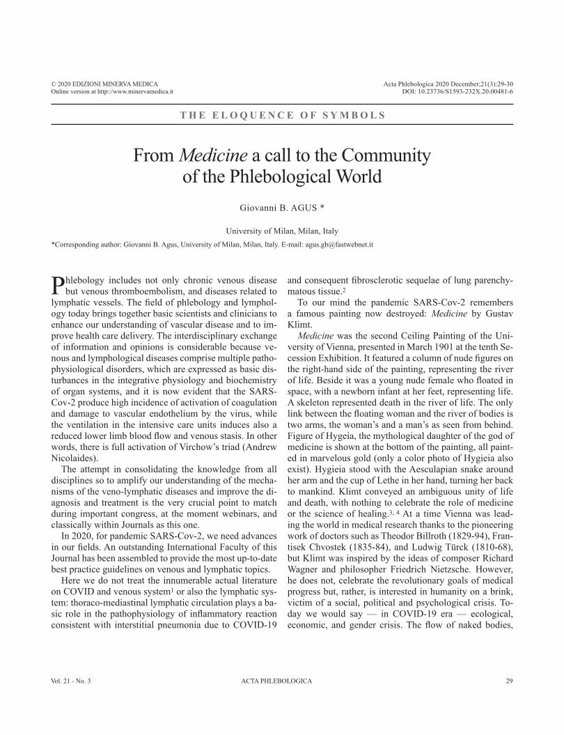
to our mind the pandemic SARS-cov-2 remembers a famous painting now destroyed: Medicine by gustav Klimt.
Medicine was the second ceiling Painting of the Uni-versity of Vienna, presented in March 1901 at the tenth Se-cession Exhibition. It featured a column of nude figures on the right-hand side of the painting, representing the river of life. Beside it was a young nude female who floated in space, with a newborn infant at her feet, representing life. A skeleton represented death in the river of life. the only link between the floating woman and the river of bodies is two arms, the woman’s and a man’s as seen from behind. Figure of hygeia, the mythological daughter of the god of medicine is shown at the bottom of the painting, all paint-ed in marvelous gold (only a color photo of hygieia also exist). hygieia stood with the Aesculapian snake around her arm and the cup of lethe in her hand, turning her back to mankind. Klimt conveyed an ambiguous unity of life and death, with nothing to celebrate the role of medicine or the science of healing.3, 4 At a time Vienna was lead-ing the world in medical research thanks to the pioneering work of doctors such as theodor billroth (1829-94), Fran-tisek chvostek (1835-84), and ludwig türck (1810-68), but Klimt was inspired by the ideas of composer Richard Wagner and philosopher Friedrich Nietzsche. however, he does not, celebrate the revolutionary goals of medical progress but, rather, is interested in humanity on a brink, victim of a social, political and psychological crisis. to-day we would say — in coViD-19 era — ecological, economic, and gender crisis. The flow of naked bodies,

AgUS gUStAV KliMt, MeDiciNe
30 ActA PhlebologicA December 2020
not only for the nudes, but also for the distorted view of medical science understood as powerless. today we must rethink it for its premonitory content; less for its beauty because it was impossible to admire it from the real and to see the use of gold with which Klimt had special affinity given his training and the work of his father, goldsmith and engraver.
in 1911 Medicine was owned by a different property, and in 1938 the painting was seized by germany. in 1943, after a final exhibition, the painting was moved to Schloss immendorf, a castle in the district of hollabrunn in the northeast of lower Austria, for protection. in May 7 1945 the painting was destroyed by retreating german SS forces that set fire to the castle to prevent it falling into enemy hands. All that remains now are preparatory sketches and a few photographs. only one black &white photograph remains of the complete painting of Medicine (Figure 1), taken just before it was destroyed.
the knowledge of the medical profession is even before an invitation to a more humble attitude of medicine today so engaged by the pandemic that, as it is now known, it concerns greatly both phlebology and lymphology. Not last, full of suggestions, gustav Klimt died in 1918 during the influenza pandemic.5
Now the italian Phlebology is proud to meet in the same Journal Acta Phlebologica many Phlebological Societies from different parts of the world and opens a closer col-laboration with them.
References1. costanzo l, Failla g, grasso SA, Palumbo FP, Ardita g, Di Pino l, et al. coViD-19 pneumonia: the impact of coagulopathy. Acta Phlebol 2020;20:1–2. 2. campisi c. thoracic and abdominal cavities, pressure gradients and lymphatic flow pathophysiology in Covid-19 patients. Summary in [email protected] 05/06/2020.3. bitsori M, galanakis e. Doctors versus artists: gustav Klimt’s Medi-cine. bMJ 2002;325:1506–8. 4. Marlowe-Storkovich t. “Medicine” by gustav Klimt. Artibus et his-toriae 2003;24:231–52.5. Grist NR. Pandemic influenza 1918. BMJ 1979;20:199.
raw and real, worn down by disease and powerless against the inexorable force of time, and yet another virus, like us now. Klimt thus represents the succession of events of hu-man existence, disrespectful of nature, which leads to the dissolution of life itself. it is undoubtedly a unique, radi-cal, countercurrent painting, and for such reasons disputed
Figure 1.—gustav Klimt, Medicine (1901) painting for the University of Vienna. Destroyed in 1945.
Conflicts of interest.—The author certifies that there is no conflict of interest with any financial organization regarding the material discussed in the manu-script.Authors’ contributions.—The author read and approved the final version of the manuscript.History.—Manuscript accepted: September 25, 2020. - Manuscript received: September 15, 2020.(Cite this article as: Agus gb. From Medicine a call to the community of the Phlebological World. Acta Phlebol 2020;21:29-30. Doi: 10.23736/S1593-232X.20.00481-6)

Vol. 21 - No. 3 ActA PhlebologicA 31
Acta PhlebologicaDecember 2020Vol. 21 - No. 3
O R I G I N A L A R T I C L E
outpatient treatment and prevention of acute hemorrhoids with sulodexide
Sergey e. KAtoRKiN 1, Pavel S. ANDReeV 2, Vasiliy M. SotNiKoV 3 *
1Department of Surgery, Samara State Medical University, Samara, Russia; 2Department of Medical Sciences, Samara State Medical University, Samara, Russia; 3Department of coloproctology, Samara State Medical University, Samara, Russia*corresponding author: Vasilii M. Sotnikov, Department of coloproctology, Samara State Medical University, Vodnikov street 49-69, 443099 Samara, Rus-sia. e-mail: [email protected]
A b S t R A c tbAcKgRoUND: Vasoactive drugs are commonly used in the treatment of hemorrhoidal pathology due to its effect on the endothelium and the vascular component of inflammation. Sulodexide is of particular interest. It has angioprotective and anti-inflammatory effects, as well as an anticoagulant effect. The objective of the study was to examine the efficacy of sulodexide in treatment and prevention of acute hemorrhoids.MethoDS: A prospective controlled study was conducted in 164 patients with acute hemorrhoids. Patients of the treatment group (N.=81) received diosmin, sulodexide, and used direct-acting anticoagulants (heparin ointments) twice a day for 7 days. two capsules of sulodexide 250 UlS were taken twice a day for 30 days. Patients of the control group (N.=83) took only diosmin 1000 mg once a day for 30 days and direct-acting anticoagulants (heparin ointments) twice a day for 7 days.The efficacy of the treatment was determined by the physician’s objective evaluation and the patient’s subjective evaluation using the ColoRectal Evaluation of Clinical Therapeutics Scale (CORECTS) questionnaire. All patients also underwent measurements of blood flow in enlarged exter-nal hemorrhoids using a Samsung Medison SonoAce R7 ultrasound diagnostic device (Samsung, taegu, South Korea). Patients were examined before starting the treatment and on days 5, 10 and 30 of the follow-up period.ReSUltS: Within three months after the end of treatment, 2 (2.5%) patients in the treatment group and 11 (13.3%) patients in the control group experienced exacerbations of hemorrhoids. No clinical signs of acute hemorrhoids were revealed in patients of the study groups during the examination on day 30 of the treatment. The measurement of blood flow in the external hemorrhoids by ultrasound showed 8.1±1.2 cm/s and 7.9±1.3 cm/s in patients of the treatment group and the control group, respectively. Three months later, a significant difference in the subjec-tive evaluation of the impact of hemorrhoids on daily activities was identified, which is associated with exacerbations of hemorrhoids. In three months after starting the treatment, the CORECTS score of all signs analyzed by a physician significantly decreased (P<0.05) in the sulodexide group versus the control group. Such symptoms as swelling decreased from 5.3±3.1 to 0, bleeding from 0.5±2.3 to 0, discomfort from 6.5±3.3 to 0, pain from 4.5±3.3 to 0, the impact on daily activities from 7.5±3.3 to 0. In the control group, itching decreased from 1.7±1.2 to 0±0.3 and the impact on daily activities from 7.3±3.6 to 1±3.4.coNclUSioNS: Sulodexide is an effective and pathogenetically substantiated drug for the conservative treatment of patients with acute hem-orrhoids. It significantly reduces the severity of clinical symptoms of hemorrhoids and improves the results of objective examinations. It has good tolerability, no adverse effects, and a better long-term effect than the standard treatment regimen.(Cite this article as: Katorkin Se, Andreev PS, Sotnikov VM. outpatient treatment and prevention of acute hemorrhoids with sulodexide. Acta Phlebol 2020;21:31-5. DOI: 10.23736/S1593-232X.20.00469-5)Key words: hemorrhoids; glucuronyl glucosamine glycan sulfate; therapeutics.
Acta Phlebologica 2020 December;21(3):31-5DOI: 10.23736/S1593-232X.20.00469-5
KAtoRKiNTREATMENT AND PREVENTION OF ACUTE HEMORRHOIDS WITH SULODEXIDE
© 2020 eDiZioNi MiNeRVA MeDicAOnline version at http://www.minervamedica.it
the most common coloproctological disease is hemor-rhoids. it affects hemorrhoidal veins of the terminal
rectum and the perianal region. the true prevalence is unknown; however, according to various authors, hemor-rhoids affect between 15% and 90% of adult patients.1, 2
this is a chronic condition with possible exacerbations. Acute hemorrhoids can be both external and internal.2, 3
exacerbations of internal hemorrhoids are treated using conservative methods, including oral phlebotropic drugs, local anticoagulants (ointments), and surgical interven-tions, as indicated.3
conservative or surgical treatment can be used for acute external hemorrhoids. Drug therapy should be the first choice in less severe cases (grade I-II) and used as a

KAtoRKiN TREATMENT AND PREVENTION OF ACUTE HEMORRHOIDS WITH SULODEXIDE
32 ActA PhlebologicA December 2020
more than 5 kg), abstinence from sports during and within a month after the end of treatment.
All patients underwent complete blood count tests be-fore starting the treatment. During the follow-up, a split-meal, high-fiber diet (at least 4 times a day) low in spicy, salty and pickled food and alcohol was recommended to patients.
All patients were randomized into two comparable groups. the treatment group included 81 patients (47 male and 34 female) at the of 31-61 (50.1±6.3) years old. The duration of the disease was 10±9.5 years.
the control group consisted of 83 patients (45 male and 38 female) at the age of 33-63 (51.1±4.3) years old with the disease duration of 10±8.2 years.
The statistical processing of the findings identified no significant differences between the groups in age (t=0.8; P=0.4), nosology (χ2=0.102; P=0.39), and sex (χ2=0.636; P=0.43).
before starting the treatment, all patients underwent a routine proctological examination (visual inspection, digi-tal investigation, anoscopy, rectoscopy), screening, ran-domization, and complete blood count testing, on the first day of treatment.
The first day of treatment was the day of the first admin-istration of medicines in both study groups. Patients of the treatment group took diosmin 1000 mg once a day for 30 days and used direct-acting anticoagulants (heparin oint-ments) twice a day for 7 days. two capsules of sulodexide (Alfasigma, bologna, italy) 250 UlS were taken twice a day for 30 days. the daily dose of sulodexide was 1000 UlS.
Patients of the control group took diosmin 1000 mg once a day for 30 days and used direct-acting anticoagu-lants (heparin ointments) twice a day for 7 days.
Patients were examined on days 5 and 30, and in 3 months after the end of conservative treatment. Physician’s objective evaluation and the patient’s subjective evaluation of the severity of hemorrhoids using the coRectS score was performed before starting the treatment and three months after the end of the treatment.12 Patients evaluated symptom severity using a scale of 1 to 10 (table i).
During the treatment, all patients also underwent mea-surements of blood flow in enlarged external hemorrhoids using a Samsung Medison SonoAce R7 ultrasound diag-nostic device (Samsung, taegu, South Korea). Patients were examined before starting the treatment and on days 5, 10, and 30.
After the end of the conservative treatment regimen, pa-tients of both groups were followed up for three months
support for surgical treatment in more severe cases (grade iii). conservative treatment in acute internal hemorrhoids includes oral phlebotropic drugs and local anticoagulants. Surgical treatment included thrombectomy from a throm-bosed hemorrhoid or excision of the entire thrombosed hemorrhoid.3
Anticoagulants are currently formulated as ointments, parenteral and oral drugs. Vasoactive drugs are of great interest as a conservative treatment because they can con-tribute to the relief of signs and symptoms by affecting the endothelium and the vascular component of inflammation in hemorrhoids. Sulodexide, glycosaminoglycan (gAg), is of particular interest. it has angioprotective and anti-inflammatory effects, as well as an anticoagulant effect.4-7 this drug is recommended for the treatment of different angiopathies with an increased risk of thrombosis. these include varicose veins of the lower extremities and micro- and macro angiopathies in diabetes mellitus.5 Sulodexide is commonly used to treat chronic venous diseases and ve-nous ulcers of the lower extremities.8, 9 Sulodexide modu-lates the activity of matrix metalloproteinases (MMPs), which are markers of inflammation most extensively in-volved in the degradation of connective tissue surrounding renal venous plexuses.10, 11
The objective of our study was to examine the efficacy of sulodexide in the conservative treatment and prevention of hemorrhoids.
Materials and methods
in 2016 to 2019, a prospective comparative study, includ-ing 164 patients with acute hemorrhoids, was conducted at teaching hospitals of Samara State Medical University. All patients signed informed consent to participate in the study, which was carried out under the applicable Russian laws, protocols, and ethical principles of the World Medi-cal Association Declaration of helsinki (Seoul, 2008) and good clinical Practice (ich gcP).
the following inclusion criteria were used: grade i-ii external and internal hemorrhoid thrombosis; 18 years and older; no confirmed pregnancy during the study pe-riod; signed informed consent; and normal complete blood counts.
the following exclusion criteria were used: withdrawal at any stage of the study; lost to follow-up; poor compli-ance; diagnosis of acute or decompensated somatic pathol-ogy; confirmed pregnancy during the study.
All patients were recommended to reduce the intensity and regularity of physical activity (lifting of weights not
TREATMENT AND PREVENTION OF ACUTE HEMORRHOIDS WITH SULODEXIDE KAtoRKiN
Vol. 21 - No. 3 ActA PhlebologicA 33
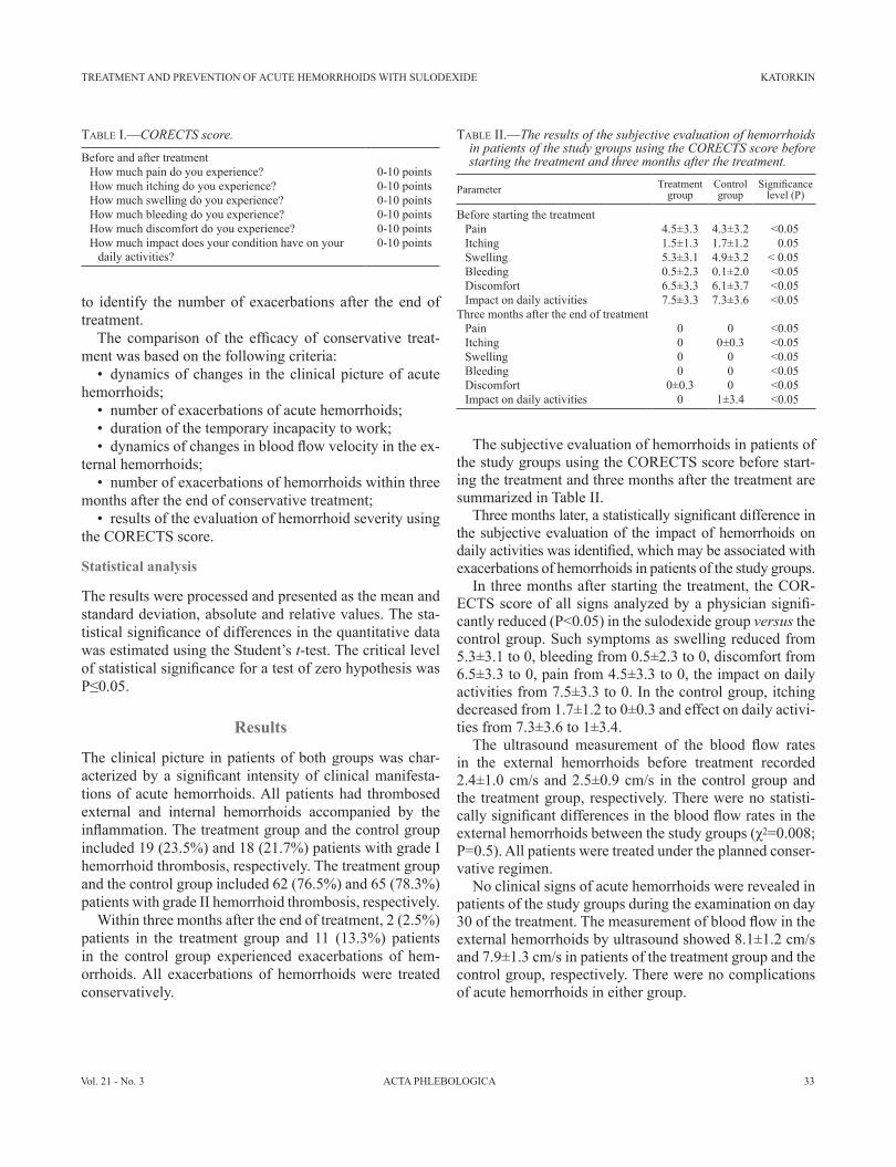
the subjective evaluation of hemorrhoids in patients of the study groups using the coRectS score before start-ing the treatment and three months after the treatment are summarized in table ii.
Three months later, a statistically significant difference in the subjective evaluation of the impact of hemorrhoids on daily activities was identified, which may be associated with exacerbations of hemorrhoids in patients of the study groups.
in three months after starting the treatment, the coR-ECTS score of all signs analyzed by a physician signifi-cantly reduced (P<0.05) in the sulodexide group versus the control group. Such symptoms as swelling reduced from 5.3±3.1 to 0, bleeding from 0.5±2.3 to 0, discomfort from 6.5±3.3 to 0, pain from 4.5±3.3 to 0, the impact on daily activities from 7.5±3.3 to 0. In the control group, itching decreased from 1.7±1.2 to 0±0.3 and effect on daily activi-ties from 7.3±3.6 to 1±3.4.
The ultrasound measurement of the blood flow rates in the external hemorrhoids before treatment recorded 2.4±1.0 cm/s and 2.5±0.9 cm/s in the control group and the treatment group, respectively. there were no statisti-cally significant differences in the blood flow rates in the external hemorrhoids between the study groups (χ2=0.008; P=0.5). All patients were treated under the planned conser-vative regimen.
No clinical signs of acute hemorrhoids were revealed in patients of the study groups during the examination on day 30 of the treatment. The measurement of blood flow in the external hemorrhoids by ultrasound showed 8.1±1.2 cm/s and 7.9±1.3 cm/s in patients of the treatment group and the control group, respectively. there were no complications of acute hemorrhoids in either group.
to identify the number of exacerbations after the end of treatment.
The comparison of the efficacy of conservative treat-ment was based on the following criteria:
• dynamics of changes in the clinical picture of acute hemorrhoids;
• number of exacerbations of acute hemorrhoids;• duration of the temporary incapacity to work;• dynamics of changes in blood flow velocity in the ex-
ternal hemorrhoids;• number of exacerbations of hemorrhoids within three
months after the end of conservative treatment;• results of the evaluation of hemorrhoid severity using
the coRectS score.
Statistical analysis
the results were processed and presented as the mean and standard deviation, absolute and relative values. the sta-tistical significance of differences in the quantitative data was estimated using the Student’s t-test. the critical level of statistical significance for a test of zero hypothesis was P≤0.05.
Results
the clinical picture in patients of both groups was char-acterized by a significant intensity of clinical manifesta-tions of acute hemorrhoids. All patients had thrombosed external and internal hemorrhoids accompanied by the inflammation. The treatment group and the control group included 19 (23.5%) and 18 (21.7%) patients with grade i hemorrhoid thrombosis, respectively. the treatment group and the control group included 62 (76.5%) and 65 (78.3%) patients with grade ii hemorrhoid thrombosis, respectively.
Within three months after the end of treatment, 2 (2.5%) patients in the treatment group and 11 (13.3%) patients in the control group experienced exacerbations of hem-orrhoids. All exacerbations of hemorrhoids were treated conservatively.
Table I.—� CORECTS score.
before and after treatmenthow much pain do you experience? 0-10 pointshow much itching do you experience? 0-10 pointshow much swelling do you experience? 0-10 pointshow much bleeding do you experience? 0-10 pointshow much discomfort do you experience? 0-10 pointshow much impact does your condition have on your
daily activities?0-10 points
Table II.—� The results of the subjective evaluation of hemorrhoids in patients of the study groups using the CORECTS score before starting the treatment and three months after the treatment.
Parameter treatment group
control group
Significance level (P)
before starting the treatmentPain 4.5±3.3 4.3±3.2 ˂0.05itching 1.5±1.3 1.7±1.2 0.05Swelling 5.3±3.1 4.9±3.2 ˂ 0.05bleeding 0.5±2.3 0.1±2.0 ˂0.05Discomfort 6.5±3.3 6.1±3.7 ˂0.05impact on daily activities 7.5±3.3 7.3±3.6 ˂0.05
three months after the end of treatmentPain 0 0 ˂0.05itching 0 0±0.3 ˂0.05Swelling 0 0 ˂0.05bleeding 0 0 ˂0.05Discomfort 0±0.3 0 ˂0.05impact on daily activities 0 1±3.4 <0.05

KAtoRKiN TREATMENT AND PREVENTION OF ACUTE HEMORRHOIDS WITH SULODEXIDE
34 ActA PhlebologicA December 2020
hemorrhoids results in a faster resolution of inflammation of hemorrhoids through to the improvement of peripheral circulation and microcirculation. Patients treated with su-lodexide experienced a prolonged angioprotective effect for up to 3 months. this effect is ensured by a compre-hensive action on the blood vessel walls, blood viscosity and lipid levels. owing to this, hemodynamic is normal-ized, especially in the microcirculatory bed. Furthermore, sulodexide affects blood clotting, platelet adhesion and ag-gregation, fibrinolysis.4 All the above profibrinolytic, an-tithrombotic, anti-inflammatory, and protective properties show that sulodexide can be used for the treatment of acute hemorrhoids. At the same time, further studies of the use of sulodexide in patients with acute hemorrhoids are required.
Conclusions
Sulodexide is an effective and pathogenetically substanti-ated drug for the conservative treatment of patients with acute hemorrhoids. It significantly reduces the severity of clinical symptoms of hemorrhoids and improves the re-sults of objective examinations. it has good tolerability, no adverse effects, and a better long-term effect than the standard treatment regimen.
References
1. Shi Y, Yang D, chen S, Wang S, li h, Ying J, et al. Factors influ-encing patient delay in individuals with haemorrhoids: A study based on theory of planned behavior and common sense model. J Adv Nurs 2019;75:1018–28. 2. Sandler RS, Peery AF. Rethinking What We Know About hemor-rhoids. clin gastroenterol hepatol 2019;17:8–15. 3. Sammarco g, trompetto M, gallo g. thrombosed external haemor-rhoids: A Clinician’s Dilemma. Rev Recent Clin Trials 2019;14:232–4. 4. bignamini AA, Matuška J. Sulodexide for the Symptoms and Signs of chronic Venous Disease: A Systematic Review and Meta-analysis. Adv ther 2020;37:1013–33. 5. Li R, Xing J, Mu X, Wang H, Zhang L, Zhao Y, et al. Sulodexide therapy for the treatment of diabetic nephropathy, a meta-analysis and lit-erature review. Drug Des Devel ther 2015;9:6275–83.6. Jiang QJ, bai J, Jin J, Shi J, Qu l. Sulodexide for Secondary Preven-tion of Recurrent Venous thromboembolism: A Systematic Review and Meta-Analysis. Front Pharmacol 2018;9:876. 7. lizza N, Urbani M, Ukovich l. Sulodexide in the treatment of grade ii and iii hemorrhoids: a retrospective study. Acta Phlebol 2019;20:15–9. 8. Katorkin SE. [Significance of endothelial protection in treatment of pa-tients with class c6 chronic venous disease and type 2 diabetes mellitus]. Angiol Sosud Khir 2015;21:99–102, 104–6.9. Chupin AV, Katorkin SE, Katel’nitskiĭ II, Katel’nitskaia OV, Prostov ii, Petrikov AS, et al. [Sulodexide in treatment of chronic venous insuffi-ciency. Results of the All-Russian multicenter programme AcVeDUct] [Sulodeksid v lechenii khronicheskoĭ venoznoĭ nedostatochnosti. Itogi Vserossiĭskoĭ mul’titsentrovoĭ programmy ACVEDUCT]. Angiol Sosud Khir 2018;24:47–55. [Russian.]
the study showed that the inclusion of Sulodexide (1000 UlS, 2 capsules twice a day for 30 days) in the com-bination therapy of patients with grade i and ii acute hem-orrhoids significantly reduces clinically objective signs (swelling, bleeding, prolapse [P<0.05]).
The efficacy of sulodexide was also confirmed by pa-tients (coRectS) who experienced an improvement in all parameters analyzed (swelling, bleeding, discomfort, pain, and impact on daily activities [P<0.05]).
Discussion
All patients were examined in a similar manner, using the same program, and were randomized into two groups comparable by sex, age, and nosology. in both groups, the clinical picture was characterized by severe clinical signs of acute hemorrhoids.
The study analyzed the objective and subjective effi-cacy of oral sulodexide 1000 UlS (2 capsules twice a day for 30 days) in the group of patients with grade i-ii acute hemorrhoids. The findings showed a significant improve-ment and absence of exacerbations in a 3-month follow-up period in 97.5% of patients taking sulodexide, and no exacerbations were identified in 86.7% of patients in the control group. We believe that this is due to the systemic angioprotective action of sulodexide.
clinically evaluated objective data collected during the proctological examination showed that sulodexide was effective in treating all signs and symptoms analyzed by the physician (swelling, bleeding, prolapse, and pain). Pa-tients reported a significant reduction of symptoms (pain and itching) and signs (swelling, bleeding), with decreased discomfort and improved wellbeing.
Our findings are consistent with the literature. Particu-larly, lizza et al.7 showed that sulodexide reversed such symptoms as swelling, bleeding, and prolapse of hemor-rhoids in more than 88% of patients with hemorrhoids.
Sulodexide is a natural component of endothelial glyco-calyx proven as beneficial for restoring the physiological function of the vascular wall through the reintegration of the damaged layer of glycosaminoglycans in acute hemor-rhoids, which can explain a significant reduction of swell-ing and observed bleeding episodes.13, 14
Finally, sulodexide is a controlled agent in its oral form with an excellent efficacy and safety profile,15 which was also confirmed in this study since no significant side ef-fects were observed.
in our opinion, the inclusion of sulodexide in the con-servative combination treatment of patients with acute
TREATMENT AND PREVENTION OF ACUTE HEMORRHOIDS WITH SULODEXIDE KAtoRKiN
Vol. 21 - No. 3 ActA PhlebologicA 35
vascular diseases: implications for treatment. Drug Des Devel ther 2013;8:49–65. 14. Flota cervera lF, Frati Munari Ac, Velázquez herrera Áe, carbajal contreras A. chronic venous disease treated with sulodexide: a survey among primary care physicians in Mexico. int Angiol 2017;36:558–64. 15. Andreozzi gM, bignamini AA, Davì g, Palareti g, Matuška J, holý M, et al.; SURVet Study investigators. Sulodexide for the prevention of recurrent venous thromboembolism: the Sulodexide in Secondary Pre-vention of Recurrent Deep Vein thrombosis (SURVet) study: a multi-center, randomized, double-blind, placebo-controlled trial. circulation 2015;132:1891–7.
10. Mannello F, Medda V, ligi D, Raffetto JD. glycosaminoglycan su-lodexide inhibition of MMP-9 gelatinase secretion and activity: possible pharmacological role against collagen degradation in vascular chronic dis-eases. curr Vasc Pharmacol 2013;11:354–65. 11. Serra R, gallelli l, grande R, Amato b, De caridi g, Sammarco g, et al. hemorrhoids and matrix metalloproteinases: A multicenter study on the predictive role of biomarkers. Surgery 2016;159:487–94. 12. ebrahimi N, Vohra-Miller S, Koren g. Anorectal symptom manage-ment in pregnancy: development of a severity scale. J Popul ther clin Pharmacol 2011;18:e99–105.13. coccheri S, Mannello F. Development and use of sulodexide in
Conflicts of interest.—The authors certify that there is no conflict of interest with any financial organization regarding the material discussed in the manuscript.Authors’ contributions.—Sergey e. Katorkin and Pavel S. Andreev gave substantial contribution to research concept, editing and design; Vasilii M. Sotnikov contributed material collection, processing and text writing. All authors read and approved the final version of the manuscript.History.—Manuscript accepted: July 4, 2020. - Manuscript received: June 16, 2020.

36 ActA PhlebologicA December 2020
Acta PhlebologicaDecember 2020Vol. 21 - No. 3
O R I G I N A L A R T I C L E
effect of four-layer dressing on venous ulcerSatyendra K. tiWARY 1 *, Katyayani K. choUbeY 1, Soumya KhANNA 2, Puneet KUMAR 1, Ajay K. KhANNA 1
1Department of general Surgery, institute of Medical Sciences, banaras hindu University, Varanasi, india; 2Department of Anatomy, institute of Medical Sciences, banaras hindu University, Varanasi, india
*corresponding author: Satyendra K. tiwary, Department of general Surgery, institute of Medical Sciences, banaras hindu University, Varanasi, india. e-mail: [email protected]
A b S t R A c tbAcKgRoUND: Venous ulcer is a chronic disease and has periods of exacerbation and remission. it takes a long time to heal, resulting in physical and psychological discomfort thereby negatively affecting the functional status of the patients. Various bandage systems, single layered, double layered and multiple layered with elastic and non elastic components have been commercialized. A requirement of sustained pressure brought the four-layer bandage into picture. We tried to study the bacteriology of the venous ulcers and the effect of our layer bandage on the healing of the ulcer.MethoDS: Sixty patients were recruited for the study. however, 4 patients had a bilateral disease and so a total of 64 limbs were taken into consideration in the study. clinical details of all patients with wound size measurement by gauze piece, wax paper and scale was done. the wounds were initially debrided and photographic records of all patients was maintained. Patients were followed up every week and the dressing was changed every week.ReSUltS: 93.8% had complete healing while 1.6% had partial healed ulcer and 4.7% had non healing ulcer. After excluding the four ulcers that did not heal, 16.6% had recurrence while 50 out 60 had no recurrence in the follow-up for one year. There was a significant correlation (P<0.001) between ulcer size and the number of dressings.coNclUSioNS: compression therapy is the mainstay of treatment of venous ulcer. compression in the range of 30 mmhg to 40 mmhg is the most effective level for uncomplicated venous ulcers with adequate arterial competency.(Cite this article as: tiwary SK, choubey KK, Khanna S, Kumar P, Khanna AK. effect of four-layer dressing on venous ulcer. Acta Phlebol 2020;21:36-41. DOI: 10.23736/S1593-232X.20.00471-3)Key words: Varicose ulcer; Varicose veins; Wound and injuries.
Acta Phlebologica 2020 December;21(3):36-41Doi: 10.23736/S1593-232X.20.00471-3
tiWARYVeNoUS UlceR
© 2020 eDiZioNi MiNeRVA MeDicAonline version at http://www.minervamedica.it
the majority of leg ulcers are secondary to chronic ve-nous insufficiency (CVI), the vascular insufficiency
caused by valvular insufficiency in either deep system or superficial system. The venous hypertension leads to leak-age of fluid and fibrinogen out of the stretched veins into the tissues forming a pericapillary fibrin cuff which leads to reduced diffusion of oxygenated blood to the tissues re-sulting in ulceration. Also, there is reduced pressure gradi-ent between the arteriolar and venular end of the capillaries resulting in sluggish movement of the blood within them and adherence of blood cells to the endothelium. Inflam-matory mediators and reactive oxygen species are released which lead to obliteration of functioning capillary loops, thus, aggravating ischemia and result in ulceration. capil-
laries can also be occluded by microthrombi and exhibit long intra-capillary stasis which in turn reduce nutrition and oxygenation of the skin and lead to ulceration.
Venous ulcer is a chronic disease and has periods of exacerbation and remission. it takes a long time to heal, resulting in physical and psychological discomfort thereby negatively affecting the functional status of the patients. these chronic ulcers are susceptible to microbial invasion and this probably contributes to the non-healing nature of the ulcer. these secondarily infected ulcers pose a chal-lenge for the clinicians and the patients. Regular dressings or antimicrobial bandages have been effective to control to some extent. however, the results are not satisfactory.
Various treatment modalities have been introduced.
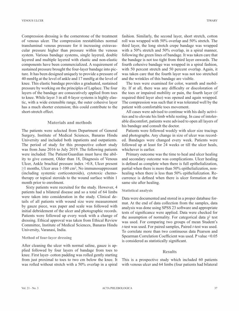
VeNoUS UlceR tiWARY
Vol. 21 - No. 3 ActA PhlebologicA 37
tiWARYVeNoUS UlceR
fashion. Similarly, the second layer, short stretch, cotton roll was wrapped with 50% overlap and 50% stretch. the third layer, the long stretch crepe bandage was wrapped with a 50% stretch and 50% overlap, in a spiral manner, following the green lines of bandage. it was taken care that the bandage is not too tight from third layer onwards. the fourth cohesive bandage was wrapped in a spiral fashion, with 50 percent stretch and 50 percent overlap. Again, it was taken care that the fourth layer was not too stretched and the wrinkles of this bandage are visible.
the toes were examined for color, warmth and mobil-ity. If at all, there was any difficulty or discoloration of the toes or impaired mobility or pain, the fourth layer (if required third layer also) was opened and again wrapped. the compression was such that it was tolerated well by the patient with comfortable toes movement.
All cases were advised to continue with his daily activi-ties and to elevate his limb while resting. in case of intoler-able discomfort, patients were advised to open all layers of his bandage and consult the doctor.
Patients were followed weekly with ulcer size tracings and photographs. Any change in size of ulcer was record-ed. bandages were changed every week. Patients were followed up at least for 24 weeks or till the ulcer heals, whichever is earlier.
Primary outcome was the time to heal and ulcer healing and secondary outcome was complications. Ulcer healing is defined as complete when there is full epithelialization, partial when there is more than 50% epithelialization, non-healing when there is less than 50% epithelialization. Re-currence is defined when there is ulcer formation at the same site after healing.
Statistical analysis
Data were documented and stored in a proper database for-mat. At the end of data collection from the samples, data analysis was done using SPSS 23 software and appropriate tests of significance were applied. Data were checked for the assumption of normality. For categorical data χ2 test was used. For comparing two groups of mean Student’s t-test was used. For paired samples, Paired t-test was used. to correlate more than two continuous data Pearson and Spearman Correlation Coefficient was used. P value <0.05 is considered as statistically significant.
Results
this is a prospective study which included 60 patients with venous ulcer and 64 limbs (four patients had bilateral
compression dressing is the cornerstone of the treatment of venous ulcer. the compression reestablishes normal transluminal venous pressure for it increasing extravas-cular pressure higher than pressure within the venous system. Various bandage systems, single layered, double layered and multiple layered with elastic and non-elastic components have been commercialized. A requirement of sustained pressure brought the four-layer bandage into pic-ture. it has been designed uniquely to provide a pressure of 40 mmhg at the level of ankle and 17 mmhg at the level of knee. this elastic bandage provides a graduated, sustained pressure by working on the principles of laplace. the four layers of the bandage are consecutively applied from toes to knee. While layer 3 in all 4-layer systems is highly elas-tic, with a wide extensible range, the outer cohesive layer has a much shorter extension; this could contribute to the short-stretch effect.
Materials and methods
the patients were selected from Department of general Surgery, institute of Medical Sciences, banaras hindu University and included both inpatients and outpatients. the period of study for this prospective cohort study was from June 2016 to July 2019. the following patients were included: the Patient/guardian must have the abil-ity to give consent, older than 18, Diagnosis of Venous Ulcer, Ankle brachial pressure index >0.8, Ulcer present ≥1 months, Ulcer area 1-100 cm2, No immunosuppressant (including systemic corticosteroids), cytotoxic chemo-therapy or topical steroids to the wound surface within 1 month prior to enrolment.
Sixty patients were recruited for the study. however, 4 patients had a bilateral disease and so a total of 64 limbs were taken into consideration in the study. clinical de-tails of all patients with wound size were measurement by gauze piece, wax paper and scale was followed with initial debridement of the ulcer and photographic records. Patients were followed up every week with a change of dressing. ethical approval was taken from ethical Review committee, institute of Medical Sciences, banaras hindu University, Varanasi, india.
Method of four-layer dressing
After cleaning the ulcer with normal saline, gauze is ap-plied followed by four layers of bandage from toes to knee. First layer- cotton padding was rolled gently starting from just proximal to toes to two cm below the knee. it was rolled without stretch with a 50% overlap in a spiral

tiWARY VeNoUS UlceR
38 ActA PhlebologicA December 2020
A percentage of 93.8% had complete healing while 1.6% had partial healed ulcer and 4.7% had non healing ulcer. After excluding the four ulcers that did not heal, 16.6% had recurrence while 50 out 60 had no recurrence in the follow-up. 7 out of 10 patients had a single recur-rence while rest of the three had recurrence twice, thrice and four times each. the incidence of recurrence that oc-curred within 1 month was (30%) patients and within 6 months in 50% patients and within 12 months in 2 out of 10(20%) patients.
There was highly significant correlation (P<0.001) be-tween ulcer size and the number of dressings done (Figure 1).There was statistically significant change (P<0.05) in ulcer with each dressings, i.e. the ulcer healed with subse-quent dressings.
on studying the other effects/ side effect of bandaging, it showed pigmentation increased from 21.6% to 90.6% after dressing. tenderness was seen to decrease; it was present initially in all patients and gradually decreased and was present in 6.2%. Skin scaling increased from 1.6% to 98.4%. Also, itching increased from 20.3% to 81.25% after the dressing.
Discussion
Venous ulcer has a prevalence rate of 1% and accounts for 80% of lower extremity ulcerations. the compression therapy remains the mainstay of treatment and of which,
ulcers). A four-layer dressing was done and the change in size of ulcer bandaging was studied.
in this study the youngest patient was 19 years and old-est was 68 years and maximum patients were between 21-40 years of age, i.e. in the productive age group. 95% of the patients were male and 5% were female. Maximum patients (26.7%) were government servants, 25% were shopkeeper while 8.3% patients were farmer, followed by barber, conductor, housewife, cook, student, butcher, laborer and teacher. Maximum (31.2%) limbs had ulcer from 2-4 months while 25% had ulcer from 6 to 12 months and 23.4% had ulcers from 4-6 months. the longest dura-tion of ulcer was 6 years. heaviness, a common symptom, was present in 87.5%. history of bleed was present in 7 limbs (10.9%).
Ulcer was present in right leg in 56.2% and in left leg in 43.8%. 4 patients had bilateral disease. All limbs had tor-tuous veins, pain at the time of presentation and 21.8% had pigmentation. Mean size of ulcer at presentation was 8.406 cm. with standard deviation of ±7.1197. the median size of ulcer at presentation was 6.00 with interquartile range between 4.00-11.50.
on studying the history of intervention in these patients, 64.1% had history of no intervention, while 14.1% had history of RFA alone and 9.4% had history of RFA with sclerotherapy and 10.9% had stripping done. eVlt was done in 1 of 64 (1.6%) cases.
Varicose veins were present in all patients, dermatitis and edema was present in 95.3% and 68.7% cases, respec-tively. lipodermatosclerosis was present in 21.8%, ery-thema was present in 18.7%, scaling and atrophie blanche in 15.6% each.
the largest ulcer was present on medial side in 50%, whereas on lateral side in 25% cases, on anterior aspect in 15.6% and posterior aspect in 6.2% and on foot in 3.1% cases. Depth of ulcer was <3 mm in 92.2% and 3-6 mm in rest of the cases.
SFJ incompetence was present in 84.4% limbs and SPJ incompetence was in 31.2%. on studying the incompe-tence of perforators, four limbs had no incompetent perfo-rator and one perforator incompetence was seen in 6.2%. two perforators were incompetent in 35.9%, three perfo-rators were incompetent in 31.2%, four perforators were incompetent in 14.1% limbs, 5 perforators were incompe-tent in 6.2% limbs.
After weekly dressings, 39.1% ulcers healed by 8 weeks, 67.1% ulcers healed by 12 weeks 60 93.8% had complete healing by 24 weeks. 1 out 64 (1.6%) had partial healing and 3 out of 64 patients (4.7%) had non healing ulcer.
Figure 1.—correlation between the size of ulcer at presentation and the number of dressings done. it shows that number of dressings depended on the size of ulcer. P value is <0.001 which shows highly significant correlation between ulcer size and the number of dressings done.
35
30
25
20
15
10
5
0
P<0.001
Number of dressings doneUlcer size
Patients
Freq
uenc
y
5 7 931 11 29 39 49 5927 37 47 57 636113 151719 21 31 41 5123 33 43 5325 35 45 55
VeNoUS UlceR tiWARY
Vol. 21 - No. 3 ActA PhlebologicA 39
3.6% others. 0.6% patient underwent thrombectomy of the iliofemoral vein.
compression therapy is the mainstay of treatment of venous ulcer. compression in the range of 30 mmhg to 40 mmhg is the most effective level for uncomplicated venous ulcers with adequate arterial competency. lorimer et al. concluded that a high degree of compression (30 mmHg to 40 mmHg) was better than low compression (<20 mmHg) with no specific bandage system recom-mended.4
compression therapies are broadly categorized into bandages, stockings (hosieries) and intermittent pneu-matic compression. Further, bandages may be either elas-tic and inelastic or single or multi-layered. the four-layer bandage is elastic and a onetime use, multilayer bandage. Partsch et al. stated that when multiple layers of elastic bandages are applied, the top layer becomes inelastic as a result of alteration of the friction capacity. thus, it is an elastic bandage with properties of inelastic bandages. the short stretch bandage on other hand is inelastic, reusable bandage. These are examples of high compression (ankle sub-bandage pressure 35-40 mmHg).
Several studies compared different elastic and inelas-tic bandages. A Rct by Polignano et al. concluded that the four layer bandage (elastic) was as effective as Unna’s boot (inelastic) for healing of venous ulcers.5 in a ran-domized control trial (RCT) of 387 adults to compare a four-layer system and short-stretch compression bandages comprised of one layer of padding and one to two layers of compression bandages). Nelson et al. found a statistically significant increase in the probability of healing with the four-layer bandage system.6 Meta-analysis by o’Meara et al., which included five RCTs (797 patients). It concluded that multilayer bandage systems are superior to short-stretch bandages as they result in more rapid healing of leg ulcers.7
Another randomized controlled trial by Mofatt et al. concluded that the four-layer bandage offers advantages over the two-layer bandage in terms of reduced with-drawal from treatment, fewer adverse incidents, and lower treatment cost.8 however, there are studies suggesting two layerbandages to have similar efficacy as four-layer ban-dage.9 The various randomized controlled trials (Harrison et al., Scriven et al., Partsch et al.)10-12 showed no differ-ences in healing between short-stretch and multilayer ban-dages.
Also, Dolibog et al. compared the compression options and concluded the most effective treatment of venous leg ulcers is the use of intermittent pneumatic compression,
the four-layer bandage system is the gold standard for treatment.
our study is a prospective study in which the effect of four-layer bandage on healing of ulcer was studied. the study included 60 patients of which 4 had a bilateral ulcer. therefore, a total of 64 limbs have been studied. Young-est patient was 19 years old and eldest was 68 years and maximum patients were between 21-40 years of age. in the study by Kota et al., from india, most of the patients were in the productive age group of 36 to 45 years.1 this disease is the disease of young and most active population.
We had 57 (95%) males and 3 (5%) females. In other studies from india by Kota et al., out of 170 patients 79% were men and 21% were female and by Alamelu, out of 29 patients 86.2% were male and female were 13.8%.2 A review by Jennifer et al. stated that the prevalence of varicose veins varies widely, from 2% to 56% in men and from1% to 73% in women.
the ergonomics and physical activity of an occupa-tion has been seen to have contributing factor in varicose ulcers. In our study, 16 out of 60 (26.7%) patients were government servants, 15 out 60(25%) were shopkeeper while 5 out of 60 (8.3%) patients were farmer, followed by barber, conductor, housewife, cook, student, butch-er, laborer and teacher. Most of them were indulged in activities where prolonged standing (>4 hours) was re-quired.
Venous ulcer was present in right leg in 36 cases out of 64 (56.2%) and left leg 28 out of 64 (43.8%) and 4 patients had bilateral disease. in a study by baker et al., of the 163 limbs with chronic venous ulcers, 43.6% were ulcerated on the right side and 56.4% on the left and in 25 patients the ulcers were bilateral.
Duration of ulcer varied, 31.2% from 2-4 months, 25% from 6 to 12 months and 23.4% from 4-6 months.12.5% of them were affected by it for over one year. Maximum patients had ulcers within one year and were non- respond-ing to any other form of treatment. the longest duration of ulcer was 6 years.
Nine out of 64 (14.1%) had Radio frequency ablation (RFA) of varicose veins and 6 out of 64 (9.4%) had RFA with sclerotherapy and 7 out of 64 (10.9) had stripping done. RFA with foam sclerotherapy was done in 6 out of 64 (9.4%). EVLT was done in 1 of 64 (1.6%) cases. In the study by baker et al. 30% had a primary procedure for varicose veins, in 17.8% patients the long saphenous vein had been stripped and in 7.4% the communicating veins had been divided.3 individual ligation of varicosi-ties was performed in 1.2% patients, with sclerotherapy in

tiWARY VeNoUS UlceR
40 ActA PhlebologicA December 2020
daged areas, such as the toes and knees.21 Also, the four consecutive layers tend to be heavy and there are chances of slippage of bandage. the foul smellingexudates from a grossly infected ulcer further affect the quality of life.
Conclusions
compression therapy is the mainstay of treatment of ve-nous ulcer. compression in the range of 30 mmhg to 40mmhg is the most effective level for uncomplicated ve-nous ulcers with adequate arterial competency.
References
1. Kota A, Selvaraj A, Premkumar P. Ponraj Sam, Agarwal S. Four layer dressing in the management of chronic venous ulcers in the outpatient set-ting of a tertiary hospital in india. Wound Medicine. 2014;5:21–4. 2. Alamelu V. is chronic venous ulcer curable? A sample survey of a plas-tic surgeon. indian J Plast Surg 2011;44:104–9. 3. baker SR, Stacey Mc. epidemiology of chronic leg ulcers in Australia. Aust N Z J Surg 1994;64:258–61. 4. lorimer KR, harrison Mb, graham iD, Friedberg e, Davies b. Venous leg ulcer care: how evidence-based is nursing practice? J Wound ostomy continence Nurs 2003;30:132–42. 5. Polignano R, bonadeo P, gasbarro S, Allegra c. A randomised con-trolled study of four-layer compression versus Unna’s boot for venous ulcers. J Wound care 2004;13:21–4. 6. Nelson eA, iglesias cP, cullum N, torgerson DJ; VenUS i collabora-tors. Randomized clinical trial of four-layer and short-stretch compression bandages for venous leg ulcers (VenUS I). Br J Surg 2004;91:1292–9. 7. o’Meara S, cullum NA, Nelson eA. compression for venous leg ul-cers. cochrane Database Syst Rev 2009;1:cD000265.8. Moffatt cJ, Mccullagh l, o’connor t, Doherty Dc, hourican c, Stevens J, et al. Randomized trial of four-layer and two-layer bandage systems in the management of chronic venous ulceration. Wound Repair Regen 2003;11:166–71. 9. iglesias c, Nelson eA, cullum NA, torgerson DJ; VenUS team. Ve-nUS i: a randomised controlled trial of two types of bandage for treating venous leg ulcers. health technol Assess 2004;8:iii, 1–105. 10. harrison Mb, Vandenkerkhof eg, hopman WM, graham iD, carley Me, Nelson eA; canadian bandaging trial group. the canadian ban-daging trial: evidence-informed leg ulcer care and the effectiveness of two compression technologies. bMc Nurs 2011;10:20. 11. Scriven JM, taylor le, Wood AJ, bell PR, Naylor AR, london NJ. A prospective randomised trial of four-layer versus short stretch compres-sion bandages for the treatment of venous leg ulcers. Ann R coll Surg engl 1998;80:215–20.12. Partsch h, Damstra RJ, tazelaar DJ, Schuller-Petrovic S, Velders AJ, de Rooij MJ, et al. Multicentre, randomised controlled trial of four-layer bandaging versus short-stretch bandaging in the treatment of venous leg ulcers. Vasa 2001;30:108–13. 13. Dolibog P, Franek A, taradaj J, Dolibog P, blaszczak e, Polak A, et al. A comparative clinical study on five types of compression therapy in patients with venous leg ulcers. int J Med Sci 2013;11:34–43. 14. Klode J, Wax c, Koerber A, Dissemond J. causes and the way of treatment of patients wth chronic leg ulcers - Analysis of a survey of 1000 practising specialists and general practitioners. Phlebologie 2009;38:211–8.
stockings and multi layer compression bandaging (similar efficacy) and two layer compression therapy with bandag-es proved to be the least effective.13
After weekly dressings, 39.1% ulcers healed by 8 weeks, 67.1% ulcers healed by 12 weeks 60 93.8% had complete healing by 24 weeks. 1 out 64 (1.6%) had partial healing and 3 out of 64 patients (4.7%) had non healing ulcer. of the 170 patients studied by Kota et al., healing was seen in 87.6%. 69 and 74 per cent of the ulcers healed by 12 weeks, as seen in different studies.1, 14-17
Recurrence is one of the major complications of four-layer bandage. here, in our study, after excluding the four ulcers that did not heal, 10 out of 60 (16.6%) had recurrence while 50 out 60 (84.3%) had no recurrence in the follow-up. the incidence of recurrence that occurred within 1 month is 3 out of 10 (30%) patients and within 6 months in 5 out of 10 (50%) patients, within 12 months in 2 out of 10 (20%) patients.
Seven out of 10 patients (70%) had a single recurrence while 1 each patient had recurrence twice, thrice and four times each. in a similar study by thomson et al., of the 318 ulcers that were completely healed, 19.5% (62) recurred; 41 of these were healed again. Recurrence still remains a challenge and a prophylactic, lifelong, class iii compres-sion stockings was advised to all the patients when the ul-cer healed.18
Apart from healing of the ulcer, the dressing has other effects on the patient and the limb. in our study, the peri-ulcer pigmentation/ lipodermato-sclerosis increased from 21.6% to 90.6%, after the dressings. Pain and tenderness were seen to decrease, it was present initially in all pa-tients and gradually decreased and was eventually pres-ent in 6.2% patients. Skin scaling during the treatment increased from 1.6% to 98.4%. itching increased from 20.3% to 81.25%. Kota et al. noted pain, itching, bleeding as the complications of four-layer bandage. one unpub-lished study from UK by iglesias et al. studied maceration, allergic reaction, eczema of periulcer skin in relation to four-layer bandage.9 Nelson et al. mentioned other com-plications like, maceration, excoriations, skin damage and deterioration, new ulcer formation.6
however, four-layer bandaging results are dependent on the bandaging skills, application technique. hopkins et al. considered the application of compression bandaging to be a specialist skill and in literature.19 Feben et al. estimated that many practitioners are failing to apply bandages in an effective and competent manner.20 As stated by callam et al., poor bandaging techniques can lead to tissue damage, necrosis, pain, chronic skin changes and edema in unban-
VeNoUS UlceR tiWARY
Vol. 21 - No. 3 ActA PhlebologicA 41
examined by culture-independent molecular methods. Wound Repair Re-gen 2010;18:38–49. 19. gowland hopkins NF, Jamieson cW. Antibiotic concentration in the exudate of venous ulcers: the prediction of ulcer healing rate. br J Surg 1983;70:532–4. 20. Feben K. how effective is training in compression bandaging tech-niques? br J community Nurs 2003;8:80–4. 21. callam MJ, harper DR, Dale JJ, brown D, gibson b, Prescott RJ, et al. lothian and forth valley leg ulcer healing trial elastic versus non-elastic bandaging in the treatment of chronic leg ulceration. Phlebology 1992;7:136–41.
15. Vowden KR, Vowden P. Preventing venous ulcer recurrence: a re-view. int Wound J 2006;3:11–21. 16. blair SD, Wright DD, backhouse cM, Riddle e, Mccollum cN. Sustained compression and healing of chronic venous ulcers. bMJ 1988;297:1159–61. 17. o’Meara S, tierney J, cullum N, bland JM, Franks PJ, Mole t, et al. Four layer bandage compared with short stretch bandage for venous leg ulcers: systematic review and meta-analysis of randomised controlled tri-als with data from individual patients. bMJ 2009;338:b1344. 18. thomsen tR, Aasholm MS, Rudkjøbing Vb, Saunders AM, bjarn-sholt t, givskov M, et al. the bacteriology of chronic venous leg ulcer
Conflicts of interest.—The authors certify that there is no conflict of interest with any financial organization regarding the material discussed in the manuscript.Authors’ contributions.—Satyendra K. tiwary drafted the manuscript, Katyayani chaubey and Soumya Khanna collected and analyzed the data, Puneet Kumar and Ajay K. Khanna reviewed the manuscript and drafted it finally. All authors read and approved the final version of the manuscript.History.—Manuscript accepted: July 3, 2020. - Manuscript received: June 18, 2020.

42 ActA PhlebologicA December 2020
Acta PhlebologicaDecember 2020Vol. 21 - No. 3
O R I G I N A L A R T I C L E
Study of quality of life in patients with varicose vein after radiofrequency ablation
and ultrasound guided foam sclerotherapySatyendra K. tiWARY 1 *, Sartaz AlAM 1, Pankaj SUReKA 2, Puneet KUMAR 1, Ajay K. KhANNA 1
1Department of general Surgery, institute of Medical Sciences, banaras hindu University, Varanasi, india; 2Department of Psychiatry, institute of Medical Sciences, Varanasi, india*corresponding author: Satyendra K. tiwary, Department of general Surgery, institute of Medical Sciences, banaras hindu University, Varanasi, india. e-mail: [email protected]
A b S t R A c tBACKGROUND: Chronic venous insufficiency manifests most commonly as varicose veins affecting quality of life in significant way due to morbidity, recurrence and economic burden. this study was done in varicose veins to assess the quality of life in varicose vein patients after treatment with radiofrequency ablation (RFA) and ultrasound guided foam sclerotherapy (UgFS) in great saphenous vein (gSV) in selected patients.MethoDS: in our study, in 30 patients UgFS and in another 30 patients RFA therapy in gSV was done. After these procedures, health related quality of life (hRQol) was assessed with help of SF-36 and Venous clinical Severity Score (VcSS). At pretreatment SF-36 and VccS scoring was assessed and then reassessed all the parameter at 1 month, 6 months and 1 year. higher the score, good is the quality of life.RESULTS: The pretreatment scoring in both the group was comparable but not significant. But in subsequent follow-up HRQOL is better in RFA group patients than the USGFS group and statistically significant (P<0.05). The eight domain of SF-36 denoting different dimension of HRQOL were insignificant and comparable (P>0.05) in pretreatment level, but attained higher scoring in follow-up more than the UGFS group and significant (P<0.05).coNclUSioNS: higher scores of SF36 and VcSS found in RFA group patients along with low recurrence of varicose veins, less complications related to RFA denotes that the hRQol is much better in patients undergoing RFA as compared to UgFS.(Cite this article as: tiwary SK, Alam S, Sureka P, Kumar P, Khanna AK. Study of quality of life in patients with varicose vein after radiofrequency ablation and ultrasound guided foam sclerotherapy. Acta Phlebol 2020;21:42-7. DOI: 10.23736/S1593-232X.20.00472-5)Key words: Varicose veins; Quality of life; Radiofrequency ablation; Sclerotherapy.
Acta Phlebologica 2020 December;21(3):42-7DOI: 10.23736/S1593-232X.20.00472-5
tiWARYQUAlitY oF liFe iN VeNoUS UlceR
© 2020 eDiZioNi MiNeRVA MeDicAonline version at http://www.minervamedica.it
the term “varicose vein” is defined as a dilated and tor-tuous vein of diameter >4 mm, and most frequently
refers to the superficial veins in the leg.Varicose veins have incompetent valves causing in-
creased venous pressure which may lead to progressive ves-sel dilatation and tortuosity, skin changes and sometimes to ulceration. Venous disease is often the cause of discomfort, pain, loss of working days, and deterioration of health-re-lated quality of life.1, 2 the treatment of symptomatic vari-cose veins imposes a high burden on health care budgets. in european countries venous disease consumes 1-2% of the health care budgets.3 For the Radiologist, it is possible to as-
sess the flow pattern in the venous system and subsequently treatments could become endovenous, minimally invasive, as they could now be performed intraluminally under ul-trasound guidance. these therapies have quickly gained popularity and are used with increasing frequency including chemical and thermal ablation of the treated vessel.
chemical ablation is performed by using ultrasound guided foam sclerotherapy (UgFS), and thermal ablation by endovenous laser ablation (eVlA) and radiofrequency ab-lation (RFA). All these treatments have single aim in oblit-erating veins with reflux. Now a days minimally invasive techniques are favored by both physicians and patients with
QUAlitY oF liFe iN VeNoUS UlceR tiWARY
Vol. 21 - No. 3 ActA PhlebologicA 43
tiWARYQUAlitY oF liFe iN VeNoUS UlceR
36 questions assessing eight health status domains, and also provides two summary scores; the physical compo-nent summary score (PcS) which represents what a per-son can do, and the mental component summary score (McS) which represents how a person feels. the SF36has been widely used in many languages in many different clinical conditions and norms’ have been created to allow comparisons with the general population. the mean PcS and MCS of the general population are 50 with a standard deviation of 10: the higher the score, the better the hRQl.
Another score to define HRQOL assessed by VCSS. It was designed by a committee to include nine hallmarks of venous disease scored on a scale of severity ranging from 0 to 3. to avoid confusion with the ceAP scale and to gen-erate a dynamic score, the categories of the VcSS are pre-sented on an elemental basis to add emphasis to the most severe sequelae of venous disease that are likely to show the greatest change in response to therapy. these include skin changes (which were expanded to include pigmenta-tion), inflammation and induration, and ulcers (including number, size and duration).
Results
For the measurement of health-Related Quality of life (hRQol) We took two scoring one is generic (SF-36) and another is disease specific score, VCSS. In our study, 60 cases were registered in age group 15 to 75 years during 1st September 2016 to 31 July 2019.
Patients registered in our study were studied on the ba-sis of dilated veins, ulcer, eczema, lipodermatosclerosis, paresthesia, pain, pigmentation and c grading of ceAP classification. A reference group of patients without vari-cose veins was defined by selecting patients in class 0 (no physical signs of venous disorders) or class 1 (presence of telangiectasias or reticular veins only on either leg) of the international CEAP classification.8
UgFS is gained popularity as a minimally invasive tech-nique for treating VVs, but its effects on hRQol are un-known. the aim of this study was to determine the effect of USGFS for VVs on generic and disease-specific HRQOL.
in this study, two Questionnaire were used, one is ge-neric that is SF-36 and another is Disease specific that is VcSS, for anatomical outcome gSV diameter was com-pared in subsequent follow-up. total 60 patients were taken, 30 underwent RFA and 30 underwent UgFS. SF-36 and VcSS were used in both.
the majority of the patients were in the age group of 18 to 30 years in both the group which was comparable and
limited role in selected few cases remains for surgical meth-ods. the rapid development towards frequent use of these alternative treatments of varicose veins warrants evaluation of efficacy, safety and costs when compared with surgery.
consequently, the treatment of varicose veins has wit-nessed a change from being performed under general anes-thesia in the operation theatre to treatment in an outpatient setting under local or tumescent anesthesia.
the World health organization issued a summary state-ment in 2002 that in part defined QoL as ‘a multidimen-sional construct relating to symptoms, impairments, func-tional status, emotional states and health domains’.
UgFS is gaining popularity as a minimally invasive technique for treating varicose veins, but its effects on health-related quality of life (hRQol) are unknown. the aim of this study was to determine the effect of UgFS for varicose veins on generic and disease-specific HRQOL.
in this study, two questionnaires were used, one is ge-neric that is SF-36 and another is disease specific that is Venous clinical Severity Score (VcSS), for anatomical outcome great saphenous vein (gSV) diameter was com-pared in subsequent follow-up.
Materials and methods
this study was carried out in Department of general Sur-gery, Sir Sunderlal hospital, institute of Medical Scienc-es, banaras hindu University. the patients were selected from department of general Surgery and included both inpatient and outpatients. total 60 patients were included in study. in 30 patients, RFA of gSV and in another 30 patient UFgS was performed.
All enrolled patients and their identification data fol-lowed by detailed history related to sign and symptoms and their duration were taken. Patients were examined clinically, color Doppler accomplished of venous system as well as arterial system.
truncal as well as non-truncal varicose veins were iden-tified and patients were selected and subjected for treat-ment according to inclusion and exclusion criteria of RFA and UFgS as given below.
After these procedures, hRQol was assessed with help of SF 36 and VcSS. At pretreatment SF36 and VccS scoring was assessed and then reassessed all the parameter at 1 month, 6 month and 1 year. higher is the score, good is the quality of score.
the Short Form-36 (SF36) from the Medical outcomes Survey is the most common generic hRQl measure used to demonstrate improvement after SVS.4-7 it consists of

tiWARY QUAlitY oF liFe iN VeNoUS UlceR
44 ActA PhlebologicA December 2020
Pain was increased from pretreatment level to 1, 6 months and at 1 year (P<0.001) which is significant. The domain of SF36 known as general health was significantly improved (<0.001) from pretreatment to post treatment level in subsequent follow-up, and highest (79.55±8.87) in 3rd follow-up in patient underwent RFA in comparison of patients underwent UgFS (table ii).
tiredness was present in 11 patients (36.7%) in RFA group and 18 patients (60%) in UgFS group which is comparable. Paresthesia present in 5 patients (16.7%) in RFA and in 9 patients (30%) in UGFS group however this relation is not significant.
Recurrence developed in 3 patients (10%) out of 30 af-ter RFA at 1 month, 1 patient (3.3%) at 6 months which is statistically significant (Table III). At two years, 92% of visible varicosities were successfully treated, 69% had complete sclerosis, and 97% had no reflux.
the diameter of gSV at pretreatment and at follow-up of 1 month and at 6 months patient was comparable and insignificant, but significant (<0.05) and decrease in diam-eter at 1 year (P-0.005) and required 3 session to occlude the vein lumen, found no DVt (table iV).
insignificant (P=0.734) we took total 60 patients in this study, 30 in RFA group and 30 in UgFS group.
in our study, multiple parameters were taken preopera-tively which were not found in other studies, like dilated veins present in 27 patients (90%) in RFA group which is statistically significant (P<0.005) 16 patients (53.3%) in UgFS group (table i).
Table I.—� Comparison of sign and symptoms at pretreatment level of patients underwent RFA and USGFS.
RFA USgFSP value
No. % No. %
Dilated veins Yes 27 90.0 16 53.3 0.007No 3 10.0 13 43.3
Pain Yes 12 40.0 12 40.0 1.000No 18 60.0 18 60.0
tiredness feeling heaviness Yes 11 36.7 18 60.0 0.071No 19 63.3 12 40.0
throbbing Yes 4 13.3 13 43.3 0.010No 26 86.7 17 56.7
itching Yes 11 36.7 9 30.0 0.584No 19 63.3 21 70.0
Swollen limb Yes 10 33.3 25 83.3 <0.001No 20 66.7 5 16.7
bleeding Yes 3 10.0 8 26.7 0.095No 27 90.0 22 73.3
Ulceration Yes 6 20.0 13 43.3 0.052No 24 80.0 17 56.7
Redness Yes 5 16.7 19 63.3 <0.001No 25 83.3 11 36.7
Exercise intolerance Yes 15 50.0 11 36.7 0.297No 15 50.0 19 63.3
Restless legs Yes 8 26.7 9 30.0 0.774No 22 73.3 21 70.0
Night cramp Yes 14 46.7 11 36.7 0.432No 16 53.3 19 63.3
Paraesthesia Yes 5 16.7 9 30.0 0.222No 25 83.3 21 70.0
Previous surgery Yes 3 10.0 0 0.0 0.237No 27 90.0 30 100
treatment received for VV Yes 9 30.0 6 20.0 0.371No 21 70.0 24 80.0
Smoking Yes 14 46.7 24 80.0 0.007No 16 53.3 6 20.0
Prolonged standing Yes 12 40.0 3 10.0 0.007No 18 60.0 27 90.0
Varicosities Yes 24 80.0 15 50.0 0.015No 6 20.0 15 50.0
limb edema Yes 16 53.3 7 23.3 0.017No 14 46.7 23 76.7
Pigmentation Yes 7 23.3 7 23.3 1.000No 23 76.7 23 76.7
eczema Yes 6 20.0 0 0.0 0.024No 24 80.0 30 100
lipodermato sclerosis Yes 4 13.3 5 16.7 0.718No 26 86.7 25 83.3
healed ulcers Yes 10 33.3 5 16.7 0.136
Table II.—� Pain.Mean±SD
t-value P valueRFA UgFS
PAiN_0 (0 month) 53.77±9.10 48.56±7.28 2.413 0.019PAiN_1 (1 month) 64.97±9.23 51.26±7.32 6.314 <0.001PAiN_2 (6 month) 76.77±9.03 54.16±7.46 10.561 <0.001PAiN_3 (1Yr) 88.29±9.17 57.26±7.76 14.481 <0.001
Table III.—� Recurrence of varicose vein in terms of recanalization in both groups.
1-Yes/ 2-No
RFA USgFSP value
N %. N %.
Recurrence at 1 month 1 3 10.0 15 50.0 <0.0012 27 90.0 15 50.0
Recurrence at 6 months 1 1 3.3 15 50.0 <0.0012 29 96.7 15 50.0
Recurrence at 1 year 1 1 3.3 15 50.0 <0.0012 29 96.7 15 50.0
Table IV.—� Great saphenous vein diameter (anatomical outcome) variation with treatment in both groups.
RFA UgFS t-value P value
gSV_0 6.600±1.2487 6.227±.9490 1.304 0.197gSV_1mo 4.700±1.3062 4.497±.9852 0.681 0.499gSV_6mo 3.407±1.2953 3.187±.8932 0.766 0.447gSV_1yr 2.647±1.3101 1.713±1.1805 2.899 0.005

QUAlitY oF liFe iN VeNoUS UlceR tiWARY
Vol. 21 - No. 3 ActA PhlebologicA 45
cant at 0 month but gradually increased in subsequent fol-low-up and (P<0.001), and highest at 1 year (79.11±10.12) and significant in comparison to UGFS group. The scoring of emotional wellbeing of both the group was insignificant at pretreatment level but at 1 month 6 months and 1-year follow-up the emotional wellbeing score of RFA group was higher than USGFS group and significant (P<0.001). So-cial Functioning Score in both the group was comparable at the pretreatment. After 1-month follow-up the SF 36 Score of RFA group higher as compare to UgFS group. On 6 month and 1-year follow-up there was also signifi-cantly higher score was present in RFA group as compared to UgFS group.
Energy was insignificant at pretreatment level, after 1-month follow-up the energy /fatigue score of RFA group was higher as compared to UgFS group. At 6 months and 1-year follow-up there was also significantly higher score present in RFA group as compared to UgFS group.
the SF36 domain described as of above further divided into PcS and McS.
Physical component Summary (PcS) Score of SF-36was insignificant at pretreatment level, after 1-month follow-up this score of RFA was higher (51.82±7) as compared to UgFS group. At 6 months and 1-year fol-low-up, there was also significantly (P<0.001) higher
In our study, maximum number 16 out of 30 (53.3%) patients who underwent RFA were male and underwent UgFS 18 out of 30 (60%) were female, which is insig-nificant because of less no of patients. The patients 23 out of 30 (76.7%) who underwent RFA were of BMI>23.9 and students (13 out of 30 [43.3%] in UgFS 20 out of 30 [66.6] were of BMI>23.9 and laborer [13] out of 30 [43.3%]) which is comparable and insignificant.
the SF-36 has been developed over time with questions in the following two categories: physical health (assessed as the patient’s level of functioning) and mental health (as-sessed as an indication of well-being). these two groups have been broken down into eight domains that include evaluation of physical and social functioning, role limita-tions due to physical or emotional problems, mental health, pain, vitality and health perception. then these domains again divided into group Physical summary component (PcS) and mental component summary (McS).
in our study, physical functioning of the patient was in-significant (P>0.483) at pretreatment level but gradually improved in subsequent follow-up. Physical health was in-significant at 0 month but gradually became improved and significant (P<0.001) in follow-up at 1, 6 months and at 1 year more than the USgFS group (table V, Vi).
Role limitation due to emotional problem was insignifi-
Table V.—� SF-36 Score analysis in RFA treatment.Pretreatment (1 month) (6 month) (1 year) P value
Physical functioning 53.06±9.71 63.06±9.81 74.06±9.75 85.56±9.71 <0.001Role limitations due to physical health 48.09±11.39 58.29±11.45 68.65±11.59 79.49±11.44 <0.001Pain 53.77±9.10 64.97±9.23 76.77±9.03 88.29±9.17 <0.001general health 44.19±9.06 55.13±9.76 67.13±9.46 79.55±8.87 <0.001Role limitations due to emotional problem 46.65±16.10 58.65±16.05 70.65±16.63 79.11±10.12 <0.001emotional well-being 57.56±6.98 70.06±6.96 83.63±6.18 92.09±4.15 <0.001Social functioning 48.75±15.52 61.70±15.57 73.27±14.08 82.54±10.00 <0.001energy fatigue 54.46±10.21 66.05±10.25 78.45±10.01 89.38±7.07 <0.001Physical component summary 51.82±7.92 63.83±7.90 76.92±6.93 87.62±5.60 <0.001Mental component summary 51.39±6.34 63.39±6.47 75.89±6.40 88.17±5.25 <0.001
Table VI.—� SF3-6 score analysis in UGFS treatment.0 (0 month) 1 (1 month) 2 (6 month) 3 (1 year) P value
Physical functioning 51.55±6.77 54.30±6.57 57.30±6.47 60.50±6.59 <0.001Role limitations due to physical health 42.05±18.67 44.55±18.25 47.15±18.75 50.05±18.66 <0.001Pain 48.56±7.28 51.26±7.32 54.16±7.46 57.26±7.76 <0.001general health 38.83±7.32 41.78±7.58 44.83±7.41 48.03±7.67 <0.001Role limitations due to emotional problem 45.44±13.22 47.70±13.14 50.40±13.18 52.40±13.43 <0.001emotional well-being 54.00±6.56 57.00±6.52 59.70±6.50 62.60±6.56 <0.001Social functioning 45.33±14.43 48.33±14.58 50.93±14.40 53.83±14.51 <0.001energy/fatigue 51.25±12.45 53.77±12.72 56.60±12.32 58.67±12.02 <0.001Physical component summary 47.60±7.41 50.04±7.49 53.14±7.79 56.64±7.40 <0.001Mental component summary 49.33±9.78 52.23±9.47 54.63±9.40 57.23±9.08 <0.001

tiWARY QUAlitY oF liFe iN VeNoUS UlceR
46 ActA PhlebologicA December 2020
hRQol is better in RFA group patients than the USgFS group and statistically significant (P<0.05).
Kaplan et al. studied 2404 patients for the presence of venous disease and application of the SF-36, finding that ‘even modest venous disease translates into func-tional limitations and limitations in daily activities. Ve-nous disease does not appear to affect emotional aspects of hRQol.9
Another large epidemiological study using the SF-36 is the bonn Vein Study. this population survey was un-dertaken in germany with 3072 participants and was de-signed to determine the rate of occurrence and severity of chronic venous disease among the general public.10
to assess the outcome of hRQol multiple scoring were used in different studies. but we used SF36 and VcSS scoring.
UgFS was found to be a safe and effective treatment for both primary and recurrent gSV and small saphenous vein (SSV) incompetence, assessed by occlusion of treat-ed veins on duplex ultrasound (DUS), and by disappear-ance of visible varicose veins (VV) on clinical examina-tion. there was some evidence that healing of chronic venous ulcers (cVU) may be improved by UgFS when combined with compression bandaging.
In our study, significant (P=0.005) occlusion was found after RFA and USgFS group but more in RFA group. in a similar study, early results show complete occlusion of treated veins in over 90% of cases, and 85- 100% occlu-sion at two-year follow-up.11
the eight Domain of SF36 denoting different di-mension of HRQOL were insignificant and comparable (P>0.05) in pretreatment level, but attained higher scor-ing in follow-up more than the USGFS group and signifi-cant (P<0.05).
In our study, recanalization developed in 50% patients underwent USGFS and <5% in patients of RFA. The range of diameter of gSV took into study was of 3 to 6 mm but in similar study treatment is limited to veins of <12 mm diameter, meaning that up to 50% of patients are unsuitable for this technique.12
in similar study, cabrera et al. published the outcome of 500 GSV treated with a foam. At three years they found that 81% of GSV were obliterated and 96.5% of superficial branches were obliterated. This required one session of sclerotherapy in 86% of patients, two in 11% and three sessions in 3% of patients. they found no seri-ous complications and no DVt in their series.13
Physical Functioning of the patient was insignificant (P>0.483) at pretreatment level but gradually improved
(63.83±7.90) score present in RFA group as compared to UgFS group.
Mental component Summary Score of SF 36was in-significant at pretreatment level, after 1-month follow-up this score of RFA was higher as compared to UgFS group. After 6 months (75.89±6.40) and 1-year (88.17±5.25) follow-up, there was also significantly higher (P<0.001) score present in RFA group as compared to UgFS group.
The next disease specific scoring deciding quality of life is VcSS. it includes nine hallmarks of venous disease scored on a scale of severity ranging from 0 to 3. higher is the scoring higher is the quality of life.
In our study, 60 limbs were examined with CEAP C2 to C6 were examined in which C2 was 44%, C3 was 24%, C4 was18%, C5 was 8% and C6 was 6%. The initial VCSS mean score was 9.17 which decreased to 2.60 at 1 year.
the ceAP clinical class breakdown was 37% c3, 30% C4, 12% C5 and 20% C6. The initial VCSS mean score was 11.5, which decreased to a mean score of 4.4 within three months of USGFS or RFA. No significant difference was noted between the VcSS values in patients undergo-ing USgFS vs. RFA. long-term follow-up is planned to evaluate the outcome of the procedure and the symptom-atic progress over time.
There is significant improvement (Table VII) of VCSS from pretreatment level (9.17±2.119) to 1 month (5.60±2.159) but less significant at 6 month (3.57±2.144) and again increased at 1 year and at significant level (P<0.001). The Quality of life in the form of VCSS im-proved in subsequent follow-up.
in this study, the anatomical outcome was measured by the occlusion of the lumen of the GSV not the reflux in patients underwent both the intervention separately.
Recurrence was developed in 3 patients (10%) out of 30 after RFA at 1 year, and in 15 patients (50%) after UGFS at 1 year. Recurrence is much less common in the patients undergoing RFA as compared to USgFS.
Discussionthe pretreatment scoring in both the group was compa-rable but not significant. But in Subsequent follow-up
Table VII.—� Venous Clinical Severity Score.
RFA UgFS t-value P value
VcSS_0 9.17±2.119 7.13±2.300 3.561 0.001VcSS_1mo 5.60±2.159 4.00±1.114 3.607 0.001VcSS_6mo 3.57±2.144 2.77±1.406 1.709 0.093VcSS_1yr 2.60±1.476 1.73±.640 2.950 0.005

QUAlitY oF liFe iN VeNoUS UlceR tiWARY
Vol. 21 - No. 3 ActA PhlebologicA 47
higher scores of SF36 and VcSS found in RFA group patients along with low recurrence of varicose veins, less complications related to RFA denotes that the hRQol is much better in patients undergoing RFA therapy as com-pared to UgFS group.
References
1. Engelhorn CA, Engelhorn AL, Cassou MF, Salles-Cunha SX. Pat-terns of saphenous reflux in women with primary varicose veins. J Vasc Surg 2005;41:645–51. 2. evans cJ, Fowkes Fg, Ruckley cV, lee AJ. Prevalence of varicose veins and chronic venous insufficiency in men and women in the gen-eral population: edinburgh Vein Study. J epidemiol community health 1999;53:149–53. 3. Eberhardt RT, Raffetto JD. Chronic venous insufficiency. Circulation 2005;111:2398–409. 4. Smith JJ, garratt AM, guest M, greenhalgh RM, Davies Ah. evalu-ating and improving health-related quality of life in patients with vari-cose veins. J Vasc Surg 1999;30:710–9. 5. baker SR, burnand Kg, Sommerville KM, thomas Ml, Wilson NM, Browse NL. Comparison of venous reflux assessed by duplex scan-ning and descending phlebography in chronic venous disease. lancet 1993;341:400–3. 6. Sam Rc, Darvall KA, Adam DJ, Silverman Sh, bradbury AW. A comparison of the changes in generic quality of life after superficial ve-nous surgery with those after laparoscopic cholecystectomy. J Vasc Surg 2006;44:606–10. 7. Sam Rc, MacKenzie RK, Paisley AM, Ruckley cV, bradbury AW. The effect of superficial venous surgery on generic health-related quality of life. Eur J Vasc Endovasc Surg 2004;28:253–6. 8. Porter JM, Moneta gl; international consensus committee on chronic Venous Disease. Reporting standards in venous disease: an up-date. J Vasc Surg 1995;21:635–45. 9. Kaplan RM, criqui Mh, Denenberg Jo, bergan J, Fronek A. Quality of life in patients with chronic venous disease: san Diego population study. J Vasc Surg 2003;37:1047–53. 10. Rabe e, Pannier F, Ko A, berboth g, hoffman b, hertel S. inci-dence of Varicose Veins, Chronic Venous Insufficiency, and Progression of he Disease in the Bonn vein Study II. J Vasc Surg 2010;51:791. 11. lurie F, creton D, eklof b, Kabnick lS, Kistner Rl, Pichot o, et al. Prospective randomized study of endovenous radiofre-quency obliteration (closure procedure) versus ligation and strip-ping in a selected patient population (eVolVeS Study). J Vasc Surg 2003;38:207–14. 12. Rautio t, ohinmaa A, Perälä J, ohtonen P, heikkinen t, Wiik h, et al. endovenous obliteration versus conventional stripping operation in the treatment of primary varicose veins: a randomized controlled trial with comparison of the costs. J Vasc Surg 2002;35:958–65. 13. cabrera J, cabrera J, garcí-olmedo A. treatment of Varicose long Saphenous Veins with Sclerosant in Microfoam Form: long-term out-comes. Phlebology 2000;15:19–23.
in subsequent follow-up and significant (P<0.001) at 1, 6, months and at 1 year.
Physical Health was insignificant at 0 month but gradu-ally became improved and significant (P<0.001) in follow-up at 1, 6 months and at 1 year more than the USgFS group.
General health was significantly improved (<0.001) from o month to subsequent follow-up, and highest (79.55±8.87) in 3rd follow-up in patient underwent RFA in comparison of patients underwent UgFS.
Social Functioning Score in both the group was compa-rable at the pretreatment. After 1-month follow-up the SF 36 score of RFA group higher as compare to UgFS group. On 6 month and 1-year follow-up there was also significant-ly higher score was present in RFA group as compared to UgFS group.
Energy/fatigue insignificant at pretreatment level, after1 month follow-up the energy/fatigue score of RFA group higher as compare to UgFS group. on 6 month and 1-year follow-up there was also significantly higher score was present in RFA group as compare to UgFS group.
Physical component Summary (PcS) Score of SF-36was insignificant at pretreatment level, after1 month fol-low-up this score of RFA was higher as compared to UgFS group. 6 month and 1-year follow-up there was also signifi-cantly higher (P<0.001) score was present in RFA group as compare to UgFS group.
Mental component Summary Score of SF 36was insig-nificant at pretreatment level in both groups. After 1-month follow-up, this score of RFA was higher as compare to UgFS group. on 6 month and 1-year follow-up, there was also significantly higher (P<0.001) score in RFA group as compared to UgFS group.
Recurrence was developed in 3 patients (10%) out of 30 after RFA at 1 month, 1 patient (3.3%) at 6 months and at 1 year which is significant (P<0.001) and in 15 patients (50%) after UGFS at 1 month, 6 month and at 1 year of follow-up which is also significant (P<0.001).
ConclusionshRQol was measured by the SF 36 and VcSS at pretreat-ment and post treatment level in patients undergoing RFA and USgFS.
Conflicts of interest.—The authors certify that there is no conflict of interest with any financial organization regarding the material discussed in the manuscript.Authors’ contributions.—Satyendra K. tiwary and Sartaz Alam drafted the manuscript, Pankaj Sureka, Puneet Kumar and Ajay K. Khanna collected and analyzed the data, Satyendra K. Tiwary, Puneet Kumar and Ajay K. Khanna reviewed the manuscript and drafted it finally. All authors read and approved the final version of the manuscript.History.—Manuscript accepted: october 7, 2020. - Manuscript received: July 6, 2020.

48 ActA PhlebologicA December 2020
Acta PhlebologicaDecember 2020Vol. 21 - No. 3
C A S E R E P O R T
takotsubo Syndrome induced by sclerotherapy with polidocanol
Juan S. ciFUeNteS 1, Jorge h. UlloA 2 *, Paula PiNto 1, Javier A. bRAVo 1, Ana c. MoNteNegRo 2
1Faculty of Medicine, Universidad de los Andes, bogotá, colombia; 2Department of Vascular Surgery, Fundación Santa Fe de bogotá, bogotá, colombia*corresponding author: Jorge h. Ulloa, Department of Vascular Surgery, Fundación Santa Fe de bogotá, carrera 7 N. 117-15, 110111 bogotá, colombia. e-mail: [email protected]
A b S t R A c ttakotsubo Syndrome or stress cardiomyopathy is reversible cardiomyopathy characterized by a sudden onset reversible episode that mimics an acute coronary syndrome; it presents with acute left ventricular dysfunction, ventricular dilation and contractility alteration, but there is no identifiable coronary artery compromise in the angiogram. It usually occurs in post-menopausal women and in some cases might be triggered by an emotional or physical stress episode; this allowed to call it “the broken heart syndrome.” We present an unexpected event in the course of treating a patient with polidocanol foam sclerotherapy, a 71 year-old woman with a sudden onset of chest oppression sensation, dyspnea, nausea and diminishment of visual acuity that began 5 minutes after a session of sclerotherapy while lying on the procedure bed at an ambulatory vas-cular surgery center. Polidocanol foam was injected in the left lower limb below the knee, to treat varicosities in the context of chronic venous insufficiency. The symptoms resolved spontaneously shortly after the episode and the patient was released. Five hours later, while being lying on her bed at home, a similar but more intense episode occurred. At the eR, the eKg showed sinus tachycardia with no other alterations, troponin i level in blood showed a frankly positive result. She was diagnosed with non-St elevation myocardial infarction (NSteMi) and received the ini-tial management. the percutaneous coronary intervention (Pci) was normal and showed no arterial abnormalities, an echocardiogram revealed left ventricular dysfunction which confirmed the diagnosis of Takotsubo Syndrome. The patient had history of several previous well tolerated sclerotherapy sessions, with no complications. Additionally, the patient was grieving due to the passing of a beloved one two months before.(Cite this article as: cifuentes JS, Ulloa Jh, Pinto P, bravo JA, Montenegro Ac. takotsubo Syndrome induced by sclerotherapy with polidocanol. Acta Phlebol 2020;21:48-51. Doi: 10.23736/S1593-232X.20.00473-7)Key words: takotsubo cardiomyopathy; Sclerotherapy; Polidocanol.
Acta Phlebologica 2020 December;21(3):48-51Doi: 10.23736/S1593-232X.20.00473-7
ciFUeNteStAKotSUbo SYNDRoMe iNDUceD bY ScleRotheRAPY With PoliDocANol
© 2020 eDiZioNi MiNeRVA MeDicAonline version at http://www.minervamedica.it
Sclerotherapy is a widely demonstrated, safe, effective and well tolerated treatment strategy for ectatic veins
and telangiectasias seen in chronic venous disease; the incidence of sclerotherapy induced cardiomyopathy is still unknown. three case reports of takotsubo Syndrome (tS) are found in the literature. tS is an acute coronary syndrome with reversible severe left ventricular dysfunc-tion due to exaggerated sympathetic response that takes place in 90% of the cases in post-menopausal woman.1 it is usually related to a severe stress trigger such as unex-pected deaths, natural disasters, or extreme physical activ-ity. Some cases have been related to drugs administration with 1/5 of patients having no identifiable stress trigger.2 the symptoms such as chest pain and dyspnea, signs and
echocardiographic and electrocardiographic findings sug-gest an acute coronary syndrome. Patients have increased creatine kinase-Mb and cardiac troponin concentrations, but angiography show normal coronary arteries with no obstruction. the eKg might show St-segment elevation, which resolves after a few hours. Although most patients fully recover after a few weeks, cardiac arrest, cardiogenic shock, and serious ventricular arrhythmias may occur, giv-ing a mortality rate in hospitalized patients of 4-5%.3
Case report
A 71 year-old woman was brought by an ambulance to our institution emergency Room (eR), after a second episode of

tAKotSUbo SYNDRoMe iNDUceD bY ScleRotheRAPY With PoliDocANol ciFUeNteS
Vol. 21 - No. 3 ActA PhlebologicA 49
Figure 2.—coronary arteriography showing patent coronaries with no signs of obstruction.
ciFUeNteStAKotSUbo SYNDRoMe iNDUceD bY ScleRotheRAPY With PoliDocANol
no significant obstruction in the epicardial coronary arter-ies (Figure 2), which confirmed the diagnosis of Takotsubo Syndrome. She received supportive treatment and antico-agulation for the left ventricular thrombus. the next day she was transferred to hospitalization and released 2 days later. After a total of three days hospitalized, the patient was evaluated after 4 weeks at a follow-up session, where an echocardiogram showed no thrombus, with resolution of the low ejection fraction, ventricular ballooning and the contractility defect (Figure 3A, b).
Discussion
in the literature there are three cases reported of sclero-therapy induced takotsubo Syndrome.5-7 out of which 1 occurred after sodium tetradecyl sulphate injections and
acute onset of chest pain, followed a session of sclerotherapy with 1 cc of polidocanol with 3 cc mix of co2/o2 70/30% in the enlarged superficial veins of the left lower limb earlier the same day. The first episode was milder and took place 5 hours before, while being in supine position 5 minutes af-ter the procedure at a private vascular surgeon’s office. She experienced acute chest pain and dysautonomia symptoms, such as diaphoresis, nausea, disturbances in the visual field, tremors, and weakness that lasted less than 5 minutes. She was released after 30 minutes of observation and sent home shortly after.
the second episode presented with similar symptoms, but with more intense chest oppression sensation and pain localized in the left side of the chest. on her way to the hos-pital by an ambulance, an electrocardiogram (ecg) was performed and showed sinus tachycardia at 127 beats per minute with no other alterations. At the hospital her symp-toms had diminished and completely disappeared 1 hour after arrival. the initial laboratory tests revealed troponin i levels of 645.8 ng/l with a troponin i control of 1.209 ng/l (upper limit: 40 ng/l).4 As a result of the biomarkers eleva-tion, the patient was transferred to the intensive care Unit for further evaluation. the patient had history of controlled hypertension, with no other relevant medical background. She underwent 3 sclerotherapy sessions one year ago.
chest X-ray, echocardiography and catheterization were performed. the chest X-ray was normal. the transthoracic echocardiogram showed left ventricular and septal akine-sia associated with a 45% ejection fraction, ventricular di-lation and a thrombus of 1.7×0.7 cm in the apex of the left ventricle (Figure 1). A diagnostic arteriography revealed
Figure 1.—transthoracic echocardiogram showing an apical thrombus of 1.7×0.7 cm and apical ballooning.
Figure 3.—A) corresponds to the echocardiogram of the day of the event, showing ventricular dilation (ballooning) and an apical thrombus; b) corresponds to the echocardiogram taken after 4 weeks with com-plete resolution.
A b

ciFUeNteS tAKotSUbo SYNDRoMe iNDUceD bY ScleRotheRAPY With PoliDocANol
50 ActA PhlebologicA December 2020
surgeon’s awareness of this disease and identify the type of patients who might be at risk of developing it to take the needed precautions and warn the patients about its presen-tation. Variables such as, the polidocanol concentration, the type of gas that it is mixed with and the ratio between co2 and o2 and the site of injection should be taken into account for further studies in order to identify if they might affect or not the incidence of adverse events.
Conclusions
takotsubo syndrome post-sclerotherapy is an adverse event that occurs with a very low incidence, and despite it can be life threatening in some cases, most of them resolve completely after a few weeks. Vascular surgeons should be aware of post-menopausal women who undergo sclero-therapy, since they are the age group most prone to develop tS, therefore, they should be warned about this possible complication. Since a clear mechanism of cardiac toxicity has not been elucidated, further studies need to be carried out assessing variables such as the concentration of the drug, site of injection and the ratio of gases that is used on the foam preparation and their influence in TS appearance.
References
1. templin c, ghadri JR, Diekmann J, Napp lc, bataiosu DR, Jagusze-wski M, et al. clinical features and outcomes of takotsubo (Stress) car-diomyopathy. N engl J Med 2015;373:929–38. 2. Pelliccia F, Kaski Jc, crea F, camici Pg. Pathophysiology of takot-subo Syndrome. circulation 2017;135:2426–41. 3. tornvall P, collste o, ehrenborg e, Järnbert-Petterson h. A case con-trol study of risk markers and mortality in takotsubo stress cardiomyopa-thy. J Am coll cardiol 2016;67:1931–6. 4. Mariathas M, Allan R, Ramamoorthy S, olechowski b, hinton J, Azor M, et al. true 99th centile of high sensitivity cardiac troponin for hospital patients: prospective, observational cohort study. bMJ 2019;364:l729. 5. Potter B, Gobeil F, Oiknine A, Laramée P. The first case of takotsubo cardiomyopathy associated with sodium tetradecyl sulphate sclerothera-py. can J cardiol 2010;26:146–8. 6. Auboire l, Alexandre J, boccadamo V, labbé c. tako-tsubo syn-drome induced by a polidecanol injection: A case report. int J cardiol 2016;223:418–9. 7. Patel S, Nabatian S, goyfman M. Sclerotherapy induced takotsubo Syndrome. case Rep cardiol 2020;2020:5626078. 8. Deshmukh A, Kumar g, Pant S, Rihal c, Murugiah K, Mehta Jl. Prevalence of takotsubo cardiomyopathy in the United States. Am heart J 2012;164:66–71.e1. 9. Prasad A, lerman A, Rihal cS. Apical ballooning syndrome (tako-tsubo or stress cardiomyopathy): a mimic of acute myocardial infarction. Am heart J 2008;155:408–17. 10. Murdock R, Murdock D. clinical characterization of Pharmaco-logically induced takotsubo Syndrome: implications for treatment and Mechanisms. int J clin cardiol 2016;3:78.
2 following polidocanol injections. All cases were women older than 65 years old, who developed symptoms a few minutes after the injections. they all had left ventricu-lar contractility dysfunction and ejection fraction reduc-tion; none of them had an identifiable stress trigger and were fully recovered a few weeks after the event. Despite that our patient went through the loss of a beloved one 3 months ago, she denied still having deep sadden feelings and did not relate the episode to an emotional situation. given the low incidence of tS (100 new cases per mil-lion per year8) the differential diagnosis should include life threatening conditions such as pulmonary embolism and myocardial infarction.
the mechanism by which tS occurs is not fully under-stood yet, the most accepted hypothesis stands that mul-tivessel epicardial spasm takes place in response to emo-tional or physical stress-induced catecholamine release,9 hence, most cases of pharmacologically-induced tS are secondary to epinephrine, dobutamine, or beta adrenergic enhancing drugs.10 tS occurs more frequently in post-menopausal women due to the lack of sympatholytic effect exhibited by estrogens, which centrally regulate the sym-pathetic tone and decrease the expression of beta-adreno-receptors.11 therefore, as soon as the systemic estrogen concentration lowers after menopause, tissues expressing beta-adrenoreceptors become more prone to sympathetic hyper response, increasing cardiovascular reactivity to catecholamines.
Polidocanol is a safe and broadly used sclerosing agent, consists of a synthetic fatty alcohol with detergent activity that cause endothelial cell death and produce vessel fibro-sis.12 Polidocanol is used in concentrations of 0.25-5% to sclerose lower limb varicose veins and telangiectasias.13 the most common adverse events include hyperpigmenta-tion and matting, with an incidence ranging from 10-30%, with complete resolution in 99% of cases after one year.14 isolated cases of severe complications such as anaphylac-tic shock, stroke, paradoxical embolism, and cardiac tox-icity have also been reported15-17 attributing polidocanol’s negative inotropic, chronotropic, and dromotropic effects as the possible cause.18 the case presented does not allow to establish a causality relationship between the polido-canol injection and the development of tS; however, the short time between the two events, just as in the other three cases reported until now, remains highly suggestive of some possible association, and should motivate to develop more studies assessing the physiological response of the heart to sclerosing agents. this new case report of tS af-ter polidocanol sclerotherapy should increase the vascular

tAKotSUbo SYNDRoMe iNDUceD bY ScleRotheRAPY With PoliDocANol ciFUeNteS
Vol. 21 - No. 3 ActA PhlebologicA 51
15. hanisch F, Müller t, Krivokuca M, Winterholler M. Stroke following variceal sclerotherapy. eur J Med Res 2004;9:282–4.16. calle e, Pulido D, Ulloa Jh. Paradoxical embolism post-sclerothera-py using physiological gas. Acta Phlebol 2017;18:57–9.17. Sylvoz N, Villier c, blaise S, Seinturier c, Mallaret M. [Polidocanol induced cardiotoxicity: a case report and review of the literature]. J Mal Vasc 2008;33:234–8. French. 18. Marrocco-trischitta MM, guerrini P, Abeni D, Stillo F. Reversible cardiac arrest after polidocanol sclerotherapy of peripheral venous mal-formation. Dermatol Surg 2002;28:153–5.
11. Akashi YJ, Nef hM, lyon AR. epidemiology and pathophysiology of takotsubo syndrome. Nat Rev cardiol 2015;12:387–97. 12. eckmann DM. Polidocanol for endovenous microfoam sclerosant therapy. expert opin investig Drugs 2009;18:1919–27. 13. Mann MW. Sclerotherapy: it is back and better. clin Plast Surg 2011;38:475–87, vii. 14. goldman MP, Sadick NS, Weiss RA. cutaneous necrosis, telan-giectatic matting, and hyperpigmentation following sclerotherapy. eti-ology, prevention, and treatment. Dermatol Surg 1995;21:19–29, quiz 31–2.
Conflicts of interest.—The authors certify that there is no conflict of interest with any financial organization regarding the material discussed in the manuscript.Authors’ contributions.—Juan S. cifuentes has given substantial contributions to work design, data acquisition and manuscript, Paula Pinto to manuscript writing, Javier A. bravo to data research and acquisition and manuscript writing, Ana c. Montenegro to work design and manuscript revision, Jorge h. Ulloa to work design and manuscript revision.All authors read and approved the final version of the manuscript.History.—Manuscript accepted: September 30, 2020. - Manuscript revised: September 25, 2020. - Manuscript received: July 8, 2020.

52 ActA PhlebologicA December 2020
Acta PhlebologicaDecember 2020Vol. 21 - No. 3
C A S E R E P O R T
Recurrent thrombosis: a case report of young patient JAK2+ without myeloproliferative disease
and other risk factors. the role of sport activityAntonello SicA 1 *, caterina SAgNelli 2, evangelista SAgNelli 2,
Alfonso FioRelli 3, beniamino cASAle 4
1Department of Precision Medicine, University of campania luigi Vanvitelli, Naples, italy; 2Department of Mental health and Public Medicine, University of campania luigi Vanvitelli, Naples, italy; 3Unit of thoracic Surgery, University of campania luigi Vanvitelli, Naples, italy, 4Department of Pneumology And tisiology, Dei colli-Monaldi hospital, Naples, italy
*corresponding author: Antonello Sica, F. Magrassi Department of clinical and experimental Medicine, University of campania luigi Vanvitelli, 80145 Naples, italy. e-mail: [email protected]
A b S t R A c tin the pathogenesis of thrombotic events, especially those of unknown origin, the role of the JAK2-V617F mutation have been underestimated so far. commonly, JAK2 mutations are associated with chronic myeloproliferative neoplasms (MPNs). this paper reports the clinical events occurred to a woman positive for JAK2-V617F mutation and no MPNs, who experienced three episodes of thrombosis, without other risk factor. only a few studies have so far described cases of thrombophilia exclusively related to a JAK2 in the absence of MPN, but none of them presented a severe clinical history like that of the patient described here. it is advisable in clinical practice, to search for the JAK2 mutation in all cases of unexplained venous thrombosis. A 46-year-old Caucasian woman was first observed at our clinic in January 2018, after an episode of partial thrombosis of the transverse sigmoid of the venous central axis still under oral treatment with warfarin. After the thrombotic episode, the patient ensured self-sufficiency, but she was unable to drive, had memory disorders and sometimes slight space-time disorientation. The patient reported other episodes of thrombosis occurred in 2000 and in August 2013. the major concern of patients who have undergone numerous thrombotic events is a further recurrence or a new episode. We searched for all thrombophilic mutations and for and for all other indicative parameters of thrombotic predisposition. only one heterozygosity was found for the c677t mutation for MthFR, with homocysteine always in the normal values. Mutation of the JAK2-V617 gene was searched and found present. the anticoagulant therapy was changed by introducing apixaban 5 mg, one tablet twice a day. the general situation gradually improved and after 2 months of treatment, the patient completely recovered their autonomy, was well oriented and started driving again. this patient achieved a stable excellent clinical condition, free of thrombotic events for a two-years follow-up. it is advisable in clinical practice, to search for the JAK2 mutation in all cases of unexplained venous thrombosis, because this mutation can involve a thrombotic risk regardless of the evidence of a concomitant myeloproliferative disease.(Cite this article as: Sica A, Sagnelli c, Sagnelli e, Fiorelli A, casale b. Recurrent thrombosis: a case report of young patient JAK2+ without myelo-proliferative disease and other risk factors. the role of sport activity. Acta Phlebol 2020;21:52-5. Doi: 10.23736/S1593-232X.20.00475-0)Key words: thrombosis; Myeloproliferative disorders; case report.
Acta Phlebologica 2020 December;21(3):52-5Doi: 10.23736/S1593-232X.20.00475-0
SicARecURReNt thRoMboSiS
© 2020 eDiZioNi MiNeRVA MeDicAonline version at http://www.minervamedica.it
in the pathogenesis of thrombotic events, especially those of unknown origin, the role of the JAK2-V617F muta-
tion have been underestimated so far.1 commonly, JAK2 mutations are associated with chronic myeloproliferative neoplasms (MPN).2 the observation of thromboembolic events in patients with MPN, even if in the early stages of the disease or in asymptomatic subjects, has raised the question of whether the JAK2-V617F mutation is re-
lated to a higher incidence of these events.3 indeed, it has been shown that aberrant activation of JAK2 is associated with hyperproliferation of myeloid progenitor cells with abnormal release of inflammatory cytokines, hyper-ag-glutination and thrombosis. therefore, the thrombophilic condition in JAK2 patients is due not only to the increase in blood viscosity due to hypercellularity, as it occurs in MPN, but also to changes in plasma coagulation, in ves-
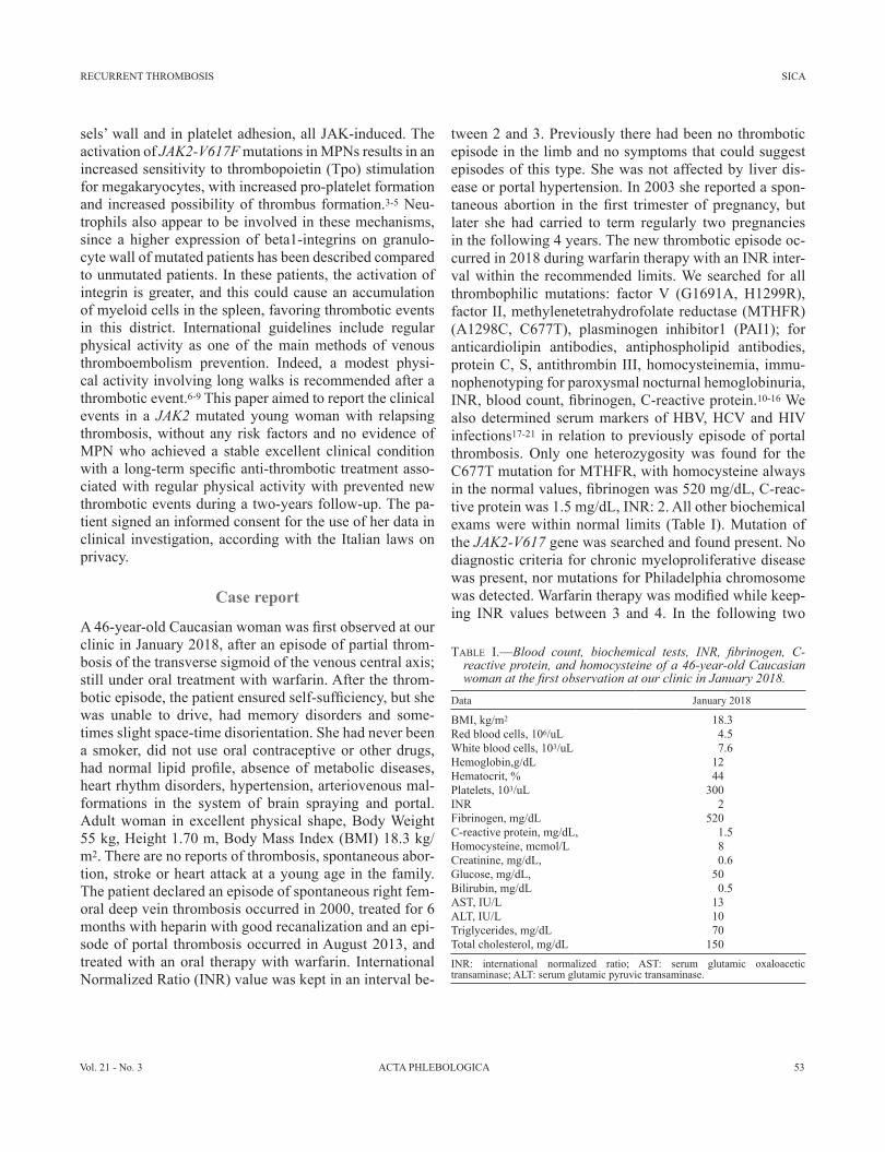
RecURReNt thRoMboSiS SicA
Vol. 21 - No. 3 ActA PhlebologicA 53
SicARecURReNt thRoMboSiS
tween 2 and 3. Previously there had been no thrombotic episode in the limb and no symptoms that could suggest episodes of this type. She was not affected by liver dis-ease or portal hypertension. in 2003 she reported a spon-taneous abortion in the first trimester of pregnancy, but later she had carried to term regularly two pregnancies in the following 4 years. the new thrombotic episode oc-curred in 2018 during warfarin therapy with an iNR inter-val within the recommended limits. We searched for all thrombophilic mutations: factor V (g1691A, h1299R), factor ii, methylenetetrahydrofolate reductase (MthFR) (A1298c, c677t), plasminogen inhibitor1 (PAi1); for anticardiolipin antibodies, antiphospholipid antibodies, protein c, S, antithrombin iii, homocysteinemia, immu-nophenotyping for paroxysmal nocturnal hemoglobinuria, INR, blood count, fibrinogen, C-reactive protein.10-16 We also determined serum markers of hbV, hcV and hiV infections17-21 in relation to previously episode of portal thrombosis. only one heterozygosity was found for the c677t mutation for MthFR, with homocysteine always in the normal values, fibrinogen was 520 mg/dL, C-reac-tive protein was 1.5 mg/dl, iNR: 2. All other biochemical exams were within normal limits (table i). Mutation of the JAK2-V617 gene was searched and found present. No diagnostic criteria for chronic myeloproliferative disease was present, nor mutations for Philadelphia chromosome was detected. Warfarin therapy was modified while keep-ing iNR values between 3 and 4. in the following two
sels’ wall and in platelet adhesion, all JAK-induced. the activation of JAK2-V617F mutations in MPNs results in an increased sensitivity to thrombopoietin (tpo) stimulation for megakaryocytes, with increased pro-platelet formation and increased possibility of thrombus formation.3-5 Neu-trophils also appear to be involved in these mechanisms, since a higher expression of beta1-integrins on granulo-cyte wall of mutated patients has been described compared to unmutated patients. in these patients, the activation of integrin is greater, and this could cause an accumulation of myeloid cells in the spleen, favoring thrombotic events in this district. international guidelines include regular physical activity as one of the main methods of venous thromboembolism prevention. indeed, a modest physi-cal activity involving long walks is recommended after a thrombotic event.6-9 this paper aimed to report the clinical events in a JAK2 mutated young woman with relapsing thrombosis, without any risk factors and no evidence of MPN who achieved a stable excellent clinical condition with a long-term specific anti-thrombotic treatment asso-ciated with regular physical activity with prevented new thrombotic events during a two-years follow-up. the pa-tient signed an informed consent for the use of her data in clinical investigation, according with the italian laws on privacy.
Case report
A 46-year-old Caucasian woman was first observed at our clinic in January 2018, after an episode of partial throm-bosis of the transverse sigmoid of the venous central axis; still under oral treatment with warfarin. After the throm-botic episode, the patient ensured self-sufficiency, but she was unable to drive, had memory disorders and some-times slight space-time disorientation. She had never been a smoker, did not use oral contraceptive or other drugs, had normal lipid profile, absence of metabolic diseases, heart rhythm disorders, hypertension, arteriovenous mal-formations in the system of brain spraying and portal. Adult woman in excellent physical shape, body Weight 55 kg, height 1.70 m, body Mass index (bMi) 18.3 kg/m2. there are no reports of thrombosis, spontaneous abor-tion, stroke or heart attack at a young age in the family. the patient declared an episode of spontaneous right fem-oral deep vein thrombosis occurred in 2000, treated for 6 months with heparin with good recanalization and an epi-sode of portal thrombosis occurred in August 2013, and treated with an oral therapy with warfarin. international Normalized Ratio (iNR) value was kept in an interval be-
Table I.—� Blood count, biochemical tests, INR, fibrinogen, C-reactive protein, and homocysteine of a 46-year-old Caucasian woman at the first observation at our clinic in January 2018.
Data January 2018
bMi, kg/m2 18.3Red blood cells, 106/ul 4.5White blood cells, 103/ul 7.6hemoglobin,g/dl 12hematocrit, % 44Platelets, 103/ul 300iNR 2Fibrinogen, mg/dl 520c-reactive protein, mg/dl, 1.5homocysteine, mcmol/l 8creatinine, mg/dl, 0.6glucose, mg/dl, 50bilirubin, mg/dl 0.5ASt, iU/l 13Alt, iU/l 10triglycerides, mg/dl 70total cholesterol, mg/dl 150iNR: international normalized ratio; ASt: serum glutamic oxaloacetic transaminase; Alt: serum glutamic pyruvic transaminase.

SicA RecURReNt thRoMboSiS
54 ActA PhlebologicA December 2020
Conclusions
in conclusion, besides hyperproliferation of myeloid pro-genitor cells, the mechanisms favoring venous thrombosis most likely includes an abnormal release of inflammatory cytokines and hyper-agglutination. it would be interesting to search for the JAK2 mutation in all cases of unexplained venous thrombosis, because this mutation can involve a thrombotic risk regardless of the evidence of a concomi-tant myeloproliferative disease. At present, specific thera-pies (JAK inhibitor) are not used for these patients, due to insufficient knowledge of the link between this mutation and thrombotic events, and for the few cases described in the literature not associated with myeloproliferative disease. therefore, adequate conventional therapy with anticoagulants and early mobilization represent the first-choice treatment of acute thrombosis of unknown origin, followed by regular physical activity to prevent further thrombotic episodes.
References
1. De Stefano V, Fiorini A, Rossi e, Za t, Farina g, chiusolo P, et al. incidence of the JAK2 V617F mutation among patients with splanchnic or cerebral venous thrombosis and without overt chronic myeloproliferative disorders. J thromb haemost 2007;5:708–14. 2. Fleischman Ag, tyner JW. causal role for JAK2 V617F in thrombosis. blood 2013;122:3705–6. 3. Zerjavic K, Zagradisnik b, Stangler herodez S, lokar l, glaser Krase-vac M, Kokalj Vokac N. is the JAK2 V617F mutation a hallmark for dif-ferent forms of thrombosis? Acta haematol 2010;124:49–56. 4. Sica A, casale b, Dato Mt, calogero A, Spada A, Sagnelli c, et al. cancer- and Non-cancer Related chronic Pain: From the Physiopatho-logical basics to Management. open Med (Wars) 2019;14:761–6. 5. Fiorelli A, D’Andrilli A, carlucci A, Vicidomini g, loizzi D, Ardò NP, et al.; lucilyS study group. Prognostic factors of lung cancer in lymphoma survivors (the lucilyS study). transl lung cancer Res 2020;9:90–102. 6. Rabinovich A, Kahn SR. how i treat the postthrombotic syndrome. blood 2018;131:2215–22. 7. Ronchi A, Zito Marino F, Vitiello P, caccavale S, Argenziano g, crisci S, et al. A case of primary cutaneous b-cell lymphoma with immature features in an old man. Diffuse large b-cell lymphoma with immature features or b-cell lymphoblastic lymphoma? J cutan Pathol 2020. [epub ahead of print] 8. Guastafierro S, Ferrara MG, Sica A, Parascandola RR, Santangelo S, Falcone U. Serum double monoclonal components and hematological malignancies: only a casual association? Review of 34 cases. leuk Res 2012;36:1274–7. 9. Partsch h. Ambulation and compression after deep vein thrombosis: dispelling myths. Semin Vasc Surg 2005;18:148–52. 10. Viscardi G, Zanaletti N, Ferrara MG, Sica A, Falcone U, Guastafierro S, et al. Atypical haemolytic-uraemic syndrome in patient with metastatic colorectal cancer treated with fluorouracil and oxaliplatin: a case report and a review of literature. eSMo open 2019;4:e000551. 11. Sica A, Vitiello P, caccavale S, Sagnelli c, calogero A, Doraro cA, et al. Primary cutaneous Dlbcl Non-gcb type: challenges of a Rare case. open Med (Wars) 2020;15:119–25.
months, the neurological symptoms were struggling to improve. Memory disturbances, disorientation and lack of autonomy made the patient more and more afraid of fac-ing everyday life. the iNR values during this period were consistently held between 3 and 4. the anticoagulant ther-apy was changed by introducing apixaban 5 mg, one tab-let twice a day. the general situation gradually improved and after 2 months of treatment, the patient completely recovered their autonomy, was well oriented and started driving again. Despite her challenging clinical history, the patient, being was in good physical condition, started practice regularly pilates, aerobic gymnastics, and long walks. Subsequently, she increased her physical activity with 3 weekly sessions at a high rate and long walks regu-larly. Regular physical activity improved health-related quality of life. currently, after a 2-year follow-up, she is in full wellbeing, in good clinical condition and, thanks to the physical training program, she has achieved an envi-able physical condition.
Discussion
Physical activity is very helpful in preventing and throm-bosis recurrence.22-25 Accordingly, early mobilization is recommended after post-thrombotic period, in order to avoid recurrence, whereas an intense physical activity is not recommended since it could facilitate the mobilization of thrombus during the disaggregation. Physical training benefits patients who have had deep vein thrombosis con-trolling body weight gain, favoring cardiovascular fitness and strengthening muscles. in addition, strengthening the muscles by improving their functionality beneficial ef-fects on venous return and on and typical skin dehydra-tion.26-31 in our case, the mutation of the JAK2-V617F was the only cause of thrombophilic condition and of the episodes of thrombotic events. only a few studies have so far described32 cases of thrombophilia exclusively re-lated to a JAK2 in the absence of MPN, but none of them presented a severe clinical history like that of the patient described here. the pathogenetic mechanisms associated to this mutation have not been fully elucidated at present. An increased incidence of cerebral and splanchnic venous thrombosis in patients with JAK2 mutation without MPN has been previously reported. in addition, JAK2-V617F mutant liver endothelial cells have been described in pa-tients with budd-chiari Syndrome, even in the absence of MPN, suggesting that the JAK2-V617F expression may favor splanchnic venous thrombosis even in the absence of hematopoietic cells.

RecURReNt thRoMboSiS SicA
Vol. 21 - No. 3 ActA PhlebologicA 55
Prevention of thrombosis Panel. executive summary: Antithrombotic therapy and Prevention of thrombosis, 9th ed: American college of chest Physicians evidence-based clinical Practice guidelines. chest 2012;141:7S–47S. 23. Sica A, Vitiello P, Papa A, calogero A, Sagnelli c, casale D, et al. Use of rituximab in Nhl malt type pregnant in i° trimester for two times. open Med (Wars) 2019;14:757–60. 24. Reginelli A, Urraro F, Sangiovanni A, Russo gM, Russo c, grassi R, et al. extranodal lymphomas: a pictorial review for ct and MRi clas-sification. Acta Biomed 2020;91:34–42.25. Kahn SR, lim W, Dunn AS, cushman M, Dentali F, Akl eA, et al. Prevention of Vte in nonsurgical patients: Antithrombotic therapy and Prevention of thrombosis, 9th ed: American college of chest Physicians evidence-based clinical Practice guidelines. chest 2012;141:e195S–226S. 26. tosetto A, Frezzato M, Rodeghiero F. Prevalence and risk factors of non-fatal venous thromboembolism in the active population of the VitA Project. J thromb haemost 2003;1:1724–9. 27. Sica A, casale b, Spada A, teresa Di Dato M, Sagnelli c, calogero A, et al. Differential Diagnosis: Retroperitoneal Fibrosis and oncological Diseases. open Med (Wars) 2019;15:22–6. 28. Visco c, Di Rocco A, evangelista A, Quaglia FM, tisi Mc, Morello l, et al. Outcomes in first relapsed-refractory younger patients with man-tle cell lymphoma: results from the MANtle-FiRSt study. leukemia 2020. [epub ahead of print] 29. caccavale S, Vitiello P, Franco R, Panarese i, Ronchi A, Sica A, et al. Dermoscopic characterization of folliculotropic mycosis fungoides selectively localized on trunk and limbs. int J Dermatol 2019;58:e187–9. 30. Sica A, Vitiello P, Sorriento A, Ronchi A, calogero A, Sagnelli c, et al. lymphomatoid papulosis. Minerva Med 2020;111:166–72. 31. Reginelli A, Belfiore MP, Russo A, Turriziani F, Moscarella E, troiani t, et al. A Preliminary Study for Quantitative Assessment with hFUS (high- Frequency Ultrasound) of Nodular Skin Melanoma bres-low thickness in Adults before Surgery: interdisciplinary team experi-ence. curr Radiopharm 2020;13:48–55. 32. De t, Prabhakar P, Nagaraja D, christopher R. Janus kinase (JAK) 2 V617F mutation in Asian indians with cerebral venous thrombosis and without overt myeloproliferative disorders. J Neurol Sci 2012;323:178–82.
12. cascone R, Sica A, Sagnelli c, carlucci A, calogero A, Santini M, et al. endoscopic treatment and Pulmonary Rehabilitation for Management of lung Abscess in elderly lymphoma Patients. int J environ Res Public health 2020;17:997. 13. Sica A, Sagnelli c, Papa A, ciccozzi M, Sagnelli e, calogero A, et al. An Anecdotal case Report of chronic lymphatic leukemia with del(11q) Treated with Ibrutinib: Artificial Nourishment and Physical Activity Pro-gram. int J environ Res Public health. 2020 Mar 16;17(6):0. Vitiello P, Sica A, Ronchi A, caccavale S, Franco R, Argenziano g. Primary cutane-ous b-cell lymphomas: An Update. Front oncol 2020;10:651.14. bagaglio S, Uberti-Foppa c, Sagnelli c, lai A, hasson h, Salpietro S, et al. hiV-1 recombinant forms in immigrants regularly residing in Mi-lan, northern italy. infection 2020;48:553–8. 15. leroyer c, Mercier b, oger e, chenu e, Abgrall JF, Férec c, et al. Prevalence of 20210 A allele of the prothrombin gene in venous thrombo-embolism patients. thromb haemost 1998;80:49–51. 16. Ridker PM, hennekens ch, Miletich JP. g20210A mutation in prothrombin gene and risk of myocardial infarction, stroke, and venous thrombosis in a large cohort of US men. circulation 1999;99:999–1004. 17. Merli M, Frigeni M, Alric l, Visco c, besson c, Mannelli l, et al. Direct-Acting Antivirals in hepatitis c Virus-Associated Diffuse large b-cell lymphomas. oncologist 2019;24:e720–9. 18. Sica A, casale D, Rossi g, casale b, ciccozzi M, Fasano M, et al. the impact of the SARS-coV-2 infection, with special reference to the hematological setting. J Med Virol 2020. [epub ahead of print] 19. Pisaturo M, Guastafierro S, Filippini P, Tonziello G, Sica A, Di Mar-tino F, et al. Absence of occult hcV infection in patients experiencing an immunodepression condition. infez Med 2013;21:296–301.20. tonziello g, Pisaturo M, Sica A, Ferrara Mg, Sagnelli c, Pasquale g, et al. transient reactivation of occult hepatitis b virus infection despite lamivudine prophylaxis in a patient treated for non-hodgkin lymphoma. infection 2013;41:225–9. 21. Sica A, Vitiello P, Ronchi A, casale b, calogero A, Sagnelli e, et al. Primary cutaneous Anaplastic large cell lymphoma (pcAlcl) in the elderly and the importance of Sport Activity training. int J environ Res Public health 2020;17:839. 22. guyatt gh, Akl eA, crowther M, gutterman DD, Schuünemann hJ; American college of chest Physicians Antithrombotic therapy and
Conflicts of interest.—The authors certify that there is no conflict of interest with any financial organization regarding the material discussed in the manuscript.Authors’ contributions.—Antonello Sica has given substantial contributions to manuscript conceptualization, writing, revision and editing, Antonello Sica and Alfonso Fiorelli to data curation and investigation. Antonello Sica and evangelista Sagnelli to methodology, Antonello Sica, caterina Sagnelli and be-niamino Casale to project administration. All authors read and approved the final version of the manuscript.History.—Manuscript accepted: october 20, 2020. - Manuscript revised: october 15, 2020. - Manuscript received: July 15, 2020.
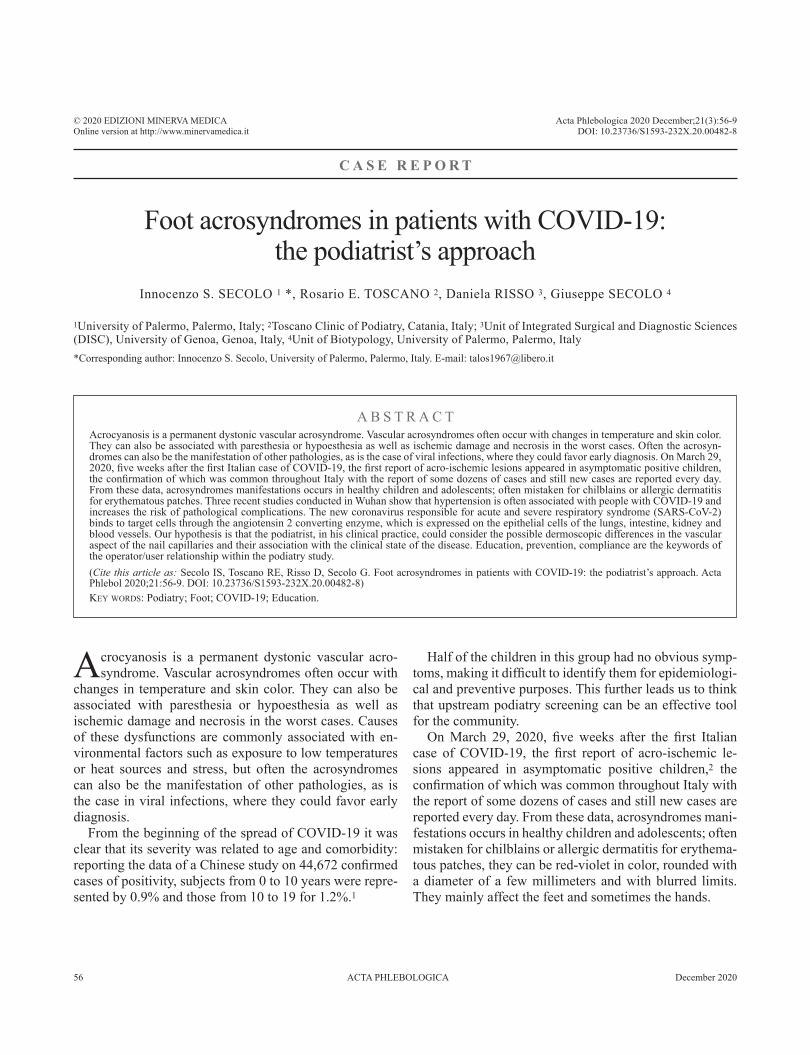
56 ActA PhlebologicA December 2020
Acta PhlebologicaDecember 2020Vol. 21 - No. 3
C A S E R E P O R T
Foot acrosyndromes in patients with coViD-19: the podiatrist’s approach
innocenzo S. Secolo 1 *, Rosario e. toScANo 2, Daniela RiSSo 3, giuseppe Secolo 4
1University of Palermo, Palermo, italy; 2toscano clinic of Podiatry, catania, italy; 3Unit of integrated Surgical and Diagnostic Sciences (DiSc), University of genoa, genoa, italy, 4Unit of biotypology, University of Palermo, Palermo, italy*corresponding author: innocenzo S. Secolo, University of Palermo, Palermo, italy. e-mail: [email protected]
A b S t R A c tAcrocyanosis is a permanent dystonic vascular acrosyndrome. Vascular acrosyndromes often occur with changes in temperature and skin color. they can also be associated with paresthesia or hypoesthesia as well as ischemic damage and necrosis in the worst cases. often the acrosyn-dromes can also be the manifestation of other pathologies, as is the case of viral infections, where they could favor early diagnosis. on March 29, 2020, five weeks after the first Italian case of COVID-19, the first report of acro-ischemic lesions appeared in asymptomatic positive children, the confirmation of which was common throughout Italy with the report of some dozens of cases and still new cases are reported every day. From these data, acrosyndromes manifestations occurs in healthy children and adolescents; often mistaken for chilblains or allergic dermatitis for erythematous patches. three recent studies conducted in Wuhan show that hypertension is often associated with people with coViD-19 and increases the risk of pathological complications. the new coronavirus responsible for acute and severe respiratory syndrome (SARS-coV-2) binds to target cells through the angiotensin 2 converting enzyme, which is expressed on the epithelial cells of the lungs, intestine, kidney and blood vessels. our hypothesis is that the podiatrist, in his clinical practice, could consider the possible dermoscopic differences in the vascular aspect of the nail capillaries and their association with the clinical state of the disease. education, prevention, compliance are the keywords of the operator/user relationship within the podiatry study.(Cite this article as: Secolo iS, toscano Re, Risso D, Secolo g. Foot acrosyndromes in patients with coViD-19: the podiatrist’s approach. Acta Phlebol 2020;21:56-9. Doi: 10.23736/S1593-232X.20.00482-8)Key words: Podiatry; Foot; coViD-19; education.
Acta Phlebologica 2020 December;21(3):56-9Doi: 10.23736/S1593-232X.20.00482-8
SecoloFoot AcRoSYNDRoMeS iN PAtieNtS With coViD-19
© 2020 eDiZioNi MiNeRVA MeDicAonline version at http://www.minervamedica.it
Acrocyanosis is a permanent dystonic vascular acro-syndrome. Vascular acrosyndromes often occur with
changes in temperature and skin color. they can also be associated with paresthesia or hypoesthesia as well as ischemic damage and necrosis in the worst cases. causes of these dysfunctions are commonly associated with en-vironmental factors such as exposure to low temperatures or heat sources and stress, but often the acrosyndromes can also be the manifestation of other pathologies, as is the case in viral infections, where they could favor early diagnosis.
From the beginning of the spread of coViD-19 it was clear that its severity was related to age and comorbidity: reporting the data of a Chinese study on 44,672 confirmed cases of positivity, subjects from 0 to 10 years were repre-sented by 0.9% and those from 10 to 19 for 1.2%.1
half of the children in this group had no obvious symp-toms, making it difficult to identify them for epidemiologi-cal and preventive purposes. this further leads us to think that upstream podiatry screening can be an effective tool for the community.
On March 29, 2020, five weeks after the first Italian case of COVID-19, the first report of acro-ischemic le-sions appeared in asymptomatic positive children,2 the confirmation of which was common throughout Italy with the report of some dozens of cases and still new cases are reported every day. From these data, acrosyndromes mani-festations occurs in healthy children and adolescents; often mistaken for chilblains or allergic dermatitis for erythema-tous patches, they can be red-violet in color, rounded with a diameter of a few millimeters and with blurred limits. they mainly affect the feet and sometimes the hands.
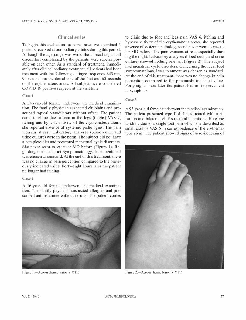
Foot AcRoSYNDRoMeS iN PAtieNtS With coViD-19 Secolo
Vol. 21 - No. 3 ActA PhlebologicA 57
SecoloFoot AcRoSYNDRoMeS iN PAtieNtS With coViD-19
to clinic due to foot and legs pain VAS 6, itching and hypersensitivity of the erythematous areas; she reported absence of systemic pathologies and never went to vascu-lar MD before. the pain worsens at rest, especially dur-ing the night. laboratory analyses (blood count and urine culture) showed nothing relevant (Figure 2). the subject had menstrual cycle disorders. concerning the local foot symptomatology, laser treatment was chosen as standard. At the end of this treatment, there was no change in pain perception compared to the previously indicated value. Forty-eight hours later the patient had no improvement in symptoms.
Case 3
A 93-year-old female underwent the medical examination. the patient presented type ii diabetes treated with met-formin and bilateral MtP structural alterations. he came to clinic due to a single foot pain which she described as small cramps VAS 5 in correspondence of the erythema-tous areas. the patient showed signs of acro-ischemia of
Clinical seriesto begin this evaluation on some cases we examined 3 patients received at our podiatry clinics during this period. Although the age range was wide, the clinical signs and discomfort complained by the patients were superimpos-able on each other. As a standard of treatment, immedi-ately after clinical podiatry treatment, all patients had laser treatment with the following settings: frequency 645 nm, 90 seconds on the dorsal side of the foot and 60 seconds on the erythematous areas. All subjects were considered coViD-19 positive suspects at the visit time.
Case 1
A 17-year-old female underwent the medical examina-tion. the family physician suspected chilblains and pre-scribed topical vasodilators without effect. the patient came to clinic due to pain in the legs (thighs) VAS 7, itching and hypersensitivity of the erythematous areas; she reported absence of systemic pathologies. the pain worsens at rest. laboratory analyses (blood count and urine culture) were in the norm. the subject did not have a complete diet and presented menstrual cycle disorders. She never went to vascular MD before (Figure 1). Re-garding the local foot symptomatology, laser treatment was chosen as standard. At the end of this treatment, there was no change in pain perception compared to the previ-ously indicated value. Forty-eight hours later the patient no longer had itching.
Case 2
A 16-year-old female underwent the medical examina-tion. the family physician suspected allergies and pre-scribed antihistamine without results. the patient comes
Figure 1.—Acro-ischemic lesion V MtP. Figure 2.—Acro-ischemic lesion V MtP.

Secolo Foot AcRoSYNDRoMeS iN PAtieNtS With coViD-19
58 ActA PhlebologicA December 2020
ing pain in the foot. however, acrocyanosis may not be the only obvious clinical sign.
the lower limbs can manifest symptoms such as pain or dyschromia, which are sometimes associated with hy-poxia, metabolic alterations in the microcirculation, or ac-rocyanosis.3
three recent studies4 conducted in Wuhan showed that hypertension is often associated with people with coV-iD-19 and increases the risk of pathological complications.
the new coronavirus – responsible for acute and severe respiratory syndrome (SARS-coV-2) – binds to target cells through the angiotensin 2 converting enzyme – which is expressed on the epithelial cells of the lungs, intestine, kidney and blood vessels.
Angiogenesis is closely related to onychodystrophy based on the activity of fibroblasts (FGF);5, 6 the evalua-tion of the skin appendages, such as the altered regrowth of the nail (onychopathy) – in addition to factors related to traumatism – varies according to nutraceutical factors, oxygenation of the matrix and systemic diseases. the low angiogenic activity causes damage to the tissues followed by ischemia or heart failure caused by coViD-19.
our hypothesis is that the podiatrist, in his clinical prac-tice, could consider the possible dermoscopic differences in the vascular aspect of the nail capillaries and their asso-ciation with the clinical state of the disease. Recent studies in hcV and hbV patients7 have shown how nail capillary abnormalities are or could be a sign of endothelial tissue damage in chronic viral hepatitis.
education, prevention, compliance are the keywords of the operator/user relationship within the podiatry study.
Synergic and interdisciplinary work would be appropri-ate; working with outpatient archives and summary sheets reported the following data:
• name and surname, gender, age;• fever states;• medical diagnosis;• clinical signs, onychopathies;• acrocyanosis.Although the Spanish guidelines of the general council
of the college of Podiatrists advise not to go to the hospi-tal for the sole appearance of symptoms, outpatient man-agement through memoranda of understanding between public and private health professionals through the use of these means and telemedicine, would allow greater speed in differential diagnosis and therapeutic administration.
ii and iii MtP of the right foot, which were painful to the touch. About the local foot symptomatology, laser treat-ment was chosen as standard (Figure 3). She presented regular peripheral pulses. laboratory analysis (blood count and urine culture) showed nothing relevant. At the end of the laser treatment, there were improvements (VAS 3) in pain perception compared to the previously indicated value. Forty-eight hours later the patient had no more pain. As for coViD-19, the patient was asymptomatic at the time of the visit, but she will be positive after the swab requested by the family doctor, 4 days later.
Discussion
the foot areas, usually most affected, are situated near the distal metatarsophalangeal joint, but in some cases, they can be found even in the plantar region. the lesions do not affect all the toes uniformly – on average they are three on five; they may have blackish crusts in the subsequent evolution – usually painful, they evolve within two weeks and do not leave any aftermath. this syndrome may be associated with muscle pain and intense itching with burn-
Figure 3.—Acro-ischemic lesion ii MtP with onychodystrophy.
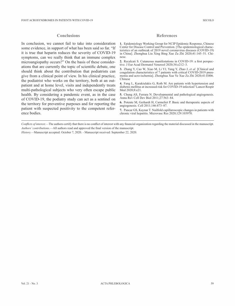
Foot AcRoSYNDRoMeS iN PAtieNtS With coViD-19 Secolo
Vol. 21 - No. 3 ActA PhlebologicA 59
References
1. epidemiology Working group for NciP epidemic Response, chinese center for Disease control and Prevention. [the epidemiological charac-teristics of an outbreak of 2019 novel coronavirus diseases (coViD-19) in china]. Zhonghua liu Xing bing Xue Za Zhi 2020;41:145–51. chi-nese.2. Recalcati S. Cutaneous manifestations in COVID-19: a first perspec-tive. J eur Acad Dermatol Venereol 2020;34:e212–3. 3. Zhang Y, cao W, Xiao M, li YJ, Yang Y, Zhao J, et al. [clinical and coagulation characteristics of 7 patients with critical coViD-2019 pneu-monia and acro-ischemia]. Zhonghua Xue Ye Xue Za Zhi 2020;41:e006. chinese4. Fang l, Karakiulakis g, Roth M. Are patients with hypertension and diabetes mellitus at increased risk for coViD-19 infection? lancet Respir Med 2020;8:e21. 5. chung AS, Ferrara N. Developmental and pathological angiogenesis. Annu Rev cell Dev biol 2011;27:563–84. 6. Potente M, gerhardt h, carmeliet P. basic and therapeutic aspects of angiogenesis. cell 2011;146:873–87. 7. Pancar gS, Kaynar t. Nailfold capillaroscopic changes in patients with chronic viral hepatitis. Microvasc Res 2020;129:103970.
Conclusions
in conclusion, we cannot fail to take into consideration some evidence, in support of what has been said so far. “if it is true that heparin reduces the severity of coViD-19 symptoms, can we really think that an immune complex microangiopathy occurs?” on the basis of these consider-ations that are currently the topic of scientific debate, one should think about the contribution that podiatrists can give from a clinical point of view. in his clinical practice, the podiatrist who works on the territory, both at an out-patient and at home level, visits and independently treats multi-pathological subjects who very often escape public health. by considering a pandemic event, as in the case of coViD-19, the podiatry study can act as a sentinel on the territory for preventive purposes and for reporting the patient with suspected positivity to the competent refer-ence bodies.
Conflicts of interest.—The authors certify that there is no conflict of interest with any financial organization regarding the material discussed in the manuscript.Authors’ contributions.—All authors read and approved the final version of the manuscript.History.—Manuscript accepted: october 7, 2020. - Manuscript received: September 22, 2020.