WSCPB1984-1985.pdf - The Joint Pathology Center (JPC)
202
I E A I, U) rd '-l CLl '-t Q -t !(01 +) o l.'{o. q- I .-t ol .Fi o_ o a a cC F{ : EO (U0 ..1=. OJ(!l t{ >-O(/)l A !EI (o(dI Ef,I o C : C al O ()l -o-ll l(oo) 0JCol o(t)l (/)f{F{l o c -rl >-cl @ .c.dl !rOf (/)OI E+rl EJJOI (o+rG = c)l Ol O lrl E O +rl Ct+rl -l Q Ol 6oj:o +)cr,l Col f CO-l .-J f 0) ro.-ll '-{ trl C <o t,l ao.i E-rdr .Io-r 8r<l co c'-i F{ o f,l ! o cr).-l o l{ O! f J-}+)l O+J O+).a.) O).-l C C (6l t{ f{.-{ o+)0) (o(I)-ll oo (J-lo) >(U8 k:lfl of (l)f f -LE..r c 9 ]f,(,l zE Z.QE "jt 'Fll dl 'Fl I ^ l oEl a 1r f | '-l qJl +) ocl (U - - t - 4 l c6 -tl ..{ -i I U) C <l.ll .F{ ocl a e ^ l n ).41 .- (l) CDI -r c (l)l 0) cL -Jl o- l.'-{ | lLr I lEl rfil 3l 0) = E U' E -lJ ..-i C'-l .-l tr lc O- rco . - L O'-l >\E Fl o- &) (! o c OJ (o F{ Ol '-l .-l I f-. (,l +) -l c o)l o (ol Tl .-,t I c -l t.l o) -l J .-l - o +) F.. 0) T E f, E o f, a ..t +J t rt ) a (o .-l (t qr a.J n f4 A e4 O a a (of 4 e OJ a G .J o-o c) oGf .J r!) A- r o0J )<..i :< o+) o ('q- +roo OC o (f)> t > o..t H .Ft .Fl C r A - (F> E Ot-{ fr-O O (o 0J o (! H >C C :<J H +r.-t.Ft (J E -l[-.Oq) = f O._t -O t!(, ..Fl a O O O f C)O = t!>=G v g c\ \o s o\ F{ I F J E, = Ir \o -t F\ \o o\ h dl C f '.{ J J cl a (ol = ctl r{ f-. I 0) ol +) = (/)l(o ol o) .Frl c Ol .Fi g) g) ojl :l'-r O .*.1 (Jo o +J (' k OJ a 0) IY X 0) +-) c V) ,-.t X .|J - (n +J o. V) o\ .{ sl ol -ll tl HI Frl HI 6l bt q-l 5l xr ;l el 'nl al >l *l ol PI El ^ l HI <"1 Gl(\ H\O J rc\ o\ co rn € o\ F.l \o o\ <t o\ r- r-- q $ o\ o\ '{ Fl f { S CDJ O\ c= $ +)< I GIJ- {f n \ o t s L n A r.\ N I f.\ |f\ q-u) o(J oc]) (FO< q-.-l O Ocot! \o ln q t\ rf o\ -l t{ +J t-.1 +) - I I *l *l 9t r<\ Ft o\ Fl f\ $ \o o .-{ h +J+ c\l {f r\ $ co l-) 0) N H H H H H H o) L C oo) f{ oq) +J q- H H H t-l 3 l (6 +sl (-)l . l ( t s 1 c*l P I H
-
Upload
khangminh22 -
Category
Documents
-
view
2 -
download
0
Transcript of WSCPB1984-1985.pdf - The Joint Pathology Center (JPC)
IE A I , U )rd '-l CLl '-t Q- t ! ( 0 1 + ) o l . ' { o .q- I .-t ol .Fi o_ o a ac C F { : E O ( U 0. . 1 = . O J ( ! l t { > - O ( / ) l
A ! E I ( o ( d I E f , Io C : C a l O ( ) l - o - l ll ( o o ) 0 J C o l o ( t ) l ( / ) f { F { lo c - r l > - c l @ . c . d l! r O f ( / ) O I E + r l E J J O I( o + r G = c ) l O l O l r lE O + r l C t+ r l - l Q O l6 o j : o + ) c r , l C o l f C O - l. -J f 0) ro.- l l ' - { t r l C <o t , l
a o . i E - r d r . I o - r 8 r < lco c ' - i F{ o f , l ! o cr) . - l ol { O ! f J - } + ) l O + J O + ) . a . )O).- l C C (6l t { f { . - {o + ) 0 ) ( o ( I ) - l l o o ( J - l o )> ( U 8 k : l f l o f ( l ) f f
- L E . . r c 9 ] f , ( , l z E Z . Q E
" j t' F l ld l'Fl I
^ l
o E l a1r f | '-l
qJ l + )o c l ( U- - t -
4 l
c6 - t l..{ -i I U)C <l.l l .F{o c l ae ^ l n) . 4 1 . -(l) CDI -rc (l)l 0)cL -Jl o-
l.'-{ |
l L r Il E lr f i l
3 l
0)=E
U ' E
-lJ ..-i
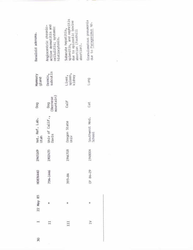
C ' - l.-l tr
lc O-
r c o. - L
O ' - l
> \EFl
o- &)
(!
ocOJ
(o
F{
O l ' - l.-l I f-.( , l +)- l c
o)l o(ol T l.-,t I c- l t . l
o)-l
J
.-l-
o
+)F..
0)T
Ef,
Eof,
a..t+Jtr t
)a
(o
.-l (t qra . J n
f 4 A
e4 O
a a( o f4 e
OJaG . Jo-o c )o G f
. J r ! )A - r
o 0 J)<.. i :<o + )o ( ' q -+ r o oO Co ( f ) > t> o . . t H
.Ft .Fl Cr A -
( F >E O t - {f r -O O (o 0Jo ( ! H > C C: < J H + r . - t . F t ( J
E - l [ - . O q )= f O ._ t -O
t ! ( ,. . F l a O O O fC ) O = t ! > = G
vgc\\oso\F{
I
F
JE,=I r
\o-tF\\oo\
h d l
Cf '.{
J J
c l a(o l =ctl r{f-. I 0)o l + )=
( / ) l ( oo l o ). F r l cOl .Fi g) g)
o j l : l ' - r O. * .1 (Jo o
+J
('
k
OJa0)
IY
X0)+-)cV)
,-.t
X
.|J-(n
+Jo.V)
o\.{
slo l- l l
t l
HIFr l
HI6 lbtq - l
5 l x r
;l el' n l
al>l
* lo l
P IEl^ lH I<"1
G l ( \H \ O
J rc\
o\corn€o\F.l
\o o\<t o\r- r--q $o\ o\'{ Fl
f { SC D J O \
c = $+ ) < IG I J - { f
n\ o ts L nA r.\N If.\ |f\
q - u )o ( Jo c ] )
( F O <q - . - l OO c o t !
\olnqt\rfo\-l
t{
+J
t-.1+)-
II* l
* l9 t
r<\
Ft
o\Fl
f\
$\o
o.-{h
+ J +c\l
{fr\
$co
l-)
0)
N
HHH
H
HH
o)L Co o )
f{o q )
+J q-
HH Ht-l
3 l(6 +sl( - ) l
. l( t s 1c * lP I
H
: i i E,. , { - { l J s
n i s E l a E ; 'P ? * H l E o ' t lb U q Q l g . , P le P d H l 6 . i i l.g ' ; .h , - i >g '1€ lH d ' ) ' i ' g l E = : ' ' 'y 6 r ' : . . q l - .8 PS: i 3 t 6 t a I ; ;PE 9e ' ! l FE s€
Eo '-4+ ) o
. . ; o , . i; E = a - c; ; - c f , { J
- H
- i b o ' . i E'; i- .-r > 0)F i 6 c a x;i
'- o oJ 'Fi
/ n r t r - EL H = i ;
I F P P t dE
- R R n , E 3
H E r E o t - i. : ' 6 f . ; ' F { +J t r ( l ) F I d U' d i
E F l o J r 0 0 )i a j s - L z c
E g q. - r q =t ) u(o (o '.'1
b 5 E. 9 o != t ) H
9 - E PE €
r-'i . r-{
a 5 a a r o.Ft CO '-l '-l +r! ) A A ' F {' d 3 8 8 6€ s F E BU I E Z . P c )
Co o -E . hj
o ( / l
O'F{t{( n c )
= F aO F t+r (l)( o f ,E =- { o=t +)( ! ( l )t{ :l( J E
dO ( o
^ > = E. d
l - . :F ^ i i E 5 o( 9 9 e Ya -
C F I L H. ( o o E
o - E OF= = f i ' F 6x = H , x : =a z g v l
R N
i i - t {; ( o + )1C - r _ ( Jo5 t r J t -
F I \
- tF l = c ) t {d d ) H > \ ( oF J :' F F ' t f f 3 EJ i X = : i 6 d
E o; € E 5 Ee b : - 8 , It r )+r c) t ! c6+l > r : lr{ C - } Fl (! '-i.!
e 8 = e 5 f i 5 2
0)C
.Fi E! a a
= a r i n a - l C
J 5 ' 6 i 6 a =h r - . r r t O ' F )i1 - ; - .2 ! ) 0)5 j . ; J = - )
FI( 0 t rF { ( 0 u-ii p 'F't
E . - ' 4 P r D Ii . a , i H r 'H U =d r ! - ) . i l . - { ) !
; n L o ) J F
F
o . r le a Q. - c e+)
? t t h ( l ) E + )F { ( D C O ' a l
E ' g € E E ba ti; i >< < o.
f{( 6 0 )
V
Y ' F- 5 =
0)
c=
0)
c-=
o)+J
- r n L ) O )O O ' F { ( J- - : < o
r { o 8 e+i ( l ) (U| ' - { )
s g P e t( 5 6 6 6 G
N r -rJ\ O\\ o NO NO\ cOFl Ft
\O Fl
O\ O\UN tf\f\ f\$ {O\ o\-l Fl
f q o l n NC ] o \ ! n @N @ N Ntn \O f- f-o s s q6 0 \ o \ c o-l F{ -l Fl
F C O O \ O@ @ 6 | r \- { u \ O \ NF I \ F No \ $ s qc o O \ 6 o \
O \ x- t @t \ o \l \ o
t.\ o@ Nz @
\I
x
HO\ l'.
s co F-{ coC { . F I F { €F c o N IL n F l r C NN o [ . r \ c c o
o\r l
\o tqco I t{ \o tc\N T \ O F \ IF { t \ ( f NO $ - l $N g F S O@ @ l c o f -
q
+J
= - r( , F(^ -1
H
+)clF\
gc0+)
a\oc\I
HH HH
H H HH H H
H H I iH H H
H
( E t o 3- . a l + )
I E N i
* ! E t : d - €EEEI ; s : i p r; f r -Rt s E: ag l3 8 3 1 6 E p € . ! lEl ; rn t ;g ; ;= l H; li F o . e l ' i . :
€ E ; t e i lsEHeI sE se'i FEI
(o.-i+)
._r | .Fro l + )
o ( 6 l a. p + J l t {
<o - .r-rl ql gF ( l ) . - ' l l L ) E6 5 o l c o1 : €o l . - - can tJ
E .3.s1 P.e E' c i + r O l . F l + ) O
E .-{ ;l (,.-r (o
F T F ] 8 E P-o- O--ll -O o- qJ
6- d-cl ..r o 1lZ = c - r l u L c
E-^ t - g
r { g d q0 J o ( I ) p'n ''-1 t*' xl ) v ,
t-.Fl
t-{ ('o 0 )> + J.-l C
I .F{
>O . H 9 G E€ 9 5 E Eg : 9 g d
(!
+J
[-{t{ O)( 6 c )
n a
J J ( !o o 0 Jt r c U )
.rJ F{ .- gr 2
s I 3'd. I
€ E.''- If{o ( / ) G>< .Ft a
. o
3 d E
4
d ao ) [ ' 4
- J> . c r u )2 ' , o o ci l I o'8, IJ o q o a l
! q F
. F { o c r - O NFa - (L . . { ( ! '
r - i = ocn -
\ o qO\ \OO \ Nf \ @$ sO\ O\F{ Fl
4 t \ \ o o \s\ ln Fl f\\ i ' t n ! n ! nt \ \ o @ Fo s o \ s6 0 \ c o o \F{ -{ Fl Fl
o =o s aC - { C ! lF l \ o $R o _ c o c oi < ' | f \x c o F C g
€cO+)v
.Ft
f-l3a
NIJ\€so\F{
z.N
o\|f\
.Fl-
=olt
q,
x0)
\orn-t|r\$o\-t
F{
sr n l
o\$
N\o
q
+J
{N
HH HH H
H
H H HH H H
l-{
\o
A i l .edrC ; i E l i l € lHE I HgI gE;I* , F i t $ l s p e : l. 9 " 3 ! q F E H E I! ) + J ( d - + ) ! J ^ X i i l
i :E g : ; t g E:3b r o . i o 6 l B 2 * P
E i ! F E : l E s E e
*E E ; i E , ,H9s i ;c ;* g e;E: E.EE Eg FE:gE€ €eg srn ggtElH iBH ctsg sgF
Etr
A i -
E gd
= ( J
c')Ca
J
0)
rn
C=)
o)a=o=
oc)
.Flh
6 ' o a ' lF r O q Lo J J E. i o
1f , .F l LL hA a l
t E a s E =o o o = H !. ; o < r o L ,= < t Z * t Z
- R mt )t ' ='
! 5 5A F t J
el(U+J
o J ( l )t { 2 O J...t +) (/) CU ( * a E0J O .-l 'Fl
o_ (/)+) :<
=F{-
q ) + , EF { C ) t r.F{ f, o)C D E C )
otN
In
O {F O \{ f , nO \ F{ s- A
F{ Fl
s$
o \ o(}\ lnc o lN SF @
'-t.r_)
O C o
(o+JoOJ
.-t=
..it-
tnq!
N
-l
IJ\Fso\-t
${i.
\o!
N
|no\-1
F{
IN
$
z.F
C\l
Nz
.F{
q
ao\Ng
o\F{
co\o'-.1
Is
+)(6
c)+)+)a
z.
r\so\
a.)(oc)
c)o)
G'
cO)tr
g@
$go\
I\oO\o-1
Ilq@
sCO
Pc)
HH Ht-.{
HFtHHH
HH
. 2 A , l A
laal EfuiHEical tcs iHg ;*sE l eEA iE ; ef i ; ; e ;qr gEg ii b - € r , - d - a l < E O -
Hl o j ' :| ( - l } ) ' d u
I - - l?l ' .9 aE , , E - r g l : F f r€:l c; ;l el s. z E . d ' ; . r l E A . : . i*i!, EeEI En nEH.s E l 'd Ed$E3 f;Ef, l F-a g;
- L lo)0Jc >:, .Ft
l F {
c-l
Ic
.F{ 0)C
-{ .F{ ts{
* ; 9 P PE o ' - t f 2( 7 J + ) - { - l )
O Ah.)r F
. 6 U
:< l-
(o+)E a 'g P €-l = '-tO- F{ :<
+J(o
aa,
c).-tFl
)< -c+ J C'n 9.1,E -Hv , U -
\os\ogo\-l
FI
Is$
I
.l-,a-
0)O
Ft
\o.tc..l
=
!
n
.Ftr
+J
+)a
=
\o$o\
l*.\oI
nI
<t
tr(oC(o
..{C
c)Et{
!n@-{|r\o\-t
lr\@
I6gN
I
0)
.-l
O 'Flr n n
o). 5 3> + , o )88 ' i
. , ;j i ( l ) o Jo E c + ). - c t ( oC E . - + r.= E- +r.-t tJ)= d E g o - o do i E o g ' r q. . ( 5 C - ! o o . E )f { H . F { ' n E< o - ! O > ! : f,r-r . . t.r) Fl .. l .- l- l€c o) c c o c t o-a)o < < r - { c l = c o ( - ) >
r\N!n o\F{ FlF I Nr-\ t
c L lf q qA A
O\ \os ot q nO \ As $o\ o\-l Fl
Fl
N
\os6-l
HI
-it
T
(\o\r\so\Fl
o!nsF
sco
oz.coN
@
OJ
ln
HH
HH HH
H
-{-l
H
OJrco
- C t ( l )o.o o)E > F {>.Fr O_J - 1 A
+JH L(o .F{
= o
ac t >
| '-lo >t { ( oC f O
t r n =) 4 )
h P Ho o ( ut { k t s {( l ) o 0 )
u v v
E0)''')
F { @
c t cC A ' F l
. ' t L l ( 6o- o F-,1u1 () cD
I
?q ' i , i f i.-{ .-l
F E , q l , 8 . t E Pb { ^ { * l q E - ! t 5 i* € 3 E e i l a g g 8 ; 8o - { . - ] J - l , ^ " ' F F -
, B - 3 i e d l i P a l a 3' 6 ' " L : 1 6 e t * 6 ' * ; . 5 ; ' 3
erEs ere l e r ;g iE : :I E A E T E E I F E ; i H E 3 5F E S d E E s l s 3 € s e F ' E E
. - 9 t s . d" ; b - ' ; o E
e 5 ' ! 9 t - ct { ( 6 ( 6 E ] i : 3
" ;o ' ! b . ' 3 t ;
Ed ) c o . t , 35 3 H E . l - o qK E E F t g H H f ;: € * i ; F E b EE g ; i s + E P E p< I . r - r : lE f { k r c ( l ) e
5 g 9 f t 8 f E ; . 3
m
+)f{
0)I
-n t
..t-c.
a( F . r {
> . F {
- l F
d -
. , R g -
nv g F ^ t
v> N G + J
.F{ f.{ -l GC '-l )< +)
A t hJ q
.Fl 'e{- F
J
o o )+ J { Jc t ( U*J +Ja o
il
.F{ --O H
|(\nqo\g@-l
s\o-tF{
Fi
-?o\F{
tf\NN
I$
o\o\F{o\
t.\sc!
Is
F\Fl
r\Fgo\F{
Fo\t ^ Fco 't\
-l \O@ tc\N l q@ @
F C f@ fc\N I
l C O f qtc\ oU cOA 4 -
N F {O Ftq \os l n$ $o\ o\F,| Fl
gN
$ Fl \ lco o-
t ls sco co
(F
P(6
q- oJF{ 0,o .c.c ) a
t-
o ot{0J 0)+) r{O OF { Oo_(5
d F= --l FlF I GO J EO).-l
\oN|r\-t
o\
c)+JG
a
z
l.f\
lt\coso\rl
$
o)
o\
s
(Jo :
N
HHHF-.1
HH
HH FIH
C\tF.t
IC
HI
O EE . c- n f r
o o ( / )+ J | o o _
+r t.r)l .-l O {/). c o f l u ) + J
O O f , O I O ( / , I= - , T 1 q l * 9 9 i 1. b i - i
o r + r B q i l H 6 - q lF{
t i I 3 : ? l i .3 b l6 E r ; . o - l g B b l9 ? H ti *,r >.-c u,ri 5 2 EF, l i 'E d- l
( l ) oI f ..{l .Fr a
er E.-r l +) - rC (o
.g - .51 ' i h 5 'dc : . - r F { l o , N o q. . c r o 1 o - t n l N a ' - <
, E o l g : l a ! g Ba a . e l ; i r 5 8 I+) C ! l C O I ( / ) l ' - r
.e *" ai l:fgie E E EH T F I B ; l I H A B 6s ; E l 9 € ' d l € f l E d
c.Fl
f<a
=g
(oE
c') o) CJ). Jn
U
f{a t
o + r- f-.r aro+, A :J ^ E(.)c) q - ! ' o)
| - r (6 O C+) =: - c s , q P , ( J A ao o c o q . I. F { r r . F r r { o 8 - b t - 5 c d ail id -t-C
- Fr +r-{ A !o q 01
E . j s . r - r > ( D O . Q E I 3 o + )oJ (t 0J C .-t 5 OE 'd ( l ' - l l { trtr oJ +r.. c..{ F{ 6 c oJ o o _0Jt r - d q = = J o - ( J < d l - - r C J
f{0)oC F {(! +) .Ft
c ) c ) o - >O-'''{
: - C . F l C( J o o -.-l F. tht{ (o .Fl (l)C O o + )o ( , ( / ) ot{ (D 'Fl +)
tJ . . E =V)
N
$rno\
FlI
N{f
\
(6[ { xc c x l / )o. l -F{ ; C ?
o) .r-l >qJ t{
5 b E = e H S) 5 E a O - J =
F{q)
J
(')5J
o= + )s g
O O S\ o \ o l r \F { \ O {' / \ o N$ o oO\ O\ O\F{ -l -1
l n N\ o q-l CO!n F-{ so\ o\Fl Fl
no\o\F\qo\
@$
rn€o\
N O \ Nq O \ - lN - t lr t o
s f . \ @c o c o t
o \ F $f q n t q|n|^ \of \ F Io o N
l l @N N I-l Fl Fr
l F tf \ AO F {o\ tc\ F{F { lCf .tt fqF I C o- { N V )
rt\
( 6 = =r-)
!n@
(o-)o\
HH H H
H HH
HH
r\F-l
H
g
n t a u )3 a a E I ! r
; l g t E = r P ; , ' 6"i -' t1.91 t .=l <.r :< I X 6 bi 8 E a l [ E l ! ^ - : ! ; e Ee:t ;'ll *EI si g E€ :; € l ' i q - 8 1 - e E
- : o. :H l E .g l Ex H€5 l - . "E E I * . e E s o , i i i i g ; Hg; F: lsEl Egn ee: :
o.-t@
o ar J . + )
. t ( 6d ) A O E- o E OE E 3 Eo . 4 . - t
.-{ > Ft (ts
! ) ( t t F l O' a 5 I 5€ 8 F { E( 6 ( J ( uo v d I->
-c 'ho < ( ( o
8 8 & I
t{n t
J
(F U)O ' . {E GF{ t4
.F.{ (o
gq)
..{J
-
C
)
-10)
-
c
Ia
o
QR
L( 6 X - l= o j oo F OF l -
b 3 b 8. : ; . = d
E A \
5 . X 5 =
E RU VN N
F{ F{
6 ( o- -o q
.Fr .'-r+J +)o ( oz. z.
d'e 5
CL -1@ O F l
€ c f , gO . t {
H 9 8
lr rFl A
tti ^-C o 0); o o o = =
*.E g aE.q 30 ( , c ' ) J z . v o - F
0)at{
T
g
o cO 'Ft
+J fi( 6 0 0E . E d ]
a l
+Jtn
@oa >
: < =
\oFl\ofq
o\Fl
|f\
Ft
-lo\F{
o\I\so\F{
@ N!n \ocf c)O\ O\s $O\ O\-l F{
N C O F(\l f (\l| / 1 n fs \ o @€ s $o\ o\ o\Fl F{ F{
. - F {
( n L ) |( ! N
- u
a J l. o | f \
= > @
-r o\@ r \F T \
l lf q t \co co
\oI
I
F{ f\ F\(\ N r.f\o c l r n
t l ls o - I f \@ t q @z . @ o
!n
?( o =r)
lf\
!nco
( ! = =r-)
F\N
HHH
HH
H H HH H
H
\q r\Fl
(oc
C
c)F.{
E(--t
N
o
= d
>'F{N O )
+Jo&
OJ ..t (E ! {''
; e i a s € ;$ . i - n F : i 6 * g t i , , E , i * t5, ' f rT EEIE i r i E e E r E ; $ : l : F i l s l l:$ t EH €Hl ;E i ; l . ig ! i . gn l *Esl H: 5 : r iEEEI iE 6Ea i i ,EEgt EH ? : ;?=El f r : E t f i n :EF ; ' " E B g g s B e H E E 5 € € 9 : :
F.{
t{
(dq)co
d c UA ( | )
n = 6 . I.:
E > ! A g - 5 Q -i t a ' - d . c E- o - P o ) + ) E
EE- :$ 5f i .H$
.r. . ! go o o> = J. a t ( ! O
- f r O @. - ( J C 0 )
o t n -
o L ( 6 t$ 2 , c o o aE . E > ' r 5
. f . { - E . - l O F iO)+J O O C- . { .F {< O c o = = h J
<ft \
s-l
Io\\oFIFl
In
rtO$tc\
o\F{
It(\NN
I
1lA 'Ft
. d u+) f{aOJ .C.
F F
- o lc t r c c g )C l f , . F t C= . - l < )J > < A J
( o ( fF ' -
(l)c 0 )
.F{ l(
8 9 8 &
F t FN O \$ ! nA N$ \ oq q
f.\ O\ \O ON cO O\ l'r\-l tf\ \O FC ) F I ' * c Ot n $ { $ qO\ O\ O\ O\F{ Ft F{ -t
lrJsFl tf\o lO No l q
I N$ l@ $
c f , o@ Nl.\ Fl \O O\N C o l t \ $\ L A C ]n = t Nc o ( n s c )6 8 2 =
rn@
-oq ) =U-
t \-l
rnco-oO = ; 3u-\o
HH
H H F-{F-{ t-.i H
H
o\FI
r-.t
cOFl
(U
t=Hl . ,E H ! .a , t l 5 F ' g 3 i e :E HEI : f iE: E!. E,El p . s t E I 5 . E ; E s ; ' ii i . E l E F x : E s ! n EE E E I ! 3 t E F E I # P*"E,91 EEEE iE€ Ef;
( / , o 5
r t l5 r l a . e l . s t E-o r
6 l - t o l . t . ) oqe"el FHI *ggc l = | e < n l 6 - " ? E.i 8l.jr -c o .8. p c
E : r E l ' ! : E e H gH 9 € l - E
A ; € l s € ; ; : E t ? Ea € : i E i ; E € E s 3 Eg - 5 1 € 3 . 9 r r - o F * d e - o8 ' E ' i l . H E 5 8 P i 5 g f t= O @ l o c l . P z o - o - ! o . . < . l
(!.F {
F{o l f
t . ( d( U F l
Q
I
Fr0)
J
+J
-t{
.a.)=
kc)
..i)
U'6
(!J
. d L( F k Oo l { J
O F {> ( , c ). F l < 4 Nc.F. t (o
- < ?
colr\Fl
o\o\@-l
I
J
\oo\l<\\o€o\Fl
gco
I6
s
{1
-
=
\oN$o\$o\-l
N
I\
-t(o
-r-): n
( l ) ( 6t{ - l
f-ro)
J
t{o
J
0)at{
o)=
(oE
f{oN
(F
o-
\oor\t\qo\
Irn
soo\|r\tr\o\FI
F{\oaFl
Cl
{scftf\cOs6
o\o\tq
I
\oo€
Is@
+Jo
IY
o(oJ-+,F{
0)NC'
T
+)
a
fi
CD
F{
qo\F{
0)c)a
8 . 5-1 O-O t {O : F
!
- c ( ! ( n. h ).-1 )< 0.)+ ) . O E.-l +., f.4 -Ct { O O Oc o > = c l
GII
J=
rt\
.1
t!
r\N
!ncO
!
o)tL
oN
HHH
HHH
HH
FI
c!N
H
l ( 0o l ' {
, A P : A f i . { EF H r r v E ' 6 ; j E
. u g . .d E . i _ . E: ; ,EI s - b s E 6 * e E 9 3; ; i ; 5 : r e t
" e o6 3 e - o g o 5 1 2 t o 2 + la € | . b F = s ' ! l 8 5 3 s ,i . s u a ! d . . g - e l E : E €g i g i 5 s E , g n l E e E ,aAZ, : ! i o f i s_d t E$HIEAE'E. 5.$8f l '9s ,HHe,=
=E ( / ) l
.Ft I- - o l
c o G l o.e 'd E l 5
. 1 a ! r t F# U F I L
o . o C l ' - lO E ( 6 I Qo o F . lC f . r t / ) l ( o
8 5 : l 8q q i l
' F. d ' d L , I L
C+r (6 1 C [d \ a m l d
E d o ;(, -c .lJ L)
@.F{ 0J& ) l l. F { ) E} ) . o( ! 0 - c a
E ' , o l ' 3o , . o l u 1 < u . - t Oo f l 3 3 , t '( / , . - t l l J O O- Of O l ( d t { O F I
3 f i l E 8 ' t( ! O l - l C u r C
5 t t E " o B EF{ O ( ! +)- lf +J f{ (! ct (EC. olt.-t E (/)( U ( l ) 0 c oF { : f > O F { Q( J E o - E J + )
=
Eon
+J
(J
G))O)Co
F-
F
vo)
.F {-(\t .Fl
H C ' rw . J
=
l'
})f{
z.i E
o o o! o F { oO J O Of { O HE J N gE E 1
J . - l =o c o v )o - < . 2 . =
|)a
+ r fO < F {
- !
F l 3 + )(I'(/) (os 1 u
. . i i .
or-oO G ' - l O
l A l r
..j+) 0)F lO .o o ( ,8 ; . 9 9+ J ( U X A( n J O Jt r =O . O )o ( / , = ( !C O O ' - t
l ^ * 2 t -
E=f-{A
o c to f {
F{ O)a 1
O t-{ tio o o )- { >o..r{ 'Fl
|4 Fl J
- f {c o r o! 5 . iA J F I
o ) ( uf < - ) <C t {-
= F
+)o
| ^ Nco rJ\c o o \\ o oO\ O\Fl Fl
o o \c o r \r \ @F Fu \ sO\ o\F{ F{
No\rn
I
t c o
F l ql\ tn\o r\N I
t qr^ coco
ccr
q)C
.Fl C
O . F l
C D J
NF\o\ri
rt\o\o\-l
t ( - )-l oU
$o\\o|r\o\-.1
oIC.l
u1
Ft
=ON
No\f.\Fs6-{
$co
I
I=-E.
-lo!no\Fl
IJ\\oFl
tf\
rncoF{( o ==
-l
HH HH
HHt-{
F\N
c!C\
HHr-{
A? h LL Y(6 .--l I, 3 ; r f ; f r A
o . l - _ j l t { r u o -= : E g l P - 1 :; : d i l b d ,E G i - o l o t ' i l cg a g o ! € ; b F
- l O + r ( / ) l ' F l D H
e E = : l ? 8 - ; 3 id O.r = -tl .-l r{ >'-l -. q
E , 3 . 9 ' 6 1 E : E 8 E 8E . 5 l a l 3 * 6 b E ' dgEFe ' l gE 5? EE
,;Fl
ut -1( ! . F l . IE = F i o t / ) t6 6 g s 3 A lf ( r ) . - r o o o F l
8 . 5 B O E " U I, i o . - r E o r l lgt f C) cu A -
1 g ' i r i € s la t - o o F l c u ) - l l- O -
o ( I ) . o ' - l - i l- 1 F 1 l < ( o F r ! ' d l
s 3 s € 6 8 ' d 3 l6 , & ! H H F H I5 € i f r S z c < l
t-.1
0)t{0)
+J
t{F{
tL
0)rE E ' F {f .Fl r)5 o F1 a 0)C F { - { O Cdt r C E C ' F to F (n.A :<
o)oo
.C,!)to l c oz i 8 . 56 - 6 = -
- o -! . - t oU O(,-o (d 0 G co -f { ( o O ' - l . O CO _ t N t r O ) O qe - ( o < > N E >
= . - P . F i . F i - C ' - {( g c 0 J c ( F c h o cd < 2 o o = < f - ) =
| / ) - o- o ( !( U J ' F l
. J Co . =. i ) C E
.Fl O (l) -C.- r i = o
F { O
.-t N lf f-l
5 f I g
+ ) P + Jfr f-{ t-t( d ( o ( o( l ) o o j
? T
C( d 0 J
^ F XH' i . I H ' F5 E - =
o
N N T qf.\ f.\ !nc o r \ $f\ cO ltr'g \ S gco o\ o\-1 -1 Fl
N F \ O \ @@ r n - t r \o F \ o @l \ c o N c os < f s so\ o\ o\ o\
$f-
N N { ff - l F ic o $ o \$ c O - l
st N€ $F{ cO O\z,s J $c o = l c o
If.\o\go\
o\\oo\N
rt\
Fi
f-t
q-Ft O)( 0 0c ) O
rt\@
t{o-
tq
H HH H H
r_{H H H
H |*i HH
c{ N
I. : d
, ; .9 A6. ; a o o 'F t
I r ) = a c )F ro . - i O ( o q J v o -n F 4 o . o t 1 - = H( U ) 6 - . 9 F HE O ' - lt { . - l - l L ) - t ' d " Jo C O ' F l - l ] . r x + r6 - q - = . 0 r o 0
O (F ''l
a ' o. a @ 'q 9D=lj a = E t s E h 9 2O. - r s c ' r O . - l (D O Oq ) ( ' ( o . F { C C o f { €E o Ft -c. '-i -c o ?'l
r t . _ t E E f 4 E ( t s . - t U )= - c . O O c o c " . | q qO - o - 2 o - c : o c + r - o
( o E.-o c - c o.! o- a'-{- t o o + )o. O- t{ -{ t'4 ''{O ( , 1 + J O . + J F r
. f - l .n c o a+). ; . o
E l > \ o t n > ?t t c = l E F { o E C< o O ' - t l o o I -; - c F { l o o ( , o o(]- O (ol +) +) '-t +r +)o - . ( / ) ( / r ) l . a D Oi a > ' f , l ( n o > o > q
E g E L I ' d , H ? , H E6 (D -l O O f E <-r f E'-l. c J ru+r o o C ' - l A E c )(.'j| c| .-l .-{ f{ O !} L{ O'-lCr - - i -o oJ x -o o Fr c c js ->F{ .-{ f O .-{ O O '-l ( lJ O
A v F E ! U - ( , O t - L ( , E
c c c c...{ .Fr .-{ ..{l < l < i ) <a ( n a v l
Ct)(.E--O C D i J !t { O O O( J o ( J c J
o 0o 0 )
- {-) -.f-r( u ( o ( o ( o
F i C - 4 CFl .Fl Fl .Ft...t o ,_l -ox t { X t {( 6 f ( o : ,= + ) = F
6 n
O '-lt-\ n
o oe r
8 8
+)C l cF{ O)o i <e ( Jh .-lo Ec) (J
. F {
! ( 6. ot'{l J . O O -( J O O E O
r \ t \ a \ n
= . C . . i > = =q - d o x ao F{ (l) FtFl
o ; ; ( ! = ' - { Q> c - c o
- o ) 0 J E
. - t C a a = o O ) ' F iC O f , O 0J . . r C C
- ^ 4 4J U U U 4 4 * -
Qc.-,1 'Fiv -
oT
F{
o o Jc > c
-c.F.l F.{o c or ? = ( J
ooN .cF { C(! 'FlC =o
.r{
+) .da -
2 . 3
NNtq
so\F{
I$
t n n - t {-l F l'.\ |(\l\ f-' cO cO\ o r / \ F F -\ o s s $o\ o\ o\ o\F-{ Fl -.{ -l
NIJ\qF$o\
$ Fco -{O r qtn co€ $O\ O\-l F{
- \ O-1 O
\ o $ N t nO \ N S Ft c \ l t j \ | I
| € F q * r l$ c o r f \ @ @@ > l
c{r'\t\r q o F c o @g t n | r \ | f \ N
| | r \ ! n t ^ NN F t F t F l C )co o_o_o_ N
!n
o=-l
rn@
F{
f\F{
H H I - IH H H
H
HH
HHH
FN
\oN
€ - A l * g ;= "{ Pl fl.- .-lg t -9 - l 3Es 3 :d* f ;E h l . EE; ! "9 ,8f c : ; i H ! : d # H : t*6 : : r x r3 ip l 3 :H+gg l lE
-3 . -u , q . - , c
ggg :lE $EEFI Ese
.H st ' i "I * l i ' !E
E 3 H 1 E g : . T g:sf; l ; aE] si lf i f lE E ar i iE.-{.-l C (U '-l > X 'n' - t . 9 ; r 3 . i : a B :E H E E I ? E ; 5 5 T$43 i l $ s€3 $ .E
a,-l+)an l
l-
c,aFr
o - >.3'd( , =o
.-l 0J(,) J-Jo ( o
.Fl +)= a
Noo\F
-..t
= + JO -1 a
O f - . - l ( l ) CC F { O+ ) ' - l0 ( 6 E C Yco E tn.a v)
ao0)
t , r OG C
4
<n(oc)F{C)
(o(L
t{ +)-+) U) l-)
C C ) O J C f {c + ) H oo c ( ! 0t u o + ) . o .-+r u1 (n o)q - F l O + Jo o c ( t a
CD O f-{. . { - O . F { + r EC O r { C O C= 2 . o f > o
t{- +J
o c ( J c+ ) c ( o( ! O C =
TA O.E Pw ,q- F.l (F O
C O O O + )6
o ) >O . ' { . F { } . F l Ol . r c C o J C Oc f = = z = v ' t
0 ) o )o qt{ ts{n n
T T
o)aI{
I
-( I ) l <o oF{ .-l
- / 1
q)6to
c.-t+J
e 4 AJ d \
(o+ )f i . ;
tot{
I
=.tt
o(ox0)
F
o\@
Io\
F @Fl \On r \\ o F$ r nO\ O\ri F{
F\ t<\
\ O NS F Io \ $@ o \-l F{
tf\sl
Is
Iz.
qcO F{t c o
@ r nc o lO \ nN c o
1li
t q t ^c o | r \l F {
lt\ F{\ o 1N tc\$ @
ou\\oo@
o\n-{o\o\cO-{
No\<lN
o\F{
NrnI
z.
!nC]-{
Il'.\@
!n
(u=|r\FI
|r\@
(o=
H HH H
H r-{H H
o\N
@(\
HH
iE: ,E.E- gr' F q E 3 g : ? H . e lE Ei*, BqEg 'g{i l g g r - - q i s = ?
H f rE 6 0 . : aH . 9 . : = ' :
5 i i o i ' r = q
$ sHE: 5 f l€ee E:
+)(o
E0)=+)aq)- - {c o+ J O- -o oa a
$OO
€o\-{
0)
+Jo
o ) >0J .Fl
Fl
\o6
Ol
=J
- c > \F r o q )( l ) ( I ) c> F t 1 f
..{ O- .-lJ l , l <
a
g r l , J - )1 6 E . F { f
E E 8 ,8t t r - { O f= o r o ( / )
o co . dO G q -
CD Ote r r6 o t { c G6 o E : L )
v E
a a
- o ( F(U 'F{| '-{
(6. C )
O ( FG , O
U'. c > . - l
& r ( 6 . F { >
9 3 5 8
6(\I
Isl@
c)
<f
I
o\
o \ o€ s$ < t\O Fls )? R
rn@
( U 3-NN
O\ lt\\o t.\FJ .iftf\ (\ls oc^ o\F{ Ft
HH
cft<\
ResultsAFIP l{ednesday Slide Conference - No- I
12 September 1984
Conferenee ModeraLor: LTC Ralph M- Bunte, VC, USADiplomate ACVPTraining OfficerDeparLment of Veterinary PathologyArmed Forces Institute of pathologyVrlashlngton, DC 20306
19474
5x5 mm nodule in thein a 3-year-o1d Hartley guinea plg; grossly therewall of the uterus which protruded into the lumen-
Conlributor's Diaqnosis- Endometrial adenoma_
AFIP Diagngsis. Adenoma, endometrium, utetus, Hartley guinea pig, rodent-
Comment- Some tlssue sections lacked.outer myometrial. layers with prominentinteffiE:iasculature, or are-i of normal endometrium, r.king-trssreidentification difficult. The myometrium was somewhat'attenuited suggesting thesmooth muscle was stretched due to. expansion of the tumor. AJ.though-Ehe mais waswell-differentiated and hyperplastic 1n appeatance, a diagnosis of-adenoma waspreferred based upon the size of the mass, its focil natuie, and attempts atencapsulation. A differential. diagnosis of adenomyosis (endometrlosis lnterna) canbe ruled out due to lack of a stromal component in-this neopLasm- Spontaneousneoplasms in the. guinea pig are rare -- their incidence being one of' the lowestamong all laboratory anlmal specles-. 0f those reported, uteiine tumors are rarelymentioned and, when descrlbed, are usually sarcomas- Adenocarcinomas, adenomatouspolyps, and adenomyomas have been described (Blumenthal & Rogers, 1950) (Lipsehutz,1959). The paueity of spontaneous neoprasms in the guinea pig, 6nd theirresistance even to experimental induetion, is thougnl to be due to a factor ln9!qir serum' which, when introduced into miee, inniUits the growth of lymphomas(Blumenthahl & Rogers, I96il-
leQlqlbulor-. Offiee of Biologics, Center for Drugs and Biologics, Food & DrugnomiiFiEE$ifreSoo Rockvilte pi[e, Bethesda, r"rarylafrd zozoS-
I - 7Historv-
was a white,

Syqges!gd. readinq-. ts'LumenthaJ-, l- I-.and Rogers, J: B-: Spontaneous and induced tumors in theguinea pig- rn Pathology of-Lab6ratory Anfi;G; Riberin, [--E:-and Mecoy, J- R.(Eds-), chartE c- Thomi! , Igr't-, p_ 1Br_
- 'Cotchin, E.: spontaneous tumors of the uterus and ovaries in animals- rnAnimal rumors of the Female Reproductive riaJi,-uy cot.hi;;-i: ."0-u.r"nint,=5-,Chapt- 2,. Springer-Verlag, 1977 r g- 3I_Lipschutz, A- 9! al, i ' sponianeous tumourigenesis in aged guinea pigs- Brlt-J- Cane- Res- 13: 496-496, IgSg_Rogets, J' B- and Blumenthal, H. T.: Studies.of guinea pig tumors; report offourteen spontaneous guinea pit-tumors witn-a-rlview 5r tni iiEerature- canc- Res.20: 191-197, L960.wagner, J- E.-,_an! Manning, p- J-, eds: The Biology of the Guinea plg.Academic press, New york, IgiS, p- ZI7.
se II - 5407 - (AFIP
009.y was submitted f*r e 7-year-old female German shepherdil-;";;;";;;i;il:
"il "-#";"r'"iioiametJr-p6ivil,ioTffi,H ;:.'3ffi,,ilt"in?"xTr:1,?331.':fiffi:.i3r'.to^- -A-;-*
...- cg?!llgu!o1':=,01 1) Rhinltls, slppurative, chronic wtth mucosalnypetpLasla and squamous mEaplasia, oue to-nhirMi;;Frt;"i"Iii, presumabJ.v o* to oi"orit""ffi 2)
^ - 4EIg i:*ggnoFeL 1) Rhlnttis, polypoid, ehronic-active, focat, moderate, wtthnumerous stromal., fylgll_"poransia and- mooeiaie-epiidli;i-;yp;ilt;.i;-"nJ-"iuifou.metaplasla, nasal. mucosa, Germin snepnero,-cJninr; etioJ.ogv _ Ibinosportoru,mseeberi- 2) t'licrofilareiria, nasal mucosa_
comment' The excised nasal polyp is characterized by intense submucosalinfiTEila of neutrophi+;;l;sma 6btls, ano-racrophages containlng hemosiderin.Numerous sporangla whleh nivb tnrct-oireiriiig";;l watts-and measlre up to 5o0mlcrons in dtameter are present -in the suorrEJ.a- sporangia-varv frun smalrer,trophlc. stages to large inature rgrmf -on6i;iil'numerous encrospores- An lntenseneutrophil response ii associated with the "eiE"se
of endospores- |€utrophlls aretraversing the hyperplastlc mucosa which in some areas has undergone squamousmetaplasla' Flbrosis and hyperemra oF-fnE suomucosa are evident, as ar.e'mlcrofiLariae in many secti-ons- The diffe"rntiar diagnosis incluctes coccidloldeswhich also undergoes endosporuration urt gilrriiry n6 smalr.er dil;;rff;thtffisporansia of Rhrnospo_tidiuh. AoiaspoiJ; ;i Eil;#j.g-;pp.-[;;e mucn thicker walls,[email protected]'u' i ' i I i .ff i-tne1ung-InBesnoitiainfectionsthe compressed hosr cett ;rctil; i, ;;;Aiiv-.pp.rrnt_ J
Rhinosporidiosis j.s a rare disease in the U-S- affecting cattle and horsesprimarily in the Gulf States- In India and Ceylon where it is a common disease lnman, infection may oecur through breaks in mucous membranes- Inflection of thenasal mucosa frequently results in polyp formation which may occlude nasalpassages- Systemic infection is rare-
Contributor- C- E- Kord Animal Disease Laboratory, P-0- Box 40627, MelroseStatffiIle, Tennessee i72o4-
Suoqested reading-Binford, C- H., and Connor, D- H- (Eds-): Pathology of Tropical and
Extraordinary Diseases, Vol. 2, AFIP, 1976, p- 597-CasteLlano, M- C-; Idiart, J- R-, and Martin, A. A-: Rhinospiridiosis in a
dog- Vet- I'led- 7Oz 45-46, 1984-Chandler, F- W-1 Kaplan, W-; Ajel lo, L- : Histopathology of Mycot ic Diseases,
Chapt- 24, Wolfe Med. PubL. Ltd-, 1980, pp 109, 278-Davidson' y{. R-, and Nettles, V- F-: Rhinosporidiosis in a wood duck- J. Am-.
Vet- Med- Assoc. 171(9)z 989-990, 1977. 'Emmons, C- hl-; Binford, C- H-, and Utz, J- P-: Medical Mycology, 3rd Ed-,
Chapt- 28, Lea & Febiger, !977, p. 464-Jungerman, P- F-, and Schwartzman, R- M-: Veterinary Medical Mycology, Chapt-
3, Lea & Febiger, 1972, p- 40-Myers, D- D- et a1-: Rhinospiridiosis in a horse- J- Am- Vet- Med- Assoc-
145(0: 345-347, 1964.Stuart, B- P-, and 0'MaIJ.ey, N-: Rhinosporidiosis in a dog- J- ftn- Vet- Med-
Assoc. 157(10) z 94I-942, 1975.
I I I -His_torv- Groups of 400-gram male Hartley guinea pigs were exposed to a human
infectious agerit via aerosoJ inoculation- Animals were killed at 3 dayspost-inoculation (pi) and 7 days pi- At 3 days pi the lungs collapsed partiallypost-inocufation (pi) and 7 days pi- At 3 days pi the lungs collapsed partiallyand had multipLe nodular foci scattered throughout- At 7 days pi the l.ungs failthroughout- At 7 days pi the l.ungs failedto collapse and had disseminated to diffuse consolidation-
Laboratorv Results- The lnfectious agent was routinely lsolated by culturefrom necropsy specimens of lungs, lpph nodes, and spleen-
Aedr:Lhu!-orrs Diaonoses- 1) Pneumonia, fibrinopurulent, acute, multifocal,m 1 n p i g ( , d p i ) - . 2 ) P n e u i n o n i a , f i b r i n o p u r u 1 e n t ,subacute, diffuse, severe, lung, guinea pig (7 dpi); et iology - Legionellapneulnophilia (Philadelphia L-J. strain)-
. AFIP Diaqnoses. 1) Pneumonia, suppurative, multifocal, minimal to mild, lung(5 days pi), Hartley guinea pig, rodent- 2) Pneumonia, f ibrinosuppurative,subacute, diffuse, moderate to severe, with segnnental bronchiolar epithellalhyperplasia and squamous metaplasia, Iung (7 days pi); etiology - consistent withLegionella pneumopnilia-
Comment- hgng lung sections of the Same stage of disease, there was variation. , - - .ln the distribution and/or severity of the lesions and in the amount of fibrlnpresent in alveoli. The organisms were not readily iOentifiable on H&E, Ci;;stains, or immunofluoresceni stains applied in the- contributor's laboiaior'-or atthe AFIP; in the contr ibutorrs exper ience, th is is usuat iv- tnr case withformalin-fixed tissue- The experience witn numan cases at the AFIp is thatorganisms are readily seen on H&E,within.phagolysosomes of macrophages or freewithin aleas of leukocytoclasis; these rl iroingi are not characteristlc of thedisease in-gulnea ptgs- At the AFrP tne Browi and Hopps Gram stain is preferyed tosilver stains sueh as the Dieterle, whicn ii tne preferred stain in at least onemajor diagnostic laboratory.
Pneumonia caused by Legione{g oneumonia tends to be more suppurative in theguinea plg than ln man- ($-]ifr-iFE FFffi'onE is frequently termed ,rhistiocytic,,)-The pneumonla ln. both species claqpiearrv inuoru". only respiratory bronchiores andalveoli; it has been suggested that L- peu@p_[iEg has tittte affinity forc o 1 u m n a r e p i t h e J ' i a 1 c e 1 ] . i o r m U c o U S ; a r f f i F n a t - t n i j - * . v . o . d u e t o a 1 a c k o fnecessary moleeular adherence mechanlsms (gaikervil le et it.). '
Urtrastructural, studies suggest that guinea pig neutrophils kil lh"fi?flf;***i:, T:" ln3: J'3, 3i3ii':3" :!i:ti 5 ;lg, "f*i*ii:" l! ru:ruli3"' -resembLlng. rgugf' endoplasmie retieulum; the -ignfieance of this is unclear- Anelectron photomicrogqaph of thls case (to ue-i6rra"Jeoj ino*J-ui"ur" ba-eirll and adegenerate neutrophll within an alveolar macropnage- Surrounding the racropnagiare other degenerate neutrophlls, and proteinabeols material_
Desplte disslmirarities between !. pneumophilia infectiony+nn states tha-u ihe r.aiter proiroEi?tb oer for ineilTTyno:rppressed. or cigarette smoker), while the rat maymoc,el. of the relatively resistant human host_
Pi9 'host
in man and the gulneasusceptible hunanprovlde an equally
Division of Pathology, u-s. Army Medical Research rnstitute for,s (USAMRIID), Fort Dl t r ick, Maryland 21701------
,in guinea Plgs'
W- etwith
, F -
et. al-: Histopathology of experlmental Leglonnaires'rhesus monkeys, and marmosets- J- patholl r39z 349-362,
?l. r Pathologie findings in guinea pigs inoculatedthe Legionnaires' disease bacterium- Ann_ Int- I'led_ 90:intraperitoneally
67L-675, 1979_Cho' S- et al-: . Experimental infection of horses with Leqionella oneumophllia-Am- J. Vet- Res- 44Q)z 662-658, IgAr-Hambleton, P- et al.; Pathological and biochemical features of Leoionellap n e u m o p h i } i q i n f e c t i o n i n g u i n e a p i g s . J - M e d . M i c r o b i o ] " - 1 6 : l t i - z f f iHorowitz' M- A- and Silverstein, s- c-: Activated human monocytes'ihniOit tneintracellular multiplication of Legionnaj-res' disease bacteria. .:- fxp- t€d- 154:1618-1634, 1981_
--Katz, S- M- and Hashemi, S-: Electron microseopic examination of theil i lgmmqtory response to Leoionella pneumophil ia in'guinei pigs- Lab- Invest-46(I): 24-32, 19BZ-
Locksley, R- M- et al-: Susceptibil i ty of Legionella pneumophil ia to oxygen-dependent microbiocidal systems- J- Immunol- 129$)z 2192, 1982.
Myerowitz, R-: Editorial- Legionnaires' disease: The ptoblem ofpathogenesis- Lab- Invest- 47(5): 507-5A9, 1982-
l,rl inn, f{. C- et al-: Legionnairest pneumonia after intrastracheal inoeulationof guinea pigs and rats- Lab. Invest- 47(6)z 568-578, 1982-
Wong, K- H- et aI- : I 'Endotoxic i ty" of the Legionnairesr disease baeter ium.Ann- Int- Med- 90: 624-627, 1979-
Case IV -a l4-year-old,
was euthanatized-visible on both the
ytrcdark
male beagle dog, which had autolmmuneIn the liver of this dog there wete manyvisceral and cut surfaces-tiny red spots
Contributorrs DiaqnoFis- Peliosis hepatis-Iike lesion-
AFIP Diagnosis- Peliosis, multifocal, moderate, with multifocal thrombosis,mild micronodul-ar hemosiderosis and bil iary stasis, I iver., beagle, canine-
Comment- Peliosis hepatls has been reported in the cat, in aged rats, in ratsexpeilffiEElly lnfected with a leukemia viius, in cattle witn St.-George iisease(associated wlth the plant 3;ineJea), and in man- The eondition in man has beenlinked to ehronic wasting disease, to inelude tubereulosis and neoplasia, and withtherapeutic doses of J.7-alpha-al.kylated steroids to include anabolic andcontraceptlve steroids- The two types of peliosis hepatis can be differentiated bythe presence or absence of an endothelial-lined membrane surrounding theblood-filled cavity- Ultrastructurally, dilatation of both the space of Disseand/or the sinusoidal lumen are seen, and occasionally passage of red blood cellsthrough the endothelial barrier of the sinusoids can be demonstrated- Thesefindings suggest that alterqtions of the sinusoidaL barrier are primary events inpeliosis-
This condition must differentiated in cattle from telangiectasis which is adilatatton of functional bLood vessels, and in dogs from changes related topost-caval syndrome as a result of D- immitis infection- Extrahepatic peliosis hasalso been reported-
In this case, there was only an occasionaL endothelial cell or membrane liningthe blood-fllled spaces- The contributor reported seeing this condition ln severa]other dogs with autoimmune hemolytic anemia- Attendees speculated that suchlesions could have -resulted from hypoxia secondary to anemia, or possibly frcrnsteroid therapy (dndrogens to stimulate erythropoiesis, or glucocorticoids tocounter autolmmune disease) had such-therapy been instituted.
Contributor-. Department of Pathology and Microbiology, Faculty of VeterinaryueoiffifrffiTooo, St--Hyacinthe, Que6-ec, Canada Jzs 745-

Suqqested readino-
l9d7:ttgt' v' v-: Virus-induced peliosis hepatis i.n rats. Science l5gt 377-37g,
.Kent' G. and.Thompson, J- R-: Peliosis hepatis- Involvement of thereticuloendotherial system. Arch- path_ lz: eise-e5O, 19di: - -
Lee' K- P-: Pel iosis hepat is- l i te lesion- in aging rats- Vet- path. 20t410-423 , I gB t - " - " ev * "v rqu r ' vsu - r cL r t - zua
^ seawri.ghtr A' A- and Francis, J-: Peliosis hepatis; a specific lesion in St-George's disease of cattre- Austr. vet- J- tz, gt-gg, rgTr_Taxy, J- B-: Peliosis: A morphologic curiosity uecor"i an iatrogenleproblem- Hum. path- 9: 33l-340, tgle_Tory' J- M- and wai-ton, R- M-: Peliosis hepatis in a cat- Brit- Vet- J_ 131:716-719, 1975_
..Zafranir E. S- et al-: Ultrastructural lesions of the l iver in humanpeIlosis. Am- J- path_ IIa: lt+)-lsg, tggi: ----
DAVID L_ FRITZ, v.M_D.Captain, VC, USARegistry of Veterinary patholoovDepartment of Veterinlry pathoi6gy
ResultsAFIP l{ednesday Slide Conference - No' 2
19 SePtember 1984
George Migaki , D-V-M-Diolomate ACVPRegistry of Comparative PathologyArmed Forces Insti tute of PathologyVlashington, DC 20306
Conference Moderator:
-o ldr DLH' female, cat ' One year prior tonose- The cyst wasis tissue, a cutaneous cyst appeared on the
ispirateO at three-month intervals unti l i t was surgically removed because of
invasion into the right nostri l-
contributor's Diagnosis. Mycotic granuloma - Lrvptocoggus lgoformans.
AFIp Diagn_osis- Inflammation, pyogranulomatous, focally.extensiver severetwith intra- and d'iracetiutar yeaiti, iubcutis, nose' domestie long hair, feline.
Corment. 0n H&E sections, the yeasts are thin-walled and some are surroundedoV iff if iv-*io" clear space suggestive of a capsule. The yeasts occasionallyai" *e"t fy cirminophil ic and stain-weakly with alcian blue. A GMS staindemonstrates the yeasts to have narrow-based budding !q form-short chains andoccasionally pseudohyphae and/or germ tubes- As a differential diagnosis' -Cryptococcus and Btaltomyces are iorthy of init ial consideration. The capsule ofeffiqo- is gEffiE-h'ought to bb strongly carminophilic in animal tissues'tet ,m'dE"ifound in the-past 4 years that 15% of their cases are not-nrucicarminophilic.
Pseudohyphae and germ tubes are occasionally seen incryptococcal infections (Chindler, Kaplan, Aje11o, 1980). The inflammatoryreiponse in this case is more typical of glastomvces but the yeasts should havethi;k, double-contoured walls and broad-bffiA'InE-- Also, their walls can rarelybe carminophitie, and pseudohyphae and germ tubes are occasionally seen- -Alternaria spp- and other funli were also considered- Although A$g$arfa has beencultured from several cases of facial mycosis in cats, the unitaifrdd-ddE-ctions v/erenot thought to be naturally pigmented.
Evaluation by fluorescent antibody techniques at CDC were negative for-Crvptococcus sp.- Similar evaluation for Blaslor=nvc?s :P. l3s not been completed-Th!'lnycoiogy Division at CDC thinks tnat ffiifinoToglcally the fungus-most closelyresembles a-pneonyphomycete. They see light pigmentation (compatible with adermatiaeeous fungus) and pleomorphic hyphal fragments and germ tubes and sorepleomorphic yeasti. This fungal morphology coupled with the tissue response issimilar to several cases of DrechsLeria and Allg,rna,ria they have seen. Furthereva1ua t ionsa rebe ingmade , f f i r acu I [ [ i 6 f f i hecausa t i veo rgan iSm'adefinitive diagnosis cannot be made-
Contributor. Syntex Research , 34oI Hil lview Ave., Palo Alto, California 94j04.
Suqqested readino.Betty' M- J-: Spontaneous cryptococcal meningitis in a group of guinea pigs
caused by_a hyphae-producing strain- J- comp. pa[n. g7t j77:3g2, r97i.chandler, F- ̂ w.- . ; Kaplan-Irr" , and Ajel lo, L- : coror At las and Text of theHistopathology of Mycotic Diseases- frolfe-Med. pub. Ltd., rggo.Ernmons, c- w.; Binford, c. H.; Utz, J. p- et a l . : Medical Mycol0gy. 3rd Ed.,Lea & Febiger, 1977-Noble ' R' c ' , , an9 Fajardor L._F:: Pr imary cutaneous cryptococcus: Review andmorphologic study- Am. J_ Clin- path. 57: 13'-22, L972.
. s isk, D: B- '? ld chandler, [ : -w.: pnaeonyJnory"or is and cryptococcosis in acat- Vat. path- 19: 554-556, I9B2-
Sousa, c ' A' ; Ihrke, P- i . , and Culbertson, R-: Subcutaneous phaeohyphomyeosis(stemphyriyr. rp-_lno cradospotiur sp. infections) in a cat. J. Am. Vet. Med.Assoc- 185(6) z 67j-675, I9B:4-wirk inson, G. T. : Fel ine eryptococeosis: A review and seven case reports- J.Sm- Anim. Pract- 20: 749-769, 1979-
Casg-II-- Path-gr AFAMRL (AFrp l946]t6\_Hia
Gross Findings- This animal had generalized lymphadenopathy, but lunginvoTffiElffiE seen.
contributo.'g*+ggs$+. Duodenitis, granulomatous, segmental, severe,Ouo iology - compalible with l l istoptasma caps_u]atum.
_ _ AFIP ?ieonosi:r_ Enteri t is, granulomatous,numerous intracellular yeasts, small intestine,compatible with Histoplasmg capsulatym
t ransmural , mult i focal , severe, wi thbasset hound, canine; et io logy --
Comment- Pulmonary disease is the most common form of infection by Histoolasmas??:mPrimaryg-astro intest ina] infect ionhasbeencr in icai iv . [ l . f f i#*-F(esul ts of exper imenta l in fect ion by the ora l route have been equivocal (Barsant i ,1984).- The organism is infective pi imari ly foi macrophages and stimulates a markedret icu lendothel ia l (RE) response wi th l i t t ie associated iecros is (un l ikeJg}gOlasma)-- rnis RE response is similar to that seen in visceral leishmaniasis;presenee of Barr . bodies (k inetoplasts) in the inLrahis t iocy i i i -amast igoteJ
- - - - -
differentiates i t from Histoplasma- African histoplasmosii eauseo by Histoolasma9uboys i i is . rare1yseen-o[ fs f f iT thatcont inentandiscnaractJ i i ieof f iyeasts which are larger than H. gapsulatur-n and which display "hourglass"-l ike
j
budding-
Contributor. AFAMRL/THP, Bldg. 79, Area B, fVright Patterson AFB, OH 4543j.
- 2 -
Ssqested rea9inq-Barsanti, J- A.: Histoplasmosis. In Clinical Microbiology and Infectious
Diseases of the Oog-and-Cai: Creene, Cl-e., Editor, Chapt- 43, Saunder & Co.'
L984, p. 687.- - e ihford, C. H-, and Connor, D. H., Eds-: . Pathology of Tropical and
Extraordinaiy Diseises- Vo1. 2, Chapt. I (12) ' AFIP' L975, p. 578.Di l lon, A. R. et a l" : Canine abdominal h istoplasmosis: A report of four
cases. J. Am. Anim. Hosp. Assoc. I8: 498-502, 1982.Emmons, C. 1r i . ; Binford, C. H.; Utz, J- P. et a l . : Medical Mycology. 3rd Ed. '
Chapt. 2O, Lea & Febiger' 1977.'Stark, D. R.: pr imaiy gastrointest inal h istoplasmosis in a cat . J. Am. Anim-
Hosp. Assoc. 18: 154-156, 1982.St ick1e, J. E. , and Hribernik, T. N-: Cl in icopathological observat ions in
disseminated histoplasmosis in dogs. J. Am. Anim. Hosp. Assoc. 14: 105-110' 1978-
Case III - 84-4894 (AFIP 1946244).-old, female, col l ie, canine dog, which had
signs of gastroenterit is for two weeks.
Gross Findings. Dehydration and severe ulcerative col i t is. l{hipworms werepresent in the caecum and proximal colon. Many variably-sized nodules weredistributed in the myocardium. Similar nodules were also observed in the Ilver,kidney, and mesentery. Algae were present in the heart, l iver, colon, kidney,lung, brain, and mesenteric lymph nodes.
Laboratorv Results. Microbiological exam: Erolotheca gopfii was isolated fromthe heart and kidney. Clinical pathologic exam: The CBC was in normal rangeexcept for a moderate eosinophil ia. Parasitological exam: fecal f lotation waspositive for whipworm eggs"
Qoltr-ibutor'-s__Q1qE1qq!q. Myocarditis, necrotizing, multifocal, severe, heart,co l l sp . '
AFIP Diaqnosis. Myodegeneration and neerosis,numerous intralesional algae, myocardium, coll ie,
mult i focal , moderate, wi theanine; etlology - -P_fe!g!hqc_esp.
Comment- Host infl lammatory response was generally absent. Only rarely was aminifrEtff irophil ic response seen.' In many 6ases of disseminated brotothbcosis,animals have had preexisting or intercurrent disease which may have increased hostsusceptibil i ty- Furthermore, it has been conjectured that high serumeoncentrations of Prototheca-soecific immunoqlobin mav cause a blockade of cellmediated immunity,n:GT[Ti'iig the entrance aid establishment of Prsr_totheca (Cox,L 9 7 4 ) . A n o t h e r p o s s i b 1 e p a t h o g e n e t i c m e c h a n i s m h a s b e e n r e p o r t e E ' . @ i o a n dco-workers who observed a deficiency in host neutrophils to destroy Protothegaafter phagocytosis. This deficiency appeared specific for prototheca, asdestructlon of several species of bacteria was readily accofi!'IGfrilffi tneneutrophils of the same host. In cutaneous cases of protothecosis in the dog andcat mly P. wickerhFmii, which is smaller than P. zopfi], has been isolated. Indogs, corTle6-d5Jfr":E6-'liive a greater susceptiniTltTfi- other breeds (Tyler, 1984).
- 3 -
Contributor. Livestock Disease Diagnostic Center, University of Kentucky, 1429ruewudilii'ffixington, Kentucky 40511.
Suoqested_ reading.Cook, J- R- ; Ty ler , D. E. ; Coul ter , D- B. e t a l . : D isseminated protothecosis
causing acute bl indness and deafness in a dog. J. Am. Vet. Med. Assoc. 184:I266-L272, 1984-
Cox, E. G. ; i r { i1son, J . D. , and Brown, P. : protothecosis : A case ofdisseminated algal infection. Lancet 2z 379-382, I974-
Imes, G. D- ; L loyd, J . C. ; Br ightman, M- D- : Disseminated protothecosis in adog. 0nderstepoort J- Vet. Res. 442 I-6, 1977.
lv ler ideth, R. E. ; Gwin, R. M. ; Samuelson, D. A.with ocular manifestations in a dog. J- Am- Anim-
Migak i , G . ; Fon t , R - L .1 Sauer , R . t * , t . e t a l . :the l i terature and report of an addit ional case.
et al . : Systemic protothecosisHosp. Assoc. 2Oz 157-156, I9A4.Canine protothecosis: Review of
J. Am. Vet. Med. Assoc. 181:794-797, I98?.
TyJ .er , D . E- :Diseases of the Doop . 7 4 7 .
Protothecosis" In clinical Microbiological and rnfectiousand Cat" Greerf l C. E., Chapt. 49, w: B. Saunders. Co., Igg4,
Tylerr D- E.; Lor ing, M. D.; Blue, J. L. et a l . : Disseminated prototheeosiswith central nervous system involvement in a dog. J. Am. Vet. Med. Assoc. I75:987-993, 1980.- Venezio, F. f . ;_Lavoo, E.; l { i l l iams, J. E. et a l . : progressive protothecosis.Am. J. Clin. Path. 77; 485-49i, 1992.
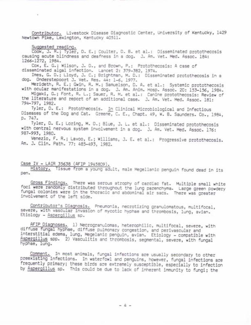
Case IV - LAIR ,tl_6]q (AFrp I945Bo9).It, male Megellanic penguin found dead in irspen.
Gross Findingg-.-Tfrele was serous atrophy of cardiac fat. Multiple small white^ - -toci were randomly distributed throughout the lung parenchyma- Large green powoeryfungal colonies were in the thoracic-and abdominal i i r saci. There was greaterinvolvement of the left side.
Pneumon ia ,nec to t i z i ngg ranu ] .omatous ,mu1 t i f oca1 ,severer wl th vaseuLar invas ion of mycot ic hyphae and thrombosis , lung, av ian.Etiology - Asperqi l lus sp.
--^^l i lgPiggo9e+ 1) Necrogranulomas, heterophil ic, mult i focal, severe, witholrruse rungal hyphae, diffuse pulmonary congestion, and perivascular and-in ters t i t ia l edema, . lung, Megelanic penlu in, av ian. ' Et io logy - - .orp. t ib le wi th$,ryi!]us. son. 2) vasculifis and thromboiis, segmentat, i-evere, with fungalhyphae, lung.
Comment- In most animars, fungal infections are usually secondary to other
frequently primary; these birds are extremely-susceptible, 6spe.i.11y to iniectionby Asp-ergillu9 sp. This could be due to lac-k of inherent immunity t6 fungi; the
- 4 -
rnost susceptible species, sea ducks and Antarctic penguins, are rarely exposed to;;h iGai- in tne wifO. Another possibility is that lhese birds are naturallyimmunosuipressed, a theory supported by the paucity of lymphoid tissue they possess.
Early in Aspergil lus infections of birds, there is an accumulation ofheterophils ii--filffii-parabronchi. Since liquifaction necrosis is poorlyaccomplished in birds, the herterophil-f i l led parabronchi are surrounded bygranuiomatous inflammation in an attempt to wall off the infection- These sitesare often resolved as discrete granulomas if the host l ives long enough.
Elaboration of elastase by some strains of &Egggi-Llw. &m,iqatus has beencorrelated with its invasiveness in pulmonary tissues.
Contributor-. Letterman Army Institute of Research, SGRD-ULV-P, San FranciscotcariFffi4E.
Suqqested readino.ehendlEi, F: Iv. l-kaplan, V'1. i Ajel lo, L.: Color Atlas and Text of the
Histopathology of Myeotic Diseases. Chapt. 5, f{olfe Med. Pub. Ltd., 1980.Kothary, M. H. ; Chase, T. Jr . ; MeMi1lan, J- D- ; Corre lat ion of e lastase
production by some strains of Aslglg$lus fumigatus with ability to cause pulmonaryi n v a s i v e a s p 6 r g i 1 1 o s i s i n m i e e f f i z 3 2 a 4 2 5 ' 1 9 8 4 .
l.brtelmans, J.: Mycotic infections in captive wild mammals and birds: Someconsiderations on epizootiology, pathology, and prophylaxis. In The ComparativePathology of Zoo Animals. Monta l i , R. J- and Migaki , G. , Eds. , Sml thsonian Inst .Press, 1980, p. 277.
DAVrD L- FRITZ, V .M.D.Captain, VC, USARegistry of Veterinary PathologyDepartment of Veterinary Pathology
- 5 -
ResultsAFIP l,lednesday Slide Conference - No.
25 September 1984
Conference lr'loderaJor: Ggorge_D- Imes' Jr-c0L, vc, usADiPlomate ACVPChairman, Department of Veterinary PathologyArmed Forces Institute of PathologyV,lashington, DC 20346
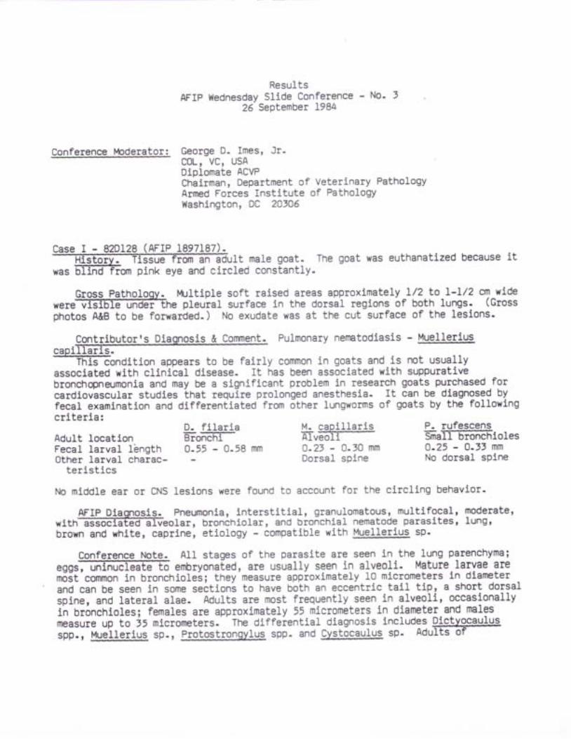
Case I - 82DL28 (RrrP 1897187).] t m a 1 e g o a t . T h e g o a t w a s e u t h a n a t i z e d b e c a u s e i t
was EiliftiEom pink eye and circled eonstantly-
Gross Patholoqy. Multiple soft raised areas approximately l/2 to I-L/2 em wide-were vrslore undfffhe pleural surface in the dorsal regions of both lungs. (Gross
photos A&B to be forwarded. ) No exudate was at the cut surface of the lesions-
C_qnLqlb_Ulor's Diaqlosis & Cgln$eil Pulmonary nematodiasis - Stellerluscap-ffi----fHifTondition
appears to be fairly common in goats and is not usuallyassociated with clinical disease- It has been associated with suppurativebronchopneumonia and may be a significant problem in research goats purchased.forcardiovascular studies that require prolonged anesthesia. It can be diagnosg9 byfecal examination and dlfferentiated from otner lungworms of goats by the followingcriteria:
Adult locationFecal larval lbngthOther larval charac-
terist ics
No middle ear or CNS
D. f i lariaE'iffifrF0 .55 - 0 .58
L capj]larisAlveoli0.23 - 0.30 mmDorsal spine
P- rufescensSmall bronchlolesO.25 - 0.13 mmNo dorsal spine
leslons were found to account for the circl ing behavior.
AFIP Dlaqnosis. Pneumonia, intersti t ial, granulomatous, mult i focal, moderate,l ung twi th f f i o Ia r ,b roneh io1a r ,andbronch ia1nematodeparas i tes ,
brown and white, caprine, etiology - compatible with Muellerius sp.
Conferenee Note. A11 staqes of the parasite are seen in the lung parenchyma;
e g g m e m b r y o n a [ e d ' a r e u S U a 1 1 y s e e n i n a I v e o 1 i . M a t u r e l a r v a e a r em6it'common in bronchiolei; they measure appioximately 10 micrometers ln dlameterand can be seen in some seitions to have both an eccentric tail tip, a short dorsalipine, and lateral alae. Adults are most frequently seen-in alveoli, oceasionallyin br6nchioles; females are approximately 55 mierometers in diameter and malesmeasure up to j5 micrometers. The di.ffeiential diagnosis includes D-ictvqcaulusspp.r Muellerius sp., Br.gtogtrongylus spp. and Cystocaulus sp- Adults of
9ictvgeayl.Ys and Cvstgcaulus are alrnost twice as large as the largest adult femaleseen in this case- Adults of _Pfglgg!ryOgy1_q9 and Muelleflus are of similar size,b u t 1 a r v a e o f t h e f o r m e r 1 a c k @ o r ] f f i i n t n i i _ c J J J . _ i n i iparasite is morphologieally compatible with Muelrerius S:apilraris
- Although many authors write that Muellerius is found primarily in subpleuralalveoli, and that this can be used tolJiTFfrffiiate it frbm other' metastrbngiiJi,this case demonstrates that it can be found in the airways to the level ofbronchloles.
Contributor-. Department of Comparative Medicine, Hershey Medical Center,eenngrFaiTEEte u;it;;;iivl Hershey, eennsyivania 170i3.
Srqgesteg rgadinq.Levlne' N' D-: Nematode Parasites of Domestic Animals and of Man. 2nd Ed.,Chapt- 7, Burgess pub., I9gO, p. Z4L.Nimmo, J' s- : . case repoit ' - six cases of verminous pneumonia (Muellerius sp. )in goats. Can. Vet- J. 20: 49-52, 1979.Rose, J-: Site of .developmen[ of the lungworm
experlmentally infected lambs. J. Comp. patn] ie:Muelte_tius capillaris in
:fl::,":;^1.;o. ??:1*Hrtal.infectibn oi r.roi -*iln
G.ir.iir. capirlaris.J. Comp. Path. 69t 414-422, IgSg.stockdale, P.H-G- ! Puimonary p-athology associated with metastrongyloidinfections. Br. Vet. J. I3Z: 595_:60g, tgle.
2-year-o breed steer from a herd known to be9 V U g
I.ilf:.!:9,|i!l bovine.viral diarrhea tevni-virus. The steer had beenoI.-er. " ii""r' *!rl'lE'"iiX#lle i a n a A f c l r r n ^ 8 ! ^ - n.iilis.o -iEi"til5';rHdfii:T l f f a a a l r a . l ^ ^ s o r r h . . : - - . -
Ill*:^:!.Trl_?L!!D virys on r-5 .:an ea.--rn-r,l.'.ili; ;;;.i;ilio""".iiiXilH:s:::::::.y::ting^?InlI:ry :lq'g9tei: zeo-oy ,righi
-r;;;;'ffi;,ffiliJXt'iiarrhea,
,:::'d:ly!1"!1ol: _l!is ltegl l"d severe *"tE'"v Jii""n""-"io-il"ir.il";;;.;il'ioi"lnuweek prior to necropsy on 16 Mar 94.
Laboratorv Rgggl!*. cytopathic BVD virus was isolated from intestine. Bloodw a s f f i v b r i o r t o d e a t h - r e i i s s h o w e d : V i r u s . I o , 2 4 oBVD-infectious units/ml 3eium (eell culture iniectious ooseij.- nntioody: BVDneutralizatlon tlter:. 4 [agalnst noncytopathic BVD virus (Nebraskail:--,BVD neutratlzation titert Ioz4 tagaini[-ivi.pit[ ic BlD virus (singer)].
Gross Pathologv... - There were erosions and ulcers of the mucosae overlylng thei n t f f i i s s u e s . - i n " " " w e r e u 1 c e r s o f t h e a b o m a s u m _ a n d t h i c k e n i n g o fthe distal i leum, eeeum, and proximal colon.
--
- 2 -
,ro" l?l';"llhi[3??';Ji!3::'"'This steer and the herd rrom-wnich it came *"i6 b"-.sistently infected with a
noncytopatn:.c strain of BvD virus. There ,". no-ligniiiint antibody circuratlng in
the steer directed against this persistent noncytopithic strain of virus and no
clinicar signs nao iIsuited from-iniection. rt is'not known whether the animar did
not produce antibody or if a smalf amount of antiOody was produeed and circulated
complexed to BVO-viius. Wnen ehaflengeO with a viruient, cytopathic BVD virus or
vaccinated with a modifieo, cytopatni" avo virus (we are'unbertain which cytopathic
uirus is involved), tne animai developed signs of chronic BVD'
AFIP Diaqnoses. l) Ulceration, focally-extensive' severe'.with a#
r ib r f f i s iudomembrane ' .smal1 ih tes t ine ,bov ine . .2 . )Enter i t i s 'f iOiinosubburative,'subacute, dif iuse, moderate to severe, with moderate vil lousatrophy, blunting and fusion, small intestine, bovine'
Conference Note. Based upon the histologic sections examined, conferencea t t e f f i e t e r m i n e i f t h e m u c o s a 1 - u ] c e r a t i o n o f t h e s m a l 1 i n t e s t i n e w a slocated over a necrotic Peyer's pateh. Lymphoid necrosis was not obvious' and theabundant tymphocytes present in the submutosa appeared essentially-normal. Thecontributoi, however, included focal necrosis of lymphoid nodules in themorphologic diagnosis. Conference attendees thought that the numerousintialesional niutrophils were probably due to secondary bacterial infection.SeveraL studies have identified a small proportion of cattle seronegative to BVD-MDvirus in seropositive herds and have further found that mucosal-disease deathsoccurred only ln these animals, sometimes many months Iater. It has been suggestedthat mucosal- dlsease might be the result of immune tolerance established as aresult of lntrauterine infections- Several studies have supported this but itremains unproven (Roeder, Drew, 1984)-
Antlgen-localization studies have shown that the major areas of antigenconcentratlon are the same as the areas with the most marked pathomorphologicalchanges - the lymphoid tissues of the distal ileum and proximal colon. Sinceinfectlons of animals by the alimentary route seems unlikely, it has been suggestedthat infection is primarily via the respiratory tract, and that dissemination tovarious tissues is carried out by eells of the mononuclear phagocyte system(Ohmann, f98r) .
Contributor_. National Animal Disease Center, P.0. Box 70, Ames, Iowa 50010.
Suooested readino.Brownlie, J.i Clarke, M. C., and Howard C. J.: Experimental production of
fatal mucosal disease in cattle. Vet. Rec. 114 z 535-536, 1984.Cut l ip, R. C.; McClurkin, A. l , { . , and Coria, M. F. : Lesions in c l in ical ly
healthy cattle persistently infected with the virus of bovine virus diarrhea -glomerulonephritis and encephalitis- Am- J. Vet. Res. 41: I918-I94I, 1980.
Lless, B. ; Frey, H. R.; Orbans Hafez, S- M.: Bovine virus dlarrhoe (BVD) -.rrMucosal Disease": Persistink BVD Feldvirus infektionen bei serologischeselektierten Rindern. Dtsch. Tierarztl. Wschr. 90: 26I-266, I98t.
- 3 -
Ohmannr H. B.: Pathogenesis of bovine viral diarrhoea -mucosal disease andsignificanee of BVDV antigen in diseased ealves. Res. Vet. Sci. 14: 5-10, I98t-
Roeder, P. L., and Drew, T. W.: Mucosal disease of cattle: A late sequel tofetal infectlon. Vet. Rec. 114 z 309-313, 1984.
Roth' J. A. ; Kaeber le, M. L. , and Gri f f i th, R" W.: Ef fects of v i ra l d iarrheavirus infection on bovine polymorphonuelear leukocyte infection. Am. J. Vet. Res.42-. 244-?5O, 1981.
a A-year-old female Hampshire sheep. Eight of l0 sheepinitial access to a new and weedymydriasis, t remors, ataxia, and
became iIl about 35 hours afterpasture.paresis.
Clinieal signs included salivation,Within four days, 5 of 8 died.
Laboratorv &Sg$s. Pertinent laboratory data on admission included elevationsin e@Iiii* -i j. c-e. ajl-i"J
-ii6tnenuria-
_-_ 9IgsF.latnofogvt {idneys bilaterally swollen, pale, moist, f luctuant. Arteoge or kloney molst with radially arranged tannish-white opaque flecks in cortex.Urine transparent, virtually eoloiless. -
"!:':""Bil!Ii".ilil-l::ii:l3::"ffi"?:#:.?::necrosl.s; renalof Rumex crispus.:--
. Hlret'rlngent erystals of oxalate were present in the rumenal mucosa but werenot observed in other loci of the body. Br4rc-x .iiipur i;;rt Jtcx) was abundant inthe pasture and had been extensivery lrazEffi EFffiep. rne ieri,inii ioriage orthe Rr-rnex contained rr% oxarate expieised as 6xalic acid.
AFIP Diaqnosls- Nephrosis, diffuse, moderate, with moderate diffuse dilationof e6TE]EETTUEIIE and'Bowmanis.spaces, intialubu]ar ano intiaductal birefringentcrystals' and medullary eosinophil ic caits, Hampshire, ovine, etiolJgv---"orp"iior.with oxalate toxicosis.
^.,^.,cgqfeEFg=Totg-. Tl. most.common causes of oxarosis are ingestion ofoxarate-containing plants (ruminants) and ethylene glycol. Als6, oxalate crystalsare often seen in 8lPglgil lus-niger infectioni ano iav oe tne-iource of systemict o x i c o s i s . I n t h e @ d o x a 1 a t e s m a y b e d e g r a d e d t o c a r b o n a t e s a n dbicarbonates, may precipitaie ai ealcium oxalate ind remain with the feces, or maybe absorbed into the bloodstream and tissues where it binds with ionic calcium toprOduce insoluble calcium oxalate. The net result is often a-jrecipitoushypocalcemia to which several pathological processes can be attributed:hyperglycemia may be attributable to [ne effect of low serum ca]cium on insulinsecretion; inhibit ion of succinate dehydrogenase may be a signii icant faetor indeath (Simensen, 1980); and there may 6e i iterferenee with the essential roles ofcalcium and magnesium ln oxidative pirosphorylation at the cellular level (VanKampen' James, 1969). A second problem'is lnat of crystari i i i f ion of calciumoxalates in vesseL walls eausing vascular necrosis
"ni n"*o";;;;", and in renal
- 4 -
tubules and ducts causing blockage and necrosis. An interesting study by VanKampen and James showed crystal deposition at different 1evels of the renalcollecting ducts and tubules at different time intervals after ingestion ofHaloqeton by sheep.
Contributql=. College of Veterinary Medicine, Oklahoma State University,stilTffi'hona tqite.
Suqqested readino.@ e r , G . D . , a n d V a n G e 1 d e r , G . A . : C 1 i n 1 c a 1 a n d D i a g n o s t i cVeterinary Toxicology. 2nd Ed., Kendall Hunt, 1976, p. 121.
- Dickie ' C- yt- i Hamann' M. H.; Carrol l , w. D- et .a l . : oxalate Rumex venosuspolsoning in cattle. J. Am. Vet. Med. Assoc. 17i(r): 7i-74,-riza_-'; '=:==:=shupe' J- L. , .and.James, ! . F. : Addi t ional physiopatnoiogic changes inHalogFlon glons,rg.tqs (oxalate) poisoning in sheep.- cornell vet. sgz
-al-ss, Lg6g.Slmesen' M- G-: Calcium, phosphorui, and magnesium metabolism. In ClinicalBiochemistry of Domestic Anifials.'frd, Ed., J.Ji Kaneko, roitor, ih.Ff.-i; ' , '--- '
Academic Press, 1980, p. 591.Kampen,- f- !-, and James, L. F.: Aeute Halogeton poisoning of sheep:Pathogenesis of lesions. Am. J. vet. Res. i0(10): t itg-,tt l3,-l.geg.
a male bovine fetus aborted approximatelypremaevidenceo
-The dam appeared normal prior to abortion'and had nodisease after expulsion of the fetus.
Gross Pathology-, -The fetus.had massive aseites and hemoperitoneum. Numerousotsc f f i1esmeasur ing f romsevera1mi1 I imeters tocent imeters indiameter were attached to the parietal and viiceral peritoneum. other g"o""changes were not observed-
W?gnosis. a comme?t. Mesothelioma, congeniLal, bovine.Mesof,'nerloma is the most commonly diagnosed congenital tumor in calves. Thegreatest incidenee is observed in fetal and neonatai calves but reporls oimesothelioma in older cattle have been documented. Peritoneal mesotheliomas maytake either of Lwo histological forms, predominantly f ibrous resembling ; -- '-!
f ibrosareoma or papil lary mimict<ing a'papil lary adenocarcinoma-
AFIP Diamosis. lvlesothelioma, abdominal peritoneum, Holstein fetus, bovine.
Conference- \ote.- Mesothelial cells are similar in structure and function to- . -epltherral and endothellal cells but are of mesodermal origin. Major diagnosticproblems can arise when mesotheliomas must be differentiat6O from 6pithelialneoplasms. Ultrastrueturally, mesotheliomas possess microvilli and desmosomescharacteristlc of epithelial cells. Using immunocytochemical techniques, Warholhas recently shown that cel1s of human mesotheliomis also contain abundant keratin,and that they irregularly express carcinoembryonic antigen (cEA), markersheretofore thought unique to epithelial cetls.
v.f
6 weeksclinical
ry_= 7U2-' (AFrP
- 5 -
rn differentiating mesotheliomas from earcinomas by light microscopy, severallPeeial staining characteristics have been suggested UV tne Oepartments of ChemicalPathology and prllmonary and Mediastinar pathoiJgy, AFIp.
1 )
2)
CarcinomaPffiIEive-diastase sen siti ve9gg to lhe presence of glycogen.PAS positive-diastase resistint dueto production of epithel ial mucin.
MesotheliomaI) PAS positive-diastase sensitivea\ due to the presence of glycogen.z) (ooes not produce epithelial
mucin ) .
Therefore, the p^resence of PAS positive-diastase resistant material should rule outthe diagnosis of mesothelioma.
3) Colloidal iron (At1p) positive due toproduction of mucopolysaccharides.
4) Hyaluronidase resistant - mucopoly_saccharide is not hyaluronic abiO.
5) Alcian-blue positive materialshould be seen.
5) Cells more pleomorphic.7) Nuclei more irregular.8) More likely to f6rm acinar
structures-
im8-ffi$ wthout diastase.2. AMP with and without hyaluronidase.f. Alcian blue-
The guidelines may-be helpful but results must be interpreted with great caredue.to the many variables involved in speeiai siaining-t..hliqr".. The abovestains were used on this r{sc case, but the results were equivocar.
* "b .wVe te r ina ryD iagnos t i cCen te r ,Un ive rs i t yo fNebraska ,L inco1n ,
ffisotherioma in theHarbison, M. L. and Godlesk i , J . J . :
Vet. Path - 2Oz 5rL-54O, l9gj"Henderson' D- !:r lnd papadimitt igu, J. M.: Ultrastructurar Appearancest,tol: :-_A_Diagnostic Atras- churchirr ' t- ivingitone, L982, p. r09.
o f
3) Colloidal iron (AMp) posit ivedue to production of mucopoly_saccharides.
4) Hyaluronidase sensit ive -hyaluronic acidacid.
5) Aleian-blue posit ive materialshould be seen.
6) Cells less pleomorphic.7) Nuclei more regular.8) Less }ikely to form acinar
structures.
ca l f . Pa th . Vet - 4 : 149.156, 1967.Mal ignant mesothel ioma in urban dogs.
Ii:*Itr tt: lumo19 in newboi; ;;il"i;.- F;fi.'-i";:";i \ialiit, re6'.schambe.r, c- J. I olsonl,c., ino-witl,-1. E:i -ru.opi*ir-in-6]ii,".'?Eo.
t r c ) - \ / o t n a l h 1 o . z . ) 6 . 2 1 l A 6 A 4 ' ' e q a Y v
!gggyg.). Vet. path. 19:t 529-537, 1982.,warhol, M- J-: The urtrastrueturar rocarization of keratin proteins andcarcinoembryonic antigen in malignant mesotheliomas. Am. J. path. 116(3) z 3g5-39O,l gR lL -
DAVID L. FRITZ, V_M.D.Captain, VC, USARegistry of Veterinary pathologyDepartment of Veterinary patholilgy
- 5 -
L984.
ResultsAFIP Wednesday Slide Conference - No' 4
3 October 1984
Conference lr4odsrator: John M. Langloss, DVM, PhDDiplomate ACVPChief, Division of ImmunopathologyArmed Forces Institute of PathologyWashington, DC 20306
a pf f iear -o1dma1ecast ra tedcockerspan ie1 .Thedog had
ive mass in the right ear canal with purulent exudate- The mass wasreinoved in May 1981 with regrowth removed in Dee 1981. The dog presented in-May1982 wlth tetiaparesis. Radiographs revealed osteolytic lesion of C5. Cervicalmyelogram indlcated space-occupying mass in the lateral spinal canal. at C5. Thedog was euthanatlzed at ownex request.
Gross Patholoov- The rioht venous sinus at the C5 is dilated and filled wlthg r a m c t i n g i i t o t n e s p i n a 1 c a n a 1 a n d c o m p r e s s i n g t h e c o r d .
Laboratory ResuLts- Results of all serum ehemistry, hematologic and urlnaryanalffiar limits.
. l,letastatic chemodectoma to the flfth
Chemodectoma was diagnosed in the first and second biopsy surgieal temoval'based on the round ovoid eplthelial-like neoplastic cells arranged in nests orcJ.usters, assoeiated or separated by dellcate fibers or blood spaces. This tumororlginatlng from the ear canal is unusual, inasmuch as chemodectomas have beenfound in the earotid, aorttc and pulmonary bodies and occasionally in the urinarybladder.
$FIP Diaonosis- Mal.ignant neoplasm, vertebial body, eoeker spaniel, canine-
ConferenceJlote_o The attendees' differential diagnosis included mast cellt u m f f i e m o d e c t o m A ) , a n d p 1 a s m a c e 1 1 t u m o r ( m y e 1 o m a ) . I n s o m esections, the morphology of the tumor cells resembles that of mast ceLJsl however'nunerous special stains failed to yield conelusive results.
Some conference attendees thought that the tumor cells were packeted by finefibrovascular septa, whiJ.e others thought this septal network was probably thesinusoidal septa normalJ.y found in the medullary cavity. A Churukian and Sehenksilver stain failed to demonstrate argyrophilic granules in the cells. TheDepartment of Endocrine Pathology, AFIP, thought this neoplasm is of neuroendocrineorigin-
r s D
It4ost conferenee attendees thought that, based on the morphology of theneoplastic cells and the radiolucencies in the affected cervical veterbrae, adiagnosis of myeloma eould be made. However, most neoplastic cells lack e6centricnuclei, a perinuclear Golgi haIo, and heterochromatin patterns typical of plasmacells- Elnvf--gleen-pyronin staining is often helpful in demonsliating nruii in tnecopious rER of plasma cells. Best results in bone sections would be oStained usingEDTA as the decalcifying agent.
Contributor- The Animal Medical center, 510 East 62 street, New york, New york1002r---
Suqgegted readinL. Patnaik, A- K.; t Iu, S. K;( extra-adrenal paragangitomas )
Hurvitz, A. I. et a1.: Canine chemodectoma-- a comparative study. J- Sm- Anim_ pract_ 15:
P- F., and Liu, S. K.: Chemodectoma of the urinaryVet- Med. Assoc- 164l. 797-BOO, 1974.
Extracutaneous mast cell tumor in the dog_ Vet. path_
785-801, 1975.Fbtnaik, A- K-; Lord,
bladder in a dog. J. Am.Patnaik, A- K- et a l . :
19: 508-515, I9BZ.
AFIPa 30-pound 9-year-old male springer spaniel canine_ This
3:g^:?:^?*:"l!:o y1!! :_z:year history- of enroniJ-pvob.*i.- il; ;;;";il;;v v . v P ,alopecia, and rapid weight lois. The dbg was'ireated with antibioties but
I I -
developed a respiratory-infeetion and wai euthanatized at the owner's request.
,- 9lPis PathoJogv- The dog had bilateral alopecia, calcinosis cutis and anurcelative pyoderma of the faee, lqg: and lateral thorax- The fung-iobJJ ,J".firm, red-brown and contalned multiife Oar[-r.O-g""V foci filled with purulentmaterial. The conducting airways wbre filled wi[n 6urulint-.iro.t". ir-ia"gJ, oartbrown, irregurar mass wai preseht in tne irea oi tni-pilrii"rv'o0x8x7 mm) andcomplessed the hypothalamus- The adrenal glands y/ere uiratJilrry enrarg"i ,itnthickened, nodular eortical tissue. rne pirithyroid grands-were moderatelyenlarged.
Laboratorv tgggl!+ At presentation, the referring veterinarian observed thattni f f iutoe! losis,e1evatea-."", 'a]ka1inephosphatase,a1anineaminotransferase, and'cholesterol,'but
" n"it-normal BUN and'ereatj.nine. Its urinespecific gravity- was _very low (1-005), oio-not respond to water cteprivatlon, butdid increase to 1-015 wiln tne vasopressin test (;Ia;ilinj.
--in" oyrners refusedfurther tests or treatment when the' respii.6;t ihfection developed shorgythereafter.
- 2 -
contributor,s Dia-s1qsi-g- j--EornCIgnt. chromophobe adenoma in the pars .distalis,*lt ession of the neurohypophysis andhypo tha lamus , p i t u i t a r y r dog - ! __^ t ___ ! r ^ ^1 , - . , aa ra r r .
In addition io-tne'|ituitary tumor this dog had bilateral eortieal hyperplasiaof its adrenal glands,
'an uleerltive dermatiti6 with epidermal atrophy and-dermal
minerallzation,-*ifo-p"iathyroid gland hyperplasia, marked lymphoid depletion ofi i l-"pi""n and'lymph nodes, and a-severe, chronic active, suppurativeUionenopneumonia'with absc6ssation and interstitial fibrosis. This dog probablyhad an endocrinologlcally active corticotroph (ACTH-seereting).adenoma of theaOenonypophysis tnit resulted in an almost classical presentation of pituitaryOepenO-eht' hyperadrenoeorticism (canine Cushing's syndrome).. RegretfuJ'lytpituitary-abrenocortical function tests were not performedl howeverr themorpnolo"glcal lesions observed at necropsy are typicgl for this-syndmme.. Thisdogis clinleal signs of diabetes insipidus were likely the result of the tumorOeitioying the noimal adenohypophysis, neurohypophysi!.and hypothalamus (Capen'
C.C., igZ6 anO 1983)- This iyndrbme of long term cortisol excess is ofteneompiicated by severe bacteribl infections.-This was seen in this case' with asevere suppurative bronchopneumonia the final outcome-
AFIP Dlagnosis. Chromophobe adenoma, pituitary gJ.and, Springer spaniel, canine-
Conference Note. Immunohistochemical stains revealed that the tumor cel1s werepro@at the . remnantso fnorma] .adenohypophys iss i tua tedaroundthebOge of lhe mass v{ere produelng prolactin. Similar staining procedures to detectthe pltultary gJ.ycoprotein hormones (TSH, FSH, LH) are often unrewarding; thesehormones share-a common beta chain which frequently leads to cross reaetivity.
ContribuLo_g- Department of Pathology, University of Maryland School ofl'4ediEil-Edltlrnore, Maryland -
Sugoested readinq.f f i r s o f t h e e n d o c r i n e g 1 a n d s - C h a p t - 1 2 ' I L t T u m o r s i n
Domestic Animals, Moulton, J. E. (ed-),2nd-Ed., Revised, Univfflty of CaliforniaPress, Berkeley, 1978, pp. 372-429.
Capen, C. C.: The pituitary, overview- In Pathology of Laboratory Anlmals -Endocrine System. Jones, T- C., Mohr, U., HunE, R. D., (eds.), Internatlonal LIfeSeiences Institute, Springer-Verlag, New York, I98t, pp. 99-I2O.
Chalifoux, L- V-; Maekey, J- J-, and King, N. w.: A sparsely granulated'nonsecreting adenoma of the pars intermedia associated with galactorrhea in a malerhesus monkey (Macaca muLatta). Vet. Path. 20: 54I-547, 1983^
Charpin, C.; HasEoun, J- et a1-: Immunohistochemical andlnmunoelectron-microscopic study of pituitary adenomas associated with Cushing'sdlsease. An. J. Path. 109: l-7, L982.
DeStephano, D. B.; LJ.oyd, R. V.; Pike, A. M- et al-: Pituitary adenoma - anlmmtrnohistochemical study of hormone pxoduction and chmmogranin loca1izatlon. Am.J. Path. L16: 464-472, 1984.
Dunbar, M. J!., and frlard, B. C.: Hyperadenoeorticism associated with diabetesincipidus and hypothyroidism in a dog. J. Am- Anim. Hosp- Assoc. l8z 737-741' 1982.
Peterson, M. E.: Hyperadrenocortieism. Small Anim- Pract. IA(t)z 73I-749,1984.
Tsuchitani, M., and Narama, I.: Pituitary thyrotroph cell adenoma in aCynomolgus monkey (Macaca faseicularis)- Vet- Path. ZLz 444-447, 1984-
- 3 -
EM slide inclrrr - 8t-2189 r, 8t-2I89 rr (ALrf L947e Pers
r v l > J - I U E J r r t , J L l u g v . , -
kltEen: Three of fiveniitbiv. Tissue from a 3-week-littEfmTFin a purebred cattery died within 24 hours after exhibiting mouth
I'co1df'. Two kittens were submitted forbreathing and lethargy, i.e. signs of a I 'co1df'. Two kittens were submlEEeo r01necroosvl A foster [ itten (1 week older) had been introduced into the l itter onenecropsy- A foster kweek prLviousLy. It died 4 days after the first 3 animals died.
cfo_s,s__Ba_t!1a]o€y"- Both kittens were fat and hydrated- One kitten had numerouso r a 1 f f i u 1 e e r s o n t h e t o n g u e - B o t h h a d 1 a r g e c o n f 1 u e n t a r e a s o fconsolidation and pneumon5.a in the Iung.
Contributorrs Diaonosis & Comment-
Pneumonia, necrotic and proliferative, subaeute, multifocal, severer lung.Necrosis, acute, multifocal, moderate, Iiver with numerous Cowdry Type Aintranuclear lneluslons ln hepatocytes.
The initial presentation of this case eorresponds with the respiratory felineviral rhinotraeheitis syndrome. However, due to the age and relatively irmatureimmunologieal status of these kittens, they rapidly developed a fulminatingsystemic disease with charaeteristic multifocal areas of parenchymal eell necrosisin several visceral organs- The electron micrographs demonstrate nurerousparticles within the nueleus, budding through the cytoplasm of a hepatocyte andfree within the spaee of Disse. The nuclear particles have an average diameter ofI05 nm consisting of a central dense eore surrounded by a clear zone bounded by anouter rembrane. Cytoplasmlc partieles have an average diameter of 151 nm and arebounded by an additional clear zone surrounded by another membrane.
AFIP Piaongses. 1) Pleuropneumonia, f ibrinous, necrotizing, subacute, diffuse,severe, with multifocal moderate arteritis and eosinophilic intranuclear inclusionbodies, lung, Persian, feline. 2) Necrosis, hepatocellular, multifocal, mild tomoderate, with eosinophilic intranuclear inelusion bodies, J.iver, etiology -compatible with feline herpesvirus.
Conference l*,lote. Conferenee attendees discussed feline calieivirus (FCV) as ao i r t @ s . t u n g 1 e s 1 o n s o f F C V w o u l d b e m o r e p a t c h y , m o r e c e 1 ] u 1 a r ,and.frequently have hyperplaiia and hypertrophy of Type II pneum6iytes. Vasculaileslons are not typically seen, nor is overt'systemib disease. oub to the ftndingof lingual ulcers at necropsyr the possibility-of a dual (FCV and t-terpes) lniectionwas dlscussed, although oral ulcers are oecasionally seen with herpesvlrusinfections alone.
Herpesviruses-are highly cdll-associated; they can mature intracellularly andspread eontlguously from cell to cell avoiding exiosure to circulating antibodies.These characteristics are responsible for the-pooi lmmunify wnicn tofiowJ-JipJsrreto.this agent- Loeal production of IgA-is thought to be of importance, and newevidence suggests an actlve role for 6e11-media[eo irnmuniiv. isolatlon of thevirus from the trigeminal ganglia of cats suggests tneie-miy be persistence of thevirus in a latent form stmitai to that seen ii bovines ano numini-iru.iiisEl lgaz>.
Contributor- Comparative Pathology Braneh, US Army Research Institute ofcnemT6ell-Dffid, noeioeJn -piovins
cr6lnol -uirvrano
21010.
- 4 -
Suqgested readinq-f f i G r e e n e , C . E . : F e 1 i n e r e S p i r a t o r y d i s e a s e s . I n
Mierobiolo6y and infectious oisiases of the oog airO Cat." Chapt. ,0, F-E- Gteene,Edi tor , Saunders, 1984, p. 527.
Gaskell, R. M-, and Povey, R. C.: Transmission of feline viralrhinotracheitis. Vet. Rec. l,LIz 359-362, 1982-
Gaskell, R. M.: The natural history of the major feline viral diseases.J. Sm. Anim. Pract. 25: 159-172, 1984.
Nasisse, M- D.: Manifestations, Diagnosis, and Treatment of 0cular HerpesvirusInfectlons in the Cat. Compend- on Cont. Educ. 4z 962-9701 1982-
Povey, R. C.: A review of feline viral rhinotraeheitis (fel.ine herpesvirus Iinfection). Comp- Immunol. Microbiol- Infect. Dis- 2: t77-t87, 1979.
I V - t I J l ) L l .
an lI-year-old male terrier cross dog. Sudden onset ofa mass ln the ventral neck region was seen 3-4 months prior to presentation.An asplrate of the mass produced a large volume of serosanguinous fluld. Thewas removed surgically but recurred 2 months after suxgery, at which time thewas euthanatlzed.
Gross Patholoqv- At necropsy the neoplasm was found to have metastaslzed toregiffi, but distbnt metastases were not noted.
Contrlbutor I a Malignant mixed thyroid tumor-umors are mole cornmon in dogs than in humans (where
they are consldered to be rare)- Some authors make a distinction between the termsmalignant ml.xed tumor (where the epithelial and mesenchymal eLements are separate)and carcinosarcoma (where these two components ate more closely intermixed). Inthls particular case, the glandular and mesenchymal components seemed, for the mostpart' to reside in separate regions of the tumor. Although not present withinevery section, both osteoid and cartilage were found within the sarcomatous r€gions.
AFIP Piaqnosis- Malignant mixed neoplasm, thyroid, with metastases to lymphnode and lung, terrler cross, canine.
Conferenee Note. There is variation in the tissues presentl some microslidest n c 1 f f i y m p h n o d e a n d / o r 1 u n g , a n d i n s o m e ! h e r e a r e r e m n a n t s o fnormal thyroid along the margins of the tumor- The amount of osseous andcartilagenous differentiation also varies, but squamous differentiation isconsistent in most sections. Some attendees noted what appeared to be emptyabortive foLlicres withln the epithelial areas of the neoplasms.Immunohlstochemieal stains for thyroglobulin and calcitonin wete negative-A recent study by lvloore et a1., suggests that only eareinomas with iollicularpatterns show any consistency in immunopositivity for thyroglobulin.
lulost carcinomas metastasize via lymphatics and are usually first detected indraining lymph nodes. Thyroid carcinomas are loeally invasivb; early invasion ofcranial and caudal thyroid veins will result in multiple pulmonary mbtastases
massdog
' 5 -
before i.nvolvement of ioeal lymph nodes is seen. Mixed neoplasms areseen i.n the mammary and salivary glands-
commonly
Contributor= Bushy Run Research Center,eenrffi=2. (-submitted for the FDAi-
Suoqested readinq-Brodey, R. S., and Kel ly, D- F. : Thyroid
clinicopathologic study of fifty-seven cases.fuergelt, C. D.: Mixed thyroid tumors in
L52t I658-I66t,1968.Johnson, J. A., and Patterson, J" M.: Multifoeal myxedema and mixed thyroid
neoplasm in a dog. Vet. Path. 18: l l-20, 19gl-Leavr I - ; Schi l ler , A. L. ; Ri jnberk, A. et a1-: Adenomas and carcinomas of the
canine and feline thyroid. Am. J. path. g3: 6I-93, 1976.l'lason, R., and l{ellsr H. G.: 0n the occurrence of true ririxed carclnomatous and
sareomatous tumors (sarcocareinoma) with report of a mixed earcinoma-chondrosareomaof the thyroid of a dog. J. Canc. Res. lJ: ZA7-2I7, IgZg^
_ Forgr F. M-; K] .edzik, G. s. ; vr lo l fe, H. J. et a i . : Thyroglobul in andcaleitonin lmmunoreactivity in canine thyroid earcinomas.
'vei. path. 2l: 16g-173,1994.
morb
R.D. 4, Mel lon Road, Expor t ,
neoplasms in the dog - aCancer 22: 406-l+16, 1958.
two dogs- J- Am- Vet. Med. Assoc.
DAVID L. FRITZ, v.M_D.Captain, VC, USAReglstry of Veterinary pathologyDepartment of Veterinary pathology
- 6 -

ResultsAFIP V,lednesday Slide Conferenee - No' 5
10 October 1984
Conference Moderator: Marion G- Valer io, D.V-M-Diplomate' ACVPDeoartment of PathologYuitton Bionetics, Inc-551.6 Nicholson LaneKensington, W 20795
tal finding at necropsy of an aged female
@ Present adjaeentmultFbulated white mass.
to the right ovary was a 2-5 cn diameter
rs Diaonosis & Comment-Endometriosis' periovarianr rhesus monkeyt
ffioiffii8ris is a common occurrence i.n rhesus monkey!; . it occurs spontaneousl/ L - - ^ a - - - ! - - . ,
q iq a common occurrence i.n rhesus monkeysl it occurs spontaneousry---6oiffie-Eii6-sis ls a common occurrence i'n rher
but has also been aisociateo wi[n-slrsiea] manipuration 'or'$3.YEry:-llf!:Iotomv)
and exposure to ioniiing radiation. 6finiea11y, T9!f?ys exhibit dysmenorrhea'*noiih"gi", rrregular tenstruai-cycles and iniirtiutt. The endometriar tissues
which are wlthln the abdomlnal cavity ate tesponsive tb hormones and can enlarge'io"r
"iits filled with brown to red truio, and rupture during menstruation'
produclng extensive adheslons. Comptications such as colonic and ureteraliliictu"E, intra-abdominal hemorrhage and anemia can result-
The cause of endometriosis is not known- There may be regurgltation of..endonetrlum.tnroulh-lh; fnrunoioulum of the oviduet into the peritoneal cavltyJriing-minitrutii6n;
-rnootretiium may be seeded into the peritoneal-:"I1ll d::lng
;6iine surgery;-ln.re may be netapiasia of . coelomic eplthelium into endotnetrlal{iiiue; ceri rlitJ oi-tne"wotrttan'duct system may undergo transformatlon toendoretrial tissue, or there may be lymphltic or hematogenous metastasls ofendometrial tlssue.
AFIp Diaonosls. Endometriosis, periovarian (per contributor)r rhesus monkeytt@imate-
Conference Note. The site of the lesion in this case cannot be determined fromt h e @ n s p r o v i d e d . E v e n t h o u g h t h e s t r o m a i n e n d o m e t r 1 o s i s i scl'raracteris[ica]]y uterihe in appearance, endometriosis generally can be- -differentiated frbm uterine adehbmyosis because the former lacks myometrlal layerswith interposed vasculature. A uterine neoplasm usually can-be ruled out becauseit general.ly would not be as well-differentiated as this Lesion, would oftenconlah rnorl glandular structures and there would be no endometrlal stromalproliferatlonl CeLLs resembling eosinophils are scattered throughout_the stroma.These are endometrlal granuloeyies which are derived from stromal cells; atrnenstruation they eongiegate sub3acent to the endometrium and secrete relaxin h,hiehcauses dissolutibn of-the stroma. They are seen in primates and rats.
Endometriosis is a condition seen only in primates and the epithelium isresponsive to estrogen and progesterone- This condition has been reprodueed byseebing of the abdoten with'miiced endometrial t issue followed by treatment withovariai steroids (Bertens et al. , 1982). Adenomyosis is seen in primates as wellas other mammals and is generally not responsive to ovarian steroids-
Contributor.- Comparative Pathology Section, National Institutes of Health,9ooffiFilllitFike, bethesda, Marylanb 2a2o5.
Suqqested readinq.Bertens, A.P.M-G. et aI-: Endometriosis 1n rhesus monkeys- Lab. Anim. 15:
281-284, t982.DiGlaeomo, R- F-: Gynecologic pathology in the rhesus monkey
mulatta)- I I . Findings in laboratory and free-ranging monkeys-5t9-546, 1977.
Fanton, J. Vrl. , and Hubbard, G. B-: Spontaneous endometriosismonkey (.l4ac?cg faselcularis). Lab. Anim. Sei. 33t 597-599, Ig87.
lvlacKenzie, w. El-EndEasey, H. t{.: Animal model: Endometriosis in rhesusmonkeys- Am- J- Path. 8Oz 34I-344, 1975.
McClurer H- M.: Endometriosis- In Spontaneous Animal Models of Hr.rmanDisease- Vol .1, Andrews, E. J. , wandle- c. and Artman, N. H. (Eds.) , AcademiePress, New York, !979, pp- 215-218.
AFIPa l5-month-old, male, African green monkey (Cercooithecug
94oF. Petechlalaxillary region and
found semicomatose, with a temperatuie of
Gross Patholoqyt. The__large intestine was externally dark red; it containedoar f f i ng i r - i d lThemUcoSahadsea t te re i rouno , ra i sedwh l tenodules up to 3 mm in diameter, whieh were most numerous in mii1-eolon. Gsentericlymph nodes were enlarged and hemorrhagic. The liver was enlirged and friable.
ffi.Lung, spleen, l iver,Lungr_sp]een, l iver, kidney: yersinia entero.colit icaGut: E. coli, Campvlobactei ffi
(lvtacacaVet- Path- 14:
in a cynomolgus
on the ventral abdomen, and spread to theHe died shortly thereafter.
necropsy:
.seve -aeute, necrotlzlng, multifodal, Afri6in
1) Colit is, acute, necrotizing, multlfocal,Yersinia entemeolit ica. 2)
- Hepatit is,
green monkey, etiology - yerslniaqnterocolitica.
I'trltiTocaf necrotiMJltlfocar necrotlzing lesions containing bacterial colonles were aLso found inspleen and kldney from this case- The baeteiia h,e?e o?em-ncnarivc.case- The bacteria were gram-negative.
ff:.*:.:1,i: :^y:1*:d:?ypl!:q disease,in nonnui.n prif;.[rr.- rn our corony:l^::.:::_ln^Io:lg animats with access to outooori',
'il-;il;;ji.'Jrtoil#: ;i;
;::':.,!:t::,,1:"lnl:-t!^:':^::l?ll lld.p:"1-9y: t; t-"ilJ'f,oi'.,r rhis ease
*:t:f :::_!Tl'T:","1,,apparenilv-orr-io i;f;;;i;;:' il; ,'nori'iriii!.i-.Errr"and the presence of bacterlal eol6nies in neciotiring lesions, however, are typical.
aethiops) v{hemorrhages
8171- I ISSUC
eh wasappeare0minutes.
- 2 -
AFrP Dlaqnoses- L) colitis, necrotizing' acute' multifoeal' mode'rate t0
."u"ff i ,-6.. ir i" 'v,.oioniJi_"no,submucosa1ph1ebit isandphlebothrombosis, large intestine,-Rtrican gttt;"il6ilev iiFicgPitnecus aelhioPs')'^primate. 2) serositis, acute, iol.riv-eil.iliue, mild, ,^ETn-FFreEitis' thrombosls'
and numerous bacirlary colonies, mesenteric.attainment'of Large intestine, etiology- compatiore *iii-iErlinG-;ti;. ii-nJp.iitis,.ne*otizing, a-ute, multifoeal'mild, with nmumerours coecobacillary colonies, liver.
Confeqence Note- Conferenee attendees notedtne ffid bY the contributor' Also'intralesional ameboid organisms, and spirochetesunaffected by the infLammatory process'
A hallmark of yersinia "nt"io"oiiii""
infections is that the lesion(s) are
u s u a 1 1 y o r m r r o - s u n u s u a 1 a s n o t e d b y t h e e o n t r i b u t o r .inJ p"tnogenesis-anJ-epiieriolo6y are not well known because infections ar€difficult-to reproOuci-e*perimeitally- Swine and dogs have been incrlminated aspotential
""""ruoiii of V. qnleqocglitiea; in conttast, human infectlon UY.L
oseudotubereurosis is associated':w-it6-taminated food. rnterestlnglYr .the peak
% - - " n i i i o ' o i i t i " " i s d u r i n g c o n v a I e s c e n c e r a t h e r t h a n d u r i n g t h e:::i:"3il"::t:i 1eB0). lr arorementioned criteria hetp [odlfferentiate dlsease caused by Y.'enteroeoLitica from an otherwise similar diseasecaused by Y- pseudotubercuLosis-
The most severe form of Y. pseudotubercglo-sis infection is seen in nonhumanpr imates,ar t iodactyJ.s ,anoEE,ef f iac ter izedbyacute,sevelertorinonicrotic entlrilis and mesenteric lymphadenitis with disseminated necrotief6i-$ the liver, lung and spleen. A 1es3 acute form is seen in blrds and somemammals in which there'is devblopment of disseminated pyogranulomatous nodulesinioughort the viscela, except fbr the intestines where lesions are absent orsimllir to the aeute f6rm but less severe- A chronic form is seen in rodentstiipiciiffy rats,-ino is characterized by the presenee of occasional granulomatousnobufei in fympn nodes, Liver and spleen (Baskin, 1980).
Slnee most strains of Y. pseudotubgrculgsis do not pmduce exotoxlns'. ft.t issueneeros1spioouceoCj [ r i f f iought tobe.duetoanendotox1nrnicn may also bb-coupled with-the effects of lysoiomal. proteolytlc enzyrnes ofdylng leukocytes (0bwolo' 1980)
Contr.lbutor- Department of Comparative l'ledlclne, Bowman gfaY gqfqol ofMedififr;-60-sorrth Hawthorne Road, l{inston-Salem, North Carollna 27IOt.
Suooested readinq.ffiparative aspects of Yersir"ria pseudoluFgrcglos=is infe.ction
animals. In The Comparative Pathology of Zoo Animalsr l'tlontali' R. J- and MlgakrtG., (Eds-)ismftnsonian Institution Press, 1980, p- 2\9-'t ironson,
R. T-; May, B. D., and Ruebner, B. H.: An outbreak of infectlon byyersinia ps6udotub6rcui6sis in'nonhuman primates. Amer. J. Path. 692 289-30t, L972.
odel: oral Yersinia enterocolitica infeetlon in mice.Am. J. Path. 8Iz 7O77O6, 1975.
McLure, H. M.; U,leaver, R- E., and Kaufman, A- F-: Pseudotuberculosfs innonhuman piimatesi fnfection with organisms of the lersinia enteroeolitica group-Lab. Anim- Sci. 21: 376-382, I97I.
areas of apparent infarction inseveral attendees saw
within colonic crYPts in areas
in
- 3 -
Obwolo, M-of Zoo Animals,Press, 1980, p.
l{ooley, R-enteroeolitica
J.: The pathogenesis of yersiniosis. In The Comparative Pathologylvtontali, R. J: and Migaki, G., Editors, gnithsonian Institution225-
E.; Shotts, E- 8., and McConnell, T- I, ' , l .: Isolation of Yersiniafrom selected animal speeies. Am. J- Vet. Res. 41: 1667-1668' 1980.
Case III - N8r-319 (AFIP 190A652).-o1d, female, spotted jaguar (Panthera onea)
which had a history of chronic 0vaban usage for 5 years. Prlor to euthanasia itdeveloped abdominal distension and obstipation- A peritoneal tap revealed anonseptic exudate with characteristics of neoplasia. Abdominal radiographs werefollowed by an exploratory laparotomy which revealed carcinomatosis involving themesentery and omentum-
Gross Patholoqv. Disseminated neoplasia, multifocal, severe, uterus, ovaxies,omen@liac lymph nodes and spienic eapsule.
1) Uterine adenocarcinoma, with metastaslsto ymph nodes, splenic eapsule and broadligament- 2) Cystic distension of endometrial glands, severe, with multifocalmineralization, utelus.
The uterine adenocarcinoma probably originated at multiple sites within theendometrium, most likely from endometrial glands- lv'letastasis oceurred vialymphaties and transperitoneaL seeding- Whether it was reLated to Ovaban usageremains unclearl so does the relationship between the endometrial gland changes and0vaban.
AFIP Diaqnosg?. 1) Adenocarcinoma, papil lary, uterus, spotted jaguar (Pantheraong?)' reJine. U Hyperplasla, cystic, endometrial glands, diffuse, severe, wlthmultifocal mj.neral5.zation, utexus.
Conferenee Note- Conference attendees discussed the possible relationshlpbetween chronic negestrol acetate (Ovaban) usage and the lesions seen ln thiscase. Studies have shown that endometrial atrophy is the end result of chronic useof. progestin preperations upon an endometrium whith has not been "priredfl byestrogens- Howeve!, in women lt is thought that the pathogenesis of endometrialcarclnoma beglns with cystic endometrial hyperplasia due to overstlmulatlon byestrogens. A progressive, step-wise change of the uterus Leads toward celltransformation, carcinoma-in-situ, and eventually invasive adenocarcinoma (Preiser,L964)
uterine neoplasms are rare in domestic cats and are most commonly seen in therabbit and bovine- Adenocarcinoma in the cow is charaeteristiealJ.y sclrrhous,often with small foci of tumor evoking a florid desmoplasia. In the doe, a higherincidence of uterine adenocarcinoma is seen in breeds whlch have a coneomlttantsusceptibility to toxemla of pregnancy- It has been suggested that llver damageresulting from the toxemla could result in failure to inactivate estrogen witheventual development of adenocareinoma of the uterus (Cotchin, Marchani, 19n).
Contrlbuto!= Department of Comparative & Experimental Pathology, Coll.ege ofveteiiiffiMffiine, University of Florida, Gainesville, Florida 32610.
- 4 -

suqqes!.ed re.adinq..,-- ? . A^imar mnr{el: Soontaneous adenocarcinoma in agedB a b a , t ' l - a n d T o n - H a a m , E a i A n i m a 1 m o d e l : S p o n t a n e o u s a o e n o
'.**i;;ril'rJt":3th"3f;;"i\t l1?''ol?l3i TyTo,, or rhe Femare Reproductive Tract:
spontaneous and #t;fi;li l i; Springer-verlag '.1977' 9' 27'
0'Rourke, M. D. and Geib, U.-W.i- rnOomeiiiii '.itlocircinoma in a cat' Cornell
ur''r33o.i3i;3?-g.t3l3'*oo*to, F.: sponraneous urerine tumors in three cats't*'J3s;r:t;.?";333;rti?ii adenocarcinoma in a cat. path. Vet- 1t 48v4eo, !e64'
a f f i f i nd ing inanadu1 t ,w i }d -cap tu red , femaIeCebuslffirouo of 15 wild-eaugnt suo-aE"ii ;;d-il',i!-b"nus tPt:It-.Y:,e..Pl1,:n'BrLvlY' ' ;; i- i i*ifo:eaught
sun-adult and adult cebus monkevs vtele nero r
ffi:iffiH"? ;:,H. .'}fl ;'3Xll", 1 *e1" ?ip:'iT;riii:1. i;?:;i?i:l ":i:*:l*:i
AFIP 18
monkey-
3i?:?l3':? ::'#"liEli-n"'i,iii"f;"'gil'i":,ili;F'i? 3'i?Tif^i;.1+::.:"^::tffi1'I?::1il'ii!'n3'l"l'0il513::"
'fi'J";iiiXi"r"ii;; i;';;uurop-'rinicar illness and histolosic
evidenee of virus lnfectlon.
gross Pgtholoqv. At necropsy a well-differentiated cholangioearclnoma ln the
liver was observed, as weu as ni.,r"rou" 0-5 to z.^s e diameter-greyish-tan nodures
o"'in" iirosal surface of the duodenum and jejunum-
Laboratorv=Results. Virus could nOt be isolated from samples of braln' Ilver'
sp1een,1rng,uff i ' ""o-I io"" i_"!^Z9^93yS.post- inocu1at1on.Intest ina1nodules from this cebus monkey inFip-ies2297-il'and 2 other animals in thls gmup
(AFIp L}5zu7-t-InJ-ra!rigt-D were examined by a parasitologist and wele
identlfled as ̂ @!!gg sp- probably M' @!g'
r s Dml1d-6 rnodeFatb' jeJunum' s$ sP.
eiioi6iil-Jiigioiis:'N66[Gt verminous (molineus) pyosranuloma'Etiolo6y: Yoilneus sp-, probablv Molineg? !oryl!il1?:Inth1sg'o., ; t iT;.-"p.]"[ i"_monfte@Sweleobservedinthe
sma11 intestlne, coJ.on, liver, pancreai irfr:ffintum of l'1 anlmal's (ZX)' An
addltlonal 2 animal.s had fiOros!.Jerotic plaques on the serosa of the smallintestlne which rav-n"u" Ueen rtmnants of parasitic nodules or mlgratlon, tracts'Njne of the Lr ini6cted animals had nodulei.in the Jejunum; pyogranulmata wereafto ouserved ln"tne-OuoOenum-(n=il ' ileum (n=3), pioiimal colon (q2) ' pancreas
i.=zll-riui"-tn=rj ano omentum (n=l). One cebus.monkey died-of diffuse sevelechronic fibrinopurulent peritonitis which was attributed to infection by this.paraslte.
The adult helmlnths in the nodules are readily identified as trichostrongylinsbased on 1) consFicrous fongitudinal ridges proSetting f5!m the cuticle,2)-platymyartan merlmyarian mu6eulature whiih ean 5e transitional, 3) strongylinintestine which i;'atfiinenl naurng a large lumen, lgrge lntestinal cell nuclel'multinucleate iniestinal cells, anE thin [o lnterrirediafe microvlllous border, 4)and thln-shelled non-larvated, morula-stage eggs-
The life cycle of Molineus sp. is unknown, but beeause the organism.ls.atricnGtiongvr6,-it-is-F;til tb oe direet.
-rt is assumed that all animars were
infeeted in the'wiId. l"tany of the animals in this group also had malaria and
tr
patent f i larid infeetions (microfi laremia). Differential diagnosis at autopsy'should include 0esophogastomum, however, this helminth is considered very rare in
l .bwt r r |o r1dmonkef f i6du1espr6dominant1y in thecoIonof01d} r lo r1dmonkeys and apes. Histologically, Oesophoqastomu[ can immediately be ruled out asa c a u i e o f t h e n o d u 1 e s , b e - a u s e t h e m a d u 1 t w o r m S w i t h t h e i r e g g s i nthe lesion.
Histologically, the nodule is a typical granuloma with a suppurativesome seetions the eore of the nodule clearly communieates with the lumensmall intestine. The non-larvated eggs are released into the intestinalinfrequently the adult worms prpject into the Lumen- Several species of
eore. Inof thelumen andMolineus
are recognlzed, but only M. torulosus has been recognized as causing nodulargranulomata; the other species ofintestinal mucosa.
Molineus typically lie unattached on the
flFIP Diaqnosis- Granuloma, eosinophilic,nematodes and eggs, serosa, small intestine,eompatible with*Moiineus torulosus.
Conference Note- Other thandifferentited from Molineus inel
SJqqested readino-Abbott, D- P. and lvlajeed, S-
laboratory-malntained prlmates :l9&2O7, 1984.
Ngcht.-lF. is a tricnoEEF6iffird but proffipapif6frErrchis. Like Molineus,iiGGal nodulFii-aEF6
stomach of 01d l|Jorl.d monkeys. In the adult worm, external to the lateral eords,the eutieular ridges-are fused and divergent from one another; this morphologiccharacterlstic is unique to Nochtia. Prosthenorehis is an acanthocephalan wnicnprodueesserosa1nodu les in f f i6ge f f iE-occas iona] .J .y the ' i1eumofNewIt{or1d monkeys; thorny-headed worms will be seen attaehed to the-mucosal surface.
. ^ L.torHlosus dlffers from other trichostrongylids in the propensity ofinfective Larvae to burrow much deeper into the intestine, often reaching thesubmucosa or oceasionally even the serosa as in this case. Larvae maturd intoadults and their-eggg are-deposited in the tissues; some make thelx way to thelumen via neerotie channeLs. M= torulosus also hai a tendency to lnvaie the hostrsv a s c u 1 a r s y s t e m ; a d u 1 t s a n d e g i ! E f f i s e e n i n v e s s e 1 s a s s o c i a t e d w 1 t hfibrinous thrombl. Another chiracteristic is invasion of the pancreas resulting inchronic,pancreatitis with adults and eggs often evident within'inflamed pancreaticducts (Brack et al-, Ig73).
Contrlbutor.- Division of Pathology, U.S. Army Medical Research Institute forInfeiffibiiFase, Fort Deirick, uariiino 2I7oI.
torulosus in capuchinChitwood, M., and
monkeys, Cebus apeIla. Lab. Anim.Licht bn f effi.-Flf-i Oent i f ieati on
focal, containing nLrmerous adul,tCebus monkey, primate; etiology -
parasites which must be
Sci . 2 l l 360r f f i ,of parasitic metazoa in
K.: A survey of parasites in wild-caught,Rhesus, cynolgus, and baboon. Vet. Path. 2Iz
Brack, M. i Myers, B- J- ; Kuntz, R. E.: Pathogenic properties of l,tolineus
tlssue sections. Exp. parasltol. 32: 407, 1972.Toftr J- D-: The pathoparasitology of the alimentary tract and pancreas of
nonhuman primates: A review- vet. path- 19(supl 7): 44-gz, lggz.
DAVID L. FRITZ, V.M.D-Captain, VC, USARegistry of Veterinary PathologyDepartment of Veterinary Pathology
- 5 -
ResultsAFIP l{ednesday Slide Conference - No' 6
17 0ctober 1984
Conference l,bderator : F. M- Garner, D.V.M.Diplomate, ACVPVeterinary Pathologist4416 Oak Hill RoadRockville' MD 2f,853
Id female Fischer 744 rat-
Gross patholoqv. The rat was found at necropsy. !o have-a.left kidney fo95-
tlres the normaL-ffie. The mass was infiltrated'witn pale friable tissue- Therewas no evldence of metastasls.
t. Embryonal nephroma (nephroblastoma).6t are uncommon in the rat (Baker, Llndsey'
f979):- NdnroOiaitcrnas (embryonil nephroma, l{ilms' tumor) ale reporle9 !o be themost-frequ'ent typi of renal tlmor ln the rat, (nottendorfr. Ingraham, 1968).Nephroblastcrnas'bccur ln younger anlmals (mean age less than one year).
AFIP DlaOnosls. Nephroblastoma, kidney, Fischer 344, rat, rodent.
Conference Note. The Department of Urogenital Pathology provlded the oplnlontna t f f iasmiss1m11arh is tomoipho1og ica11y toscmecasesof | { i1ms|tunpr ln chtldren- A difference they note is in the cells whlch have undergoneepltheltal dlfferentlatlon fronr the blastema. In thls caser many cells have. smallnirclel wlth uniform chrornatln stlppling and appeat benign ln corparlson to thecells l.n tumors of children which have large and vesleular nuclel, lmpartlng a moremallgnant appeatanee. This case ls slmllai to those tumors ln chlldren whlch havea preOomtnahtly epltheltal element formlng small cysts (some of whlch representabortive gloneiult). I'bre comrnonly, they see tumors wlth a promlnent mesenchymalelementr. and occaslonally tumors that are purely mesenchymal-
Ffard and Grasso Q976) suggest that the rat nephroblastoma is the eounterpartof the eplthellal eomponent of human l{ilms' tumor, whereas the rat mesenchymaltumor seems to correspond to the mesenchymal component of the same htman tunnot.Because of the hlstologlc spectrum ln the human tunor, and the occunence ln therat of tfe eplthellal (nephroblastoma) and mesenchymal tumors as separate entltlestthe concept that l{llmsr tumor of man may often represent a eorposltlon tumor(corposed of two or more mallgnant cell populations) should be consldeted.
A slmllar turnor ln rabbits serves as a model for thls neoplasm ln hunans.Slmllarltles are seen in hlstlogBnesis, size of the primary tr.mor, and mallgantcharacter ln both rabblts and humans. Polycythemia is seen eormonly ln the rabblt,and occaslonally ln hunans, ln conjunctlon with nephroblastomas. This is posslbly
due to excesslve erythropoeitin produetlon by the tumor or by remaining normalrenal tissue, in response to renal hypoxia from tumor impingement upon the bloodsupply.
Contrlbutor. Pathology Department, Eli Lilly & Company, P.0. Box 708,c reeiTleETnldi-an a 451 40 I
Suoqested readinq-- - f f i e y , J . R . , a n d W e i s b r o t h , s - H . : T h e L a b o r a t o r y R e t . V o l .1, Biology and Diseases, Academic press, New york, 1979, p. 355.
_ DaYr L. E-: _ Embryonal adenosarcoma of the kidney in swine. 24th Annual Reportof the Bureau of Animal rndustry for the year J.907, USDA, pp. 247-2s7, r9o9.
l'lanlehen' T. and Stavton, D-: Animal model: Nephroblastoma in rabbttstransplacentally lnduced by ethylnitrosourea. Comp- Path. 8u11. 1I:4, 2 & 4, 1979.
Hard, G.. c., ang Grasso, P.: Nephroblastoma in the rat: Hlstology of aspontaneous tumorr_ldentlty with respect to renal mesenehymal neoplalirs, and arevlew of prevlously recorded cases. J. Nat. Canc. Inst. 47t 323-329, i976.. !'lardt G- C.,. and Fox, R. R.: Hlstologlc characterlzatlon of renai tr.mors(nephroblastomas) lnduced transplacentally in IIIVO/J and WH/J rabblts byN-ethyJ.nltrosourea. Am. J. path- llj: g-ig, Iggt.
Hottendorlr G. H., and Ingraham, K- J.: Spontaneous nephroblastomas inlaboratory rats. J. Am. Vet.-Med. Assoc. L53r 926-g29, 196:8.. l{lgaklr G., and Casey, H. hl.: Comparative pathololy of nephroblastoma inhumans and anlmaLs- In Tumors of Early Llfe in Man and- Animals. Edlted by L.Severl, l€etlng 8, pfrgia, 1979, p. Lb5t.
wardropr_K. i.., Nakamuran T., and Gllddens, l{. E.: Nephroblastcrna wlthsecondary polycythemia ln a New Zealand whlte iabbit. tab. nnlm. scl. 32t 2go-2g2,1982.
I I -
passlve, anddeath.
&..!-!2a A-year-old Yel1ow
a pet store 10 days earl.ier. Itdepressed. Loose green droppings
Nape Amazon parrot. The blrd wasbeeame progresslvely rrcre doelJ.e,were noted; it regurgltated prlor to
Gross Pathol.gqv. Petechiae-seen on epicarctium. Edema found ln lung. m.rlttple...1..i.r:.r.=.-:yerJ.ow rocl In J.Iver. Lemgn ye1low exudate noted ln mesentery (spleen ias notswollen but was showing evidehee of postmortem change). kiOney was swollen andpale ollve color.
L?bor?torY Besults., Microscopie: Tapeworm ova seen In fecal. fulture:Negatlve for SalmonelLa and other_bacterlal pathogens. Also negative for Cardlda.vlrology: itagatlve for avian lnfluenza, Newbastl6, and pacrreco;s. inrirlrvofa-'oslttaci was lsolated.
9?ft-tflbutor's .Dlagngsls- Focal necrotlc hepatitis, acute psittacosls.Llver - severe multlple variable slze focl of eoagulatlon neerosis was seenthroughout the parenchyma. LCL (pslttacosls) bodles ieiJ iouno_Spleen - focal areas of necrosis were seen.[f9*V - lnterstltta]. lymphoeytic paravascular lnfiltratlon seen.Other organs - no slgnifieant- changes.
-2-
AFIP Diaqnosls- Flepatitis, subacute, diffuset-mildmufffi, ne'irogranulomas, and intracellularmiEiooigin!.sms, iiu6r, yeliow nape Amazon parrot, avian'Chlamvdla gslttael.
confe.rence Note. Attendees noted qgny random areas of coagulative neeloslswhleh were thouffiur' ;;-g[Gg@:- olr,6r less numerous areas of caseous necmsisalesurroUnoeotiv-ni[ i ropn1ff iccasiona11ymu1tinuc1eatedg1antce1Is.These'attendees thoughi, rai-oi due to-a-eoncurrent-infection,.posslbly collbaclllosls.mr or the Lesioni ti6n in this ease are rsnlniscent of those descrlbed ln thefivei oi a parrot-wiin pitttaeosis and eoncutrent colibacillosls (Rapheel. and..i"itio", fgboi.- specfai stains falled to demonstrate ?ny illggtlous agents-otherinan-oiganlsms sug-giitiue-oi chLamydial elementary bodles wlthln Kupffer-cells andnepatocites. rnei6 lnfected cells'are not associLted wtth the necrotle leslons-Al1 of ihe organlsms ln thls case are of simtlar slze whereas in most eases ofcl'rfamyOfosh i vartatlon ls noted representing initial, intermedlater andelementary bodles.
rfre Oifterenlla]. diagnosls included Pacheco's dlsease (herpesvlrug)rmycoU-eteitosls, collbacillosls, and salmonellosl.s. AJ.though collbaclllosls andsitmonettosts pioduce focaL areis of necrosls, the necrosls is resolved by.macmphages re'sultlng ln formatlon of necrogranulomas. It ls of lnterest to notethat iariots. are oftEn infected wlth hunan ind bovlne stralns of mycobacterlabecauie tl'ey are frequently kept as house pets. Therefore, in mycobacterlal-infectlon o-f tne pariot, tFre ni.rrerous lntraleslonal baclllt eharacteristlcally seenln avLan E, rey lnstead be seant.
to moderate' withcoccobaclllaryetiologY--compatlble wtth
Con_t_r_lbgtOL lvlaryland Departnent of Agriculture, Anlmal Health Laboratoryt49of-€-:Iil;TE-86?d, College Park, lvlaryland 2ot4o.
F., revlsed by J. Schachter: Pslttaeosls andCage and Aviary Birdsl M.L. Petrak, Ed., 2nd Ed.r Lea &
Suoqested rea4!ro.Arnsteln, P.; Meyer,
Ornlthosl.s. In DlseasesFebiger, L982, p. 528.
Emerson, J. K.: Psittaeosis. J. Am. Vet. l,led. Assoc. 180: 612-61t, L982.l,blnn, R.: Epldemlol.oglc and laboratory observatlons of Chlamvdla psl.tlFcl
lnfectlons ln pet blrds. J. Ar. Vet. l,{ed. Assoc. I84z L372-It74, 1984.Pege, L. A.: Avlan ChJ.amydlosls (0rnlthosls). .In Dlseases of Poultry, 7th
Ed., Chapt. lt, edlted by M.S. Hofstad, Iowa State ttnlversity Press, L978, p.3V7.Raphael, B. L. and lverson, ftl. 0.: Collgranuloma and pslttacosls ln an &nazorr
parrot. J. Att. Vet. Med. Assoc. l77z 927-929, L98O.
yedr old Flartley straln gulnea pig frun agulnea plgs used for the safety testlng of tubereulln. Thts
was anorexlc and loslng welght wlth alopecla.
Gross Patholoqv. Kldneys had an irregular surface, were flrm, and had rultlplesmall cysts througfrout the eortex and medulla; multlple, 2mm ln diameter ulcers lnstomach.
K.of
herd ofanlmal
4.4 gn/dI2.4 gn/dl2.0 Enld\I42 neq/tII2 neq/L50 mu/ml
C o n t r 1 b u t o D i f f u s e l S B V 9 t € , e h r o n i c i n t e r s t i t i a 1nepniiTF-
Dlffuse lnvolvement of the kidney was charaeterlzed by tubular ectasla wlthflattenlng of qPllheliar eerls, inteistittat itorosis, miid interstltlelmononuelear inflltrate, occasi.onal. hyalin tubular casisr-nv"fin droplets ln sometubular eplthellum, mineral deposits- withln luUuta" epilheiium and tubular lunen,dllated Bowmanrs spaee, and mlid-capsurar fiorosiJ oi-giore"uri. spJciar-st"rnsdid not reveal a causailve organism.
5eo.lt3!"Hh#ffii#t* €f;"Euffii":loo 33X'"::::iSiE! ,r.nhistologic leslons-. lgPtgspira navb Seen ts6iateJ iiom gri""" plgs but atso havenot been associated with lnterstitiar nefniilfs.
CrN has been reported to be a common cause_of death in pet gulnea plgs butJ'aboratory animals are. rarely allowed io ii""-rong "norgn
t["i reslons can beseen' The animals in this cblony are kept until death 6n"e [n"y are TB-sensltlzed,and this change has been seen in'several' pigi.
The cllnlcal-path-ehanges of moderately elevated BUN and low totat protelns lsconslstent wlth CIN although anorexia proUiUiy- also contributed to the lower Tp.The mlldlv elevated serum k+ would # ffi;ial6nt with a A;;;;sed abllity ofproxlmal tubular cells to excrete lt.
__- AT! Dl?gno?1i.... I'bphritis, fibrosing, interstltlal, chronic, dtffuse,moderate' rlth tubular dilatation and cystic dilatatlon'oi eo*ranrs spaces, kldney,Hartley gulnea plg, rodent.
Conference l{o!%. Conference attendees noted multipJ.e subcapsurar lndentatlonso f@i "o t "a ' I i ' eo f su r f acep1 t t 1ng .A1sono tedwas thes1zeo fthe kldney lhtch. was .enlarged-up to two [rmei-inat-of a norr"i-guinea-pig-ioi
'unknown ?9e). The ldentlty-of lhe-globufarr-amorphous, eosfnopnfLic materlel lnthe medullary.and cortical-tntersti[rum wai'oiicussed; most attendees did not thtnkthls resenbred amylold. Thls materlal. ls srightii-positirJ
"iin . pAS pmeedure,and stalns-poorly wlth.congo. red but.shows ;"6pr"'gieen';-biriiiingence ffi;--polarized llgnt: Based on-these. flndingi-tG-fopaitment of cnlmr6if-palnof6gy
9?$o! posltlvel.y.tdentlfy it but statei tnat amyroid cannot-L'e rured out.Attendees agreed that grebt care must oe uiea in'rntJrprrlrnn-;.pple greennblrefrlngenee- 0ther than occaslonal irnoings-or giomeiuiar"atropny, glorneruriwere thought to be essentially normal.
nq 9gpqttment.of-Urogenital Pathology states that similar lesions ane seen Lnman and that the. etloJ'ogy-ln these cases-is llkewlse unknown.- Tfrey thlni-tnal tfeglobular eoslnophllic miterlal is pathologicai'ano that these deposrts in the
LaboJatorv Results.BUNCholesterolCaPKSGPT
37 ng/dL54 mg/d1I2.5 ng/dl6.1 mgldl8-1 meq/l
11 mulml
Total ProteinAlbumlnGlobulinNac1SGOT
-4-
nedulla probably cause tubular dilatation and cystic Bowman's spaces. They also
see, ln hunan kldneys, tE Urighlly-eoiinopnifit gylqPlasm present in many of tfe
tubular epltheriai-liirs ano oSfigt"[i-it-fioniocvioidh cha19e: Th" eoslnophllla ls
d;-6-tinirv-eosinopniii- granuiEt (probably mitochondria) as opposed to tlrclarger globules seen wlth hyallne droplet change-
gne study of age-related renal lesions ln the_guinea.Fl9. founO the presence of
deposlts of IgG ant-eompfement in gtomeruli (Stebliy,-Rudofity' 1971), whlle ln
"nbinEi-.iuoi-(riteta,
'Grollman, rFzo), nephroscrerosis due to vascular changes was
seen. Also,'cnaneJs-ieen in tni tattei stiroy are said to be similar to those seenln rnn. Vascular-leslons are not evident in this ease.
Contrlbutor. OfftcePlke, Bethesda, l{aryland
of BioJ.ogics, Food & Drug Administratlon, 8800 Rockvlllen205-
fuqqested readlnq.f f iRudo fsky ,U . :Spon taneous rena11es ions?4g}9qqru }T
Oepostii o'f'fgC ani compLement in gulnea pigs. J. Immunol. 107: 1192-1196' 1971.' Taketa, TI, and Groil.man, A.:
-spontaneously occurring renal dlsease ln the
gulnea plg. Afi. J. Pathol. 50: 103-117r I97O.- Waghei, J. E.: I'llscellaneous disease conditlons of guinea plgs.. $-TE
Blologi of'the Guinea Plg. J.S. l{agner and P.J. Mannlng, Eds., Chapt. 15r AcademicPress, 1976, p.23O.
were from a 2-year-oldon a chronlc oral toxtcity study for two
Gross PathoLoov. An oval mass, L5 x 15 x 5 mmr wastissuE of the rlght lateral abdomlnal reglon. The massdark red, and tan-
. Carcinoma with souamous celld1 ), marmary gland, BgCff mouser rodentla.
type, mafinary gland, B5C3F1 rnouser rodentla.lvlaltgnant lymphoma, mlxed cellBecause of llmited tlssue,
EYFer mat|lnary gfano, E.6vtr I fiK]user roclEl-lEra.the sectlons were prepared from two blocks from the
same tumor.The tumor ls nodular, non-encapsulated, and charactertzed by the presence of
neoplastlc eplthelial cells arranged in aclnar, ductile, paplllary, or comedo-Ilkestructures and sheets. The neoplastie epithelial tissue ls separated by a rathersparse connective tlssue stroma which contains irregularly arranged populatlons ofrnoplastlc lymphocytes of mixed cell type. The neoplastic epithellal cells appearto be mamnary gland eplthellal eells. In many areas, the neopJ,astle eptthellalqells have undergone sguamous metaplasla and are fomlng nests or cystlc spac€sdlstended vlth keratln. Mlneralization is also seen ln some sectlons. Mttotlcflgures are promlnent especially ln neoplastic epithelium that has not undergonesquamous netaplasla. l'*ecrotlc cells are scattered throughout the mass.
Adenoacanthomas are often invasive and/or oecasionally metastasize to theJ.ung. l.&tastases were not found tn this animal.
female B5C3F1 mouseyears.
located ln the subcutaneouswas flrm and mottled redt
fvlallgnant lymphoma was found in spleen, lymph nodes, and marnnary gland fronthis anlmal.. The lymphoma was ealled a mixed-ceIl type. In some sectlons, tl'relyqphoma may be dlffieult to subclassify or differentiate from lymphocytic-inflltrates or lymphoid hyperplasia.
AFIP Diaonosis. Adenoacanthoma, mammary gland, B5C3F1, mouse, rodent.
Conference NotP. Many conference attendees were of the opinion that this tumorm o s t @ s o n i o f b a s a 1 c e i i o " i g i n , p a r t i c u I a r 1 y , a n a d n e x o m a -Apocrlne and occaslonally sebaeeous differen[iatibn, iOor[iv6 haii fofiicfes liredb.y cells.with prominent trlehohyaline granules, and cell polarlty
"ere ieaiuris
discussed- lvlost sections eontain at l6ast a few acini of'normal'marnrary-gfanO .tthe perlphery-of the tumor. Attendees also discussed the possibility tna[-[nesetumors arlse from basal cells wlthin the mammary gIand. prominent virfaOfy-ltzeOaggregates of lymphocytes in the eonnective tisiud of this tumor proOiOfi-'
--
eornesptrld to populatlons of neoplastie lymphocytes noted by-the contrlbutor invarlous other organs. -However, a diagnosis'of iymphoma coriO not be made based onthe morphology of the lymphocyies in [hese tissub sections.
Other mammary neopJ.asms of the mouse were discussed, to inelude adenocarclnomatypes A, B, and c. The former two a"J "sioeiated
with lhe-eittner agent (mammarytumor vlrus, MTv)' whlle the latter is uiuiiry not- noenoacanthomas llkewlse arenot generally assoclated with the eittnei-ageit.
The eonference moderator stressed the importanee of being famillar wtth thevarlous stralns of mlce, each usuiiii nauing-partrcurar pathologieal entltlesassociated wlth it. Th6 B5c5F1 nas-ir uJrv io['rn.iJJnce-oi ilftr.ry neoprasms.
.^. . ,WDepartmentof-Patho1ogy,HazletonLaborator ies,330] 'K1nsmantsouJ.evarcl, l{adlson, l{lsconsLn 5j7A4. . -
9uooesled_readlno.uunnt T' E-:- lbrpholqgy or mammary tumors in miee- rn physlopathology ofcaneer. 'romburger, F,.'.-goll napgi Gro6o""i;-N";"vlji[l-r9#,-oi. r**.Frith, c' H:, ana wtrJv; ;-D: t
' Y.;i6iogicar classiri6atlon and corretattonof lncldence of frvoerplsstic ano_rygRraslic-nEmatopoletlc leslons in mlce wlthage. J. Gerontol. J6: 514-045, fgil.
squartlnt, F: ! Tumors or fne mamnary. grand. rn pathorogy of Tumors inLaboratory Anl.mals. yol. rr, iumors or tnE-r,rouse, edltgd by-V.s. Tunrsov,rnternatlonat Apncy for Rei6arch-on c"nJ"lr"16zg, p. 43.
DAVID L_ FRITZ, v.M.D.Captaln, VC, USAReglstry of Veterinary patholoovDepartment of Veterlniry patnoiirgy
-6-
ResultsAFIP V'lednesday slide Conference - No' 7
24 October 1984
Conference Moderator: John D- Strandberg' DVM' PhDDioomate ACVPAssociate ProfessorCornparative Medicine & PathologyJohns Hopkins University School of MedicineBaltimore, MD 2L2A5
AFIP 1945ack and tan bitch was plesented with a 5-month
and chronic weight loss and anemia-
Gross Pattplogy. At necropsy there was a liter of ascites fluid in the abdomena n o f f i d i s t a 1 s m a i 1 i n t e s t i n e a n d p r o x i m a 1 c o 1 o n . T h e r e w a smoderate veriucous endocardiosis of the right and left AV valves-
Laboratory Resul.ts- The animal was hypoproteinemic and anemic (normocytichypocFiomld-wlfnormal liver enzymes and BUN.
Contrlbutor's Dlaqnoses & Conrment- I) F€patitis' perieholangealts u o a i s , m i I d , p i g m e n t , m i n e r a 1 1 z e d t r e m a t o d eova, Heterobilhaizia. americana- D Enteritis, miJ.d, subacute/chronicr -noni , mild, trematode ova, [eterobilharzla am-qrlcPla-
Ova of t-€terobilharzia ameiicana were found in the-lamina propria and submueosao f a 1 I p o r t e ' c e c u m a n d c o } o n . T h e r e w a s 1 l t t 1 e h e p a t i cparenchymal compromise and it was concluded that the hypoproteinemia, -aseites' andbnemia were the product of a protein-losing enteropathy and endoparasitism. Thehost inflanrnatory reponse to the ova was minimal in all organs involved; althoughin areas of the imall intestine where ova vrere numerous the submueosa was thickenedby mature connective tissue. Adult fLukes were not observed-
AFIP Diaonoses. 1) Trematode eggs, predominantly portal, multifocal' lithasso@ranu].omatousinf1al l rmat ionandmoderatef1ukepi$Ent,1iver,black and tan hound, canine. 2) Trematode eggs, multifocal, with associatedminimal granulomatous inflarrnation, mucosa and submucosa, intestine;etiology--compatible with l-bterobilhqrzia americana
Conference lrlote- The microslide sections vary in the viabil-ity of eggs presentin t f f ia runatory responsetoparas i teeggsa1sovar iessomewhat ,be ingtotally absent ln some sections, to a minimal granulomatous response in others.The laek of response lndicates either an underlying inrmunologic problem ln thispartieular animal, or that leleqqb_lhaqzfa aCIer:lgglA is a well-adapted parasite oft n e o o g . t e e Q g Z z ) n a s s n , h a m s t e r , a n d m o u s e a r e i d e a ]hosts for this parasite, whereas the cat, guinea pig, and rabbit are unfavorablehosts. In the latter three, infective cereariae develop to adults, but eggsproduced dre not usually viable- The raccoon is considered the princlple naturalmarnralian host; natural infections have also been described in the dog, nutria'opossLtn, white-tailed deer, bobeat, and svramp rabbit, and range from tFesoutheastern U.S. to central Texas- Sehistosomatirrn doutlri-lli, the only other
HislorJ-- An E-year-orohistory of recurrent ascites
mammalian schistosome endemic in the U-S-, infects the mouse and is enzootic inMichigan, Minnesota, and Vrlisconsin. It is the schistosome egg'which causes diseaseby evoking a granulomatous inflammatory tesponse in the bowel and liver. Adultschistosomes appear to protect t.hemselves from immunologic attack by incorporatinghost moleeules into their integument as they mature.
Contributor. Department of Veterinary Pathology, College of Veterinary77843.Medicine, Texas A&M University, College Station, Texas
Sugqested readinq.Bartsch, R- C- and ft lard, B. C-: Viseeral lesions in raccoons naturally
infected with Heterobilharziq,americana. Vet. Path. 1l: 24I-249, 1976-G o f f , w - L @ . f f i t a i n a s p e c t s o f t h e b i o 1 o g y a n o 1 i f e c y c 1 e
of Heterobilhagia amerieana in east central Texas. Am- J- Vet- Res. 422t77>1777, l98l-
Pietee, K. R.: Heterobilharzia americana infection in a dog. J- Am. Vet. Med.Assoc- I43z 496-499,ffi
Thrasher, J- P.: canine schistosomiasis. J. Am. Vet. Med- Assoc. 144:1119-1125, 1964.
3-4hadtime
Leer H- F.: Susceptibil i ty of mammalian hosts to experimental infection withHeterobilharzia americana. J. Parasitol- 482 74a-745, 1962.
V{arren, K. S.: The pathology, pathobiology and pathogenesis ofschistosomiasis. Nature 273: 6A9-612, 1978.
Case II - AFIP 1947996) TEM (84-541) are inclyoung Arrican Grey parrot was presented for necropsy following a: n Jvv r r g n r t r ea t I u r sy pc l r l L rL wc r> p IE>E t t L t r u I u t '
nical course consisting of lethargyr anorexia andshlpped from a Florida distributor one week orior tobird was normal.
Gross Pathol.oqyr. _Signrificant gross lesions were confined to the liver, whichwas @led-tan ano reo.
Laboratorv Rg?ul!F. l$on bacterial culture, E. coli was obtained from the lung. ---F----.
and intestine while there was no growth from the Tiver. Splenic impression smearswere negative for Chlamvd*a sp- utilizing fluorescent antibody methods. VirusisoLation attempts were r.rnsuecessful-
L i v e r , n e c r o s i s n m u 1 t i f o c a I , s e V e l e , a c U t e ,O.rg
The liver is characterized by numerous foci of hepatocelLular necrosis whichare becoming confluent in several areas- Remaining hepatoeytes are regenerativewith moderate nuclear pleomorphism and a few binucleatb celis. An occisionalportal triad contains a mild to moderate lymphocytic infiltrate. Seatteredhepatocellular vacuolar change is evident-
Ultrastructural examination of thin sections of liver revealed many hepatocyteseontaining intracytoplasmic viral aggregates. Viral particles were round andeomposed of a moderately dense external ring around an electron dense cole- SoflEgmPty capsid particles were present. Negative staining of proeessed fresh Jiverclemonstrated 70 nanometer diameter viral particles as well. Morphology of thevirus in negative staining preparations and by transmission electron ilicroscopy wasconsistent with a reo-like virus which has been deseribed in oarrots-
oaybeenthe
diarrhea- The birdits death, at which
AFIp D!aq!1qslg- Necrosis, multifocal to coalescing, severe, liver, African.---+
gray parrol, avl.an-
Conference note. Aside from the numerous foci of neerosis present in allm ic r f f i ros ] . idesec t ionsa1soconta inedoccas iona1foc io feaseousnecrosis surrounded by heterophils and a few multinucleated giant cells- Specialstains failed to demonstrate causative organisms in the granulomas, and these aleasv{ete considered due to an intercurrent disease, possibl-y colibacil losis-
Attendees discussed the common causes of viral necrotizing hepatitls in birds.Although the electronphotomicrographs do not indicate the size of the virusparticles, they do demonstrate particles with some cubic symmetry, a &nse centralcore, and no evidence of an envelope. In negatively stained micrographs of thevirus, capsometes cannot be distlnguished. This is not the case for herpes andadenovituses-
A recent study of 01d t'lorld psittacine birds imported into Belgium revealed awidespread prevalence of orthoreoviruses- The primary lesions noted werehepatitis, enteritis, and pneumonia, resulting in up to 100% mortality of someshipments of imported birds (Meulemans et,a1., 1983).
Contributor- C. E- Kord Animal Disease Laboratory, P.0. Box 40627r l'4elroseStat"idi'filffiffile, Tennessee 37204.
SJqoested Readino-Ashton, ht.L.G. et al-: Suspected reovirus - associated hepatit is in parrots.
Vet. Rec- L!42476, 1984-Mandell.i, G. et al.: Experirrental reovirus hepatitis in newborn chicks. Vet.
Path. I5z 57I-543, 1978.lbulemans, G. et al.: Isolation of orthoreoviruses from psittacine birds. J.
Comp. Path. 93t L27-I34, 1983.Val.der, u{- A-; Cauhl, C-i Luthgen, frf. et a1-: Viral Liver disease in parrots.
I- Clinical, pathological, epidemlologieal observations and transmissionexperirents. Praktische. Tierarzt. 6!z IZ7-I29, 1980.
I{inston, J. R. et a1-: Isolatlon of a new reovirus from chum salmon in Japan.Fish Path. 15: 155-162, 1981.
III - Univ of (AFIP 1-yeat hound was D w i t h a f i r m 6 c m x 4 c m m a s s
within-the subcutis of the left inguinal regional, and a history of weight loss,intermittent lameness, and pyrexia (104.9o) for two months. Signs were-transientlyresponsive to antibiotic therapy- Popliteal lymph nodes were enl.arged and theanimal was icteric. Biopsy of the inguinal mass revealed ehronic-actirecellulttis- The animaL was hospitalized, no improvements were noted, and the dogwas euthanatized.
Gross Fbtholgqv-_ The animal was emaciated and icteric. Popliteal lynph nodesw e r @ a r g e 6 e m x 2 c m m a s S w a s p r e s e n t w i t h i n t h e s u b c u t i s o f t h eleft inguinal area. Internal lymph nodes and spleen were enlarged and firm. The]iver was approxlmately three times normal size, mottled tan/br5wn, friable, andhad an aecenuated lobular pattern- (Gross photo).
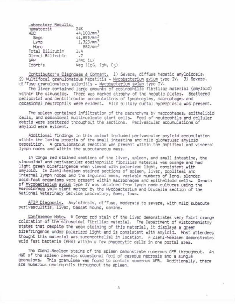
Laboratorv Resu1ts.Hematoeritll{Bc
SegsLympbno
Total BilirubinDirect BilirubinSAPCoomb fs
24%44,!OO/nn74I,895/nnsI ,323/nn2
882/nns1 . 4
. 7I44A Lu/Neg ( rgG, rgM, c)
& C_o-mmeo!. 1) Severe, diffuse hepatic amyloidosis.2) s - Mvcgbacterium gyjllTr type Iv. 3) Severe,diffuse granulomatous splenitis - Mvcobacterium avian type IV-
T h e 1 i v e r c o n t a i n e d 1 a r g e a m o @ f i b r i 1 1 a r m a t e r i a ] ( a m y 1 o i d )within the sinusoids. There was marked atrophy of the hepatic plates- Seatteredperiportal and centriLobular aecumulations of lymphocytes, macrophages andoecasional neutrophils were evident. Mild biliary duetal hyperplasia was present.
The spleen contained infiltration of the parenchyma by macrophages, epithelioideells, and oecasional muJtinucleate giant cells. Foci of neutrophils and cellulardebris were scattered throughout the seetions. Perivascu.Lar accumulations ofamyloid were evident-
Additional findings in this anlmal ineluded perivaseular amyloid accumulationwithln the lamina proprla of the small intestine and mild glomerular amyloiddeposition- A granulomatous reaction was present within the popliteal and visceraJlymph nodes and within the subcutaneous mass.
In Congo red stained sections of the l iver, spleen, and small intestlner thesinusoidaL and perivascular eosinophilic fibrillar material was orange and hadligftt green birefringence when viewed with polarized light, consistent witnamyloid. In Ziehl-Neelsen stained sections of spleen, llver, popliteal andinternal lymph nodes and the inguinal mass, variable nr-nnbers of long, slenderacid-fast organisms wete present within macrophages and epithelioid-cells- Growthof Mvgo,b.acteriyp aylqm type tv was obtained from-lymph node cultures using theFtsmold/egg yolk slant method by the Mycobacterium and Brucella section oi tneNational Veterinary Service Labbratory, Ames, Iowa.
AFIP Diaonosis- Amyloidosis, diffuse, moderate to severe, with mild subacuteperivasculit is, I iver, basset hound, canine.
Conference ltcte. A Congo red stain of the liver demonstrates very faint orangecoloration of the sinusoidal fibrillar material. The Departnpnt of Histochemistr!states-that despite the weak staining of this material, it displays a greenbirefringence under polarized light and is consistent with amyloid- l-bst attendeesthought this material was subendothelial in location. A Zieni-Neelsen demonstratesacid fast bacteria (AFB) within a few phagocytic cells in one portal area.
The Ziehl-Neelsen stains of the spleen demonstrate numerous AFB throughout- AnH&E of the spleen reveals oecasional foci of caseous necrosis and a singlSgranulona- This granuloma was found to eontain nurnerous AFB- Additionilly, thereare numerous neutrophils throughout the spleen_
A differential diagnosis to include other speeies of Uy,cob?cterium and alsoNocardia, was oiicrlriE_!v-itt"n;?;;:--.Ngs.fdla has a more-FeEii6d'-an-lfilamentousappearancethan-o tn . '=nrB;[email protected]@1?1!h9yghmorecommoninthe dog than u- avium, are not F numerous wf'tffi:n ph- ytes as-the AFB in thiscase-
Despite resistance of dogs to infection by t '4- ayiurJr, even experimentally'.thereare severar receni-"epoits oi naturat infectibnf
-T[Ts suspected that infection
usually oecurs as a xesult of a defect in cell-mediated immunity, and thatorganlims often enter through the gastrointestinal- tract-
Contqi_Lu_t_qL University of Pennsylvania School of Veterinary lvledicine' 3800Pennsylvania I9104-Spruee ffif,-Firi ladelphia'
fuooested readino.f f i , A - i K o f f , R . s - e t a 1 - l H e p a t i c a m y 1 o i d o s i s - a
histopathoiogf6 analysi! of'primary (AL) and secondary (AA) forms- Am. J- Path.115(2): 186-193' 1984.
B e u n o n t , P . R - ; J e z y k r P . F - , a n d H a s k i n s , M . E - : M V c o b a c t e r i u m a v l u minfection ln a dog. J. Srn. Anim. Pract. 22t 9I-97' 1981"
Fe1dnan, }{. H:: The pathogenicity for dogs of bacllli of avian tuberculosis-J. Ao. Vet. |€d. Assoc- 75: ,99-419, I93O.
Friend, S-C.E- et al.: Infection of a dog with Mvcobacterium avium selotypeII. Vet. Path. L5: tAI-384, 1979.
Glenner, G. a.: Amyloii Oeposits and amyloidosis. l ' tew Eng- J. Med- ,O2Q3)lI28>I292; tO2(24) t L333-L343, 1980.
Jakob, Vt.t Spontaneous amyJ-oidosis of mammals. Vet. Path. 8t 292-3(X, I97I-Kisilevsky, R.: Amyloidosis: A familiar problem ln l ight of cunent
pathogenetic developments. Lab- Invest. 49: tBI-787, 198r-' witsn, K- M. and Losco, P- E.: Canine myeobacteriosis: A ease report. J. Am-
Anim. l-bsp- Assoc. 2Oz 29*299, 1984.Wolinsky, E.: Nontuberculous mycobaeteria and associated diseases.
Resp- Dis. 119: 107-159, 1979.
Art'ale cat was
ch had an acute onset about 10examinatlon revealed bilateral multifocal,tapetal and nontapetal regions. There wasreflexes and muscle strength.
Gross
Tfe onlyscattered
presented with ataxia, anorexiar and adays prior to admission- Physlcal
white, raised fundic lesions in both theno nystagmus and the cat had normal
10 days with no signrs of cl-inical improvement the cat wasgross lesions at necropsy were multifocal, small (2-5 mm)throughout the mesentery.
Am. Rev.
euthanatized-white nodules
Thethe
Laboratorv Results-Serum antibody titers included:FIP 1;1000Toxoplasma no titerBlastomyees no titerHistoplasma no titerCryptococcus no titercat was negative for feline leukemia virus, and had a normal l{BC- Culture ofleft eye produced pure colonies of Qandi_{e .alb-icans.
histoplasmos j.s,refraetile walIs5.ze, stainingthe morphologysporotriehosispronounced hostmineralization
q o - O E n c e p h a l i t i s , s u b a c u t e , m u l t i f o c a l 'moOe
r ' r o r p n o 1 o g i c a u y , @ t h e e e r e b r U m w e r e 4 - 8 u m b 1 a s t o s p o r e s w i t hirregular, nlrrow-bised budding and oecasional pseudohyphae (rare in thesesectlons)- fn addition to the-cerebral lesions, there vlas a multifocalpyogranulomatous retinit is, nephrit is and peritonitis. Similar organisms werefound in aLl of these tissues.
AFIP Diaqnosis. Polioencephalit is, perivascular,.granulomatous, multifocal,m i 1 f f i i o n a 1 f u n g a 1 y e a s t s , c e r e b r u m , b r a i n , d o m e s t i c s h o r t h a l r 'feline.
Note. Conference attendees noted a predominantly perivasculart-fiis infection. The differential diagnosis includesmlcrosporidiosis, and toxoplasmosis. The size variation andof organisms in this case help to rule out Histopl.asma. 0rganism
( H & E ) i n d 1 o c a t i o n a r e n o t c h a r a c t e r i s t i c o @ , - n o t i scompatible with Toxoplasma. Several attendeeEGdi3iffii'which is rarely ffifia-ted and usually stimulates a more
response- Additionally, there was possibly some fibrosis andof the meninges, probably unrelated to the disease process. l
Disseminated candidiasis is rare and often associated with prolongedimmunosuppression, antibiotic therapy, diabetus mellitus, and/or intravenouscatheterizatlon. Circulating neutrophils are thought to be a major defense agalnstcandidiasis; dissemination, therefore, i.s seen in neutropenic humans and dogs withexperimentally-induced neutropenia. 0nce bloodborne, organisms are filtered bylungs, I iver, and kidneys, and also by the microcirculation of peripheral t issuessuch as muscle and skin. Embolic colonization results at these sites.
Lipton et al. (1984) have found Candida to be the most prevalent cause of humancerebral mycosis. A clue to eentraFE;f-ous-system candidal infections is astrikingly high percentage of patients who also have myocardial or valvularendocardial infections. Parenchymal presentations have included thrombosis,vaseulitis, abscess, hernorrhage, demyelination, and two previously unrecognizedlesions: fungus balls in both the white and gray matter, and mycotic aneuilsms.
Contrlbutor. Department of Pathology and Parasitology, Auburn UniversityScndTldFrGEffinary Mediclne, Auburn, Aiaaama^
Suqoested readino._ Lipton, S. A- et a1.: Candidal infeetion in the central nervous system- Am.J. l€d- 75: 101-108, 1984.
fvlcCausland, I- P- : Systemic mycoses of two cats- New Zealand Vet. J. 20:10-12, 1972.
l '{cCaw' D. et al.:__Pyothorax caused by Candida aLbieans in a cat. J. Am. Vet.fvted. Assoc- 185: 3II-1IZ, 1984._ Soltys' M. A., and Sumner-Smith, G.: Systemic mycoses in dogs and cats.Canad- Vet. J- 12: 191-199, I97I.
. Stone' H- H.; Kolbl L" D. I Currie, C. A- et a1.: Candida sjpsrs.: pathogenesisand principles of treatment- Ann. surg. r79z 697-7rl,W
-
DAVID L. FRITZCaptain, VC, USARegistry of Veterinary PathologyDepartment of Veterinary Pathology
F,
Conference Moderatol:
ResultsAFIP V,lednesday Slide Conference - No' 8
31 October 1984
Ronald W. Trotter,MAJ, VC, USADiplomate' ACVPDepartment of Veterinary PathologyArmed Forces Institute of Pathologyl{ashington, DC 2OtO6
384).ffi-t,lanx cat presented with abdominal distension of
Gross Pathologv.i @ a d b e e n r e m o v e d d u r i n g a n e x p 1 o r a t o r y - 1 a p a r o t o m y . . A m a s s w a s
discover6d at the hilus of the liver in the area of the bile duct lnvolvlng severaLaOjacent lobes of liver. At necropsy temnants of a neoplastic mass were found attnE ligated stgmp of the bile duct'ahd involving several centlmeters of adJaeentliver. No neoplasia was found in any other olgans.
LaboratorL ReSl$F. The abdominal fluid was classified as a modifiedtranEUdate on cytologic examination-
Contributorts Diggnosis & Commgnt. Carcinosarcoma of the bile duct.mixture of ell lptlgal_cells ln bundles
intersptrsed'with ?6ca1 areab of small ducts. The elliptical cells stain positivefor smooth muscle with Van Gieson and a few of the small ducts stain with theLuna-Ishak method for bile. Thus there is both leiomyosalcoma and bile ductcarcinoma making the diagnosis carcinosarcoma.
The origin of this neoplasm is believed to be within the cystie ol cqilnon bileduct. The iection submitted shows a muscular wall of a tubular strueture along oneedge, and some sections have an epithelial lining covering this mr.lscular layer-Otiei sectlons (not submitted) show the neoplasm within the muscular wall of thega11b1adder.
Mixed tumors of both mesenchymal and epithelial components are reported mostcommonly in mammary and salivary glands, and are usually benign. Malignant mixedtr.rnors also occur but only one component is usually malignant, most often theepithelial t issue.
AFIP Diaqnosis. Leiomyosarcoma, stump of bile duct (per contributor)1 ltanx,feIi.ne.
Conference f,lot,e,. The presence of large nerve trunks and thrornbosed or neerotica r t e @ t h e m a s s a n d w e l l - d i f f e r e n t i a t e d s m o o t h m u s c ] e i n o n eperipheral area of the mass is consistent with the with the necropsy observationsthat the mass involved the ligated stump of the bile duct.
The attendees noted elongated spindle cells arranged in prominent bundles' andmost were of the opinion that this neoplasm was a ]eiomyosarcoma. The Department
t606-3 (AFrP
of Soft Tissue Pathology, AFIP, also thought th is neoplasm was most I ikely to be aleiomyosarcoma.
The ductular structures in microslides examined by conference attendees wereinterpreted by most attendees as pre-existing; howevei, some attendees considered adiagnosis of carcinosarcoma due to the wide ipacing of the glandular elements andoccasional bizarre-looking epithelial cells. The departmeni of Hepatic pathology,AFIP' also considered the glandular structures to be benign ol reactive.
Leiomyosarcomas are more common in cats than othet species; cholangiocellularcarcinomas also oceur in the cat, but careinosarcomas involvin! tnese [wo neoptasmshave not been reported in this species, and are rarely reportei in man. Briefreviews of eurrent concepts on the,histiogenesis of eircinosarcomas may be read inrecent papers by Huszar et al. (1994) and-by Born et al. (tgg4).
- -r
contributort- Division of Pathobiology, College of veterinary Medicine,virgT;EGF,-6-Ia ctsOuig, virgini a Z4A5I.
Suooested readino.Elorn' M' lv.i Ramey, I{. G. I Ryan, S. F. et a1.: Carcinosarcoma and carcinoma ofthe gallbladder. Cancer Sjt ZI77-2I77, I9g4:Huszarr M. i HeIgzPg, E. i Liebermani Y. et aJ.: Distinctive immunofluorescentlabeling of epithelial-and mesenchymal elements of carcinoia"c*. with antibodiesspecific for different intermediat6 filaments. Hr.rm. path. 15: 5tz-5tg, 1994.lvbulton, J- E. (Ed. ): Tumors in Domestic Rnimats. znd--d., uniuiisi iv otCalifornia Press, Berkeley, 1979.
th-old spayed female, 1ilac point Himalayan catan
Gross Pathol99y- Bilateral. corneal protrusion with central (2-3mm) erosions.t - i ve@* i .h ; ; t t e red ,ua le i v -u i . i n t " , . g rey -wh i te foc i .Le f t k idneyhas a group of 2-lnrm, raised nodules'along-tne anterolateral midline.
. . ^ W A n t i b o d y t i t e r t o I n f e c t i o u s F e 1 i n e P e r i t o n i t i s v i r u s a tthe time of adnissiorr was Z l:gbOOO.
the a :.:n:::i::_:::i:i:_and chorioretinitis. rne'eouri. b.t""i";;;'il i.i"twhere the cat was euthanatized.
t. Hydrocephalus.Ependymitis,t is, granulomatous, chronic.Leptomeningoencepha li ti s,
edema.Etiologic Diagnosis: Feline Infectious Peritonitis Virus. FIp (CNS forrn).Lateral ventricles are bilaterally dilated. A moderate to marked inflanrmatorycell infiltrate.invoLves the ependymai lining of the ventricles and extends intothe adjacent white matter and neuropil. llos[ or these c"tti-."" rymphocytes andplasma cells with lesser nr.rnbers of rac"opniges and neutrophils. Cells arefrequentlv located perivascularly but do hoi"ippe"r to be directly affectingvessels except small veins which are necrotic and have eosinophilic materiaLexuding from them indicative of protein loss. The ependymal 1ining is distortedbecause of the inflarrmatory proeess. rn severar areii,-"pJnovral cells areabsent' The brain parencnyma adjacent to ine-ventriel6s-is pire anO vacuolated.
granulomatous, chronic. Vasculit is. Interstit ial brain
AFsue
- 2 -
In more severely affected areas, there is a-marked gemistPcylie gIiOSiS and
numetous axonal spheroids. Cavitation is "pptoacntd
in tne most affected areas'
The cerebral. cortex peripherar io-ine peiirbi-,t i icri"r area is_ considerably less
affected. A mild inflammatory "tff
infi l trate can be identif ied in the
leptomeninges.
AFIP Diaonoses. 1) Periventriculit is and ependymitis' granulomatous' diffuse'
severe, witn maffia,tivoiol"il;i;;,-;nJ-oirtu.e sutepeng'Ft perivasculitis'cetebtum, l i lac point Himalayanl-i"i i"".
-i) f-eptomeningitis, granu]gmatous,.
multifocal, minimar to mild' tt i toi 'm'-etioiogyl-"otp"tiute witn feline colonavirus
( F I P ) .
Conference Note. Conference attendees noted a variation in the tissue sections
submitted; in #e:se"iio"I,'il; ;;;i;;niiicuritis eontains more neutrophils than
others and was therefore diagnosed aS pyoglanulomatous' The inflarrnation of the
meninges is llmited to the ueni""i ctr'el,rif surface in many sectlons, while in
otheri it extends deep into the ventral longitudinal fissure'
The possibLe eauses of the hydrocephalus seen in this case v{ele dlscussed' rn
man, hydrocephalus is a frequent'sequeta to.sclerosis of the cerebral aqueduct
ioiio-i"g-iniection by reo- ano-roid.ckie viruses. Attendees speculated as to a
slmilar effect by feline .oron.uirur. rn still other infections, obstruction to
the flow of csF'is carrio by fibrosis of the subarachnoid meninges and vessels'
leading to reduced absorption of CSF.
pedersen (1gg4) and August (1gg4) have recently written.r-rp-to-date sunnarles of
feline coronavirus disease. These papers are wortiry of-review. In the catr the
FIp coronavirui-nas a tropism for tissue macrophages, blood.monocytes, andretlcul.oenclothelial cel.ls. The-serurm antibody pr6ouieo i1_lhe various forms of the
disease is not-neuiralizing bui opsonizes the- virions facllitating their-pnigoeytosls by targJt cefis within which viral replication then takes place' Inion6 animals, ine ficX of cell-mediated immunity with tFre plesence of high serum.ntiOoOV levils results in immune-conplex disorders and abundant effuslonscnaractiristic of the effusive form of this disease. In those animals wlth apailiar cetr-meoiated immunity, ine virus-infected macrophages ate "walled-offrr byi-granulomatous inflamnratory iesponse in target olgans, resuJting.ln the--noneffusive form of the dlseaie.' Those animils which develop a stong cel}-mediatedinmunity are able to suppress the infection; however, virus-infected macrophagescan periist, and the latent infection may be reactivated by stress orinmr-rnosuppression (FeLV, glucocorticoids, etc. ).
Contributor. School of Veterinary lu{edieine, North Carolina State Llniversity'Raleigfr, North Carolina 27606.
Suooested readinq.f f i ineinfectiousperitonit is:Aninrnune-rediatedcoronavira}
vasculit is. rn The vit ir j.nary clinics of North America, small Animal Practice, Vol
14(5), Saunders, 1984, P. 79"' xium, S.1 Johnson, k., and l{i lson, J.: Hydrocephalus associated with the
noneffusive iorm of F;l i# Infectious Peritonitis- J. Am. Vet. l 'bd. Assoc' 157(8):746-758, L975.
pedersen, N. C. : Feline coronovaris infections. Il)J Clinical .Microbiology andInfectious Diseases of the Dog and Cat. Chapt. 29, C.T. Green, (Ed.), Saunders,1984, p. 514.
Pratt , P. W.: Fel ine MediCine. Ist Ed., Am. Vet. Publ. , Inc., 1983, l I6-L23'
- 3 -
Case II I - 8a-1580 (AFIP 1?aa?99Lreed bitch presented with 2-month history
. - . r ' _ r ,
anorexta, poryoipi i l , polyur ia and lethargy. A per i rectal mass was detectedphysical exam.
Gross Patholoov.f f i 1 i n i c a 1 I y w a s s u r g i c a 1 1 y r e m o v e d . I t w a s a p p a r e n t 1 y w e 1 1
demarcated, lobulated, tan, and ovoid (3x2x4 cm).
Laboratorv Results.f f i . 7 n g / d I 2 4 h o u r s p r i o r t o s u r g e r y a n d 1 0 . 5 m g / d 1 2 4 h o u r s
post-surgery.
Contributor's Diaonosls & Commpnt. Adenocarcinoma of apocrine glands of analsac with tumor associated hypercalcemia.
Sections markedly vary in the ratio of solid to glandular tissue. A poorprognosis was given because of the high rate of recurrence and metastasis expectedwith this neoplasm.
AFIP Diaonosis. Adenocarcinoma, tubulosolid, apoerine, perirectal (percontr ibutor) , mixed breed, canine.
o fon
Conference Note. A11 Conference attendees diaonosed aoocrine olandadenocarcinoma based on the histomorphology of the bimorphic tumor (solid andglandular patterns) and the history of hypercalcemia which resolved after tumorexclsion. A differential diagnosis of endocrine neoplasm, based upon tFreendocrine-like rrnestingl of the eellsr carr be ruled out because of the overallmorphology and polarity of the cells.
Pseudohyperparathyroidism (PHP) of malignancy in the dog is most connonlyassociated with lymphosarcoma, but is also seen in adenocarcinomas of the apocrineglands of the anal sacs, mammary gland adenocarcinomas, and pancreatic carcinomas.The effectors of hypercalcemia are currently the subjects of intensive research.In man, osteoclast-activating factor (OAF) is a product of normal lynphocytes, andis known to increase in multiple myeloma and lymphoma (potts, lg8r).Prostaglandlns, especiaLly of the E seriesr fidy be locally-actlve mediators of boneresorptlon and have been associated with PHP in neoplasms of man, mice, and rabbits(l'leuten, et aL., 1981). The hypercalcemia in dogs with adenocarcinonra-of theapocrine glands of the anal sacs appears to be due to a factor distlnct from OAF,prostaglandins' PTH, and vitamin D, but is thought to alter vitamin-D metabolism byincreasing the activity of l alpha-hydroxylase in the kidney (|'teuten, Capen,Kociba, Cooper, 1982).
Bone changes due to PFF show no increases in osteoblasts or in osteoid seams incontrast to changes produced by hyperparathyroidism. Direct tumor involvement ofbone is not a feature of PHP.
Contributor. Deoartment ofueoiEiifrdi$Etate uni versity,
Suqqested readino.Hause, hl. R. I Stevenson, S. I
associated with adenocarcinomasHosp. Assoc. 17: 37r-379, 1981.
Veterinary Pathobiology, College of VeterinaryColumbus, 0hio 4t2!O.
Meuten, D. J. et al..: Pseudohyperparathyroidismof anal sac origin in four dogs. J. A$. Anim.
_ l t _
Path. 18: 454-4I' 1981.l€uten, D. J. i Segre, G. V.; Capen, C.. C. ̂ et a1. : HyperCalCemia in dOgS with
adenocarcinoma derived rrom apoltinE-gi.nds of the anal sacs: Biochemical and
niitotorpnometric-invJstigations. La5. Invest. 48: 428-47', 1983't€uten, D. i . ; Capen,-C. C.; Kociba, G. J. et a1. : Animal model of human
disease: nypercif6emib oi malignancy; hypercalcemia associatEd with an
adenocarcinoma ;i-t$ apocrine"gla;d; or'tne anal sacs- Am. J. Path' lo9t v66-37o,
1982.Meuten, D. J.i Capen, C. C.; KOCiba, G. J. et a1.: Ultrastructural evaluation
of adenocarcinomas derives from'apocrine glands of the anal sac assoclated with
hypercalcemia in dogs. Am- J. Path. 107: 167-175, 1982''--loiiOin, R. v{., and powers, B. E.: Bone changes in hypercalcemia of malignancy
in dogs. J: Am. Vet. Med. Assoc- Ig3: 44L-444, l?83.-p6tts, J. T. Jr.: Disorders of the parathyroid g1ands.,_.Ih Hartlsonrs
principles of Internal tledicine. -pe[eisboif,
Adams 5t al- (EG.), Chapt. tt9, 10th
Meuten, D. J . ; Cooper , B. J . ; Capen, C. C ' e twith an adenocarcinoma derived from the apoerine
Edition, McGraw Hill ' 1983, P- I9r4-Ri jnberk ' A. ; Els inghorst , A. M.1
Pseudohyperparathyroidism associatedfemale dogs. TiJdschr. Diergeneeskcl.
al. : HyPercalcemia associatedglands of the anal sac. Vet'
Koeman, J. P. et aL.:with peiireetal adenocarcinomas in elderly103: 1059-1075, 1978.
i canine developed polyurla/polydypsia onglucosuria with a blood glucose level of 100-1.5).
At this time Fanconl-llke syndromewas presented in renal failure; and
Case IV
was suspected. In June 1983 the dogsubsequently died.
Gross Patholoov. There was ulceration ln the oral cavity and gastric_n e m o f f i y s w e r e s 1 i g h t 1 y s m a 1 1 e r t h a n n o r m a 1 a n d h a d u n e v e n 1 ynarrowed cortices. tni capsule s[ripied cleanly, but the cranial pole of the leftkidney was distorted by a segrmental cleft and a cone-shaped depression, which wasabout .5xl cm. The tips of the renal papillae were pa1e.
Laboratorv Resu1ts.f f i o n 6 / 2 8 / 8 3 w e r e : S A P : 4 3 m u / m ] , c r e a t i n i n e 5 . 7 m g / c , } ' B U N
138 mg/dl, serum phosphorus 9.3 mgldl, serum Ca L2.9 mgldl, alblrmin 3.9 gn/dl,serum-chl.oride-91 rneq/L, serum sodium IlO/neq/I, serum potassium 5.I/teq/I.
Contributor's Diaqnosis & Comment. Nephropathy, tubulointerstitigl' chronic,d i f a } k a r y o m e 9 a I y a n d r e n a 1 p a p i 1 I a r y n e c r o s i s ,acute, bilateral, consistent with Fanconi-Iike syndrome (renal tubular dysfunctiondefect) of Besenji dogs.
Kidney - There is moderate to marked diffuse interstltial fibrosis. Manyconvoluted tubul.es contain either numerous hyaline droplets ot some necroticepithelium; most convoluted tubules eontain attenuated epithelium which have hugehyperchromatic nuclei with large, eosinophilic nucleoli; some of these karyomegalicnuclei are up to l0 mierons in diameter. There are a few obsolescent glomerulitbut most glomeruli appear fairly normaL. Many collecting ducts in the medullacontain granular hyaline casts. The distal l/3 of the renal papilla is necrotic;at the junction with viable tissue, there is a zone of neutrophils.
AFIP 1
2/8I. -TrEri t
1983 thls dog had 4+mg./dl. It was also hypothyroid (T4 =
tr
.^^^4l4$qgen. 1) FibrosS.s, interst i t ia l , cort ical , moderate, k idney,Basenj i , canine. 2) Tubular degenerat ion and necrosis, d i l fuse, moderater 'wi thmild multifocal subacute inters[it:.ai nJpniit is, and multifoca] tubularkaryomegaly, k idney. v) Necrosis, .co"gr ia l i ; ; , 'o i f fuse, i .urr" , renal papi l la,kidney, etiology--consistent with' ischjmia.
9onfepnq? Note. In addition to the histomorphologic ehanges included ln theT:l:?+:?Tf5gi31"?i ..!!.!d,", not.J ; ffi;;;5;;;"F;L!''lr,!irJ'i;i:=in.inJ::::i:i::ir.,?:li:: yitf_ll diiiyirn-i,i,",,i;;";";;. r.ffi,; #i#:"iirlil ili:::t:::"1"::l?^::,i:l:T!:-.19!ilii;iit-ir-;;'-;o;;'l-ii,Ii'"il'illo;';'iloii.,l3:;":?: ;::":i:ff*,:':::"pltt*":F!;,
'aii""l".I"iil,ini 'iil,'lnl"ll3Tri!?i:?.:::+:"::: ::::::ll,::. * :irf;-.-,"""".-,,i,.tiJi"Y.'uiljlJ ff0ff'li.3"l,,,rtffi ;;:ilsi:#i;':.;13?il,'1,"fe l e e t r n ] v f c i m h a ' l a n a o - ^ - ^ - ^ - - : - - ! .electrolyte imbaiances, 6r-a comnination of these.
DAvrD L. FRITZ, v.M.D.Captain, VC, USARegistry of Veterinary pathologyDepartment of Veteriniry pathoiogy
Renal tubular dysfunction (RTD) has been reported in Basenjies and severalother breeds of dog, and has been compared to idiopathic Fanconi (DeToni-Debre-Fanconi)'syndrom. in r.n oii.tio"i by Heptinstari eg74). A stucty byBreitschwerdt, ochoa,-and wJriman-rrg8il,-hJirur", suggests that the pathogenesisof RTD in the Basenji t.y oJ-ore at leasi in'part to hypereortisorism. rn-additionto documented renat-tuouiai-transport defects ii".irolig'girlg:r, amino acids,phosphate, sodium, potassium;-il-;;i.-I.i i i l Basenjies with RrD have anantidiuretic hormone-resistant urinary concentrating defect indrcative ofnephrogenlc diabetes insipidus.. itypeiadrenoeortisoiiir i, thought to contribute tothe urinarv-co?9e?trating-Jeiect "i;
a;;;;al-mecnaniqms, Any rerationshlp betweenthese renal deflciencies-ano otn"r poiiioiv-il.,herited oisorolrs of the BasenjiSiT:itffi":tliilili:i-'
-gl'i"op'tnv, Jno' rvmphocvtic-;1;;;;cvtic enterrtis ), has
, , .F:0,m** 'T; l f f in tofVeter inaryPatobio1ogy, |Jnivers i tyofMinnesota,
Sr/ocestqd leadinq.uoveet K' c' et al': characterization of renal defects in dogs with a syndromesimilar to Fanconi-syndromi in rrn. J. Am. Vet. Med.-A;;;. r74z to94-r099 , rg79.Bovee, K' c' (Ed:): Genetic ano r.t.o.ri. diseases.-E canrne |€phrology.Chapt. 16, Harvard publ. co. l- fgao, p.-t | t : ---Breitschwerdt, E' B' et aI-: -lfultiple endocrine abnormalities ln Basenji dogswith renal tubular oysrunc{lon. J. Am.'vet. Med. Assoc. lgt; rt4g-1i53, 1963.Easley, J' R' et al.: Glueosuria arsoci.ted with renai-iuoular dysfunction inthree Basenji_ 9ogs. J. Am. vit. ueo. Assoc. iee, 9rg, 1976.Heptinstall '-R' H.: sundry conditions afieeting renar iubules. rn pathologyof the kidney. 2nd Ed-, cnapl. 27, Liial;,-Brown & co., rg74, p. rOfr
- 6 -
AFIP Wednesday7
ResuIt.sSl ide Conference - No. 9
November 1984
Conference Moderator:
controls.were killed
Wll l iam C. Hal1, VMD, PhDDiplomaLe, ACVPMicrobiological Associates5221 River RoadBethesda, MD 20816
J mouse.withThe
L2
Sendai virus. They weretest was to determine the
Mice were controls from a vaccine studytunvaccinated and represent positiveeffectiveness of Sendaj. vaccine. They
days following inoculation.
Gross Patholoqv. Mult iple areas of atelectasis were observed and associatedwitn@y-shaped l- l mm foci.
Contributor's Diagnosis & Comment. Bronchiol i t is, prol i ferativer l i th squamousm e t a s t i t i a 1 p n e u m o n i a , m u 1 t i f o c a 1 , w i t h s q u a m o U SmetapIas5.a, moderate to severe.
This r6sponse is characterist ic of Sendai-infected mice of the L29/J strain.This strain is highly susceptible to lethali ty from infection with Sendai virus.Necrotizing changEs of airways typical of Sendai in more resistant mouse straj-nsoccur earl ler in-resistant mbuse- strains compared with the I29/J. Nude athymicmice develop similar lesions to these of the L29/J-
AFIP Diaonosis.+
subacute, minimal toetiology--compatible
Bronchiol i t is and peribronchial pneumonit is, prol i ferative,moderate, with squamous metaplasia, lung, I29/J mouse, rodenLtwith Sendai virus.
Conference Note. The differential diagnosis discussed incl-uded Mycoplasma andneopffiions caused by Mycoplasma pulmonis, nodules of exuberantt y m i n o i o t i s s u e a r e p r o m i n e n t a r o u h d f f i o 1 e s 1 s u c h n o d u 1 e s a r e n o tevident in this case. Another feaLure of Mycoplasma infections is the terminalbronchiolar ectasia with abundant intralumfhETJre ophils, also not evident inthis case. Many attendees considered neoplasia of the alveolar and bronchiolarepithelial cells due to the marked proliferation of these cells. However, themi.rlt i focal peribronchiolar pattern of proliferation which extends out into alveoliis not typical of a primary lung neoplasm.
The proliferative response of bronchiolar and al-veolar epithelial cells isprobably never seen in rats, and is also unusual in the mouse with the exception ofthe sevlral strains (to inciude Lhe I29/J) tnat are susceptible to Sendai virus(Parker, V, /h i teman, Richter , LglS) (Brown-Stein et aI . , 1981). In suscept ib le-strains, airway and alveolar epithelial proliferation occurs first, followed ]ater
Case I - 82-0011 (AFIP 1950246).
by necrosis. In nonsuscept ib le strains necrosis of ten occurs f i rst , fo l lowed byreparat ive epi thel ia l prol i ferat ion. Necrosis of infected cel ls is thought to bedue to a delayed immunological response (immune complex plus complement) ratherthan to a pr imary ef fect of the v i rus.
Numerous syncyt ia l cel ls are also evident in af fected aieas of the lung in th iscase. Paramyxoviruses contain a fusion (F) protein on the vi r ion surface thatinduces fusion of virion with plasma membrane once attachment is established byhemagglutj-nin. Susceptibil i ty of host cel1s depends upon their contenL ofproteases which cleave F into two fragments. V/hen adjacent host cells generatelarge amounts of F fragments, their plasma membranes may reduplicate and be inducedto fuse, resul t ing in syncyt ia l ceI ls. In th is way, i t ' is bel ieved, the v i rus canspread from ceII to ceII without being exposed to host immune surveil lance(Chevi l le, 1983).
COnt r ibu tor Minrnh in lnn i^a1 Assoc ' ia tps , 522L R iver ROad, BetheSda, t " fa ry landz o g r E : : ' ' - v i v u r u r v u i u s :
r r J r u e r q u u e t
Suqqested readino.Brownste in, .D. G. ; smi th, A. L . ; Johnson, E. A. : Sendai v i rus in fect ion ingenetical ly resistant and susceptible mice. Amer. J. path. 105: I56-I6j, 19gi.
. . . Cast leman, W- L. : Respi ratory t ract les ions in weanl ing outbred rats in fectedwlth Sendai virus. Am. J. Vet. Res. 44: LOZ4.IOIL, IgBi.
-Chevi l le , N. F. : Cel l Pathology. 2nd Ed. , Iowa State Univers i ty press, I9g3,pp 105, 45I.
- -Parker , J . -C. , and Richter , C. B. : V i ra l d iseases of the respi ratorv svstem.rn The Mouse BiomedicaL Research, Vol. rr , Foster, smarl, Fox, aa;; : i ; - f t" ; i ' i .-Hress, New york, pp 109-159.
. . Pa rke r , J . C .1 .V ' /h i t eman , M . D : ; R i ch te r , c . B . : suscep t i b i t i t y o f i nb red andoutbred mouse strains to Sendai yglg and prevalence of infection in laboratoryrodents. Infect. ImmunoL. 19: IZ7-I1O, L979.
leeq rr - 84488 (AFrP r94758r). (Gross stide is included).iversity-herd known to
be persistently infected with bovine viral di.arrhea virus. The stebr had beenvaccinated with a modified l ive virus BVD vaccine on I Dec 8i. No clinicalabnormalit ies were noted after vaccination. 0n 15 Jan 84 the animal was exposed tovirulent cytopathic BVD virus. It was clinically normal unti l 15 Mar g4 whbn itdeveloped anorexia, dehydrat ion and severe, wateiy diarrhea. I t deter ioratedprogressively and was ki l led 22 l ' |ar 84 after deveioping respiratory signs.
Gross Pathology. Approximately 2O% of total lung volume was affected with(sl ide 84437). In addit i .on there were
the intestinal lymphoid areas, ulcers ofi leum, cecum, and proximal colon.
severe, subacute, serofibrinous pneumoniaerosions and ulcers of mucosae overlyingthe abomasum and thickeninq of the distalKidneys were enlarged and iate.
- 2 -
t issue at necroPsY:Pasteurel lamultocida ' 7 x L A 9Laboratory Results. LungCFU/gram of lung tissue.
Blood immediately Prior toBVD neutral izat ion t i ters:
death z 2550 BVD-infectious units/m].Against noncytopathic v i rus (Nebraska,au togenous) : < 4
ngain6t cytopathic v i rus (Singer strain):IO24/nI
Contr ibutor 'S Diagnosis & Comm severe, subacute, mult i focalsero umonia Pasteurell,? multocida'
T h i s s t e e r h a d c h r o n i c p e r s i s t e n i i n r e c t i o @ V D v i r u s .Challenged with virulenl cytopathic BVD virus, it developed the chronic 'rmucosal
diseasei form of bovine viial diarrhea. Because of severe acquired viral-inducedimmunodeficiency, the animal developed disseminated necrobacil losis due toFusobacterium necrophorum and pneumonia caused by PasteurelLa multocida.
The current hypothesis is that cattle become persistently infected withnoncytopathic BVD- virus after in utero infection. When, in post-nataI l i fe, theybecorne infected with a cytopathic virus, then mucosal disease ensues.
AFIP DiaeoccoEa-ILli,
Conference Note. Discussion centered on the identity of clusters and whorls ofs p i n f f i b r o n c h i o ] . e s a n d a 1 v e o 1 i . S o m e a t t e n d e e s c o n s i d e r e d t h e s e t ocoincide with the oat-shape macrophages described by Jubb and Kennedy (L97O, VoI 1'p. ?O2) who believe these cells to be derived from monocytes. Once these ceLlsenter the a1veo1i, their cytoplasm increases in volume and they elongate andaggregate to form whorls which extend into adjacent alveolar ducts and respiratorybronchioles. When these cells are accompanied by fibrin, the inflammation issometimes termed "fibrinocellular". Some pathologists associate oat-shaped cellswith chronicity.
PasteureLla spp are a normal part of the upper respiratory tract f lora ofrumiiEFffbliilare not normally present in the lung. Viral infections, to includeBVD, PI3, and IBR, especially if augmented by stress, render cattle moresusceptible and enhance the proliferation of Pasteurella in the upper respiratorytract. These bacteria are inhaled into alveolf-w-Fffi-5onre (Pasteurella hemolytica)a r e c a p a b 1 e o f k i 1 1 i n g a 1 v e o 1 a r m a c r o p h a g e s . R e 1 e a s e o f b i o @substances from dying macrophages causes inflammation and deposition of f ibrin.
Pneumonia, f ibrinocellular, diffuse,Hereford, bovine, etiology--compatible
severe, with colonies ofwith Pasteurella spp.
whereas those of P.amounts of fibrinl
Endotoxin elaborated in the case of Pasteurel la hemolytica is thought to be a majorinitiator of the lesions. A study 6y SFffi,-Tar{- an? Moffatt concluded thatlesions of P. hemolytica fulf i l l the crj-teria of a f ibrinous pleuropneumonia,
multocida are more typical ly bronchopneumonia with moderaLe
fibrinous pneumonia in cattle included Hemophiluswhich do not have septae to contain the
Discussion of other causes ofand Mycoplasma. In other species
z
in fect ion, St reptococcus is the c lass ic causealso a commiF-ErlFfFTaboratory animals.
Contr ibutor. Nat ional Animal Disease Center , P.0. Box 70, Ames, Iowa 50010.
Suggested readinq.L less r B . e t a I . : Bov ine V i r r i s r l i a : . rhno f RVD) . D tsch . T ie ra rz t . Wschr . 90 :
2 5 r - 2 6 6 , L g g 3 . ' - - v v v r g r r " v e \ u v
Rehmtul la , A. i . , and Thompson, R. G. : A rev iew of the les ions j .n sh ippingfeve r o f ca t t l e . Can . Ve t . J . 22 : I -g , 1981 .Snh i e fon E lve t r r s ' ! E ; ! r o . l . .Ward l G . E . ; Mo f fa t t , R . E . : Co r re la t i on o f m ic rob io log i ca l andhistological f indings in bovine f ibrinous pneumonia. vet. path. L5: tI t-1z1, Ig7g.
- s l ocombe , R . -F : i D i r ksen , F . J . , e t a1 . : rn te rac t i ons o f co r_d s t ress andPasteurel la hemotytica in the pathogenesis of pneumonic pasteurel losis in carves:f f i n . ' . i o i o g i c a n d p a t h o 1 o g i e c h a n g e s . A m . J . V e t ' . R e s . 4 5 :I757-t76t, 1984.
Steck, F. e t a1. : Immune responsiveness in cat t le fa ta l ly a f fected by bovinevirus diarrhea-mucosal disease. Zbr. Vet. Med. B 27: azg-aai rgeo.
of iobar pneumonia. K lebs ie l la l s
Case III - 72890 [2 s] idesl (nrrp IB4947o).lffioroximately t2ranglng monkey colony, noted with enrargement surrouniinc
Y r s . rr i n h f
in large freefemur.
AFrP Diagnosis. chondrosarcoma, skin and lung, rhesus monkey, primate.conferenc"
HLg+_ carti lagenous neoplasms arise from costochondral junctions,| r a t @ , a n d a 1 i o r a r e 1 y i r o m t h e s u b c u t i s . T h e m a t r i x o f t h e 1 u n gneoprasm shows weak positivity with sirfranin o, and. slightly greater positivitywith al-cian brue, indicative 6r acid mucoporv.i".r 'raride, probabry ehondroitinsulfate' Skin sections are negati.ve with both stains.
'Some H&E sections containcondensed pink material, suggestive of osteoiJ, within the tumor.
The history in this case suggests that this neoplasm arose from the femur. rnthe experience of the Departmen[-of orthop"oi.-p"thblogy, -an-osteosarcoma
whichbreeches the bone cortex can have- the hislologic appearance of a bone, carti lage,or f ibrous connective tissue neoplasm outsioe-or t i-r" bonel *itnin the bone,however' it wil l always 100k l ike an osteogenic neopr_asm.
some attendees also noted a granular green to yelrow black pigment within thelung, suggestive of mite pigmenti rporiioiv-t"*lyrr*1.
Contributor.- National- center for Drugs and Biologics, FDA, gg00 Rockvil lePite, effiffi]arytano- - ozos.
Contributor I s
containing over 6000 animals.
Chondrosarcoma with metastasis to Luno.observed in our rhesus breeding coloi ies
- 4 -
S u g g e s t e d l e a d i n g - n _ r ^ ^ . r - a a a r ^ A h - i n a n A f r i e a n c r e s t e duooper, u. f f id- Green|oodr A.-9:i A chondrosarcoma in an African creste
po""rp ih" . ' Vet . Path. 16: 7 t4-735, 1979' .Gregson, R. L . , and Of fer , J . M. : Metastas iz ing chondrosarcoma in labolato ly
rats . J . Cbmp. Path. 91: 4A9-4L7' 1981'Hall iwell , \ l1. H.: Chondrosarcoma: A l ight and el-ectron microscopic study of a
case j -n a dog. Am. J . Vet . Res. 3Bz 1647'1652, L977'
Case IV - 84-5644 (AFIP 1947594).- i s a m e b i r d f r o m a b a c k y a r d f 1 o c k ' a I 1 o f w h i c h h a d ahistory of lameness.
Gross Patlrplg,$/.- Both legs and toes are diffusely eovered by a thick layer of
, . . t f f i t , . T h e 1 u n g s c o n t a i n m u 1 t i p 1 e , s m a 1 I , d i s c r e t e t o c o a I e s c i n g p a 1 etan to grey foci.
Contributor's Morphologic Dipgngsig. Bronchitis, granulomatousr,chronic,Oiff es, lung, avian; eti logy--pulmonaryacariasis.
The most l ike1y etiologic agent is Cytodites nudus, the air sac mite ofpoultry. This mitl has been foinO in afrlEllG$s-anC bone cavities of many
different avian -pecies. There is controversy ovet the paLhological effects ofthis mite and some believe it to be harmless. In this case and in other reportedcases when ther" is
" heavy inlestation there are severe pathological changes'
Parabronchit is, granulomatous, mult i focal, moderate, withpaias i tes, lung, SreeO unspeci f ied, av ian. 2) Granulomas'mi1d, lung.
AFIP Dia noses . 1 )intrEllutminal arthrooodmultifocal, mlnimal to
conference Note. Some attendees noted squamous metaplasia of the epithelium of
s e v e f f i a n d a f o c a 1 n e c r o g r a n u 1 9 m a t h o u g h t t o b e a f o r e i g n P o o y -response to a dead ' i l i t". Speciat staiis failed to demonstrate any additloF]infectious agents. Although the mites are well-preserved, a diagnosls.of Cyt9ditessp. could not be made based upon the morphology seen j-n these histologic sections.
The 1ife cycle of Cytodites has not been fully elucidated. It is thought'however, that mites fafeggsT the lower air passages, the eggs are coughed up,
swallowed, and reach the ground in the droppings. The mode of infection is
unknown. Some authors fe61 that the mites are predisposing factors for
tuberculosis. Lindt and Kutzer (1955) found C. nudus to cause granulomatous
pneumonia which was sometimes fatal. The mitE Effrequently seen grossly as
white nodules on the air sacs.
0ther mites of the respiratory system of poultry include SlerlrostoIe, Neonyssus
and Rhinonyssus although none is an important parasite commercialty. l 'neumonyssus
spp Effin the airways of monkeys and dogs.
Some attendees speculated that the history of lameness in this f1ock, and grossnecropsy findings of thickened scales and crusts on the lower legs was suggestiveof infect ion by the scaly leg mite (Knemidocoptes).
Contributor. State of Alabama, Dept of Agriculture & Industries, Veterinaryoiagnffiboratory , Auburn, Alabama i6$I-22o9 .
Suggested reading.Ho fs tad , M . S . e t a l . : D j - seases o f Pn r r l t r v . I owa S ta te
Ames, 1978, pp 697-698.Universi ty Press,
L1ndt, S., and Kutzet, E. : Air sac mites as a cause of granulomatous pneumoniain ch ickens. Path. Vet . 2 : 254-216, 1965.
Matheyr V'/. J.: Respiratory ascariasis due to Sternostome tracheacoluum in thebudger igar . J . Am. Vet . Med. Assoc. 150: 777-780,8: -
McOr is t , S. : Cytodi tes nudus in festat ion of ch ickens. Av ian Path.151-155, 1987.
DAVID L . FRITZ , V .M.D .Capta in, VC, USARegistry of Veterinary PathologyDepartment of Veterinary Pathology
- 6 -
Conf ererce l.loderator :
ResultsAFIP f{ednesday Slide Conference - No. 10
28 November 1984
Michael R. Elwell' DVM, ffiDMAJ, VC, USADlplmate, ACVPStaff PathologistDept of Anatornic PathologYl{a1ter Reed Army Institute of ResearchWashirqton, DC 2O3O7
Tt9'cow with
5 5-ilEntfFold fetus.no previous history of reproductive problens
The dam is one of 52 miJ.king cors in a tie-stallbarn. TheBVD.
cows were vaccinated against rabies, leptospirosis, IBR, Brueella and
Gross Pathotoqv. TfE placenta was mar*edJ.y edematous. There was marked palein te f f icken1rrgofha1fof thepIacentap1us1ipp ingof th ickenedcoiyieOons. Imagular necr6tic plaques were present on the arnrniotic surface. l'bqro;s fetal lesids. The placental lesions were suggestive of mycotj.c orureaplasm infection.
Laboratonr Results.ffi - no bacterial growth.Stmch a [iacenta: l{et mount negative. Brucel],a stain negative. Large
ntmbers bt ahna Sbep. A few nonhenplytie E. coli from placenta.Uycopia.srnai Ureaplasrna .isolated frorn lun! anOTLffita..Slrology: Agglutination test for Leptospira ggmona and L. hardjo - negative.
Brucella abortus rapid test - negative.
Contrl"butorts Diaonoses & CcnnruEnt. Severe subacute, generaU.zed necrotizingptac ild alveolitis with marked pulmonary ]VmFnoiOiggfegate formation. Both fetal and plaeental lesions due to Ureaplasma infection.
Tfe severe pfacentltis in this case is characterized Oy neffiEi-6Fcfioriellantois and annion, with marked infiltration primariLy by mononucleatcells, edema, fibmsis, and marked vasculitis. (Two blocks wete cut and thereaction is rirore severi in one of the sections of chorioallantois). Large numbersof streptococci are pt€sent in section without reaction and are likely tenninalinvaders. E. coli streptococci, Yersinia, and othel mycoplasmas are occasionallyisolated frffi EiFes of i.lreaplasira-a5ffii6'n and are of unknown significance.Diffuse alveolitis, sepElTh:iEk-ening, and formation ofperibroncfriolar/perivascular lyrnphoid aggregates are common findings in bothnatural and experimental Ureaplasma abortion.
Ureaolasma should be considered as a cause of bovine abortion, especially whenplacenEltls7amnionitis is noted grossly.
AFrP Diaon?;9:i 1). Placen!^iri:r-n:croti-ing, acute, _mlrtifocar, moderate, withs@'thr€moosiS 'andnuneroUs.coccobaci11 i ,p1acenta,HoIs te in
iil#fut!"[ffi0r3]r::ffiif;l-su6acut", oiiiLe, moderate, witn perioioncniorar
,n"M,?silTi"hii;::i.dil:::,ffi:ff::ls.ll3i:,:i!l"!f,!t,it,"the neutrophils were shed iro*.in" pi"JJ"i"*Ynto m"
"*iliii ruuio ano [nJf tneywere then aspirated into thi airwav!,lni-il'tn"t t1"-onrv"IJ"io' caused byFflh=$d:"';ffi ':t['Hx;::i1$*h":fr i:Iffi "i:"ffi .ffi i",t!",shtroneutrophlls rere paft or trre-ir"4git"-, -ffi-1x"
inJ-iy,,;ioid nodures, were rhenesult of ureaolSima-:nr."dioi. wnuJ-in""!'*", general agleerEnt that thebacteria fiTiE p*enta were iargely *i"-iJ."rt 3l-oy:1g"irth,. rhe rindirs of some#?E':ffRr*;;3;f{ #;ffiilTFTt!#,:li:ff#:i. i*it $il;s,"a mol€
ffii**iniT"pg;"irll"'lF yffi:it::r^g:.;1- g-grpplgsE is rreq.en*yr l 4 1 1 . . .iili$filiil.:i:;:"*gi[:::r""li$h.:in33'H;.*.ffi**Hn':Jffi iit,U:t'isgi,no*t ffiil t i;bx+"1:i:i:i:T iffi .f;'ffig' am*irlrst isori[ron-1"*-;#!il-i"'ie$:T.'ff":i:th weights or inra$4, 5iil]-it,
vulvar. discfiargesr erEfiular vurviric T-E=.#. has.been comnonry rsorJtej i"ot;H":Jf*"I9i1 e#,i;; "ijrillii,;"#ffii:flrrcn aoilil;;i't vrsru4ar vurv:'Elst preputfdl-washirqs and iemen of bulJs, andcerre.l _or-*o--j|?!": .A study Oy Uii lbrr'-CJnd ".
Doio- er : l ia roo2 -._-:_^::ffi ::k.g.lti. F."r. I il"X,iilfi";#!il!l; llln:":l_?k i}.fgr.::fr"*. .:*tr l:T-g.1*3;'-k"dfu;ffii#r:Sff and abortion and./or Oirin-oi
m:.:. :+:_-Tfu 1i,i'i# i. ;'"ff oilff ., iT, Tr:ffi iliT' lj T, Ioi"I"uv' i,.uwith chrontc inrertirilt"in-iiix.yr.
ffouOnt, by artacrrJt tospernatozoa with resultani loi.---=- rrr IJCr{L' LE_rs Enoughtr by, attaclnent io
"ith .h;;i."iir."tiritv in tr"uli"rnotilitY' ureaprasmi rpp.'have arso been rinked
.ndm"';:fiI3il,HTffi::il.'fiil'ffi3:0ntarioMinistryofAgniculture
.-.-*ffi!Il-I: ,J.; rrwin, D.: persistence ofi n fec t ion innaturauy I6 i i " i "o - . , . " .e i .ve t .J .140,347- ,5#gen i ta1Doj.g, p. A.:^ aovini geniiar q,v"oor""ro':.. can.-ve[...{, zz: :J:J9-j43, l98l.Livingston' c' w' ana-eau"",.4. g.,
-iii."t or venere"t-i".n.*ission of ovineureaplasma on ieproductive-erii6iin"v-or ;il: Am. .:.
-vJt. ri"s. 432 *90-*9r,Lggz.
l'{il[et, R' B:; Ruhnke, H. L. i ?9ig, B. J. et al. : Tfe effects of Ureaplasmas x ' = i n o c u 1 a t i o i n t o - i n . - * n i o i i . = i " i i t i i n c o w s . r n " r i o g e n o 1 o g y f f i l + ,
Mirler, R. 8.1 Ruhnke, H. L:; Doig, B. J. et ar.: Bovine_abortion caused by
ffi#.fH;.,P^h]6;*jif'li:'u;j''::'i::l*::"il*:tliiI3*].,984,pp.Ruhnke' H' L' i .Palmer, N' c.; ' .Doig, P. A. et al . : Bovine abort ion and neonatardeath associated with u:t"ipri.* oiveilum. ineriogenology 21: ?95_3or, 1984.S t i p k o v i t s . L . " t f f i o ] e o f - u r e a p l a s m a i n a c o n t i n u o u s
infertilitv pr6ote* in-[u"r."v.l nviin-ois'.-it', 5L3_523, r9s3.
- 2 -
Case II - H84-1 (AFIP L946v:7D:whichbe1ongstoTaiwanS9garCorporat i !nrconta ins
ao*rE=iffior;';-50-il;ii, zim ieicer pigs-and f850 sucktins pigs.. Arl 9f thebrding stcks'had been vaccinated with ittenuated erysipelas vaccine beforebreedlng and had been bred natural service.
1984 in breeding stock. This outbreakand 1l boars were affected; anrtng these'
The disease first appeared on May 13,lasted abcnrt 3 weeks. A total of 74 sows17 sows ard I boars died.
Synptons, c61trnon to maJority of sick animals (OreeOing age) were arlorexiar highfever'o? agrS-eri.SaC, eyan6sis inO aOortion (4 cases),-whereas both the feeder andsgcklirq pi6s dicl not show any sigs of the disease.
.A total of 6 dead animaLs
were sqflmitleo ror neeropsy airo looratory examination. Tissues in this case arefmm a 3-year-old crossbred sow.
Gmss Patholoqv. The sick anirnals had purpJ.ish red discoloration of the earst% in thesk in ,andpe tech ia ] ' he rno r rhageswereseen ink idney 'ecchymotic hqnol
nearl and semus meibranes of visceral organs. Lynph nodes and spleen wereenlarged.
Laboratorrr ResuJ.ts.a) Bloocl samles ancl
lrct.tts. E. rfrusiopalhiaeproperti6.-
visceral olgan sanples were cultured on bloocl agar for 96was identified by growth characteristics and biochenical,
b) Fq.r mice were inoculated intraperitoneally with whole blood of the infectedpigs collected during the early s of the disease. All 4 mice died within 16hours after inoculation and was isolated frcrn the visceral organs.
ae vacci.ne: The bacteria in thec) Exanination of the avaccine nrrnbered 20 millionat 100 million oer ml.
teclper mI,
-ower than the standard levels
intravascuLar coagulation withContributorr s Momholoqic Diaqnosis.
minatedfocal necmsls, acute, diffuse, kidney, porcine.
2) Etiology - Erysipelqlhr:Lx rhusiopathiae.Gross, niitopaEffi'Iol:8ft?fand EiCtafio'IogFa1 findings show that the enzootic
was caused by. E. I[ggiggg
conference-Ng!*- , Tissue sict+911.,yary in severity of the Lesions. Thepno@nemaioivi in(PTAH)-;t . i ;demonstratedthepinkf ibr i11ar
material within capillaries inc arteriole.-to o. fibrin.--eram stains faired todmnstrate bacterl: in any iissues examined. Discussion centered around thepossible presence or lestons in the renal iGules. Despite the presence ofapparently viable, well-preserved red blood celrs in-iri-se"iion" exami.ned, rnanyattendees thouqht {+ $q.graduateo rosi-or basophitic staining of tr-burareplthelial celi nuclei inorEaies that the ceri.s ai"-iutoifiil. Randonly rhroughourtubures sqre tr-bular epithiiiar ".u-n,*I"i-ii." xarvoirn#ii" ano karyolytic,irdicating there is iniivid;i epitheriai-""ii necrosis. rn some sections mildinterstltitaL nephritis wai-nJtei oui-wis nJl-tnoueht io ue"a:e to erysiperas.
Neuraml'nidase is prodr'aed by all strains of E. rhusiopathiae and must beprofucedin1argeamountstobe'patnogeni; i ! .vaETiff ioErdbet|Ecaseinmassive septicemia. rt acts in a u""i;tt;a-;"ys and produces a varlety ofeffects, so'e of whictr are-ircreased perfieaoiiiiv ;i $rl;;branes, fonmtion ofexcess fibrin anj fibril999n, anc stiinui;a6-or'erytnro"vti"'.gglutination leadirpto hemolysis' These patnoro6iear aciiviti;;'can account ior most aspects of theearlv pathogenesis of' acuii-E"viipiri, iiil"; r's4).
lso,9f;s.offiffit.Animal rndustry Research rnstitute, Tapu, chunan, Miaoli, Taiwan
, , - . . , f f i t ! t : : I nD iseaseso fSw ine .15 thEd i t i on ,The IowaSta teuniversity press, 199i,'pi- +ii+76.'-YYsree vr
l{oods, R' L':.^:'{9.Lry9r091as. - 3 review of prevalenee and research. J. Am.vgt. Med. Assoc. r&ft: gM_949:- 19g4. v' p'sycrrEr*t alrs researcn. -
specimen is from the kidnevof a group of 25 calves wniin
of a lO-month-old feedlotwere treated for signs ofdisease (coughing, dyspnea). This carf-Jio-not responays after the initi.ation of therapy. A totar of 12 ofdays after the
, dyspnea). This calf did not respond
presented for necropsy with similai'lesioni.
Fglop"y findings were simj.lar in al1 calves.
tothe
flytg containing fibrin clots were found withinRHi*l:* "llitl:' -A9gll*ltar-ieiion;-il;i;;- il"il;"r"IiLll;:"$il:i;d;;il;; ;TiFi u$'ifiifi o",lir.nnahaaabmnchopnetmonia were found in arl- carvei-Jiaiined.
Largethe
ffiPeritoneal fluid urea nitrooenSerr-m phosphorusSerum total proteinSerum calcium
2I8 ng/dl Lung specimens:191 mgldl Pasteurella multocida culturedt ? .5ns /d l f f i uJ - - - -5-.3 g/d\ Liver and kidn6y speiimens:8.9 mg/d1 Negative for toxic amounts
of lead, mercury, atsenic.
nrsEory. The tissucalfffialf was oneanorexia and respiratonrtreatnent and died four25 calves dled and were
- 4 -
Contributgrrs Diagnosii & Corrnent.Tubular nephmsisr acuter severe.Oxytetracycllne toxicosis.After arrival at the feedlot, all calves were given long-acting oxytetracycline
(OTC) IM at tuo tines the reccrmrlnO"O Oose. The n-xt day, this dose was repeatedIV in 5gven calves, followed by IM infections at the reconnended dose for fotlrdays. Tfre remaining eight"en
""lu"i tr."" given the recornentled dose lVr-followed
Oi'tn" sate dose for four days. 0n day 101 ealves began to die, eventuallyresulting in 48ft nnrtalitY.
Ttre adninistration of excessive doses of OTC jn these calves may FYg^directlycontributed to tfe-reiuiting ren-L Lesions. Nephmtoxic properties of OTC areatttihrtable in;;"t-6-ine-inninitory effects of the drug on the oxidative enzymesof tr.rbular eelsl In addition, tetratyclines have been shown to inhibit tFteaOUiti of tne kidney to concentrate uiine. The cardiovascular effects of OTC and,iiioG vehiclej niv6 Ueen invesligated in calves. Intravenqrs adninistration ofoiC-in-proeylerre giycol and propyiEne qfycol alone induces systemic-hypotension andoecreis,bo birrlrpnail'ana renal arterial-biood flow. Polyvinyl-pyrrolidone vehicl'efijggaA-fV fn awake calves caused an ircrease in systemic resistarce due toconsttictlon of-piiiprr-iaf artarioles. These vehicies in variotts forms are used innpaa ilFtable bfC broCrrcts, irrcluding tlose involved in this case report. - l'lanyfactors other tnan iGt tne ni6r oosag6 of the tetracyclhg *y have 9911tr.ibtltedto tfre rcnal tesioni in the ca.Ives of this.repott. Tl're calves were given Ivboluses of oTC in vehicles with known systemic vascular effectsr-which ltaY haveconttihrted to reOuceO renal fLood flow. The degnee of dehydration could not beaccurately asseiieO, n t may nave contributed to-reduced gloncrular filtration ardirareased urea nitr6gen Lev6ls. The calves did not have frepatocellularOegen&"tfon, as has-been reported in rmn and cattle treated with tetracyclineprodtrcts.
AFIP Diamosis. Necrosis, tubuJ.ar, cortical, diffuse, severe' kidney'cnar@ine.
ConferelEe Note. In additlon to the tubular necrosis, attendees also noted 1)dif f@1oepithe1ial-ce11regenerat ion,2)mi1d1ycongestedvesseIsatthe corticonedullary Jtrnctlon, and 3) small scattered interstitial aggregates ofla.d<ocytes. It was speculated that lyrnphocytic aggregates were not related to tl'ettlrular lesions slnce they are a conmcn finding in bovine kidneys. The pinkgranular material within tubules was thought to be debris from sloughed tubularepithelial celIs. The differential diagnosis included toxi.cosis due toantibiotics, heavy metals, and oak ingestion. It was agreed that these could notbe differentiated histologically, but that toxicosis due to the latter two would beunllkely in feedlot anlmals.
This case report was recently published by the contributor (Larimore et a1.,1984), and ncre detaiLs concerning this incident may be found in that paper. Thenephrotoxic effects of commonly-used antibiotics have been surnmarized by Appel andNeu (1977). In man, the production pf a nephrogenic diabetes insipidus andreversible Fanconi Syndromen as well as fatty metamorphosis of hepatocytes, acutepancreatitis, and hematopoietic disorders have been associated with tetracyclines(Fox, Berengi, Straus, 1976).
- 5 -
\
contributor. Departnent of Pathology, -college of veterinary Medici'ne, colorado
statE , Fort corlins, colorado 8o52t'
S.rooestect readiru.f f i e v , H . C . : T h e n e p h r o t o x i c i t y o f a n t i m i c r o b i a 1 a g e n t s . N e w
Erg. J. !led. lrz 72.-728, L977.- - Fox, S. A.; Berengi, M. R., and Straus, B.: Tetracycfi lg toxicity presenting
as a muitisystin diseisi. Mt. Sinai J. Med. 472 129-)35, ry76.Gri f f in, D.0.1 l r {or ter , R. D.; Amstutz, H. E. et a l . : Exper imental
oxyt-tracicii,'re toiicity in reeol6t heifer!. Bovine Pract. 14: ,74L, L979.emss, D. R.; Dod, K. T.; l{ i l l iams, J. D. et al.: Adverse cardiovascular
effects oi oxytelracyiline pieparationi and vehicles in intact awake calves. Am.J. Vet. Res. 422 L57L-L5fi, 198I.
Larimore, M.1 Alexardei, A.1 Powers, B. et aI. : 0xytetracycline !!:ocl!lednephrotoxicoiis in feedlot ialves. J. Am. Vet. Med. Assoc. 1852 793-795' L984.
Stevenson, S.: Oxytetracycline nephrotoxicosis in two dogs. J. Am. Vet. Med.Assoc. L76z 5fr-53l.., 1980.
Case IV - Wt2I9 (AFIP 1948t06)litter of nine l-day-oJ.d Land/York female pigs
all Eom-Elive tut with red spots all over their bodj.es. They appeared to begettirg weak and this one died at I day of age. They were in farowirg crates in a230 sov unit.
Gmss Pathobgv, The whole surface of the skin of this piglet was covered withrot . r r f f ia tedves ic lesandpustu1es.Thenosewbs-verysevere1yaffected, with the leslons becoming confluent'and necrotic with little normaltissue remainirp. SimiJ.ar, though less severe, Lesions were present on the tonguefrcrn which the suhnitted section was taken. -There were a few flecks of milk in thestomach.
Laboratorv ResuJ.ts.ffi: heavy growth of Staph. auleus and Staph g!$.Virology and EM on tongue: pox virus demonstrated.
Contributor's Diagnrosis & Cornnent. Glossitis, ballooning degeneration of thes t r a e o s i n o p h i I i c i n t r a c y t 6 p 1 a s m i c i n c 1 u s i o n s .There is acanthosis and secondary bacterial infection. Gl6ssitis - pox virus. .Pox lesl.ons in pigs can be caused by the cowpox virus, the vaccihia virus andthe swinepox virus and in this case we vrere not able Lo establish which virus wasimplicated. Pox is well docunented in suckling pigs, but we have included thiscase as the infecti.on was obviously obtained in utero. In this case the pigletswere bom alive but in two other cases (separate farms) the piglets were stIUOomand had extensive pox lesions over the skin, nose and in the m6uth. Although theseare isolated incidents, pox virus is not usually considered to be an intrauterineinfection even though there is on occasion a viiemic stage.
The lesions on the slides vgxyf however, all demonstrate the ballooningdegeneration and intracytoplasmic inclusioni.
- 6 -
AFIP Diaqnosis. GlOSSitis, ulcerative, acute' focal' moderate to severe' with
epitnffigdegener"lion_andeosinopniii.intlacytco].asrnicinc1usionbodies, tongue,-ilnOiaceTvorr.snlie,-f-rcine, Sii6fogy-"*p"tible with poxvirus'
confererae Noter- The differential diagnosis includes foot and mouth disease'
vesiff i ,vesicu1aiexanth*.,-u"i l i"[ iu"_o.rmatit is,skin1esionsassociated with erysiperas .nJnoe-.horeri, .ii'6in.t.l--nottever, the histological
. firdir€ of hydropic degeneration-6r-liirs in fne-stratr.n spinosum, and the presence
of intracvtoprifric in;rusion-bodies are charilteristic of poxvinrs infections'
Furthennor€, vacuor.ation ot ffrl-ini..i"o epitneiiil cell nGlei is said to be
patfogrumnic for swine poXr oiri.t"ntiatihg il-ffir-vaccinii uirus (Kasza, 1981) '
rnfection with swine pox is thought to interrere "iin o"u"lopnrent of titers to hog
cholera vaccines.
Thepap i l la rys t ruc tu resonthe tor rguesur facewere thoughtbyat te r rdees tobemarEinal papirlae which
""" p""""ni on lne tdngr"" oi-iucxriig animals and furction
I.-Fo*-"'sbal between tongue and nipple'
Contributor. British Co]r-rnbia Veterinary Laboratory,. B.'.C' Ministry of
ne"rffit*ur ol-F.ol eoi-1001-Rouotsrord, a'c', Canada VzS 4l'18'
S gEstrd-readins' (. Am- J. vet' Res' 23: 44t45L; L96?'ffi Experirental swine Po>Kasza, L.: Swinepox. lgr Oii-ases of
'Swine. 5th Ed., Leman, A' D' et al'
(Ed.);-iila State Universit!-Pless' 881' p' 254'lr{eyer, R. C. and Conroy, J:-D:i Eipirinental swine Px in gnotobiotic
pigleti. Res. Vet. Sci. 132 73bV3,8, L?7?'Teppam, J. S. and DeBoer-r-G:-F.i
-uftrastructural aspects of experilental
swinepox with special referenie-to irclusion UoOies. Arch' Virol' 492 )S]-L65'
L975.
DAVID L. FRITZ, V.M.D.Captain, VC, USARegistry of VeterinarY Patholo$YDepartment of Veterinary Pathology
ResultsAFIP Wednesday S1ide Conference - No' 11
5 December 1984
Lester W. Schwartz, DWIDiplomate' ACVPAs'sociate Director of PathologYsniih-itine & French LaboratoriesPhiladelPhia, PA 19101
Conference l'bderator :
Case I - AC-24?4 AFrP 1951858).
diarrhea Periods.
iiffii.;i;; oieo iouowins 7 to 4 dav course
"Jpo"t"O t6 have occasional piriods of diarrhea
-oId female canarY. nr.mber of Ybung canariesdiarrhea. Adult birdsno mortalitY associated
of small intestine Ptonouncedt
smears of intestine by referring plactitioner
s & Corrnent.
Aof
butfromwetewith
Gross Pathology. Muscle wasting, inflammation
xitr@.
Laboratorv Resu1ts. Directrevealed no cocclola.
subacute entEritis; -moderate
diffuseEtiologic Diagnosis: Protozoal
present in all tissuesl severe diffuse-hepatitisl pulmonary congestion'and hePatit is.
changesubacute
enteritisEtiology: Isospora serini.
The enteritis is characterized by marked infiltration of the lamina propria by
macrophag"", rany of nni"n contain piotozoaf organisms' Large numbers of similar
organisms are atlo-io""i-i""" in tnb lamina proFria. The liver has a diffuse
infiltration of f""ge-*ononuclear inflarrnatory cells which is most prominent in
triad areas and il;;rnJing foci-oi hepatic nicrosis. In the liver and lung'ptotozoa
"re u.u"i1y-withii phagocytes'but-are also free in the parenchyma. No
'cnanges were found in the section of ventriculus'
AFIP Diaqnoses. 1) Enteritis, histiocytic, diffuse, severe' transmuralt-within t r f f i t r l cEr iu r i i -p io io .oa , ima1 i in tes t ine '^canary ,av ian . " )Fneunonia, lnter-tiii;t, -istiocytic, diifuse, -mild, with intrahistiocytic-p"oio=oar'Iung. 3) Hepatit is, histi6cytic, Oiffuse, mild, with intracellular
brotozoa, 1iv6r. 4) Piotozoemia, intracellular, Iiver and 1ung.
Conference Note. A similar case in a crested wood partridge rlas recently% b y t h e A F I P . T h e l u n g 1 e s i o n s i n t h a t c a s e r e S e m b 1 e dreceived and ret
ioxoplasmosis due to muitirocal areas of necrosis. Also in the small intestine of
that case were multiple foci of necrosis and a more severe transmural inflarmatoryresponse. Although necrosis is not a feature in this caser the lesions areotherwise similar-but mild in comparison. The differential diagnosis in both casesircludes Toxoolasma qondii. Isospora spp., and other members of the subphylumRpieomp]ei l .o i f rerEiTi6-t ionr i l r ]gh-Lbepois ib1etnroughcioseexaminat ionofoocysts but they are absent in this case as are all sexual stages.
Box (I98f) has suggested that
ffi?"+#'and fatal disease'
In this case' the unaff.ected
thin Portion of the gizzard'
,#ffi{f#ffi:':l!i :i?:r :k;" "tissue section was identified by attendees as a
f f i -? :a l^eXtra in test inaIparas i teofpasser inebi rds.":. P*16=oot. za(2ffi246, L98r'
B o x , E . D . : - L i f e c y c l e s o f ' t w o l s q g p o x a ' s p e c i e s i n t h e c a n a r y , S e r i n u scanar iusL innaeus l -J . -F"o tozooI .2 f f i ,L977.-of E. D.: Exogenous stase? ;i Isgry tttini (Arasao) ?T +#ffi #sp. in the canarv (SgTinus $ius uTiffi)-'-FrobozooI' 22
petrak, M. L.: -Dlseases of ffig.-""d-Auiir, airOi' 2nd Ed" Lea & Febiger'
L982, P. 54I.
Contributor. Animal Disease Diagnostic Laboratory'
uafa@na 47907.
Caserr - 84-P-571 (AFrP 1945658).male Duroc, Porclne.
stock noted some coughing among pigs
;;;;;"ErIo.-'o""i"g the next 2 divs, sneezinswii noteo. 0n the-foliowing day, ong.pig was deadi"ooi"O-respiration, persistent coughing and were
Purdue UniversitYr West
A ourebred Producerhoused in an open-frontand coughing bY most ofand the others had
gaunt.uni.t on athe pigsslightly
G r o s s P a t h o l o g y . T h e a n t e r i o r a n d v e n t r a l p a r t s ( a b o u t 7 0 % ) o f b o t h l u n g s w e r epu"pf f i "o. . [o."J i i i l ; . . - In ier1obuIarseptawelewidened.Therewaslppl"iir"tliv +o cc of crear, yetlow thoracic fruid.' The tracheal mucosa wasdiffusely recrctened, rough ano nal-muriipre tan foci on the surface. serosanguinous
fluid exuded from the cut surface of the lung and white, tenacious material was
present in smalle"-.i"niyr. ft."neobronchial lympn nod6s were swollen, edematous
and hemorrhagic. The meninges were congested'
. Laboratorv Resu1ts. The fluorescent antibody test on tonsil was positive for-
pseudorabies uirffiigen.- pseudorabj.es virus was isolated from brain and lung.flacteriologic cuJ.ture yielded a mi-xed population of beta hemolytic Streotococcus-'pseudornonas and proteu! spp. st""pto"6"[ri-"qrisiCIlfi-9 was th-e preffiIiTntGaffi urtu;m iiinopnffiFTffi ne.ttir/e .
Contributorts Diaqnosis & Comment. Sevete acute diffuse necrotizingOron s with serofibrinous pneumonia andeosinophilic intranuclear inclusion bodies. Pseudorabies virus.
Microscopic lesions were also found in sections of brain (mild acute focalnonsuppurative encephalitis) and tonsil, spleen and liver (focal necrosis). Thebacteiia isolated wete considered to be-opportunists or contaminants. 0utbreaks ofsevere disease and some deaths in larger fattening hogs (75-100 kg) andnear-breeding age Pigs, in which PRV is the primary pathogenr appear to beincreasing in the midwest.
- 2 -
AFIP Diagnosi!. Bronchopneumonia, fibrinonecrotic' suppuTative, multifocal to
diffuse, severe'iitn--"oiino'pnnil i;tTl":l?:"i!:i*iilr::i:"'' runs' Duroc'
;il;;;; "ii"iogv--compatible with Herpes suls \l
conference Note. cOnference attendees noted the necrosis to be primarily
invo@epi !n9: , iy ' ; I i ' i r " r Iy , in t ranuc lear inc1usionSaleSeenorimariry in airway- ep*heri"r ""iirl-;;;'6.""rilnlii;;"-;i;-9i"t
pnewocvtes, -and
endotheliar cerrs irr otooo u"r."ii-l"o-iyr[n"Ii"r---niveorar septa are thickened
due to congestion and fibrin o"p6i:.[ion.' rne pr!sen"" oi-ribrin can be expected in
infections with pantropic ui"u""I-iiJil;. in this-cir"l *ni"h can cause endothelial
damage and reakage of prasr" p"oi"in. ?iio"inog"i i""rro"oi from affected vessels'
Atthough lung relions due to lgnoifriG wouro-ne"u!"i-Jt+itt-!: those seen in this
case ' i tcano"- iur "ooutasTf f i *y" i i " ro6vo, i t91! "^r : ' "nceof inc1us ionbodies and the negative *"E*nftt;"uf[i,r"' Sot6 attendees commented that there
would probably be more vE5cufitiit.En-nitn Hemophilus infection'
The moderator cited that after experimental damage to.alveolar pneumocytes
(species unknown), type ff.pneumi-c,t"-p"oiiferation is noL seen beforc 24-35
hours. Although regeneration "iie|
O"t"g" OV a-ilfuraf igent (virus' etc') maYl
take stightly longer, lack of ":;;n";ffi;
il tnit-"ii" slggests that the lung nas
o""n inr6lvei for-Iess than 24 hours'
Two forms of pseudorabies are recongized: a purely cNS disease, which is the
flpre cormon fotm, and a combined-respiritory and-CNS iorm with nervous signs
forlowins tn" oi3"i"Ir-"!Ipi;;#d;ii;;;i-h 24 davs. Affinitv for the
respiratoly ttact might vary from'ouiOreaX t-o ouiOre'"X O" to thb differeraes in
the various infecting strain. oi"'til-;iil; aB";k;iliir", McFerran, Dow, L97t) '
Reactivation of ratent ps"uuor"tiJ.-"i""i-ii tnought to'be important in dj'sease
transmission to susceptibr" pig.;-as with n"rpeluirus-infectibn in other species,
the trigeminar gangrion is tnotlifit'io"-0. '.
r-i[.-oi- the latent virus. rt has been
suggested tnat [oni*s ano rv*H"noil.-i"i arto n.roor the ratent virus in pigs
(vii oi".chot, Gielkens, 1984)
contributor. Department of veterinary Pathotogy, Iowa State university' Ames'
Iowa56'dlF
Suqoested readingf f i . r . , A u j e s z k y | s d i s e a s e i n p i g s . V e t . B u 1 1 . 4 3 : 4 6 5 - 4 8 o ,
1973... DonaLd, A. I.1 Martin, s.; Ferris, N. P.: .Experimenta.l
Aujeszkyfs disease j 'n
pigs: Excretion, lurvival'and'transmilsion of the virus' Vet' Rec' 1l3z 49O494'
I98t.Gustafson, D. p.: pseudorabies. In Diseases of Swine. 5th Ed., Leman, A'D'
et ar.-iEoi.ir rowa state univ. PressrTgel'.P' 2o9'Narita, u.;'inuil-i.; Snimizu;-i::
-roniiiq cf?ry9:-in-pigs siven pseudorabies
(nuiesz[V;i Oii"""e)'virus. Arn. i. Vet. Res. 452 247-251' 1984.van girscnotl-ina Gielkens, n.i.l.: rn vivo and invitro reactivation of latent
pseuOoraOies virus in pigs born to vaccinated sows. Am' J' Vet' Res' 452 467-47I'
1984.
- 3 -
3il#$';"::ii:l"i.3ii";: t;:X.:ilT:1ffi;:;H ;il-1;ng-imaeino studies indicatedthat a pulmonary embotus n", p""ii"i i"O t6at Oynlti"-iuig-coilpliance was reduced
nearly 50%. carciiieo-ioci in tne alveolar p"t"n.nynta weie n9!ed on radiographs'
Lysodren (0,p,-ooo) therapy *".'tlsii'i;;"i;oi in6 hvP:"::::isolism' A repeat
ACTH response teSt_-three montns-iiier showed a return- to normal varues. Pulmonary
functi.on tests, however, inoicai"i'"-ptogiession of abnormalities' with gas
exchange being markedly impaireil- inE-"iierial-aiveolar PC02 difference and the
percent arveotar o."oib...'ventiiation were i.ncrEaseo indicaling ventiration of
urperfused alveoli. A secono seriet-or puho"l!v-i""t[ion '::'? three months later
showed further deterioration of -pufton""-y
functi'on' one month later' the dog
developed ctyspnea with a ""rpir"[Jil-;;te
oi rri-p"r minute' She was euthanized in
t"'"oftl--totYi!5rltr*#tXt?i,t-vear-old puT?':g^f il'^??i?ll; .?:":o:;t t'
@l':i::"::.I"loi".il"';::;ilgil;-b;..oonclinica1signsofvears of ager a
bersisrent uilatli;il;-.t*"rri""r'liop""i", aooJilinar oistention and weisht ross
or 7 months duration and on Ac*t'i$!i1!i:l^:*"::i;n::";:::r::03ffii]3"tl;ill;
respj.ratorY failure.
Gross PathoLogy. The lungs did not collapse when the thorax was opened' They
f f i " i c i 67=g ' ' i ! . 8o . ' . ; ; . t h i ss i zebeag le ) ,a I thou9hno-edemawere heav1el Enalwas present. The pleural surfac"i-r""" irregrfarly mottled with pale yellow areas
that, on cut surface, had.a-somewhat dry appearancL and gritty consistency' No
tnrorirOi were seen in-the pulmonary arteries'
LaboratOrv Results. No abnormalities were seen in serum Ca and P levels or in
ratios.
Contributorr s Diagnosis & Comment'
Uutg - afveofir iebtJt calcification with microlith formation.Pulilonary arteries, elastic - subintimal edema'Pulnonary arteries, muscular - acute segmental vasculitis'This dog p""iintrO'wiin typical clinical signs (bilaterallV_sVrnmelric$
alopecia, gE#ratized muscle itropny and abdominal distention) and initiallaboratory aonorm-Iities (eosinopenia, Iymphopenia, elevated SAP and urine of low
$;ii;-g;tiiy) assocj.ated witii the'overproduction of endogenous grucocorticoids.
Radiographic evidence of soft tissue mineralization in the respiratory systemhas been iepbrteO in dogs with Cushing's diseases. The radiographic changes^described in thii repori are of inter6st from the standpoint of the progression ofiung-afterations wniln, over the period of 19 monthsr_became increasingly moresJv6re and mineiaijlio'ln spite of tnerapy with O,P'-DDD. some of the increase inlurq density can be attributed to changes which occur in the lungs of aged dog:.HowEver, thb severity and extent of involvement suggest that this endocrinopathywas the major contributing factor of these radiographic changes.
Thromboembolic complications are a recognized sequella in human patients withCushingts disease and have been documented several times in dogs withCushing's-like disease. It has been suggested that the hypercortisolism may causea hypeicoagulable state by increasing coagulation factors !, Vttf ' IXr -X'antithrombin III, fibrj-noben and plasminogen, whieh could lead to the increasedincidence of thr6mboembolism. Although clotting factor studies were not performed
on this patient, the ventilation-perfusion scan -showed the presence of a perfusion
defect in the rrsht apicar rung iEEi;";;;g"tling"ii;-presence of purmonarv
thromboembolism'
Th iscasedemonst ra tedsomehis to log ic lung 'changesnormal ly found inageddoqs, but a number of procesr", nEi" !16o pre?"ni"inii are found in some dogs with
cuining,s-like ji,"".".- Calcifi!E,lr"""i"i-'i""ir;;;; l;;:_?:,:l
itporteo and their
distribution anj-moipnorogic-r"aiures were simiil-r-io the previous report' Thg
mechanism or mineririzarion n"r'iJi-6een-ioentiii"o,"o"!-:i::*:d steroid revers
may alter proteiii-.u"n "" "orf#n ""ii
"fittil Ot iearranging 'their molecular
stiucrure in such a way as !q pH;lj";'i ""r.iri.6i"-r"tii"l Additionauv '
treatment witn 6',ei-DDb woulo "pp""Is
glucoco"li"oiJ-ievers.and thereby a110w
fibrosis, whicn-iour-Jnoiri*v EE'iinioitJo ov ivi"i"oiiiiorittn' to proceed'
The vascular lesions present in this c9se. aopeared to be of recent origin and
it is unrikely rhar they. y"r: ;;"#";' "i in" t#E";;-;"n[iiition-perfusion luns
scanning. rne cause of'these #;;; ii untcnowt-o't miy ne related to the
i"i""tlltial pnewonia found at necropsy'
Thepresenceofachrornophobeadenoma. in . theanter iorp i tu i tary inaddi t iontothe response to ACTH and 0"".'nJii"!ii"-"orinirii"il;;-ffipbit
tn" biagnosis of
iit"iiiiv-oepenoent hyperadrenocorticism'
Regionsof f ibros isandminera l izat ion involv ingthek idneysseemtosuggestanetiorogy sim*ar-ii-i["t-io" tn"^ii"g-ri""oritn.'o.E"uie ol thb lacx of renal
secondary hyperparathyroidisr .io-noimli-catciuil rtu.it, although dystrophic
calcification clnnoi'0" ruled out as a possible cause'
AFIP Diagnolrg:- Microlithiasis,-interstitial and alveolar. multifocal'
%isand.mu1t i foca1osseous ' " l "p i i ' i i , tung,beagIe,canine.moderate witn
conference Note. The focal mineral deposits seen in this case were not
remineraHl" i ion- i "en. , i . tn-uremiaorv i taminDtox ic i ty .Someconsidered tYFattendees di.ct, howevet, note tii""""fiiiiion of blood vessels and small
bronchioles. As mentioned by lne contr5-butor, pulmonary-calcification has been
reported in oogl witn_cusnin6's di."".". rn i liuoy 9f 57 cases, Lorenz (1982)
found calcificition in the tunds-"i-a-Jfo". ini"""'stingty, 7 of those 4 dogs had
been treated with orp'-DDD. rfii intimal-myxomatous changil seen in sore vessels in
this case, was thought by most ittendees tb ne-eOema' A few,.however' thought it
represented tne-iniimaf iusnion normally present at vessel bifurcatj-ons'
Contributor. Lovelace Inhalation Toxicology Research Institute, P.0' Box 5890t
AlouliG@ Mexico 87185.
Suggested reading. . ^! -r - .,..1-AABurns, l, l .ff i iy, A. 8.1 Hornof, 14. J. et aI.: PuLmonary artery-thrombosis
in three ctogs with hyperadrenoiorticism. J. Am. Vet. Med. Assoc. 178: 388-V9t'1981.
Feldman, H. C.: Distinguishingfrom dogs with pituitary-dependent183: 195-2OO, 1983.
dogs with functioning adrenocortical tumorshyp6radrenocorticism. J. Am. Vet. Med. Assoc.
- 5 -
Lorenz, M. D.: Diagnosis and medical management of canine Cushingfs syndrome:
A study of 57 consecutive cases.- i:-ffi:-Rnim.-nosp. Assoc. L* 7o7-7L6, L982.
schechter, R. D.; Stabenfe1dr,"c."r i . ; c i iooi-,-P. l . et aI. : Treatment of
cushi.ng,s synorom"-il ih;-oog *iif, an adienocortiiotytic agent (F'-DDD)' J' Am'
vet. u6d. Assoc. L62z 629-639, L977'
Case IV - R-1484-81 (AFrP L946t46). 2 kodachrome sl ides).rom a
a l c m r ulcer of the caudal abdominal skin.ffimaIe rat found dead with
G r o s s P a t h o } o g V . E x c e s s i v e c l e a r f l u i d w a s p r e s e n t ' i n b o t h t h e p e r i t o n e a l a n dtnorffih;^il;;; ' iaientarged-andcontainednumerouSwhitish-ye1Iowareas randomly distributed throutnout aif lobes' The lung contained similar
appearing disbrete nodules in all lobes'
Contributorrs Diagnosis. Histiocytic sarcoma of liver and lung'
AFIP Diagnosis. Histiocytic sarcoma, Iiver and lung, Sprague-Dawley rat,
rodent.
Conference Note. In addition to histiocytic sarcoma, reported by Squire et aI.
inr f f i t tendeesconsideredmal ignantf ibroushist iocytoma,andvarious agents which might incite formation of necrogranulomas. (myi:obacteriar. etc.)
in the differential diafinosis. Presence of some atypical mitotic figures and a
OIgr". oi ciifufir atypia persuaded most attendees that the mass was a neoplasm.inE-ingiocintiiciiy oi'tne'neoplasm in the lung and presence of multinucleatedgiani 6ells in 1un6 and liver are features described by Squire. Tfe pattern of.lnis neoplasm is m6re typical of the granulomatous pattern described by Squire incontrast to the sarcomatous pattern also seen in these neoplasms of Sprague-Dawleyrats.
The conference moderator, who also contributed this case, said that of the 6-10histiocyti.c sarcomas he has seen in the past year, this case most resembles aninflanmitory process, due to the predominance of macrophages and giant cells. - Heis in agreehent with l{ard (1981) who thinks that these neoplasms predomS.nate in theskin, in contrast to Squire who finds liver and lung to be the organs most cormonlyinvolved. Squire reports an incidence of up to 7.5% Ln male Sprague-Dawley rats;in the moderatorrs experience, the incidence is as high as L2% in male rats.
Greaves, Martin, and Masson (1982) report that histiocytic sarcoma (malignanthistiocytoma) is part of a distinct group of histiocytic neoplasms of the rat.Tumors of this group range from pure populations of spindle cells which have manyultrastructural characteristics of fibroblasts, to almost pure populations of roundhistiocytic cells with frequent, well--differentiated multinucleated giant cells.This group, they feel, bears remarkable similarities to malignant histiocytomas inman.
Histiocytic sarcomas are spontaneous tumors of Sprague-Dawley rats, and have'also been reported in Osborn-Mendel rats. They can be induced by subcutaneousinstillatlon of rrSupergluerf. A similar tumor has been reported to arisespontaneously from the ovaries of mice (Talmadge, Hart, 1983).
- 6 -
,0, iffi , J"fl "'rlTi:: "ii.?::' E llll l'?llllKline & French Laboratories' 1500
19101.
ff i,.,-NeopIastieandnonneopla'o|,,"-t,r1,,,?Biol^"n'n:.-^^.,..,Goodmanr u'o,ooIlH'iiEii:*.::*i"*'5li :1'!l' :i?iii::i:iit":;iil"#"iil!*' 'pon:":::-:'
""'tg.i:,:Jl:i ,:i*l'i:*:i fi:i'::?:tl'i.1"1;"ntaneous rat-marisn"!!-tuTe:: or
fibrohistiocytic orisini An ultraiiiu.tu".r stuoil-'v;i: parh. tgz +9t-so5, Le'?'
Squ i re ,R.A ' ; .a i inxnoYl { : ' i l : ; -F9 io" i ' : ' -6 'e t -a r ' :H is t iocv t icsarcomawith a granuroma-iike corpgnglr'o..i"rin'g in , rltg;".lionv ii spra-gue-Dawley
;;;;.- ffi: i: p.tn. Lo5z 2L-7a, re8r' Jrine reticulum^cerr
,.""H:T*?:il,,t f ::*:i# i,-i l. *::i??:' Bl?,?:' :ffi"?: i"T:;#";: - -v ;t . p a rh';3:"#-);il"tt;-;'.; *,*n *i i:':?!?JHi'?il:"Tf,; fll#l""r
neoprasm orsoft-tissu" o"rsin in rhe-rar !'r!! i;;;ii";ent-ii6m the'human counterpart' Arch'
;;ih. -t;b:
ueo.-tos z 3L3-3L6' lesl'
DAVLD L. FRrrzr v.M.D'Captain, VC' USAffii;t;i of'VeterinarY Pathglogy;;;ffie"i of-VeterinlrY Patholosy
- 7 -
ResultsAFIP wednesdaY
;::*.::^$ffi"""- No. 12
C o nf ere ng e-!!gd.ex a t ol : Linda C. Cork' DVM' PfD
Diolomate' ACVPo"6i"itn"nt of PathologYio-nn. HoPkins HosPitale"ititno"b, ND 2L2o5
*", ffio "t " "-io"- ro ng i t ud in a I
ine calf was never able.lo !::
y-old female !"-I:.!:i:i,, ., ll 3?*":*:3:i*t?":3::r ' rur-us'r ' "- - t i tn excessive force'
and 'was Pu l led u . - ! ^ :1 *^6o . r,?:T:::"1i3[.i"0-'H:;"%:'::.l;i# ":iil "'tmi?nli3'n,li"il' irp"iulmeni witn symptomati.c therapvtn"r"-n"a urinary incontinenc.e' Inere wd= rru r
Iiii-in"-iarf was- euthanatized'
o,",ffi . Jl;,;xi:,i?i":^i:{'!!=i::'" 3: #;:*'Hi"il #iol'"lt=oincreased in amouni over the ruiloo-I..iii coro'ano-neiue roots in this area were
edematous. The spinal cord was"!!.iio""O "ft"r-fi*tiiont Tan discoLoration
lim*ed to the grey matter.was'p;;;;; in in".iutb"t cord' The lesion was
birateral excepr ih tne ,nterioi'ilil;t-;"to in which it was present on the right
side. No lesiotit-*nt" detected in tne thoracic cord'
c r?:3:!ii:'s!:l'il{!i!l:i'ie'!!!:l1illilil"-_------MalaCiC afeas In spJ.lr.rr. LUJ.TJJ 'Yr'::;:"]-^-i^l-^r r{amanF nr eord COfDfeSSlOn.
forrowing tr.*.-orf *!.inout o"*6n.riiore ierteoiJ o.rnage or .cord corpression'
Vascurar injury is probabrv . ,.jor-.o"tiioutor-io-spinai cord necrosis due to
i;il;:- vaicui"r ehooti wete not found'
l E T p o i a g n P s i : . I n f a r c t , d i f f u s e ' s e v e r e , w i t h m i l d m u l t i f o c a l h e m o r r h a g e a n dmoderate perrpnffill histiocytic-initammation, g;"y tiit"t, spinar cord, Holstein'
b;;iil; eliorbsv--compatiblb with infarction'
conference.Note. The lesions noted in this case were bilateral diffuse malacia
of the g""y *"ffin0 "*"riini'"r
ri"v or tne-ventrar rootlets. Asyrmetry was
noted in the lesions in scrne """iionr.'
The oemaication of the affected gray matter
from the aOiacent-unaifecteO wniie-matter is striking and is.suooestive of a
vascular etiology. A revj.ew of-ine arterial u"".ufiirie of the-lpinal coid (De
LaHunta, tgar) i6vears that the ;;;y;;aa-t 9r the cord is nourished almost
exclusively by tne central brancF o',r tne ventrai ianterior)_spinal artery, which
courses inside the spinal canal.. Oisruption oi the blood flow in the ventral
spinal artery could prodme.th;-1e;ilni-slen in this case and spale the whitematter because of the collaterir-"i""ur"ti-on from the ventral' radicular artery'
Lesions resulting directly trom-trauma would involve both the gray and.whitg..
matter. rn dogs with cartilaginori-"*oori, or embori resulting from minerarized
atheromatow pirquEi rr r".n in oirnetes mellitus, the resions are more patchy than
those present in this case.
Case I - 8t+'874 (AF]P =L9442O?

Doctor cork stated that the age of the lesions seen in this case would be a
minimum or four d.y.. rn_the ffil;;";;v;-"ti.i iniiifu insurt' arr that mav be
seen is a change ii,-tn, tinctorili"q"liitv'or-tn!'ti;;";;:. rt oenerarrv, however'
takes a minimum of four days foi macrophag" tgiitti-ttiril to ippear in the
lesion, as in this case'
Examination of spinal cord sections cranial and caudal to the site of this
injury would snoi felions to be most seve"" u"nt"tify in caudal sections' and
dorsal-ty in cranial-sections. rnt-i""ifing 9f ifte vlntral rootlets suggests
disrption of anterograde axoprisilic iG; il:.tn "ontinJeo
retrograde flowl this
results in suoslq*n[ t""*ulation of organelles'
I t i s p o s s i b l e t h a t f o r c e f u l e x t r a c t i o n o f t h i s c a l f c a u s e d s u b ] . u x a t i o n o rfracture of the spinal column which then comp""rrli ipin"i orood flow' Forceful
extraction can arso resurt in "uuirion
or arteriei entering the cord with the
spinal roots. lnjury to the spinal column tty-0" subtle and not be apparent at
necropsy.
Mineralization Of the nervous tissue, as is seen j'n some sections in this caset
can occur several hours after injury. Some participants also noted mineralization
in tn" walls of small blood vessels'
contrj_butor. Department of Veterinary PathobiologYr.college of-Veterinary---
t"1"-[niu"rsity, lgzs-cofiey Road, columbus, ghio 432LO.Medicine' Ohio
Suggested reading.Balentine, ffi pathology of experimental spinal cord trauma' I' The
necrotic lesion as a function 6'f vascular 1njuiy. l"?. Invest ' 79: 2t6-251, l-978'
De LaHunt., ' n.:- veterinary Neuroanatomy ani Ctinical Neurology. Znd Ed' '
Saunders & Co., 1981, P. 2O6.Griff iths, I. R.: Spinal cord injuries: A pathological^stu9y- ol^11turally
o""uriing lesions in the dog and cat. J. Comp. Path. 88: 7o3-7I5, L978'
Case II - 84-P-724 (AFIP 194567I) -old Suffolk ewe' The ewe was treated for
exteiffia-rasites three month! ago but continued to rub. She was presented with
ioss of wbol around the tailhead, hyperesthesia, mild hypermetria and soreoiiiicurty in prehension and swailowing. The ewe was euthanatized. The brain was
removed airO promptly immersed in formalin.
Gross Patholoqv. The denuded skin around the tailhead was thickened andr e a t f f i h e s i o n s w e r e p r e S e n t b e t w e e n t h e r i g h t 1 u n g a n d r i b c a g e .
Contributorr s Diaqnosis & Comment.ence
Severe chronic diffuse spongiform
Sections of medulla or mesencephalon are provided. Lesions are well developedin this case. The size and frequency of neuronal cytoplasmic vacuoles in themedulla are greater than that seen in most cases of ovine scrapie. Sonre vacuolescontain wisps or globules of eosinophil ic material but most are empty. Other
- 2 -
degenerative changes-1p|-ude angular, shrunken-and pyknotic neurons' ReactiVe
astrocytes are ioEniiri"br.e as-ilil'lr-in;oiir".E.biiar respgP"- Note that
microcavitation-oi tne neuropilPili"[r"-"pongio"u.)'is more-pronounced in some glay
areas than others. 'ccasionar ,,J"""r"r""i-""rii-ii"ii in-vilcnow-Robin spaces but
typical inflammatory chang"r."""-"5f".'t. Scrapie is a reportable disease'
USDA-AeHrS conoucts the eraoicaiii"-i""gttt' in-rV-igaa i ZZ positive sheep on 19
farms were found. scrapie is ciuiJo-by a transmissiote,'filterable agent and has
been calrect an atypical virus, ii"oio-l," p"ion."'-inieclivity is associated with a
row motecular weilht protein C..-iiloqqi gli-tne agen[ *1i p9-oevoid of nrcleic
acid. Bv Ey, scripie-associatel ;;6;i1; isnrl o""it as 2 short filaments' b6 rm
i n d i a m e t e r t h a t a r e w o u n d " r o u n d e a c h o t h e r . o n e - n v p o t h e s i s p r o p o s e s s A F t o b etFre etioLogic agent.
AFIP Diaqnoses. 1) Degeneration and vacuolation' -neuronal' diffuse' moderate'
or"iffir-ss.,-#.oT; ovine. 2)'$;;siigT cnanlE,-9iily:t'^Tild to moderate' with
mird astro"yto.i- ind occasronai-ia[erritosis,-brainstem, etiorogy--compatible with
ovine scrapie.
Conference Note. The neuronal vacuolation and degeneratj-on and the spongiform
c n a n @ 1 i n t h i s S u f f o 1 k e w e a r e q , i t " i l a r k e d . f o r o v i n e s c r a p i e . I ncontrast, these changes in othei OreeOs ire often very subtle and the moderator
stressed the need in many .utpE.tEO-lcrapie cases. to compare areas of the CNS with
simirar anatomical'areas in age-matched controii.' The inferior corriculus, ventral
tharamic nucr"il-ino-r"oru" inouio arways be examined. And, since dark shrunken
neurons with pyknotic nucr"i t;i"il n"r"i,nr') .an o" seen hiitologically in normal
brains, similai neurons in scrafie-.iiJli"O 6riins should be compared to normal
controls. Neurons which are degenerated as a result of a pathogenic agent.will
almost arways be accompanied bt-in-;;i"o"vti" response in- that area. Astrocytosis'
fi["-.pongiiorm-change'of the neuropil anb neuronal vacuolation, is a
characteristic finding in ovine scrapie.
The incubation period of Lhe scrapie agent presents dif.ficulties in control of
the disease. rne a'ge of onset in'mo"'" l.fiai lsx of affected animals is over 1-2years; some animifi l nowever l ive up_to.six. years before clinical signs are seen.Such anj.mals could 6e asymptomatically incubating and shedding the agent many years
after an outbreak.
The nature of the causative agent is not yet known. A twisted particle whichforms into. beta-pleated sheets (atyJ.oid) has been associated with infectivity. Itis possible, nowbver, that the true agent, if it is not the twisted particle, isnner'ety entripped in tne amyloid sheet. The agent is known to be very hydrophobic'makin6 it extrenrely resistant to disinfection and to environmental factors(freezing, desicat ion, etc. ) .
ovine scrapie is histologically indistinguishable from Kuru andCreutzfeldt-Jakob disease in-man and from mink encephalopathy. The agent of thelatter is not infective for mice but is infective for sheep suggesting the diseaseis a natural disease of sheep and that mink (and other species) are dead-end hosts.
Contributor. Department of Veterinary Pathology, Iowa State University, Anes,rowa-ffi-
- 3 -
Suggested reading.Barirger, J.R. et al.: Replication of the scrapie agent in hamster brain
precedes neuronal vacuolation. J. Neuropath. Exp. Neurol. 42: 539-547, I98t.Diringer, H. et a1.: Scrapie infectivity, f ibri ls and low moleeular weight
protein. Nature 306: 425-426, 1983.Ehlers, B. ; Rudolph, R.; Dir inger, H.: The ret iculoendothel ia l system in
scrapie pathogenesis. J. Gen. Virol. 65: 427-428, 1984.-_ Hadlowl W.J. et al.: Virologic and neurohistologic findings in dairy goats
affected with natural. scrapie. Vet, path. 17: lB7-I99, 1980.- l'lackenzie, A. : Intrarreuronal enzymic inclusions in the histological diagnrosis
of scrapie. J. Conp. Path. 94: 9-24, 4O5-4I5, 1994.l€rz, P.A. et aI.: Infection-specific particle from unconventional slow virus
diseases. Sei. 2252 437-44O, 1994.Prusiner, s.B. et al.: scrapie prions aggregate to form amyloid-l ike
birefringent rods. Cell 15 z 57-62, 749-358,-11a3.Zlotnik, I.: The histopathology of the brain stem of sheep affected with
natural scrapie. J. comp. path. & Therap. 69: 14g-166, 1959.
III - 8t-?97 A&B (z stides) (Arup 1948533). 2 kodachrome slides).ssues from a 2-yea emal-e tiaffithe doowas fine one night, then found comatose the next morning. Another Scottie was kept
i:,:|".:.T I:T:!-I1'!,y191 fui; aiF"g 'u.r.9prr;;id. in.-,n"onscious dos was
QtosS Patholof f i I?:^::: ' :1^*..:n^':! :": |e1a1e.b1ackScott ishterrieringood33g'33$sffiIi!ry:-.:"!lil'.-iil;j;;-"n"i"i"#;:;!ffiil:'.l"Tff."'j.iiiioii. ;ifi;:..iil']ffi,J:.:iaf lheq i nn E l i l r la ra l ' l ' r . . - - ^ r - r ^ - rl?ll,iili: ^.r.:l"l:=_11l :y,*"tricai-cl;ii"il;;;-Iru"ii=il;-:"ffi:l!="ffii!i"!"1cingulate gyri (necroiis, malacia).
taken to an emergency clinic where tfrey identif ieo nysiigmus,-anisocoria and arectar terperature of 94a F. Treatment.eonsisted of amoiiciitin, manniior inointravenous fluids. Over the next 12 hours the_rectal temperature graduarly roseto 1000 F., the dog remained unconscious, "paddled'r and vomited. The vomituscontained pi.eces. of a plastie bag (used to *i"p "thick slice baconr') that wascoated with blackened-bacon-fat. ind what appeaieo to be mold. The vomitus had asickening sweet odor like butyric acid. rub'anarys"r o" r"rpi"" were saved from thevomitus or plastic uJrap. rt was two- days before the dog courd recognize and reactto its owner. The dog improved sl-owlv ou"i-tne next 5 weeks, to the point where itcould walk with assistance and respon-d to externar stimuli. 'rmprovement
stopped,however, and at this time the dog was tetraparetic, worse on the reft side and hadsome cervical hyperesthesia. The dog was killed and necropsieo approximately 60days from the init ial i lepisodef'.
Labordtorv Results. Serum lead and eholinesterase concentrations wereoeteff ir- inJ' ' . init i .rp"oor. ' 'nowerewithinnormaIreferencerarEe for the laboratory performing inE-af,;1t.;.. At the same tine liver enzymes(serum alanine aminotrantF"".i" anf alkaline'pnosphatase) h,ere moderatelyincreased' Near the time oF nJcropsy no abnormalit ies were detected in amyel0gram' cBc, cr in ical chemii t ry panel , and csF anarysis.
- 4 -
Contributor's ?iagnosis & Brain, caudate nucleus, cingulate gyrus -
cavitation, nectosis, neovasculSiiTffion, locally extensive and severe'
Brain, cerebellum -ai-Oepfetion-oi-purkinje ce11s, multifocal, moderate to severe
ni Oibietion (thinning) of granular cell- layerr_moderatei) multifocal necrosii of eiternal granular cellsd) status spongiosus of white matter
Brain, vestibular iucleus - u""uofi" d"g"n"tation, bilaterally synmetrical;etioloEy-unknown.
.The central nervous system lesions in this dog are.conparable to wFrat is
described OV Frontgonety iitO Stoiii, as. well as others'in Kerry blue terriels'
Doctor Storts examined kodachromes of the gross lesions and microscopic sections
from the brain ;i this dog and iuft tn. moiphologic leslons were similar if not
identical to what he and Montgomeiy reporteb. Tnese authors discuss possible
mechanisms that may-involve aitere-o neurotransmitter systems in the cerebellar
cortex and caudate nucleus.
Cerebellar degeneration (abiotrophy) is not a recognized problem in the
scottish terrier breed. This dog nai iro clinical signi referrable to the CNS
system, o" tp".ifi-ally to a "ceiebellar ataxia'r' pligl to finding the dog
comatose. furtnermoie, the dog was from a l ittei of f ive, and all FUPSr as well as
the sire and dam ano aif prior progeny have no known problems referrable to the
C15.--in" possibil i t ies of a heieOitaiy or viral etiology sge1 unlikely. V'/e.-
suspect thb orain lesions started at the time of the initial incident, steadilyprogressed and that some toxin may be the primary cause. We are Llnaware of anytoxin that has produced comparable lesions'. The dog may also have had two separateproblems, one t'he I'toxic-incident" that eventually lead- to her death, and secondlya cerebeilar abiotrophy that was never detected clinically.
AFIP Diaqnoses. 1) Cavitation, multifocal, mildr grdY matter, cerebrum,-
s c o t @ n i n e . 2 ) N e u r o n a l c e 1 1 1 o s s a n d d e g e n e r a t i o n , s e g m e n t a 1 ,moderate to seveie, Purkinje, Golgi, and granular cells, cerebellr..nntetiology--cqnpatibie with a severe hypoxic episode. 3) Neuronal ce}I losstdiffuse, mild, brainstem
Conference Note. Participants discussed the lesions seen in the histologicalsec t f f i i b1e in te r re1at ion ,andposs ib1eet io1ogy /e t io Iog ies .Thecavitati6n in the cingulate gyrus resembles an old infarct. In the moderatorrsexperience, such a cavitation in the depth of a sulcus is suggestive of decreasedperfuiion. A thin rim of subpial neural tissue will be spared in many mild casesof ischemia.because of perfusion of some nutrients and oxygen into that area frqnthe CSF. Nurnerous viable endothelial cells in and around the cavitation reflectstheir resistance to. such insults compared to surrounding neuraL tissue.
The slit-like cavitation and degeneration of the caudate nucleus (seen to bebilateral in gross photos) is surrounded by numerous intensely-eosinophilicelongate tapeiing bodies or I'spikes" which represent astrocytic processes callednosenthal f j.bers. . These signify an intense astrocytic reaction. In this casetbilateral cavitatibn of the striatum is once again suggestive of an infarct'possibly due to thrombosis, or to arteriovenous malformation.
In the vestibular nuclei there are diminished numbers of neurons accompanied byastrocytic gliosis. Marked Purkinje and granuLar cell loss is present, especlallynear the tips of some folia, while other folia appear normal. In the contributorrsexperience, these histological findings are suggestive of partial cerebellarherniation. A1so, cerebellar herniation would increase pressure on the vestibularnuclei causing neulonal degeneration and eventual loss.
Since all of the histologic lesions appear to be of essentialj.y the saredurationr they are probably the result of the same process. In the moderatortsopinion they are due to a sing],s hypoxic episode. The areas of the brain mostsensltive to hypoxia are fed by the anterior cerebral artery and include thehippocampus, striatun, and Purkinje cells especially at foliar tips. Lesions inthe latter two areas are evident.in this case but the hippocampus cannot beevaluated.
Init ial treatment of this animal with mannitol indicates the clinician(s)thought it had cerebral edema. Cerebral edema causes increased intracranialpressure, which $el_severe, causes tentorial herniation (hypotnermia, unevenpupils) and eventually cerebellar herniation.
The cause of the proposed ieyere hypoxic episode can only be speculated. Someparticipants suggested gastric dil i tatibn or choke (plastic in the'rvomitusr') aspossible causes. There is also a possibll i ty that t ire putative nypoxia was relatedto an unknown toxin as suggested by the contiibutor.
contributor.. Department of_Microbiology, Pathology & parasitology, School ofveteiffilMEFine, North carolina state University, Rareigh, North carolina 27606.
Suggested readino.@ n d S t o r t s , R . W . : H e r e d i t a r y s t r i a t o n i g r a 1 a n d
cerebello-olivary degeneration oi the Kerry blue tbrrier. ve[. patn. zo: I4t-I5g,L98t.
.Morr is, J.H., and-schoene, hl .c. : The nervous system. chaptet 29. rnPathologic Basis of Disease. l lg_id., Robbins, s.t-., cotran, R:i:,-xrri t v.(Eds)r .W.B. Saunders, I9g4, p, I77O.ol iver, J.E., Jr . and Greene, c.E.: Diseases of the brain. chapter 34. rnTextbook of Veterinary Internal.Medieine; 0ii..... of the Dog & c;l:--ina-rj.,-Ett inger, S.J. (Ed. ), l{.8. Saunders Co. ,
' t iaj r- p. he}.
decreasedcoughing,
ry' llssue llm a 7-year-oId spayed female Chocolate Point Siamese cat.progressive illness over a 2-week-period was characterized by ietnargy andappetite. phvsicar examination discroseo crJJr bubbrt-ilrJi iruio,weakness, ataxia, and a slight head tilt to the right.NeurologicaL examination revealed decreased proprioception in all linbs withgreatest Loss in the left forerimb. eoth-opiic ano tactiLe placing reflexes weresl'ower in the 1:f!-tn3l the.right forerimb.'-
-nopping refrex was diminished more inthe fore than in the hind r imbi, and the lef t forel imb again had the greatestdiminished ref lex.
- 5 -
There was no improvement after the administration of 1 gram/kg of marnitol
I.V. The eat was euthanatized at ownerts request'
Skull and thoracic radiographs were j.nterpreted as normal.Proteinuria 4+HYPeramYlacemia trA27 unltsSPinal fluid analYsis
tl{Bc 19RBC 22,176protein 87 rng.frpressure - not recorded
Laboratorv Resu1ts.resegs 89%lymphs 7monocytes 4PCV '7
Contributor's Diagnosis & Comment. Malignant ependymoma, cerebrunn, Siaresecatr tes, right mandibular and bothretiopharyrgeal nodes, (sections of the cerebral tumor and a retropharyngeal nodehdve been stained with HE).
This is the first primary brain tumor of cats in the files of Angell MemorialAnimal Hospital with noda] metastases. The neoplasm involved the anterior rightcerebral. hemisphere more than the left. Its dimensions were 3.8 x 1..5 x 1.4 cm.It extended from above the optic chiasm adjacent to the third ventricle forwardcausing conpression of the lateral ventricles and elevation of the corpuscallosum. It replaced much of the frontal and olfactory lobes and both olfactorybulbs. Both ethmoid turbinates contained neoplastic thrombi. It caused focallysis and lateral displacernent of the orbital process of the right frontal bone.Metastases were found in the right mandibular and both retropharyngeal nodes.Ependymal rosettes with demonstrable blepharoplasts by PTAH and Bielschowsky si.lvertechnics were the essential diagnostic features in both the primary and metastatictumors.
Interstitial nephritis with sclerotic glomeruli vras responsible for theproteinuria. No,pancreatic lesions were found to explain an elevated serum amylasein the absence of azotemia.
AFIP Diagnosis. Ependymoblastoma, cerebrum, Siamese, feline.
Conference Note. The discussion first centered around whether this neoplasm isp r i m @ . M e t a s t a t i c n e o p 1 a s m s o f t h e c e r e b r a l c o r t e x f i r s t a p p e a r a Sball-like masses generally in the gray matter bordering on white matter. They theninfiltrate out into the surrounding parenchyma. In this case, that pattern is mtseen and for that reason participants thought the neoplasm to be primary to thebrain. A neoplasm of more fibrillar character would be expected in neoplasms ofoligiclenclroglial or astrocytic origin, due to their extensive processes.Participants did not find this turnor to be especially fibrillar in nature. Thepresence of many small neoplastic cells with hyperchromatic nuclei is reminiscentof germ cells and suggests a primitive-cell origin. In many areas, however,neoplastic cells do show definite differentiation; cells line up to forn whorls androsettes around empty lurnina and sorne pseudorosettes are forrned around vessels, or
. more corrnonly, around pink fibrillar neuritic processes (Homer-Wright rosettes).Special stains suggest the presence of blepharoplasts (basal bodies) in some cellsalthough cil ia are not present. These findings are indicative of ependymaldifferentiation and since differentiation towards neural elements is not seen,participants thought this neoplasm most cl-osely fits an ependymoblastoma.
- 7 -
_ _The DgOtl of Neuropathology thinks this is a primary neuroectodermal tunordifferentiating along neuroblastic pathways. In their-experience, neoplasmsarising.from_ependymal cel-ls break into the ventricles and are se6ded throughoutthe brain. They prefer a diagnosis of neuroblastoma or primitive neuroectoiermaltumor.
As a diff"""nir", diagnosis, choroid plexus papillomas are usually moreintimately associated with the ventriculai system,'and the cells are usuallycuboidalr-as.opposed to the tall columnar ceils seen j.n this case. Ependym6mas andepenclymoblastomas. can deveLop some stroma but a variety of special st'ains'is neededto diffenentiate tumor-produced stroma from preexistin! stroiira. It was notpossible to deterrnine the origin of the pink fibril-lar material seen between nestsof turnor cells in this case.
Contributor. -Angell Memorial Animal Hospital, ,50 S. Huntington Ave., Boston,uassEcTGFEF-oZIto. -
Suggested readino.ff iVandeveld€, M.
deer. Vet. Path. 15: Itt-ItS, 1979.Rubenstein, L.J. : Tumors-of the
Pathology. Second series. AmericanPathology, 1972.
Sawrders, G.K. :t98t.
: Thalamic ependymoma in a white-tailed
central nervous system. Ail.as of TunorCancer Soc. and Armed Forces Institute of
Zackary, J.F:1.0'Brien, ?.p:t alg Fly, R.w.: rntramedullary spinal ependymomain a_dog. Vet. path. lg: kgz-loo, 19g1." . .- . ' '
.Zakir l. 'A., and Hurvitz, A.r.j spontaneous neoplasms of the centraL nervoussystem of the cat. J. sm. Anim. prac!. 17: 773-7g2', 1976.
DAVID L . FRITZ, V .M.D.Captain, VC, USARegistry of Veterinary pathologyDepartment of Veterinary patholbgy
Ependymoblastoma in a dairy calf. Vet. path. zr: 52g-529,
- 8 -
ResultsAFIP Wednesday Slide Conference - No. 13
19 December 1984
Conference Moderator: Robert M. Kovatch, D.V.M.Diplomate, ACVPProgram Resourees, IrE.Frederick Cancer Research FacilityFrederick, MD 2I7OL
Case I - 828I,8287,8?97,8335or 8354 (AFIP I9477I7 (l kodachrome slide).. Z-year-old ma
on a chronic oral toxicitYIey raE. l ] .ssuesstudy for 2 years.
were co rom ratsthat
Gross Patholoqy. Dental malocclusion and/or dentaL fractures were foundr r e q f f i a n i m a 1 s . T h e s u r f a c e s o f t h e i n c i s o r s w e r e w h i t e , t h i c k e n e dor pitted. Frequently, the nasal, maxillary, frontaL or parietal borres of treatedrats were white, thickened, and rough, compared to those of the control animals.These changes were bilaterally symmetrical and were quite obvious on the periostealsurfaces of tne bones forming- the roof of the cranial vault (photo).
Contributor I s & Comment. Ameloblastic dysplasia, diffuser severetisural malformation, incisor,
Sprague-Dawley rat, rodentia. Subperiosteal hyperostoseS, diffuse, mild tomoderate. (depending on section), cranial bones, Sprague-Dawley rat, rodentia.Calciun fluoride granules, moderate, di.ffuse, bones and teeth, Sprague-Dawley rat,rodentia. Inflammation, nonsuppurative, chronic, multifocal, with keratinized cyst(sofile sections only), nasoJ.acrimal duct, Spragtre-Dawley rat, rodentia.Etiology--chronic experi.mentaL fluorosis.
These sections were pr6pared from tissue collected from rats that were given 10or 25 ng/kg/daylos of sodium fluoride for 2 years. Although the sections were cutfrom several blocks, each slide shows lesions that are expected in rats wj.thchronic fluorosis.
The ameloblastic layer is dysplastic and characterized by irregular flatteningand loss of ameloblasts, and herniation of ameloblasts into enamel. In soresections there are inclusions of enameL in the ameloblastic layer. Theameloblastic Lesions were diffuse in these sectibns, but were multifocal and lesssevere in rats given Lower doses. Enamel was lncomiletely and irregularly formed.The incisors were frequently malfonred as demonstrated by the irregular shape ofthe teeth. However, vre did not demonstrate treatment-related lesions inodontoblasts, predentin or dentin in our study.
Subperiosteal hyperostoses were found frequently in cranial bones, ribs,sternum, and vertebra. The hyperostoses were bilaterally synrmetrical andcharacterized by the subperiosteal proliferation of mature bone. In membranousbones, the new bone appeared to form diffuse sheets of lamellar bone that resultedin thickened bones with a rough periosteal surface. In sections submitted for thisconference, the excessive bone proliferation is most obvious in dorsal portions of
the frontal and maxillary bones. Because the subperiosteal deposition of new boneis often uniform, one must be familiar with the normal shape and thickness of thrbones or the microscopic lesions may be missed.
Darkly eosinophilic or basophilic granules of variable size were scatteredthroughout the bones and teeth and are similar to those reported in previousstudies in which rats were given fluoride sal-ts. The granuLes are calcium-fluoride(CaF2) crystals that precipitate when formalin-fixed tissue is routinelydecalcified in the presence of fluoride ions. These granules are not biologicallyimportant except to indicate that fluoride is present. Cement lines are prominentbecause small CaF2 crystals are precipitated in those areas.
The mucosal and submucosa of the nasolacrimal ducts contained variable numbersof lymphocytes, mast_cells, and macrophages. In addition, a keratinized cyst isfound in nasolacrimal duct of some sections. The lesions in nasolacrimal buctswere not caused by sodium fluorlde.
AFIP Diaonoses. 1) Hyperostosis, di.ffuse, mild to moderate, with calcium I
--rruori.cle granules, cranial bones, Sprague-Dawley rat, rodent, etiology--compatiblewith-fluoqosis 2) Ameloblastic dysplaiia, bilateral, diffus6, moOerile, wilncaLciun fluoride granules, enamel otgan, incisor (pei contriOutor)3) Inflamrnation, mononuclear, miId, Iirfuse, wiLh areas or iquamous differentiationand keratin cysts (not present in all sections), nasoLacrimal duct.
Conference _N9!+ some_participants preferred a diagnosis of osteopetrosis overhypef f id . (197 '8 ) ,how'ever ,de f ineshyperos [os isasbonehyper t rophywhich results from mechanisms other thin those of iOaptive-reJonstructibn; fLuorinepolsoning is given as a cause of a hyperostotic process involving bone r"io"piiotand regeneration.. Osteopetrosis on the other n9ho, is probably best known as asequela of retrovirus-induced hyperostosis in birdi; it is iiio r""n as a specifieentity in various animal species and is often thought lo be congenital.
. Most participants thought the scalloped appearance of the enamel organ was dueto.segmental hyperplasia oi ameloblasts iwnich'were sometimes piled up 5no --
undulating). Enamel was absent in some sections.
The differential- diagnosis discussed included toxicities due to fluoride, lead,zinc, eremental phosphorus, hypervitami.nosis D3 or ; ;i;;t-;iin ry-ril<eactivity.
. Contributor- The Procter & Gamble Company, Miami VaJ-J-ey Laboratories, p.0. Box1e175,-eiiidTfrfrE:fi, ohio 45241 .
Suggested readino.tsuckr w.B.r 0sweiler, G..?-.,...va1 Gelder, G.A.: clinical and Diagnosticveterinary Toxicol!9y, K6ndallTHunt publ. 6o.-, J,976r Fp. eg-il'.sutro, C.J.: Changes in the teeth and bone in 6nibnic fluoride poisoning.Arch. Path. 19: I5i-173, 1935.walton, R-E., Eisenmann, D.R.: Ul t rastructural examinat ion of var ious stagesgf 3mglooenesis in the rat iollowing parenterat fluoride aominisir"ii"il-"q""i.Oral Bio. 19: I7l-IgZ, 1974.
- 2 -
t{oodard, J.c.: Bones. In Pathology of_L.aboratory Animals. Vol' I,
Bernischke, K., Garner, F.M., Jo; ; ; - i :6.- t rOi . ) , Spr inger-Ver lag, 1978, pp 740'
757-758.Yaeger, J.A,: The effects of high-fluoride diets on developing enamel and
dentrin in the incisors of rats. nm.:. Anat. 1182 665-684, 1966'
-old female Rhode Island red chickens (Gal1us
: fne-oGer received a shipment of 'newly hatched chicks from ag v r r p g 9 - Y w r .
;rr*iilffiifr'natcnery 11 days previously. Losses staiteo inrmediatelv.:- ?X !P^!]f 2live and 2 dead birds were submitted ior diagnosis losses had reached 50 percent.
iign. of illness were not observed until snoitty before death. Prostration andopisthotonus predominated.
Gross pathoLoqy. Hearts were enlarged and waxy-white. There v{as a smalla m o u f f i a r d i a 1 f 1 u i d . S p 1 6 e n s w e r e e n I a r g e d s 1 i g h t 1 y . P a 1 e f o c i u pto Z mm in diameter were noted in lungs. Cecums were distended with caseous casts.
Laboratorv Results. Salmonella pullorum was cultured from pericardial fluidt*
rive@w@faces. Proteus also grew in profusionon meiia inoculated from Liver.' The National Veterinaffivices Laboratory,APHIS, USDA, Ames, Iowa typed the S. pullorum isolate as the standard strain.
Contributorts Diaqnosis & Comment. Myocarditis, chronic, diffuser severe, dueto i standard strain.
Pu l1orumdiseaf f i -d toare1at ive1yef fec t i vecont ro1programsince L975 and is no longer the subject of much interest to diagnosticians. Asurvey of medical indices back to 1975 produced 2 referencesr one to pullorundisease in the Somati Democratic Republic. However, the disease persists amongchickens raised by hobbyists, and as in this example, occasionally intrudes upon acormercial hatchery. It remains a diagnostic ehallenge. Multiple tissues are.affected (heart muicle, pericardium, lungs, Iiver, large intestine and gizzard)with necrosis, abscessation and diffuse inflammation. The birds submitted in thiscase had severe vasculitis with perivascular cuffing and leukocyte infiltration ofadjacent brain, principally in and around the cerebellum. Heart was selected forsubmission not because it provides cLues to a diagnosis but as an example of theextreme degree to which tissue damage may be attributable to this organism.
AFIP Diagnosis. Myocarditis, granulomatous,Islan@hEfn', avian, etiology--compatibie
diffuse, severe,with Salmonella
heart, Rhodespp.
Conference Note. In the moderatorts opinion the modifier of granulomatous isinap@the in f1ar rmat ion1acksmu1t inuc1eatedg ian tce l Is . | tbs tparticipants thougrht the severe infiltration of the heart by a relatively purepopulation of macrophages could initially be confused with Marekrs disease orlymphoid leukosis. The birds in this case, however, began dying at sevetal days ofage. Birds afflicted with Marekrs disease usually do not show clinical signsOefore 2-3 months of age. The neoplastic cel l population is pleomorphic with cel lsranging from sma1l to medium in size. Lymphoid leukosis usually affects olderbirds 14-15 weeks of age; the neoplastic cell population is rnonornorphic ardcomposed of large blast cells.
- 3 -
Infection by Salmonella pullorum may be histological']Y indistinguishable frcrnparatyphoid (S.'ty-FF:itffi h-offiommoniy) and typhoid (S. geflilalgm). AcuteE;;;;'5i-p"riai"r'5 noio
""" usuiity only'seen.ifr-y6fns-bTitld-(less than I
weekg), whereas typhoid may' preferentially -affect
growing or mature birds(Whiteman, Bickford; 1979).
Although S. pullorum is known to elaborate a toxin which is highly pathogenicfor some sFecles-iffiFEnan chickens, the toxj-n produces no syttptons whenintroduced'into chicks by any route. The disease is therefore thought to be due tosepticemia rather than to toxemia (Snowyenbos, 1978)
Contributor. Department:0, Ttid$ififfibna'8572I.
Suggested reading.
of Veterinary Science, University of Arizonar Bldg.
Carl ton, W.W, and Hunt. R.D.: Bacter ia l -Laboratory Animals. VoI. II, Benirschke, K.,Sprirger-Verlag, 1978, p. 1444.
Gillespie, J.H. and Timoney, J.F.: Hagan and Bruner's Infectious Diseases ofDomestic Animals. 7th Ed., Cornell University Press, 1981, p. 90.
Snoeyenbos, G.H.: Pullorum disease. In Diseases of Poultry. Hofstad, M.S.,Calnek, B.W., Helmboldt, C.F. et al. (Eds.f, lowa State University Press, 1978, pp.80-100.
I t lh i teman, C.E. and Bickford, A.A.: AvianEckroade, R.J . , F le tcher , 0 .J . e t a l . (Eds . ) ,L979, p, 96.
Case III - 84-8221 (AFIP 1948001).old female Angus. Four of I00 cattle ill over
period of 3-4 weeks, each with crusty dermatitis over face, neck, back, perineum.salivation and dyspnea in some. Two died after illness of 1-2 weeks.
Gloqs Pathology. Multifocal grayish nodules and or streaks in renal cortex,adreffiortex, and mfocirdium.
Contributor' s MorohoLooic Diaonoses.SKJ.n, dermatitis, granulomatous.Heart, myocarditis, granulomatous.
AFIP Di?9noseg. - 1) Myocarditis, granulomatous, eosinophilic, multifocal,moderate, with multinucleated giant cells and myofiber degeneration, heart, Angus,bovine. 2) Dermatitis, subacute, eosinophilic, diffuse, mitO to moderater'skin,etiology--corpatible with hairy vetch (Vicia villosa notn) toxicosis.
'
3) Sarcocysts, muLtifocal, myoiardium.
Conference |!9!q. Discussion of the lesions centered on the giant cellp o p u f f i t i c i p a n t s ' t h o u g h t t h e r e w e r e t w o t y p e s p r e s 5 n t . l ) 0 n e b e i n gclassical Langhans' cells involved in inflammation, and', 2) the other, which aresometires elongate, and containing numerous lntensely e6sinophilic arinules,suspected of being degenerated, attenuated myofibers. fne lesions in tne st<inresemble those seen in allergic dermatitis.
diseases. Chapt 14. In Pathology ofGarner , F .M. , Jones , T IC. (Eds . ) r
Disease Manual. Barnes, H.J. ,Colorado State University Press,
- 4 -
The presence of numetous intralesional multinucleated giant cells' in the
absence of an infectious agent, -ii-"onridered
characteristic of hairy vetch
toxicity. The lesions in lome'""i"i-i"" said to resemble those seen in
tuberculosis, malign"nt-""ilrrnj fever, and even marignant lymphoma'
Three synclromes ale associated with hairy vetch toxicity' -The first is an
acute fatal neuroJ,ogic disease i"t"*niinO 1"bi"t, qF is associated with irpestion
of the seeds arone. The seconO-coniisis-of nasai discnarge, -mucous rprnbrane
congestion and subcutieular sweLii.€t it is associated witn inflanmation of the
forestomachs, brorahitir, pn.u*ili."ino-.-y"iro*iln tiver, The third syrdrore is
seen in ca.ttre usually ouer l-y"jis-olO which have grazed hairy vetch pastures for
several weeksl the primary tinOing; ;; OJrmatitis, conjunctivitis' and edema of
gr eyelids. Eosinophilic granuf6r.lor. infiltratis (includinp multinucleated
giant celts) """-i"il
in th6 kidneys, heart, .it.nii anO Oermi6' The mortality
reaches 5O%.'
The toxic principle has not been elucidatedl -an attempt to reproduce the
oiseasl experiheitiriv-i" cattle v{as unsuccessfur. There has recently been one
report of hairy veicn'toxicity' in "-no"re
(Anderson, Divers, 198t). other species
of Vicia are also toxic to animits including man. it nas been reported that seeds
Ino6"tlil""-po"iions or the frants contaln-cyanogenic glycosides and otherpotential toxins. Hemolytic anLmia known as favism is seen in people who ingest
the bean of V. ftpg.
Contributor. College of Veterinary Medicine, 0klahoma State Universitytstifi@homa 74078.
Suooested readinq.f f i r V e r s , T . J . : S y s t e m i c g r a n u } o m a t o u s i n f 1 a r r y n a t i o n i n a h o r s eAnderson, c
grazing nairi vetch. J. Am. Vet. Med.' Assoc. I1V:. 569-57A, I?8t." Keir , L.A.r-E-wards, l , l .C. : Hairy vetch poisoning of cat t le. Vet. Med./Sm. An.C1in. 257-258, 1982.
panciera, R.J.: Hairy vetch (Vicia viI]osa Roth). Poisoning in_Cat!f:.Effects of P6isonous P1ants on tivEffi-eXldldemic Press, L978, pp 555-561.
panciera, R.J. , Johnson, L. , Osburn, B.I . : A disease of cat t le grazing hairyvetch. J. Am. Vet. Med. Assoc. 148: 804, 1966-
Case IV - Ildt (AFIP 1849457).n 1 S x , o 3 V . r f f i / 2 Y e e R o 1 d f e m a 1 e H e r e f o r d c a 1 f . T w o c a 1 v e s
10 cEFhTO anorexia, labored breathing, and elevated ternperatures of l07oClinical signs appeareil approximately 2 weeks after birth and death usuaLlyoccurred within 3 days.
out oftr
Gross Pathology. The anterior-ventral aspects of the left and right apical andc a r d @ r e d a r k r e d , f i r m , a n d e d e m a t o u s . T h e a b o m a s t . r n , s m a I 1intestine, and large intestine contained abundant watery yellow green material.
- 5 -
Laboratory Resufts.f f ia l f lu id)Parasitology: Negative.Microbiology: Liver, sPleen:Lung: alpha Strep, rough E.Intestine: alPha Strep.
: Adenovirus identified by electron microscopy'
No growth.n n ] i
Contr 1) Moderate acute to subacute multifocalhultifocal necrotizing splenitis with
I IS9M-C- ] .V Mr /v
ild;i;G-t;tnp#i;-n""iorii. -
J) Severe acute multifocaL necrotitilg,llPh?9.:iiti:;iatnpoeritb lymphoia recrosis. 4) Vascular thrornbosisr.liver, spleen, lymph node'
i) fntr"nuclear'ainorphous basophilic inclusions, endothelial andreticuloendothelial cells, liver, spleen, lymph node'
Etiology: Adenovirus.
AFIp Diaqnoses. 1) Hepatit is, necrotizing, acute, multifocal, mild to--*
mooei;ffiIififfi[rous basophilii intranuclear inclusion bodies, 1iver,-Hereford,b;;il;:-'zj-ipfenitis and lyinphadenitis, necrotizing, acute, diffuse, mild, withmultifocal basophilic intranuclear inclusion bodiesl'spleen and lymph node. 7)Depletion, lympi'roid, diffuse, moderate, spleen and lymph nodel etiology--corpatiblewith adenovirus.
Conference Note. Intranuclear inclusion bodies are seen primarily in cells oft n e f f i i a J . s y s t e m ; i n t h e 1 i v e r , h o w e v e r , i n c 1 u s i o n s a r e o c c a s i o n a 1 1 ypresent in hepatocytes. ihe vascular thromboses noted by the contributor are notpresent in any sections examined by partlcipants.
Bovine adenovirus (BAV) nas been incriminated as an etiologic agent in calveswith respiratory and enteric infections and in cattle with hemorrhagicgastroenteritis (ttenzootic diarrheal diseasert). The virus is isolated fromilinically norrnal cattle, and overt disease due to BAV is thought to be uncomlDn.Ten serotypes have been isolated to date, and of those, two (types 3 and 5) aremore pathogenic than the others. BAV is also thought to be the cause of "weak calfsyndromeil of neonatal calves, which is characterized by subcutaneous and synovialIesions of the legs and by gistrointestj-nal disease. Cutlip and McClurkin (1975)found herd morbidity in areas of the northwestern U.S. to range from 6-15% withmortality ranging from 60-80% in untreated calves. Other authors think that BAVinfection is aided by inadequate colostrum intake or by concunent infection withIBR and BVD (Thompson, Thomson, Henry, 1981). This is especially likely sinceinun.rnologic incompetence is thought to be a factor in equine and hr.man adenoviraldiseases (Bulrner, Tsair- Little, L975)
Tl're lesions in calves are attributed, by most authors, to BAV infection ofendothelial cells with resultant vasculitis, thrombosis, and subsequent ischemicnecrosis of dependent tissues. These Lesions differ from those in foals and pigsin which adenovirus incl,usions are usually seen only in epithelial cells, andresults in a less severe di.sease,
There has been considerable interest in bovine adenovirus type , (BAV l) as aresult of its oncogenic capability and due to the 25% homology between the DNA ofBAV , and human adenovirus type 5.
- 6 -
UniversitY of Illinois'
Suoqested reading.f f i .S . ,L l ! !19r -P.?: i .Adenovi rus in fect ion in twoca1ves.
J. Am. Vet. t',tei. Ass6c - 166z 27V-238, L9.75'CutJ-ip, R.C., McClurkin, A..W.i
-iesions and pathogenesis-of disease in young
calves experimentally induced OV-i Oo"lne adenov!'ius [ype 5-isolated from a calf
with weak carr svnJi6r". Am. J. Vet. Res. 36: 1095-1098, \975-Gillespie, J.H., and Tj.monei, !.F.: Hagan & Brunerti Infectious Diseases of
Domestic gnimafs. -itn
-0., Corn6lt Uniuersity Press, 1981, p' 515'
0rr, J.P.: Necroti.zi!6 enteiit is in a caif infected with adenovirus'
Can. Vet. J. 25t 72-'74, 1984.Reed, D.E., V, lheeler, J.G., Lupton, -H.W.: IsOlat ion of bOvine adenovirus type
from calves with fn"u*oni" ani et' ie"i l i .s. Am. J. Vet. Res. 39: I968-L97I, L978'--
Thompson, R.el , - fnomsJn, G.! . , and^Henry, J.N.: Al imentary t ractmanifestations of bovine adenovirus infectibns. Can. Vet. J. 222 68-7I, I97L'
DAVID L. . FRITZ; V.M.D.Captai-n, VC' USARegistry of VeterinarY PathologVDepartmlnt of Veterinary Pathology
- 7 -
Conference Moderator:
ResultsAFIP V{ednesday Slide Co1f9len9e - No' 14
9 JanuarY L98fg'
Jerrold M. Ward, DVM' PhDDiplomate ACVPLaboratory of Comparative CarcinogenesisNational Cancer InstituteFCRC, Bldg 538Fred6rick, luD 2I7OL
and 7 (nrrP 1945149)inu N:NIHnth-otd femare rnu/rnu r\;r\.un
lurea. It developed wastingrat was injected at 2 months ofdisease at 10-11 months of age.
age t|
Grosq -& !h9 ]ggL .Lungs -d idno tco l l apse ,d i f f use l yd i sco lo redandconsolidated.
Contli lnterstitial pneumoni'a' Pneumocystis..-
catlnll.-Tfi:is rat was one of three injected with NMU and which developed Pneumonia. due,
to p. carinii. No other nuoe rall-in tne colony developed.this_pneumonia arthough
It"nffi:*virar pneumonia oi-oacterial bronchopneumonia. The 3 rats had thesanre atypical leiions for e. carinii, i.e. -few area's of foamy material within
atveori, tne cralliitlr'r""r5'n=amitJo witn this orgal+sm. with GMS stainins'miny-oiganist" cin-OJ seen' frequently individually within alveoLi'
AFIP Diaqnoses. 1) Pneumonia, interstitial, granulomato.us, multifocal' .'i!1.i n t r f f i i I i c io rmi , - rLng, rnu / rnu t . t i t . t iHra t , rodent .? ) t -vPnaden i ' t i s 'giinulomatous,
'iJdilG; diffu6e, m66erate, with numerous eosinophils, lyrpn node.
Conference Note. The discussion centered on the lung lesions which wereconsff i t ic ipantstobeatypica1^ofpneumocystosis.Thehoney-combedioanrv_mater i ,a1*niEn_tvpibaU.yf i11sa1v'eo1i inW-infect ionison1yseenrareiy in these sections. Thl number of organisffi-ilfffess than seen iniypicif cases of pneumocystosis. Some participants speculated that thepn-ewpiysiosis was being'resolved by host defense mechanisms, and that theinterstitial inflammati6n may have been due to other causes' possibly a virus.
The differential diagnosis included histoplasmosis, toxoplasmosis, candidiasis'
mycoplasmosis and Sendai-virus infection. Most animals which develop cliniealpizunpcystosis are irrnune suppressed; in thj.s case, th9 rats were given.anin*,no.i,ppressive drug (nitrbsomethyiurea) 9 months prior to their sacrifice. Therelation bf tne drug [o the lesions present in this case can therefore not bedetermined.
Bronchial associated lymphoid ti.ssue (BALT) is normally found in rats' even
nude rats such as the N:NIH strain. BALT is not prominent- in. normal mice' The
moderator considered the arnount of BALT in these sections to be less than'normal
indicating some lymphoid depletion. Beaver and Jung-(1984),describe intra- and
extracellurar pnaiti oi the life fvcre-or Pneumocysfis based on ultrastructuralstudies of the lungs of Young rats.
Contributor. Laboratory of Comparative Carcinogenesis, Frederick Cancerneseffi NrH, Frederick, Maryland, 2r7or-
grooested reading.f f i , R . C . a n d C u p p , E . W . : C l i n i c a 1 P a r a s i t o 1 o g y . 9 t h E d . '
Lea & Febiler, L984, I67-L7O.- CnanOlirr 'f. W., Frenkel, J. K. and Campbell, W. D.: Pneumocystis pnetrmonia'
Am. J. Path. 95: 57I-574, 1979.Frer*<e1, J. K., Goodi J. T. and Shultz, J. A.: Latent Pneumocvstis infection
of rats, relapse, and chenntnerapy. tq6. invest. tS tSSg-fW96THenshaw, N. 6., Caison, J. L'.; Coll ier, A._M.: Ultrastructural observations of
pner.mcystii carinii attachment L6 rat lun6. J. Infect. Dis. 151: 181-186' 1985.
f f i i i t i c D i s e a s e s i n t h e - L a b o r a t o r y R a t . . Y o 1 : . l r ( H . J . - B a k e r 'J.R. Lindsey, S.H. Weisbroth, Eds.), Academic Press, New.York, L?79t tLy.tL-7'-..
Seed, T.'t ' ,t. and Aikawa, fr.: Pn6umocystlq. In Parasitic Protozoa. Vol. IV' J.P. K;Gi (Ed.i, Academic Fress, TffiTRl.
hospital withspayed female springer
complaint of anorexia aanorexia and depression of about aspaniel lrras presented to a
monthts duration. Physical examination revealed hepatosplenornegaly and icterusr .and a mass in the right inguinal mamrnary g1and. The owner elected euthanasia andthe animal was neerdpsied at the clinic.
Gross Patholoqv. A 1x2 cm ulcerated skin lesion was noted on the left carpus.r t e @ n e x u d a t e w h e n s q u e e z e d . T h e r i g h t i n g u i n a I m a r m a l y g 1 a n dcontained a 6 cm white mass. Mucous membranes, fat and viscera were stainedyellow. The liver was enlarged, bulge on section and stipled with 1-2 nrn
iray-rvfrite foci. The spleen-wai swollen and dark. The left renal cortex containedi +- cm yellow caseopurulent mass. Large numbers of heartworms were found i.n theright alriun and ventricle, pulnnnary arteries and vena cava. The leftOiiphragnatic lung lobe hail a 2 cm nodule in the parenchyma. It had broun-whitecaseous appearance on section.
Laboratory ResuLts.ffi' Neutrophils
BandsLymphocytesMonocytesPlatelets
8]%15
1%none _25xLO2
Bilirubin 1.9 mg/dl
- 2 -
Contributor's Diaqnosi-s & Comment.acute-subacute necrotizing hepatitis'MDX: LiVCr
Z) UifO stbacute multifocal nonsuppurative pericholangitis'3) Intrahepatic bile stasis.ai Uinimal multifocal extramedullary hematopoiesis'
Diagnosis: Tyzzerrs disease. Etiology: Bacillus pilifgrm*:.Disseminateoihroughoutthesectionof-i iVmquent1y
coarlsiing foci of neciosis surrounded by j-ntense infrannnatory infiltratesil;Gii"g pilm"rilv-oi-neutrophirs. octasional hepatocytes at the margins of somenectotic areas contain faint criss-crossing patterns. Kupffer cells are frequentlydistended with yellov{-green granular pigment (bile?). Bile canaliculi areiiequentiy plug6ed witfr nitel Scatteiei portal areas contain moderately abundantplasma ceils.
-En occasional sinusoid contains erythroid, granulocytic and
megakaryocytic precursors.
The renal lesion histologi.cally was a necrotizing granulomatous reactionassocj.ated with i fungu" naving slbnder dichotomously branching, septate hyphaeJonpatiOle in morphol6gy with Aspergillu:. AIso present in the kidney were;rliipG-gr-nufomis coiiainingffiaEtieTarvae. The mammary mass was a mixedmaflnary tumot. fh; pufmJn"ry"lesion represented a previous verminous embolism ofoi"oiiiaria inunitis. The animal was apparently infected by ingesting rodent fecalcontaminated feed.
Special sta1ns confirmed the presence of intracell!}ar, criss-crossingrod-sFraped oacteiia-compatible in morphology.with Bacillu? ?1I+I9g+:. II1T:::'disease'usually affects young and weakened, S.rrnunocompromised animals. Fr€quenEry
an enteritis is also preient] unfortunately, the GI tract was not examined by the
submitting practitionbr. The source of the mycotic nephritis was not determined.
AFIp Diaonosis. Hepatitis, necrotizing, subacute, multifocal, moderate, with
e ton6EF |n t :E ta rbac i11 i ,1 i ve r , sp r i nge rspan ie l , can ine ,etiology--compatible with Bacillus piliformis.
Conference Note. In some sectj.ons there are varying amounts of herprrhage. The granulomatous response associated with most necroticwithin necroti.c
areas is unusual, and was c6nsidered by participants to indicate a more chronicdi.sease course than is usually seen. in'the foal, for instance, acute necrosiswith minimal inflarmation is typical.
Tyzzerts disease has only been reported several times in the dog and^is thoughtto be'acq.rired by contacting- or ingesting rodent feces containj-ng bacterialspores. Followihg infectioi, loca1 proliferation of organisms is thought -to_takepiace in intestinil epitne1iil cells'with subsequent spread to the liver followingitressful conditions. Colonization of the hepatic parenchyma results in multifocalnipiiic-neiiosis presunably as the result of an unidentified toxin. In rodents andIagornorphs, spread to the heart is also seen and thought to occur via thelynphatics.
Contributor. College of Veterinary Medicine, Mississippi State University,uissiffiFFffiate, Mississippi 39762.
- t -
fuooested readinq.f f i e r , F . M . a n d q 9 n e ? r ^ I . . 9 . ( E d s . ) : P a t h o 1 o g y o f L a b o r a t o r y
Animals. Vol. 'II, 'Springei-Verlag, 1978, 1380-1'84'- Greene, C. E. i btinicar uici6oiorogy ano Infectious Diseases of the Dog and
Cat. U{. B. Satnders, 1984, 628-629.poonacha, K.-B:i' rrr"iui"iiy o""u"ring Tyzzer's disease as a corqrlicatign 9!
distemper ani mycoiic pn"rmonii in a dogl .:. nm. Vet. Med. Assoc. 169z 4I9-42O,L976.
Qtrereshi, S. R.: Tyzzers disease in a dog. J. Am. Vet. Med. Assoc' 168:6o2-60,4, L976.
Case III - 12-97539 or I2-075V7 (AFIP 1945165) -LBlc nu/+ llpuse. The dam of this
mousf f iede thy1n i t rosourea in t raper i tonea l1yonday16ofges ta t ion .Thefndrse was sacrificed at 15 months old.
Gross Pathology. lvtrltiple nodules were found throughout the lungs.
Cmtributorts Diagnosis & Comment. I) Papillary adenocarcinomas of the lung( o f S o 1 i d a d e n o m a s o f t h e 1 u n g ( o f a 1 v e o 1 a r t y p eII celL origin). t) Hyperplasia of alveolar macrophages.
me papillary adenbcarcinomas have been described as derived frcrn Clara cells(Kauffman, 1979). Our investigation of the immunocytochemistry of these and otherpulnnnary tunors of the mouse has revealed that these tumors contained theapoprotein of surfactant but no Clara cell antigens. These findings suggest thatthese tr-rnors are derived from alveolar type II celLs.
Adenocarcinoma, papillary and solid, lung, BALB/c, mouse,
Conference Note. Participants noted several morphologic patterns within thisp u h f f i 1 n s o m e a r e a s , t h e t u m o r a p p e a r S s o J - i d , a n d a t h i g h e rmagnification neoplastic ceLls can be seen to line alveol-ar septa and eventuallyfiU tfe alveolus. Such a pattern is described for alveolar tumors of type IIpnarrpnocytes described by Kauffman (L979). In other areas, the tunor consists ofpapillary or tubular arrays and corresponds with the Clara cell neoplasm describedby Kauffman. Aside from morphologic differences seen at the light microscopiclevel, these two types of neoplasms were further differentiated by Kauffman basedupon ultrastructural morphology.
In a study (l{ard et a1., 1985) by the moderator, who contributed this case, allareas of this neoplasn are negative for Clara cell antigen and positive forsurfactant apoprotein, suggesting even papillary neoplasms are &rived from type IIpneumrocytes. However, as pointed out by the moderator, cells from papillaryneoplasms may be Clara cells which have not yet produced Clara ce1l antigen. Theinportance in differentiating solid from papillary tumors resides in theirbiological behaviors: solid tumors grow very slowly and are considered benign,while papillary tumors grow and invade very rapidly and are considered malignant.Both the solid and papillary neoplasms of mice bear some morphologic similaritiesto bronchioloalveolar carcinomas of man.
- 4 -
Path. LOiz 174-180' 198L.Kauffman, S. L:, Alexander, L. and Sass, L.:_ Histologic_and ultrastructural
features of the Clara ce1l adenoma of the mbuse lung. Lab. Invest 40:7o8-716,L979.
I{arct, J. M., singh, G., Katyal, s. L.-9t a}:: Irrnunocytochemical localizationof the surfactant ap6piotein anb Ciara eeI1 antigen_in chemically induced andnaturally occurring'pulmonary n"opi"rms or mice.- (rn press Am. J. Path. 1I8: 1985).
The supra-numerous alveolar macrophages i.n alveoli surrounding the tumots are
typically seen in mice with multiple lung turnors'
Contributor. Laboratory of Comparative Carcinogenesi-s, Frederick Cancer
neseEiffiE NrH, Frederick, Maryrand, zr'/oL.
gJqqested reading.f f i , C . H . a n d W a n g , [ . M : : A m o r p h o 1 o g i c . c 1 a s s i f i c a t i o n o f
alveolar-bronchi6lar-ne6ptasms in BALBT; female mice. Lab.-Anim. Sci. 12i 638-647,L982.
Kauffman, S. L.: Histogenesisof the papillary Clara celI adenoma. Am. J.
Sprague-Dawley male rat that was one of 600
; "
2:ye-r carcinogenicity study;-clinical signs.pliol 1o "1?lllig:il;i"dfi-poiterioi faral.ysis, brown biscot6ied urine and slight body welght loss.
Cmtributor's DiaOnosis & Comment. Stem cell leukemia characterized by markedp r o r m a r r o w ( 1 u n b a r v e r t e b r a e ) w i t h e n c r o a c h m e n tof sp ina l co ld . _ _ _ ! L ! 1 _ . a . . !L^ ! - . , r - . . --Elioiogy
of stem ce].1 leukemia is not knownl however, possibility that a virus(rnouse leukemia virus) may be involved.
Three cases oF itirn c611 leuxemia were found, one in each control, mid-and highdose giorps of a-carcinogenicity study, consisting of 600 rats. The age of each.
""i-*ii-aiproxim"iJiv 6 ilonths,-with typicar clinical signs of posterior paralysis
anO prim# involvembnt of boni marrow, liver and spleen in each case.
stem cell neoplasia, vertebral body and liver, sprague-Dawleyrat,
Conference Note. Neoplastic cells in both the vertebral body and liver have...nff iprai in,andp1eo[E$hi9nuc1eiwhichareindentedanddeformed. Nucrei 6iiEn n"u" larle nucrboH and special stains fail to demonstrate.Vtopiairic granulis. In the exferience of the moderator, these findings areiiggEiiiue oi stem cells, in contrast to the round nuclei of blast cells and,ovEioio-"itts. The modeiator further stated that even the slightest amount of
[i..*-.utolysis wi1]. eause distortion of round nuclei, and that blood smears arethe onty proier means of determining cellular rnorphology._--'-
TG'Oiaghosis of stem cell leukEmia in young Sprague-Dawley.rats is based uponthe lack of peroxidase in neoptaiiic ce1ls.- spont'aneous leukemias in rats lessthan I year old are tegaxded as rate; the occuirence of stem cell leukemj'a in young
AFIP Diaqnosis.
- 5 -
rats (rEan age 180 days) is reminiscent of childhood leukemias. cellular
infiltrati.on of tne iivir, spleen and bone marrow is an outstanding feature of stem
iell leukemias. The bone marrow is most extensively ihvolved, and a t-tighpeicentage of rats show evidence of CNS involvement, as is seen in this case.
Contributor. Sterling-l'linthrop Researeh Institute, Rensselaer, New York 12144.
Suooested readinq.f f i e s , P . C . a n d T e n n a n t , R . | { . : S p o n t a n e o u s s t e m c e 1 I
leukemia in'young Sprague-Oawley rats. Lab. Invest. 262 419-428, 1972.- Swaen, e.:.Vl ahO Van Heerde, P.: Tumours of the haematopoie!1c system._ In
Pathology'of Tr.mours in Laboratory Animals. VoI. 1- Turnours of the Ralr V.S.__^Turusov-(Ed.-in-chief), Internatiilnal ngency for Research on Cancer, L973, p. 188.
DAVID L. FRITZ, V.M.D.Captain, VC, USARegistry of Veterinary PathologYDepartment of Veterinary Pathology
- 6 -
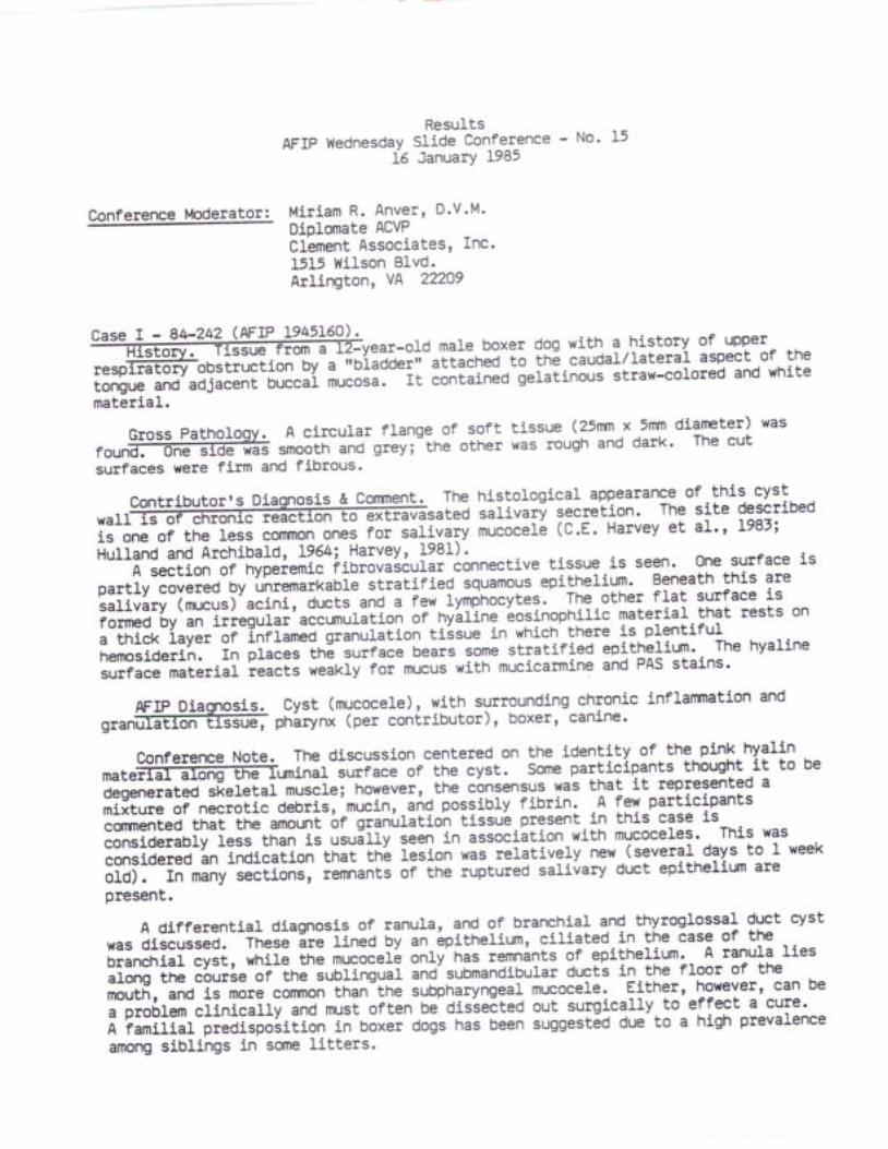
Conference Moderatol;
Case I -
D . V . M .
Inc.
flange of softgrey; the other
AFIP V{ednesdaYL6
Miriam R. Anver'Diplomate ACVPClement Assoeiatest1515 l{ilson B1vd.Arlington, VA 22209
ResultsSlide Conference - No' 15January 1985
ffiear-old male boxer dog with a history of upper-obstruction by .-;oiioo"r" attaihA 6 !ng.91'9"11i1:"T+^::1"::ro:"ll:
iffiEi:"ffit":3::ffiHi";i ;d;:--it conta:.neo-teiatinous sraw-colored and whitemateri.al.
Gross Pathology. A circularrounffi smooth andsurfaces were firm and fibrous.
tissue (25nm x 5nrn dianeter) waswas rough and dark. The cut
contributor's Diagnosis & conrnent. The histological appearance of this cyst
* " r r a t e a i i r i u a r i s e c r e t i o n . T h e s i t e d e s c r i b e dis one of the less corunon ones for salivary mucocefl (C.E. Harvey et al', L987;
Huffana and Arehibaldn L964i Harvey, 1981) --..^ : r-.,aa a,,rran6 'A section or-[vpJiemi" fiorovi36urar connective tissue is seen. One surface is
partly covered by unremarkable stratified squamous epithelium' Beneath this are
salivary (rnrcus) acini, ducts .nO-. few lymphocytes' The other flat surface is
forned by an it""gu1"i'accunnutation oi nyiUne 'eosinophilic
material that rests on
a thick 1ayer of inflamed granuliiion tiisue in which there is plentiful
henosideri.n. rn piaces-in6 surrace bears some stratified epitheriurn. The hyaline
surface material ;!g|G n.if.f' foi mucus with mucicarmine and PAS stains'
AFIP Diaonosis. cySt (mucocele), with surrounding chronic inflanmation and
g"anf f ipn i rynx(percont r ibu tor ) ,boxer ,can ine .
conference Note. The discussion centered on the identity of the pinf. ny9lin
' " t " f f i i n i r - iu r t . " " -o r tnecys t .Sornepar t i c ipants thought i t tobeO"i*""it"O-"["iJiai musc]e; however, the cbnsensus was that it represented a
mixture or necrotic oeoris,'mucin, and possibly fibrin. A few participants
cqrmented tnat lne-amount of granufitioh tissub-present.in this case is
considerabry reis-th;;-ia usuiriv-li*-:.n association with mucoceles. This was
considered an indication that thl lesion was relatively new.(several days to I week
OId). In many Sections, remnants of the ruptured salivary duct epitheliun are
present.
A differential diagnosis of ranular lnd of branchial and thyroglossal duct cyst
was discussed. -inese
ire lined OV an 6pitneUum, ciliat"d.i,n the case of the
branchial cyst, while the mucoceli only'has remnints of epithelium' A ranula lies
along the course oi-ine sublinguaf inO'submandibular ducts in the floor of the
mouth, and is more cofimon than the subpharyngeal mucocele. Either, however, can be
"-pto6fer-cfinicifly and must often be dissecteO out surgically to effeet a cure'
A familial predisposition in ooxJi dogs has been suggested due to a high prevalence
among siblings in scrne litters.
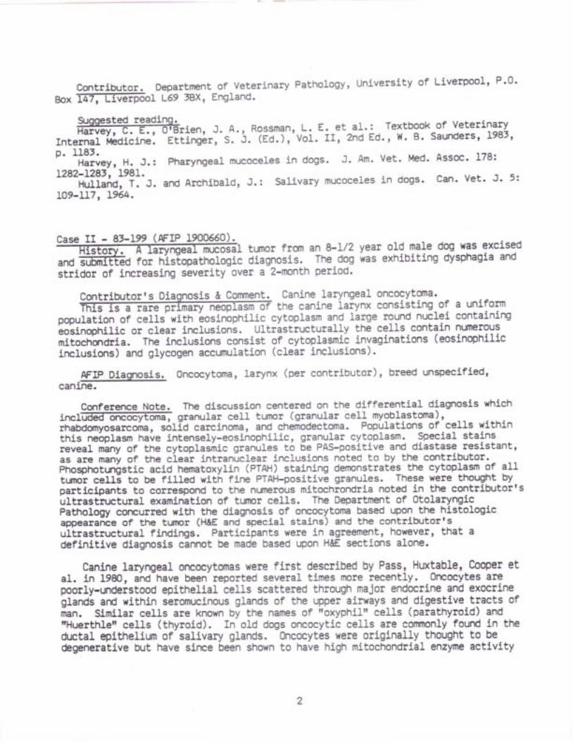
contributor. Department of Veterinary Pathology, university of Liverpool, P'0'
aox lffiol L69 3BX, England.
Suoqested reading.f f i i "n , J . A. , Rossman, . .L : E. e t a1. : Textbook
In te rna l l , t ed i c ine . E t t i ng l r , S . J . (Ed . ) , Vo l . I I , 2nd Ed ' ,1 { ' B '
p. 1181.r- -H"rrey, H. J.: pharyngeal mucoceles in dogs. J. Am. Vet. Med.
L282-L28t, 1981.----nuuano, t. J. and Archibald, J. i sal ivary mucoceles in dogs'
109-117, L964.
of VeterinarYSaunders, L983,
Assoc. 178:
Can. Vet . J . 5 :
Case II - 87-19muc tumor frorn an 8-L/2 year old male dog tras excised
and ior-nijlopatnofogi- diagnosis. The dog was exhibiting dysphagia and
stridor of increasing severity over a 2-month period.
Contributorrs Diagnosis & Comment. Canine laryngeal oncocytoma.i the canine laiynx consisting ol a uniform
population of celLs with bosinbphilic cytoplasm and large round nuclei containingioiinopnilic or clear inclusions. UltristrucLurally the cells.contain numelousmitochqrdria. The incLusions consist of cytoplasmic invaginations (eosinophilic
inclusions) and glycogen accumulation (clear inclusions).
AFIp Diagnosis. oncocytoma, larynx (per contributor), breed unspeci'fiedt.-
canlne.
Conference Note. The discussj.on centered on the differential diagnosis whiehinc1@granu1arce11 tumor (g ranu1arce I ]myob1as toma) ,rhabdomyosarcoma, s6fiO carcinoma, and chemodectoma. Populations of cells withinthis ne6plasrn have intensely-eosinophilic, granular cytoplasm. Special stainsreveal miny of the cytoplasmic granules t6 6e PAS-positive and diastase resistant'as are many of the ciear intranuclear inclusions noted to by the contributor.FnospnotgnistS.c acid hematoxylin (PTAH) staining demonstrates the cytoplasm 91 3I1turnoi celll to be filled with fine PTAH-positive granules. These were thought byparticipants to correspond to the nLrnerous mitochrondria noted in the contributorrsirttrast'ructural examination of tumor ceIIs. The Department of OtolaryngicPathology coraurred with the diagnosis of oncocytoma based upon the histologicappearante of the tLfillr (H&E and special stains) and the contributorrsultrastructural findings. Participants wete i-n agreement, however, that adefinitive diagnosis cannot be made based upon HEE sections alone.
Canine laryngeal oneocytomas wete first described by Pass, Huxtable' Cooper etal. in 1980, and have been reported several times more recently. 0rcocytes arepoorly-undeistood epithelial cells scattered through major endocrine and exocxinegfanOi ard within seromucinous glands of the upper airways and.digestive-tracts ofilan. Similar cells are known Oy tne names ofrroxyphil" ce1ls (parathyroid) andttHuerthlett cells (thyroid). In old dogs oncocytic cells ate eommonly fourd in thed.ntal epithelium of salivary glands. 0ncocytes were originally thought to bedegenerative but have since been shown to have high mitochondrial enzyme activity
2
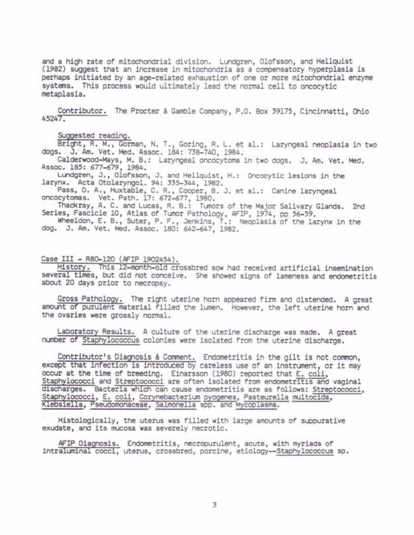
and a high rate of mitochondrial division. Lundgren, Olofsson, and Hellquist(L982) suggest that an increase in mitochondria as a compensatory hyperplasia isperhaps initiated by an age-related exhaustion of one or more mitochondrial enzymesystems. This process would ultimately lead the normal celI to oncocyticmetaplasia.
Contributor. The Procter & Gamble Company, P.0. Box V9I75, Cincinnatti, Stio45247---
Suggested reading.Br ight , R. M., Gorman, N. T. , Gor ing, R. L. et aI . : Laryngeal neoplasia in two
dogs. J.'Am. Vet. Med. Assoc. I84: 738-74O, L984.Calderwood-Mays, M. B.: Laryngeal oncocytoma in two dogs. J. Am. Vet. l,1ed.
Assoc. I85z 677-679, 1984.Lundgren, J., Olofsson, J. and Hellquist, H.: Oncocytic lesions in the
larynx. Acta 0tolaryngol. 94: 735-i44, 1982.Pass, D. A., Huxtabla, C. R., Cooper, B. J. et a l . : Canine laryngeal
oncocytomas. Vet. Path. I7z 672-677, 1980.Thackray, A. C. and Lucas, R. B.: Tumors of the Major Salivary Glands. Znd
Series, Fascicle I0, Atlas of Tumor Pathology, AFIP, 1974, pp.56-59.wheeldon, E.8.1 suter, P. F. , Jenk5.ns, T. : Neoplasia of the rarynx in the
dog. J. Am. Vet. Med. Assoc. 180: 642-647, I9AZ.
Case III - R80-120(AFrP 1902454).crossbred
severabout
-mondid not conceive. Sheto necropsy.
sow had received artificial inseminationshowed signs of lameness and endcrnetritis
Gross Patho'logy. The right uterine horn appeared firm and distended. A greata r r n @ t e r i a 1 F i t t e o t h e 1 u m e n . ' i { o w e v e r , t h e 1 e f t u t e r i n e h o r n a n dthe ovaries were grossly normal.
Laboratory ResuLts. A cuLture of the uterine discharge was made. A greatn .on@co1on ieswere iso1ated f romthe[ te r ined ischarg6.
Cmtributorrs D & Comment. Endometritis in the gilt is not comnon,careless use of an instrument, or it may
occur at the time of breeding. Elnarsson (1980) reported that E. coli,Staphylococci and Strept_gqocci are often isolated from endometrffifr-n'O vaginal
: - . : r , _ - ? . . -)Eapnyrococcr ano sErepEococc]. are orEen lsolateo rrom endometritls and vagj-naJ.discfiatges. Bacteria which can cause endometritis are as follows: Streptococci,e ! - - L . . a - ^ - ^ ^ : - - ^ a : A - - ^ , - ^ L ^ ^ - - - r - - . # '
9!+nyfggocci, E. co1i, CorylePactelium pyogenes, Pasleurella multoEldilKIeOS].eIIa, PSzuOOmOlqCeqq, salmoneJ.la spp. and Mycoplasrna.. \ - 9 v 9 1 9 - - 9 . | - g v V 9 l l l g r l q g g q g . g q l l l l v l l g 4 4 q Mycoplasma.
Histologically, the uterus was filled with large amounts of suppurativeexudate, and its mucosa was severely necrotic.
AFIP Diagnosis. Endofietritis, necropurulent, acute, with myriads ofintrE@uterus,cross6red,pbrci .ne,6t io1ogi-st"pnlro"*. .p.
3
. In].s, but
20 days prior
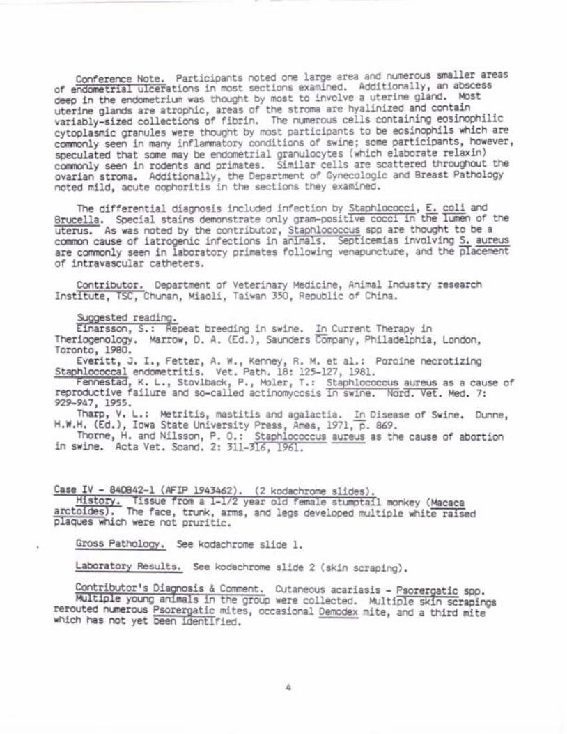
Conference Note. Participants noted one large area_and numerous smaller areaso r e f f i t i o n s i n . m o s t s e c t i o n s e x a m i n e d . A d d i t i o n a 1 1 y , a n a b s c e s sdeep in the endometriun was thought by most to involve a uterj.ne glard.. Pttut.iine glands are atrophic, arels of- the stroma are hyalinized.and containt;iGbiy:sizio collectibns 6r fibrin. The numerous ceils containing eosinophiliccytoplaimic granules were thought by most participants to be eosinophils which arec6nmbnly seen in many inflanrnatory conditions of swine; some participants, howevet,specukted that some- may be endom-etrial granulocytes (which elaborate relaxin)cbrnonly seen in rodent! and primates. Similar cells are scattered throughout theovarian- stloma. Additionally, the Department of Gynecologic and Breast Pathologynoted mild, acute oophoritis in the sections they examined.
The differential diagnosis included infection by Staphlococci, E. coli andBruce l Ia .Spec ia is ta in ioemonscraceon iygram-pos ic f f i ' tn -e f f i r 'o f inef f i A s w a s n o t e d b y t h e c o n t r i b u t o r , S t a p h 1 o c o c c u s s p p a r e t h o u g h t t o b e acomnon cause of iatrogreiric infections in anifrEl-s.@icemias involving S. auteusare coflnonly seen in laboratory primates following venapuncture, and the placementof intravascular catheters.
Contributor._ Department of Veterinary Medicine, AnimaL lndustry researchrnstiEff{-TSefbhunan, Miaoli, Taiwan 350, Republic of China.
9rggested reading.Einarsson, S. ! Repeat breeding in swine.
Theriogenology. Marrow, D. A. (Ed.), SaundersToronto, 1980.
In Current Therapy inGmpany, Philadelphia, London,
Staphlococcal endometritis. Vet.--n'es-Fa , K. L., Stovlback,reproductive faiLure and so-calle
P., l'4oler, T.: Staphlococcus aureus as a cause ofd actinomycosis ffi. Med. 7:
929-947, 1955.Tharp, V. L.: lvletritls, mastitis and agalactia. In Disease of Swine. Dunne
H.t't.H.'-iEi. l, ii*a state universi[y-nrJti, -Eila;-riir,;.-il;:-- -
Thorne, H. and Nilsson, P. 0.: Staphlococcus aureus as the cause of abortionin swine. Acta Vet. Scand. 2: 7LI-7T6,4767
Ever i t t , J . I . , Fe t te r , A . W. , Kenney, R. M. et al.: Porcine necrotizingPath. 18: 125-127, I98!.
Case IV - U(f,42-L (AFrP t94tM2). (Z kodachrome slidesa L-uz yeat ema monkey (Macaca
The face, trunk, arrns, andwere not pruritic.
legs developed multiple whitfiiil-sed
Gross Pathol.ggy. See kodachrome slide 1.
Laboratorv Results. See kodachronre slide 2 (skin scraping).
Contributorts Diagnogi,s"& Conrnent. Cutaneous acariasis - psorergatic spp.were coti;a;e:--Murtiffir#ilst
rerouted nt Elous Psoreroatic mites, occasional Dernodex mite, ino a third mite-which has not yet 6E?i-TTeTEriea.
' - -- '
4
intracorneal acaridAFIP Diaonosis. Hyperkeratosis and acanthosis, segmental, moderater with
parasites, stumptail macaque, primate.
Conference Note. As a differential diagnosis for the most numerous parasitespres f f iog ica1sec t ions ,par t i c ipantscons ideredI iceandmi tes .Mosttholght these parasite- are too small to be lice. The parasite in the skinscrafing (kodachrome slide 2) has 4 pairs of legs and is cornpatible with a mite;lice have only 3 pairs. This organism has a round body differentiating it fromDerpdex, the body of which is elongate. Sarcoptes, and several other skin mites'hE-ve taisal suckers and the first [wo paiirc.o 3 are ioca-red ciose to thecapitulurn while the second two pair are further caudad. The mite in this case doesnot have tarsal suckers. The morphologic characteristics of this mite arecompatible with Psorergates. Participants thought that the dermatitis present inm o s t c a s e s o f S a f f i l i 1 d b e m o r e S e V e r e . I n s o m e s e c t i o n s , a 1 a r g e r e 1 o n g a t e dmite is presenfrn--E-fi?'ir follicle. Although it could not be identified based uponits morphology 1n the sections examined, most felt it was compatible with Denodex'as noted by the contributor.
A very mild, sometimes minimaL (as in this case) inflammatory response istypical of Psorergates infection. Mites live in the stratum corneum, where it isb e 1 i e v e d t h d @ y e x c a v a t a t i o n o f k e r a t i n w i t h s a w - 1 i k e t e e t h a n d f e e d b ypiercing ceIls with styliform chelicerae.
Contributor. Department of Comparative Medicine, Hershey Medlcal CenLer,eenn6yliiffiiiSffite University, Hershey, Pennsylvania I7O73.
Suoqested readino.f f i , J . C., Atyeo, W. T. : psorergates glaucgmys sp. N. (Acar i :
Psorer !a t ioae i ,acys togenousmi te f romtnesof f i (GtaucomF.# ' .
volan5l,-wffi'histoiath6logic notes on a mite induced o6rmit cyst. J.-Tffiifol.5#eg-174, 1973.
Baskin, G.8. , Eberhard, M. L. , f ' latson et a l . : Diagnost ic exercize. Lab.Anim. Sci. 342 6O2-6O7, 1984.
Lee, K. J. , Lang, C. M., Hughs, H. C. et a1. : Psorergat ic mange (Acar i :Psp4lgqliQqq) of the str-rmptail macaque (Macaca arctoides). Lab. Anim. Sci. 31:77="t9'-96tr-
Sheldon, l{. G.: Psorergatic mange in the sooty mangabey (Cercocebus torquatesatvs) monkey. Lab. Anim. Care 16z 276-279, !966.
DAVID L. FRTTZ, V.M.D.Captain, VC, USARegistry of Vetetinary PathologYDepartment of Veterinary Pathology
Conference lvloderator :
ResultsAFIP llednesday Slide Conference - No. 15
2f January 1985
George A. Parker, D.V.M.Diplomate, ACVPVeterinary Pathology Consultant11101 Streamview CourtGreat Fal1s, VA 22066
Case I - N84-0210 (AFIP L944522) (1 kodachrome slide).majority of the rats in
arrlval quarantine began exhibiting photophobia, exophthalmos, andchromodacryorrhea. The rats had been in quarantine for 9 days when clinical signsbegan. Rats sacrificed from the same lot 9 days earlier for quality controlmonitoring were clinically, grossly, and histologically normal.
Gross Pathology. There was unilateral, focally-extensive, reddish-grayd iSc f f imaaf fec t ingone.ha ] . f to two- th i rdso f theharder i ing lands .
laloqqtSrry Results. Affected anj.mals had positive titers forsialo@s (SDAV), while serum samples from normal quality controlanimals taken earlier from the same lot were negative.
Cqqtributorts Diagnos_is & Comment. DacryoadeniLis, necrotizing and suppurativew i t n y e x t 6 n s i v e , u n i 1 a t e r a 1 , t t a i d e r i a n g 1 a n d ,Sprague-Dawley rat.
Dacryoadenitis caused by sialodacryoadenitis virus (SDAV) was diagnosed basedon typical clinical signs and gross and histological lesions. Confirmation of SDAVwas made by identification of anti-SDAV antibodies by the ELISA test.Retrospective ELISA tests on serun from animals sacrificed on amival for qualitycontrol monitoring were negative for SDAV. l{e were unable to positively identifywhere the affected rats contacted SDAV. Neither our facility nor the vendor have arecent history of SDAV outbreaks.
Both the Harderian and extraorbital lacrimal glands from affected animalsexhibited lesions. Examined salivary glands were free of disease.
AFIP 9r Dacryoadenitis, necrotizing, subacute, diffuse, moderate, with, unilateral, Harderj.an gland, Sprague-Dawley rat,
rodent, etiology--compatible with siaLodacryoadenitis virus (SDAV).
The kodachrome slide in this ease illustrates the cervicalswel f f is ,andehromodacryorrhea(a1thoughmi1d)whicharecharacteristic of but not pathognomonic for sialodacryoadenitis virus (SDAV)infection of rodents. Chromodacryorrhea, or red tears, can be caused by any formof stress. The unilateral distribution of the histologic lesions is, in themoderatorts experience, a common finding in cases of SDAV infection, as is thehistory of onset of clinical signs one week to 10 days after arrival toquarantine. The differential diagnosis included cytomegalovirus and papovavirus
Conference Note.
(polyoma virus), both of which produce intranuclear inclusions which were not seen
in this case. Dacryoadenitis oi-ln" Harderian gitlo :11..11:9^:ttult from orbital
bleeding as a result of venapunct"i" of the orbital venous plexus for obtaining
blood samples (McGee, Maronpotr-It7il. In some slides there is a subacute
siiioaoenitis in a section of salivary gland'
sDAV is a coronavirus which is antigenically related to mouse hepatitis yi.rus
(l,,ll-lv), rat coronavirus, and human-cJronivirus OClg' Serological titers to SDAV are
presently indistinguishable from those to rat coronavirus which causes interstitial
pnzumonitis in adult rats.
Contributor. Veterinary Sciences Division, Comparative Pathology Branch (VSP)'
scrro6ffiFace Medicinb, Brooks AFB, Texas 78235.
Suooested reading.f f i Y , R . 0 . a n d J o n a s , A . M . : R e s p i r a t o r y i n f e c t i o n i n m i c e
with sialodacryoadenitis virus, a coronavirus of rats. Infect. & Immunol. 18(3):
82t-827, L977.Eisenbrandt, D. L. , Hubbard, G. B. and Schmidt, R.-_E.:_^A subcl in ical epizoot ic
of siaLodacryoailenitis' in rats.- Lab. Anim. Sci. 322 655'659, 1982'Jacoby , n .0 . , Bhat t , P . N. and Jonas , A . |4 . : Pa thogenes is o f
si.afoOicry6Oeniti i in gn6toniotic rats. Vet. Path. L2z 196'209, ,L975'..Jonas, A. M., Craf [ , J. and Black, C: L. . : -Sia]odacryoadeni t is in the rat (a
figni- jnO' electr6n micr6scopic study). Rrcn. .Path. 882 613-622, +919'- Mccee, M. A. and Maronpot, R. R.: Harderian glalq-dac,ryoadenitis in rats
resutting'from orbitat nf#Oing. Lab. Anim. Sci. 29(5): 639-64L' 1979. -perci , D. H., Hanna, P. E.] Paturzo, F. et a1. : Comparison of strain
suscepti6ifity t6 experimental'sialodaciyoadenitis in rats. Lab. Anim. Sci. f4:25*260, 1984.
Snith, A. L.: An immunoflfuorescence test for detection of serum antibody torodent coionaviruses. Lab. Anim. Sci. i7z 157-160, I98t.
Case II -
turbedweigfrt on left front
equilibrium with poorreg.
female thoroughbred, equine, wh5.chcoordination and was unable to sustain
Gross Pathology. Soft tissue edema, fluid exudation and mild hyperemia werefounf f iund ing the le f tmetacarpa11 igamentsapprox imated5cmdis ta1to joint. A large (11x5x4 cm), off-white nodule was found in the right apical lobeof the lung.
LaboratorY ResuLts.f f i5+ tmooeraEe/.eacterioffi-iathogens were isolated.AGID - EIA: negative.
Contributorrs Diaqnosis & Comment. Granular cel1 tumor (granular cellmyob ughbred, equine.-
This interesting tumor occuii in a variety of speci.es at many anatomicallocations.
8tP-227 (AFIP Le465V8) .r-old
AFrP Diagnosis. Granular ce1I tumor, lung, thoroughbred, equi.ne.
Conference Notg. Parlicipants were in agreement with a diagnosis of granular- . -cerr tunor (formerly-granular cell myoblastoma), and all listed oncocytomi as adifferential diagnosis. Two different celL types are noted; the moxe nLrnerous::++:.,"""^1iI9? polvhedral celrs with eosinopnilic s""nuri"'cvtopriil ag;ilI;"ceus). cytopl3sm+c granules stain variably with PAS and are diastase iesistant.The second population-is smaller and more-siinole-shaped; these cel1s fie iJjacentto vessels. They were thought to be angulate body cells and were shown to containtiFl" to rultiple PAS-posifive, diastaie resiitant in[racviofiasmic globufJiwhichr,according to the moderator, correspond to the angulit"-oooi"s seenultrastructurally.
.^ -EIi9:l::_supqo$ing the theory that the cell of origin of granular cetl tunorsrs a precursor of the Schwann cell- is: 1) frequent cloie anat6mical relationshipof granular cell tumors to peripheral nerve, Z) presence of substances similar todegradation products of.myelin in granular ieti trrot., rj presence of highconcentrations of gangliosides and cerebrosides in granular'cell tumors, ind a) thehistologic similarity-between granular ce1ls and Schwann cells near axonsundergoing wallerian degeneration (parker, Novilla, Brown et al. , r97g).
It has been-suggested that the intracranial granular cell tumors seen in agingrats may arise from Schwann ce1ls (or a common piogenitor celli-oi-;.;ii'perivascular nerves_in the leptomeninges and centril nervous -ystem (Hollander,Elurek, Boorman et aL. , 1976). The deilonstrition of sr00 proteii]-;,];;:iIiiri"enolase, and two peripheraL nerve-myeli! prot-ins in rat granular cerl turnors, iscited-by Sanford, Hoover, and Millei (198[) and provioes Furtnir evidence for aneural origin of granulai cell tumors.
Contributor. AnimaL Health Laboratory, _Maryland Department of Agriculture,a901TEffiE-R6?d, College parx, Maryland' 20740.
Suggested readino.f f i , J . M . a n d S t r a n d b e r g , J . : G r a n u 1 a r c e 1 ] . v a r i a n t s i n a r a t
schwannqna. Vet. Path. 15: 725-73L, 1979.Hollander, C. F., Burek, J. 0.,
-Boorman, G. A. et aI.: Granular cell tumors of
the central nervous system of rats. Arch. Path. Lab. Med. 100: 445-447, Ig7:a:-, f3rketr 9. A. , Novi l la, M. N., Brown, A. c. et a l . : Granurar cel l tumor(myobrastoma) in the lung of a horse. J. comp. path. g9z 42I-4to, Ig7g., l?"kgr, 9. .A., Bothdt W., Van Cellen, A. et a1.: Cerebral grinular ce1l tumor(myoblastoma) in a dog: Case report and literature review. Cornell Vet. 68:506-520, L978.. safod, S. E..., Hoove!, D. M., Mil ler, R. B.: primary cardiac granular celltumor in a dog. Vet. Path. 2L: 489-494, 1994.. IYtkr M.l.M1 Johnson, G. C. Gal1ima, A. M. et al.: Canine granular cell tumor(myoblastoma): A report of four cases and review of the llteratuie. J. Sm. Anim.Pract. 242 677-645, L98t.
Turk, M.A.M. and Breeze, R. G.: Histochemical and ultrastructural features of?! geuine pulmonary granular cell turnor (myoblastoma). J. Comp. Path. 9I:A7I-IEI,1981.
Case III - D8t-513 (AFIP L948t27) (2 kodachrome slides).rom an Sf,e1n cow
mas s . was down the next daY, stargazing, with nowas no irnorovement
treated for toxicuse of the left front
leg. She was eating normally. There over a 5-day period.
Gross Patholoqy. Cervical nerves were firm, 4-5 cm in diarneter. Brachialp 1 e x f f i . c 8 h a s a n 8 c m d i a m e t e r f i r m m a s s i n t h e a x i 1 1 a . T h ecervico-thoracic ganglia are 6x5x8 cm diameter, firm and white. Intereostal nervesare thj.ckened with multiple ?-3 cn nodules. The vagus is 4 cm diameter andepicardial nerves are prominent. Celiac ganglion is 15 cm diameter.
Laboratorv Results. CSF was normal.
Contributorfs D sis & Comment. Multiple schwannomas (bovine neurofibro-matosis), etj.oLogy--unknown.
In this section of brachial plexus, multiple nerves are greatly enlargedreplacerent by nodules and interlacing bundles of neoplastic spindle cells.most areas, the cells are densely packed wi.th scanty extracellular materialsimilar to Antoni type A tissue. fr0ni.on bulb-likerr structures are prominent
due toIn
and arein
Iess affected nerve fascicles.
M.rltiple schwannomas are refatively common benign tumors of cattle. The sitesmost frequently involved are the brachial plexus, intercostal nerves, andsyrnpathetic nervous system, although other sites, including skin, have beendescribed. The rnost likely cell of origin is considered to be the Schwann cell,however, the perineurialtosis (von Recklinghausents disease) of man.
_ AFIP Diagnosis. Peripheral nerve sheath neoplasm, mu1tiple, brachial plexus(per contributor), Holstein, bovine; condition--compatible with bovineneurofibromatosis.
Conference Note. Tissue sections contain nerve fascicles which ensheathv a r i @ s t i c g r o w t h s . T h e 1 a r g e s t t u m o r i s a s o 1 i d c e 1 1 u 1 a r m a s scomposed of spindle cells amanged in some interlacing bundles, but otherwise in arandom array. |'lore loosely-arranEed myxomatous areas with plurnp round neoplasticcells are seen in some sections. Participants felt the largest turnor ispoorly-differentiated, in contrast to surrounding smaller tumors which are alsosolid cellular masses but in which spindle ceI1s form Verocay bodies and storiformpatterns which ate suggestive of a well-differentiated tumor of neural origin.SFcial stains reveal Little collagen, little intercellular ground substance, andfew lf any nerve fibers wlthin the tumor masses. Particlpants were tfrerefor6 inagreement with the contributorrs diagnosis of multiple schwannomas.
Bovine neurofibromatosis has been corpared to von Recklinghausenfs disease inman. The latter, however, is a genetically-transmitted phakomatosis and involvesnumerous organ systems, as opposed to only peri.phereal nerves in the bovine.|}tnguO.many_simllarities do exist betwein bovine and human neurofibromatosis, thebicolor damselfish has been tecently proposed as a more useful animal rnodel(Schmale, Hensley, Udey, 1983).
The diffuse invoLvement of peripheral nerves in bovine neurofibromatosis hassuggested an infectious etiology. The finding of virus-Iike particles innJ5fiiastic cells of bovine nerv6 sheath tumori has been reported (Canfield,Doughty, 1980).
Contributor. Department of Pathology, Cornell University, Ithaca, New York14850-
Srooested readinq.f f igh tmic roscop ics tudyofbov ineper iphera1nervesheath
tumors. Vet. Path. 15: 283-29I, 1978.Canfield, P.: The ultrastructure of bovine peripheral nerve sheath tumors.
Vet. Path. 15: 292-3OO, 1978.Canfield, P. J. and Doughty, F. R.: A study of virus-like particles present in
bovine netve sheath tumours. Austr. Vet. J. 562 257-261' 1980.Mitcham, S. A., Kasar i , T. R., Parent, J. M. et aI . : Intracranial schwannoma
in a cow. Can. Vet. J. ?.5: 118-141, L984.Monlux, A. W. and Davis, C. L.: Multiple schwannomas of cattle. Am. J. Vet.
Res. 14: 4g9-5O9, L95V. ' ' -
Schmale, M. C., Hensley, G., Udey, L. R.: Neurof ibromatosis, vonRecklinghausent.s disease, multiple schwannomas, malignant schwannomas. Amer. J.Path. LL2(2)z 238-24I, 1983.
Case IV - 8t-1810-6 (AFIP I9L36I6)n n e c r o p s y i n g a c a s t r a t e d m a 1 e q u a r t e r h o r s e ( 1 8
montfr3-oTti)-suff ering ftom nonregenerative anemia.
Gross Pathglogy. l{.rltiple 1-3 nrm ulcers on the tongue and gums adjacent toteetfr.-
Corttributorfs Diagnosis & Comment. Glossitis, pyogranulomatous, ulcerativeDiptera (myiasis). Gasterophiluswith larval star
intestinalis inFection.-ne-mfrating larvae had a chitinous exoskeleton with spines, striated somaticmusculature, OoOi cavity, digestive system and trachea. The burrowing parasite inthe tongue is prbOaOly Gisteiophilus intestinalis which invades the tongue andgums. G. nasalis invades only the gums.
AFIP Diaonosis.-suom@with Gastrophilus sp.
Conference Note. Most sections examined contain l-l larvae within thes u @ n o c c a s i o n a 1 1 a r v a i s p r e s e n t i n t h e m U c o s a . S o m epirticip"nti feft-that the hostrs inflammatbry tesponse was out of proportion tothe num'ber of larvae ptesent and speculated that there was another underlyingdisease in progress. The mucosa contains occasional pustules, possibly migrationtracts, and also some plant material.
Glossitis, ulcerative, sr-bacute, diffuse, moderater withparasites, tongue, quarterhorse, equine, eti.ology-cornpatible
ThetheG .
ofand
Although large numbers of mature larvae may be found in the stomach of thehorse, perforating ulcers or other gastric disorders which result from attachedlarvae are uncdnnon. Esophagitis and laryngitis caused by irilrnature migratinglarvae are sometimes seen, and the latter may be one cause of choke in the horse.
different species of Gastrophilus are most easily identified by examinationt h i r d s t a g e ] a r v a e . G @ i s b y f a r t h e m o s t c o m m o n i n t h e U . S . ,hemorrhoidalis is thoG'hFE6-TEEfr.iteo to the Pacific northwest.
Contrilrutor._ Department of Pathology, College of Veterinary Medicine, KansasStatE-iiifire Ef, Manhattan, Kansas ee5o6.
Suqoested readino.- f f i B i o 1 o g y o f A n i m a 1 P a r a s i t e s . W . B . S a u n d e r s C o . ,Philade1phia, 1964, pp 516-619._ lvlansnann, R. A.1 McAllister, E. s., pratt, p. w. (Eds.): Equine Medicine andlyry""y. lrd Ed., Vol. 1, American Veterinary Publications, Santa Barbara, CA,1982, p T5-75.
Shefstad' D. K.: Scanning elecLron microscopy of Gastrophilus intestinalisl e s i o n s o f t h e e q u i n e s t o m a c h . J . A m . V e t . M e d . n s s o c
Soulsby, E.J.L.: Helminths, Arthropods and Protozoa of Domesticiteo Animals.7th Ed., Lea & Febiger, Philadelphia, L.982, pp 400-404.
DAVID L. FRITZ, V.M.D.Captain, VC, USARegistry of Veterinary PathologyDepartment of Veterinary Pathology
6
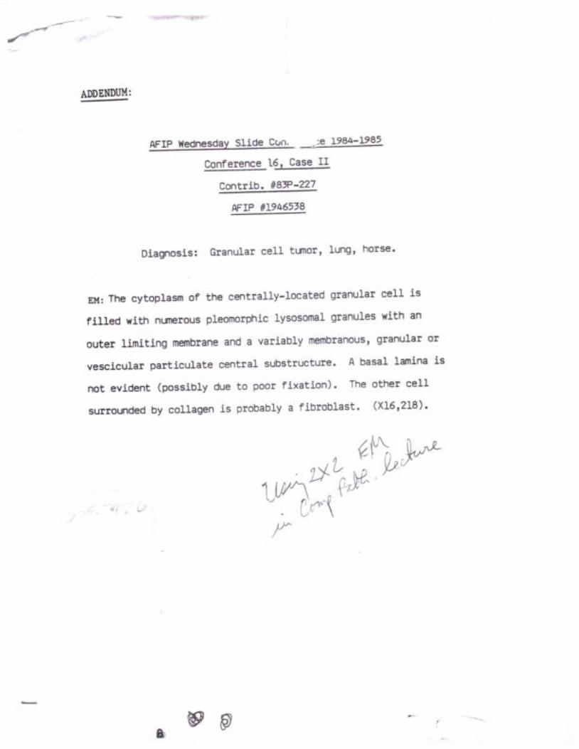
{ b i " r d d h l ' ! ' : . k E * y
ADDEMUI.I:
AFIP Wednesdav Slide Csn. -.-:e le84-1982
Conference t€z Case Il
Contrib. llST-q
MrP tf|9465t8
Diagnosis: Granular cell tumot, lungt horse'
nu :Thecy top lasmof thecent ra l l y - loca tedgranu larce l l i s
filled with nunerous pleomorphic lysosomal granules with an
outer limiting membrane and a variably membranous, granular ol
vescicular particulate central substructure. A basal lamina is
not evident (possibly due to poor fixation) ' The other cell
surrounded by collagen is probably a fibroblast. (x16r218).
1w\'',^'&\'w'- - cr'*\l"
b@ p
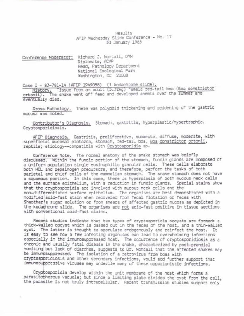
AFIP f'/ednesday3A
ResultsSlide Conference - No-Januarv 1985
L7
Conference Moderator: Richard J. Montali, DVMDiplomate, ACVPH;;-, Pathology DepartmentNational Zoological ParkWashington, DC 20008
Case I - 8t-78I-I4 (AFIP 1949058) (1 kodachrome slide)'i i boa (Boa constqicior
ortoifif.The snake went off feed and developed anemia over the summer and
ffirry died.
There was polypoid thickening and reddening of the gastric
Contributoqls Diagnosis. Stomach, gastritis, hyperplastic/hypertrophic.crypTo:@EiGTs.
AFIP Diagnosis. Gastri t is, prol i ferative, subacute,supeiffilillffif protozoa, stomach, red-tail boa, Boareptile; etiology--compatible with Cryptosporidia sp.
Confererae Note. The normal anatomy of the snake stomach was brieflyo i s c f f i e f u n d i c p o r t i o n o f t h e s t o m a c h , f u n d i c g 1 a n d s a l e c d n p o s e d o fa uniform population sinEle eosinophilic granular cells. These cells elaborateboth F€L and pepsinogen ptecursors, and therefore, perform the tasks of bothparietaL and chief cells of the mammalian stomach. The snake stomach does not havea squamous portion. In this case, there is hyperplasia of both mucous neck cellsand the surface epithelium, wiLh a reduction in fundic glands. Special stains showthat theicryptosporidia are involved with mucous neck cells and thenon-diff iated surface epithelium. The organisms are best demonstratedwith amodifiedlacid-fast stain when recovered from fecal flotation or feces withSheotherfs sugar solution or from smears of affected gastric mucosa as depicted inthe slide. The organisms are not acid-fast positive in tissue sectionswith onal acid-fast stains.
studies indicate that two types of cryptosporidia oocysts are formed: athiekcyst .
oocyst which is passed out in the feces of the host, and a thin-walledlatter is thought to sporulate endogenously and reinfect the host. It
dif fuse, moderate, withconstrictor ortonii ,
support thatinfections.
is easyespecialehronicvomitingbecrypt sis and other secondary infections, would add further
ive viruses may underlie many of these opportunistic
idia develop within the unit membrane of the host whichparasitophorous vacuolel but since a limiting plate divides the cystthe parasite is not truly intracellular. Recent transmission stuiies
o see how a few infecting organisms can lead to overwhelming infectionsy in the inrnunosuppressed host. The occurrence of cryptosporidiosis as a
usually fatal disease in the snake, characterized by post-prandialbut Lack of diarrhea, suggests to Dr. Montali that the affected snakes may
ressed. The isolation of a retrovirus from boas with
forms afrom the celI,support only
Gross Patholomucosa was noted.
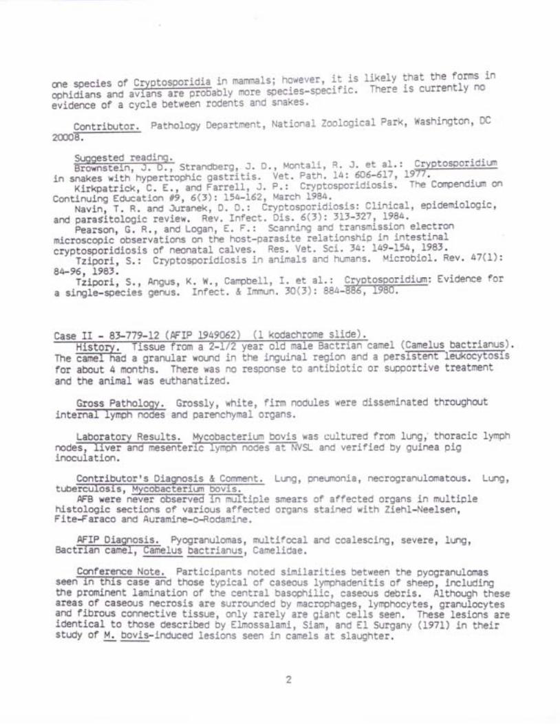
me species of crypto?poridiS in mammarsl !91:u:t, i ! . is l ikely that the forms in
ophidians and avff irrs are probably more.species-specif ic ' There is currently no
evidence of a cycle between rodents and snal<es'
Contributor. Pathology Department, National Zoological Park, l4ashington' DC
20008.
Suooested readinq.B r o w n S t e i n ' J . D . , S t r a n d b e r g , . J . D . , M o n t a 1 i , R . J . - 9 t - 3 ! . ' . * @
in snakes with hypert iophic gasti i t is. Vet. Path . L4z 606-617, L977 '-k i rkpatr ick , 'b . f . , and Farre l l , J . P. : Cryptospor id ios is ' The Compendium on
Continuing Educit ion /t9, 6(3)z I54-L62, March L984'Navini T. R. and Juranek, D. D.: Cryptosporidiosis: Clinical, epidemiologic,
and parasitotogic review. Rev. Infect. Dis. 6(7)z 313-327 ' 1984.Fearson, gl n., and Logan, E. F.: Scanning and transmission electron
microscopic observitions on tne host-parasite relationship in intestinalcryptospbridiosis of neonatal ealves. Res. Vet. Sci. 74: I49-L54, I98' '- "Tzipori-, S.r Cryptosporidiosis in animals and humans. Microbiol. Rev. 47(I)z
8b96, 1983.Tzipor i , S. , Angus, K. W., Campbel l , f . q ! .a I : i g lXPtg:Pgr id ium: Evidence for
a single-spiciei genus. Infect. & Immun. tO(3): 884-886, 1980.
Case II - 8t-779-f2 (AFIP 1949062 (1 kodachrome slidectrian
'camel (Camelus bactrianus).Theforand
a granular wound inabout 4 months. There was nothe animal was euthanatized.
& Comment.
AFB were neverhistologic sections of varj.ous affectedFite-Faraco and Auramine-o-Rodamine.
a persffilETx-oc@isor suppoxtive treatment
region andantibiotic
Gross Patholggg Grossly, white, firm nodules were disseminated throughoutinteffi and parenchymal organs.
Laboratory Resu1ts. Mycobacterium bovis was cultured from lungr' thoracic lymphnode@rffipfr-'iiGi6s-Eilvst- and verified by guinea piginoculati.on.
Lung, pneumonia, necrogranulomatous. Lung,
smears of affected organs in multipleorgans stained with Ziehl-Nee1sen,
AFIP Diagnosis. Pyogranulomas, muLtifocal and coalesci.ng, severe, lung,Bactrian cameI, Camelus bactrianus, Camelidae.
Conference Note. Participants noted similarities between the pyogranulomasseenf f i thosetyp ica1ofcaseous1ymphaden i t i so fsh 'e6p l inc1ud ingthe prominent lamination of the central basophilic, caseous debris. Although theseareas of caseous necrosis are surrounded by macrophages, lymphocytes, granulocytesand fibrous connective tissue, only rarely are giant cells seen. These lesions areidentical to those described by Elmossalami, Sj-am, and El Surgany (197f) in theirstudy of M. bovis-induced lesions seen i_n camels at slaughter.
Contributorrs D
The severity of lesions in the host is a reflection of the adaptabil ity of that
mycobacteriat species to that hosi. Usually, the greater,the hypersensitivity of
the host to the organism, the mole severe the reac[ion and necrosis witl be to the
presence or even-a-i"un-oi,g"nisms i".q. Koch's re"ciion). camels, l ike guinea pigs'
are extremely sensitive to mycoba"i"ii" in contrast to birds in which early in the
disease, the fesions often cbnsist of numerous macrophages containing myriads of
acid-fast bacilli with minimal reaction- The hypersensitivity of camels to
mycobacteria is ir io ierrected in tubercurin telt ing; many false positive reactions
axe seen, probably as a result oi "*poiut"
of .these animafs to atypical
mycoOact6ria such'as M. avium scrofulaceum and kansasii '
The moderator used this case to emphasize the importance of freezing tissue at
necropsy. A diagnosis was ultimately based upon cult 'I lng M' bqvis from lung and
rymph nodes, wheieas in all or tne-ipeciar stained sections-?xEiih-sd onry one
equivocal mycobacterium was seen.
InterestingfY, camels also appear to be very sensitive to isoniazid, an ..antitubercular Oiug commonly used for treatment or prophylaxis in humans and other
mammals. fsoniiiii appears to affect the bone marrow in camels causing sevexegranulocyto- and thrombocytopenia.
The moderator noted that the tissue in this case could be identified asoriginating from a camelid due to the elipti-caI shape of the red blood celIs.
Contributor. Pathology Department, National Zoological Park, Washington, D.C.20008.
guggested re?diLg.Dafifien$erg, A. Jx.: Hacnogenesis of pulmonary tuberculosis in man and animals:
protection of-personnel against tuberculosis. In MycobacLerlal Infections of ZooAnimals. . Ed., 'R. J. I ' lont l l i , Smithsonian Inst i f r t ion Press, Wash., 0.C., 1978, pp65-74.
Elmossalami, E., Siam, M. A. and El Sergany, M.: S!u!:,gs on tuberculous-likelesions in slaulhteied camels. ZbI. Vet. Med. B 182 253-26I,.I97I.
Kennedy, S,, and Bush, M. ! Evaluation of tuberculin testing and lymphgcyletransformation in Bactrian camels. Mycobact. Infecl, Zoo Anim. , 139-142, 1978.
Mann, P. C., Bush, M., Janssen, D. L. et a l . : Cl in icopatholog+9 9orT911t i9nsof tuberiulosis' in laige 2oo mammais. J. Am. Vet. Med. Assoc. L79(11): 1121'1129,1981.
Thorns, C. J. , Morr is, J. A. and Li t t le, T.W.A.: A spectrum of immuneresponses and pathological conditions between certain animal species toexperimental Mycobactirium bovis infection. Br. J. Exp. Path. 6f: 562'572, L982.
platyrhynchos) was inoculatedFGt-natcning witn 0.5 ml 1:10 suspenffi'fnjh- ofd6iilfife buffered saline
IM
plus antibiotics of liver tissue from wild adult female mallard found dead. At 4days post-inoculation the bird was severely depressed, eyes pasted shut, head t i l t ,frequent trchewingrr.
Case III - U. of S. Vet. Path. N84-3445 (1947498).
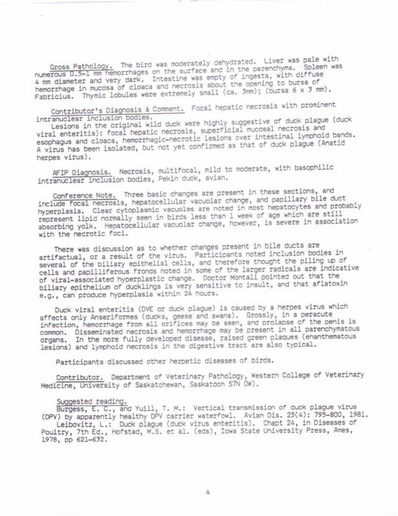
G r o s s P a t h o } o g y . T h e b i r d w a s m o d e r a t e l y d e h y d r a l g d : . - ! i f : * ' ' p a l e w i t hnUmef f io r rhageson- in " - 'u i i " .u ,and in theparenchyma.Sp}eenwas4 mm diameter and very dark. rnteli ine was empty of ingesta, with diffuse
hemorrhage in mucosa 'of
cloaca .nO-nE""osis abou't the oiening to bursa of
Fabricius. Thymic lobules nur" "It l"t"i '-tt"ff
ica' -:mi');
(5ursa 5 x 3 mm) '
Contributor,s Diaglqgi!@mment- Focal hepatic necrosis with prominent
int"ffiffi-iTl:'::?n?l3i"i;,.0 duck were hishrv :,gs:'!ly:,ol-d,"k prasue (ducxviral enterit is): focal hepatic-n".iotit, sup6rficial-mucosal necrosis and
esophagus and croaca, hemorrnagil-necrotic r'esioni over intestinal rymphoid bands'
A virus has been isolated, but ""t
V"i coniirmed is that of duck plague (Anatid
herpes virus).
A F l p D i a q n o s i s . N e c r o s i s , m u l t i f o c a l , m i l d t o m o d e r a t e , w i t h b a s o p h i l i c#
intranuclear rnffiion bodies, Pekin duck, avian'
conference Note, Three basic changes are present in these sections' and
f f i . ,n"p" io"" r rur i ' "vacuo1archange,and.papi11aryb i1eductinclude focal nthyperplasia. Clear cytoplasmic vacuoles are notea ii rirost hepatocytes and probably
represent fipiO-nort"iiy-i""n in birds less than I week of age which are stil}
absorbing yolk. Hepatocellular vicuotar change, hon"u"t, is-Severe in association
with the necrotic foci.
There was discussion as to whether changes present in,bile.ducts are
artifactual, or a resuLt of the virus. parlicibants noted inclusion bodies in
several of the Oifiiry-epithelial-""ift, and therefore thought Lhe piling Y? of
cells and papilliferoiis fronds noted in'some of the rarger iadicals are indicative
of viral-associaieJ-nyp"rpiastic change. Doctor Montali pointed out that the
bil iary epitherium of'bucktings-is veiv sensitive to insurt, and that aflatoxin
€.g. r tan produce hyperplasia within 24 hours'
Duck viraL enteriLis (DVE or duck plague) is eaused by a herpes virus. which
affects only nnie"iio"*er'(ducks, geesb and swans). Grossly, in a peracuteinfection, nemorin"g" i"or'aff oiiFices may be seen, and prolapse of Lhe penis is
corffnon. Disseminat6d necrosis and hemorrhige may bi present_in all parenchymatous
&d;a. In tne-more fuffy developed disease, raised green plaques-(enanthematousieiionsl anO fympnoid neciosis in the digestive tract are also typical.
Participants discussed other herpetic diseases of birds.
Contributor. Department of Veterinary Pathology, Western College of Veterinaryueo i f f i f f i s i t yo fSaskatchewan,SaskatoonS7N0|v ) .
Suooested readlnq.f f i Y u i 1 1 , T . M . : V e r t i c a ] t r a n s m i s s i o n o f d u c k p 1 a g u e v i r u s
(DpV) Oi apiarently healthy DPV carrier waterfowl. Avian Dis. 25(4):795-800' 198I-' Leifiovilz, L.:- Duck piague (duck virus enterit is). Chapt ?4, in Diseases of
poul t ry, 7th Ed., Hofstad' , l " t .S. et a l . (eds), Iowa State Universi ty Press, Ames,L978, pp 62I-632.
M o n t a l j - , R . J . , M i t c h e l 1 , . B . , _ a n 9 G r e , e n w e 1 l , G . A . : A n e p o r n i t i c o f d u c k v i r a lenterit is j.n a zoological park.
':. Am. Vet' Med'-Attot ' L69('9)z-954-958' L976'
Montgomery, R.0. , steinr 9. , -Noui i r" , ' r ' , : N: et a1' : An outbtreak of duck
virus enterit is (duck plague) in'" ' i" i i i i ! i io"x oi mixeo waterfowl' Avian Dis'
t"t}i"3?1;it3: 3??t parhosenesis or duck plasue in rhe bursa or Fabricius, thvmus,
and spieen.' Am. J. Vet Rei. 372 427'43I, L976'
Case IV - 83-102-1 (AFrP l9rorl!S f f i ,ma1e,redbi rdofp.3rad, ise. -^ Ih i : -P: :d . : : : . th"ssue from a matu
;il:;;;.;,;-;;""ii"s-ii cap!lYlll' !1:^!:Y?1.1"'l-!"L:,v:*:;i;'j"i"":*;=;Hi ilffi"*-r;;-ffi il;-e911i1i?L ^?1il::**' .:::.:::: :?*"0ffii li";iil;'H; ;;;;';;;"i;: -Hi ;;::!i:31:o,il!^:1ll:::h,,:: ::'::T:::":'puul uul lLJr L-LIJI I ql
first, he revert"J "nb
died] b"ottiy, the only lesion noted was an enlarged deep
brown liver
Gross PathoLogy. The only lesion noted was an enlarged deep brown liver'
Contributor's Diagnos-ee !r lernrnent. 1) Hemosiderosis, chronic' severe, etiology
unkn e, secondarY to {} 'Iron cont"ining-pifimeniation of liveis is a common finding in many families of
birds. often tne pisrTrEnijiion is-accompanied with Liver !i9ea1e;,^Mt-9i::::"",appears to be particularly common in birds of paradise. Griner (1983) reports 32
;F-40 birds of paradise niO brown pigment, presumed to be lipofuscinr in the
liver. Assink and Frankenhuis sen! I comprehensive report on hemosiderosis of the
liver in birds of-paradlse to all zoos housing these species. They point out that
most commercial diets have iron contents too hign for these species. The HoustonZoo had eliminated alL foods with excessively high iron prior to this birdrsdeath. A11 birds of paradise receiving necropsies have shown this lesion. Mosthave been associated with severe liver disease. Analysis of available dietconstituents has shown reaching low dietary levels of iron recommended byFrankenhuis impossible.
This bird had been treated with j.ron chelating agents for over a year. At
first the response was dramatic but eventually he died. His daughter, the only-OirO of paradise born in captivity, is now two years o1d and is slow1y dying- -Shehas been treated for over a year. Mynah birds are also susceptible and are beingused at the Houston Zoo to dlvelop an implant for slow release of Lhe chelatingagents. Hopefully, with more consLant blood levels, the treatment will be moresuccessful over the long term.
AFIP Diagnoses. I) Hepatit is, chronic, dif fuse, mild, with diffusei n t r f f i p a t o c y t e s a n d K u p f f e r c e ] - ] - s ) g r a n u 1 a r p i g m e n t , I 9 d b i r d - o fparadile, avian. 2) Acid fast bacil l i , intracellular, dif fuse, Kupffer cel ls,liver.
Conference Note. Participants noled the brown granular pigment is presentp r i m f f i t o c y t e s a 1 t h o u g h s o m e i s s e e n w i t h i n K u p f f e r c e 1 1 s . S p e c i a 1stains confirm the pigment as iron. The lesions are cornpatible with conditions ofexcessive iron storage which are commonly seen in birds of paradise, mynah birds,and quetzals. In a study of thi-s condition in mynah birds, Gosselin and Kramer
(]9g3) report that stainable iron is delectable as early as l0 days-after hatching'
and that the earl iest lesions ooieived (1 month of age) were hydronig peggneration
of hepatocytes with mitd periporial f ibrosis. They have concluded that this
condit ion is not diet=induced. In African Bantu natives, lesions typical of
diet-induced iron overload are characterized by iron deposits withinreticuloendothelial cel ls of the l iver, spleen and bone marrow. In contrast, iron
OJpo"it" in affected exotic birds, and in humans with idiopathic hemochromatosis,are heaviest within hepatocytes. Hepatocellular carcinoma is frequently seen inbirds and in humans with high hepatocellular i .ron concentrations.
In humans with idiopathic hemochromatosis, i t has been suggested that defectivereticuloendothelial uptake of i .ron results in high saturation of circulatingtransferrinl this, in turn, st imulates uptake of iron by parenchymal cel lsr_especially hepatoiytes. There is evidence that iron may then produce cellu1arOahage by- weakening lysosomal membranes so that acid hydrolases are released intothe iyto|lasm. This iction on the membranes may be the result of the formation offree radicals which then produce l ipid peroxidation (stanbury, i t /yngaarden'Fredr ickson et a1. , 1981) . -
Another finding in this case was numerous acid-fast bacilli in the macrophagesthat also contained some of the iron pigment. These were confirmed with anAuramine-o-Rhodomine stain with fluorescent microscopy. The identity of the pinkfibrillar material which surrounds vessels and radiates out between sinusoids,could not be confirmed as being either collagen or amyloid with special stains.l{hile some participants thought the material to be collagen, the moderator feltthat it was characteristic of amyloid which he commonly sees in bird liversparticularly in view of the attendant mycobacteriosis.
Contributor.- University of Texas Medical School, Comparative Medicine, P.0.gox Zffiqffion, Texas 77025.
Suggested reading.Frye, F. L. t l ion storage disease (hemosiderosis) in an African rock hyrax
(Procavia capensis). J. Zoo An. Med. 13: 152-156, 1982.@ . , a n d K r a m e r , L . W . : P a t h o p h y s i o 1 o g y o f e x c e s s i v e i r o n s t o r a g e
in mynah birds. J. Am. Vet. Med. Assoc. 181(11): I238-I24A, 1983.Griner, L. A.: Pathology of Zoo Animals, Zoological Society of San Diego,
p9 262-264.Lowenstine, L. J. and Petrak, M. L.: Iron pigment in the l ivers of birds. In
The Comparative Pathology of Zoo Animals. Montal i , R. J., and Migaki, G. (Eds.),9nithsonian Insti tut ion Press, 1980, pp I27-L75.
Randel l , M. G. , Patnaik , A. K. , and Gould, w. J . : Hepatopathy associated wi thexcessive iron storage in mynah birds. J. Am. Vet. Med. Assoc. 179(11) : ] :ZI/t-IZr7,1981.
stanbury, J .8 . , wyngaarden, J .8 . , Fredr ickson, D. s . e t a l . : The Metabor icBasis of Inherited Disease, 5th Ed., McGraw-Hil l Co., 1983.
DAVrD L . FRTTZ, V .M.D .Captain, VC, USARegistry of Veterinary PathologyDepartment of Veterinary Pathology
AFIP Wednesday/
ResultsSlide Conference - No' 18
FebruarY 1985
Anthonv J. Johnson, LTC, VCt USA
Diolomite, AcvPCnitf, PathologY DivisionuS Army Medica] R;;;;;;h rnstitute of rnfectious Disease
Ft. Delrick, MD 2L7OL
Conference Moderator:
t"'"-T=;=Ut"?'#$-5f!*?utF??*3ih heirer. r5 out or iz F:tesL-T "11::,: =:l*::-EStor:v.- .i"?:[3i'o!iili]""c[;i;i ,iinr-or-anorexia, pale mucus nembtanes'wggks died ovgr "--"*: : ; - : -=-, ,^^^,rrr , ' r anr naca' l . ischarog. gxcess
Conference Note. In the lymph node ofmeou@ Prominent and thereparacort-ex. Al1, however, agreed that itnode.
ffifi i#^!"i:r3,";XiT"l!fi'l9its,-cougnins,-'nr.-pururent nasa] discharse' excesssalivation and palpably enlargei-;lp;;ii;iai'tympn' pdqs were-apparent for 7-10
days prior to oeiin. calves *"i"-o;ing-riiseo'ii-r old dog runs (dogs removed 3
months prior to ri") and being rec mill replacer and crushed oats'
G r o s s P a t h o l o g y . A u t o p s i e s o n ] c a l v e s s h o w e d e m a c i a t i o n w i t h m o b i l i z a t i o n o fbody##;;1;;- i6""" ' i i i="orv*pn"o"noplt|Y:1y'phnodesen}argedand oedematous, skeletar musctel-nere pare ano'nad a mottied appearance. rn one
calf there was pale streaking oF-tn"-my-ocaroium. petechial haemorrhages were seen
iuopf"u""ffy, suOendocardially and subcutaneously
Laboratory ResuLts. clinically affected calves had a nolmocytic nornochrcrnic
anamrow as 14%.
Contributor r s Diagnoqis-g-gomment. Lymphadenitis ' Glomerulonephritis '
9i1f i" SarcocYsto::s,'Lvmph ngde: iytpnifuitis'cniracterized
by lymphoid hyperplasia in the cortex
and paracortex. ihJ-t"Ouffary coiOi """
OistehOe'O by lymphocytes and plasma
ce1ls. Medullary sinuses """
o"O"titort and filleO with haemosiderin laden
macrophages. tmberate numbers oi-i"nitonts of a sporozoan parasite are pr€sent'
Kidnev: There are multiple foci of mononucleai cell aggregations which-are+
more promlnenE in perivascular'"nO-fi.ligiornerular sites in the cortex and between
tubures in the renal medulra. s.r"-gl.ilruli contained proteinaceous materiar
within Bowman's space, while otneis ilere undergoing.degeheration and necrosis'
proteinaceous exudate and ceuurir debris is pieseit ii some tubuLes' snall
numbers of schizonts are present, usually-located in.glomeruli'Luno: A diffuse subacute inierstitiil pneumonitil with mild thickening of
"ru"#"ni int"trobuLar septa due to interstitial oedema and mononucLear cerl
infiltration. M"rltifocat fraemorrhage into alveoli' Sone arterioles and
capillaries contained fibrinous-miciothrombi. A small number of schizonts wete
found in endothJfiji-ceUs of blood vessels throughout the.lyngt'The gross pathology and hi;[;pitnoiogy is in [eeping with naturally occurring
and expeiimentilly probuced bovine Sarcosystosis.
A F I P D i a q n o s e s . } ) P r o t o z o a l s c h i z o n t s , e n d o t h e ] i a I c e l l s , . I u n g ' } v m o | . l o d e ' -k idn f f i i ;6 . - i i -FnJ , 'on i t i s ,subacute ,d i f fuse ,mi1d ,w i thmu} t i foca1intra-alveolar fr"rno""n"g", fung.- 3l Nephritis, interstitial, subacute, diffuse'
tito, kidney; etiology-lcompatiole with sarcocystis sp'
this case, participants ngte! that theis i-ncreased cellularitY of thereoresents a normal immunoreactive lymph
In the differential diagnosig, toxoplasmosis must be ruled out' unlike
Sarcocystis spp, ToxePlaff i readily. infeg!1-parenchvmal cel ls as well as
en?orffir- celis-ThEGions of toxoptasmori.!'"t6 ivpicarrv more necrotizing
than most other protozoal diseasEs.--;i;G;;i, in acute salcocystosis' diffuse
endothelial damage resulting in-JJaguf"iion disorders (DIC) and hemorrhage may
result in widespiead necrosis re!!*6ii"g-t"xoplismosis' Ultrastructurally' the
meronts of Sarcocystiq "nO
ro*oiitt*. 9in Oe biii"i"ntiated in that those of the
Iatter are sepaFatEd from the rffi"fi-c'iopl-asm by a parasitophorous vacuole;
those of the ro"r"i are within ine cytoprism of the host cell (0uney, speer'
Epling, I9a2). Finally, acute ;;;di;;osis is rarely observed in cattle'
A recent study by Frelier and Lewis (1984) has provided some evidence involving
Coombs-positive ihti'OoOies and-"otpi"t"ni, yiln the profound anemia seen in acute
sarcocystosis. An excellent reviei-of tn" life cycle of Sarcocystis of domestic
animal! was written by Dubey in L976'
contributor. Agricultural Research & Veterinary centre, Forest Road, oranget
N.s.fr;Tffi 2800.
Suooested reading.f f i s t w o o d , A . K . , A d a m s , D . D . e t a 1 . : D e v e l . o p m e n t 9 !Barrows, P.
sarcocvstis suicanis Erber, 1977 in lrre pig. J. parasitol. 58(4)z 674-680, L982'-------.-
uuoey, ,. lfr ;;;i& -f sarcocystis of domestic animals'and of other
c o c c i d i a o f c a t i a n d d o g s . J . E m f f i d . A s s o c . 1 6 9 ( 1 ) : 1 0 5 1 - 1 0 7 8 , l 9 7 6 .Dubey, J. p. , Spe"r l C. A.,
"nC Epl ing, G. P.: Sarcocystosis in newborn calves
reo Grc66vstis cruzj. ,po"o"y.is frorn coy6tes. Am. J. Vet. Res. 4l(12):2147-2164,
L982.- - OuOey, J. p.: Clinical Sarcocystosis in calves fed Sarcocystis hirsuta
sporocyst3. Vet. Path . 2o: 90-9,8, 19.83_- - -- - - - i i6f ier , p , ' ; . , -1 l"yne*, L. G.
'and Pol lock, R. : Bovine sarcocystosis:
pathologic ieaturel ;i'naturally occurri-ng iniection with Sarcocystis cruzi.Amer. J. Vet. Res. 40(5) z 65L-657, 1979.-
Frelier, p. F. and Lewis, R. i l .: Hematologic and coaguLation abnormalit iesacute bovine sarcocystosis.
'Am. J. Vet. Res. 45(1): 40-48r_l?84.
Mc0ausland, I . F. , Badman, R. T. , Hidesr_S. et a l . : Mu1t ip le appargl tSarcocyiiii ab6rtion in four bovine herds. Cornell Yet. 74: I47-I54, 1'9W.
in
Case II - BGSM. Deot Corno Med 8120 (AFIP 1947389)-irrel monkeY
The ETiifr?ffi'as discovered weak, dehydraLed and hypothermic. Itthereafter.
Gross pathology. Both kidneys were shrunken, firm and diffusely tan to palebro@pi t tedcor t ica1sur faces.The1ungs.were-marked1yedematousand the thorax contained approximately 15 ml of light yellow fluid.
Laboratorv Resu1ts. Results of examination of blood samples drawn immediatelyp r i o f f i a t h a r e a s f o I 1 o w s : H C I . 2 8 % ; s o d i u m . I 5 4 m e q / L ;potassium - 6.5 meq/L; TSP - 5.2 gm/d1; BUN - 155 mg/dl; cleatinine - 2.1 mg/dI'
Contributor's Diagnosis & Comment. Glomerulonephritis, chronic, diffusetseve interstitial fibrosis.
(Saimiri sciureus).died shortly
Renal J.esions, principally glomerular changes, are common-neeropsy findings in
adurt squirrel monkeys in our. "Jiil:--;;"it-si;*l"uirt
changes have been found in
animals as young as Five months';i :;".--L"iionr ippirentry, frogress in severity
with increasing age and in older .niilir. can leid ib secon-oary tubular damage and
death from renal iailure. Proteinuri-a, Oehydraiion and azotemia are found in
affected animals and some animals may develgp d; nephrotic.syndrome' Although the
etiology is unknown there is . iugg"lt1on of a familial predisposition to the
oevelo[inent of glomerulonephritis.
AFIP Diagnosis. GlOmerulonephritis, membranoproliferative, chronic, diffuset
severe, witn secffi""V-ir6"fo-inlerstitial lesions, kidney, squirrel monkey'r e v e - v t
Saimiri sciureus, Primate.
Conference Note. Virtually all glomeruli in this case are hypercellul.ar; lh"i .n " " f f i (enoo iner i i ruer6usmesang ia1)cannotbed is t ingu ished.a t theligha-;i;"oi"opi.-level. Withfi most glomeruii, there^i.s an increase in pink
-ofiogeneous material and 'wire Ioops" 6haracteristic of membranous thj-ckening are
seen around the periphery of many capillary loops,-the changes are interpreted asrerOrinoproUferitiv'e. itOOitionllly, some glomeruli contain focal areas of
|"1i;aji-epitheriat nyperptasia (caiping) rEminiscent of earry crescent formation.Crescents ate aggregat'e" of monocytes aid hyperplastic parietal epithelial ceLlswhich l ine Bowmaits-space, sometimes compreising the capil lary-tyf! ' ald are thehallnrark of severe gtbmerutar damage (nobbins, -otran, Kumar' 1984). Occasional-glomerular synechiations were aLso noted.
Tubular and interstitial changes were considered by participants to be aconsequence of the glomerular lesions. The changes include focal tubuLarOilatation and epitFe1ial degeneration and atrophy. Many tubules contain anamorphous eosinoirnilic material (proteinaceous). A considerable amount ofintelstitia1, sometimes tubular epithelial, mineralization is presentr aEhemosiderin iigment is present in diffuse aggregates. Adventitial expansion andthickening of iessel walls (similar to changes seen in hypertension) were alsonoted.
The Departnent of Nephropathology found the glomerular changes to be consistentwith membranoproliferative glornerulonephritis, and are similar to changes^they seein systemic li.rpus and post-itreptococcal glomerulonephritis. They enphasize,.however, that accurate assessrnent of glomerular lesions can only be made on thin,l-micron sections (not submitted in this case), ot on ultrastructural examination.
Renal lesions in squirreJ- monkeys similar to those seen in this case, werereported by Stills and Bullock in 1981.
Contributor. Department of Comparative Medicine, Bowman Gray School ofr,rcdiiTiilfrifrffih-Salem, North Carolina 27Lo3 .
g"rqoested readino.-f f i Lehner ,N .D .M. :Nephro t i csyndrome incho1es te ro1 - fed
squirrel monkeys (Saimiri sciureus). Fed. Proc. Vl: 652, 1972.. F e 1 d m a n , D : B . @ . : T h e n e p h r o t i c s y n d r o m e a s s o c i a t e d w i t h
glomerulonephrit is in a rhesus monkey (Macaca mlrlatta). J. Am. Vet. Med. Assoc.155(7) : 1249-L252, 1969.
Robbins, S. L. , Cotran, R. S., and Kumar, V. : Pathologic Basis of Disease.l rd Ed., W. B. Saunders Co., 1984, pp 1009-1011.
Stil ls, H. F. Jr., and Bullock, B. C.: Rena1 disease in squirrel monkeys(Saimiri sciureus). Vet. Path. l8(Suppl 6): 38-44, 198L.-frm,mT-
Evidence for sol-uble j.mmune complexes in the pathogenesis of theglomerulonephritis of quartan malaria. The Lancet, 283-285, Feb 8, 1969.
Case III - N84-555 (NIP 1947696) '==.---iffiold male Golden retriever. This dog had
Hr-storv. rr.ssue.,:Y,l '^:^'_:{=::;: i:_';:-: ;;; i" ""inr ro oresentation. He hadsufferecr ,nt"*iilEnt Uif"lerai epistaxis^5. months Dlior to pre
been treated with sub-therapeutic-dor", of tetracycline. fq".? months prior to
presentation. The dog had been ;"i;;A in Floridt, Out had lived in Sicily for l
years before onr"i.--E Oi"gnosis was rendered on i random skin punch biopsy and
bone maxrow aspirate. The owner elected euthanasia.
G r o s s P a t h o l o g y . T h e b o n e m a r r o w w a s a b u n d a n t , t h i c k ' a n d v e r y r e d ' T h e r e w a s' i ro f f i ' . ' ' n I " ' i -1ympnadenopathy .Nosk in1es ionSwereapprec ia tedgrossly.
LaboratorY Results. WY=24%1nype@16,700 withtiter=l:2 1560.
The differential diagnosis included other(amastigote) stages, namely Trypanosoma cruzi.be seen within macrophages, @chiefly myocardial ceLIs. Morphologically theTrypanosoma are virtually indistinguishable tocomparative experience.
Total protein=I0.0 grams/dl withmature neutrophilia, Ehrlichia can].s
Contributorts Diaqnosis & Comment. Dermatit is, superficial, granulomatous,seve
The protozoii'oiEEfrfms-fi'ad'a distinet nucleus and kinetoplast and were thoughtto be moit liXely Leishmania donovani. Although no skin lesions were notedc1 in ica l1y ,o rgan i f f i i nh is t iocy tes f romvar iouspar tso f thebody. Octisional Demodex mites were considered to be incidental in this caser asthe inflarrnation wESaFcentered around hair follicles. The dog also had amultifocal granulomatous hepatitis, splenitis, lymphadenitis, and pneunonia, withorganisms in each organ, as well as in bone marrow. No organisms consistent withEhilichia canis were discovered. An antemortem diagnosis was made in this case byf f i t h e f 1 a n k , a n d b o n e m a r r o w a s p i r a t e s . S k i n 1 e s i o n s a n dhyperglobulinemia are important findings in diagnosing Leishmania. Phlebotomus sp.are one type of vector.
AFIP Diagnosis, Derrnatitis, granulomatous, multifocal Lo diffuse, rnild, withnumeF6ffiElfill$iocytic protozoal amastigotes, skin, golden retriever, canine.
Conference Note. In Lhese HE€ sections, kinetoplasts are difficult tovisuf f ia-hist iocyt icamast igotesperhapsduetodegradat iveeffectsof lysosomal enzymes. A Price-Giemsa stain demonstrates the presence of numerouskinetoplasts.
protozoa with leishmaniaLAlthough stages of the latter can
are seen within somatic cells,amastigotes of Leishmania andail excepr unosEfffiTor or-
Viscerai- leishmaniasis, as a chronic disease of man, is known as kala-azar.Various animaL breeds axe known to be reservoirs of human viscetal leishmaniasis inthe Mediterranean, China,,and South America. In the moderatorts experience, dogshave been implicated in all forms of leishmaniasis, including the cutaneous,visceral, and mueocutaneous forms. One focus of endemic canine leishmaniasis hasbeen reported in an 0klahoma kennel where dogs had not traveled outside the UnitedStates (Anderson, Buckner, Glenn et aI., I9g0).
L e i s h m a n i a S p p . h a v e a d i g e n e i c t i f e c y c l e d u r i n g w h i c h . l h u y a s s u m eextrE-cEffir-fr'a'geuated forms in-tn" ari-mentaiv-i""ct of the.insect vector and
obrigate intraceii;il;;";iig"t. roims *itn'n *lttopntges of the spleen' Iiver'
and Sone marlow of the mammalian host'
There are indications that tubulin and perhaps actin axe plesent in membranes
of L. donovani.. tt is possible in"t-noit "'ntiooby,i:
gi:::!ed-aoainst these
elements in tnein host cell-s (Pateraki, Portocala, LaBrousse et al ' ' 1983)'
contributor. Department of comparative & Experimental.Pathotogy, college of-.
in"l-[niu"iiity "i
FioiiOa, Gai-nesvi1le, Florida 32610'Veterinary Medi(
Suooested reading.f f ickner , Rr G. , e ]g l l , B. L . : e t ar ' :
leishmaniasi3. Vet. Path. L7z 94-96' 1980'Chapman, l rJ . U. : r . , and Hanson, W'-L, . : Le ishmanias is '
Microbiblogy anO Infectious Diseases of the Dog and Cat.
pp 764-770.Flemmings, B. J. , Pappas, M. 9. , Keenan, C' M' et a1' : Immune complex
deconplementatj-on of'canihe iera foi use in'a complement-fixation test for
diagnosis of visJeial leishmanlasis. Am. J. Trop'.M"9' Hy^g' f iU)z 55V-559' L984'
HilI, J. 0.: Resistance to-cri"n"ou" leishmaniasis: A-quired abil ity of the
host to kirr parasites at the siie oi infection. rnfect. & rmmun. 45(1) z L27-L3?'
July 1984.Hoover, D. L., Berger, M:, oppenheim, M: l: et al ': Cytotoxicity of human
serum for Leishmania donovani am'aitigotei: Antibody facititation of alternate
; ' p i# ; t f f i r r i ng . - In fec t .& I r r rnuno1 .47 (Dz247-252 ,L985 .Keenan, C. M:; fflnOri.Xt, D:, i igntner, L. et a1.: Visceral leisl'rmaniasis in
the German srrepneid ;og. r.'rniicli6n, clinical disease, and clinical pathology'
Vet . Pa th . 2 I z 74 -79 r 1984 . - - i r ^ : ^q - - - i ^ ^ r ^Keenan, C. M., H6n-ricks, D., Lightner., f. et aI.: VisceraL leishmaniasis in
tn. eiitin'snepneid dog. ft. patnology. Vet. Path. ?It 80-85t.1984'.- - i l$aki, t., eort6cala, R., u"e"6usse, H..et aJ.: Antiactin and antitubulin
antibodies in canine vi.sceral l6isnmaniasi!. rnfect. & rmrnrn. 422 496-500' 1983'Robbins, S.-L., Cotran, R. S. ' and Kumarr-V.: Pathologic Basis of Disease'
trl. B. Sarnrders Co., fniladelphia, 1984, 9p 37I-773'
Endemic canine
Chaot 51, in Clinicalw. B. SaunbeE Co., I98/r,
Case IV - XMC2$29 LilI Res Labs (AFIP L948T5O).e 50ra ley rat that was given a single
. r * 9 9 V - t . ' - - " ' - - - - -4
oral savase oose-oi-zo *s of difiethylbenzint6racene at s-e *""k:^glj5:, lillil^1History. SSUE
s r q 4 v s v e
months, mamnaty tumors dEveloped. ine rat was then given an experimental compound.
Gross pathology. At necropsy, multiple firm white nodules were present within
rungf f ie lewere 'mu i t ip rumammary .masses .0 thers imi1ar1y t rea tedratf nad only mamrnary tumors without visceral l-esions.
Contributorts Diaqnosis & Comment. Mast ceIl tumor, vi-sceral'poorly circumscrioed noduies conposed of
moderately large fiolygonat to stellat! cells. The cells had round to ovalmiOiunr-.si2eO, 6peh-fiied nuclei and abundant cytoplasm containing brigr-'tlYeosinophilic granules. The granules were meta-chromatic when stained with toluidine
blue. Mitotic figures were moderately numerous. Electron microscopy demonstratedsecretory granuleS that were bounded by thin membranes and contained finely
granular matxices and occasionally, small, tight whirls. (The rodent mast cellgranule does not contain well-formed, membranous scrolls that are often seen innonrodents. )
Cutaneous or intestinal mast cel-1 tumors were not observed. The mast celltumor was felt to be an incidental finding, not relaLed to treatment with eitherDMBA or the experi.mental compound.
0ther lesions included mononuclear infiltrates of peribronchial areas andportal triads and a suppurative bronchiolitis associated with a pulmonar tumor.
A|IP Diagngses. 1) Mast cell tumor, lung, Sprague-Dawley rat, rodent. z)uroncnopneumonia.r pyogranulomatous, subacute to chronic, diffuse, severe, withbronchieetatic abscesses, lung; etiology--suggestive of
't4ycopias;a puirotiii--
infection.
*_^ 9P?!?rgnce-,Note. Epithelial,hyperplasia is present in many of the ectaticoroncnloJ-es- Numerous plasma celIs, some of which contain Rusie1l bodies, ar-present in the lung parenchyma surrounding the tumors.
Tumor cells in various areas of the tumor contain numerous granules which stainwith Luna mast cell_stain, but equivocally with Giemsa. The fact that thesegranules are variably eosinophilic with H&E is unusual for mast cell gr.nri"s. Thepaucity of eosinophils in this tumor is unusual for most mast cell tr-rfro;s;-;agranular constituents include an eosinophil cnJmotactic factor. nooitioniliy,histamine has been shown to be chemotact:.c tor-eosinophils, if only mildly.
Mast cell tumors, rare in rats, are more comrnon in other domestic species. rndogs and cats they aie associated. witn gastii"-nyp""acidity and gastroduodenalurceration' This is in part due to hisiamine-type tr receilto"i on gastric parieta1::+1:^:1!i1ng^lvp"rstimuration and resultins hyperchrorhydria, and to theoeJ-etereous effects of mast cerl proteolyil6 enzynes on the walls of vessels in thestomach and other organs.
' - ---r vr ' e"u wqr
Contributor. Lilly Research Labs, p.0. Box 70g, Greenfield, rndiana 46L4o.S?ggested leading.LLngeman' c:-H:i- comparative aspects of mastocytoses. Nationar cancer Inst.Monograph 32: 289-295, 19'69.Moulton, J' E. (Ea. ): Tunors in Domestic Animals. Second Edition, universityof California press, Berkeley, pp 26_33.sass, B': The occurrence'or'a bilaieral mandibular mast cell neoplasm in amouse with lymphocytic leukemia. Lab. Anim. Sci. z9(4), iii_i)a, 1979.
,rrol"t*, E.: Tumors of the ixin.
-erii. ri"id. Hrrh.'org.-soir-zl r 595-596, u17,
DAVID L. FRITZ. V.M.D.Captain, VC, USARegistry of Veterinary patholoqyDepartment of Veteriniry pathoibgy
Conference Moderator:
ResultsAFIP lrJednesday Slide Conf erence - No. 19
13 FebruarY 1985
James B. Moe, DVM, PhDLTC, VC, USADiplomate, ACVPDirector, Division of PathologyWalter Reed Army Institute of ResearchViashington, DC 2O3O7
Case I - 84-0001 (AFIP L94942D.n t h - o 1 d m a 1 e F i s c h e r f 4 4 r a t . T h i s a n i m a 1 w a s i n
the Contiolgroup on a 2-year chronic toxicity-oncogenicity study and wassacrificed in a moribund condition.
Gross Patho'logy. t{lltiple yel1ow-tan nodules, 2 mm'or less in diametet wetep r e s f f i a 1 a n d v i s c e r a 1 p e r i t o n e u m , w i t h t h e m o s t e x t e n s i v einvolvement occurring in the scrotal sac. There were aLso multiple pale massesnoted in both testicles which were 2-5 mm in diameter.
Contributorts Diagnoses & Comment. Mesothelioma, tuniea vaginalis propria.Leyd rat.
In our laboratory, naturally-occwring mesotheliomas have been observed inapproximately V.6% of the male Fischer 344 rats on oncogenicity studies.lv'lesotheliomas appear to arise from the serosal surface of the testes/epididymidesand have a rather characteristic shaggy-granular appearance. These tunorsfrequentJ.y extend into the abdominal cavity and affect the visceral and parietalperitoneum with the accumulation of dark ascitie fluid. Tumors histologicallyappear as dense accumulations of mesothelial cells or papillary structures coveredby a single layer of neoplastic mesothelial celLs. Small tumors are noti.nfrequently seen in random sections of testes and epididymides which grosslyappeared norrml and must be differentiated from atypical mesothelialproliferations. The pathogenesis of this naturally-occurrj-ng tumor is unknwon,however, audioradiographic studies of rat mesothelium with ['Hl tnymidine havedemonstrated an increase incorporation of the radioaetive label in mesotheliuncovering the testis as compared to other sites.
Interstitial celL tumors of the testis were also present and are observed inalmost all Fischer rats in long-term studies in our laboratory.
AFIP Diagnoses. 1) Interstitial (Leydig) cell tumor, testis, Fischer 344 rat,rooelrEf-ZfTti6ffi, seminiferous tubule, diffuse, moderite, testis. t)lGsothelioma, tunica vaginalis, testis.
Qq4lerence NA!q. The atrophy of the seminiferous tubules which surround the1 a r @ 1 ] . t u m o r ( I b T ) w a s t h o u g h t b y p a r t i c i p a n t s t o b e d u e t o t h epressure from tumor expansion. Correspondingly, many tubules contain pink granularmaterial thought by most to be normal secretory products which have accumulatedwithin the tubules due to compression of the tubule and blockage of the outflow.
Some slides contain additional sections of testicle in which small foci ofinterstitial cell hyperplasia oecasionally coalesce to form a larger nodule whichwould be termed an adenoma by some. The moderator pointed out that in the opinionof some workers, the interstitial cel1 hyperplasia becomes an adenoma when the sizeof the nodule exceeds the size of a normal seminiferous tubule.
ICT is a common finding in ol-der Fl44 rats; commonly, there will be unilateralinvolvement, with atrophy of the contralateral testicle. ICT in the rat is often afunctional-neoplasm, and participants speculated that contralateral atrophy couldbe accomplished through negative feed mechanisms stemming from increasedtestosterone, or due to the direct effects on the contralateral testicle of ametabolite of testosterone--estrogen.
l'lesotheliomas arising from the tunica vaginalis are also seen with variableincidence in different strains of rat. Occaiionally, they extend into theabdominal cavity and are responsible for a brown ffuiO which fills the abdomen-Abdominal mesotheliomas may strangulate the intestines.
ICT must occasionally-be differentiated from tumors of adrenal rests; this maybe difficult histologically and ultrastructurally due to numerous similaiities.The latter usually occurs on the surface of the ipermatic cord or in the region ofthe rete, but very rarely in the substanee of the testis (Mostpfi, Bresler r-lil1.
Dow chemical co., Marnmarian and Environmental Toxicology ResearchBuilding, Midland, Michigan 48640.
Case II - 4-2V2-3tE (nffp 1962597).year-old female
a toxicity study.test was negative.
9f?:ftPiitto+9slr Th:_lly=oid,^conrainins dirfuse muiarv white spots, was rirmw s 9 r : 4slightfyGilarged., .Scattered'foci-of poisible lymphoio hyperjiasia were noted
:*^:t13.,. gl:, :d:1! female pirofilaria. irTiai,r' ;;;-i;u;e-Ii'tn" rishtX"0."::+:."::.o-:?:}l^l:*"1esanoffip,r'""""y-;d;;y:-"in"right
Suggested readino.f f i R i c e , M . M . : M e s o t h e 1 i o m a s a n d p r o 1 i f e r a t i v e 1 e s i o n s o f t h e
testicular mesothelllrt-produced in_Fischer, Sprague-Dawley ind Buffalo rats byMethyl(aeetoxynethyl)nitrosamj.ne (DMN-oAcj] -Vet.
path. Ikz 574-582, I97t:- -'
Goodman, D_. G:, Ward, J: M., Squire, R. A. et a l . : Neoplast ic andnonneoplastic lesions in aging F344 rati. Toxicol. & Appl,-Fnarmcol. 48 t 237-248,1979.t'bstofir F. K., and Bresler, V. M.: Tumours of the testis. In pathology ofTumours in Laboratory Animals, Vol. r, Part 2, rnternational Agency for Research onCancer, Lyon, L976, pp 115-150.
' ' i
Turusov,.V. S. (Ed.): Pathol0gy of Tumours in Laboratory Animals, Vol. n -Tumours of the Mouse. rnternationii Rgency for Research on bancer, r-yonl-illi, ppt 1338.
beagle. This moderately overweightThe findinEs were incidental at-
andi af t l
Contributor.
LUry. tJ .SSUe I rOm awas a control doo in
necropsy. Modified Knottrs
A.V. valve was thi.ckened.
1_s6erstsry Results. Over 6 months of biweekly bleeding for CBC and SS9l^tt9:.!
o"'"ffiiX'io"i"r-;;6";.ln:l..y:;^i:::*::?:t-:::inophi1ia(up$';fi:il ;$;hgi"-Gp to zxi-chorEiieror revers consistentrv ransed from2OI-245 mg/dI.
C o n t r i b u 1 ) T h y r o i d i t i s , 1 y m p h o p 1 a s m a c y t i c , c h r o n j . c 'Oiss id glahd' 2) Arteriosclerosis' early'multifocat, iritO to moderate, trabecular arteries, spleen.--
Familiif :.ympno"Viic thyioiditis has been repbrted 1n numerous colonies oflaboratorv oea6f!s.- 'rne majority of the animals'are asymptomatic though occasionalilh;ia-Oispfai the cl-assic-clinical si.gns of hypothyroidism including obesity'thinned aloirecic skin, heat seeking activity, and varying degrees ofhyperlipidemia. In our colony, tha overall incidence of thyroiditis in those dogsexim:.neb appeals to be 10-15%- with a relatively high incidence of hyperplastic andbenign neoplastic lesions in affected animals. In this study, the incj-dence waseven-highei. out of a total of 24 animals in the study, 9 were found to havelymphoid or lymphoplasmacytic thyroiditis of variable severity; of these, -5 alson!O'mitO arteiibsclerotic- lesion3. Blood lipid levels range from normal in animalswith histologically severe lymphocytic thyroiditis to consistent leve1s near 400mg/dl in animals witn mild thyroid lesions. Chronic high leve1 of circulatingblood lipid are attributed to a net decrease in lipid catabolism in the hypothyroidanimal and can lead to severe systemic atherosclerosis. In our. animalsarteriosclerotic lesions have been demonstrated only in the spleen. The relativelymild lesions noted in the splenic arteries of this animal may be related to chroniclow grade hyperLipidemia. It is interesting to speculate what lesions might haveoccurred over time in this animal.
AFI! Diagnoses. I) Thyroiditis, interstitial, lymphocytic (Hashimotors-liked i s e f f i o d e r a t e , w i t h f o 1 1 i e u 1 a r a t r o p h y , t h y r o i d , b e a g 1 e , c a n i n e . 2 )Branchial cyst (on some microslides only), focaI, perithyroidal connective tissue.
Confererce Note. Splenic arteriosclerosis diagnosed by the contributor was notpres f f im i i redbypar t i c ipants .SomeIp1en ic i r te r ies ,however ,contain pale J.aminated areas interposed between muscular (medial) layers; this wasthought by sone participants to be elastosis. The presence of numerousmegakaryocytes seattered throughout the spleen was thought to be normaL in a dog ofthis age (5 yrs).
Unlike the disease in laboratory beagles, lymphocytic thyroiditis in pet dogsof various breeds is accompanied by characteristic and someti.mes severe clinicalmanifestations of hypothyroidism. Thyroiditis in the beagle has many irportantsimilarities to Hashimoto's disease (HD), to include a peak incidenc-e inmiddle-aged adults, and antibodies directed toward thyroglobulin, a second colloidanti.gen, and a microsomal antigen. Decreased efficacy of suppressor T-lymphocyteshas been thought to play a role in the disease. Due to these and othersimilaritigs, the laboratory beagle is an animal model for HD (Gosse1in, Capen,[4artin, f978).
In a recent study by Aichinger, FiII, and Wick (1985), it has been shown thatthe thyroid gland is not only the target, but is probably also intimately involvedin the_production of auLoantibodies. They have demonstrlted the expression ofClass II (HLA-DR) antigen by thyroid epithelial cells in human patients with HD.Class II histocompatibility antigens are normally only seen on antigen-presenting
ce1ls (macrophages, dendrit ic cells, lymphocytes). Their presence on thyroidepithelial eelli oi patients with HD suggests these cells are instrumental inpromoting autoantibody production by plasma cells within the gland, _and areprobably-aLso instrumbntal in inducing cytotoxic T-cel1s (Rose, 1985).
Spontaneous forms of thyroiditis are also commonly seen and studied in theobese strain of leghorn chicken and the Buffalo rat.
Contributor. Division of Veterinary Medical Research, FDA, AgriculturalneseEffi-Gin-Efl Be1tsville, Maryland 2o7O5.
Suqqested readino.-Aichinger, G., FiI l , H., and Wick, G.: t ' In situ" immune complexes, lymphocyte
subpopulations, and HLA-DR positive epithelial cells in Hashimoto thyroiditis.Lab. lnvest. 52(2)z I32-I4O, 1985.
Fritz, T. E., Zeman, R. C., and ZeJ,Je, M. R.: Pathology and famil ial incidenceof thyroidit is in a closed beagle colony. Exper. & Mo1ec. Path. 12: I4-VO, I97O.
Gossel in , S. , Capen, C. C. , and Mart in , S. L . : Hashimotors thyro id i t is .Ani.mal model of human disease. Am. J. Path. 9O: 285-288, L978.
Gossel in , S. J . , Capen, C. C. , and Mart in , S. L . : His to log ic andultrastructural evaluation of thyroid lesions associated with hypothyroidism indogs. Vet. Path. 18: 299-309, 1981.
Haines, D. M., Lording, P. M., and Penhale, Irrt . J.: Survey of thyroglobulinautoantibodies in dogs. Am. J. Vet. Res. 45(8)z 1497-1497, 1984,. l'lanning, P. J.: Thyroid gland and arterial lesions of
.beagles with familial
hypothyroidism and hyperlipoproteinemia. Am. J. Vet. Res. 40(6)t BZA-929, 1979.Manning, P. J . , Corwin, L . A. , and Middleton, C. C. : Fami t ia l
hyperlipoproteinemia and thyroid dysfunction of beagles. Exper. & t'trclec. Path. l-9:t7ut88, L97V .
Rose, N. R.: Editorial: The thyroid gland as a souree and target ofautoirmunity. Lab. Invest. 52(2): l l7-119, fge5.. _!" lgn!, J..-R., Senhauser, D. A., Yates, A. J. eL aJ.: Spontaneous thyroidit isin BB lrlistar diabetic rats. Vet. path. ZOz 522-530, Iggt,
eaver (Castor carrqglqqsls). Multiple deathsno ctinTcTEiffin.-
Gross Patholgg)r. An adult beaver was examined. There was moderate postmortemaut f f i con ta i .nedmu1t ip1esma11pa1efoc io f randomdis t r ibu t ion .No other significant gross lesions were seen.
Cultures of the liver resulted in the isolation of aon bLood cystine agar. Subsequent biochernical
the isol-ate to be Francisella tularensis.
contributor's:?iagngsis & com Hepatic necrosis, acute, multifocar,:severe. FrancLselJ_a tuJ.arensis.luraremia is a plague-like disease affecting a variety of animals and man.
Previous reports from Montana have appeared in Lne literature (Hanrmersland, Jones,Child, 1940) (Jel l ison, Koh1, Butler et a1.).
I I r -J.SSUe fXOm an a
one stream drainage yet
Resul.ts.
Hepatit is, necrotizi-ng, acute, multifocal, l iver, beaver,rodent.
Conference Note. Particlpants thought the walls of some veins adjacent to focio f n f f i o t i c , o f i e n w i t h a i s o c i a t e d t h r o m b i . T h e d i s c u s s i o n c e n t e r e daround the differential diagnosis which included infectious and toxic agents. Mostagreed that a toxin would aifect the same portion of each liver lobule, whereas thedistribution of lesions in this case is random. Infectious etiologies couldinclude Francisella, Yersinia spp, viral agents (herpes & reoviruses), Sa1monellaa n o B a c i f f i o m e i n i e c c i o i s t t e r s i n i a a n o T y z z e r ' s o i s E i lb a c t f f i u s u a ] . V r e a d i 1 y s e e n . T h e o t h e r s G i l d b e d i f f i c u 1 t t o d i f f e r e n t i a t ehistopathologically. The amount of karyorrhectic debris present in necrotic fociwas thought by most participants to exceed what would be present from dissolutionof hepatocyte nucJ.ei alonel most thought the debris included fragments ofinflammatory cells.
A study by the moderator (Moe, Canonico, Stookey et al., 1975) suggested thatthe ability of the host to mount a rapid ceIl-mediated immune response is paramountin the outcome of Francisella infections; the development of pyogranulomatouslesions was thougnTT6-5ffiq'uired to trigger the humoral response. In animalsunable to develop cell-rnediated inrnunity rapidly enough, like the beaver in thiscase, survivability was J-ow. A recent study by Sandstrom and po-workers (1984)suggests that the bacteriurm must first multiply within macrophages before effectiveneutraU.zing antibodies can be produced by immunoeytes.
Transmission of tuLaremia is somewhat unusual in that it may be acconplishedvia insect vectors (and transtadially within these), via water, or via aerosol.Sorne sources report infection through intact human skin.
Contributor. Montana Veterinary Diagnostic Laboratory, Department ofLiveFfotTlMar
'Laboratory, P.0. Box 991r. Bozeman, Montina i977L.
Suqqested readino.f f i i 1 1 y , J . R . : T u 1 a r e m i a . I n I n f e c t i o u s D i s e a s e s o f | { i 1 d
Marmals, Chapt 18. Iowa State University Press, Ifts, pp 214-231.Buchanan, T. M., and Hook, E. |,rl. III: Tularemia. In Principles of Internal
Medicine, lvlcGraw-Hill Book Co., New York, I98V, pp 977-Tf9.Hamrnersland, H. L., and Joneschild, E. M.: Tularemia in a beaver. Clinical
Data, 9t97, Jan 1940.Jell ison, V{. L., Kohls, G. M., Butler, hl. J. et al.: Epizootic tularemia in
the beaver, Castor canadensis, and the contamination of stream water withFasteurella @. Hyg. i6z 16&182, Ig4z.@ v i x , A . , w b t r - w a t z , H . e t a 1 . : A n t i g e n f r o m F r a n c i s e 1 1 a
. tularensis: Nonidentity between determinants participating in cell-rnffiEEti:ErffiirEffiactisrs. Iniect. & Immun. 45(1): tbt-toe, I9B4:
Case IV - R-I169-81 (AFIP L946348) (2 kodachrome slidesrom a I /-mon ey rat .
Gross Pathology. Lesion involving the right external ear canal and right sideo f h f f i e d 4 x 4 x 3 c m a n d o n c u t s u r f a c e c o n s i s t e d m a i n 1 y o f y e 1 1 o w i s hfriable material. Occasional cystic spaces were present.
Contributorrs Diagnosis. Adenoma, auditory sebaceous gland (Zymbalts glandtumoiF
AFIP Diagnosis. Carcinoma, Zymbal's gland, Sprague-Dawley rat, rodent.
Conference Note. There i.s variation between tumor sections in this ease. Ins o m e @ i m a r i 1 y s e b a c e o u s , i t i s w e 1 1 - c i r c u m s e r i b e d b y a t h i n f i b r o u scapsule, and the mitotic rate is 1ow. In other sections the neoplasm ispredominantly squamous, there is no visible capsule around the mass, the mitoticrate j.s in excess af L2 in some high power fields, and some neoplastic epithelialcells can be seen invading the surrounding mesenchyme. The malignant character ofthis tumot, at least in some portions, is supported by the gross photographs whichdepict invasion, ulceration and hemorrhage at the base of the ear. Vrtithin largecystic areas of the tumor are variable numbers of inflammatory cells, sorrebacterial colonies, and keratinaceous debris.
Tumors-of Zymbalrs gland are rare in the rat accounting for 1% of spontaneousneoplasms in this species. Mixed sebaceous and squarnous neoplasms of the rat mayalso arise from preputial and clitoral glands. One conference participant reporledseeing spontaneous tumors of Zymbalts gland in mice on several occasions.
Contribulor.- Department of Pathology, Smith K1ine & French Laboratories, 1500sprin]ffii-3Ereet, p.0. Box 79?9, pnliioerphia, pennsylvania 19101.
^ ,, Syggestgd-reading, P1iss, G. B.: Tumors of the auditory sebaceous glands. InF'amoJ.ogy of Tumours in Laboratory Animals, Turusov, V. S. (Ed.), InternitionalAgency for Research on Cancern Lyon, I97tr'pp ?VJA'.
DAVID L. FRITZ, V.M.D.Captain, VC, USARegistry of Veterinary PathologyDepartment of Veterinary Pathology
6
Conference Moderator:
ResultsAFIP f'lednesday Slide Conference - No. 20
20 February 1985
Richard lt l . Voelker, DVM, PhDDiplomate, ACVPDepartment of PathologyHazleton Laboratories9200 Leesburg TurnpikeVienna, VA 22LBA
84-406 (AFrP 19477A6t female Long-Evans rat. The animal was pregnant
and pupped 7 days after arriving from the vendor. One of the 14 pupsat age 2 days. Dam and pups appeared healthy until 5 days after pupping attime the dam became lethargic, ataxic, dyspneic, and would not nurse herShe had a reddish nasal and ocular discharoe.
Gross
whendiedwhichPUPS.
tan-whitewere seen.
The lungs were diffusely consolidated with widespread 1-3 rrnIiver had a pale mottled appearance. No other gross lesions
. Liboratory,Re?ultq. Samples of lung were cultured and CorynebacteriunKUESCner1 $'aS lsolaEed.
Contributor'e liqgnqs_is & Comment. Pneumonia, diffuse, caseopurulent;suba s, subacute, moderate,etiology--Corynebacterir.m kutscheri.
S p e c i a i t i v e , a c i d f a s t n e g a t i v e , 1 o n g r o d s h a p e dbacteria (larger colonies did not stain well).
The random, diffuse, coalescing foci of inflanrmation suggest hematogenousspread of the organism to the lung which is considered the likely route of entry inpulmonary infections. No primary source of infection could be found however. Thegross appearance of the liver corresponded histologically to central lobularcongestion and periportal hepatocellular vacuolation.
Overt disease arising from latent infections have been associated withconditions lowering the host's resistence. In this case shipping, pregnarcy,and/or tlrc subsequent nursing of 13 pups likely aided jn exacerbating a Latentinfection. Based on the degree of fibroplasia and type 2 ceII hyperplasia seen insome areas, it is speculated that the earliest inflammation began at least 4-6 daysprior to sacrifice (at ot near the time the rat pupped).
A list of differential etiologies would include bacteria causing septicemia(Pseudomonllg, galmonella) or possibly those causing bronchopneumoniat - - - -( streoEococcus. PasEeuretJ_a ).
AFI.P, Diagnosis. Pneumonia, necrosuppurative, chronic, multifocal andcoaLescing, moderate, with numerous intralesional bacterial colonies, and diffusemild pleuritis, lung, Long-Evans rat, rodent; etiology--compatibl,e withCorynebacterium kutcheri.
Conference tlsle. Some participants preferred a diagnosis of granulomatous orpyog@urnon ia ;most ,however , IeSerVegranu1omatous io r in f1ar r rna toryprocesses involving predominantly macrophages, unLike this case. One participantnoted that the multiple aggregates of neutrophils resemble fairly old abscesses inthat they ate surrounded by macrophages, fibroblasts, and fibrous connective tissue(although scant). Finally, participants noted mild phlebitis of several pulrnonaryveins within some sections.
Case I -
Patholoqv.&foci. The
The features of latency involving the 'tAtt strain of the bacterium, andsubsequent induction of active infection involving the "K" strain, have not beenthoroughly elucidated. Some authors feeL that stress merely enhances conditionsfor bacterial proliferation within target organs which have been seeded duringlatent j-nfections. Others feel that during active infections, the target organs(Iiver, 1ung, kidney) fi l ter out bl-ood-borne virulent (K) bacteria more efficientlythan other organs, and are therefore affected most severely. The source ofbacteremia is not known, but at least one author feels the colon and mesentericveins may be involved, particularly in mice (hteisbroth, scher, 1968).
Contributor. Pfizer Central Researeh, Eastern Point Road, Groton, Connecticuto634-
Suggested reading.^ Ebnirschke, K., Garner, F. M., Jones, T. C. (Eds.) : Pathology of LaboratoryAnimalsr. Vol. r, springer-Verlage, New york, L978, pp 89-90, zsr-zsz, r3g#L397-.
Ford, T. M., and Joiner, G. N.: pneumonia in a rat associateo witnCorynebaeterium pseudo tuberculosis. A case report and literature survey. Lab.Anim. Care 18:. 22A-223, !968. _ G iddens, w. E . , Keahey, K . K . , car te r , G. R. e t a l . : pneumonia in ra ts due toinfection with ccrynelggtelium kutscheri.. path. Vet. 5z zz7-2t7, 196g.
Reddy' c. A;-ffid-m;l:T T:Eiffition of cytochromes in s6lected species ofcorynebacteria pathogenic to animals. J. Gen. MicrobioL. I28: 2379-ZVgt,' IggZ.
Weisbrothr S. H., and Scher, S.! Corynebacterium kutscheri infection in them o u s e . . I . R e p o r t o f a n o u t b r e a k , o a c t s p o n t a n e o u sinfections. Lab. Anirn. Care 18: 451-458, p56:
Case II - 8t-79A9 GFIP 1948104).-old male Arab cross equine. The horse was
dppey, anorexic and was.losing.weight rapidly. It was treated with antibiotics,vitamins and steroids with no improvement and was euthanatized after approximit6tyone month. Grossly, the liver was pale and firm and slightly reduced ih size.
. |abor,$Y-le9l-l1ts. Liver copper wet .weight was 45 ppm, Lead--background1 e v @ i v e f o r r a b i e s a n d n o s i g n i f i c a n t b a c t e r i a w e r e i s o 1 a t e d .
nYPeContributorrs Di is & C6mment. Hepatic fibrosis periportal, bile duct
osis due to pyrrolizidine alkaloid toxicoses.rn th is.case, ber ieved to be_!v.Tansv lagwort (sbnegio jacobaea).
- A c u t e p o i s o n i n g P y p y r r o I i z i d i n e a 1 x a 1 o i o s f f i u r a 1 1 y a s t h eplants are unpalatable and animals do not consume enough for acute toxicity.Horses_ and pigs appear to be more susceptive than cattle and sheep. FiOroiisgenerally is much more marked in cattl-e than in horses. In this case megalocytosisis very marked with some cells being many times the normal size. The nu6lear-membrane is accepting-the basic dye very strongly and the chromatin is fragmented.The nucleoli are singJ-e and very large.- The megilocytosis is thought to be due toan inhibit ion of mitosis although the synthesis-of ONn is normal.
-This results in
disrupted regeneration of hepatocytes witn increase in size instead of division.
This case came from the northern part of the Province from which we also recivecases of A1sike Clover poisoning, (TrefoLium hydridum), however, in this poisoningh e p a t o c y t e n e c r o s i s d o e s n o t o c c u r @ s n o t a f e a t u r e . A 1 t h o u g h i nthe early stages of Alsike poisoning bile duct hyperplasia does occur in the literstages periportal f ibrosis is a feature.

Pvrrolizidine alkaloids have been demonstrated to cause lesions in youngreceiving thej toxin through the milk. Spongy degeneration of the CNS has beendemonstrated in P.A. ooisonino.
AFIP Diagnqsis. Cirrhosis, diffuse, moderate to severe, with multifocal mildto moderate peripoital hepatit is, and diffuse megalocytosis, l iver, Arab-cross,equinel etiology--compatible with pyrrolizidine alkaloid toxicity.
Conference Ng!g. The diagnosis of cirrhosis implies the presence of four major1 e s i @ a r n e c r o i i s , b r i d g i n g p o r t a 1 f i b r o s i s , n o d u 1 a r h e p a t o c e 1 1 u 1 i rregeneration, and bil iary ductal hyperplasia. A11 are present in this case, as isa variable amount of bile stasis.
Pyrrolizidine alkaloids (PA) are eonverted by hepatocellular (as well as othercelLs) microsomal enzymes to metabolites including reaetive alkylatingagents--pyrroles. The latter have an affinity for negatively charged molecules,many of which are in the nucleus; they have the capacity to bind and cross-link-strands of DNA, but lor some reason, DNA synthesis proceeds. Studies withmonocrotaline, a PA found in Crotalaria spp: have shown that one of its pyrrolemetabolite is capable of bindffifi- sulffiOryl groups of tubulin, a component of themitotic spindle. This results in the inabil ity to form a miLotic spindle. Theresult is the formation of megalocytes. Partiiipants noted that cel1s and nucleiof hepatocytgs within regenerative- nodules are of normal size; it stands to reasonthat only cells unaffected.by PA would be free to divide. In addition,monocrotaline metabolites inhibit plasma membtane sodir-m-potassium ATPise and othersulfhydryl containing membrane components, resulting in cell swelling and death.Finally, it is believed that PA metabolites are com[lete carcinogens through theiralkylation of cellular DNA, RNA and protein. (Allen, Robertson, Johnson ei al,,IeTe).
Hepatic encephalopathy.may resuLt from the inability of a severely damagedliver to catabolize aromatic lmino acids allowing their ratio to increase inrelation to the branched chain amino acids which are catabolized at a normal rateby muscle. Imbalance of amino acids causes, at least in theory, an imbalance incertain neurotransmitters. Elevated levels of anmonia may ;i;6'play a role inhepatic encephal.opathy (pearson, l9g4).
The effects of PA are not limited to the liver, as they may be metabolized bycells in the kidney, lung, and elsewhere. -- 7 -"-r +*3ev
Contributor. Brit ish Columbia-Veterinary Laboratory, B.C. Ministry ofAgriE'i lFure-f,Eod, p.o. aoi-ioo, nnnotiior;, B.c., v2s 4N8, canada.
Suggested readino.^ f f i t s o n , | . . | : , J o h n s o n , W . D . e t a 1 . : T o x i c o 1 o g i c a 1 e f f e c t sof monocrotaline and its rnetabolites. proc. iymposium, oregon state University,1979, pp 37-42.
---
Gar re t t r -8 . J . , .Ho l ta l , D . w. , . cheekg, p . R . e t a r . : E f fec ts o f d ie ta rysupplernentation with butytiteo hydroxyaniiole, cysteine, ano vitamins B on tansyragwort (senecio jacobaea) toxicosis in poniei . Am. J. Vet Res.45( l ) : asg_iea,1984.Goeger, D- E., cheeke, P. R., schmitz, J. A. et a1. : Ef fect of feeding mi lkfrom goats fed tansy_ragwort (senecio jac6baea) to rats and calves. Am. J. Vet.Res. 4V(9)z I6VL-I633, ISAZ.
Gul i ck , B . A . , L iu , I .K .M. , Qua l l s , C . W. e t a I . : E f fec t o f py r ro l i z i d inealkaloid-induced hepatic disease on plasma amino acid patterns in the horse. Am.J . Ve t . Res . 41 (11 ) : 1894-1898 , 1980 .
Johnson, A. E. , and Molyneux, R. J . : Tox ic i ty o l threadleaf groundsel (Seneciodoublas i i var longi lobus) to cat t le . Am. J . Vet . Res. 45(1) z 26-3\ , 1984.
Johnson, A. E. , and Smart , R. A. : Ef fects on cat t le and the i r ca lves of tansyragwort (Senecio jacobaea) fed in ear ly gestat ion. Am. J . Vet . Res.44(7)ztz15-Lzr9, rglt.
Pearson, E. G.: Pyrrol izidine alkaloid l iver toxicosis in domestic animals.Proc. Second Annual Form & 12th Annual Scientif ic Program, American College ofVeterinary Internal Medicine, Irr lashingtotr, D.C., May 1984, pp 110-120.
Case III - 511951 (AFIP I9559O4).Hampshire
weakness progressing to collapse, recumbencyexamination. The oraL mucous membranes wererefi11.
Gross Pathology. At necropsy icterus washemoffited,
Laboratory Resu1ts.ffiLiver Cu = 380 Dom
ewe which exhibited sudden onset ofand convulsion. IL died duringpale and icteric, with slow capil lary
diffuse throughout body and
Normal = .75 - 1.40 ppmN o r m a l = 5 - 5 0 p p m
Contributorrs Diaonosis & Comment. Centri lobular hepatocytic necrosis, triadalrior t poiioning, (intravascular hemolysis).The mechanism of-copper toxicity most' probabl! ' invol-ves free radicalproduction. 9oppgr_is.taken up by hepatoeytes and concentrate in lysosornes. Beinga transitional metal, it can donate or accept single electrons as it changesvalence. These single unpaired electrons can com6ine with O2 Eo form suplr
oxides-or-hydrogen peroxide, forming the hydroxyl radical. ?nese free radicals cancause lipid peroxidation of lysosomil membianes and subsequently lysosomat eniymerelease and cellular rrecrosis.
AFIP Diaonoses. 1) Necrosis, hepatocellul-ar, individual ceII, muLtifocal to, . ^ - r
g+fIff ierate,.y1!hiobu]arcondensaiion,-r ivJrlH' 'pini.re,-ovine.z) lJLLe stasis, .moclerater diffuse, l iver. 7) Vacuolir chanfe, hbpatoc6lfufai,diffuse, ml]d, l iver. /+) Hepatit is, centrolobular and p""i6oit"f ' , ,rOi"u[El-'diffuse, mild, l iver.
Conference Yg!e. , Participants noted that hepatic l-obutes are small, indicatingL ---=nepaEocelJ-urar l-oss which has resulted in collapse of the lobules. BiIi Oucthyperplasia is also present in most portal areas, althougn gooo ductules are notforned. Variably-sized globular eosinophilic bodier .r."p"Er.nt *itnin hepatocytesand also within sinusoids. The identity of these globules could not be delermined,but they are reminiscent of-cytoplasmic condensati6ns (Mali;rt-bodies) seen inhepatocytes and sinusoids of human alcoholics.
Rhodanine stain, which has recently been shown to be a very sensitive indicatorof t issue-bound copper (Johnion,.Gilbeitson, eororischer Jt Jil, l9g4) failed todemonstrate copper in the l iver in this case. The Department of Histochemical
Pathology was also unable to demonstrate copper through the use of other specialstains and scanning electron microscopy. The Department of Hepatic Pathologystated that, although copper could not be demonstrated in this case, the lesionsare in keeping with a metal poisoning. They further cited several cases offVilsonrs disease in which tissue-bound copper could not be demonstrated.
Participants questioned serum and liver copper levels from this case and nortnal]9v91s cited by the contributor. According to some sources (Smith, Jones & Hunt)(Jubb and Kennedy), the copper levels from this case are not abnormall levels citedby Buck, Van Gelder and Osweiler (1976) do, however, support the contributorrsfigures, assurning that copper levels in this case were determined from wet j-iversamples (not stated by the contributor).
Mogt participants thought that the more chronic changes in this liver wereprobably due to another toxin, most likely a plant toxin, and that the more recentchanges could be due to eopper. It has blen thought thai previous liver damige byhepatotoxic plants may result in.excessive copper accumulation in the l iver, "
tgll?Y"|^by a-fatal hemolytic crisis. A receni study by white, Swick, and cheexe,(rv6/+) ralts to support that theory.
A paper by Sternlieb (1980) discusses effects of excessive copper which arevery similar to those caused by PA metabolites, to include interference withtubulin and subseque!! spindle formation. Mild hepatocellular megalocytosis seenin this-casel maY reflect those changes. Additionally, siernfi"o-repoitsdeposition of Ma119ry.'s hyalinn as in this case, in the nepatocytes in variousconditions in which there is a-great excess of hepatic
"opbur;
-inese changes mayonce again be related to the efFect of copper upon tubulin.
Contributor, Merck sharp & Dohme Research Labs, west point, pennsylvania 194g6.
Suoqested readi.no.f f i M c C . H o w e 1 1 , J . ' a n d C o o k , R . D . : A n u 1 t r a s t r u c t u r a 1 a n d
morphoretric study of the liver of normal and copper-poisoned sheep. Am. J. path.99(2)z 429-448, 1980.
Johnson, G. F. , .Gi lbertson, s. R., Goldf ischer, s. et a l . : cytochemicaldetection of inherited copper toxicosis of Bedlinglon terriers. Vet. path
" ZIz57-6CI, 1984.
Sternlieb, I:t Copper.and the Liver. Gastroenterol. 78: 1615-162}, l9gg.Van Gelder, G. A. (Ed.): Copper-Molybdenum. In Clinical and Diagn6stie
Veterinary_Toxicology, 2nd Ed., Kendall/Hunt Publi$ing Co., Dubuque, Iowa , 1976,pp .297-3O9.
_ lvhi te, R. D., swick, R. A., and cheeke, p. R.: Ef fects of d ietary copper andmolybdenum on tansy ragwort (Senecio jacobaea) toxicity in sheep. Am. J. Vet. Res.45(1): 159-161, L984. hlilhelmsen' C. L.: An immunohematological study of chronic copper toxicity in
sheep. Cornell Vet. 69: 225-232, 1979.
Case IV - 85-1 (nffp 1948310).uft
foundGtl oncaudoventral
day 531 of a chronicabdomen.
male Fischer rat. This mid-dose male rat wasfeeding study with a 2.5 cm diameter mass of the
Gross Pathology. Mass, 2.5 cn diameter, anterior to penis.
Contributor's Diaqnosis & Comment. Preputial gland carcinoma'i s o 1 i d n e s t s , d u c t - 1 i k e s t r u c t u r e S ' a n d c o r d s
of epithelial cells in a desmoplastic stroma. The tumor cells are pleomorphictsquamous in character, and showed vesicular nuclei. Mitotic f igures are numerous.The duct-l ike structures contained keratin and cel] debris admixed withinflammatory ceI1s. Accumulations of lymphoid cel-ls are focal and multiple. Insome sections, tumor emboli are evident in adjacent blood vessels.
AFIP Diagnosis. Carcinoma, preputial gland (per contributor), Fischer rat,rodent.
Conference Note. The preputial and clitoral gLands, l ike Zymbalrs gland, arec o m p f f i a n c h e d t u b u 1 o a 1 v e o 1 a r g 1 a n d s . A c i n a r c e 1 1 - s a r e s e 5 a c e o u s a n dall ducts are lined by stratified squamous epithelir.rm. Tumors arising frompreputial or elitoral glands are squamous, as in this case, sebaceous, or acombination of the two. Due to their location, they must be differentiated frommarmary gland neoplasms. Combined glandular and squamous patterns, such as in thiscase, are common in preputial gland carcinomas and would be uncommon in a mammaryneoplasm. A feature of epithelial cells of preputial glands is the abundance ofeosinophilic cytoplasmic granules, not evident in this case. However, in sectionsstained with H&E at the AFIP, these granules are evident in many tumor ceLls.Finally, some partieipants thought that the combination of adenocarcinoma and solid(comedo) carcinoma patterns would be unusual in a mammary gland tumor of the rat.A large cystic duct within the'tumor in many sections, wis-thought by the moderatorto represent a pre-existing preputial gland ductl these tend to become cystic inolder animals. Numerous elements of normal mammiry gland, with epitheliil cellscontaining a brown pigment, are present around or have been incorporated into tFetumor.
Preputial gland tumors are uncommon in the rat with an incidence of less than5% as spontaneous neoplasms. Their incidence can increase drastically due to theeffects of chemical carcinogens.
Contributor. Bio/dynamics, Inc., Department of Pathology, Mettlers Road, EastMi11E6i6-'l'iilTersey obszt .
Suggested readino.. , . f f i e y , J . R . , a n d h r e i s b r o t h , S . H . ( E d s . ) : T h e L a b o r a t o r y R a t ,Vor. . . r ' Biology and_Diseases, Academic press, 1979, pp.737-319.--^ !1I"9?r_[ . and Fuj i i , T. i Tumors of prefut iar 'gr ind in raLs. Gann 68:369-370, 1977.. Reznibr G. and lr lard, J. M.:. Morphology of hyperprastic and neoplastic lesionsin the clitoral and_preputial glano br tnE'F344 rat Vet. path. 18: 228-2t8, 1981.Reznik, G. and Reznik-Schuller, H.: Pathology of the cl i toral ano prepuiiafglands in aging Fr44 rats. Lab. anim. sci. l0(5ii a+s-8501-ig8o.
DAVID L . FRITZ , V .M.D .Captain, VC, USARegistry of Veterinary pathologyDepartment of Veterinary pathol6gy
ResultsAFIP Vrtednesday Slide Conference - No. 21
27 Febtuary 1985
Conference Moderator: Sidney R. Jones, DVM phDDiplomate, ACVPDirector, Department of pathologyHazleton Laboratories9200 Leesburg TurnpikeVi-enna, VA 2ZIBO
Case I - HLA-2year-oId spayed female mix-breed eanine. The dogwas pre with-a 3-day history of anorexia, polydipsia, polyuria, and
vomiting. ^Physical examination rbvealed a temperature of iOOof ' 6j.i"q: pulse
rate of 180 beats/minute and respiration characterized by panting. t,tucu! membraneswere pale with a-capillary refill time greater than 2 seconds. Sehydrati.on wasestimated at 7-8%. The dog responded f-vorably to treatment of renalinsufficiency, but relapsed and was euthanatiz6d one week Later.
Gross Patho'logy. The abdominal cavity contained l-4 1iters of fluid. Thek i d n f f i n k e n . T h e ] i v e r c o n t a i n e d m u 1 t i p 1 e ] m m d i a m e t e r y e 1 1 o w f o c iscattered throughout the surface and parenchyma.
Laboratorv ResuLts. AbnormaL serum chemistries included a BUN of 121 mg/dl andcrea@
Liver: Multifocal parasitic granulomas.t ica.ple circumscribed lesions characterized by a
thick connective tissue wa1l surrounding large numbers of ova, necrotic debris andmineralized material which results in aitifactual knife marks'in many sections.The.ova were typical trichurid eggs with prominent bipolar plugs. Hlpaticcapillariasis was reported as an-incidental finding. The diagiosis miy -iso ueeonfirmed with impression smears of unfixed lesionl. Some slioes also contain asection of kidney with severe diffuse chronic glomerulonephropathy.
Capillaria frepalica j.nfection of the liver of dogs may be observed as an. . - - qJ-ncLdentar finding during abdominal surgery or at the time of necropsy. Infectionwith this nematode parasite has been observed in mice, rats, cats, dogs, beavers,muskrats, hares, pigs, gerbi ls, ground squirrels, rabbits, peccariesr-nonhumanprimates and man.
- - Thg major host is the wild rat with infection being rare in the dog. Althoughinfection is inapparent in these species, it may cause hepatitis and death innonhuman prlmates and has been associated with severe hepatic fibrosis anddisordered l-iver function in man.
The adults of Capillaria hepatica live in the liver of various mammalianspecies with wild raT-ffig EFe-mffir host. Unembryonated eggs are produced bythe nematode in the Liver where they are retained. Eggs reach the environment onlythrough the decay of an infected carcass or when they are discharged in the feces
AFIP 1899058.
& Comment.Hepa@
The liver sections
of carnivorous animals feeding on infected mammals. Eggs become infective in twoto six weeks following exposure to air. Infection results from ingestion ofembryonated eggs from the soil. Eggs hatch in the cecum and larvae penetrate themucosa and reaeh the liver via the portal vein. Larvae mature into adult wormswithin the liver in approximately three weeks and subsequently degenerate releasinglarge nurnbers of eggs into the liver parenchyma.
_ 4fIP Diagnoses. 1) Hepatit is, subacute, multifocal to diffuse, mild, withmul-tifocal adventitious cysts containing nematode eggs, liver, mixbreed, caninel9li9logY--conpatible with Capillaria hepatica. 2) Glomerulonephritis, chronic,di f f u se, wi th t ubul oi nt ersTtETlliTiidn!frTitiney .
Conference Note. Parasite eggs released into the parenchyma of an organ arei n i t f f i w i t h i n a g r i i u 1 o n a . T h e e n d s t a g e o f s u c h g r a n u 1 o n a i ' a yleave the parasite eggs encircled only by a fibrous cipsule as ii preseni :.n-iniscase.__Cysts forygg by the host in response to foreignr bodies, parasites, etc. maybe called adventitious cysts.
. T!" eggs in this case range from 60 to 90 microns in length and 25-VO micronsin width; in sorne sections, they can be seen to be bioperculite. The egg wallcontains nLrnerous striations which are characteristic bf capiiiaria n"piEii".-Participants discussed the life cycle of this parasite.
Sore microslides contain a section of kidney as well as liver; although theformer is not a focus of interest in this case,-the changes aie compatibl; with animmune mediated glomerulonephritis. A variable number of plasma ceils aii presentin the cortical interstitium and cortical tubules contain,-;;; are sometimesdistended by, pink homogeneous material. Additioniirv,-. 'cnrJnic pverit iJ-ispresent in some sections.
Contributor. Hazleton Laboratories, Inc. , 92oo Leesburg Turnpike, Vienna,virsffii-aT2l8T-
Suqqested readino.unlEwood' M' and Lichtenfels, J._R.: Identif ication of parasitic Metazoa inTissue Sections. Academic eressi lgi, pp-soj_sff.Flynn, R- J.: Parasites of [aooraioii nnimafs, rne lowa State UniversityPressr.Ames, I97t , pp 254, 296.
^ Grigoni ! ' G: J. , Jr . , 'a ld solomon, _G. B.: capi l lar ia hepat ica: Fine structureo f e g g . s h e 1 t . E x p e i . P a i a s i t o 1 . 4 0 : z a Z - z f i , r E f f i | | v P q t
. Solqnon, G. B., ?19 Grigonis, g._i., Jr.i capil laria hepatica: Relation ofstructure and composition oF egg'shelt io-.niigffii#.ffi;e"rasitor. 40:298-to7, 1976.soulsby, E'J.L.: Helminthsr_Arthropods and Protozoa of Domesticated Animals.Lea & Febiger, philadelphia, JSiaZ, p. tAO;-
- -
Case II - 84-m02 (nffP 19494?6).t h - o 1 d f e m a 1 e F i s c h e r v 4 4 t a t . T h i s a n i m a 1 w a s
. . - r i : .in the control group on a 2-year toxicity-oncogenicity study and was sacrificedwhile in apparent good health at the termination of the study.
Gross Pathology. A firm, multilobulated, centrally necrotic, circumscribedm a s s @ i m a t e 1 y 4 c m i n d i a m e t e r w a s 1 o c a t e d a t t h e j u n c t i o n o f t h ebody of the uterus and cervi.x.
Contributorts Diagnosis & Comment. Malignant schwannoma, uterus, Fischert44rffi
Nonepithelial tumors of the uterus are common in Fischet 344 rats. Themajority of the tumors are small, originate in the uterine horns, benign and areclassified as endometrial stromal polyps. This tr-rmor is atypical in that it waslarge, localized to the body of the uterus/cervix and was mal-ignant. Significantfeatures of this tumor were nuclear palisading, interlacing of parallel bundles ofcel,ls and marked nuclear pleomorphism characterized by giant cells with multiplenuclei and abnormal mitosis. The differential diagnoses included malignantschwannoma, stromal sarcoma and leiomyosarcoma.
AFIP Diagnosis. Sarcoma, undifferentiated, uterus, F344 tat, rodent.
Conference Note. The differential diagnosis included endometrial stromalsarc@a,ma1ignantschwann5ma,andma1ignant f ib rou in is t io -y to 'a .Sore participants thought this turnor represented a fairly uncomplicatedleiomyosarcoma except for occasional areas where tumor c61ls become rounded whichwould be unusual'for malignant smooth muscle ceIls. Participants thought the turnorlacked the fibrous character which is typical of malignant fibrous hisiiocytoma.Most participants were in agreement with a diagnosis of endometrial stromai sarcoma.
The Department of Immunochemistry was able to demonstrate the presence of S-I0Oprotein in most tunor cells and, based upon that finding and the histologicappearance of the tumor, they concurred with the contributorts diagnosis-ofmalignant schwanncrna. -The Department of Neuropathology failed to iee any patternssuggesting a neural origin in this tumor. Further, they did not feel thit tne5-100. positivity of ceIls was typical of the degree or itaining they conmonly seein schwannomas.
^ S-+00 protein, formerly thought to be neural-specific, is now known to also befound in nunerous non-neural tislues (trleiss, Langloss, Eniinger, I9g3). n"--ntfy,several different lypes of 5-100 proteins hive b6en eiucidatJo (raxanisni,.-iroo",0htsuki et a l . , 1984).
_ -Malignant schwannomas have been described in the uteri of mice (stewart,Deringer, Dunn et a1., 1974) and in the dorsal spinal nerve root of a laboritoryrat (Abbot' 1982), but not in or associated with the rat uterus. Much morecommonly, endometrial stromal. sarcomas and leiomyosarcomas arise from the ratutelus, and have. the sane general appearance as the tumor in this ciie, io-incfuOethe nunerous multinueleated giant cells. Differentiation of the two miy bedifficult without special inrnunochemical stains such as for the intermeiiatefilament f'desmi-n' found in muscle. However, differentiation may be acao-mic, sinceoften the neoplasms will represent an incid6ntal finding at neciopsy as in thiscase.
Contributor. Dow Chemical Company, Marnmalian & Environmental Toxi'cology
neseEffih u--rauory Bldg., Midland, Michigan 48540.
ted reaschwannoma of the dorsal spinal nerve root in a
Stewar t , H. L . ,netve roots, uterus,1749-1758, L974,
Takahashi , K. , Isobe, T.r 0htsuki , Y.and distribution of 5-100 proteins in thePath. U5(r): 497-5A7, 1984.
Turusov, V. S. (Ed.): Tumours of the vagina, uterus, placenta and oviduct. InPathology oi Tumours in Laboratory Animals.
-Vol. I, Part 2, InternationaL Ageney-
for Research on Cancer, Lyon, L976, pp 151-185.Weiss, S. l t { . , LanglosS, J. M., and Enzinger, F. M.: Lab. Invest. 49(3)z
299-78, L98t.
Case III - 10419-84 (AFIP I946t96 2 kodachrome slides included. u.ssue rrom a l-year-o1d temale domestlc shotthair ca . The cat was
as a kitten from the Humane Society. At 6 months of age it was taken toveterinarian to be spayed. The cat was ieteric and the veterinarian refused tothe cat. Six months later it began losing weight and was still jaundiced.
owners refused treatment and the cat was killed.
Gqoss Patholggy. The liver showed micronoduLar cirrhosis in alL lobes, withb r o a f f i r f a c e s o f s o m e 1 o b e s . T h e h i 1 u s o f t h e 1 i v e r w a s f i b r o s e d t othe pancreas and the duodenum. Common bile duct and all extrahepatic bile ductsshowed marked dilation with very thin walls. :.
f?boTatory Results. Lab resul_ts at time of euthanasia:ffirase IzI5 U/L (25-7s)Alkaline phosphatase 379 U/L (8-94)Total bilirubinRtsCr{Bc
. Anim. 16: 265-266, L982.Deringer, M. K., Dunn, T. B. et al.: Malignant schwannomas andanO 6piiidymis-in miie. J. Natl. Cancer Inst. 53(6)z
et al.: Immunohistochemical Localizationhuman lymphoreticular system. Am. J.
obtathespayThe
12.5 ng /d l (0 .0 -0 .7 )T.TIxIOo11.5x101 (55 segs, 31 lynphs, I mono, j eos)
Contributorts Diagnosis & Comment. The tissue rvas characterized byp s e u i z e . - i n " p . " , d o I o b u 1 e s w e r e i s o ] . a t e d a n dsurrounded- by thick scar tissue septae. Small bile ducts permeated all septat.This. pathology is characteristic.of biliary cirrhosis. In this cat the biliarycj.rrhosis was secondary to chronic extrahepatic bile duct obsiru"iion.- i[E-".r""of the extensive fibroiis around the common oi.fe duct, duodenum and pancreas couldnot be detennined at necropsy.
Tissue sections stained with rubeanic acid revealed nLrnerous hepatocytes withcopper-containing granules. Atomic. absorption analysi. ""u"ii.d
a copper 1evel of2,500.parts per million 9? dry wqlolt oasis. -
This varue is-compared to a normaLhepatic copper concentration -of
60:70 ppr-OW (Center, Baldwin, King et aI.r l9g3).The exeess copper is secondary to chronic bile duct obstruction.
AFIP Diagnosis. Cirrhosis, diffuse, severe, l iver, domestic shorthair, feline-
Conference Note. Although conferenee participants were in agreenent that the1 i v e f f i c i r r h o t I c , t h e r e w a s d i s c u s s i o n a b o u t t h e p a t t e r n o fcirrhosis. The nodules of hepatocytes rarely contain either a central vein or anyportal structures. Sonre participants thought the nodules are being so severelyconstricted by the encircling fibrous eonnective tissue bands that the central- veinis obscured by compression. Others thought the nodules h,ere purely regenerativeand would therefore contain no preexisting structures.
The Department of Hepatic Pathology diagnosed micronodular biliary typecirrhosis. They cornmented that the lesions do not represent rrreverse lobulationtt(centraL to central linkage, characteristic of congestive cardiac cirrhosis). Thedisease process started as and has progressed beyond portal fibrosis and bridging.
Based upon guidelines set by Anthony, et al. (1978), the nodules present in themicronodular pattern of cirrhosis are l-ess than f mm in diameter, are regular insize rel.ative to'one another, generally lack any normal anatomical reLationships,and are seen rel.atively early in the course of the disease. Etiologies of thistype of cirrhosis include biliary obstruction, alcoholism, and hemochromatosis.0ther patterns of cirrhosis are macronodular and mixed.
Cholestatic hepatic diseases in the cat inctude neoplastic, parasitic andinfectious conditions, cholelithiasis, congenitaL malformations, hepatic lipidosis,3lq_? poorly defined iyndrome of cholingiofiepatit is (Center, Baidwin, Kingr-Ji at. 'l-ggv).
Contributor.- College of Veterinary Medicine, University of Missouri, Columbia,uiss6liiffi57TF
Suqoested readi.no.@ a k , [ : G . , N a y a k , N . C . e t a 1 . : T h e m o r p h o 1 o g y o fcirrhosis. J. Cl in. path. 11: t95-4l4r '1979.
. centerr s. A., Baldwin, B. H., King, J. M.: Hematologic and biochemicalabnormalities associated with induced extrahepatic bile dlct obstruction in thecat. Am. J. Vet. Res. 44(10): 1822-1829, 1983.
Hi.rsch, V. | | : : .ang_?oi .ge, c. E. : suppurat ive chorangi t is in cats. J. Am. vet .Med. Assoc. 182(11): 1227-1226, L9B3
, Luc_ke, V, -M. r.and_Davies, J. D.: Progressive lymphocytic cholangitis in thecat. J. Small Anim. Pract. 25: 249-260, fgeA.. -Prasse, {. w.., Mahaffey, E. A., DeNovo, R. et al.: chronj.c lymphocytic
cholangitis in three cats. Vet. path. 19: 99-IOg, I9g2.-^^.$cnqldson, D. C.: Sclerosing cholangi t is in 'a cat . J. Am. Vet. Med. Assoc.Ie2(7): 7IO-7I2, 1983.^. strome!'er, F. 1r,., and rshak, K. G.: Nodular transformation (nodurarfrregenerativerr hyperplasia) of the l iver. Human path. l2(I): 60-7I, l9gl.
5
Case IV - HLA-I (nffP 1899158).e e k - o 1 d f e m a I e C D - 1 m o u s e w h i c h w a s a c o n t r o 1 f r o m
a chronic dietary study found dead.at 88 weeks.
Gross Pathology. A 1.8 x 1.6 x 1.2 cm mass involved the muscle and surroundingtissffiide of the head.
qo$ffDutorrs Diagnosis & Comment. Hardian gland carcinoma.This Harderian gland carcinoma had invaded the brain.In this 2-year chronic study, eyes examined from terminally
animals demonstrated the following incidence of Harderian gland
Nunber of animaLs with eyes examinedHarderian gland adenomaHarderian gland carcinoma
MaIe3I2 ( 6 . 4 % )0 (0%)
sacrificed controlneoplasms.Female4T2 (4 .e%)r (2 .4%)
AFIP Diagnosis. Adenocarcinoma, Harderian gland, head, CD-1 mouse, rodent.
Conference rycle-1 The Harderian gland is a horseshoe-shaped lobulatedt u b u f f i a 1 g 1 a n d 1 o c a t e d w i t h i n t h e o r b i t . T [ ' g 1 a n d h a s a s i n g l eexcretory duct which opens at the base of the nictitating nrembrane. Spontaneousneoplasms of this gland have been reported as incidental-findings in mice, rats,hamsters and rabbits. UntiL recently the function of the gland was thougnt to 6ethe production of oily secretion that lubricates the surfaie of tne eve-inJnictitating membrane. Recent findings suggest a link between this gilno-jno aretinal-pineal-gonadal axis which may inOiiate the Harderian gland is a source ofpheromones (Foster, Small, Fox, IggZ).
The Harderian gland can be identified by the presence of porphyrins and bothnormal and neoplastic ceLls contain charactLristi'c cytoplaimil-fipiO OropfJti.
WeLl-circr.mscribed adenomas are moretumors of mice, while the adenocarcinomas
common, accounting for 5-6% of spontaneousaccount for about 1%.
Contributor. Hazleton Laboratories, Inc., 9200 Leesburg Turnpike, Vienna,virsTfrTaZTeO:-
Suggested -reading.HorJ-andr J. M., and-Fry, J: M.: The Harderian gland. rn The Mouse inBiomedical Research,^Vor. iV, Academic Fress ,-liaar-iiiz-izr
'sheldon, w. G. , cur t . is , M. , KodeJ- l , R. L . .a e l . :_
pr imary Harder ian g landneoplasms j.n mice. J. Nati. Cincer rnlt. zf i f j : 5I_68, I; iBt-. '
DAVID L . FRITZ , V .M.D .Captain, VC, USARegistry of Veterinary pathologyDepartment of Veterinary pathoi6gy
6
Conference Moderators:
had alopecia with
ResultsAFIP Wednesday Slide Conference - No.
Ll March 1985
COL George D. Imes, Jr.LTC John M. PletcherDepartment of Veterinary PathologyArmed Forces Institute of PathologyI'lashington, DC 20306
Nguni bull (bovine). Tv{o out of tenchronic skin lesions. Repeated skin scrapings atfor parasites. l{eekly dipping ceased I weeki prioeno effect on the skin lesions.
Gross Patholegp .The lesions v/ere chronie in nature. There was thickening of-r'ne sKln wltn dry g.c?ly areas of alopecia, especially around eyes, dewlap,forelegs, .fJ.anks, thighs and the escutcne6n area. Aiopecia wJi ress-mai[60 overthe shoulder area but the skin was also dry, scaly, and thickened. The nasalmucosa contained numelous I/2 nn diameter tysts thit were easiJ.y visibLe io thenaked eye and easily palpable inside the no3tril.
Contributorts Diaqnosis & Cornrnqq!. Dermatitis, nonsuppurative with nr.nerousp r o t " ; ; i ; ; - i r . i n , o o v i n l i _ _ a e l n o i t i a ' b e s n o i t i .Besnoi t iabesn9i l i . i : l .cyst- formingspgiozoan.withaz-nf f i- -causes a cnronic' .debilitatj.ng, and occasionalJ.y fataL disease in cattll, horses,and bunos. The disease has 6oth cutaneous and"systemii-maniieitations.' naOOitsare experimentalJ.y susceptible, and simiLar organisms have been seen in bluewildebeest, inpala, and kudu.
l{hen an intermediate host ingests the oocysts, the organisms invade numelouscell types producing tachyzoites that invade iior6otasts ino-produce large
"yri.that are filled with nr.rmerous bradyzoites. The cyst has an atellular *aii wnicn isencapsulated by- dense fibmus connbctive tissue and it contains fibroblasts thathave been greatly expanded by proliferating bradyzoites. r'rucfei of thesefibroblasts are m9v9.d to the periphery or ine ceil wheri tn"v-."e flattered andcornpressed against the inner surface of the acellular wall oi ine cyst.
. . Grossl'y skin. invaded-by these cysts appears thj.ckened and rugose, and the cystsIFgn-may bg-up to 2 rrn in-diameter'are eiiily reen with the naked eye.Histologigally nunerous }"ryg spherical pqsnoit& lggpiti cysts are present in thedermis* 'h ich isusua1] .y in f i1 t rb teowi tn f f i# t ; ; yce I1swhi1e theepidermis is moderately thickened and somelrhat hyperkeratotie.
AFIP Diaonot+. Dermatitis,. granulomatous, diffuse, mild to mocterate, with::sff i::}}: i: i-pr6t5zoarcysti, ixin,Nguni,bovine,etioIogy-
Case I -
compatible with Besnoitia spp.
Confqrence \qte. In some histologic sections, there is follicular keratosisw i t f f i f in t ra fo11 icu1a ibac ter ia .Somepar t i c ipantsnoteds1 igh tthickening of the epidermis due to mild hyperkeratosis. Speciil stains demonitratethat the thick hyaline walls of the protoibal cysts are composed of fibrousconnective tissue. The walls stain weakly with PAS and the bradyzoites within thecysts are strongly pAS positive.
Some facets of the ]ife cycle of Eqsloitia spp. are stil] unelear. Theconference moderators have had some efFffifidd wiin besnoitiosis as an endemicdisease in native African cattle. The disease there is serious ano has warrantedthe- development of a vaccine. In_ one early cycle of the disease, tachyzoites:,:?1i"?:"-il_l?:t endothelial cells much rike sarcocvsris ,pp.;
-it is tnii pnase-v f ' - -esev 4 ' r ' r v -L Er ruLrLr rs r r -cJ . ucJ , rs Inucr l I J . .Ke >a lcag lsx ' fg Spp. ; i t iS th is phase
]::.|-:::.::?:*T9yi!h^::Y?Ieq1l?i:lt.signs@iiy-!li!.r..l,ano-s["uiuo"':::-:!?n^::?l:?:^!?^t!:.:1"9!1?, debilitat:.nd rorm
-oi-f[J-6i;;;;;. --fi il;"-;;;;;several participants noted small intracelluiar--ysts in dermaf oiool"u".!!f!l-='possibly within endothetiat ce1ts. _A]though Souisby (fg8ij ."d Kreier Cfinj fist
possibly within endothelial ce1ls. Aj.thouoh -p.itii!.p""t !"irJlii"iJ'*"inlir a 5 r - 1 - t r - -cats alone couJd support the widespFeaE-6tiEfrF'dl;;;;;-5"rr'-ir Africa. rt wasspeculated that other animals might serve ii oerinitive hosts as weII.
Contributor- Veterinary Research Institute, onderstepoort, South Africa.
luggested readinq.uneema' A' H., and Toofanian, -l:: Besnoitiosis in wild and domestic goats inIran. CorneLl Vei. 69: 159_l5g r'1979.
..Frenkelr. J. K.: Besnoitia waLlacei of cats and rodents: lr l i th a recLassifi-cat ionofo thercys t - f f i "o " " i j i " . J .Paras i to r .e l (+ j i - i i i -eze ,L9T/.
Kreier, J. P. (Ed.): Parasitic Protozoa, Vol. IIr. Academic press, IgT7,pp L94-n2.soulsby, E'J'L. (Ed.): Helminthsr frthropods and protozoa of DomesticatedAnimats. Lea & Febiser, phitaoet;;i;; lil;;; 686_688.Temell, T' G., ind-stookev, J. L,: aJinSltia oennitt i in two Mexican burros.Vet. Path. t0: I77-IA4, Ig7t.
Case II - V8/.754 (1947839).- , - y e . t I . - o J . o m a I e A r r 1 c a n J - i o n ( P a n t h e r a 1 e o ) . F o u roavslffito this inimali i6r[n he was noiiceo io o"-oir y beforehis death the animaJ' was oosJiveo to be oepreJseo and dyspneic. He died the nextday when he was imnrobilizeO ior examination.
Gross Patholggg l4arked icterus and moderate generarized muscre atrophy wereevioff i" 'o*i-oF-creiiv.rr"-- i i i io.*".presentinthethoracicanctabdominal cavities. The liver was moder"iery-i"of;; ffi-*Jj 6ro*n with a fireyellow reticulated pattern evident throughout ir," parenchyma. The lungs weremoderately reddened and *ere ii"ter than no*"i. Nunnerous r.-2 mm in dianeter darkfi3"1;:i ;iffi ;liffint
throulnoul tne p.iun.nv'a'or iri-iuns-iools. rhe spreen was
Laboratory ResuLts. A CBC was done on a blood sample drawn just prior to theanimaLrs death. Thg results were as follows:
WBC = 11.0 x 10: WBC/u1Hemoglobih = 8.3 g/dIHct = 27.22(Segmented neutrophils 74%Band neutrophils 57%Lymphocytes 7Xt'tonocytes 2XP1atel.et estimate - 1-I per o.i.f. ( low)
Contributorts Diagnoges & Conrnent. 1) Acute interstitial and microfocalp y o g o c a 1 p u 1 m o n a l y h e m o r r h i g J . - i l - i u o i c J [ efocaL grqrylcmtous and necrotizing hepatitis. Etioiogy--Sah6nella sp. (Group D).
Our differential diagnosis after microscopic examiiiti6?i-6TTfitiisues - --
included Flpr.toxoplasmosi.s, and systemic salmonellosis. Tests on an antemortem:?rm sample (collecteg jyst prior to the animals death) and an untixeJ poiiro**liver specimen revealed the fo[owino:
Fd-V test - negativeFIP titer - negativeToxoplasmosis titer - neoatj.veF"*.eJ.ectrophorys+: - ilifOfy elevated gamma globulin (gamma gtob.Aerobic culture of liver - saimonella sp] isorilea. The sarmonellanonmotile and thus could nET-Effityped.
= 2. I g /d l )was
AFrP Diaonoses. 1) Pner.rmonia, interstitial, _pyogranulomatous, diffuse, severe,w i th f f io i i sJno iemorrnage,1ung,Af r i can1 ion ,Panthera ] .eo ' fe1 ine .2) Hepatitis, necrosranuromatous, murliioEafi'roo"".te, riverjffig#i.iiii .noIipoidal charpe, hefatocellularr'centroloourir to diffuse, mird to moderate, wj.thcanalicular bile stasis, liver.
Conference Ne!9. In the lung and especially the liver, megakaryocytes arepres f f i " ,n* . "oJs-nuc1eatbareob1oodce] - ] .sa lepresenta1ongwi thother erythrocyte precursols, indicativ" or-""t"amedullary hematopoiesis.Hyperprasia of t{p:, T pneurn6cytes is p"""eni oitrusery throughout the lung. gneparticipant noted that hyperprisia of Lhese
""ur armolt ;i;;i; accanpaniesinterstitial pneurnonia. Focir-oiscrete i"i"i-or pulmonary fremorrhage and necrosisarc suggestive of microinfarcts; some.participants reported seeing fibrirpusthronbi within septal capiJ-J-aries. wilnin tni-river, degeneration and piecenrealnecrosis of heoatocytes leaves hepatic coroi Jisrupteo. An occasional mitoticfigure is seen'in hLpatoivlei-inoic.t_inl-io-r*l participants that a substantialamount of necrosis has taken place. naioom n"pitic
";;ilg;;rior* in this easewere not thougFrt^by inany participants to ?" !vpi".i tfih;il-noilr.r. rn somespecies, calves for inslahce, these hepatic r6iions oi'sarmoneriosis may be nothingmore than foci of coagulative necrosis'in
"nicn-in" J"gJii;il"lln ort"n i,e se"n.
Although salg}]g.l?p:_1"" invasive, the invasion is superficiar and isfollowed by mdtlpffiEion and production'of enterotoxins. These toxins aresimilar to the enterotoxins probuced by r. -coii-and
Vibrio cholerae in that thevstimul'ate intestina] cryp! cblt JoenviltF"ffi.e wnIc-hEsdTffi-excessivesecretion into the l*nen'leading to iiarrn"i ano dehydration (Greene, 1gg4).
. - +ptic pulmonary thrombophlebitis, which may have beenthis lion, has been reported and is evidently a'feature ofof hamsters (Innes, frrilson, Ross, L956)
present in theS. enteritidis
J.ung ofinfection
Contributor. New Mexico Veterinary Diagnostic Services, 700 Camino de Saluct,Albu@Tqre--reil Mexico ezioe . -
ffixtbook of Microbiology. Chapt 19, 1,i,. B. Saundets Co. , L9gS,pp 464477.Greene, C. E. :
Cat. Chapt ,8, y{.
- _ I nnes , J .R .M. ,infection causino98t I77-I4I, L952.
clinical Microbiolggy ano rnfectious Diseases of the Dog andB, Saunders Co., L994, pp 6I7-62j.wiJ'son, !.r and Rogsr'M. A.: Epizootic Sarmonerra enterit idisseptic pulrnonary phle6othrombosi! in n.r.'ffi
- Kgo, f .g.w., Petersol, J: wrr.Houston, c. w. et ar. : pathogenesis ofexperimentar salmonellosii: rnni6ition -oi'p"ot;i"-;y;in".i! "L'i' eyrotoxin. rnrect.& Immun. 4r(l): 93-100, J99,4.
Timoney, J- F:t-Neibertr_H. c., and seott, F. y{.: Ferine sarmonellosis.cornell Vet. 5Bt Zit-ztg, t9tA.
mobdistrictdead, no
h-old Hereford x Friesian steer, bovj.ne. The!r9- O.g"1 grazing an irrigatedo'riw -silih t;i:3Tiiifrn.l,i:"iffl:i. oT:,ifu tl,,l?lio.,:i::i :::.:L1:As the subject animal *as-iornOclinical. signs were obseived.
W rhe autopsy wasu"tffio,ffi".F:iiiil'x3i%::T3,"}::?,?::?T|I^:::-:?::gi!!i"g,e.*q,, usDu',€s Ene rorJ.ory_lng; _9999 body condition, bl.bod_stai*e_i;i;;m a t e r i a 1 a b o u t t h e m o u t h a n o - i i i e r n a 1 n a r e s ^ n n n n c c } i n n a f a r l l r a . . } - - ^
:!l:;i:l i:ffii#, i: f :";:ff f :;:il": "t3 iEil ::t3::H3"o3lo;,$i*tT$^.tHmirrver ano severely enrarged congested purpv ,pr."n-.itn-i,iJi",l,ic haemomhages.
Laboratorv ffig]$:.^rrpression smears of the spreen, when stained with::*lffi.ffiEi;,.fi$;':'"5!"uri[i;;d;;;;s*itni-riorpnorogyr s D
t .ffF:h,
ll^*tt*ernic B?cittus anthracis ihfection.z)*:!:l-i:t snleffiil;;"*nY*:?l'?I' " 31, f:::: "::g'|lrii_ iili61"d#;i: ;:"'lij;r.fl.Ilitr :i;",t*,'-#i*i: l::F:li. *fi ri:gjffi ': [.?:"; F"illl'i;,t h wa]es;;:"lilH":'iln"H:"T:: ::ilr;- ii;i=, _ . 4. liJ;d;Eff 3:. H.'3$frl3*,il,o*iR 3*Ti,3"j!.:ffH "::ll**" li;; "+il"iii:]EH.lniii ^533,' 3ll,i:s :.1::i *:;i;.,i.8i3",;.iliilllil, ;n::*:*ii:"" i:r_i:{1.::!, 3"il#Tl',:lj,ilTi:ffif ,;i" lli"li::i :;"trI*Bi:ii r - oir.i " ffi ; ; J;=":"ruil"l: :. I;T,ll, XIT:I I "ili T::
S
rn the present case the river, and spleen contained nunerous septic enboliconsisting of large bacitli *iin, ttrncaleo enos, oigpraying no sporulation ardoften occurtino :'i cnains.- iiltn"" "uii"i";';;'septicefriJ-*rJ provided by mildperiacinar con!estio^-i^-in.'iiu.", severe congestio"-"^o-n"ii"ln"g. in the spreen,
hepatic neutrophilia,the liver and spleen.
selous hepatitis and fibrin thrombi in the sinusoids of both
Two characteristics of Bacilus anthracis favor pathogenicity. The capsule,w h o s e m a j o r c o m p o n e n t . i s p @ 1 o c k s t h e h o s t ' s d L f e n s e s b ypreventing phagocytosis. rn- iodition, the'bicteria elaboratJ a toxin with threecomponents, factots I, II, and III, which increases capiJ.Iary perneability andpresumably initiated intrivascular'coaguraiion in tni.'caiJ. Late in the course ofthe diseaser. systemic effeets of the t5xin ire teut<ocytosis and increases in broodpotassiurn' chloride and phosphate togeihJi *iin nvpogiv.Jri.-and rowering of broodsodiurn, calcium and cholinesterase.
*^-.RoiaglgsP-. 1) conges!+gl, diffuse, severe, with rymphoid necrosis andnemormage, and myriads of bacil l ir,spleenr'.nolstein rresia'n'l"orr, bovine.2) Thrombosis, muitifocal, drg: *itn-mviiios-or bacilli, liverletiolosy--compatibre witn' eaciiiui inthi,llil]conference+!3 rt was. generally agreed that the presence of 'red
blood eellsint f f ipybwniEnsur ioLnoi-""nt""1arter io]esconst i tuteshemorrhage. Differeniialio;-;f anthrax-ha-irii tro*-posirJitJ, conraminants (orten9rgs-trioiS spp.) can pose a prooiem-irtn"rgh-ine contamj.nants are said to berarger' pfurperr and more robust. Anthrax-bacili-"re ;I;;o be squaredrrbox-ear-shapedi baeiLli, iiinougn in tiiCue-lections thail Jnos *.y be roundecr.Due to the presence of nunerous intrasinu"Jio.r fibrinouJ-inioroi, participantsspeculated that disseminated intravascuLar-coigufopathy (DIC) may have played amajor rore in this animalrs-dJath. scii[ireo-inflanrmatory ceJ.ls in the liver wereconsidered norrnaL backgrounO-ior the bovin€.
-
The three fragrnents of the.anthrax toxin alluded,to.by the eontributor, areaLso known as edetra factor ierl, p"oiJ.iiu".ii_ig"n It!,-.nJ-r.tnrr factor (LF);none is toxic aLone. rt has r!6ehtrv oJ.n-.no*-that F'i; in aoenylate cycrasewFtich is activated when it-enlers tnl eutarvoti. cer].. A proposed model envisionsPA reacting with nost celi receptors causin6-cn.ng". (perhaps aposteric) wfrichall'ow EF and LF to enter [n.-".iil-'r;-il;"Eyiopri* er'-ir-Ictivateo when it reactswith a heat-stabte subsia;;;,-;lF.bil ;;;riJoi;*i".- cvcrlI-Rup ro"*d by theactivated EF leads,to the roirltou.-
"6sponJ"-Inaracteristic of the disease(Freeman, 1985): r!-niJ-u""n'iostulet"o-tn.tleath ii iiirioutaue to respiratorydistress resulting trom filil ioss into tne-iuig, and to circuiatory emba*assmentctue to pressure fiom fruiJ-iosi intJ ine-*oiJ*lnrr-Cii=Jiil-iseal. rhe anthraxtoxin differs rrcrn enterotoxins_or cnored-i-;gtt; ;il-Ieiil?fu which activatenost cell' cvclase, in that "ntn""r toxin-ei G iGir-.-"ffi. virulerce, in;3fi;#i' j: ::;il;:;#iij{ff}r t,':i,1,s"?F33ili^ :l;li, itffi".,on or a slutamyl
rncubator areas for anthrax are regions with alkaline soil, high nitroqenlevers caused bv decaving u.se[.t6;;d"i"ro-Ji"l;;l;;IJ]3 or r5.5oc.- Dwarrand possibly otFrer swine-navE-.n ,nr"ual and birective;";;;;;-mechanism againstthe spores of B'-^anllracig in inat spores do not readily germinate and are rapidrycreared from tEEsEf,iTmTrE' even rorio*ing-i-"!iu" cha*enge (warker, 1967).contributor'- Regional u"a"li:1ly..Laborat9ry, New south wales Departrent ofAgriEEr@Fitrvate-laiii-a"i, -w"gga
fvagga, Ns,, 2650, Austraria.
Suggested reading.EzzeIL, J. t{., Iv5.ns, B. E., and Leppla, S. H.: Immunoelectrophotetic
analysisr_toxiclty, and kinetics of in vitro production of the protective antigenand lethal factor components of Bacillus anthracis toxin. Infect. & Irmun. 45(t).76I-767, 1984.
_fregl1n' B. A.: Textbook of Microbiology. Chapt 27, W. B. Saunders Co., 1985,pp 564-57O... . Nungeiler, ri. J. (chairman): proc. of the conf. on progress in theunderstanding_of Anthrax. Fed. proc. 26(5): 1485-1571, 1967.
OrBrien, J., FriedJ.ander, A., Dreier, T. et aI. : Effects of anthrax of toxincomponents on human neutrophils. Infect. & Immun. 47(I)z 1lO6_1]0, 1995.
van. Ness, G. 8..r.- Ecology of anthrax. science r72: Ljol-Eo7: rg7r.-frlalker, J. s., Klein, F., Lincoln, R. E. et al.: A unique defense mechanism
against anthrax demonstrated in dwarf -swine.
J.. Bacteriol. '91(6):
zojvioiir'Lsgz.- Young_t.i: B::, Epizootic of anthrax in Falls county, Texas. J. Am. Vet. Med.Assoc. I67(9)t 842-843, 1975.
Case IV - RW|4-B 8-84 ( AFI? 1947392). (1 kodachrolrle slide included).suenJ.srory. t lssue l lom an-
cat was presented to the tretreating clinic because of a prolonged digestivedisturbance.
r, feline. The
Gross Pathq*g9y. *The livet was tan colored, slightly enJ.arged and wasd i f f f f iTheabdominar rym|h- ; ; ; ;wereen] .a rged.The1ymphnodeshad
muJ'tifocal white areasr which wnen cut'were firmer than tfie-surounding tissue.Laboratory Results.@ 4 % ; B u n * : - 1 1 o m g ; C r e a t i n } ? " ; T o t a 1 p r o t e i n - 7 . 3 ;Atbumin - 2.er; scor -'r15; sGpr - iig;-cioourin'_ i:it F"L"rx _ Nesative.
,no" Bile duct carcinoma, liver, domesticBile duct carcinoma (cholangiocellular carcinoma) is an unconrnon tLrnor in bothman and domestic anj'mals. rn t6e-ferine,-it-i! consiaereo to be a rel.ativeLy raretumor' rn one studv (Patnaik, riz:i-spa'ining-"rJy;-;;;;; iio'r,t+s necmpsies, no
:$:iffifi::T"f:T"srl;fin6'". were reporfed altholsh-t Jxlranepatic oiii-JuctAFrP Diaonosis' cholangiocarcinoma, liver, domestic shorthair, feline.conference
H*F.* Il^:r?:-unaffecreg oy rhe. neoplalmr the liver is cirrhoticano@constr ic te9by"n. i " . i in foanosof f ibrousconnect ivetissue' rn sqne loblJ'es,-."i;;;]
"3i"r".i"'i"3r"nt, while absent in others.variablv-sized lymphoid-iniirirates are piJs5ni in portal areas and bile ductproliferation is-diffuse. ;;;; participani"-tnorght that these resions werecompatibre with the secono or inirJ ;i;;; oi .r''"oni9 ryrpf,g.ytic chotangitis ofcats as described by prasie,-urn-iiev,-dIrurrol".t .t. in r9g2.
Additionallv' a bile duct papillary cystadenoma is present in most sections.Participants soliuuieo-tili'tfr. oiseiSe-1.;T;;-cat progressed frorn chor.ansitis orcirrhosis' which stimulaieJ-oii. ouct piorireriiion, to cystadJnoma and eventuarly
to. choLangiocareinoma. C"f:., lining the cystadenoma do not pile up and theirmitotic rate is Low in contrast to surrounbing carcinomatous cells'which arerapidly dividing and piling up in some placesl
Itlhile neoplasms of the biliary tree are rare in man and domestic animals,carcinomas of both the intra- and-extrahepatic biliary tiei nave Oeen rJpoii6O inthe cat. Interestiplyt clonorchio infeciion of tne 6iuary iract of man isassociated with 3 high incidence of neoplasia of that tissul. Clonorchis infectionh a s a 1 s o b e e n r e 1 a t e d w i t h b i t . i a r y n e o p 1 a s i a i n t h e o o g - i n J c ' t f f i i . . ' i n g r 'causaL agent responsible for tumoiigenesis in three different rp"Ji-i-tla"rft"",r97g).
Contributor. Division^of Pathology, FFF-100, Center for Food Safety andApplEti'ifuTffil-on, FDA, 200-c street s.w., washington, DC zozo4.
. Susqestgd reaging.. tsarsantir i: A.,r Higginsr R. Jlr Spano, J. s. et al.: Adenocarcinoma of theextrahepatic bile duct in a iat. J. smau'Anim. pract. 17: ii,9-6o5, 1976.Itloulton, J. E. (Ed. ): Turnors in Domeiti"-A'niriir.
- ini io., university ofCalifornia press, Ig7g, pp Z7g_iAI.
Patnaik, A. K. , L iu, : - K. ,_fyTyi t4r A. r . et ar . : Nonhematopoiet ic neoprasmsin c_ats. J. Natl Cancei Inst. '54(4): eis_eeo', \975.Patnaik, A. K: , .Hurvi tzr_4. Tj , '9no-5d;; ; " ; G. F. : canine bir .e ductcarcinsna. Vet. pith. Iaz +lg_+44', I9gI.- ..--",
Prasse, K. w,., Mahaffey, E. n., oeNouo, _8. et ar. : chronic rymphocyticcholangitis in thiee cats. 'Vet.
pitn. p;gg-to|, r9gz.
DAVID L. FRITZ, V.M.D.Captain, VC, USARegistry of Veterinary patholoorDepartment of Veteriniry eathoi6gy
ResuLtsAFIP f{ednesday Slide Conference - No. 25
20 March 1985
Conference Moderator: Charles G. McLeod, Jr.LTC, VC, USADiplornate AC!trCligf, Conparative pathology
!i Amv Medical Research lnstitute of chemical DefenseAberdeen proving Ground, Edgewood, MD ZIOI0
e rtom a five-rrDnth-old lamb. This clinically normal lamb wasand passed the routine antemortem inspection.'
Gross PatholggL_ Postmortem examination revealed severaL papules, 3-5 rm ind [email protected] t iesweredeteeted.
Contributorts Subacute muLtifocal glossitislial pustules, bacteiial
witl'i
Etiologyr.pox virus with secondary bacterial infeetion.Nane Of disease: eontaninrre cafhrrrneIflF of. disease; contagious ecthyma.H::"pLl":+g'j1,.:o?!.gi:u:_:?t!I1?-occul most cmmonty on rhe feet and lips
f,lj"St.;gl*:o. ?::H. ::^1ry^:rl":i:41i. -$F ildilJ'i"li"i!'i"fii,iffi :
Hfl .f m"5*, :l^:r::ry*1. il": _ f t_ iq+yil;i -#;#!il5:;ffi
;::*I#ri,i::rH..S Xl**1" "::l_:F:i: !::i:*{. qi$;*"fi #Aii=iillJi"ffllii;"iilLesions of contasious ec*ryra-;;; fiil;;;i!ii..
AFIP Diaonosgs., r) Grossitis, pustular, erosiver_?cu!e, mur.tifocar, seveDe,witnffiooningoegJneialionano'eolinopnilicintracytop1asmicirclusionbodies, tongue, oI9:9 r-nsFeciFied, ovine;
"tioiogy-conpatible with parapoxvirus.2) Sarcocysts, multlfocal., intiamuscuiai, tJngu".
conference !9I9i,e3r!.rcioants noted baeterial colonies superficially on thetongmrnr<tniv-*ereassqciaieo-withfi;"fiii"#5lo"yresponse.
The differential-diagnosis oisto on tne-[i"top"tnorogy alone inctuded foot arrdmouth disease, ?lf .ionguer. sheep pox, and ullerative dermatosis. The severeproliferative response irriln sombtimei rciurts-in .n exophytic lesion can serve todifferentiate contagious ecthyma from othe"-0i"""."s, but is not evident in thiscase' Preserce of intracytopiasmic inciuiion-oioi""'ii not-a"reriabLe featr.'e ofthis disease since they aie irien.transient; in-contrast, inclusion bocties arereliably fourrd in shaei pox. lisioni oi-oi]e-Iongue are often angiocentric crre tothe predirection of thlt'virus-ior endotheliir-""irs. cunicariy, brue tongrc andsfeep pox are usually accunpanied by a;Griltsigniq-disease, w,l-rire contagiousecthyma rarery is. intereslingiy, iorostrar-lniioooi., """-,iot
pJotqctiver_ and thedisease is conunonly seen in failOi'in their.""onO week of life.
p"rr€#onffi, [:*T::illof."tn Animal r{ealth Laborarory, p.0. Box 1654,
Suooested readino.f f i t i t s , o . M . , a n d H e n d e r s o n , J . A . : V e t e r i n a r y M e d i c i r e ' 6 t h
Ed., Baillieri Tindal.l, 198t, pp 837-8V8.Dieterich, R. A., Spencer, G. R., Burger, D. et al.. : Contagious ecthyma in
Alaskan musk-oxen and Dall sheep. J. Am. Vet. f4ed. Assoc. L79(11)': 1140-114r, 1981.tr ickson, G. A., Carbrey, E. A., and Gustafson, G. A.: Generalized contaaious
ecthyma in a sheep rancher: Diagnostic considerations. J. Am. Vet. Med. Assoc.I66(t): 262-263, 1975.
Jensen, Rue, and Swift, B. L.: Diseases of Sheep, 2nd Ed., Lea & Febiger,1982, pp 109-11I.
JtJbb, K. V., and Kennedy, P. C.: PathoLogy of Domestic Anima1s, Vol. 2,Academic Press, L97O, pp 595-596.
Lance, l{., Adrian, Vrl., and Widhalm, B. : An epizootic of contagious ecthyma inRocky llountain Bighorn sheep in Colorado. J. Wldlife Dis. 17(4): 601-501, l8l.
Case II - 81-I995A, 8i.-1.9 81-1995C (AFIP L947250). (I EMa re Angora goat.
ing an experimental procedute.
Gross Patholggy: _Numerous_ gray-white lobular masses were found thrurgrhout tfte1ive@5cm1obu] ' i rmassprotrudedfrcrntheserosa1sr-atace-of_theabomasun. The cut surface of the abomasal mass revealed several rpchrles withnecrotic and ligfttJ.y-mineralized centers extending fron the mrcosal s.lrface throughtfe serosa. In the liver, I?lg"t, necrotic and civj.tated masses varying imrn +-to8 cm in dianeter, srnalJ.er'solid misses, ranging fqon 0.5 ti t.A g|n weBe obseled.
Cmtr r s D & Corment. Gastric careinoidr.abomastrn with frepatic
l"leoplastic cells in both the abqnasum and liver were identical. lr€tastaticsites in the liver differed only in that minerilization *is-no[ seen in arees ofnecrosis. Diagnosis was based 6n the characteriiiielign[-ricroscopic appJiianceof the tumor and the uLtrasturctural findings oi Gnse-ili#ti;piaimic------- i
membrane-bound secretory granules in tumor 6e[s.
AFrP Diagnosis. Marignant neoplasm, abomasun, Argora, caprine.conference No!e.. The differential diagnosis incLuded coaleseirg gnanrlotras,carc f f i .= "oo*" .Themode i " io " ,whocont r iou i io th iscase,showed
a section of liver to which the_neoplasm haJ-mEiastasized fotmirg ribbons,papit'lary' all solid patterns of srbwth. iirr.";;;-;r'' ihf!o-,.sal rrassrevealed smarl nests of cerls witfr wnat ad;arca to be *gy;dhypic gnaruleslilnl.the-cytopJasml however,. si.nee tneiE-n"it. *""" aoiilenl lo areas ofcarcification' the stain was ieemed equivocal.
- Trre uLtriJiir.rctural features of theneoplastic c€lIs' as.seen by the. erec[mnpnotonnicrofifi;';;il"Lt considered byparticipants to be. diagnostic; the urtra.Ltu"ir"ir ;;tili"gi-J."tion of thecontributing institutlon repoiteo tinoing-chi"i"t""irtic ciicinoio g"anules innurrcrous sections they examined.
2
Carcinoids have nevet been reported in the goat, and are extremely rare indomestic speeies other than the African rodent, Praornys Qgg$gqyg) natalensis,which has a high incidenee in old animals. The iiEtFFmsffiargentaffin and norFargentaffin cells which are part of the amine precursor uptakeand decarboxylation (AP|JD) group of cells. These cells were all formerly thoughtto be derived from the neural crest, although this is no longer held to be true ofmost APID cells. In man, carcinoids ar€ iesponsibl.e for a carcinoid syndrome whichresuLts from the release by the neopJ,asm of vasoactive amines, primarily serotoninand kaLlikrein (Sykes, 1982).
Contributqr.- Comparative Pathology Branch, US Army Medicat Research Instituteof Chffiic?TEFEnse, Aberdeen proving-bround, Maryl-and' Zl-010.
Suqqested readino.f f i i . r ' o e o r a n o E , P . K . a n o M o n E g o m e r Y , C . A . } . : C a r c i n o i o
tumor in the small intestine of a dog. Vet. path. tt:- vao-lal, 1974.l{oultonr_J. E. (Ed.): Tumors in Domestie Animals. Univeriity of Califomia
Press, Berkeley, L978, pp 266-268.. Patnaikr A. K., Hurvitz, A. .r., and Johnson, G. F.: canine intestinal
adenocarcincrna and carcnoid. Vet. path. L7: I49-L€t, IggO.Snellr K: C.r.and Stewart, H. L.: I'talignant argyrophilic gastric carcinoids ofPraomvs (lhSlcrnp) nataLensis. Scienee I6t;. 47O, Veg.'- - 5 y " e s l ] | f r a h @ B . j . . _ c a n i n e i n i 6 s t i n a i c a r c i n o i d s . V e t . P a t i r . l 9 :
La-I3L, L982.
Case III - no contrib'# blood snear) (AFIP 19621885)'.
varrous unrvetsj.tj.es; priornaseo as oneuse of these
of a grotp ofanlmals varied frcrnreseatEtl.
acquired frcrnpsychologicalexperinental surgery to
Aug L9822 Initial
RtsC 4.96 x 10⁶Hbg 10.9 g/d1HCT t6.*AMCV 74 fLMCH 22.Ol,trH9 29.5
fetal pLumbism studies tophysical exams
Normal (+/-)B4reT1 1 . 0 ( 1 . 0 )t z .e (2 .e )62.0 ( r .0)
l{Bc 11.'% Neutrophilit Lymphocyte% Monocyte
Norrnal )+/-)ET73T
].]' Auq L9822-''i'v-f-:trTd.Bilateral alopeciaHematologic and serum chemical values were within normal limits for thelaboratory.
. T3 .= 47.5T4 = 6.2T7 = 2.69
Gmss Patholoqv.r) Dj.ffuse, severe, glomerulonephritis, bilateral.z, urrruse pigrxent_deposition, liver and spJ.een.?l ldenopa, adrenal eortex. (V x 4 cm nass).4) Atrophy, epidermis, mild, with fragmentation and follicular keratosis.
Laboratory Results.ffi
65.28.07.
lvfarch L987:HffiloIo-6FRBc e.}e x fo6Hbg I3.3 g/dlHCT 45.2%MCV 65 fLMCH lg.1MCHC 29.2
wBc 20.t% Neutrophil 59% Lymphocyte 23% Monocyte 4% Eosinophil 4
Note: Serum chemistries within two standard deviations of laboratory and/orpublished norrnal values in January and March.
Hematol.oovFB-ffi5i ro6Hbg 9.5 g/dLHCT 35.6%l.GV 69.0 fLwBC 7.5 x Ifr% NeutrophiL TS% Lymphoeyte 18% lvlonocyte z% Bands 2
IWBC = 74lL00 }ilBC
Interpretation:
incoordinated and anorexic, and was euthanatized.Normal (+/-) Chemistries (STAT) Normal G/_)T.frrcT ffims/d_ ffito'11.0 (1.0) creatinine 2.Bt tg/dL
- . is C
-.2>'
32.9 (2.9) TProt 5.2 gldL 8.2 t r .o i6?.0 (1.0) Phosphate 17.4 ;q/d_ 4.2 i i. l l
Glucose 86.9 rng/dL 56 (9)ChoLesterol ZZS.7 mg/ctL *I5l (Zl)Triglycerides 145., tg/d- *1.I7 (76)
BLood smear: Anisgcyto:is, pq+xirocytosifr 'targett.ceJ.Is, nucleated RBCrs,Hematoros' i3fii.#'*l,Hil;i; :13 ffiffi:"frffi1'iltun*"*..chemistries: lrlarked phosphalemia with etev;Ed;;;ti^ffi;"ffi',*l6orestirot,
glucose and with hypoproteinemra.
corrment: Trophozoite (ring) and schizont stages are present in parasitizederythrocytes. The percent of fiarasitizeo-iiitnrocytes, the stages presentconculrently' and granules within the trophoioites'are'typicii-or plasmodiurn inui.Th isan ima1was in- te rmina1rena1ra i1 .u re-wt r "n - tn i "smear* " " ' "o " f f :lvlaLaria parasitism in the cynomolgus ronxey ii uiJaiiv rir"nt, wrlessexacerbated by-stress or spleneitomy.There are four plasmodial parasites that conunonly infect macques. p. vivax isdistinguished by marked erythrbcylig rrypg:i"opny and characterisiic-"t"Iniffi tngametes' P. knoJ4esit_lle main dirrerintiat hoi p. inui,-iJ-i-rurminating disease,with two t6-t@iisites per erythrocyte,
"ni rnr#iiin'ir'tbox of theerythrocytes; survival is rare. p. cvnomol6i infections-""" ifre, and rimited tor h e s u s m o n k e y s . P . i n u i i s r e 1 a t T I e f f i , - a n d i n f e c t i o n o f 8 0 - 8 5 * o f t h e
circulating erythE-c!ffi is considerei to oe i neavy infection. The disease isusually cFrronic'.*+tf eyclic (waft ino *rn.i parasitemia. The garetes haveeharacteristic itaining-quarities, but oasopniric stippling is i runction of pH.AFrP Diaonosjls.,- Erythroparasitemia, protozoal, diffuse, moderate, [email protected],-l lGiif"*i9,1"gg].primate;etio1ogy-conpatib1ewith
Plasrnodir-rm spp.
22 Auoust L9832Animtf was
Conferenee No'te. Participants noted ring lorms and trophozoites withinpara f f i ce1 ] ' s .Ma1ar ia1p igmei t (hemazo in)present inassoe ia t ionwith trophozoites, cannot be differentiated from acid hematin histochemically; itis, however, positive for iron foltowing microincineration, whereas acid hembtin isnot.
In the differential diagnosis Hepatocyslis spp. must be included sinceparasitized red blood cei.ls also cii%fi-Tiiffizoites, gametocytes, and hemazoin,
Nucleated erythrocytes were reported in the laboratory results but participantsdid not consider them a remarkable feature in this smea!.' Also tne fyminopeniiseen.terminally couj.d have been due to the immunosuppressive effects 6t'g-re adrenalcortical tunor or the high BUN. Some also thought lfre fact that the animal wasirmunosuppressed was additional.ly reflected oy Fypers"En-ntaiion or neutropnifi*h+.h were staying in circulation Longer because- young6r cells could not bereLeased from the bone marrow.
Contrj-butor. Division of Pathology, Walter Reed Army Institute of ResearchWasrrinS i]-DF?O7W.
-.-----J r 'r"t *I ree*evLs vr ^sDscuur It
Suggested readino.f f iePr imateMa1ar ias .U.S.GovernmentPr in t ingof f5 'ce ,L974.Loeb' |Y. F.: Hematologic disorders. In Pathology oi iaooratory Animals,(Benirschke, Garner, Joresl Eds. ) r:5p"ing"i-ve"r"g,
-ig7e, il-d;o_1050.
.schmidt, L. H.: The eharacteiislics-of ptlsm6oiumgylq*rgi infections inv a r i o u s 0 1 d t { o r ] . d p t i m a t e s . h ' J ' ' " 6 . , f f i i ; ; : - * - , ' - . ' 'stokes' et al.: Acqte clinical maiirii-iila3'tocErnn jaqi) in a cynonolgustonk:L
{@risl. r-ao. nni.-5"i.ffi5i,=i;ft.*'- vgller, A.lTllEffiiiium ancl hepato"v"ii". in pathoiogy of simian prinates,Part II, KarBer, Basel, Ig7Z, pp 57_23. .':
Case IV - 81-592 (AFIP 190Og5Z).week-o1dma1ebroadbreas tedwhi te tu rkeypou1ts .-lnese two poults axe from a fLock which hao looie droppings-i""o,rp.nied by a modestirnrease in mortali!.v oeoin.i?g at 3 weeki-oF-ige. etinoffesi-*itn rens opacitieswas seen in about 1% of lne oiios, ano-ati"i. rno torticolli; i; about 0.1x of tFrebirds.
-- '
Gross Patholggg A white-yelLow exudate was present in the lateralv e n t f f i g n o " = - i ' l i o n . w e r e S e e n . ' " . - - = | | L I r l t n e
Laboratorv Results. salmonella arizonae was cultured from the eyes.
but intraerythrocytic schizogony does not occut as it'does witn pfismooiurn jpp.
& Comlent. 1), sevete,
diffuse, chronic, sever,e, turkey.
be seen in theand probably
Ventriculitis, . granulomatous, diffuse,. 2) Leptomeningitis, p1eoc6l1u1ar,
Not all sections coniain both r.esions. Bacteriar. rods canexudate within the Lateral ventricles. in""E-r"" grarr-negativerepresent S. arizonae.
S. arizonae infection of the brain has been reported in turkeys (west, L974)anO FniEEl?ffiilva, 1980). Granulomatous inflammation was the ptominent reactionin the chieken, but was not a feature in the turkey. This is the first report ofS. arizonae infection producing a granulomatous reaction in the brain of the turkeycJo;tiieT-e8r).
AFIP Diagnoses. l.) Ventriculitis, neerogranulomatous, segmental, severe, withbacterial colonies, lateraL ventricle, brain, broad breasted white turkey, avian.2) Meningoencephalitis, granulomatous, diffuse, mild to moderate, brain;etiology--compatible with Salmonella arizonae.
9g4ference Note. Some histologic sectj.ons do not contain lateral ventricle.sa1mff iormer1yArizonahinshawii isafrequentinfect ionofturkeyff ieeksof-aff ierbirdsaleoccasiona1Iyinfect!das are reptiles and man. The organisms locaLize in the ovary of infected birdsoften leading to eggsheJ.l penetration. The disease is seen most frequently as asystepic illness which affects many organ systems. Opacities of tfe eye(ophthalmitis) as were seen in a smalJ. percentage of the flock represeirted by thiEcase, ale seen in a significant nunber of-poults infected with S. atizonae, but mayalso be seen in birds afflicted with paratyphoid, NewcastLe disES's*SFr6illosis,or colibacillosis. Caseous caecal and intestinal plugs are characteristic-of S.arizonae but also of other SalmoneLla spp. Granui.omatous l,esions in the brainFofEfr,Gkfrnd poults must be oEEi@Feo rrom oaciviirii-ggUgru, a dematiaciousfurpus.
Contributor., Virginia-Maryland Regionalvirgfift-fE;lffilacks6urg, vir'ginia 2466I.
Suggested reaclinq,ffi, D. Earl, smith, E.
arizonosis in the reptile collection at thelvfed. Assoc. 177(9): 800-803, 1980.
Hofstad, M. S. (Ed. ): DiseasesPress, 1978, pp 169-179.
CoJ.lege of Veterinary Medicine,
E. . et al. : Salmonellosis'andNational ZooJ.ogica1 Park. J. Am. Vet.
of Poultry, 7th Ed., Iowa State University
- Jortnerr B. S. and Larsen, C.: Granulomatous ventriculitis of the brain inarizonosis of turkeys. Vet. path. 2I: ll4-115, 1994.
-Si]va, E. N.: Natural and experimental Saimonella arizonae infection inb ro i1e rs .Bac te r i o1og ica1andh is topa tno1o@es ions . .Av ianD is .242 63I-6t6, I98O.- .. Wg"tt J.. L., and Mohanty, G. C.: Arizona hingFrqli i infection.in tr.nkey poults:Pathologic changes. Avian Dis. L7: t]WIgT
DAVID L. FRITZ, V.M.D.Captain, VC, USARegistry of Veterinary PathologyDepartrent of Veterinary Pathology
Conference Moderator:
ResultsAFIP l, lednesday S1ide Conference - No. 24
J Aoril 1985
Leonard H. Bil lups, DVMDiplomate ACVPEnvironmental Pathology Services809 Viers Mill RoadRockville, MD 2O85I
Case I - 84N14t (AFIP 1947882).a r - o 1 d m a 1 e c o o n h o u n d w h i c h e x h i b i t e d a c u t e o n s e t
or rear l imb paralysis fol lowing several months of rear l imb weaxness.
Gross Patholoov.@ of thoracolumbar spine.2) Fistulous inflammation of sublumbar musculature.3) Multifocal pneumonia.4) Porcupine quills found in stomach, mesentery and spinal column.
from the sublumbar muscles.
migration in dogs and cats:Med. Assoc. 182(11): 120I-120/r,
Laboratory Results.ffia was cultureo
Contributorts Diaqnosis & Commer!L. Fistulous pyogranulomatous inflammation oftne ur-J-oy-. mi6riting-poicupine quitl.0n-subsequent.investigation, the owner reiorted-that lhe dog had exposure to aporcupine two months prior to the start of the illness. The lung lesion appearedas if there were migralory tracts through the lungs toward the dorsal and caudallobes. similar migrationi of plant awni can resuit in the thoraco-lumbar
osteomyelitis. The fact that P. multocida was isolated might be additionalevidence^suggesting that the qEilffiigiETed to the T-L juicture througn ihelungs. Quills lodged in the stomach ano duodenum were viry short.' AFIP Diaonosis- Pyogranulomas, chronic, multifocal, severe, with associatedcent@ri i r lo*Jn i , ' tp" ' "on i i iouto i l ; - ; ;6"h;unolcanine.
Conference Note. The nature of the central foreign material present within thepyogf f icussed1p1antmater i i j ,_porcupin"q, i i i , - inosuturemater ia1v{ere considered the most. likely.. The presencb or i oart< tiJnuiar pigment Jno-inelaminated structure of the matlrial is'highly suggestive 6r a hair shaft or similarkeratinaceous structure. Plant material wouio O6-pnS positive and highlyanisotropie. The material in this case is not PAS positive and ia ;it-;eaklyani.sotropi-c. r'n many sections, however, several rrigm-n-s-oi prant r"teiiirr'clearly identifiable by the thick ceII walls, are present within thepyogranulomas. Presumably, this material was carried in wiln [he quill.
contributor.., Departmen!_of .Veterinary science, School of Veterinary tledicine,un iv6TTiEyl-FTscons in, 16 55 L inden Dri ve, - l,t" Ji ron, t{iscon s j. n 5t7 06 .
Suggested readino.- . - .
_ Hrennan, K. E. , and lhrke, p . J . : Grass awnA retrospective study of 1g2 cases. J. Am. Vet.1993.
. Mirakhur, K.^K., and Khanna, A. K.: Rernoval of a porcupine quil l from thetemporal fossa of a dog - a case report. Canine pract. I0(3): 100_101, 198r.
. lvlorshead, D. : Submucosal urethral calcul-us secondary to foxtail awn migrationin a dog. J. Am. Vet. Med. Assoc. lg2(11): !ZL7_I?L|, L9g3.
Case II - HLA 8424 (AFIP 1948733).ld 90 tb. male Holstein calf. This herd was
found-to-be-experiencing an increased incidence, of metrit is, retained placenta,sti l lbirth, and weak calves in a group of f irst calf heifers. Calves born to damsin this group usually exhibited swell ing of the ventral neck region below thelarynx and gross lesions similar to those noted in this case. In contrast, calvesbcrn to mature cows were normal. In several instances, weak calves were treatedwith injectable Polyflex (Bristol Laboratories brand of ampicil l in), and showed amarked clinical improvement with reduction of the swell ing in l-5 days.
Gross Pathology. Enlarged thyroids with several hemorrhagic areas. No othergrosffi
Contributor's Morpholoqic Diaqnosis & Comment. Hvoerolastic ooiter.congenlEal.. LcroJ-ogy, 1odine deficiency.
Further investigation revealed that the group of f irst calf heifers hadinadvertently been supplemented with salt only, while the mature cows had receiveda balanced mineral mix that included iodine. The marked clinical improvement withreduction in thyroid size in the calves treated with Polyflex apparently resultedfrom the presence of 500 mg of Povidone-Iodine as a presirvative in each 25 om vialof Po1yflex.
- Histologic changes included hypertrophy of the follicular epithelium to acoLumnar tyPel with hyperplasia and papil lary infoldings. Essentially no colloidwas present within follicles.
AFIP ?iSgnoses. JJ ttyperplasia, foll icular, papil lary, diffuse, moderate tosevere, with scant colloid, thyroid, Holstej-n, bovine; condition consistent with a'rhyperplastic goiterr'. 2) Hemorrhage, fibrin-deposition and organization,subcapsular, subacute, multifocal, moderate, thyroid.
Conference ryo!9,. Most histologic sections conlain some small follictes'whichi d e n f f i s t h y r o i d . o i r r e r e n t i a t i o n o f t h i s h y p e r p J . a s t i c g o i t e " i " o ' "thyroid adenocarcinoma is based on the degree of organizatibn of the tlssues andthe presence of normal structures such as nerves which would be effaeed by an-inf i l trative neoplasm.
The discussion centered on the types of goiter seen in domestic animals, commongoitrogenic substances' and some aspects or lne ecology of iodine deficiency inanimals. Generally speaking, hyperplastic goiter is Jie to-froround iodinedeficiency. The hyperplasii.results from t6e effects or tny'r-id stimuLatinghormone upon the follicular epithelium. Introduction of ioiine to the diet of ananj.maL with hyperplastic goiter or to one experiencing miio iooine oefici"n"y ""nresult in colloid goiter. Less common are nodular goiterr-*nicfi oeii [isior6gi""rsimilarities to both hyperplastic and colloi! Ooiteis and are usually clinicallysilent. Toxic goiter is characterized by mild hyperplasia and is common in man(Grave's diseasE) out rare in-otner animlls.
The cause of the subcapsular and interstitial hemorrhage with organizingfibrinous clots was speculated by participants to have been the result of traumaduring parturition, possibly due- to the prominence of the enlarged thyroid. Thehemorrhage and tissue-lamage from such trauma would explain tne mifo mj.xedinflanrnatory celI infiltration present in the connective tissues adjacent to thethyroid seen in most sections.
Contributor. Hazleton Laboratories, 9200 Leesburg Turnpike, Vienna, Virginia2218-
fuqqested readino.Blood, D. C., Radost i ts, 0. M., and Henderson, J. A. : Veter inary Medicine, 5th
Edition, Bail l iere Tindall, !983, pF. 1011-1014.Doige, C. E., and McLaughl in, B. G.: Hyperplast ic goi t re in newborn foals in
Western Canada. Can. Vet. J. 22: 42-45, 198I.Hoenig, M., Goldschmidt, M. H., Ferguson, D. C. et a} . : Toxic nodular goi t re
in the cat. J. Small Anj_m. Pract. 23: I-IZ, I9BZ.Jubb' K.V.F., and Kennedy, P. C.: Pathology of domest ic Animals, 2nd Edi t ion,
Academic Press, New York, I97A, pp. 411-415.Mc1-aughl in, B. G., and Doige, C. E.: Congeni ta l musculoskeletal lesions and
hyperplast ic goi t re in foals. Can. Vet. J. ZZ: L3O-I j j , I9gl .
case rrr - no c@. (2 radiographic slides).--.''.'..-i-i
. his dog presentedwith a one-week history of anorexia and depression and 4-day history of-diarrhea.0n physical exam, conjunctiva were pale and the abdomen was tense. Radiographyrevealed pulmonary increased density, possi.ble renal calculi and an abdomlnal
'circumscribed mass.
Gross Pathology. At surgery, a mass found incorporating the duodenum wasr e s e f f i a s n e c r o t i b , w i t h p e r f o r a i i n g u 1 c e r a t i o i a n o w a s f i r m t o h a r dand white, with a fibrous texture. There was allo a firm omental nodule. The dogrecovered uneventfually, but returned one month later with recurrence of theabdominaL mass. .The dog was euthanatized; at necropsy, nodules were foundthroughout the liver, omentum and gastrointestinaL tract. No skeLetal masses werefound.
Laboratory ResuLts.-rresurotcaJ_:.r+_HemaEology:WBC fi.6 x LO7RBC 27.79 x 10⁵HgB 6.4 g/dIHCT 19.A%MCV 59.1 f lMCH 22.8 pg|,4eHC 33.5 g/dl-PLT eo.5 ; 103
Differential:Seg 54%Band 10%Lynph ?7%lvlono 4%Eos 0%
Prior to death:
38,7 x IO33.I4 x IO38 .0 g /d l23 .3%74.O f I?5 .6 pg34.5 q/dI84.0 x 10,
1 1 vI Lrn
Rq/m
5%11%8%
Presurgical: slight polychromasia and moderate anisocytosis, micro-cytosis and hypochromasib. Reticulocyt; a;i l i 3.8.
Prior to death: Slight poikilocytosismacrocytosis and polychromasia.
Serum chemistrv:Presurgical:Urea 15 mg%Creat in ine 0.5Sodium 149 meq/LPotassium 4.5Phosphate 4.? na%Calcium 8.0Total protein 5.2 on%Albumin 2.0 gn%Alkaline phosphatase 5A6 IU/LsGoP 49
and moderate microcytosis,
Prior to death:Yel1ow1031Non
It
il
il
f t
il
Pos' 7 n
g=9-Qonrnent. Osteosarcoma, duodenum, with metastasis toasm.
UA:Presurgical:Color yellowSpecific gravity 1030Urobilinogen NegOccuJ.t bLood IBile frKetones IGLucose rlBilirubin frProtein pospl-l 7.s
::liil:,::."::T:1"*1-::::::?1.?T"1^arso variousry diasnosed as mausnantffiffi!:ltlgli: ?:i::.ill:::::":*y.:l-::t! ;i";;;;io'r,""1''#;"":;"'ii'i3l3ll"o
".?;:''1:1, lyg:., l,:^ln?^.*:F; "i'#i.i-iqE"ii#.ii#. "Til"'f, i:tld:3::i:'";theossifyingtumorinextr."t."t"ff i-; ; ; ; ; , ' ;"" i$"! i :Hi:
AFIP DiaonoEig. 0steosarcoma, with chondromatous differentiation, sma1lin teFtTnEffif ";
;i;; : -- - -
conference Note. The radiographic slides reveal abdominal radiodensities whicho n t @ . ' . - r o . . | " i i n t n e r e g i o n o f L 5 a n d L 5 v e n t r a 1 t o t h ekidney.
Extraskeletal osteosarcomas are known to occur in association with otherconditions such as mixed mammary tumors, mixed turnors or tne-inyroid, andSpirocerca luoi granulomas or ine Jropnidr"ifle"ander, 1979). They are also=-Enought to arise from areas of myositis 6ssificans or irom foci of dystrophicossification' Visceral osteosarcomas are considered to be manifestations ofteratoma development (Eckerlin , 1976). in"-ni-.tiog"n"ris oi-ossifying tr.rmors inextraskeletal soft t issues is 6ontroversiar. rn rie mixed-tumors (mammary,thvroid) it is
:yi::"Ill t!9ueft that cailiiase develops rrom meraprasia ofmyoepithelial cells; bone develops from enooefionoral ossification of thecartilage' In tissues other than thyroid and mammary gland, it is thoughtosteosarcomas may arise from malign.nt ry""pitnelial .itt. *ni.n can originate from
mqny-glandular t issues (Bardet, .1997). The neoplasm in th is case has manysimilarit ies with the jejunal neoplasm reported by Eckerlin, Garman, and Fowler in1976.
Contributor. ^Department of Veterinary Medicine, Ioth Medical Laboratory,npo NeTToTR'-0Eeo.
Suggested readino. Arexander,
^J. l r ' / . r walker, M. -A., and Easley, J. R,: Extraskeletal osteosarcomain a dog. J. Amer. .Al i l . Hoip. nss6c. 15: 99_IAZ, 1979.Bardet, J.
, f . . . , ̂f {e isbrode, S., and DeHoff , w. D. : Extraskeletal osteosarmmas:Li terature review and a case presentat ion. j . Amer. Anim. Hoip. Assoc. 19:501-504, Igg3.Moore, R' w., Snyder; S. P., Houchens, J. w. et a l - . : Mal ignant mesenchymoma ina dog. J. Amer. Anim. Hosp. Asloc. 19: rcl_pO, I9g3Ecker l in.
T. T:r . -Garman, R. H., and Fowrerr 'E. H.: J. Amer. Vet. Med. Assoc.168(e) z egv4g=, ts ie . ' - - ' ' "
Jeraj , K' Yano, B.r Osborne, c. A. et ar . : - pr imary hepat ic osteosarcoma j .n adog. J. Am. Vet. Med.-Assoc. I79(Io), iooo_ioo=, 198I.Pollock, s., Frankrin, G. A.,, ; l i w"g"""]-g. M.: crinicar signficance oftrauma, myositis ossificans, and malignJit-rir"nchymoma in the ctog: Report of anunusual case- J. Amer. Anir i r . Hosp. Aisoc. rz z i7-242, !g7a.
Case_IY-- ga-9l (AFIP 1948878).__fZ_qqfostides)tit.
breed canine. The dog waspresented with clini:. l signs 6r.tetrapaS-iisl it couio nol-l i .no unassisted, andhad a swollen, ho!,, left cirpal joint. ' rn"-J6g had a history of unilateral:H'f!:l?l'i:i:,i: two montn'' s oiralion
"ni l-lweuing or ih! rert carpal joint or
Gross Pathology. The ventricular-myocardium contained multifocal pale yellowareaf f i iameter .Thespreen-anJ-x ion"ysconta ineds imi1ar1es ionsvarying in size from I mm to 1 cm in'oiameier. There was destruction ofintervertebrar discs ano aoja-ent metapnyses-or !z_z ana rd_s. similarextensive loss of bone had 5ccurred in'the
".oiat carpar bonb, and the articular
il:ft:"J:""::lli":ll::t"" and-rocauy erooeJ.--in"-"#;";1F'rh" r"rt eye was
Laboratory Results.f f i y , B 1 o o d - + + + , P r o t e i n + + , N e u t r o p h i 1 s + + , R e d b 1 o o d c e 1 1 s
+++' Transitional epitheiiar ""tt.
*i, a;;;;e granurar case ++, Fungal hyphaepresent.Microbiology: Asperqillus
and tnoracic vertebffi--terreus was isolated from the spleen, heart, kidney
Cont r i bu to r '@Nephr i t i s , .mu1 t i f oca1 ,pyog ranu1omatous ,
: " . b ' 5 i 1 j i ] ' n i o n e y l - ' i i u J - 6 , " e o , d o g . E t i o 1 o g y1 - Jrne casriS$!ffir of several cases of disseminated A. terreus infection seenover the past four years. The majority hive n""n in in" F"ffipn"ii"ii!"0.The reasons for the'nel'ativeiv sudden ippe"rance of the disease is unclear.
AFIP Diaonosis. Nephrit is, necrogranulomatous, chronic-active, mult i focal,s e v e f f i t i s ' , i n i ] i c t i o n , ; " ; i ; i ; ; 1 e s i o n a 1 f u n g a 1 h y p h a e , k i d n e y , m i x e dbreed, canj.ne.
conference Note. A vascular d is t r ibut ion of the et in tnn i rov ' f f i ; ' ; ; ; ; ' . t thecor t icome! , ' i ] i " - l i : l??fagent issuggestedt with areas ofinfarction in the deeper cortex.
Fungal hyphae ean be seen on both the H&E and pAS-stained sections, but areseen best with GMS. The hyphae are para11el-wa1led, r.pirtrl and ranoe from 5-12microns in diameter. Terminal, up to 20 micron burbous sweli ings are present onhyphae' as are occasional spores which bud laferally off of hyphae. Since nopseudohyphae or yeast forms are present, organj.im--ir.n rr c.ioior ano qrasfon:r..,can be ruled out. Hyphae of zvgomycetes are rarely septatelhffin;;#iii#walls, and are generauy wider inan tnose in this iase. rn6 rarge terminal andoccasionar laterar aleuiospores seen on hyphae in this ".r.-"""
diagnostic forw^d1I| :ry"t iat ingi t i i f i_; therAspergiJ1ui 'spp.anop a e 1 1 o m y c e t e S . T h e s e s p o r e S ' h o w e v e r ' a r e * t " - " o i i y f f i w i t h o u t G M S s t a i n s .This case bears many-siinilarities to cases of dissemiiateJll teneus in dogsreported by t{ood (1978) and by Mu1lhanJy (fie:),
--- :.
Participants noted the monotonous population of lymphoblastic cells in theinterstit ium of this kidn"y "n-
hypothesizeo inat.an underlying neoplastic diseaseof the rymphoreticular-sytt '"t may-nave s"rued-to rmmunosuppress the animal andpredispose it to fungal infection. Participants arso noted glomerular changeswhich were interpret6c as rioiin or amyloiJ-Jeposition.
*"o3ffi*,,?';"Ti:in:'-:"r":E5]' '. 'ogy,SchoolofVeterinaryStudies,
luggested readinq.. rqrr ' rnaney' T- p. , Levin, s. l , "ng^I?gr i" I i , R. J. : Disseminated aspergi l l0sis: .n a dog. J. Am. Vet. Med..Assoc. IgZ(51, i f&_Sfe, 19gt:weitkamp, R. A.: Aspergitroma in two o"gi. .:. nmer. Anir. Hosp. Assoc. 1g:503-506, 1982.
wcod, G' L. , . Hirsh, D. C., sgrg?t: R- R- et ar- . : Disseminated aspergi l l0sis ina dog. J. Am. Vet. Med. Assoc. I7Z(6i : 7Ot+_lOj, L97g.zook, B. c. , ,3,nd Migaki , G.;_ nspergi i lo. i r ' in animars. rn Aspergi l rosis.3i;?llil;rl; il$ri'Bll'ig:'E. (Ed;i;-a;il"iil cn,,re, c..TnEffias F,oii.ni"sl'
DAVID L . FRITZ , V .M.D .Captain, VC, USARegistry of Veterinary pathologyDepartment of Veterinary pathoi6gy
6
Conference Moderators :
Large numbers ofear and meninges. An
ResultsAFIP l{ednesday Slide Conference - No. 25
t0 Aori l 1985
non-seDtateAbsidia so.
COL George D. Imes, Jr .LTC John M. Pl-etcherDepartment of Veterinary pathologyArmed Forces Insti tute of patholoov\{ashington, DC 20306
Case..I -: 82/ 9I€9_ (AFI? I9a%a8) .HlsEorv. l lssue from a lA-month-old male Scottish highland ferret (Muste1a+putorius furo). Seven of 700 femets on this property oieo arfe"-""nioiffi--F.--
varrable nervous. signs which included head tilt, circiing, lethargy anddepression. Both sexes were affected and all were young-adults.
Gross PathoLogf. ..I!:_l1g!t external ear canal was fi lred with thick, darkb r o w f f i y m p a n i c m e m b r a n e w a s r u p t u r e d a n d t h e m i d d 1 e e a r c o n t a i n e d
purulent exudate.,.-The adjacent bone and rrri iJ were sworlen and softened for up to2 cm from the middle ear. rn this.".gion ihe-meninqe. wrrc tnicreneo and firmlyadherent to the bone. The underlying'-er"oiur, especially the right pyriform lobewas dark red, swollen and malacic. .Laboratorv Resu1ts. Larqe numbers of ear mites (Otodectqs cynotis) were in theexuo@*i"" i"rearcana1.st" ini io i f f i f f i 'wascu1turedfrom
the right ear and meninges.
fungal hyphae were seen in smears from the rightwas cultured.
Contributorrs Morpholoqic OlEgnosis & Comrent.- th vascular thrombosj-s, necrosisand numerous non-septate fungal nyfihae.
Etiology: Apsidia sp.This entitvl?FbE-en recognized on several ferret farms throughout NewZealand. The iungal inreciioi appears to originate in the external ear canal andspread to the middle ear and adjacent structui-s, including-oon", muscre, menj.ngesand brain. circumstantial evidence suggests thai heavy eai mite inrestationspredispose to the disease, perhaps by iieatinj a suita6te enuironment for fungalgrowth in the external eai canal'.
' r
I'bst positive fungal cultures have yielded Absidia spp.(These slides are stained with eomoii'. r"tEffin-"'!iiu"" plus haemotoxylinand eosin).
AFrP Diaonosis. Meningoencephalitis and ventriculitis, necrogranulomatous,foca@i thva icur i t i s , tn romoos i i lano in t ra1es iona1funga1hyphae,cerebrum, scottish higrrrand femet, prusteia-zuIgG-6,;;Iteuo. "- .-'
Conference Note. In addition to lesions noted in the morphologic diagnosis,tner@i t i sandmicroabsceSseSscat te red throughout tneneu iop i1adjacent to severely affected areas. Fungal hyphae range fr6m 9-I5 microns indiameter, have non-parallel walls, are raiely septate, ind branch irregularly.Zygomycosis is common in man secondary to unlreated oi poorly controlled diabetesmellitus where high blood sugar leveli stimulate ornanisms tn qrow into vesselsfrom surrounding i issues
: 'u! '4v"'v ev \
contrlbutor. Parmerston North Animal HeaLth I ahnrarnrv p_ _ i _ r t n A n l m a r H e a r t , , g u v e & s , _ . , " , , . 0 . B o x 1 5 5 4 ,raJ-merston North, New Zealand.
Suggested readinounanorer ' F. w. ' [apranr w., , . and_Ajel lo, L, : ,zygomycosis. rn Histopathologyof Mycotic Diseases, Chapt 27, Year Book Medical publishers, Chicago, 19g0, pp.L22-I?7, 294-tOL
. corbel , ,M. J. , Hambletonr -p. , Baskervir le, A. et ar . : Biochemicar andpathological ehanges in expeiimental phycomy-6sis. .:.-comp.-Fitn. 9v: zr9-23a,L98V.
Case II - 48724 (AFIP IB77B3?).ea r -o1d fema1eDobermanp inscherwh ichexh ib i t ed_____graoual ctevelopment of weakness and weight loss over sj.x monLhs. Gastrointestinalbleeding developed.three days prior to dreseniation. profounJ tetraparesisdeveloped and the dog was euthanatized.' ni-necropsy, vertebrar l-esions, cardiacventricular dilitation, and blood throughout ine smiir and large intestines werenoted. ,
lloss PatholqgL rntervertebral discs c5-5 and_c-7 protruded into the spinalc a n a f f i g r o b o i d - d u e t o v e n t r i c u 1 a r d i 1 a t i o h . T h e s t o m a c h w a s n o r m a l 1blood was present throughout smalr ano-iaite-intestine.
LaQoratory Results.f f i t ransfusion.ECG - Ventricular premature contractions every l0th to ]Zth beat.
Contributorrs Diagnosis & commgq!-. chronic myocardial degeneration withi n t e ; . ; ; l t i n f i ] t r a t i o n ; m o d e r a t e a r t e r i o 1 a rintimal cushion formation.
(other: cervical intervertebral- disc-protrusion with l,rlallerian degeneration ofspinar cord; hemomhagic diathesis, p"."i6rv-oue to von wirlebrand,s disease.)This case demonstiates part of'tire sp""t"ur of myocardial degeneration andfibrosis rrytrich is 99r-nmonly seen in yount-io-*ioor"-"iruo-oou"*"n=. Thecardiomyopathy in this oo! nao not yet 5".n "*p"essed "s
congestive heart failure,due to the intercession of concunent disease: the wobbler sindrome and ahemorrhagic diathesis. Ventricular dilatiJn, -ryo""rdial
fibrosis andatrioventricular val-vular endocardiosis are ihe prominent gross findings inDobermans with advanced heart disease.
_^- lrrlp oi?flo.si+. Degeneration.and fibrosis, multifocal and coalescing,moderate, with l ipidosis, myocardium, Doberman pinsch"", canine.
Conference Note. The lesions present in this case are essential ly the same ast h o s @ z 1 e t t e t a 1 . ( 1 9 8 3 ) a n d C a 1 v e r t e t a ] . . ( 1 9 8 2 ) . T h e D e p a r t m e n tof Cardiovascular Pathology interpreted the extensive intersti t ial f ibrosistmyosit is (occasional mononuclear inf lammatory cel ls and rare aggregates ofneutrophils), and variat ion in f iber size as eompatible with a healing phase ofmyocardit is which can be correlated cl inical ly with human patients having di latedcardiomyopathy. They also noted concentric and eccentric thickening of intramuralcoronary arteries and compare it to similar changes seen in humans withhypertrophic cardiomyopathy. This change was also noted in Doberman heartsexamined by Calvert and Hazlett, but was not readily apparent in sections examinedby conference part icipants.
Other acquired heart diseases reported in Dobermans include hypertrophiccardiomyopathy (Liu, L979) and atr ioventricular bundle degeneration causing suddendeath (James, 1962; Sandusky, L979).
The term "fatty/ l ipid inf i l trat ion", as is seen in this case, is probably notappropriate since these adipocytes are thought to arise in situ from perivascularpleuripotential mesenchymal eells.
Contrilutor.- Veterinary Laboratory Serviees, Ontario Ministry of Agricultureand [email protected] V6I2, Cubtpn, 0ntario NIH 5R8.
Suggested reading.CaLvert , C. A., Chapman, y. / . L. , and Toal , R. L. : Congest ive cardiomyopathy in
Doberman pinscher dogs. J. Am. Vet. Med. Assoc. 181(6): 598-6O2, 1982.Hazlet t , M. J. , Maxie, M. G., Al len, D. G. et a1. : A retrospect ive study of
heart disease in doberman pinscher dogs. Can. Vet. J. 24(7)z 2O5-2I0, 1983.James, T. N., and Drake, E, H.: Sudden death in Doberman pinschers. Ann. Int.
Med. 58(4): 82I-8?9, 1968.Liu, Si-Kwang, Maron, B.
the dog. Am. J. Path. 9a7)zSandusky , G. E . , Ker r , K .
aging in the atrioventricularI93z 88V-9O2, L979.
Case III - 84-2174 (AFIP 1948332).- o 1 d m a 1 e L a b r a d o r r e t r i e v e r ( C a n i s f a m i 1 i a r i s ) .
Ine dog was a research subject in a project relating to therapy offipT5'Si5-ffigheat and radiation. Squamous cell carcinoma of the lateral digits, right forefool,was diagnosed in Jan L984 and treated with radiation, The tum6r did n6t regtessbut remained stable until Apr 30, r98/l-. New growth was noted on May 20.Amputation was undertaken but abondoned when nodules in the neck discovered durinosurgery proved upon examination of frozen sections to contain metastases.
Gross Patholo L_ The right thyroid is represented by a fibrous massL-I/z x 1 cm. An irregularly nodular mass about I/Z cn. inapproxim-tefy3- x
thickness extendsnode is enlargedirregularly whitescattered througil-r
J., and Til ley, L. P.: Hypertrophic cardiomyopathy in497-507, 1979.M., and Capen, C. C.: Morphologic var iat ions andconduction system of large breed doqs. Anat. Rec.
over ventraL part of trachea and larynx. Right axillary lymphto approximately 7 x r-r/2 x l- cm. 0n cut surface this nod6 isand tan in noduLar pattern. There are several buckshot pelletsneck and subcutaneous tissue. There are several I to L-L/? nn.
whitish focj. in ventricle. Coronary arteries are enlarged approximately 2X, .areirregular and grayish white. Atrial ventricular valves-are enlargeO an'd mu6oid,Left lung is reddish-purple and poorly inflated. There are Z or i approximately 3mm. whitish-gray nodules in diaphragmitic lobe. Shotgun pellets are in lung.
contributor's Diaqnosis & cpnrnrglt. Atherosclerosis (perhaps arteriosclerosisi s n " ' " r i i " t i o n p r e s e n t i n S o m e V e S s e 1 s ) i s n o ta common finding at necropsy of the dog. Attached reports l ist a number ofneoplastic and degenerative- abnormalit ies and even the residuars of earlierphysicar v io lence, the embedded shotgun pel lets. This is a bi t unusuar, even for adog 13 years o1o.
Excerpts from the submitted necropsy report are as follows: -
Microscopic exam: There are severai smbtt foci Ji rattv inriltration inventricle. WaII of coronary arteries is thickened Z ta jX ind there is reductj.onof diameter of lurnen. rn virious areas these walLs contain co]lections of largelipid'Iaden macrophages, imegular .p"""i i"or which lipid nas apparently beenremoved by processing, cholesterol clefts, and sometimes a few small mononuclearcelIs' Aorta does not have signficant roipnoiogic cnange. i; much of lung thereis thickening of interalveolar septae with increased connective tissue and smar]mononuclear celLs. Near pleura there is a well circumscribed, but not encapsulatedfocus of discrete, cuboidal to polyheoiar. cJiii which are ctoiery packed togetherand in which groups of cells are divideo inio-iobures oy-tiioecuLae of connectivetissue' The neoplastic mass is well vaseularized. Theie is a small artery inwhich there is rnrch proliferation of media to nearly obliterate lumen. A fewlipid-laden maclophages are in wall of an arteriole'betwe*n-iiu"" and gallbLadder' Tfrere are [wo tiny foci of minerariiit:.on in the warl of this vesser.some larger splenic arterills have atherosel-""oii" "n"ng"r."-ii,.".
is increasedfibrous tissue in-many capillary,tufts. Sometimes there are atheroscleroticchanges in wall of arcuate vessbls of the kidney. Atherosclerotic cnangiJ-ire inwall of artery and arterioles associateo witn'p.nc"".r. Neoplastic t issue frommass in neck has morphologic characteristics like those described in luno nodules.Squamous cell carcinoma in tne foot is conrirmeo. There ir . .iJri"ri!.=or"="'neoplastic tissue like neoplastic tissue oeicrioeo in lung and in neck adjacent toone adrenal. There are mild atheroscLerotic changes in aiterioles i l;"bt:---Arteries and arterioles of the lymph node have atheroscrerotic changes. There areatherosclerotic changes in blood vascuLature in bone marrow.
Diagnosis: Carotid body tumor with metastasis to lung and periadrenal tissue;squamous celI carcinoma,_footl atherosclerosis, severe, dlrfuse'; papil laiy "v=ti"hyperplasia, prostate; glomerulosclerosis.
Atheroscrerosis, diffuse, npderate to severe intra- andarteries, Labrador retriever, canine.
*^^ 99!fgle?99 l!?!9., The dog provides a useful contrast to many orher speciesoecause or its resistance to the development of hypercholesteroiemia andatherosclerosis- h/hen the latter does occur in the dog it generally has litg-eclinical signficance and in this context alone bears little rel-ation to vasculardisease j-n man- The diffuse atherosclerotic lesj-ons noted in the necropsy reporton this animaL caused conference participants to speculate that the dog *is
---
hypothyroid. Thyroxine affects synlhesis, catabolism, and oiiiary excretion ofchoLesteroL. In hypothyroid animals cholesterof catabolism is reduced relative to
AFIP Diaonosis.extramural Coronarv
i ts synthesis; bi l iary excretion is also reduced with the net result beinghypercholesterolemi a .
Thomas and Kim (1983) wrote an excellent review of human atherosclerosis whichinc ludes current concepts on i ts parhogenesis ; rh is paper is wor thy of rev iew.
ContributOr. ncnarimcni- nf Vslsrinary SCience, University of ArizOna, Bldg.ro, ffiffi2o";-6;iir:
Suooested readino.-Dahme, E. G.: Atherosclerosis and arter iosclerosis in domest ic animals. Ann.
N.Y. Acad. Sc i . L27(L)z 557-658, 1965.corr i i r r a G.: The role of the monocyte in atherogenesis. Am. J. Path.v v - 5 4 v t t r \ .
lO7(?): 181-190, 1981.Mahrey, R. I,{., and weisgraber, K. H.: canine hyperlipoproteinemia and
atherosclerosis. Am. J. Path. 87( l )z ZO5-2I9, 1977._ _Robinson, M.: Generalized atherosclerosis in a dog. J. Sm. Anim. Pract. I7z
45-50, 1976.Thomas, l l /. A., and Kim, D. N.: Atherosclerosis as a hyperplastic and/or
neoplastic process. Lab. Invest. 4g(7): 2L5-25I, Iggr._ _Whitney, J. C.: Some aspects of the pathogenesis of canine arteriosclerosis.J .
IV - 19144-36 (pFI,Pan adurt female cynomolgus monkey (t"tacaca fasciculari.s).
ied while on intrauterine doxyeycline- study.
GastifE-Enciffitlena1 ulceration and hemomhage were noted plus moderate tosevere renal and hepatic fatty change.
Contributorf s Diagnoses & Comment.s is , d i f fuse .
Heart, myocardial ceroid-l ipofuscinosis, diffuse.Heart, f ibrosis, myocardial, moderate, multifocal.
. The unusual, bright red, cytoplasmic granules were autofluorescent and veryweakly acid-fast positive. Similar granules were found in nr.rne";.-ti.;*r'--'including-skeletal muscle, bil iary-efi ithelium, ductal cells of the salivary gland,?:::l?1^91?ld:_?nd pancreas. rhe-lisht microicopy or rhis case is
"ii"ntiirivldentj'cal to the changes recently described in an- unusual case of ceroid-lipofuscinosis in a Cynoro]gug mbnkey (Jasty, Kowalski, €t if., fg8a). The monkeyin this report showed no crinical silns or iitness pri6r to-iairirice as a controlil, .. drug- safety evaluation. The cause of death of the present monkey was mostl ikely related to doxycycline toxicity.
.,,^,,S-^-Qi39l-g:is.-.. 1) Fibro.it,-interstitial, multifocal, moderate, myocardium,uynomoJ-gus monl(eyr Macaca fascicularis, primate-. 2) Eosinophil ic granuies,intracytoplasmic, difTGr-m€d-ffit4-n-eurons, glia, -no ryJFio""r, brain and heart.
Case
Conference Nole. _Eosinophil ic granul-es in the myocardial f ibers of this monkeya r e f f i 1 e a r , w h i c h i s t h e c o m m o n 1 o c a t i o n o f m y o c a r d i a 1 1 i p o f u s c i npigment' Similar granules are present in endothelial cells of some cerebralvessels. Neuronal granules are less eosinophil ic than those in the heart andfrequently have a yellow-green hue typical of l ipofuscin pigment. The DepartmentoF Histochemistry confirmed mild acib-fast staining ano aut6fluorescence of thecytoplasmic granules. They reported that the pigm5nt has the usual properties ofceroid-l ipofuscin pigments, but that the amount cresent jn rhis case wourd not beconsidered pathologic in an elderly human.
In man and various domestic.animals, accumulations of l ipopigments may resultfrom, an underlying gllyme deficiency and, in accumulating, ray olsrupt ceilularfunctions (Dowson, 1982). Although- Jasty (1tea; nas CesJriOei seueral types ofcytoplasmic granules in the monkey heart- and simil.t g".nrl-J.-in other ti isuei or amonl(eyr none of these has ever been associated with oiert disease.
The marked nuclear atypia of myocytes, and extensive inLerstit ial f ibrosispresent in this case' were not.considlred by participanLs to be unusual f indings inan older primate. The cause of such cnange6 is not known.
Contributor. Division of Comparative Medicine, Sehool of Medicine, JohnsH o p x f f i I t y , 7 2 a n u t i " n o A v e n u e , B a 1 t i m o r e , M a r y 1 a n d 2 7 2 o 5 .
Suggested readino.uowsonr J. H. ' -ArmstrQng, D., Koppang, N. et a1. : Autof luorescence emissionspectra of neuronal ripopigment in animal and human ceroidoses(ceroid-lipofuscinoses).' Acta Neuropathor. ie: $z-tiZ-r-Ig*r:
. Hoover, P. 'M.,..Litt le, p: B:l a1!_c919r w. D.: Neuional ceroid-l ipofuscj.nosisin a-mature..dog. Vet. pathol. zlz 359_vel: iiaa.Jasty, V. , Kowal?ki t R. .L. , Fonseca, E. H. et ar . : An unusual case ofgeneralized ceroid-lipofuscinoiis in a Cynomolgus monkey. Vet. pathol. ZIz tt6-5o,
1984.Jastyr V., Jamison, J. R.,-and-Hartnagel , R. E.: Three types of cytoplasmicgranules in cardiac muscle ceils of Cynom6lgus monkeys (uicaca'rascicularis).
Vet. Pathol. 2I: 505-5OB , Iggtt -- - - '
Nimnn lrt i lkie' J. S.., and Hudson, E. B.: Neuronal- and generalized ceroid-l ipofuscinosis in a cocker spanier. ' vet. pathor. rg : e%-Zif.r- paz.Pr i tchard , D. ,H. , ig l fY , R.^D: , Howel l , L . J . e t a l . : Cero id_ l ip idos is : An
:9!ulTd storage-type disease of l iver and'hepatic fvmpn noo..- Vet. pathol. 20:242-2tt4, 1983.
DAVID L. FRITZ, V.M.D.Captain, VC, USARegistry of Veterinary PathologyDepartment of Veterinary patholbgy
HistoriesAFIP ltlednesday Slide Conference - No. 26
L7 Aoril 1985
ve double-crested cormorants which ranged in age fromS. Their diet eonsisted-of.unsupplemented whoLe snreLt lingestei by theadult and-regurgitated to the offspring). Thby were presented with deiormitiel ofbeaks and limbs.
Case II - PL550A PE 7A, P1558A (AFIP I9483I7 (1 kodaehrome slide.sues vrete on feed
!'rain contaminated with a fungus .in.i-inr-i.v of age.
-old female Alaskan mal.amute dog from a litter. sqne pups in the litter died approximately one moitn later witn:i,*,.::-d*1Tl!i:--l p""y:Iii1i_t*I:l ;a a;;t iiil-ffi-ffi;riu;. "-rrre i!fiini;;I | | re ag t tB l t aJ i I t ( l
l:pi"'::"^l!!--"::9::"1:l_-R" submitted puppy weished onrv ir iui. cii-r""s'i6"ibr.would be considered normaL).
Gross Patholoov@te, finn with pebbly surface.Multiple rib fractures.Bones soft, could be cut with a knife.Gross swelllno and distortion of facial borres and mandibre.Enlarsed parafnyroid-aGil;. 'Anemic.
ffiSodiunr I48 nnpl./LChLoride l@r nrnol/LCalcium 8.8 mgldlProtein 5.4 sTatAlbumin Z.e otAtSGOT 42 TIJ/LSGPT. 26 IU/LLDrf 70 IU/L
l"l* Phosphate ZB.B rq/dIPotassiun 6,2 rfuI/IzuN 150 rgldlCreatinine 5.1 frgldJ.Cholesterol ZIS nglal.Alk. Phosphatase 276 Iil/L
RBC 210701000Hct 19.25Hb 5.7MCV 77.99uj|,fCH 27.53 w.tgMCHC 29.6L%Retic. 6%hlBC IIr27Z/n1n3
Neut. 68?6 Eosino.Lyrph. 26% Mono.
2%4%
case.,Tv - p8a-8 (AFIP l9alt?z). 2 kodachrome slides). rr5sue lrom a pig, which was ore of af if^S:.ni""]lJ "::: ft, li: :i-.- ;t' :"8.?iJlThlii"ff"i[ngroup
:ffi:i,Tlll?l*"ln.r;^-I0",:ii.lil-:i_ii5,Ji"!'#;i## ;#:'rli:S'*ll'.]iln *::i3;3;" Ti:j::: "t"::*":;i;l:!:i;sl'r$r"iJTfi i$":,fi fi , nlXi"nllll"ficoncrete froors. There was a.+-week historv;;
t':": u,rlly)r ano nousect ondecreased feed-consumotion- anrJ innr^oeco,{ -r^^.-:l11lt:-staggering, weak, down,ctecreased feed consumption, iio in""";;;";.;i"ffi$Tj::
DAVID L. FRITZ, V.M.D.Captain, VC, USARegistry of Veterinary patholoqvuepartnent of Veterinary pathol6gy
AFIP 1947452).
HistoriesAFIP Wednesday Slide Conference - No. 27
1 May 1985
Case T - 8a-l9g (AFIP 1956713).HrsEorv. Tissue from a wild groundhog of unknown age and sex.
Yorkshire terrier dog. The clog2 crn mass on the dorsal surface of the nose which-had oeen-iresentF'r-esenEeo wrEn a z cm mass on the dorsal surface of the nose which-had been ires
fol aogr-oximately 5 months (v84-241). During-inii tirne td-oog-nao been treatedintermittently with antibioties with nnlv miiinar er,^AA6- Tur rarr\- t-!-- A!i::"fl::::l:Jr,,yilh^"llil:p::_!lIl:?"1tF;$fj-",e1J."'.'" iiB ,3itffi=i";:;;Til??s,::::"?:::_yi.:^:_:.ly:::.ylg"t_!f _3ii;-;F tn"-"r,""i-ivea:;7#.'"";d$ftJJf'i"!iEi "d;:;il;i"o iH"if;I;lfiil.t l r i a r -^^ - - ! . . ^ i - -This has not yei been biopsj.ed.
7grla J'-year-ord male dcrnestic shorthair cat. This animal.*:ty^:fp{g_F:ig!: on the skin of the race, ears, chin and between':*'l3r:^".T"_i::i?l-d::f-*re severe over a ;;ffi=;#il: 'iil'
;#rTltr:ia'i.""*F"!!!; ti.i# !ffi 3F:lHn: i I lrar{e rxf ^t .^-!. .- t r13*.T:';""1*jl'H^::::*:iit -:irii?F -i;eru; il;i"il;;:" i#":ffi.:'.:!
ll:il*^t*:?!5g,-I!:1.1pl_ lith' svsretic-corlicosteroids and *ti[ilii!!i:fifi33!!i?::gd therapy-iaJ-ilte',eted witho,t a,Jll!i:" fi# lli'ili3"iii.rrveuthanatized.
MrP 1947834).rom a ro-year-old spayed femare domestic shorthair cat.sores near the base of the tail had been p"eseni for abouttwo months.
DAVID L. FRITZ, V.M.D.Captain, VC, USARegistry of Veterinary patholoovDepartment of Veteriniry eatnoi6gy
Case II - V84-24L or V84-375 (AFIP L945TI5
HistoriesAFIP Wednesday Slide Conference -
8 May 1985No. 28
Case I - 80650 AFIP 1910089), (2 kodachrome slidesa se mare with a history ofprogressive weight
AFIP 189458tbian fiLly was prcsented to teaching hospitalof progressive abdominal disteirsion ino
"".rtr"nt-;;lil"Er'i-tlv,
F*ois;Paglo1?gh ^lp?:oli1'!:ll_1:-to 20.titer: gl glolqy, red fruid conrainingli!:ffi::LIF'"?gitilli-".uiIv.--^;i,>?;i},L^.,.}[;1;.il:iili' mass was present in the ventral abdomen,
"no-"it."hed in p."t-ti tni=iiif,i-fi#[hnrnholn.
case,TIT - 83-tt55B (AFIp LlALZS1). 3 lodachrome slides,300 layers that hadwas rrom a
drop in egg production fiorn S8%'to 19% owi-tneexpepast
Grosi Patholggg _Depressed birds with comb lesions were selected forexa@eyer ic i_ -nowat t1e i_* " " " . ' i 1d1y tosevere lyedematous .On
some seveeely edematoui cornbs there were multipie round rough brown ctepressedareas' rn some birds the scal.es of the "ninx-6"
the tracheal mucosa weDey"Sffifil, or the esophasear-*rco." was petecniitea. -ss y.rx"feri[onilis *as
lercecl an 8fr rnrtality and aseven days.
a 4-year-o1d-quarter horse starlion with a 'L/z rnmtht - 1 . 3 , , V ) t V
::lt3g-r:i:r_T.i$!_ lf r: . Feces . ""riiniE -as
cowpie_LikegiliT":T"l3lu:r"t"y:yi:.1::^::l-::;g:f:"_*0,'.-'fi!iiii'"il$ ;:,":$:ftjif;rapid for 4 weLks prior to-Eutnanasia ano-n"."Jprv.
DAVID L. FRITZ, V.M.D.Captain, VC, USARegistry of Veterinary patholoovDepartment of Veteriniry pathoi6ey
Kanamyci-n. Ten days later it bbgan breebing frJm venipuncturJ-sites and from themouth. There were petechiae on [he screra ino oral meinuranJi-nith b1;aing-i;;the nose and mouth.
"l-year-old standardbred male'horse. This colt
edema two weeks prior to presentation and was treated with
II - 2988-84 (AFIP I945tI7). L kodachrome slidea . e - g v . I
prior to death.s foal presented with a
HistoriesAFIP Wednesday Slide Conference - No.
15 May 198529
Last year, a similarDlooc,y dianhea (frank blood) 12-hourscondition affected one foaL frorn tnii same fam.
sue rrom an aclult equine. Two tumors appeared on the Fread of thethe medial canthus of each eye. rrre noilt--orrJ scratch thesebled- Both tr.rnors rrere rembved srrgicarlv ana sonitted for
DAVID L. FRITZ, V.M.D.Captain, VC, USTRegistry of Veterinary patholowDepartnent of Veteriniry F;il;ifoy
TV,- N-84-r4' (AFrP Le47se2).
Case I - H08id440 (AFIP t945ld9).-o1d, in tact , fema1e,Germanshor tha i redpointer .
old mar,e Bernese mountain dog. This animlexniEiE;o-afimni.c anorexia, t el.gnt- lois, st""Ii"orr_ respiration, conjurativitiswith proninent chemosls an6 triiipr" "ui"nJJrr
nooules distributect over tfre entirebody, but especially-in nasal region, ;Glil;-and scroturn. The crinicar course wascharacterized .by remissions -ino-iefa6sei. ---- -''
HistoriesAFIP Wednesday S1ide Conferenee - No. j0
22 May I9B5
female Hereford fetus. This aborted fitus::":lfi * :?::"_iLll' 1"{ po1,gr, il "-b;;i ";';;.il"iiirbl'"iu3:3:::i l:iiJ"::i'*!- g;*r,ryltn' -oi'iJ"l;#l';r
lnSl=' oii..ffi "3"15 #3' #lriff:i:'?JRffT.gr::T":';l l*^:"r^*iii i!;'i"Eiiiu'.:ffi;,T:ilf:"f":I:l.iil
":S Hr":::=:ao t*J-ii' three aooilions'tF ffiJi:Hriffiii"ll#'"1if, ,o"for the past few yeats.
r?^.1r*_?!!_"lt:_ No abnormat ctinical signs werethe animalts use in a chronic iiercii" ituoy.
DAVID L. FRTTZ, V.M.D.Captain, VC, USARegistry of Veterinary patholowDepartment of Veteriniry pathoi6gy
AFIP 1946'].8
Conferencel.bderator: Michael H. Goldschmidt, MSe., BVMS, MRCVSDiplomate ACVpAssistant professor of pathologyDepartment of Veterinary pathoibgySehool of Veterinary tteiicine--University of pennsylvaniaPhiladelphia, pA f-gfO+
ResultsAFIP tr/ednesday Slide Conference _ No. 27
1 May 1985
Lt).wild groundhogthe home of a
of unknown age and sex. This wildveterinary student. The animal wassuspected of rabies, tl're animaL was killed.
Gross Pathology' The animal had a severe crusting dermatitis, aecorpanied byalop@s of its body at necropsy.
inf. Chronic active dermatitis due to Dermatophilus sp.
AFrP Diamosis'- Dermatitis,_plasmacytic, diffuse, rilg, with hyperkeratosis,paraffinosii,ino-ini"a-xerl[in6usderrnaioe[iris-1ikeorganisns,
3l*:iHj::"; sround nos, iodenti Jtiorogy-:Iorilaibi;;iii-0.*efe;firr;' -'
conferenc" I9!*.Due.to the.abrupt transition of haired to glabrous skin inmany@ipanti-surmisedlnat-iuc[sectionsrepresenEarnjcocutaneous
jr'nction' the biscusslon-"entereo on tne inr*n" status oi-tnis animal, qr.restioningwhether derrnatophilosis-was primary or secondary in this case. The infection inthis case appeals stperficiai;-tne'ciis"icar-fiternate layering of nantrophirs andkeratin, so often s?-el il pririrary dermatopniroii", ii-not'i"!".nt. Additionally,the presence of. a-1ir! plaimacvtl"-fiiii#J;;'wiln occario"ir Russet]. body cetls,in the papillary dermis, was-iuggestive or an'immune-mediated disease.The skin of susceptible animals is infected by zoospores shed from animalshhich have been chronicaliy iniecteo. lrlotile zoospores migrate chenptacticalry toareas of the susceptible hbst's skin trom-wnicn.tn! i"ipiiiloiy efflux of co2 ishighest - thin skinned areas. Zoopores ierminate to produce hypnae which peretratethe living epidermis and elicit an acute inflanmatory'response'which is probabry aresponse to produets of celLular_damage. ilnn99 i.ater divide, first transversery,thm longituclinatlY'. !o produce.zoosp6ies ctSfiiei-*ni.t'riiiiitr to ar€as or towerc02 concentration (ixin i;urface) wneie-inev
"i"-i"riit-Jtii"i'1"".. of the host,sskin, or infect gthgr.hosts-by contact or via mechanical arthropoct vectors(Scanlon, Garrett, Geiger, Igt4).
There is nov evidence for a pathogenic role for Q, congolensis in tissues otherthan the skin. simiLar orsanisms hav6 been o"ron"l"-T"ititi-fr.ritv-gil"Jr*r,and lymph nodes of cats, tonsillar abscesies-in pigs, subcutaneous nodular disease
Hi.gtory. TfssrrtffigroLndnog wandered close to
in man, and lymph node and subcutaneous abscesses in various other species (Gibson,Thomas, Domjahn, 19gr).
Contributor.- Laboratory of^Pathology, School of Veterinary Medicine,univETTirv-6TEnnsvrvanii-,'3d0a apil;-3i"#; philadelphia, pennsylvania 1e104.lgggesteg. regdins.utDsonr J. A.r^Thomas,. f, J., and-Domjahn, R. L.: Subcutaneous and rynph node
i;8lt*"s due to Dermato6hilus congoreniis ii . steer. vet. path . 2oz t2o;,-rz?,
Lloyd, D' H', and Nobre, w.-c.-: Dermatophilus congolensis as a moder pathogeni n m i e e f o r t h e i n v e s t i g a c i o n o r - i i " t t i o n . B r . V e t . J .138: .51-50, I9BZ.
lbtnctani' E' , rnui, s., rshikawa, Y. et al. : Granulomatous sub-dermal lesionsin sheep ino6ulaled wiin o6rmatrn o b b r t s , D . - . : ; 6 : . , j l , l " T g i ' i ? ' ! * . , ; : , # 7 , ] - . i ; ; 4 .Roberts, D. l:: Ilg hislopathoiogy-oi-eiioermar infection with theactinomycete Der
. ^ _ $ i l 1 ; ; ; . j i , ' " i l ^ ; u ; : : : ; : ; J m i l . : " i i : : ; o l e n s i si n fec t ionsofca t t ieanosn6ep.c6mp.con i . - i iu . .g i i l : t f f i i 956
rr4eev.rt' rrDDus rrcrn a o-year-ord remale Yorkshire terrier dog. The doqpresentecl with a 2 cm mass on the dorsal, surface of the nose whiii=n"o "o!"i-ir"""n.
f:1"*Il:l3?:.ly.i ryl*: rvea-iari.--D,;;;;-inii ti'" fnJ-oJg had been 6eateointermittentlv witn antioioiics_rit1'oniv-riXd;t-J;;;r;'.- ;;: Li3l,f,!"1.;:;:T"dog presented with a 'tumor' under the skin oi tn" chest (vg4:t75). Subsequ6ntry(1 month tater), another simiLar-appearing g"or, resion has appeared on the trunk.This has not yet been biopsied.
Contributorts Diaconsi.stent wof nose and chest.
Corment. Panniculitigr pyogranulomatous,FilFi-o'iiu la r p a n n i c u r i i i i,
- E r i n i c a u v
- i 16,
nodular,subcutis
l{hile microbiologic cultures were not performed on the biopsj.ed tissues,speciar stains (PAS' Gffi, acid fast.no-g"!rj iailed to reveai-.ny ".urative
agents.. Nodular panniculitis is probab.ly not-a specific disease Jntitv,-ili-it*'"Il."is not known. rt is characterized 6y tne oeieiopment of sterile subcutaneousgranulomas (lobular panniculitis) or-pyogranuior."r_that may, on occasion undergocystic necrosis and ulceration. tlost'Lpisodes develop first in ctogs less than 5month.s of. age, but. it qray-al-so be of aoutt onset. n breeo pr-oilection is seen forthe dachshund- A benefitial repsonse to systemic grucocorti"oio" has been reported.rn addition to. ruring out the presence of infedti;G ;#;i", on" shourdconsider rupus erythematosis and pbncreatitis in the diffeientiir oiag toii- anoalso search for the presence of vasculitis before making the diagnosii ofidiopathic nodular panniculitis.
AFIP Diaonosis. Panniculitis, nodular, granulomatous, diffuse, severe, skin,York@aninel-
2
Conference Note. In most sections, scattered aggregates of neutrophils leadso{ne@favoramodi f ie r6 tpyogranu1omatousoVelgranu1omatous .^ Insome sections, focally-extensive edema and- himorrhage is present in the superficialsubcutis.
The moderator did not think this case was typical of the cases of nodularpanniculitis he has seen. The occurrence in this dog of several isolated nodulescontrasts with the usuaL finding of large numbers of nodules which arise as a'rcropfr. In other cases he has seen, the moderator noted a more characteristicpyogranulomatous response, with more eosinophils, and more macrophages withgranules of partial.J.y cataboLized lipids. This is most readily evident, in hisexperience, in thin (1 micron) sectj.ons. Some participants did, however, notealeas of obvious fat necrosis with what appeared to be macrophages and occasionalgiant cells reacting to that process.
The differential diagnosis included infection by Sporotrichum, atyoicalmycobacieria, and fungi. Although special stains oonE-aFi6?T['p weie equivocal(as were the contributors), the moderator stressed that neither Sporotrichun noratypical mycobacteria is easily demonstrated by routine special EEaTilfisirportant wfen c.ulturing such lesions to cultuie from an bxcision biopsy, as-inematerial which drains from the nodules is invariably contaminated.
Baker and Stannard (1975) divide canine nodular panniculitis into 2 forms: oneforrn occurs in young dogs, is steroid-responsive, and regresses. The ou-rer formoccurs in older doggr_is-also steroid responsive'but recirs when steroiJ inerapy isreduced. AcJ<erman (I98/t) adds three more forms to include a form associated withlupus.erytherytosus,. a form associated with pancreaiitii,
-ana a third formassoci.ated with erythema nodosum which is rare.
Contributor. Ettrshy Run Research Center, RD lt|, Mellon Road, Export,Penn5ffifrii-f5632.
Suggestecl readino.^ . ^ @ a n j ' n e n o d u 1 a r p a n n i e u 1 i t i s . C o m p . C o n t . E d u c . 6 ( 9 ) :81H24, lgg4.
$ker'-8. B.'- l l9^:ta!!?r9r A. A-: Nodurar pannicurit is in the dog. J. Am.Vet. l,bd. Assoc. 167(g) z 7F2-7b5, 1975., Fdgq"r T-. P., ?nq Furrow, R. D.: Idiopathic nodular panniculitis in a Gerrnanshepherd. J. AT. Anir. nosp. Assoc. ZO: eOte06, l_9g4.
lvbreau, P. M., Fisker.R. A.r_Lees, G. E. et i l .: Disseninated necrotizingpannicuJ.itis and pancreatic nodular nvperpiisii in a dog. J. Am. Vet. !ted. Assoc.180(4) z 422-425, Ls8Z.l 'fuller, G- H.r_Kirk, R. rt|., and_scottr_D. V,r.: Animal Dermatorogylnr{. B.Saunders Conpany, philadelphia, I9gt, pp.'ZOi_iff.
. Ptglr f:r Lott-stoll'.9.''and litbr P.: Generalized nodurar dermatofibrosisin six Alsations. Vet. path.'2Oz 6j2_e3+r'I98;.
7
Case III - VW4ZO6) Rffp IgttTBtI).Id male domestic shorthair cat. This animaL, - -oeveloped crusty weeping lesions on the skin of the face, ears, chin and o"t*e.nthe toes. The lesion became more severe over a l-rnonth p""ioo. The toes becameswollen and a greenish-white creamy material eould ne exire-ieo from smre of tfrenail beds. Tha claws eventuailv sioJgn;-ilo; the front paws. No response wasnoted to prolonged, therapy with- systeilie coriicosteroids tno antiuiotics.cyclophosphamide therapy was attempteJ-witnori ,r"."... The cat was finallyeuthanatized.
Gross Patholggy. A crusty thickening of the skin was evident around the eyes,o v e r f f i * i i p e J t o f t h i n " i o , a r o u n d t h e m o u t h ' o V e r t h e n o s e , a n d o nthe ears' The skin around the anus and eovering the scrotum'was roughened andhyperemic. There was crusty thickenilg;'tn" ixin between the toes and the toeswere swollen' A caseous gn6en-ye11ow ixudaie-couro be expressed frqn the thirdphalynx of many of the to6s. N6 signiii""ni-internal lesions were found.
Laboratory Results.r/ FeLv test negative 2 months prior to euthanasia.2) E' cori and Froteus !!"++$' tr"""-irJiated from a toe 2 months prior tonecropsy.Jy-eastnrtro.o@j-*i i- i 'orateo-irom-Iieexuoateexpressed
from a toe at tne tifr6-6ffi6Fopsy.
Contributorr o r r . i l ? " : : i l i l ] , i i * " H . . i l 3 : : i : i : . : l i " .Specific disease:
- pemphigus foLiaceus.The distribution of thb rEsions-ii.".-i"""r. ea,rs, paws and anus) is typical ofperphigus foLiaceus. Vessicre formati;'in-ilth the epidermis and hair follicresand the presence of acantholytic eelli *itnin-tne vesiltir-.""-typical histoJ.ogicfeatures of pemphigus foliaa"ul. . Ih" h6;;k;ratosii,-i""ntnoii., acantholysis,inflammatory ceJ'l Infiltratis-ino focal b"o.ion are considered nonspecific changes.AFrP Diaonosis. Pustules, .subcorneal, epidermal and follicglar, multifocal,mooef f in9ryp is- - io ' in i ' .1 .o ; I1 ;_subacuteoermi t i t is ,sk inand
footpad (per contrioutoi), domestic-inorinai"-]' r"rin".conferera"
Bla Histologig-Sections vary in that some subcorneal pustules areintaff i r iab].eni 'o"""ofneutroinir"andacantho1yticce].1swhileothers have ruptured and are empty. Acanthoilsis, Jno ;;;;-;;;"sional pustules,are present in many hair follicres.
The differential diagnrosis for the presence of subcorneal epidermal pustulesiry-rupes.pemphigus folia6eous and pemphigus erygrrematosis, subcorneal pustulardermatosis, and.inpetigo contagiosa.
'su6eornEfu p"iiJrJi'oJ"r"tosis does notinvolve hair follig+"tl a featire seen in this case, and afflicted animals areusually very prurili.;-pruritis was not noted in this cat. rrpetigo isindistirguishable.histologically from subcotnear puiturir-oeillrtosis and does notinvolve hair follicles- ^ 5taphlococci and streptococci are often not seen withintlrc lesj'on. Pemphigus foliaceus-and erythematbsis are indistinguishable exceptthat the latter i.s usually restricted to tne heao, unlike the lesions in thiscase. . Scott, and co-workers (1990) reported the presence of subcorneal pustules intwo cats (and one dog) witn systemic rlpus-ervthematosus.
-lrueirer, Kirk and scott(198t) further state-in sorne 6inin" ano-ierinl-cases of pemphigus foriaceous,
deposition of immunoglobulin in the basement membrane zone is accompanied by apositive anti-nuclear antibody (ANA); this has prompted some workers to contemplatethe existence of a crossover syndrome between pemphigus and lupus erythematosus.
The unresponsi.veness of this cat to immunosuppression caused some participantsto question a diagnosis of pemphigus. However, hi.storicalJ-y feline pemphigusfoliaceus and erythematosus are characterized by refractoriness to routine doses ofcorticosteroids (t'lanning et aI., 1982). Involvenent of the nail beds, as in thiscase, is more characteristic of pemphigus vulgaris.
The Department of Dermatopathology coneurred with a diagnosis of penphigusfoliaceous and offered a differential diagnrosis of subcorneal pustular clermatosisand irpetigo contagiosa.
Contributor.- New Mexico Veterinary Diagnostic Services, 700 Camino de Sa1ud,NE, @ New tGxico 87106.
Suooested readino.Ackerman, L. J.: canine and feline pemphigus and pemphigoid. part l.
Pernphignls. Corp. Cont. Educ. 7(Z): B9-9h, 19g5.. .caciolo, l. L.,_Nesbitt, G. H., and Hurvitz, A. r.: pemphigus foliaceus in
eight cats and results of induction therapy using azathioprine.- J. Am. Anim. Hosp.Assoc. 2Oz 57L-57/, L984.
et a1.: Pemphigus diseases in theAm. Anim. Hosp. Assoc. IBz 477-M3,
W.: Animal DerrnatoJ.ogy, hl. B.
scottr. D:-w., l{olfer..M. J., and Lewis, R. M.: Tfre conparative pathology ofnon-viral bullous skin diseasei in donestic animals. Vet. Path. L7z 257-25i, lSeO.
CaseIV _ A8/r_75 (AFIP L947at4).year-old spayed femaLe donrestic shorthair cat.
- _-Marnir tg, T. 0. , Scott , D. yy. , Smith, C. A.feline: Seven case reports and discussi6n. J.L982._ lt lullel, G. H., Kirk, R. l{., and Scott, D.Saunders Co., I983r FF. Mg-46V.
of the tail,the ventral
Physicalpitting
abdominal
sores near the base of the tail had been present for aboutfindings incruded 2 firm ulcerated sxin lbiions near the base
edema of the rear^legs, and a finn, fibrous band leading towall which restricted rear limb movement.
contributorrs Diagnosis & comment.- Malignant basal cell tunor with vascularinva triJi""iiginated from the skin at the baseof the tail. The fibroui tissue palpated in the ventraL abdominal wall was due todesmoplasia acconpanying lygphatib mbtastaies. There ,e"e *etistases to regionallymph nodes, lungs, bnO-sXlfbtaL muscle.Basal cell tunors are generally considered to. be benign neoplasms. However, gof 81 basal cell turnors in-cats sebn at AMAH had histotoglc i""tu"es indicative ofmalignancy. Features indicative of malign"n.y-incluoeJ it"orir and capsularinvasjon.(8), desmoplasia (8), foci of nE""oiLr-ieil-rit"ii|"ino"* of 2 or qreaterper. HFF- (8), - vascuLar invasion 1t+) r ano meiiit".ii'ir.' ;-;6;o;;r-;;; iri. "oF' tn"81 basaL cell tr-rmots, 2296 were cysfic, 40% w"re urcerated, and 50% were pigrnented.None of the 8 marignant tumors wlre pigmented or cystic.
_,.,_4fP**qS*L Carcinorna, with basaloid and hair follicle differentiation,skin, tail base (per contributor), domestic shorthair, feline.
conference Nqte. All participants agreed that this is a malignant neoplasm.Nest f f iopras [ ic_ce11snave ' inv idedthesubcut iswhere t | re re isa Isoasevere sclerosing-reaction and convineing evidence of vascular (veint inozo"
-lymphatics) invaiion. Discussion theref6re centered on the histogenesis of theneoplasm. Due to the formation of trabeculaJ-anO packets of bland cells delineatedby relati'veLv avascular stroma, many participants felt that Merkel. cell(trabecular)-carcinoma shoulo-6e in'ti'le difierential diagnosis. other participanrsnoted populations of pale cells with abundani cytoplasm, sometimes aligned onconnective tissue.elements, and others associateo ivitn ha$ i;liicr"i,=Jo iavoreoa follicular origin for th6 neopl"", ip"rii"uiarfy tricholemmoma). In sorehistologic-sections, cords of neoprasiic cJirs-ar6 emigrating-from the basal.epiderrnis into the dermis; this. rinoing, ii."E with th5-prominant packeting ofcells, lead other particiiants to agre6'ri[n ihe contrioirtoris diganosis ofmalignrant basal cell tumoi.
contributor. Angell Memorial Animal Hospital, t50 s. Hr.rntington Avenue,eost6iffiili[settI ozuo.Sy"q.steg
"e"di"g.ulEerst R' w: r :?d !3r:!r f. M.: Feline basal cell turnors: A review of LZttcases. Vet. path. ZI: 5I_56', L994.bldschmidt' M. H.: gas6r- and squamous-ceLl neoplasms of dogs and cats. Am.J. Dermatopath.' 6(2) :- tgg-loZ] tgaa.
----Ftyr,D'-W..r.and Reynoldsi H. A.: The incidence, characteristics and cLinicalmanagement of skin turnors of cats. J. Am. A;fi. Hosi. nssoc.-rz, roz&ilot4 l9gl.,"r*S:ilol;.';r1;r:to.)t Tr.mors in oomeiticln:.mari, u"iv.-oi c.urornia F"..r,
DAVID L. FRITZ. V.M.D.Captain, VC, USARegistry of Veterinary patholoqyDepartment of Veteriniry pathoi6gy
Resu l tsAFIP Wednesday Sl ide Conference
8 Mav 1985
H e l e n M . A c l a n d , B . V . S c .Diplornate ACVPDepartment of PathobiologySchool of Veter inary MedicineUniversi ty of PennsylvaniaNew BoIton CenterKennett Square, PA 19348
- N o . 2 8
Conference Moderator:
Case I - 0650 (AFIP 191-0089) . (2 kodaehrome s l ides)Histgry. Tissue from a l9-year-old quarter horse mare with a history of
p rogress ive we igh t loss and uLcera t ive coron i t i s .
There was ulcerat ion of the coronary band on al l feet. Therewas di f fuse ulcerat ive col i t is and typhi l i t is. There r i lere several 1-3 cm f i rmnodules in the pancreas. There was patchy f ibrosis noEed grossly in the l iver.There were a few granuloma in the liver.
Laboratorv Results. There was no eosinophi l ia, and the serum protein prof i lewas nor:maI. There l tas a noderate increase in serum aLkal ine phosphatase.
Contr ibgtor 'g Diagnosis & Co'n'uent. Chronic f ibrosing pancreat i t is withg r a n a , a n d e o s i n o p h i t I n i i 1 t r a t i o n . P r e s u u p t i v ej trongvl.us equinue migrat ion.
This pancreat ic lesion nas an incidental f inding at necropsy. Al thoughS. equinus frequent ly migrates to the pancreas the agent induces a minimal lesion.The extensive involvement of the pancreas seen grossly was unusual. Histological lythe fea tures normal .Ly assoc ia ted w i th paras i te migra t ion ( f ib ros is , eos inopt r i l s ,granulomas and parenchymal loss) were noted, and in addition lhere rlas a tremendoushyperplasia of the pancreat ic ducts.
Th is horse a lso had a d i f fuse u lcera t ive co l i t i s and typh l i t i s w i th a heavyinf i l t rate of eosinophi l .s in the submucosa. In areas, there rdas an exudate ofeosinophi ls on the ulcerated mucosaL surface. There rras e heavy mixed mononuclearceLl inf i l t rate around mucosal vesseLs of the large bowel. There was bi le ducthyperplasia and f ibrosis with parasi t ic granuloma for-mation in the l iver. Thecomposite of lesions is simi lar to that descr ibed as chronic eosinophi l icgastroenter i t is in horses. Our case certainly supports the hypottresis that suchgeneral ized lesions are manifestat ions of a hypersensit iv i ty t ; parasi t ic ant igens.
AFrP Piagnos*s. Parrcreatit is, eosinophil ic and f ibrosing, focallysevere, with eoeinophil ic granulonas, pancreas, quarter horse, equine;compat ib le wi th Strongvlus equinus migrat ion.
extensive,et iology--
conference Note. In addit ion lo changes noted in the morphologic diagnosis '
many sect ioos cJiEfned ectat ic inter lobular ducts surrounded by inf la ' r*at ion and
f ibrosis. There was discussion as to whether the epithel ium is hyperplast ic or
whether the apparent tryperplasia is art i factual. The moderator comented that, in
sect ions of pancreat ic duct adjacent to i ts juncture with the duodenum, the
morphology oi t t" duct is complex and is thus easi ly misinterpreted'In the gross photographs, one t issue can be tentat ively ident i f ied as Pancreas
due to the presence of some lobulat ion at the apex. The large white nodules
represent per iducEular f ibrosis and inf lamation. In the colon, numerousuLcerat iot l are present and the greenish discolorat ion of much of the remainingmucose suggests the presence of granulocytes.
This cage is considered to be typical of chronic pancreat i t is result ing fromS. equinus migrat ion. This parasi te also migrates through the abdorninal cavi ty andTlvff i ronic pancreat i t is in the horse is usual ly subcl inical and often remainsso unt i l there is f ibrot ic replacement of the is l .ets of Langerhans. Unl ike otherspecies, pancreat ic exocrine secreEions of the horse are not thought to be highlyimportant in digestion, but may be important in providing a rnedium for ion exchangein the terminal i leun ( l {orr is, 1983). The various skin disorders oftenaccompanying equine chronic pancreat i t is (and other forms of GI disease) ' arethought to resu l t f rom a de fec t in essent ia l fa t ty ac id absorp t ion (Rooney, 1985) .
Th is case o f chron ic eos inoph i l i c pancreat i t i s and u lcera t ive co l i t i s in ahorse nas recent ly repor ted by Bre ider , K ie ly , and Edwards (1985) .
Contr ibutor. Department of Veter inary Pathology, CoLLege of Veter inaryueaiiliEl-doiTEe Station, Texas 77843.
Suggested reading.Baker l R . H. : Acute necro t iz ing pancreat i t i s in a horse . J . &n. Vet . Med.
Assoc . L72(3)z 268-270, L978.Bre ider , !1 . A . , K ie ly , R . G. , and Edwards , J . F . : Chron ic eos inoph i l i c
pancreat i t i s and u lcera t ive coL i t i s in a horse . J . An. VeE. l . led . Assoc . 186(8) :8 0 9 - 8 1 1 , 1 9 8 5 .
Bu lg in , M. S . , and Anderson, B . C. : Verminous ar te r i t i s and pancreat icnecros is w i th d iabetes mel l i tus in a pony . Comp. ConE. Educ. 5 (9) : S482-S485, 1983.
Morr is, D. D.: Chronic pancreat i t is. In Current Therapy in Equine l ' ledicine,W. B . Saunders Co. , 1983, pp . 253 '254
Rooney, J . R . : Au topsy o f the Horse , Wi l l iams & Wi lk ins Co. , Ba l t imore , 1970,pp . 69-70 .
Rooney, J. R.: LecLure on Equine Pathology, Charles L. Davis Course in GrossMorbid Anatony of the Diseases of Animals, Bethesda, Maryland, Apri l 1985.
Case II - 4265-83 (AFIP 1894683).abian f i l lY was
witb history of progressive. abdominal distensiondurat ion. Diarrhea was observed intermi t tent ly .severe. Tt re f i1 ly was euthanat ized.
presented to teaching hosPitaland recurrent col ic of 3 daYs
Col ic became proEracted and
gross parhologv. &9ro111ate1y 15 to 20. l i ters of c loudy' red f lu id eontaining
fibrin rags was presenr in the .iJfuir,"r cavity. A 32x27x27 cm' 7 kg' tan-yellow
ress nas present ; . ;" ; t ; " ."ai t r
abdomen' r t was "t t" tnta
to the distal end of the
r ight urer ine horn. The mass was mult i lobulatel ' Fluid f i l led cvsts were observed
on the surface as weLL as on the eut surfaces. - i t i -aty
,oaler iat was palpated in
some areas. There was 3600 volvulus of the mass involving the ovarian l igament '
mesovarium and broad ligament'
L a b o r a t o r v R e s u l t s . C l i n i c a l p a t h o l o g y a t h o s P i t a l a d m i s s i o n :HematologY:
HemoglobinPCVRBCI{Bc
seg.Lynph
Pla te le tsPlasma ProteinFibrinogen
ChemisEry:BI'NTotal ProteinAlbuminBi l i rub in , to taLALk. Phos.Gama GTSGOTSodiumPotassitrmChlorideCalciumPhosphorousl,lagneeium
Abdo'rninal Fluid:PCVPLasma proteinCytology
Red, cloudy
P H 6Spec. gravi tyTot . p ro t .Cel l count
D i f fe ren t iaL
L 6 . 2 g l d L427"13.84 x 1g67sgn325. I x 1937pp326:Z 19 ,076/un3i t z 6 ' ,024/w3Adequate5 . 1 g / d l600 rng/dl
19 . 6 rng ld13 . 6 e l d LL . 0 7 g l d L3 . 2 m g / d L
140 ru/L1 l_ .0 ru /L105 IU/L138 l,leq/L3.64 r ieq/LLO8 Meq/L9 . 5 n g / d L5.85 rng /d11 . 3 r n g / d L
2.25"42 '6 e la t
1 . 0 2 02 . 7 g l d L ̂1 . 0 x 1 0 rSet 247"Lyroph 12%llononuclear 647
A few bac ter ia (bac i l l i )
HistoPathologY:Ovarian neoPlasm:Masses of c l t ts with epithel ial appearance are Present. Ln some areas
roseLte , fo l l i cuLar and cys t fo rmat ion are observed. The ep i the l ia l - l i ke ce l l s
have round nuclei wiLh varying amounts of s l ight ly basophi l ic cytoplasm thus
resembling granulosa celLs. Anorphous, basophi l ic mater ial Probably rePresent ing
muc in i s p resent w iEh in the fo11 ic les and cys ts . The neop las t ic g ranu losa ,ce l l s
are suppott"a Uy stroma composed of prominent plumb spindle shaped cel ls with foany
cytopl lsn resembling theca celts. Connect ive t issue and vessels are intermixed.
Contr ib, t lort s Diagnosis & Co re Granulosa theca eel l tumor, r ight ovaryt
nrauffi-The noiphology. of this ovarian tumor is more consistent with granulosa theca
cel l tumor. Slai i ( , intracytoplasmic granules were observed in a smaLl- number of
the theca-1ike cel ls of this neoplasm when post fortal in, osmium tetroxide f ixed
sect ions were examined. Theca cel ls do contain fat drqplets and steroids.
Granulosa cell tumors are the most common ovarian tunor in the adult equine. They
are uncormon in young animals and human beings and have not been reported in the
young egu ine . Pub l ica t ion o f th is case is in Process .Ttrese gunors have the capacity to form hormonal ly act ive steroids; metastasis
is reported. No evidence for hormonal ef fects or metastasis were Present in this
case. I t was thought that the 360o volvulus resulted in the severe aseit is and
c o l i c .
AFIP Diagnosis. Granulosa ceJ- l tumor, juveni le tyPe, r ight ovary (per
cont r ibu tor ) , Arab ian , equ ine .
Conference Note. This neoplasm has several features which suPPort the
a i a g f f i g r a n u 1 o s a c e 1 1 t u m o r i n c 1 u d i n g t h e n u I L i p I e 1 o b u ] ' e s o fvar iably-sized fol l ic l .es which are f i l led with mucinous mater ial and l ined by
granulosa cel ls supported by stroma containing a protninent populat ion of theca
c e l 1 s .Dif ferent ial ly, cystadenomas lack the theca cel l component and are general ly
cyst ic and papi l l "ry, unl ike this case. They are uncoff i lon in young animals.In ovarian tuu6rs of the human female, juvenile granulosa cell tumors are
dif ferent iated from the adult type by their more cyst ic appearance and by theprominent populat ion of thecal ceLls, many of which are luteinized. The adult type
is always potent ial ly rnal ignant but is usual ly slow growing and not Part icular ly.ggt""r i . r . . Al thougir a sral l ,er percentage of juveni le tumors.are nal ignant (52) '
the6e are much more aggressive than the adult type (Fox' 1985).
Contr ibutor. School of Veter inary l" lediciae, Oregeon State Universi ty,
Corval l is, Oregon 9733L.
Suseested r9@.
f f i r ' , G . M . , a n d B o w e n , J . M . : G r a n u 1 o s a t h e c a c e 1 1 t u m o r i n aEare . Cohp. Cont . Educ . 5 (3) : 5141-5144, 1983.
Fox, I t . : Sex cord-stromal. tumours of the ovary. J. Pathol. L45z 127-L48, L985.
I {ughes , J . P . , S tabenfe ld t , G. H. , and Kennedy, P . C. : The es t rous cyc le and
selected funcEional and pathoLo9ic ovarian abnormal i t ies in the mare. Vet. Cl inicsNo. Amer. 2Q): 225-239 ' l -980.
Mou l ton , J . E . (Ed. ) : Tumors in Domest ic An imals , Un iv . Ca l i f . Press , Berke ley '
L 9 7 8 , p p . 3 3 1 - 3 3 6 .
case I I I - e3-1f558 (SIP 19a125 kodachrorne q!!lgg)-niEtorv.
ThisexpeEnced an 8Z
9O-week-old bird was f to t a -1ock o f 21 ,300 Layers that had
582 to 19% over themortal i tY and a droP in egg Production from
past seven daYs.
G r o s s P a t h o l o g y . D e p r e s s e d b i r d s w i t h c o m b ] ' e s i o n s w e r e s e l e c t e d f o rexaminat ion. Gombs, eyel ids and watt les l tele ni ldly to sevelely edematous' on
some severeLy ed-naio.rs cotb" tieie-tere nuLtiple rlund rough brown depressed
areas. In some birds the scales of the shank Or the tracheal ' mucosa were
hyperemic, or the esophagear t t"o". htas Petechiated' Egg yolk peri toni t is was
frequent.
Laboratory ResulEs. The standard technique was used for determining ther y ; ; - ; ; " i n o c u 1 a t i o n o f c h i c k e m b r y o s , . t e € t i n g o f
:iffi[:"':"1$iTliiu"ll]"i.i.ger,,rinating "".i"iry, e*aminaril" or chorio-
all.antoic membranes for the pt.i!o"" of type A internal antigen by an agar gel
precipi t in teEt. Inf luenza .r i t , rs was ident i f ied in bLood, brain' eorob' heart '
k idney, Iung, pancre€ra, proventr iculus, t rachea, Eurbinates and l iver of this
bird. The isolate nas subtyped as I15N2 by the Nat ional Veter inary Services
Laboratory.
Cont r ibu tor r s D i?g+oses . * .C .ggenq: - -1 ) Severe ' acu te ' mu l t i f oca l coa lesc ing
n e c r o t i z i n g p a n c f f i f f u s e , g r a n u 1 o m a ! o u s p e r i t o n i t i s .s t1orog i i D i ; ; ; ; ; ; : L ) Av ian in f luenza v i rus in fec t ion ' 2 ) Egg vo lk
per i ton i t i s .This f lock was involved in the eporni t ic of highly Pathogenie avian inf luenza
virus infect ion that occurred in Pennsylvania in 1983-84. The Lesions most
frequent ly seen were nonsuPPurat ive encephal ' i t is, subacute myocardi t is, necrot iz ing
pancreat i t is ana necrot iz ini myosit is. The pancreat i t is var ied belween birds fron
very ni ld to aevere. Ln ni iafy affected pancreases, the inf l 'amation was
mult i focal and in the exocrine areas rnost distant f rom the is lets. In the more
severely affected pancreasee the focaL lesions had coalesced so that only the
islets and a surrounding zone of exocrine t issue were spared. Such is the case in
the t issue submitted, ai though is lets are rare or not Present in some sect ions.
Acinar cel l .s are replaced by vacuoles containing an eosinophi l ic globule or
irregular eosinophi i i " gr"ol1es and chromatin debris. SmalL numbers of heBerophi ls
ana rni ta edema are present in Ehe affected parenchyna. In some is lets there are
pale eosinophiLie single or mult ip le intranuclear incLusions.
'AFIP D iagnoses . 1 ) Pancreat i t i s , necro t iz ing , aeute to subacute , mu l t i foca lana 6EgE severe, pancreas, breed unspecif ied, chicken, avian; et iology--compatible with orthomyxovirus (avian inf luenza). 2) Peri toni t is, granulomatous'di f fuse, moderate, pancrest ic mesenter ic attachment; et iology--compatible with areact ion to free yoke mater ial in the peri toneal cavi ty.
' Conference Note. In some sect ions, part ic ipants noted intranuclear structures
t t ' o u f f i i o n b o d i e s w i t h i n c e 1 1 s o f i s 1 e t s o f L a n g e r h a n s . H o w e v e r , t h emodera tor , who cont r ibu ted th is case, sa id tha t no t a l l sec t ions conta in i s le ts ,thus, inclusion bodies are not present in many sect ions. In a paper recent lypubl ished by the moderator (Acland, Si lverman, Bachin, and Eckroade, 1984), i t isnoted that i rmunospecif ic staining consistent ly occurs in Ehe nuclei of chickembryo celLs infected with the infLuenza virus. I {hi le inclusion bodies are not:ypical of inf luenza viraL infect ion of any speeies, perhaps their presence in Ehiscase re f lec ts sequest ra t ion o f v i ra l 'p ro te in in the nuc leus as is someLimes seen incanine distemper virus infect ion.
The di f ferent ia l d iagnosis for the gross les ions Present in the kodachrome
s t i des i nc luded ve logen i c v i sce ro t rop i c Newcas t l e ' s d i sease , a l t hough pa r t i c iPen ts
thought that pancr" . i i t i " was rare ly i f ever seen in that d isease' Par t ic ipants
furEher thought that there was no gooa d i f ferent ia l d iagnosis for the h is to logic
les ions preseat in th is case. In some of the o l -der l i terature on av ian in f luenza'
however, Pancreat i t is is not a constant f ind ing '
In some sect ions, more chronic areas of ac inar degenerat ion and f ibros is
suggest that the more acute les ions of av ian in f luenza are suPer imposed on an
unreLated chronic condi t ion.
contr ibutor. Laborator ies of Large Animal Pathology and Poultry Pathology'
Universi ty of Pennsylania, New BolEon CenEer, KenneEt Square, Pennsulvania 19348'
Suggested readi+FtAcLand, t i . Ulff i fverman Bachin, L. A., ao9 Eckroade, R. J.: Lesions in
broi ler and layer chickens in an outbreak of highly pathogenic avian inf luenza
virus infection. Vet. Path. 2Lz 564'569, 1984'Beaudet te, F. R. , I ludson, c . 8 . , and Saxe, A. H. : An outbreak of fowl p lague
in New Jersey in 1929. J . Agr icu l . Res. 49(1) | 83 '92, 1934.
Eckroade, R. J . , s i lverman, L. A. , and Acland, H. l '1 . : Av ian in f luenza in
Pennsylvania. 33rd West . Poul t ry Dis . Conf . , Univ . Cal i f ' , Davis , Feb ' 1984'
Narayan, O. , Lang, G. , and Rtuse, B. T. : A new in f luenza A v i rus in fect ion in
turkeys. Archiv Vi rusforschung 26: 166-182, 1959'Stubbsl E. L . : FowL pest . J . Am. Vet . I led. Assoc. ZLz 3-L2, 1926.
Uys , C . J . , and Becke r , W. B . : J . Comp. Pa th ' 772 L67 -L72 , 1967 '
case rv - N83--852 (AFIP 1902492).old quarter horse stal l ion with a 5-L/2 roonth
i,ist6friE'iarrhea and progressive weight loss' Feces remained as cowpie-like
despite therapy. !{eight loss was especiat ly raPid for 4 weeks pr ior to euthanasia
and necropsy. The horse was trealed with lve:mect ins and cort icosteroids.
Gros.s pathoLogv. The glandul-ar port ion of the gastr ic wal l was di f fuseJ-y
t n i . @ e a 1 , e d e m a t o u s , w h i t e t i s s u e a n d h y p e r p 1 a s i a o f t h e m u c o s awhich fornei f i r :m nodular rnasses interrupted on1.y by l inear f issures and smal l
areas of ulcerat ion. The pylorus nas very distended, i ' r"nobi le and f i rm with
simi lar waLl thickening. The duodenum and to a lesser extent the upper jejunum
were dl lated and f inn i i t t r rhickening and r igidi ty of the wal ls cireumferent ial ly
by submueosaL f ibrosis and edema. The mucosa of these areas was mult i focal ly
tryperpl .ast ic and nodular or ulcerated and covered with a yel low diphtheri t ic
menbrane. The intest inal content was of f lu id consistency but otherwise no:mal '
The large colon wal ls were di f fusely edematous with a few smal l areas of uucosal
ttrictenlng and nodularity in the large colon and cecum. The lower jejunun and
i leum were f laccid but otherwise normal. The smal l colon wal l was moderately
thickened and focal ly ulcerated.Intest inaL lynph nodes were enlarged and edemaLous'No gross ev idence o f gas t ro in tes t ina l paras i t i sm was Present .
ffi,"llii'l.l,ffi::}:::;":*3:::'':r;:::';"::"::F1,..'..hypoProte:.nem:.arnrest inal funct ion tests stoweJ al"r"""La glucose tolerance, xyl0se absorpt ion and
starch absorpt ion tests. Bone marrow aspirates showed hyperpLasia of the
eosinophi l ic and basophi l i " ," t1"" ' Nunlrous fecal cul tures were rePeatedly
negat ive for Salmongl la sPP" $rong"le "?l :- l l l and other pathogenic
microorganisms. -A
Laparotomy tffiGa-enlarged colon and mesenteric lynph nodest
distended colonic serosal lyrnphat ics and noderate edema of the colonic wal l ' In
addit ion, a f i rn-narr-r" , pi f i "Ui. in the region of rhe pylorus'
gnETiqurl ' r ' s DiaFnogig.&..99l9n9g!;-^: l :?*" ' act ive' ulcerat ive and
eosinophir ic gastroenter i t is wiEGlrnophir ic-gr. i . t r*" , submucosal and mucosal
f ibrosis, glandular hyperplasia, arter iosclerosis and vascular int imal body
f ormations.Addir ioni l diagnoses incLuded lyrnphocyt ic col- i t is, typhi l i t is and proct i t is '
bone marrow eosinophi l ic and Uasopl i fL" ser ies hyperplasia, intra-abdominal
hyperplast ic l5rnphadenopathy- and iocal ulcerat iv l 'derrnat i t is of the Pastern regions'
1,he et iologi ' i " o"t"o*t t but Lesions are suggest ive of a persistent or
inrermit tent hypersensit iv i ty react ion to some-ieed related ant igen or to parasi tes'
The slide submitted is takei-from approximateLy the niddle of the duodenum and
is represenrar i ; ; of the histopathologit- changes a! seen in the slomach' pylorus'
duodenum and multifoeally in the uPPer jejunum, col'on and cecum' Ttre aecuuulAtions
of eosinophi ls surrounded by giant- ce11s, uacrophages and epitheLioid cel ls '
referred to here as , ,eosinopt i i i " -gi" i "ro.""" have been described in a var iety of
; ; ; ; ; i i i . condit ions in horses, et- . , but are also seen in some other
imuneaediated condit ions such as in "eosinophiLic granulorna' of the l ip of dogs
and cats. The locat ion of these granulomas varies from the lamina propria tO
irrtra-gtandular to submucosal and do not contain larvae or oEher structures to
suggest Eheir et iologY.Another pronineni feature of this case is the large numbets of Russell' bodies
in plaena cel ls, again of unknown signf icance. I t is presumed that the extensive
intest inal and gaeir ic waLL f ibrosis is a result of chronic edema'
The paEhofoi ic features of this case suggest an i rmuneqediated pathogenesis '
possibly involvlng an on-going type I hypersensit iv i ty react ion to a feed al lergen
ot p"r. l i t "" . f t re targe numbers of mast celLs and eosinophiLs in the
g"" l io i t test inal t issues, and bone marrow eosinophi l ic and basophi l" ic hyperplasia
would also eorrel .ate wit t r t t r is suggested pathogenesis. The vascular lesions are
probab!.y from released vaso-act ive amines local ly in the t issues'
Two other si .ui lar cases with sornewhat sini lar lesions have been seen in horses
in the Last Ehree years in our laboratory. The Lesions are sini lar to those
described by Dr. A. Pass and J. R. Bolton as referenced below and to our knowledge
is the only report of this condit ion in the veter inary l i terature.The contributor would weLcome any further contributions and conrnents on our
suggested et iologies and pathogenesis.
AFIP D iagnos is . Enter i t i s , g ranu lomatous , eos inoph i l i c , chron ic , d i f fuse ,-odeiEte to eevere, with eosinophi l ic granulomas, mucose and submucose, smal lintest ine, guarler horse, equine.
Conference Note. I l is to logic changes Present in th is case suggest that
. o " i f f i " ; - ; ; i ; " " i n t h e l r " o a " o f t h e d e e p s u b m u c o s a ( B r u n n e r ' s - . . t - - - - ^ E Jg 9 g l t r v y t l r ! 5 v 6 5 s
glands). EosinophiLs may aggregate l r l th in these glands' and Ot . : :1: : : : - : :^t : : : t
f , roreolyt ic enzymes, destroy the epithel ium l in ing the glands' Macrophages are
drawn to these "r"""
where they surround Ehe gland and fom occasionaL
nult inucleated giant cel ls through syneyt iat ion'Focal erosions of the mucosa are Present, but none of Ehese is seen to breech
rhe muscular is mucosa. Addit ional ly, some vi l l i in many of the sect ions are
a t roph ied .The Departgent of Gastrointest inal Pathology agrees that eosinophi l ic
granulonas appear to involve Brunner 's glands. They note that, al though there is a
fom of eosinophi l ic gasEroenter i t is in humans, eosinophi l ic granulomas or
abscesses are not a feature of the disease in man.Chron ie eos inoph i l i c en ter i t i s o f horses has been descr ibed in Aus t ra l ia as a
dist inct syndrome. In a report of four equine cases, and a discussion of s ini lar
cond i t ions in o ther spec ies , Pass and Bo l ton (1982) suggest tha t eos inoph i l i cgastroenter i t is in the dog and in man is not a simple al lergic react ion within the
gut. This is because the process cannot be reversed by the removaL of the
suspected o f fend ing foods . Ins tead, they be l ieve , the cond i t ion is a
sel i -perpetuaEing pro"""" of unknown ini t ia l cause that is exacerbated by certain
foods. Ttre conait ion in the horse, they beLieve, may be sirni lar to the one in
man. The lesions suggesE an ongoing process against an unkno\tn antigen which has
been ei ther ingested or secreted into the gut.The same authors , w i th MiL ls (1934) descr ibed a case o f basoph i l i c
enterocol i t is in a horee; that condit ion shares many simi lar features with cheeosinophi l ic for:n.
Contr ibutor. Department of Veter inary Pathology, Universi ty of Saskatchewan,Saskatoon, Saskatchewan, Canada.
Suggested reading.f f iedy , P . c . ; and Pa lmer , N. : Pa tho logy o f
3 r d E d . 1 V o l . 2 , A c a d e m i c P r e s s , 1 9 8 5 , P P . 7 5 - 7 7 .Lindberg, R.: Pathology of equine granulomatous enter i t is.
233-247, 1984.
Domestic Aninals,
J . Comp. Path . 94 :
pass , D. A . , and Bo l ton , J . R . : Chron ic eos inoph i l i c gas t roenter i t i s in thehorse . Vet . Path . L9 : 486-496 ' T982.
P a s s , D . A . , B o l t o n , J . R . , a n d M i l l s , J . N . : B a s o p h i l i c e n t e r o c o l i t i s i n ahorse. Vet. Peth. 2L: 362-364, 1984.
Qu ig ley , P . J . , and Henry , K . : Eos inoph i l i c en ter i t i s in the dog: A caserepor t tn i t t r a b r ie f rev iew o f the l i te ra tu re . J . Comp. Path . 91 : 387-392,1981.
DAVrD L. FRTTZ, V.! l .D.Capta in , VC, USARegistry of veter inary PathologyDepartment of VeEerinary Pathology
Resu l tsAFIP 'vJednesday Sl ide Conference - No'
1 5 l a Y 1 9 8 5
Conf erence l ' loderator : Diane E. Gunson, PhD, BVSc, MRCVS
Diplomate ACVPBox 94, RR tf 1Perryr i l le RoadPi t tsEown, NJ 08867
case I - 83-105 . (AFIP -189915?) 'ear-old standardbred male horse. Ttr is col t
a..rel6i"-a lirb edema lwo weeks prior to PresenEation and was Ereated with
lCanamytin. Ten days later it began bleeding from venipuncture sites and from
the mouth. fhere ,0"t" p.t"chiae on the scl'era and oral membranes with
bleeding fron the nose and mouth.
Gross Psthologv. The yearl ing was eoaciated'S t o u a c b : r i r f f i e r e h e a v y i n f e s t a t i o n s , o f G a s t r o p h i 1 u s s p . a n d @ s p .Snal l inteet ine: Severe Parascaris eouorum infestat ion.Cecus: ltild Agg$ggglg}g, infestation.
Laboratonr ReeulEs.f f ie let count 7000iult{BC ' 14,0007t@p with 312 eosinophils including msny i@ature fo:os
Contr iburor 's Diagnosis & Comnent. i lyelophchisis with neoplaet iceol i ukeuia.
Although noroal erythroid Precursors, neutrophil precursors andnegakaryocytes are present, there is an over abundance of eosinophilicpaecursor cella, nany vith abnormal. nuclei and cytoplasmic granules. Many areoversized and contein ault ip le nuelei . The sbnoroal eel ls were posit ive withthe Luna stain ( for eosinophi ls) and Sudan bLack (for granulocytes).
AFI? Diesnosis. Myeloprol i ferat ive disease consistenr with eosinophi l icleukernia, bone uarrow, standardbred, equine.
Conference Note. Ln the bone marrow of this case, myeloid eleutsgreatly outnunber erythroid; this finding suggests a myelophthisic coaditionwhich is further supported by the Leukocytosis and the aevere throabocytopcniaand aneoia Bcen in this case. Ihe predoninant ce11 type reseables aa atypicaleosinophi l . Part ic i .pants noted some cel ls in which there is extreoe veriet ionin the size of granules. In st i l - l otber eel- ls, granules are of nomaL sizefor a rneture cel l but nuclei in such cel ls are large, not wel l -eeguentcd, anduore t ; tpical of an imature cel l , This asynchrony of naturat ion of thecy top lasn and nuc l .eus is o f t .en a fea ture o f neop las t ic ce l l s .
2 9
Myeloprol i ferat ive disorders are rare in the horse; the report of thisease (Morr is, Bloorn, Roby et a1., 1984) is only the fourth of oyelogenousleukemia, with only one report of erythrocytosis. The above ci ted reporteontains a good review of eosinophi l ic disorders in man and domestic animals.
ConEributor.- New Bolton Center, Universi ty of Pennsylvania, 382 t{eststr"ffi??ffirrerr Squar", r"oorylvania 1934g.
Suggested reading.Bnrabaugh, G. I .1 . , S t i t ze l , K . A . ,
nyeloprol i ferat ive disease ia a horse.3 1 3 - 3 1 5 , 1 9 9 2 .
Lewis, I1. B,, andhorse. Proc. Later.1 4 1 . - 1 4 3 , L 9 7 5 .
Z ink l , J . G. e t a l . : Mye looonocy t icJ . A l n . V e r . l G d . A s s o c . 1 8 0 ( 3 ) :
Eos inoph i l i cI l e d . A s s o c . 1 8 5 ( 9 ) :
Le i tch , M. A . : A case o f g ranu locy t ic leukemia in theSynp. Equine l lematol . Arn. Assoe. Equine pracE. l :
M o r r i s , D . D . , B l o o m , J . C . , R o b y , K . A . W . e t a l . :uyeloprol i ferat ive disorder in a horse. J. An. Vet.993-996, 1984.
Searcy , C. P- , and Or r , J . P . : Chron ic g ranu locy t ic leukemia in a horse .Can. Vet . J . 22 : . I48- f51 , l9g t .
Case II - 29QA-A+,jALM9463l7l_r__ lt kodachrome slide)s foal preseated witha bloody diarrhea (frank blood) 12 hours pr ior to deeth. Last year, a sini larcondition affected one foal fron this
""ri f.ra.
. . - .GTo?s.Pathologv.. serosauguinous f lu id inte:mixed with clot ted bloodlr l recl 4-5 qetere of the nid-sual1 intest ine. Ttr is intest inal walI sasaarkedly thickened with a necrotic yellow granular membrane, abundant edemaand heuorrhage. lhe serosal surface was dark red and at the proxinal anddistal uargins heuorrhagic streaks nere evident. The remeining portion of chedistal s8811 intest ines and the ent ire colon contained clotted brood wichintheil lunen and the Ducog€ was deep red. MuLtifocal areas of dark redroottliag wss preEent in the liver.
LaboratofY RggultF. clgslfidiun perfring-eE type c Eoxin was confi::ued byBouse aeutralization testG'ffi;frfi th=;ilJi_"tes of Srerne and Batry.
c.oatr iblr toTr s Diagnosis & corr 'ent. Acute, di f fuse, severe,neer vascular f ibr inoid necrosis.
The najority of the DorPhoLogical changes in the intestines are due ro theftt:t i i l iF Peffri inens. and its ioxins beciuse rhe foal was necropsiedL@edtately fol lowing death and the t issues were placed in l0z bufferedlOEOEl ] . I lo
A Brown and Hopprs sta ined h is to logieal sect ion revealed oassive numberso f l a rge bac te r i a l r ods , sugges t i ve o f c l os t r i d i uo spec ies . i i " r o tog i " " t
changes in other organs included: mult i focal coagulat ive necrosis of a
nesenter ic lynph node, mi ld hePatic vacuolar chaige, and hepat ic and renal
congest ion.
rhis foal had l i t t le mi lk in i ts stomach nor was there reErospeet ive
history Ehat the mare $as a heavy mil-ker. No evidence of intestinal euphyseoa
was present as cornonly occurs in piglets '
AFIp D iagnos is . Enter i t i s , necrohemorrhag ic , acu te , d i f fuse , severe , w i th
superf ie iat o 'cf f i i b; ; i i i i , -" ia vascular f ibr inoid ehange, thoroughbred'
;&i ;" ; condir ion--consistent with clostr idial enterotoxeuia'
conference Note. The,discussion centered on vascular ehanges in the
suboueosa. ln nost vessels, the var ious layers of the wal l ' are separated by a
; i l ; ; ; t i r i C.a*" f f , r ia)- impart ing a nyai ine aPPearance' Soae part ic ipaars
speculaEed that in some arter ioles, t t re tryal ine change aPPears due to lhe
intensely eosinoptr i l ie cytoplasm oi s\ tol len smooth rouscle ceLl 's resembliag
sacs of edema f l ,uid. rn many of these vessels, f ibr in is precipi tated oo the
vesse l wa l l .
In a sBal ler proport ion of vessels, however, nuclei of sorne tedial $rooth
muscle cel ls are pytcnot ic and scatteted neutrophi ls are also preseat ' In
theee veseels, th; te: :u f ibr inoid necrosls was preferred to descr ibe the
chaege. The rpderator preferred the te13 leukoclast ic vascul i t is, in r tr ich
Ehe i1itial stage is fiirinoid necrosis, and likened the changes in the
vessels of this foal with those described in a horse with snaPhylactoid
purpura (Guocon, RooneY, L977).
part ic ipaote apeculated as to the ident i ty of c lustcrs of fair ly large
monontrclcar cclls rrithin the sub,mucosa. To sone' they reeenble ncuroos of the
paragympathetic ganglia. ln some areag, however, they a6sul1€ a liaear
arratrgeDcnE 8s if asaociated with a vessel. Sone participants thougbt they
nay b- hypertrophie crypt epithelial cells. Tbe Department of Gastroiatestioal
eaiUofogy bel ieves that these eel1s are neurons of lGisener 's plexus. Th. l
furlher co@ent that these cells appear imature in comparison to neuroas in
the firyenteric plexus.
The various diseases aesociated with Clostr idiun perfr ingens enterotoxeoiawere diseussed. I'he noderaEor stressed Ehe importance of exarnining the gut
i- .ediately, as autol . ,ysis occurs rapidly affect ing the t ips of the vi l l if i rst , aad uaeking lesioas there.
Berke!, a R. ' and Van Drer:mel, A. A.: The al' iuentatT systeln.
Pathology of Dmest ic Aniuals , 3rd Ed. , Jubb, K.V.F. , Kennedy, P.Pa1ner , N . , Acaden ie Press , 1985, Vo l 2 , PP. 149-155.
Dickie, C. S., Kl inkerman, D. L., and Petr ie, R. J. : Enterotoxeuia in twof o a l s . J . A D . V e r . l l e d . A s s o c . 1 7 3 ( 3 ) : 3 0 6 - 3 0 7 , 1 9 7 8 -
Gunson, D. 8., and Rooney, J. R.: Anaphylactoid PutPura in a horse. Vet.Path . 14z 325-331 , 1977.
-roC. and
Coatributor.
N i l o r L . r - a n d C h a l m e r s r G ' A ' : H e m o r r h a g i c e n t e r o E o x e m i a c a u s e d b yClos t r id iun per { f ineePq. tyPe C. in a foa l ' Can ' Vet ' J ' 23 : 299-301 ' 1982 '
N i lo , L . : c rosr r ia i ' ro eeTf i inge is in aa ima l d isease: A rev iew o f cur reo t
knowredge. can-ffi1980'oehoa, R., and Kern, s. R.,- i i , . 'ef fects of c l?PF5idl iT gi i fTinsei :s:v! : -A
enreroloxin in Shet land ponies - c l in icat, rorp6lEl ic and cl in icoPeEhologic
changes. Vet . Path . L7 : 738-747 ' 1980 '
Groes Pathologv. The growth removed fron the left side measured 1/2 to 1
cm in dloeter "ffin"
iioi.tr from below the right eye ltas 2-3 ern in di@eter'
I I I - 81 (Eiscory. Tissue- li aEurt equine.
ttt. Eiffioe below the rnedial canthus ofthese tunors until they bled. Both tumors
subroitted for histoPathology.
T$o tuoors aPPeared on the head of
each eye. Ttre horee would sctatch
rrere reBoved surgieallY and
graauloustous,nenatodewirh
Contr
Et io log icEriology:
' s D i c . Dermal i t is, Prol i ferat ive,eoeinophi l ic ' " ,
skin, nediel canthuses' equiae'
diagnoeis: cutaneous habronemiasis'
@ s P P .
ttre lesiooe of cutaneous habroaemiasis are due to the activity of
aberrenlly situated larvae of !g@ spp. equine stouach wome, eepecially
Draeehia t g""tot". the ttrira@r:rrae of chese neoatodes are usually
EEifficea-5!:6e-eomon houge fly (!tuscae douestica) or by the st,able f1ya$ggg-tr cit"irraus). Theee la:ruaETlffiEnJenetrate deePly iat,o the dcloisoflorr- iffiFanee or open wounds where they elicit a granulomatous resPonse
with abundant eosinophils. the granulations usually begin suddenly' gros
rapidly, bleed freely, and shon Little tendency to heal. Larvae reEn'ants Eay
be- eeen as emall flecks in caseous necrotic foci composed of degeaerate
eosinophi ls.
Ttre lesion is usual ly eonpoeed of highly vascular granulat ion t igsue
inf i l t rated by a nearly pure populat ion of eosinophi ls. Occasional giant
cell.s aay be.fouod aear the larvae and neutrophils predooinate at the
ulceraEed surfacc.
AFIP Diaroosis. Dermat, i t is, ulceraEive, eosinophi l ie and
difmth focaL eosinophil.ic granulooas contaioing
la:rrae, skin, breed unspecified, equine; condition-cotrPatiblehabroneniasis.
Conference Note. The parasices sithia denal eosinophil ic granulosas have
. u o @ a r i a n E r r a c u 1 a ! u r e , a n d a n i n t e s t i n a 1 t r a c t , i d e n t i f y i n gthem as neEatodes. Since reproductive organs are not present in any parsites
exanined, it oay be assuned they are larvae rather than adults.
The .mic roscop ic d i f fe ren t ia l d iagnos is inc ludes nodu lar necrob ios is ,habronemiasis, hypoderoiasis, and infection by fungus such as 3Ug!!1g spp.Occasional ly, mult ip le sect ions are needed to conf im the presence of nenatodelarvae. Since llabronema spp. and Draschia Begaslona frequentl.y invade woundsof t t re prepuce and penis, resultant lesions rsust be di f ferent iated grosslyfrom squamous ce11 carcinooa and sarcoid.
The nomal l i fe cyc le o f these paras i tes invo lves depos i t ion o f 3 rd s tegelarvae.around the nouth of the horse; they are eventual l .y swal lowed aud theirl i fe cyele is completed, Occasional ly larvae uake their way into thenostr i ls ' are inhaLedr srrd pulmonary habronemiasis resul. ts. Ttre rooderaEor,who has studied nunerous caaes of equine exercise-induced pulmoaaryheoorrhage, deoonstrated several cases of the l-atter with peribroachiolar andperiarter ioLar eosinophi l ic inf i l t rates. She hypothesizes that there ispuloonary vascular danage, perhaps the result of llabronema spp. infection,which predisposes horses to pulmonary hemorrhage ma;-unA;r physiologic srress.
Contr ibutor. Veter inary Research Inst i tute, Onderstepoort , Republ. ie ofSouth Afriea.
Suggesced reading.Lyons, E. T. , Drudge, J . H. , To11iver , S. C, . e t a l . : prevalence of
Snoglo.ceehala perfoliat? and lesions of Drasehia Eegastopa ia thoroughbredsKentucky at oecropsy. ao. J. ver. n s. Ft(Tt;-tgEllEfl-iFaa.
Maybew, r . G. , L ich ten fe ls , J . R . , Gre iner , E . c . e t a l . : l t i g ra t ion o fspiruroid Dc@rtode through the brain of a horse. J. ArD. Vet. !Ied. A3soc.1 8 0 ( r l ) : 1 3 0 6 - 1 3 1 1 , 1 9 8 2 .
uoutes , L . F . , and vaughan, J . T . : sk ia D iseases o f the Eorse . w. B .Saunders Co. , ph i lade lph ia , 1983, pp . 1104- l1 lL .
Rcbhuu, l { . C., Mirro, E. J. , Georgi , I , t . E. et al . : Bebronenicblepharoconjunet iv i t is in horsee. J. AD. Vet. Med, Aseoc. L7g(i l . 469-472,1981 .
Case IV -
Id uale quarter
tn
notBabecemesurgery
dietended with what wasto repair the ruptured
signs of col ic whichident i f ied as ur ine.urinary bladder.
horse. lhe foal appearedimproved. The abdmea
The foal died duriag
Testis - ihysiologic involutioa(notua-f
rhe eection is of a nornal 5-day-old foal testie. The semiuiferoustubules are l ined prinari ly by undifferentiared cells and occssioaalsPerBatogonia. The interstitiurn contains abundant large oval eells( in ters t i t ia l ce l ls?) whose cytoplasn is d isrended by granular l ight brownpignent ( l ipofusc in) .
B y t h e 6 0 t h d a y o f g e s E a t i o n t h e f e t a l g o n a d s o f t h e b o r s e h a v e e n l a r g e dgreat ly and are
"orpo".d fargely oi- typ"t .r iphied, hyperplast ic interst ic ial
c e l l s . T h e i n t e r s t i t i a l c e l l t ' v p . = p r . " i a r e a c h e s i t s p e a k a r o u n d d a y 2 5 0 .Degenerat ive changes ( involut iorr) Ulgirr ! !o.aPPear
in the interst i t ia l cel ls
by day 300 and th! s ize of the gonadi diniaish great ly by bir th '
Ttre uarked hypertrophy and hyperplasia of fetal gonadal interst i t ia l eel ls
followed Uy invoiution Ls- "pp.t"oity
unique to the farnily Equidae'
AFIP Diae.nosis. Essent ial ly norual t issue, test is ' quarter horse, equine'
Coofete* Notg. lhe moderator showed several sections of testis from
equine fetuses ;i.-""tyi;g iu"t"tional age. In younger fetuses, the restis
conEains sheers of interst i t iaL cel ls wich inteosely eosinophi l ic cytoplasos'
wh ich c lose ly resenb le hepatocy tes h is to log ica l l y ' Ln ters t i t iaL ce l l
develogmeot Peeks at aPProximaEely 50 days of gestation' At aPProxiuately 250
J"y.-oi gestat ion, t t te"e cel ls Uei in to degenerate' and the brown pigneat
granules aPPear in their cytoplasn. In Ehe younger fetus, hovever' the
ir .""o"" oi-nrt""ous pink iofygonaf cel ls, with a few scaEtered senini ferous
Eubules, can be aisealen f l r i inet. Several part ic iPants reported having
,,"" top"i"d young pigs wit t r pieces of test icular t iseues geeded ebout the
peri tonegn; chei ton-eot.d that these are easi ly mistaken for ectoPic l iver '
Lt hag beea observed that the uassive interstitial cell developeat ia the
fetal testis correspoads with ulErastructural observations of iacreaeed sEooth
eodoplasric reticulqn in those cells, plonpting investigators to speculste the
celle are involved in gteroid synChesis (Gonzalez-Angulo, Eeraandea-Jeuregui,l {art inez-Zedi lo, 1975) .
csEEiluleE-a college of veterinary Medicine, ltississippi stateunivEisityl nisi issippi State, l f ississippi 39762.
Suggested reading.
f f i 1 e a , W . R . : J . R e p r o d . F e r t . , S u p p 1 2 3 , 5 5 7 - 5 6 L , 1 9 7 5 .Gonzalez-Angulo, A., Berrraadez-Jauregui, P., and l'lartinez-Zedilol G. !
Fine stnrcture of the gonads of the horse aud its functional iraplicatious. J.Reprod. Fer t . , Suppl 23t 563-567, 1975.
TrauCnaan, A., and Fiebiger, J.: The l i istology of Donestic Aniuals'pp.259-263.
Van VorsEenbosch, C.J.A.H.V. , Colenbrander , B. , and l {ens ing, C.J.G. :Leydig ce1l developrnent in the pig testis during the 18te fetal and earlyposBnatel period: An electron microscopic study with attention to theinfluenee of fetal decapitation. An. J. Anacony 169: 121-136' 19E4.
DAVrD L. FRTTZ, V.M.D.Captain, VC, USARegistry of Veter inary PathologyDepartuent of VeEerinary Pathology
Resu lEsAFIP Wednesday Sl ide Conference - No' 30
22 r[ay L985
conference Moderator: #:: T;,t l:;"n"'Chief, Divis ion of Veter inary Pathology
Araed Forces Inst i tute of PathologyWashington, DC 20305
case r - H0836440 (AFIP 1945169) .Historv. Tmar-old, intact female Gernan shorthair
pointe!. This dog was examined because of a soft enlargement in the area of
the left 3ra namlry gLand' The animal was given Eestosterone' One uonth
later tbe Bass ras s, .r ig ical ly excised. The surgeon described-the Bass as
having a cysi ic "pp""t l r r"e
wit t r nul t ip le pel let- l ike masses of f i la t issue
scatteted through i t .
contributorrs Diagnosis and con'ment. Basaloid adenotm of the E€l6aty
gland.
Itamary basaloid adenmas have been reported in experioental dogs
receiviag proge6togens for prolonged periods. A recent rePort deecribes
-onrrs.tll ias"foia edenmas ." "
o"Lnrally occurring entity. The dog in this
"""" ,"".ived testoGterone at Ehe tirne it was initially exeaincd for 8 Eslcltf
Bass, but it had no history of having received Progestogens or any othet
steroids pr ior to the ini t ia l presentat ion.
l{amary basaloid adenomas are reported to be benign. At our leb' ne hsve
seen a few mg!@ary basal,oid adenomas which histologically appeared to be nore
aggressive with apparent stromal invasion. }Je have also expericnccd
oceasioaal locel recurreBce of these lesions, but have not docrneatcd
uetgstasis.
AFLP Diaenosis. Basaloid adenoma, marurarT gland, Geman shorthairpointer, caaine.
Conferenee Note. Discussion centered on several features of this ueoplaam. t r i c f f i o g e d e s c r i b e d i n t h e E r ' o p r e v i o u s r e P o r t s o f c a a i n ebasaloid adenonas (Kwapien, Gi les, Gel l , et 41. , L977) (Espl in, Berrretein,ltelaughlia, 1984). Itris neoplasn has many foci of squanous ehaagc vitbcentral kcratiniaation which are suggestive of a hair follic1e tuoor; inseversl areas neoplast ic cel ls are extending out of the Lobule, invedingsurrounding connecBive tissue, presenting a uore aggressive ePPeeraace than isdescribed for basaloid adenomas. Some part ic ipants noted that the DeoPlaso inthis case f i ts the deseript ion of Ehe mamarT sguauous cel l carcinoua in theBul let in of the World t leal th Organizat ion ( i lanpe, Misdorp, L974).
Personal comunicat ion with one of Ehe auEhors (RCG) of the iniCial
descr ip t ion o f can ine basa lo id " iu roro" ,
(Kwap ien , G i1es , Ge l1 , L977) , revea led
that Ehe neoplasm in this case probably has more areas of squamous change than
i i" torots eiar ined ia chat study, but that the basal cel l conponents are
essenr ia1 ly iaent ica l . Dr . G i les fu r ther s ta ted tba t he has seen 10-12
sini lar neoplaems in the past 4 years in bi tctres with no history of
progestogen therapy. Likewise, i r , ""rr . ral
animals which had been treated with
progesterones and in whon sirnilar tunors night be expected, no neoPlasos sere
seen. In one sPontaneous tumor, the neoplasru was found in a draining I'ynph
node and i t is suspected that local extension rather than LyaPhatic Eetastasis
was Ehe explanaCion. Dr. Gi les suggested lhat rrbasaloid tumor" Bay Prove to
be more appropriate so that the toil "gg"essive
neoplasms could be subdivided
as basaloid carcinoElss, and the benig3 neoplasos as basaloid adenomas'
Verer inary Reference Laboratory, P.O. Box 30633, SaIt Lake
City, I I I E413o.
Suggested readingf f i n s t e i n , N . M . a n d } 1 c L a u g h 1 i n , H . : B a s a l o i d a d e n o u a o f
rhe nra'r .ar7 gland in two dogs. J. AE. Vet. l {ed. Assoc. f84(7): 855-857, 1984'
G i les , -R: C. : persona l coornun icag ion w i th Co l . G. Imes, 30 Uay 1985.
Ilanpe, J. F. and Misdorp, W,: Tumors and dysplasias of the BaEnarTg land. 8u11. I {1d . I t l th . Org . 50 : f l ' 1 -133, L974.-
Kwap ier , R . P . , G iLes , R. C. , Ge l , 1 , R . G. e ! a1 . : Basa l .o id adenornss o f
Ehe maoary gland in beagle dogs adrninistered investigational contracePtives tero ids . J . Nat l . Caacer lns t . 59 ; 933-939, L977.
Case I I - 75N-1&6 (AIIP 1902435).old Berneee Eounlaia dog. ttris aninal
presented with chronic anorexia, weight loss, stel torous resPirat ion'conjunctivitis with prominent chemosis and multiple cutaneous noduleedistr ibuted over the ent ire body, but especial ly in nasal region, eyel ids andscrotuo. The cl ia ical course is character ized by remissions and relapses.
Gross Pathologv. Necropsy revealed widespread nodular to di f fuse lesionsin luag, l iver, bone narrow, spleen, lynph nodes, kidneys, eyes, testes andvarious other t issues in addit ion to the cutaneous lesions aeen on el iniealexaaiaatioa.
Laboratorv Rcsults. Lesions have been cultured for bacter ie and fungi;significaat orgauisus were Bot recovered. Lfunited virological studies wereaiso nonproductive.
Cont r ibu tor rs D iggnos is and Couoent . Sys temic h is t iocy tos is .
Special sEains were perfo:med on rnany lesions Eo screen for possiblee t io log ica l agents . None were de tec ted . The s ta ins inc luded ac id fas t ,Giemsa, Methenanine Si lver, Periodic Acid-schif f and Brown and Brenn.
Contr ibuEor.ur 84130.
The hist iocyt ic nature of the major inf i l t rat ing cel l type is supported by
cytocheruical and electron microseopi l observat ions' Ttre cel ls react strongly
for acid phosphatase and nonspecif ic esEerase and have the ul trastructural
eharac ter is t i cs o f macrophages. The cond i t ion is no t c lear ly neop las t ie ; in
fact the episodic nature of the condit ion would suPPort the not ion that i t is
not neoplast ic. Ttre cl in ical course and the aPPearance of - the- lesions
reseuble those seen in l t ist iocytosis x of man; however, Birbeck's graaules
have not been demonstrated in hist iocytes in Ehe canine c3s9s'
The condit ion has only been observed in 5 closely-related Beroese uouaEain
dogs. A genet ic bas is i s suspec ted bu t has no t been proven.
AFIp Diagnosis, Denmti t is and eel lul i t is, angioeentr ic, chroaic-act ive'
a i t t f f i - e c r o t i z i n g v a s c u 1 i t i s a n d u r r 1 t i f o e a 1 n e c r o s i s , d e : m i s a n dsubcut is, Bernese mountain, canine.
@ N o t e . T h e m a r k e d a n g i o c e n t r i c d i s t r i b u t i o n o f i a f i 1 t r a r i n grooofrc fs suggestive of an inflauatory Process, Iet in s@e areas
the cel ls inf i l t rate into muscle and nerves and laek angiocenErici ty.
AlEhough the cells in perivascular spaces share sctne features with
hist iocytes, special stains done to clar i fy their ident i ty were equivocal.
Oceas ioaa l l y , vesee l wa l ls a re in f i l t ra ted by these ce l l s ; the na l la o f theee
vessels are neerot ic, end part ic ipants related these vascul 'ar legioas with the
areag of aecrosis in the subcut is ( i .e. iufarct ion) '
ttris case rcpresents systemic histiocytosis of Berneae normtria dogs ntrich
wes Raceot.Iy rep;*ed by the contributor ittoore, 1984; llore aad Bosio, 1984).
lhis condition reeanUles malignant histiocytosis of nan aad is cberretcrigcdhiarologiealLy by infiltration of oultiple organs by pleooorphie phagocytie
rnonoouclear cclls and rnultinucleated giant celIs. InfiltratQs erceonsisteatly preaeat in luags and hilar lyuph nodes, and often. in thc CNS.Itre cl ia ical . couree is raPidLy progreesive (Moore, Rosiu, 1984).
Birbeck granulee, uentioned in the contributortG co&cnt, arc ridclyaccepted as the ttsine gua aontt for identifying Langerhans cells ia the hunenepidett is. A Becond cel. l , which travels in bLood and Ly'nph, cal led the'rvei l"cel l is thought aleo to be of Eonocyte or igin and wi l l oecasional ly coataiaBirbeck granules (ueiss, 1983). Their function renains unclear, and ia theopinion of uany conference participants these granules ate not fouad in theLaagerhans cells of the dog.
Contritnrtor. Dcpartnent of VeterinaryDavis Carupus,nedicine, 1125 Hariag Hell,
@.Moore, P. F. and Rosin, A.: !{al ignant
dogs. Proceedings, 35th ann. lieeting, An.12-15, Toronto, Canada, 1984 p. 28.
l loore, P. F. : Sysreuic h is t iocytos isPathol . ZLz 554-563, l -984.
Pathology, School of VeterinaryDavis, CA 95616.
histiocytosis of Bernese mouataiaCol. lege Vet. Pathologists, Nov.
of Bernese mountain dogs. Ve!.
S e o t t , D . ' w . , l t i l l e r , W ' H ' , T a s k e r ' J ' B ' e t a l : L y r n p h o r e t i c u l a r
neoplasia in a dog reseubl ing mal igrrant hist iocytosis ( i r ist iocyt ic nedul lary
r e t i c u l o s i s i n o , " i ) . C o r n e l l V e t ' 5 9 2 1 7 6 - 1 9 7 ' L 9 7 9 '
W e i s s , L - : I l i s t o l o g y ; C e l L a n d T i s s u e B i o l o g y ' 5 E h E d " E l s e v i e rB i o m e d i c a l , 1 9 8 3 , P P . 5 3 5 - 5 3 5 , 5 8 1 '
c " " . I I I - 393-84 (AFIP 194$) 'gdinttr-otd female llereford fetus. This aborted
ferus rras rhe " i ; i i l
abort ion !o occut in a month in a beef eow herd of 120'
A11 Eborted fet i were eight to nine months of gestat ional age' The cows had
u".o p*"ent, in the same Pastlrre with the same bulls Ehe previous su@er'
falI, and winter. Ttre herd has had tlto to three abortions of undeter:nined
et ioiogy each year for the Past several years'
Gross Patt lologl , The ly*ptt nodes, i iver, thyroids, and spleen were al l
ooderately eotaffi and pall. Petechia and ecchymoses ltere present in these
orgaas as we l1 . I a i f f " "u fy io Ehe subcuEaneous t ' i ssues , o ra l cav i ty , and
eonjuuct ivae. Asci tes and anasarca wele also present '
L8boraBorv Results.
Bacter iology: Stouach contents' l .ng, and l iver: No signi f icant isolates'
Spleen: Gimenez stained sgears were negative for chlanydia.
Virology: Kidoey, l iver, spl ,eea: virus isolat ion: negat ive'Kidney.: fLuorescent antibody: IBA: negative
Bl/D: posi t ive
Serology: Fetal blood: ELuetongue AGID: negat ive.Ctreuistry: Fetal l iver: Copper: 35.7 Ppn dry weight.
Selenium: 0.354 PPE dry weight.
Cgqqrrb,r t"r t s Diag"se . 1. Spleaic, hepat ic, and renalctrro ocyt ic vascul i t is and perivascul i t is.2 . Sp len ic , acu le , nu l t i foca l , necro t iz ing vascu l i t i s . Ep izoot ic bov ineabor t ion ( fooch i l l abor t ion) .
lhe morphologic changes present in the spleen, l iver, and kidneys of thisbovine feEug are Ecrst consiscent with epizoot ic bovine abort ion. However,there was evidence for bovine viral di,arrhea virus infection es well. as copperand seleniuo def ieiency. Epizoot ic bovine abort ion has been pr imari lyreported ia Cal i fornia; this caee was from a fam in southeastern Oregon.Ctrluydial orgaai*oa are the presumed etiolcgic agent but Ehat has not beeneonf ined.
AFIP D iagnoses . l . t tepa t i t i s , subacute , por ta l , d i f fuse , n i ld , l i ve r ,i ie re fo rd fe tus , bov ine . z . sp len i t i s , subacu le , d i f fuse , modera te , w i thmul t i foca l necros is and re t i cu loendothe l ia l p ro l i fe ra t ion , sp leen.3 . Capsu l i t i s , subacu le , mu l t i foca l , m i ld , spLeen and l i ver .
Conference Note. Within centrolobular areas of the l iver, hepat ic cords
" ' " @ r e g u l a r a n d s i n u s o i d s a r e d i ] . a t e d . P a r t i c i p a a t s d i s c u s s e dthe poss i t le re la t ion be tween those les ions and pass ive coBgest ioa due to
cardiovascul.ar comproroise several days pr ior to fetal death. l1re loss of
centrolobular hepatrocyces sugtests a chronic Process, Yet neither f ibrosis nor
hemosiderosis are pr"".rr t . Jubb, Kennedy, and Palmer (1985) state thsl fetal
hepat ic eongest ion which results frorn epizoot ic bovine abort ion (EBA) often
re!enbles congesl ion which occurs in fetuses with cardiac dieeeee.
par r ie ipants b r ie f l . y d iscussed the h is t iogenes is o f fe ta l les ions o f EBA
as proposed by Kennedy, Casaro, Kimsey, et al . (1983). Infect ion wiCh the
"g"ot i f gSA,, 'now bel. ieved to be a virus, induces a lprphoprol i ferat ive
response with subeequent inf i l t rat ion of maerophages. As the fetus EaEurestplasma celLs produce ant ibody result ing iu deposit ion of ant igen-aat ibodylomplexes. With further uaturat ion of the fetus, conplemeat is produeed wbiehf ixes to the ant igen-ant ibody complexes deposited earl ier in the disease.This result .s in neclot iz ing lesions, of ten necrot iz ing vascul i t is as ispresent in some splenic vessels in this ease'
I,n the late stages of the disease, the fetal thyuus, not sub,nitted ia tbiscase, developes unique ehanges that chalacter ize this diseaee; chey iacludesevere atrophy of the thynic cortex and infiltration of both corEer andmedul,l,a by nacrophages. UnIike other diseases causing bovioe abortioa, theplaceata in EBA is nornal. other than mild edema" E:ctraoedullarTheraatopoiesis, vhieh is evident in tbe l iver aad spleen of this c,8fe, isconsidered normaL in a cal f of this gestat ional age (8 oonths).
Contributsr. Vetcrinary Diagnostic Laboratory, Oregon State llaivcreity,P.O. Box 429, Corval l is, Oregon 973394129.
Suggested readins:Jubb, K , V . F . , Kennedy, P . C. and Pa lmer , N. :
An i -a ls , 3 rd Ed. , Acadc- ic Press , 1985, vo l . 3 , ppKenncdy , P . C. , Cesaro , A . P . , K imse! , P . B . e t
abort ion: I l istogenesis of the feeal lesioas. As.1 0 4 0 - 1 0 4 8 , 1 9 9 3 .
Kennedy, P. C., Olander, H. J. and Howarth, J,bovine abort ion. Cornel l Vet 5Az 417429, 1950.
K iueey , P . 8 . , Kennedy, P . C. and Buehae1 l , R .psthogenesis of epizoot ic bovine abort ion. An. J.1 9 8 3 .
Kwap ien , R. P . , L inco ln , S . D. and Reed, D. E , :placeatas frm herfers with experiuentally inducedA D . J . V e ! . R e s . 3 f ( 6 ) : 9 9 9 - 1 0 1 5 , 1 9 7 0 .
Pathology of DoEeatic363-35 5.
al . : Epiaoot ic bovioeJ . V e t . R e s . 4 4 ( 6 ) :
A. : Psthol.ogy of cpizootic
B. e t a l . : S tud ies on thcVet. Res. 44(7): L266-L27L,
PaEhologic cbeagce ofepizootic bovine abortioa.
t"""gii;;ii tfr33;!Titii3*?33^laurr DSE. car. No abnormar crlnicar sigas, , , rg the aniBar ' , ol"- jo . ch"orr ic exercise study; At the
I te re no ted dunng t : t ' t t t : : : : - ; - - t - - , " t . . .oo" i "d and a foca l les ion ! 'as
concfrrsion of ttri study, the animal ltas necroPs
found involving the caudaf pott io i of the r ight diaphragruat ic lobe cf the lung'
contributor' : PigFnosis-?1}{ cg@eng' Verminous pneumonia and pleuritis'
granulooagous. '
AFIP Diagno?is. Pneumonia' granulomacous' focal'
trenatode p"t""tE-trrl "gg",
I'uni' dotnestic shorthair'
etiology-coupacible with Pa-"Eg-orri*"' "p'
severe, wi th encYsted
f e l i n e ;
Conference Note. Part ic ipants thought Ehat the adult parasi te was
surroundea Uy grffii"ait"-.i""i.-.,,a grin'' lomatous inflama.ion' espeeially
evident sorroundiii p"t."iEe eggs. Nrrnerous eosinophilic abscesses are
scattered thloughout the lesion'
The parasi te ean be ident i f ied as a l reBatode because i t is f lat tened' has
a tegunent with spines, has. "
p". ,rao"oelon with vi tel lar ian has paired ceeca
(not evident on every sLide) and an excrecory bladder. Egg norphology can be
helpful in further iientifying the trematode as BaraFoniquq--the eggs have a
seared operculum (flattened at che top with fittEhoufaers extending on
;i;;t siae of it) and a knobby protuberance oa Ehe end opposite the
operculr,rn. Very few eggs in Che secfioos exained are well enough preserved
to orke thege disCinctions. The shape of tegunental spiaee caa also aid in
diffcreatiating the species of Paragoairus'
A 1 t h o u g h t h e y a r e h e r t a p h r o d i t i c , . E g @ s p p p r e f e r t o e x i s t i n p a i r sand cross-fcrtil iae. Mosr of rt,e rissFl@ffs done when unpaired flukes
migrate exteasivelY.
Itre high degree of association of flukes with the caudaL lung lobes is
thought to be dlatea to their proxiroity to the diaphragp through which the
fl.ukls Eig!8te fron rhe emsLl intestine. The locaEion of the upPer soall
intestine on the right side of the abdooen is thought responsible for a high
incidence of lesions in the caudal r ight ] -ung lobeJ (Stroraberg, Dubey, 1978) '
Coatributor. Southrreet lledical School, Division of Comparative l{edieine,B lvd . , De l las , TX 75235.5323 tlarrry Eines
Suggested readiag., i ts l i fe history and distr ibut ioa in Norch
Anerica aud ils ea*5il6ili:TE6atoda): Troglotrenatidal). AD. J. Eygiene1 9 ( 2 ) z 2 7 9 - 3 L 2 , L 9 % .
B isgard , G. E . and Lewis , R. E . : Paragon imias is in a dog and a ca t . J .A o . V e t . l l e d . A s s o c . 1 4 4 ( 5 ) : 5 0 1 - 5 0 7 , L 9 6 4 .
Dubey, J . P . , S t romberg , P . L . and Toussant ' !1 . J . : InducedParagoniuiasis in cats: CLinical s igns and diagnosis. J. Aro. Vet. l led.A s s o c . 1 7 3 ( 6 ) z 7 3 4 - 7 4 2 , 1 9 7 8 .
AFIp Diaqnosis. Glossitis, ulcerative, acute, focal, moderate to sevete, with
"piffigoigenerationandeosinophi1icintracytop1asmicinc1usionbodies, tongue, LandiaceTYorkshire, porcine, etiology-ccrnpatible with poxvirus.
Conference Note. The differential diagnosis incLudes foot and mouth disease'. rc ,ves icu1arexanthema'Vegetat ivedermat i t is ,sk in Ies ionsvesrcuJ.ar sEqnal
associated with erysipelas and hog choleri, and others. However, the histologicalfirdirg of hydropic degeneration of cells in the stratum spinosum' and thd presenceof intracytoplasrnic inclusion bodies ate characteristic of poxvirus infections.Furthemore, vacuolation of the infected epithelial cell nuclei is said to bepathognornnic for swine pox, differentiating it from vaccinia virus (Kasza, 1981).Infection with swine pox is thought to interfere with development of titers to hogcholera vaccines.
The papillary structures on the tongue surface were thought by attendees to bemarginal papillae which are ptesent on the tongues of suckling animals and furctionto forrn a seal between tongue and nipple.
Contributor. Briti.sh Columbia Veterinary Laboratory, B.C. Ministry ofAgridftFiFTtod, P.o. Box 100, Abbotsforo, 4.C., Canada V2S 4t,18.
Suooested readino.-Kasza, L. et al.: Experirental swine pox. Am. J. Vet. Res. E: 443-451, 1962.Kasza, L.: Swi.nepox. In Diseases of Swine. 5th Ed., Leman, A. D. et al.
(Ed.), Iowa State UniversitflPress, ].981, p. 254.l{eyet, R. C. and Conroy, J. D.: Experimental swine px in gnotobiotic
piglets. Res. Vet. Sci. 13: V3bV38, L972.Teppema, J. S. and De8oer, G. F.: Ultrastructural aspects of experinrental
swinepox with special reference to iralusion bodies. Arch. Virol. 49: L51-15t,L975.
DAVID L. FRITZ, V.M.D.Captain, VC, USARegistry of Veterinary PathologyDepartment of Veterinary Pathology