Brooding of pelagic-type larvae in Ophiopeza spinosa: reproduction and development in a tropical...
10
Brooding of pelagic-type larvae in Ophiopeza spinosa: reproduction and development in a tropical ophiodermatid brittlestar Maria Byrne, 1,a Paula Cisternas, 2 and Tim O’Hara 2 1 Anatomy and Histology, University of Sydney, New South Wales 2006, Australia 2 Museum Victoria, Melbourne, Victoria 3001, Australia Abstract. Ophiopeza spinosa, a small ophiodermatid ophiuroid, is locally abundant in shallow water rubble habitat at Lizard Island, northern Great Barrier Reef, Australia. This species is a protantric hermaphrodite. The switch from reproduction as a male to a female is progressive, involving a simultaneous hermaphrodite as a transitional stage. Members of O. spinosa brood their young in the respiratory bursae. Cohorts of eggs (280 mm diameter) develop synchronously in the gonad and are spawned as a group into the bursa. Despite non- pelagic development, the larvae of O. spinosa are a vitellaria type typical of broadcast-spawn- ing ophiodermatids, providing a link to an ancestral form with a dispersive larva. The vitell- aria has prominent ciliary bands and swims in the same manner as pelagic vitellaria. In vitro, the larvae developed to the juvenile stage independent of the parent. There was no evidence of extraembryonic nutrition; a proportion of the maternal provisions were retained through metamorphosis. This is the first ophiuroid known to brood a pelagic-type vitellaria larva. Juveniles appear to leave the parent at the two- to three-arm segment stage, slightly larger than the newly settled juveniles of ophiodermatids with pelagic vitellariae. The presence of functional larvae in the bursa suggests a recent switch to the incubatory life history in O. spinosa and the possibility of a reversal back to a dispersive life history. O. spinosa have the potential to both brood and broadcast their young. Additional key words: Ophiuroidea, life history, evolution Echinoderms are a conspicuous component of the marine invertebrate fauna of tropical regions. In par- ticular, ophiuroids (brittle stars) are common shore- ward of coral habitats, where they are often found aggregated in mixed species assemblages under slabs of coral rubble and in crevices (Sloan et al. 1979; Sloan 1982; Byrne et al. 2004b; Oak & Scheibling 2006). In the tropical Indo-West Pacific, these assem- blages are comprised of large-bodied brittlestars, in- cluding those of Ophiocoma, Ophiarachnella, and Macrophiothrix species (Sloan 1982; Byrne et al. 2004b; Oak & Scheibling 2006). Of B 170 species of Ophiuroidea reported from the Great Barrier Reef (GBR), most of these have a broad distribution in the Indo-West Pacific and most species that have been studied have a pelagic stage in their life history (Mor- tensen 1921, 1931; Endean 1957; Clark & Rowe 1971; Rowe 1985; Guille et al. 1986; Hendler 1991; Byrne et al. 2004a; Cisternas et al. 2004). Their dispersive life history undoubtedly contributes to their pan- tropical Indo-West Pacific distribution, as shown for other Indo-West Pacific echinoderms (Lessios et al. 1998; Uthicke & Conand 2005). Some ophiur- oid genera are characterized by a feeding ophioplu- teus larva (e.g., Ophiocoma, Macrophiothrix) and others have a non-feeding vitellaria larva (e.g., Ophiarachnella, Ophiomastix) (Cisternas et al. 2004; Cisternas & Byrne 2005; Fourgon et al. 2005; Podol- sky & McAlister 2005). Larval type (ophiopluteus, vitellaria) is clade-related, indicating the influence of phylogeny on life-history evolution in ophiuroids (Byrne & Selvakumaraswamy 2002; Cisternas & Byrne 2005; Selvakumaraswamy & Byrne 2006). Brooding ophiuroids lack a pelagic larva and only three species from the Indo-West Pacific are reported to have this life-history, including the cosmopolitan species Amphipholis squamata (Mortensen 1933a,b; Fell 1946; Clark & Rowe 1971; Falkner & Byrne 2006). The paradox of the widespread distribution of A. squamata, despite the absence of a dispersive Invertebrate Biology 127(1): 98–107. r 2008, The Authors Journal compilation r 2008, The American Microscopical Society, Inc. DOI: 10.1111/j.1744-7410.2007.00110.x a Author for correspondence. E-mail: [email protected]
Transcript of Brooding of pelagic-type larvae in Ophiopeza spinosa: reproduction and development in a tropical...

Brooding of pelagic-type larvae in Ophiopeza spinosa:reproduction and development in a tropical ophiodermatid brittlestar
Maria Byrne,1,a Paula Cisternas,2 and Tim O’Hara2
1 Anatomy and Histology, University of Sydney, New South Wales 2006, Australia2 Museum Victoria, Melbourne, Victoria 3001, Australia
Abstract. Ophiopeza spinosa, a small ophiodermatid ophiuroid, is locally abundant inshallow water rubble habitat at Lizard Island, northern Great Barrier Reef, Australia. Thisspecies is a protantric hermaphrodite. The switch from reproduction as a male to a female isprogressive, involving a simultaneous hermaphrodite as a transitional stage. Members ofO. spinosa brood their young in the respiratory bursae. Cohorts of eggs (280mm diameter)develop synchronously in the gonad and are spawned as a group into the bursa. Despite non-pelagic development, the larvae ofO. spinosa are a vitellaria type typical of broadcast-spawn-ing ophiodermatids, providing a link to an ancestral form with a dispersive larva. The vitell-aria has prominent ciliary bands and swims in the same manner as pelagic vitellaria. In vitro,the larvae developed to the juvenile stage independent of the parent. There was no evidence ofextraembryonic nutrition; a proportion of the maternal provisions were retained throughmetamorphosis. This is the first ophiuroid known to brood a pelagic-type vitellaria larva.Juveniles appear to leave the parent at the two- to three-arm segment stage, slightly largerthan the newly settled juveniles of ophiodermatids with pelagic vitellariae. The presence offunctional larvae in the bursa suggests a recent switch to the incubatory life history inO. spinosa and the possibility of a reversal back to a dispersive life history.O. spinosa have thepotential to both brood and broadcast their young.
Additional key words: Ophiuroidea, life history, evolution
Echinoderms are a conspicuous component of themarine invertebrate fauna of tropical regions. In par-ticular, ophiuroids (brittle stars) are common shore-ward of coral habitats, where they are often foundaggregated in mixed species assemblages under slabsof coral rubble and in crevices (Sloan et al. 1979;Sloan 1982; Byrne et al. 2004b; Oak & Scheibling2006). In the tropical Indo-West Pacific, these assem-blages are comprised of large-bodied brittlestars, in-cluding those of Ophiocoma, Ophiarachnella, andMacrophiothrix species (Sloan 1982; Byrne et al.2004b; Oak & Scheibling 2006). Of B170 species ofOphiuroidea reported from the Great Barrier Reef(GBR), most of these have a broad distribution in theIndo-West Pacific and most species that have beenstudied have a pelagic stage in their life history (Mor-tensen 1921, 1931; Endean 1957; Clark & Rowe 1971;Rowe 1985; Guille et al. 1986; Hendler 1991; Byrne
et al. 2004a; Cisternas et al. 2004). Their dispersivelife history undoubtedly contributes to their pan-tropical Indo-West Pacific distribution, as shownfor other Indo-West Pacific echinoderms (Lessioset al. 1998; Uthicke & Conand 2005). Some ophiur-oid genera are characterized by a feeding ophioplu-teus larva (e.g., Ophiocoma, Macrophiothrix) andothers have a non-feeding vitellaria larva (e.g.,Ophiarachnella, Ophiomastix) (Cisternas et al. 2004;Cisternas & Byrne 2005; Fourgon et al. 2005; Podol-sky & McAlister 2005). Larval type (ophiopluteus,vitellaria) is clade-related, indicating the influence ofphylogeny on life-history evolution in ophiuroids(Byrne & Selvakumaraswamy 2002; Cisternas &Byrne 2005; Selvakumaraswamy & Byrne 2006).Brooding ophiuroids lack a pelagic larva and onlythree species from the Indo-West Pacific are reportedto have this life-history, including the cosmopolitanspecies Amphipholis squamata (Mortensen 1933a,b;Fell 1946; Clark & Rowe 1971; Falkner & Byrne2006). The paradox of the widespread distributionof A. squamata, despite the absence of a dispersive
Invertebrate Biology 127(1): 98–107.
r 2008, The Authors
Journal compilation r 2008, The American Microscopical Society, Inc.
DOI: 10.1111/j.1744-7410.2007.00110.x
aAuthor for correspondence.
E-mail: [email protected]

larva, is discussed in a number of studies (see Le Gacet al. 2004). Here, we report a fourth broodingophiuroid from the region,Ophiopeza spinosa LJUNG-
MAN 1867, an ophiodermatid with a broad distribu-tion in the Indo-West Pacific (Vail & Rowe 1988).
In the Ophiuroidea, parental care is most prevalentin small-bodied species that incubate their embryos inten respiratory bursae, thin-walled invaginations atthe base of each arm (Mortensen 1920; Byrne 1991;Hendler 1991). The body profile in O. spinosa, withits small disc (maximum diameter 9mm) and shortarms (r30mm) (Vail & Rowe 1988), indicates thatthis species might brood its young, and this was con-firmed here. A link between small size, hermaphrodi-tism, and brooding has been identified for ophiuroids(Mortensen 1920; Byrne 1991; Hendler 1991) and thiswas examined forO. spinosa.Development through apelagic feeding larva is considered to be the ancestral-type life-history mode in echinoderms (Strathmann1978; Smith 1997), and so evolution of benthic de-velopment in O. spinosa would have involved a lossof the ancestral-type pelagic feeding larva, a switch toa lecithotrophic mode of larval nutrition, and the lossof a dispersive stage. Development in O. spinosa iscompared with that of other ophiuroids to assess themodifications associated with the evolution of le-cithotrophic development and brooding. Despite itsbenthic life-history, development inO. spinosa is sim-ilar to that seen in planktonic developers, and thusprovides a link to an ancestral form with a dispersivelarva.
Methods
Specimens of Ophiopeza spinosa were collected atlow tide from a shallow water (1–2m depth) rubblehabitat at Coconut Beach (14512802400E; 1414002500S),Lizard Island in October 2005 (n5 32) and March2006 (n5 11). The disc diameter of each specimenwas measured and the bursae were examined for thepresence of developing young. Late vitellogenic eggsand embryos were measured with an ocular micro-meter. The eggs were dissected from the gonad formeasurement. The stage of maturation of the gonadswas scored on dissection as in previous studies (Byrne1991) and the embryos were staged and counted. Fol-lowing removal of the aboral body wall to score go-nad maturity, the discs were placed in Bouin’s fluidfor histology. The tissue was embedded in paraffin,sectioned (7mm thick), and the slides were stainedwith hematoxylin and eosin.
To document development, larvae from entirebroods were removed from the bursa and placed inglass culture dishes (200mL) with filtered seawater
(1-mm filter, Millepore, Billerica, MA) at 281–291C.Approximately 50 larvae were placed in each dish. Thewater was renewed every second day, and reared to thejuvenile stage. Whole specimens and sections werephotographed using an Olympus SZX9 stereomicro-scope and digital camera (Olympus, Tokyo, Japan).
Results
As is typical of its occurrence elsewhere (Vail &Rowe 1988), Ophiopeza spinosa is a secretive ophiur-oid occurring under coral rubble at the CoconutBeach site. It is common at this location, where itco-occurs with a diverse assemblage of large ophiuro-ids, including Ophiocoma scolopendrina, Ophiocomadentata, Ophiocoma schoenlenii, Ophiomastix mixta,Ophiarthrum elegans, Ophiarachnella gorgonia, andMacrophiothrix species.
Reproduction
The disc diameter inO. spinosa ranged 3.5–9.0mm(Fig. 1). In October 2005, the population had maturegonads, attached to the genital plates alongside therespiratory bursa (Figs. 2A,B, 3A,B). The smallestspecimens appeared to lack identifiable gonadson dissection and this was verified by histology. On
Fig 1. Size–frequency histogram showing the distribution
of indeterminate (no gonad), male, female, and
hermaphroditic individuals of Ophiopeza spinoza. This
species is a protandric hermaphrodite with small and
large specimens being male and female, respectively. The
switch from male to female reproduction occurs at a 5–
8mm disc diameter.
Brooding pelagic-type larvae in a brittlestar 99
Invertebrate Biologyvol. 127, no. 1, winter 2008

dissection, individuals appeared to be a protandrichermaphrodite with small individuals (4–5.5mm discdiameter) being male and large individuals (5–9mmdisc diameter) being female (Fig. 2A). One simulta-neous hermaphrodite (6.0mm disc diameter) wasfound that had both ovaries and testes evident.Histological sections revealed that some gonadsscored as testes contained developing oocytes andthat some individuals scored as female had a fewsmall (B100mm diameter) testes distributed amongthe ovaries (Fig. 3C–F). These observations indicatethat small and large specimens are predominantlymale and female, respectively, although both sexesoccur over the size range of 5–8mm disc diameter.The switch from reproduction as a male to a femalein most individuals occurs at 5–6mm disc diameterand involves a simultaneous hermaphrodite as a tran-sitional stage (Fig. 1).
Each bursa had two to four testes, ovaries, or ovo-testes (Figs. 2A, 3A). Ovotestes had developing oo-cytes along the germinal epithelium and spermatozoain the gonad lumen (Fig. 3D–F). Some large individ-uals that appeared female had minute testes and somay also function as males. For these specimens, thepresence of testes was only detected by histology(Fig. 3C–F).
Within individuals, the ovaries were at the samestage of development and these varied in size depend-ing on the stage of oogenesis (Figs. 2A, 3A). Mature
ovaries contained large brown-orange eggs, rangingin diameter 150–300mm (n5 20) (Fig. 2b). The larg-est eggs dissected from the ovaries had a mean diam-eter of 278mm (SE5 8.25; n5 20). Some matureovaries also contained developing previtellogenicand midvitellogenic oocytes (Fig. 3B). Ovary devel-opment proceeded in parallel with embryonic devel-opment (Fig. 3H). Brooding individuals had ovarieswith eggs at various stages of development andyoung in the bursae (Fig. 3H). Each ovary containedgroups of synchronously developing oocytes. Theseoocytes appear to be spawned as a group into thebursa, as indicated by the presence of embryos at thesame stage of development.
Partly spawned ovaries contained fewer large eggsand had an accumulation of dark brown lipid-likedroplets in the gonad lumen and around the remain-ing large eggs (Figs. 2C, 3G). This brown pigmentresembled lipofuscin (Fig. 3G). Post-spawned andspent ovaries contained material from relict oocytesand accumulations of brown pigment (Fig. 3G). Allthe specimens examined in March 2006 had smallgonads in a post-spawning state.
Development
The eggs are fertilized in the bursa. Of the 18 fe-males examined in October 2005, six were brooding.The number of young brooded by individual females
Fig 2. Specimen ofOphiopeza
spinosa with the aboral sur-
face of the disc removed to
view the gonads and bursae.
A. Two to four ovaries are
associated with each bursa.
B. Mature ovaries with fully
grown eggs. C. Partially
spawned ovary with an accu-
mulation of brown pigment
(arrow) around the remain-
ing eggs. D. Female with the
bursae filled with juveniles
(arrows). The stomach (st)
has been removed in places
to show developing young
(arrows). A large foraminife-
ran (frm) is present in the
stomach. E, F. Juveniles (j)
in the bursae (arrows) with
one arm segment and the
terminal arm plate. Scale
bars, A, D51mm; B, C, E,
F5300mm.
100 Byrne, Cisternas, & O’Hara
Invertebrate Biologyvol. 127, no. 1, winter 2008
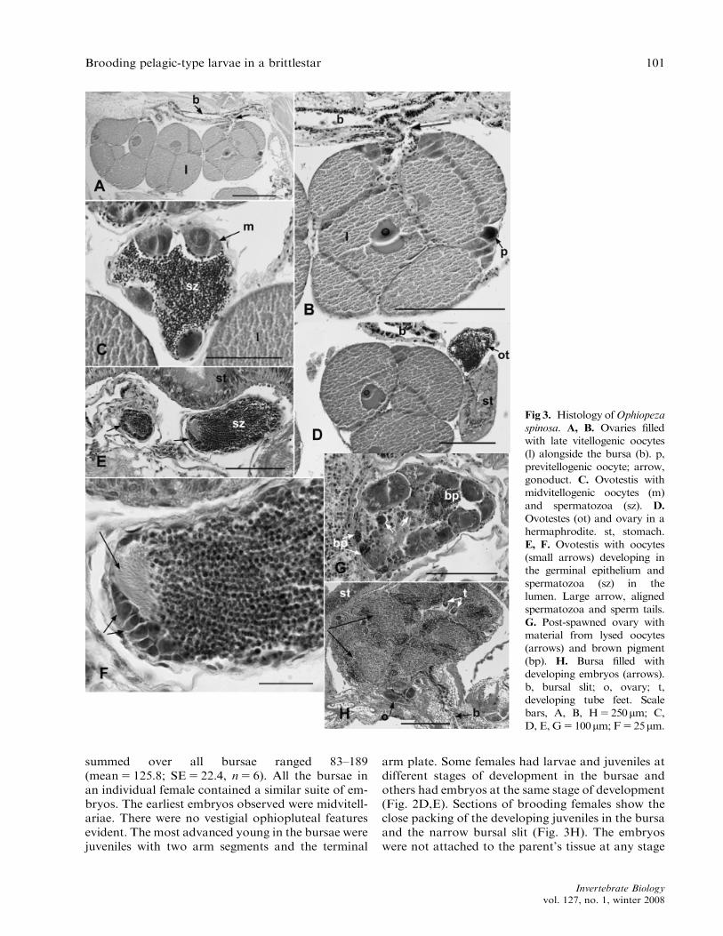
summed over all bursae ranged 83–189(mean5 125.8; SE5 22.4, n5 6). All the bursae inan individual female contained a similar suite of em-bryos. The earliest embryos observed were midvitell-ariae. There were no vestigial ophiopluteal featuresevident. The most advanced young in the bursae werejuveniles with two arm segments and the terminal
arm plate. Some females had larvae and juveniles atdifferent stages of development in the bursae andothers had embryos at the same stage of development(Fig. 2D,E). Sections of brooding females show theclose packing of the developing juveniles in the bursaand the narrow bursal slit (Fig. 3H). The embryoswere not attached to the parent’s tissue at any stage
Fig 3. Histology ofOphiopeza
spinosa. A, B. Ovaries filled
with late vitellogenic oocytes
(l) alongside the bursa (b). p,
previtellogenic oocyte; arrow,
gonoduct. C. Ovotestis with
midvitellogenic oocytes (m)
and spermatozoa (sz). D.
Ovotestes (ot) and ovary in a
hermaphrodite. st, stomach.
E, F. Ovotestis with oocytes
(small arrows) developing in
the germinal epithelium and
spermatozoa (sz) in the
lumen. Large arrow, aligned
spermatozoa and sperm tails.
G. Post-spawned ovary with
material from lysed oocytes
(arrows) and brown pigment
(bp). H. Bursa filled with
developing embryos (arrows).
b, bursal slit; o, ovary; t,
developing tube feet. Scale
bars, A, B, H5250mm; C,
D, E, G5100mm; F525mm.
Brooding pelagic-type larvae in a brittlestar 101
Invertebrate Biologyvol. 127, no. 1, winter 2008

of development. Newly metamorphosed juvenileshad a pentagonal shape and a mean disc diameterof 283mm (SE5 7.2; n5 14). The similar size of theeggs and juveniles indicates that development doesnot depend on extraembryonic (post-vitellogenic) nu-trition. The nutrients laid down in the egg supportdevelopment to the crawl-away juvenile stage. Thejuveniles were strongly pigmented an orange-amber
color due to the presence of maternal reserves in theirstomach (Fig. 4D,E).
Members ofO. spinosa develop through a vitellarialarva (Fig. 4). When released from the bursa, the lar-vae swam in culture dishes in the same manner as thevitellariae of non-brooding species. Development ofembryos reared in vitro was identical to that of em-bryos in the bursa. The fully developed larva is a
Fig 4. Vitellaria and juveniles
ofOphiopeza spinosa. A.Oral
and B. aboral views of
vitellaria larva showing the
location of transverse
ciliated bands: the anterior
ciliated band (acb) on the
pre-oral lobe (po), mid-
ciliated band (mcb), and
posterior ciliated band (pcb).
Developing juvenile arm
buds numbered I–V. C.
Advanced vitellaria, the pre-
oral lobe is reduced and the
primary pair of buccal podia
(pp) is evident. D. First
juvenile stage with develop-
ing terminal arm plates (tap).
E. Juvenile with one arm
segment and the terminal
arm plate (tap). s, developing
spines. F. Advanced juvenile
reared in vitro with the first
and second arm segments
(1as, 2as), and the terminal
arm plate (tap). ls, lateral
arm spines; s surface spines.
Scale bar, 100mm.
102 Byrne, Cisternas, & O’Hara
Invertebrate Biologyvol. 127, no. 1, winter 2008

typical vitellaria, with three transverse ciliary bandsand the developing juvenile rudiment in the mid-lar-val region. The ciliary bands were located on prom-inent epithelial elevations. In advanced larvae, thefirst pair of buccal podia was evident in the mouthregion of the developing juvenile (Fig. 4C). The no-menclature of the developing arm buds used in Fig. 4follows the numbering system of Brooks & Grave(1899).
Three ciliary bands cross the aboral side of the vi-tellaria: the anterior, mid-, and posterior ciliarybands. The anterior ciliary band loops around thepre-oral lobe (Fig. 4A,B). The mid-ciliary band ex-tended from the right oral side between developingjuvenile arm buds IV–V, across the aboral surface tothe left oral side between arm buds I and II (Fig.4A,B). This ciliary band has a distinct curve in itspath on the aboral surface (Fig. 4B). The posteriorciliary band bordered the edge of the posterior larvalregion aborally and extended orally, in the regionsbetween arm buds II–III and III–IV (Fig. 4A,B).
Vitellariae metamorphose in the bursa by resorp-tion of the larval body. Larvae reared in vitro settledonto the bottom of the culture dishes with their pri-mary podia, followed by metamorphosis. As the ter-minal arm plates formed, the early pentagonaljuveniles became star-shaped (Fig. 2D). The mouthskeleton was not fully formed. During development,maternal reserves are sequestered to the developinggut, giving the central region an orange color (Fig.4D–F). These juveniles also had small spots of redpigment scattered over the aboral surface. The firstpair of buccal podia was present.
Juveniles with one arm segment and the terminalarm plate were the most common stage seen in thebursae (Fig. 2E,F). As they developed, the arms in-creased in length with addition of arm segments. Themost advanced juveniles in the bursae had two armsegments and the terminal plate (Fig. 4F). These ju-veniles still had extensive maternal reserves as indi-cated by the orange color of their stomach. Themouth was not yet open.
Larvae reared in vitro developed from the early vi-tellaria stage to the juvenile stage in 3 d. Juvenileswith three arm segments and the terminal arm platehad reduced pigment as their maternal reserves werediminished. Their mouth was open and the secondpair of buccal podia had developed. These juvenileswere competent to feed. Juveniles appear to leave thebursa with two to three arm segments, but this wasnot observed. The aboral surface of exotrophic juve-niles was covered with thorny spines that extendedalong the mid-dorsal aboral surface of the arms andon the oral shields (Fig. 4F). The lateral arm spines
also had thorny projections. The armature of the discand spines are juvenile characters as they are notpresent in the adults.
Discussion
The Ophiodermatidae is an important tropicalophiuroid family comprising 23 genera with one ge-nus in the tropical Atlantic and ten genera in theIndo-West Pacific (Clark & Rowe 1971; Hendler etal. 1995). Their members are scavengers and preda-tors, and are among the largest ophiuroids on coralreefs (disc diameter 445mm) (Morin 1988). As typ-ical of brooding ophiuroids, specimens of Ophiopezaspinosa are small (disc diametero10mm) in compar-ison with confamilial ophiodermatids (Hendler 1991;Hendler & Tran 2001). Several echinoderm taxa onthe GBR have been discovered to be species com-plexes, with new species discerned by differences inmorphology, life history, and molecular phylogeny(O’Hara et al. 2004; Byrne & Walker 2007). Severalmorphs appear to exist within O. spinosa and thesemay prove to be different species (T. O’Hara, unpubl.data). Examination of populations of O. spinosa forthe presence or the absence of brooding, across itsbroad range, may aid in distinguishing cryptic spe-cies. Robust taxonomy is required to confirm the ap-parently broad distribution of this ophiuroid in theabsence of a dispersive stage. The problem of thepresence of cryptic species has been highlighted forother pan-Indo-West Pacific echinoderms and mol-luscs (O’Hara et al. 2004; O’Loughlin & Rowe 2005;Paulay & Meyer 2006; Byrne & Walker 2007).
Development through a non-feeding vitellaria ap-pears to be a synapomorphy for the Ophiodermat-idae and most species have a planktonic phase intheir life history (for a review, see Cisternas & Byrne2005). In addition to O. spinosa, brooding in theOphiodermatidae is known from Ophiopeza cylin-drica, two species of both Cryptopelta andOphiurochaeta, and one species of Ophioconis (Mor-tensen 1924, 1925, 1933a,b; Hotchkiss 1982; Byrne1991; Hendler 1991). Most of these are temperatespecies, with the exception of Cryptopelta granuliferafrom the Indo-West Pacific and Ophiurochaeta sp.from the Caribbean (Mortensen 1933a; Hotchkiss1982). Only four shallow water ophiuroids from thetropical Indo-West Pacific, comprising two species intwo families (Ophiodermatidae: O. spinosa, C.granulifera; Amphiuridae: Amphiura constricta,Amphipholis squamata), are known to brood theiryoung (Mortensen 1933a; Fell 1946; Clark & Rowe1971; Falkner & Byrne 2006). Detailed examinationof the shallow water ophiuroid fauna of the GBR
Brooding pelagic-type larvae in a brittlestar 103
Invertebrate Biologyvol. 127, no. 1, winter 2008

failed to locate other brooding species (M. Byrne,unpubl. data). In contrast, brooding ophiuroids arecommon in shallow water reef and back reef habitatsin the Caribbean (Hendler 1979; Hendler & Littman1986; Byrne 1991). Brooding has evolved indepen-dently numerous times in Caribbean ophiuroids andincludes approximately ten species from five families(Mortensen 1920; Hendler 1979; Hotchkiss 1982;Hendler & Littman 1986; Byrne 1989, 1991; Schoppe& Holl 1994). The rationale underlying this differ-ence in life-history evolution of the ophiuroid faunaof the two coral reef regions is not known.
The link between small size, hermaphroditism, andbrooding in ophiuroids has been noted for some time(Mortensen 1920; Hendler 1991). These features werealso exhibited in O. spinosa. Small and large individ-uals of O. spinosa were largely male and female, re-spectively, and so this ophiuroid is protandrous. Thedetection of ovotestes and minute testes in histolog-ical sections indicated that the switch from reproduc-tion as a male to a female involves a transitional stageof simultaneous hermaphrodism, similar to that de-scribed for several other brooding ophiuroids (Hen-dler 1979; Schoppe & Holl 1994). Some largespecimens of O. spinosa that appeared to be femalehad minute testes, and so retained some capacity toproduce sperm. A similar trend in increased alloca-tion to ovary production with increasing size occursin the brooding Caribbean ophiuroid, Ophiolepispaucispina (M. Byrne, unpubl. data). In this species,however, simultaneous hermaproditism is readily de-tected (Byrne 1989). Most ophiuroids that broodtheir young are simultaneous hermaphrodites (Mor-tensen 1920; Byrne 1991, 1994; Hendler 1991).
Oogenesis in O. spinosa involved synchronous de-velopment of groups of oocytes, followed by theirrelease into the bursa. The eggs are fertilized in thebursa, presumably by sperm drawn in by the respi-ratory current created by this structure, althoughself-fertilization remains a possibility. Through thebrooding season, release of eggs may be triggered bydeparture of clutches of juveniles. Very early embry-os and juveniles were not present in the same bursae.Embryogenesis and gametogenesis occurred in par-allel in O. spinosa, a feature described for severalother brooders (e.g., O. paucispina), but contrastswith Ophionereis olivacea where gametogenesis, ga-mete release, and incubation of progeny are tempo-rally separate events (Byrne 1989, 1991; Hendler1991). Partly spawned and spent gonads in O. spinosahad material from lysed oocytes and an accumulationof a brown lipid-like pigment. These lipfuscin-like de-posits occur in the ovaries of other ophiuroids at theend of the spawning season (Falkner & Byrne 2003).
Except for A. squamata and Ophionotus hexactis,which have small eggs (120 and 200mm diameter, re-spectively) and support their embryos with extraem-bryonic nutrition (Mortensen 1921; Turner &Dearborn 1979; Walker & Lesser 1989), mostophiuroids that brood their young have large eggs(4001mm diameter) and the embryos are not depen-dent on exogenous nutrients (Byrne 1991, 1994; Hen-dler 1991). The egg inO. spinosa (280mm diameter) isone of the smallest known for brooding ophiuroidsand is similar in size to those of ophiodermatids withpelagic larvae (Ophioderma brevispinum: 300mm di-ameter; Ophiarachnella gorgonia: 320mm diameter)(Hendler 1991; Cisternas & Byrne 2005). Juveniles ofO. spinosa appear to leave the parent at the two tothree arm segment stage, similar to that seen in O.olivacea, but considerably smaller than that in otherspecies, where advanced juveniles are retained in thebursae well beyond metamorphosis (seven to 12 armsegment juveniles) (Byrne 1989, 1991; Hendler 1991).Release of large juveniles is likely to enhance the sur-vival of newly independent juveniles (Hendler 1991).The evolution of an incubatory life history inO. spin-osa is not associated with release of advanced juve-niles. At a disc diameter of 283mm and with two tothree arm segments, juveniles of O. spinosa are onlyslightly larger than the newly settled juveniles ofophiodermatids with pelagic vitellariae (Cisternas &Byrne 2005).
Juveniles of O. spinosa have a prominent spinouscover over the disc and the lateral arm spines havethorny projections. These features are not present inadults and are likely to be defensive structures for thejuveniles to deter predators during the early benthicstage. Newly settled juveniles of several ophiuroidspecies have a similar armature, also putatively defen-sive (Turon et al. 2000; Stohr 2005; Falkner & Byrne2006). As seen here in O. spinosa, juvenile ophiuroidsoften look different from the adults and lack the de-fining morphological characters required for identifi-cation to species (Stohr 2005; Falkner & Byrne 2006).
In the Type II (ophiopluteus1vitellaria) pattern ofophiuroid development, the vitellaria is the metamor-phic larval form, contrasting with the Type I pattern(ophiopluteus only), where the ophiopluteus is themetamorphorphic larva (Byrne & Selvakumarasw-amy 2002; Cisternas & Byrne 2005; Selvakumarasw-amy & Byrne 2006). Development in O. spinosafollows the Type II pattern, as characteristic of theOphiodermatidae (Cisternas & Byrne 2005). A recentstudy, however, showed that some ophiuroids havefeatures of both patterns in their development, indi-cating that there may be a continuum of develop-mental patterns in the Ophiuroidea and that the Type
104 Byrne, Cisternas, & O’Hara
Invertebrate Biologyvol. 127, no. 1, winter 2008

I–Type II dichotomy may not be appropriate(Selvakumaraswamy & Byrne 2006).
The feeding ophiopluteus larva is considered to bethe ancestral larval form for the Ophiuroidea(Strathmann 1978). Several species with non-feedinglecithotrophic development have vestiges of thefeeding pluteus in their development, providing alink with an ancestral feeding larva (Selvakumarasw-amy & Byrne 2000, 2004). The ciliary bands in reducedophioplutei and vitellaria larvae are considered to haveoriginated from the ciliary bands of an ancestral feed-ing ophiopluteus (Mladenov 1985; Selvakumaraswamy& Byrne 2000, 2006; Cisternas & Byrne 2005;Fourgon et al. 2005). The larva of O. spinosa is typ-ical of ophiuroid vitellariae in possessing three well-developed ciliary bands and a central region wherethe juvenile rudiment develops. Vestigial ophioplu-teal features were not evident. The ciliary bands ofO.spinosa are positioned on prominent epithelial eleva-tions, a feature that facilitates efficient swimming(Emlet 1991). In contrast, the short-lived pelagicvitellaria larva of the sympatric ophiodermatidO. gorgonia has reduced ciliary bands that are notpositioned on prominent epithelial elevations (Cis-ternas & Byrne 2005). These features are suggested toreflect a decrease in selection for swimming capacityin the larva of O. gorgonia and prompt settlement(Cisternas & Byrne 2005). The earliest stage encoun-tered in the bursa of O. spinosa was mid-vitellariaand, based on the development in the sympatric spe-cies O. gorgonia, these larvae are estimated to be 3 dold. Development to the juvenile stage inO. spinosa isestimated to take B5 d.
Ophiuroid bursae are densely ciliated, thin-walled,body-wall invaginations used for respiration and areconsidered to be preadaptive features for the evolu-tion of brooding (Hendler 1979). In this scenario, theshift to brooding involved a relatively simple changein reproduction: the retention of eggs in the bursarather than release of eggs through the bursal slit asoccurs in broadcast spawners. Several ophiuroidsthat brood their young have a number of vestigiallarval features in their development (Mortensen1921; Fell 1946; Byrne 1991). Intragonadal larvaeoccur in O. hexactis, and intrabursal larvae havebeen observed in A. squamata and O. olivacea (Mor-tensen 1921; Fell 1946; Byrne 1991). The intragonad-al larva in O. hexactis develops through arudimentary pluteus stage with a ciliary band andlarval skeleton, and the intrabursal larva in A. squa-mata is minute and highly reduced (Mortensen 1921;Fell 1946). The intrabursal larvae in O. olivacea lackciliary bands, although they can swim due to theirciliary cover (Byrne 1991). In contrast to these three
species, the brooded larvae in O. spinosa are welldeveloped, and their morphology and behavior aretypical of pelagic larvae.
Ophiopeza spinosa is the first brooding ophiuroidknown to incubate a pelagic-type larva in its bursae.This larva provides a connection to an ancestral formthat had a dispersive life history and indicates thatthe switch to benthic development by O. spinosamayhave been an evolutionarily recent change. It also in-dicates that the loss of pelagic larval characters in theontogeny of other ophiuroids that brood their youngmay have occurred following the evolution of intra-bursal development. Ciliary bands are not needed inthe bursa, as evidenced by the reduced larva inO. olivacea. The reduced ciliary bands of the vitellariainO. gorgonia, however, show that morphological fea-tures used for larval swimming can be reduced withina planktonic life history (Cisternas & Byrne 2005).
The presence of a functional vitellaria larva andjuveniles in the bursae of O. spinosa is similar to theobservation of functional brachiolaria larvae and ju-veniles in the gonads of Cryptasterina species, vivip-arous asterinid sea stars that give birth to juveniles(Byrne 2005). Like that seen in O. spinosa, the larvaeof these asterinids can develop in vitro to the juvenilestage with no dependence on extraembryonic nutri-tion. These observations indicate the potential for areversal back to a pelagic dispersive life stage byO. spinosa and the Cryptasterina species. The brood-ing sea star, Pteraster militaris, releases some of itsprogeny as larvae and retains others to the juvenilestage (McClary & Mladenov 1989). Pteraster tess-elatus may have made the complete reversal to apelagic larval stage from a brooded one (McEdward& Janies 1993).O. spinosa, Cryptasterina species, andP. militaris are thus far the only echinoderms with thepotential to both brood and broadcast their young.
Acknowledgments. An Australian Research CouncilDiscovery Grant to M. Byrne and grant from theAustralian Geographic Society to T. O’Hara supportedthis research. P. Selvakumaraswamy, J. Lee, and T.Prowse provided technical assistance. Thanks are due toS. Uthicke and B. Schaffelke for assistance with specimencollection. The Directors and Managers of the LizardIsland Research Station, a facility of the AustralianMuseum, are thanked for assistance and use of facilities.Two reviewers are thanked for helpful comments.
References
Brooks WK & Grave C 1899. Ophiura brevispina. Mem.
Nat. Acad. Sci. 8: 79–100.
Brooding pelagic-type larvae in a brittlestar 105
Invertebrate Biologyvol. 127, no. 1, winter 2008

ByrneM 1989. Ultrastructure of the ovary and oogenesis in
the ovoviviparous brittlestar Ophiolepis paucispina
(Echinodermata: Ophiuroidea). Biol. Bull. 176: 79–95.
FFF 1991. Reproduction, development and population
biology of the Caribbean ophiuroid Ophionereis oliv-
acea, a protandric hermaphrodite that broods its young.
Mar. Biol. 111: 387–399.
FFF 1994. Class Ophiuroidea. In: Microanatomy of In-
vertebrates, Vol. 14, Echinodermata. Harrison F & Chia
FS, eds., pp. 247–343. Wiley-Liss, New York, NY.
FFF 2005. Viviparity in the sea star Cryptasterina hyst-
era (Asterinidae)—conserved and modified features in
reproduction and development. Biol. Bull. 208: 81–91.
Byrne M & Selvakumaraswamy P 2002. Phylum Echi-
nodermata: Ophiuroidea. In: Atlas of Marine Inverte-
brate Larvae. Young CM & Sewell ME, eds., pp. 483–
498. Academic Press, London, UK.
ByrneM&Walker SJ 2007. Distribution and reproduction
new intertidal Aquilonastra and Cryptasterina species
(Asterinidae) from One Tree Island, Southern Great
Barrier Reef. Bull. Mar. Sci. 81: 209–218.
Byrne M, Cisternas P, Hoggett A, O’Hara T, & Uthicke S
2004a. Diversity of echinoderms at Raine Island, Great
Barrier Reef. In: Echinoderms: Munchen. Heinzeller T
& Nebelsick JH, eds., pp. 159–164. Munchen, Taylor &
Francis Group, London, UK.
Byrne M, Smoothey A, Hoggett A, & Uthicke S 2004b.
Population biology of shallow water ophiuroids
and holothuroids at Raine Island and Moulter Cay,
Northern Great Barrier Reef. In: Echinoderms. Heinz-
eller T & Nebelsick JH, eds., pp. 165–170. Munchen,
Taylor & Francis Group, London, UK.
Cisternas P & Byrne M 2005. Larval development in the
ophiuroid Ophiarachnella gorgonia (Ophiodermatidae):
evidence for developmental heterochronies between spe-
cies with type I and type II development. Can. J. Zool.
83: 1067–1079.
Cisternas P, Selvakumaraswamy P, & Byrne M 2004. Evo-
lution of development and the Ophiuroidea—revisited.
In: Echinoderms. Heinzeller T & Nebelsick JH, eds., pp.
521–526. Munchen, Taylor & Francis Group, London,
UK.
Clark AM & Rowe FWE 1971. Monograph of shallow-
water Indo-West Pacific echinoderms. Trustees of the
British Museum (Natural History), London, UK: 238
pp. 100 figs 31 pls.
Emlet RB 1991. Functional constraints on the evolution of
larval forms of marine invertebrates: experimental and
comparative evidence. Am. Zool. 31: 707–725.
Endean R 1957. The biogeography of Queensland’s
shallow water echinoderm fauna (excluding Crinoidea)
with a re-arrangement of the faunistic provinces of
tropical Australia. Aust. J. Mar. Freshwater Res. 8:
233–273.
Falkner I & Byrne M 2003. Reproduction of Ophiactis re-
siliens (Echinodermata: Ophiuroidea) in New South
Wales with observations on recruitment. Mar. Biol.
143: 459–466.
FFF 2006. Skeletal characters for identification of juve-
nile Ophiactis resiliens and Amphiura constricta (Echi-
nodermata): cryptic ophiuroids in turf habitat. J. Mar.
Biol. Assoc. UK 86: 1199–1207.
Fell B 1946. The embryology of the viviparous ophiuroid
Amphipholis squamata. Trans. R. Soc. NZ 75: 419–464.
FourgonD, Eeckhaut I, VaitilingonD, & JangouxM 2005.
Lecithotrophic development and metamorphosis in the
Indo-West Pacific brittle star Ophiomastix venosa (Echi-
nodermata: Ophiuroidea). Invertebr. Reprod. Dev. 47:
155–165.
Guille AL, Laboute P, &Menou JL 1986. Guide des etoiles
de mer, oursins et autres echinoderms du lagon de No-
uvelle-Caledonie. ORSTOM, Paris, France.
Hendler G 1979. Sex-reversal and viviparity in Ophiolepis
kieri, n. sp., with notes on viviparous brittlestars from
the Caribbean (Echinodermata: Ophiuroidea). Proc.
Biol. Soc. Wash. 92: 783–795.
FFF 1991. Echinodermata: Ophiuroidea. In: Reproduc-
tion of Marine Invertebrates, Vol. VI, Echinoderms and
Lophophorates. Giese AC, Pearse JS, & Pearse VB, eds.,
pp. 356–479. Boxwood Press, Pacific Grove, CA.
Hendler G & Littman BS 1986. The ploys of sex: relation-
ships among the mode of reproduction, body size and
habitats of coral-reef brittlestars. Coral Reefs 5: 31–42.
Hendler G & Tran LU 2001. Reproductive biology of a
deep-sea brittle star Amphiura carchara (Echinodermata:
Ophiuroidea). Mar. Biol. 138: 113–123.
Hendler G, Miller JE, Pawson DL, & Kier PM 1995. Sea
stars, Sea Urchins, and Allies. Echinoderms of the Flor-
ida and the Caribbean. Smithsonian Institution Press,
Washington, DC.
Hotchkiss FHC 1982. To have embryos in the bursa in
Ophiuroidea (Echinodermata) from Carrie Bow Cay,
Belize. Smithsonian Contrib. Mar. Sci. 12: 387–412.
Le Gac M, Feral JP, Poulin E, Veyret M, & Chenuil A
2004. Identification of allopatric clades in the cosmopol-
itan ophiuroid species complex Amphipholis squamate
(Echinodermata). The end of a paradox? Mar. Ecol.
Prog. Ser. 278: 171–178.
Lessios HA, Kessing BD, & Robertson DR 1998. Massive
gene flow across the world’s most potent marine
biogeographic barrier. Proc. R. Soc. Lond. B 265:
583–588.
McClary DJ & Mladenov PV 1989. Reproductive pattern
in the brooding and broadcasting sea star Pteraster mi-
litaris. Mar. Biol. 103: 531–540.
McEdward LR & Janies DA 1993. Life cycle evolution in
Asteroids: What is a larva? Biol. Bull. 184: 255–268.
Mladenov PV 1985. Development and metamorphosis of
the brittle star Ophiocoma pumila: evolutionary and eco-
logical implications. Biol. Bull. 168: 285–295.
Morin JG 1988. Picivorous behavior and activity patterns
in the tropical ophiuroid Ophirachna incrassata
(Ophiuroidea: Ophiodermatidae). In: Echinoderm
Biology. Burke RD, Mladenov PV, Lambert P, &
Parsley RL, eds., pp. 401–407. Balkema, Rotterdam,
the Netherlands.
106 Byrne, Cisternas, & O’Hara
Invertebrate Biologyvol. 127, no. 1, winter 2008

Mortensen T 1920. On hermaphroditism in viviparous
ophiurids. Acta Zool. 1: 1–19.
FFF 1921. Studies of the development and larval forms
of echinoderms, pp. 171–177. Gad GEC, Copenhagen,
Denmark.
FFF 1924. Papers from Dr Mortensen’s Pacific expedi-
tion 1924. Echinoderms of New Zealand and the Auck-
land–Campbell Islands II. Ophiuroidea. Videnks. Medd.
Dan. Naturhist. Foren. 77: 91–177.
FFF 1925. Echinodermes du Maroc et de Mauritanie.
Bull. Biol. Soc. Sci. Nat. Maroc. 5: 178–187.
FFF 1931. Contributions to the study of the develop-
ment and larval forms of echinoderms. I–II. D. Kgl.
Danske. Vidensk. Selsk. Skrifter. Naturv. Og. Math. 4:
1–39.
FFF 1933a. Papers from Dr Mortensen’s Pacific expe-
dition 1914–16 LXIII. Biological observations on
ophiuroids, with descriptions of two new genera and
four new species. Vidensk. Medd. Dan. Naturhist.
Foren. 93: 171–194.
FFF 1933b. Papers from Dr Mortensen’s Pacific expe-
dition 1914–16 LXV. Echinoderms of South Africa (As-
teroidea and Ophiuroidea). Vidensk. Medd. Dan.
Naturhist. Foren. 93: 215–400.
Oak T & Scheibling RE 2006. Tidal activity pattern and
feeding behaviour of the ophiuroidOphiocoma scolopen-
drina on a Kenyan reef flat. Coral Reefs 25: 213–222.
O’Hara TD, Byrne M, & Cisternas PA 2004. The Ophio-
coma erinaceus complex: another case of cryptic speciat-
ion in echinoderms. In: Echinoderms: Munchen.
Heinzeller T & Nebelsick JH, eds., pp. 537–542. Taylor,
Francis Group, London, UK.
O’Loughlin PM&Rowe FWE 2005. A new asterinid genus
from the Indo-West Pacific region, including five new
species (Echinodermata: Asteroidea: Asterinidae). Mem.
Mus. Vic. 62: 181–189.
PaulayG&Meyer C 2006. Dispersal and divergence across
the greatest ocean region: do larvae matter? Integr.
Comp. Biol. 46: 269–281.
Podolsky RD & McAlister JS 2005. Developmental plas-
ticity inMacrophiothrix brittlestars: are morphologically
convergent larvae also convergently plastic? Biol. Bull.
209: 127–138.
Rowe FWE 1985. Preliminary analysis of distribution pat-
terns of Australia’s non-endemic, tropical echinoderms.
In: Proceedings of the Fifth International Echinoderms
Conference. Keegan BF & O’Connor BDS, eds., pp. 91–
98. Balkema, Rotterdam, the Netherlands.
Schoppe S & Holl A 1994. Ophiothrix n. sp. (Ophiuroidea:
Ophiotrichidae) from Columbia, a protandric hermaph-
rodite that broods its young. In: Echinoderms Through
Time. David B, Guille A, Feral JP, & Roux M, eds., pp.
471–475. Balkema, Rotterdam, the Netherlands.
Selvakumaraswamy P & Byrne M 2000. Vestigial
ophiopluteal structures in the lecithotrophic ophiuroid
Ophioneries schayeri. Biol. Bull. 198: 379–386.
FFF 2004. Metamorphosis and development evolution
in Ophionereis (Echinodermata: Ophiuroidea). Mar.
Biol. 145: 87–99.
FFF 2006. Evolution of larval form in ophiuroids, in-
sights from the metamorphic phenotype of Ophiothrix
(Echinodermata: Ophiuroidea). Evol. Dev. 8: 183–190.
Sloan NA 1982. Size and structure of echinoderm popula-
tions associated with different coexisting coral species at
Aldabra Atoll, Seychelles. Mar. Biol. 66: 67–75.
Sloan NA, Clark AM, & Taylor JD 1979. The echinoderms
of Aldabra and their habitats. Bull. Br. Mus. Nat. Hist.
(Zoology) 37: 81–128.
Smith AB 1997. Echinoderm larvae and phylogeny. Annu.
Rev. Ecol. Syst. 28: 219–241.
Stohr S 2005. Who’s who among baby brittle stars (Echi-
nodermata: Ophiuroidea): postmetamorphic develop-
ment of some North Atlantic forms. Zool. J. Linn.
Soc. 143: 543–576.
Strathmann RR 1978. The evolution and loss of feeding
larval stages of marine invertebrates. Evolution 32:
894–906.
Turner RI & Dearborn JH 1979. Organic and inorganic
composition of post-metamorphic growth stages of
Ophionotus hexactis (E.A. Smith) (Echinodermata:
Ophiuroidea) during intraovarian incubation. J. Exp.
Mar. Biol. Ecol. 36: 41–51.
Turon X, Codina M, Tarjuelo I, Uriz MJ, & Becerro MA
2000. Mass recruitment of Ophiothrix fragilis (Ophiuro-
idea) on sponges: settlement patterns and post-settlement
dynamics. Mar. Ecol. Prog. Ser. 200: 201–212.
Vail LL & Rowe FW 1988. Status of the genera Ophiopeza
and Ophiopsammus (Echinodermata: Ophiuroidea) in
Australian waters, with the description of a new species.
Proc. Linn. Soc. NSW 110: 267–288.
Uthicke S & Conand C 2005. Amplified fragment length
polymorphism (AFLP) analysis indicates the importance
of both asexual and sexual reproduction in the fi-
ssiparous holothurian Stichopus chloronotus (Aspidochi-
rotida) in the Indian and Pacific Ocean. Coral Reefs 24:
103–111.
Walker CW & Lesser MP 1989. Nutrition and develop-
ment of brooded embryos in the brittle star Amphipholis
squamata: do endosymbiotic bacteria play a role? Mar.
Biol. 103: 519–530.
Brooding pelagic-type larvae in a brittlestar 107
Invertebrate Biologyvol. 127, no. 1, winter 2008