BPPV A Review of Current Published Literature - CiteSeerX
11
From the SelectedWorks of Balasubramanian iagarajan February 17, 2012 BPPV A Review of Current Published Literature Balasubramanian iagarajan Available at: hp://works.bepress.com/drtbalu/37/
-
Upload
khangminh22 -
Category
Documents
-
view
2 -
download
0
Transcript of BPPV A Review of Current Published Literature - CiteSeerX

From the SelectedWorks of Balasubramanian Thiagarajan
February 17, 2012
BPPV A Review of Current Published LiteratureBalasubramanian Thiagarajan
Available at: http://works.bepress.com/drtbalu/37/
4/9/13 BPPV – Ent Scholar
entscholar.wordpress.com/article/bppv/ 1/10
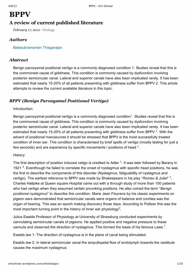
BPPVA review of current published literature
February 17, 2012 · Otology
Benign paroxysmal positional vertigo is a commonly diagnosed condition 1. Studies reveal that this isthe commonest cause of giddiness. This condition is commonly caused by dysfunction involvingposterior semicircular canal. Lateral and superior canals have also been implicated rarely. It has beenestimated that nearly 1520% of all patients presenting with giddiness suffer from BPPV 2. This articleattempts to review the current available literature in this topic.
Introduction:
Benign paroxysmal positional vertigo is a commonly diagnosed condition . Studies reveal that this isthe commonest cause of giddiness. This condition is commonly caused by dysfunction involvingposterior semicircular canal. Lateral and superior canals have also been implicated rarely. It has beenestimated that nearly 1520% of all patients presenting with giddiness suffer from BPPV . With theadvent of positional manoeuvres it should be stressed that BPPV is the most successfully treatedcondition of inner ear. This condition is characterised by brief spells of vertigo (mostly lasting for just afew seconds) and are experience by specific movements / positions of head .
History:
The first description of position induced vetigo is credited to Adler . It was later followed by Barany in1921 . Eventhough he failed to correlate the onset of nystagmus with specific head positions, he wasthe first to describe the components of this disorder (Nystagmus, fatiguability of nystagmus andvertigo). The earliest reference to BPPV was made by Shakespeare in his play “Romeo & Juliet” .Charles Hallpike at Queen square Hospital came out with a through study of more than 100 patientswho had vertigo when they assumed certain provoking positions. He also coined the term “Benignpositional nystagmus” to describe this condition. Marie Jean Flourens by his classic experiments onpigeon ears demonstrated that semicircular canals were organs of balance and cochlea was theorgan of hearing. This was an epoch making discovery those days. According to Politzer this was themost important turning point in the history of inner ear physiology .
Julius Ewalds Professor of Physiology at University of Strassburg conducted experiments bycannulating semicircular canals of pigeons. He applied positive and negative pressure to thesecannula and observed the direction of nystagmus. This formed the basis of his famous Laws .
Ewalds law 1: The direction of nystagmus is in the plane of canal being stimulated.
Ewalds law 2: In lateral semicircular canal the ampullopetal flow of endolymph towards the vestibulecauses the maximum nystagmus
Abstract
BPPV (Benign Paroxysmal Positional Vertigo)
1
2
3
4
3
5
6
7
Authors
Balasubramanian Thiagarajan

4/9/13 BPPV – Ent Scholar
entscholar.wordpress.com/article/bppv/ 2/10
Ewalds law 3: In posterior and superior semicircular canals ampullofugal flow of endolymph awayfrom the vestibule causes the maximum nystagmus.
Invention of Electron microscope some 30 years later opened up new avenues for studyingultrastructural architecture of inner ear. In 1954 Wersall demonstrated that each vestibular sensorycell had one kinocilium and many stereocilia. In the lateral canal crista the kinocilium was found onthe vestibule side of stereocilia; where as in the vertical canals the kinocilium was found on the canalside of stereocilia. This discovery went a long way in validating Ewald’s laws.
The exact relationship between the canal receptors and extraocular muscles became evident only in1960′s after experiments with cat’s labyrinth . Each receptor is connected to one ipsilateral and onecontralateral muscle.
Theories accounting for BPPV:
Schuknecht Theory:
Schuknecht proposed that BPPV is caused by loose otoconia from the utricle, which in certainpositions displaces the cupula of posterior semicircular canal. He went on to modify his theory latersuggesting that deposition of otoconia in the cupula of posterior canal could be the cause. He alsocoined the term cupulolithiasis. In a nutshell this theory proposes that calcium deposits becomeembedded on the cupula making the posterior semicircular canal sensitive to gravity .
Hall and Ruby theory :
This theory proposed jointly by Hall & Ruby suggests that deflection of cupula of posterior canal couldbe caused by debris in the posterior canal. This theory is also known as canal lithiasis theory.According to this theory the calcium deposits does not become adherent to the cupula of posteriorcanal but floats freely within the canal. Provoking head movements like looking up, down or rollingover to the affected side would cause giddiness and nystagmus.
Types of BPPV as described by Hall and Ruby:
Hall and Ruby while explaining their theory went on to describe two types of BPPV.
Type I BPPV: Nystagmus in this condition is fatigable and the calcium deposits are freely mobilewithin the cupula of posterior canal.
Type II BPPV: Nystagmus in this condition is nonfatiguing because the calcium deposits are fixed tothe cupula of posterior canal.
Features of BPPV (As described by Hall & Ruby)
1. Canalithiasis mechanism – This explains the latency of the nystagmus as a result of the timeneeded for motion of the material within the posterior canal to be initiated by the gravity.
2. Duration of the nystagmus – is correlated with the length of time required for the dense material toreach the lowest part of the posterior canal.
3. The vertical (upbeating) and torsional (superior poles of the eye beating towards the lowermostear). The nystagmus is more vertical when the patient looks away from the lowermost ear, andmoretorsional when looking towards the lowermost ear.
4. The reversal of nystagmus when the patient returns to the sitting position is due to retrogrademovement of material in the lumen of the posterior canal back towards the ampula, resulting in
8
9
10
11
:3
4/9/13 BPPV – Ent Scholar
entscholar.wordpress.com/article/bppv/ 3/10
ampulo petal deflection of the cupula.
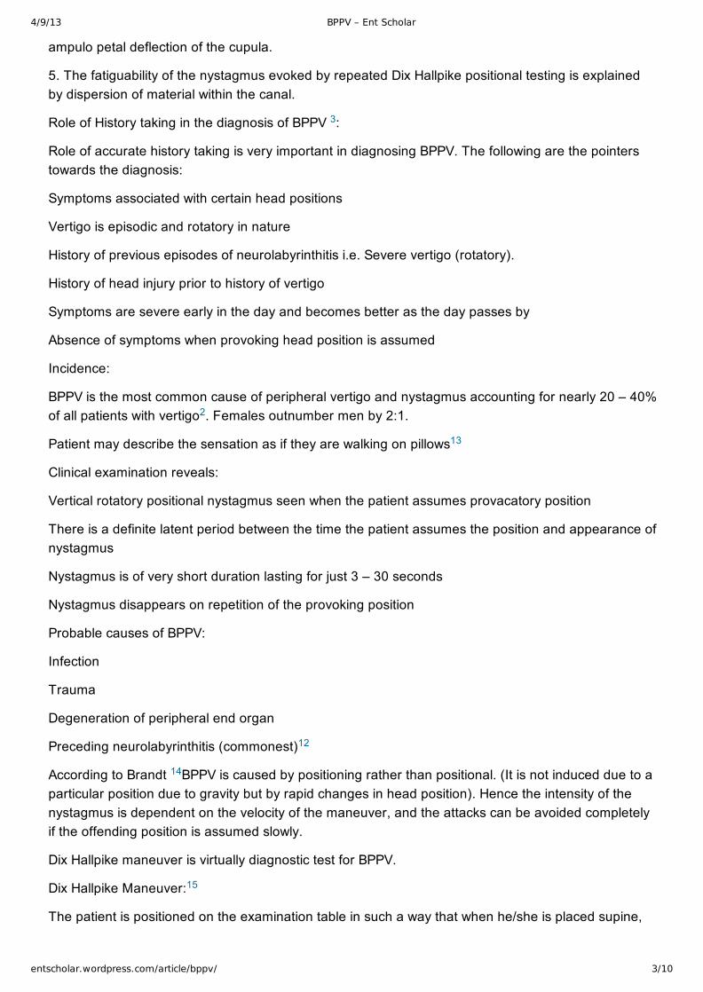
5. The fatiguability of the nystagmus evoked by repeated Dix Hallpike positional testing is explainedby dispersion of material within the canal.
Role of History taking in the diagnosis of BPPV :
Role of accurate history taking is very important in diagnosing BPPV. The following are the pointerstowards the diagnosis:
Symptoms associated with certain head positions
Vertigo is episodic and rotatory in nature
History of previous episodes of neurolabyrinthitis i.e. Severe vertigo (rotatory).
History of head injury prior to history of vertigo
Symptoms are severe early in the day and becomes better as the day passes by
Absence of symptoms when provoking head position is assumed
Incidence:
BPPV is the most common cause of peripheral vertigo and nystagmus accounting for nearly 20 – 40%of all patients with vertigo . Females outnumber men by 2:1.
Patient may describe the sensation as if they are walking on pillows
Clinical examination reveals:
Vertical rotatory positional nystagmus seen when the patient assumes provacatory position
There is a definite latent period between the time the patient assumes the position and appearance ofnystagmus
Nystagmus is of very short duration lasting for just 3 – 30 seconds
Nystagmus disappears on repetition of the provoking position
Probable causes of BPPV:
Infection
Trauma
Degeneration of peripheral end organ
Preceding neurolabyrinthitis (commonest)
According to Brandt BPPV is caused by positioning rather than positional. (It is not induced due to aparticular position due to gravity but by rapid changes in head position). Hence the intensity of thenystagmus is dependent on the velocity of the maneuver, and the attacks can be avoided completelyif the offending position is assumed slowly.
Dix Hallpike maneuver is virtually diagnostic test for BPPV.
Dix Hallpike Maneuver:
The patient is positioned on the examination table in such a way that when he/she is placed supine,
3
2
13
12
14
15
4/9/13 BPPV – Ent Scholar
entscholar.wordpress.com/article/bppv/ 4/10
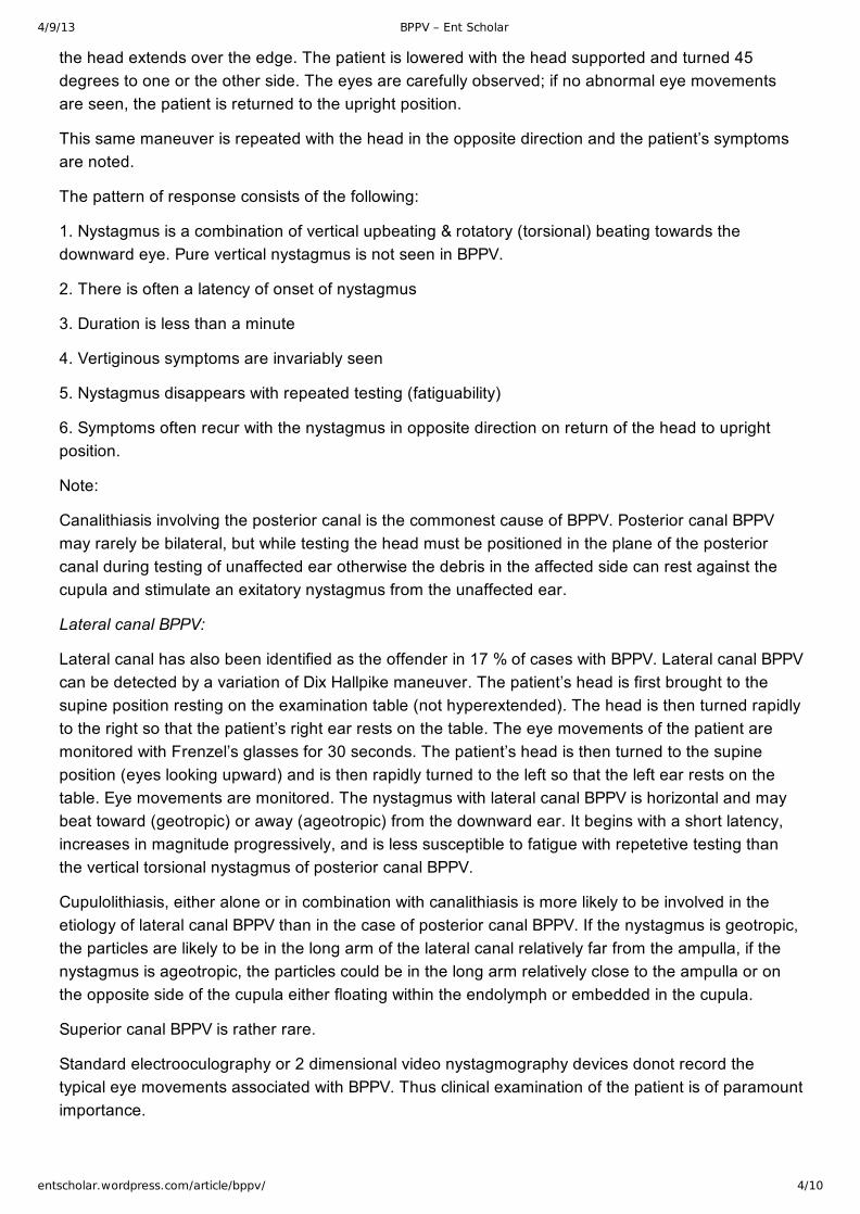
the head extends over the edge. The patient is lowered with the head supported and turned 45degrees to one or the other side. The eyes are carefully observed; if no abnormal eye movementsare seen, the patient is returned to the upright position.
This same maneuver is repeated with the head in the opposite direction and the patient’s symptomsare noted.
The pattern of response consists of the following:
1. Nystagmus is a combination of vertical upbeating & rotatory (torsional) beating towards thedownward eye. Pure vertical nystagmus is not seen in BPPV.
2. There is often a latency of onset of nystagmus
3. Duration is less than a minute
4. Vertiginous symptoms are invariably seen
5. Nystagmus disappears with repeated testing (fatiguability)
6. Symptoms often recur with the nystagmus in opposite direction on return of the head to uprightposition.
Note:
Canalithiasis involving the posterior canal is the commonest cause of BPPV. Posterior canal BPPVmay rarely be bilateral, but while testing the head must be positioned in the plane of the posteriorcanal during testing of unaffected ear otherwise the debris in the affected side can rest against thecupula and stimulate an exitatory nystagmus from the unaffected ear.
Lateral canal BPPV:
Lateral canal has also been identified as the offender in 17 % of cases with BPPV. Lateral canal BPPVcan be detected by a variation of Dix Hallpike maneuver. The patient’s head is first brought to thesupine position resting on the examination table (not hyperextended). The head is then turned rapidlyto the right so that the patient’s right ear rests on the table. The eye movements of the patient aremonitored with Frenzel’s glasses for 30 seconds. The patient’s head is then turned to the supineposition (eyes looking upward) and is then rapidly turned to the left so that the left ear rests on thetable. Eye movements are monitored. The nystagmus with lateral canal BPPV is horizontal and maybeat toward (geotropic) or away (ageotropic) from the downward ear. It begins with a short latency,increases in magnitude progressively, and is less susceptible to fatigue with repetetive testing thanthe vertical torsional nystagmus of posterior canal BPPV.
Cupulolithiasis, either alone or in combination with canalithiasis is more likely to be involved in theetiology of lateral canal BPPV than in the case of posterior canal BPPV. If the nystagmus is geotropic,the particles are likely to be in the long arm of the lateral canal relatively far from the ampulla, if thenystagmus is ageotropic, the particles could be in the long arm relatively close to the ampulla or onthe opposite side of the cupula either floating within the endolymph or embedded in the cupula.
Superior canal BPPV is rather rare.
Standard electrooculography or 2 dimensional video nystagmography devices donot record thetypical eye movements associated with BPPV. Thus clinical examination of the patient is of paramountimportance.
4/9/13 BPPV – Ent Scholar
entscholar.wordpress.com/article/bppv/ 5/10
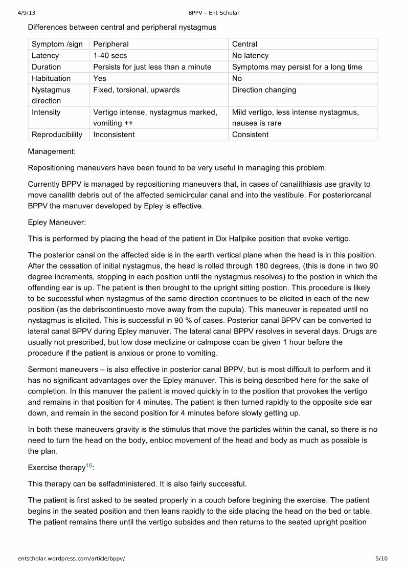
Differences between central and peripheral nystagmus
Symptom /sign Peripheral CentralLatency 140 secs No latencyDuration Persists for just less than a minute Symptoms may persist for a long timeHabituation Yes NoNystagmusdirection
Fixed, torsional, upwards Direction changing
Intensity Vertigo intense, nystagmus marked,vomiting ++
Mild vertigo, less intense nystagmus,nausea is rare
Reproducibility Inconsistent Consistent
Management:
Repositioning maneuvers have been found to be very useful in managing this problem.
Currently BPPV is managed by repositioning maneuvers that, in cases of canalithiasis use gravity tomove canalith debris out of the affected semicircular canal and into the vestibule. For posteriorcanalBPPV the manuver developed by Epley is effective.
Epley Maneuver:
This is performed by placing the head of the patient in Dix Hallpike position that evoke vertigo.
The posterior canal on the affected side is in the earth vertical plane when the head is in this position.After the cessation of initial nystagmus, the head is rolled through 180 degrees, (this is done in two 90degree increments, stopping in each position until the nystagmus resolves) to the postion in which theoffending ear is up. The patient is then brought to the upright sitting postion. This procedure is likelyto be successful when nystagmus of the same direction ccontinues to be elicited in each of the newposition (as the debriscontinuesto move away from the cupula). This maneuver is repeated until nonystagmus is elicited. This is successful in 90 % of cases. Posterior canal BPPV can be converted tolateral canal BPPV during Epley manuver. The lateral canal BPPV resolves in several days. Drugs areusually not prescribed, but low dose meclizine or calmpose ccan be given 1 hour before theprocedure if the patient is anxious or prone to vomiting.
Sermont maneuvers – is also effective in posterior canal BPPV, but is most difficult to perform and ithas no significant advantages over the Epley manuver. This is being described here for the sake ofcompletion. In this manuver the patient is moved quickly in to the position that provokes the vertigoand remains in that position for 4 minutes. The patient is then turned rapidly to the opposite side eardown, and remain in the second position for 4 minutes before slowly getting up.
In both these maneuvers gravity is the stimulus that move the particles within the canal, so there is noneed to turn the head on the body, enbloc movement of the head and body as much as possible isthe plan.
Exercise therapy :
This therapy can be selfadministered. It is also fairly successful.
The patient is first asked to be seated properly in a couch before begining the exercise. The patientbegins in the seated position and then leans rapidly to the side placing the head on the bed or table.The patient remains there until the vertigo subsides and then returns to the seated upright position
16
4/9/13 BPPV – Ent Scholar
entscholar.wordpress.com/article/bppv/ 6/10
remaining there until all symptoms subside. The maneuver is repeated toward the opposite sidecompleting one full repetition. Ten to twenty repetitions should be performed three times a day.Patients should be cautioned not to do this procedure forcibly as this could cause neck injuries.
It the patient has intense symptoms of giddiness / vomiting following exercise therapy then they needto be admitted and administered labyrinthine sedative and intravenous fluids.
Semont’s Liberatory maneuver:
This maneuver has been found to be helpful in managing BPPV.
This maneuver is begun with the patient seated on a couch. The patient is swung down quickly withthe offending ear down and head slightly inclined. If nystagmus appears, the patient is kept in thatposition until nystagmus disappears. Wait for 3 minutes.
If nystagmus doesnt appear the head is turned so that face is up 45 degrees from the horizontal.Nystagmus should appear. The patient is kept in this position for 23 minutes after nystagmus stops
The head and neck of the patient is held with two hands and is swung quickly to the opposite side.The speed of the head should be zero the moment it touches the table.
In patients with classic BPPV, a rotatory nystagmus will develop with the fast phase of nystagmusbeating towards the offending ear (the topmost ear). If the fast component of nystagmus beatstowards the bottom ear then in all probablity the diagnosis of BPPV is wrong.
If no nystagmus develops still, the head is moved slowly to 90 degrees facing up. The head is thenturned to the opposite direction to 135 degrees so that it is 45 degrees facing down below thehorizontal (with sick ear up). Nystagmus should occur.
If nystagmus is produced the patient is held in this position for full 5 minutes and then the patient isbrought back to the sitting position rather gradually and very slowly.
After this exercise patient is instructed to keep their head absolutely vertical in space during the next48 hours. If this maneuver is not successful then it can be repeated a week later.
Horizontal canal BPPV:
This is commonly identified when the patient is undergoing Dix Hallpike test. Sometimes it canbecome apparant after a successful canal repositioning maneuver for posterior canal BPPV. Vertigohappens to be more intense than that of posterior canal BPPV.
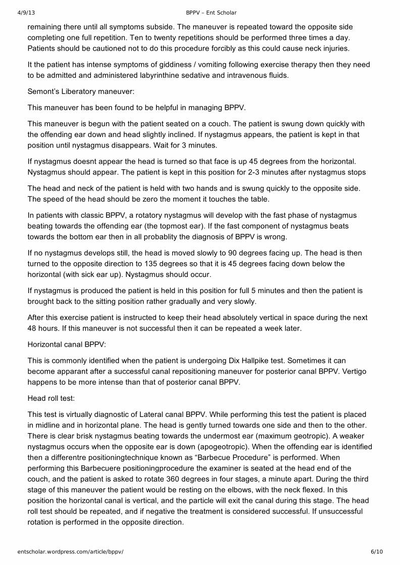
Head roll test:
This test is virtually diagnostic of Lateral canal BPPV. While performing this test the patient is placedin midline and in horizontal plane. The head is gently turned towards one side and then to the other.There is clear brisk nystagmus beating towards the undermost ear (maximum geotropic). A weakernystagmus occurs when the opposite ear is down (apogeotropic). When the offending ear is identifiedthen a differentre positioningtechnique known as “Barbecue Procedure” is performed. Whenperforming this Barbecuere positioningprocedure the examiner is seated at the head end of thecouch, and the patient is asked to rotate 360 degrees in four stages, a minute apart. During the thirdstage of this maneuver the patient would be resting on the elbows, with the neck flexed. In thisposition the horizontal canal is vertical, and the particle will exit the canal during this stage. The headroll test should be repeated, and if negative the treatment is considered successful. If unsuccessfulrotation is performed in the opposite direction.
4/9/13 BPPV – Ent Scholar
entscholar.wordpress.com/article/bppv/ 7/10
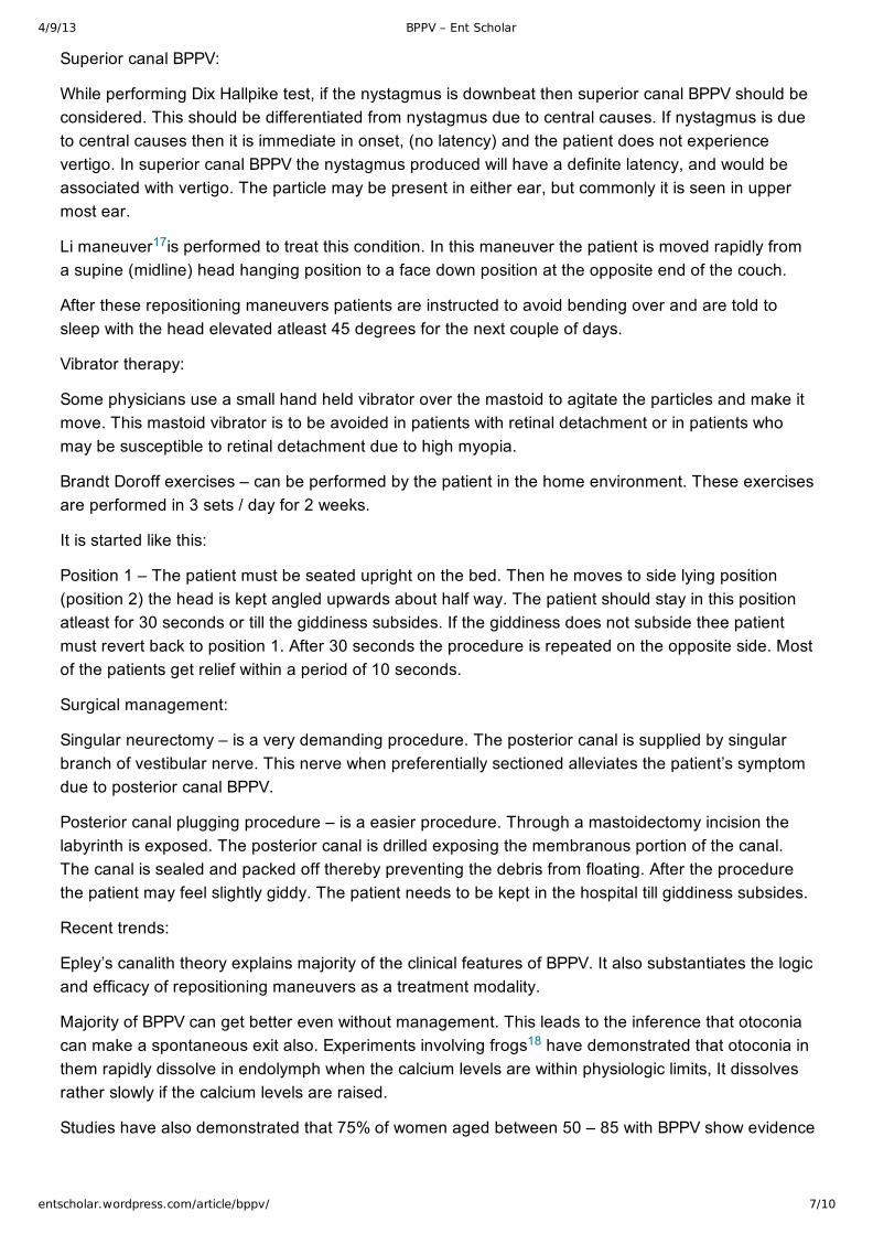
Superior canal BPPV:
While performing Dix Hallpike test, if the nystagmus is downbeat then superior canal BPPV should beconsidered. This should be differentiated from nystagmus due to central causes. If nystagmus is dueto central causes then it is immediate in onset, (no latency) and the patient does not experiencevertigo. In superior canal BPPV the nystagmus produced will have a definite latency, and would beassociated with vertigo. The particle may be present in either ear, but commonly it is seen in uppermost ear.
Li maneuver is performed to treat this condition. In this maneuver the patient is moved rapidly froma supine (midline) head hanging position to a face down position at the opposite end of the couch.
After these repositioning maneuvers patients are instructed to avoid bending over and are told tosleep with the head elevated atleast 45 degrees for the next couple of days.
Vibrator therapy:
Some physicians use a small hand held vibrator over the mastoid to agitate the particles and make itmove. This mastoid vibrator is to be avoided in patients with retinal detachment or in patients whomay be susceptible to retinal detachment due to high myopia.
Brandt Doroff exercises – can be performed by the patient in the home environment. These exercisesare performed in 3 sets / day for 2 weeks.
It is started like this:
Position 1 – The patient must be seated upright on the bed. Then he moves to side lying position(position 2) the head is kept angled upwards about half way. The patient should stay in this positionatleast for 30 seconds or till the giddiness subsides. If the giddiness does not subside thee patientmust revert back to position 1. After 30 seconds the procedure is repeated on the opposite side. Mostof the patients get relief within a period of 10 seconds.
Surgical management:
Singular neurectomy – is a very demanding procedure. The posterior canal is supplied by singularbranch of vestibular nerve. This nerve when preferentially sectioned alleviates the patient’s symptomdue to posterior canal BPPV.
Posterior canal plugging procedure – is a easier procedure. Through a mastoidectomy incision thelabyrinth is exposed. The posterior canal is drilled exposing the membranous portion of the canal.The canal is sealed and packed off thereby preventing the debris from floating. After the procedurethe patient may feel slightly giddy. The patient needs to be kept in the hospital till giddiness subsides.
Recent trends:
Epley’s canalith theory explains majority of the clinical features of BPPV. It also substantiates the logicand efficacy of repositioning maneuvers as a treatment modality.
Majority of BPPV can get better even without management. This leads to the inference that otoconiacan make a spontaneous exit also. Experiments involving frogs have demonstrated that otoconia inthem rapidly dissolve in endolymph when the calcium levels are within physiologic limits, It dissolvesrather slowly if the calcium levels are raised.
Studies have also demonstrated that 75% of women aged between 50 – 85 with BPPV show evidence
17
18
19

4/9/13 BPPV – Ent Scholar
entscholar.wordpress.com/article/bppv/ 8/10
of osteopenia / osteoporosis .
Role of VEMP’s in diagnosis of BPPV:
Measurement of vestibular evoked myogenic potential is good for measuring saccule and otolithfunction. In patients with vestibular neuronitis absense of cervical vemps is a good predictive indicatorfor non development of BPPV. It occured only if cervical VEMP is present. The recently identifiedocular vemp also measures utricle function and its presence could be seen as a recovery indicator inpatients with BPPV.
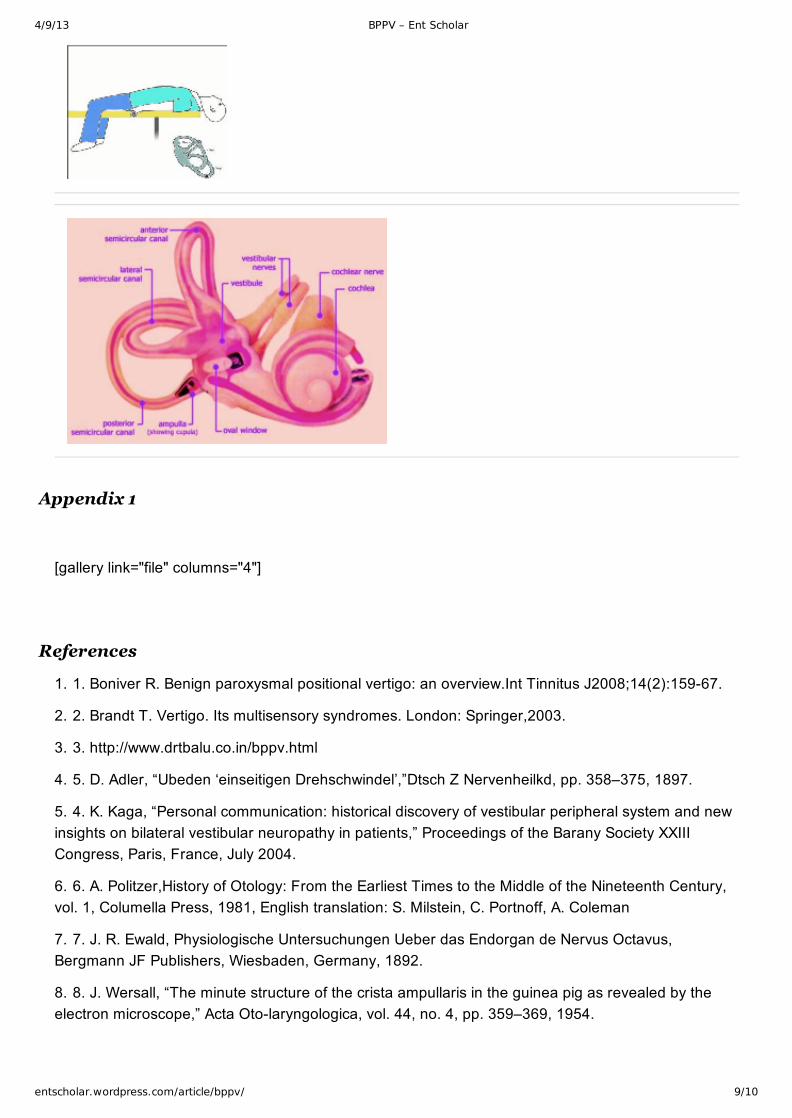
Anatomy of inner ear
Epley’s repositioning maneuver
Barbecue maneuver
Li maneuver
19
4/9/13 BPPV – Ent Scholar
entscholar.wordpress.com/article/bppv/ 9/10
[gallery link="file" columns="4"]
1. 1. Boniver R. Benign paroxysmal positional vertigo: an overview.Int Tinnitus J2008;14(2):15967.
2. 2. Brandt T. Vertigo. Its multisensory syndromes. London: Springer,2003.
3. 3. http://www.drtbalu.co.in/bppv.html
4. 5. D. Adler, “Ubeden ‘einseitigen Drehschwindel’,”Dtsch Z Nervenheilkd, pp. 358–375, 1897.
5. 4. K. Kaga, “Personal communication: historical discovery of vestibular peripheral system and newinsights on bilateral vestibular neuropathy in patients,” Proceedings of the Barany Society XXIIICongress, Paris, France, July 2004.
6. 6. A. Politzer,History of Otology: From the Earliest Times to the Middle of the Nineteenth Century,vol. 1, Columella Press, 1981, English translation: S. Milstein, C. Portnoff, A. Coleman
7. 7. J. R. Ewald, Physiologische Untersuchungen Ueber das Endorgan de Nervus Octavus,Bergmann JF Publishers, Wiesbaden, Germany, 1892.
8. 8. J. Wersall, “The minute structure of the crista ampullaris in the guinea pig as revealed by theelectron microscope,” Acta Otolaryngologica, vol. 44, no. 4, pp. 359–369, 1954.
Appendix 1
References

4/9/13 BPPV – Ent Scholar
entscholar.wordpress.com/article/bppv/ 10/10
9. 9. B. Cohen and J. I. Suzuki, “Annals of otology, rhinology and laryngology eye movements fromsemicircular canal stimulation in the cat,” Annals of Otology, Rhinology and Laryngology, vol. 73, pp.153–169, 1964.
10. 11. H. F. Schuknecht and R. R. F. Ruby, “Cupulolithiasis,” Advanced OtoRhinoLaryng, vol. 20,pp. 434–443, 1962.
11. 14. S. F. Hall, R. R. Ruby, and J. A. McClure, “The mechanics of benign positional vertigo,”Journal of Otolaryngology, vol. 8, pp. 151–158, 1979.
12. 15. Baloh RW, Honrubia V, Jacobsen K: Benign positional vertigo: Clinical and oculographicfeatures in 240 cases. Neurology 37: 371378,1987.
13. 17. Brandt T: Positional and positioning vertigo nystagmus. J Neurol Sci 995: 328, 1990.
14. 17. Brandt T: Positional and positioning vertigo nystagmus. J Neurol Sci 995: 328, 1990.
15. 18. M. R. Dix and C. S. Hallpike, “The pathology, sympomatology and diagnosis of certaincommon disorders of the vestibular system,” Annals of Otology, Rhinology and Laryngology, vol. 61,pp. 987–1016, 1952.
16. 19. Brandt T, Daroff RB: Physical therapy for benign paroxysmal positional vertigo. ArchOtolaryngol 106: 484, 1980.
17. 20. J. Li and H. Li, “New repositioning techniques for benign paroxysmal positional vertigo: the Lirepositioning manoeuvres,” Journal of Laryngology and Otology, vol. 124, no. 8, pp. 905–908, 2010.
18. 21. G. Zucca, S. Valli, P. Valli, P. Perin, and E. Mira, “Why do benign paroxysmal positionalvertigo episodes recover spontaneously?” Journal of Vestibular Research: Equilibrium andOrientation, vol. 8, no. 4, pp. 325–329, 1998.
19. 22. D. Vibert, M. Kompis, and R. Hausler, “Benign paroxysmal vertigo in older women may berelated to osteoporosis and osteopenia,” Annals of Otology, Rhinology and Laryngology, vol. 112, pp.885–889, 2003.