Diglycolamide-functionalized resorcinarene for rare earths ...
Upload
khangminh22Category
view
3download
0
Book of abstracts

Sponsors

Welcome
PRE’19 is the eighth event in the series of International Workshops covering a wide range ofresearch topics concerning the properties and applications of rare-earth ions in optoelectronics andphotonics. The series begun in Trento, Italy in May 2005 (PRE’05), followed by PRE’07 again inTrento, PRE’10 in Firenze (Italy), PRE’12 in Kyoto (Japan), PRE’14 in San Sebastián (Spain), PRE’16 inGreenville (USA) and PRE'17 in Roma (Italy).
The Photoluminescence in Rare Earths: Photonic Materials and Devices (PRE) workshop aims toprovide a global forum for material scientists, chemists and physicists to discuss and debate thestate of the art and future perspectives of rare earth based materials for optoelectronic andphotonic applications. Fundamental aspects, properties, and applications of photoluminescentmaterials are considered.
More than 700 scientists and students from more than 30 countries, in total, have attended theprevious Workshops. The average size of each event, around 100 people, makes for a collegialenvironment where students and world experts can mingle and talk in an informal and effectiveway.
We welcome you in PRE'19, to be held on 4-6 September 2019 in Nice, France, to discuss on yourmost recent findings and join the community of scholars advancing the field of light emission basedon rare-earth doped materials.
Looking forward to meeting you in the French Riviera !
Wilfried Blanc, Pieter Dorenbos, Fiorenzo Vetrone, PRE'19 Co-chairs
Giancarlo C. Righini, PRE'19 Honorary chair
https://pre19.sciencesconf.org/

Committees
PRE Steering Committee (2018-2019)
• John Ballato (USA) • Wilfried Blanc (France) • Dominik Dorosz (Poland) • Maurizio Ferrari (Italy) • Giancarlo C. Righini (Italy) • Setsuhisa Tanabe (Japan)
PRE Honorary Chair
• Giancarlo C. Righini (Italy)
PRE'19 Co-chairs
• Wilfried Blanc (France) • Pieter Dorenbos (Netherlands) • Fiorenzo Vetrone (Canada)
Scientific Committee
• Jean-Luc Adam (France)• Rolindes Balda (Spain) • John Ballato (USA) • Marco Bettinelli (Italy) • Shivakiran Bhaktha (India) • Geroges Boulon (France) • Thierry Cardinal (France)• Stéphane Chaussedent (France) • Dominik Dorosz (Poland) • Maurizio Ferrari (Italy) • Luiz Jacobsohn (USA) • Shibin Jiang (USA) • Safa Kasap (Canada)• Andries Meijerink (Netherlands) • Masayoshi Mikami (Japan)
• Yasutake Ohishi (Japan)• Laeticia Petit (Finland) • Francesco Prudenzano (Italy) • Sidney Ribeiro (Brazil) • Giancarlo C. Righini (Italy) • Luis Seijo (Spain)• Alok Srivastava (USA) • Stefano Taccheo (UK) • Setsuhisa Tanabe (Japan)• Dingyuan Tang (Singapore)• Bruno Viana (France)
Local Organizing Committee
• Franck Mady (INPHYNI, Nice, France) • Mourad Benadesselam (INPHYNI, Nice,
France) • Maurizio Ferrari (IFN, Trento, Italy) • Franck Pigeonneau (CEMEF, Sophia
Antipolis, France) • Zhuorui Lu (INPHYNI/CEMEF, Nice,
France) • Angela Guttilla (INPHYNI, Nice, France) • Michèle Ude (INPHYNI, Nice, France) • Stanislaw Trzesien (INPHYNI, Nice,
France) • Bernard Dussardier (INPHYNI, Nice,
France)

Tuesday, 3 Sept. Wednesday, 4 Sept. Thursday, 5 Sept. Friday, 6. Sept.
8:30-9:00Opening Session
8:00-9:30Nanoparticles
and phosphors I
8:00-9:30Organic and inorganic II
8:30-10:30Phosphors
8:30-10:30Glass and
applications
9:00-10:00Plenary
10:00-11:10Lasers and
applications
10:00-11:10Persistent and
phosphors
11:00-12 :50Nanocomposite
glasses
11:00-12:50Scintillators
10:30-12:00Structure and
properties
10:30-12:00Thermometry
and scintillators
11:10-12:30Plenary
12:50-13:10Best paper award ceremony &
Closing session
Lunch Lunch Lunch
15:00-19:00Registration
13:30-15:00Fibers ang
glasses I
13:30-15:00Fundamentals
and theory
14:00-15:30Nanoparticles
and phosphors II
14:00-15:30Sustainability
15:15-16:15Energy transfer and clustering
15:15-16:15Organic and inorganic I
16:00-17:30Fibers and glasses II
16:00-17:30Nanoparticles
and bio-applications
16:30-19:30Poster session
20:00Diner

Wednesday, 4 September 2019
8:30-9:00 Opening Ceremony | Room: Chagall
9:00-10:00 Plenary | Chair: Pieter Dorenbos | Room: Chagall
Non-luminescent defects in solids: enemies or friends?Philippe F. SMET - LumiLab, Department of Solid State Sciences, Ghent University, Belgium
Coffee Break
10:30-12:00 Structure and properties | Chair: Dominik Dorosz | Room Chagall
10:30-11:00 Rare earth elements in glasses, a multiscale approach - invitedMaria Rita CICCONI - Institut de Physique du Globe, Paris, France
11:00-11:20 Optical sensing properties based on a reversible redox processVéronique JUBERA - ICMCB - Université de Bordeaux, CNRS, Pessac, France
11:20-11:40 Evidence of Ce4+ ions by XANES spectroscopy in the new fast scintillator crystal: Ce3+-Mg2+ -co-doped Gd3Al2Ga3O12 garnetGeorges BOULON - Institut Lumière Matière, CNRS- Université Claude Bernard Lyon 1, Université de Lyon, Villeurbanne, France
11:40-12:00 Towards tetravalent praseodymiumMathias WICKLEDER - University of Cologne, Department of Chemistry, Germany
10:30-12:00 Thermometry and scintillators | Chair: Jumpei Ueda | Room: Dufy - Renoir
10:30-11:00 Nd3+ doped garnet-type nanocrystals for temperture sensing at the nanoscale - invitedGéraldine DANTELLE - Univ. Grenoble Alpes, CNRS, Grenoble INP, Institut Néel, Grenoble, France
11:00-11:20 Primary luminescent thermometer in the visible range based on Er,Yb:GdVO4 microcrystals and its excitation power dependenceMaria CINTA PUJOL - Universitat Rovira i Virgili, Departament de Química Física i Inorgànica, Física i Cristal·lografia de Materials i Nanomaterials (FiCMA-FiCNA) and EmaS, Tarragona, Spain
11:20-11:40 Cerium concentration effect on scintillation properties and temperature dependence of (Gd, La)2Si2O7 scintillatorMasao YOSHINO - Institute for Materials Research, Tohoku University, Japan
11:40-12:00 Difference of Mg2+ and Mo6+ co-doping effects on luminescence and scintillation properties of Ce:LuAG single crystal scintillatorsKyoung JIN KIM - Institute for Materials Research, Tohoku University, Sendai, Japan
Lunch
13:30-15:00 Fibers and glasses I | Chair: Daniele Milanese | Room: Chagall
13:30-14:00 Towards laser cooling in rare earth doped silicate glass fibers - invitedPeter DRAGIC - Department of Electrical and Computer Engineering, University of Illinois at Urbana-Champaign,IL, USA
14:00-14:20 Local field effect in structured optical fiber co-doped with noble metal nanoparticles and lanthanide ionsJacek ZMOJDA - Bialystok University of Technology, Faculty of Electrical Engineering, Bialystok, Poland
14:20-14:40 Multicolor emission of polymer optical fibers co-doped with RE and fluorescent dyesPiotr MILUSKI - Bialystok University of Technology, Department of Electrical Engineering, Bialystok, Poland
14:40-15:00 All optical methane sensor based on rare-earth doped fibersImen HAFIENNE - CIMAP, CEA-CNRS-ENSICaen, Université de Caen Normandie, France

13:30-15:00 Fundamentals and theory | Chair: Mathias Wickleder | Room: Dufy - Renoir
13:30-14:00 Nephelauxetic effect on the binding energy in the lanthanide 4fq ground states - invitedPieter DORENBOS - Delft University of Technology, Faculty of Applied Sciences, Delft, The Netherlands
14:00-14:20 Pauli antisymmetry interactions between active center and host: The R1-line of Cr3+ in garnetsLuis SEIJO - Departamento de Química, Instituto Universitario de Ciencia de Materiales Nicolás Cabrera, and Condensed Matter Physics Center (IFIMAC), Universidad Autónoma de Madrid, Madrid, Spain
14:20-14:40 Evidence for intervalence charge-transfer (IVCT) states in Eu-doped phosphorsJonas JOOS - LumiLab, Dept. of Solid State Sciences, Ghent University, Ghent, Belgium
14:40-15:00 cancelled
15:15-16:15 Energy transfer and clustering | Chair: Géraldine Dantelle | Room: Chagall
15:15-15:35 Luminescence properties of Eu2+-Mn2+ co-doped Ba2MgSi2O7
Atul SONTAKKE - Debye Institute for Nanomaterials Science, Utrecht University, Utrecht, the Netherlands
15:35-15:55 cancelled
15:55-16:15 Luminescence and energy transfer in fluoroindate glasses co-doped with Er3+/Ho3+
Marcin KOCHANOWICZ - Bialystok University of Technology, Bialystok, Poland
15:15-16:15 Organic and inorganic I | Chair: Animesh Jha | Room: Dufy - Renoir
15:15-15:35 Molecular logical arrays through Ln3+-ions using exclusively physical inputsCarlos BRITES - CICECO-Institute of Materials, Physics Department, Universidade de Aveiro, Portugal
15:35-15:55 Cellulose fibres and paper modified by nanophosphors based on rare earth elements activated by UV and IR radiationAgata SZCZESZAK - Adam Mickiewicz University in Poznan, Poznan, Poland
15:55-16:15 RE3+ based phosphors embedded into organic polyethylene filmsSalvador CARMONA-TELLEZ - Cátedras CONACyT/Benemérita Universidad Autónoma de Puebla, Facultad de Ciencias Físico-Matemáticas, Puebla Mexico
Coffee break
16:30:19:30 Poster session | Room: “Lunch”

Thursday, 5 September 2019
8:00-9:30 Nanoparticles and phosphors I | Chair: Claudia Wickleder | Room: Chagall
8:00-8:30 Inorganic nanomaterials and doping strategies for future perspectives in scintillation applications and biomedicine - invitedIrene VILLA - Department of Materials Science, University of Milano-Bicocca, Milano, Italy
8:30-8:50 Structural modification of nanohydroxyapatite Ca10(PO4)6(OH)2 related to Eu3+ and Sr2+ ions doping and its spectroscopic and antimicrobial propertiesKatarzyna SZYSZKA - Institute of Low Temperature and Structure Research PAS, Wroclaw, Poland
8:50-9:10 Novel Microemulsion approach for the synthesis of Eu2+ doped nanoparticlesAdrian MATTNER - Inorganic Chemistry, Faculty of Science and Technology, University of Siegen, Germany
9:10-9:30 Controlled synthesis and photoluminescence properties of hexagonal Eu3+ activated Na(Y,Gd)F4 microphosphorsSuryanarayan DASH - Dept. of Physics and Astronomy, National Institute of Technology Rourkela, Odisha, India
8:00-9:30 Organic and inorganic II | Chair: Ciro Falcony | Room: Dufy - Renoir
8:00-8:30 Metal-organic frameworks as near-infrared emitting materials based on lanthanide cations: from fundamental science to biological imaging - invitedStéphane PETOUD - Centre de Biophysique Moléculaire CNRS, Orléans, France & Department of Chemistry, University of Pittsburgh, Pittsburgh, PA, USA
8:30-8:50 Mixed Eu3+-Tb3+ metal-organic frameworks built on isophtalic acid ligand as ratiometric luminescent thermometerHélène BRAULT - Institut des Matériaux Jean Rouxel, Université de Nantes, CNRS, Nantes, France
8:50-9:10 Microwave assisted synthesis of Tb-metal-organic frameworks with luminescent propertiesGilberto ALARCÓN-FLORES - Instituto Politécnico Nacional, Centro de Investigación en Ciencia Aplicada y Tecnología Avanzada, Ciudad de México, México
9:10-9:30 Lanthanide-doped organic-inorganic materials for downshifting layers in solar carsSandra CORREIA - Department of Physics and CICECO-Aveiro Institute of Materials, University of Aveiro, Portugal
Coffee break
10:00-11:10 Lasers and applications | Chair: Peter Dragic | Room: Chagall
10:00-10:30 Erbium doped GaN for Laser Applications – invitedJohn ZAVADA - Tandon School of Engineering, New York University, New York, USA
10:30-10:50 Rare earth doped transparent ceramics for laser gain medium Simon GUENE-GIRARD - ICMCB - Université de Bordeaux, CNRS, Pessac, France
10:50-11:10 Numerical investigation of simultaneous lasing at three different wavelengths in an Yb:Er:Tm:Ho co-doped germanate glassFrancesco PRUDENZANO - Department of Electrical and Information Engineering, Politecnico di Bari, Bari, Italy
10:00-11:10 Persistent and phosphors | Chair: Luis Seijo | Room: Dufy - Renoir
10:00-10:30 Traps with controllable depths in persistent luminescence phosphors – invitedYixi ZHUANG - College of Materials, Xiamen University, China
10:30-10:50 Hexagonal Sr1-x/2Al2-xSixO4:Eu2+,Dy3+ transparent ceramics exhibiting white persistent luminescence excitable by visible lightVictor CASTAING - PSL Research University, Chimie ParisTech – CNRS, Institut de Recherche de Chimie Paris, Paris, France
10:50-11:10 Afterglow phosphors based on lanthanide-doped germanates in the system CaO–GeO2–Y2O3
Ivan LEONIDOV - Institute of Solid State Chemistry, UB RAS, Ekaterinburg, Russia

11:10-11:30 Plenary | Chair: Wilfried Blanc | Room: Chagall
In memory of Marc De Micheli - Contribution to rare-earth doped Lithium Niobate integrated devicesPascal BALDI – Institut de Physique de Nice, Université Côte d'Azur, CNRS, Nice, France
11:30-12:30 Plenary | Chair: Fiorenzo Vetrone | Room: Chagall
Lanthanide based thermometers at the cutting edge of luminescence thermometry: from biomedical ‐ ‐applications to the Internet of ThingsLuis CARLOS - Physics Department and CICECO-Aveiro Institute of Materials, University of Aveiro, Aveiro, Portugal
Lunch
14:00-15:30 Nanoparticles and phosphors II | Chair: Bruno Viana | Room: Chagall
14:00-14:30 New directions in luminescent nanoparticles – invitedClaudia WICKLEDER - Inorganic Chemistry, School of Science and Technology, University of Siegen, Siegen, Germany
14:30-14:50 Precursor-directed synthesis of upconverting LiYF4:Yb3+, Tm3+ nanoparticles and their composites designed for near infra-red driven photocatalysisBhagyesh PUROHIT - Univ Lyon, ILM CNRS-Univ Lyon 1, Villeurbanne, France
14:50-15:10 Rare earth based nanomaterials; dopant variations and its luminescent propertiesRajesh KOMBAN - Fraunhofer Center for Applied Nanotechnology CAN (Fraunhofer CAN)*, Hamburg, Germany
15:10-15:30 Bi3+ influence on physicochemical properties of Ba2REV3O11 upconverting nanoparticlesNina KACZOROWSKA - Adam Mickiewicz University in Poznań, Faculty of Chemistry, Department of Rare Earths, Poznań, Poland
14:00-15:30 Sustainability | Chair: Franck Mady | Room: Dufy - Renoir
14:00-14:30 Rare earth elements and urban mines: critical stategies for sustainable development – invitedMaurizio FERRARI -IFN-CNR CSMFO Lab. and FBK Photonics Unit, Trento, Italy
14:30-14:50 Spectroscopic Analysis of Rare-Earth-ion (RE3+) and Mn2+ ions in CdS Q-Dot bearing Silicate GlassesAnimesh JHA - School of Chemical and Process Engineering, University of Leeds, Leeds, U.K.
14:50-15:10 Advances in Rare Earth characterization by optical spectroscopyCélia OLIVERO, Horiba Scientific
15:10-15:30 cancelled
Coffee break
16:00-17:30 Fibers and glasses II | Chair: Luiz Jacobsohn | Room: Chagall
16:00-16:30 Specific mechanisms associated with rare-earth dopants (Yb, Er, Ce) in the radiation-induced attenuation of silica-based optical fibers – invitedFranck MADY - Université Côte d’Azur, CNRS, INPHYNI, Nice, France
16:30-16:50 New insights into the spectroscopic properties of Yb-doped YAG-derived all-glass optical fibersMagnus ENGHOLM - Mid Sweden University, Sundsvall, Sweden
16:50-17:10 Electrons and protons irradiation of Er3+, Yb3+ codoped phosphate glassesLaeticia PETIT - Photonics Laboratory, Tampere University, Tampere, Finland
17:10-17:30 Drawing of glass containing rare-earth-doped oxide nanoparticles : A study by Molecular dynamics simulationsJorel FOURMONT - Laboratoire de Photonique d’Angers (LPhiA), Université d’Angers, Angers, France

16:00-17:30 Nanoparticles and bio-applications | Chair: Maria Cinta Pujol Baiges | Room: Dufy - Renoir
16:00-16:30 Lanthanide ions activated optical nanothermometers - invitedAdolfo SPEGHINI - NRG, Department of Biotechnology and INSTM, RU Verona, University of Verona, Verona, Italy
16:30-16:50 Dye-sensitized blue-to-UVB upconversion nanocrystals for phototherapyYu DECHAO - Condensed Matter and Interfaces, Debye Institute for Nanomaterials Science, Utrecht, University, Utrecht, The Netherlands
16:50-17:10 Rare-Earth codoped nanocrystals Cr3+,RE3+:ZnGa2O4 for bioimaging applicationsBruno VIANA - PSL Research University, Chimie ParisTech – CNRS, Institute de Recherche de Chimie Paris, France
17:10-17:30 Up-conversion luminescence of nanoparticles sensitized by Nd3+, Ho3+, Er3+ and Tm3+ ionsTomasz GRZYB - Adam Mickiewicz University in Poznań, Faculty of Chemistry, Department of Rare Earths, Poznań, Poland
19:30-23:30 Conference diner

Friday, 6 September 2019
8:30-10:30 Phosphors | Chair: Francesco Prudenzano | Room: Chagall
8:30-8:50 cancelled
8:50-9:10 Spectroscopic investigation of the YVxAs1-xO4 doped with Tb3+ ionsRafael WIGLUSZ - Institute of Low Temperature and Structure Research PAS, Wroclaw, Poland
9:10-9:30 Highly-transparent efficient sol–gel-derived silica–(Gd,Pr)PO4 glass-ceramic narrow-band UVB phosphorsKoichi KAJIHARA - Department of Applied Chemistry for Environment, Graduate School of Urban Environmental Sciences,Tokyo Metropolitan University, Tokyo, Japan
9:30-9:50 Optical properties of novel nitridic and oxidic phosphors doped with Eu2+
Jasmin SCHMIDT - Inorganic Chemistry, Faculty for Science and Technology, University of Siegen, Siegen, Germany
9:50-10:10 Effect of post-preparation annealing on powder and pulsed laser deposited thin film phosphors of oxyorthosilicate doped with rare-earthsMartin NTWAEABORWA - School of Physics, University of the Witwatersrand, Johannesburg, South Africa
10:10-10:30 Synthesis of orange emitting Sm3+ doped sodium calcium silicate phosphor by sol-gel method for photonic device applicationsM. JAYASIMHADRI - Luminescence Materials Research Lab (LMRL), Department of Applied Physics, DelhiTechnological University, Delhi, India
8:30-10:30 Glass and applications | Chair: Laeticia Petit | Room: Dufy - Renoir
8:30-8:50 Luminescence performance of Eu3+ ions doped Alkaline-Earth Boro Tellurite glassesKoneru SWAPNA - Department of Physics, Koneru Lakshmaiah Education foundation, Green Fields, Vaddeswaram, Guntur, Andhra Pradesh, India
8:50-9:10 Up-conversion luminescence of erbium ion in sodium-germanate glassesVladimir ASEEV - ITMO University, Saint-Petersburg, Russia
9:10-9:30 Radiative analysis of orange emitting silica borate glasses for photonic applicationsAllam S. RAO - Department of Applied Physics, Delhi Technological University, Shahbad Daulatpur, New Delhi, India
9:30-9:50 Er3+/Yb3+ doped 1-D Microcavity based on alternating aluminosilicate glass and titania sol-gel layers for visible emission and efficient up-conversionRojas HERNANDEZ ROCIO - Department of Materials Engineering, Tallinn Univ. of Technology, Tallinn, Estonia
9:50-10:10 Tb3+ and Sm3+ doped Ga5Ge20Sb10Se65 fibers long-wave IR luminescence around 8µmFlorent STARECKI - CIMAP, CEA-CNRS-ENSICAEN, Université de Caen, Caen, France
10:10-10:30 Fabrication, structural and spectroscopic characterizations of first translucent ceramics from cubic nano-crystalline La2MoWO9 activated by Nd3+ ionsMalgorzata GUZIK - Faculty of Chemistry, University of Wrocław, Wrocław, Poland
Coffee break

11:00-12:50 Nanocomposite glasses | Chair: Véronique Jubera | Room: Chagall
11:00-11:30 Progress on the preparation of glass-based phosphate materials for photonics – invitedLaeticia PETIT - Photonics Laboratory, Tampere University, Tampere, Finland
11:30-11:50 Transparent oxyfluoride glass-ceramics prepared by Spark Plasma Sintering (SPS) for optical applicationsSingarapu BABU - Dept.of Coating Processes, FunGlass, Alexander Dubcek Univ. of Trencín, Trencín, Slovakia
11:50-12:10 Chemical Characterization of LaF3:Tm3+ Doped Phase-Separated Dielectric Nano- Particles (DNPs) via Secondary Ion Mass Spectrometry (SIMS) ImagingWilfried BLANC - Université Côte d’Azur, CNRS, INPHYNI, Nice, France
12:10-12-30 Crystallization study of Er3+ doped glasses in NaPO3-CaF2-TiO2/MgO/ZnO systemNirajan OJHA - Photonics Laboratory, Tampere University, Tampere, Finland
12:30-12:50 cancelled
11:00-12:50 Scintillators | Chair: Georges Boulon | Room: Dufy - Renoir
11:00-11:30 Understanding the luminescence properties of Ce3+-doped garnet phosphors on the basis of composition, crystal and electronic structure - invitedJumpei UEDA - Graduate School of Human and Environmental Studies, Kyoto University, Kyoto, Japan
11:30-11:50 Comparison of Mo ion co-doping effects in Ce:Y3Al5O12 and Ce:YAlO3 single crystal scintillatorsMutsumi UENO - Institute for Materials Research, Tohoku University, Sendai, Japan
11:50-12:10 Melt growth and luminescence properties Lu2O3 based high dense single crystals grown by indirect heating method using arc plasmaKei KAMADA - New Industry Creation Hatchery Center, Tohoku University, Sendai, Japan
12:10-12-30 Characterization of Ce3+ or Pr3+- single doped type III KGd(PO3)4 bulk crystals as scintillator materialsMaria CINTA PUJOL - Universitat Rovira i Virgili, Departament Química Física i Inorgànica, Física i Cristal·lografia de Materials i Nanomaterials (FiCMA-FiCNA) - EMaS, Tarragona, Spain
12:30-12:50 Optical study on garnet-type scintillator with longer-wave-emittingShunsuke KUROSAWA - New Industry Creation Hatchery Center, Tohoku University, Sendai, Japan
12:50-13:10 Best paper award ceremony and closing session | Chair: Giancarlo Righini | Room: Chagall
Lunch

List of postersP1 The pathway to an optimum luminescent thermometer – Bending the Boltzmann distribution
Markus SUTA, Condensed Matter and Interfaces, Debye Institute for Nanomaterials Science, Departmentof Chemistry, Utrecht University, Utrecht, Netherlands
P2 Luminescence investigation of Ln3+ -doped inorganic materials in high-pressurePrzemysław WOŹNY, Adam Mickiewicz University in Poznań, ul. Uniwersytetu Poznańskiego 8, Poznań,Poland
P3 Up-conversion luminescence and energy transfer mechanism of ZnTiO3: Er3+,Yb3+ phosphorMokhotjwa DHLAMINI, Department of Physics, College of Science Engineering and Technology, Universityof South Africa, Johannesburg, South Africa
P4 Influence of the synthesis route on the structural and spectroscopic properties of Nd 3+-doped YPO4 nano andmicro-powdersJakub PAWLOW, Faculty of Chemistry, University of Wrocław, Wrocław, Poland
P5 Site selective spectroscopy as an efficient tool for structural and spectroscopic studies of Nd 3+ -doped LuPO4
nano/micro-powdersKacper PROKOP, Faculty of Chemistry, University of Wrocław, Wrocław, Poland
P6 Tb2(BDC)3 high-quality luminescent metal-organic framework filmsCiro FALCONY, Centro de Investigación en Materiales Avanzados, Unidad Monterrey, Apodaca, NuevoLeón, México
P7 Dy3+ ions as optical probes for structural study of K4SrGe3O9
Ivan LEONIDOV, Institute of Solid State Chemistry, UB RAS, Ekaterinburg, Russia
P8 Core-Shell architecture to enhance RE doped UC NanoCrystals Luminescence Emissions for PhotocatalyticApplicationsPablo ACOSTA-MORA, Departamento de Física, Universidad de La Laguna, Tenerife, Spain
P9 Structural and optical characterization of Tm3+-doped apatite related NaLa9(GeO4)6O2 phosphorsOlga A. LIPINA, Institute of Solid State Chemistry, UB RAS, Ekaterinburg, Russia
P10 Synthesis and spectroscopic properties of red-emitting lithium tantalate garnet phosphors for solid statelightingOlga A. LIPINA, Institute of Solid State Chemistry, UB RAS, Ekaterinburg, Russia
P11 Rare earth-doped phosphate and germanate glasses for near-infrared power amplifiers and laser sourcesDaniel Milanese, DIA and RU INSTM, Università di Parma, Parma, Italy
P12 Insight into the effect of Li+ concentration on the structure and photoluminescence properties ofCa10(PO4)6(OH)2: Sm3+ intended for theranostic applicationPaulina SOBIERAJSKA, Institute of Low Temperature and Structure Research, PAS, Wroclaw, Poland
P13 Study of luminescence properties of Eu2+ ion depending on changes of Eu3+ ion concentration in the silicate-substituted apatiteSara TARGOŃSKA, Institute of Low Temperature and Structure Research, Polish Academy of Sciences,Wroclaw, Poland
P14 The effects of local symmetry on the upconversion emission intensity, color and dynamics under ns pulsedexcitationDaniel AVRAM, Faculty of Physics, University of Bucharest, Magurele, Ilfov, Romania
P15 Spectral, luminescent, laser and holographic properties of photo-thermo-refractive glass doped with rare earthionsNikolay NIKONOROV, ITMO University, Saint Petersburg, Russia
P16 Praseodymium-doped Type III KGd(PO3)4 nanocrystals: synthesis and characterizationMaria Cinta PUJOL BAIGES, Universitat Rovira I Virgili, Departament Quimica Fisica I Inorganica, Física iCristal·lografia de Materials i Nanomaterials-EMaS, Campus Sescelades, Tarragona, Spain

P17 The changes of the photoluminescence properties caused by ion implantation of erbium into single-crystallineand nano-crystalline ZnOPavla NEKVINDOVÁ, Department of Inorganic Chemistry, University of Chemistry and Technology, Prague,Czech Republic
P18 Experimental and theoretical study of erbium incorporation in various crystal materials – ZnO, LiNbO3 and Al2O3
Jakub CAJZL, Department of Inorganic Chemistry, University of Chemistry and Technology, Prague,Czech Republic
P19 beta-NaYF4 nanoparticles with core@shell morphology doped with Er3+, Tm3+ and Yb3+ ions: their synthesis,characterisation and photoluminescence studyPiotr KAMINSKI, Adam Mickiewicz University in Poznań, Faculty of Chemistry, Department of Rare Earths,Poznań, Poland
P20 Detailed analysis of Nd3+,X3+ (X=Gd, Y, Sc, Lu, Ce, La) codoped CaF2 laser crystals for broadband laser operationCesare MERONI, Centre de recherche sur les Ions, les Matériaux et la Photonique (CIMAP), UMR 6252 CEA-CNRS-ENSICAEN, Université de Caen, 6 Blvd Maréchal Juin, 14050 Caen, France
P21 Concentration dependence of spectroscopic properties and energy transfer analysis of the fluorophosphateglasses with small phosphates additives doped with Er3+/Yb3+ ionsElena KOLOBKOVA, ITMO University, Saint-Petersburg, Russia
P22 Study of persistent luminescence in SrSi2N2O2:Eu2+, M (M=Ce, Cr, Er, Dy, Nd) Natalia MAJEWSKA, Institute of Experimental Physics, Faculty of Mathematics, Physics and Informatics,University of Gdansk, Gdansk, Poland
P23 Tm3+ photoluminescence in Si0.75xAl1-xN libraries grown by combinatorial magnetron sputteringGiacomo BOSCO, Delft University of Technology, Faculty of Applied Sciences, Delft, The Netherlands
P24 Up-conversion emission in strontium fluoride doped with erbium ions upon 1532 nm excitationSylwia WASILEWSKA, Department of Rare Earth, Faculty of Chemistry, Adam Mickiewicz University,Uniwersytetu Poznańskiego, Poznań, Poland
P25 Radioluminescence of Lu3Al5O12:Ce single crystal and transparent polycrystalline ceramic at high temperaturesLuiz JACOBSOHN, Department of Materials Science and Engineering, Clemson University, Clemson, SC,USA
P26 Up-conversion phenomenon of core@shell nanoparticles based on SrF2, doped with Yb3+, Er3+ and Nd3+ ionsexcited at 808 nm and 975 nm wavelengthDominika PRZYBYLSKA, Department of Rare Earths, Faculty of Chemistry, Adam Mickiewicz University inPoznań, ul. Uniwersytetu Poznańskiego, Poznań, Poland
P27 Scintillation properties of Tm-doped GdAlO3 crystals doped with different Tm concentrationsMasaki AKATSUKA, Nara Institute of Science and Technology, Nara, Japan
P28 A NIR emitting scintillator material YAlO3:Re3+ (Re=Er, Ho, Pr, Tm)Masaki AKATSUKA, Nara Institute of Science and Technology, Nara, Japan
P29 Rare-earth doped optical fiber employing in-situ metal oxidationCourtney KUCERA, Center for Optical Materials Science and Engineering Technologies (COMSET) and theDepartment of Materials Science and Engineering, Clemson University, Clemson, SC, 29625, USA
P30 Photoluminescence and photoluminescence excitation spectra of Eu and Si co-doped AlN films for visible light-emitting devicesHiroshi KATSUMATA, School of Science and Technology, Meiji University, Kawasaki, Japan
P31 Luminescent properties of Titania doped with nanoparticles of Gadolinium oxide and EuropiumPablo Marco TREJO GARCÍA, Facultad de Ciencias Físico Matemáticas, Benemerita UniversidadAutonoma de Puebla, C.P. 72570 Puebla, México

P32 Synthesis and characterization of an hybrid SiO2-PMMA material doped with luminescent Eu doped Gd2O3
nanoparticlesPablo Marco TREJO GARCÍA, Facultad de Ciencias Físico Matemáticas, Benemerita UniversidadAutonoma de Puebla, C.P. 72570 Puebla, México
P33 The effect of Zn, Al and Ge on the phonon energy and Er3+ photoluminescence in silicate glassesPetr VARAK, Department of Inorganic Chemistry, University of Chemistry and Technology, Prague, CzechRepublic
P34 Surface modification of NaYF4: Yb3+, Er3+@NaYF4 up-conversion nanoparticles for biological applicationsNatalia JURGA, Adam Mickiewicz University in Poznań, Faculty of Chemistry Department of Rare EarthsUniwersytetu Poznańskiego, Poznań, Poland
P35 How to tune the UC luminescence in YPO4 nanoparticles doped with lanthanide ions?Artur TYMIŃSKI, Department of Rare Earths, Faculty of Chemistry, Adam Mickiewicz University in Poznań,Uniwersytetu Poznańskiego, Poznań, Poland
P36 Photoluminescence and scintillation properties of Cs3PrCl6 crystalYutaka FUJIMOTO, School of Engineering, Tohoku University, Aramaki, Aoba-ku, Sendai, Japan
P37 Luminescence characteristics of Cs3ScCl6:Ce crystalsTakahashi KEISUKE, Department of Engineering, Tohoku University, Miyagi, Japan
P38 Tb3+- and Dy3+-doped CaO–Al2O3–B2O3-based glasses for neutron detectionIchiro KAWAMURA, Tohoku University, Miyagi, Japan
P39 Photoluminescence and scintillation properties of Al(PO3)3ー CeCl3ー CsCl ー CsPO3ー based glass scintillatorsKei KAGAMI, Department of Engineering, Tohoku University, Miyagi, Japan
P40 Optical, Scintillation and Dosimetric Properties of Ca3(PO4)2 Transparent Ceramics Doped with Rare Earth IonsTakumi KATO, Division of Materials Science, Nara Institute of Science and Technology (NAIST), Nara,Japan
P41 Emission Properties of Dy-doped MgAl2O4 Transparent CeramicsTakumi KATO, Division of Materials Science, Nara Institute of Science and Technology (NAIST), Nara,Japan
P42 Model-Free Sliding Mode Control For Shaping 800 nm Pulses Of Yb/Tm Co-Doped LaserMaison CLOUATRE, Department of Electrical and Computer Engineering, Mercer University, Macon, GA,USA
P43 SrAl2O4:Eu2+, Dy3+ ultra-small particles obtained by pulsed laser ablation in liquid and their luminescent featuresHongli DU, PSL Research University, Chimie ParisTech – CNRS, Institute de Recherche de Chimie Paris,France
P44 Photoluminescence and scintillation properties of Ce-doped barium silicate glasses synthesized by the FZmethodDaiki SHIRATORI, Nara Institute of Science and Technology, Nara, Japan
P45 Evaluation of optical and X-ray induced Scintillation properties in Ce-doped Gd2O3-BaO-Al2O3-SiO2 glassesDaiki SHIRATORI, Nara Institute of Science and Technology, Nara, Japan
P46 Effect of La3+ and Gd3+ concentration on the NIR emission of XNbO4:Tm3+ for bio-labelling applicationSusane B. MOSCARDINI, Universidade De Franca, Av. Armando Salles Oliveira, Franca, SP, Brazil
P47 Luminescent properties of nanosized LiYF4:Eu modified with gold nanoparticles assisted by atmosphericplasma jetJ. M. GONCALVES, University of São Paulo, Ribeirão Preto, Brazil
P48 Evaluation of photoluminescence and scintillation properties of Ce-doped Ca3Hf2SiAl2O12 crystalsHiroyuki FUKUSHIMA, Nara Institute of Science and Technology (NAIST), Nara, Japan

P49 Photoluminescence and scintillation properties of Eu-doped CaZrO3 single crystals synthesized by the floatingzone methodHiroyuki FUKUSHIMA, Nara Institute of Science and Technology (NAIST), Nara, Japan
P50 Optical and radiation induced luminescence properties of SrBr2 transparent ceramics doped with different EuconcentrationsHiromi KIMURA, Division of Materials Science, Nara Institute of Science and Technology (NAIST),Nara, Japan
P51 Photostimulated luminescence properties of Eu-doped Cs(Cl, Br) transparent ceramicsHiromi KIMURA, Division of Materials Science, Nara Institute of Science and Technology (NAIST),Nara, Japan
P52 Scintillation and dosimetric properties of Eu-doped BaCaBO3F ceramicsNaoki KAWANO, Akita University, Akita, Japan
P53 Scintillation properties of Nd-doped TeO2-Al2O3-WO3 glassesNaoki KAWANO, Akita University, Akita, Japan
P54 Examination of luminescence initiated by ultrashort pulse excitation in Gd3Al2.5Ga2.5O12:Tm, Yb crystalsRadosław LISIECKI, Institute of Low Temperature and Structure Research, Polish Academy of Sciences,Wrocław, Poland
P55 Direct femtosecond laser printing of rare earth-doped silk microstructuresMoliria SANTOS, São Carlos Institute of Physics, University of São Paulo, São Carlos, SP-Brazil
P56 Exploring the tunneling luminescence process in strontium aluminate phosphorsVirginija VITOLA, Institute of Solid State Physics, University of Latvia, Riga, Latvia
P57 Dynamic tunability of upconverting NaYF4 colloidal nanoparticles by Ce3+ co-dopingAleksandra PILCH-WRÓBEL, Institute of Low Temperature and Structure Research, Polish Academy ofSciences, Wroclaw, Poland
P58 Photoluminescence properties of coumarins containing copolymersBeata DERKOWSKA-ZIELINSKA, Institute of Physics, Faculty of Physics, Astronomy and Informatics,Nicolaus Copernicus University, Torun, Poland
P59 Microscopic parameters characterizing thermoluminescence in Ce-doped garnetsVasilii KHANIN, Utrecht University, Utrecht, the Netherlands
P60 Alkali cations effects on structural, morphological and photoluminescence properties of zinc oxide materials(ZnO) synthesized from seed growth by hydrothermal processWidad BEKHTI, Laboratoire des Sciences de la Matière Condensée, Département de physique, Facultédes Sciences, Université d’Oran I Ahmed Ben bella, Oran, Algeria,
P61 Visualizing the infection dynamics of pathogenic bacteria labelled by upconverting nanoparticles inside mousegutGokhan DUMLUPINAR, Tyndall National Institute, IPIC, Biophotonics, Lee Maltings Complex, Cork, Ireland
P62 Upconversion emission studies of structural phase flips in Er3+/Yb3+: KLaF4 bio-compatible nanoparticlesRajat BAJAJ, Delhi Technological University, New Delhi, India
P63 Structural and spectroscopic properties of thermally stable Eu3+ doped barium zinc orthophosphate phosphorfor w-LEDsMukesh K. SAHU, Luminescent Materials Research Lab (LMRL), Department of Applied Physics, DelhiTechnological University, Delhi, India
P64 Photoluminescence studies on Er3+-doped sodium-bismo-phosphate glasses for optoelectronic applicationsA.S. RAO, Department of Applied Physics, Delhi Technological University, New Delhi, India

Plenary presentations

Plenary
Non-luminescent defects in solids: enemies or friends?
Philippe F. Smet, David Van der Heggen, Jonas J. Joos, Simon Michels,
Robin Petit, Ang Feng, Dirk Poelman
LumiLab, Department of Solid State Sciences, Ghent University, Krijgslaan 281-S1, 9000 Gent,
Belgium
This talk deals with a specific class of luminescent materials, or phosphors, which are able to store
excitation energy in their lattice, in such a way that the time between excitation and emission can be
extended up to minutes, hours or even many thousands of years [1]. They find applications in
geological dating and as x-ray storage phosphors (involving optically stimulated luminescence), in
persistent phosphors (driven by thermally driven release) or in mechanoluminescent phosphors (where
the application of pressure leads to emission of light [2]). Rare earth ions turn out to be versatile in that
respect, both as dopants, creating the appropriate emission and excitation behavior, and as co-dopants,
influencing or creating the trapping centers.
Quite surprisingly, the nature of the defect(s) responsible for the energy storage is for many materials
still unknown or assignments are speculative. Advanced analytical techniques (such as XANES or
EPR) or quantum mechanical calculations are required to probe their exact role [3-5]. Especially when
multiple techniques are combined, puzzles can be solved.
Those defects are often playing a role which goes beyond the mere energy storage reservoir which a
phosphor designer has in mind. Different types of defects present in energy storage phosphors can
interact with each other and with disturbances (be it light, heat and/or pressure), thereby influencing the
storage and release conditions. Finally, we make the connection between situations when the same
defects are considered friends – in order to increase the storage capacity – or are treated as enemies, i.e.
when reducing quantum efficiency of phosphors [6].
Acknowledgements This talk is a result of more than ten years of research activities on energy storage phosphors at the
LumiLab research group and through research collaborations. Hence, many more people than listed as
author have contributed to the current understanding, by painstakingly optimizing materials, bringing
up inspiring ideas and overthrowing wild hypotheses by dedicated experiments. Thanks to all for
countless hours of darkness - inherent to research on persistent phosphors - and many sparks of
brightness!
References [1] K. Van den Eeckhout, P.F. Smet, D. Poelman, Materials, 3, 2536 (2010)
[2] A. Feng, P.F. Smet, Materials, 11, 484 (2018)
[3] K. Korthout et al., Phys. Rev. B, 84, 085140 (2011)
[4] D. Gourier et al., Journal of Physics and Chemistry of Solids, 75, 826 (2014)
[5] A. De Vos et al., Inorganic Chemistry, 55, 2402 (2016)
[6] D. Van der Heggen et al, ACS Photonics, 5, 4529 (2018)

Plenary
Lanthanide‐Based Thermometers at the Cutting‐Edge of Luminescence Thermometry: From Biomedical Applications to the Internet of Things
L. D. Carlos
Physics Department and CICECO-Aveiro Institute of Materials, University of Aveiro, Campus
Universitário de Santiago, 3810-193 Aveiro, Portugal
The emergence of luminescent nanothermometry during the last decade opened up the possibility of measure thermal flows at spatial scales below 1 μm, unreachable by conventional electrical methods [1]. In fact, diverse phosphors capable of providing a contactless thermal reading through their light emission properties have been examined, e.g., polymers, DNA or protein conjugated systems, organic dyes, quantum dots, and trivalent lanthanide ions incorporated in organic-inorganic hybrids, multifunctional heater-thermometer nanoplatforms, upconverting, downconverting and downshifting nanoparticles. In the last couple of years, the focus of luminescence thermometry has gradually shifted from the fabrication of more sensitive nanoarchitectures towards the use of the technique as a tool for thermal bioimaging and for the unveiling of properties of the thermometers themselves and of their local surroundings [2-4]. After a general perspective of the work done on ratiometric luminescent nanothermometers since the explosion of the field at one decade ago, the lecture will be focused on recent examples illustrating the potential of the technology on biomedical and mobile-based Internet of Things (IoT) applications. Acknowledgements This work was developed within the scope of the project CICECO-Aveiro Institute of Materials, FCT Ref. UID/CTM/50011/2019). References [1] C. D. S. Brites, S. Balabhadra, L. D. Carlos, Adv. Opt. Mater., 7, 1801239 (2019). [2] R. Piñol, C. D. S. Brites, R. Bustamante, A. Martínez, N. J. O. Silva, J. L. Murillo, R. Cases, J. Carrey, C. Estepa, C. Sosa, F. Palacio, L. D. Carlos, A. Millán, ACS Nano 9 3134–3142 (2015). [3] C. D. S. Brites, X. Xie, M. L. Debasu, X. Qin, R. Chen, W. Huang, J. Rocha, X. Liu, L. D. Carlos, Nature Nanotech., 11 851–857 (2016). [4] C. D. S. Brites, M. C. Fuertes, P. C. Angelomé, E. D. Martínez, P. P. Lima, G. J. A. A. Soler-Illia, L. D. Carlos, Nano Lett., 17, 4746-4752 (2017).

Invited presentations

Invited
Rare earth elements in glasses, a multiscale approach
Cicconi M.R.1, Neuville D.R. 1, Blanc W.2
1 Institut de Physique du Globe de Paris, 1, rue Jussieu F-75005 Paris cedex 05
1 Institut de Physique de Nice (INPHYNI), 06100, Nice, France
Rare-Earth Elements (REE) have very unique optical, magnetic and electronic properties because of the
particular electronic configuration. The use of these elements has increased exponentially with the
development of cutting-edge technologies, and besides the attractiveness for their applications in
materials science, REE are particularly important as well in geosciences, and for biomedical
applications.
To exploit REE characteristic properties, there is the need to derive the structure-property relationships,
and then, it is required to bridge atomistic and macroscopic scales. Element selective techniques, such
as X-Ray Absorption Spectroscopy (XAS) coupled with Raman, Optical Absorption,
Photoluminescence spectroscopy, and thermoanalytical techniques provide the information needed to
successfully link the macroscopic behavior to the microscale properties.
Examples on the study of REE-doped glasses and melts and on the parameters influencing their
speciation will be provided.

Invited
Nd3+ doped garnet-type nanocrystals for temperature sensing at the nanoscale
Geraldine Dantelle, Marija Matulionyte, Denis Testemale, Alexandra Cantarano, Alain Ibanez,
Fiorenzo Vetrone
1 Univ. Grenoble Alpes, CNRS, Grenoble INP, Institut Néel, 38000 Grenoble, France
2 Institut National de la Recherche Scientifique, Centre Énergie, Matériaux et Télécommunications,
Université du Québec, 1650, boulevard Lionel-Boulet, Varennes (Québec) J3X 1S2, Canada
Accurate and non-invasive techniques allowing for temperature measurements at the nanoscale have
widely spread out with the major development of nanotechnologies and, more specifically, of
nanomedicine [1]. Luminescent nanoprobes (quantum dots, dyes, fluorescent polymers, rare-earth
doped nanoparticles, etc) have ever since competed to ensure the best thermal sensitivity in the 20 to
50°C temperature range, as well as high biocompatibility and appropriate size (below 100 nm) for in
vivo applications. Due to their high chemical stability and intrinsic luminescence in the near infrared
(NIR) region, overlapping with the biological optical transparency window, rare-earth doped oxide
nanoparticles (NPs) are promising candidates for in vivo nanothermometry.
In this work, Nd3+-doped Y3Al5O12 (YAG:Nd3+) and Gd3Sc2Al3O12 (GSAG:Nd3+) NPs are investigated
as luminescent nanothermometers. The development of a new solvothermal synthesis method,
combining high temperature (400°C) with high external pressure (200 bar), allows for the elaboration
of luminescent YAG:Nd3+ and GSAG:Nd3+ NPs with an average size of 80 nm [2]. The liquid-based
solvothermal reaction induces the adsorption of hydroxyl and alcoholic molecules onto the NP surface,
leading to the stabilisation of the NPs in water. Typical Nd3+ emission was observed under a 808-nm
excitation, with two emission bands centred at 938 and 945 nm, corresponding to the electronic 4F3/2 → 4
I9/2 transition split by the crystal field effect. Thermal sensing properties of Nd3+-doped GSAG NPs
were found to be excellent for contactless NIR temperature measurements with even higher relative
thermal sensitivity as compared to the conventional Nd3+-doped YAG NPs [3,4]. Additionally, the
presence of Gd3+ ions in the GSAG structure offers a further property (MRI contrast), defining
GSAG:Nd NPs as multifunctional nanoprobes.
Acknowledgements The authors greatly acknowledge the France-Québec International Research Network “Nanomatériaux
Multifonctionnels Contrôlés” for its financial support.
References [1] C. D. S. Brites, P. P. Lima, N. J. O. Silva, Angel Millan, V. S. Amaral, F. Palacio, L. D. Carlos,
Nanoscale, 4, 4799 (2012)
[2] G. Dantelle, D. Testemale, E. Homeyer, A. Cantarano, S. Kodjikian, C. Dujardin, J.L. Hazemann, A.
Ibanez, RSC Advances 8, 26857 (2018)
[3] A. Benayas, B. del Rosal, A. Pérez-Delgado, K. Santacruz-Gómez, D. Jaque, G. A. Hirata,
F. Vetrone, Adv. Opt. Mater. 3, 687 (2015)
[4] G. Dantelle, M. Matulionyte, D.Testemale, A. Cantarano, A. Ibanez, F. Vetrone, Phys. Chem. Chem.
Phys. 21, 11132 (2019)

Invited
Towards laser cooling in rare earth doped silicate glass fibers
P. Dragic,1 N. Yu,1 J. Knall,2 C. Kucera,3 T. W. Hawkins,3 M. Engholm,4 A. Mironov,1 J.G. Eden,1 M. Digonnet,2 and J. Ballato3
1 Department of Electrical and Computer Engineering, University of Illinois at Urbana-Champaign,
Urbana, IL 61822, USA 2 Electrical Engineering Department, Stanford University, Stanford, CA, USA 94305
3 Center for Optical Materials Science and Engineering Technologies (COMSET) and the Department of Materials Science and Engineering, Clemson University, Clemson,
SC, 29625, USA 4 Mid Sweden University, S-851 70 Sundsvall, Sweden
Laser cooling in solids can be achieved by pumping a rare earth (RE) element at an energy less than its average spontaneous emission energy. Phonons then can be extracted from the host material; under proper conditions giving rise to temperatures reaching the cryogenic range [1]. Since laser cooling efficiency usually is only on the order of a few percent, the material system must be free of parasitic heating processes, such as non-radiative decay, impurity absorption, etc. This places particularly strict requirements on the quality of the host and the local environment of the RE [2]. Owing in part to its simple two-level f-f transition diagram, possessing only one excited state (and therefore lacking upconversion or excited state absorption processes), and a relatively high concentration quenching limit, Yb3+ is a preferred RE ion for the cooling application. While laser cooling has been demonstrated in several Yb-doped crystalline materials [1,3], laser cooling of Yb-doped glass has been most successful in fluorides, namely ZBLAN [4,5], whereas the observation of cooling in silicate hosts has remained elusive. Due to its being physically robust, and having a relatively high optical damage threshold, doped silica is the material of choice for high power fiber lasers in the near infrared. Demonstration of fluorescence cooling in these materials could therefore enable, for example, lasers that generate no internal heat [6]. As stated, meeting the requirements of laser cooling necessitates the consideration of a wide range of physical characteristics associated with the RE and its host. Ideally the host will possess neither absorbing impurities nor defects and render a relatively low average emission wavelength. The RE3+ will remain unquenched, emitting with 100% quantum efficiency from the preferred valency (3+). The correlated efforts to tailor the host to achieve cooling in silicate glass fibers will be presented. Both conventional (including nanoparticle doped) and unconventional fabrication methods, as well as procedures to measure and quantify glass properties and cooling, will be discussed. Acknowledgements Funding from the Air Force Office of Scientific Research (AFOSR) (MURI FA9550-16-1-0383) is gratefully acknowledged. References [1] D.V. Seletskiy, S.D. Melgaard, S. Bigotta, A. Di Lieto, M. Tonelli, and M. Sheik-Bahae, Nat. Photon., 4, 161-164 (2010) [2] M.P. Hehlen, R.I. Epstein, and I. Hiroyuki, Phys. Rev. B, 75, 144302 (2007) [3] L. Cheng, L. Andre, and S.C. Rand, Laser Science, paper JW3A.46 (2018) [4] C.E. Mungan, M.I. Buchwald, B.C. Edwards, R.I. Epstein, and T.R. Gosnell, Phys. Rev. Lett., 78,1030–1033 (1997) [5] J. Knall, A. Arora, M. Bernier, S. Cozic, and M.J.F. Digonnet, Opt. Lett. 44, 2338-2341 (2019). [6] S.R. Bowman, IEEE J. Quant. Electron., 35, 115-122 (1999)

Invited
Nephelauxetic effect on the binding energy in the lanthanide 4fq ground states
P. Dorenbos
1
Delft University of Technology, Faculty of Applied Sciences, Mekelweg 15, 2629JB, Delft, The
Netherlands
Curve 1) in the figure shows the vacuum referred binding energy (VRBE) of a 4f-electron in the
ground state of the trivalent lanthanides. In a compound the curve is shifted upward and due to the
lanthanide contraction it is also slightly tilted as illustrated by curve 3). In the construction of a VRBE
diagram with the lanthanide 4fq ground states, the shift is compound dependent but otherwise the shape
of the curve is always assumed to be compound independent. Experimental data will be presented that
indicates that a few 0.1 eV compound dependence does exist. To explain its origin we will first apply
Jörgensen spin pairing theory to determine the 4f-4f inter-electron repulsion as function of q from the
free lanthanide Racah E1, Racah E
3, and spin orbit coupling ζff parameters. Subtracting that repulsion
energy from curve 1) yields curve 2) representing the binding of a 4f-electron to the nucleus and the
[Xe] closed shell electron configuration. Racah and spin-orbit parameter values in compounds are
reduced by the nephelauxetic effect. In this work the effect of such reduction on the ground state VRBE
is evaluated. It turns out that the effect on the lanthanides with q<8 is barely significant but for q>7 it
increases the binding energy by 0.1 to 0.5 eV depending on type of compound. In other words, there is
a compound dependent lowering of the right hand branch (q>7) of the zigzag curve as illustrated by the
dashed curve 4).
0 1 2 3 4 5 6 7 8 9 10 11 12 13 14-52
-50
-48
-46
-44
-42
-40
-38
-36
-34
-32
Tb3+
Pr3+
4)
3)
2)
VR
BE
(eV
)
number of electrons q in 4f
1)
Ce3+
The few 0.1 eV compound dependent lowering of the right hand branch has escaped attention in
lanthanide emission and excitation spectroscopy. With much more sensitive thermoluminescence (TL)
spectroscopy the few 0.1 eV energy changes between Pr3+
and Tb3+
ground state level locations will be
demonstrated.

Invited
Inorganic nanomaterials and doping strategies for future perspectives in
scintillation applications and biomedicine
Irene Villa1
1 Department of Materials Science, University of Milano-Bicocca, Via R. Cozzi 55, 20125 Milano, Italy
Luminescent materials have found a wide variety of applications as phosphors for fluorescent lighting,
display devices, X-ray monitoring and imaging, scintillators, and in biomedical imaging. The research
on nanostructured materials resulted in the development of novel synthetic methods to control their
structure, morphology, and doping. When the size of crystalline powders is tailored down to the
nanoscale, several advantages are achieved, like enhanced tunability of the physico-chemical properties
and reduction of the emitted light scattering when fabricating optical nanocomposites. For instance, the
realization of high density optical ceramics by nanoparticles (NPs) compaction can be pursued,
especially with materials that possess cubic crystalline structure, leading to the fabrication of a new
class of luminescent materials. Nanoscale dimensions are also necessary in biotech applications where
the material is required to travel in blood vessels and penetrate into cells.
Recently, hafnium oxide (HfO2) has gained interest as an attractive nanophosphor because of its
excellent physical and chemical properties, especially when doped by rare earth (RE) or transition
metal ions. In particular, we have investigated the luminescence properties of RE doped HfO2 NPs with
a diameter below 5 nm, obtained through a purposely designed doping strategy. The tuning and the
optimization of HfO2 optical features have been pursued by multiple RE doping (such as trivalent Tb
and Eu), while deputing the NP cubic structural stabilization both to optically inert Lu3+ and to Eu3+
ions at concentration higher than 5 mol%. [1] Besides the doping strategy, also the intrinsic
luminescence of HfO2 NPs, due to optically active intrinsic defects and self-trapped excitons, can be
controlled by annealing treatments and defect engineering, resulting in crystal growth, rearrangement
of lattice defects, structural modifications, and surface restructuring. In this framework, we have
explored the suitability of an intrinsic blue scintillation emission of HfO2, with a decay time in the ns
range, for future purposes as new fast scintillator nanomaterial. [2-3]
In parallel, we have investigated the use of emitting NPs for biological applications. In the in vitro
imaging field, we have demonstrated that Eu:HfO2 NPs can be effectively used as non-toxic, highly
stable probes for cell optical imaging and, potentially, as radiosensitive materials for clinical
treatments. [4] In turn, novel in vivo fluorescence imaging approaches are based on infrared-emitting
NPs working in the biological window from 1000 to 1400 nm. In this spectral range, the partial
transparency of human skin allows for the acquisition of high resolution, deep tissue images, and the
reduction of auto-fluorescence from tissues and specimen's diet. We have proved the feasibility of
performing auto-fluorescence free, high contrast fluorescence images by using Nd3+ doped SrF2
emitting at 1.3 m. The strong brightness, the chemical and physical stability as well as high
biocompatibility make these NPs very promising infrared nanoprobes for in vivo imaging and real-time
bio-distribution studies. [5]
References [1] A. Lauria et al., ACS Nano, 7, 7041 (2013)
[2] I. Villa, et al., Phys. Chem. Chem. Phys., 20, 15907 (2018)
[3] I. Villa, et al., Chem. Mater, 28, 103245 (2016)
[4] I. Villa, et al., Nanoscale, 10, 7933 (2018)
[5] I. Villa, et al., Nano Res, 8, 649 (2015)

Invited
Metal-organic Frameworks as Near-infrared Emitting Materials Based on
Lanthanide Cations: from Fundamental Science to Biological Imaging
Stéphane Petoud,1,2 Guillaume Collet,1 Svetlana, V. Eliseeva,1 Antonio Hrvat,1 Patrick F. Muldoon,2
Kiley A. White,2 Demetra A. Chengelis,2 Kristy A. Gogick2 and Nathaniel L. Rosi.2
1 Centre de Biophysique Moléculaire CNRS, Orléans, 45071, France
2 Department of Chemistry, University of Pittsburgh, Pittsburgh, PA 15260, USA
Fluorescence and luminescence are detection techniques that possess important advantages for
bioanalytical applications and biologic imaging: high sensitivity, versatility and low costs of
instrumentation. A common characteristic of biologic analytes is their presence in small quantities among
complex matrices such as blood, cells, tissue and organs. These matrices emit significant background
fluorescence (autofluorescence), limiting detection sensitivity.
The luminescence of lanthanide cations has several complementary advantages over the fluorescence of
organic fluorophores and semiconductor nanocrystals, such as sharp emission bands for spectral
discrimination from background emission, long luminescence lifetimes for temporal discrimination and
strong resistance to photobleaching. In addition, several lanthanides emit near-infrared (NIR) photons
that can cross important depths of tissues for non-invasive investigations and that result in improved
detection sensitivity due to the absence of native NIR luminescence from tissues and cells
(autofluorescence). The main requirement to generate lanthanide emission is to sensitize them with an
appropriate chromophore (“antenna effect”)[1].
We have proposed few years ago an innovative concept for such sensitization of NIR-emitting
lanthanides is proposed herein. The current limitation of low quantum yields experienced by most
mononuclear lanthanide complexes is compensated for by using a large number of lanthanide cations and
by maximizing the absorption of each discrete molecule, thereby increasing the number of emitted
photons per unit of volume and the overall sensitivity of the measurement. To apply this concept, we
have created several metal-organic frameworks and succeeded in generating highly emissive NIR MOF
reporters.[2,3] We will discuss their structures, photophysical properties and examples of applications
for biological imaging in cells with NIR microscopy.
Acknowledgements
This research has received funding from la Région Centre, la Ligue Contre le Cancer, le Cancéropôle
Grand Ouest and INSERM.
References
[1] I. Martinic, S. V. Eliseeva, S. Petoud, J. lumin., 189, 19-43 (2017)
[2] K. A. White, D. A. Chengelis, K. A. Gogick, J. Stehman, N. L. Rosi, S. Petoud, J. Am. Chem. Soc.,
131, 18069-18071 (2009)
[3] A. Foucault-Collet, K. A. Gogick, K. A. White, S. Villette, A. Pallier, G. Collet, C. Kieda, T. Lib, S.
J. Geib, N. L. Rosi, S. Petoud, Proc. Natl. Acad. Sci. USA, 110, 17199-17204 (2013)

Invited
Title: Erbium doped GaN for Laser Applications
J. M. Zavada,1 H. X. Jiang,2 J. Y. Lin,2 and N. Q. Vinh3
1Tandon School of Engineering, New York University, New York, USA 2Department of Electrical and Computer Engineering,
Texas Tech University, Texas, USA 3Department of Physics, Virginia Tech, Virginia, USA
The interest in doping semiconductor materials with rare earth elements is largely due to the possibility of forming electrically pumped light emitters integrated with silicon microelectronics. In this regard, erbium (Er) has been the prime candidate as it has an optical emission at 1.5 µm, which is important for telecommunications. However, there is strong thermal quenching of light emission from Er-doped silicon. Research has shown that thermal quenching of 1.5 µm emission in Er-doped III-nitride semiconductors is greatly reduced. We review MOCVD synthesis of Er-doped gallium nitride (GaN:Er) thin films and progress in the formation of laser gain media. Initially, a p-i-n light emitting diode based on GaN:Er was fabricated and emission at 1.5 µm and in the green spectral region was observed [1]. Subsequently, optical gain in GaN:Er thin films in an optically pumped configuration was demonstrated. Experiment data indicated that optical gain was enhanced through growth of multi-quantum well (MQW) structures that included GaN:Er layers. Recently, stimulated emission was achieved through optical pumping of specific Er centers in the MQWs [2]. We also discuss the optical properties of bulk GaN:Er layers grown by HVPE and the prospects for achieving high power lasing.
1 10 10010-3
10-2
10-1
100
β = 1.0
0.3
0.030.1
I AS
E (
a.u.
)
Pump fluence (mJ cm-2)
Eobs
= 805.2 meV
0 30 60
I AS
E (
a.u.
)
Figure 1: L-L data showing the dependence of the edge-emission intensity at 805.3 meV on the pump fluence.
Linear behavior is observed below threshold and superlinear at higher pump fluences. References [1] R. Dahal, C. Ugolini, J.Y. Lin, H.X. Jiang, J.M. Zavada, Appl. Phys. Lett., Vol. 93, 033502-033505 (2008). [2] V.X. Ho, T.M. Al Tahtamouni, H.X. Jiang, J.Y. Lin, J.M. Zavada, and N Q. Vinh, ACS Photonics Vol. 5, 1303-1309 (2018).

Invited
Traps with controllable depths in persistent luminescence phosphors
Yixi Zhuang1, Setsuhisa Tanabe
2, Rong-Jun Xie
1
1
College of Materials, Xiamen University 2 Graduate School of Human and Environmental Studies, Kyoto University
Persistent luminescence materials have attracted much attention due to their unique delayed
luminescence properties. Composition/structure design, regulation of afterglow properties, discussion
of afterglow mechanism, and exploration of new applications of persistent luminescence materials are
the main research topics in this field. This report summarizes our efforts to design new persistent
luminescence materials based on trap depth control in recent years, explores the possibility of the
obtained materials in new applications such as optical information storage and bio-optical imaging [1-3]
,
and analyses the future development trend.
Figure 1: Photographs of persistent luminescent materials prepared by our groups..
Acknowledgements This work was financially supported by the National Key Research and Development Program and the
National Natural Science Foundation of China. The authors greatly acknowledge the invitation of the
organization committee.
References [1] Y. Zhuang, L. Wang, Y. Lv, T.-L. Zhou, R.-J. Xie, Adv. Funct. Mater., 28, 1705769 (2018)
[2] Y. Zhuang, Y. Lv, L. Wang, W. Chen, T.-L. Zhou, T. Takeda, N. Hirosaki, R.-J. Xie, ACS Appl.
Mater. Interfaces, 10, 1854-1864 (2018)
[3] W. Li, Y. Zhuang, P. Zheng, T.-L. Zhou, J. Xu, J. Ueda, S. Tanabe, L. Wang, R.-J. Xie, ACS Appl.
Mater. Interfaces, 10, 27150-27159 (2018)

Invited
New Directions in Luminescent Nanoparticles
Lucas Zimmermann, Adrian Mattner, Huayna Terraschke, Claudia Wickleder
Inorganic Chemistry, School of Science and Technology, University of Siegen, Adolf-Reichwein-Straße
2, 57068 Siegen, Germany
Due to the continuous miniaturization of electronic and optical devices, the importance of functionalized nanoparticles (NPs) has drastically increased in the past few years. In contradiction to the analogous bulk samples, lanthanide doped luminescent NPs present several advantages as high packing density, low light scattering effects, energy saving synthesis (with shorter preparation time and lower sintering temperatures) and are easily suspendable in liquid media [1-3]. Due to the small size, nanophosphors are able to build thinner films simply by printing e.g. on the surface of light emitting diodes (LEDs) and are less subjected to concentration quenching effects, in comparison to doped micron-sized phosphors. Moreover, core-shell NPs can be functionalized on their surface and are therefore suitable as biological sensors or medical markers [3]. In this talk, we present several nanocrystalline materials doped with divalent lanthanides. However, the common preparation methods using water as solvent in general do not work due to the oxidation behavior of Ln2+ ions [4]. Therefore, two fast and mild synthesis routes for luminescent nanoparticles (NPs) are presented for the first time. On one hand, Eu2+ containing NPs of varying host lattices can be obtained even in aqueous solution using Zn as reducing agents for Eu3+. On the other hand, ionic liquids can serve as solvent as well as fluorine source for fluoride NPs, which are not available by common melting methods. In detail, we were successful preparing phase pure samples of several flouridic Eu2+ doped materials with small sizes and low size distributions (30-40 nm) with extremely high luminescence quantum yields. Using this method we were successful to prepare Yb2+ containing NPs for the first time, which are very promising candidates for nanothermometry applications. Finally, some applications in biological and medical fields and also multifunctional magnetic, afterglow NPs (Fig. 1, right) are presented, showing their large benefits compared to common materials due to their higher stability at extremely low toxicity.
Figure 1: Left: emission of several Eu2+ containing nanoparticles suspended in water, right: Eu2+ containing afterglow nanoparticles injected in mice, with excitation of the luminescence before emission.
References [1] H. Cerqueira Streit, J. Kramer, M. Suta, C. Wickleder, Materials 2013, 6, 3079. [2] H. Terraschke, M. Suta, M. Adlung, S. Mammadova, N. Musayeva, R. Jabbarov, M. Nazarov, C. Wickleder, J. Spectroscopy 2015, Article ID 541958. [3] H. Terraschke, M. F. T. Meier, Y. Voss, H. Schönherr, C. Wickleder, J. Cer. Proc. Res. 2015, 16, 59. [4] H. Terrasche, C. Wickleder, Chem. Rev. 2015, 115, 11352.

Invited
Rare earth elements and urban mines: critical strategies for sustainable
development
Anna Lukowiak1, Lidia Zur3, Robert Tomala1, Thi Ngoc LamTran2,3,4, Adel Bouajaj5, Wieslaw Strek1,
Giancarlo C. Righini6,7, Maurizio Ferrari3,7
1Institute of Low Temperature and Structure Research, PAS, Wroclaw, Poland. 2IFN-CNR CSMFO Lab. and FBK Photonics Unit, Povo, 38123 Trento, Italy. 3Department of Civil, Environmental and Mechanical Engineering, University of Trento, Mesiano-
Trento, Italy. 4Ho Chi Minh City University of Technology and Education, Ho Chi Minh City, Vietnam. 5Laboratoire des Technologies Innovantes, LTI, Département de Génie industriel ENSA – Tanger.
Université Abdelmalek Essaâdi. Tanger, Morocco. 6Museo Storico della Fisica e Centro Studi e Ricerche “Enrico Fermi”, Piazza del Viminale 1, 00184
Roma, Italy. 7MipLAB, Nello Carrara Institute of Applied Physics, CNR-IFAC, Sesto Fiorentino 50019, Italy
The production of waste electrical and electronic equipment (e-waste, WEEE) is continuously
increasing. Consequently, there is strong demand of suitable policies to manage the situation and find
effective mitigating solutions [1]-[3]. Many economically interesting materials can be recovered from
this kind of waste. Rare earth elements are one of them. Therefore, it is necessary to develop new
technologies that enable to obtain rare earths from e-waste in order to increase the recycling
capabilities in the countries without rare earth reserves. The overall objective is to change the strategic
material recovery procedure in a way that will allow economic benefits without affecting the
environment. This communication will give an outline of the current situation regarding e-waste, rare
earths availability, and recycling policy and techniques.
Acknowledgements
This research is performed in the framework of the projects ERANET-LAC “RECOLA” (2017-2019).
References
[1] www.industry.gov.au/sites/default/files/2019-03/australias-critical-minerals-strategy-2019.pdf
[2] Roderick G. Eggert Nature Chemistry 3 pp. 688-691 (2011)
[3] Alan J. Hurd, Ronald L. Kelley, Roderick G. Eggert, and Min-Ha Lee MRS Bulletin 37 pp. 405-410
(2012)

Invited
Specific mechanisms associated with rare-earth dopants (Yb, Er, Ce) in the radiation-induced attenuation of silica-based optical fibers
Franck MADY, Angela GUTTILLA, Mourad BENABDESSELAM, Wilfried BLANC
Université Côte d’Azur, CNRS, INPHYNI, UMR 7010, Nice, France
We report on an original approach enabling an in-depth investigation of the mechanisms of the radiation-induced attenuation (RIA) development and annealing in silica-based fiber preforms [1]. The experimental protocol combines temperature- and spectrally-resolved TSL measurements with the characterization of the RIA annealing in TSL conditions. TSL allows the identification of those dopant- or defect-related centers that capture free carriers under irradiation (to form color centers responsible for RIA) and that are restored by recombination during the thermal annealing induced by the TSL readout. The simultaneous monitoring of the RIA bleaching reveals correlations between these reformation processes and the recovery of radiation-induced optical absorption bands attributed to well-known color centers. Such correlations bring decisive indications on RIA mechanisms in terms of trapping, detrapping and recombination for both electrons and holes. Our protocol is applied in a systematic manner to glass compositions of increasing complexity to elucidate the specific role played by each dopant. Silicate, aluminosilicate, Yb-doped silicate, Er-doped silicate, Yb-doped and Yb,Ce-codoped aluminosilicate preform samples are notably examined. For each composition, annealing processes can be described in detail as a function of temperature and their non-monotonic character is demonstrated. They indeed pass through various stages along heating, where color centers are well destructed or transitorily created due to the thermally-stimulated transformation of less stable radiation-induced states. In the main, temperature effects on the color-center stability and enhancement are thus resolved. Temperature ranges at which trapped carriers forming intrinsic or dopant-related color centers are released are highlighted, thus enabling the assessment of their thermal activation energies. Specific attention is paid on the way rare-earth ions take part in RIA processes. Trivalent Yb and Er ions capture electrons very efficiently, then preventing from the formation of other trapped-electron states at oxygen deficient centers (the formation of AlE’ centers is notably quenched in Al-containing samples). Electrons trapped into Yb2+ or Er2+ ions are thermally stable, at least up to 600 °C in TSL conditions. Their recovery is then due to their recombination by thermally-released holes. Ce3+ ions are shown to trap even more efficiently both electrons and holes to form Ce2+ and Ce3++ ions, respectively. Both contribute to the reduction of the RIA: holes captured into Ce3++ ions do not form other trapped-hole centers (notably Al-OHC centers, harmful to the NIR-VIS RIA in Al-containing samples), whereas electrons captured into metastable Ce2+ ions do not contribute to the formation of much more stable Yb2+ or Er2+ species. Electrons at Ce2+ states can be readily released, even at room temperature (RT). Part of them recombine at trapped-hole color centers, including Al-OHC, thus strengthening their mitigation. This explains why Ce enhances the isothermal recovery of RIA, after but also during irradiations conducted close to RT. Thus, the RIA mitigation brought by Ce should be enhanced if the ionizing dose is delivered through long irradiations, i.e. at low dose rates.
References [1] Franck Mady, Angela Guttilla, Mourad Benabdesselam, and Wilfried Blanc, Opt. Mat. Express, 9 (6), 2466-2489 (2019)

Invited
Lanthanide ions activated optical nanothermometers
G. Lucchini1, P. Cortelletti
1, L. Rolla
1, K. Grzeszkiewicz
2, L. Marciniak
2, D. Hreniak
2,
A. Skripka3, F. Vetrone
3, A. Speghini
1
1NRG, Department of Biotechnology and INSTM, RU Verona, University of Verona,
Strada le Grazie 15, Verona, Italy
2Institute of Low Temperatures and Structural Research,
Polish Academy of Science, 50-422 Wroclaw, Poland
3Institut National de la Recherche Scientifique - Energie, Materiaux et Telecommunications (INRS -
EMT), Universite du Quebec, 1650 Boul. Lionel-Boulet, Varennes, QC, J3X 1S2, Canada
Lanthanide doped nanofluorides have been the subject of many investigations for their possible use as
diagnostic probes in nanomedicine. In this presentation, we focus on binary (e.g. CaF2 or SrF2) and
ternary (e.g. KY3F10) nanofluorides, activated with several lanthanide ions (such as Yb3+
, Er3+
, Nd3+
,
Tm3+
, Eu3+
), prepared by hydrothermal synthesis1-2
. Upon infrared excitation in the first biological
window, the lanthanide activated nanofluorides exhibit strong luminescence, in the ultraviolet, visible
and near infrared optical regions. The emission intensities show significant variations with
temperature changes, suggesting that their thermometric properties can be exploited for estimating the
temperature at the nanoscale. The thermal sensitivities have been evaluated using a ratiometric
approach, by considering the ratio of emission bands in different optical regions. The binary and
ternary nanofluorides show relevant sensitivities in the biological window. A core@shell architecture
is also described, pertinent to enhance the emission efficiency of the nanoparticles and to tune the
thermometric performance.
Acknowledgements The authors thank the University of Verona for funding in the frame of the project named “Ricerca di
Base 2015”.
References
[1] Cortelletti, P.; Skripka, A.; Facciotti, C.; Pedroni, M.; Caputo, G.; Pinna, N.; Quintanilla, M.; Benayas, A.; Vetrone, F.; Speghini, A., Tuning the sensitivity of lanthanide-activated NIR nanothermometers in the biological windows. Nanoscale 10, 2568-2576 (2018). [2] Pedroni, M.; Cortelletti, P.; Cantarelli, I. X.; Pinna, N.; Canton, P.; Quintanilla, M.; Vetrone, F.; Speghini, A., Colloidal nanothermometers based on neodymium doped alkaline-earth fluorides in the first and second biological windows. Sensor Actuat B-Chem 250, 147-155 (2017).

Invited
Progress on the preparation of glass-based phosphate materials for photonics
L. Petit
Photonics Laboratory, Tampere University, FI-33101 Tampere, Finland
Email: [email protected]
Phosphate glasses have created a lot of research interest for optical applications because of
their high rare earth solubility and good chemical, mechanical and optical properties. Phosphate
glasses containing rare-earth (RE) ions are therefore good candidates to be used in applications like
optical amplifiers and laser sources.
Rare-earth doped phosphate glass–ceramics (GCs) have been also of great interest as they can
exhibit significantly enhanced optical properties (such as absorption and emission cross-section and
energy transfer rates) compared to their glassy counterparts, if the crystalline phase of the desired
nature and structure precipitates around the rare-earth ions. Indeed, due to the crystalline
environment surrounding the RE ions, the RE-doped glass-ceramics have shown to combine glass
properties (large flexibility of composition and geometry) with some advantages of the RE-doped
single crystals (higher absorption, emission and lifetimes). The GCs are usually prepared by
conventional melting followed by a thermal treatment. However, the heat treatment does not
necessarily lead to the bulk precipitation of rare-earth doped crystals. Therefore, a new route was
developed in order to prepare glasses, which contain rare-earth doped crystals. In this technique
called direct doping method, the crystals doped with RE are first synthetized and then added in the
glass batch after the melting. The main challenge with this novel route of preparing glasses is to
balance the survival and dispersion of the particles in the glasses.
In this presentation, we will review our work on the development of new phosphate glasses,
glass-ceramics and glasses which contain particles. First, we explain how these materials are
prepared and characterized. We discuss the challenges related to preparation of the glasses which
contain crystals.
Acknowledgements LP would like to acknowledge the financial support of the Academy of Finland (Flagship
Programme, Photonics Research and Innovation, PREIN-320165 and Academy Projects-308558
and 316483).

Invited
Understanding the luminescence properties of Ce3+-doped garnet phosphors on the
basis of composition, crystal and electronic structure
Jumpei Ueda1
1 Graduate School of Human and Environmental Studies, Kyoto University, Kyoto 606-8501, Japan
The series of Ce3+-doped garnet luminescent materials with 5d-4f emission is a fascinating group of
compounds from the viewpoints of optical application and scientific interest. However, unsolved
mysteries for the 5d energy positions of Ce3+ and the quenching process with respect to different
compositional garnets still remains. In order to understand these optical properties in the series of
Ce3+-doped garnet, we analyzed the optical data on the basis of composition, crystal and electronic
structure.
Firstly, the absorption and luminescence properties (centroid shift and crystal field splitting of 5d
orbitals) of Ce3+-doped garnets are summarized from the viewpoints of chemical composition (electron
negativity and optical basicity) and the local crystal structure of the Ce3+ ion (bond length and
distortion). It is found that clear trends exist between (1) the centroid shift of 5d energy (c) and the
optical basicity of the host garnets and between (2) the crystal field splitting of the lowest 5d1-5d2
levels (12) and a new distortion parameter obtained from the crystal structure data.
Secondary, the data of quantum yield and quenching temperatures of Ce3+-doped garnets are also
considered, indicating the thermal ionization process as the main mechanisms for the quenching
process. Based on our recent experimental results, to prove the thermal ionization process, the
principle and the most important features of the photocurrent excitation (PCE) and thermoluminescence
excitation (TLE) spectra measurements are summarized. Finally, a general trend for the quenching
temperature of Ce3+ luminescence in the garnets is discussed in terms of the energy gap between the
lowest 5d1 level and the conduction band bottom obtained from the vacuum referred binding energy
(VRBE) diagram.
Figure 1: Analysis of Ce3+-doped garnet optical properties based on composition, crystal and electronic structure.
References
[1] Jumpei Ueda and Setsuhisa Tanabe, Optical Materials X, in press (2019)

Oral presentations

Oral
Optical Sensing Properties Based on a Reversible Redox Process
V. Jubera1, I. Andron
1, C. Frayret
2, M. Gaudon
1
1
ICMCB - Université de Bordeaux, UMR 5026, 87av. A. Schweitzer, 33600 PESSAC
2 LRCS – UMR 7314, 15 Rue Baudelocque - 80000 Amiens
Development of selective and specific sensor has been attracted considerable attention for a decade.
Optical sensing systems are compatible with distance testing keeping the excitation and detection
system out of the sensing zone. They are suited to the on board technologies and multiple light sources
and detectors are now available for the obtaining of compact and robust systems. To detect an event
means that a significant change of the optical properties has to occur.
We focus this work on interesting luminescent materials that present reversible redox phenomena under
irradiation or temperature. The ignition and the extinction of a blue and red emission is related to the
oxidation and reduction processes between trivalent cerium and a p element under UV irradiation. The
specific crystallographic packing and the location of the doping element seem to be a key point to
observe such luminescent properties. DFT calculation is proposed to support the assignation of the
optical properties performed between 10 K and 300 K. Cycling of the materials also illustrates the
capability of these materials to be used as optical sensors.
15000 20000 25000 30000
0
200000
400000
600000
800000
1000000
I (u
.a.)
E (cm-1)
800 700 600 500 400
0
200000
400000
600000
800000
1000000
(nm)
T= 300K
exc
= 315 nm Ce3+Oh
800 600 450 350λ (nm)
T=300K
exc= 315nm
I (u.
a.)
15000 25000 E (cm-1)
a) b) c)
Figure 1: Ce doped Elpasolite structure : crystal structure (a) Crystal structure, Emission of Ce3+
(b), Emission resulting
from the redox processus (cycling) (c)
Acknowledgements This investigation was supported by the CNRS and the Nouvelle Aquitaine region. This study was
carried out with financial support from the French State, managed by the French National Research
Agency (ANR) in the frame of a non-thematic programs (AMOS-ANR-2010-BLANC- 0820 / PRIDE -
ANR-16-CE08-0029) and in the frame of “the Investments for the future” Program IdEx Bordeaux –
LAPHIA (ANR-10-IDEX-03-02). The author would like to associate Alexandre Fargues, ICMCB for
his contribution in the technical aspect of luminescence spectroscopy (calibration and correction file
validation).
References [1] L. Cornu, M. Gaudon, P. Veber, A. Villesuzanne, S. Pechev, A. Garcia, V. Jubera, Chem. Eur. J., 21
(13), 5242-5251 (2015)
[2] L. Cornu, M. Gaudon, O. Toulemonde, P. Veber, V. Jubera, Dalton Trans., 45, 3380 – 3387 (2016)

Oral
Evidence of Ce4+ ions by XANES spectroscopy in the new fast scintillator crystal:
Ce3+-Mg2+-co-doped Gd3Al2Ga3O12 garnet
Géraldine DANTELLE1, Yannick GUYOT2, Georges BOULON2, Malgorzata GUZIK3, Shunsuke KUROSAWA4, Kei KAMADA 4, Akira YOSHIKAWA4
1Institut NEEL, CNRS/UGA UPR2940, 25 rue des Martyrs BP166, 38042 Grenoble cedex 9 2Institut Lumière Matière, UMR5306 CNRS- Université Claude Bernard Lyon 1, Université de Lyon, Campus LyonTech-La Doua, 69622 Villeurbanne, France
3Faculty of Chemistry, University of Wroclaw, ul. Joliot-Curie 14, Wroclaw 50-383, Poland 4Tohoku University, Institute for Material Reseach, 2-1-1 Katahira Aoba-ku,
Sendai, Miyagi 980-8577, Japan
There is a strong demand of scintillator materials combined to detect high energy photons and accelerated particles in many fields. As a result, great R&D effort brought several new single crystals, namely the Ce3+-doped orthosilicates as Gd2SiO5 (GSO), Lu2SiO5 (LSO), (Lu1_xYx)2SiO5 (LYSO), pyrosilicates based on RE2Si2O7 (RE = Lu, Y,Gd), LaX3 (X = Cl,Br) and Lu3Al 5O12, Gd3Ga3Al 2O12 garnet single crystals.
Indeed, garnet single crystals are well mastered technology developed before for laser, phosphor and magneto-optic hosts. Recently, Lu3Al 5O12 [1,2] and Gd3Al 2Ga3O12 [3,4] single crystals have been demontrated to be efficient scintillators exceeding the light yield values achieved for the best Ce3+-doped orthosilicate scintillators. Among hypothesis of scintillation mechanisms, it has been claimed that Ce4+ ions are stabilized by the addition of Mg2+/Ca2+ divalent impurities. The sequential charge capture of an electron hole pair by Ce3+ has been described as [5]:
1) Ce3+ + hole+Ce4+ (hole trapping)
2) Ce4+ + e- (Ce3+) * (electron trapping leading to Ce3+ in an excited state) 3) (Ce3+) *
Ce3+ +hν (radiative de-excitation followed by photon emission) 4) Ce3+ + hole+
Ce4+ (hole trapping again for another cycle) It means the skipping of the first hole trapping stage could result in acceleration of the decay by suppression of slow components.
The presence of Ce4+ in Ce3+-doped single crystals has always been a big issue. If the crystal growth and the spectroscopic properties have been deeply analyzed by Yoshikawa’s group in Sendai [3,4], the confirmation of Ce4+ concentration from quantitative values of the Ce3+/Ce4+ ratio in the new scintillator Ce3+-Mg2+-co-doped Gd3Al 2Ga3O12 garnet host has never been done before. Consequently, the main goal of this communication is to report this ratio measured by XANES spectroscopy at the Ce L3 threshold of the ESRF-BM16 in Grenoble and to compare it with Ce3+-doped YAG and Ce3+-doped Gd3Al 2Ga3O12 crystals without any Mg2+ cation.
References [1] J. Pejchal & al, Optical Materials 86, 213–232 (2018) [2] M.V. Derdzyan, & al, Cryst Eng Comm, DOI: 10.1039/c7ce02194a, 17 (2018) [3] K. Kamada & al, Optical Materials 41, 63–66 (2015) [4] M. Yoshino & al, Journal of Crystal Growth 491, 1–5 (2018) [5] S. Blahuta & al, IEEE Trans. Nucl. Sci., 60, 3134–3141 (2013)

Oral
Towards tetravalent praseodymium
Mathias S. Wickleder1
1 University of Cologne, Department of Chemistry
The chemistry of the Rare Earth Elements (REE) is strongly stamped by the oxidation sate +III.
However, some of the metals are also known in their divalent (Eu, Sm, Yb, Tm) or tetravalent (Ce, Pr,
Tb). Compared to divalent rare earth compounds, which have been reported in a great variety
especially for Eu2+, the knowledge of tetravalent rare earth compounds is still limited. They are
essentially known in the form of oxides and fluorides, e.g. with the most electronegative elements as
bonding partners.[1,2] Indeed, the oxidation potentials are high and elaborated preparation techniques,
often involving elemental fluorine, are mandatory.
The work in our research group has a strong focus on metals compounds displaying unusual oxidation
states. However, in contrast to literature findings we are especially interested in stabilizing the high-
valent metal ions by complex oxoanions. The preparation of the first oxoanionic compound of
tetravalent palladium, K2[Pd(S2O7)3], by oxidizing palladium with a mixture of SO3 and XeF2 may
serve as a striking example.[3] Accordingly, we attempted to transfer similar reaction to the element
praseodymium. It appears that Pr4+ would be a worthwhile ion for fundamental investigations. It
exhibits an electronic f1-configuration and is isoelectronic to the well-investigates Ce3+ ion. As a
potential starting material for our investigations we have chosen the Pr(IV) oxide Li8PrO6 which has
been prepared according to literature reports from P6O11 and Li2O2 in sealed gold ampoules.[4]
However, subsequent reactions of this compound with SO3 did not lead to a sulfate but afforded well-
shaped single crystal of the oxides. It is interesting, that SO3 acts as a solvent here without taking part
in the reaction. Further results and ideas for the formation of defined oxoanionic Pr(IV) compounds
will be discussed in the lecture.
Figure 1: Crystal structure of Li8PrO6 with octahedral Pr4+ coordination
References [1] J. Soriano, M. Givon, J. Shamir, Inorg. Nucl. Chern. Letters 2, 13 (1966).
[2] L. B. Asprey, J. S. Coleman, M. J. J. S., Reisfeld, Adv. Chem. Ser. 71, 122 (1967).
[3] J. Bruns, D. van Gerven, T. Klüner, M. S. Wickleder, Angew. Chem. Int. Ed. 128(28), 8121 (216).
[4] K. Feldner, R. Hoppe, R., Rev. Chim. Min. 20, 351 (1983).

Oral
Primary luminescent thermometer in the visible range based on Er,Yb:GdVO4
microcrystals and its excitation power dependence
N. M. Bhiri1,2
, M. Dammak1, J.J. Carvajal
2, M.C. Pujol
2, M. Aguiló
2, F. Díaz
2
1 Université de Sfax, Laboratoire de Physique Appliquée, Groupe de Physique des matériaux
luminescents, Faculté des Sciences de Sfax, Département de Physique, BP 1171, Université
de Sfax, 3018 SFAX, Tunisie 2
Universitat Rovira i Virgili, Departament de Química Física i Inorgànica, Física i
Cristal·lografia de Materials i Nanomaterials (FiCMA-FiCNA) and EMaS,
Marcel·li Domingo 1, Tarragona, 43007, Spain
Fluorescence intensity ratio (FIR) thermometry based on lanthanide ion (Ln3+
) spectroscopy was
already reported by Kusama et al. in 1976 [1]. From then until now, this field of research has been
largely growing due to their multiple applications, the increasing availability of new Ln3+
doped
nanocrystalline materials and the demonstrated potential of these noncontact thermal nanoprobes [2].
In the present work, it is obtained the primary calibration of the Er,Yb:GdVO4 microcrystals as
luminescent thermometers based on the erbium green emission by upconversion mechanism. Besides, it
is known that the FIR performance can depend on the excitation power of the source used to excite the
crystals, so, the effect of this excitation power dependence on the FIR parameters of the Er,Yb:GdVO4
is evaluated. As expected, as lower is the excitation power, higher thermal relative sensitivity was
obtained, and the values describing the dependency of the green emission as function of the
temperature are more similar to the ones obtained solely taking into account Boltzmann mechanism for
describing the electronic population ratio of the two thermally coupled Erbium emitting states.
Figure 1: a) FIR for the Erbium emission from the
2H11/2 (IH) and
4S3/2 (IS) levels in Er,Yb:GdVO4 MCs under 980 nm diode
laser excitation. Primary calibration was obtained by measuring the emission at RT under different excitation powers. b)
Calculated absolute temperature using the primary calibration versus the experimental temperature.
Acknowledgements The authors acknowledge the support from Spanish Government Projects MAT2016-75716-C2-1-R
(AEI/ FEDER,UE) and from Catalan Government under Project 2017 SGR 755.
References [1] H. Kusama, O. J. Sovers, T. Yoshioka, Jpn. J. Appl. Phys. 15, 2349-2358 (1976)
[2] C. D. S. Brites, S. Balabhadra, L. D. Carlos, Adv. Optical Mater.7, 1801239 (2019)

Oral
Cerium concentration effect on scintillation properties and temperature dependence of (Gd, La)2Si2O7 scintillator
Masao Yoshino1, Takahiko Horiai1, Tatsuki Takasugi1, Rikito Murakami3, Yasuhiro Shoji3, Shunsuke
Kurosawa2, Kei Kamada2,3, Yuui Yokota2, Yuji Ohashi2, Akihiro Yamaji1, Hiroki Sato, Satoshi Toyoda2, and Akira Yoshikawa1,2,3
1 Institute for Materials Research, Tohoku University,
2 New Industry Creation Hatchery Center, Tohoku University, 3 C&A Corporation,
Scintillation materials are applied to detect high energy photons and particles. The oxide materials in the form of the single crystal are promising candidates for the next generation of the ultra-efficient scintillators. Cerium-doped (Gd, La)2Si2O7 (LaGPS:Ce) are potentially interesting scintillators due to their high light yield. Kurosawa et al. [1] has reported the scintillation properties of LaGPS:Ce at high temperature (150 °C). However, the knowledge of the Ce concentration effects on the luminescence properties of LaGPS:Ce single crystal is still not well studied nor understood. The aim of this research is to improve the understanding of Ce concentration effect on the scintillation properties of LaGPS single crystals. To improve the understanding, we prepared the LaGPS single crystals with Ce 0.1, 0.5, 1.0, 1.5 % concentration by Cz method. The samples are characterized by absorption, PL / PLE, decay kinetics and light yield measurements. We found that the LaGPS with Ce 1.5% sample shows the highest light yield. To reveal the mechanism responsible for light yield improvement, we will also present the temperature dependence measurements of photoluminescence decay time and PL in the range 77-500 K.
Figure 1: (left) PL/PLE spectra of LaGPS with various Ce concentration, (right) The energy level scheme in LaGPS with Ce
1.5%, 0.5%, and 0.1% concentration References [1] S. Kurosawa, T. Shishido, A. Suzuki, J. Pejchal, Y. Yokota, A. Yoshikawa, Performance of Ce-Doped (La,Gd)2Si2O7 Scintillator with an Avalanche Photodiode. Nucl. Instrum. Methods Phys. Res. A, 744, 30−34, (2014)

Oral
Difference of Mg2+
and Mo6+
co-doping effects on luminescence and scintillation
properties of Ce:LuAG single crystal scintillators
Kyoung Jin Kim1, Kei Kamada
2,3, Masao Yoshino
1, Yasuhiro Shoji
3, Vladimir V. Kochurikhin
3, Akihiro
Yamaji1, Satoshi Toyoda
2, Hiroki Sato
2, Shunsuke Kurosawa
2,4, Yuui Yokota
2, Yuji Ohashi
2, Akira
Yoshikawa1,2,3
1
Institute for Materials Research, Tohoku University, Sendai, Japan, 2
New Industry Creation Hatchery
Center, Tohoku University, Sendai, Japan, 3
C&A Corporation, Sendai, Japan, 4
Faculty of Science,
Yamagata University, Yamagata, Japan
In the last two decades, a number of excellent scintillators based on the Ce3+
and Pr3+
doped materials
were discovered and studied in depth. Recent studies have shown that the divalent ion Me2+
(Me=Ca,
Sr, Mg) co-doping could improve both the light yield and scintillation decay of Ce:Lu2SiO5 (Ce:LSO)
and Ce:LaBr3 single crystals [1,2]. Some light yield decrease was also observed in the Mg2+
/Ca2+
co-
doped Ce:Gd3Al2Ga3O12 (Ce:GAGG) garnet single crystals [3].
In this study, the Mg2+
or Mo6+
ion co-doped Ce:LuAG single crystals were prepared by micro-pulling
down (-PD) method with various co-dopant concentrations. Co-doping effect on the luminescence and
scintillation properties were systematically investigated. Optical absorption, radioluminescence spectra,
PLE and PL spectra, light yield and scintillation decay times were measured and compared with those
of the non co-doped Ce:LuAG single crystal sample. The Mg2+
co-doped sample showed higher light
yield value and accelerated scintillation decay time compared to that of the non co-doped one as
reported in Ref. [4]. Positive effect of Mo co-dopant consists mainly in the significant increase of light
yield. The light yield values of Ce,Mo:LuAG increase with increasing of Mo concentration and the
highest light yield value of about 120% compared to the non co-doped LuAG sample is observed with
Mo 3000 ppm co-doping. However, the Mo6+
ion co-doped Ce:LuAG samples show slightly slow
scintillation decay time than that of the non co-doped one and the relative weight of the fast decay
component decrease in all Mo co-doped samples.
0 500 1000 1500
1
10
100
1000
Co
un
ts
MCA Chanel
GAGG Mirror STD
Ce0.5%
Ce0.5% Mo500
Ce0.5% Mo1000
Ce0.5% Mo3000
Ce0.5% Mo5000
0 250 500 750 1000
1E-3
0.01
0.1
Ce0.5%
Ce0.5% Mo500
Ce0.5% Mo1000
Ce0.5% Mo3000
Ce0.5% Mo5000
Inte
nsity /
arb
. unit
Time / ns
Figure 1: The pulse height spectra and scintillation decay curves of non co-doped and Mo co-doped Ce:LuAG crystals.
(Excited by 662 keV gamma-ray of 137
Cs at R.T. and measured using the APD photodetector)
References [1] M.A. Spurrier et al, J. Cryst. Growth, 310, 2110-2114 (2008)
[2] M.S. Alekhin et al., Appl. Phys. Lett., 102, 161915, (2013)
[3] K. Kamada, A. Yoshikawa et al., Opt. Mater., 41, 63-66 (2015)
[4] K. Kamada, A. Yoshikawa et al., Nucl. Instrum. Methods Phys Res. A, 782, 9-12 (2015)

Oral
Local field effect in structured optical fiber co-doped with noble metal nanoparticles and lanthanide ions
Jacek Zmojda1, Marcin Kochanowicz1, Piotr Miluski1, Karol Czajkowski1, Agata Baranowska2, Tomasz
Ragiń2, Magdalena Leśniak3, Dominik Dorosz3
1Bialystok University of Technology, Faculty of Electrical Engineering, 45D Wiejska St., 15-351 Bialystok, Poland
2Bialystok University of Technology, Faculty of Mechanical Engineering, 45C Wiejska St., 15-351 Bialystok, Poland
3AGH - University of Science and Technology, Faculty of Materials Science and Ceramics, 30 Mickiewicza Av., 30-059 Krakow, Poland
Nanocomposite optical glasses and fibers give a novel way to manage luminescent properties of lanthanides ions, mainly due to their unique properties, obtained as a result of the interaction of metallic nanoparticles with photons [1-3]. The most advanced systems are found in the currently used waveguide structures, characterized by sophisticated optical properties and an excellent thermal stability parameter required in modern optical fiber technology. State-of-art is a combination of noble metals properties, ie. silver (Ag+), gold (Au+) and technology of rare earth (RE) ions doped glassy materials. Modification of emission properties of glassy materials doped with lanthanide ions, which is obtained by co-doping with nano-sized metal particles, is an innovative research area. One of the reasons of introducing nanoparticles of noble metal to glassy materials is the possibility of increasing the luminescence signal in the visible range by energy transfer between metal nanoparticles and rare earth elements and by enhancement of electric field within rare earth ions that results from the surface plasmon resonance.
Studies concerning interaction mechanisms of noble metal nanoparticles on luminescent properties of active glasses are currently the subject of research being carried out by many scientific centers around the world. This will lead to new possibilities in the production of optoelectronic devices, including optical fibers sensors and sources currently being used in medicine, optics and nanophotonics (SERS - Surface-Enhanced Raman Scattering, LSPR - Local Surface Plasmon Resonance sensors).
In this paper, an effect of silver ions on luminescent properties of europium ions in antimony-germanate-borate SGB structured optical fiber is presented. The glass was synthesized by standard melt-quenching technique and glass fiber was drown at the temperature of 580. In the experiment, the interaction mechanism was investigated in terms of localized SPR in each step of the glass fiber fabrication process. Moreover, we demonstrate that self-assembling of silver nanoparticles on internal capillary surface of SGB fiber exist only for fiber co-doped with AgNO3 ions. The non-conventional bottom-up technique of thin film of Ag nanoparticles was analysed by SEM measurements. Acknowledgements
The project was funded by National Science Centre (Poland) granted on the basis of the decision No. DEC-2016/21/D/ST7/03453. The COST Action MP1401 "Advanced fibre laser and coherent source as tools for society, manufacturing and lifescience" is also acknowledged. References [1] H. A. Atwater, and A. Polman, Nature Materials 9, 205 (2010). [2] P. Candeloro, E. Iuele, G. Perozziello et al., Microelectronic Engineering 175, 30-33 (2017). [3] W. Q. Lim, and Z. Gao, Nano Today 11, 168-188 (2016).

Oral
Multicolor emission of polymer optical fibers co-doped with RE and fluorescent
dyes
P. Miluski1, M. Kochanowicz
1, J. Zmojda
1, A. Baranowska
2, T. Ragin
2, D. Dorosz
3
1
Bialystok University of Technology, Department of Electrical Engineering, Wiejska 45D, 15-
351 Bialystok, Poland 2 Bialystok University of Technology, Department of Mechanical Engineering, Wiejska 45C, 15-
351 Bialystok, Poland, 3
AGH University of Science and Technology, Department of Silicate Chemistry, 30 Mickiewicza
Av.,30-059 Krakow, Poland
Luminescent compounds enable the development of new optical fibers. The technology of polymer
optical fibers seems to be suitable for the production of optical fibers doped with organic and
organometallic compounds, because the temperature of their extraction is usually lower than the
decomposition temperature of organic molecules. There are a number of dyes with useful luminescent
properties to obtain multicolor emission. The aromatic hydrocarbons, xanthene compounds and
lanthanide complexes can be incorporated in the polymer matrix [1-4]. Their properties can be
modified additionally be co-doping fabrication process. The high absorption cross section of organic
dyes and antenna effect observed in organometallic complexes of lanthanides allow to efficient
excitation and luminescence. Different mechanism of luminescence modification in polymeric fibers
(e.g. reabsorption, energy transfer) will be presented to obtain specific luminescent properties in the
visible spectrum range. The poly(methyl methacrylate) fibres co-doped with organic dyes lanthanides
(Tb3+
- Eu3+
, Tb3+
- Rhodamine B) will be shown. The luminescence spectrum modification vs. the
fibre length will be investigated. Finally the proposition of white light emission in co-coped fibers
(Perylene – Rhodamine 6G, 1,4-Bis(2-methylstyryl) benzene - Rhodamine B) will be presented. The
obtained wide emission band and mechanisms of modification of the fluorescence profile with the use
of optical fiber structure indicate the possibilities of using the produced optical fibers in the
constructions of miniature fluorescent fiber optic illuminators.
Acknowledgements This work was supported by project no. S/WE/3/2018.
References [1] Bilro L., Alberto N., Pinto J.L., Nogueira R., Sensors 12(9) 12184 (2012).
[2] Kakuichi M., Kasatani K., Morita Y., Okamoto H., Kawamata J., Nano Studies 7, 177–184 (2013)
[3] Miluski P., Kochanowicz M., Zmojda J., Dorosz D. Chinese Optics Letters, 14(12), 121602–121607
(2016).
[4] Al-Hamdani A.H., Al-Ethawi A.S., Al-Hamdani R., Journal of Materials Science and Engineering
4(12), 57–61 (2010)
# corresponding author: [email protected]

Oral
All optical methane sensor based on rare-earth doped fibers
I.Hafienne1, A. Braud1, F. Starecki1, G.Louvet2, V.Nazabal2, J-L.Doualan1 and P. Camy1
1CIMAP UMR 6252 CEA-CNRS-ENSICaen, Université de Caen Normandie, France 2Institut des Sciences Chimiques de Rennes, (ISCR), UMR-CNRS 6226, Université de Rennes 1,
France
Many radicals spectroscopic signatures associated to gases of interest are in the 2.5–15 µm spectral
range (4000-350 cm-1). This spectral range can be addressed by emissions from rare-earth ions
embedded into chalcogenide glasses which are well-known for having low phonon energies. We will
show our latest work on the development of an all-optical methane sensor at 3.5µm based on rare-earth
doped chalcogenide glasses. The sensor concept (Fig. 1) comprises first an infrared source at 3.5µm
based on Pr3+ doped fiber. The 3.5µm infrared signal is then sent to the gas cell to probe the methane
absorption. After the gas cell, the transmitted IR probe signal at 3.5µm is converted to 660nm by
excited state absorption within an Er3+ doped chalcogenide fiber which is simultaneously pumped at
804 nm. This frequency conversion makes possible the transport of the probe signal through silica
optical fibers over large distances considerably increasing the scope of possible applications.
The optimization of the Pr3+ or Yb3+, Pr3+ doped fibers as the IR source and the Er3+ doped
chalcogenide fiber as wavelength converter, which will be presented, is based on a comparison between
experimental data and the modeling of the different luminescent fibers. A special emphasis is made on
the modeling of this original conversion process. The dependence of the converted signal with the
pump and probe photon fluxes will be presented. We will also discuss, using experimental results and
simulations, critical parameters for the energy conversion and the sensor itself such as the dopant
concentrations, the fibers geometry or the impact of pump upconversion parasitic signals on the
converted signal to noise ratio.
Figure 1: All optical methane sensor concept.
References [1] F. Charpentier, F. Starecki, J. L. Doualan, P. Jóvári, P. Camy, J. Troles, S. Belin, B. Bureau, V.
Nazabal, Mat. Letters, 2013.
[2] A.L. Pelé, A. Braud, J.L. Doualan, R. Chahal, V. Nazabal, C. Boussard-Plédel, B. Bureau, R.
Moncorgé, and P. Camy, Optics Express, 2015.

Oral
Pauli antisymmetry interactions between active center and host: The R1-line of Cr3+ in garnets.
Luis Seijo1, Sergey P. Feofilov2, and Zoila Barandiarán1
1 Departamento de Química, Instituto Universitario de Ciencia de Materiales Nicolás Cabrera, and Condensed Matter Physics Center (IFIMAC), Universidad Autónoma de
Madrid, 28049 Madrid, Spain. 2 Ioffe Institute, St. Petersburg 194021, Russia.
The shifts and multiplets of the Cr3+ R1-line fluorescence (2E-4A2) in the mixed LuAG/YAG and YAG/TbAG garnets Lu3xY3-3xAl5O12:Cr3+ and Tb3zY3-3zAl5O12:Cr3+ [1,2] cannot be associated with changes in the Cr-O covalency. This raises questions on the nature of the interactions between the CrO6 active centre and its environment, hence on the impact of local disorder.
Multiconfigurational ab initio calculations show that Pauli antisymmetry interactions with next-nearest neighbour ions induce a nephelauxetic effect on the CrO6 active centre that shifts the R1-line. The strength of this Pauli antisymmetry induced nephelauxetic effect increases with the ionic radius and number of co-dopant ions, all of it resulting in controlled shifts and multiplets of R1-lines [3].
Figure 1: Cr3+ R1-line multiplets: experiments and calculations. Shifts with respect to YAG:Cr3+ are shown.
References [1] S. Feofilov, A. Kulinkin, K. Ovanesyan, A. Petrosyan, C. Dujardin, Phys. Chem. Chem. Phys.,
16, 22583–22587 (2014). [2] S. Feofilov, A. Kulinkin, K. Ovanesyan, A. Petrosyan, Solid State Commun., 226, 39–43 (2016). [3] L. Seijo, S. Feofilov, Z. Barandiarán, J. Phys. Chem. Lett., 10, 3176–3180 (2019).

Oral
Evidence for intervalence charge-transfer (IVCT) states in Eu-doped phosphors
Jonas J. Joos 1)
, Philippe F. Smet 1)
, Luis Seijo 2)
, Zoila Barandiarán 2)
1)
LumiLab, Dept. of Solid State Sciences, Ghent University, Ghent, Belgium 2)
Departamento de Química and Instituto Universitario de Ciencia de Materiales Nicolás
Cabrera, Universidad Autónoma de Madrid, Madrid, Spain
Divalent europium is one of the most studied luminescence activators, due to its huge application
potential thanks to its color tunability across the visible spectral range. This is reflected in the large-
scale use of Eu-based phosphors in state-of-the-art white light emitting diodes (LEDs) [1]. Despite the
large efforts that have been devoted to optimize phosphors, high efficiencies are often not been reached
due to loss mechanisms of which the nature is often not known, yielding suboptimal quantum
efficiencies or stronger thermal quenching.
The multivalent nature of Eu is yet another challenge, as Eu2+
and Eu3+
are both stable ions in many
hosts. Distributions of both activators can be expected in Eu-doped materials, which enables
intervalence charge transfer (IVCT) transitions, potentially drastically affecting luminescence
properties. Indeed, it was recently shown that IVCT is responsible for the anomalous emission in Ce-
doped elpasolites and Yb-doped fluorides [2,3].
Here, we show direct evidence of Eu2+
/Eu3+
IVCT states in a series of representative luminescent
materials: Eu-activated CaF2, SrF2, and BaF2. Evidence of IVCT states in Eu-doped materials was so
far missing. Independently and unanimously, state-of-the-art relativistic multiconfigurational ab initio
embedded cluster calculations and diffuse reflection spectroscopy show that the IVCT states intercalate
between the ground (4f7) and the luminescent (4f
65d) states of Eu
2+. We show how the location of
IVCT levels is affected by changing the chemical composition of the host compound [4].
References
[1] Smet, P. F., Parmentier, A. B., Poelman, D. "Selecting Conversion Phosphors for White Light-
Emitting Diodes." Journal of the Electrochemical Society 158, 2011, R37-R54.
[2] Seijo, L., and Barandiarán, Z. "Intervalence Charge Transfer Luminescence: The Anomalous
Luminescence of Cerium-Doped Cs2LiLuCl6 Elpasolite." Journal of Chemical Physics 141, 2014,
214706.
[3] Barandiarán, Z., and Seijo, L. "Intervalence Charge Transfer Luminescence: Interplay between
Anomalous and 5d-4f Emissions in Yb-Doped Fluorite-Type Crystals." Journal of Chemical Physics
141, 2014, 234704.
[4] Joos, J. J., Seijo, L., Barandiarán, Z. “Direct Evidence of Intervalence Charge-Transfer States of Eu-
Doped Luminescent Materials.” The Journal of Physical Chemistry Letters 10, 2019, 1581-1586.

Oral
Luminescence properties of Eu2+-Mn2+ co-doped Ba2MgSi2O7
Atul D. Sontakke, Sam Meijers, A. J. van Bunningen, Andries Meijerink
Debye Institute for Nanomaterials Science, Utrecht University, Utrecht, the Netherlands
Rare earths activated silicates exhibit excellent luminescent properties ideal for many commercial
applications including illumination phosphors and radiation scintillators.1 A rigid, stable crystalline
network and easy synthesis are some of the advantages of silicate hosts. Among rare earths, the Eu2+
and Ce3+ ions can be ideal sensitizers for Mn2+ (and other weakly absorbing luminescent ions) owing to
their allowed and therefore strong 4f↔5d absorption.2 In present work, we investigated the Eu2+
sensitization to Mn2+ ions in Ba2MgSi2O7. The Eu2+ shows intense bluish-cyan emission, whereas Mn2+
ions exhibit narrow band green emission in monoclinic Ba2MgSi2O7.3,4 On Mn2+ co-doping in
Ba2MgSi2O7:Eu2+, the emission profile broadens and a red shift in peak position appears due to the
contribution of sensitized Mn2+ emission (Figure 1). The peak shift occurs till 7 at.% Mn2+ co-doping,
but the luminescence intensity also showed a monotonous decrease on Mn2+ co-doping in
Ba2MgSi2O7:Eu2+. The intensity decrease may be obvious as the thermal quenching of Mn2+
luminescence in Ba2MgSi2O7 starts below room temperature.4 Among the different doping
concentrations investigated, the 5 at.% Eu2+ and 7 at.% Mn2+ co-doped Ba2MgSi2O7 showed a
prominent Mn2+ emission contribution, and the Eu2+-Mn2+ energy transfer efficiency is about 30% as
obtained from the donor decay kinetics. For higher Mn2+ co-doping, the emission spectral profile
remains nearly unchanged, and the quenching becomes more prominent.
Figure 1: Room temperature photoluminescence (PL) and PL excitation (PLE) spectra of Eu2+ singly doped and Eu2+-Mn2+
co-doped Ba2MgSi2O7. The Eu2+ and Mn2+ concentrations are 5 at.% and 7 at.%, respectively.
Acknowledgements
This work was supported by a project collaboration between Utrecht University and Nichia Inc., Japan.
References
[1] G. Blasse, et al., Philips Res. Rep. 23, 189-200 (1968); L. Pidol, et al., IEEE Trans. Nucl. Sci. 51,
1084-1087 (2004); K. Toda, et al., IEICE Trans. Electron. E89-C, 1406-1412 (2006).
[2] W.-C. Ke et al., J. Electrochem. Soc. 157, J307-J309 (2010); L. Liu, et al., Inorg. Chem. 54, 5556-
5565 (2015); X. Zhang, et al., Inorg. Chem. Front., 6, 1289-1298 (2019).
[3] J. Yan, et al., J. Mater. Chem. C 2, 8328-8332 (2014).
[4] J. Yan, et al., Opt. Mater. 43, 59-65 (2015).

Oral
Ion clustering in Tm3+
:CaF2: Effect of doping concentration and buffer ions
Pavel Loiko*, Lauren Guillemot, Alain Braud, Jean-Louis Doualan, Abdelmjid Benayad
and Patrice Camy
Centre de recherche sur les Ions, les Matériaux et la Photonique (CIMAP), UMR 6252 CEA-CNRS-
ENSICAEN, Université de Caen, 6 Boulevard du Maréchal Juin, 14050 Caen Cedex 4, France
*E-mail: [email protected]
Cubic calcium fluoride crystals (CaF2) are well-known optical materials. They are suitable for doping
with trivalent rare-earth ions (RE3+
) such as Thulium (Tm3+
). The latter replace the Ca2+
cations, while
the charge compensation is achieved by interstitial F- anions entering the lattice. Therefore, RE
3+-doped
CaF2 exhibits a strong ion clustering [1] leading, even at low doping levels, to a glassy-like
spectroscopic behavior. For Tm3+
ions, this is also revealed by a very efficient cross-relaxation (CR)
process, 3H4 +
3H6 →
3F4 +
3F4, leading to a strong depopulation of the
3H4 state. CR can be an
advantage for the ~2 μm (3F4 →
3H6) Tm laser emission and simultaneously a key limitation for the
~2.3 μm (3H4 →
3H5) one. Thus, a precise control of ion clustering and CR for Tm
3+:CaF2 is important
for its laser applications [2].
In the present work, we report on a detailed spectroscopic study of ion clustering in Tm3+
:CaF2 crystals.
Two types of crystals were studied: (i) singly-doped Tm3+
:CaF2 crystals with a varied Tm3+
doping of
0.01–7 at.%) and (ii) crystals codoped with buffer ions, Tm3+
,Ln3+
:CaF2 (Ln = Y, Gd, Lu, etc.).
For singly doped crystals, the effect of the Tm3+
concentration on ion clustering and the CR rate was
revealed. In Fig. 1(a), we show the luminescence decay curves from the 3H4 state which were further
analyzed within a model comprising distinct ion classes (isolated ions and ions with neighbors). For the
Tm3+
,Ln3+
:CaF2 crystals, we studied the absorption spectra and analyzed the transition intensities using
the Judd-Ofelt theory. Then, the effect of buffer ions on Tm3+
spectroscopy was studied. In Fig. 1(b),
we show the spectra of ~2 μm (3F4 →
3H6) luminescence for the codoped Tm
3+,Ln
3+:CaF2 crystals.
As a result, we can determine the concentration-dependent fractions of ions forming clusters and the
CR rates which are important to find the optimized conditions for laser operation.
(a) (b)
Figure 1: Spectroscopy of Tm3+
:CaF2 crystals: (a) The effect of Tm3+
concentration on the decay of luminescence from the 3H4 state; (b) luminescence spectra of Tm
3+ ions in Tm
3+,Ln
3+:CaF2 crystals corresponding to the
3F4 →
3H6 transition.
References [1] V. Petit, P. Camy, J.L. Doualan, X. Portier, and R. Moncorgé, Phys. Rev. B 78, 085131 (2008).
[2] P. Camy, J.L. Doualan, S. Renard, A. Braud, V. Menard, and R. Moncorgé, Opt. Commun. 236,
395-402 (2004).

Oral
Luminescence and energy transfer in fluoroindate glasses co-doped with Er3+/Ho3+
Marcin Kochanowicz1*, Jacek Żmojda1, Piotr Miluski1, Agata Baranowska1, Marta Kuwik2, Wojciech
A. Pisarski2, Joanna Pisarska2, Magdalena Leśniak3, Maurizio Ferrari4,5, Jan, Dorosz1, Dominik
Dorosz3
1Bialystok University of Technology, Wiejska 45D Street, 15-351 Bialystok, Poland
2Institute of Chemistry, University of Silesia, Szkolna 9, 40-007 Katowice, Poland 3AGH University of Science and Technology, 30 Mickiewicza Av., 30-059 Krakow, Poland
4IFN - CNR CSMFO Lab. and FBK, Via allaCascata 56/C Povo, 38123 Trento, Italy. 5Enrico Fermi Centre, Piazza del Viminale 1, 00184 Roma, Italy.
Keywords: fluoroindate glass, RE-doped glass, energy transfer, infrared luminescence
The wide photonic applications of rare - earth (RE) doped glasses and especially optical fibers
emitting in the near and mid-infrared spectral region have attracted the interest of many scientific
groups [1-3]. The key parameter of the host material in case of MID-IR emission is the phonon energy.
Therefore, fluoroindate glass systems which are characterized by lower than ZBLAN glasses phonon
energy (510 cm-1), extended transparency in the infrared range, better thermal stability, mechanical
rigidity and resistance to chemical corrosion [2-5] are very attractive glass host. Thus fluoroindate
glasses with their excellent spectroscopic properties offer the possibility of using them as a host for RE-
doping and emitting in NIR and especially MID-IR radiation. It’s also known that under the specified,
non-oxide technological conditions, it is possible to draw fluoroindate optical fibers.
This work reports the fabrication and the effect of optical pumping by laser diodes (@980 nm,
488 nm) on spectroscopic properties of the InF3–ZnF2–BaF2–SrF2–GaF3–LaF3 fluoroindate glasses co-
doped with Er3+/Ho3+ ions. Special attention will be paid on the analysis of donor-acceptor energy
transfer mechanisms leading to the near infrared and mid-infrared luminescence. Analysis of the
spectroscopic properties of the 0.8ErF3/1.4HoF3 co-doped glass showed efficient Er3+ → Ho3+ energy
transfer leading to the 1.2 m and 2 m emission under 980 nm laser diode excitation. Moreover,
described sensitization mechanism of Ho3+ via energy transfer from Er3+ pumped by common 980 nm
laser diode showed the possibility of obtaining strong, broadband luminescence in the (2.6-2.9) m
spectral range with FWHM=206 nm.
Acknowledgments
The project was funded by the National Science Centre (Poland) granted on the basis of the
decision No. 2017/25/B/ST8/02530. The COST Action MP1401 "Advanced fiber laser and coherent
source as tools for society, manufacturing and lifescience" is also acknowledged.
References
[1] D. Dong, Z. Bo, J. Zhu, F. Ma, , J. Non-Cryst. Solids 204, 260 (1996),
[2] A. Akella, E.A. Downing, L. Hesselink, New fluoroindate glass compositions, J. Non-Cryst. Solids
161, 213-214 (1997) ,
[3] J. Pisarska, J. Non-Cryst. Solids 382, 345-346 (2004).
[4] M.A.S. de Oliveira, C.B. de Araujo, Y. Messaddeq, Opt. Express 19, 5620 (2011),
[5] N. Rakov, G.S. Maciel, C.B. de Araujo, Y. Messaddeq, J. Appl. Phys. 91, 1272 (2002).

OralMolecular logical arrays through Ln3+-ions
using exclusively physical inputs
Carlos D.S. Brites1, Miguel A.R. Hernandez1, Rafael Piñol2, Angel Millan2, Luís D.
Carlos1
1CICECO-Institute of Materials, Physics Department, Universidade de Aveiro, Portugal
2ICMA, Departamento de Física de la Matéria Condensada, Universidad de Zaragoza,
Spain
Since the first digital-programmable computer was developed the information technology
of the has been growing up continually1. Computers and related systems have become
more efficient and smaller because of the scaling shrinkage of the silicon-based
components over a relatively short period of time. Nevertheless, there is a general interest
to become computers even smaller, smarter and powerful and the only way to achieve
such purpose, is designing a miniaturised scale of the computer circuit components2.
Classical miniaturisation of computer with silicon-based components, eventually is
reaching it limits, in terms of physical limitations3 and high economical costs4 . In this
sense, it is when molecular logic gates come into consideration, allowing the integration
of self-assembly molecular technology whose dimensions are in the order of atom sizes.
Since the 80’s molecular logic devices constituted by luminescent signalling molecules
are being reported as an alternative approach towards molecular computing, showing that
molecules can perform some of the computational functions achieved in semiconductor
technology. The examples listed in the literature present molecules are actuated almost
exclusively by chemical species.
In 2016 some of us reported a Eu3+/Tb3+ based -self-assembled polymer monolayer
functionalized Si surface is used as an optically active molecular demultiplexer5, being
until now the only example of a molecular logical device using Ln3+ ions and physical
inputs. In this presentation we will discuss on our recent advances on molecular logic
devices actuated exclusively by physical inputs (light and heat) and their implementation
in logical gates and in logical operations.
Acknowledgements
CDSB thank the grant financed by the SusPhotoSolutions (CENTRO-01-0145-FEDER-
00000) project.
References
1. B. Randell, in The Origins of Digital Computers, Springer, 1982, pp. 349-354.
2. M. a. Reed and J. M. Tour, Scientific American, 2000, 18, 53-58.
3. J. M. Tour, Accounts of Chemical Research, 2000, 33, 791-804.
4. L. Sun, Y. A. Diaz-Fernandez, T. A. Gschneidtner, F. Westerlund, S. Lara-Avila
and K. Moth-Poulsen, Chemical Society Reviews, 2014, 43, 7378-7411.
5. M. Rodrigues, R. Piñol, G. Antorrena, C. D. S. Brites, N. J. O. Silva, J. L.
Murillo, R. Cases, I. Díez, F. Palacio, N. Torras, J. A. Plaza, L. Pérez-García, L.
D. Carlos and A. Millán, Advanced Functional Materials, 2016, 26, 200-209.

Oral
Cellulose fibres and paper modified by nanophosphors based on rare earth
elements activated by UV and IR radiation
A. Szczeszak1, N. Kaczorowska1, D. Przybylska 1, M. Skwierczyńska 1, M. Runowski1,
E. Śmiechowicz2, A. Erdman3
1Adam Mickiewicz University in Poznan, Uniwersytetu Poznanskiego 8, 61 – 614 Poznan, Poland
e-mail: [email protected]
2 Technical University of Lodz, Zeromskiego 116, 90-924 Lodz, Poland
e-mail: [email protected]
3 Technical University of Lodz, Wolczanska 223, 90-924 Lodz, Poland
e-mail: [email protected]
Rare earth elements due to their exceptional spectroscopic features are very good candidate for anti-
counterfeiting applications. Sharp and well-defined emission bands related to the f-f transitions, tunable
color of the luminescence and many possibilities of excitation wavelength enable to create and produce
materials which can prevent falsifying the important documents [1]. Fluorides matrices are suitable for
doping with lanthanides ions due to their low phonon energy, which enable to avoid luminescence
quenching. For this reason, nanomaterials based on fluorides doped with Yb3+/Er3+ ions and Eu3+ ions
were used to modify the Lyocell fibers cellulose fibers and paper. It resulted in green and red emission
of the improved products under IR and UV excitation, respectively. Such modifications make possible
to produce the luminescent ecological materials for documents and goods protection against
counterfeiting.
The NMMO (N-Methylomorpholine-N-Oxide) method of fibers formation allows to modify them in
the process of spinning dope preparation and as a result, the modifier nanoparticles are incorporated
into polymer matrix of fibers, which provides durability and increases usability of the fibers [2,3].
Furthermore, modified fibers are used to prepare the series of luminescent papers with different
concentration of luminescent Lyocell fibers in paper mass.
Figure 1: Green emission of the modified fibers and paper activated by IR radiation.
Acknowledgements The presented results were financially supported by The National Centre for Research and
Development, LIDER/39/0141/L-9/17/NCBR/2018 Ecological cellulose fibers and paper modified
with micro and nanoluminophores activated with ultraviolet and infrared radiation.
References [1] A. Szczeszak, M. Runowski, R.J. Wiglusz, T. Grzyb, S. Lis, Opt. Mater., 1–7 (2017)
[2] A. Erdman, P. Kulpinski, T. Grzyb, S. Lis, J. Lumin. 169, 520–527 (2016).
[3] P. Kulpinski, M. Namyslak, T. Grzyb, S. Lis, Cellulose 19 1271–1278 (2012).

Oral
RE3+ based phosphors embedded into organic polyethylene films
Salvador Carmona-Tellez1, Raúl Iván Sánchez-Alarcón2, Rosendo Leovigildo Lozada-Morales3,
Abraham Nehemías Meza-Rocha1, Gilberto Alarcón-Flores2 and Ciro Falcony4.
1 Cátedras CONACyT/Benemérita Universidad Autónoma de Puebla, Facultad de Ciencias Físico-
Matemáticas
2 Instituto Politécnico Nacional - Centro de Investigación en Ciencia Aplicada y Tecnología Avanzada
Unidad Legaria
3 Facultad de Ciencias Físico Matemáticas de la Benemérita Universidad Autónoma de Puebla.
4 Centro de Investigación y de Estudios Avanzados del Instituto Politécnico Nacional
In the present work the synthesis and characterization of polyethylene/phosphors composites are studied.
RE3+-based inorganic phosphors were blended with the low-density polyethylene (LDPE) and then thin
films were deposited by means of the dip coating technique. Their optical structural and luminescent
properties were evaluated. All composites were made from virgin polyethylene, but easily scalable to
waste-polyethylene and high volumes. FTIR measurements confirm the formation of
polyethylene/phosphors composites, UV-Vis allows to characterize the transparency of the LDPE matrix
after the incorporation of the phosphors. Finally, a complete luminescent study was carried out to
characterize the optical properties of the composites and the possibility to use them in technological
applications.
Acknowledgments
The authors acknowledge the financial support from CONACyT Mexico through project number CB-
2015/253342. A.N. Meza-Rocha, S. Carmona-Téllez and R. Lozada-Morales thank Cátedras-CONACYT,
grants 2801 and 572 (2018).
References
[1] S. Carmona-Téllez, G. Alarcón-Flores, E. Zaleta-Alejandre, Z. Rivera-Alvarez, A. N. Meza-Rocha, R.
Martínez-Martínezd, H. Murrieta S., M. Aguilar-Frutis and C. Falcony, Luminescent polystyrene films, a
novel way to reduce styrofoam residues, Revista Mexicana de Física, 61, 2015, 323–329
[2] S. Carmona-Téllez, G. Alarcón-Flores, A. Meza-Rocha, E. Zaleta-Alejandre, M. Aguilar-Futis, H.
Murrieta S. and C. Falcony, Polyethylene terephthalate thin films; a luminescence study, Optical
Materials, 42, 2015, 99–105.

Oral
Structural modification of nanohydroxyapatite Ca10(PO4)6(OH)2 related to Eu3+
and Sr2+ ions doping and its spectroscopic and antimicrobial properties
Katarzyna Szyszka1*, Justyna Rewak-Soroczynska2, Agata Dorotkiewicz-Jach2, Karolina Ledwa1,
Agata Piecuch2, Michael Giersig3,4, Zuzanna Drulis-Kawa2 and Rafal J. Wiglusz1*
1Institute of Low Temperature and Structure Research PAS, ul. Okolna 2, PL-50-422 Wroclaw, Poland
2Institute of Genetics and Microbiology, University of Wroclaw, Przybyszewskiego 63/77, 51-148
Wroclaw, Poland 3Institute of Experimental Physics, Freie Universität Berlin, Arnimallee 14, 14195 Berlin, Germany
4International Academy of Optoelectronics at Zhaoqing, South China Normal University, 526238
Guangdong, P. R. China
* corresponding author: [email protected]; [email protected]
The calcium phosphates are the most important family to be used in biomedical applications
due to high similarity to the inorganic part of hard tissues, their good biocompatibility,
osteoconductivity, non-toxicity, non-immunogenicity and ability to prompt the direct chemical bond
with living tissues[1,2]. The Eu3+ and Sr2+ ions co-doped hydroxyapatite nanopowders
(Ca10(PO4)6(OH)2) were synthesized via a precipitation method and post heat-treated in 500°C per 3 h.
The structural and morphological properties of the obtained materials were checked by the XRD (X-ray
powder diffraction) and TEM (transmission electron microscopy) techniques and IR (infrared)
spectroscopy. The average grain size of the obtained materials was calculated based on the Rietveld
refinement method. The luminescence properties in dependence on dopant concentration and applied
temperature of the Eu3+/Sr2+ ions-doped Ca10(PO4)6(OH)2 were investigated. The antibacterial
properties of the Eu3+ and Sr2+ ions doped and co-doped hydroxyapatite nanopowders were also
determined against Gram-negative pathogens such as Pseudomonas aeruginosa, Klebsiella pneumoniae
and Escherichia coli. The obtained results suggest that both europium and strontium ions may
implement antibacterial properties for hydroxyapatites.
Acknowledgements The authors would like to acknowledge from National Science Centre for financial support (no. UMO-
2012/05/E/ST5/03904 and no. UMO-2015/19/B/ST5/01330). Katarzyna Szyszka received financial
resources within the confines of financing the ETIUDA doctoral scholarship from the National Science
Centre Poland (no. UMO-2016/20/T/ST5/00493). Michael Giersig thanks Guangdong Innovative and
Entrepreneurial Team Program (no. 2016ZT06C517) for financial support.
References [1] K. Zawisza, A. Strzep, R.J. Wiglusz, New J. Chem. 41, 9990–9999 (2017).
[2] S. Targonska, K. Szyszka, J. Rewak-Soroczynska, R.J. Wiglusz, Dalton Trans. (2019)
DOI:10.1039/c9dt01025d.

Oral
Novel Microemulsion approach for the synthesis of Eu2+
doped nanoparticles
Adrian Mattner, Claudia Wickleder
Inorganic Chemistry, Faculty of Science and Technology, University of Siegen, Germany
Nanoparticles become more and more important for different applications like medical sensing,[1]
cosmetics[2]
or even lightning in displays.[3]
For this reason the research increases rapidly in the field of
nanoscience. The advantages of inorganic nanoparticles are the high stability and low toxicity in
comparison to other nanoparticles. In the context of luminescent materials, Eu2+
is the most promising
dopant due to its very efficient and fast transitions and tunable emission energy.[4]
The only
disadvantage of Eu2+
ions is the unstable oxidation state which prevents the synthesis of non-
agglomerated nanoparticles with an efficient luminescence by the common methods using water or
alcohols as solvents. The often used strategy of doping Eu3+
into the particles with subsequent
reduction at high temperature leads to an unwanted growth an aggregation of the nanoparticles.
Therefore, the development of new synthesis methods for Eu2+
containing inorganic nanoparticles is
necessary.
In this work we present a novel synthesis technique for these nanoparticles for the first time. In detail
we adapted a modified microemulsion approach with prior reduction of Eu3+
to Eu2+
and subsequent
stabilization give rise to direct Eu2+
doped nanoparticles with small sizes, showing efficient
luminescence. The potential of this novel technique is shown for MSO4: Eu2+
(M = Sr, Ba) and
different fluorides, like CaF2 BaSiF6 and Ba2ZrF8. Moreover, the synthesis of EuSO4 nanoparticles and
their respective luminescence at low temperature will be presented.
Figure 1: SEM image of BaSiF6: Eu
2+ nanorods.
References
[1] V. Biju, Chem. Soc. Rev., 43, 744 (2014).
[2] S. Raj, S. Jose, U. S. Sumod, M. Sabitha, J. Pharm. Bioallied Sci., 4, 186 (2012).
[3] M. K. Choi, J. Yang, T. Hyeon, D.-H. Kim, npj Flex Electron, 2, 49 (2018).
[4] H. Terraschke, C. Wickleder, Chem. Rev., 115, 11352 (2015).

Oral
Controlled synthesis and photoluminescence properties of hexagonal Eu3+ activated Na(Y,Gd)F4 microphosphors
Sushri Sangita Nanda, Priyanka Nayak and S. Dash*
Dept. of Physics and Astronomy, National Institute of Technology Rourkela, Odisha-769008, India
Rare earth (RE) doped upconversion materials capable of turning the incident NIR photons into UV–vis photons have shown promising potentials in the fields of solar cells [1], sensors [2], and so on. The efficient upconversion materials are based on fluorides specially NaYF4, which are codoped with rare earth (RE) ions. These are generally exhibit multicolor fluorescence emission from ultraviolet to infrared (IR) because of the abundance of the 4fN electronic states of the RE ions. Therefore, different technologies have been explored to obtain and tune fluorescence emission, which remains a considerable challenge for researchers. In this work, a series of hexagonal phase NaYF4:Gd3+/Eu3+ microcrystals have been successfully synthesized through controlled hydrothermal methods. The rietveld refinement of XRD and TEM SAED pattern has been employed to investigate the structure and morphology. Formation of single phase hexagonal microcrystals is detected for all the doping concentrations. Raman and FTIR spectra show the functional group and vibrational modes present in the system. The luminescence and excitation spectra of Eu activated NaYGdF4 have been measured. From the emission spectra (Fig.1), the maximum intense peak observed at 612 nm excited at 394 nm may be assigned to the Eu3+ emission line 5D0-7F2 under the influence of the host lattice [3]. The Eu3+ (3, 5, 7, 10 at %) co-doped Na(Y, Gd) F4 show the CIE chromaticity coordinates transitions from blue region for low doping to red region for higher doping concentration of Eu3+ as shown in Fig.1 We would expect in such phosphors, mutual correlations and competitions among the lanthanides especially in this fluoride tuned the luminescent properties of the host. The current study suggests that the NaYF4: Gd3+/Eu3+ microcrystals with the strong red emission may have great potential applications in micro-fiber laser, multicolor display etc.
Figure 1: Emission spectra and CIE chromaticiity coordinates of NaYF4: 5%Gd, x%Eu References [1] P. Ramasamy, P. Manivasakan, J. Kim, RSC Adv. 4, 34873 (2014) [2] U. Rocha etal, ACS Nano 7, 1188 (2013). [3] Ruoxue Yan and Y, Li, Advanced Functional Material, 15, 763 (2005).
450 500 550 600 650 700 750
0
2
4
6
Eu10
Eu7
Eu5
Eu3
λex = 394 nm
5 D 0 - 7 F 4
5 D 0 - 7 F 3
5 D 0 - 7 F 2
5 D 0 - 7 F 1
Eu10 Eu7 Eu5 Eu3
Wavelength (nm)
5 D 1 - 7 F J
Inte
nsity
(x 1
05 a.u
.)

Oral
Mixed Eu3+
-Tb3+
Metal-Organic Frameworks built on Isophtalic acid ligand as
Ratiometric Luminescent Thermometer
Authors: Hélène Serier-Brault, Virgile Trannoy
Institut des Matériaux Jean Rouxel, Université deNantes, CNRS, 2 Rue de la Houssinière, BP 32229,
44322 Nantes cedex 3, France
Metal-Organic Frameworks (MOFs) are crystalline hybrid materials built-up from metal ions as nodes
linked by bridging ligands. They have attracted great interest due to their versatile chemistry, high
surface areas and chemical functionality. Recently, lanthanide-based luminescent MOFs (Ln-MOFs),
whose optical properties may come from the organic or inorganic part, have emerged as luminescent
thermometers.[1]
Indeed, compared to conventional contact thermometers, luminescent thermometers
have unique and distinct advantages of fast response, high accuracy, non-invasive nature, high spatial
resolution characteristics at the submicron scale where traditional methods are ineffective. The most
promising non-invasive technique to measure the temperature relies on ratiometric luminescent
thermometers where the absolute temperature is directly linked to the intensities of two transitions of
distinct emitting centers. In the case of mixed Ln-MOFs, the emitting centers are Ln3+
cations,
generally the transitions 5D4-
7F5 of the Tb
3+ and
5D4-
7F2 of the Eu
3+, respectively.
[2]
In this work, we designed a series of mixed Eu-Tb MOF with isophthalic acid as principal ligand. We
studied the impact of modifications in Ln3+
coordination sphere on the thermometric performances.
Indeed, mixed Eu-Tb MOF synthetized with isophthalic acid gets a 2D structure and exhibit
performance in the cryogenic range3 but mixed Eu-Tb MOF synthetized with isophthalic acid and
acetic acid exhibits a 3D structure and is sensitive in the physiological range (Fig.1).
Figure 1: Left) Crystal structure and emission spectra of the thermometer in the 12-230 K range of the mixed Eu-Tb MOF
with isophthalic acid. (Right) Crystal structure and emission spectra of the thermometer in the 150-330 K range of the
mixed Eu-Tb MOF with isophthalic acid and acetic acid.
Acknowledgements The Chairs and the Committees greatly acknowledge all the contributors for submitting their abstracts
to PRE'19.
References [1] J. Rocha, C. D. S. Brites and L. D. Carlos, Chem. Eur. J., 22, 14782-14794 (2016).
[2] Y. Cui, H. Xu, Y. Yue, Z. Guo, J. Yu, Z. Chen, J. Gao, Y. Yang, G. Qian and B. Chen, JACS, 134,
3979-1982 (2012).
[3] I. N’Dala-Louika, D. Ananias, C. Latouche, R. Dessapt, L.D. Carlos, H. Serier-Brault, J. Mater.
Chem. C, 5, 10933-10937 (2017).

Oral
Microwave assisted synthesis of Tb-metal-organic frameworks with luminescent
properties
G. Alarcón-Flores1, A. Carro-Gastélum
1, S. Loera-Serna
2, R. I. Sánchez-Alarcón
1, S. Carmona-Téllez
3,
M. A. Aguilar Frutis1, I. A. Garduño-Wilches
4
1Instituto Politécnico Nacional, Centro de Investigación en Ciencia Aplicada y Tecnología Avanzada,
Calzada Legaria 694, Col. Irrigación, Alcaldía Miguel Hidalgo, C.P. 11500, Ciudad de México, México 2Departamento de Ciencias Básicas, Universidad Autónoma Metropolitana Azcapotzalco, Ciudad de
México, México. 3Cátedras Conacyt/Benemérita Universidad Autónoma de Puebla, Posgrado en Física Aplicada,
Facultad de Ciencias Físico-Matemáticas, Av. San Claudio y Av. 18 sur, Col. San Manuel Ciudad
Universitaria, Puebla Pue C. P. 72570, México 4Cátedras Conacyt/Instituto Politécnico Nacional, Centro de Investigación en Ciencia Aplicada y
Tecnología Avanzada, Calzada Legaria 694, Col. Irrigación, Alcaldía Miguel Hidalgo, C.P. 11500
Ciudad de México, México
The structural and luminescent properties of Metal-Organic Frameworks (MOFs) derived from
benzenecarboxylic acids with terbium such as Tb2 (BDC)3, Tb (BTC) and Tb2 (TDC)3 are reported.
These MOFs were synthesized by the microwave-assisted solvothermal method and show the
characteristic green emission of the Tb3+
ion. The photoluminescent emission is due to the antenna
effect where the energy is transferred from the ligands, singlet levels in benzenecarboxylic acids, to the
excited level 5D4 of the Tb
3+ from which the energy decays to its basal levels
7FJ (J = 6, 5, 4, 3).
Surprisingly, the emission intensity in all samples increases by two orders of magnitude (from 104 to
106), when they are thermally treated (Fig. 1a). The excitation spectra obtained by PL confirm that the
excitation of the MOFs is by means of the organic sensitizers. XRD measurements confirm the
presence of crystalline MOFs. By FTIR, the vibrations corresponding to the carboxylate functional
groups were determined, in addition to the metal-oxygen bond. The Nitrogen adsorption isotherms
denote the presence of porosity in the MOFs and a thermogravimetric analysis (TGA) was also carried
out. SEM images are obtained for all samples indicating the formation of large crystals (Fig.1b).
a) b) Figure 1. Luminescent emission of samples before and after the calcination process (a) and SEM image of the Tb2(TDC)3
sample
Acknowledgements
Authors acknowledge the partial financial support of projects: SIP-IPN #20181597, SIP-IPN
#20181456, CB-2015/253342 and 871(2017) and 572(2018) of Cátedras Conacyt

Oral
Lanthanide-doped organic-inorganic materials for downshifting layers in solar cars
Sandra F. H. Correia1, Luís D. Carlos1, Paulo S. André2, Rute A. S. Ferreira1
1 Department of Physics and CICECO-Aveiro Institute of Materials, University of Aveiro, Portugal 2 Department of Electronics, Telecommunications and Informatics, Instituto de Telecomunicacoes,
Campus Universitario de Santiago, University of Aveiro, Portugal
The objectives of the United Nations stated the urgency in improving photovoltaic (PV) cells
performance and integrate them into buildings, with the goal of attaining zero-energy buildings. The
mismatch between the solar spectrum and the Si absorption is a crucial parameter that may be
overcome by luminescent down-shifting (LDS) layers and luminescent solar concentrators (LSCs).
Both devices are based on films which absorb the incident radiation complementary to that of the PV
cell and re-emit it at a specific wavelength, posteriorly refracted (LDS) or guided through total internal
reflection (LSC) towards the PV cell. The LSCs have an extra advantage of making possible the
integration of PV elements in the built environment, as they could be embedded in façades or windows.
Lanthanide (Ln3+)-based materials have been used in the development of PV complementary devices
due to their high absorption coefficient overlapping the AM1.5G spectrum, emission in the visible-NIR
spectral range (Si PV cells absorption) with high emission quantum yield and large ligands-induced
Stokes shifts.
Over the last years, our group developed several complementary PV devices, including LSCs with
planar [1] and cylindrical geometry [2,3,4] and LDS layers [5], based on Ln3+-doped organic-inorganic
hybrid or polymeric matrices. Our top-performance devices presented optical conversion efficiency
values of 2.3% [3] (Eu3+-based hollow-core cylindrical LSC) and provided an absolute external
quantum efficiency increase in the UV spectral region of ~25% compared to the bare PV cell in use
(Tb3+ and Eu3+ based LDS layers) [5]. Here, we focus on latter mentioned work about Tb3+ and Eu3+
based LDS layers which provided a performance enhancement in the UV spectral region of all devices
tested. Moreover, the Eu3+-based LDS layer was successfully tested in a race between small solar
vehicles with bare and coated PV cells, in which the solar car with the coated PV cell presented a
relative increase in the velocity of ~ 9%, comparing with the one with the bare PV cell [5].
Acknowledgements This work was developed within the scope of the projects CICECO-Aveiro Institute of Materials
(UID/CTM/50011/2019), Instituto de Telecomunicações (UID/EEA/50008/2019), SolarFlex
(CENTRO-01-0145-FEDER-030186) and SusPhotoSolutions (CENTRO-01-0145-FEDER-000005).
References [1] A.R. Frias, M.A. Cardoso, A.R.N. Bastos, S.F.H. Correia, P.S. Andre, L.D. Carlos, V. de Zea
Bermudez, R.A.S. Ferreira, Energies, 12, 451 (2019)
[2] S.F.H. Correia, A.R. Frias, L.S. Fu, R.Rondao, E. Pecoraro, S.J.L. Ribeiro, P.S. Andre, R.A.S.
Ferreira, L.D. Carlos, Advanced Sustainable Systems, 2, 1800002 (2018)
[3] S.F.H. Correia, P.P. Lima, E.Pecoraro, S.J.L. Ribeiro, P.S. André, R.A.S. Ferreira, L.D. Carlos,
Progress in Photovoltaics: Research and Applications, 24, 1178-1193 (2016)
[4] S.F.H. Correia, P.P. Lima, P.S. André, R.A.S. Ferreira, L.D. Carlos, Solar Energy Materials and
Solar Cells, 138, 51-57 (2015)
[5] S.F.H. Correia, A.R.N. Bastos, L. Fu, L.D. Carlos, P.S. André, R.A.S. Ferreira, Opto-Electronic
Advances, 2, 19006 (2019)

Oral
Rare earth doped transparent ceramics for laser gain medium
S. Guene-Girard1, R. Boulesteix2, J.M. Heintz1, A. Maitre2, V. Jubera1
1 ICMCB - Université de Bordeaux, UMR 5026, 87av. A. Schweitzer, 33600 PESSAC
2 IRCER – UMR 7315, 12 rue Atlantis - 87068 Limoges
Since two decades, transparent ceramics used as gain medium for high power laser have shown their
efficiency [1]. Moreover, ceramic fabrication process are faster and more versatile than those of single
crystals, allowing production of bigger and more complex pieces. Infrared lasers are useful for many
applications, from surgery to telecommunications. However, for 4 or 5 µm laser sources, high optical
quality materials are not available yet to produce high power laser. Consequently, development of
transparent ceramics holmium doped will lead to gain medium capable of generating laser emission
around 2 µm. Among eligible ceramics, we can list cubic oxide as YAG [1], LuAG, Lu2O3 and more
recently promising results with Lu3NbO7. In this project isostructural compound Y3NbO7 [2] was
chosen and doped with different rare earth, europium and holmium.
Europium ion is used as a local probe to study the crystalline structure of the synthetized powders and
the nature of the sintered pellets. Different shaping techniques were performed and compared (pressure
less process, SPS, vacuum sintering…). Crystalline structure, phase decomposition, migration and
reduction of the elements were characterized by photoluminescence, micro-luminescence, XPS, EDX,
EPR… The niobate phase is then doped with Holmium ions to study the effect of concentration on the
crystalline structure and the infrared emission.
Figure 1: a) Surface of a pellet sintered in air, 1600 °C, 6h, thermally revealed and b) corresponding micro-luminescence
cartography indicating local decomposition. Acknowledgements This investigation was supported by the CNRS and the Nouvelle Aquitaine region in the frame of the
MISTRAL project. This study was carried out with financial support from the French State, managed
by the French National Research Agency (ANR) in the frame of “the Investments for the future”
Program IdEx Bordeaux – LAPHIA (ANR-10-IDEX-03-02) and PIXUFAST (ANR). The author would
like to associate Alexandre Fargues, ICMCB for his contribution in the technical aspect of
luminescence spectroscopy (calibration and correction file validation).
References [1] Ikesue, A., Kinoshita, T., Kamata, K., & Yoshida, K. Journal of the American Ceramic Society, 78,
1033-1040 (1995) [2] K-Y Kim, U-C Chung, B. Mutulet, F. Weill, A. Demourgues, J. Rossit, J-M Heintz, A. Veillere, V.
Jubera, Inorg. Chem., 56 (8), 4495–4503 (2017)

Oral
Numerical investigation of simultaneous lasing at three different wavelengths in an
Yb:Er:Tm:Ho co-doped germanate glass
Mario Christian Falconi1, Dario Laneve1, Vincenza Portosi1, Stefano Taccheo2, Francesco Prudenzano1*
1 Department of Electrical and Information Engineering, Politecnico di Bari, 70125 Bari, Italy
2 Laser Group, Swansea University, Bay Campus, Swansea, United Kingdom
The development of broadband optical amplifiers and lasers relies on the availability of active media
with an ultra-wide gain bandwidth. Glasses doped with multiple rare earth ions constitute an attractive
solution for obtaining optical gain bandwidths spanning several hundreds of nanometers. In this work, a
theoretical model of a quadruple-doped Yb:Er:Tm:Ho germanate glass [1] is developed in order to
investigate simultaneous laser emission at three different wavelengths, i.e. 1550 nm, 1800 nm and 2050
nm. The system is pumped at 980 nm. The employed germanate glass exhibits some advantages with
respect to both chalcogenide and fluoride glasses, such as high solubility of rare earth ions and high
physicochemical stability. The performed simulations show that an appropriate concentration ratio
between the different dopants is required in order to obtain a homogeneous optical gain at all the three
operating wavelengths.
An accurate numerical code is implemented to solve for the rare earth populations and the optical
powers inside the laser cavity [2–4]. The system is composed of 11 rate equations and 7 power
propagation equations. In addition to the absorption and emission transitions, cross-relaxation and
energy transfer processes are also included. Optical and spectroscopic parameters from experimental
measurements reported in literature are used in the simulation. A step-index fiber with a 4 µm core
diameter, which exhibits single mode propagation from 980 nm to 2050 nm, is considered. The rare
earth doping profile is homogeneous and covers the entire fiber core.
By employing a fiber with a length of 0.3 m and an Yb:Er:Tm:Ho concentration ratio of 1:4.5:4.2:3.9,
the ytterbium concentration being 1.5 × 1025 ions/m3, the output powers of the three signals are 64 mW,
59 mW and 44 mW at 1550 nm, 1800 nm and 2050 nm, respectively, when the pump power is 0.5 W
and the input and output couplers reflectivities are fixed to 99% and 50%, respectively, for all
wavelengths. These results show that it is possible not only to obtain multi-wavelength laser emission,
but also to balance the three output powers by varying the fiber parameters and the input pump power.
Future work will include the optimization of the performance of the system, e.g. obtaining three output
powers as close as possible, by employing an evolutionary algorithm such as the particle swarm
optimization (PSO) [3].
References
[1] A. Albalawi, M. Kochanowicz, J. Zmojda, P. Miluski, D. Dorosz, S. Taccheo, Laser Congress 2018
(ASSL), OSA Technical Digest, Boston, United States, paper ATu2A.4 (2018).
[2] G. Palma, M. C. Falconi, F. Starecki, V. Nazabal, J. Ari, L. Bodiou, J. Charrier, Y. Dumeige, E.
Baudet, F. Prudenzano, Optics Express, 25, 7014-7030 (2017).
[3] M. C. Falconi, G. Palma, F. Starecki, V. Nazabal, J. Trolès, J.-L. Adam, S. Taccheo, M. Ferrari, F.
Prudenzano, Journal of Lightwave Technology, 35, 265-273 (2017).
[4] M. C. Falconi, D. Laneve, M. Bozzetti, T. T. Fernandez, G. Galzerano, F. Prudenzano, Journal of
Lightwave Technology, 36, 5327-5333 (2018).

Oral
Hexagonal Sr1-x/2Al2-xSixO4:Eu2+,Dy3+ transparent ceramics exhibiting white
persistent luminescence excitable by visible light
Victor Castaing1, Charlotte Monteiro2, Atul Sontakke1, Mathieu Allix2, Bruno Viana1
1 PSL Research University, Chimie ParisTech – CNRS, Institut de Recherche de Chimie Paris,
UMR8247, 11 rue P. et M. Curie, 75005 Paris, France, 2 CNRS, CEMHTI UPR3079, University of Orléans, F-45071 Orléans, France.
Rare earth doped aluminates, such as SrAl2O4:Eu2+,Dy3+ and CaAl2O4:Eu2+,Nd3+, are among the best
known materials for afterglow emission [1]. Numerous studies have been carried out on this strontium
aluminate to find different elaboration ways and to elucidate its persistent luminescence mechanism
[2]. It recently appeared that its hexagonal polymorph, which is the high temperature polymorph,
should have promising properties too. However, an efficient way has been developed to elaborate the
hexagonal Sr1-x/2Al1-xSixO4 :Eu2+,Dy3+ phases by glass crystallization. This elaboration method also
takes its advantage to produce transparent materials particularly interesting due to volume effect. Here
we report the optical studies on Eu2+, Dy3+ co-doped Sr1-x/2Al1-xSixO4 (0 ≤ x ≤ 0.5) transparent ceramics
elaborated by this method. The experimental results show that the photoluminescence as well as the
afterglow emissions shift from green to blueish white as x increases. This color change appears related
to two effects: the blue shift of Eu2+ broad emission and the appearance of Dy3+ emission. Interestingly,
the near white persistent emission can be activated using low power white LEDs. Advanced optical
properties such as radiance decay, thermoluminescence and 2D afterglow excitation/emission
measurements combined with theoretical calculations have been performed in order to learn more
about the influence of the Si content on the optical properties and to try to elucidate the evolution of the
persistent luminescence mechanism.
Figure 1: (a) Persistent luminescence spectra of monoclinic x = 0 (bottom) and hexagonal Sr1-x/2Al2-xSixO4:Eu2+,Dy3+ x = 0.1
to x = 0.5 with λexc = 365 nm for 5 min. (b) CIE coordinates and (c) pictures of the samples.
Acknowledgements The authors acknowledge the ANR (PERSIST) for funding and colleagues from Kyoto University.
References [1] T. Matsuzawa, Y. Aoki, N. Takeuchi, Y. Murayama, J. Electrochem. Soc., 143, 2670-2673 (1996)
[1] P. Smet, Handbook on the physics and chemistry of rare earths, including actinides, 48, 1-108
(2015)

Oral
Afterglow phosphors based on lanthanide-doped germanates in the system
CaO–GeO2–Y2O3
Ivan I. Leonidov1, Irina D. Yushina2, Aleksey V. Ishchenko3
1 Institute of Solid State Chemistry, UB RAS, Ekaterinburg 620990, Russia
2 SEC Nanotechnology, South Ural State University, Chelyabinsk 454080, Russia 2 Institute of Physics and Technology, Ural Federal University, Ekaterinburg 620002, Russia
Persistent luminescence materials which can emit light for several hours after removal of an irradiation
source continue receiving considerable attention due to their applications in emergency signalization,
micro defect sensing, optoelectronics for image storage, thermal sensors, optical bioimaging, etc. [1].
Germanates are less widely studied for further development of white afterglow phosphors, in contrast
to silicates, aluminates, phosphates and other inorganic hosts doped with lanthanide and transition
metal ions [2]. To enlarge our view of persistent luminescence in germanate-based compounds,
we have focused on thermoluminescence and afterglow properties of several lanthanide-doped
germanate hosts. Ca2GeO4 (Eg = 5.65 eV / indirect band gap) [3], CaGe2O5 (Eg = 4.87 eV / indirect),
Y2Ge2O7 (Eg = 6.08 eV / direct) and CaY2Ge4O12 (Eg = 4.95 eV / indirect) belong to the system CaO–
GeO2–Y2O3 and are characterized by very different crystal structure types. Doping the hosts listed
above with Ce3+, Pr3+, Sm3+, Dy3+ ions allows constructing phosphors which reveal white persistent
luminescence up to 1–5 h. CaGe2O5 is a self-activated phosphor with white-light afterglow. Ab initio
calculations in the DFT framework have been performed to study electronic properties of the hosts and
to support the findings in optical spectroscopy of the synthesized phosphors.
Figure 1: Isothermal section of the system CaO–GeO2–Y2O3 at 1200 °С and optical hosts for afterglow phosphors in the
present study denoted with frames.
Acknowledgements
This work was supported by the RSF Grant No. 17–73–10314. DFT calculations were performed on
the supercomputer «Tornado» at South Ural State University (Russia).
References
[1] J. Xu, S. Tanabe, J. Lumin., 205, 581–620 (2019).
[2] K. Van den Eeckhout, D. Poelman, P.F. Smet, Materials, 6, 2789–2818 (2013).
[3] L.V. Ermakova, I.I. Leonidov, Mater. Lett., 233, 39–41 (2018).

OralPrecursor-directed synthesis of upconverting LiYF4:Yb3+
, Tm3+
nanoparticles
and their composites designed for near infra-red driven photocatalysis
Bhagyesh PUROHIT,a)b)
Shashank MISHRA,b)
Benoit MAHLER,a)
David AMANS,a)
Christophe
DUJARDIN,a)
Yannick GUYOT,a)
Marie-France JOUBERT,a)
Stephane DANIELE,c)
Gilles
LEDOUX,a)
a) Univ Lyon, ILM UMR5306 CNRS-Univ Lyon 1, 10 rue Ada Byron, 69622, Villeurbanne, France
b) Univ Lyon, IRCELyon UMR5256 CNRS-Univ Lyon 1, 2 Av. Albert Einstein, 69626 Villeurbanne,
France
c) Univ Lyon, C2P2 UMR5265 CNRS-CPE Lyon-Univ Lyon 1, 43 Bd du 11 Nov. 1918, 69616
Villeurbanne cedex France
E-mail : [email protected]
Titanium dioxide (TiO2) and graphitic carbon nitride (g-C3N4) are among the most promising
photocatalysts due to their low cost, non-toxicity and high catalytic activity. However, these
photocatalysts also have certain limitations in terms of low efficiency of visible light utilization and
high recombination rate of the photo-generated electron-hole pairs. Coupling of these photocatalysts
with lanthanide-based upconverting nanoparticles (UCNPs) can lead to utilization of near-infrared
part of the solar spectrum and, therefore, enhance the photocatalytic efficiency many folds.1 In this
context, upconverting nanomaterials LiYF4: Tm3+
/Yb3+
have great potential as they show intense
upconverted emissions in the deep-UV region,2 which would greatly facilitate efficiency of above
photocatalysts. However, in comparison to the more thoroughly investigated NaLnF4-based systems,
the studies on upconverting nanomaterials based on the LiYF4 host matrix remain in its nascent
state.3
Here we describe bottom-up synthesis of a series of LiYF4 nanocrystals (NCs) co-doped with
varying amount of Yb3+
and Tm3+
ions using new molecular precursors [Ln(TFA)3(monoglyme)] [Ln
= Y, Tm, Yb] and [Li(TFA)(monoglyme)] (where TFA = trifluoroacetate, and monoglyme =
dimethoxyethane). These precursors, synthesized in a simple one-pot reaction and characterized by
spectroscopy, single crystal X-ray structures and thermogravimetric studies, are advantageous in
terms of being anhydrous. The multicolour up-conversion fluorescence studies of the obtained
LiYF4:Tm3+
/Yb3+
NCs established them to be promising UC nanophosphors, which were further
used to prepare composites with earlier-mentioned photocatalysts to realize near IR-driven
photocatalysis.
[1] a) S. Wu1, J. Lv, F. Wang, N. Duan, Q. Li, Z. Wang, Sci. Rep., 7, 14435-14746 (2018); b) Y. Chen, S. Mishra, G. Ledoux, E.
Jeanneau, M. Daniel, J. Zhang, S. Daniele, Chem. Asian J. 9, 2415‒2421 (2014).
[2] V. Mahalingam, F. Vetrone, R. Naccache, A. Speghini, J. A. Capobianco, Adv. Mater., 21, 4025–4028 (2009).
[3] T. Cheng, R. Marin, A. Skripka, F. Vetrone, J. Am. Chem. Soc., 140, 12890−12899 (2018).

Oral
Rare earth based nanomaterials; dopant variations and its luminescent properties
Dr. Rajesh Komban1, Mr. Julian Petry
1, Dr. Christoph Gimmler
1 and Prof. Dr. Horst Weller
1,2
1 Fraunhofer Center for Applied Nanotechnology CAN (Fraunhofer CAN)*, Grindelallee 117, 20146
Hamburg, Germany 2 Department of Physical Chemistry, University of Hamburg, Grindelallee 117, 20146 Hamburg,
Germany
Enriched and unaltered energy levels of rare earth ions provide unique and fingerprint properties to the
materials containing these ions. Their luminescence characteristics are well investigated which lead to
variety of applications, such sensors, solar cells, catalysis, life sciences, lighting etc. The doping
variations provide opportunity to excite these materials from UV to infra-red region, i.e. with high to
lower energy radiation, and it also concludes the corresponding emission wavelength: the dopants
decide downshifting, downcoverting and upconverting luminescence characteristics of these materials.
Though such uniqueness is well investigated in the bulk materials, the macroscopic size of such
particles limits their use in application areas like life sciences and solar cells. Where nanoparticles
(NPs) play important roles in addressing issues related to surface chemistry, as well nanotechnology
enables to develop core@shell nanostructures with active or inactive multilayer having multifunctional
properties, which are necessary for cutting edge applications. While considerable efforts have been
reported on the synthesis of such NPs, harnessing the properties is still a big challenge to make use
these materials in a more efficient way. We have recently demonstrated a fine tuning in the optically
inactive shell of upconversion NPs by doping the shell with optically active rare earth ions providing
additional excitation wavelengths. The dopants were chosen such that the additional excitation
wavelengths lie within the so-called second optical window, making these particles better viable for life
science applications. Furthermore, co-doped materials result in multifunctional properties like magnetic
and optical in the same nanomaterials. These materials could be used as a bimodal imaging (see the
TEM in the figure to the left) probe. Photographs of different downshifting nanomaterials obtained by
varying rare earth ions as well as host lattices are also shown below (figure in the middle and to the
right).
Figure 1: Rare earth ions dopant variation results tuning multifunctional properties. TEM of core@shell upconversion NPs
having manetic and optical properties (left); downshifting nanoparticles in water, under daylight (centre) and under UV light
(right).
*Fraunhofer CAN is a research division of Fraunhofer Institute for Applied Polymer Research IAP
situated in Potsdam-Golm, Germany.

Oral
Bi3+ influence on physicochemical properties of Ba2REV3O11 upconverting
nanoparticles
Nina Kaczorowska, Agata Szczeszak, Stefan Lis
Adam Mickiewicz University in Poznań, Faculty of Chemistry, Department of Rare Earths,
ul. Uniwersytetu Poznańskiego 8, 61-614 Poznań
The field of upconversion luminescence (UC) has become of interest to scientists
since 1959 [1]. The UC process, based on the sequential absorption of two or more photons with lower
energy and resulting emission of photon at shorter wavelength, has already found its possible
application in, for example, photovoltaics or bioimaging [2-6]. Research on both upconversion
and downconversion luminescence has already demonstrated the influence of other than lanthanides
on emission and its intensity, such as Bi3+, which exhibits a broad visible emission under UV excitation
and plays an important role in enhancing the luminescence intensity in Yb/Bi/Ln systems [7-8].
The presented research is related to the role of the Bi3+ ion introduced to upconverting
Ba2REV3O11: Yb, Ln (RE = La, Gd; Ln = Er, Ho, Tm) nanomaterials synthesized by the hydrothermal
method. Sodium citrate (Na3Cit) in an aqueous environment at pH = 9.3 favored the formation
of [V3O11]7- ions, and prevented from further agglomeration of nanoparticles. Studies have shown
improved luminescence properties (extended up-conversion luminescence decay, increased
up-conversion luminescence intensity), as well as tuned luminescence color due to increased Bi3+
content. Moreover, the structure, morphology and grain size of the discussed materials were confirmed
by TEM and XRD methods.
Acknowledgements The work was supported by grant no. POWR.03.02.00-00-I023/17 co-financed by the European Union
through the European Social Fund under the Operational Program Knowledge Education Development.
References
[1] N. Bloembergen, Phys. Rev. Lett. 2, 84–85 (1959)
[2] X. Liu, C.H. Yan, J.A. Capobianco, Chem. Soc. Rev., 44, 1299–1301 (2015)
[3] D. Yang, P. Ma, Z. Hou, Z. Cheng, C. Li, J. Lin, Chem. Soc. Rev., 44, 1416-1448 (2015)
[4] W. Yin, L. Zhou, Z. Gu, G. Tian, S. Jin, L. Yan, X. Liu, G. Xing, W. Ren, F. Liu, Z. Pan, Y. Zhao,
J.Mater. Chem., 22, 6974 (2012)
[5] W.GJHM van Sark, J. de Wild, J. K. Rath, A. Meijerink, R. EI Schropp, Nanoscale Res. Lett.,
8, 81 (2013)
[6] N. Kaczorowska, A. Szczeszak, S. Lis, J. Lumin., 200, 59-65 (2018)
[7] M. Zhao, G. Li, J. Zheng, L. Li, L. Yang, CrystEngComm., 14, 2062 (2012)
[8] K. Lenczewska, M. Stefanski, D. Hreniak, J. Rare Earths, 34, 837–842 (2016)

Oral
Spectroscopic Analysis of Rare-Earth-ion (RE3+) and Mn2+ ions in CdS Q-Dot bearing Silicate Glasses
S P. Tekalea, R P. Panmanda, K D. Dawareb, S W. Gosavib , B B. Kalea, N Petrouc, M El-Murishc, J G Addisc, C Maddic, Animesh Jhac,*
a Center for Materials for Electronics Technology (C-MET), Ministry of Electronics and Information Technology (MeitY), Off Pashan road, Panchawati, Pune–411008, India.
bDepartment of Physics, Savitribai Phule Pune University (SPPU), Pune -411007, India. c School of Chemical and Process Engineering, University of Leeds, Leeds LS2 9JT, U.K.
We have studied the effect of incorporation of Mn2+ and Tb3+ ions on the spectroscopic properties in CdS-QD containing silicate glasses. In this investigation, we have also compared the spectroscopic properties of the standard rare-earth doped glasses without CdS-QDs, for comparing and analysing the influence of local environment in CdS on the rare-earth and Mn2+-ion dopants. The room temperature photoluminescence properties of dopant-CdS Q-dots interactions were investigated using UV-flash-lamp excitation and lifetime analysis. The influence of controlling the QD-structures in (Tb3+:CdS) and (Mn2+:CdS) via glass melting, quenching and annealing after casting is also discussed for controlling the absorption and fluorescence emission properties. The effects of heat treatment on the absorption and emission properties of CdS and Mn2+-doped CdS, for example, are compared in Figures 1 and 2, respectively. The heat treatment induced colour changes correspond to the shift in the resonance bands, as shown in Figure 3.
The absorption and photo-luminescence studies were investigated for both the Mn2+ and Tb3+-containing QD CdS and the results demonstrate the energy exchange mechanism amongst the RE3+-ion and Mn2+-ion states with CdS Q-dots. The Q-dot size analysis has also been performed from the X-ray powder diffraction, Bohr radius analysis, and analytical TEM technique.
Figure 1: a) and b) compare the room-temperature absorption spectra of CdS and Mn2+-doped CdS glasses. Figure 2: a) and b) are the room temperature photoluminescence spectra of CdS and Mn2+-doped CdS glasses. Figure 3: a) and b) compare the colour changes in the as-cast and annealed glass samples at different temperatures Acknowledgements: The authors acknowledge the financial support from the Royal Society (London) for the /International Collaboration with the CMET-Lab in Pune, India, and from the Ministry of Electronics and Information Technology, India.

Oral
New insights into the spectroscopic properties of Yb-doped YAG-derived all-glass
optical fibers
M. Engholm,1 M. Tuggle,
2 C. Kucera,
2 T. W. Hawkins,
2 P. D. Dragic,
3 and J. Ballato
2
1
Mid Sweden University, S-851 70 Sundsvall, Sweden 2 Center for Optical Materials Science and Engineering Technologies (COMSET) and the Department
of Materials Science and Engineering, Clemson University, Clemson, SC, 29625, USA 3 Department of Electrical and Computer Engineering, University of Illinois at Urbana-Champaign,
Urbana, IL 61822, USA
The high powers of light propagating along fiber in a variety of modern applications induces a plethora
of parasitic nonlinearities that limit the continued scaling to higher powers. One route to combat these
detrimental effects is to employ core glass compositions that exhibit intrinsically low nonlinearities.
The molten core method (MCM) has proven particularly effective in realizing fibers from a wide range
of materials [1] and has led to numerous insights into the connection between optical nonlinearities and
glass structure and composition [2,3]. Of this new palette of optical fiber compositions, glasses derived
from crystals are of particular interest. Such “crystal-derived” glasses originate from a precursor
crystal, such as sapphire that is sleeved in a silica cladding tube and drawn into fiber. At the draw
temperature of the fiber, the core melts and this liquid, which chemically interacts with the cladding
composition, quenches into a glass as the fiber cools. The fibers derived from YAG (yttrium aluminum
garnet, Y3Al5O12) are particularly interesting [6] as they exhibit reductions in multiple nonlinearities,
including about – 6dB in Brillouin gain coefficient [4] and – 3dB in Raman gain coefficient [5],
relative to conventional silica fibers. They have further been shown to have potential as fiber laser and
gain media [7] and also exhibit novel athermal Brillouin properties [8]. Perhaps most importantly, these
fibers exhibit the lowest attenuation values yet achieved in molten-core fibers; typical values being on
the order of 125 dB/km (0.125 dB/m) [6]. In this work, the properties of Yb-doped YAG-derived
optical fibers is further studied. In particular, spectroscopic measurements in the UV spectral range are
performed and compared with other commonly used Yb-doped silica fibers. Some of the most
interesting findings are a relatively large shift of the charge-transfer (CT) band to shorter wavelengths
(compared to conventional Yb/Al co-doped silica glass), reduced cooperative luminescence and a very
high resistance to photodarkening. A model for the observed results will be presented and discussed.
Acknowledgements This work was supported by the US Department of Defense High Energy Laser Joint Technology
Office (HEL JTO) [grant number N00014-17-1-2546] as well as through the J. E. Sirrine endowment.
References [1] J. Ballato and A. C. Peacock, APL Photonics 3, 120903 (2018).
[2] J. Ballato and P. Dragic, Journal of the American Ceramic Society 96, 2675 (2013).
[3] J. Ballato, M. Cavillon, and P. D. Dragic, International Journal of Applied Glass Science 9, 263
(2018).
[4] P. Dragic, P.-C. Law, J. Ballato, et al, Optics Express 18, 10055 (2010).
[5] P. Dragic and J. Ballato, Electronics Letters 49, 895 (2013).
[6] J. Ballato, T. Hawkins, P. Foy, et al., Journal of Applied Physics 105, 053110 (2009).
[7] P. Dragic, J. Ballato, T. Hawkins, et al. Optical Materials 34, 1294 (2012).
[8] P. Dragic, M. Pamato, V. Iordache, et al., New Journal of Physics 18, 015004 (2016).

OralElectrons and protons irradiation of Er3+, Yb3+ codoped phosphate glasses
R. Sen1, L. Mihai2, D. Sporea2, M. Straticiuc3, I. Burducea3, D. Ighigeanu4, L. Petit1
1Laboratory of Photonics, Tampere University of Technology FI-33101 Tampere, Finland
2National Institute for Laser, Plasma and Radiation Physics, Center for Advanced Laser
Technologies, Măgurele, Romania 3”Horia Hulubei” National Institute of Physics and Nuclear Engineering, Măgurele,
Romania 4National Institute for Laser, Plasma and Radiation Physics, Linear Accelerators
Laboratory, Măgurele, Romania
Email: [email protected]
Er3+ doped glasses are of great interest due to their potential use in telecommunications,
LIDAR, spectroscopy, and bioimaging, just to cite a few examples. However, recently, such
materials have been found to be also useful for space applications. Therefore, in the last years,
a special attention has been paid on the ionizing radiation induced effects in doped glasses used
for fiber amplifiers, targeting their use in space missions. Phosphate glasses are of interest for
the engineering of photonic devices, due to the following properties: easy processing, good
thermal stability and excellent optical characteristics, such as high transparency in the UV,
visible and near-IR region. Besides, phosphate glasses allow high rare earth (RE) ions
solubility. Due to these properties, phosphate glasses have recently become appealing for space
application.
In this presentation, we will report a study on the electrons and protons irradiation of Er3+,
Yb3+ codoped phosphate glasses with the composition (100-x) (0.5 P2O5–0.4 SrO–0.1Na2O)-
xZnO-0.5Er2O3-1.5Yb2O3 (in mol%). We will explain first how the glasses were prepared and
the impact of the progressive addition of ZnO on various glass properties. Then we will discuss
the impact of irradiation with electrons and protons on the surface roughness as well as on the
optical and the luminescence properties of these glasses. Finally, we will show that the
electrons and protons radiation induced change is a reversible process and that these materials
can be also considered for posible use in radiation monitoring.
Acknowledgements The work was done as part of the bilateral cooperation between Tampere University and
National Institute for Laser, Plasma and Radiation Physics, in the frame of the COST Action
MP1401 “Advanced fibre laser and coherent source as tools for society, manufacturing and
lifescience”. LP would like to acknowledge the financial support of the Academy of Finland
(Flagship Programme, Photonics Research and Innovation, PREIN-320165 and Academy
Projects-308558 and 316483). The Romanian authors acknowledge the financial support of the
Romanian Executive Agency for Higher Education, Research, Development and Innovation
funding (UEFISCDI), under grant 24P/2017, contact ”Photonics devices under extreme
operating conditions – PHOENIX”.

Oral
Drawing of glass containing rare-earth-doped oxide nanoparticles: A study by
Molecular Dynamics simulations
J. Fourmont1, J. Turlier1, D. Guichaoua1, N. Gaumer1, P. Raso1, S. Chaussedent1 and W. Blanc2
1 Laboratoire de Photonique d’Angers (LPhiA) – UPRES EA 4464 – Université d’Angers, 2 Bd
Lavoisier 49045 Angers, Cedex 01, France 2 INPHYNI – CNRS UMR 7010 – Université Côte d’Azur, Parc Valrose, 06108 Nice Cedex 2, France
A new family of optical fiber lasers has been developed for few years, consisting in incorporating rare-
earth (RE)-doped nanoparticles in silica matrix. Following this approach, D’Acapito et al. shown an
improvement of the spectroscopic properties when Er3+ ions are embedded in Mg-silicate nanoparticles
compared to a pure silica matrix [1]. Moreover, nanoparticles less than 50 nm are required to minimize
light scattering induced losses. However, it is difficult to maintain the integrity of the small
nanoparticles in the preform during the drawing process of such fibers. The idea is to take advantages
of the drawing step to obtain small nanoparticles in the fiber starting from “big” nanoparticles in the
preform. Using this approach, Blanc et al. [2] have observed elongation and break-up of nanoparticles
during the drawing process of preforms already containing nanoparticles.
By means of Molecular Dynamics simulations, RE-doped nanoparticles are formed in situ through
phase separation mechanism within MgO-SiO2 binary melt using a simple adaptive interatomic
potential [3]. Then, we reproduce the drawing process by heating and applying a uniaxial stress to
study the influence of the drawing parameters on the size’s distribution and on the morphology of the
nanoparticles. The modifications of the structural and mechanical properties of the silica matrix under
these drawing conditions are also studied.
Figure 1: Numerical drawing process of a preform into a fiber. A nanoparticle is shown before and after the drawing step.
Acknowledgements This study was funded by Université d’Angers and Agence Nationale de la Recherche (ANR-17-CE08-
0002, NanoSlim).
References [1] D’Acapito, F., Blanc, W., and Dussardier, B., J. Non-Cryst. Solids, 401, 50-53 (2014).
[2] Vermillac M., Lupi J.F., Peters F., et al., J Am Ceram Soc., 100, 1814-1819 (2017).
[3] Bidault, X., Chaussedent, S., and Blanc, W., J. Chem. Phys., 143, 154501 (2015).

OralDye-sensitized blue-to-UVB upconversion nanocrystals for phototherapy
Dechao Yu1, Difei Zhou2, Georgia Samara1, Mathijs de Jong1, Marie Anne van de Haar3, Jürgen Honold3, Jan C. Hummelen2, Andries Meijerink1
1Condensed Matter and Interfaces, Debye Institute for Nanomaterials Science, Utrecht University, Princetonplein 5, 3584 CC Utrecht, The Netherlands
2Stratingh Institute for Chemistry, University of Groningen, Nijenborgh 4, 9747 AG Groningen, The Netherlands
3Seaborough Research BV, Science Park 106, 1098 XG Amsterdam, The Netherlands
Phototherapy using narrowband UVB artificial light sources (λ=311-313 nm) is widely used to effectively treat skin diseases like psoriasis and erythema. [1] However, severe side effects can occur by the UVB exposure of healthy skin surrounding affected areas. [2] Here, we propose a new method based on a blue-to-UVB upconversion material, NaYF4:Ho3+,Gd3+ as an alternative and safer treatment in which UVB radiation is applied locally. Following the absorption of two blue photons around 447 nm by Ho3+ and cross-relaxation between neighboring Ho3+ ions, a highly excited Ho3+ ion resonantly transfers its energy to a nearby Gd3+ ion, which emits narrow UVB emission at 311 nm. The concentrations of Ho3+ and Gd3+ were optimized and found to be ~ 1% and 25%, respectively. Energy transfer mechanisms for upconversion were investigated by additionally exciting the green-/red-emitting states of Ho3+, by power-dependent blue-to-UVB upconversion (UC), and by analysing decay curves of UV UC emission of both Ho3+ and Gd3+ as a function of Ho3+ concentration.
Absorption for the parity forbidden intra-4f10 Ho3+ transitions is weak. To enhance the UV upconversion emission intensity, dye molecules with strong blue absorption were designed and synthesized to serve as antennas after binding with NaYF4:Ho3+,Gd3+ nanoparticles (NPs). By employing a direct binding procedure, we succeeded in obtaining various systems of PPA2 dyes bound to NaYF4:x%Ho3+,25%Gd3+ NPs. NPs with PPA2 dyes excited at ~430 nm show blue-to-UVB UC with a 10-100 times higher UC emission intensity compared to bare NaYF4:Ho3+,Gd3+ NPs. Optimization of concentrations showed that NPs with about 10%Ho3+ yield the strongest UVB UC intensity. This observation serves as proof for dye binding. Dye-sensitized upconversion involves strong absorption of blue light by the PPA2 dye ‘antenna’ followed by energy transfer to Ho3+, upconversion by energy transfer between excited Ho3+ neighbours and energy transfer from Ho3+ to Gd3+. Further proof for dye-Ho3+ ET was provided by the reduced lifetime of PPA2 emission after binding, and the similarity of the excitation band monitoring Gd3+ UVB UC emission to the absorption spectrum of PPA2 dyes. Blue-to-UVB upconversion is a novel field of research. The encouraging results obtained for the Ho3+-Gd3+ couple will be further investigated and optimized to realize safer therapy for skin diseases relying on blue LEDs and suitable for home treatment.
Acknowledgements
This work is supported financially by Nederlandse Organisatie voor Wetenschappelijk Onderzoek (NWO), grant number 731.017.302.
References
[1] M. Berneburn, M. Röcken and F. Benedix, Acta Derm. Venereol., 85, 98 (2005).
[2] V. Mysore, Indian J Dermatol Venereol Leprol, 75, 119 (2009).

Oral
Rare-Earth codoped nanocrystals Cr3+
,RE3+
:ZnGa2O4 for bioimaging applications
B. Viana1*
, V. Castaing1, C. Chanéac
2, E. Glais
1,2, S. Tanabe
3, C. Richard
4, M. Allix
5
1
PSL Research University, Chimie ParisTech – CNRS, Institute de Recherche de Chimie Paris, France 2
Sorbonne Université, UPMC Univ Paris 06, CNRS, Collège de France, Laboratoire de Chimie de la
Matière Condensée de Paris, Paris, France 3 Graduate School of Human and Environmental Studies, Kyoto University, Kyoto 6068501, Japan
4 UTCBS, CNRS UMR 8258, Inserm U 1022, Université Paris Descartes, Sorbonne Paris Cité, Faculté
des Sciences Pharmaceutiques et Biologiques, 75006 Paris, France. 5 CEMHTI CNRS UPR3079, University of Orléans, 45071 Orléans, France
Chromium doped ZnGa2O4 nanomaterials have been investigated for bio-imaging applications in
the recent years [1]. Introduction of rare-earth (RE) cations in the spinel host is indeed possible for the
smallest RE cations. This opens the path to new applications and new energy transfer schemes.
Cr3+
,RE3+
:ZnGa2O4 (ZGO) nanoparticles are synthesized by soft chemistry or could be embedded in
glass for the investigation of their optical properties, then further characterized by X-ray diffraction,
transmission electron microscopy, EPR and optical spectroscopies. Doped with Yb3+
this gives access
to persistent luminescence in two biological windows (namely deep red and NIR windows) as seen
figure 1-A. Codoped with Yb/Er, upconversion mechanisms allow both excitation and emission in the
biological tissues transparency window and nanothermometry measurements (see figure 1-B). The
photoluminescence and fluorescence decays versus temperature are studied in a large temperature
range in order to investigate the ability of the host to be a temperature sensor. This kind of nanoprobes
could allow a local temperature measurement for various applications such as temperature control
during hyperthermia treatments. Furthermore, with Gd3+
, bimodal ie. optical and magnetic imaging as
seen Fig. 1-C can be obtained [3].
1-A : Deep red and NIR persistent
luminescence in ZGO:Cr/Yb
nanoparticles
1-B: Variation of the emission of
ZGO:Cr/Yb/Er nanoparticles with
temperature
1-C: Example of MRI images with
ZGO:Cr/Gd nanoparticles for
bimodal (optical/magnetic) imaging
purpose
Acknowledgements
This work was supported by MATISSE, ANR PERSIST and by DGA, France.
References
[1] J. Liu, T. Lecuyer, J. Seguin, B. Viana, C. Richard; Adv. Drug Delivery Reviews, 138, 193 (2019)
[2] E. Glais PhD Thesis Sorbonne University, October (2018)
[3] T Maldiney, BT Doan, D Alloyeau, M Bessodes, D. Scherman, C. Richard, Adv. Functional
Materials 25 (2), 331-338 (2015)

Oral
Up-conversion luminescence of nanoparticles sensitized by Nd3+, Ho3+, Er3+ and
Tm3+ ions
Tomasz Grzyb, Piotr Kamiński, Dominika Przybylska, Sylwia Wasilewska
Adam Mickiewicz University in Poznań, Faculty of Chemistry, Department of Rare Earths,
Uniwersytetu Poznańskiego 8, 61-614 Poznań, Poland, E-mail: [email protected]
Up-conversion (UC) luminescence of nanoparticles (NPs) is a subject of intense studies from the
beginning of the XXI century. Receiving luminescence under excitation with near-infrared (NIR) light
in the ultraviolet and visible range allowed for numerous applications of materials showing such
properties, e.g. in optoelectronics, lightening, security markers or solar cells.1 However, the most of on-
going research in the field of UC is devoted to biomedical applications, as the unique property of
achieving luminescence under NIR light can be utilized in bioimaging and therapies, e.g. photodynamic
therapy or photo-triggered drug release.2 Analytical assays and tests, as well as temperature sensors,
are other examples of the use of UC.3–4
The phenomenon of UC is the most efficient in inorganic materials doped with lanthanide (Ln3+) ions.
The most frequently investigated systems are these based on sensitization via Yb3+ ions at around
975 nm. The absorbed by Yb3+ ions excitation energy can be transferred to other Ln3+ ions, such as
Ho3+, Er3+ or Tm3+, resulting in the emission of visible light. However, excitation by 975 nm light, has
some limitations, especially in nanomedicine, where irradiation may lead to heating and damage of
tissue. To avoid harmful effects of excitation light the alternative NPs, based on Nd3+ absorption have
been developed. In such materials, a good match of Nd3+ absorption at around 808 nm with biological
tissue transparency, allow for better penetration of excitation light and low autofluorescence.
In our studies, we are focused on UC luminescence under excitation at 808, 1208 and 1532 nm. The
selected excitation wavelengths, cover with biological transparency windows, i.e. first 700-950 nm
(NIR-I), second (NIR-II) 1000-1350 nm and third 1500-1850 nm (NIR-III). To achieve emission under
above-mentioned wavelengths, the properties of Nd3+, Ho3+, Er3+ and Tm3+ ions were utilized. These
ions are capable of UC luminescence via ground/excited state absorption (GSA/ESA) or can transfer
absorbed energy to another Ln3+ ion through energy transfer up-conversion process (ETU).
To obtain UCNPs, showing luminescence under 808, 1208 or 1532 nm, two fluorides were tested as
host compounds: SrF2 and NaYF4, and hydrothermal or precipitation in high-boiling solvents methods
for their synthesis. Additionally, core/shell NPs, based on NaYF4:Ln(1)3+@NaYF4:Ln(2)3+ structure
were synthesized, allowing for avoiding cross-relaxation between Ln3+ ions a multi-mode excitation.
Development of new NPs, excitable under 808, 1208 or 1532 nm wavelengths is important not only
because of biological applications but also for the production of efficient converters of energy, useful
e.g. in solar cells production.
Acknowledgments
The Authors are thankful for financial support from the National Science Centre (Poland) (research
project SONATA-BIS, grant no. 2016/22/E/ST5/00016).
References
[1] J. Zhou, Q. Liu, W. Feng, Y. Sun and F. Li, Chem. Rev., 115, 395–465 (2015)
[2] M. R. Hamblin, Dalt. Trans., 47, 8571–8580 (2018)
[3] H. H. Gorris and U. Resch-Genger, Anal. Bioanal. Chem., 409, 5875–5890 (2017)
[4] P. Cortelletti, A. Skripka, C. Facciotti, M. Pedroni, et al., Nanoscale, 10, 2568–2576 (2018).

OralOptimization of Er3+
induced intense green emission in calcium
aluminozincate phosphor for display devices
Sumandeep Kaur,*, A.S.Rao and M. Jayasimhadri
Department of Applied Physics, Delhi Technological University, Shahbad Daulatpur,
Bawana Road, New Delhi 110041, India
Rare earth (RE) ions have attained profound attention of researchers owing to their unique
characteristics attributed to f-f transitions. RE ions doped in inorganic hosts exhibit utility in
display devices such as white light emitting diodes (wLEDs) and lasers [1,2]. Calcium
aluminozincate phosphors activated with Er3+
ions were prepared using citrate sol-gel
method. The X-ray diffraction (XRD) patterns reveal monophasic orthorhombic structure for
the as prepared phosphors without any influence of doping on the structure of calcium
aluminozincate phosphor. The scanning electron microscope (SEM) was employed to record
the micrograph of calcium aluminozincate phosphor. The photoluminescence (PL) analysis
delineate the intense emission of the as prepared phosphors in green regime of visible
spectrum recorded under 377 nm excitation wavelength at room temperature. The
concentration optimization of Er3+
ions in the host is achieved by analysing the PL spectra.
The PL decay measurements describes lifetime of the order of microsecond which tend to
decrease with increase in the concentration of Er3+
in the host. The energy transfer
mechanism studied, and CIE coordinates were evaluated for the as prepared phosphors using
visible emission spectrum. These results dictate the use of as prepared phosphors as green
component in display devices.
Acknowledgements
Ms. Sumandeep Kaur is grateful to University Grants Commission (UGC) (Ref.No:
22/06/2014(i)EU-V) for providing financial support in the form of Senior Research
Fellowship (SRF) to her.
References
[1] L. Li, H.Y. Wu, J. Alloys Compd,. 702, 106–119 (2017).
[2] P. Du, J.S. Yu, J. Lumin., 203, 172–178 (2018).

OralSpectroscopic investigation of the YVxAs1-xO4 doped with Tb3+ ions
M. Wujczyk1, A. Watras1, Ph. Boutinaud2, M. Bettinelli3 and R.J. Wiglusz 1,*
1Institute of Low Temperature and Structure Research PAS, Okolna 2 str. 50-422 Wroclaw, Poland2Université Clermont Auvergne, SIGMA Clermont, Institut de Chimie de Clermont-Ferrand, BP 10448, F-63000 Clermont-Ferrand, France
3Luminescent Materials Laboratory, Dept. of Biotechnology, University of Verona, Strada Le Grazie 15, 37134 Verona, Italy *Corresponding author e-mail address: [email protected]
Keywords: phosphor, Tb3+ ions doping, quenching, yttrium orthooxides
From the beginning of phosphor research history, up to the present time, we still do not know how toefficiently search for novel phosphors with superior spectroscopic properties. Development ofphosphor with desirable properties reminds more roaming around instead heading straight to thetarget. This situation takes place because we still do not know how some factors ie: how crystalstructure impact on material spectroscopic properties1,2.
In the last decade, it has been observed a wide investigation of YVO4 doped with rare earthions. In the case of Tb3+ ions doped YVO4, there has not be observed the emission from dopant ions3,4.To determine quenching mechanism between the Tb3+ and V5+ ions, the As5+ ion has been doped theYVO4 matrix leading to obtaining the mixed YVxAs1-xO4 matrix. The doping with the As5+ ions hasallowed a determination of maximum concentration of the V5+ ions to be involved in quenchingprocess. In our case the main attention has been paid on Tb3+ and V5+ ions interactions and a maximum value ofthe V5+ ions quenching the Tb3+ ions to be found. An effort has been devoted to synthesize via co-precipitation method a series of nanosized YVxAs1-xO4 doped with1 mol% Tb3+ ions. The spectroscopicproperties of the obtained materials have been studied. Additionally, morphology and structuralproperties have been investigated related to optically active ion in the host lattice.
References[1] R. J. Wiglusz, A. Bednarkiewicz, and W. Strek Inorg. Chem. 51 (2012) 1180.[2] T. Grzyb, R.J. Wiglusz, A. Gruszeczka and S. Lis, Dalton Trans. 43 (2014) 17255.[3] E.F. Gibbons, R.G. DeLosh, P.J. Zacmanidis, H.L. Stadler, T.Y. Tien, J. Chem. Phys. 53 (2004) 681–685. [4] L. Yang, S. Peng, M. Zhao, L. Yu, Opt. Mater. Express. 8 (2018) 3805.

Oral
Highly-transparent efficient sol–gel-derived silica–(Gd,Pr)PO4 glass-ceramic narrow-band UVB phosphors
Koichi Kajihara, Mayu Suda, Ryosui Nakagawa and Kiyoshi Kanamura
Department of Applied Chemistry for Environment, Graduate School of Urban Environmental Sciences,
Tokyo Metropolitan University, 1-1 Minami-Osawa, Hachioji, Tokyo 192-0397, Japan
Sol−gel method is a wet-chemical process and the low processing temperature would be suitable for preparing glasses with unique nanostructures. However, fracture during drying and sintering interferes the formation of monolithic dried gels and glasses. We have developed a method to form macroporous dried gels suitable as precursors of monolithic silica glasses from a tetraethoxysilane−water binary system without using organic additives including cosolvents (alcohols), organic solvents, and polymers. Macropores in the resultant wet gels facilitate both of drying and sintering, and diminish the processing time of fully densified glasses [1,2]. We found that rare-earth (RE) and phosphorus codoped glasses prepared based on the method [3] are highly-transparent glass-ceramics containing REPO4 nanocrystals as small as ~5–10 nm [4,5]. Because of the small crystal size optical loss due to Rayleigh scattering is negligible, as typically seen from the optical absorption spectra of silica−(Gd,Pr)PO4 glass-ceramics shown in Figure 1 (left). Figure 1 (right) summarizes the quantum efficiencies of photoluminescence at ~313 nm from the 6P7/2 → 8S7/2 transition of Gd3+ ions. Although the glasses contain SiOH groups as much as ~1–3×1020 cm−3, the internal quantum efficiency of emission under excitation at 220 nm was close to unity at the fraction of Pr3+ ions (zPr/zRE) less than ~0.1, demonstrating that the silica−(Gd,Pr)PO4 glass-ceramics are efficient narrow-band UVB phosphors [6].
Figure 1: (left) Appearance and optical absorption spectra of silica–(Gd,Pr)PO4 glass-ceramics prepared at various fractions of Pr3+ ions, zPr/zRE. (right) Absorption factor, fA, internal quantum efficiency, IQE, and external quantum efficiency, EQE, of photoluminescence at ~313 nm from the 6P7/2 → 8S7/2 transition of Gd3+ ions in silica–(Gd,Pr)PO4 glass-ceramics under excitation via the 8S7/2 → 6Ij transition of Gd3+ ions at 275 nm or the 3H4 → 5d transition of Pr3+ ions at 220 nm. Acknowledgement This work was partly supported by the Asahi Glass Foundation and JSPS KAKENHI Grant No. JP19H02802. References [1] K. Kajihara, J. Asian Ceram. Soc., 1, 121–133 (2013) [2] K. Kajihara, M. Hirano, H. Hosono, Chem. Commun., 2580–2582 (2009) [3] K. Kajihara, S. Kuwatani, K. Kanamura, Appl. Phys. Express, 5, 012601 (2012) [4] K. Kajihara, S. Yamaguchi, K. Kaneko, K. Kanamura, RSC Adv., 4, 26692–26696 (2014) [5] S. Yamaguchi, K. Moriyama, K. Kajihara, K. Kanamura, J. Mater. Chem. C, 3, 9894–9901 (2015) [6] M. Suda, R. Nakagawa, K. Kanamura, K. Kajihara, Dalton Trans., 47, 12085–12091 (2018)

Oral
Optical properties of novel nitridic and oxidic phosphors doped with Eu2+
Jasmin Schmidt, Claudia Wickleder1
1
Inorganic Chemistry, Faculty for Science and Technology, University of Siegen, Adolf-Reichwein-
Straße 2, 57068 Siegen, Germany
Among divalent lanthanides Eu2+
is the most frequent investigated ion, due to its relative high stability
as well as the variability of the position of the states leading to different emission colors in the UV/Vis
range. For some materials very intense 5d-4f emission bands due to the parity-allowed nature of the
transitions are observed. By changing the composition of the host lattice and hence the local
environment of Eu2+
it is possible to modify the emission color. Therefore Eu2+
is well investigated in
several inorganic hosts as bulk [1, 2] and as nanoscale phosphor [3]. However, there is still a lack of
studies for alkaline-earth nitridoborate halides and oxyhalides, although it can be assumed, that the
emission and excitation bands are located at low energy, which is important for applications in LEDs.
In this work we present Ca2(BN2)Cl doped with Eu2+
ions for the first time. The synthesis was
performed using Ca3N2, BN and CaCl2 and EuCl2 in a tantalum container at 1000°C. The emission
band is located in the red range peaking at 643 nm. The low lying excitation spectrum shows that
excitation even at 500 nm is possible. Therefore, Ca2(BN2)Cl:Eu2+
is a promising candidate for future
energy saving LEDs.
Moreover, we investigated Ca4OCl6:Eu2+
prepared from CaO and CaCl2 in tantalum container at
600°C. Again, EuCl2 was used for direct doping. In this case, an emission band in the blue range
peaking at 470 nm is located. The excitation spectrum shows a structured band, which can be assigned
to 4f7 → 4f
6 (
7FJ) 5d
1 transitions. The redshifted emission of Ca2(BN2)Cl:Eu
2+ in comparison to
Ca4OCl6:Eu2+
can be attributed mainly by the increasing covalence due to the nephelauxetic effect
(Fig. 1).
15 20 25 30 35 40 45 50
norm
aliz
ed i
nte
nsi
ty
energy / 103 cm
-1
Ca2(BN
2)Cl: 1% Eu
2+
ex
= 435 nm
(22989 cm-1
)
em
= 625 nm
(21739 cm-1
)
Ca4OCl
6: 1% Eu
2+
ex
= 418 nm
(23923 cm-1
)
em
= 468 nm
(21368 cm-1
)
800700 600 500 400 300 200
wavelength / nm
Figure 1: Emission- and excitation spectra of Ca2(BN2)Cl:1% Eu
2+ and Ca4OCl6:1% Eu
2+ at T = 10 K
References [1] P. Dorenbos, J. Lumin., 104, 239-260 (2003)
[2] M. Suta, C. Wickleder, J. Lumin., 210, 210-238 (2019)
[3] H. Terraschke, C. Wickleder, Chem. Rev., 115, 11352-11378 (2015)

Oral
Effect of post-preparation annealing on powder and pulsed laser deposited thin
film phosphors of oxyorthosilicate doped with rare-earths
S.N. Ogugua1, H.C. Swart
1, O.M. Ntwaeaborwa
2
1
Department of the Physics, University of the Free State, Bloemfontein, South Africa 2 School of Physics, University of the Witwatersrand, Johannesburg, South Africa
The effect of post-annealing atmospheres (air and Ar-H2) on the optical properties of La2-xYxSiO5 (x =
0, 0.5, 1, 1.5 and 2) co-doped with 0.25Pr3+
and 0.25Dy3+
and La2-xGdxSiO5:Dy3+
powder and pulsed laser
deposited thin film phosphors were studied. The X-ray diffraction patterns showed that the as-prepared
samples have higher full width at half maximum (FWHM) than the annealed samples. The elemental
composition, oxidation states, and chemical and electronic states of the phosphors were determined
using the X-ray photoelectron spectroscopy. The band gap values determined from the diffuse
reflectance spectra were shown to increase following the post-annealing treatments (with the values
increasing in the following manner: Ar-H2-annealed > air-annealed > as-prepared). The
photoluminescence intensities of the phosphors also followed the same trend as the band gap values.
Two different cases of the photoluminescence mechanisms were proposed for the La2-
xYxSiO5:0.25Pr3+
,0.25Dy3+
phosphors. Case 1: The 4f5d energy level of Pr3+
lies within the band gap of
the host and energy transfer was observed from Pr3+
to Dy3+
via the overlapping 4f5d emission peak of
Pr3+
and the 4I15/2 excitation peak of Dy
3+. Case II: The 4f5d energy level of Pr
3+ lies close or inside the
conduction band of the host and the energy transfer was not observed from Pr3+
to Dy3+
. The CIE
colour coordinates of the phosphors showed tunable emission colours (blue, red and white).
Figure 1: PL emission spectra of the La2-xGdxSiO5:Dy3+
phosphor. The insets are the CIE colour coordinates and the real-
time snapshot of the white light emission from the phosphor

OralSynthesis of orange emitting Sm3+
doped sodium calcium silicate phosphor
by sol-gel method for photonic device applications
Harpreet Kaur, Sumandeep Kaur, Mukesh K. Sahu, M. Jayasimhadri
Luminescence Materials Research Lab (LMRL), Department of Applied Physics, Delhi
Technological University, Delhi 110-042, India
Abstract
Samarium (Sm3+
) doped sodium calcium silicate (NCS) phosphors were prepared by
employing sol-gel technique with varying Sm3+
concentrations. The crystal structure,
morphology, dopant concentration dependent excitation and emission spectra were
investigated for the as prepared phosphors. The purity in forming single phase NCS phosphor
with cubic structure was confirmed by X-ray diffraction (XRD) pattern. The scanning
electron microscopic images revealed irregular morphology with an average particle size in
micrometre range. The excitation spectra of Sm3+
doped NCS phosphors disclose strong
absorption in the UV (ultraviolet), n-UV (near UV) and blue spectral region. Based on the
excitation spectrum, the emission spectral measurements were carried out at 402 nm
excitation wavelength in the visible region for the Sm3+
doped NCS phosphors. The emission
spectra under 402 nm excitation exhibit characteristic peaks at 565, 602 and 649 nm
attributed to 4f-4f transitions of Sm3+
ions [1-2]. The Sm3+
concentration was optimized for
NCS host as 1.0 mol%, and concentration quenching phenomenon was discussed. The
chromaticity coordinates for the optimized NCS: Sm3+
phosphor were found to exist in the
pure orange region. Above mentioned results substantiate the potentiality of Sm3+
doped NCS
phosphor as an orange emitting component in photonic devices.
References
[1] A. K. Vishwakarma, M. Jayasimhadri, J. Lumin., 176 112-117 (2016).
[2] X. Shi, Y. Liu, J. Zhang, K. Zhang, P. Li, H. Zuo, J. Li, Ceram. Int., 41 3162-3168
(2015).

Oral
Luminescence performance of Eu3+ ions doped Alkaline-Earth Boro Tellurite
glasses
K. Swapna
Department of Physics, Koneru Lakshmaiah Education foundation, Green Fields, Vaddeswaram,
Guntur, Andhra Pradesh 522502, India.
Luminescence performance of Alkaline-Earth Boro Tellurite (AEBT) glasses containing Eu3+ ions
has been studied. Absorption spectra of Eu3+ doped AEBT glasses have been utilized to find out
the bonding nature between Eu3+ ions and their surrounding ligands by calculating the bonding
parameters (δ). Emission spectra of these glasses show characteristic emission of Eu3+ ion with an
intense and most prominent red emission (613 nm) which is attributed
to 5D0→7F2 transition. Judd-Ofelt (Ω2, Ω4) parameters have been evaluated from
the luminescence intensity ratios of 5D0→7FJ (where J = 2 and 4) to 5D0→
7F1 transition as well as
absorption spectra under different constraints [1-2] . Using Photoluminescence decay spectra, the
experimental lifetimes (τexp) were evaluated and combined with radiative lifetimes (τR) to
determine quantum efficiencies (η) of the titled glasses. Emission spectral data of the titled glasses
were used to deduce the CIE chromaticity coordinates. The CIE chromaticity coordinates of the
titled glasses indicate that the actual emission of the glasses is in red region. The higher values of
the stimulated emission cross-sections and quantum efficiencies suggest that the AEBT glass
doped with 1.0 mol % of Eu3+ ions is quite suitable for designing intense red emitting luminescent
devices at 613 nm wavelength corresponding to 5D0 → 7F2 transition.
Acknowledgements
The author Dr. K. Swapna (ECR/2015/000335) is thankful to Department of Science and
Technology, Govt. of India, New Delhi for the award of major research project. I also thank
Department of Science and Technology (DST), Govt. of India, for the award of DST-FIST Level-
1 (SR/FST/PS-1/2018/35) scheme to Department of Physics, KLEF.
References
1. B.R. Judd, Phys. Rev. ,127, 750-761 (1962)
2. G.S. Ofelt, the Journal of Chemical Physics, 37, 511-520 (1962)

Oral
Up-conversion luminescence of erbium ion in sodium-germanate glasses
Aseev V1., Kuzmenko N. 1, Fedorov Y. 1, Khodasevich M. 2, Nikonorov N. 1
1 ITMO University, Saint-Petersburg, Russia 2Institute of Physics, National Academy of Sciences of Belarus, Minsk, Belarus
The system of Er3+ ions containing germanate glass have steady structure, and the weak ionicity and lower phonon energy lead to higher Er3+ emission quantum efficiency and less non-radiative losses in luminescence. Furthermore, significant enhancement of the intensity of up-conversion emission in the visible range can be observed. The thermal stability of glasses is also improved simultaneously. The up-conversion enjoy potentially apply probability in many areas, such as optoelectronics, color display, and medical diagnostics, it is worth to pay attention to the mechanism of up-conversion emission in Er3+-Yb3+ co-doped germanate glasses. Multi-component Er3+-Yb3+ co-doped germanate glass 49GeO2−13Na2O−27Yb2O3−11La2O3 −0.25Er2O3 have been studied to analyze the mechanism of up-conversion emission and thermal sensitivity. In addition, the germanate glass was added with 5 mol% oxide materials, such as BaO, PbO, Al2O3, Nb2O5, SiO2, MgO, P2O5 and TiO2, to study the effect of these additives in the germanate glassy matrix. The dominant mechanism for green up-conversion emission transitions is ESA, and red up-conversion emission is mainly due to ETU mechanism. Most intense red emission was observed on initial germanate glass. Additive Nb2O5 increase the relative intensity ratio of green and red emission. Compare with original germanate glass, the up-conversion emission spectrum of glass P2O5 show redistribution of the relative intensity of red and green up-conversion emissions. Furthermore, the Judd-Ofelt (JO) parameters, line strength and radiative properties of germanate glasses were analyzed by Judd-Ofelt theory. JO analysis gives positive relationship between the JO parameters Ωt with red and green emissions. Higher parameter Ω6 implies more green emission and the redder emission, the larger values of parameter Ω4. The obtained relationship Ω2 > Ω4 > Ω6 also give a good explanation about why the red emission of the studied germanate glasses is stronger than green emission. Hypersensitive transitions 4I15/2→4G11/2 show sensitivity with the change of rare earth ligand field and covalence bond Er-O. Spontaneous radiative probability, the branching ratio and the radiative lifetime were derived from the experimental absorption spectrum and JO analysis. High value of branching ratio for emission transitions 4F9/2→4I15/2 and 4S3/2→4I15/2 also show good possibility to obtain the efficient red and green emission transition. Raman spectrum was used to give probable structural reasons behind the different behavior of the studied germanate glasses. The loose structure of germanate glass that found by introduced with P2O5 explain the reason that the relative intensity ratio of green and red in up-conversion emission spectrum inverse with initial germanate glass. Interesting found for additive Nb2O5 in Raman spectrum show no change of the network forming with single mixed grid with tetrahedrons [GeO4] when compared with original germanate glass. Multi-center structures were observed for glasses PbO, MgO, Al2O3 and TiO2. The spectra of the green up-conversion luminescence of the erbium ion were measured in the temperature range of 80–700 Kelvin. The maximum value of absolute thermal sensitivity S of 120× 10−4 K−1 was obtained for the initial glass sample. These Er3+-Yb3+ co-doped germanate glasses could be used as potential material for optical up-conversion device application

OralRadiative analysis of orange emitting silica borate glasses for
photonic applications
A.S. Rao
Department of Applied Physics, Delhi Technological University, Shahbad Daulatpur,
Bawana Road, New Delhi 110041, India
Samarium (Sm3+
) ions doped silica borate glasses prepared by well-known melt quench
method studied for structural and luminescent characteristics. The amorphous nature of the as
prepared glasses was confirmed from X-ray Diffraction (XRD) pattern. The Fourier
Transform-Infrared (FT-IR) Spectroscopy was used to identify various functional groups
present in the as prepared glasses. The absorption, photoluminescence and lifetime spectral
measurements were observed at room temperature. The excitation spectra recorded by
monitoring 600nm emission wavelength exhibit various excitation bands in UV and blue
region with highest excitation at 402nm. The emission spectra under 402nm excitation
wavelength exhibits emission at 600 nm attributed to the transition from excited state (4G5/2)
to ground state (6H7/2). The optimized concentration of Sm
3+ ions is identified as 1.0 mol% of
Sm3+
ions in silica borate glasses. The CIE chromaticity coordinates and correlated color
temperature (CCT) were evaluated to understand the utility of the titled glasses in white light
generation. The decay curves recorded for the as prepared glasses shows bi-exponential
behaviour. All these results indicate the potentiality of Sm3+
doped silica borate glasses as red
component in white light generation and are useful in designing white light emitting
diodes (w-LEDs) and devices [1,2].
Acknowledgement
Prof. A.S. Rao is grateful to DST-SERB, Govt. of India for the sanction of a research project
(EMR/2016/007766).
References
[1] F. Steudel, S. Loos, B. Ahrens, S. Schweizer, J. Lumin., 170, 770–777 (2016).
[2] K. Swapna, S. Mahamuda, A. Srinivasa Rao, M. Jayasimhadri, T. Sasikala, L. Rama
Moorthy, Ceram. Int., 39, 8459–8465 (2013).

Oral
Er3+
/ Yb3+
doped 1-D Microcavity based on alternating aluminosilicate glass and
titania sol-gel layers for visible emission and efficient
up-conversion Rocío Estefanía Rojas-Hernandez
1, Luís F. Santos
2, Rui M. Almeida
2
1 Department of Materials Engineering, Tallinn University of Technology, Ehitajate 5, 19180 Tallinn,
Estonia. 2Centro de Química Estrutural, Instituto Superior Técnico, Universidade de Lisboa
Av. Rovisco Pais, 1049-001 Lisboa, Portugal
Conventional energy sources have severe drawbacks in terms of environmental pollution and limited
availability. Therefore, the solar energy market has been growing quickly in the last decades, but one of
the challenges is to increase the efficiency of solar photovoltaic technologies. Different paths have been
followed, including the matching of the solar spectrum with the solar cell spectral efficiency through
down-conversion and up-conversion (UC) processes. In particular, UC is a promising route to solve the
transparency losses of sub-bandgap photons [1]. While Ln-doped luminescent layers may be deposited
by methods like CVD or PVD, sol-gel (SG) processing is a low-cost technology, which can be a
versatile and scalable alternative. This technique has the added advantage that it can easily be coupled
with the deposition of multilayered 1-D photonic crystal (PC) structures like Bragg Mirrors (BMs) and
microcavities (MCs) in order to integrate the spectral conversion function into a more efficient
photonic structure. In the present work, we report the synthesis of a a potential demonstrator in the
form of a 1-D photonic crystal (PC) microcavity (MC), with the aim of tailoring the emission of an
aluminosilicate glass film co-doped with Er3+
/Yb3+
, sandwiched between alternating layers of undoped
aluminosilicate glass and TiO2. For the fabrication of the MC, a careful control of their thickness and
refractive index were carried out, in order to mimic the MC structure obtained by computer simulation
for a pre-defined position of the active pass band defect (547 nm) within the stop band of the cavity. An
enhancement of up to 13 times of the Er3+
green up-conversion (UC) emission, excited at 975 nm, was
observed for a MC when compared with the same doped material without the PC structure.
Er3+
Yb3+
NIR
Yb3+Er
3+ Yb3+Er
3+
Yb3+Er
3+Yb
3+Er
3+
Figure 1: FEG-SEM micrographs of MC structure. The defect layer is highlighted in green. Only the aluminosilicate defect
layers are doped with Er/ Yb. The Er /Yb UC PL for the MC structure, pumped with the 975 nm diode laser line, is plotted
References [1] A. Chiasera, C. Meroni, F. Scotognella, Y.G. Boucher, G. Galzerano, A. Lukowiak, D. Ristic, G.
Speranza, S. Valligatla, S. Varas, L. Zur, M. Ivanda, G.C. Righini, S. Taccheo, R. Ramponi, M. Ferrari,
Opt. Mater. 87. 107-111, 2019

Oral
Tb3+ and Sm3+ doped Ga5Ge20Sb10Se65 fibers long-wave IR luminescence around
8µm
F. Starecki1, A. Braud1, N. Abdellaoui2, J-L Doualan1, C. Boussard-Plédel2, P. Camy1, V. Nazabal2
1 CIMAP - UMR 6252 CEA-CNRS-ENSICAEN Université de Caen, France
2 ISCR UMR-CNRS 6226, Glass and Ceramics Team, Rennes, France
Rare earth (RE) doped chalcogenide applications in the mid-IR spectral domain are abundant in the
fields of IR laser sources, amplifiers or fluorescence-based detection systems. These low phonon
energy materials enable radiative transitions between RE low-lying manifolds like Pr3+, Dy3+ or Tb3+
for instance. We present here the RE luminescence in the mid and long-wave infrared domains (MWIR
and LWIR) of Sm3+ and Tb3+ ions in a selenide-based glass (Ga5Ge20Sb10Se65). 500 ppm and 1000 ppm
Sm3+ and Tb3+ doped Ga5Ge20Sb10Se65 optical fibers of high purity and optical quality were elaborated
from conventional methods. In Fig. 1(a), the Tb3+ 8 µm centered emission (7F4 →7F5) is obtained
following a 2 µm optical pumping [2] and will be discussed in details along with the careful removal of
all mid-IR parasitic signals, which could be related to thermal noise or higher-order diffracted signals.
Other infrared luminescence from the 7F4 and 7F5 manifolds at 3.1 µm (7F4 →7F6) and 5 µm (7F5 →7F6)
are clearly observed and used to confirm the 8 µm emission from the 7F4 level.
In Fig. 1(b), a LWIR emission band spanning from 6 to 9 µm with an emission peak at 7.3 µm is shown
for Sm3+ doped Ga5Ge20Sb10Se65 fibers [1]. This emission is attributed to the 6H13/2→6H11/2 transition.
This emission from the 6H13/2 level is consistent with other IR emissions from the same level at 1.95
µm and 3.7 µm which will be also presented and discussed. A maximum LWIR intensity is obtained in
a 1000 ppm Sm3+ doped and 100 mm long fiber, exhibiting a brighter LWIR luminescence than the 8
µm emission observed in Tb3+ doped Ga5Ge20Sb10Se65 fibers.
Figure 1: LWIR fiber luminescence of Sm3+ and Tb3+ doped Ga5Ge20Sb10Se65 fibers, near-IR optical pumping.
References [1] F. Starecki, N. Abdellaoui, A. Braud, J.-L. Doualan, C. Boussard-Plédel, B. Bureau, P. Camy, and V.
Nazabal, "8 µm luminescence from a Tb3+ GaGeSbSe fiber," Opt. Lett. 43(6), 1211-1214 (2018).
[2] F. Starecki, A. Braud, N. Abdellaoui, J.-L. Doualan, C. Boussard-Plédel, B. Bureau, P. Camy, and V.
Nazabal, "7 to 8 µm emission from Sm3+ doped selenide fibers," Opt. Express 26(20), 26462-26469
(2018).

Oral
Fabrication, structural and spectroscopic characterizations
of first translucent ceramics
from cubic nano-crystalline La2MoWO9 activated by Nd3+
ions
Małgorzata GUZIK1,*
, Magdalena BIEZA1, Elzbieta TOMASZEWICZ
2,
Yannick GUYOT3, Georges BOULON
3
1Faculty of Chemistry, University of Wrocław, ul. F. Joliot-Curie 14, 50-383 Wrocław, Poland
2Department of Inorganic and Analytical Chemistry, West Pomeranian University of
Technology, Al. Piastów 42,71-065 Szczecin, Poland 3Institut Lumière Matière (ILM), UMR5306 CNRS- Université Claude Bernard Lyon 1,
Université de Lyon, 69622 Villeurbanne, France
e-mail:[email protected]
Nd3+
-doped mixed molybdato-tungstate powders of chemical formula La2MoWO9, which
crystallize in the cubic system (space group P213 (No. 198)), were studied so that in further
research they could serve to the fabrication of transparent optical ceramics. Previously, we
reported first translucent micro-ceramics activated by Yb3+
ions. However, in these ceramics the
segregation of the second phase on the grain boundary, containing much higher Yb3+
ion
concentration than inside the grains was observed [1,2].
With Nd3+
ions we obtained much better results. Similar ionic radius of Nd3+
ion to La3+
one
leads to receipt the mono-phase translucent micro-ceramics.
The purpose of these studies was to carry out a detailed structural analysis by XRD technique
to confirm that in case of this chemical composition only one, pure cubic phase is observed,
regardless of the Nd3+
activator concentration, which is a great advantage of this host lattice
compared to the simple di-lanthanum di-molybdates. SEM technique confirmed very high
homogeneity and phase purity of the obtained materials. The micro-crystalline solid solutions
obtained by the high-temperature solid-state reaction characterized by an intense luminescence are
useful for both detailed fundamental analysis and for application as efficient phosphors and ultra-
short pulses (pico, femto) laser materials.
Both in nano/micro-crystalline molibdato-tungstates, and in first translucent ceramics a big
structural disorder was observed. High-resolution techniques, such as absorption spectra at 4.2K,
as well as low-temperature site selective laser spectroscopy confirmed a multisite character of
Nd3+
ions in this host lattice.
Acknowledgements
Authors would like to thank the National Science Centre of Poland for the grant HARMONIA 8
No UMO-2016/22/M/ST5/00546.
References:
[1] M. Bieza, M. Guzik, E. Tomaszewicz, Y. Guyot, K. Lebbou, E. Zych, and G.Boulon,
J. Phys. Chem. C, 121, 3290−13302 (2017).
[2] M. Bieza, M. Guzik, E. Tomaszewicz, Y. Guyot, K. Lebbou, and G.Boulon, J. Phys. Chem. C,
121, 13303−13313 (2017).

Oral
Transparent oxyfluoride glass-ceramics prepared by Spark Plasma Sintering (SPS)
for optical applications
S. Babu1,7*
, R. Balda2,3
, J. Fernández4, A.A. Cabral
5, D. Galusek
6, A. Durán
7, M.J. Pascual
7*
1 Depart.of Coating Processes, FunGlass, Alexander Dubček University of Trenčín, Študentská 2, 911 50 Trenčín, Slovakia 2 Depart. Física Aplicada I, Escuela Superior de Ingeniería, Universidad del País Vasco (UPV-EHU), 48013 Bilbao, Spain
3 Centro de Física de Materiales, (UPV/EHU-CSIC), 20018 San Sebastian Spain
4 Donostia International Physics Center DIPC, 20018 San Sebastian, Spain
5 Instituto Federal do Maranhão, 65030001 Sao Luis, MA- Brasil
6 Joint glass centre of the IIC SAS, TnUAD, and FChFT STU, FunGlass, Alexander Dubček University of Trenčín, 911 50
Trenčín, Slovakia 7 Instituto de Cerámica y Vidrio (ICV-CSIC), 28049, Madrid, Spain
*Email: [email protected], [email protected]
Oxyfluoride glass-ceramics (GCs) containing KLaF4 nanocrystals doped with different concentrations
of Nd3+
ions (0.1 and 0.5 mol%) were synthesized by combining melt quenching and spark plasma
sintering. The base glass compositions of the system SiO2-Al2O3-K2O-Na2O-LaF3-NdF3 (mol%.) were
melted and the obtained glasses were milled and sieved with different particle sizes (40-100 µm) and
<60 µm. Glass powder pellets were sintered by SPS under vacuum atmosphere optimizing temperature,
pressure and holding time. A systematic study has been carried out to achieve final full densification
and maximum transparency. Carbon contamination that comes from the employed dies in the SPS
experiments was highly reduced by covering the die with platinum foil. The prepared oxyfluoride GCs
exhibit excellent stability and are transparent in visible region. The KLaF4 crystalline phase was
identified by XRD and its nanometric size was confirmed by transmission electron microscopy. Optical
properties of the glass and GCs have been investigated by measuring absorption, photoluminescence
spectra and fluorescence lifetimes. Enhanced intensity and spectral change of GCs compared with the
precursor glass confirmed the incorporation of Nd3+
ions in the crystalline phase. The
photoluminescence results are comparable with those of transparent GCs obtained by a conventional
route which requires comparably much longer treatment times. Selective excitation measurements
allowed the luminescence response of the RE ions incorporated in the fluoride crystalline phase to be
isolated.
Keywords: Transparent glass-ceramics; Spark plasma sintering; Optical materials
Acknowledgements This paper is a part of dissemination activities of project FunGlass.
This project has received funding from the European Union´s Horizon 2020
research and innovation programme under grant agreement No 739566.
Funding from the project MAT2017-87035-C2-1-P/2-P (AEI/FEDER, UE) is also acknowledged.
References [1] A. A. Cabral, R. Balda, J. Fernández, G. Gorni, J. J. Velázquez, L. Pascual, A. Durán, M. J. Pascual,
CrystEngComm, 20, 5760-5771 (2018).
[2] P. Riello, S. Bucella, L. Zamengo, U. Anselmi-Tamburini, R. Francini, S. Pietrantoni, Z.A. Munir.
Journal of the European Ceramic Society, 26 3301–3306 (2006).
[3] A. de Pablos-Martín, F. Muñoz, G. C. Mather, C. Patzig, S. Bhattacharyya, J. R. Jinschek, Th.
Höche, A. Durán and M. J. Pascual. CrystEngComm, 15, 10323-10332 (2013).

Oral
Chemical Characterization of LaF3:Tm3+ Doped Phase-Separated Dielectric Nano-Particles (DNPs) via Secondary Ion Mass Spectrometry (SIMS) Imaging
Christelle Guillermier1, Manuel Vermillac2, Zhuorui Lu2, Courtney Kucera3, John Ballato3, BrettLewis1, Wilfried Blanc2
1Carl Zeiss SMT, Inc., One Corporation Way, Peabody, MA 01960 USA
2Université Côte d’Azur, CNRS, Institut de Physique de Nice, UMR7010, 06108 Nice, France3Center for Optical Materials Science and Engineering Technologies (COMSET) and the Department
of Materials Science and Engineering, Clemson University, Clemson,SC, 29625, USA
Rare-earth (RE) doped optical fibers are extensively used in lasers and optical amplifier devices.Among all the luminescent ions, thulium (Tm3+) has demonstrated emissions in a 0.45 to 3.9 µm rangein low phonon fluoride glasses. However, the high fabrication cost, poor reliability, difficulty toconnect to silica-based fibers, and low resistance to heating are major impediments that limit utilizationof fluoride glasses. Silica glasses, on the other hand, are one of the rare materials which can sustainoperation at high optical power in fiber devices. Yet, silica glass propensity for favoring RE elementclustering is detrimental to the development of devices with new wavelengths and high-power laserfibers. To overcome this limitation, one recent strategy consists of developing a fabrication methodwhich triggers RE encapsulation in Phase-Separated Dielectric Nanoparticles (DNPs) [1]. However, thedevelopment of this approach depends on our ability to measure the composition at the nm-scale.Here, we used the ZEISS ORION NanoFab, configured with a Secondary Ion Mass Spectrometer(SIMS) to characterize LaF3:Tm3+doped phase-separated DNPs in optical fiber cores. The ORIONNanoFab is an ion microscope [2] that allows for high resolution secondary electron (SE) imaging witha He+ ion beam that can be focused to a 0.5 nm probe size. The same instrument offers a Ne + ion beamwith a focused probe size of 2 nm. Importantly, SIMS with neon provides elemental imaging with anunprecedented 15 nm spatial resolution [3,4]. The Si+ and La+ ion images obtained with this instrumentclearly reveal a change in composition with the NPs size: the larger ones have elevated Lanthanum andlow Silicon contents while the reverse is observed for the very small.
Figure 1: Si+ and La+ secondary ions images of an optical fiber’s core doped with LaF3:Tm3+ NPs. 6 m field of view
References[1] M. Vermillac et al., Optical Materials, 68, 24-28 (2017).[2] B. W. Ward, J. A. Notte, N. P. Economou, Journal of Vacuum Science & Technology, B24, 287 (2006).[3] D. Dowsett, T. Wirtz, Analytical Chemistry, 89, 8957 (2017).[4] S. Sijbrandij et al., Microscopy Today 27.3, 22-27 (2019).
28Si+ 139La+

Oral
Crystallization study of Er3+ doped glasses in NaPO3-CaF2-TiO2/MgO/ZnO system
N. Ojha1, A. Szczodra1, N. G. Boetti2, M. Hokka3, J. Massera4, L. Petit1
1Photonics Laboratory, Tampere University, FI-33720 Tampere, Finland 2Fondazione LINKS – Leading Innovation & Knowledge for Society, 10138 Torino, Italy
3 Engineering Materials Science, Tampere University, FI-33720 Tampere, Finland 4Faculty of Medicine and Health Technology, Tampere University, Finland
Fluorophosphate glasses are known to have low phonon energies and wide optical transmission
window from Ultraviolet (UV) to mid-IR regions which make them good glass candidates for Er3+
doping [1]. Transparent oxyfluoride glass–ceramics (GCs) possess lower phonon energy than oxide
glasses and higher mechanical and chemical stability than fluoride glasses [2]. The precipitation of
fluoride crystals can significantly improve the luminescence efficiency of the rare-earth ions due to the
low phonon energy and the structure of the fluoride crystals if the crystals are doped with the rare-earth
(RE) ions. Therefore, oxyfluoride GCs have been of great interest as fiber lasers and optical amplifiers
[3].
In our previous study [4], we showed that the glass composition in the system (75 NaPO3-(25-x) CaO-
5CaF2) (in mol%) had an impact on the diffusion mechanism of Er3+ ions during heat treatment. The
heat treatment of the glass with x=25 led to transparent glass-ceramic with the precipitation of CaF2
crystals doped with Er3+ ions. However, this glass had a poor glass stability preventing us to drawn it
into fibers.
In our current study, new glasses with the composition (100-x-0.25) (75 NaPO3-25 CaF2) - x
(TiO2/ZnO/MgO), doped with 0.25mol% Er2O3, with x=0 and x=1.5 were prepared in order to increase
the thermal stability of the glass without modifying the nucleation and growth properties of the glass.
In this presentation, the effect of the glass composition and crystallization on the spectroscopic
properties of the newly developed glasses will be presented. We will explain how to prepare the glass-
ceramics from the as-prepared glasses using controlled heat-treatment. A complete study of the
nucleation and growth will be reported such as the activation energy associated with the crystallization
peak, the Johnson–Mehl–Avrami (JMA) exponent of the glasses which was calculated using the
equation proposed by Augis and Bennet and later verified by the method proposed by Ozawa to
determine the dimensionality of the crystallization. We will also present the crystal growth rate for
these glasses which is used in order to control the growth of the crystals in the glass. We will confirm
that the changes in the glass composition has no impact on the crystallization: crystallization in the new
glasses still occurs in the bulk with the precipitation of CaF2 crystals doped with Er3+. We will show
that the addition of TiO2, ZnO and MgO increases slightly the thermal stability of the glasses while
increasing the crystallization tendency of the glasses.
Acknowledgements
The authors would like to acknowledge the financial support of the Academy of Finland (Academy
Project-308558 and Flagship Programme, Photonics Research and Innovation (PREIN)-320165).
References [1] S.E. Stokowski, W.E. Martin, S.M. Yarema, J. Non-Cryst. Solids 40 (1980) 481–487
[2] B. Karmakar, P. Kundu, R.N. Dwivedi,, Mater. Lett. 57 (2002) 953–958
[3] M.C Goncalves, L.F Santos, R.M Almeida, C.R. Chim. 5 (2002) 845-854
[4] A. Nommeots-Nomm, .N.G. Boetti, T. Salminen, J. Massera, M. Hokka,, L. Petit, J. Alloys and
Comp 751 (2018) 224-230

Oral
Characterization of Ce3+
or Pr3+
- single doped type III KGd(PO3)4 bulk crystals as
scintillator materials
Irina Adell
1, Rosa Maria Solé
1, Maria Cinta Pujol
1, Matthieu Lancry
2, Nadège Ollier
3,
Magdalena Aguiló1 and Francesc Díaz
1
1 Universitat Rovira i Virgili, Departament Química Física i Inorgànica, Física i Cristal·lografia de
Materials i Nanomaterials (FiCMA-FiCNA) - EMaS, Campus Sescelades, c/Marcel·lí Domingo, 1, E-
43007, Tarragona, Spain. 2
Institut de Chimie Moléculaire et des Matériaux d’Orsay, CNRS-Université Paris Sud, Université de
Paris Saclay, Bât.410, 91405, Orsay, France. 3
Laboratoire des Solides Irradiés, CEA-CNRS-Ecole Polytechnique, Université Paris- Saclay,
Palaiseau, France.
Scintillator materials are widely used for a variety of applications such as high energy physics,
astrophysics and medical imaging. Novel research is focused on finding new scintillator materials with
alternative properties that suit the different needs of each application. Here, Ce3+
or Pr3+
- single doped
KGd(PO3)4 bulk crystals with P21 monoclinic structure are grown from high temperature solutions and
their optical properties are studied as a new candidates for scintillation material. These polyphosphate
crystals are interesting due to their wide optical transparency window, the large Ln3+
-Ln3+
distance
allowing high doping levels, and the possibility of being prepared in both bulk single crystal, micro and
nanopowder [1,2]. The luminescence of KGd0.996Ce0.004(PO3)4 crystal by exciting with VUV-UV
synchrotron radiation shows strong emissions at 322 and 342 nm from the 5d1 to 2F5/2 and
2F7/2 levels
with a decay time of ~16 ns. The absorption bands from the ground state of Pr3+
to the 5d3, 5d2 and 5d1
levels are at 166, 196 and 218 nm, respectively. The luminescence of KGd0.989Pr0.011(PO3)4 by exciting
the 5d levels shows intense emissions centered at 256 and 265 nm from with a short decay time of 6 ns.
Due to the presence of the gadolinium in the host, energy transfer processes between this ion and the
active Ce3+
and Pr3+
ions have been observed. Radioluminescence studies of these crystals under X-ray
synchrotron excitation have been carried out showing down-conversion processes. In the
radioluminescence spectra of Ce:KGdP, after excitation with different X-ray energies, an intense band
between 300 and 385 nm, is observed, with the absence of the emission from the 6PJ →
8S7/2 transitions
of Gd3+
(~312 nm). On the contrary, in Pr:KGdP crystals, this Gd3+
emission band is present, being the
most intense one, together with the bands for scintillator applications, centred at 236 and 259 nm
corresponding to the 5d1 → 3
HJ and 5d1 → 3
F3,4,1G4 electronic transitions of Pr
3+.
Acknowledgements The authors acknowledge the financial support provided by the Spanish Government Projects
MAT2016-75716-C2-1-R (AEI/ FEDER,UE) and from the Catalan Government Project 2017 SGR
755. I. Adell gratefully acknowledges the financial support of the Catalan Government with grants
2015 FI_B 00711, 2016 FI_B1 00113 and 2017 FI_B2 00017. The authors thank ALBA Synchrotron
for allocating beamtime at BL22-CLAESS beamline under project 2016091932, and C. Marini for his
help and technical support. We are grateful to N. de Oliveira for assistance and to the SOLEIL staff for
running the facility under the projects with proposal numbers 20151215 and 20161324 (Standard).
References [1] I. Adell, R.M.Solé, M.C. Pujol, M. Lancry, N.Ollier, M.Aguiló, F. Díaz, Sci. Rep., 8, 11002 (2018)
[2] I. Adell, R.M.Solé, M.C. Pujol, M. Aguiló, F. Díaz, ACS Omega, 3, 11307-11316 (2018)

Oral
Melt growth and luminescence properties Lu2O3 based high dense single crystals
grown by indirect heating method using arc plasma
Kei Kamada1,2
, Rikito Murakami2,3
, Vladimir. V. Kochurikhin2,4
, Masao Yoshino3, Akihiro Yamaji
3,
Hiroki Sato1,Shunsuke Kurosawa
1, Yuui Yokota
1, Yuji Ohashi
1, Akira Yoshikawa
1,2,3
1New Industry Creation Hatchery Center, Tohoku University, Sendai, Japan,
2C&A Corporation, Sendai,
Japan, 3Institute for Materials Research, Tohoku University, Sendai, Japan,
4General Physics Institute,
Russian Academy of Sciences, Moscow, Russia
Inorganic scintillators have been playing a major role in many fields of radiation detection, including
medical imaging, security, astrophysics, and well logging. In these applications, heavy scintillator
composed of high effective atomic number ions are strongly needed, For example, Lu2O3 have 9.42
g/cm3 of density and containing high effective atomic ion of (Lu:71). HfO2 has also attracted attention
due to its high density of 9.7 g/cm3, a high effective atomic number (Hf:72) and negligible intrinsic
background. These materials itself and their compounds can be promising host materials as
acintilllators. However Lu2O3 and HfO2 have generally high melting temperatures of more than 2490
and 2700 °C, respectively. Solid solution of (Lu,Hf)Ox have extremely high melting point of >2900
at 44 at.% of Lu. Conventional growth technique using Ir crucible such Chochralski, Bridgman-
Stockbarger and micro-pulling down method. Especially, quick materials survey using single crystals is
important to discover novel scintillators. Crucible free growth technique such skull melting and floating
zone (FZ) method can be adapted to high melting temperature materials, but these technique are
unsuitable for quick materials survey due to their difficulty coming from massive raw material and
power supply capacity, frequent replacement of power supply equipment, time-consuming sintering
and preparing of raw materials, difficulty of crystal growth and heating difficulty for high transmittance
crystal against to FZ beams.
In this study, we propose a novel indirect heating growth method using arc plasma and Ir metal melt for
materials survey. Arc plasma preferentially head for metal due to the difference of electrical
conductivity. Oxide material melts on the molten Ir metal were melted by the heated molten metal.
After the gradual cooling by decreasing arc plasma power, single crystal of synthesized oxide can be
obtained. The presence of metal enables control of heating and cooling, and promotes directional
solidification. In this study Nd, Tb, Ru doped Lu2O3 and (Lu0.44Hf0.56)O1.78 single crystals were grown
by indirect heating growth method using arc plasma. Figure 1 shows an example photograph of the
grown undope and Tb doped Lu2O3 crystals. These crystals shows enough transparency and size for
luminescence and radiation response measurements. The Tb doped sample shows green Tb3+
emission
under UV light. Details of growth procedure, crystal phase, XRC, optical and scintillation properties
will be presented.
Figure 1: (left) Grown undope and Tb doped Lu2O3 crystals and (right) photograph under UV eradiation.

Oral
Comparison of Mo ion co-doping effects in Ce:Y3Al5O12 and Ce:YAlO3
single crystal scintillators
Mutsumi Ueno1, Kyoung Jin Kim
1, Kei Kamada
2,3, Takayuki Nihei
3, Masao Yoshino
1, Akihiro Yamaji
1,
Hiroki Sato2,Shunsuke Kurosawa
2, Yuui Yokota
2, Yuji Ohashi
2, Akira Yoshikawa
1,2,3
1Institute for Materials Research, Tohoku University, Sendai, Japan,
2New Industry Creation Hatchery
Center, Tohoku University, Sendai, Japan, 3C&A Corporation, Sendai, Japan
Inorganic scintillators with high light yield and high energy resolution are of major interest for
application in high energy physics and medical imaging detectors, etc[1]. Ce:Y3Al5O12(YAG) and
Ce:YAlO3(YAP) are common scintillator materials and used for detectors in X-ray spectroscopy,
mamma camera and Compton camera, etc. Most recently, Ce:YAG is a promising scintillator for new
generation detectors in high energy physics. CERN Intelum project are planning to use pixelized
Ce:YAG scintillator array as the detector (https://intelum.web.cern.ch/). Up to now, it was reported that
co-doping of a small amount of divalent ions increaseed the light yield and accelerate decay time.
Radioluminescence (RL) spectra shown anti-site defects (Y(Al) or Al(Y)) related emission band at 310
nm was decreased with increasing co-dopant concentration[2]. In opposite, divalent ions co-doping in
Ce:YAP increase Ce4+
absorption at around 350 nm and decrease its light yield[3].
In this study, the effects of co-doping Mo6+
ions are reported by comparing Ce:YAG and Ce:YAP. The
Mo codoped Ce:YAG and Ce:YAP single crystals have been grown by micro pulling down (μ-PD)
method with various Mo concentration of 200, 500, 1000 and 3000 ppm. As a result, Mo 1000 ppm co-
doped Ce:YAG showed the largest increase of 121% light yield comparing to non co-doped one. The
first decay time was accelerated from 92 ns to 85 ns by Mo cooping. On the other hand, Ce:YAP
showed the decrease of light yield of 52% light yield comparing to non co-doped on. The first decay
time was accelerated from 26 ns to 22 ns. From RL spectra, intensity of anti-site defects (Y(Al) or
Al(Y)) related emission at 310 nm was decreased by Mo codoping in Ce:YAG (fig,1 left). In the case of
Ce:YAP only Ce3+
4f5d emission was observed (fig.1 right). The emission intensity was decreased by
Mo co-doping. Ce4+
absorption was not observed in both Mo co-doped Ce:YAG and Ce:YAP, although
confirmed in divalent ions co-doping. In my presentation, details of crystal growth, chemical
composition analysis, optical, luminescence and radiation response properties will be reported.
Figure 1: (a) RL spectra of Ce:YAG single crystal and comparison of (b) Ce:YAP single crystal
References [1] M. Nikl et al., Meas. Sci. Technol, 17 R37, (2006)
[2] N.Aya et al., Japanese Journal of Appl. Phys., 54, 45, (2015)
[3] F. Moretti et al., Chem. Phys. Chem., 18, 493-499, (2017)
b a

Oral
Figure 1: Emission wavelength for Ce:Y3(Mgx Al5-2x Six)O12
(x=0.0, 0.5 and 2.0) samples excited by 450, 460 and 470-
nm photons, respectively.
Optical Study on Garnet-Type Scintillator with Longer-Wave-Emitting
Shunsuke Kurosawa1, Akihiro Yamaji2, Shohei Kodama2, Shinnosuke Yamato2, Yutaro Kurashima2, Kei
Kamada1,3, Masao Yoshino2, Satoshi Toyoda1, Hiroki Sato1, Yuui Yokota1, Yuji Ohashi1, Akira
Yoshikawa1,2,3
1New Industry Creation Hatchery Center, Tohoku University, Sendai, Japan, 2Institute for Materials
Research, Tohoku University, Sendai, Japan, 3C&A Corporation, Sendai, Japan
Alpha-ray particle imager is strongly required for decommissioning of Fukushima Daiichi Nuclear
Power Station and other station before workers enter the inside, because we must check some dusts
emitting alpha rays. The diameter of the dust size is predicted to be around 10 m, and the dusts have
risk of the body-exposure for the workers. Alpha rays are visualized with scintillation materials, and Si-
based photo-imagers with fine position resolution of less to 10 m, such as CMOS or CCD cameras,
have high sensitivity wavelength region of around 550 - 800 nm. Since conventional scintillators have
ultra-violet to green emission band (350 - 500 nm), we have developed novel scintillation materials
with longer emission wavelength. Ce-doped garnet scintillators, like Ce:Y3Al5O12 (Ce:YAG), have
green emission wavelengths of around 550 nm, and some researcher reported optical properties of
Ce:Y3(Mg, Al, Si)5O12 powders or ceramics as phosphors with loner emission wavelengths than the
emission of Ce:YAG [1,2]. In this paper, we fabricated the single crystal samples of Ce:Y3(Mg, Al,
Si)5O12 to obtain high transparency and evaluated its optical and scintillation properties.
We succeeded in growing Ce:Y3(Mgx Al5-2x Six)O12 (x=0.0, 0.5, 2.0) crystals grown by the micro-
pulling-down method. After cutting and polishing the samples, even Mg and Si-admixed samples with
thicknesses of 1 mm reached transmittances of
over 70% at 550 nm. Figure 1 shows the red-
shift of emission bands were observed for Mg
and Si-admixed samples due to changing lattice
constants and crystal fields. Moreover,
scintillation photons were also observed at the
same emission bands as photo-luminescence
band for each sample.
In this paper, we show the above properties,
band gap structure and scintillation light yields,
decay time and band structures. Especially, our
purpose is to detect alpha-ray particles, and
discrimination among gamma-ray (photon)
excitation and charged-particle excitation by
decay profile.
Acknowledgements
This work is partially supported by JSPS
Kakenhi 19H02422, 19H04684, Leading Initiative for Excellent Young Researchers(LEADER),
MeXT, Grant Number 16809648
References
[1] A. Katelnikovas, et al., Opt. Mater., 32, 126 (2010)
[2] Q. Du, et al., J. Mater. Chem. C, 6, 12200 (2018)

Poster presentations

Poster
The pathway to an optimum luminescent thermometer – Bending the Boltzmanndistribution
Markus Suta and Andries Meijerink
Condensed Matter and Interfaces, Debye Institute for Nanomaterials Science, Department of Chemistry, UtrechtUniversity, Princetonplein 1, 3584 CC Utrecht, Netherlands
Luminescence (nano)thermometry has evolved to a valuable technique for remote temperature sensingwith appreciable spatial resolution below 10 µm [1]. Its applicability has already been successfullyproven in, e.g. in situ monitoring of the temperature changes in catalytic reactions [2] or in vivo thermalimaging [3]. In most cases, ratiometric thermometry is used that is based on a calibration of atemperature signal by detection of an intensity ratio of the emitted light stemming from two thermallycoupled excited states that obey a Boltzmann equilibrium. Trivalent lanthanides with their rich 4fn-based electronic level structure and narrow line-like emissions are especially suited for that purpose.In some cases, however, the Boltzmann distribution as a model for the temperature-dependent intensityratio is doomed to fail for certain temperature ranges [4,5]. Moreover, the often-attempted trial-and-error principle for a lanthanide ion and its choice based on a desired emission color may actually leadto a substantial loss in otherwise potentially gainable thermal sensitivity. On the presented poster, theoptimum conditions to gain the maximum thermal sensitivity out of such a thermometer will bedemonstrated in connection to some real-case examples based on these predictions. Moreover, theinfluencing factors by the lanthanide and host to be in the validity regime of the Boltzmann distributionwill be discussed in a quantitative manner. Finally, more general models for the case of a failure of theBoltzmann distribution as well as possible approximations frequently encountered in the applied fieldof in vivo imaging are shortly presented. In total, these considerations aim at offering clear guidelinestowards the choice of an optimum Boltzmann-type luminescent thermometer [5].
AcknowledgementsThe authors gratefully acknowledge funding from the EU-funded nanoTBtech FET-Open project (grantagreement number: 801305) for financial support of this research.
References[1] a) D. Jaque, F. Vetrone, Nanoscale 2012, 4, 4301-4326; b) C. D. S. Brites et al., Nanoscale 2012, 4, 4799-4829; c) C. D. S. Brites, A. Millán, L. D. Carlos, Lanthanides in Luminescent Thermometry, in: Handbook on the Physics and Chemistry of Rare Earths (eds.: J.-C. Bünzli, V. K. Pecharsky), Vol. 49, Ch. 281, 2016, pp. 339-427, Elsevier, Amsterdam.[2] a) R. G. Geitenbeek et al., ACS Catal. 2018, 8, 2397-2401; b) R. G. Geitenbeek et al., Chem. Eng. Sci. 2019, 198, 235-240; c) R. G. Geitenbeek, Lab Chip 2019, 19, 1236-1246.[3] a) U. Rocha et al., ACS Nano 2013, 7, 1188-1199; b) A. Benayas et al., Adv. Opt. Mater. 2015, 3, 687-694; c) E. C. Ximendes et al., Nano Lett. 2016, 16, 1695-1703.[4] R. G. Geitenbeek, H. W. de Wijn, A. Meijerink, Phys. Rev. Appl. 2018, 10, 064006.[5] M. Suta, A. Meijerink, submitted 2019.

Poster
Luminescence investigation of Ln3+-doped inorganic materials in high-pressure
Przemysław Woźny, Marcin Runowski, Stefan Lis1
1 Adam Mickiewicz University in Poznań, ul. Uniwersytetu Poznańskiego 8, 61-614 Poznań, Poland
Materials doped with lanthanide Ln3+ ions exhibit visible luminescence after excitation by ultraviolet
(UV), and co-doped with Yb3+/Er3+ ions shows up-convertion luminescence after near-infrared (NIR)
radiation. Upconversion (UC) is a phenomenon when two or more low-energy photons are converted
into one photon with higher-energy (Vis) [1]. Ln3+-doped materials can be applied as light-emitting
diodes, bioimaging materials, photovoltaics, forensic, security markers and sensors.
The properties of materials are highly-dependent on their composition, structure, and morphology.
The synthesis of a high-quality materials with good luminescent properties is not a trivial task.
However, in this work, well-defined luminescent inorganic nanoparticles (YVO4, SrB2O4, etc.) were
successfully synthesized by the different method. The structure, morphology and luminescence
properties of the obtained products were investigated [2].
Measurements of luminescence and up-conversion luminescence were carried out under high-
pressure in a Diamond Anvil Cell (DAC) to investigate the influence of pressure on the materials
properties. The obtained materials exhibit the red-shift of the emitted light as the pressure increases due
to increasing interactions between the ions and the decreasing differences between their ground and
excited energy states. Structural changes (plastic and elastic deformations) induced by pressure have a
large impact on the luminescence properties of Ln3+-doped materials. This phenomenon in Ln3+-doped
materials can be applied as novel pressure sensors [3].
References [1] P. Woźny, M. Runowski, S. Lis, J. Lumin. 209, 321-327 (2019)
[2] P. Woźny, A. Szczeszak, S. Lis, Opt. Mater., 76, 400-406 (2018)
[3] M. Runowski, P. Woźny, V. Lavín, S. Lis, Sensors Actuators B Chem. 273, 585-591 (2018)
Figure 1: Scheme of high-pressure luminescence investigation.
AcknowledgementsThis work was supported by the Polish National Science Centre (UMO-2016/23/D/ST4/00296) and by by grant no. POWR.03.02.00-00-I023/17 co-financed by the European Union through the European Social Fund under the Operational Program knowledge education Development.

PosterUp-conversion luminescence and energy transfer mechanism of ZnTiO3:
Er3+,Yb3+ phosphor
S.J. Mofokeng1, L.L. Noto1, R.E. Kroon2, O.M. Ntwaeaborwa3 and M.S. Dhlamini1,*
1Department of Physics, College of Science Engineering and Technology, University of
South Africa, Johannesburg, 1710, South Africa 2Department of Physics, University of the Free State, Bloemfontein, ZA9300, South Africa
3School of Physics, University of the Witwatersrand, Private Bag 3, Wits 2050, South Africa
Er3+ and Er3+/Yb3+ doped zinc titanate (ZnTiO3) were successfully synthesized using
conventional solid-state reaction method. The spectroscopic characterization of these samples
was carried out using X-ray diffraction, scanning electron microscopy, absorption and
luminescence spectroscopy. Microscopy analysis showed the formation of agglomerated
uniform particle shapes formed during the process of calcination. The results from the optical
properties showed an interesting band gap engineering of the ZnTiO3 due to doping. The UC
luminescence properties of the phosphors ZnTiO3:Er3+,Yb3+ phosphor was carried out under
near infra-red excitation at 980 nm. The prominent green and red visible emission peaks were
observed at 527, 545 and 665 nm for Er3+ doped systems. The co-doping with Yb3+ intensifies
the Er3+ up-converted visible emission region through energy transfer. The energy transfer
mechanisms involved between Er3+- Yb3+ co-doped systems were illustrated and discussed in
detail.
Figure 1: Schematic energy level diagram of Er3+/Yb3+ showing the possible energy transfer mechanism
involved in UC process under an excitation of 980 nm.
Acknowledgements
This work was sponsored by the University of South Africa and National Research Foundation
of South Africa (Grant no. 106015) and NRF Freestanding, Innovation and Scarce Skills
Masters and Doctoral Scholarships (Grant no. 102590). The UC conversion measurements
from the University of the Free State is highly recognized.
References
[1] B.P. Kore, A. Kumar, L. Erasmus, R.E. Kroon, J.J. Terblans, S.J. Dhoble and H.C. Swart,
Inorganic chemistry, 57, 288−299 (2018).

Poster
Influence of the synthesis route on the structural and spectroscopic properties
of Nd3+
-doped YPO4 nano and micro-powders
Jakub PAWŁÓW1, Malgorzata GUZIK
1, Yannick GUYOT
2,
Georges BOULON2, Joanna CYBIŃSKA
1
1Faculty of Chemistry, University of Wrocław, ul. F. Joliot-Curie 14, 50-838 Wrocław,
Poland 2Institut Lumière Matière (ILM), UMR5306 CNRS- Université Claude Bernard Lyon 1,
Université de Lyon, 69622 Villeurbanne, France
e-mail: [email protected]
Nowadays, the usage of materials with luminescent properties is increasing in modern
technologies. The requirements for luminescent material include not only chemical, mechanical or
spectroscopic properties, but also concern grain size or morphology of the material being designed
[1,2]. By using different synthesis routes it is possible to obtain compounds with specific purity,
grain size, morphology and other physico-chemical properties. Previous studies showed that the size
of lanthanide(Eu3+
or Tb3+
) -doped YPO4 nanoparticles can modify the intensity of luminescence
[3].
In this work we would like to show how the synthesis method could change the structural and
spectroscopic properties of Nd3+
-doped yttrium orthophosphate. Nano and micro-materials with
different concentrations of active ion were prepared by both, combustion method (nano-powders)
and solid-state reaction method (micro-powders). The phase purity of obtained samples were
controlled by X-ray diffraction method (XRD). The morphology of particles were analyzed by
using electron microscopy techniques (SEM and TEM). The compound crystallizes in the tetragonal
system conforming to space group model I41/amd (D19
4h, No. 141), Z = 4 and is isostructural with
Nd3+
-LuPO4. The spectroscopic properties were analyzed by using low-temperature high-resolution
techniques like absorption spectroscopy at 4.2K and tuneable laser site selective spectroscopy at 77K.
Detailed structural and spectroscopic studies of Nd3+
-doped YPO4 micro- and nano-powders were performed,
taking account the role of Nd3+
dopant ion as a structural probe. We will show that the different methods of
synthesis has a strong influence on the luminescence properties of samples under investigation.
Acknowledgements
Authors would like to thank the National Science Centre of Poland
for the grant HARMONIA 9 UMO2017/26/M/ST5/00563.
References
[1] J. Cybinska, M. Guzik, C. Lorbeer, E. Zych, Y. Guyot, G. Boulon, A.-V. Mudring, Optical
Materials, 63, 76-87 (2017).
[2] J. Cybinska, C. Lorbeer, E. Zych, A.-V. Mudring, Journal of Luminescence, 189, 99-112
(2017).
[3] Z. Way, M. K. Wallace, A.L. Diaz, ECS Journal of Solid State Science and Technology,
7, R3163-R3170 (2018).

Poster
Site selective spectroscopy as an efficient tool for structural
and spectroscopic studies of Nd3+
-doped LuPO4 nano/micro-powders
Kacper PROKOP1, Małgorzata GUZIK
1, Marcin SOBCZYK
1, Yannick GUYOT
2,
Georges BOULON2, Joanna CYBIŃSKA
1
1Faculty of Chemistry, University of Wrocław, ul. F. Joliot-Curie 14, 50-383 Wrocław, Poland
2Institut Lumière Matière (ILM), UMR5306 CNRS- Université Claude Bernard Lyon 1,
Université de Lyon, 69622 Villeurbanne, France
e-mail: [email protected]
Recent studies show that Nd3+
-doped orthophosphates could be potential good materials for
both bio-imaging and also laser sources due to their high-thermal stability, high insolubility and
relatively cheap synthesis. Surprisingly, despite many studies conducted, for well-known
luminescent material RE3+
-doped LuPO4, the basic structural research is still missing. It is very
important to fulfill this gap in order to properly understand potential applications.
That is why, nano- and micro-powders of Nd3+
-doped LuPO4 were synthesized by two
common methods: hydrothermal based on ionic liquid (IL) and solid-state reaction. Both synthetic
routes allow to obtain pure phase of Nd3+
-doped LuPO4. The hydrothermal method with IL leads to
forming nano-powders ranging a few nanometers. This synthesis route possesses many advantages
as it is a fast and facile preparation method of the desired phosphate nanomaterials without the
necessity to obtain a pure phosphate phase for post-reaction heat treatment.
The compound crystallizes in the tetragonal system
conforming to space group model I41/amd (D19
4h, No.
141), Z = 4. Solid-state reaction however, leads to
creationof bigger grains with better crystallinity. Nd3+
ion, an usual laser ion, can be used as structural probe by
applying the low-temperature high-resolution technique of
absorption spectroscopy at 4.2K (Fig.1) and site selective
tuneable laser spectroscopy at 77K. Detailed structural and
spectroscopic studies of nano- and micro-powders show only
one crystallographic site for the Nd3+
dopant.
In addition, we have focused on the possible
application of this material as a thermometer by
analysing the thermal dependence of the intensity of 4F5/2
4I9/2 and
4F3/2
4I9/2 emission lines under
λex=532 nm excitation of a YAG:Nd3+
laser,
in the temperature range between 293 and 613 K.
Acknowledgments: Authors would like to thank the National Science Centre of Poland
for the grant HARMONIA 9 UMO 2017/26/M/ST5/00563.
300 400 500 600 700 800
860 870 880570 580 590 600 610420 430 440
RT
4.2K
Ab
sorb
an
ce (
a.u
.)
3% Nd3+ - doped LuPO4 (Solid State 1300°C/12h)
4F3/2
Wavelenght (nm)
4G5/2
2G7/2
2P1/2
Fig. 1 Absorption spectra at RT and 4.2K of
3% Nd3+
-doped LuPO4 obtained by solid
state reaction.

Poster
Tb2(BDC)3 high-quality luminescent metal-organic framework films
J.U. Balderasa,∗, D. Navarro
b, V. Vargas
c, M.M. Tellez-Cruz
d, S. Carmona
e, C. Falcony
f
a Centro de Investigación en Materiales Avanzados, Unidad Monterrey, Av. Alianza Norte 202, PIIT,
Apodaca, Nuevo León, México. b Centro de Investigación en Química Aplicada, Departamento de Materiales Avanzados, Blvd. Enrique
Reyna Hermosillo 140, 25294, Saltillo, Coahuila, Mexico c Centro de Investigación y de Estudios Avanzados del IPN, Programa de Doctorado en Nanociencias y
Nanotecnología, Av. IPN 2508, 07360, CDMX, Mexico dCentro de Investigación y de Estudios Avanzados del IPN, Departamento de Química, Av. Instituto
Politécnico Nacional 2508, San Pedro Zacatenco, Gustavo A. Madero CDMX, 07360, Mexico e CONACYT-Benemérita Universidad Autónoma de Puebla, Posgrado en Física Aplicada, Facultad de
Ciencias Físico-Matemáticas, Av. San Claudio y Av. 18 sur, San Manuel Ciudad Universitaria, Puebla,
Puebla, 72570, Mexico f Centro de Investigación y de Estudios Avanzados del IPN, Departamento de Física, Av. Instituto
Politécnico Nacional 2508, San Pedro Zacatenco, Gustavo A. MaderoCDMX, 07360, Mexico
The luminescent characteristics of Tb2(BDC)3 (Tb=terbium) (BDC=1,4-benzenedicarboxylate) MOF
films using ultrasonic spray deposition on top of several types of substrates including glass slides,
metal oxides films, filter paper, and polyimide sheets are presented. The glass supported films were
characterized by X-ray diffraction (XRD), scanning electron microscopy (SEM), profilometry, infrared
(IR), and luminescence spectroscopies. As expected, the luminescence emission spectra present high
intensity characteristic emission bands of the sensitized terbium ion for all the films. Nevertheless, the
temperature of the substrate, during deposition, plays a crucial role in the final structural and
morphological characteristics of the MOFs, which in turn, dramatically modifies the excitation
features.
Acknowledgements
References [1] J.U. Balderas, et al., Journal of Luminescence 212 (2019) 322–327

Poster
Dy3+ ions as optical probes for structural study of K4SrGe3O9
Alexander V. Serdtsev1, Ivan D. Popov1, Irina D. Yushina2, Vladimir A. Chernyshev3, Ivan I. Leonidov1
1 Institute of Solid State Chemistry, UB RAS, Ekaterinburg 620990, Russia
2 SEC Nanotechnology, South Ural State University, Chelyabinsk 454080, Russia 3 Institute of Natural Sciences and Mathematics, Ural Federal University, Ekaterinburg 620002, Russia
K4SrGe3O9 belongs to the MI4M
IIX3O9 (MI = Na, K; MII = Ca, Sr; X = Si, Ge) family of silicates and
germanates whose unit cells adopt cubic symmetry (the space group Pa 3 ) and contain highly distorted
twelve-membered [X12O36]-rings (X = Si, Ge) centered on the 3-fold inverse axis [1]. Whereas its basic
features are well established in crystalline and amorphous optical hosts, Dy3+ ion is used as a
spectroscopic probe to study the crystal structure and local symmetry of K4SrGe3O9. Furthermore, a
new series of promising phosphors based on K4Sr1−3xDy2xGe3O9 (x = 0.005–0.05) have been
synthesized using a solid-state method. Structural analysis shows that the solid solution
K4Sr1−3xDy2xGe3O9 (x = 0.005–0.05) crystallizes at 298 K in the space group Pa 3 , Z = 16. The trivalent
Dy3+ ions occupy two crystallographically different Sr2+ sites (both 8c) with the C3 symmetry. Raman
spectroscopy corroborates the X-ray diffraction results and confirms the crystal structure features of
K4SrGe3O9 with the maximum phonon energy of ~820 cm−1. Ab initio calculations within the DFT
framework employing B3PW, WC1LYP and B3LYP hybrid functionals have been performed to
describe the crystal structure, electronic properties and vibrational spectra of K4SrGe3O9 (Eg = 5.29 eV;
direct allowed transition). The comparison between calculated and experimental frequencies shows a
general good agreement for the spectral region below 700 cm−1. The relationship between selected
infrared bands and Raman lines, the vibrations of O–Ge–O and Ge–O–Ge bonds in the [Ge12O36] ring
unit and metal-oxygen bonds has been analyzed to distinguish the isostructural compounds in the
family of MI4M
IIX3O9 (MI = Na, K; MII = Ca, Sr; X = Si, Ge). The K4SrGe3O9:Dy3+ phosphors exhibit
yellow emission under excitation at 350 nm. Calculation of the Ω2 and Ω4 Judd–Ofelt intensity
parameters and quantum efficiencies from the recorded emission spectra allows a more extensive study
of the luminescence properties of Dy3+-doped K4SrGe3O9 and related compounds. The maximum light
emission is revealed at the activator content of x = 0.015 in K4Sr1−3xDy2xGe3O9. CIE chromaticity
coordinates of x = 0.473 and y = 0.471 are calculated for K4Sr0.955Dy0.03Ge3O9 which is characterized
by internal quantum yield of 47%.
Acknowledgements
Theoretical chemistry studies were supported by the RSF Grant No. 17–73–10314. DFT calculations
were performed in the Ural Federal University (INSM) and South Ural State University (the
supercomputer «Tornado»).
References
[1] E. Arroyabe, R. Kaindl, V. Kahlenberg, Z. Anorg. Allg. Chem., 635, 337–345 (2009).

Poster
540 560 580 600 620 640 660 680 700
(d)
(c)
(b)
43.3
%
Ab
so
rba
nce
(a
.u.)
Wavelength (nm)
MB0
MBT1 (adsorption)
MBTC1 (core)
MBTCS1 (core@shell) 32.2
%(a)
Core-Shell architecture to enhance RE doped UC NanoCrystals Luminescence
Emissions for Photocatalytic Applications
Acosta-Mora, P.1, Del-Castillo, J.1, Méndez-Ramos, J.1, Yanes A.C.1
1 Departamento de Física, Universidad de La Laguna (Tenerife, La Laguna 38206, Spain)
The low-usage of sunlight has been restraining the photocatalytic efficiency of pure TiO2 in the
environmental remediation. Different strategies have been adopted to take advantage of visible light to
modify TiO2, such as to enhance the range and capacity of photocatalysts to absorb the incident
radiation and generate electron-hole pairs, extending the activation ranges to more convenient
wavelengths in order to optimize the harnessing of the available solar radiation.[1] On the other hand,
spectral conversion of light is an emerging route for enhancing the efficiency of various solar energy
harvesting schemes by means of wavelength shifting luminescence processes (quantum cutting, down-
shifting, up-conversion…), as a very promising, and not yet fully explored, route.[2] In this work we
present intense UV-blue UC emissions coming from solvothermal synthetized Sr2YbF7:Tm3+@Sr2YF7
UC agent under low-power commercial 980 nm laser diode radiation for photocatalytic degradation of
an organic dye (MB) using a commercial TiO2 photocatalyst, to show clearly evidence that UC
emission of itself can activate photocatalytic processes. Besides Core-shell nanoparticles luminescent
improve by a factor 10 due to the presence of an outer inert shell is discussed.
Figure 1: (Left) TEM image of Sr2YbF7@Sr2YF7 core-shell NCs including size histogram. (Right) Absorbance spectra of
methylene blue solutions for peak maxima at 664 nm. (a) Starting solution. (b) After 1.5h in the dark due to the adsorption
effect of the TiO2 particles. Finally, after 1.5h under illumination with two 300 mW 980 nm laser diodes focused onto the
up-conversion of the Sr2YbF7 NCs (c) and of the Sr2YbF7@Sr2YF7 core-shell NCs (d).
Acknowledgements The Chairs and the Committees greatly acknowledge all the contributors for submitting their abstracts
to PRE'19. Authors thank the support to “Ministerio de Economía y Competitividad” of Spain, research
project ENE2016-74889-C4-2-R and “Agencia Canaria de Investigación - Gobierno de Canarias”.
References [1] P. Acosta-Mora, K. Domen, T. Hisatomi, H. Lyu, J. Méndez-Ramos, J. C. Ruiz-Morales and N. M.
Khaidukov, Chem. Commun., 54, 1905, (2018)
[2] P. Acosta-Mora, K. Domen, T. Hisatomi, H. Lyu, J. Méndez-Ramos, J.C. Ruiz-Morales and N.M.
Khaidukov, Optical Materials, 83, 315-320, (2018)
4 6 8 10 12 14 16 18 20
Counts
Size (nm)

Poster
Structural and optical characterization of Tm3+-doped apatite related NaLa9(GeO4)6O2 phosphors
O.A. Lipina, Ya.V. Baklanova, L.L. Surat, A.Yu. Chufarov, A.P. Tyutyunnik, V.G. Zubkov
Institute of Solid State Chemistry, UB RAS, Ekaterinburg, Russia
Thulium-doped materials are drawing a great deal of interest for numerous laser applications in the near and mid-infrared regions of the spectrum, since they provide broad emission bands in the 1400–2700 nm region not covered by neodymium and erbium activated systems [1]. In addition, strong water absorption observed at these wavelengths makes thulium-doped compounds very attractive for biomedical applications [2]. In this work, new NaLa9-xTmx(GeO4)6O2 (x = 0.025-0.2) germanates with the apatite structure type (space group P63/m, Z = 1) have been prepared using the solid state method. The samples were characterized by powder X-ray diffraction, scanning electron microscopy and diffuse reflectance spectroscopy. Photoluminescence measurements of NaLa9-xTmx(GeO4)6O2 compounds were carried out both in the 425–850 nm range under 356 nm excitation and in the infrared region under 808 nm excitation. The typical luminescence spectrum of the samples measured under 356 nm excitation is presented in Fig. 1a. The most intensive line at 478 nm is associated with the 1G4 → 3H6 radiative transition, the peak at a shorter wavelength (453 nm) is assigned to the 1D2 → 3F4 transition, the additional lines located at 625–850 nm are caused by the 1G4 → 3F4, 3F2 → 3H6 and 3H4 → 3H6 transitions in Tm3+. The spectra measured in the infrared spectral range (Fig. 1b) consist of two broad emission bands centered at 1450 nm and 1810 nm, which correspond to the series of cascade transitions: 3H4°→°3F4 and 3F4°→°3H6, respectively. The composition with dopant concentration x = 0.1 possesses the most intensive emission in this wavelength range.
Figure 1: Photoluminescence spectra of NaLa9-xTmx(GeO4)6O2, λex = 356 nm (a) and λex = 808 nm (b)
Acknowledgements This work was supported by the Russian Science Foundation (Grant No 16–13−10111). References [1] J. Wu, Z. Yao, J. Zong, S. Jiang, Opt. Lett., 32, 638–640 (2007). [2] T. Bilici, H.O. Tabakoglu, N. Topaloglu, H. Kalaycioglu, A. Kurt, A. Sennaroglu, M. Gulsoy, J. Biomed. Opt., 15, 038001 (2010).

Poster
Synthesis and spectroscopic properties of red-emitting lithium tantalate
garnet phosphors for solid state lighting
Yana V. Baklanova, Lidiya G. Maksimova, Alexander Yu. Chufarov, Alexander P. Tyutyunnik,
Inna V. Baklanova, Olga. A. Lipina, Vladimir G. Zubkov and Tatyana A. Denisova
Institute of Solid State Chemistry, UB RAS, 91, Pervomaiskaya st., Ekaterinburg, Russia
Luminescent materials based on garnet-type compounds are among the most widely used components
for white light-emitting diodes (wLEDs). Conventional commercial w-LED consists of a yellow cubic
garnet phosphor YAG:Ce3+ with a blue In-doped GaN chip or the combination of an RGB phosphor
with a near-UV chip. Along with excellent advantages, such as high quantum efficiency and excellent
thermal stability, YAG based phosphors possess a poor color rendering index (~ 76) and excessively
high CCT value (above 4000 K) due to the lack of the red spectral component1. In recent years cubic
garnets Li3Ln3Te2O12:Eu3+, Sm3+ (Ln = Y, Gd) have been considered as efficient phosphors for near-UV
chip-based wLEDs and for display devices2,3. Compared with commercial red-emitting phosphors
lithium garnets has some benefits, e.g., good excitation profile, stable luminescence properties at high
temperature and red color purity2,3. Such promising characteristics allow us to predict the high
luminescence performance for the related class of compounds. The present report is an overview on
spectroscopic properties of new potential red lithium garnet Li5+xCaxLa3-xTa2O12:Sm3+ phosphors.
The garnet-type Li5+xCaxLa3-x-ySmyTa2O12 (x = 0, 1, y = 0.0–0.3) compounds synthesized using a
conventional solid state and citrate methods crystallize in the cubic space group Ia-3d (Z = 8). The
phosphors exhibit good thermal stability over a wide temperature range and an intense luminescence in
the orange-red spectral range upon UV excitation. The wide band located at ~ 230 nm in the excitation
spectra is associated with the O2-–Ta5+ charge transfer process. The lines below 300 nm originate from
the 4f–4f transitions in Sm3+. The excitation band at 407 nm caused by the 6H5/2→4K7/2,
6P3/2 transitions
is sufficiently intense, which indicates that the Sm3+-doped Li5+xCaxLa3-xTa2O12 garnet phosphors can
be suitable for excitation by near-UV based LEDs. The photoluminescence spectra contain the well-
known emission lines in a spectral range of 550–750 nm assigned to the characteristic transitions from
the excited 4G5/2 level to the 6HJ (J = 5/2, 7/2, 9/2 and 11/2) states in samarium ions. The energy
transfer among the dipole–dipole interaction plays a major role in the mechanism of concentration
quenching in Li6CaLa2Ta2O12:Sm3+ phosphors. The maximum light emission is revealed for
Li5+xCaxLa3-x-ySmyTa2O12 compounds with 3–4 mol% samarium concentration. CIE chromaticity
coordinates of Li5+xCaxLa3-xTa2O12:Sm3+ (0.60, 0.39) are close to those of the commercially available
red Y2O3:Eu3+ phosphor (0.62, 0.37). The Li6CaLa2Ta2O12:Sm3+ phosphors have good thermal
properties and can retain up to 65% of their luminescence at 420 K, which significantly exceeds the
values obtained for Li3Gd3Te2O12:Sm3+ compounds3.
Acknowledgements
The work was carried out in accordance with the state assignment for the ISSC, UB RAS. Y. Baklanova
would like to acknowledge the support from the Russian President Fellowship SP–3310.2018.1.
References
[1] J. Li, J. Yan, D. Wen, W. Khan, J. Shi, M. Wu, Q. Sua, P. Tanner, J. Mater. Chem. С, 4, 8611–8623
(2016).
[2] W.S. Zhang, H.J. Seo, J. Alloys Compd., 553, 183–187 (2013).
[3] Z. Gao, N. Xue, J.H. Jeong, R. Yu, J. Mater. Sci.: Mater. Electron., 28, 12640–12645 (2017).

Poster
Rare earth-doped phosphate and germanate glasses for near-infrared power
amplifiers and laser sources
Diego Pugliese1,6, Joris Lousteau2,6, Nadia G. Boetti3, Fedia Ben Slimen4, Davide Janner1,6, Duccio
Gallichi-Nottiani1,6, Daniel Milanese5,6
1 DISAT and RU INSTM, Politecnico di Torino, C.so Duca degli Abruzzi 24, 10129 Torino, Italy
2CMIC and RU INSTM, Politecnico di Milano, Piazza Leonardo da Vinci 32, 20133 Milano, Italy 3Fondazione Links Foundation, Via P. C. Boggio 61, 10138 Torino, Italy
4University of Southampton, SO17 1TW, Southampton, UK 5DIA and RU INSTM, Università di Parma, Parco Area delle Scienze 181/A, 43124 Parma, Italy
In recent decades, multicomponent oxide glasses have demonstrated the capability to outperform silica
glass as host material for emitters in the near-infrared (NIR) wavelength region. In particular,
phosphate and germanate glass compositions can be doped with high amounts of rare earth ions (up to
ten times more ions per unit volume with respect to silica) and thus allow the possibility of realizing
compact optical amplifiers and fiber lasers able to minimize non-linear effects [1].
We report on the recent advances regarding Yb-Er co-doped phosphate glasses for power amplifiers
and Tm-doped germanate glasses for NIR laser sources. Phosphate glasses offer an interesting platform
for the realization of optical power amplifiers for ns pulsed sources at 1.5 μm wavelength, by using Er3+
ions as activators and Yb3+ ions as sensitizers. They have been properly engineered to be suitable for
crystal-free fiber drawing and subsequently shaped into rods and optical fibers for testing as coherent
sources for LIDAR systems.
Table 1: Yb-Er co-doped phosphate glass compositions for optical amplifiers. 2:1, 4:1 and 6:1 refer to the Yb3+/Er3+ molar
ratio.
Glass
name
Er3+
[1020 ions/cm3]
Yb3+
[1020 ions/cm3]
YE 2:1 1.93 3.86
YE 4:1 1.92 7.69
YE 6:1 1.92 11.50
With the aim to develop new compact amplifiers operating in the 2um wavelength region, novel
germanate glass compositions have been developed. Thermal analysis and preliminary fiber drawing
test reveal suitable glass thermal stability against crystallization and good glass homogeneity towards
the manufacture of performing fiber amplifier.
Acknowledgements
The Authors acknowledge the Interdepartmental Center PhotoNext of Politecnico di Torino and the
NATO-SPS Caliber project for their support.
References
[1] D. Milanese, J. Lousteau, X. Zhu, A. Chavez-Pirson, D. Pugliese, N. G. Boetti and N.
Peyghambarian. Nonsilica Oxide Glass Fiber Laser Sources: Part I, Advances in Glass Science and
Technology, Vincenzo M. Sglavo, IntechOpen (2018).

Poster
Insight into the effect of Li+ concentration on the structure and photoluminescence
properties of Ca10(PO4)6(OH)2: Sm3+ intended for theranostic application
Paulina Sobierajska1*, Adam Watras1, Robert M. Kowalski1 and Rafal J. Wiglusz1*
1Institute of Low Temperature and Structure Research, PAS, Okolna 2, 50-422 Wroclaw, Poland
*corresponding author: [email protected] ; [email protected]
Hydroxyapatite is an attractive biomaterial for bone regeneration due to its high biocompatibility
and osteoconductive properties as well as relatively low genotoxicity. Lithium ion as dopant possess an
excellent bioactivity and it’s one of the most well-studied element in bone-tissue engineering. The rare-
earth ion doped nanoapatite with detectable and stable fluorescent signal can be a promising candidate
for bio-imaging in order to understand a biological interaction of such biomaterial with tissues and cells
at the molecular level.
Nanocrystalline hydroxyapatite (Ca10(PO4)6(OH)2, nHAp) co-doped with Sm3+ and Li+ ions was
synthesized using a co-precipitation method at relatively low temperature. The influence of Li+ ion
concentration on the structure and luminescence properties of the nHAp doped with Sm3+ was analyzed
in details. The lithium ion was used as a charge compensator to improve luminescence properties of the
nHAP and as an ion directly affecting the acceleration of the regeneration process. The structural and
morphological properties of the obtained materials were determined by using XRPD (X-ray powder
diffraction) and SEM (Scanning electron microscopy) techniques, as well as infrared (FT-IR)
spectroscopy. The charge compensation mechanism has been proposed based on Kröger–Vink-notation
[1]. The spectroscopic properties of obtained materials have been investigated in detail using emission
kinetics and emission spectra measured at different temperatures.
Figure: The emission spectrum of the nHAp:Sm3+/Li+ measured at room temperature (left) and the projection of the nHAp
unit cell with the indication of the Ca1 and Ca2 crystallographic position (right).
Acknowledgements The authors would like to acknowledge from National Science Centre Poland (NCN) for financial
support within the projects ‘Preparation and investigation of multifunctional biomaterials based on
nanoapatites for possible application in bone tumor treatment’ (No. UMO-2017/27/N/ST5/02976),
‘Preparation and characterization of biocomposites based on nanoapatites for theranostics’ (No. UMO-
2015/19/B/ST5/01330) and P. Sobierajska received financial resources within the scope of financing
the ETIUDA doctoral scholarship from the National Science Centre Poland (NCN) (No. UMO-
2018/28/T/ST5/00326).
References
[1] F.A. Krӧger, H.J. Vink, Solid State Phys. 3, 30–435 (1956).

Poster
Study of luminescence properties of Eu2+ ion depending on changes of Eu3+ ion
concentration in the silicate-substituted apatite
S. Targonska* and R. J. Wiglusz*
Institute of Low Temperature and Structure Research Polish Academy of Sciences, Wroclaw, 50-422,
Poland
*corresponding author: [email protected]; [email protected]
Apatite doped with divalent europium ion is the well-known as a blue phosphor in high
efficiency fluorescent lamps [1]. The broad emission band with maximum around 440 nm is assigned
to 4f65d 4f7 (8S7/2) transition. The luminescence properties of Eu2+ centers depend on the chemical
environment into the matrix. In apatite lattice the Ca2+ ion could be replaced by Eu3+ ions as result of
creation of negative vacancy in the calcium site. To compensation of a total charge a significant amount
of Ca2+ site could be occupied by Eu2+ ions. In our case, it has been observed the luminescence
properties of both europium(III) and europium(II) ions in the obtained silicate-substituted apatites [2,3].
The Eu3+/Eu2+ ions-doped nanostructured silicon-substituted apatite was obtained using a
microwave assisted hydrothermal method. The concentration of europium ions was established to be at
1.0 mol% in a ratio to entire calcium ions molar content.
The structural and morphological properties of the obtained materials were determined by using
XRD (X-ray powder diffraction), SEM (Scanning Electron Microscopy) technique as well as infrared
(IR) spectroscopy. The particle size was verified and calculated by the Rietveld method. The
luminescence properties (the emission, excitation spectra and emission kinetics) of the Eu2+ and Eu3+
ions were recorded and analyzed as a function of dopant concentration and heat-treatment temperature.
There has been paid attention to structural and spectroscopic properties as a function of an amount
silicate group in the obtained materials.
Acknowledgements The authors would like to acknowledge from National Science Centre (NCN) for financial support
within the project ‘Preparation and characterisation of biocomposites based on nanoapatites for
theranostics’ (No. UMO-2015/19/B/ST5/01330).
References
[1] A. Madadi, K.C. Glass, B.C. Williams, O.A. Graeve, R. Kanakala, Biomaterials, 31, 4259-4267
(2010)
[2] R. Jagannathan, T.R.N. Kutty, Journal of Luminescence, 71, 115–121 (1997)
[3] S. Targonska, R.J. Wiglusz, K. Szyszka, J. Rewak-Soroczynska, Dalton Transactions, (2019)

Poster
The effects of local symmetry on the upconversion emission intensity, color and
dynamics under ns pulsed excitation
Daniel Avram1, 2, Claudiu Colbea1, Mihaela Florea3 and Carmen Tiseanu1
1National Institute for Laser, Plasma and Radiation Physics, P.O. Box MG-36, RO 76900, Bucharest-
Magurele, Romania 2Faculty of Physics, University of Bucharest, Magurele, Ilfov, Romania
3National Institute of Materials Physics, 405A Atomistilor Street, 077125 Magurele-Ilfov, Romania
The increase of the radiative transition rates by lowering the local symmetry around lanthanide
activators represents an effective way for the enhancement of quantum yield and colour selectivity of
cw excited upconversion (UPC) emission [1]. Here, we present our latest results regarding the effects
of local symmetry on the upconversion emission intensity, colour and mechanisms of Er doped ZrO2
using ns excitation around 980 and 1530 nm [2]. The tetragonal and monoclinic zirconia polymorphs
ensure distinct local symmetry around Er activator (higher D2d and lower ~C1 symmetry, respectively)
while these display close structural and vibrational properties. The samples investigated and
experiments were carefully designed in order to get an accurate comparison between the UPC
properties. The local symmetry defines not only the intensity and shape of UPC emission but also the
“composition” of UPC mechanisms (ground state absorption followed by excited state absorption or
energy transfer) with the energy transfer being obviously more active in the higher local symmetry, as a
result of the longer lived intermediary levels. This issue is overlooked in the well-investigated Er/Tm,
Yb based UPC, since the activators emission is inherently due to energy transfer via Yb sensitizer. The
upconversion emission is comparable and up to 110 more intense in the higher symmetry of the
tetragonal polymorph than in the lower local symmetry of the monoclinic one, a result that contrasts
with the general trend observed for cw excited UPC.
Acknowledgements
The authors acknowledge Romanian National Authority for Scientific Research and Innovation
(CNCS-UEFISCDI) through project PN-III-P4-ID-PCE-2016-0305, contract PCE 67/2017 for the
financial support.
References
[1] B. Zhou, B. Shi, D. Jin, X. Liu, Nature nanotechnology, 10(11), 924 (2015); S. Han, R. Deng, X.
Xie, X. Liu, Angew. Chem. Int. Ed., 53(44), 11702-11715 (2014).
[2] D. Avram, C. Colbea, M. Florea, C. Tiseanu, paper submitted for publication.

Poster
Spectral, luminescent, laser and holographic properties of photo-thermo-refractive glass doped with rare earth ions
Nikonorov N., Nasser K., Aseev V., Ivanov S.
ITMO University, Saint Petersburg, Russia
Today active photo-thermo-refractive (PTR) glass is a very promising medium for monolithic integration of laser and holographic elements on one chip. Spectral, luminescent, laser and holographic properties of PTR glass doped with neodymium, ytterbium and erbium ions has been studied in present work. The spectroscopic analysis of Nd3+, Yb3+ and Er3+ in PTR glass has been reported and discussed. Spectroscopic intensity parameters Ωt have been calculated using Judd-Ofelt theory. Radiative lifetimes, branching ratio and emission cross section have also been calculated for each transition of each glass. Fluorescence decay has been analyzed. The dependence of fluorescence lifetime on temperature has been detected. For example, it was found that, for the sample doped with 1 mol% of Nd2O3, the fluorescence lifetime at 1063 nm increases from 211 μs to 249 μs with a change in temperature from 290 K to 90 K, this happens due to the multiphonon relaxation, that disappears in very low temperatures. It was demonstrated that the induced emission cross section of the enhanced transition 4F3/2→ 4I11/2 increases from 1.743×10⁻²⁰ cm2 to 2.041×10⁻²⁰ cm2, but the quantum efficiency deceases from 90.3% to 26.2% as the Nd2O3 concentration changes from 2.5×1020 cm-3 to 10.2×1020 cm-3. It was also found that, for the sample doped with 0.5 mol% of Nd2O3, the saturation intensity is about 26 kW/cm2, which is lower than that in some commercially produced glasses of same Nd3+ concentration. We demonstrated laser action of neodymium and ytterbium-erbium doped PTR glasses. A laser oscillator with 0.5 mol% Nd2O3 doped PTR glass active element was built, and it was shown that the round-trip losses are 0.34%, this value is low, and is comparable to that in some commercially fabricated Nd-YAG crystals. Also, we have got the laser oscillation for PTR glass active element doped with ytterbium-erbium. Efficiency of the Yb-Er laser was comparable for that obtained in a bulk Yb-Er-doped silicate glasses. Round-trip losses for Yb-Er doped system was as low as for the Nd-doped system, which may attribute to a good quality of the glass. In addition, it was shown that thermal treatment of undoped and doped PTR glass results in increase of scattering in UV and VIS spectral range caused by growth of NaF nanocrystals. In near IR range (at wavelengths of Nd-, Yb- and Er-lasers) the scattering effect is small. Volume Bragg grating are recorded in laser PTR glasses. We measured the refractive index modulation amplitude of the gratings as a function of heat treatment duration time. This experiment allowed us to investigate crystallization kinetics of laser PTR glasses during photo-thermo-induced crystallization process. It was shown that doping RE ions and increasing their concentration resulted in a slowdown of crystallization kinetics. Thus, in order to achieve the same refractive index modulation amplitude the highly concentrated PTR glasses require more long time heat treatment than the lowly concentrated glasses. We made a conclusion that the rare earth ions hold fluorine and prevent it from participation in crystal growth. Thus, we can conclude that addition of the rare-earth ions in the glass composition lowers the photo-refractive properties of the material. To compensate this negative effect, we had to increase fluorine concentration in the glass composition to ensure that there is enough fluorine for the crystallization.

Poster
Praseodymium-doped Type III KGd(PO3)4 nanocrystals: synthesis and
characterization
, , Francesc Díaz
1 nica, Física i Cristal·lografia de
Materials i Nanomaterials (FiCMA-FiCNA)-EMaS, Campus Sescelades, E-43007 Tarragona, Spain
Potassium gadolinium phosphate, KGd(PO3)4, hereafter KGdP, is a monoclinic crystal that belongs to
the wide family of condensed phosphates of double phosphates of alkali and lanthanide ions. KGdP can
crystallize in three different monoclinic crystalline structures: type III phase (P21), type IV (P21/n), and
type B phase (C2/c) [1]. KGdP can be easily doped with lanthanide ions, such as praseodymium (Pr3+
),
because gadolinium (Gd3+
) is a constitutive ion of the host. Besides, the optical transparency window
of KGdP extends from 160 nm to 4 μm. This large transparency allows the observation of the d−f
electronic transitions of Pr3+
.
In this work, non-centrosymmetric monoclinic Pr:KGd(PO3)4 nanocrystals have been obtained by the
modified Pechini method. The experimental parameters have been optimized to obtain a single
crystalline phase product [2]. The unit cell parameters of the obtained products have been refined. By
the excitation of the material by X-ray radiation, the emission bands corresponding to the 5d1 → 3H4,
3H5,
3H6 and 5d1 →
3F3,
3F4,
1G4 transitions of Pr
3+ and the
6P3/2,
6P5/2,
6P7/2 →
8S7/2 transitions of Gd
3+
were observed.
Figure 1: Representative Transmission electron microscope image of the Pr
3+ doped KGdP nanocrystals.
Acknowledgements The authors acknowledge the financial support from the Spanish Government under Projects
MAT2016-75716-C2-1-R (AEI/FEDER, UE) and TEC 2014-55948-R and from the Catalan
Government under the project 2017 SGR 755. I.A. thanks the Catalan Government for the financial
support under grants 2015 FI_B 00711, 2016 FI_B1 00113, and 2017 FI_B2 00017 as well.
References [1] I. Parreu, J.J. Carvajal, X. Solans, F. Díaz, M. , Chem. Mater. 18, 221−228 (2006)
[2] I. Adell, R.M.Solé, M.C. Pujol, M. Aguiló, F. Díaz, ACS Omega, 3, 11307-11316 (2018)

Poster
The changes of the photoluminescence properties caused by ion implantation of
erbium into single-crystalline and nano-crystalline ZnO
P. Nekvindová 1, J. Cajzl
1, K. Jeníčková
1, A. Jagerová
2,3, P. Malinský
2,3, Z. Remeš
4, J. Oswald
4,
U. Kentsch 5, A. Macková
2,3
1 Department of Inorganic Chemistry, University of Chemistry and Technology, Prague, Czech
Republic 2 Nuclear Physics Institute, Czech Academy of Sciences, v. v. i., Řež, Czech Republic 3 Department of Physics, J.E. Purkinje University, Ústí nad Labem, Czech Republic 4 Institute of Physics, Czech Academy of Sciences, v.v.i., Prague, Czech Republic
5 Helmholtz-Zentrum Dresden-Rossendorf, Institute of Ion Beam Physics and Materials Research,
Dresden, Germany
Zinc oxide (ZnO, hexagonal wurtzite structure type) is a II-VI type wide-bandgap semiconductor
(Eg ≈ 3.3 eV) with low phonon energy and very good physical and chemical stability [1]. ZnO crystal
materials are studied in photonics for many applications – e.g. nanostructures such as 1D nanowires for
nanophotonics. For advanced device applications, however, a doping of the semiconducting
nanostructures with various ions is often required. The structure changes induced by ion doping are
used for the modification of the optical, electric and magnetic properties of ZnO. Erbium (+III) ions
belong to the lanthanide rare-earth ions with the main emission transitions at the wavelength of around
1.5 μm. Ion doping of nanostructured ZnO with erbium ions enables to utilize both special properties of
ZnO as well as light emission of erbium ions.
In this contribution we report on the results of Er+ ion implantation into various ZnO structures –
single-crystal ZnO, ZnO nanocrystalline thin films and ZnO nanorods. Er+ ions were implanted using
400 keV energy and implantation fluences in the range from 5×10¹⁴
to 5×10¹⁵
ions/cm2.
The nanostructured thin films were deposited using reactive magnetron sputtering technique. ZnO
nanorods were grown on fused silica glass substrates by hydrothermal process. Images of
nanostructured ZnO were done by Scanning Electron Microscopy (SEM). Er concentration depth
profiles and a degree of crystal damage were measured using Rutherford Backscattering Spectrometry
(RBS) and RBS/Channeling. Additionally, Raman spectroscopy was used to analyse structural
modifications of the prepared samples. Luminescence properties of the prepared thin films were
investigated by photoluminescence spectroscopy in the ultraviolet-visible (UV-VIS) and near-infrared
(NIR) spectral regions. Particular attention was given to the detailed analysis of the changes in complex
UV-VIS luminescence of ZnO structures which originates from exciton-related and defect-related
luminescence [2]. Substantial differences in luminescence found between different structure types of
ZnO for both non-implanted and Er-implanted as well as annealed samples were studied and discussed.
Acknowledgements
The research was carried out at the CANAM (Centre of Accelerators and Nuclear Analytical Methods)
infrastructure LM 2015056. The research was supported by OP RDE, MEYS, Czech Republic under
the project CANAM OP, CZ.02.1.01/0.0/0.0/16_013/0001812 and by the Czech Science Foundation
(GACR No. 18-03346S).
References
[1] Ü. Özgür, Ya. I. Alivov, C. Liu, et al., Journal of Applied Physics, 98, 041301 (2005)
[2] P. A. Rodnyi, I. V. Khodyuk, Optics and Spectroscopy, 111, 776–785 (201)

Poster
Experimental and theoretical study of erbium incorporation in various crystal
materials – ZnO, LiNbO3 and Al2O3
J. Cajzl1, P. Nekvindová
1, A. Macková
2,3, J. Oswald
4
1 Department of Inorganic Chemistry, University of Chemistry and Technology, Technická 5, 166 28
Prague, Czech Republic 2 Nuclear Physics Institute, Czech Academy of Sciences, v. v. i., 250 68 Řež, Czech Republic
3 Department of Physics, J.E. Purkinje University, České mládeže 8, 400 96 Ústí nad Labem, Czech
Republic 4 Institute of Physics, Czech Academy of Sciences, v.v.i., Cukrovarnická 10/112, 162 00 Prague, Czech
Republic
The presented work deals with the study of luminescence properties of erbium ions incorporated in
different crystal materials for the use in photonics and optoelectronics. Nowadays, various laser-active
ion-doped crystalline materials are studied in the field of photonics. Zinc oxide (ZnO, wurtzite structure
type) is a II-VI type wide-bandgap semiconductor (Eg ≈ 3.3 eV) hexagonal crystal with low phonon
energy and very good physical and chemical stability. Lithium niobate (LiNbO3), on the other hand, is
a trigonal crystal with unique combination of special properties – especially acoustooptic and
electrooptic effect. Aluminium oxide in alpha-modification (also called sapphire) is a trigonal crystal
with very similar structure type as LiNbO3, exhibiting high optical transparency, very good physical
and chemical stability and various special optical properties utilised for the modulation of optical
radiation. Selected crystals are nowadays in photonics broadly studied and represent different crystal
surroundings for the incorporated erbium. Erbium (+III) ions belong to the rare-earth lanthanide ions
with the main emission transition at around the wavelength of 1.5 μm. Ion doping of crystals with laser-
active ions is enabling to utilize both the emission of rare-earth ions and the properties (and special
effects) of the crystals.
In this contribution we present a theoretical and experimental study of Er+ ion implantation doping into
the crystals of ZnO, LiNbO3 and Al2O3. A study of subsequent annealing at the temperatures of 400–
1000 °C in oxygen was done, as well. Samples were characterized by means of Er concentration depth
profiles, disorder of the structures, positions of erbium as well as luminescence properties.
For the better understanding of Er-doped crystal structures, several theoretical models containing Er-
ions in the most probable site locations were studied. Using density functional theory (DFT) ab-initio
approach various properties were calculated including total energies of the structures, cohesive
energies, Er defect-formation energies, etc. The theoretical results were compared with the
experimental ones and the detailed study of the positions of Er atoms in the crystal structures was
discussed.
It was found that in order to achieve good luminescence properties, erbium ions must occupy
substitutional positions connecting with oxygen atoms, ideally with octahedral geometry. If erbium ions
are incorporated in the tight structures, such as ZnO, they occupy unstable tetrahedral positions and
the resulting intensity of erbium luminescence and the cohesive energy are low.
Acknowledgements
The research was carried out at the CANAM (Centre of Accelerators and Nuclear Analytical Methods)
infrastructure LM 2015056. The research was supported by OP RDE, MEYS, Czech Republic under
the project CANAM OP, CZ.02.1.01/0.0/0.0/16_013/0001812 and by the Czech Science Foundation
(GACR No. 18-03346S).

Poster
5 0 n m1 0 0 n m
-NaYF4 nanoparticles with core@shell morphology doped with Er3+, Tm3+
and Yb3+ ions: their synthesis, characterisation and photoluminescence study
Piotr Kaminski, Tomasz Grzyb
Adam Mickiewicz University in Poznań, Faculty of Chemistry, Department of Rare Earths,
ul. Uniwersytetu Poznańskiego 8, 61-614 Poznań, Poland, [email protected]
Rare earth fluorides (NaREF4) can be applied as a matrix for various optical active lanthanide
ions (Ln3+), and these systems can be able to the photon upconversion process. They possess high
refractive index and low phonon energy, which lead to the low probability of nonradiative decay, which
consequently make the luminescent quantum yields are higher than in many inorganic matrixes [1].
These unique properties made that this group of materials could be applied in light-emitting devices,
optical amplifiers, displays as well as biomedical fields including bioimaging, therapy and drug
delivery, which make them significantly important in modern chemistry and material science [2].
In our work, the sodium-yttrium fluorides doped with lanthanide ions in the form of core@shell
structure were prepared by a co-precipitation in high boiling solvents, because this method guarantees
to obtain nanoparticles with hexagonal crystal phase and their diameter should be below 50 nm [2].
Rare earth fluorides were prepared in several steps, the first led to obtain NaYF4 nanoparticles
with -phase morphology. Then the nanoparticles were transformed to -phase morphology with larger
diameter (around 15-20 nm). The final materials were achieved by the co-precipitation in high boiling
solvents in which -NaYF4 shell nanoparticles were mixed with -NaYF4 core nanoparticles
in selected molar ratio (phases were doped with Yb3+, Tm3+ and/or Er3+ ions). The novelty of this work
was the verification of influence of lanthanides precursors on the size and shape of nanoparticles (Fig.
1) and their photoluminescence properties. The materials were characterised using selected techniques,
e.g. XRD and TEM were applied in the order to estimate the morphology and size of nanoparticles,
photoluminescence spectroscopy was applied to understand the mechanism of photon up-conversion.
Figure 1: TEM images of -NaYF4: 18% Yb, 2% Er@-NaYF4 (as source of Ln3+ ions were used acetates or chlorides).
Acknowledgements The Authors are thankful for a financial support the Polish National Science Centre in Cracow (Poland)
(research project SONATA-BIS, grant no. 2016/22/E/ST5/00016).
References [1] Ch. Li, J. Lin , J. Mater. Chem. 20 6831-6847 (2010)
[2] S. Duhnen, T. Rinkel, M. Haase, Chem. Mater. 27 4033-4039 (2015)
1 0 0 n m
Av. diameter
19.4/14.9 nm
10 nm
Av. diameter
12.9 nm
-NaYF4: 18%Yb3+,
2% Er3+
-NaYF4
core@shell nanoparticle in hexagonal coordination

Poster
Detailed analysis of Nd3+,X3+ (X=Gd,Y,Sc,Lu,Ce,La) codoped CaF2 laser crystals
for broadband laser operation
C.Meroni1, A. Braud1, J.L. Doualan1, C.Maunier2, D. Penninckx2 and P.Camy1
1Centre de recherche sur les Ions, les Matériaux et la Photonique (CIMAP), UMR 6252 CEA-CNRS-
ENSICAEN, Université de Caen, 6 Blvd Maréchal Juin, 14050 Caen, France
2CEA CESTA, 15 avenue des Sablières, CS 60001, 33116 Le Barp Cedex, France
Research on neodymium-doped fluoride crystals for laser applications has greatly advanced over the past
few years, leading to very promising results [1] for broadband laser emission around 1.05µm. This near-
infrared emission, previously known as completely quenched because of cross-relaxation energy
transfers in singly Nd3+ doped materials increases spectacularly when co-doping the crystals with non-
optically active “buffer” ions like Y3+ or Lu3+. Broadband laser emission and ultra-short laser operation
have been demonstrated and large scale high peak power diode-pumped amplifiers, because of a well
mastered crystal growth, can be already anticipated. However, a detailed analysis of the spectroscopic
and thermo-mechanical properties of these materials is yet to be done. In this work, we detail the main
properties of different Nd3+ incorporation sites, which enable the optimisation of these materials as
oscillators and laser amplifiers. Absorption and excitation spectroscopy measurements on CaF2 samples
with 0.5% Nd and various buffer ions show that the absorption band shape substantially changes when
changing the buffer ion indicating the existence of different centres comprising Nd-Nd and Nd-X
(X=Gd,Y,Sc,Lu,Ce,La) clusters. Furthermore, fluorescence spectra recorded around 1.05µm change
significantly when pumping at different wavelengths further confirming the presence of different
emitting Nd3+-X3+ centres. Time-resolved excitation and emission spectroscopy has been used to
distinguish these different emitting centres by carefully choosing the buffer co-dopant, the recording time
window and the emission or excitation wavelength for the excitation and emission spectra respectively.
700 720 740 760 780 800 820 8400.0
0.2
0.4
0.6
0.8
1.0
inte
nsi
ty [
arb
itra
ry u
nits
]
wavelength [nm]
em=1049nm
1020 1040 1060 1080 1100 1120 11400.0
0.2
0.4
0.6
0.8
1.0
exc
=791nm
inte
nsity [arb
itra
ry u
nits]
wavelength [nm]
Figure 1: Left, excitation spectra of CaF2:0.5%Nd,5%Gd detected at 1049nm. Right, emission spectra of
CaF2:0.5%Nd,5%Gd excited at 791nm.
Acknowledgements This research was performed in collaboration with CEA in the framework of CRISTALLIN project
supported by CEA-CESTA in Bordeaux, and the Grande Aquitaine region, France.
References [1] J. L. Doulan, L. B. Su, G. Brasse, A. Benayad, V. Mènard, Y. Y. Zhan, A. Braud, P. Camy, J. Xu and
R. Moncorge, J. Opt. Soc. Am. B, 30, 3018-3021 (2013)

Poster
Concentration dependence of spectroscopic properties and energy transfer analysis
of the fluorophosphate glasses with small phosphates additives doped with
Er3+
/Yb3+
ions E.Kolobkova
1,2, A. Alkhlef
1,A. and N Nikonorov
1,
1ITMO University, 49 Kronverskii Pr., 199053, Saint-Petersburg, Russia
2Saint-Petersburg State Institute of Technology (Technical University), St. Petersburg, 190013 Russia,
Fluorophosphate glasses (FPG) have been the subject of numerous studies [1]. Among various glasses,
fluorophosphate glasses have been regarded as important host due to their, physical properties: low
phonon energy, high concentration of doped rare earth (RE) ions, high transparency, negative variation
of the refractive index with the temperature and low nonlinear refractive index (n2).
In the work the new fluorophosphate glasses co-doped with Er3+
/Yb3+
ions with composition 5Ba(PO3)2–(85-x)(AlF3 –CaF2 –MgF2 –BaF2–SrF2)–5 YbF3-xErF3, x=0, 0.1, 0.5, 1.0, 2.0, 3.0,4.0, 5.0 have been developed. Influence of the concentration of the rare earth ions on the luminescent
properties and the crystallization ability has been studied. Using the Judd-Ofelt theory the
phenomenological intensity parameters Ωt (t= 2; 4; 6) were obtained for all glasses studied. The
stimulated emission cross-sections of 4I13/2→
4I15/2 transitions have been determined by the Fuchtbauer-
Landenburg equation (FL) and the modified reciprocity method (MR) for glasses and ceramics.
Emission decay behavior at 1.5 µm have been examined with respect to energy transfer between
ytterbium and erbium. The analysis of the luminescence quenching depending on the glass composition
has been carried out. The gain cross-section spectra have been calculated for the glasses.
Figure 1: Spectra of gain cross-sections glasses at different values of 2
. Acknowledgements
This work was funded by Russian Science Foundation (Agreement #19-13-00343).
References [1] J.F. Philipps, T. Töpfer, H. Ebendorff-Heidepriem, D. Ehrt, R. Sauerbrey, Appl. Phys. B Lasers Opt.
72, 399–405(2001).

Poster
Study of persistent luminescence in SrSi2N2O2:Eu2+, M (M=Ce, Cr, Er, Dy, Nd)
Natalia Majewska1, Tadeusz Leśniewski1, Sebastian Mahlik1, Marek Grinberg1, Alicja Chruścińska2,
Małgorzata Sopicka-Lizer3
1 Institute of Experimental Physics, Faculty of Mathematics, Physics and Informatics, University of
Gdansk, Wita Stwosza 57, 80-308 Gdansk, Poland
2 Institute of Physics, Faculty of Physics, Astronomy and Informatics, Nicolaus Copernicus University,
Grudziadzka 5/7, 87-100 Torun, Poland
3 Department of Material Science and Metallurgy, Silesian University of Technology, 40-019 Katowice,
Poland
Persistent luminescent (PersL) materials are capable of emitting light for several minutes to
hours after the source of the excitation light has been removed. The most popular applications of the
PersL materials are safety signage, displays, decorations and toys. [1, 2] However, persistent
luminescent phosphors are recently in interest of the application like bioimaging and information
storage materials [3,4] due to their storage ability of excitation light. In this work the extensive
spectroscopic study of novel persistent luminescence materials SrSi2N2O2:Eu2+, M (SSON:Eu, M, M =
Ce3+, Cr3+, Er3+, Dy3+, Nd3+) are presented. Steady state luminescence and luminescence excitation
spectra, as well as the time resolved spectra, temperature dependent luminescence kinetics and
thermoluminescence investigations were performed. The luminescence of SSON:Eu and SSON:Eu, M
consists of broad band peaked at 550 nm related to transitions from the lowest 4f65d1 state to the
ground state 4f7 (8S7/2) of Eu2+ ions.
To investigate the PersL phenomena in SSON:Eu, M the thermoluminescence (TL) and
temperature influence on the time evolution of luminescence were analyzed. The TL glow curve of
SSON:Eu shows a broad band with maximum around 335 K. The samples codoped with Er, Cr and Ce
show similar TL glow curves like SSON:Eu. The TL glow curves of samples codoped with Nd and Dy
are significantly different and shifted towards higher temperature compared to SSON:Eu. The codoping
with Nd and Dy enhances the population of deeper traps producing TL band peak around 370 K and
two overlapping bands at 350 and 370 K for Dy and Nd, respectively. Samples SSON:Eu, M (M=Ce,
Cr, Er) exhibit persistent luminescence which is visible a few minutes after switching off UV light,
while for M=Dy, Nd which do not exhibit PersL at RT, the PersL appear at higher temperature which
confirms that the deeper traps are responsible for PersL in this samples.
Acknowledgements
References
[1] Matsuzawa, T.; Aoki, Y.; Takeuchi, N.; Murayama, Y., J. Electrochem. Soc., 143, 2670– 2673
(1996)
[2] Trojan-Piegza, J.; Niittykoski, J.; Hölsä, J.; Zych, E., Chem. Mater., 20, 2252– 2261 (2008)
[3] Pan, Z.; Lu, Y. Y.; Liu, F. ,Nat. Mater., 11, 58– 63 (2012)
[4] Abdukayum, A.; Chen, J. T.; Zhao, Q.; Yan, X. P., J. Am. Chem. Soc., 135, 14125– 14133 (2013)

PosterTm3+
photoluminescence in Si0.75xAl1-xN libraries grown by combinatorial
magnetron sputtering
Authors: Giacomo B. F. Bosco¹ and Erik van der Kolk¹
¹ Institution: Delft University of Technology, Faculty of Applied Sciences, Section
Luminescence Materials, Delft, The Netherlands.
Divalent thulium (Tm2+
), as a dopant in industry standard materials (e.g. SiAlONs), has
a good potential to establish higher efficiency luminescence solar concentrators (LSC)
for electricity generating glass applications. Tm2+
has been reported to provide 65%
absorption of the solar spectral range with no losses due to self-absorption, while
remaining relatively colourless [1]. However, understanding the valence stability of Tm
in SiAlONs is still a big challenge. Mostly because direct experimental information of
luminescent RE-doped SiAlONs thin films is still scarce. This work contributes to this
challenge by adding new excitation and emission photoluminescence information of
Tm3+
doped Si0.75xAl1-xN alloys grown by combinatorial magnetron co-sputtering
deposition. The resulting libraries have gradient composition with a wide range of Al/Si
to N ratios. They were characterized with automated xy-characterisation (transmission,
reflection, photoluminescence) methods combined with position dependent EDX
elemental composition [2,3]. Phase diagrams were constructed from this data correlating
properties versus composition. There is a linear relationship between the lowest d → f
excitation energy transition of Tm3+
and the relative content of Al to Si, according to the
polarization model and the ligands average electronegativity [4,5].
References
[1] O.M. ten Kate, K.W. Krämer, and E. van der Kolk, Sol. Energy Mater. Sol. Cells,
140, 115 (2015).
[2] E.P.J. Merkx, S. van Overbeek, and E. van der Kolk, J. Lumin., 208, 51 (2019).
[3] E.P.J. Merkx and E. van der Kolk, ACS Comb. Sci., 20, 595 (2018).
[4] C.A. Morrison, J. Chem. Phys., 72, 1001 (1980).
[5] P. Dorenbos, Phys. Rev. B, 65, 235110 (2002).

Poster
Up-conversion emission in strontium fluoride doped with erbium ions upon
1532 nm excitation
Sylwia Wasilewska1,2
, Tomasz Grzyb1
1Department of Rare Earth, Faculty of Chemistry
Adam Mickiewicz University, Uniwersytetu Poznańskiego 8, 61-614 Poznań, Poland 2NanoBioMedical Centre
Adam Mickiewicz University, Wszechnicy Piastowskiej 3, 61-614 Poznań, Poland
Lanthanide ions (Ln3+
) are characterized by unique photo-chemical properties [1]. Nanoparticles based
on pairs of Ln3+
e. g. Yb3+
/Er3+
, reveals energy transfer up-conversion (ETU) and their up-conversion
properties are the most often investigated [2,3]. Nanoparticles doped with one type of Ln3+
ion can also
present up-conversion luminescence, usually resulting from ground and excited state absorption
processes, what can be utilized in optoelectronics, photovoltaics or phosphors production [4,5].
In our work nanocrystals of strontium fluoride doped with 2.5% to 20% of erbium ions (SrF2:x%Er3+
)
were obtained by hydrothermal synthesis, in the form of powders and aqueous colloids. The proper
reagents ratio and synthesis conditions were established and applied.
The structural and morphological characteristic of synthesized nanoparticles was carried out by using
X-ray powder diffraction, transmission electron microscopy, inductive coupled plasma and dynamic
light scattering methods.
Spectroscopic properties of obtained nanocrystals were investigated by measurements of excitation and
emission under excitation in near-infrared range at 978 nm and 1532 nm (second and third biological
window), luminescence lifetimes and emission power dependencies. The mechanisms responsible for
the observed up-conversion behavior was proposed.
The structural, morphological and spectroscopics properties of obtained nanocrystals were compared,
showing dependence on the amount of Er3+
ions in products.
Acknowledgements This work is co-financed by the projects "A new look at the phenomenon of up-conversion in
nanoparticles doped with lanthanide ions: synthesis of core/shell structures and studies of emission
under radiation from the 2nd and 3rd biological window", National Science Centre, Sonata-Bis grant
No. UMO-2016/22/E/ST5/00016 and "Środowiskowe interdyscyplinarne studia doktoranckie w
zakresie nanotechnologii" No. POWR.03.02.00-00-I032/16 under the European Social Fund –
Operational Programme Knowledge Education Development, Axis III Higher Education for Economy
and Development, Action 3.2 PhD Programme.
References [1] J.-C. G. Bunzli, Accounts of Chemical Research., 39, 53-61 (2006)
[2] M. Runowski, J. Marciniak, T. Grzyb, D. Przybylska, A. Shyichuk, B. Barszcz, A. Katrusiak, S. Lis,
Nanoscale, 9, 16030-16037 (2017)
[3] C. Zhang, P. an Ma, C. Li, G. Li, S. Huang, D. Yang, M. Shang, X. Kang, J. Lin, Journal of
Materials Chemistry, 21, 717-723 (2011)
[4] K. V. Ivanovskikh, V. A. Pustovarov, A. Smirnov, B. V. Shulgin, Journal of Alloys and Compounds,
451, 65-67 (2008)
[5] K. Zheng, W. Song, G. He, Z. Yuan, W. Qin, Optics Express, 23, 7653-7658 (2015)

Poster
Radioluminescence of Lu3Al5O12:Ce single crystal and transparent
polycrystalline ceramic at high temperatures
A.A. Trofimov1 and L.G. Jacobsohn1,*
1 Department of Materials Science and Engineering, Clemson University, Clemson, SC, USA
Scintillators are luminescent materials used for the detection and measurement of ionizing radiation
that find application in many fields, including medical imaging, oil exploration, security, and high
energy physics experiments. Among the many possible forms, polycrystalline ceramic scintillators have
received increasing attention due to advantages over single crystals, including faster and lower cost
fabrication methods, higher homogeneity of the dopant, greater shape control, and easier fabrication of
high melting temperature materials. Presently, it is important to establish a benchmark between the
performance of single crystal and polycrystalline ceramic scintillators. Also, the luminescence response
of garnets at high temperatures is of current technological relevance [1,2]. Within this context, the
radioluminescence (RL) performance of a Lu3Al5O12:Ce (LuAG:Ce) single crystal and a transparent
polycrystalline ceramic was comparatively evaluated under X-ray excitation at high temperatures, from
room temperature (RT) up to 600 oC. Two emission bands were observed, one within 1.8-2.8 eV
ascribed to Ce3+, and another within 2.8-4.5 eV ascribed to the collective behavior of different types of
defects. The RL intensity of the Ce3+ band increased by a factor of 1.4x from RT up to 450 oC for the
single crystal, and by a factor of 2x up to 300 oC for the polycrystalline ceramic. Beyond these
respective temperatures, it decreased linearly with a rate ~1.3x faster for the single crystal. At about
530 oC, RL intensity was the same as at RT for both materials. These results demonstrated that
LuAG:Ce scintillators can be effectively used over a broad range of temperatures.
Figure 1: RL intensity normalized for the room temperature intensity as a function of the temperature for the single crystal
(solid squares), polycrystalline ceramic (this work = solid circles; data from ref. [1] = open circles).
Acknowledgements
This material is based upon work supported by the National Science Foundation under Grant No.
1653016.
References
[1] J. Xu, L. Fan, Y. Shi, J. Li, J. Xie, F. Lei, IEEE Trans. Nucl. Sci., 61, 373-379 (2014)
[2] A.A. Trofimov, C. Li, K.S. Brinkman, and L.G. Jacobsohn, Opt. Mater., 68, 7-10 (2017)

Poster
Up-conversion phenomenon of core@shell nanoparticles based on SrF2, doped with
Yb3+, Er3+ and Nd3+ ions excited at 808 nm and 975 nm wavelength
Dominika Przybylska, Tomasz Grzyb
Department of Rare Earths, Faculty of Chemistry, Adam Mickiewicz University in Poznań,
ul. Uniwersytetu Poznańskiego 8, 61-614 Poznań, Poland
A significant part of the present study relates to nanoparticles showing luminescence, especially
up -conversion phenomenon. This process is a conversion of energy from the infrared to visible or
ultraviolet light. In contrast to the classical luminescence, up-conversion is an anti–Stokes
phenomenon. By sequential absorption of two or more photons (or simultaneously in some cases), the
emission of light occurs, with a shorter wavelength than the excitation, which is the emission of
radiation of higher energy [1]. The use of fluoride nanomaterials doped with lanthanide ions Ln3+
(Ln = Yb, Er, Nd) have reasonable grounds - they have low phonon energy, high chemical stability, are
good electron acceptors and phosphors based on them could have a high quantum yield of emission
[2,3]. Due to their properties, fluorides can be used in the production of efficient phosphors, new light
sources, semiconductor lasers, televisions or computers. Also important are their potential biomedical
applications [4]. Nanomaterials presented in this research contain Nd3+ ions in the active shell structure.
Thanks to this they can be excited by two different wavelengths from near infrared radiation (NIR)-
808 nm and 975 nm. The use of NIR light allows the penetration of tissue localized deep below the
skin. This is a result of the existence of "optical window" for biological tissues, thanks to which the
harmful effect of radiation to healthy cells can be reduced. Furthermore, excitation under 808 nm
enables to avoid heating of cells, due to smaller absorbance of mentioned radiation by tissue
components [5]. To obtain efficient luminescence from the products, structures with active core and
active shell have been synthesized [6]. The above-mentioned luminophores were prepared by the
hydrothermal method. Morphology and structure of obtained materials were described using X-ray
diffraction (XRD), transmission electron microscopy (TEM), Fourier- Transmission Infrared
Spectroscopy (FT-IR), dynamic light scattering (DLS) and zeta potentials. Obtained nanoparticles were
characterized by small size not exceeding 60 nm and spherical shape. Spectroscopic properties of
obtained nanopowders were analyzed on the basis of emission and excitation spectra, luminescence
decays and dependencies of luminescence intensity on laser powers measured for all observed
transitions.
Acknowledgments This work was financially supported by the National Science Centre, grant ID: UMO -
2017/27/N/ST5/02149, Preludium
References [1] F. Auzel, Chem. Rev., 104, 139–73, (2004)
[2] G. De, W. Qin, W. Wang, and B. Gui, Opt. Commun., 282, 2950–2953 (2009)
[3] T. Grzyb, S. Balabhadra, D. Przybylska, M. Węcławiak, J. Alloys Compd., 649,606-616,(2015)
[4] M. V. DaCosta, S. Doughan, Y. Han, U.J. Krull, Anal. Chim. Acta, 832, 1–33(2014)
[5] B. Liu, C. Li, P. Yang, Z. Hou, J. Lin, Adv. Mater. 29, 1605434 (2017).
[6] Y. Chen, B. Liu, X. Deng, S. Huang, Z. Hou, C. Li, J. Lin, Nanoscale, 7, 8574–8583 (2015)

Poster
Scintillation properties of Tm-doped GdAlO3 crystals
doped with different Tm concentrations
M. Akatsuka, N. Kawaguchi, T. Yanagida
Nara Institute of Science and Technology, Nara, Japan
Scintillators which convert high energy ionizing radiation to thousands of low-energy photons, have
been playing a major role in many fields of radiation detection, including medical imaging, security,
environmental monitoring, and high-energy physics. Recently, scintillators emitting near infrared (NIR)
photons have attracted much attention because NIR photons have unique characteristics such as a high
penetration power without a fatal damage to a human body [1]. Since an optical window is from 700-
1200 mm in the human body, the scintillator materials emitting NIR photons can be applied for
radiation-based bio imaging applications [2]. However, in spite of such usefulness as above, there have
been few reports on NIR-emitting scintillators, and there remains much room for studying on this topic.
In order to expand the research on scintillators emitting NIR photons, in this study, we synthesized
GdAlO3 (GAP) single crystals doped with different concentrations of Tm by the Floating Zone method
and then evaluated the photoluminescence (PL), and scintillation properties. Since Rare earth doped
GAP exhibit high density, chemical and thermal stability, and unique electronic and spectroscopic
properties, their photoluminescence properties have been already studied. However, Scintillation
properties of Tm-doped GAP have not been studied yet.
Figure 1 shows PL emission map of 1% Tm-doped GAP. The sample showed two emission and
these can be identified as the electronic transitions of Tm3+
as: 1G4→
3H6 (440 nm) and
3H4→
3H6 (800
nm) [3]. Figure 2 represents X-ray induced scintillation spectra of 1% Tm-doped GAP in NIR
wavelength. The sample showed emission due to electronic transitions of 3H4→
3H6 (800 nm) and
3H4→
3F4 (1460 nm). Throughout the present work, we confirm that Tm-doped GAP single crystals
work as a scintillator.
800 1000 1200 1400 1600
Sci
nti
llat
ion I
nte
nsi
ty (
a.u.)
Wavelength (nm) Figure 1. PL maps of 1% Tm-doped GAP samples. The
horizontal and vertical axes show emission and excitation
wavelength, respectively.
Figure 2. X-ray induced scintillation spectra of 1% Tm-
doped GAP in NIR wavelength.
References [1] J.-L. Boulnois, Lasers Med. Sci., 1, 47 (1986).
[2] L. Sudheendra, et al., Chem. Mater., 26, 1881 (2014).
[3] H. Zhang, et al., Solid State Commun., 132, 527 (2004)

Poster
A NIR emitting scintillator material YAlO3: Re3+
(Re= Er, Ho, Pr, Tm)
M. Akatsuka, N. Kawaguchi, T. Yanagida
Nara Institute of Science and Technology, Nara, Japan
Inorganic scintillators are a kind of the fluorescent materials, and they have a function to convert the
absorbed energy of ionizing radiation with typically keV-GeV orders of energy to thousands of photons
with a few eV immediately [1]. They are used in various different fields; for example, medical imaging,
security, oil-logging, environmental monitoring and high energy physics. Among scintillator materials,
perovskite materials have, in particular, superior fluorescence properties, and many studies were
performed to investigate their scintillation properties [2]. In recent years, the scintillators emitting near-
infrared (NIR) photons have attracted much attention because NIR photons are more suitable for
biomedical imaging and probe than ultraviolet or visible emitting ones due to the high transparency to
the human body [3]. However, there have been few reports on NIR emitting scintillators, and there
remains a large room for study about this topic. In this study, we synthesized Rare-earth (Er, Ho, Pr,
Tm) doped YAlO3 perovskite single crystals by the Floating Zone (FZ) method. After the crystal
growth, we evaluated their photoluminescence (PL), and scintillation properties.
Figure 1 illustrates all the samples used in this study. We consider that color of these samples is
influenced by Rare-earth. Figure 2 represents X-ray induced scintillation spectra of all samples in the
NIR range. The Er doped sample emitted around 1000 and 1550 nm. The Ho doped sample showed
emission around 1000 and 1200 nm. The emission peak of Pr doped sample appeared 900 and 1250 nm.
At last, the Tm doped sample emitted around 800 and 1500 nm. Throughout the present work, we
report PL and scintillation properties of Rare-earth doped YAlO3 single crystals.
800 1000 1200 1400 1600
Er
Ho
Pr
Tm
Wavelength (nm)
Scin
till
ati
on I
nte
nsi
ty (
a.u.)
Figure 1. Photograph of Rare-earth doped YAlO3 samples. Figure 2. X-ray induced scintillation spectra of Rare-earth
doped YAlO3 in the NIR range
References [1] T. Yanagida, Opt. Mater., 35, 1987 (2013).
[2] T. Kuro, D. Nakauchi, G. Okada, N. Kawaguchi, T. Yanagida, Opt. Mater., 64, 282 (2017)
[3] R. Weissleder, Nat. Biotechnol., 19, 316 (2001).

Poster
Rare-earth doped optical fiber employing in-situ metal oxidation
C. Kucera,1 M. Tuggle,1 T. W. Hawkins,1 M. Cavillon,2 G. Pan,3 N. Yu,3 P. D. Dragic,3 and J. Ballato1
1 Center for Optical Materials Science and Engineering Technologies (COMSET) and the Department
of Materials Science and Engineering, Clemson University, Clemson,
SC, 29625, USA 2 Institut de Chimie Moléculaire et des Matériaux d’Orsay (ICMMO), Université
Paris-Saclay, Université Paris-Sud, CNRS, UMR 8182, Orsay 91405, France 3 Department of Electrical and Computer Engineering, University of Illinois at Urbana-Champaign,
Urbana, IL 61822, USA
This paper will highlight a new twist on the molten core method [1]; namely, a reactive molten core
(rMC) whereby metallic core precursors are employed, which undergo oxidation during fiber
fabrication. More specifically, two cases will be discussed: (1) an erbium doped yttrium-aluminosilicate
optical fiber, and resultant optical amplifier [2], originating from Er:YAG wrapped in an aluminum foil,
drawn in a silica cladding and (2) a ytterbium metal wire encapsulated in a sapphire tube, sleeved in
silica and drawn into fiber. In the former example, the sesquioxide dopants promote an intrinsically low
Brillouin gain coefficient ( 11 dB below conventional optical fiber), with losses of about 0.25 dB/m,
and a slope efficiency of 38.2% (limited by background losses). In the latter, the high Yb2O3 content (
15 mole %) completely quenches the Yb3+ emission such that pump light is entirely converted to heat.
This then was used as a flexible fiber micro-heater that had very fast on/off rates that followed closely
the pump power (Figure 1). An all-fiber modulator based on this same fiber also will be discussed.
Figure 1: Temperature at an optical fiber micro-heater obtained through the in-situ oxidation of a rare-earth metal during
molten core fiber fabrication.
Acknowledgements
US DoD High Energy Laser Joint Technology Office and the J. E. Sirrine Foundation.
References
[1] J. Ballato and A. C. Peacock, APL Photonics 3, 120903 (2018).
[2] M. Tuggle, C. Kucera, T. Hawkins, M. Cavillon, G. Pan, N. Yu, P. D. Dragic, and J. Ballato, Optical
Materials X 1, 100009 (2019).
[3] N. Yu, M. Tuggle, C. Kucera, T. Hawkins, A. Mironov, J. Ballato, and P. Dragic, Conference on
Lasers and Electro-Optics, OSA Technical Digest (online) (Optical Society of America, 2019); in press.

Poster
Photoluminescence and photoluminescence excitation spectra of
Eu and Si co-doped AlN films for visible light-emitting devices
Hiroshi Katsumata1, Hayato Kasuya
1, Kazusa Morimoto1 and Kazuya Iwade
1
1
School of Science and Technology, Meiji University, Kawasaki 214-8571, Japan
1. Introduction GaN/InGaN multi quantum wells (MQWs) have been widely used as active region for blue LEDs,
but the use of rare elements (Ga, In) and harmful elements (As, P) should be avoided to build a
sustainable future. AlN is an environmentally friendly material and it has attracted the attention as an
optoelectronic material in the ultraviolet spectral range due to its wide direct band-gap of 6.2 eV.
Furthermore, visible EL and CL have been observed from p-Si/AlN/Au heterostructured films [1] and
Eu, Si co-doped AlN powders [2], respectively. Thus, AlN can be one of alternative materials to replace
existing visible LED materials. It has been reported that singly Eu doped AlN exhibits a sharp red
emission due to the Eu3+
4f shell transition, while the Si and Eu co-doped AlN shows a broad blue
emission from Eu2+
[2]. In this work, Eu and Si co-doped AlN films were formed by reactive co-
sputtering to optimize blue PL from Eu2+
in AlN by changing Eu precursors and annealing temperature.
2. Experimental
Eu and Si co-doped AlN films were deposited on c-axis oriented sapphire substrates by reactive RF
magnetron co-sputtering using an Al target with Eu2O3 or EuS chips and Si chips in Ar/N2 flow. After
the deposition, the samples were subjected to thermal annealing at Ta=1000-1200°C for 60 min in N2
atmosphere to promote crystallization of AlN and optical activation of Eu2+
. They were characterized
by XRD, TEM, CL, PL (λex = 325 nm) and PLE measurements.
3. Results and Discussion
XRD measurements showed that the AlN (0002) peak is dominant for Eu2O3 and Si co-doped AlN
films (Eu2O3-films), while the AlN (10-12) peak is dominant for EuS and Si co-doped AlN films (EuS-
films). A surface decomposition was observed from Eu2O3-films after the thermal annealing at 1100°C,
but it was hardly seen from EuS-films. Figures 1 and 2 show PL and PLE spectra for Eu2O3-films
annealed at 1100°C and EuS-films annealed at 1200°C, respectively. PL from Eu2+
is efficiently excited
above the band-gap of AlN in both cases. PLE intensity at 275 nm (4.51 eV) strongly increases for
Eu2O3-films, while it remarkably decreased for EuS-films, which suggests that the density of Ni
(interstitial N atom)-related defect levels (4.51 eV) [3] was reduced by using EuS as a doping precursor.
Figure 1 PL and PLE spectra for Eu2O3-films Figure 2 PL and PLE spectra for EuS-films
References [1] J. L. Zhao et al., Appl. Phys. Lett., 94, 093506 (2009). [2] Benjamin Dierre et al., J. Am. Ceram.
Soc., 92, 1272-1275 (2009). [3] I. Gorczyca et al., Solid State Communications, 101, 747 (1997).

Poster
Luminescent properties of Titania doped with nanoparticles of Gadolinium oxide
and Europium
P. Trejo-García 1, R. Palomino-Merino *1, J. E. Espinosa1, R. Aceves Torres 2, J. De la Cruz1, J.J.
Gervacio-Arciniega1, E. Moreno-Barbosa1, B de Celis Alonso1, E. Soto3, R. Agustín-Serrano1, I. Pérez
López1, M. Conti del Castillo1.
1 Facultad de Ciencias Físico Matemáticas, Benemérita Universidad Autónoma de Puebla, Avenida San Claudio y 18 Sur, Colonia San Manuel, Ciudad
Universitaria C.P. 72570 Puebla, México.
2 Departamento de Investigación en Física, Universidad de Sonora, Apartado postal 5-088, C.P. 83190. Hermosillo, México.
3 Instituto de Fisiología, Benemérita Universidad Autónoma de Puebla, Avenida San Claudio y 18 Sur, Colonia San Manuel, Ciudad Universitaria C.P.
72570 Puebla, México.
In this work were synthetized four samples of Titania doped with nanoparticles of gadolinium oxide
which, previously, were doped with Europium at 2 mol %. The doped samples of Titania were
synthetized using the sol gel method and the nanoparticles of gadolinium oxide were synthetized by the
precipitation reaction of precursors of gadolinium nitrate hexahydrate and europium nitrate
pentahydrate [1]. All the samples were submitted to a thermal treatment to obtain the Anatase as
crystalline structure. The optical properties as the absorption, emission and emission decay times,
besides the X-ray diffraction patterns and the Raman spectra were recorded and analysed. The band gap
was calculated from the extrapolation of the linear fit in the absorption edge range in the Tauc plot, the
electronic transitions of the Europium were identified, and the behaviour of the ion was explained.
Finally, due that the high luminescent intensity of the nanoparticles of gadolinium oxide doped with
europium, the possible incorporation of these into the matrix of Titania, the possibility for exciting
these using lower energy (393 nm) and consequently the shift in the band gap; made it possible to
propose the material be used for photonic applications, for example, LED´s or sensors for 393 nm and
for photocatalytic applications.
Figure 1: Absorption spectra of the four TiO2 samples and of the nanoparticles of Gadolinium oxide. The absorption spectra
for the undoped and doped samples are shown in the range between 240 and 2500 nm, while for the nanoparticles is shown
between 240 and 750 nm. In the inset (F(R)hν)2 vs photon energy is presented in the absorption edge range. The image
shows the emission of the nanoparticles using a source of 365 nm. Acknowledgements
The Chairs and the Committees greatly acknowledge all the contributors for submitting their abstracts
to PRE'19.
References
[1] Ranu K Dutta, Avinash C. Pandey, Nanoscience & Technology, 2(2), 1-6 (2015)

Poster
Synthesis and characterization of an hybrid SiO2-PMMA material doped withluminescent Eu doped Gd2O3 nanoparticles
J. De la Cruz1, R. Palomino Merino1, P. Trejo-García1 , J. E. Espinosa1, E. Moreno-Barbosa1, M. Contidel Castillo1, I. Perez Lopez1, E. Soto
1 Facultad de Ciencias Físico Matemáticas, Benemerita Universidad Autonoma de Puebla, C.P. 72570
Puebla, México.
Luminescent gadolinium oxide nanoparticles doped with europium were synthesized by precipitationreaction using gadolinium and europium nitrates as precursors. This particles were used because theirmagnetic and luminescent properties that make them useful for optoelectronic and imagingapplications. [1]
The Eu3+ doped Gd2O3 nanoparticles were introduced in a hybrid SiO2-PMMA matrix synthesized usingthe sol-gel method by the simultaneous hydrolysis of tetraethyl orthosilicate (TEOS) and thepolymerization of methyl methacrylate (MMA) using ethanol as solvent. Trimethoxysilyl propylmethacrylate (TMSPM) was used as bonding agent between the polymer and the SiO2 molecules. Thehybrid matrix was chosen because its high transparency and improved mechanical properties over asimple SiO2 matrix. Also the sol-gel method allows for a easy doping the material and the fabricationof thin films and monoliths. [2]
The material was characterized by UV-vis and luminescence spectroscopy, the Eu3+ doped Gd2O3
nanoparticles preserved their luminescent properties inside the matrix as shown in figure 1. Also aphase separation was observed between the nanoparticles and the matrix forming a two layer material.The nanoparticles formed a thin luminescent layer and the SiO2-PMMA matrix functioned as substrate.This could be useful in sensors development by the functionalization of the nanoparticles layer.
Figure 1: Emission spectra of the Eu3+ doped Gd2O3 nanoparticles inside and outside the matrix.
References[1] Chaudhary, Savita & Kumar, Suraj & Umar, Ahmad & Singh, Jasmeet & Rawat, Mohit & Mehta, Surinder, Sensors and Actuators B Chemical, 243, 579-588 (2017)[2] J. De la Cruz, R. Palomino Merino, P. Trejo-García, J. E. Espinosa, R. Aceves Torres, E. Moreno-Barbosa, J. J. Gervacio-Arciniega, E. Soto, Optical Materials, 87, 42-47 (2019)

Poster
The effect of Zn, Al and Ge on the phonon energy and Er3+ photoluminescence in
silicate glasses
Petr Vařák1, Pavla Nekvindová1, Jakub Cajzl1
1 Department of Inorganic Chemistry, University of Chemistry and Technology, Technická 5, 166 28
Prague
Glasses doped with rare-earth (RE) ions have been extensively studied in past decades due to their vast
potential in photonics and optoelectronics. One of the most studied ions for the use in visible and
infrared spectral regions is erbium ion. Er3+ ions are very perspective in photonics due to their
photoluminescence around 1.5 μm, which corresponds to second telecommunication window. Silicate
glasses are amongst the most widely used materials as matrices for RE ions because of their low cost,
easy manufacturing and good compatibility with existing optical networks. However,
photoluminescence of RE ions in silicate glasses is severely limited due to their high phonon energy
and low solubility of RE ions. The exact relationship between glass composition and
photoluminescence of RE ions is not yet fully understood due to a hardly defined amorphous structure
of glass and bonds between RE ions and glass network. In the past, our group has confirmed positive
influence of zinc on photoluminescence of Er3+ ions in silicate glasses [1]. In this work, we aim to
expand on this research by preparing silicate glasses with different intermediate oxides and study the
relationship between glass structure, phonon energy and Er3+ photoluminescence.
In this work, we have prepared a group of zinc(Zn2+)-silicate, aluminium(Al3+)-silicate and
germanium(Ge4+)-silicate glasses doped with erbium and studied the impact of aforementioned
elements on glass structure, phonon energy of the glasses and photoluminescence of erbium. The
glasses were prepared by standard melt-quenching technique. Basic optical and physical properties of
the glasses were measured, such as refractive index by m-line spectroscopy, density by pycnometer and
glass transition temperature by differential scanning calorimetry. Transmission of glasses was measured
by absorption spectroscopy, phonon energy was measured by Raman spectroscopy. The influence of
zinc, aluminium and germanium on the photoluminescence of glasses was measured by
photoluminescence spectroscopy in the infrared region and the relationship between glass composition,
structure, phonon energy and Er3+ photoluminescence was evaluated.
Acknowledgements
The authors of this work greatly acknowledge the financial support from specific university research
(MSMT No 21-SVV/2019)
References [1] Vytykacova, S. et al., Ceramics-Silikáty, 62, 188-193 (2018)

Poster
Surface modification of NaYF4: Yb3+, Er3+@NaYF4 up-conversion nanoparticles
for biological applications
Natalia Jurga, Tomasz Grzyb
Adam Mickiewicz University in Poznań, Faculty of Chemistry
Department of Rare Earths
Uniwersytetu Poznańskiego 8, 61-614 Poznań, Poland
Nanoparticles (NPs) have at least one dimension in less than 100 nm range. Core/shell type
nanostructures are composed of at least two different compounds and matrices: core surrounded by
the external layers of the shells. These structures have both the properties of the core and the shells,
which is a reason of their unique multifunctionality. Intense and multicolored luminescence
is a result of 4f-4f electronic transitions in lanthanide ions. Inorganic materials doped with some of
the lanthanide ions are known as nanoluminophores. They can emit light under irradiation by
wavelengths from ultraviolet to the near infrared range (NIR) [1]. Particularly interesting
is the up-conversion phenomenon (UC). Up-conversion is the process in which the sequential
absorption of two or more photons leads to the emission of light at a shorter wavelength than
the excitation wavelength, e.g. conversion from infrared to visible light [2].
The NaYF4:Yb3+, Er3+@NaYF4 nanoparticles were obtained using precipitation in high-boiling
solvents. Because of hydrophobic ligand (oleic acid) coordinating NPs after synthesis, their surface
is hydrophobic. Further functionalization is necessary to perform biocompatibility. NPs were
transferred to aqueous solutions by ligand-free modification. The NPs were treated with HCl, which
protonates the oleate ligand resulting in the release of oleic acid from the surface. The biocompatibility
of NPs means that e.g. they are non-toxic for the organism, and the immune system.
Modification of nanoparticle’s surface should not significantly increase the size of nanoparticles.
Appropriately functionalized NPs can be applied in biosensing and biomedical imaging [3].
Moreover, biocompatible NPs were obtained by modification and functionalization of their surface
with e.g. silica and polyacrylic acid. In the described study, the stability of colloids, size of
nanoparticles and emission spectra upon continuous-wave excitation at 980 nm were presented
(confirmed by zeta potential measurement, dynamic light scattering and spectrometer with CCD
camera, respectively) and measured directly after surface modification and additionally after
24, 48 hours and one month. Furthermore, the TEM images showed changes in size of the NPs coated
with an appropriate ligand and ligand-free NPs.
Acknowledgements The work was supported by grant no. POWR.03.02.00 00 I02 /1 co financed by the European Union
through the European Social Fund under the Operational Program Knowledge Education Development
and by grant SONATA BIS no. UMO-2016/22/E/ST5/00016.
References [1] Sun, L.; Li, L.; Gao, R.; Tang, K.; Fu, L.; Ai, X.; Zhang, J.; Physical Chemistry Chemical Physics,
20, 17141-17147 (2018)
[2] Grzyb, T.; Przybylska, D.; Inorganic Chemistry, 57, 6410-6420 (2018)
[3] Chen, X.; Tang, Y.; Liu, A.; Zhu, Y.; Gao, D.; Yang, Y.; Sun, J.; Fan, H.; Zhang, X.; ACS Applied
Materials & Interfaces, 10, 14378-14388 (2018)

Poster
How to tune the UC luminescence in YPO4 nanoparticles doped with lanthanide
ions?
Artur Tymiński1, Inocencio R. Martin Benenzuela2, Tomasz Grzyb1
1 Department of Rare Earths, Faculty of Chemistry, Adam Mickiewicz University in Poznań,
Uniwersytetu Poznańskiego 8, 61-614 Poznań, Poland 2 Departamento de Física, Insituto de Materiales y Nanotecnología (IMN), Universidad de La Laguna,
Apartado 456, 38200 San Cristobal de La Laguna. S/C de Tenerife. Spain
*Corresponding author: [email protected]
Trivalent lanthanide ions (Ln3+) are widely used for luminescence purposes due to abundant
energy levels and unique spectroscopic properties, like high luminescence efficiency, sharp emission
bands (being the result of the infra f-f transitions), long lifetimes and ability of showing down - (DC)
and up-conversion (UC) [1]. The latter is a particularly interesting process, especially when occurs in
inorganic phosphate matrices, like YPO4 [2]. However, obtaining the typical UC emission in
Yb3+/Er3+/Tm3+ doped nanomaterials is not a challenge. The really interesting thing is to tune the UC
luminescence to obtain a multicolor and intense emission. It can be achieved by changing the type of
the ions and its’ concentrations or using different equipment (excitation source, laser beam frequency,
etc.) [3]. For us, it was important to synthesize samples with fixed amount of Ln3+ ions, without using
multiple complex syntheses. Tetragonal YPO4:20%Yb3+/x%Tm3+/x%Er3+ ions (x = 1 and 0.75)
nanomaterials were synthesized using the co-precipitation method.
To answer the title question, we have used different laser sources (Ti:sapphire, Optical
Parametric Oscillator), switched the excitation wavelengths – 975 and 800 nm (to avoid sample
overheating), performed the time-resolved spectroscopy and changed the laser pump power. Then,
there was a SUCCESS! As a result, we have observed a very high color shift (from green, through
white and finally to blue – confirmed by the CIE chromaticity calculations), being the result of
intensity changes of the emission bands of Er3+ (green and red) and Tm3+ (blue and red). We have also
measured the luminescence lifetimes, temporal evolution and the pump power dependences in order to
propose the UC mechanisms between the emitting ions.
This kind of materials may draw worldwide attention and potentially may be used in light
emitting diodes, solid state multicolor displays, bioimaging or in solar cells (utilizing different
excitation wavelength). Especially interesting may be functionalized anti-counterfeiting materials
(documents or clothes) [4-5] composed of several UC emitting ions within one particle that could be
excited only in specific conditions, dependent on the excitation source. Then, these kind of tunable
materials may be very difficult to falsify.
Acknowledgements The Chairs and the Committees greatly acknowledge all the contributors for submitting their abstracts
to PRE'19.
References [1] G. Chen, H. Qiu, P.N. Prasad, X. Chen, Chem. Rev., 114 [10] 5161–214 (2014)
[2] A. Tymiński, T.Grzyb, J. Lumin, 181 411–420 (2017)
[3] S. Tabanli, G. Bilir, G. Eryurek, J. Lumin., 182 146–153 (2017)
[4] A. Zhou, F. Song, Y. Han, F. Song, D. Ju, K. Adnan, L. Liu, J. Lumin., 194 225–230 (2018)
[5] V.A.G. Rivera, Y. Ledemi, M. El-Amraoui et al. J. Non. Cryst. Solids, 392–393 45–50 (2014)

Poster
Photoluminescence and scintillation properties of Cs3PrCl6 crystal
Yutaka Fujimoto1*, Daisuke Nakauchi2, Takayuki Yanagida2, Masanori Koshimizu1,
and Keisuke Asai1
1School of Engineering, Tohoku University, 6-6-07 Aoba, Aramaki, Aoba-ku, Sendai 980-8579, Japan
2Nara Institute of Science and Technology, 8916-5 Takayama-Cho, Ikoma, Nara 630-0192, Japan
Scintillation materials plays a central role in the detection and measurement of ionizing radiations
such as X-rays and gamma-rays as they provide an efficient means of converting high-energy
electromagnetic radiation into UV-visible photons of light that can be detected with photosensitive
sensors. For the development of a scintillation detector with good coincidence timing and high count-
rate capability, a scintillator with a combination of high light yield and short decay time is strongly
required. Some researchers currently focus on CsCl-CeCl3 based self-activated chloride scintillators
such as Cs3CeCl6 and CsCe2Cl7 [1] crystals, because of the large atomic number of Ce (Z = 58), as well
as their high light yields and short decay times due to the 5d-4f allowed transitions of Ce3+. Thus, in
this study, we investigated a newly Cs3PrCl6 crystalline scintillator because the decay time of Pr3+ 5d-4f
luminescence is usually shorter than that of Ce3+. To the best of our knowledge, no other study on the
photoluminescence and scintillation properties of crystalline Cs3PrCl6 has been reported so far.
A sample of Cs3PrCl6 crystal was grown under vacuum using the vertical Bridgman–Stockbarger
method. The excitation and emission spectra were recorded using a Hitachi F-7000 fluorescence
spectrophotometer equipped with a xenon lamp as the excitation source. The obtained excitation and
emission spectra are shown in Fig. 1. The excitation spectrum, monitored at an emission wavelength of
310 nm, showed at least three excitation bands in the wavelength range from 200 to 275 nm. These
excitation bands corresponded to the transitions from 4f ground states to 5d excited states of Pr3+ split
by the ligand-field interaction and the spin-orbit coupling. Upon UV excitation at 260 nm, the
characteristic Pr3+ 5d–4f (3HJ and 3FJ) emission band was
observed at 280 and 310 nm. A shoulder band at around
360 nm is due to allowed transitions from 5d excited state
to 4f (1G4) state. Similar emission bands were reported
previously for crystalline Cs2LiYCl6:Pr3+ [2]. The
scintillation spectrum obtained under X-ray excitation is
provided in Fig. 2. The spectrum shows two emission
bands in the UV region peaked at 280 and 310 nm, which
is consistent with the photoluminescence, and can thus be
assigned to the transitions from the 5d excited state to the
4f (3HJ and 3FJ) ground states owing to Pr3+.
Acknowledgements
This work was supported by Konica Minolta Science and Technology Foundation.
References
[1] M. Zhuravleva, K. Yang, and C. L. Melcher, J. Cryst. Growth, 318, (2011) 809–812.
[2] E.V.D. van Loef et al., IEEE Trans. Nucl. Sci., 52(5), (2005) 1819–1822.
Figure 1: Excitation and emission spectra of
the Cs3PrCl6 crystal.

Poster
PRE’19
Luminescence characteristics of Cs3ScCl6:Ce crystals
Keisuke Takahashi1*, Miki Arai1, Masanori Koshimizu1, Yutaka Fujimoto1,
Takayuki Yanagida2, and Keisuke Asai1
1 Department of Engineering, Tohoku University, Miyagi, Japan 2 Department of Material Science, Nara Institute of Science and Technology, Nara, Japan
[Introduction] Scintillators doped with Ce as a luminescence center are known to have the desirable property of a fast decay constant of approximately 40 ns, which is attributed to Ce3+ 5d–4f luminescence and the high light yield. For instance, Gd2SiO5:Ce (GSO) exhibits a decay time of 60 ns [1] and a light yield of 10000 photons/MeV [2]. The properties of a Ce-doped scintillator depend on the host components. In this study, we fabricated Cs3ScCl6:Ce crystals. Owing to the similar ionic radii of Ce and Sc, Ce3+ ions can be expected to enter Sc3+ sites. Further, we investigated the luminescence characteristics of Cs3ScCl6:Ce crystals with various concentrations of Ce.
[Experimental procedure] Crystal samples of Cs3ScCl6:Ce were fabricated using the solidification method. CsCl, ScCl3‧6H2O, and CeCl3‧7H2O powders were mixed in the desired stoichiometric ratios. The concentrations of Ce were 0.5, 1.0, and 2.0 mol%. For the samples, the photoluminescence (PL), X-ray excited radioluminescence (XRL), and γ-ray excited pulse-height spectra were measured.
[Results and discussion] Fig.1 shows the emission and excitation spectra of Cs3ScCl6:Ce. The emission band at 390 nm and shoulder at 420 nm were observed. Excitation bands at 240 and 355 nm were observed. These excitation wavelengths are typical for the Ce3+ 5d–4f transition. Therefore, the emission band at 390 nm can be ascribed to the Ce3+ 5d–4f transition. Fig. 2 shows the XRL spectra of Cs3ScCl6:Ce. The Cs3ScCl6:Ce peaks were observed at approximately 390 and 410 nm. These bands correspond to the bands in PL emission spectra and can be ascribed to Ce3+ emission. Fig. 3 shows the pulse-height spectra of GSO and Cs3ScCl6:Ce. Broad bands were observed at approximately 500 ch for Cs3ScCl6:Ce and at 550 ch for GSO. Assuming the light yield of GSO was 10000 photons/MeV and considering the wavelength-dependent sensitivity of the photomultiplier tubes the light yields of Cs3ScCl6:Ce samples were estimated to be approximately 9100 photons/MeV. For Cs3ScCl6:Ce at 2 mol%, the channel of the peak was slightly lower than that of the other samples. This can be ascribed to concentration quenching.
[1] C. L. Melcher et al., IEEE Trans. Nucl. Sci., 37, 2, 161–164 (1990).
[2]C. W. E. Van Eijk et al., Nucl. Instrum. Methods, A, 348, 546–550, (1994).
Fig. 1 Emission and excitation spectra of
Cs3ScCl6:Ce.
Fig. 2 XRL spectra of Cs3ScCl6:Ce.
Fig. 3 Pulse-height spectra of GSO and
Cs3ScCl6:Ce.

Poster
Tb3+- and Dy3+-doped CaO–Al2O3–B2O3-based glasses for neutron detection
Ichiro Kawamra1, Hiroki Kawamoto1, Yutaka Fujimoto1, Masanori Koshimozu1, Go Okada2,
Yusuke Koba3, Ryo Ogawara3, Takayuki Yanagida4, Keisuke Asai1
1 Tohoku University, Miyagi, Japan
2 Kanazawa Institute of Technology, Ishikawa, Japan, 3 National Institute for Quantum and Radiological Science and Technology
4 Nara Institute of Science and Technology, Nara, Japan
Introduction Borate-based glass with 10B is used for the window glass in nuclear reactor facilities. 10B has a large reaction cross section to thermal neutrons and has an excellent neutron capture ability.
When 10B absorbs neutrons, α particles, 7Li, and γ-ray are emitted (10B (n, α)7Li reaction). We intend to
develop a new neutron dosimeter material using this property. We previously investigated the
thermoluminescence (TL) properties of Tb3+-doped CaO–Al2O3–B2O3-based glass irradiated with X-ray
and reported that this glass exhibits a good response to X-rays[1]. In this study, we fabricated Tb3+- and
Dy3+-doped glasses using 10B and 11B, and investigated the TL properties after irradiation with X-rays,
α-rays, or neutrons.
Materials and Methods Glasses were prepared
according to a conventional melt quenching method using an
alumina crucible at atmospheric pressure in air. High-purity
CaCO3 (4N), H310BO3 or H3
11BO3, Al2O3 (4N), Tb4O7 (3N),
and Dy2O3 (3N) powders were used as starting materials. The
concentrations of Tb3+ and Dy3+ were 0.5–5.0 mol% each. The
mixture in the alumina cubicle was melted at 1100°C for 20
min and quenched to room temperature, and 10B glass and 11B
glass were fabricated. These glasses were irradiated with X-
rays (in our laboratory), α-rays (at HIMAC) or neutrons (at
NASBEE) and we measured the TL photons (at a range of
300–700 K with a heating rate of 0.5 or 1.0 K/s).
Results and discussion Figure 1 shows the TL glow
curves of the Dy3+-doped glasses irradiated with α-rays (150
MeV/n). A TL peak was observed at 390 K. Figure 2 shows
the TL spectra at 390 K of these glasses irradiated with α-rays
(150 MeV/n). TL emission bands were observed at 485 and
580 nm, which are attributed to the 4F9/2→6H15/2 and
4F9/2→6H13/2 electronic transitions of Dy3+. These results
suggest that these glasses have a high sensitivity to α-rays.
Figure 3 shows the TL glow curves of the Tb3+-doped
glasses irradiated with neutrons (1011 n/cm2). The TL intensity
of the 10B glass is much larger than that of 11B glass. This result
suggests that the α-rays and γ-rays from 10B (n, α) efficiently
induced TL. This glass can be used in a new neutron dosimeter.
References
[1] H. Ohno et al., Opt. Mater., 87, 24–28 (2019).
Figure 1: TL glow curves of Dy3+-doped
glasses irradiated with α-rays (150 MeV/n).
300 400 500 600
Inte
nsi
ty [
a.u.]
Temperature [K]
10B glass
11B glass
Figure 3: TL glow curves of Tb3+-doped
glasses irradiated with neutrons (1011
n/cm2).
300 400 500 600 700
Inte
nsi
ty [
a.u.]
Temperature [K]
10B glass
11B glass
Figure 2: TL spectra at 390 K of Dy3+-doped
glasses irradiated with α-rays (150 MeV/n).
350 450 550 650
Inte
nsi
ty [
a.u.]
Wavelength [nm]
10B glass
11B glass

Poster
Photoluminescence and scintillation properties of Al(PO3)3–CeCl3–CsCl–CsPO3–
based glass scintillators
Kei Kagami1*, Yutaka Fujimoto1, Masanori Koshimizu1
Takayuki Yanagida2, Kenji Shinozaki3, Keisuke Asai1 1 Department of Engineering, Tohoku University, Miyagi, Japan
2 Department of Material Science, Nara Institute of Science and Technology, Nara, Japan 3 National Institute of Advanced Industrial Science and Technology, Osaka, Japan
[Introduction] Scintillators are phosphor that can convert the energy of absorbed ionizing radiation, such as X-rays, gamma rays, or charged particles, to UV or visible light photons. These scintillators play critical roles in the detection and measurement of radiation. Halide-based single crystals having high light yelds, such as NaI:Tl, are conventionally used for gamma ray detection. However, the main disadvantages of those crystal scintillators include deliquescency and long decay time (longer than 100 ns).
Glasses are alternatives to scintillators because of their high transparency, low cost, feasibility of large-scale production, and their thermal, mechanical, and chemical stabilities. In this study, we aimed to develop an oxide–halide hybrid glass scintillator that exhibits chemical stability and excellent scintillation properties. We used CsPO3–Al(PO3)3 as a host glass because phosphate glasses can easily dissolve halide compounds. We investigated the photoluminescence and scintillation properties of Al(PO3)3–CsPO3–CeCl3 and Al(PO3)3–CsPO3–CsCl–CeCl3 glasses. [Experimental Approach] The Al(PO3)3–0.5CeCl3–1.5CsPO3 and Al(PO3)3–0.5CeCl3–0.5CsCl–CsPO3 glasses (reported in molar ratio) were prepared according to the melt quenching method under vacuum conditions. The powders of Al(PO3)3, CeCl3‧7H2O, CsCl, and CsH2PO4 were used as starting materials for the production of the glass samples. The mixtures were placed in quartz tubes and melted at 1100°C in an electric furnace for 30 min. After completely melting all compounds, the samples were quenched at room temperature. The X-ray diffraction (XRD) patterns, photoluminescence emission and excitation spectra, and 137Cs gamma ray-irradiated pulse height spectra of the samples were measured. [Results] The XRD patterns of the glasses are shown in Fig. 1. All the XRD patterns showed a broad peak without any specific diffraction peaks, which indicate the amorphous nature of the samples. Fig. 2 shows the photoluminescence emission and excitation spectra. The glasses showed an emission peak at 350 nm and two excitation bands peaking between 240–260 and 300–330 nm, which are attributed to the transition between the Ce3+ 5d excited state and the 4f ground state. The pulse height spectra of the glasses and a commercialized glass scintillator, GS20, used as reference are shown in Fig.3. A clear photoabsorption peak of 662 keV gamma rays can be observed in all the glasses. The light yields of the glasses were estimated using the light yield of GS20 (4000 photons/MeV). The light yield of the Al(PO3)3–0.5CeCl3–0.5CsCl–CsPO3 glass is estimated to be 2000 photons/MeV, which is 1.2 times higher than that of the Al(PO3)3–CsPO3–CeCl3 glass.
Fig. 1 X-ray diffraction patterns of the
glass samples.
Fig. 2 Photoluminescence emission and
excitation spectra.
Fig. 3 137Cs-gamma-ray-irradiated pulse
height spectra

Poster
Optical, Scintillation and Dosimetric Properties of Ca3(PO4)2
Transparent Ceramics Doped with Rare Earth Ions
Takumi Kato1, Noriaki Kawaguchi
1, Takayuki Yanagida
1
1 Division of Materials Science, Nara Institute of Science and Technology (NAIST),
8916-5 Takayama, Ikoma, Nara 630-0192, Japan
Radiation monitoring with high precision are important from the viewpoint of medical, security and
personal dosimetry. In these applications, dosimeter materials for personal dosimetry are required
several performances; however, dosimeter materials developed up to now do not full all required
performances. Hence, our research group focused a form of dosimeter material and undertook
developments on new dosimeter materials. In particular, we have reported that transparent ceramics
recently attracted as new optical materials have great dosimetric properties [1].
In this study, we prepared Ca3(PO4)2 transparent ceramics doped with rare earth ions (Ce, Eu and
Tb) and investigated optical, scintillation and dosimetric properties of them. There is only study
reported by Kawagoe et al. about Ca3(PO4)2 transparent ceramics so far [2]. On the other hand, the
number of study about the emission properties such as photoluminescence (PL), scintillation and
thermally stimulated luminescence (TSL) of Ca3(PO4)2 is less. An effective atomic number of
Ca3(PO4)2 is close to that of human soft tissue, which is potentially applicable as dosimeter materials.
Rare earth ions doped Ca3(PO4)2 transparent ceramics were prepared by the spark plasm sintering
method, and their in-line transmittance spectra, PL spectra, PL decay time constants, scintillation
spectra and TSL glow curves were measured. Moreover, pure Ca3(PO4)2 transparent ceramics was also
prepared to compare.
Figure 1 shows appearances of the obtained samples. It can be confirmed by naked eyes that the
horizontal lines on the back of samples are clearly seen. Scintillation spectra are shown in Figure 2.
The Eu- and Tb-doped samples showed sharp emission peaks due to 4f-4f transitions. On the other
hand, the pure sample showed broad emission peak centering around 450 nm. This spectral shape is
same to one of the Ce-doped sample; therefore, it is assumed that an emission from 5d-4f transitions
of Ce3+
ions dose not detected.
200 300 400 500 600 7000
500
1000
1500
Wavelength (nm)
Sci
nti
llat
ion
in
ten
sity
(a.
u.)
Eu-doped
Tb-doped
undoped
Ce-doped
Figure 1 Appearances of the obtained samples. Figure 2 Scintillation spectra.
References [1] T. Kato et al., Ceram. Inter., 42, 5617-5622 (2016)
[2] D.Kawagoe et al., J. Ceram. Soci. Jpn., 112, 462-463 (2004)

Poster
Emission Properties of Dy-doped MgAl2O4 Transparent Ceramics
Takumi Kato1, Noriaki Kawaguchi
1, Takayuki Yanagida
1
1 Division of Materials Science, Nara Institute of Science and Technology (NAIST),
8916-5 Takayama, Ikoma, Nara 630-0192, Japan
Dosimeter materials are one of phosphors used for personal dosimetry. When such materials are
irradiated a radiation, they store a radiation energy temporarily. Then, they show a luminescence by a
stimulation of heat or light. Since the luminescence intensity in this case is proportional to the
radiation dose, we can use the materials as personal dosimetry. Several properties are required in
dosimeter materials, which are dose linearity, suitable sensitivity, low fading and energy response. In
addition to these properties, it is important that an effective atomic number (Zeff) of the dosimeter
material is close to that of human soft tissue from the viewpoint of bioequivalence.
Dosimetric properties of undoped, Tb-doped and Dy-doped MgAl2O4 in form of a single crystal
and opaque ceramic have been investigated because Zeff of MgAl2O4 is relatively close to that of human
soft tissue. On the other hand, C. Wang et al. reported how to prepare MgAl2O4 transparent ceramics in
recent years. However, there is no report on dosimetric properties of MgAl2O4 transparent ceramics so
far.
In this study, we prepared Dy-doped MgAl2O4 transparent ceramics by using the spark plasma
sintering method, and then investigated emission properties such as photoluminescence (PL),
scintillation and thermally stimulated luminescence (TSL) properties.
Figure 1 shows PL excitation and emission spectra. In the excitation spectra, peaks at 300, 360 and
390 nm were observed. On the other hand, under 360 nm excitation, peaks at 480 and 570 nm were
detected in the emission spectra, which were due to 4F9/2 →
6H15/2 and
4F9/2 →
6H13/2 transitions of Dy
3+
ions. Figure 2 indicates TSL glow curves of an irradiated dose of 100 mGy. All the samples showed
three glow peaks which is located at 60, 200 and 460 °C. A TSL intensity of the 1.0% Dy-doped sample
was maximum among the present samples.
300 400 500 600 7000
50
100
150
Wavelength (nm)
PL
in
ten
sity
(a.
u.)
Excitationλem: 570 nm
Emissionλex: 360 nm
0.01% Dy
0.1% Dy
1.0% Dy
50 100 150 200 250 300 350 400 450
5
10
15
Temperature (°C)
TS
L i
nte
nsi
ty (
a.u
.)
100mGy 0.01% Dy 0.1% Dy 1.0% Dy
Figure 1 PL excitation and emission spectra. Figure 2 TSL glow curves.
References [1] E.A. Raja et al., J. lumin., 129, 829-835 (2009)
[2] F, Khan et al., Int. J. Lumin. Appl., 5, 26-28 (2015)
[3] C, Wang et al., Scripta Mater., 61, 193-196 (2009)

Poster
Model-Free Sliding Mode Control For Shaping 800 nm Pulses Of Yb/Tm Co-Doped
Laser
Maison Clouatre1, Makhin Thitsa
1
1Department of Electrical and Computer Engineering, Mercer University, Macon, GA, 31207,
USA
Modern literature has demonstrated the use of fluorescent probes in stimulated emission
depletion (STED) microscopy. Most recently, lanthanide-doped nanocrystals have been used to
achieve the imaging of single up-conversion nanoparticles [1]. However, precise control of the
stimulated emission at 800 nm caused by the discharge of the metastable 3H4 energy level would
provide enhanced imaging in modern nanoscopy. In this interdisciplinary paper, model-free
control is combined with sliding mode control to shape 800 nm pulsed emission of the Yb/Tm
co-doped dual-laser system. This is achieved by controlling the stimulated emission rate via
automatic adjustment of the probing laser pump power. This new type of controller will not be
impacted by model errors like its model-based counter parts and can be fabricated by CMOS
processes paving way to photonic integration. Simulation of the controller is executed in
MATLAB Simulink, which shows negligible tracking error and low power consumption.
Formal mathematical analysis of this new control technique and simulation results will be
presented.
References
[1] Y. Liu, Y. Lu, X. Yang, X. Zhang, S. Wen, F. Wang, X. Vidal, J. Zhao, D. Liu, Z. Zhou, C.
Ma, J. Zhou, J. Piper, P. Xi and D. Jin, Nature, 543, 229-233 (2017)

Poster
SrAl2O4:Eu2+
, Dy3+
ultra-small particles obtained by pulsed laser ablation in liquid
and their luminescent features
Hongli Du1,2
, Victor Castaing1, Dongcai Guo
2, Bruno Viana
1
1
PSL Research University, Chimie ParisTech – CNRS, Institute de Recherche de Chimie Paris, France 2
College of Chemistry and Chemical Engineering, Hunan University, Changsha, China
In the present study, we proposed to prepare green long persistent SrAl2O4:Eu2+
, Dy3+
(SAO)
ultra-small particles by using the pulsed laser ablation in liquid (PLAL) technique. Unlike the
traditional bottom-up methods, special top-down method—PLAL, has several advantages such as a
one-step production, easy experimental set-up and large choice of solvent and experimental parameters
[1]. PLAL method has been used for several long persistent nanomaterials such as Ca2Si5N8:Eu2+
, Tm3+
;
ZnGa2O4:Cr3+
[2]. However, complicated physical and chemical processes occur over a very small-time
order (ns or µs) which are not fully understood, and thus it is necessary to analyze and define the most
suitable conditions to prepare different materials. To the best of our knowledge, this is the first time that
this technique has been used to prepare SAO ultra-small particles.
Different solutions such as water, ethanol, and acetone and different concentration of CTAB as
surfactant were used. In water, the reaction between CO2 with the laser created Sr2+
ion prevents the
formation of monoclinic-SAO phase. In acetone and ethanol, correct monoclinic-SAO phase is
obtained but the size is difficult to control. CTAB is a cationic surfactant that can form micelles or
reverse micelles which further affect the morphology of the inorganic nanomaterials [3]. The
photoluminescence spectra, the persistent luminescence of the particles obtained by laser ablation in
liquid (PLAL) technique are presented. We also focused our attention in the site occupancy and in the
persistent luminescence properties. If controlled size particles can be obtained within this technique,
this can open the path to new applications and new modality in the bio-imaging field.
Figure1: Schematic diagram of the experimental setup used in this study and optical features.
Acknowledgments: CSC Grant and ANR Persist for their support.
References
[1] D. Zhang, et al. Chemical Reviews, 117, 3990-4103 (2017).
[2] T. Maldiney, B. Viana, et al. Optical Materials express, 2, 261-268(2012); M.S. Relvas, et al.
Journal of Physics and Chemistry of Solids, 129, 413-423(2019).
[3] A. Zamiranvari, et al. Optics & Laser Technology, 97, 209-218 (2017).

Poster
Photoluminescence and scintillation properties of Ce-doped barium silicate glasses
synthesized by the FZ method
Daiki Shiratori, Daisuke Nakauchi, Hiroyuki Fukushima, Noriaki Kawaguchi, Takayuki Yanagida
Nara Institute of Science and Technology
One of the ways to measure ionizing radiation is achieved by the process of converting high energy
radiation into a large number of photons. The converted photons from ionizing radiation are detected
by photodetectors such as photomultiplier tube or photodiode. In order to convert the ionizing radiation
to photons, phosphors called a scintillator have been used. Now, the material form of a phosphor for
radiation detection is mainly single crystal. However, SiO2 glass has a high transmittance from vacuum
ultraviolet to infrared regions and is excellent in low coefficient of thermal expansion and chemical
durability. Therefore, SiO2 glass can be expected as a host material for radiation measurement. In this
study, to increase the sensitivity for X-rays, we focused on BaO-SiO2 glass which contains Ba as heavy
elements and evaluated its radiation response characteristics.
When photoluminescence (PL) excitation/emission spectrum was measured, we observed a broad
emission band from 320 to 450 nm. Based on the PL emission wavelength, the PL decay curve was
measured, and the decay time profile was approximated by a single exponential decay function. Figure
1 shows a PL decay curve of the Ce-doped glass sample. The decay time resulted 45.8 ns, and the
origin was ascribed to the 5d-4f transition of Ce3+
[1].
Figure 2 shows scintillation spectra of the non- and Ce-doped samples under X-ray irradiation. The Ce-
doped glass sample showed an intense emission from 350 to 600 nm under X-ray irradiation. This
emission feature is typical for the 5d-4f transitions of Ce3+
. On the other hand, the undoped glass
sample also showed the emission from 300 to 500 nm.
0 100 200 300 400 500100
101
102
103
104
IRF
0.1% Ce-doped t = 45.8 ns
Ex. : 340 nmEm. : 420 nm
Time [ns]
Co
un
ts
200 300 400 500 6000
1000
2000
3000
Wavelength [nm]
Sci
ntill
atio
n i
nte
nsi
ty [
arb
. u
nit
]
0.1% Ce non-doped
Figure 1 PL decay time profile of Ce-doped sample.
Excitation and observation wavelength are 340 and 420 nm,
respectively. Inset is a photograph of the Ce-doped sample
under irradiation of 354 nm UV light.
Figure 2 Scintillation spectra of the non- and Ce-doped glass
samples under X-ray irradiation.
Acknowledgements This work was supported by Grant-in-Aid for Scientific Research A (17H01375) and B (18H03468),
Young Scientists B (17K14911) and Early-Career Scientists (18K14158) from JSPS. The Cooperative
Research Project of Research Institute of Electronics, Shizuoka University is also acknowledged.
References
[1] D. Shiratori et al., J. Mater. Sci. Mater. El., 30, 2464-2469 (2019)

Poster
Evaluation of optical and X-ray induced Scintillation properties in Ce-doped
Gd2O3-BaO-Al2O3-SiO2 glasses
Daiki Shiratori, Noriaki Kawaguchi, Takayuki Yanagida
Nara Institute of Science and Technology
A scintillator promptly converts the absorbed energy of ionizing radiations into a large number of low
energy photons. It is widely applied in many scientific and industrial fields. The material form of a
practical scintillator is mainly single crystal or ceramics, and at present, commercial glass scintillator is
only 6Li glass for neutron detection [1]. In general, most commercial glasses for optical applications
compose of low atomic number elements. However, in order to detect X-rays, compositions based on
heavy elements are required since the detection efficiency of the scintillator for high energy photons
depends on the effective atomic number and density. In this study, to increase the sensitivity for X-rays,
we focused on BaO-Gd2O3-Al2O3-SiO2 which contains Ba and Gd as heavy elements and evaluated its
radiation response characteristics.
Figure 1 exhibits photoluminescence (PL) excitation/emission map of the Ce-doped glass sample. We
observed a broad emission band from 350 to 550 nm. Based on the PL map, PL decay curve of the Ce-
doped sample was measured monitoring at 420 nm under 340 nm excitation. The decay time constant
resulted 42 ns, and the emission origin was ascribed to the 5d-4f transition of Ce3+
[2].
Figure 2 shows scintillation spectra of the non- and Ce-doped samples under X-ray irradiation. The
non-doped sample exhibits three emission peaks, and the line around at 300 nm is typical for the 4f-4f
transitions of Gd3+
in the host glass. On the other hands, the Ce-doped glass sample showed an intense
emission from 350 to 550 nm under X-ray irradiation. This emission feature is typical for the 5d-4f
transitions of Ce3+
.
300 400 500 600250
300
350
400
Em. [nm]
Ex.
[nm
]
200 300 400 500 6000
200
400
600
800
1000
Wavelength [nm]
Sci
nti
llat
ion
in
ten
sity
[ar
b.
unit
]
Gd
3+
4f-
4f
Ce3
+5d
-4f
Figure 1 PL emission map of Ce-doped glass sample.
Horizontal and vertical axes show emission and excitation
wavelengths, respectively.
Figure 2 Scintillation spectra of the non- and Ce-doped glass
samples under X-ray irradiation.
References
[1] J. Czirr, et al., Nucl. Instrum. Meth. A, 424, 15-19 (1999)
[2] D. Shiratori et al., J. Mater. Sci. Mater. El., 30, 2464-2469 (2019)

Poster
Effect of La3+ and Gd3+ concentration on the NIR emission of XNbO4:Tm3+ for bio-
labelling application
Susane B. Moscardini1, Eduardo J. Nassar1, Marc Vereslt2, Helena Brunckova3, Lucas A. Rocha1
1Universidade De Franca, Av. Armando Salles Oliveira, 201, 14.404-600, Franca, SP, Brazil
2CEMES-CNRS, Université de Toulouse, 29, rue Jeanne Marvig, BP 94347, 31055 Toulouse, France 3Institute of Materials Research, Slovak Academy of Sciences, Watsonova 47, 040 01 Kosice, Slovakia
Luminescent bioimaging is widely used in biomedical sciences for applications ranging from the
morphological analysis of anatomical structure to sensitive measurements of intracellular molecular
events and it is associated with several advantages like selectivity and sensitivity nature compared to
conventional imaging methods[1]. Near-infrared (NIR) fluorescent probes offer advantages of, very
high sensitivity, reasonably high photon penetration, low light scattering and minimal autofluorescence
from living tissues. Among conventional fluorescent labels, the most used are organic dyes, fluorescent
proteins, lanthanide chelates, semiconductor quantum dots and lanthanide doped inorganic
nanoparticles (NP). The development of NP agents continues to receive considerable attention due to
its greater biocompatibility and reduced toxicity compared to more conventional chemical agents[2]. In
this context, this work reports the synthesis of X(y)NbO4:Tm(3.0%), (where X = La3+ or Gd3+ and y = 1.0,
6.0 or 10.0% in mol) and its effect on the luminescent properties for bio-labelling application. The
phosphors were obtained by the adapted non-hydrolytic sol-gel route and annealed at 900°C for 4 h.
The results revealed that, when monitored at 811 nm, all samples presented bands in the excitation
spectra at 762, 672, 462 and 358 nm ascribed to the 1D2 3F2, 1G4 3F4,
3H6 1G4 and 1G4 1D2
transitions of Tm3+, respectively. For the sample containing La3+ ion, at least 3 unfolding were observed
for the 1G4 3F4 transition at 672 nm, while for samples prepared with Gd3+ ions, these unfolding
were not observed. In addition, the intensity of broad band at 262 nm, which is related to the transfer
charge band from O2- 2p orbital to Nb5+ empty 4d orbital, increased in function of ion concentration for
both LaNbO4 and GdNbO4 samples. The emission spectra (λexc: 652 nm) revealed a band at 762 nm
attributed to the 1D2 3F2 only for the LaNbO4:Tm sample, in addition to the unfolded band at 811 nm
related to the 3H4 3H6. For the samples containing Gd3+ ion, the band at 811 nm was also observed,
however, its profile was broader. Finally, when excited at deep-red wavelength (762 nm), all samples
presented bands at around 811 nm related to the 3H4 3H6 transition of Tm3+, confirming that these
samples are useful as NIR fluorescent probes for bio-labeling application.
Acknowledgements
The authors acknowledge FAPESP (grants: 2015/20298-0 L.A.R. and 2018/02387-3, 2019/06761-0
S.B.M.) and CNPq (grants: 304307/2015-7 L.A.R. and 302668/2017-9 E.J.N). This study was financed
in part by the Coordenação de Aperfeiçoamento de Pessoal de Nível Superior - Brasil (CAPES) -
Finance Code 001 and by the Grant Agency of the Slovak Academy of Sciences through project VEGA
No. 2/0036/17. The authors thank Companhia Brasileira de Metalurgia e Mineração (CBMM) for
donating NbCl5.
References [1] Syamchand, S. S.; Sony, G., J. Lumin. 2015, 165, 190.
[2] Wang, X.; Chang, H.; Xie, J.; Zhao, B.; Liu, B.; Xu, S.; Pei, W.; Ren, N.; Huang, L.; Huang, W.,
Coord. Chem. Rev. 2014, 273, 201.

Poster
Luminescent properties of nanosized LiYF4:Eu modified with gold nanoparticles
assisted by atmospheric plasma jet
Gonçalves, J. M.1 Guillot, P.2, Caiut, J. M. A.1 and Caillier, B.2
1 University of São Paulo, Ribeirão Preto, Brazil
2Laboratoire Diagnostics des Plasmas Hors Équilibre (DPHE) Université de Toulouse, INU
Champollion, Albi, France
LiYF4 has been described as a highly efficient host for upconversion emission, specially compared with
other fluoride or oxide hosts, exhibiting a strong emission in the visible region when excited in the near
infrared providing the adequate doping ions [1]. Since Eu3+ can be used as a fluorescent probe, its
insertion in the host could provide some insight about the structure and environment of the activator
ions present in the LiYF4. Gold modification of upconversion nanoparticles has been used to improve
the emission of upconverted emission via enhancement of local electrical field [2], however, to achieve
it a spacer must be present to minimize emission quenching. Nevertheless, it is possible that the
luminescence quenching could be used as a way of converting the emission into heat, using the surface
plasmon resonance of nanosized gold. Atmospheric plasma jet was used to reduce HAuCl4 into gold
nanoparticles in order to decorate the europium-doped LiYF4 nanocrystals [3]. The luminescent
properties of europium were obtained before and after gold functionalization, and a strong quenching
was observed.
300 350 400 450 500
No
rma
lize
d in
ten
sity (
a.u
.)
Wavelenght (nm)
LiYF4:Eu + Au
LiYF4:Eu
500 550 600 650 700 750
LiYF4:Eu + Au
Wavelenght (nm)
LiYF4:Eu
Figure 1: (a) excitation and (b) emission spectra of LiYF4:Eu before and after gold nanoparticle modification.
Acknowledgements
Capes and Fapesp.
References
[1] Zhang, L., Wang, Z., Lu, Z. Xia, K., Deng, Y., Li, S., Zhang, C., Huang, Y., He, N., Journal of
Nanoscience and Nanotechnology, 14, 4710-4713 (2014).
[2] Rasskazov, I. L., Wang, L., Murphy, C. J., Bhargava, R., Carney, P. S., Optical Materials Express, 8,
3797-3804 (2018).
[3] Gonçalves, J. M., Guillot, P., Caiut J. M. A., Caillier, B., Journal of Materials Science: Materials in
Electronics, accepted paper, doi: 10.1007/s10854-019-01011-x (2019).

Poster
Evaluation of photoluminescence and scintillation properties of Ce-doped
Ca3Hf2SiAl2O12 crystals
Authors: Hiroyuki Fukushima, Daisuke Nakauchi, Noriaki kawaguchi, and Takayuki Yanagida1
1
Institutions: Nara Institute of Science and Technology (NAIST)
Scintillation materials convert the energy of ionizing radiation such as gamma- or X-rays into
visible or UV light. In usual, scintillators are combined with a photodetector such as photodiode or
photomultiplier (PMT). The typical wavelength sensitivity range of PMT is between UV to bluish-
green light region. Thus, Ce-doped materials are a good candidate for scintillators owing to 5d-4f
transitions of Ce3+
emit suitable light (400−550 nm). In recent, hafnium oxide scintillators have been
developed [1]. In usual, hafnium oxide materials have high melting point above 2,200 °C. Thus, the
report of crystal synthesis using a melt-derived method is limited. Ca3Hf2SiAl2O12 (CHSA) is a new
hafnium oxide material which belongs to garnet structure [2]. Hafnium oxide materials are good
candidate owing to its high density and high effective atomic number. In this study, the Ce-doped
CHSA crystals were synthesized using the floating zone method equipped with four xenon arc lamps
and evaluated photoluminescence (PL) and scintillation properties.
Figure 1 shows the X-ray-induced scintillation spectrum of the 0.5 % Ce-doped CHSA crystal. The
emission peak appears at around 480 nm due to 5d-4f transitions of Ce3+
. Figure 2 shows the PL decay
curve of the 0.5 % Ce-doped CHSA crystal. The monitoring wavelength is 420 nm under the excitation
wavelength at 365 nm. The decay curve was approximated by a sum of two exponential decay
component. The primary decay component is an instrumental response function, and the secondary
decay component is typical value of 5d-4f transitions of Ce3+
(36.3 ns). In the conference, further
results including Ce concentration dependence will be presented.
Figure 1. X-ray-induced scintillation spectra of 0.5 % Ce-
doped CHSA crystal. Figure 2. PL decay curve of 0.5 % Ce-doped CHSA crystal.
Ex. and Em. indicates excitation and emission, respectively.
References
[1] H. Fukushima, D. Nakauchi, G. Okada, N. Kawaguchi, T. Yanagida, J. Mater. Sci. Mater.
Electron. 29 (2018) 21033–21039.
[2] X. Ding, G. Zhu, W. Geng, Q. Wang, Y. Wang, CrystEngComm. 17 (2015) 3235–3242.

Poster
Photoluminescence and scintillation properties of Eu-doped CaZrO3 single crystals
synthesized by the floating zone method
Authors: Hiroyuki Fukushima, Daisuke Nakauchi, Noriaki kawaguchi, and Takayuki Yanagida1
1
Institutions: Nara Institute of Science and Technology (NAIST)
Scintillation materials emit thousands of photons with interactions between material and ionizing
radiation such as gamma- or X-rays. The typical application fields of scintillators are medical imaging,
security, oil-logging, and environmental monitoring. In recent, Ce- and Ti-doped CaHfO3 crystals
which have a high density and effective atomic number (Zeff) have been developed as a scintillator [1,2].
The detection efficiency for high energy photons such as gamma- or X-rays depends on a density and
Zeff. On the analogy of CaHfO3, we focused on CaZrO3 which have relatively high density (5.11 g/cm3).
CaZrO3 has high melting temperature (2,375 °C), thus the reports of synthesizing single crystal and
scintillation properties are limited [3]. In this study, the undoped and Eu-doped CaZrO3 single crystals
were synthesized by the floating zone method equipped four xenon arc lamps, and their
photoluminescence (PL) and scintillation properties were evaluated.
Figure 1 shows the X-ray-induced scintillation spectra of the undoped and 1.0 % Eu-doped CaZrO3
single crystals. The emission peak appears at around 450 nm in the undoped CaZrO3, while Eu-doped
CaZrO3 indicates typical emission of 4f-4f transition of Eu3+
. Figure 2 shows the PL decay curve of the
1.0 % Eu-doped CaZrO3 single crystal. The decay curve is approximated by one exponential decay
function. The primary decay component is close to typical decay time constant of Eu3+
(906 μs) [4]. In
the conference, further results will be discussed and presented.
Figure 1. X-ray-induced scintillation spectra of undoped and
1.0 % Eu-doped CaZrO3 single crystals. Figure 2. PL decay curve of 1.0 % Eu-doped CaZrO3 single
crystal.
References
[1] H. Fukushima, D. Nakauchi, G. Okada, N. Kawaguchi, T. Yanagida, J. Mater. Sci. Mater.
Electron. 29 (2018) 21033–21039
[2] H. Fukushima, D. Nakauchi, N. Kawaguchi, T. Yanagida, Jpn. J. Appl. Phys. 58 (2019) 052005
[3] D.B. Gasson, B. Cockayne, Oxide crystal growth using gas lasers, J. Mater. Sci. 5 (1970) 100–
104
[4] L. Guan, L. Jin, S. Guo, Y. Liu, X. Li, Q. Guo, Z. Yang, G. Fu, J. Rare Earths. 28 (2010) 292–
294

Poster
Optical and radiation induced luminescence properties of SrBr2
transparent ceramics doped with different Eu concentrations
Hiromi Kimura1, Takumi Kato1, Noriaki Kawaguchi1, Takayuki Yanagida1
1 Division of Materials Science, Nara Institute of Science and Technology (NAIST),
8916-5 Takayama, Ikoma, Nara 630-0192, Japan
Scintillators are type of phosphors, and they have been widely used in radiation detection fields
such as security, medicine and environmental monitoring. Scintillators have a function to immediately
convert the absorbed energy of ionizing radiation into many low energy photons, and these photons are
generally converted to electrons by photodetectors. Conventionally, most scintillator materials have
been used mainly in a form of bulk single crystal because of their high optical qualities. In recent years,
transparent ceramics have attracted much attention as radiation detector applications because
transparent ceramic samples (e.g. GAGG:Ce, LuAG:Ce) showed superior scintillation properties than
those of single crystals with the same chemical compositions in previous some literatures [1,2]. So far,
Eu-doped halide scintillators have been studied, and it has been reported that SrI2:Eu and BaBr:Eu have
high light yield [3,4]. According to a previous report, SrBr2:Eu has a light yield of 20,000ph/MeV and
an energy resolution of 7% at 662keV [5]. However, there are no reports on scintillation properties of
SrBr2:Eu in the form of a transparent ceramic. In this study, we have developed SrBr2:Eu transparent
ceramics with different concentrations of Eu by the spark plasma sintering method and evaluated the
optical and radiation induced luminescence properties.
Figure 1 shows PL excitation and emission map of SrBr2:Eu (0.01%) transparent ceramic as a
representative. The ceramic sample showed an emission peak around 400 nm under an excitation 280-
400 nm. The spectral feature was similar to past report of SrBr2:Eu single crystal sample. The PL decay
curves of the SrBr2:Eu samples is shown in Figure 2. The PL decay curves of all the samples were
approximated by a single exponential decay function. The decay time constants of all the samples are
typical values of the 5d-4f transitions of Eu2+ [5]. In this conference, we will report the optical and
radiation induced luminescence properties in detail.
Figure 1: PL excitation and emission map of SrBr2:Eu
(0.01%) transparent ceramic. Figure 2: PL decay curves of SrBr2:Eu (0.01, 0.1, 1%)
transparent ceramics
References [1] T. Yanagida, et al., Opt. Mater., 35, 2480-2485 (2013) [2] T. Yanagida, et al., Radiat. Meas., 46,
1503-1505 (2011) [3] J. Selling, et al., IEEE Trans. Nucl. Sci., 55, 1183-1185 (2008) [4] T. Yanagida, et
al., Opt. Mater., 61, 119-124 (2016) [5] N. J. Cherepy, et al., IEEE Trans. Nucl. Sci., 56, 873-880 (2009)

Poster
Photostimulated luminescence properties of
Eu-doped Cs(Cl, Br) transparent ceramics
Hiromi Kimura1, Takumi Kato1, Noriaki Kawaguchi1, Takayuki Yanagida1
1 Division of Materials Science, Nara Institute of Science and Technology (NAIST),
8916-5 Takayama, Ikoma, Nara 630-0192, Japan
Storage phosphors have a function to store incident radiation energy in the form of carrier
trapping at localized trapping centers. The stored energy can be released by the stimulation of heat or
light to emit photons. The resultant emission by the stimulation of heat or light are called thermally
stimulated luminescence (TSL) and photostimulated luminescence (PSL), respectively. Such phosphors
have been used for individual radiation monitoring devices and imaging plates (IPs). Since the late
1990s, Eu-doped CsBr has attracted much attention as the material for IPs, and many investigations on
optical and PSL properties of Eu-doped CsBr have been reported on single crystals [1] and thin films
[2]. In addition, the mixed anion compounds have been researched, and it has been reported that
emission wavelength or lifetime can be controlled by changing the anion ratio of Eu-doped Cs(Cl, Br)
in the form of single crystal [3]. In recent years, our research group has reported that transparent
ceramics show better performance for radiation detection than single crystal counterparts [4]. However,
there are no reports on optical and PSL properties of Eu-doped Cs(Cl, Br) transparent ceramics. In this
study, we have synthesized Eu-doped Cs(Cl, Br) transparent ceramic samples with different anion ratio
by the spark plasma sintering method. Subsequently, we have investigated the optical, scintillation and
PSL properties.
Figure 1 shows a photograph of synthesized Eu-doped Cs(Cl, Br) transparent ceramic samples.
It was confirmed that the mesh patterns on the back of the samples were clearly seen through the
samples. The PSL and stimulation spectra of Eu-doped Cs(Cl, Br) transparent ceramic samples are
shown in Figure 2. The peak position of stimulated spectrum shifted to long wavelength with
increasing Br concentration. The PSL spectra of Eu-doped CsCl, Cs(Cl, Br) and CsBr showed a broad
emission around 450 nm under the 610, 660 and 680 nm, respectively. The emission was due to the 5d-
4f transitions of Eu2+.
Figure 1: Synthesized Eu-doped Cs(Cl, Br) transparent
ceramic samples under the room light. Figure 2: PSL (solid line) and stimulation (dotted line)
spectra of Eu-doped Cs(Cl, Br) transparent ceramic samples.
References [1] Y. V. Zorenko, et al., J. Appl. Spectrosc., 73, 406–410 (2006) [2] H. Nanto, et al., Nicl. Instrum.
Methods Phys. Sect. A, 580, 278–281 (2007) [3] N. Ganapathi Subramaniam, et al., J. Lumin., 102-103,
807–814 (2003) [4] H. Kimura, et al., Optik., 157 421–428 (2018)

Poster
Scintillation and dosimetric properties of Eu-doped BaCaBO3F ceramics
Naoki Kawano1, Hiromi Kimura2, Go Okada3, Takayuki Yanagida2
1 Akita University, 2 Nara Institute of Science and Technology, 3 Kanazawa Institute of Technology
Dosimeters based on phosphors have been widely used in many applications of radiation detectors
such as environmental dosimetry, retrospective accidental dosimetry and individual radiation
monitoring. Recently, fluoroborate compounds have been paid much attention for dosimetric
applications owing to high quantum efficiency (QE) and high thermally-stimulated luminescence (TSL)
sensitivity. For example, Ce-doped BaF2-Al2O3-B2O3 glasses show high QE over 70% in
photoluminescence (PL) [1]. Furthermore, the fluoroborate glasses show efficient TSL and the dynamic
range are confirmed to be 0.1−1000 mGy [1]. Therefore, fluoroborate compounds are considered to be
promising materials for dosimeter materials. In this study, we synthesized BaCaBO3F ceramics with
different concentrations of Eu (0, 0.1, 1, 10%) by the conventional solid-state reaction. After the
synthesis, we investigated their PL, scintillation and TSL properties.
B2O3, Ca(OH)2, BaCO3, BaF2 and Eu2O3 were used as the starting materials. In the first step, B2O3,
Ca(OH)2 and BaCO3 in a molar ratio of 1:2:1 were mixed for 1h and the mixture was sintered at
1000°C for 6 h in air. Then, BaCa2(BO3)2 polycrystal was obtained. After that, BaCa2(BO3)2, BaF2 and
Eu2O3 in a stoichiometric ratio were mixed sufficiently again, and subsequently sintered at 1000°C for
6 h in air. Finally, the undoped and Eu-doped BaCaBO3F ceramics were obtained.
The X-ray induced scintillation spectra of the undoped and Eu-doped BaCaBO3F were shown in
Fig.1. The Eu-doped samples showed emissions due to the 4f-4f transitions of Eu3+ [2]. The 0.1% and
1% Eu-doped samples exhibited high scintillation intensity among the sample tested.
Figure.2 represents TSL glow curves of the undoped and Eu-doped BaCaBO3F after X-ray
irradiation of 10 Gy. The 1.0% Eu-doped sample showed two glow peaks at 120 and 210°C while the
0.1% Eu-doped sample showed a glow peak at 120°C. Among the present samples, the 0.1% Eu-doped
sample showed the highest TSL intensity. In this presentation, we also introduce PL and TSL properties
in detail.
0
200
400
600
800
200 300 400 500 600 700
Wavelength [nm]
Inte
nsi
ty [
arb.u
nit
s]
Eu 1%Eu 10%
Eu 0.1%Eu 0%
0
20000
40000
60000
80000
100000
50 100 150 200 250 300
Eu 0.1%Eu 0%
Eu 1%Eu 10%
Inte
nsi
ty [
arb.u
nit
s]
Temperature [°C]
Figure 1: X-ray induced scintillation spectra. Figure 2: TSL glow curves after X-ray irradiation of 10 Gy.
References [1] H. Samizu, K. Shinozaki, T. Kato, G. Okada, H. Masai, T. Yanagida, Opt. Mater. 90, 64 (2019)
[2] C. Bensalem, M. Mortier, D. Vivien, M. Diaf, Opt. Mater. 33, 791 (2011)
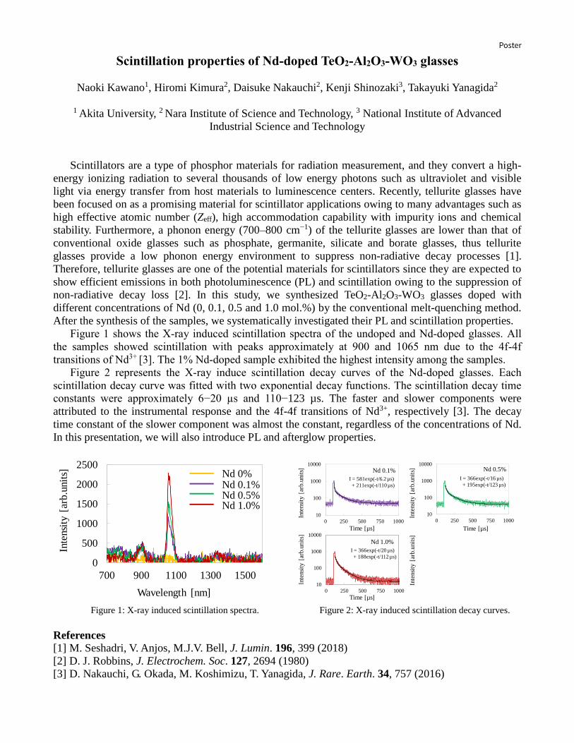
Poster
Scintillation properties of Nd-doped TeO2-Al2O3-WO3 glasses
Naoki Kawano1, Hiromi Kimura2, Daisuke Nakauchi2, Kenji Shinozaki3, Takayuki Yanagida2
1 Akita University, 2 Nara Institute of Science and Technology, 3 National Institute of Advanced
Industrial Science and Technology
Scintillators are a type of phosphor materials for radiation measurement, and they convert a high-
energy ionizing radiation to several thousands of low energy photons such as ultraviolet and visible
light via energy transfer from host materials to luminescence centers. Recently, tellurite glasses have
been focused on as a promising material for scintillator applications owing to many advantages such as
high effective atomic number (Zeff), high accommodation capability with impurity ions and chemical
stability. Furthermore, a phonon energy (700–800 cm−1) of the tellurite glasses are lower than that of
conventional oxide glasses such as phosphate, germanite, silicate and borate glasses, thus tellurite
glasses provide a low phonon energy environment to suppress non-radiative decay processes [1].
Therefore, tellurite glasses are one of the potential materials for scintillators since they are expected to
show efficient emissions in both photoluminescence (PL) and scintillation owing to the suppression of
non-radiative decay loss [2]. In this study, we synthesized TeO2-Al2O3-WO3 glasses doped with
different concentrations of Nd (0, 0.1, 0.5 and 1.0 mol.%) by the conventional melt-quenching method.
After the synthesis of the samples, we systematically investigated their PL and scintillation properties.
Figure 1 shows the X-ray induced scintillation spectra of the undoped and Nd-doped glasses. All
the samples showed scintillation with peaks approximately at 900 and 1065 nm due to the 4f-4f
transitions of Nd3+ [3]. The 1% Nd-doped sample exhibited the highest intensity among the samples.
Figure 2 represents the X-ray induce scintillation decay curves of the Nd-doped glasses. Each
scintillation decay curve was fitted with two exponential decay functions. The scintillation decay time
constants were approximately 6−20 µs and 110−123 µs. The faster and slower components were
attributed to the instrumental response and the 4f-4f transitions of Nd3+, respectively [3]. The decay
time constant of the slower component was almost the constant, regardless of the concentrations of Nd.
In this presentation, we will also introduce PL and afterglow properties.
0
500
1000
1500
2000
2500
700 900 1100 1300 1500
Wavelength [nm]
Inte
nsi
ty [
arb
.unit
s]
Nd 0.1%Nd 0.5%Nd 1.0%
Nd 0%
10
100
1000
10000
0 250 500 750 1000
10
100
1000
10000
0 250 500 750 1000
Inte
nsi
ty [
arb
.unit
s] Nd 0.1%
I = 581exp(-t/6.2 µs)
+ 211exp(-t/110 µs)
Time [µs]
Inte
nsi
ty [
arb
.unit
s]
Time [µs]
Nd 0.5%
I = 57exp(-t/0.37 ms)
Inte
nsi
ty [
arb
.unit
s]
Time [µs]
Nd 1.0%
Inte
nsi
ty [
arb.u
nit
s]
10
100
1000
10000
0 250 500 750 1000
Nd 0.5%
I = 366exp(-t/16 µs)
+ 195exp(-t/123 µs)
I = 366exp(-t/20 µs)
+ 188exp(-t/112 µs)
Figure 1: X-ray induced scintillation spectra. Figure 2: X-ray induced scintillation decay curves.
References [1] M. Seshadri, V. Anjos, M.J.V. Bell, J. Lumin. 196, 399 (2018)
[2] D. J. Robbins, J. Electrochem. Soc. 127, 2694 (1980)
[3] D. Nakauchi, G. Okada, M. Koshimizu, T. Yanagida, J. Rare. Earth. 34, 757 (2016)

Poster
Examination of luminescence initiated by ultrashort pulse excitation in Gd3Al2.5Ga2.5O12:Tm, Yb crystals.
Radosław Lisiecki1, Robert Kowalski1, Jarosław Komar1, Michał Głowacki2,
Marek Berkowski2, Witold Ryba-Romanowski1
1 Institute of Low Temperature and Structure Research, Polish Academy of Sciences, Okólna 2, 50-422
Wrocław, Poland 2 Institute of Physics, Polish Academy of Sciences, Al. Lotnikow 32/46, 02-668, Warsaw, Poland
Gd3Ga2.5Al2.5O12 (GAGG) crystals doped with 1 at.% Tm and co –doped with 1 at.%, 3 at.% and 5 at.% Yb3+ were grown by the Czochralski technique. GAGG crystals belong to cubic garnet system with Ia-3d space group [1]. For Tm3+ and Yb3+ dopants various types of sites are available. Gadolinium ions reside into dodecahedral positions, surrounded by 8 oxygens while Ga3+ and Al3+ occupies tetrahedral and octahedral positions surrounded by 4 and 6 oxygen ions, respectively. In this material partial substitution of aluminum ions by gallium ions occurs and as result of that structural disorder brings about a considerable inhomogeneous broadening of luminescent ion spectral lines. Excitation of single crystal samples of Tm,Yb co-doped GAGG by femtosecond pulses results in an intense NIR-red emission originating in 3H4, 1G4 and 1D2 thulium excited states while blue emission related to 1G4-3H6 transition is characterized by lower intensity. It has been found that emission of thulium decreases effectively in GAGG:Tm,Yb crystals co-doped with higher concentrations of ytterbium. The rise time and decay time analysis was performed to examine effectiveness of population and relaxation of involved excited states. It was observed that higher Yb3+ concentration affects significantly relaxation of considered luminescent levels in materials under study.
Figure 1: Image obtained with a streak camera showing details of spectral distribution of GAGG:1%Tm,1%Yb luminescence intensity in spectral region 620–840 nm.
Acknowledgements This work was supported by the National Science Centre, Poland under grant number DEC 2016/21/B/ST5/00890 References [1] Filippo Maglia, Vincenzo Buscaglia, Silvia Gennari, Paolo Ghigna, Monica Dapiaggi, Adolfo Speghini, Marco Bettinelli, J. Phys. Chem. B 110 6561-6568 (2006)

PosterDirect femtosecond laser printing of rare earth-doped silk microstructures.
Moliria V. Santos1)
, Kelly T. Paula1)
, Sabrina N. C. Santos1)
, Lippy F. Marques2)
,
Sidney J. L. Ribeiro3)
, Cléber R. Mendonça1)
.
1- São Carlos Institute of Physics, University of São Paulo, São Carlos, SP-Brazil.
2-Institute of Chemistry, Rio de Janeiro State University, Rio de Janeiro, RJ, Brazil.
3-Institute of Chemistry, São Paulo State University (UNESP), Araraquara, SP, Brazil.
Silk fibroin (SF) is an abundant natural polymer, which in the last years has been
studied as an attractive high-technology material platform [1]. Precise patterning of SF-
based materials for functional microstructures has extensive applications including
tissue engineering, biosensing and photonic devices. Considerable progress has been
made based on lithographic methods and self-assembly approach [2]. However, most
methods require chemical modification of silk fibroin to be photoreactive and demand
photoinitiators, which significantly affect the optical, mechanical and biological
properties of the material. At the same time, femtosecond laser induced forward transfer
(fs-LIFT) has been explored as an attractive processing tool for microprinting, which
can be used in the fabrication, for instance, of high-resolution complex 2D structures
[3]. Herein, we were able to fabricate luminescent structures of regenerated silk fibroin
(RSF) through maskless fs-LIFT technique, using a Ti:Sapphire laser oscillator
delivering 50-fs pulses at 800 nm with a repetition rate of 5 MHz. Eu3+
/Tb3+
complexes
were added to RSF as a gain medium, to further demonstrate the flexibility of this
approach. The silk fibroin patterns were printed as hemisphere structures with diameter
of 3µm and height 1 µm. Analyses of luminescence and FTIR spectroscopies confirmed
that fs-LIFT does not change the optical and structural properties of silk fibroin
composites. Our ability to precisely position and fabricate functionalized RSF
microstructures in a repetitive and reproducible manner presents several opportunities
for future innovative sensors, photonic and biomedical devices.
Acknowledgements
We acknowledge financial support from Fundação de Amparo à Ciência e Tecnologia
do Estado de São Paulo (FAPESP - 2018/11039-9).
References
[1] H. Tao, D.L. Kaplan, F.G. Omenetto, Adv. Mater., 24, 2824–2837 (2012).
[2] L.D. Koh, J. Yeo, Y.Y. Lee, Q. Ong, M. Han, B.C.K. Tee, Mater. Sci. Eng. C.,
86, 151–172 (2018).
[3] A. Piqué, J. Laser Micro/Nanoengineering, 9, 192–197, (2014).
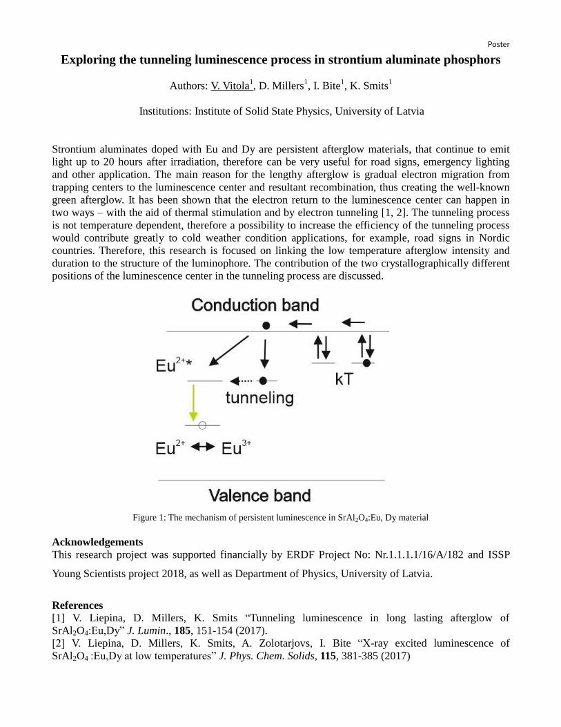
Poster
Exploring the tunneling luminescence process in strontium aluminate phosphors
Authors: V. Vitola1, D. Millers
1, I. Bite
1, K. Smits
1
Institutions: Institute of Solid State Physics, University of Latvia
Strontium aluminates doped with Eu and Dy are persistent afterglow materials, that continue to emit
light up to 20 hours after irradiation, therefore can be very useful for road signs, emergency lighting
and other application. The main reason for the lengthy afterglow is gradual electron migration from
trapping centers to the luminescence center and resultant recombination, thus creating the well-known
green afterglow. It has been shown that the electron return to the luminescence center can happen in
two ways – with the aid of thermal stimulation and by electron tunneling [1, 2]. The tunneling process
is not temperature dependent, therefore a possibility to increase the efficiency of the tunneling process
would contribute greatly to cold weather condition applications, for example, road signs in Nordic
countries. Therefore, this research is focused on linking the low temperature afterglow intensity and
duration to the structure of the luminophore. The contribution of the two crystallographically different
positions of the luminescence center in the tunneling process are discussed.
Figure 1: The mechanism of persistent luminescence in SrAl2O4:Eu, Dy material
Acknowledgements
This research project was supported financially by ERDF Project No: Nr.1.1.1.1/16/A/182 and ISSP
Young Scientists project 2018, as well as Department of Physics, University of Latvia.
References [1] V. Liepina, D. Millers, K. Smits “Tunneling luminescence in long lasting afterglow of
SrAl2O4:Eu,Dy” J. Lumin., 185, 151-154 (2017).
[2] V. Liepina, D. Millers, K. Smits, A. Zolotarjovs, I. Bite “X-ray excited luminescence of
SrAl2O4 :Eu,Dy at low temperatures” J. Phys. Chem. Solids, 115, 381-385 (2017)

Poster
Figure 1. TEM and TEM-EDS images of
NaYF4:20%Yb3+, 2%Ho3+, 8%Ce3+ showing
the distribution of Yb3+(red) Ho3+ (green)
and Ce3+ (yellow) codopants.
Dynamic tunability of upconverting NaYF4 colloidal nanoparticles
by Ce3+ co-doping
Aleksandra Pilch-Wróbel1,*, Joanna Zasada1, Marcin Birski1, Artur Bednarkiewicz1
1 Institute of Low Temperature and Structure Research, Polish Academy of Sciences, Okólna 2, 50-422
Wroclaw, Poland
*Corresponding author: [email protected]
The possibility to tune and manipulate the UC emission color of lanthanide doped NP’s is very
important in case of their possible applications such as information storage, biological sensing, as well
as graphics imaging and display.[1] For that reason new approaches are sought, which allow to obtain
higher emission intensity or to dynamically modulate the
luminescent properties of those materials. One of the
possible way to obtain such goals is incorporation of
passive impurities, which affect activator distribution within
the UCNP, or active impurities which play an active role in
energy transfer processes. Usually, such an approach leads
to important modification of energy distribution and energy
transfer upconversion. In consequence, the relative
intensities of emission bands change and modify the overall
emission color.[2,3]
Several research groups observed that by the incorporating
the Ce3+ to the NP’s the red emission can be enhanced, and
thus with proper selection of concentration of the co-
dopants and excitation conditions the green and red
emission can be obtained in Yb3+ /Ho3+ /Ce3+ doped
systems.[4] The reason for this is that the Ce3+ ion has a
small energy gap of about 3000 cm−1, which only affects
energy transfers that involve similar energy gaps.
In order to verify the influence of Ce3+ codoping on
the luminescent properties of UCNP’s based on Yb/Er and
Yb/Ho co-doped doped colloidal NaYF4 nanoparticles, the series of materials with different Ce3+
concentration was synthesized. The full structural (X-Ray Diffraction), morphological (Transmission
Electron Microscopy) and spectroscopic characterization (emission spectra in function of temperature,
decay and rise times) will be presented. In addition, the power dependence measurements, PWM (pulse
width modulation) dependent and time resolved luminescent spectra measurements will be shown.
Acknowledgements Financial support from 2017/25/N/ST5/01687 NCN PRELUDIUM 13 is greatly acknowledged.
References
[1] Deng, R.; Qin, F.; Chen, R.; Huang, W.; Hong, M.; Liu, X., Nat Nanotechnol, 10, 237-242 (2015)
[2] Cong, T.; Ding, Y.; Yu, X.; Mu, Y.; Hong, X.; Liu, Y., Mater. Res. Bull., 90, 151-155 (2017)
[3] Kamimura, M.; Omoto, A.; Chiu, H.-C; Soga, K.; Chem. Lett., 46, 1076-1078 (2017)
[4] Chen, G.; Liu, H.; Somesfalean, G.; Liang, H.; Zhang, Z., Nanotechnology, 20, 81542-81551(2009)

Poster
Photoluminescence properties of coumarins containing copolymers
Beata Derkowska-Zielinska1, Dariusz Chomicki
1, Joanna Slota
1, Vitaliy Smokal
2, Oksana Krupka
2
1Institute of Physics, Faculty of Physics, Astronomy and Informatics, Nicolaus Copernicus
University, Grudziadzka 5, 87-100 Torun, Poland 2Taras Shevchenko National University of Kyiv – 60 Volodymyrska – 01033 Kyiv Ukraine
Coumarins belong to the class of heterocyclic compounds known as benzopyrones. They consist of
fused pyrone and benzene rings with the pyrone carbonyl group at position 2. Coumarins are well-
known for their photochemical and photophysical properties. There are also recognized due to their
photoinduced cyclodimerisation properties leading to cyclobutane-based dimers with four possible
isomeric configurations syn head-to-head, anti head-to-head, syn head-to-tail, and anti head-to-tail [1-
4]. Coumarin system offers a unique opportunity for applications involving conjugated molecular
frameworks, and photo-induced dimerisation. The photocleavage can be induced upon irradiation with
higher energy photons at shorter wavelengths (see Fig. 1).
R
O
O
O
O
R
> 300 nm
254 nm
R
O
O
O
O
R
Figure 1: Reversible photoinduced cyclodimerisation of coumarins
Polymers with coumarin fragment are useful in many fields such as photoalignment of liquid
crystalline molecules, photoactive surface application, electroluminescence, photovoltaic, fluorescence
materials and laser dyes.
In this work, we report photoluminescence properties of thin films of coumarin containing copolymers.
Acknowledgements The authors would like to thank M. Salle (Angers University) for support.
References [1] D. Gindre, K. Iliopoulos, O. Krupka, E. Champigny, Y. Morille, M. Sallé, Image storage in
coumarin-based copolymer thin films by photoinduced dimerization, Optics Letters 38, 4636- 4639
(2013)
[2] Z. Essaidi, O. Krupka, K. Iliopoulos, E. Champigny, B. Sahraoui, M. Sallé, D. Gindre, Synthesis
and functionalization of coumarin-containing copolymers for second order optical nonlinearities,
Optical Materials 35, 576-581 (2013)
[3] K. Iliopoulos, O. Krupka, D. Gindre, M. Salle, Reversible Two-Photon Optical Data Storage in
Coumarin-Based Copolymers, Journal of the American Chemical Society 132, 14343-14345 (2010)
[4] L. Skowronski, O. Krupka, V. Smokal, A. Grabowski, M. Naparty, B. Derkowska-Zielinska, Optical
properties of coumarins containing copolymers, Optical Materials 47, 18-23 (2015)

Poster
Microscopic parameters characterizing thermoluminescence in Ce-doped garnets
Vasilii Khanin1, Ivan Vrubel2, Tancu Tukhvatulina3, Piotr Rodnyi3, Cees Ronda4 and Andries Meijerink1
1 Utrecht University, Princetonplein 1 3584 CC Utrecht, the Netherlands2 ITMO University, Kronverksky 49 197101, St. Petersburg, Russia
3 St. Petersburg Polytechnic University, Polytechnicheskaya 29 195251, St. Petersburg, Russia 4 Philips Research, High-Tech Campus 34 3556 AE Eindhoven, the Netherlands
Complex Ce-doped garnets receive a lot of attention recently as promising scintillators for medicalimaging [1], phosphors for LED applications [2] and persistent phosphors [3]. The research on garnetsgenerally consists of careful tuning of composition, stoichiometry and impurity (doping)concentrations. One of the main goals of these modifications is to influence the complex interplaybetween recombination and trapping/detrapping processes [4]. Extensive research has contributed to agood qualitative understanding of how traps, impurities and the presence of Ce4+ affect the materialsproperties. In this work we present a quantitative model that can explain the thermoluminescence andafterglow behavior of complex garnets.In particular, the model allows the determination of capture rates and effective capture radii forelectrons by traps and recombination centers in Lu1Gd2Ga3Al2O12:Ce garnet ceramics. The model relieson solving a set of coupled rate equations describing charge carrier trapping and recombination ingarnet ceramics doped with Ce and also co-doped with a known concentration of an intentionally addedelectron trap, Yb3+. The model is supported by analysis of a complete set of experimental data onafterglow, rise-time kinetics and X-ray excited luminescence which show that thermoluminescence isgoverned by trapping/detrapping processes following interactive kinetics with dominant recombinationchannel. The underlying reason for dominant recombination is the presence of a small fraction of Ce4+
(~2 ppm in the 0.2 % Ce-doped sample) which have a very high capture cross-section (~2.7 Å effectiveradius) because of the Coulomb attractive nature of this recombination center. The quantitative insights on capture cross-sections and concentrations of Ce4+ help to better understandthe optical properties of Ce-doped garnet scintillators and persistent luminescent materials and servesin optimizing synthesis procedures by tuning the Ce3+/4+ ratio by co-doping with divalent cations andannealing in an oxygen containing atmosphere.
References[1] C. W. E. van Eijk, Phys. Med. Biol., 47, R85 (2002)[2] Z. Xia, A. Meijerink, Chem. Soc. Rev., 46, 275–299 (2017)[3] J. Ueda, P. Dorenbos, A. J. J. Bos, K. Kuroishi, S. Tanabe, J. Mater. Chem. C, 3, 5642–5651 (2015)[4] M. Nikl, V. Babin, J. A. Mares, K. Kamada, S. Kurosawa, A. Yoshikawa, J. Tous, J. Houzvicka, K.Blazek, J. Lumin., 169, 539-543 (2016)

PosterAlkali cations effects on structural, morphological and photoluminescence properties of zinc oxide materials (ZnO) synthesized from seed growth by
hydrothermal process.
W. Bekhti1,2*, L.Guerbous3 and M. Ghamnia1
1) Laboratoire des Sciences de la Matière Condensée, Département de physique, Faculté des Sciences, Université d’Oran I Ahmed Ben bella, BP 1620 El-Ménaouer, 3100 Oran, Algeria,
2) Département de Physique, Université de Blida 1 Saad-Dahleb, Route de soumaa BP 270 Blida, Algeria,
3) Département Laser, DTN-C.R.N.A 02, Bd Frantz Fanon, BP 399, Algiers 16000, Algeria, * [email protected]
One-dimensional semiconducting nanostructures have stimulated great interest due to their interesting properties that differ from those of the bulk or spherical nanoparticles of the same composition. ZnO is one of the most attracting semiconductors due to its intrinsic properties that are enhanced at the nanoscale. In particular, one can cite the wide direct band-gap (3,37 eV), the large exciton binding energy (60 meV) at room temperature and an excellent chemical and thermal stability. Moreover, it is a versatile smart material with unique applications in sensors, photovoltaic, surface acoustic wave devices, light emitting diodes and luminescent devices, transparent conductive coatings and others [1-4].
In the present work, we describe the seed growth hydrothermal method of different shaped ZnO nano/microrods. Alkaline colloidal solutions are prepared separately and added with different quantities to the principal solution. The growth ZnO particles are ended hydrothermally in autoclave. The impact of colloidal solutions volume added as well as alkali cation’s type on the morphology, crystallite size and their distributions is presented and discussed. Furthermore, the vibrational and optical properties of the obtained particles are also investigated.
[1] Z.W. Pan, Z. R. Dai, Z. L. Wang, Science, 291, 1947 (2001). [2] C. Klingshirna, J. Fallerta, O. Gogolinb, M. Wissingera, R. Hauschilda, M. Hausera, H. Kalta, H. Zhoua, J of luminescence, 128, 792 (2008). [4] D.Vanmaekelbergh and L. K. van Vugt, Nanoscale, 3, 2783 (2011). [5] V.Kumar, O. M. Ntwaeaborwa, T. Soga, V. Dutta and H. C. Swart, ACS Photonics, 4 (11), 2613 (2017).

Poster
Visualizing the infection dynamics of pathogenic bacteria labelled by upconverting
nanoparticles inside mouse gut
Gokhan Dumlupinar1, 2 , Raminder Singh3, Katarzyna Komolibus1, Sanathana Konugolu Venkata
Sekar1, Jean Matias1, 2 Silvia Melgar3 and Stefan Andersson-Engels1, 2
1. Tyndall National Institute, IPIC, Biophotonics, Lee Maltings Complex, Dyke Parade,
T12R5CP, Cork, Ireland
2. Department of Physics , University College Cork, Kane Building, T12YN60, Cork, Ireland
3. APC Microbiome Institute, Bioscience Building, T12YN60, Cork, Ireland
This study intends to show the potential use of near-infrared light (NIR) triggered luminous
nanoparticles, also known as upconverting nanoparticles (UCNPs), to investigate the colonization and
infection dynamics of a natural murine intestinal pathogen, Citrobacter rodentium (C.rodentium),
which colonizes in mice GI tract, and induces inflammation. Remarkably, this pathogen expresses
genetic similarities to human pathogens, including enteropathogenic Escherichia coli (EPEC) and
enterohemorrhagic E.coli (EHEC) that are well associated with significant human gut diseases
including inflammatory bowel disease (IBD) development. Although the exact causes of IBD are still
unknown, the recent studies show that gut microbiota is a contributing factor to IBD [1]. Although the
role of intestinal pathogens in the development of IBD in animals is revealed, the interaction
mechanisms between the host and pathogen are still to be defined precisely. C. rodentium infection
model is considered as a useful preliminary method to understand these interactions in vivo [2]. It has
been studied by different optical imaging techniques in vivo, including fluorescence imaging (FI) and
bioluminescence (BL) [3, 4]. FI employs custom designed organic fluorophores as contrast markers,
whereas BL rely on genetically modified pathogen strains that are able to catalyse luciferin molecule by
luciferase enzyme. However, using organic fluorescent dyes in vivo suffers tissue autofluorescence and
photobleaching. In the case of BL, it is not always possible to genetically engineer all types of bacteria
strains for BL use. Nevertheless, in this study, NIR light triggered lanthanide (Ln) doped UCNPs are
employed as both labelling and contrast agents to visualise the infection dynamics of C.rodentium.
First, surface of ligand free Ln-UCNPs are functionalized through citrate modification method for
further antibody (Ab) conjugation. Next, anti-C.rodentium-Ab are bioconjugated with Ln-UCNPs to
label C.rodentium. Upconversion (UC) photoluminescence from Ab-UCNP labelled C.rodentium in
mouse is imaged by a commercial imaging system (IVIS Lumina III, Perkin Elmer). Herein, it should
be noted that major optical design modifications has been done to IVIS Lumina III to make it use in
NIR region. Ln-UCNPs with unique optical properties yield noticeable advantages including free tissue
autofluorescence, high penetration depths, and high photochemical stability. Finally, the infection
profile of C.rodentium has been mapped through UC emission from labelled bacteria in vivo.
Acknowledgements This work is supported by a grant from the Science Foundation of Ireland (SFI-15/RP/2828).
References
[1] Wirtz S., and Neurath M. F., Advanced drug delivery reviews, 59, 1073-1083 (2007).
[2] Silberger D.J., Zindl C.L., and Weaver C.T., Mucosal Immunology, 10, 1108 (2017).
[3] Leevy W. Matthew, et al., Journal of the American Chemical Society, 128, 16476-16477 (2006).
[4] Andreu N., Zelmer A., and Wiles S., FEMS microbiology reviews, 35, 360-394 (2011).

Poster
Upconversion emission studies of structural phase flips in Er3+
/Yb3+
: KLaF4
bio-compatible nanoparticles
Rajat Bajaj1, Mohini Gupta
3, A. Srinivasa Rao
1, R. Nagarajan
3 and G. Vijaya Prakash
2
1Delhi Technological University, Bawana road, New Delhi, 110042
2Indian Institute of Technology Delhi, Hauz Khas, New Delhi, 110016
3Delhi University, North Campus, New Delhi, 110007
Abstract
Up-conversion nanoparticles (UCNPs) are emerging as exciting alternatives to conventional
organic dyes and quantum dots in the field of bio-imaging and targeted drug delivery. Due to the
superior properties, it is more advantageous and effective to develop these UCNPs as a possible
replacement of conventional imaging organic/inorganic light emitters. Herein, we report up-
conversion emission monitoring of structural phase flips from cubic to hexagonal ordered KLaF4
UCNPs doped with Er3+
and Yb3+
as sensitizer, fabricated from conventional chemical synthesis
route1, 2
. Morphological studies were carried out on these UCNPs to demonstrate their crystal
phase and purity. Under the infrared (CW and femtosecond) laser excitation, these UCNPs emit
intense up-converted green to red (4S3/2→
4I15/2 and
4F9/2→
4I15/2) color emission, based on the
structural transformation from cubic to hexagonal phases. Details of up-conversion emission
process based on double photon excitation, energy transfer (ET) and Ground/Excited states
absorption (GSA/ESA) processes are discussed. Finally, we demonstrate that these UCNPs are
potential host lattices for low-cost bio-imaging and targeted drug delivery applications.
Figure 1: Up-conversion Emission spectra of KLaF4:Er3+
/Yb3+
UCNPs. exe= 980nm.
Acknowledgements:
AS and GVP acknowledge the funding from DST-SERB project.
References:
1. M. Gupta , M. Adnan, R. Nagarajan , G. Vijaya Prakash, ACS Omega, 4, 2275 (2019)
2. A. Prasad, A. Srinivasa Rao, M. Gupta, G Vijaya Prakash, Mater. Chem. Phys, 219,
13(2018)
No
rma
lize
d U
CL
in
ten
sity
(a
.u.)
700650600550500wavelength(nm)
8 Hrs
14 Hrs
4H11/2
4I15/2
4S3/2
4I15/2
4F9/2
4I15/2

Poster
Structural and spectroscopic properties of thermally stable Eu3+ doped
barium zinc orthophosphate phosphor for w-LEDs
Mukesh K. Sahu, Kaushal Jha and M. Jayasimhadri*
Luminescent Materials Research Lab (LMRL), Department of Applied Physics, Delhi
Technological University, Delhi 110 042, India
*Email: [email protected]
Monoclinic crystalline structure of europium (Eu3+) activated barium zinc orthophosphate (BZP)
phosphor has been synthesized via solid-state reaction method in ambient atmosphere. The phase
purity, structural and emissive characteristics have been analyzed by recording X-ray diffraction
pattern and photoluminescence (PL) spectra, respectively. The diffraction pattern demonstrated
the single phase formation of BZP sample and is similar with the standard data (JCPDS-16-0554).
The results indicate that BZP sample consist of monoclinic structure with space group P21/c and
the lattice constant a, b and c values are 8.59, 9.76 Å, and 9.15 Å, respectively. Scanning electronic
micrograph of BZP powder specifies the uniform spherical shape with some agglomeration of the
synthesized particles. The particle size for BZP powder sample varies from 2 to 8 μm. The PL
spectra exhibit the most intense emission peak centered at 616 nm owing to 5D0 →7F2 transition
of europium ions under the 393 nm (n-UV) and 466 nm (blue) excitations. Emission spectra reveal
that the PL intensity improves with the concentration of Eu3+ ions up to 4.0 mol% and beyond that,
the PL intensity decreases. The CIE coordinates evaluated for the as-synthesized phosphor, which
lies in the red region and close to commercial red phosphors Y2O2S: Eu (0.647, 0.343). The average
decay time of the Eu3+ doped BZP phosphor is in the millisecond range and the value decreases
with increasing Eu3+ concentration. The emission intensity remains to 68.41% at 423 K of that at
room temperature, which indicates good thermal stability in the BZP phosphor. All these results
indicate the suitability of the as-synthesized phosphors for white light emitting (w-LEDs) devices
[1, 2].
References
[1] T. Pulli, T. Dönsberg, T. Poikonen, F. Manoocheri, P. Kärhä, E. Ikonen, Light Sci Appl., 4(9),
e332 (2015).
[2] J. Chen, W. Zhao, J. Wang, N. Wang, J. Mater. Sci. Mater. Electron,. 27, 237–244 (2016).

Poster
Photoluminescence studies on Er3+
-doped sodium-bismo-phosphate glasses for
optoelectronic applications
Arti Yadav, A. S. Rao
Department of Applied Physics, Delhi Technological University, New Delhi-110 042, India
Erbium (Er3+
) ion doped Na2O - Bi2O3- P2O5- SrO glasses were fabricated by using melt quenching
technique and studied the influence of Er3+
ion concentration on the photoluminescence (PL)
performance using the spectroscopic techniques to understand the utility of the titled glasses for optical
fiber amplifiers. The XRD pattern reveals amorphous nature of the as-synthesized glass. The emission
spectra exhibit intense band at around 535 nm corresponding to the 4S3/2 →
4I15/2 transition under near
ultraviolet (n-UV) excitation. The chromaticity coordinates of the glasses are calculated by evaluating
the tristimulus values from the emission spectra when excited at 376 nm wavelength. The PL decay
curves for 4S3/2 (visible) level are discussed and they are exhibiting non-exponential nature for the as
prepared glasses. Optimum value for green emission intensity was obtained at dopant concentration 1.0
mol%. The above-mentioned studies show potentiality of the as-synthesized Er3+
doped Na2O - Bi2O3-
P2O5- SrO glasses in optoelectronic applications.
Figure 1: Photoluminescence spectra of Er
3+ for NBSP glasses under 376 nm excitation.
Acknowledgements Authors are thankful to Dr D S Kothari Post Doctoral Fellowship scheme for providing financial
support under award No. F. 4-2/2006(BSR)/PH/18-19/0056.
References 1. Rupesh A. Talewar, Sk. Mahamuda, A. Vyas, A.S. Rao and S.V. Moharil, J. Alloys Compds, 775,
810-817 (2019).
Copyright © 2022 FDOKUMEN