Adenosine receptors and brain diseases: Neuroprotection and neurodegeneration
ORIGINAL ARTICLE
Caffeine and Adenosine A2A ReceptorInactivation Decrease Striatal
Neuropathology in a Lentiviral-BasedModel of Machado–Joseph Disease
N!elio Goncalves, PharmD,1,2 Ana T. Sim~oes, PhD,1,2 Rodrigo A. Cunha, PhD,1,3
and Lu!ıs Pereira de Almeida, PhD1,2
Objective: Machado–Joseph disease (MJD) is a neurodegenerative disorder associated with an abnormal CAG expan-sion, which translates into an expanded polyglutamine tract within ataxin-3. There is no therapy to prevent or modifydisease progression. Because caffeine (a nonselective adenosine receptor antagonist) and selective adenosine A2A
receptor (A2AR) blockade alleviate neurodegeneration in different brain diseases, namely at early stages of anotherpolyglutamine-related disorder such as Huntington’s disease, we now tested their ability to control MJD-associatedneurodegeneration.Methods: MJD was modeled by transducing the striatum of male adult C57Bl/6 mice with lentiviral vectors encodingmutant ataxin-3 in one hemisphere and wild-type ataxin-3 in the other hemisphere (as internal control). Caffeine(1g/L) was applied through the drinking water. Mice were killed at different time points (from 2 to 12 weeks) toprobe for the appearance of different morphological changes using immunohistochemical analysis.Results: Mutant ataxin-3 caused an evolving neuronal dysfunction (loss of DARPP-32 staining) leading to neurode-generation (cresyl violet and neuronal nuclei staining) associated with increased number of mutant ataxin-3 inclusionsin the basal ganglia. Notably, mutant ataxin-3 triggered early synaptotoxicity (decreased synaptophysin and microtu-bule-associated protein-2 staining) and reactive gliosis (glial fibrillary acidic protein and CD11b staining), which pre-dated neuronal dysfunction and damage. Caffeine reduced the appearance of all these morphological modifications,which were also abrogated in mice with a global A2AR inactivation (knockout).Interpretation: Our findings provide a demonstration that synaptotoxicity and gliosis are precocious events in MJDand that caffeine and A2AR inactivation decrease MJD-associated striatal pathology, which paves the way to considerA2ARs as novel therapeutic targets to manage MJD.
ANN NEUROL 2013;73:655–666
Various inherited neurodegenerative diseases resultfrom an increase in the number of CAG codon
repeats within the open reading frame of the responsiblegene.1 Machado–Joseph disease (MJD), or spinocerebel-lar ataxia type 3, is one such dominantly-inherited poly-glutamine (polyQ) neurodegenerative disease and themost common among ataxias.2 It is characterized by anadult age of onset and causes premature death associatedwith unstable expansion of a CAG stretch (over 55repeats) in the MJD1 gene that encodes a polyQ repeat
in the corresponding ataxin-3 protein.3,4 The clinicalhallmarks of MJD include progressive ataxia, dysfunctionof motor coordination, postural instability, and parkin-sonism among other symptoms.5,6 The neuropathologyof MJD involves multiple systems, such as cerebellar sys-tems, substantia nigra, and cranial nerve motor nuclei,3,7
as well as the striatum.8–11 Degeneration and loss of neu-ronal cells in MJD are accompanied by the presence ofprotein aggregates,12 designated as neuronal intranuclearinclusion bodies (NIIs). Although transgenic animal
View this article online at wileyonlinelibrary.com. DOI: 10.1002/ana.23866
Received Jun 11, 2012, and in revised form Jan 21, 2013. Accepted for publication Feb 1, 2013.
Address correspondence to Dr Pereira de Almeida, CNC-Center for Neuroscience and Cell Biology,University of Coimbra, 3004-517 Coimbra, Portugal. E-mail: [email protected]
From the 1CNC-Center for Neuroscience and Cell Biology, 2Faculty of Pharmacy, and 3Faculty of Medicine,University of Coimbra, Coimbra, Portugal.
VC 2013 American Neurological Association 655

models closely mimicking the human pathology8,13–15
have bolstered our understanding of MJD, the mecha-nisms accounting for neuronal degeneration are stilllargely unknown. Albeit not yet explored in MJD, stud-ies in other polyQ disorders suggest that neuronal dys-function and synaptotoxicity may precede degenerationand appearance of clinical symptoms,16,17 and that neu-roinflammation18 may function as an amplificatory loopexacerbating neuronal damage.19–21
There is currently no therapy to manage MJD. Weposed that chronic caffeine consumption, which affordsneuroprotection through the antagonism of adenosine A2A
receptors (A2ARs),22 might be a candidate strategy to man-
age MJD neurodegeneration. Caffeine and A2AR blockadeafford robust neuroprotection in different neurodegenera-tive disorders, in accordance with the key role of A2ARs incontrolling synaptic viability, apoptotic neuronal death,astrocytic function, and neuroinflammation.23,24 In partic-ular, in Huntington’s disease, another polyQ disorder,A2AR blockade at the prodrome or early stages of the dis-ease seems to delay the appearance of clinical symptoms,mainly through a normalization of striatal glutamatergictransmission, which impedes the characteristic degenera-tion of striatal neurons.25 Importantly, it has recently beenshown in induced pluripotent stem cell–derived neuronsthat glutamate overstimulation raises intracellular calciumlevels, activating the cysteine proteases calpains and pro-moting the proteolysis and aggregation of ataxin-3.26
Moreover, calpain-mediated proteolysis of ataxin-3 in arodent model of MJD mediates translocation of ataxin-3to the cell nucleus, aggregation, and neurodegeneration,which can be prevented by calpain inhibition.27
In view of this proposed key role of glutamate over-stimulation in MJD, we investigated the time course ofneuropathological modifications in a genetic model ofMJD and tested the novel hypothesis that the manipula-tion of A2AR function might also be beneficial in MJD.
Materials and Methods
AnimalsMale C57BL/6 mice (Charles River, Barcelona, Spain) were keptunder a conventional 12-hour light–dark cycle in a temperature-controlled room with food and water provided ad libitum and usedat 7 weeks of age. C57Bl/6-background A2AR knockout (A2ARKO) and age-matched wild-type control mice were obtained fromparallel breeding of our colony of A2AR KO mice, initially obtainedfrom J. F. Chen (Boston University).28 The experiments were car-ried out in accordance with the European Community directive(86/609/EEC) for the care and use of laboratory animals.
Drug TreatmentWe chose the dose of caffeine (1g/L), administered throughdrinking water, as a maximally effective and nontoxic dose,
which we have previously shown to result in a plasma concentra-tion of 50lM,29 and similar concentration in the brain paren-chyma,30 corresponding to a diary human consumption of circa 5cups of coffee. Treatment with caffeine was begun 3 weeks beforeviral delivery onward, because we have previously reported that aminimum period of 2 weeks is required to allow a metabolic sta-bilization after beginning the free access to caffeine drinking.29,31
Viral Vectors ProductionLentiviral vectors encoding human wild-type ataxin-3 (atx3–27Q) or mutant ataxin-3 (atx3–72Q)8 were produced in 293Tcells with a 4-plasmid system, as previously described.32 Thelentiviral particles were resuspended in 1% bovine serum albu-min in phosphate-buffered saline (PBS). The viral particle con-tent of batches was determined by assessing HIV-1 p24 antigenlevels (RETROtek; Gentaur, Paris, France). Viral stocks werestored at 280!C until use.
In Vivo Injection into the StriatumConcentrated viral stocks were thawed on ice. After anesthesiaof the mice with Avertin (12lL/g, intraperitoneally [i.p.]), lenti-viral vectors encoding atx3–27Q or atx3–72Q were stereotaxi-cally injected into the striatum in the following coordinates:anteroposterior, 10.6mm; lateral, 61.8mm; ventral, 23.3mm;tooth bar, 0; which corresponds to the internal capsule, a largefiber tract passing through the middle of the striatum dividingboth dorsoventral and mediolateral structures. Wild-type andA2AR KO mice received 2lL injections of lentivirus (200000ngof p24/mL) in each hemisphere, administering atx3–72Q in theright hemisphere and control atx3–27Q in the left hemisphere.Different groups of mice were kept in their home cages for dif-ferent periods of 2, 4, 8, and 12 weeks, before being killed forimmunohistochemical analysis of morphological and neuro-chemical changes in the striatum.
Immunohistochemical ProcedureAfter an overdose of Avertin (2.5 3 12lL/g, i.p.), transcardiacperfusion of the mice was performed with PBS followed by fixa-tion with 4% paraformaldehyde. The brains were then removedand postfixed in 4% paraformaldehyde for 24 hours and cryo-protected by incubation in 25% sucrose/phosphate buffer for 48hours. The brains were frozen, and 25lm coronal sections werecut using a cryostat (CM3050 S; Leica, Heidelberg, Germany) at221!C. Slices throughout the entire striatum were collected inanatomical series and stored in 48-well trays as free-floating sec-tions in PBS supplemented with 0.05lM sodium azide. Thetrays were stored at 4!C until immunohistochemical processing.
Sections were processed overnight at 4!C with the follow-ing primary antibodies: a mouse monoclonal anti–ataxin-3 anti-body (1H9; 1:5000; Chemicon, Temecula, CA), a rabbit anti–DARPP-32 antibody (1:1000; Chemicon), or a mouse anti–neuronal nuclei (NeuN) antibody (1:1000; Chemicon), fol-lowed by 2-hour incubation at room temperature (RT) withthe respective biotinylated secondary antibodies (1:200; VectorLaboratories, Burlingame, CA). Bound antibodies were visual-ized using the Vectastain ABC kit, with 3,30-diaminobenzidine
ANNALS of Neurology
656 Volume 73, No. 5
tetrahydrochloride (DAB metal concentrate; Pierce, Burlingame,CA) as substrate.
Triple staining for synaptophysin (rabbit polyclonal, 1:300;Chemicon) and microtubule-associated protein-2 (MAP-2; mousemonoclonal AP20, 1:500; Santa Cruz Biotechnology, Santa Cruz,CA), glial fibrillary acidic protein (GFAP; rabbit polyclonal,1:1000; Dako, Glostrup, Denmark), and cluster of differentiationmolecule 11b (CD11b; rat monoclonal 5C6, 1:500; AbD Serotec,Oxford, UK) together with DAPI (Sigma, St Louis, MO) wereperformed. Free-floating sections were kept at RT for 2 hours inPBS with 0.1% Triton X-100 containing 10% normal goat serum(Gibco–Invitrogen, Barcelona, Spain), then overnight at 4!C inblocking solution with the primary antibodies. Sections werewashed 33 and incubated for 2 hours at RTwith the correspond-ing secondary antibodies coupled to fluorophores goat anti-mouse,goat anti-rabbit, or goat anti-rat Alexa Fluor 488 or Alexa Fluor594 (1:200; Molecular Probes–Invitrogen, Eugene, OR) diluted inthe blocking solution. The sections were washed 33 and thenmounted in Mowiol reagent (Sigma) on microscope slides.
Definition and analysis of protein immunoreactivitieswere made from the striatal center (site of injection) to themediolateral and dorsoventral striatal periphery from the needletract. This disease model is based on the intrastriatal injectionof lentivirus, which triggers physiological alterations evolvingover time radially. Therefore, comparable striatal sectionsbetween animals were defined from the site of injection in bothrostral and caudal directions, using the needle tract due to thesurgical procedure as reference point.
Staining was visualized using Zeiss Axioskop 2 plus, ZeissAxiovert 200, or Zeiss LSM 510 Meta imaging microscopes(Carl Zeiss MicroImaging, Oberkochen, Germany) equippedwith AxioCam HR color digital cameras (Carl Zeiss Microi-maging) and 35, 320, 340, and 363 Plan-Neofluar or 363Plan/Apochromat objectives using the AxioVision 4.7 softwarepackage (Carl Zeiss Microimaging). Quantitative analysis of flu-orescence was performed with a semiautomated image-analysissoftware package (ImageJ; NIH, Bethesda, MD).
Cresyl Violet StainingCoronal sections were premounted and stained with cresyl vio-let for 45 seconds, differentiated in 70% ethanol, dehydrated
by passing twice through 95% ethanol, 100% ethanol, and xy-lene solutions, and mounted onto microscope slides with Eukitt(Sigma).
Evaluation of DARPP-32 DepletionThe extent of ataxin-3 lesions in the striatum was analyzed byphotographing, with a 31.25 objective, 8 sections stained withDARPP-32 per animal (25lm-thick sections at 200lm inter-vals), selected so as to obtain complete rostrocaudal sampling ofthe striatum, and by quantifying the area of the lesion with asemiautomated image-analysis software package (ImageJ). Thevolume was then estimated with the following formula: vol-ume5 d(a11 a21 a3 …), where d is the distance between se-rial sections (200lm) and a11 a21 a3 are DARPP-32–depleted areas for individual serial sections. The average grayvalue of all pixels measured in the lesioned area was recordedfor each depleted area. Results are expressed as index of immu-noreactivity of DARPP-32 considering unlesioned striatal areaas 100% immunoreactivity.
Cell Counts of Ataxin-3 InclusionsCoronal sections showing complete rostrocaudal sampling (1 of8 sections) of the striatum were scanned with a 320 objective.The analyzed areas of the striatum encompassed the entire regioncontaining ataxin-3 inclusions, as revealed by staining with theanti–ataxin-3 antibody. All inclusions were manually countedusing a semiautomated image-analysis software package (ImageJ).
Statistical AnalysisStatistical comparisons were performed using either an unpairedStudent’s t test or 1-way analysis of variance followed by Dun-nett’s multiple comparison post hoc test. Results are expressed asmean6 standard error of the mean. Significance thresholds wereset at p< 0.05, p< 0.01, or p< 0.001, as defined in the text.
Results
Time CourseWe took advantage of using a lentiviral model of MJDto perform a temporal analysis of the relative appearanceof different features from early dysfunction to late neuro-nal loss. Thus, we carried out a time course study from 2
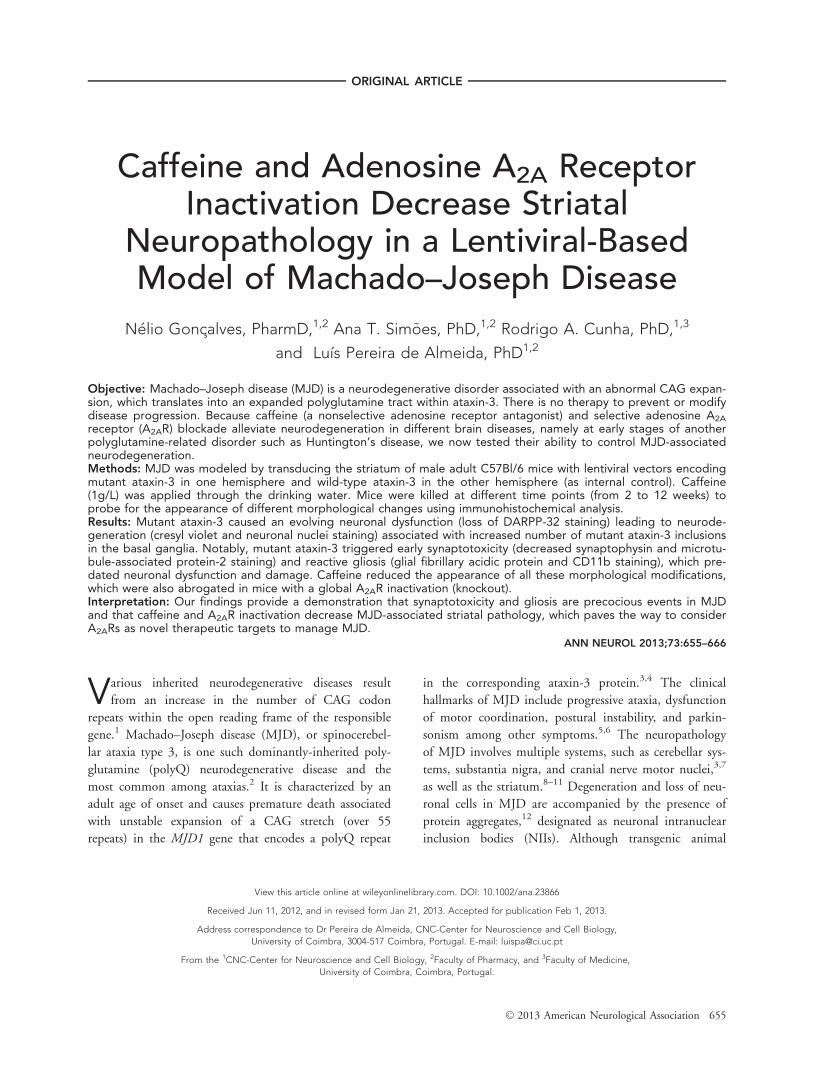
FIGURE 1: Summary of the time course of appearance of different morphological features in a lentiviral-based model ofMachado–Joseph disease. Loss of synaptic markers and astrogliosis were among the most precocious morphological altera-tions, closely followed by microgliosis and neuronal dysfunction, as well as increases in the number of ataxin-3 inclusions,whereas overt neuronal damage occurred later in the development of the disease. Microgliosis would probably be found atlater time points; however, that was not evaluated.
Goncalves et al: Caffeine Alleviates MJD
May 2013 657
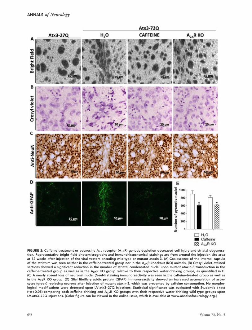
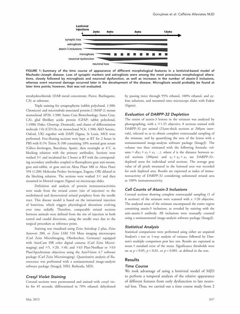
FIGURE 2: Caffeine treatment or adenosine A2A receptor (A2AR) genetic depletion decreased cell injury and striatal degenera-tion. Representative bright field photomicrographs and immunohistochemical stainings are from around the injection site areaat 12 weeks after injection of the viral vectors encoding wild-type or mutant ataxin-3. (A) Coalescence of the internal capsuleof the striatum was seen neither in the caffeine-treated group nor in the A2AR knockout (KO) animals. (B) Cresyl violet–stainedsections showed a significant reduction in the number of striatal condensated nuclei upon mutant ataxin-3 transduction in thecaffeine-treated group as well as in the A2AR KO group relative to their respective water-drinking groups, as quantified in E.(C) A nearly absent loss of neuronal nuclei (NeuN) staining immunoreactivity was seen in the caffeine-treated group as well asin the A2AR KO group. (D) Glial fibrillary acidic protein (GFAP) immunoreactivity showed an increased accumulation of astro-cytes (green) replacing neurons after injection of mutant ataxin-3, which was prevented by caffeine consumption. No morpho-logical modifications were detected upon LV-atx3–27Q injections. Statistical significance was evaluated with Student’s t test(*p<0.05) comparing both caffeine-drinking and A2AR KO groups with their respective water-drinking wild-type groups uponLV-atx3–72Q injections. [Color figure can be viewed in the online issue, which is available at www.annalsofneurology.org.]
ANNALS of Neurology
658 Volume 73, No. 5
to 12 weeks upon lentiviral-mediated expression of wild-type and mutant ataxin-3, and we analyzed different neu-ropathological features such as markers of synaptic loss,neuronal dysfunction, neuronal loss, astrogliosis, andmicrogliosis as well as ataxin-3 inclusions. Figure 1 sum-marizes the temporal evolution of each of these changes,which will be further detailed below.
Neuronal Degeneration and LossTo directly test whether caffeine and A2AR inactivationmitigate neurodegeneration, we first analyzed photomicro-graphs under bright field and then upon cresyl violet stain-ing. Lentiviral-mediated expression of mutant ataxin-3 (LV-atx3272Q) caused a clear condensation of the internalcapsule attributable to striatal tissue shrinkage, which wasevident in water-drinking mice at 12 weeks, but absent onbright field sections of both caffeine-drinking animals andglobal A2AR KO mice, as well as in the contralateral stria-tum challenged with wild-type ataxin-3 (LV-atx3–27Q; Fig2A). Cresyl violet staining revealed a marked reduction(p< 0.05) of the number of degenerated shrunken hyper-chromatic nuclei in caffeine-drinking mice, although itremained considerably high; the specificity of the effectover A2ARs was confirmed in A2AR KO animals, whichreproduced the alleviation of pathology observed in caf-feine-treated animals (p< 0.05; see Fig 2B, E).
To further investigate the neuroprotective effects ofcaffeine at a late time point in this model of MJD, weevaluated the immunoreactivity of the neuronal nuclearmarker NeuN. A clear loss of NeuN-stained neuronscould be seen in the water-drinking group at 12 weeksafter mutant ataxin-3 transduction, which was not detect-able after wild-type ataxin-3 transduction, and was nearlyabsent in mutant ataxin-3–expressing animals upon caf-feine treatment (see Fig 2C).
These data suggest that there is a progression in thedegeneration pattern in MJD-associated striatal pathologyleading to loss of neuronal markers, which is reduced bychronic caffeine consumption as well as genetically delet-ing the A2ARs.
Neuronal Functional ModificationsPrevious reports have indicated that striatal neuronal dys-function may precede degeneration and appearance ofclinical symptoms in MJD.33 Additionally, DARPP-32(dopamine and cyclic adenosine monophosphate–regu-lated phosphoprotein) was previously shown to be a sen-sitive marker that allows immunohistochemical detectionof this early neuronal dysfunction.8,27,34,35 Accordingly,DARPP-32 immunohistochemistry revealed a largedepleted staining volume of 0.606 0.12mm3 (n5 5) at4 weeks after injection of lentivirus encoding atx3–72Q
FIGURE 3: Caffeine treatment or adenosine A2A receptor (A2AR) genetic depletion reduced neuronal dysfunction. (A, upperpanel) A large DARPP-32–depleted volume was observed 4 weeks after injection of the viral vectors encoding mutant ataxin-3in the water-drinking group, whereas caffeine-drinking mice exhibited a much smaller lesion area at this time point. This isquantified in B as depleted volume of DARPP-32 staining. No loss of DARPP-32 staining was observed upon LV-atx3–27Qinjections. (A, lower panel) At higher magnification, 12 weeks exposure to mutated ataxin-3 revealed a clear condensation ofthe internal capsule, which joined together fiber patches (arrows). This fiber accumulation was not observed in the caffeine-treated animals or in A2AR knockout (KO) mice. Statistical significance was evaluated with Student’s t test (*p<0.05) comparingcaffeine-treated (n56) with water-drinking animals (n55). [Color figure can be viewed in the online issue, which is available atwww.annalsofneurology.org.]
Goncalves et al: Caffeine Alleviates MJD
May 2013 659
in the water-drinking group, whereas caffeine-treated ani-mals exhibited significantly smaller dysfunctional volume(0.216 0.06mm3 [n5 6]; p< 0.05) at this time point.No loss of DARPP-32 immunoreactivity was detectedupon LV-atx3–27Q injections (Fig 3A upper panel, B).
At higher magnification, analysis of the striatalDARPP-32–depleted area of water-drinking animals at12 weeks after injection of lentiviral vectors encodingmutant ataxin-3 revealed that the DARPP-32–stainedcell bodies and the corresponding tissue (internal capsule)were no longer present, originating a collapse of the tis-sue, which joins together fiber patches (see arrows in Fig3A, lower panel). This fiber accumulation presumablyresults from neuronal degeneration, which was notobserved in the caffeine-treated or in A2AR KO micechallenged with mutant ataxin-3. Density analysis ofDARPP-32 immunoreactivity (Table 1) showed a signifi-cant preservation of this marker in caffeine-treated(p< 0.01) and A2AR KO groups (p< 0.05) at 12 weeksafter injection of lentivirus encoding atx3–72Q, as com-pared to the respective water-drinking groups.
These data suggest that both chronic caffeine con-sumption and the genetic deletion of A2ARs are able toreduce neuronal dysfunction in MJD.
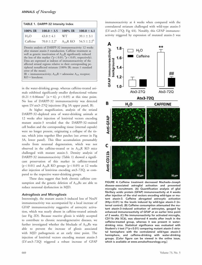
Astrogliosis and MicrogliosisInterestingly, the mutant ataxin-3–induced loss of NeuNimmunoreactivity was accompanied by a local increase ofGFAP immunoreactivity suggestive of astrocytic activa-tion, which was nearly absent in caffeine-treated animals(see Fig 2D). Because reactive gliosis is widely acceptedto contribute to chronic neurodegenerative diseases, wefurther investigated whether the blockade of A2ARs wasable to prevent the increase of gliosis associatedwith MJD pathogenesis at an early time point. Theinjection of lentiviral vectors encoding mutant ataxin-3(LV-atx3–72Q) triggered a robust increase of GFAP
immunoreactivity at 4 weeks when compared with thecontralateral striatum challenged with wild-type ataxin-3(LV-atx3–27Q; Fig 4A). Notably, this GFAP immunore-activity triggered by expression of mutated ataxin-3 was
TABLE 1. DARPP-32 Intensity Index
100% IR 100.06 3.5 100% IR 100.06 4.1
H2O 43.06 4.1 WT 39.16 3.1
Caffeine 70.06 2.2a A2AR KO 54.56 2.2b
Density analysis of DARPP-32 immunoreactivity 12 weeksafter mutant ataxin-3 transduction. Caffeine treatment aswell as genetic inactivation of A2AR significantly reducedthe loss of this marker (ap< 0.01; bp< 0.05, respectively).Data are expressed as indexes of immunoreactivity of theaffected striatal regions relative to their corresponding pe-ripheral nonaffected striatum (100% IR; mean6 standarderror of the mean).IR = immunoreactivity; A2AR5 adenosine A2A receptor;KO5 knockout.
FIGURE 4: Caffeine treatment decreased Machado–Josephdisease–associated astroglial activation and preventedmicroglia recruitment. (A) Quantification analysis of glialfibrillary acidic protein (GFAP) immunoreactivity at 4 weeksafter injection of the viral vectors encoding wild-type or mu-tant ataxin-3. Caffeine abrogated astrocytic activation(##p<0.01) to the levels induced by wild-type ataxin-3 (in-ternal control). (B) Caffeine consumption attenuated the mu-tant ataxin-3–induced activation of astrocytes, gauged byenhanced immunoreactivity of GFAP at an earlier time pointof 2 weeks. (C) No immunoreactivity for activated microglia,CD11b (Ab 5C6), was observed 4 weeks after insult in thecaffeine-treated group, whereas it was present in water-drinking mice. Statistical significance was evaluated withStudent’s t test (**p<0.01) comparing mutant ataxin-3 stria-tal hemisphere with the contralateral wild-type ataxin-3hemisphere, and caffeine-drinking with water-drinkinggroups. [Color figure can be viewed in the online issue,which is available at www.annalsofneurology.org.]
ANNALS of Neurology
660 Volume 73, No. 5
observed as early as 2 weeks (see Fig 4B). Additionally,strong immunoreactivity for the microglial proteinCD11b was found at 4 weeks, revealing microglialrecruitment (see Fig 4C), which was significantly androbustly reduced in caffeine-treated animals. This clearlyestablishes the presence of reactive gliosis in the striatumin this genetic model of MJD.
Importantly, treatment with caffeine (1g/L), pre-vented both the astrogliosis (see Fig 4A, B) and the puta-tive microgliosis (see Fig 4C) triggered by mutant ataxin-3, indicating that chronic caffeine consumption can pre-vent reactive gliosis associated with MJD.
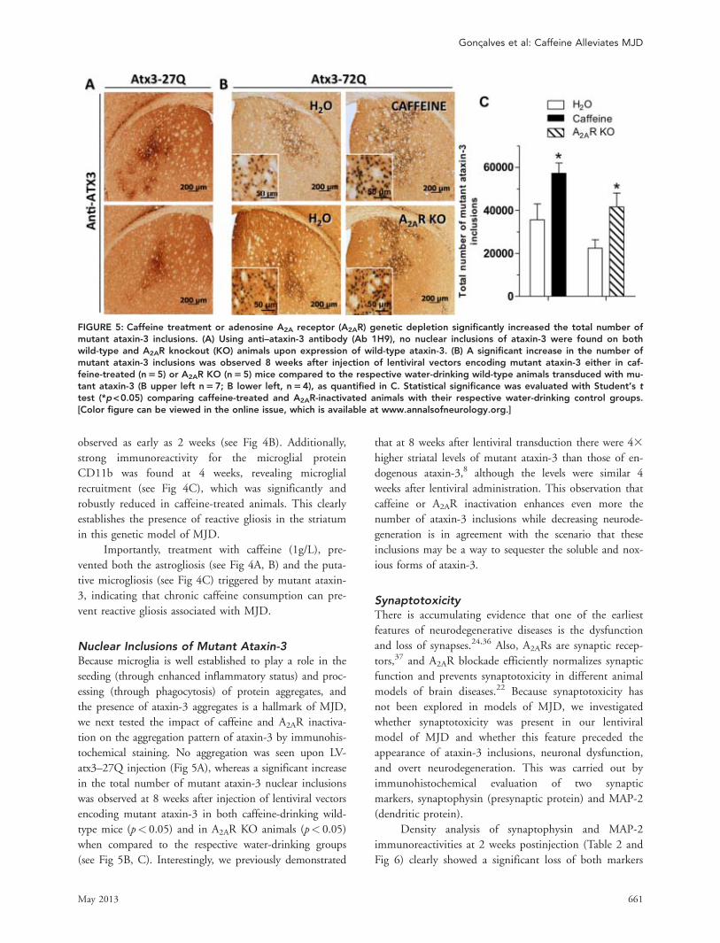
Nuclear Inclusions of Mutant Ataxin-3Because microglia is well established to play a role in theseeding (through enhanced inflammatory status) and proc-essing (through phagocytosis) of protein aggregates, andthe presence of ataxin-3 aggregates is a hallmark of MJD,we next tested the impact of caffeine and A2AR inactiva-tion on the aggregation pattern of ataxin-3 by immunohis-tochemical staining. No aggregation was seen upon LV-atx3–27Q injection (Fig 5A), whereas a significant increasein the total number of mutant ataxin-3 nuclear inclusionswas observed at 8 weeks after injection of lentiviral vectorsencoding mutant ataxin-3 in both caffeine-drinking wild-type mice (p< 0.05) and in A2AR KO animals (p< 0.05)when compared to the respective water-drinking groups(see Fig 5B, C). Interestingly, we previously demonstrated
that at 8 weeks after lentiviral transduction there were 43higher striatal levels of mutant ataxin-3 than those of en-dogenous ataxin-3,8 although the levels were similar 4weeks after lentiviral administration. This observation thatcaffeine or A2AR inactivation enhances even more thenumber of ataxin-3 inclusions while decreasing neurode-generation is in agreement with the scenario that theseinclusions may be a way to sequester the soluble and nox-ious forms of ataxin-3.
SynaptotoxicityThere is accumulating evidence that one of the earliestfeatures of neurodegenerative diseases is the dysfunctionand loss of synapses.24,36 Also, A2ARs are synaptic recep-tors,37 and A2AR blockade efficiently normalizes synapticfunction and prevents synaptotoxicity in different animalmodels of brain diseases.22 Because synaptotoxicity hasnot been explored in models of MJD, we investigatedwhether synaptotoxicity was present in our lentiviralmodel of MJD and whether this feature preceded theappearance of ataxin-3 inclusions, neuronal dysfunction,and overt neurodegeneration. This was carried out byimmunohistochemical evaluation of two synapticmarkers, synaptophysin (presynaptic protein) and MAP-2(dendritic protein).
Density analysis of synaptophysin and MAP-2immunoreactivities at 2 weeks postinjection (Table 2 andFig 6) clearly showed a significant loss of both markers
FIGURE 5: Caffeine treatment or adenosine A2A receptor (A2AR) genetic depletion significantly increased the total number ofmutant ataxin-3 inclusions. (A) Using anti–ataxin-3 antibody (Ab 1H9), no nuclear inclusions of ataxin-3 were found on bothwild-type and A2AR knockout (KO) animals upon expression of wild-type ataxin-3. (B) A significant increase in the number ofmutant ataxin-3 inclusions was observed 8 weeks after injection of lentiviral vectors encoding mutant ataxin-3 either in caf-feine-treated (n55) or A2AR KO (n55) mice compared to the respective water-drinking wild-type animals transduced with mu-tant ataxin-3 (B upper left n57; B lower left, n54), as quantified in C. Statistical significance was evaluated with Student’s ttest (*p<0.05) comparing caffeine-treated and A2AR-inactivated animals with their respective water-drinking control groups.[Color figure can be viewed in the online issue, which is available at www.annalsofneurology.org.]
Goncalves et al: Caffeine Alleviates MJD
May 2013 661
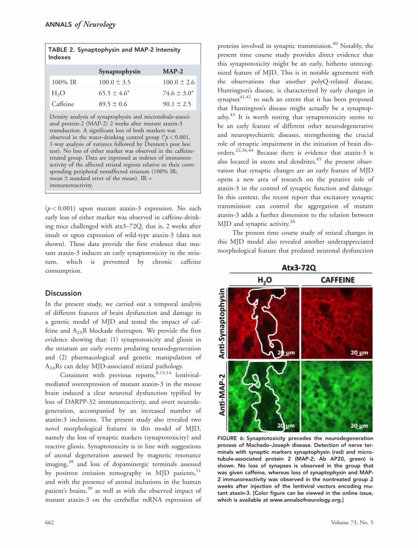
(p< 0.001) upon mutant ataxin-3 expression. No suchearly loss of either marker was observed in caffeine-drink-ing mice challenged with atx3–72Q, that is, 2 weeks afterinsult or upon expression of wild-type ataxin-3 (data notshown). These data provide the first evidence that mu-tant ataxin-3 induces an early synaptotoxicity in the stria-tum, which is prevented by chronic caffeineconsumption.
DiscussionIn the present study, we carried out a temporal analysisof different features of brain dysfunction and damage ina genetic model of MJD and tested the impact of caf-feine and A2AR blockade thereupon. We provide the firstevidence showing that: (1) synaptotoxicity and gliosis inthe striatum are early events predating neurodegenerationand (2) pharmacological and genetic manipulation ofA2ARs can delay MJD-associated striatal pathology.
Consistent with previous reports,8,13,14 lentiviral-mediated overexpression of mutant ataxin-3 in the mousebrain induced a clear neuronal dysfunction typified byloss of DARPP-32 immunoreactivity, and overt neurode-generation, accompanied by an increased number ofataxin-3 inclusions. The present study also revealed twonovel morphological features in this model of MJD,namely the loss of synaptic markers (synaptotoxicity) andreactive gliosis. Synaptotoxicity is in line with suggestionsof axonal degeneration assessed by magnetic resonanceimaging,38 and loss of dopaminergic terminals assessedby positron emission tomography in MJD patients,11
and with the presence of axonal inclusions in the humanpatient’s brains,39 as well as with the observed impact ofmutant ataxin-3 on the cerebellar mRNA expression of
proteins involved in synaptic transmission.40 Notably, thepresent time course study provides direct evidence thatthis synaptotoxicity might be an early, hitherto unrecog-nized feature of MJD. This is in notable agreement withthe observations that another polyQ-related disease,Huntington’s disease, is characterized by early changes insynapses41,42 to such an extent that it has been proposedthat Huntington’s disease might actually be a synaptop-athy.43 It is worth noting that synaptotoxicity seems tobe an early feature of different other neurodegenerativeand neuropsychiatric diseases, strengthening the crucialrole of synaptic impairment in the initiation of brain dis-orders.22,36,44 Because there is evidence that ataxin-3 isalso located in axons and dendrites,45 the present obser-vation that synaptic changes are an early feature of MJDopens a new area of research on the putative role ofataxin-3 in the control of synaptic function and damage.In this context, the recent report that excitatory synaptictransmission can control the aggregation of mutantataxin-3 adds a further dimension to the relation betweenMJD and synaptic activity.26
The present time course study of striatal changes inthis MJD model also revealed another underappreciatedmorphological feature that predated neuronal dysfunction
TABLE 2. Synaptophysin and MAP-2 IntensityIndexes
Synaptophysin MAP-2
100% IR 100.06 3.5 100.06 2.6
H2O 65.56 4.6a 74.66 3.0a
Caffeine 89.56 0.6 90.16 2.5
Density analysis of synaptophysin and microtubule-associ-ated protein-2 (MAP-2) 2 weeks after mutant ataxin-3transduction. A significant loss of both markers wasobserved in the water-drinking control group (ap< 0.001,1-way analysis of variance followed by Dunnett’s post hoctest). No loss of either marker was observed in the caffeine-treated group. Data are expressed as indexes of immunore-activity of the affected striatal regions relative to their corre-sponding peripheral nonaffected striatum (100% IR;mean6 standard error of the mean). IR =immunoreactivity.
FIGURE 6: Synaptotoxicity precedes the neurodegenerationprocess of Machado–Joseph disease. Detection of nerve ter-minals with synaptic markers synaptophysin (red) and micro-tubule-associated protein 2 (MAP-2; Ab AP20, green) isshown. No loss of synapses is observed in the group thatwas given caffeine, whereas loss of synaptophysin and MAP-2 immunoreactivity was observed in the nontreated group 2weeks after injection of the lentiviral vectors encoding mu-tant ataxin-3. [Color figure can be viewed in the online issue,which is available at www.annalsofneurology.org.]
ANNALS of Neurology
662 Volume 73, No. 5

and damage, reactive gliosis. There have been episodicreports of astrogliosis and microgliosis, typified bychanges in astrocytes and microglia morphology both inpatients,46 and in transgenic models of MJD,47 as well asincreased expression of cytokines and proinflammatorychemokines, which are compatible with mutant ataxin-3–induced changes in brain inflammatory mediators.48
However, in keeping with the fact that our lentiviral–mouse model resulted in an overexpression of mutantataxin-3 even at levels comparable to the endogenousform, the present report provides evidence that reactiveastrogliosis might be an early feature of MJD, which isparticularly relevant in view of the surge of interest inthe role of non-neuronal brain cells in the etiology ofneurodegenerative disorders.49 A putative role of glialcells in MJD is further heralded by evidence of the pres-ence of ataxin-3 in glial cells.12,50 Again, this observationshould open a novel area of research, fostering a betterunderstanding of the role of ataxin-3 in astrocytes and ofthe consequences of astrocytic adaptation upon accumu-lation of mutated ataxin-3.
Thus, the present time course exploration of neuro-pathological features associated with this genetic model ofMJD identifies synaptotoxicity and astrogliosis as preco-cious modifications followed by microgliosis and neuronaldysfunction, appearance of NIIs, and overt neuronal dam-age. This time course is in general agreement with the rec-ognition that synaptotoxicity and astrocyte-relatedmetabolic imbalance are among the most precocious mod-ifications in different neurodegenerative disorders and thatneuroinflammation, previously implicated in MJD,48 maybe a candidate process to mediate the spreading andamplification of damage until overt neuronal dysfunctionand damage can be recorded.24,36,49
The second prominent conclusion of this study isthe demonstration that the chronic consumption of a rea-sonable dose of caffeine compatible with a significantblockade of adenosine effects on A2A (most potent) andA1 receptors51 or the genetic elimination of A2ARs miti-gated the striatal neuropathological modifications causedby the expression of mutated ataxin-3. This is in agree-ment with the ability of A2ARs, mainly targeted bychronic caffeine consumption,22,52 to afford neuroprotec-tion against different neurodegenerative disorders, namelyAlzheimer’s, Parkinson’s, and Huntington’s disease.22,25,53
Notably, most of the compartments that we have nowshown to be affected in this genetic model of MJD areeffectively normalized by A2AR blockade in chronic braindiseases; thus, A2AR blockade prevents synaptotoxicity,54,55
in accordance with the enrichment of A2ARs in synapses,37
and also controls astrogliosis,56–58 microgliosis,59 striatalneurodegeneration,60 and neuronal death.23,61 Accordingly,
in our genetic model of MJD, chronic caffeine consump-tion or genetic deletion of A2ARs abrogated the loss ofsynaptic markers, prevented astrogliosis and microglia acti-vation, and reduced cell injury and striatal degeneration,rescuing its normal cytoarchitecture. Therefore, this pro-vides the first evidence that the manipulation of a neuro-modulation system operated by A2ARs is effective incontrolling the initial cascade of events triggered by thepathogenic ataxin-3 protein (synaptotoxicity and gliosis),resulting in reduced neuronal degeneration.
It is worth noting that this general neuroprotectionafforded by caffeine and A2AR blockade is accompanied byan accumulation of ataxin-3 aggregates into intracellularnuclear inclusions. The inverse correlation between theimpact of caffeine and A2AR blockade on mutant ataxin-3–induced NIIs and neuropathology strongly suggests thatthe aggregation of ataxin-3 could correspond to a cellulardefensive mechanism against soluble, more toxic species,62–65 rather than being the main cause of degeneration. There-fore, although it cannot be excluded that this might resultfrom better neuronal survival in treated animals, wehypothesized that aggregates may be neuroprotective.Nevertheless, the neuroprotection conveyed by caffeine andA2AR blockade might be of limited duration, because thenumber of pyknotic nuclei remained relatively high.
The present exploration of the time course ofMJD-associated neuropathological features and theirmodification by A2ARs provides a novel insight into theneuropathology of MJD but does not explore the under-lying mechanistic processes. Ataxin-3 is a polyubiquitin-binding protein whose physiological function has beenlinked to deubiquitination,66–69 and MJD is argued toresult from a toxic gain of function of mutant ataxin-3.In keeping with our proposal that synaptotoxicity mightbe a precocious modification in MJD, several studieshighlight the importance of the ubiquitin–proteasomesystem (UPS) in synapses,70,71 namely in presynaptic ter-minals,72,73 where it is affected in other polyQ neurolog-ical diseases74; furthermore, there is preliminary evidencethat A2ARs directly bind to UPS components75 and con-trol UPS activity,76 paving the way for a putative directcontrol by A2ARs of synaptic UPS. The proposal that theability of A2ARs to control another polyQ neurologicaldisorder (Huntington’s disease) depends on the control ofglutamatergic transmission prompts an alternative mecha-nism by which the A2AR-mediated control of the initialevent in MJD might be related to the ability of A2ARs tocontrol abnormal glutamatergic transmission throughdirect synaptic effects,77 or indirectly through control ofastrocytic glutamate uptake.78,79 Thus, albeit the molecu-lar mechanism of A2AR-mediated control of MJDremains to be determined, the present study provides
Goncalves et al: Caffeine Alleviates MJD
May 2013 663

new clues for particular compartments where such mech-anisms should be explored.
In conclusion, the present study provides a novelinsight into the pathology of MJD, bringing to centerstage synaptotoxicity and gliosis as precocious events inMJD. Furthermore, it provides the first realistic and safepromising lifestyle prophylactic strategy to delay theonset of this inherited disorder, based on the consump-tion of caffeine. Finally, it provides the first suggestionthat A2ARs might be a novel therapeutic target to inter-fere with the inexorable evolution of this neurodegenera-tive disease.
AcknowledgmentThis work was supported by Fundac~ao para a Ciencia ea Tecnologia (FCT), SAU-FCF/70384/2006 (L.P.A.),SAU-NEU/099307/2008 (L.P.A.), SAU-NEU/108668/2008 (R.A.C.), the Center for Neuroscience and CellBiology, University of Coimbra, PEst-C/SAU/LA0001/2011, the Richard Chin and Lily Lock Machado JosephDisease Research Fund (L.P.A.), the Association FrancaiseContre les Myopathies (L.P.A.), the Defense AdvancedResearch Projects Agency DARPA-09-68-ESR-FP-010(R.A.C.), and the National Ataxia Foundation (L.P.A.).N.G. and A.T.S. were supported by FCT fellowshipsBD/33186/2007 and BD/38636/2007.
Potential Conflicts of InterestL.P.A.: grants/grants pending, FCT, AFM, NAF,RCLLMJDRF. R.A.C.: grants/grants pending, FCT,MJFF.
References1. Koshy BT, Zoghbi HY. The CAG/polyglutamine tract diseases:
gene products and molecular pathogenesis. Brain Pathol1997;7:927–942.
2. Ranum LP, Lundgren JK, Schut LJ, et al. Spinocerebellar ataxiatype 1 and Machado-Joseph disease: incidence of CAG expan-sions among adult-onset ataxia patients from 311 families withdominant, recessive, or sporadic ataxia. Am J Hum Genet1995;57:603–608.
3. Durr A, Stevanin G, Cancel G, et al. Spinocerebellar ataxia 3 andMachado-Joseph disease: clinical, molecular, and neuropathologi-cal features. Ann Neurol 1996;39:490–499.
4. Kawaguchi Y, Okamoto T, Taniwaki M, et al. CAG expansions in anovel gene for Machado-Joseph disease at chromosome 14q32.1.Nat Genet 1994;8:221–228.
5. Gwinn-Hardy K, Singleton A, O’Suilleabhain P, et al. Spinocerebel-lar ataxia type 3 phenotypically resembling Parkinson disease in ablack family. Arch Neurol 2001;58:296–299.
6. Taroni F, DiDonato S. Pathways to motor incoordination: theinherited ataxias. Nat Rev Neurosci 2004;5:641–655.
7. Sudarsky L, Coutinho P. Machado-Joseph disease. Clin Neurosci1995;3:17–22.
8. Alves S, Regulier E, Nascimento-Ferreira I, et al. Striatal and nigralpathology in a lentiviral rat model of Machado-Joseph disease.Hum Mol Genet 2008;17:2071–2083.
9. Klockgether T, Skalej M, Wedekind D, et al. Autosomal dominantcerebellar ataxia type I. MRI-based volumetry of posterior fossastructures and basal ganglia in spinocerebellar ataxia types 1, 2and 3. Brain 1998;121:1687–1693.
10. Taniwaki T, Sakai T, Kobayashi T, et al. Positron emission tomog-raphy (PET) in Machado-Joseph disease. J Neurol Sci1997;145:63–67.
11. Wullner U, Reimold M, Abele M, et al. Dopamine transporterpositron emission tomography in spinocerebellar ataxias type 1,2, 3, and 6. Arch Neurol 2005;62:1280–1285.
12. Paulson HL, Perez MK, Trottier Y, et al. Intranuclear inclusions ofexpanded polyglutamine protein in spinocerebellar ataxia type 3.Neuron 1997;19:333–344.
13. Bichelmeier U, Schmidt T, Hubener J, et al. Nuclear localization ofataxin-3 is required for the manifestation of symptoms in SCA3: invivo evidence. J Neurosci 2007;27:7418–7428.
14. Goti D, Katzen SM, Mez J, et al. A mutant ataxin-3 putative-cleav-age fragment in brains of Machado-Joseph disease patients andtransgenic mice is cytotoxic above a critical concentration. J Neu-rosci 2004;24:10266–10279.
15. Cemal CK, Carroll CJ, Lawrence L, et al. YAC transgenic mice car-rying pathological alleles of the MJD1 locus exhibit a mild andslowly progressive cerebellar deficit. Hum Mol Genet2002;11:1075–1094.
16. Andrews TC, Weeks RA, Turjanski N, et al. Huntington’s diseaseprogression. PET and clinical observations. Brain 1999;122:2353–2363.
17. Li H, Li SH, Yu ZX, et al. Huntingtin aggregate-associated axonaldegeneration is an early pathological event in Huntington’s dis-ease mice. J Neurosci 2001;21:8473–8481.
18. Bantubungi K, Jacquard C, Greco A, et al. Minocycline in phenotypicmodels of Huntington’s disease. Neurobiol Dis 2005;18:206–217.
19. Aktas O, Smorodchenko A, Brocke S, et al. Neuronal damage inautoimmune neuroinflammation mediated by the death ligandTRAIL. Neuron 2005;46:421–432.
20. Gao HM, Kotzbauer PT, Uryu K, et al. Neuroinflammation and oxi-dation/nitration of a-synuclein linked to dopaminergic neurode-generation. J Neurosci 2008;28:7687–7698.
21. Lee JW, Lee YK, Yuk DY, et al. Neuro-inflammation induced by li-popolysaccharide causes cognitive impairment through enhance-ment of beta-amyloid generation. J Neuroinflammation 2008;5:37.
22. Cunha RA, Agostinho PM. Chronic caffeine consumption preventsmemory disturbance in different animal models of memorydecline. J Alzheimers Dis 2010;20(suppl 1):S95–S116.
23. Chen JF, Sonsalla PK, Pedata F, et al. Adenosine A2A receptorsand brain injury: broad spectrum of neuroprotection, multifacetedactions and “fine tuning” modulation. Prog Neurobiol2007;83:310–331.
24. Gomes CV, Kaster MP, Tome AR, et al. Adenosine receptors andbrain diseases: neuroprotection and neurodegeneration. BiochimBiophys Acta 2011;1808:1380–1399.
25. Popoli P, Blum D, Martire A, et al. Functions, dysfunctions andpossible therapeutic relevance of adenosine A2A receptors in Hun-tington’s disease. Prog Neurobiol 2007;81:331–348.
26. Koch P, Breuer P, Peitz M, et al. Excitation-induced ataxin-3aggregation in neurons from patients with Machado-Joseph dis-ease. Nature 2011;480:543–546.
27. Sim~oes AT, Goncalves N, Koeppen A, et al. Calpastatin-mediatedinhibition of calpains in the mouse brain prevents mutant ataxin-3proteolysis, nuclear localization and aggregation, relievingMachado-Joseph disease. Brain 2012;135:2428–2439.
ANNALS of Neurology
664 Volume 73, No. 5

28. Chen JF, Huang Z, Ma J, et al. A2A adenosine receptor deficiencyattenuates brain injury induced by transient focal ischemia inmice. J Neurosci 1999;19:9192–9200.
29. Duarte JM, Agostinho PM, Carvalho RA, Cunha RA. Caffeine con-sumption prevents diabetes-induced memory impairment and syn-aptotoxicity in the hippocampus of NONcZNO10/LTJ mice. PLoSOne 2012;7:e21899.
30. Costenla AR, Cunha RA, de Mendonca A. Caffeine, adenosinereceptors, and synaptic plasticity. J Alzheimers Dis 2010;20(suppl1):S25–S34.
31. Duarte JM, Carvalho RA, Cunha RA, Gruetter R. Caffeine con-sumption attenuates neurochemical modifications in the hippo-campus of streptozotocin-induced diabetic rats. J Neurochem2009;111:368–379.
32. de Almeida LP, Zala D, Aebischer P, Deglon N. Neuroprotectiveeffect of a CNTF-expressing lentiviral vector in the quinolinic acidrat model of Huntington’s disease. Neurobiol Dis 2001;8:433–446.
33. Yen TC, Tzen KY, Chen MC, et al. Dopamine transporter concen-tration is reduced in asymptomatic Machado-Joseph disease genecarriers. J Nucl Med 2002;43:153–159.
34. de Almeida LP, Ross CA, Zala D, et al. Lentiviral-mediated deliv-ery of mutant huntingtin in the striatum of rats induces a selectiveneuropathology modulated by polyglutamine repeat size, hunting-tin expression levels, and protein length. J Neurosci2002;22:3473–3483.
35. Cyr M, Beaulieu JM, Laakso A, et al. Sustained elevation of extrac-ellular dopamine causes motor dysfunction and selective degener-ation of striatal GABAergic neurons. Proc Natl Acad Sci U S A2003;100:11035–11040.
36. Coleman P, Federoff H, Kurlan R. A focus on the synapse for neu-roprotection in Alzheimer disease and other dementias. Neurol-ogy 2004;63:1155–1162.
37. Rebola N, Canas PM, Oliveira CR, Cunha RA. Different synapticand subsynaptic localization of adenosine A2A receptors in thehippocampus and striatum of the rat. Neuroscience2005;132:893–903.
38. D’Abreu A, Franca M Jr, Appenzeller S, et al. Axonal dysfunctionin the deep white matter in Machado-Joseph disease. J Neuroi-maging 2009;19:9–12.
39. Seidel K, den Dunnen WF, Schultz C, et al. Axonal inclusions inspinocerebellar ataxia type 3. Acta Neuropathol 2010;120:449–460.
40. Chou AH, Chen SY, Yeh TH, et al. HDAC inhibitor sodium butyr-ate reverses transcriptional downregulation and ameliorates ataxicsymptoms in a transgenic mouse model of SCA3. Neurobiol Dis2011;41:481–488.
41. DiProspero NA, Chen EY, Charles V, et al. Early changes in Hun-tington’s disease patient brains involve alterations in cytoskeletaland synaptic elements. J Neurocytol 2004;33:517–533.
42. Smith R, Klein P, Koc-Schmitz Y, et al. Loss of SNAP-25 and rab-philin 3a in sensory-motor cortex in Huntington’s disease. J Neu-rochem 2007;103:115–123.
43. Li JY, Plomann M, Brundin P. Huntington’s disease: a synaptop-athy? Trends Mol Med 2003;9:414–420.
44. Wishart TM, Parson SH, Gillingwater TH. Synaptic vulnerability inneurodegenerative disease. J Neuropathol Exp Neurol2006;65:733–739.
45. Trottier Y, Cancel G, An-Gourfinkel I, et al. Heterogeneous intra-cellular localization and expression of ataxin-3. Neurobiol Dis1998;5:335–347.
46. Horimoto Y, Matsumoto M, Akatsu H, et al. Longitudinal study onMRI intensity changes of Machado-Joseph disease: correlationbetween MRI findings and neuropathological changes. J Neurol2011;258:1657–1664.
47. Silva-Fernandes A, Costa Mdo C, Duarte-Silva S, et al. Motoruncoordination and neuropathology in a transgenic mouse modelof Machado-Joseph disease lacking intranuclear inclusions andataxin-3 cleavage products. Neurobiol Dis 2010;40:163–176.
48. Evert BO, Vogt IR, Kindermann C, et al. Inflammatory genes areupregulated in expanded ataxin-3-expressing cell lines and spino-cerebellar ataxia type 3 brains. J Neurosci 2001;21:5389–5396.
49. Lobsiger CS, Cleveland DW. Glial cells as intrinsic components ofnon-cell-autonomous neurodegenerative disease. Nat Neurosci2007;10:1355–1360.
50. Wang G, Ide K, Nukina N, et al. Machado-Joseph disease geneproduct identified in lymphocytes and brain. Biochem BiophysRes Commun 1997;233:476–479.
51. Fredholm BB, Battig K, Holmen J, et al. Actions of caffeine in thebrain with special reference to factors that contribute to its wide-spread use. Pharmacol Rev 1999;51:83–133.
52. Ferr!e S. An update on the mechanisms of the psychostimulanteffects of caffeine. J Neurochem 2008;105:1067–1079.
53. Schwarzschild MA, Agnati L, Fuxe K, et al. Targeting adenosine A2A
receptors in Parkinson’s disease. Trends Neurosci 2006;29:647–654.
54. Canas PM, Porciuncula LO, Cunha GM, et al. Adenosine A2A re-ceptor blockade prevents synaptotoxicity and memory dysfunctioncaused by beta-amyloid peptides via p38 mitogen-activated pro-tein kinase pathway. J Neurosci 2009;29:14741–14751.
55. Silva CG, Porciuncula LO, Canas PM, et al. Blockade of adenosineA2A receptors prevents staurosporine-induced apoptosis of rathippocampal neurons. Neurobiol Dis 2007;27:182–189.
56. Brambilla R, Cottini L, Fumagalli M, et al. Blockade of A2A adenosinereceptors prevents basic fibroblast growth factor-induced reactiveastrogliosis in rat striatal primary astrocytes. Glia 2003;43:190–194.
57. Minghetti L, Greco A, Potenza RL, et al. Effects of the adenosineA2A receptor antagonist SCH 58621 on cyclooxygenase-2 expres-sion, glial activation, and brain-derived neurotrophic factor avail-ability in a rat model of striatal neurodegeneration. J NeuropatholExp Neurol 2007;66:363–371.
58. Yu L, Shen HY, Coelho JE, et al. Adenosine A2A receptor antago-nists exert motor and neuroprotective effects by distinct cellularmechanisms. Ann Neurol 2008;63:338–346.
59. Rebola N, Sim~oes AP, Canas PM, et al. Adenosine A2A receptorscontrol neuroinflammation and consequent hippocampal neuronaldysfunction. J Neurochem 2011;117:100–111.
60. Schiffmann SN, Fisone G, Moresco R, et al. Adenosine A2A receptorsand basal ganglia physiology. Prog Neurobiol 2007;83:277–292.
61. Cunha RA. Neuroprotection by adenosine in the brain: from A1 re-ceptor activation to A2A receptor blockade. Purinergic Signal2005;1:111–134.
62. Arrasate M, Mitra S, Schweitzer ES, et al. Inclusion body formationreduces levels of mutant huntingtin and the risk of neuronaldeath. Nature 2004;431:805–810.
63. Saudou F, Finkbeiner S, Devys D, Greenberg ME. Huntingtin actsin the nucleus to induce apoptosis but death does not correlatewith the formation of intranuclear inclusions. Cell 1998;95:55–66.
64. Takahashi T, Kikuchi S, Katada S, et al. Soluble polyglutamineoligomers formed prior to inclusion body formation are cytotoxic.Hum Mol Genet 2008;17:345–356.
65. Taylor JP, Tanaka F, Robitschek J, et al. Aggresomes protect cellsby enhancing the degradation of toxic polyglutamine-containingprotein. Hum Mol Genet 2003;12:749–757.
66. Burnett B, Li F, Pittman RN. The polyglutamine neurodegenerativeprotein ataxin-3 binds polyubiquitylated proteins and has ubiqui-tin protease activity. Hum Mol Genet 2003;12:3195–3205.
67. Doss-Pepe EW, Stenroos ES, Johnson WG, Madura K. Ataxin-3interactions with rad23 and valosin-containing protein and its
Goncalves et al: Caffeine Alleviates MJD
May 2013 665

associations with ubiquitin chains and the proteasome are consist-ent with a role in ubiquitin-mediated proteolysis. Mol Cell Biol2003;23:6469–6483.
68. Kuhlbrodt K, Janiesch PC, Kevei E, et al. The Machado-Josephdisease deubiquitylase ATX-3 couples longevity and proteostasis.Nat Cell Biol 2011;13:273–281.
69. Warrick JM, Morabito LM, Bilen J, et al. Ataxin-3 suppresses poly-glutamine neurodegeneration in Drosophila by a ubiquitin-associ-ated mechanism. Mol Cell 2005;18:37–48.
70. Cajigas IJ, Will T, Schuman EM. Protein homeostasis and synapticplasticity. EMBO J 2010;29:2746–2752.
71. DiAntonio A, Hicke L. Ubiquitin-dependent regulation of the syn-apse. Annu Rev Neurosci 2004;27:223–246.
72. Jiang X, Litkowski PE, Taylor AA, et al. A role for the ubiquitin-proteasome system in activity-dependent presynaptic silencing. JNeurosci 2010;30:1798–1809.
73. Rinetti GV, Schweizer FE. Ubiquitination acutely regulates presyn-aptic neurotransmitter release in mammalian neurons. J Neurosci2010;30:3157–3166.
74. Wang J, Wang CE, Orr A, et al. Impaired ubiquitin-proteasomesystem activity in the synapses of Huntington’s disease mice. JCell Biol 2008;180:1177–1189.
75. Milojevic T, Reiterer V, Stefan E, et al. The ubiquitin-specific pro-tease Usp4 regulates the cell surface level of the A2A receptor.Mol Pharmacol 2006;69:1083–1094.
76. Chiang MC, Chen HM, Lai HL, et al. The A2A adenosine receptorrescues the urea cycle deficiency of Huntington’s disease byenhancing the activity of the ubiquitin-proteasome system. HumMol Genet 2009;18:2929–2942.
77. Rebola N, Lujan R, Cunha RA, Mulle C. Adenosine A2A receptorsare essential for long-term potentiation of NMDA-EPSCs at hippo-campal mossy fiber synapses. Neuron 2008;57:121–134.
78. Matos M, Augusto E, Santos-Rodrigues AD, et al. Adenosine A2A
receptors modulate glutamate uptake in cultured astrocytes andgliosomes. Glia 2012;60:702–716.
79. Nishizaki T, Nagai K, Nomura T, et al. A new neuromodulatorypathway with a glial contribution mediated via A2A adenosinereceptors. Glia 2002;39:133–147.
ANNALS of Neurology
666 Volume 73, No. 5
Copyright © 2022 FDOKUMEN