Lymphatic uptake and biodistribution of liposomes after subcutaneous injection
www.transonc.com
Trans la t iona l Onco logy Volume 7 Number 5 October 2014 pp. 570–579 570
Address alReceived 2
© 2014 Tlicenses/by1936-5233http://dx.d
Biodistribution and EfficacyStudies of the ProteasomeInhibitor BSc2118 in a MouseMelanoma Model
l correspondence to: Izabela Mlynarczuk-Bialy, MD, PhD, Chalubinskiego 5,4 February 2014; Revised 13 July 2014; Accepted 18 July 2014
he Authors. Published by Elsevier Inc. on behalf of Neoplasia Press, Inc. This is-nc-nd/3.0/)./14oi.org/10.1016/j.tranon.2014.07.002
Izabela Mlynarczuk-Bialy*, Thorsten R. Doeppner†,Jakub Golab‡, Dominika Nowis‡,Grzegorz M. Wilczynski§, Kamil Parobczak§,Moritz E. Wigand¶, Malgorzata Hajdamowicz*,Łukasz P. Biały*, Olga Aniolek#, Petra Henklein¶,Mathias Bähr**, Boris Schmidt†, Ulrike Kuckelkorn¶
and Peter-M. Kloetzel¶
*Department of Histology and Embryology, Warsaw MedicalUniversity, Warsaw, Poland; †Department of Neurology,University of Duisburg-Essen, Essen, Germany;‡Department of Immunology, Warsaw Medical University,Warsaw, Poland; §Nencki Institute of Experimental Biology,Polish Academy of Science, Warsaw, Poland; ¶Institute ofBiochemistry, Charité – Universitätsmedizin, Berlin, Germany;#Warsaw University of Life Sciences Faculty of VeterinaryMedicine Department of Large Animal Diseases with theClinic Division of Large Animal Internal Diseases, Warsaw,Poland; **Department of Neurology, University ofGoettingen, Goettingen, Germany; ††Clemens SchapfInstitute for Organic Chemistry and Biochemistry, TUDarmstadt, Darmstadt, Germany
AbstractInhibition of the proteasome offers many therapeutic possibilities in inflammation as well as in neoplasticdiseases. However, clinical use of proteasome inhibitors is limited by the development of resistance or severe sideeffects. In our study we characterized the anti-tumor properties of the novel proteasome inhibitor BSc2118. Thesensitivity of tumor lines to BSc2118 was analyzed in comparison to bortezomib using crystal violet staining inorder to assess cell viability. The In Vivo distribution of BSc2118 in mouse tissues was tracked by a fluorescent-modified form of BSc2118 (BSc2118-FL) and visualized by confocal microscopy. Inhibition of the 20S proteasomewas monitored both in cultured cell lines and in mice, respectively. Finally, safety and efficacy of BSc2118 wasevaluated in a mouse melanoma model. BSc2118 inhibits proliferation of different tumor cell lines with a similarpotency as compared with bortezomib. Systemic administration of BSc2118 in mice is well tolerated, even whengiven in a dose of 60 mg/kg body weight. After systemic injection of BSc2118 or bortezomib similar proteasomeinhibition patterns are observed within the murine organs. Detection of BSc2118-FL revealed correlation ofdistribution pattern of BSc2118 with inhibition of proteasomal activity in cells or mouse tissues. Finally,administration of BSc2118 in a mouse melanoma model shows significant local anti-tumor effects. Concluding,BSc2118 represents a novel low-toxic agent that might be alternatively used for known proteasome inhibitors inanti-cancer treatment.
Translational Oncology (2014) 7, 570–579
02-004 Warszawa, Poland. E-mail: [email protected]
an open access article under the CC BY-NC-ND license (http://creativecommons.org/
Translational Oncology Vol. 7, No. 5, 2014 Anti-tumor Efficacy of BSc2118 Mlynarczuk-Bialy et al. 571
Introduction Another proteasome inhibitor that has been frequently used in
The ubiquitin-proteasome system plays a crucial role in themaintenance of cellular homeostasis by participation in thedegradation of the majority of cytosolic proteins. This system isinvolved in the regulation of the cell cycle, apoptosis, transcription,cell signaling, antigen presentation, inflammation and development[1]. This situation makes the ubiquitin-proteasome system to one ofthe most promising therapeutic targets for further drug screening anddevelopment.The proteasome is an abundant cytosolic and nuclear proteasecomplex, which contains a 20S proteasome core complex as centralcatalytic unit that harbors different proteolytic activities, i.e. a trypsin-like (T-L within the β2 subunit), a chymotrypsin-like (ChT-L withinthe β5 subunit) and a caspase-like (within the β1 subunit) [2]. Itsactivity within the cell is regulated by interaction of the 20S core withthe regulatory 19S complex and with the PA28 complex at both ends ofthe proteasome cylinder [3]. The proteasome system is coupled with theubiquitin system for controlled protein degradation [4,5]. Therefore,inhibition of the proteasome leads in the first line to accumulation ofpolyubiquitinated proteins. Imbalance in cell cycle turn over andsubsequent cell cycle arrest as well as the inhibition of NF-κB as a resultfrom stabilization of IκBα are other hallmarks of proteasomal inhibition.Finally, inhibition of the 20S proteasome leads to induction of apoptosisthat is a summary effect of the inability to degrade injurious substrates.In this context, the ChT-L activity is likely to be essential for mostproteasomal functions and for the viability of cells. Irreversible inhibitionor deletion of the β5 subunit carrying the ChT-L activity is thereforeknown to be lethal [6,7].Proteasome inhibition is an established therapeutic approach in
anti-tumor drug development. In this context, proteasome inhibitorsinduce apoptosis more selectively in tumor than in normal cells,which is the most important rationale for application of theseinhibitors in anti-tumor therapy. By stabilization of IκBα,proteasome inhibitors exert anti-inflammatory effects and promotedeath of tumor cells [8–13].Based on the catalytic specificity of the proteasome complex, a
number of short peptide derived inhibitors (e.g., peptide boronicacids, vinyl sulfonates or peptide aldehydes) have been developed[14–16]. However, many of these were ultimately discarded fromconsideration for clinical use because of poor stability, lowbioavailability and lack of specificity. The first drug applied inhuman diseases was bortezomib, a dipeptidyl boronic acid also knownas PS-341 or Velcade (Millennium Pharmaceuticals, USA). Bortezo-mib selectively targets the catalytic β-subunits of the proteasome in aconcentration dependent manner, thus inhibiting the chymotrypsin-like (β5/β5i) and to a lesser degree the caspase-like (β1/β1i)activity [17,18]. The compound was initially approved for thetreatment of drug-resistant multiple myeloma in 2003 [19].Furthermore, this inhibitor was approved by the FDA for thetreatment of previously untreated multiple myeloma as well asin Waldenström's macroglobulinemia and mantle cell lymphoma[20–22]. However, prolonged treatment with bortezomib inducessubstantial toxicity with frequent and potentially severe adverseevents, such as peripheral neurotoxicity, cardiotoxicity, thrombocy-topenia, anaemia, gastrointestinal symptoms, neuropathy, fever,fatigue, headache, arthralgia, rash as well as electrolyte disturbancesand the development of drug resistance [23–26]. Therefore, thesearch for new effective and low toxic inhibitors of the proteasomesystem is urgently needed.
various experimental designs is MG132 (zLLL-CHO). In the presentproject, we characterized the novel inhibitor BSc2118 (patent noT30305), which is an analogue of MG132 with a better proteasomeinhibition profile than MG132 [27]. Previously, BSc2118 has beenshown to inhibit the ChTL activity of the proteasome, induce G2/Mcell cycle arrest and promote apoptosis. BSc2118 further stabilizesIκBα and prevents NF-κB activation [27].
In the current work, we analyzed the distribution pattern ofBsc2118 both in various tumor cell lines and in mice as well as theinhibition profile in selected mice organs after intraperitonealadministration. We finally show that application of BSc2118 inmice induces local anti-tumor effects and is tolerated at higher dosesas compared to bortezomib.
Material and Methods
Proteasome InhibitorsBSc2118 and fluorescent Bodipy-BSc2118 was dissolved in DMSO
at 20 mM and kept at −20°C. Bortezomib (Lilly, Germany) wasdissolved in distilled water at 4 mM and kept at −20°C until usage.
Synthesis of BSc2118BSc2118 was synthesized as described previously [28]. The
fluorescent variant of BSc2118 (further named as BSc2118-FL) wassynthesized as follows: a solution of 10 mg of the peptide NH2-Leu-Asp(tBu)-Leu-CHO (BSc2118) in 1 ml dimethylformamide DMF(pH = 8) was given to 10 mg of Bodipy-FL, SE (Molecular Probes,Germany). The reaction mixture was stirred overnight in darkness.Preparative purification by high-pressure liquid chromatography(HPLC) was carried out on a Shimadzu LC-8A system with anAgilent Zorbax, C18 SE column (250 × 21.2 mm), 7 μl, (buffer A:0.2% trifluoroacetic acid TFA in water, buffer B: 0.2% TFA in water:acetonitrile, 1:4). The peptides were purified with a gradient of 60%B to 95% B in 50 min.
Cell CultureHeLa cells and C26 murine colon cancer cells were cultured in
RPMI-1640 (Biochrom AG, Germany) with stable glutamax, bothsupplemented with 10% heat-inactivated FBS (Invitrogen, Germany),100 μg/ml streptomycin and 250 ng/ml amphotericin B (Invitrogen,Germany). For assessment of In Vitro cytostatic/cytotoxic activityof BSc2118, established human and mouse tumor cell lines wereused. These cells were in particular: HCT116, PC3, LoVo,CaPan2, MDA435, Panc02, EMT6, C26, B16F10 (all ATCC)MeWo, MeWoCis, MeWoVin, MeWoEto, MeWoFote (obtainedfrom Dr. H. Roekmann [29]), Mel-6, Mel-15, Mel-18, Mel-21,MaMel-63a (obtained from Dr. D. Schadendorf, Skin CancerUnit Deutsches Krebsforschungszentrum, Heidelberg, Germany),WM35, WM902B, WM9 (obtained from Dr. M. Herlyn,Wistar Institute, Philadelphia, PA). All cells were grown either inDMEM or in RPMI-1640 and supplemented with 10% FCSplus antibiotics.
Estimation of GI50 (growth inhibition) of BSc2118 in a panelof tumor cell lines
The influence of BSc2118 on the growth of 22 tumor cell lines wasanalyzed using a crystal violet assay similarly as described forbortezomib by Adams et al [30]. GI50 is defined as the concentration

572 Anti-tumor Efficacy of BSc2118 Mlynarczuk-Bialy et al. Translational Oncology Vol. 7, No. 5, 2014
needed to reduce the growth of treated cells to half that of untreatedcells. Briefly, cells were seeded in quadruplicates on 96-well plates,exposed for 48 hours to proteasome inhibitors in 7 dilutions rangingfrom 10 nM to 1000 nM (for BSc2118) and from 1 nM to 100 nM(for bortezomib). The cytostatic/cytotoxic effects of both BSc2118and bortezomib on treated cells were compared to that of controlcells. The mean viability for the whole cell panel was calculated in twoways, thereafter. First, average viability for the entire panel wascalculated for each concentration point followed by calculation of theaverage GI50 value. Second, GI50 was averaged for each cell lineindividually. Both methods of calculation resulted in similar results.
Isolation and Activity Measurement of 20S Proteasomes20S proteasomes were isolated from both red blood cells of healthy
volunteers and from murine organs [31]. Lysates from murine organsafter injection of inhibitors were obtained by homogenization in 100mM HEPES, pH 7.4, 2 mM MgCl2, 0.1% NP-40, 5 mMdithiothreitol and completed by Ultra-Turrax T8 (IKA-Werke).Chymotrypsin-like activity of the 20S proteasome was measured witha fluorogenic substrate (Suc-LLVY-AMC, Bachem, Germany).Briefly, 100 ng of purified proteasomes were exposed to proteasomeinhibitors (0-1000 nM) and incubated with 50 μM of fluorogenicsubstrate for up to 60 min. Lysates normalized to protein contentwere directly incubated with 50 μM of fluorogenic substrate. Thefluorescence was measured with POLARstar reader (BMG Labtech,Germany). The excitation and emission wavelengths were 390 nmand 460 nm, respectively. All experiments were performed inquadruplicates and repeated at least three times. Differences betweengroups were calculated by a Student’s t test. A P value of b0.05 wasconsidered to be statistically significant.
The Influence of Microsomal Enzymes on Inhibitor StabilityFor analysis of inhibitor stability in the presence of microsomal
enzymes, BSc2118 and MG132 (at 0.1 to 5 μM, respectively) wereincubated with mouse (Balb/c) microsomes (GIBCO) for up to24 hours according to manufacturer instructions. The proteasomeactivity (20S isolated from mouse muscles) was measured in thepresence of inhibitors with/without microsomal fraction as described inthe section above. The results are displayed as relative 20S activity in thepresence of inhibitors incubated with microsomal fraction. Inhibitorsincubated with PBS were defined as 100%. The data are displayed asdecrease of inhibitory activity in the presence of microsomal enzymes.Differences between groups were calculated by Student’s t test. AP value of b0.05 was considered to be statistically significant.
Immunoblot AnalysisThe procedure has been described previously [27]. The poly-
ubiquitinated proteins were detected with a polyclonal rabbit anti-ubiquitin antibody (Dako, Germany), and GAPDH was detectedusing a monoclonal anti-GAPDH (glyceraldehyde 3-phosphatedehydrogenase) antibody (Santa Cruz, Germany). A peroxidase-coupled anti-rabbit or anti-mouse antibody was used as secondaryantibody (Jackson Immunoresearch, UK).
Incubation with BSc2118-FL and Confocal MicroscopyHeLa cells and C26 cells were directly incubated with BSc2118-FL,
washedwith PBS, fixed with 70% ethanol and probedwith either rabbitanti-proteasome antibody (anti α4 subunit, Inst. for Biochemistry,Charité, Berlin, Germany) or with a mouse anti-ubiquitin conjugated
antibody FK1 (Biomol, Germany). After washing, they were probedwith secondary antibodies coupled with a fluorescent dye such as FITCor Alexa-fluor 540 (Jackson Immunoresearch, UK). All antibodysolutions were made up in PBS containing 5% bovine serum albumin.Actin filaments were stained by incubation for 1 hour with Phalloidincoupled to Atto647N (Sigma Aldrich, Germany). Cell nuclei werevisualized by stainingwithDAPI (SigmaAldrich,Germany). Specimenswere embedded in Vecta shield medium (Reactolab SA, Switzerland).The sections were examined with a Leica confocal laser scanningmicroscope (Leica Microsystems, Germany) equipped with akrypton-argon laser. Sequential scans at a series of optical planeswere performed with a 63× oil immersion objective lens throughspecimens.
Experimental Design for In Vivo StudiesFemale C57BL/6 and BALB/c mice, 8 to 12 weeks of age, were
obtained from the Animal House of the Polish Academy of Sciences,Medical Research Center (Warsaw, Poland). All In Vivo experimentswere performed according to EU guidelines for the care and use oflaboratory animals and approved by local authorities. The inhibitorBSc2118 was administered intraperitoneally (i.p.) in 50 μl DMSO orintratumorally (i.t.) in 20 μl DMSO. As controls, mice were treatedwith appropriate volumes of DMSO. Bortezomib was given to miceat 1 mg/kg i.p. in 50 μl PBS, for which mice treated with 50 μl PBSi.p. served as controls.
For studies on biodistribution and kinetics of inhibitors, BALB/cmice received one single dose of inhibitor at 5 and 10 mg/kg. After 1hour or 24 hours post injection, mice were sacrificed; tissue sampleswere collected and divided into halves. For direct observations ofBSc2118-FL tissue samples were embedded in OCT and immedi-ately frozen at −70°C. For biochemical analysis, tissue samples werefrozen at −70°C and kept until preparation. An initial study wascarried out in melanoma bearing C57BL/6 mice to determine thepotency of BSc2118 to inhibit the proteasome activity In Vivo eitherwithin red blood cells or within tumor tissue after i.t. or i.p. injectionof the inhibitor.
Melanoma Anti-Tumor Therapy in Mice by BSc2118Female C57BL/6mice of 8 to 9 weeks of age were inoculated into the
footpad on day 0 with 1 × 10⁶ B16F10 cells in 20μl of PBS. Tumor cellviability measured by trypan blue exclusion was above 98%. Beginningfromday 7 after inoculation,melanoma bearingmice were injectedwith5, 10, or 15mg per kg body weight of BSc2118 in a volume of 20 μl i.t.for 7 consecutive days. For i.p. injection protocols, BSc2118 was givenat dose of 15, 30, or 60 mg per kg body weight for seven consecutivedays. Bortezomib was given at 1 mg/kg i.p. 7 times every second day.Each group contained 7 animals. Appropriate volumes of the solventswere given as control. During the experiments melanoma bearing micewere observed daily for survival and adverse effects. Tumor size ofmelanoma-bearingmice wasmeasured every 2 days. Tumor volumewasdetermined according to the formula: tumor volume = (shorterdiameter2 × longer diameter)/2. Differences in tumor volume wereanalyzed for significance by the Student’s t test. A P value of b0.05 wasconsidered to be statistically significant. Log-rank test was used toanalyze survival.
In Vivo Angiogenesis AssayMice were anesthetized and received sterile abdominal injections of
250 μl of Matrigel (Becton Dickinson, Germany) subcutaneously
Translational Oncology Vol. 7, No. 5, 2014 Anti-tumor Efficacy of BSc2118 Mlynarczuk-Bialy et al. 573
containing 50 nM βFGF (Sigma Aldrich, Germany). Thereafter, micewere i.p. treated with BSc2118 at 30 mg/kg for 7 consecutive days. Atday 8, vascularization of the Matrigel was quantified by intravenousinjecting of 0.1 ml (0.25 mg/ml stock solution) of FITC-dextran(125,000 molecular weight, Sigma Aldrich, Germany) into mice,which allowed blood vessels within plugs to be visualized. Animalswere sacrificed 20 minutes after injection, when Matrigel plugs wereremoved and digested in Dispase reagent (Becton Dickinson,Germany). The fluorescence of the solution obtained was measuredusing a fluorimeter (POLARstar, BMG Labtech, Germany) with anexcitation at 480 nm and an emission wavelength at 520 nm.Differences between groups were calculated by Student’s t test. AP value of b0.05 was considered to be statistically significant.
In Vivo Metastasis AssayExperimental lung metastases were performed as described by
Feleszko et al. [32]. Briefly, experimental lung metastases wereinduced by injection of 2 × 10⁵ of B16F10 cells/100 μl PBS into thetail vein of anesthetized female C57BL/6 mice. Mice (5 to 7 pergroup) were i.p. injected for 7 consecutive days with either DMSO orBSc2118 (15 mg/kg body weight/day). The animals were thensacrificed on day 21 after inoculation of tumor cells. An averagenumber of metastases were calculated for every mouse by twoindependent observers blinded to the experimental groups. Differencesbetween experimental groups were analyzed using the Mann–WhitneyU test. A P value of b0.05 was considered to be statistically significant.
Results
BSc2118 is Cytotoxic for a Panel of Solid Tumor Cell LinesAn In Vitro cytotoxicity screening was performed to characterize
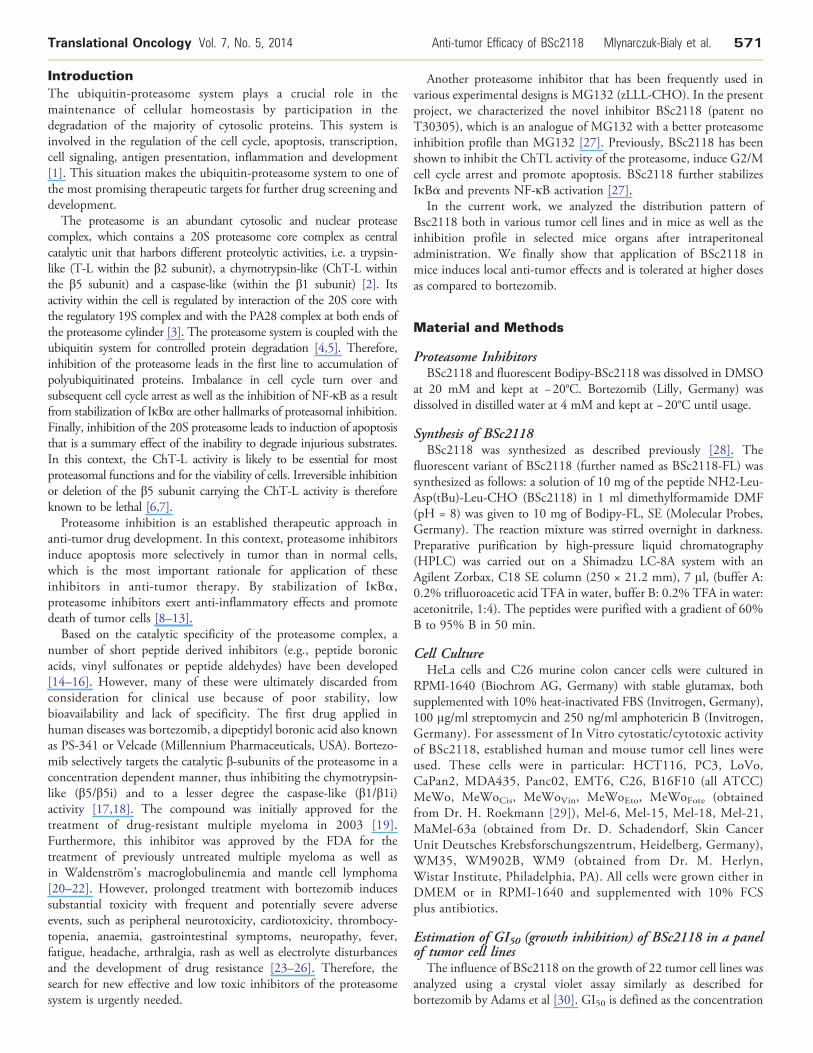
the anti-tumor potential of BSc2118. For this purpose, a panel ofsolid tumor cell lines, most of them originating from malignantmelanoma, was incubated either with BSc2118 or with bortezomib asa reference inhibitor (Figure 1). The average GI50 value was estimatedfor each cell line and across the entire tumor cell panel. We found anaverage GI50 value of 76 nM for BSc2118, whereas the average GI50value for bortezomib across 22 cell lines was 6.3 nM. For comparison,on NCI cell panel the GI50 value for bortezomib was given at 7 nM
Figure 1. BSc2118 affects viability of 22 tumor cell lines. Graphswere prepared by averaging viability of 22 cell lines from sevenconcentration points of inhibitors. Results are displayed aspercentage of controls. The discrepancy in the concentrations ofinhibitors is a result of different Ki-values for both drugs.
[30]. Figure 1 shows average viability for seven concentrations aspercentages regarding controls for both BSc2118 and bortezomib.
Characterization of Inhibitory Properties of BSc2118and BSc2118-FL
The proteasome inhibitor BSc2118 was described previously byBraun et al. [28]. To better track the biological properties of this
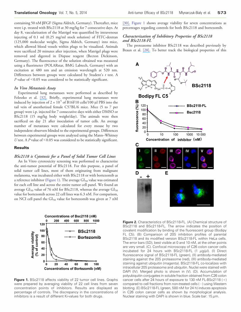
igure 2. Characteristics of BSc2118-FL. (A) Chemical structure ofSc2118 and BSc2118-FL. The arrow indicates the position ofovalent modification by binding of the fluorescent group (BodipyL C5). (B) Comparison of 20S inhibition profiles of parentalSc2118 and its modified version BSc2118-FL within HeLa cells.he error bars (SD), best visible at 0 and 10 nM, at the other pointsre very small. (C). Confocal microscopy of C26 colon cancer cellscubated for 24 hours with BSc2118-FL (1 μg/μl). (I) Directluorescence signal of BSc2118-FL (green), (II) antibody-mediatedtaining against the 20S proteasome (red), (III) antibody-mediatedtaining against ubiquitin (magenta). BSc2118-FL co-localizes withtracellular 20S proteasome and ubiquitin. Nuclei were stained withAPI (IV). Merged photo is shown in (V). (D). Accumulation ofolyubiquitin conjugates in soluble fraction obtained fromC26 colonancer cells after 24 hours of exposure to 130 nM FL-BSc2118 (+)ompared to cell fractions from non-treated cells (−) using Westernlotting. (E) BSc2118-FL (green, 500 nM for 24 h) induces apoptosisC26 colon cancer cells as shown by morphological analysis.
uclear staining with DAPI is shown in blue. Scale bar: 15 μm.
FBcFBTainfssinDpccbinN
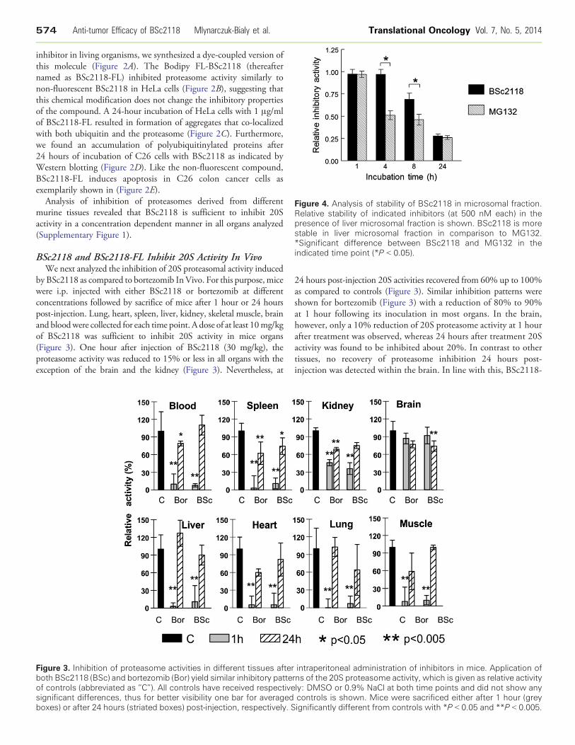
Figure 4. Analysis of stability of BSc2118 in microsomal fraction.Relative stability of indicated inhibitors (at 500 nM each) in thepresence of liver microsomal fraction is shown. BSc2118 is morestable in liver microsomal fraction in comparison to MG132.*Significant difference between BSc2118 and MG132 in theindicated time point (*P b 0.05).
574 Anti-tumor Efficacy of BSc2118 Mlynarczuk-Bialy et al. Translational Oncology Vol. 7, No. 5, 2014
inhibitor in living organisms, we synthesized a dye-coupled version ofthis molecule (Figure 2A). The Bodipy FL-BSc2118 (thereafternamed as BSc2118-FL) inhibited proteasome activity similarly tonon-fluorescent BSc2118 in HeLa cells (Figure 2B), suggesting thatthis chemical modification does not change the inhibitory propertiesof the compound. A 24-hour incubation of HeLa cells with 1 μg/mlof BSc2118-FL resulted in formation of aggregates that co-localizedwith both ubiquitin and the proteasome (Figure 2C). Furthermore,we found an accumulation of polyubiquitinylated proteins after24 hours of incubation of C26 cells with BSc2118 as indicated byWestern blotting (Figure 2D). Like the non-fluorescent compound,BSc2118-FL induces apoptosis in C26 colon cancer cells asexemplarily shown in (Figure 2E).
Analysis of inhibition of proteasomes derived from differentmurine tissues revealed that BSc2118 is sufficient to inhibit 20Sactivity in a concentration dependent manner in all organs analyzed(Supplementary Figure 1).
BSc2118 and BSc2118-FL Inhibit 20S Activity In VivoWe next analyzed the inhibition of 20S proteasomal activity induced
by BSc2118 as compared to bortezomib InVivo. For this purpose, micewere i.p. injected with either BSc2118 or bortezomib at differentconcentrations followed by sacrifice of mice after 1 hour or 24 hourspost-injection. Lung, heart, spleen, liver, kidney, skeletal muscle, brainand bloodwere collected for each time point. A dose of at least 10mg/kgof BSc2118 was sufficient to inhibit 20S activity in mice organs(Figure 3). One hour after injection of BSc2118 (30 mg/kg), theproteasome activity was reduced to 15% or less in all organs with theexception of the brain and the kidney (Figure 3). Nevertheless, at
Figure 3. Inhibition of proteasome activities in different tissues afterboth BSc2118 (BSc) and bortezomib (Bor) yield similar inhibitory patteof controls (abbreviated as “C”). All controls have received respectivesignificant differences, thus for better visibility one bar for averagedboxes) or after 24 hours (striated boxes) post-injection, respectively. S
24 hours post-injection 20S activities recovered from 60% up to 100%as compared to controls (Figure 3). Similar inhibition patterns wereshown for bortezomib (Figure 3) with a reduction of 80% to 90%at 1 hour following its inoculation in most organs. In the brain,however, only a 10% reduction of 20S proteasome activity at 1 hourafter treatment was observed, whereas 24 hours after treatment 20Sactivity was found to be inhibited about 20%. In contrast to othertissues, no recovery of proteasome inhibition 24 hours post-injection was detected within the brain. In line with this, BSc2118-
intraperitoneal administration of inhibitors in mice. Application ofrns of the 20S proteasome activity, which is given as relative activityly: DMSO or 0.9% NaCl at both time points and did not show anycontrols is shown. Mice were sacrificed either after 1 hour (greyignificantly different from controls with *P b 0.05 and **P b 0.005.
Translational Oncology Vol. 7, No. 5, 2014 Anti-tumor Efficacy of BSc2118 Mlynarczuk-Bialy et al. 575
FL at a dose of 5 mg/kg effectively inhibited the 20S activity in miceafter i.p. administration (Supplementary Figure 2). In the selectedorgans, there was a strong 20S inhibition after 1 hour, whereas after24 hours proteasomal activity was almost completely restored.
None of the BSc2118-treated animals (n = 7) demonstrated anysigns of toxicity that could be identified by observation. No weightloss, diarrhea, hair loss, or neurological symptoms (like tremor, ataxia,paralysis) were observed in BSc2118-treated mice, even if mice weretreated daily with a 60 mg/kg dose for 7 consecutive days. In micetreated with bortezomib the drug was administered at 1 mg/kg everysecond day. Animals that were given higher daily doses of bortezomibdied on day three after start of treatment (data not shown).Concluding, BSc2118 inhibits the proteasome activity In Vivo to asimilar extend as bortezomib (Figure 3), but is better tolerated andless toxic in spite of daily administration.
Analysis of Stability of the BSc2118AgainstMicrosomal ProteasesSome proteasome inhibitors like MG132 become instable in
presence of microsomal fractions due to various mechanismsinvolving binding or modification. To analyze the resistance ofBSc2118 against microsomal proteins, measurement of 20Sinhibitory activity in the presence/absence of liver microsomes wasperformed. As shown in Figure 4, BSc2118 is stable for up to 4 hoursof incubation with microsomes. After 8 hours of incubation with livermicrosomes, there is an observed 30% loss of inhibitory activity.Thus, BSc2118 turned out to be more stable than MG132, whichlost its activity at 4 hours in a dose and time dependent manner, i.e.,50% of activity loss at 4 hours and 70% of activity loss at 8 hours,After 24 hours of treatment, the activity loss for both MG132 andBSc2118 was comparative.
Analysis of Biodistribution Using BSc2118-FLTo track the biodistribution of BSc2118, the Bodipy-labeled
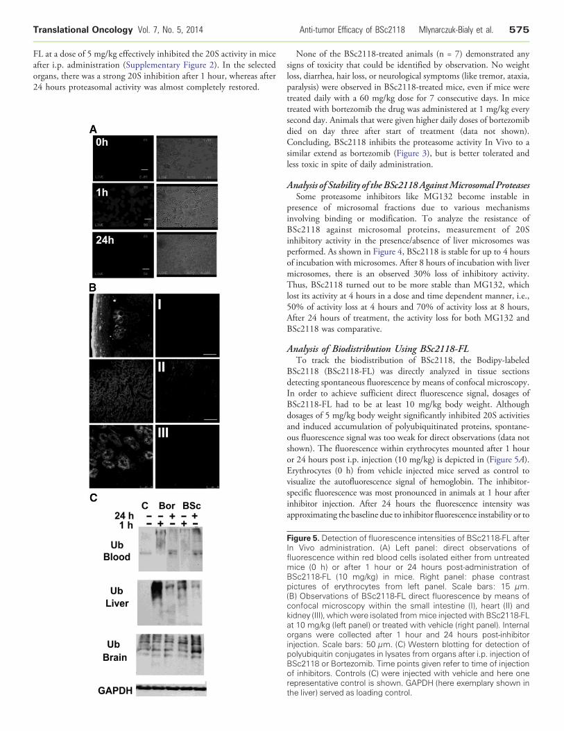
BSc2118 (BSc2118-FL) was directly analyzed in tissue sectionsdetecting spontaneous fluorescence by means of confocal microscopy.In order to achieve sufficient direct fluorescence signal, dosages ofBSc2118-FL had to be at least 10 mg/kg body weight. Althoughdosages of 5 mg/kg body weight significantly inhibited 20S activitiesand induced accumulation of polyubiquitinated proteins, spontane-ous fluorescence signal was too weak for direct observations (data notshown). The fluorescence within erythrocytes mounted after 1 houror 24 hours post i.p. injection (10 mg/kg) is depicted in (Figure 5A).Erythrocytes (0 h) from vehicle injected mice served as control tovisualize the autofluorescence signal of hemoglobin. The inhibitor-specific fluorescence was most pronounced in animals at 1 hour afterinhibitor injection. After 24 hours the fluorescence intensity wasapproximating the baseline due to inhibitor fluorescence instability or to
Figure 5. Detection of fluorescence intensities of BSc2118-FL afterIn Vivo administration. (A) Left panel: direct observations offluorescence within red blood cells isolated either from untreatedmice (0 h) or after 1 hour or 24 hours post-administration ofBSc2118-FL (10 mg/kg) in mice. Right panel: phase contrastpictures of erythrocytes from left panel. Scale bars: 15 μm.(B) Observations of BSc2118-FL direct fluorescence by means ofconfocal microscopy within the small intestine (I), heart (II) andkidney (III), which were isolated frommice injected with BSc2118-FLat 10 mg/kg (left panel) or treated with vehicle (right panel). Internalorgans were collected after 1 hour and 24 hours post-inhibitorinjection. Scale bars: 50 μm. (C) Western blotting for detection ofpolyubiquitin conjugates in lysates from organs after i.p. injection ofBSc2118 or Bortezomib. Time points given refer to time of injectionof inhibitors. Controls (C) were injected with vehicle and here onerepresentative control is shown. GAPDH (here exemplary shown inthe liver) served as loading control.
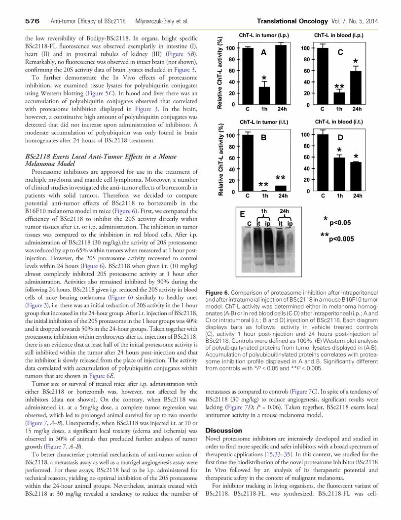
Figure 6. Comparison of proteasome inhibition after intraperitonealand after intratumoral injectionofBSc2118 in amouseB16F10 tumormodel. ChT-L activity was determined either in melanoma homog-enates (A-B) or in red blood cells (C-D) after intraperitoneal (i.p.; A andC) or intratumoral (i.t.; B and D) injection of BSc2118. Each diagramdisplays bars as follows: activity in vehicle treated controls(C), activity 1 hour post-injection and 24 hours post-injection ofBSc2118. Controls were defined as 100%. (E) Western blot analysisof polyubiquitynated proteins from tumor lysates displayed in (A-B).Accumulation of polyubiquitinylated proteins correlates with protea-some inhibition profile displayed in A and B. Significantly differentfrom controls with *P b 0.05 and **P b 0.005.
576 Anti-tumor Efficacy of BSc2118 Mlynarczuk-Bialy et al. Translational Oncology Vol. 7, No. 5, 2014
the low reversibility of Bodipy-BSc2118. In organs, bright specificBSc2118-FL fluorescence was observed exemplarily in intestine (I),heart (II) and in proximal tubules of kidney (III) (Figure 5B).Remarkably, no fluorescence was observed in intact brain (not shown),confirming the 20S activity data of brain lysates included in Figure 3.
To further demonstrate the In Vivo effects of proteasomeinhibition, we examined tissue lysates for polyubiquitin conjugatesusing Western blotting (Figure 5C). In blood and liver there was anaccumulation of polyubiquitin conjugates observed that correlatedwith proteasome inhibition displayed in Figure 3. In the brain,however, a constitutive high amount of polyubiquitin conjugates wasdetected that did not increase upon administration of inhibitors. Amoderate accumulation of polyubiquitin was only found in brainhomogenates after 24 hours of BSc2118 treatment.
BSc2118 Exerts Local Anti-Tumor Effects in a MouseMelanoma Model
Proteasome inhibitors are approved for use in the treatment ofmultiple myeloma and mantle cell lymphoma. Moreover, a numberof clinical studies investigated the anti-tumor effects of bortezomib inpatients with solid tumors. Therefore, we decided to comparepotential anti-tumor effects of BSc2118 to bortezomib in theB16F10 melanoma model in mice (Figure 6). First, we compared theefficiency of BSc2118 to inhibit the 20S activity directly withintumor tissues after i.t. or i.p. administration. The inhibition in tumortissues was compared to the inhibition in red blood cells. After i.p.administration of BSc2118 (30 mg/kg),the activity of 20S proteasomeswas reduced by up to 65%within tumors whenmeasured at 1 hour post-injection. However, the 20S proteasome activity recovered to controllevels within 24 hours (Figure 6). BSc2118 when given i.t. (10 mg/kg)almost completely inhibited 20S proteasome activity at 1 hour afteradministration. Activities also remained inhibited by 90% during thefollowing 24 hours. BSc2118 given i.p. reduced the 20S activity in bloodcells of mice bearing melanoma (Figure 6) similarly to healthy ones(Figure 3), i.e. there was an initial reduction of 20S activity in the 1-hourgroup that increased in the 24-hour group. After i.t. injection of BSc2118,the initial inhibition of the 20S proteasome in the 1 hour groups was 40%and it dropped towards 50% in the 24-hour groups. Taken together withproteasome inhibition within erythrocytes after i.t. injection of BSc2118,there is an evidence that at least half of the initial proteasome activity isstill inhibited within the tumor after 24 hours post-injection and thatthe inhibitor is slowly released from the place of injection. The activitydata correlated with accumulation of polyubiquitin conjugates withintumors that are shown in Figure 6E.
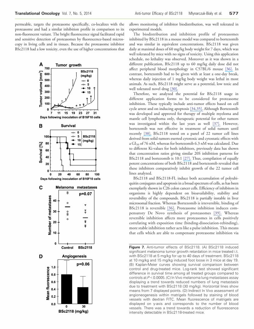
Tumor size or survival of treated mice after i.p. administration witheither BSc2118 or bortezomib was, however, not affected by theinhibitors (data not shown). On the contrary, when BSc2118 wasadministered i.t. at a 5mg/kg dose, a complete tumor regression wasobserved, which led to prolonged animal survival for up to two months(Figure 7, A–B). Unexpectedly, when BSc2118 was injected i.t. at 10 or15 mg/kg doses, a significant local toxicity (edema and ischemia) wasobserved in 30% of animals that precluded further analysis of tumorgrowth (Figure 7, A–B).
To better characterize potential mechanisms of anti-tumor action ofBSc2118, a metastasis assay as well as a matrigel angiogenesis assay wereperformed. For these assays, BSc2118 had to be i.p. administered fortechnical reasons, yielding no optimal inhibition of the 20S proteasomewithin the 24-hour animal groups. Nevertheless, animals treated withBSc2118 at 30 mg/kg revealed a tendency to reduce the number of
metastases as compared to controls (Figure 7C). In spite of a tendency ofBSc2118 (30 mg/kg) to reduce angiogenesis, significant results werelacking (Figure 7D; P = 0.06). Taken together, BSc2118 exerts localantitumor activity in a mouse melanoma model.
DiscussionNovel proteasome inhibitors are intensively developed and studied inorder to find more specific and safer inhibitors with a broad spectrum oftherapeutic applications [15,33–35]. In this context, we studied for thefirst time the biodistribution of the novel proteasome inhibitor BSc2118In Vivo followed by an analysis of its therapeutic potential andtherapeutic safety in the context of malignant melanoma.
For inhibitor tracking in living organisms, the fluorescent variant ofBSc2118, BSc2118-FL, was synthesized. BSc2118-FL was cell-
Translational Oncology Vol. 7, No. 5, 2014 Anti-tumor Efficacy of BSc2118 Mlynarczuk-Bialy et al. 577
permeable, targets the proteasome specifically, co-localizes with theproteasome and had a similar inhibition profile in comparison to itsnon-fluorescent variant. The bright fluorescence signal facilitated rapidand sensitive detection of proteasomes by fluorescence-based micros-copy in living cells and in tissues. Because the proteasome inhibitorBSc2118 had a low toxicity, even the use of higher concentrations that
allows monitoring of inhibitor biodistribution, was well tolerated inexperimental models.
The biodistribution and inhibition profile of proteasomesinhibited by BSc2118 in a mouse model was compared to bortezomiband was similar in equivalent concentrations. BSc2118 was givendaily at maximal doses of 60 mg/kg body weight for 7 days, which waswell tolerated by mice with no signs of toxicity. Using this applicationschedule, no lethality was observed. Moreover as it was shown in adifferent publication, BSc2118 up to 60 mg/kg daily dose did notaffect peripheral blood morphology in C57BL/6 mouse [36]. Incontrast, bortezomib had to be given with at least a one-day break,whereas daily injection of 1 mg/kg body weight was lethal in mostanimals. As such, BSc2118 might serve as a potential, low toxic andwell tolerated novel drug [30].
Therefore, we analyzed the potential for BSc2118 usage indifferent application forms to be considered for proteasomeinhibition. These typically include anti-tumor effects based on cellcycle arrest and on inducing apoptosis [34,35]. Although Bortezomibwas developed and approved for therapy of multiple myeloma andmantle cell lymphoma only, therapeutic potential for other tumorswas investigated within the last years as well [37]. However,bortezomib was not effective in treatment of solid tumors untilrecently [38]. BSc2118 tested on a panel of 22 tumor cell linesderived from solid tumors exerted cytotoxic and cytostatic effects witha GI50 of 76 nM, whereas for bortezomib 6.3 nM was calculated. Dueto different Ki-values for both inhibitors, previously data has shownthat concentration ratios giving similar 20S inhibition patterns forBSc2118 and bortezomib is 10:1 [27]. Thus, compilation of equallypotent concentrations of both BSc2118 and bortezomib revealed thatthese inhibitors comparatively inhibit growth of the 22 tumor celllines analyzed.
BSc2118 and BSc2118-FL induce both accumulation of polyubi-quitin conjugates and apoptosis in a broad spectrum of cells, as has beenexemplarily shown in C26 colon cancer cells. Efficiency of inhibitors inorganisms is highly dependent on bioavailability, stability andreversibility of the compounds. BSc2118 is partially instable in livermicrosomal fraction. Whereas Bortezomib is irreversible, binding ofBSc2118 is reversible [36]. Proteasome inhibition induces com-pensatory De Novo synthesis of proteasomes [39]. Whereasreversible inhibition affects more proteasomes in cells positivelycorrelating with exposition time (binding-dissociation-rebinding),more stable inhibition rather acts like a pulse inhibition. This meansthat cells which are able to compensate proteasome inhibition via
Figure 7. Anti-tumor effects of BSc2118. (A) BSc2118 inducedsignificant melanoma tumor growth retardation in mice treated i.t.with BSc2118 at 5 mg/kg for up to 40 days of treatment. BSc2118at 10 mg/kg and 15 mg/kg induced foot loose in 3 mice at day 19.(B) Kaplan-Meier curves showing survival comparison betweencontrol and drug-treated mice. Log-rank test showed significantdifference in survival time among all treated groups compared tocontrols at P b 0.0005. (C) In Vivo melanoma lungmetastases assaydisplaying a trend towards reduced numbers of lung metastasisdue to treatment with BSc2118 (30 mg/kg). Horizontal lines showmeans from 7 displayed points. (D) Indirect In Vivo assessment ofangioneogenesis within matrigels followed by staining of bloodvessels with dextran FITC. Mean fluorescence of matrigels aredisplayed on y-axis and corresponds to the number of bloodvessels. There was a trend towards a reduction of fluorescenceintensity detectable in BSc2118-treated mice.
578 Anti-tumor Efficacy of BSc2118 Mlynarczuk-Bialy et al. Translational Oncology Vol. 7, No. 5, 2014
De Novo synthesis do survive, but cells that are incapable of doingso suffer from UPR stress and accumulation of oxidized proteins[40]. In this context, the majority of tumor cells are more sensibleto proteasome inhibition than their parental cells [27].
In order to study possible therapeutic potentials of BSc2118, westudied BSc2118-mediated effects in a mouse model of malignantmelanoma. BSc2118 in experimental melanoma therapy revealedsome unexpected findings. First of all, neither BSc2118 norbortezomib injected i.p. had any effects on tumor growth or survivalof B16F10 tumor bearing mice (data not shown). It is known thattumor tissue has its own milieu and drugs working well In Vitromight not be effective In Vivo due to the existence of the tumormatrix [41]. Therefore, the inhibitor was injected directly into thetumor. Comparison of proteasome inhibition profiles after both i.p.and i.t. injection of BSc2118 revealed that BSc2118 completelyinhibited proteasome activity after i.t. injection, which lasted for atleast 24 h. This result prompted us to check the effects of BSc2118 ontumor growth when injected i.t. We obtained tumor growthretardation and complete remission with a survival for up to twomonths in 38.5% of mice receiving BSc2118 from all experimentalgroups. However, BSc2118 at 10 and 15 mg/kg induced localtoxicity, suggesting that local levels of proteasome inhibition withinthe tissue should not exceed 80%. On the contrary, increasedproteasome inhibition might be toxic as has been demonstrated forbortezomib in primates [42]. In humans the inhibition of 20S activitywith bortezomib does not exceed 70% [43].
To better characterize the mechanisms of action of BSc2118 in atumor model we studied BSc2118-mediated De Novo angiogenesisand metastasis in a model of malignant melanoma. Noteworthy, ourexperimental model needed i.p. injections of BSc2118 that might notinduce satisfactory level of proteasome inhibition and had no effectson tumor growth. However, in this application design there was atendency at the border of significance to reduce the number ofmetastases and De Novo arising blood vessels. It seems that BSc2118retards tumor growth by means of 20S inhibition within tumor cellsand additionally might reduce both metastasis and angiogenesis.
In conclusion, we characterized a novel proteasome inhibitor thathas a similar proteasome inhibition spectrum compared tobortezomib In Vitro and In Vivo, but has under the conditionstested less signs of toxicity. We hypothesize that BSc2118 is atherapeutic alternative to bortezomib in therapy of solid tumors, forwhich further studies will be needed.
Supplementary data to this article can be found online at http://dx.doi.org/10.1016/j.tranon.2014.07.002.
AcknowledgmentsThis work was in part supported by the Polish Ministry of Science andhigher Education (grant number: N405 007 31/0544 to IMB).
We thank Dr. Anna Ratajska from the Department of PathologicalAnatomy, Medical University of Warsaw, Poland, for assistance inpreliminary experiments on BSc2118-FL stability in mice.
References
[1] Voges D, Zwickl P, and Baumeister W (1999). The 26S proteasome: a molecularmachine designed for controlled proteolysis. Annu Rev Biochem 68, 1015–1068.
[2] GrollM,Ditzel L, Lowe J, StockD, BochtlerM,BartunikHD, andHuber R (1997).Structure of 20S proteasome from yeast at 2.4 A resolution. Nature 386, 463–471.
[3] Rechsteiner M, Hoffman L, and Dubiel W (1993). The multicatalytic and 26 Sproteases. J Biol Chem 268, 6065–6068.
[4] Thrower JS, Hoffman L, Rechsteiner M, and Pickart CM (2000). Recognition ofthe polyubiquitin proteolytic signal. EMBO J 19, 94–102.
[5] Seufert W and Jentsch S (1992). In vivo function of the proteasome in theubiquitin pathway. EMBO J 11, 3077–3080.
[6] Gardner RC, Assinder SJ, Christie G, Mason GG, Markwell R, Wadsworth H,McLaughlin M, King R, Chabot-Fletcher MC, and Breton JJ, et al (2000).Characterization of peptidyl boronic acid inhibitors of mammalian 20 S and 26S proteasomes and their inhibition of proteasomes in cultured cells. Biochem J346(Pt 2), 447–454.
[7] Wojcik C (1999). Proteasomes in apoptosis: villains or guardians? Cell Mol LifeSci 56, 908–917.
[8] Muchamuel T, Basler M, Aujay MA, Suzuki E, Kalim KW, Lauer C, Sylvain C,Ring ER, Shields J, and Jiang J, et al (2009). A selective inhibitor of theimmunoproteasome subunit LMP7 blocks cytokine production and attenuatesprogression of experimental arthritis. Nat Med 15, 781–787.
[9] Neubert K, Meister S, Moser K, Weisel F, Maseda D, Amann K, Wiethe C,Winkler TH, Kalden JR, and Manz RA, et al (2008). The proteasome inhibitorbortezomib depletes plasma cells and protects mice with lupus-like disease fromnephritis. Nat Med 14, 748–755.
[10] Dahlmann B, Ruppert T, Kuehn L, Merforth S, and Kloetzel PM (2000).Different proteasome subtypes in a single tissue exhibit different enzymaticproperties. J Mol Biol 303, 643–653.
[11] Marfella R, Di Filippo C, Portoghese M, Siniscalchi M, Martis S, Ferraraccio F,Guastafierro S, Nicoletti G, Barbieri M, and Coppola A, et al (2009). Theubiquitin-proteasome system contributes to the inflammatory injury in ischemicdiabetic myocardium: the role of glycemic control. Cardiovasc Pathol 18,332–345.
[12] Tie L, Xu Y, Lin YH, Yao XH, Wu HL, Li YH, Shen ZF, Yu HM, and Li XJ(2008). Down-regulation of brain-pancreas relative protein in diabetic rats andby high glucose in PC12 cells: prevention by calpain inhibitors. J Pharmacol Sci106, 28–37.
[13] Petroski MD (2008). The ubiquitin system, disease, and drug discovery. BMCBiochem 9(Suppl. 1), S7. http://dx.doi.org/10.1186/1471-2091-9-S1-S7.
[14] Fenical W, Jensen PR, Palladino MA, Lam KS, Lloyd GK, and Potts BC (2009).Discovery and development of the anticancer agent salinosporamide A (NPI-0052).Bioorg Med Chem 17, 2175–2180.
[15] Dick LR and Fleming PE (2010). Building on bortezomib: second-generationproteasome inhibitors as anti-cancer therapy. Drug Discov Today 15, 243–249.
[16] Kuhn DJ, Chen Q, Voorhees PM, Strader JS, Shenk KD, Sun CM, Demo SD,Bennett MK, van Leeuwen FW, and Chanan-Khan AA, et al (2007). Potentactivity of carfilzomib, a novel, irreversible inhibitor of the ubiquitin-proteasomepathway, against preclinical models of multiple myeloma. Blood 110,3281–3290.
[17] Teicher BA, Ara G, Herbst R, Palombella VJ, and Adams J (1999). Theproteasome inhibitor PS-341 in cancer therapy. Clin Cancer Res 5, 2638–2645.
[18] Adams J and Kauffman M (2004). Development of the proteasome inhibitorVelcade (Bortezomib). Cancer Invest 22, 304–311.
[19] Adams J (2003). Potential for proteasome inhibition in the treatment of cancer.Drug Discov Today 8, 307–315.
[20] O'Connor OA and CzuczmanMS (2008). Novel approaches for the treatment ofNHL: Proteasome inhibition and immune modulation. Leuk Lymphoma 49(Suppl. 1), 59–66.
[21] Goy A, Bernstein SH, Kahl BS, Djulbegovic B, Robertson MJ, de Vos S, EpnerE, Krishnan A, Leonard JP, and Lonial S, et al (2009). Bortezomib in patientswith relapsed or refractory mantle cell lymphoma: updated time-to-event analysesof the multicenter phase 2 PINNACLE study. Ann Oncol 20, 520–525.
[22] Einsele H (2010). Bortezomib. Recent Results Cancer Res 184, 173–187.[23] Gilardini A, Marmiroli P, and Cavaletti G (2008). Proteasome inhibition: a
promising strategy for treating cancer, but what about neurotoxicity? Curr MedChem 15, 3025–3035.
[24] Meregalli C, Canta A, Carozzi VA, Chiorazzi A, Oggioni N, Gilardini A, CeresaC, Avezza F, Crippa L, and Marmiroli P, et al (2010). Bortezomib-inducedpainful neuropathy in rats: a behavioral, neurophysiological and pathologicalstudy in rats. Eur J Pain 14, 343–350.
[25] Argyriou AA, Iconomou G, and Kalofonos HP (2008). Bortezomib-inducedperipheral neuropathy in multiple myeloma: a comprehensive review of theliterature. Blood 112, 1593–1599.

Translational Oncology Vol. 7, No. 5, 2014 Anti-tumor Efficacy of BSc2118 Mlynarczuk-Bialy et al. 579
[26] Nowis D, Maczewski M, Mackiewicz U, Kujawa M, Ratajska A, WieckowskiMR, Wilczynski GM, Malinowska M, Bil J, and Salwa P, et al (2010).Cardiotoxicity of the anticancer therapeutic agent bortezomib. Am J Pathol 176,2658–2668.
[27] Mlynarczuk-Bialy I, Roeckmann H, Kuckelkorn U, Schmidt B, Umbreen S,Golab J, Ludwig A, Montag C, Wiebusch L, and Hagemeier C, et al (2006).Combined effect of proteasome and calpain inhibition on cisplatin-resistanthuman melanoma cells. Cancer Res 66, 7598–7605.
[28] Braun HA, Umbreen S, Groll M, Kuckelkorn U, Mlynarczuk I, Wigand ME,Drung I, Kloetzel PM, and Schmidt B (2005). Tripeptide mimetics inhibit the20 S proteasome by covalent bonding to the active threonines. J Biol Chem 280,28394–28401.
[29] Kern MA, Helmbach H, Artuc M, Karmann D, Jurgovsky K, and SchadendorfD (1997). Human melanoma cell lines selected in vitro displaying various levelsof drug resistance against cisplatin, fotemustine, vindesine or etoposide:modulation of proto-oncogene expression. Anticancer Res 17, 4359–4370.
[30] Adams J, Palombella VJ, Sausville EA, Johnson J, Destree A, Lazarus DD, MaasJ, Pien CS, Prakash S, and Elliott PJ (1999). Proteasome inhibitors: a novel classof potent and effective antitumor agents. Cancer Res 59, 2615–2622.
[31] Kuckelkorn U, Ruppert T, Strehl B, Jungblut PR, Zimny-Arndt U, Lamer S, PrinzI, Drung I, Kloetzel PM, and Kaufmann SH, et al (2002). Link between organ-specific antigen processing by 20S proteasomes and CD8(+) T cell-mediatedautoimmunity. J Exp Med 195, 983–990.
[32] Feleszko W, Mlynarczuk I, Olszewska D, Jalili A, Grzela T, Lasek W, Hoser G,Korczak-Kowalska G, and Jakobisiak M (2002). Lovastatin potentiatesantitumor activity of doxorubicin in murine melanoma via an apoptosis-dependent mechanism. Int J Cancer 100, 111–118.
[33] Ling YH, Liebes L, Jiang JD, Holland JF, Elliott PJ, Adams J, Muggia FM, andPerez-Soler R (2003). Mechanisms of proteasome inhibitor PS-341-induced G(2)-M-phase arrest and apoptosis in human non-small cell lung cancer cell lines.Clin Cancer Res 9, 1145–1154.
[34] Hideshima T, Richardson P, Chauhan D, Palombella VJ, Elliott PJ, Adams J,and Anderson KC (2001). The proteasome inhibitor PS-341 inhibits growth,
induces apoptosis, and overcomes drug resistance in human multiple myelomacells. Cancer Res 61, 3071–3076.
[35] Lu G, Punj V, and Chaudhary PM (2008). Proteasome inhibitor Bortezomibinduces cell cycle arrest and apoptosis in cell lines derived from Ewing's sarcomafamily of tumors and synergizes with TRAIL. Cancer Biol Ther 7, 603–608.
[36] Doeppner TR, Mlynarczuk-Bialy I, Kuckelkorn U, Kaltwasser B, Herz J, HasanMR, Hermann DM, and Bahr M (2012). The novel proteasome inhibitorBSc2118 protects against cerebral ischaemia through HIF1A accumulation andenhanced angioneurogenesis. Brain 135, 3282–3297.
[37] Jones RJ, Chen Q, Voorhees PM, Young KH, Bruey-Sedano N, Yang D, andOrlowski RZ (2008). Inhibition of the p53 E3 ligase HDM-2 induces apoptosisand DNA damage–independent p53 phosphorylation in mantle cell lymphoma.Clin Cancer Res 14, 5416–5425.
[38] Yang H, Zonder JA, and Dou QP (2009). Clinical development of novelproteasome inhibitors for cancer treatment. Expert Opin Investig Drugs 18,957–971.
[39] Meiners S, Heyken D,Weller A, Ludwig A, Stangl K, Kloetzel PM, and Kruger E(2003). Inhibition of proteasome activity induces concerted expression ofproteasome genes and de novo formation of Mammalian proteasomes. J BiolChem 278, 21517–21525.
[40] Suzuki E, Demo S, Deu E, Keats J, Arastu-Kapur S, Bergsagel PL, Bennett MK,and Kirk CJ (2011). Molecular mechanisms of bortezomib resistant adenocar-cinoma cells. PLoS One 6, e27996.
[41] Gabelloni P, Da Pozzo E, Bendinelli S, Costa B, Nuti E, Casalini F, Orlandini E,Da Settimo F, Rossello A, and Martini C (2010). Inhibition of metalloprotein-ases derived from tumours: new insights in the treatment of human glioblastoma.Neuroscience 168, 514–522.
[42] Adams J (2002). Development of the proteasome inhibitor PS-341. Oncologist 7,9–16.
[43] Hamilton AL, Eder JP, Pavlick AC, Clark JW, Liebes L, Garcia-Carbonero R,Chachoua A, Ryan DP, Soma V, and Farrell K, et al (2005). Proteasome inhibitionwith bortezomib (PS-341): a phase I study with pharmacodynamic end points usinga day 1 and day 4 schedule in a 14-day cycle. J Clin Oncol 23, 6107–6116.
Copyright © 2022 FDOKUMEN