99mTc-glycopeptide: Synthesis, biodistribution and imaging in breast tumor-bearing rodents
12
Applied Radiation and Isotopes 66 (2008) 320–331 99m Tc-glycopeptide: Synthesis, biodistribution and imaging in breast tumor-bearing rodents I-Chien Wei a , Ning Tsao b , Ya-Hui Huang a , Yen-Sheng Ho a , Chung-Chin Wu a , Dong-Fang Yu b , David J. Yang b, a Center for Innovation Development, Taiwan Hopax Chems Mfg. Co., Ltd., No. 28, Hua Dong Road, Daliao, Kaohsiung 83162, Taiwan b Division of Diagnostic Imaging, The University of Texas M.D. Anderson Cancer Center, 1515 Holcombe Blvd., Box 59, Houston, TX 77030, USA Received 8 August 2007; received in revised form 1 October 2007; accepted 7 October 2007 Abstract This study was aimed to develop a glycopeptide (GP) to be used as a carrier for anti-cancer drug delivery. GP was synthesized by conjugating glutamate peptide and chitosan using carbodiimide as a coupling agent. Elemental analysis and capillary electrophoresis confirmed the purity was 495%. GP was labeled with sodium pertechnetate (Na 99m TcO 4 ) for in vitro and in vivo studies. Rhenium-GP was synthesized to support the binding site of 99m Tc at the glutamate positions 3–5. In vitro cellular uptake of 99m Tc-GP was performed in breast cancer cells. Cytosol had 60% whereas nucleus had 40% uptake of 99m Tc-GP. When cancer cells were incubated with glutamate or aspartate, followed by 99m Tc-GP, there was decreased uptake in cells treated with glutamate but not aspartate. The findings indicated that cellular uptake of 99m Tc-GP was via glutamate transporters. In addition, 99m Tc-GP was able to measure uptake differences after cells treated with paclitaxel. Biodistribution and planar imaging were conducted in breast tumor-bearing rats. Biodistribution of 99m Tc-GP showed increased tumor-to-tissue ratios as a function of time. Planar images confirmed that 99m Tc-GP could assess tumor uptake changes after paclitaxel treatment. In vitro and in vivo studies indicated that GP could target tumor cells, thus, GP may be a useful carrier for anti-cancer drug delivery. Published by Elsevier Ltd. Keywords: Glycopeptide; Radiolabeled; Biodistribution; Imaging 1. Introduction Polysaccharides such as heparin, chitosan and chondroi- tin play a potent role in tumor growth, tissue repair and angiogenesis (Polykratis et al., 2005; Pieper et al., 2000; Biagini et al., 1991). Chitosan, a natural-based polymer obtained by alkaline deacetylation of chitin, is nontoxic, biocompatible, and biodegradable. These properties make chitosan a good candidate for conventional and novel drug delivery systems. Other polysaccharides such as collagen, chondroitin and hyauraniate have also been applied to drug delivery (Prabaharan and Mano, 2005). Despite the outstanding advances made in the field of angiogenesis, some significant limitations still remain in the treatment of various diseases via the delivery of cytotoxic drugs in vivo. In order to reach high therapeutic index of an agent in cancer treatment, it would be necessary to design a carrier to target tumor vascular and cellular component. Amino acids are important biologic substrates that play crucial roles in virtually all biological processes. These ionic nutrients serve not only as basic modules of proteins and hormones but also as neurotransmitters, synaptic modulators, or neurotransmitter precursors. Amino acid transport is generally increased in malignant transforma- tion (Waniewski and Martin, 1984). However, the process of malignant transformation requires that cells acquire and use nutrients efficiently for energy, protein synthesis, and cell division. Proteins, which play crucial roles in virtually all biological processes, are built from amino acids linked through peptide bonds. The excitatory amino acid gluta- mate (Glu) is a potent neurotransmitter in the central nervous system and exerts its action via a variety of glutamate receptors (GluRs). It has been reported that ARTICLE IN PRESS www.elsevier.com/locate/apradiso 0969-8043/$ - see front matter Published by Elsevier Ltd. doi:10.1016/j.apradiso.2007.10.002 Corresponding author. Tel.: +1 713 794 1053; fax: +1 713 794 5456. E-mail address: [email protected] (D.J. Yang).
-
Upload
independent -
Category
Documents
-
view
0 -
download
0
Transcript of 99mTc-glycopeptide: Synthesis, biodistribution and imaging in breast tumor-bearing rodents
ARTICLE IN PRESS
0969-8043/$ - se
doi:10.1016/j.ap
�CorrespondE-mail addr
Applied Radiation and Isotopes 66 (2008) 320–331
www.elsevier.com/locate/apradiso
99mTc-glycopeptide: Synthesis, biodistribution and imagingin breast tumor-bearing rodents
I-Chien Weia, Ning Tsaob, Ya-Hui Huanga, Yen-Sheng Hoa, Chung-Chin Wua,Dong-Fang Yub, David J. Yangb,�
aCenter for Innovation Development, Taiwan Hopax Chems Mfg. Co., Ltd., No. 28, Hua Dong Road, Daliao, Kaohsiung 83162, TaiwanbDivision of Diagnostic Imaging, The University of Texas M.D. Anderson Cancer Center, 1515 Holcombe Blvd., Box 59, Houston, TX 77030, USA
Received 8 August 2007; received in revised form 1 October 2007; accepted 7 October 2007
Abstract
This study was aimed to develop a glycopeptide (GP) to be used as a carrier for anti-cancer drug delivery. GP was synthesized by
conjugating glutamate peptide and chitosan using carbodiimide as a coupling agent. Elemental analysis and capillary electrophoresis
confirmed the purity was 495%. GP was labeled with sodium pertechnetate (Na99mTcO4) for in vitro and in vivo studies. Rhenium-GP
was synthesized to support the binding site of 99mTc at the glutamate positions 3–5. In vitro cellular uptake of 99mTc-GP was performed
in breast cancer cells. Cytosol had 60% whereas nucleus had 40% uptake of 99mTc-GP. When cancer cells were incubated with glutamate
or aspartate, followed by 99mTc-GP, there was decreased uptake in cells treated with glutamate but not aspartate. The findings indicated
that cellular uptake of 99mTc-GP was via glutamate transporters. In addition, 99mTc-GP was able to measure uptake differences after cells
treated with paclitaxel. Biodistribution and planar imaging were conducted in breast tumor-bearing rats. Biodistribution of 99mTc-GP
showed increased tumor-to-tissue ratios as a function of time. Planar images confirmed that 99mTc-GP could assess tumor uptake
changes after paclitaxel treatment. In vitro and in vivo studies indicated that GP could target tumor cells, thus, GP may be a useful
carrier for anti-cancer drug delivery.
Published by Elsevier Ltd.
Keywords: Glycopeptide; Radiolabeled; Biodistribution; Imaging
1. Introduction
Polysaccharides such as heparin, chitosan and chondroi-tin play a potent role in tumor growth, tissue repair andangiogenesis (Polykratis et al., 2005; Pieper et al., 2000;Biagini et al., 1991). Chitosan, a natural-based polymerobtained by alkaline deacetylation of chitin, is nontoxic,biocompatible, and biodegradable. These properties makechitosan a good candidate for conventional and novel drugdelivery systems. Other polysaccharides such as collagen,chondroitin and hyauraniate have also been applied todrug delivery (Prabaharan and Mano, 2005). Despite theoutstanding advances made in the field of angiogenesis,some significant limitations still remain in the treatment ofvarious diseases via the delivery of cytotoxic drugs in vivo.
e front matter Published by Elsevier Ltd.
radiso.2007.10.002
ing author. Tel.: +1713 794 1053; fax: +1 713 794 5456.
ess: [email protected] (D.J. Yang).
In order to reach high therapeutic index of an agent incancer treatment, it would be necessary to design a carrierto target tumor vascular and cellular component.Amino acids are important biologic substrates that play
crucial roles in virtually all biological processes. Theseionic nutrients serve not only as basic modules of proteinsand hormones but also as neurotransmitters, synapticmodulators, or neurotransmitter precursors. Amino acidtransport is generally increased in malignant transforma-tion (Waniewski and Martin, 1984). However, the processof malignant transformation requires that cells acquire anduse nutrients efficiently for energy, protein synthesis, andcell division. Proteins, which play crucial roles in virtuallyall biological processes, are built from amino acids linkedthrough peptide bonds. The excitatory amino acid gluta-mate (Glu) is a potent neurotransmitter in the centralnervous system and exerts its action via a variety ofglutamate receptors (GluRs). It has been reported that

ARTICLE IN PRESS
Fig. 1. Structure of glycopeptide (GP).
I.-C. Wei et al. / Applied Radiation and Isotopes 66 (2008) 320–331 321
glutamate peptide stimulates bone resorption in vitro, aneffect specific to glutamate transporter (Chenu et al., 1998).Chains of five or more glutamic acids could reduce renaluptake in animal models (Behe et al., 2005). It has beenreported that glutamate peptide–drug conjugates providebetter efficacy against solid tumor growth than drug alone(Veronese et al., 2005). In addition, glutamate peptidecould be chelated with gadolinium for magnetic resonanceimaging applications (Lu et al., 2003). Glycopeptide (GP),a copolymer of chitosan and glutamate peptide, wouldprovide an opportunity to target tumor vasculature andcellular via glutamate transporters. Because the repeatedunits of glycopeptide have amino and acid functionalgroups, a cytotoxic agent could be conjugated to GP. Thechemotherapeutic agent may be gradually released fromGP, thus, acute systemic toxicity could be decreased. Inaddition, drugs with poor water solubility and tumortargeting capability could be improved, thus, the thera-peutic index (toxicity/efficacy) of an agent will beincreased.
In order to assess cellular uptake and biodistribution ofGP, GP needs to be labeled with a radioisotope for in vitroand in vivo studies. Due to favorable physical character-istics and its low cost, 99mTc has been preferred for labelingradiopharmaceuticals. Diethylenetriaminepentaacetic acid(DTPA) can be labeled with 99mTc easily and efficientlywith high radiochemical purity and stability and is excretedthrough kidney (Itoh, 2003). Several compounds have beenlabeled with 99mTc using DTPA as a chelator (Trump et al.,2002; Sato et al., 2000; Barth et al., 1996). DTPA couldalso be labeled with other metals for gamma imaging. Forinstance, Cu-61/64 is used for positron emission tomogra-phy (PET) imaging (Li et al., 2002), gadolinium or copperfor MRI (Lai et al., 1987; Rijpkema et al., 2004), andrhenium (Re-188) and holmium (Ho-166) (Koppe et al.,2004) for internal radiotherapeutic needs. Glutamatepeptide has repeated units of glutamic acid which mimicsDTPA chelation. Thus, GP was labeled with 99mTc. Here,we report synthesis, in vitro cellular uptake assays,biodistribution and planar imaging studies of 99mTc-GP.
2. Materials and methods
2.1. Chemicals and analysis
Nuclear magnetic resonance (NMR) spectra wererecorded on a Bruker 300 spectrometer at the Universityof Texas MD Anderson Cancer Center (Houston, TX).The elemental analysis data were obtained from GalbraithLaboratories (Knoxville, TN). Radioactive tissue sampleswere recorded on a Packard gamma counter (DownersGrove, IL). Scintigraphic images were obtained from agamma camera equipped with low-energy, parallel-holecollimator (M-camera, Siemens Medical Systems, Inc.,Hoffman Estates, IL). 1-Ethyl-3-(3-dimethylaminopropyl)carbodiimide–HCl and nuclear and cytoplasmic extractionreagents (NE–PER) were purchased from Pierce Chemical
Company (Rockford, IL). Chitosan (MW ¼ 3500–5000,60% deacetylated) was obtained from Taiwan HopaxChems Mfg Company (Kaohsiung, Taiwan). All otherchemicals were purchased from Sigma Chemical Company(St. Louis, MO). Silica-gel coated instant thin-layerchromatography (ITLC) plates were purchased from Gel-man Sciences (Ann Arbor, MI). Spectra/POR molecularporous membrane with cut-off at 5000 was obtained fromSpectrum Medical Industries Inc. (Houston, TX). Immu-nofluorescence double staining deoxynucleotidyl transfer-ase-mediated 20-deoxyuridine 50-triphosphate-biotin nickend labeling (TUNEL) assay kit was purchased fromPromega Corporation (Madison, WI). Sodium pertechne-tate was obtained from a commercial 99Mo/99mTc gen-erator (Ultratechnekow FMTM, Mallinckrodt Diagnostics,Houston, TX).
2.2. Synthesis of glycopeptide (GP)
Unlike high molecular weight chitosan (MW ¼ 100,000),low molecular weight chitosan (MW ¼ 3500–5000; 60%deacetylated) is water soluble. Glutamate peptide sodiumsalt is water soluble at pH 7.4. GP conjugation wasconducted in aqueous solution at pH 7.4 using watersoluble 3-ethyl-3-(3-dimethylaminopropyl) carbodiimi-de–HCl as a coupling agent. There was no gel formationunder this reaction condition. Briefly, to a stirred solutionof chitosan (200mg, MW ¼ 3500–5000) in water (4mL)and 3-ethyl-3-(3-dimethylaminopropyl) carbodiimide–HCl(128.5mg, 0.67mmol) was added. Glutamate peptidesodium salt (200mg, MW ¼ 1308) was then added. Themixture was stirred at room temperature for 24 h. Themixture was dialyzed for 48 h using Spectra/POR mole-cular porous membrane with cut-off at 5000. After dialysis,the product was filtered and dried using a freeze dryer. TheGP in the salt form weighed 320mg. The chemicalstructure is shown in Fig. 1. Elemental analysis(C11H14N2Na2O6, C, H, N) showed C: 41.54; H: 10:62;
ARTICLE IN PRESSI.-C. Wei et al. / Applied Radiation and Isotopes 66 (2008) 320–331322
N: 7.19 (calculated C: 41.77; H: 8.86; N: 4.43). Capillaryelectrophoresis (CE) was used to analyze the purityof GP.
2.3. Radiolabeling of GP with 99mTc
To determine the binding site of 99mTc in GP, sodiumperrheniate (NaReO4) was used in the cold chemistry. Rechemistry has been used to support the binding site orstructure of 99mTc (Tzanopoulou et al., 2006). It is knownthat 99mTc binds to GP through the carboxylic acid moietyof glutamate peptide, thus, glutamate peptide was selectedfor Re chemistry instead of GP. Rhenium oxotrichloridetriphenylphosphine (41.7mg, 0.05mmol) was added toglutamate peptide (65.5mg, 0.05mmol) in a mixture ofethanol (3mL) and dimethylformamide (5mL) solution.The reaction was refluxed for 30min. The solution wasfiltered and the solvent was evaporated to dryness, yielded30mg (40%). Proton and C-13 NMR spectra wereperformed to determine the structure of Re-glutamatepeptide. Radiolabeling of GP with 99mTc was performed ina standard manner (Barth et al., 1996). Briefly, GP (10mg)was dissolved in water (0.3mL). Tin chloride (0.1mg in0.1mL of water) and Na99mTcO4 (1mCi) were added atroom temperature. Radiochemical purity was determinedby ITLC silica-gel coated plate eluted with saline.
2.4. In vitro stability assays of 99mTc-GP
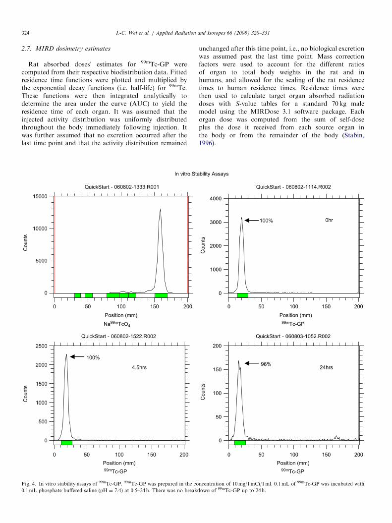
GP was prepared in the concentration of 10mg/ml andNa99mTcO4 (1mCi) was added. 0.1mL of 99mTc-GP wasincubated with 0.1mL phosphate buffered saline (PBS;pH ¼ 7.4) for 0.5–24 h. In vitro stability of 99mTc-GP wasdetermined by chromatographic analysis, as described inthe above section.
2.5. In vitro cell culture assays
To evaluate whether there is a correlation of uptake as afunction of time, a breast tumor cell line (13,762) wasselected because the same cell line was used to createanimal models. The cells were plated to 12 wells tissueculture plate that contained 50,000 per each well. GP(1mg) was dissolved in water (1mL). An aliquot of GPsolution (0.1mg) was added with tin chloride (0.1mg in0.1mL of water) and sodium pertechnetate (0.1mCi).99mTc-GP (0.1mCi) was prepared in the concentration of0.1mg/mL. The cells were incubated with 99mTc-GP (2 mCi/well, 0.02mg/well) at 37 1C for 0.5–6 h. After incubation,cells were washed with ice-cold PBS twice and trypsinizedwith 0.5mL of trypsin solution to detach tumor cells. Thencells were collected by centrifugation. The cells wereweighed and the radioactivity was measured by gammacounter. Data are expressed in mean7S.D. percent ofcellular uptake per mg of cell weight from three measure-ments.
To determine the distribution pattern of 99mTc-GPwithin the cells, NE–PER extraction reagents were usedstepwise to isolate cytoplasmic and nuclear extracts frombreast cancer cells. 99mTc-GP (0.1mCi) was prepared in theconcentration of 0.1mg/mL. After 20 ml of 99mTc-GP(0.02mg/well, 2 mCi) or Na99mTcO4 (control) is dispensedinto each well of 6-well plate, cells (50,000 per well) wereincubated at 37 1C for 2 h. Cells were transferred to a 2mLmicrocentrifuge tube and isolated by centrifugation at500 g for 2–3min. Two hundred microliters of ice-cold kitreagent (CER I) was added to the cell pellet in otherremaining tubes. Following 10min incubation, 11 ml of ice-cold CER II was added and incubated for an additional1min. The tubes were centrifuged for 5min at maxi-mum speed in a microcentrifuge (�16,000� g) and thesupernatant (cytoplasmic extract) fractions were trans-ferred to other tubes for counting. Insoluble (pellet)fractions, which contain nuclei, were resuspended in100 mL of ice-cold NER. Following a total of 40minincubation, the tubes were recentrifuged at a maximumspeed (�16,000� g) for 10min and the supernatant(nuclear extract) fractions were transferred to other tubesfor counting. Insoluble (pellet) fraction, which containsnuclear material, was resuspended in 100 mL of 1�PBSand transferred to other tubes for counting. Samples werecounted in a gamma counter.To determine whether 99mTc-GP could monitor post-
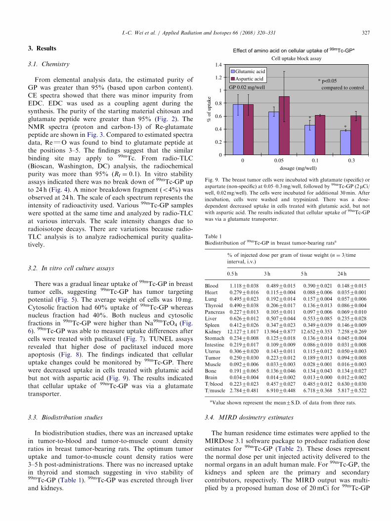
treatment changes, breast tumor cells (50,000 per each well)were incubated with paclitaxel (1–5 mM) for 48 h. 99mTc-GPwas prepared in the concentration of 0.1mg/0.1mCi/1mL.99mTc-GP (2 mCi/well, 0.02mg/well) was added to the wellsand incubated for 4 h before and after paclitaxel treatment.TUNEL assays were used to assess paclitaxel-inducedapoptosis in these cells. TUNEL assays were described inour previously published procedure (Yang et al., 2001). Todetermine whether cellular uptake of 99mTc-GP was via aglutamate transporter, the breast tumor cells were incu-bated with glutamate (specific) or aspartate (non-specific)at 0.05–0.3mg/well, followed by 99mTc-GP (2 mCi/well,0.02mg/well). The cells were incubated for additional30min. After incubation, cells were washed and trypsinizedas described previously.
2.6. Biodistribution studies
The animals were housed in the University of TexasM.D. Anderson Cancer Center facility. The protocolsinvolving rats and radioisotopes were approved by theM.D. Anderson Animal Use and Care Committee,and Radiation Safety Committee. Female Fischer 344rats (150–175 g) (Harlan Sprague–Dawley, Inc., Indiana-polis, IN) were inoculated subcutaneously in the rightleg with breast cancer cells (106 cells/rat) from the breastcell line (known as DMBA-induced breast cancer cell line).Biodistribution studies were performed on day 14 afterinoculation. A group of female Fischer 344 tumor-bearingrats was injected intravenously with 99mTc-GP (30 mCi/rat,
ARTICLE IN PRESSI.-C. Wei et al. / Applied Radiation and Isotopes 66 (2008) 320–331 323
n ¼ 3 rats/time point) through the tail vein. The injectedmass was 30 mg per rat. At 30min, 3, 5 and 24 h followingadministration of the radiotracers, the animals weresacrificed and the tumor and selected tissues were excised,
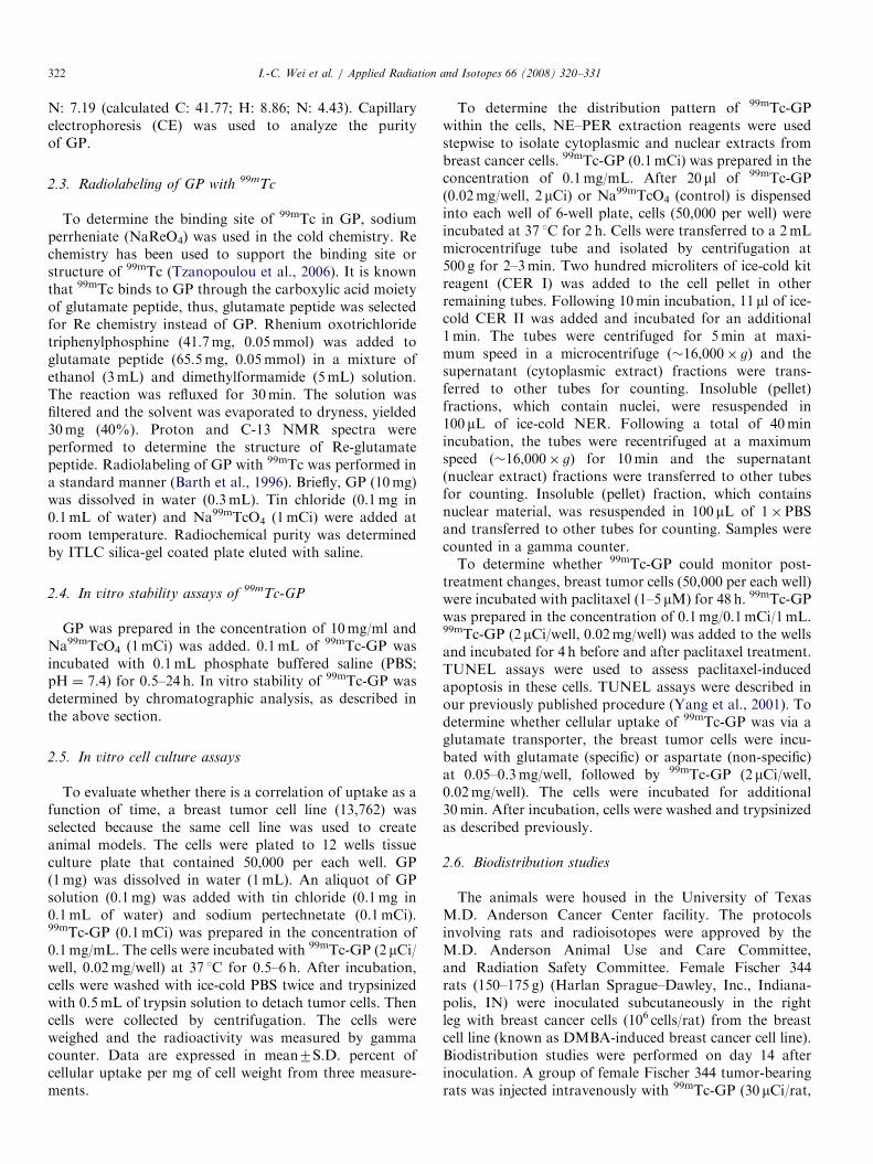
Fig. 2. Capillary electrophoresis (CE) analysis of GP. The spectra shows
carbodiimide–HCl (EDC). EDC was used as a coupling agent during the synthe
greater than 95%.
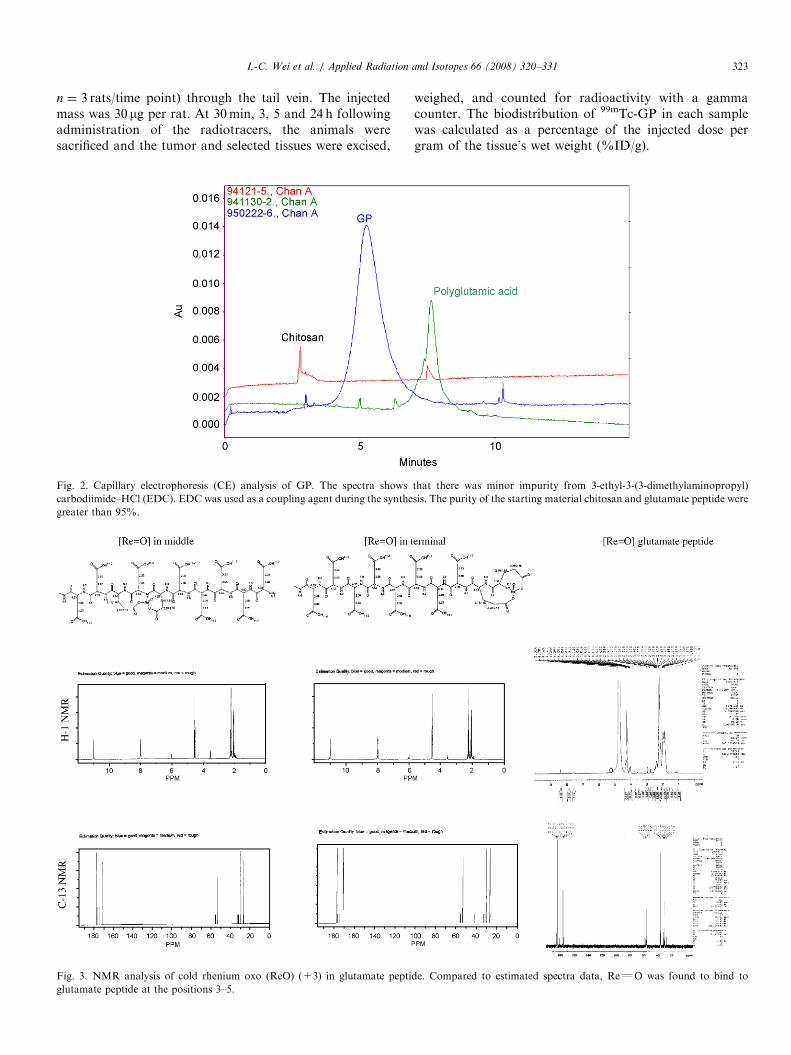
Fig. 3. NMR analysis of cold rhenium oxo (ReO) (+3) in glutamate pepti
glutamate peptide at the positions 3–5.
weighed, and counted for radioactivity with a gammacounter. The biodistribution of 99mTc-GP in each samplewas calculated as a percentage of the injected dose pergram of the tissue’s wet weight (%ID/g).
that there was minor impurity from 3-ethyl-3-(3-dimethylaminopropyl)
sis. The purity of the starting material chitosan and glutamate peptide were
de. Compared to estimated spectra data, ReQO was found to bind to
ARTICLE IN PRESSI.-C. Wei et al. / Applied Radiation and Isotopes 66 (2008) 320–331324
2.7. MIRD dosimetry estimates
Rat absorbed doses’ estimates for 99mTc-GP werecomputed from their respective biodistribution data. Fittedresidence time functions were plotted and multiplied bythe exponential decay functions (i.e. half-life) for 99mTc.These functions were then integrated analytically todetermine the area under the curve (AUC) to yield theresidence time of each organ. It was assumed that theinjected activity distribution was uniformly distributedthroughout the body immediately following injection. Itwas further assumed that no excretion occurred after thelast time point and that the activity distribution remained
0 150
0
15000
Counts
0 100 150
0
500
Counts
4.5hrs
In vitro Sta
100%
10000
5000
2500
2000
1500
1000
Position (mm)
99mTc-GP
50 200
QuickStart - 060802-1522.R002
Na99mTcO4
Position (mm)
50 100 200
QuickStart - 060802-1333.R001
Fig. 4. In vitro stability assays of 99mTc-GP. 99mTc-GP was prepared in the co
0.1mL phosphate buffered saline (pH ¼ 7.4) at 0.5–24 h. There was no break
unchanged after this time point, i.e., no biological excretionwas assumed past the last time point. Mass correctionfactors were used to account for the different ratiosof organ to total body weights in the rat and inhumans, and allowed for the scaling of the rat residencetimes to human residence times. Residence times werethen used to calculate target organ absorbed radiationdoses with S-value tables for a standard 70 kg malemodel using the MIRDose 3.1 software package. Eachorgan dose was computed from the sum of self-doseplus the dose it received from each source organ inthe body or from the remainder of the body (Stabin,1996).
0 100 150
0
Counts
0 100 150
0
50
100
150
200
Counts
0hr
bility Assays
100%
96%
4000
3000
2000
1000
50
Position (mm)
99mTc-GP
200
QuickStart - 060802-1114.R002
QuickStart - 060803-1052.R002
24hrs
99mTc-GP
Position (mm)
50 200
ncentration of 10mg/1mCi/1ml. 0.1mL of 99mTc-GP was incubated with
down of 99mTc-GP up to 24 h.

ARTICLE IN PRESSI.-C. Wei et al. / Applied Radiation and Isotopes 66 (2008) 320–331 325
2.8. Scintigraphic imaging studies
To demonstrate the application of 99mTc-GP in mon-itoring treatment response, three breast tumor-bearing ratswere administered with 99mTc-GP (300 mCi/rat, 3mgphysical amount, i.v.) at pre- and post-paclitaxel treatment(60mg/kg, i.v., single dose) for 48 h. Scintigraphic images,
In Vitro Cellular
0 100
% o
f C
ellu
lar
Upta
ke / m
g o
f cell
weig
ht
0.016
0.014
0.012
0.010
0.008
0.006
0.004
0.002
0.00050 150
Tim
Fig. 5. In vitro breast tumor cells (50,000 per each well) were incubated with 2 m99mTc-GP in mammary tumor cells. Data are expressed in mean7S.D. percen
weight was 10mg.
% o
f u
pta
ke
Com
NaTcO4
0.12
0.09
0.06
0.03
0.00
In vitro Cellular F
Fig. 6. After 2ml of radioactive media containing 2 mCi (0.074MBq) of 99mT
each well of 6-well plate cellular fractions were isolated by centrifugation. Cyto
40%. Both nucleus and cytosolic fractions in 99mTc-GP were higher than Na9
using a gamma camera equipped with low-energy, parallel-hole collimator, were obtained after 0.5 and 1 h. Thestandard source used for calibration was 30 mCi ofNa99mTcO4. Computer outlined region of interest (countsper pixel) of the tumor and muscle (at the symmetric site)was used to determine tumor-to-muscle count densityratios.
Uptake of 99mTc-GP
e (min.)
200 250 300 350 400
y = 1E-05x + 0.0086
R2 = 0.7754
Ci of 99mTc-GP at 37 1C at 0.5–6h. There was a gradually linear uptake of
t of cellular uptake per mg of cell weight from three measurements. Cell
99mTc-GP
Cytosol
Nucleus
pounds
ractions of 99m
Tc-GP
c-GP (0.02mg/well) or sodium pertechnetate (control) was dispensed into
solic fraction had 60% uptake of 99mTc-GP whereas nucleus fraction had9mTcO4.

ARTICLE IN PRESSI.-C. Wei et al. / Applied Radiation and Isotopes 66 (2008) 320–331326
2.9. Scintigraphic imaging studies between breast tumor-
bearing rats with and without inflammation
To demonstrate differential diagnosis of 99mTc-GPbetween tumor and inflammation, the left legs of breasttumor-bearing rats (tumor model described in previousBiodistribution Studies) were administered with terpentine(0.1mL, i.m.). The following day, breast tumor-bearing
Cellular Uptake of 99mTc-GP in
0.0160
% o
f U
pta
ke/m
g o
f cells
0.0140
0.0120
0.0100
0.0080
0.0060
0.0040
0.0020
0.0000
DMSO 1 uM
Fig. 7. Breast tumor cells (50,000 cells per each well) were incubated with pa
incubated for 4 h before and after paclitaxel treatment. There was a decreased
able to measure uptake differences after cells were treated with paclitaxel.
DMSO (Control)
2.5 uM
In Vitro TUNEL Assay P
Fig. 8. TUNEL assays of breast tumor cells (50,000 cells per each well) treated
induced more apoptosis.
rats were administered with 99mTc-chitosan, 99mTc-gluta-mate peptide and 99mTc-GP (300 mCi/rat, 3mg physicalamount, i.v.) and scintigraphic images were obtained 2 h.The standard source used for calibration was 30 mCi ofNa99mTcO4. Computer outlined region of interest (countsper pixel) of the tumor and muscle; tumor and inflamma-tion (at the symmetric site) was used to determine countdensity ratios.
Breast Cells Pretreated with Paclitaxel
4 hour
Agents
2.5 uM 5 uM
clitaxel (1–5mM) for 48 h. 99mTc-GP (2mCi) was added to the wells and
uptake after paclitaxel treatment. The findings indicate that 99mTc-GP was
5 uM
ost-paclitaxel treatment
1 uM
with paclitaxel (1–5mM). The assays revealed that higher dose of paclitaxel
ARTICLE IN PRESS
Effect of amino acid on cellular uptake of 99mTc-GP*
0
1
0
% o
f up
take
Glutamic acidAspartic acid
GP 0.02 mg/well
*
*
* p<0.05compared to control
Cell uptake block assay1.4
1.2
0.8
0.6
0.4
0.2
0.05
dosage (mg/well)
0.1 0.3
Fig. 9. The breast tumor cells were incubated with glutamate (specific) or
aspartate (non-specific) at 0.05–0.3mg/well, followed by 99mTc-GP (2mCi/well, 0.02mg/well). The cells were incubated for additional 30min. After
incubation, cells were washed and trypsinized. There was a dose-
dependent decreased uptake in cells treated with glutamic acid, but not
with aspartic acid. The results indicated that cellular uptake of 99mTc-GP
was via a glutamate transporter.
Table 1
Biodistribution of 99mTc-GP in breast tumor-bearing ratsa
I.-C. Wei et al. / Applied Radiation and Isotopes 66 (2008) 320–331 327
3. Results
3.1. Chemistry
From elemental analysis data, the estimated purity ofGP was greater than 95% (based upon carbon content).CE spectra showed that there was minor impurity fromEDC. EDC was used as a coupling agent during thesynthesis. The purity of the starting material chitosan andglutamate peptide were greater than 95% (Fig. 2). TheNMR spectra (proton and carbon-13) of Re-glutamatepeptide are shown in Fig. 3. Compared to estimated spectradata, ReQO was found to bind to glutamate peptide atthe positions 3–5. The findings suggest that the similarbinding site may apply to 99mTc. From radio-TLC(Bioscan, Washington, DC) analysis, the radiochemicalpurity was more than 95% (Rf ¼ 0.1). In vitro stabilityassays indicated there was no break down of 99mTc-GP upto 24 h (Fig. 4). A minor breakdown fragment (o4%) wasobserved at 24 h. The scale of each spectrum represents theintensity of radioactivity used. Various 99mTc-GP sampleswere spotted at the same time and analyzed by radio-TLCat various intervals. The scale intensity changes due toradioisotope decays. There are variations because radio-TLC analysis is to analyze radiochemical purity qualita-tively.
% of injected dose per gram of tissue weight (n ¼ 3/time
interval, i.v.)
0.5 h 3 h 5 h 24 h
Blood 1.11870.038 0.48970.015 0.39070.021 0.14870.015
Heart 0.27970.016 0.11570.004 0.08870.006 0.03570.001
Lung 0.49570.023 0.19270.014 0.15770.004 0.05770.006
Thyroid 0.49070.038 0.20670.017 0.13670.013 0.08670.004
Pancreas 0.22770.013 0.10570.011 0.09770.006 0.06970.010
Liver 0.62670.012 0.50770.044 0.55370.085 0.23570.028
Spleen 0.41270.026 0.34770.023 0.34970.039 0.14670.009
Kidney 12.12771.017 13.96470.877 12.65270.353 7.25870.269
Stomach 0.23470.008 0.12570.018 0.13670.014 0.04570.004
Intestine 0.21970.017 0.10970.009 0.08670.010 0.03170.008
Uterus 0.30670.020 0.14370.011 0.11570.012 0.05070.003
Tumor 0.25070.030 0.22370.012 0.18970.013 0.09470.008
Muscle 0.09270.006 0.03370.003 0.02870.001 0.01670.003
Bone 0.19170.065 0.13670.046 0.13470.043 0.13470.027
Brain 0.03470.004 0.01470.002 0.01370.000 0.01270.002
T/blood 0.22370.023 0.45770.027 0.48570.012 0.63070.030
T/muscle 2.78470.481 6.91070.448 6.71870.368 5.81770.522
3.2. In vitro cell culture assays
There was a gradual linear uptake of 99mTc-GP in breasttumor cells, suggesting 99mTc-GP has tumor targetingpotential (Fig. 5). The average weight of cells was 10mg.Cytosolic fraction had 60% uptake of 99mTc-GP whereasnucleus fraction had 40%. Both nucleus and cytosolicfractions in 99mTc-GP were higher than Na99mTcO4 (Fig.6). 99mTc-GP was able to measure uptake differences aftercells were treated with paclitaxel (Fig. 7). TUNEL assaysrevealed that higher dose of paclitaxel induced moreapoptosis (Fig. 8). The findings indicated that cellularuptake changes could be monitored by 99mTc-GP. Therewere decreased uptake in cells treated with glutamic acidbut not with aspartic acid (Fig. 9). The results indicatedthat cellular uptake of 99mTc-GP was via a glutamatetransporter.
aValue shown represent the mean7S.D. of data from three rats.
3.3. Biodistribution studies
In biodistribution studies, there was an increased uptakein tumor-to-blood and tumor-to-muscle count densityratios in breast tumor-bearing rats. The optimum tumoruptake and tumor-to-muscle count density ratios were3–5 h post-administrations. There was no increased uptakein thyroid and stomach suggesting in vivo stability of99mTc-GP (Table 1). 99mTc-GP was excreted through liverand kidneys.
3.4. MIRD dosimetry estimates
The human residence time estimates were applied to theMIRDose 3.1 software package to produce radiation doseestimates for 99mTc-GP (Table 2). These doses representthe normal dose per unit injected activity delivered to thenormal organs in an adult human male. For 99mTc-GP, thekidneys and spleen are the primary and secondarycontributors, respectively. The MIRD output was multi-plied by a proposed human dose of 20mCi for 99mTc-GP
ARTICLE IN PRESS
Planar images of 99m
Post-trPaclitaxel (60 m
4.083.164.30
3.293.364.36
T
Rat 2 3
Rat 1
Pre-treatment
0.5 HR
1HR
1
Fig. 10. Planar scintigraphy of 99mTc-GP (300 mCi/rat, i.v., acquired 500,000
treatment response. The numbers indicate tumor-to-muscle ratios (counts/pixe
Table 2
Radiation dose estimates of the reference adult for 99mTc-GP
Target organ Total dose Human dose
(20mCi)
mGy/MBq rad/mCi rad
1. Adrenals 4.09E�03 1.51E�02 0.302
2. Brain 1.11E�04 4.09E�04 0.008
3. Breasts 1.37E�04 5.05E�04 0.011
4. Gallbladder wall 2.37E�03 8.77E�03 0.175
5 LLI wall 3.11E�04 1.15E�03 0.023
6. Small intestine 1.19E�03 4.42E�03 0.088
7. Stomach 1.53E�03 5.65E�03 0.130
8. ULI wall 1.20E�03 4.45E�03 0.089
9. Heart wall 1.08E�03 4.01E�03 0.080
10. Kidneys 4.84E�02 1.79E�01 3.580
11. Liver 2.02E�03 7.48E�03 0.150
12. Lungs 4.36E�04 1.61E�03 0.032
13. Muscle 5.67E�04 2.10E�03 0.042
14. Ovaries 3.96E�04 1.47E�03 0.029
15. Pancreas 2.97E�03 1.10E�02 0.220
16. Red marrow 9.65E�04 3.57E�03 0.070
17. Bone surfaces 9.35E�04 3.46E�03 0.069
18. Skin 2.20E�04 8.15E�04 0.016
19. Spleen 7.08E�03 2.62E�02 0.524
20. Testes 1.89E�05 7.00E�05 0.001
21. Thymus 1.44E�04 5.31E�04 0.011
22. Thyroid 2.54E�05 9.41E�05 0.002
23. Urine bladder
wall
1.07E�04 3.96E�04 0.008
24. Uterus 3.61E�04 1.34E�03 0.027
27. Total body 9.31E�04 3.45E�03 0.069
28. EFF dose
equivalent
5.83E�03 2.16E�02 0.432
29. EFF dose 2.69E�03 9.95E�03 0.200
I.-C. Wei et al. / Applied Radiation and Isotopes 66 (2008) 320–331328
(Table 2) to yield total absorbed dose from an imagingstudy. 99mTc-GP showed total rad absorbed by each organwas below the proposed annual and total limits. Basedupon preclinical studies, dosimetry was estimated withMIRDose. Radiation exposure to the whole body, blood-forming organs (red marrow, spleen), gonads (testes,ovaries), and effective dose equivalent for the proposedhuman single dose at 20mCi fall below the limits of 3 radannually and 5 rad total. The absorbed dose in all otherorgans (e.g. kidneys) was below the limits of single dose of5 rad annually and 15 rad in total (Table 2). Theseradiation exposure values are commonly accepted ascriteria for in vivo radiation safety measurement (Stabin,1996).
3.5. Scintigraphic imaging studies
In planar imaging studies, the tumors could be wellvisualized in breast tumor-bearing rats. From computeroutlined region of interest analysis, the average tumor-to-muscle count density ratios at 0.5 and 1 h was 3.6770.60and 3.8570.60 (before paclitaxel therapy) and changed to2.6070.61 and 3.2070.52 (post-paclitaxel therapy). Therewas decreased tumor-to-muscle count density ratios (avg.17–30%) post-paclitaxel treatment determined by 99mTc-GP at 0.5–1 h. We previously have reported that the timingof apoptosis occurred at earlier time interval on days 2, 3post-paclitaxel treatment (Yang et al., 2001) which wassupported by histopathology. Though the decrease inradionuclide retention from tumor-to-muscle count densityratios before and after paclitaxel treatment is minimal, this
Tc-GP at Pre-and 48 hrseatment with g/kg, iv, single dose).
3.63 3.35 2.62
2.11 2.423.28
1 2 3
Post-treatment
count) in breast tumor-bearing rats showed that 99mTc-GP could monitor
l).

ARTICLE IN PRESS
Comparison of Tumor-to-Muscle and Tumor-to-Inflammation RatiosBetween Breast tumor-
bearing rats with and without inflammation
T=tumor, M = muscle
and I=Inflammation.
T
Š Š
Š
T
T ®
®
®
I
T/M=3.91 T/M=3.03
T/I=1.81
Breast tumor-bearing rats
Breast tumor-bearing rats with
inflammation
99m Tc-C
hitos
an
99m Tc-c
hitos
an
99m Tc-g
lutam
ate pe
ptide
99m Tc-G
P
99m Tc-C
hitos
an
99m Tc-c
hitos
an
99m Tc-g
lutam
ate pe
ptide
99m Tc-G
P
T/I=1.81 T/I=1.99 T/I=2.55
T/M=3.95 T/M=4.89
Fig. 11. Planar scintigraphy of 99mTc-chitosan (left two rats), 99mTc-glutamate peptide and 99mTc-GP (300 mCi/rat, i.v., acquired 500,000 count) in breast
tumor-bearing rats with and without terpentine-induced inflammation. The data showed that 99mTc-GP had highest tumor and muscle as well as tumor
and inflammation count density ratios compared to 99mTc-chitosan and 99mTc-glutamate peptide.
I.-C. Wei et al. / Applied Radiation and Isotopes 66 (2008) 320–331 329
might be due to apoptosis occurred at earlier time. Thefindings from this study indicated that 99mTc-GP is usefulin monitoring treatment response (Fig. 10).
3.6. Scintigraphic imaging studies between breast tumor-
bearing rats with and without inflammation
99mTc-GP had highest tumor and muscle as well astumor and inflammation count density ratios compared to99mTc-chitosan and 99mTc-glutamate peptide (Fig. 11).99mTc-GP was able to differentiate tumor and inflamma-tion.
4. Discussion
In the Re chemistry performed using sodium perrheniateand tin(II) chloride under similar condition used for 99mTc-labeling was not achievable. Unlike trace amount ofsodium pertechnetate, reduction of cold sodium perrheni-ate needs more quantity of tin(II) chloride due todifferences in redox potential (Horiuchi et al., 1999). Moretin(II) chloride in the aqueous solution causes colloidal
particles. In addition, the reaction is under various pHconditions (Horiuchi et al., 1999; Liu et al., 2006), which isnot suitable for GP labeling. Rhenium oxotrichloride andtriphenylphosphine (a reducing chemical) have been usedfor cold Re chemistry (Bereau et al., 2001). Thus, thismethod was adopted for our cold Re chemistry. Our NMRanalysis showed that oxorhenium bound to glutamatepeptide at the positions 3–5. Therefore, rhenium oxo-trichloride and triphenylphosphine were used instead ofsodium perrheniate and tin(II) chloride.Chitosan, a cationic biopolymer derived by deacetylation
of chitin. Chitin and chitosan derivatives are used asexcipients and drug carriers in the pharmaceutical field.For instance, films prepared using chitin or chitosan havebeen developed as wound dressings, oral mucoadhesive andwater-resisting adhesive by virtue of their release char-acteristics and adhesion. Intratumoral administration ofgadopentetic acid–chitosan complex nanoparticles(approximately 430 nm in diameter) has been moreeffective for gadolinium neutron-capture therapy com-pared with a group treated with the solution. Chitin andchitosan have been contributed to expansion of application
ARTICLE IN PRESSI.-C. Wei et al. / Applied Radiation and Isotopes 66 (2008) 320–331330
in drug delivery and sustained drug release (Wang et al.,2005; Fujita et al., 2005; Kato et al., 2003). It has beenreported that chitosan could directly conjugate drugs or bemodified by the introduction of thioglycolic acid (TGA)resulting in chitosan–TGA conjugates with thiol groups.Chitosan–thioglycolic acid conjugates has been found to bea promising candidate in tissue engineering due to theirphysicochemical properties (Bernkop-Schnurch et al.,2004). Despite the broadened application of chitosanderivatives, one major limitation of chitosan is lack ofcellular targeting.
Previous studies have shown that glutamate peptide is acandidate of drug carrier (Tansey et al., 2004; Yang et al.,1994). Radiolabeled glutamic acid has shown to be usefulin tumor cellular imaging (Reiman et al., 1982). Tooptimize the intracellular uptake of chitosan derivatives,we have synthesized glutamate peptide-chitosan, a GPcopolymer. This copolymer would be ideal to conjugate thedrugs with either amino or acid groups which provides anadvantage over either chitosan or glutamate peptide. Inaddition, GP could easily be labeled with 99mTc. Resultsfrom our in vitro cellular uptake studies showed thatcellular uptake of GP is via glutamate transporters (Fig. 9).To demonstrate 99mTc-GP could monitor treatmentresponse, we have used paclitaxel to treat tumor cells forin vitro and in vivo studies. Though cell culture datashowed less linear decrease in uptake of 99mTc-GP aftertreatment with paclitaxel, in the pilot cell apoptosis studies,we did not perform quatification of apoptotic cells atvarious phases yet there was an increase in apoptotic cellswith increasing concentration of paclitaxel. The variationsbetween cell uptake and cell apoptotic assays are due toseparate experiments in different days. Though it is desiredto have more than one cell line for in vitro and in vivostudies, our preliminary findings indicated that 99mTc-GPwas able to monitor paclitaxel treatment changes (Figs. 7, 8and 10). In vitro and in vivo studies using cell lines withdifferent sensitivities to paclitaxel are currently underwayand will be reported elsewhere. Our animal data showedthat 99mTc-GP could differentiate tumor and inflammation(Fig. 11). This targeted drug carrier may provide anadvantage not only to improve drug solubility but also toovercome drug resistance during chemotherapy.
In biodistribution as well as imaging studies (Figs. 9 and10 and Table 1), 99mTc-GP presented high liver and renaluptake. That might be caused by the characteristics ofchitosan and glutamate peptide, which were metabolized inliver and washed out though intestines and kidneys. Thishigh liver and renal uptake may interfere the tumordiagnosis and therapy monitoring of the lesion in thesearea, therefore, 3-dimentional imaging modalities would beneeded to assess lesions.
We demonstrated that 99mTc-GP was able to measureuptake differences after cells treated with paclitaxel in vitroand in vivo (Figs. 7 and 10). TUNEL assays revealed thathigher dose of paclitaxel induced more apoptosis (Fig. 8).These data indicated that the differences of cell uptake and
tumor-to-muscle count density ratios before and afterpaclitaxel treatment might due to the differences ofapoptotic cells killed by paclitaxel. In vitro and in vivofindings suggest that 99mTc-GP is useful in monitoringdrug-induced apoptosis not only in paclitaxel but also inany chemotherapy drugs.In summary, 99mTc-GP could assess efficiency of
chemotherapy. GP has multi-amino and acid groups,which could conjugate with anti-cancer drugs for drugdelivery and therapy.
Acknowledgments
The authors wish to thank Fay Wang for her secretarialsupport. This work was supported in part by TaiwanHopax Sponsored Research Grant to M.D. AndersonCancer Center (SR051-2825). The animal research issupported by M.D. Anderson Cancer Center (CORE)Grant NIH CA-16672.
References
Barth, A., Haldemann, A.R., Reubi, J.C., Godoy, N., Rosler, H., Kinser,
J.A., Seiler, R.W., 1996. Noninvasive differentiation of meningiomas
from other brain tumours using combined 111In-octreotide/99mTc-
DTPA brain scintigraphy. Acta Neurochir. (Wien) 138, 1179–1185.
Behe, M., Kluge, G., Becker, W., Gotthardt, M., Behr, T.M., 2005. Use of
polyglutamic acids to reduce uptake of radiometal-labeled minigastrin
in the kidneys. J. Nucl. Med. 46, 1012–1015.
Bereau, V.M., Khan, S.I., Abu-Omar, M.M., 2001. Synthesis of
enantiopure oxorhenium(V) and arylimidorhenium(V) ‘‘3+2’’ Schiff
base complexes. X-ray diffraction, cyclic voltammetry, UV–vis, and
circular dichroism characterizations. Inorg. Chem. 40, 6767–6773.
Bernkop-Schnurch, A., Hornof, M., Guggi, D., 2004. Thiolated chitosans.
Eur. J. Pharm. Biopharm. 57, 9–17.
Biagini, G., Pugnaloni, A., Damadei, A., Bertani, A., Belligolli, A.,
Bicchiega, V., Muzzarelli, R., 1991. Morphological study of the
capsular organization around tissue expanders coated with N-
carboxybutyl chitosan. Biomaterials 12, 287–291.
Chenu, C., Serre, C.M., Raynal, C., Burt-Pichat, B., Delmas, P.D., 1998.
Glutamate receptors are expressed by bone cells and are involved in
bone resorption. Bone 22, 295–299.
Fujita, M., Ishihara, M., Morimoto, Y., Simizu, M., Saito, Y., Yura, H.,
Matsui, T., Takase, B., Hattori, H., Kanatani, Y., Kikuchi, M.,
Maehara, T., 2005. Efficacy of photocrosslinkable chitosan hydrogel
containing fibroblast growth factor-2 in a rabbit model of chronic
myocardial infarction. J. Surg. Res. 126, 27–33.
Horiuchi, K., Saji, H., Yokoyama, A., 1999. Carrier effect on radiolabel-
ing the polynuclear pentavalent rhenium-186 complex of dimercapto-
succinic acid at alkaline pH: 186Re(V)-DMS. Nucl. Med. Biol. 26,
771–779.
Itoh, K., 2003. Comparison of methods for determination of glomerular
filtration rate: Tc-99m-DTPA renography, predicted creatinine clear-
ance method and plasma sample method. Ann. Nucl. Med. 17,
561–565.
Kato, Y., Onishi, H., Machida, Y., 2003. Application of chitin and
chitosan derivatives in the pharmaceutical field. Curr. Pharm.
Biotechnol. 4, 303–309.
Koppe, M.J., Bleichrodt, R.P., Soede, A.C., Verhofstad, A.A., Gold-
enberg, D.M., Oyen, W.J., Boerman, O.C., 2004. Biodistribution and
therapeutic efficacy of (125/131)I-, (186)Re-, (88/90)Y-, or (177)Lu-
labeled monoclonal antibody MN-14 to carcinoembryonic antigen in
mice with small peritoneal metastases of colorectal origin. J. Nucl.
Med. 45, 1224–1232.
ARTICLE IN PRESSI.-C. Wei et al. / Applied Radiation and Isotopes 66 (2008) 320–331 331
Lai, C.S., Froncisz, W., Hopwood, L.E., 1987. An evaluation of
paramagnetic broadening agents for spin probe studies of intact
mammalian cells. Biophys. J. 52, 625–628.
Li, W.P., Lewis, J.S., Kim, J., Bugaj, J.E., Johnson, M.A., Erion, J.L.,
Anderson, C.J., 2002. DOTA-D-Tyr(1)-octreotate: a somatostatin
analogue for labeling with metal and halogen radionuclides for cancer
imaging and therapy. Bioconjugate Chem. 13, 721–728.
Liu, G., Dou, S., He, J., Yin, D., Gupta, S., Zhang, S., Wang, Y.,
Rusckowski, M., Hnatowich, D.J., 2006. Radiolabeling of MAG3-
morpholino oligomers with 188Re at high labeling efficiency and
specific radioactivity for tumor pretargeting. Appl. Radiat. Isot. 64,
971–978.
Lu, Z.R., Wang, X., Parker, D.L., Goodrich, K.C., Buswell, H.R., 2003.
Poly(L-glutamic acid) Gd(III)–DOTA conjugate with a degradable
spacer for magnetic resonance imaging. Bioconjugate Chem. 14,
715–719.
Pieper, J.S., van Wachem, P.B., van Luyn, M.J.A., Brouwer, L.A.,
Hafmans, T., Veerkamp, J.H., van Kuppevelt, T.H., 2000. Attachment
of glycosaminoglycans to collagenous matrices modulates the tissue
response in rats. Biomaterials 21, 1689–1699.
Polykratis, A., Katsoris, P., Courty, J., Papadimitriou, E., 2005.
Characterization of heparin affinity regulatory peptide signaling in
human endothelial cells. J. Biol. Chem. 280, 22454–22461.
Prabaharan, M., Mano, J.F., 2005. Chitosan-based particles as controlled
drug delivery systems. Drug Deliv. 12, 41–57.
Reiman, R.E., Benua, R.S., Gelbard, A.S., Allen, J.C., Vomero, J.J.,
Laughlin, J.S., 1982. Imaging of brain tumors after administration of L-
(N-13)glutamate: concise communication. J. Nucl. Med. 23, 682–687.
Rijpkema, M., Schuuring, J., Bernsen, P.L., Bernsen, H.J., Kaanders, J.H.,
van der Kogel, A.J., Heerschap, A., 2004. BOLD MRI response to
hypercapnic hyperoxia in patients with meningiomas: correlation with
gadolinium-DTPA uptake rate. Magn. Reson. Imaging 22, 761–767.
Sato, T., Yamaguchi, K., Morita, Y., Noikura, T., Sugihara, K., Matsune,
S., 2000. Lymphoscintigraphy for interpretation of changes of cervical
lymph node function in patients with oral malignant tumors:
comparison of Tc-99m-Re and Tc-99m-HSA-D. Oral Surg. Oral Med.
Oral Pathol. Oral Radiol. Endod. 90, 525–537.
Stabin, M., 1996. MIRDOSE: the personal computer software for use in
internal dose assessment in nuclear medicine. J. Nucl. Med. 37,
538–546.
Tansey, W., Ke, S., Cao, X.Y., Pasuelo, M.J., Wallace, S., Li, C., 2004.
Synthesis and characterization of branched poly(L-glutamic acid) as a
biodegradable drug carrier. J. Control Release 94, 39–51.
Trump, D.P., Mathias, C.J., Yang, Z., Low, P.S., Marmion, M., Green,
M.A., 2002. Synthesis and evaluation of 99mTc(CO)(3)-DTPA-folate as
a folate-receptor-targeted radiopharmaceutical. Nucl. Med. Biol. 29,
569–573.
Tzanopoulou, S., Pirmettis, I.C., Patsis, G., Raptopoulou, C., Terzis, A.,
Papadopoulos, M., Pelecanou, M., 2006. Oxorhenium(V) and ox-
otechnetium(V) [NN][S]3 complexes of 2-phenylbenzothiazole deriva-
tives. Inorg. Chem. 45, 902–909.
Veronese, M.L., Flaherty, K., Kramer, A., Konkle, B.A., Morgan, M.,
Stevenson, J.P., O’Dwyer, P.J., 2005. Phase I study of the novel taxane
CT-2103 in patients with advanced solid tumors. Cancer Chemother.
Pharmacol. 55, 497–501.
Wang, L.Y., Ma, G.H., Su, Z.G., 2005. Preparation of uniform sized
chitosan microspheres by membrane emulsification technique and
application as a carrier of protein drug. J. Control Release 106, 62–75.
Waniewski, R.A., Martin, D.L., 1984. Characterization of L-glutamic acid
transport by glioma cells in culture: evidence for sodium-independent,
chloride-dependent high affinity influx. J. Neurosci. 4, 2234–2237.
Yang, D.J., Li, C., Nikiforow, S., Gretzer, M.B., Kuang, L.R., Lopez,
M.S., Vargas, K., Wallace, S., 1994. Diagnostic and therapeutic
potential of poly(benzyl L-glutamate). J. Pharm. Sci. 83, 328–331.
Yang, D.J., Azhdarinia, A., Wu, P., Yu, D-F., Tansey, W., Kohanim, S.,
Kim, E.E., Pdoloff, D.A., 2001. In vivo and in vitro measurement of
apoptosis in breast cancer cells using 99mTc-EC- annexin V. Cancer
Biother. Radiopharm. 16, 73–84.


![Biodistribution and stability studies of [18F]Fluoroethylrhodamine B, a potential PET myocardial perfusion agent](https://static.fdokumen.com/doc/165x107/633f91a74188bdd1a3054f24/biodistribution-and-stability-studies-of-18ffluoroethylrhodamine-b-a-potential.jpg)

