Radiochemical and biological characteristics of 99mTc-UBI 29–41 for imaging of bacterial...
10
Radiochemical and biological characteristics of 99m Tc-UBI 29 – 41 for imaging of bacterial infections Mick M. Welling a , Sandra Mongera b , Antonella Lupetti c,d , Henia S. Balter e , Valeria Bonetto b , Ulderico Mazzi b , Ernest K.J. Pauwels a, *, Peter H. Nibbering c a Department of Radiology, Division of Nuclear Medicine, Leiden University Medical Center (LUMC), Leiden, The Netherlands b Dipartimento di Scienze Farmaceutiche, Universita ` degli Studi di Padova, Padova, Italy c Department of Infectious Diseases, LUMC, Leiden, The Netherlands d Dipartimento di Patologia Sperimentale, Biotecnologie Mediche, Infettivologia ed Epidemiologia, Universita ` degli Studi di Pisa, Pisa, Italy e Centro de Investigaciones Nucleares, Facultad de Ciencias, Montevideo, Uruguay Received 11 June 2001; received in revised form 30 October 2001; accepted 12 January 2002 Abstract A technetium-99m-labeled peptide derived from ubiquicidine, further referred to as 99m Tc-UBI 29 – 41, targets bacterial and fungal infections, but not sterile inflammatory processes, in experimental animals. This paper reports on the radiochemical and biological features of this radioactive agent and the importance of the amino acid sequence of UBI 29 – 41 for imaging of infections. Radiochemical analyses of 99m Tc-UBI 29 – 41 and a radiolabeled scrambled version of this peptide, i.e. 99m Tc-Sc-UBI 29 – 41, revealed that both peptides were labeled rapidly (within 10 min) and effectively with little colloid formation (less than 5% of the total radioactivity) and very little free pertechnetate (or radioactive intermediates) in the preparations containing radiolabeled peptide. Furthermore, association of the peptides with bacteria could be competed with excess unlabeled peptide and this association proved to be temperature-dependent. Based on this in vitro data we concluded that labeling of peptides with 99m Tc by this direct method is rapid, efficient, and safe. Scintigraphy demonstrated that radioactivity is rapidly removed from the circulation (half-lifes of UBI 29 – 41 and Sc-UBI 29 – 41 were 16 and 21 min, respectively) mainly by renal clearance. Analysis of murine blood revealed that only a small proportion of the intravenously injected 99m Tc-peptides is associated with blood cells. Although both radiolabeled peptides accumulated rapidly at sites of infection, the values for 99m Tc-UBI 29 – 41 were higher (P 0.05) than for 99m Tc-Sc-UBI 29 – 41. Moreover, injection of excess unlabeled UBI 29 – 41, but not Sc-UBI 29 – 41, into Staphylococcus aureus-infected mice prior to injection of 99m Tc-UBI 29 – 41 significantly (P 0.05) reduced the accumulation of this radiopharmaceutical at the site of infection. In addition, we observed significantly (P 0.01) higher amounts of 99m Tc-UBI 29 – 41 at the site of infection in mice using a carrier-free radiolabeled UBI 29 – 41 as compared with unpurified preparations containing radiolabeled UBI 29 – 41. This in vivo data indicates that the amino acid sequence of 99m Tc-UBI 29 – 41 contributes to its accumulation at the site of infection. © 2002 Elsevier Science Inc. All rights reserved. Keywords: Ubiquicidine; 99m Tc-labeling; Radiochemistry; Scintigraphy; Infection detection; Competition 1. Introduction In several previous papers, we introduced 99m Tc-labeled cationic antimicrobial peptides derived from human ubiqui- cidine (UBI) and lactoferrin (hLF) for the detection of bacterial and fungal infections in mice and rabbits [5,13– 15]. Preferential tagging of these pathogens over leukocytes and other host cells by these peptides may explain the ability of the peptides to discriminate between infections and sterile inflammatory processes in these animals [13,15]. Furthermore, radiolabeled UBI-derived peptides allowed us to monitor the efficacy of antimicrobial treatment in mice having an experimental infection [6]. These preclinical ex- periments were carried out using peptides that had been labeled by a method involving reduction of 99m Tc pertech- netate, the production of a TcO(pyrophosphate) intermedi- ate, the substitution reaction transferring the reduced Tc from the intermediate to the cationic peptide [9]. Since these 99m Tc-peptides are successful at imaging the infections in experimental animals, the aim of the present study was to * Corresponding author. Tel.: 31-71-5263475; fax: 31-71- 5266751. E-mail address: [email protected] (E.K.J. Pauwels). Nuclear Medicine and Biology 29 (2002) 413– 422 0969-8051/02/$ – see front matter © 2002 Elsevier Science Inc. All rights reserved. PII: S0969-8051(02)00292-5
-
Upload
independent -
Category
Documents
-
view
1 -
download
0
Transcript of Radiochemical and biological characteristics of 99mTc-UBI 29–41 for imaging of bacterial...

Radiochemical and biological characteristics of99mTc-UBI 29–41 forimaging of bacterial infections
Mick M. Wellinga, Sandra Mongerab, Antonella Lupettic,d, Henia S. Baltere, Valeria Bonettob,Ulderico Mazzib, Ernest K.J. Pauwelsa,*, Peter H. Nibberingc
aDepartment of Radiology, Division of Nuclear Medicine, Leiden University Medical Center (LUMC), Leiden, The NetherlandsbDipartimento di Scienze Farmaceutiche, Universita degli Studi di Padova, Padova, Italy
cDepartment of Infectious Diseases, LUMC, Leiden, The NetherlandsdDipartimento di Patologia Sperimentale, Biotecnologie Mediche, Infettivologia ed Epidemiologia, Universita degli Studi di Pisa, Pisa, Italy
eCentro de Investigaciones Nucleares, Facultad de Ciencias, Montevideo, Uruguay
Received 11 June 2001; received in revised form 30 October 2001; accepted 12 January 2002
Abstract
A technetium-99m-labeled peptide derived from ubiquicidine, further referred to as99mTc-UBI 29–41, targets bacterial and fungalinfections, but not sterile inflammatory processes, in experimental animals. This paper reports on the radiochemical and biological featuresof this radioactive agent and the importance of the amino acid sequence of UBI 29–41 for imaging of infections.
Radiochemical analyses of99mTc-UBI 29–41 and a radiolabeled scrambled version of this peptide, i.e.99mTc-Sc-UBI 29–41, revealedthat both peptides were labeled rapidly (within 10 min) and effectively with little colloid formation (less than 5% of the total radioactivity)and very little free pertechnetate (or radioactive intermediates) in the preparations containing radiolabeled peptide. Furthermore, associationof the peptides with bacteria could be competed with excess unlabeled peptide and this association proved to be temperature-dependent.Based on thisin vitro data we concluded that labeling of peptides with99mTc by this direct method is rapid, efficient, and safe.
Scintigraphy demonstrated that radioactivity is rapidly removed from the circulation (half-lifes of UBI 29–41 and Sc-UBI 29–41 were16 and 21 min, respectively) mainly by renal clearance. Analysis of murine blood revealed that only a small proportion of the intravenouslyinjected99mTc-peptides is associated with blood cells. Although both radiolabeled peptides accumulated rapidly at sites of infection, thevalues for99mTc-UBI 29–41 were higher (P � 0.05) than for99mTc-Sc-UBI 29–41. Moreover, injection of excess unlabeled UBI 29–41,but not Sc-UBI 29–41, intoStaphylococcus aureus-infected mice prior to injection of99mTc-UBI 29–41 significantly (P � 0.05) reducedthe accumulation of this radiopharmaceutical at the site of infection. In addition, we observed significantly (P � 0.01) higher amounts of99mTc-UBI 29–41 at the site of infection in mice using a carrier-free radiolabeled UBI 29–41 as compared with unpurified preparationscontaining radiolabeled UBI 29–41. Thisin vivo data indicates that the amino acid sequence of99mTc-UBI 29–41 contributes to itsaccumulation at the site of infection. © 2002 Elsevier Science Inc. All rights reserved.
Keywords: Ubiquicidine;99mTc-labeling; Radiochemistry; Scintigraphy; Infection detection; Competition
1. Introduction
In several previous papers, we introduced99mTc-labeledcationic antimicrobial peptides derived from human ubiqui-cidine (UBI) and lactoferrin (hLF) for the detection ofbacterial and fungal infections in mice and rabbits [5,13–15]. Preferential tagging of these pathogens over leukocytesand other host cells by these peptides may explain the
ability of the peptides to discriminate between infectionsand sterile inflammatory processes in these animals [13,15].Furthermore, radiolabeled UBI-derived peptides allowed usto monitor the efficacy of antimicrobial treatment in micehaving an experimental infection [6]. These preclinical ex-periments were carried out using peptides that had beenlabeled by a method involving reduction of99mTc pertech-netate, the production of a TcO(pyrophosphate) intermedi-ate, the substitution reaction transferring the reduced Tcfrom the intermediate to the cationic peptide [9]. Since these99mTc-peptides are successful at imaging the infections inexperimental animals, the aim of the present study was to
* Corresponding author. Tel.:�31-71-5263475; fax: �31-71-5266751.
E-mail address: [email protected] (E.K.J. Pauwels).
Nuclear Medicine and Biology 29 (2002) 413–422
0969-8051/02/$ – see front matter © 2002 Elsevier Science Inc. All rights reserved.PII: S0969-8051(02)00292-5

investigate the radiochemical and biological characteristicsof our lead peptide, i.e. UBI 29–41, and the contribution ofits amino acid sequence to its ability to visualize infectionsin experimental animals.
2. Materials and methods
2.1. Peptides
Peptides based on cationic, �-helical domains of humanubiquicidine (UBI) were synthesized as described before[2]; the lead peptide for infection detection being UBI29–41 (TGRAKRRMQYNRR, 1,693 Da). To investigatethe importance of the amino acid sequence for its ability tovisualize of infections, a scrambled version (Sc-UBI 29–41,KRNQRMARYRRGT, 1,693 Da) was synthesized. Bothpeptides were diluted to 1 mM in 5% (v/v) of acetic acid(HAc; pH 4) and stored at �20oC.
2.2. Labeling of UBI peptides with technetium-99m
UBI 29–41 and Sc-UBI 29–41 were directly labeledwith 99m-technetium as described earlier [13,15]. Briefly,10 �l of a peptide stock solution was added to 4 �l of anaseptic mixture of 950 mg/L Sn(Cl)2.2H2O and 2 g/L so-dium pyrophosphate.10H2O (Department of Clinical Phar-macy and Toxicology, LUMC) in saline in a 1.5 mL Ep-pendorf vial. Immediately thereafter, 2 �L of a solutioncontaining 10 mg/mL of KBH4 (crystalline, Sigma Chemi-cal Company, St. Louis, MO) in 0.1 M NaOH were pipettedinto this mixture. After addition of 0.1 mL of 99mTc-sodiumpertechnetate solution (200 MBq/mL, Technekow,Mallinckrodt Medical BV, Petten, The Netherlands) themixture was gently stirred at room temperature for at least10 min and was then ready for use. This preparation isfurther referred to as the preparation containing radiolabeledpeptide.
2.3. HPLC analysis
Identification of 99mTc-species in preparations contain-ing radiolabeled peptide (10–50 �L-samples) was achievedby reverse-phase high performance liquid chromatography(HPLC) on a 4.6 x 250 mm C18 column (Vydac, TheSeparations Group, Hesperia, CA) using a linear gradientprepared with water supplemented with. 0.1% (v/v) triflu-oroacetic acid (TFA) as ion-pairing agent (solution A) anda water/methanol (20/80%, v/v) mixture containing 0.1%(v/v) TFA (solution B) at a flow-rate of 1 mL/min [2]. Thegradient was prepared with a gradient pump (LKB BrommaHPLC 2249, Uppsala, Sweden) as follows: 100% A for 5min, 0–100% B in 10 min, 100% B for 5 min, 100–0% Bin 5 min, and finally 100% A for 5 min. Recordings of thevarious fractions were made using an on-line UV-detectormeasuring the absorbance at 220 nm and a NaI (Tl)-crystal
gamma detection system (Steffi, Raytest Benelux B.V., Til-burg, The Netherlands) monitoring the radioactivity.
2.4. ITLC analysis
In order to gain additional data on the labeling yield andstability of the 99mTc-labeled peptides in time, instant thinlayer chromatography (ITLC) was performed. In short, 1 �6 cm ITLC/SG strips (Gelman Sciences, Ann Arbor, MI),were dried in a microwave at 500 W for 60 s directly beforeuse. Samples of the preparations containing labeled peptides(2 �L) were applied at approximately 1 cm from the bottom(baseline) of the ITLC strips and were immediately placedin air-tight containers with saline or methyl ethyl keton.After 2 min of elution, the strips were mounted on thedetector of a gamma camera (Toshiba GCA 7100/UI, To-kyo, Japan) equipped with a low energy general purposeparallel-hole collimator. Counts were collected for at least 2min in a 512 � 512 matrix with a window centered at 20%at 140 KeV. Quantification of radioactivity in each singlefraction (front, intermediate, and baseline) was carried outby determining the 99mTc-activity in rectangle regions ofinterest (ROI) drawn over the fraction. Results are ex-pressed as percentage of total 99mTc-activity. In addition,we determined the radioactivity after cutting the ITLC stripsin three fractions corresponding to scintigraphically definedROI in a dose-calibrator (VDC 101, Veenstra Instruments,Joure, The Netherlands).
2.5. Sep-Pak analysis
In order to find out whether traces of 99mTc-colloidswere present, preparations containing labeled peptideswere applied onto Sep-Pak Light C18 cartridges (Waters,Milford, MA). In short, we applied 10 �L of the prepa-rations containing radiolabeled peptides diluted in 10 mLof 0.1% (v/v) HAc to Sep-Pak cartridges, pre-flushedwith 10 mL of 0.1% (v/v) HAc. After rinsing with an-other 10 mL of 0.1% (v/v) HAc until no detectableamount of radioactivity was released from the column,we eluted the peptide from the cartridge with 20 mL ofmethanol and measured the trapped radioactivity (col-loids) on the cartridge in a dose-calibrator.
2.6. Stability of 99mTc-labeled peptides in diluted humanserum
Stability of the 99mTc-labeled peptides was challengedby incubating one volume of the labeling solution with onevolume of 20% (v/v) human serum in saline for 1 and 24 hat 37oC. Next, the amounts of free pertechnetate and 99mTc-peptide in the samples were determined by ITLC usingmethyl ethyl keton as eluent.
414 M.M. Welling et al. / Nuclear Medicine and Biology 29 (2002) 413–422

2.7. Staphylococcus aureus
Staphylococcus aureus 25923 (S. aureus) was obtainedfrom the American Type Culture Collection (ATCC, Rock-ville, MD). Overnight cultures of viable bacteria were pre-pared in brain-heart infusion broth (Oxoid, Basingstoke,UK) in a shaking water-bath at 37oC. The next day, bacteriawere washed, counted, aseptically aliquoted at a concentra-tion of 1 � 108 colony forming units (CFU)/mL, stored at�20oC and thawed immediately before use.
2.8. In vitro competition assay for the binding/associationof 99mTc-labeled peptides with bacteria
Binding/association of 99mTc-UBI 29–41 and 99mTc-Sc-UBI 29–41 with S. aureus was assessed at 4oC or 37oC,respectively, as described earlier [15]. In short, 0.1 mL of 15mM sodiumphosphate buffer (PB, pH 7.5) containing 1/10of the preparation containing labeled peptide was trans-ferred to an Eppendorf vial. Next, 0.8 mL of the incubationbuffer containing 50% of 0.1% (v/v) HAc in PB supple-mented with 0.01% (v/v) Tween-80 and 0.1 mL of PBcontaining 2 � 107 CFU bacteria were added. The final pHof this mixture was 5. In competition experiments, bacteriawere pre-incubated for 1 h with 10- or 100-fold excess ofunlabeled peptides before addition of radiolabeled peptide.After incubation of the bacteria with radiolabeled peptidesfor 1 h, the vials were centrifuged in a pre-cooled centrifugeat 1,000 � g for 5 min. The supernatant was removed andthe pellet containing the bacteria was gently resuspended in1 mL of incubation buffer and re-centrifuged as describedabove. The supernatant was removed and the radioactivityin the pellet containing bacteria was determined in a dosecalibrator. The radioactivity associated with bacteria is ex-pressed as percentage of added 99mTc-activity/2 � 107 CFUbacteria.
2.9. Animals
Specific pathogen-free, male, Swiss mice, weighing25–30 g were used in this study. The animals were housedin the animal housing facilities of the LUMC for at least 1week before the onset of the experiments. Food and waterwere given ad libitum. All animal studies were done incompliance with the Dutch laws related to the conduct ofanimal experiments.
2.10. Biodistribution of 99mTc-labeled UBI 29–41 andSc-UBI 29–41
Mice were anesthetized with a single intraperitoneal in-jection of 0.1 mL of saline containing 1 mg of fluanisoneand 0.03 mg of fentanyl citrate (Hypnorm, Janssen Pharma-ceutics, Tilburg, The Netherlands). Preparations containingradiolabeled peptide (0.1 mL equals 1 nmol of peptide) werediluted in saline and then injected into a tail vein. After
injection of 99mTc-UBI 29–41 (2 MBq/mouse), accumula-tion of radioactivity in various organs was determined usingscintigraphy and ex-vivo countings. Using scintigraphy,whole body images of each mouse were made until 4 hpost-injection for at least 2 min. Radioactivity counts intissues of the mice were estimated at the various intervals bymeans of anatomically fitted ROI drawn over various organsand data are expressed as the percentage of total 99mTc-activity of the injected dose (%ID). Thereafter, the micewere sacrificed with an intraperitoneal injection of 0.25 mLof sodium pentobarbiturate (60 mg/mL, Nembutal, SanofiBV, Division Algin, Maassluis, The Netherlands). The var-ious organs were dissected, weighed and counted for radio-activity. Data are expressed as the percentage of total 99mTc-activity (%ID) and then calculated as %ID per gram oftissue (%ID/g).
2.11. Distribution of 99mTc-peptides in murine blood
To determine the half-lifes of 99mTc-UBI 29–41 and99mTc-Sc-UBI 29–41, 20-�L blood samples (about 20 kBq/sample) were taken from a tail vein between 1 min and 1.5 hpost injection and then counted for radioactivity. To estab-lish the association of the 99mTc-peptides with the differentcomponents of murine blood, 10 �L-samples were dilutedwith PBS and then fractionated on 4 mL of a 75% (density1.056) and 100% (density 1.077) two-layer isotonic Ficollgrade (Department of Clinical Pharmacy and Toxicology,LUMC) and centrifuged for 10 min at 1,000xg at roomtemperature. The amount of radioactivity in the variousfractions, i.e., plasma (top layer), the fraction containingleukocytes (middle layer) and those containing erythrocytes(bottom layer) was measured using a dose-calibrator.
2.12. Analysis of 99mTc activity in urine
After sacrificing the mice, the urine was collected fromthe bladder, weighed, and counted for radioactivity. In ad-dition, a urine sample was analyzed using ITLC and methylethyl keton as eluent, as described above, to determine thepercentage of released/free 99mTc-activity in the urine.
2.13. Scintigraphic detection of infections in mice
Mice were injected in the right thigh muscle with 0.1 mLsaline containing 2 � 107 CFU of S. aureus 18 h beforeinjection of 1 mL containing 1 nmol of 99mTc-UBI 29–41or 99mTc-Sc-UBI 29–41. Accumulation of the radiophar-maceutical in infected thigh muscles at various intervalsuntil 4 h post injection was determined scintigraphically aspreviously described [14,15]. Quantification of radioactivityin infected thigh muscles was carried out by means ofanatomically fitted ROI over the entire infected (target) andnon-infected (non-target) thigh muscle and data are ex-pressed as target-to-non-target (T/NT) ratios. Thereafter, themice were sacrificed as described above and the infected
415M.M. Welling et al. / Nuclear Medicine and Biology 29 (2002) 413–422

and non-infected thigh muscles were removed, weighed,counted for radioactivity, and again the T/NT ratios werecalculated and expressed as % injected dose/gram tissue. Toconfirm the bacterial infection the infected thigh muscleswere dissected, homogenized, and the number of viablebacteria was determined microbiologically. Four mice witha bacterial infection were injected with carrier-free 99mTc-UBI 29–41. Such a preparation was obtained after separat-ing the HPLC fractions containing radioactivity from thefractions containing unlabeled peptide, as can be seen in theHPLC chromatograms (Fig. 1a). The eluent in the radioac-tive fraction was removed using Sep-Pak cartridges, asdescribed above, followed by evaporation of methanol byhot air.
2.14. Effect of competitor peptides on the detection ofinfection with 99mTc-UBI 29–41 or 99mTc-Sc-UBI 29–41
To assess the specificity, of the tracers on infectiondetection, the effect of injections with 100-fold excess ofunlabeled peptides UBI 29–41 or Sc-UBI 29–41 at 5 minprior to administration of 99mTc-peptides on the accumula-tion in thigh muscles of mice infected with S. aureus wasstudied scintigraphically.
2.15. Statistical analysis
Differences in the data were evaluated with the Student ttest. Results for P using the 2-tailed test are reported and allresults are given as mean � SEM. The correlation between exvivo counting and scintigraphy was analyzed using Pearson’scorrelation test. The level of significance was set at P � 0.05.
3. Results
3.1. Radiochemical analysis of 99mTc-labeled ubi peptides
HPLC chromatograms for UBI 29–41 and Sc-UBI29–41 indicated that the peptide preparations were morethan 95% pure (Fig. 1a–b, top panels).
The UV profile of preparations containing radiolabeled UBI29–41 revealed that the major peak of peptide eluted at 12 �1.0 min (n � 6; Fig. 1a; middle panel), which was identical tothe retention time of unlabeled peptide, i.e., 12 � 1.0 min (n �6; Fig. 1a; top panel), indicating that the labeling procedure didnot affect the lipophilicity of the unlabeled peptide. The radio-activity profile of 99mTc-UBI 29–41 (Fig. 1a; bottom panel)showed two peaks, a minor peak of radioactivity co-elutingwith the mass of peptide, whereas the major peak (83% of totalactivity) eluted significantly (P � 0.05) later (13 � 0.2 min)than the unlabeled peptide.
Similar results in UV and radioactivity profiles wereobserved with regard to the HPLC analysis of Sc-UBI29–41 before and after labeling (Fig. 1b). The recovery ofradioactivity amounted to 83 � 13% for 99mTc-UBI 29–41
(n � 10) and to 84 � 6% for 99mTc-Sc-UBI 29–41 (n � 8).After repetitive runs of the same sample the recovery in-creased to �98% for both peptides, indicating that the initialrecovery data for these radioactive peptides were unreliableprobably due to non-specific adhesion of the labeled com-pounds to glass and metal surfaces in the HPLC system.Therefore, at least three subsequential runs with the samesample were performed on reverse-phase HPLC before theresults were considered reliable.
Fig. 1a. Typical UV profile (220 nm) of unlabeled UBI 29–41 (top panel)and of this peptide after 0.5 h of labeling with 99mTc (middle panel). Thesecond “peak” represent the absorbance by methanol. The radiogram of99mTc-UBI 29–41 is depicted in the bottom panel and values are expressedas counts per second.
416 M.M. Welling et al. / Nuclear Medicine and Biology 29 (2002) 413–422
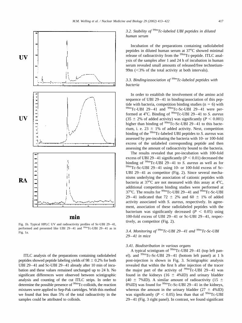
ITLC analysis of the preparations containing radiolabeledpeptides showed peptide labeling yields of 98 � 0.2% for bothUBI 29–41 and Sc-UBI 29–41 already after 10 min of incu-bation and these values remained unchanged up to 24 h. Nosignificant differences were observed between scintigraphicanalysis and counting of the cut ITLC strips. In order todetermine the possible presence of 99mTc-colloids, the reactionmixtures were applied to Sep-Pak cartridges. With this methodwe found that less than 5% of the total radioactivity in thesamples could be attributed to colloids.
3.2. Stability of 99mTc-labeled UBI peptides in dilutedhuman serum
Incubation of the preparations containing radiolabeledpeptides in diluted human serum at 37oC showed minimalrelease of radioactivity from the 99mTc-peptide. ITLC anal-ysis of the samples after 1 and 24 h of incubation in humanserum revealed small amounts of released/free technetium-99m (�3% of the total activity at both intervals).
3.3. Binding/association of 99mTc-labeled peptides withbacteria
In order to establish the involvement of the amino acidsequence of UBI 29–41 in binding/association of this pep-tide with bacteria, competition binding studies (n � 6) with99mTc-UBI 29–41 and 99mTc-Sc-UBI 29–41 were per-formed at 4oC. Binding of 99mTc-UBI 29–41 to S. aureus(35 � 2% of added activity) was significantly (P � 0.001)higher than binding of 99mTc-Sc-UBI 29–41 to this bacte-rium, i. e. 23 � 1% of added activity. Next, competitionbinding of the 99mTc-labeled UBI peptides to S. aureus wasassessed by pre-incubating the bacteria with 10- or 100-foldexcess of the unlabeled corresponding peptide and thenassessing the amount of radioactivity bound to the bacteria.
The results revealed that pre-incubation with 100-foldexcess of UBI 29–41 significantly (P � 0.01) decreased thebinding of 99mTc-UBI 29–41 to S. aureus as well as for99mTc-Sc-UBI 29–41 using 10- or 100-fold excess of Sc-UBI 29–41 as competitor (Fig. 2). Since several mecha-nisms underlying the association of cationic peptides withbacteria at 37oC are not measured with this assay at 4oC,additional competition binding studies were performed at37oC. The results for 99mTc-UBI 29–41 and 99mTc-Sc-UBI29–41 indicated that 72 � 2% and 60 � 1% of addedactivity associated with S. aureus, respectively. In agree-ment, association of these radiolabeled peptides with thebacterium was significantly decreased (P � 0.05) using100-fold excess of UBI 29–41 or Sc-UBI 29–41, respec-tively, as competitor (Fig. 2).
3.4. Monitoring of 99mTc-UBI 29–41 and 99mTc-Sc-UBI29–41 in mice
3.41. Biodistribution in various organsA typical scintigram of 99mTc-UBI 29–41 (top left pan-
el). and 99mTc-Sc-UBI 29–41 (bottom left panel) at 1 hpost-injection is shown in Fig. 3. Scintigraphic analysisrevealed that within the first h after injection of the tracerthe major part of the activity of 99mTc-UBI 29–41 wasfound in the kidneys (16 � 4%ID) and urinary bladder(40 � 7%ID). A similar amount of radioactivity (15 �8%ID) was found for 99mTc-Sc-UBI 29–41 in the kidneys,whereas the amount in the urinary bladder (27 � 4%ID)was significantly (P � 0.05) less than that of 99mTc-UBI29–41 (Fig. 3 right panel). In contrast, we found significant
Fig. 1b. Typical HPLC UV and radioactivity profiles of Sc-UBI 29–41,performed and presented like UBI 29–41 and 99mTc-UBI 29–41 as inFig. 1a.
417M.M. Welling et al. / Nuclear Medicine and Biology 29 (2002) 413–422

(P � 0.05) larger amounts of 99mTc-Sc-UBI 29–41 activityin the liver (15 � 4%ID) compared to the amounts of99mTc-UBI 29–41 (6 � 3%ID).
In Table 1 we depict data from ex-vivo countings ex-pressed as the percentage of injected dose of 99mTc-UBIpeptides per gram tissue (%ID/g) at 1 and 2 h after injectionof the tracer. At 2 h after injection, the %ID/g for 99mTc-UBI 29–41 and 99mTc-Sc-UBI 29–41 in the infected thighmuscles amounted to 2.4 � 0.60% and 0.6 � 0.14%)respectively (n � 4). For both peptides data obtained by exvivo countings correlated (R � 0.847; P � 0.001) withvalues obtained by scintigraphy (n � 20). In experimentswith mice preinjected with unlabeled peptide we determineddecreased accumulation for both 99mTc-UBI 29–41 and99mTc-Sc-UBI 29–41 (0.9 � 0.3 and 0.3 � 0.04%ID/grespectively).
3.5. Analysis of 99mTc-labeled peptides in murine blood
On the basis of the 99mTc-activity counting in bloodsamples of mice taken at various time points up to 90 minafter injection of the tracer, the half-lifes for 99mTc-UBI29–41 and 99mTc-Sc-UBI 29–41 were determined. The
results revealed a significant difference (P � 0.05) betweenthe half-lifes of the two tracers, i.e. for 99mTc-UBI 29–41and 99mTc-Sc-UBI 29–41 the values amounted to 16 � 0.8min and 21 � 1.2 min, respectively (n � 3). Next, theamount of radioactivity in the fractions containing plasma,leukocytes, and erythrocytes was assessed. The results re-vealed for both peptides that most of the relative radioac-tivity was present in the plasma/platelet fraction (�86%ID)and minor amounts of activity were associated with leuko-cytes (�8%ID) and erythrocytes (�6%ID). The distributionpattern remained the same during the period of analysis.
3.6. ITLC analysis of 99mTc-labeled peptides in urine
At 2 hr after intravenous administration of respectively99mTc-labeled UBI 29–41 and Sc-UBI 29–41 approxi-mately 90% and 70% of the added activity were retrievedin the urinary bladder. ITLC analysis revealed that be-tween 2 and 4% of the total 99mTc-activity in the urinesamples represents free 99mTc-activity, indicating that thebreakdown of the radiolabeled peptides in urine wasminimal.
Fig. 2. In vitro binding/association of 99mTc-UBI 29–41 (open bars) and 99mTc-Sc-UBI 29–41 (closed bars) at 4oC to 2 � 107 S. aureus pre-incubated with10- or 100-fold excess of unlabeled corresponding peptide or none (no competitor). Results of competition studies at 37oC are presented in diagonally hatchedbars (99mTc-UBI 29–41) and horizontally hatched bars (99mTc-Sc-UBI 29–41). Results are means (� SEM) and are expressed as percentage of addedradioactivity associated with 2 � 107 CFU bacteria. *P � 0.05, compared to the values for 99mTc-labeled peptide in the absence of competitor, accordingto Student t-test.
418 M.M. Welling et al. / Nuclear Medicine and Biology 29 (2002) 413–422

3.7. Scintigraphic detection of infections in mice
To find out whether the accumulation of UBI peptide29–41 is different from that of the scrambled peptide at thesite of infection, mice with a S. aureus thigh muscle infec-tion were injected with these 99mTc-labeled peptides (n � 6)and T/NT ratios were calculated. Already at 60 min afterinjection of 99mTc-UBI 29–41, the infected thigh musclebecame clearly visible (Fig. 3, top left panel). Furthermore,starting from 2 h post-injection the T/NT ratios for 99mTc-UBI 29–41 were significantly (P � 0.05) higher than for99mTc-labeled scrambled peptide (Fig. 3, bottom left panel).
This difference remained significant during the entireperiod of analysis (Fig. 4 and Fig. 5).
3.8. Effect of competitor peptides on the scintigraphicdetection of infection
To find out if the amino acid sequence of 99mTc-UBI29–41 is important in its ability to detect S. aureus-infec-
tions, mice were injected with 100-fold excess of unlabeledUBI 29–41 or Sc-UBI 29–41 five min before injection withthe radiolabeled peptides. Next, scintigraphic analysis wasperformed (Fig. 3, top and bottom right panels) and T/NTratios were determined until 4 h after administration of99mTc-UBI 29–41 or 99mTc-Sc-UBI 29–41 (Fig. 4 and Fig.5, respectively). Interestingly, significantly (P�0.05)smaller T/NT ratios for 99mTc-UBI 29–41 were seen in S.aureus-infected mice pre-injected with unlabeled UBI 29–41, but not unlabeled Sc-UBI 29–41 (Fig. 4), whereas noeffect of pre-injection of such mice with either unlabeledSc-UBI 29–41 or UBI 29–41 on the T/NT ratios for 99mTc-Sc-UBI 29–41 was observed (Fig. 5). Furthermore, nosignificant differences between the T/NT ratios for 99mTc-UBI 29–41 in S. aureus-infected mice obtained by scintig-raphy and by obduction were seen.
Finally, we injected carrier-free 99mTc-UBI 29–41 intoinfected mice and found at all intervals during the period ofanalysis that this tracer yielded significantly (P � 0.01)higher T/NT ratios as compared to preparations containing99mTc-UBI 29–41 (Fig. 4). A typical scintigram of carrier-free 99mTc-UBI 29–41 at 2 h after injection into mice witha infection with S. aureus is depicted in Fig. 6.
4. Discussion
The first conclusion from the present results pertains toour method of labeling peptides with technetium-99m forimaging of infections. The results showed that labeling ofUBI 29–41 and Sc-UBI 29–41 with the direct method [15]is rapid and effective, i.e. a high (95%) yield already at 10mins after reaction, and safe, as assessed with an in vitrokilling assay. In addition, the radiochemical purity (littlefree radionuclide or colloid formation) and stability of theradiolabeled peptides in diluted human serum are excellent
Fig. 3. Typical scintigrams of 99mTc-labeled UBI 29–41 (top left) and99mTc-Sc-UBI 29–41 (bottom left) at 1 h after injection into mice with athigh muscle S. aureus infection. An example of competition with 100-foldexcess of unlabeled corresponding peptide is shown for 99mTc-UBI 29–41(top right) and 99mTc-Sc-UBI 29–41 (bottom right) at 1 h after injection ofthe tracer. The infection is indicated by an arrow.
Table 1Biodistribution data for 99mTc-UBI 29–41 and 99mTc-Sc-UBI 29–41from ex vivo countings expressed as the percentage of injected dose of99mTc-labeled UBI peptides per gram tissue (%ID/g) at 1 and 2 h afterthe injection of the tracer (n � 4)
Tissue 99mTc-UBI 29–41 99mTc-Sc UBI 29–41
Time after injection
1 hr 2 h 1 hr 2 h
Urine 125 � 6 140 � 3 95 � 7* 122 � 3*Urinary bladder 3 � 0.1 3 � 2 4 � 3 4 � 2Kidneys 47 � 3 11 � 2.1 34 � 8* 26 � 2*Liver 2 � 0.1 2 � 0.3 22 � 5* 14 � 2*Spleen 0.8 � 0.3 0.3 � 0.1 16 � 5* 5 � 2*Stomach 0.3 � 0.1 0.1 � 0.02 11 � 0.5* 2 � 0.4*Heart 0.8 � 0.3 0.1 � 0.03 4 � 0.4* 2 � 0.5*Lungs 0.3 � 0.1 0.2 � 0.01 3 � 2 2 � 0.2*Thyroid 2 � 1 0.3 � 0.6 2 � 2 2 � 0.3Intestines 8 � 0.7 2 � 0.7 5 � 5 0.6 � 0.7
* � P � 0.05 compared to 99mTc-UBI 29–41 according to Student t test.
419M.M. Welling et al. / Nuclear Medicine and Biology 29 (2002) 413–422

and the preparations containing radiolabeled peptides can beused as such for scintigraphic imaging of infections. Sincethe present linear peptides lack disulfide bridges and pep-tides and proteins, e.g. �-defensins, containing amino acidswith sulfhydryl groups can be labeled without affectingtheir biological activities [7,12,15], it is likely that labelingof peptides with our method does not involve the reductionof disulfide bridges in peptides/proteins. Finally, the directmethod has been successfully used to radiolabel a variety ofbiologically active molecules, ranging from anti-infectiveagents [5,6,13] and small peptides [7,9,13–15] to large pro-teins, such as human lactoferrin [15], fibrinogen [7], IgG[13–15], and various monoclonal antibodies [11] withoutthe labeling procedure affecting the biological activity ofthe peptides. Although the reaction mechanisms underlyingthis 99mTc-labeling of peptides have not been elucidated inthis study, recent evidence indicated that it involves thereduction of pertechnetate by stannous ions and KBH4, theproduction of a TcO(pyrophosphate) intermediate, and thesubstitution reaction transferring the reduced technetiumfrom this intermediate to the cationic peptide [9]. In pre-liminary studies with UBI 29–41 the same species havebeen obtained with technetium-99m and cold rhenium andthe final product from the reaction could be a reducedmetal(N4) complex, as reported by Vanbilloen et al. [10] formany tetrapeptides. Further analyses are in progress to re-
cover and characterize the structure of 99mTc-UBI 29–41complexes.
Another important conclusion to be drawn from thepresent results is that the amino acid sequence of 99mTc-UBI29–41 contributes to its ability to detect infections. Thisconclusion is based on the following findings. An accumu-lation of 99mTc-UBI 29–41 at the site of infection in micecould be inhibited by injection of an excess of the unlabeledUBI 29–41, but not by Sc-UBI 29–41, prior to injection ofthe tracer. Our observation that accumulation of 99mTc-Sc-UBI 29–41 at the site of infection is not inhibited byinjection of excess Sc-UBI 29–41 (or UBI 29–41) mayindicate that the amounts of unlabeled peptides in this studyare not sufficient to block the electrostatic interactions be-tween cationic peptides and negatively charged componentsat the site of infection. It should be kept in mind that in ourin vitro competitions assays we observed a dose-dependentinhibition of binding of 99mTc-Sc-UBI 29–41 to S. aureusby unlabeled Sc-UBI 29–41 with maximal inhibition ofapproximately 90% when 100-fold excess of unlabeled pep-tide was used as competitor. Since the conditions of our invitro competition assay do not represent the physiologicaltexture at the site of infection, we cannot assume that apeptide would be successful at infection imaging on thebasis of good in vitro binding to bacteria. Furthermore, itshould be realized that the preparations containing radiola-
Fig. 4. Accumulation of 99mTc-UBI 29–41 (open bars) in thigh muscles of mice infected with S. aureus or such animals pre-injected with 100-fold excessof unlabeled UBI 29–41 (closed bars) or Sc-UBI 29–41 (hatched bars). Accumulation of carrier-free 99mTc-UBI 29–41 in thigh muscles of mice infectedwith S. aureus is represented by blocked bars. Results are means (�SEM) of at least four animals performed in two independent experiments. *P � 0.05,compared to 99mTc-UBI 29–41 according to Student t-test.
420 M.M. Welling et al. / Nuclear Medicine and Biology 29 (2002) 413–422

beled peptides are not carrier-free. Based on our resultsfrom competition studies in infected animals and in vitroassays it is very likely that accumulation of 99mTc-UBI
29–41 at sites of infection after injection of an unpurifiedpreparation containing this radiolabeled peptide is less thanafter injection of carrier-free 99mTc-UBI 29–41. Indeed, weobserved significantly (P � 0.01) higher values for carrier-free 99mTc-UBI 29–41 at sites of infection than for unpu-rified preparations of this radiolabeled peptide (Fig. 4).There is a different biodistribution of the purified and theunpurified agent, the former showing a higher liver accu-mulation and a lower accumulation in kidneys and bladder.We do not have a definite explanation for this, but we knowthis phenomenon of changing biodistribution also fromother agents, like indium-111 pentatreotide (octeotride).The explanation, which is usually given, refers to the factthat the excess of cold (unlabeled) material, present in theunpurified agent, occupies receptors in the liver. If suchreceptors are not occupied, as is the case in the purifiedcarrier-free material, there is a higher probability for theradiolabeled agent to accumulate in the liver. Finally, wefound for 99mTc-Sc-UBI 29–41 a significantly (P � 0.01)lower in vitro binding and smaller T/NT ratios at the site ofinfection as compared to the values for 99mTc-UBI 29–41.Assuming that the charge of UBI 29–41 and Sc-UBI 29–41does not differ, this is another indication that the amino acidsequence of UBI 29–41 contributes to the accumulation ofthis radiolabeled peptide at the site of infection.
A definitive explanation for the observation that theassociation of 99mTc-UBI 29–41 with S. aureus at 37oC is
Fig. 5. Accumulation of 99mTc-Sc-UBI 29–41 (open bars) in thigh muscles of mice infected with S. aureus or such animals pre-injected with 100-fold excessof unlabeled UBI 29–41 (closed bars) or Sc-UBI 29–41(hatched bars). Results are means (�SEM) of at least four animals in two independent experiments.
Fig. 6. Typical scintigram of carrier-free 99mTc-UBI 29–41 at 1 h afterinjection into mice with a thigh muscle S. aureus infection. Note: activity in thebladder of these mice is much less than in the infected mice that received theunpurified preparation containing radiolabeled UBI 29–41 (Fig. 4, top left).
421M.M. Welling et al. / Nuclear Medicine and Biology 29 (2002) 413–422

considerable higher than at 4oC cannot be offered. How-ever, it should be kept in mind that interactions of cationicpeptides with bacterial cell envelopes involve the binding ofthe peptide to membrane lipids [1] and/or insertion of thepeptide into microbial membranes [4], and possibly a se-quence dependent interaction of antimicrobial peptides withthe micro-organisms [3]. Since insertion of cationic pep-tides into the bacterial cell membrane is severely reduced at4oC, this may explain the energy-dependency of the asso-ciation of 99mTc-UBI 29–41 with bacteria.
Finally, the present pharmacological data extend ourprevious findings with respect to the favorable biodistribu-tion profile of 99mTc-UBI 29–41 and its rapid accumulationat sites of bacterial/fungal infection [13–15]. It is reassuringthat in mice only a minor proportion of the 99mTc-UBI29–41 similar to that of the scrambled peptide is probablynon-specifically associated with leukocytes, which is inagreement with our earlier findings that sterile inflammatorylesions in mice and rabbits were not visible with 99mTc-UBI29–41 [13,15]. From the current results and previouslypublished [13–15] data we conclude that 99mTc-labeled UBI29–41 fulfils all the criteria that were proposed as the idealtracer for infections [8].
Acknowledgments
The technical assistance of Dr. Geoff Colmer, PetraDibbets-Schneider, Bram Sinon, Graciela Rodriuez, BeatrizSouto, and Stella Lanzzeri is highly appreciated.
References
[1] S.E. Blondelle, K. Lohner, M.I. Aguilar, Lipid-induced conformationand lipid-binding properties of cytolytic and antimicrobial peptides:determination and biological specificity, B. B. A. 1462 (1999) 89–108.
[2] H.S. de Koster, R. Amons, W.E. Benckhuijsen, M. Feijlbrief, G.A.Schellekens, J.W. Drijfhout, The use of dedicated peptide librariespermits the discovery of high affinity binding peptides, J. Immunol.Methods 187 (1995) 179–188.
[3] M. Edgerton, S.E. Koshlukova, T.E. Lo, B.G. Chrzan, R.M. Straub-inger, P.A. Raj, Candidacidal activity of salivary histatins, J. Biol.Chem. 273 (1998) 20438–20447.
[4] R.M. Epand, H.J. Vogel, Diversity of antimicrobial peptides and theirmechanisms of action, Biochimica et Biophysica Acta. 1462 (1999)11–28.
[5] A. Lupetti, M.M. Welling, A. Paulusma-Annema, E.K.J. Pauwels,P.H. Nibbering, Imaging of infections with Candida albicans with99mTc-labelled antimicrobial peptides and the antifungal agent flu-conazole, Eur. J. Nucl. Med. (2002) In press.
[6] P.H. Nibbering, M.M. Welling, A. Paulusma-Annema, M.T. van denBarselaar, E.K.J. Pauwels, Monitoring the efficacy of antibacterialtreatments of infections with 99mTc-labelled antimicrobial peptides,Nucl. Med. Commun. 21 (2000) 575–576 (abstract).
[7] E.K.J. Pauwels, M.M. Welling, R.I.J. Feitsma, D.E. Atsma, W. Nieu-wenhuizen, The labeling of proteins and LDL with 99mTc: a newdirect method employing KBH4 and stannous chloride, Nucl. Med.Biol. 20 (1993) 825–833.
[8] H.J.J.M. Rennen, O.C. Boerman, W.J.G. Oyen, F.H.M. Corstens,Imaging infection/inflammation in the new millennium, Eur. J. Nucl.Med. 28 (2001) 214–252.
[9] R. Rossin, D. Blok, R.I.J. Visentin, R. Feitsma, M.C. Giron, E.K.J.Pauwels, U. Mazzi, 99mTc-labelling experiments on CCK4 by a directmethod, Nucl. Med. Biol. 28 (2001) 865–873.
[10] H.P. Vanbilloen, M.J. De Roo, A.M. Verbruggen, Complexes oftechnetium-99m with tetrapeptides containing one alanyl and threeglycyl moieties, Eur. J. Nucl. Med. 23 (1996) 40–48.
[11] M.M. Welling, R.I.J. Feitsma, W. Calame, E.K.J. Pauwels, Detectionof experimental infections with 99mTc-labelled monoclonal antibodiesagainst TNF-� and interleukin-8, Nucl. Med. Biol. 24 (1997) 649–655.
[12] M.M. Welling, P.S. Hiemstra, M.T. van den Barselaar, A. Paulusma-Annema, P.H. Nibbering, E.K.J. Pauwels, W. Calame, Antibacterialactivity of human neutrophil defensins in experimental infections inmice is accompanied by increased leukocyte accumulation, J. Clin.Invest. 102 (1998) 1583–1590.
[13] M.M. Welling, A. Lupetti, H.S. Balter, S. Lanzzeri, B. Souto, A.M.Rey, E.O. Savio, A. Paulusma-Annema, E.K.J. Pauwels, P.H. Nib-bering, Technetium-99m labeled antimicrobial peptides for detectionof bacterial and Candida albicans infections, J. Nucl. Med. 42 (2001)788–794.
[14] M.M. Welling, P.H. Nibbering, A. Paulusma-Annema, P.S. Hiemstra,E.K.J. Pauwels, W. Calame, Imaging of bacterial infections with99mTc-labeled human neutrophil peptide-1, J. Nucl. Med. 40 (1999)2073–2080.
[15] M.M. Welling, A. Paulusma-Annema, H.S. Balter, E.K.J. Pauwels,P.H. Nibbering, Technetium-99m labelled antimicrobial peptides dis-criminate between bacterial infections and sterile inflammations, Eur.J. Nucl. Med. 27 (2000) 292–301.
422 M.M. Welling et al. / Nuclear Medicine and Biology 29 (2002) 413–422