S1-ln5616706-1570301650-1939656818Hwf1824382083Id V-11449873945616706PDF HI0001
Upload
khangminh22Category
view
3download
0
�����������������
Citation: Hinuma, S.; Kuroda, S.
Binding of Hepatitis B Virus Pre-S1
Domain-Derived Synthetic
Myristoylated Peptide to Scavenger
Receptor Class B Type 1 with
Differential Properties from Sodium
Taurocholate Cotransporting
Polypeptide. Viruses 2022, 14, 105.
https://doi.org/10.3390/v14010105
Academic Editor: Qiang Ding
Received: 9 December 2021
Accepted: 5 January 2022
Published: 7 January 2022
Publisher’s Note: MDPI stays neutral
with regard to jurisdictional claims in
published maps and institutional affil-
iations.
Copyright: © 2022 by the authors.
Licensee MDPI, Basel, Switzerland.
This article is an open access article
distributed under the terms and
conditions of the Creative Commons
Attribution (CC BY) license (https://
creativecommons.org/licenses/by/
4.0/).
viruses
Article
Binding of Hepatitis B Virus Pre-S1 Domain-Derived SyntheticMyristoylated Peptide to Scavenger Receptor Class B Type 1with Differential Properties from Sodium TaurocholateCotransporting PolypeptideShuji Hinuma * and Shun’ichi Kuroda *
The Institute of Scientific and Industrial Research (SANKEN), Osaka University, Mihogaoka 8-1,Ibaraki 567-0047, Osaka, Japan* Correspondence: [email protected] (S.H.); [email protected] (S.K.)
Abstract: (1) Background: The myristoylated pre-S1 peptide (Myr47) synthesized to mimic pre-S1domain (2-48) in large (L) surface protein of hepatitis B virus (HBV) prevents HBV infection tohepatocytes by binding to sodium taurocholate cotransporting polypeptide (NTCP). We previouslydemonstrated that yeast-derived nanoparticles containing L protein (bio-nanocapsules: BNCs) bindscavenger receptor class B type 1 (SR-B1). In this study, we examined the binding of Mry47 toSR-B1. (2) Methods: The binding and endocytosis of fluorescence-labeled Myr47 to SR-B1 (and itsmutants)-green fluorescence protein (GFP) fusion proteins expressed in HEK293T cells were analyzedusing flow cytometry and laser scanning microscopy (LSM). Various ligand-binding properties werecompared between SR-B1-GFP and NTCP-GFP. Furthermore, the binding of biotinylated Myr47to SR-B1-GFP expressed on HEK293T cells was analyzed via pull-down assays using a crosslinkerand streptavidin-conjugated beads. (3) Conclusions: SR-B1 bound not only Myr47 but also itsmyristoylated analog and BNCs, but failed to bind a peptide without myristoylation. However,NTCP only bound Myr47 among the ligands tested. Studies using SR-B1 mutants suggested thatboth BNCs and Myr47 bind to similar sites of SR-B1. Crosslinking studies indicated that Myr47 bindspreferentially SR-B1 multimer than monomer in both HEK293T and HepG2 cells.
Keywords: hepatitis B virus (HBV); myristoylated pre-S1 peptide (Myr47); scavenger receptor classB type 1 (SR-B1); sodium taurocholate cotransporting polypeptide (NTCP); HEK293T cells; yeast-derived nanoparticles containing L protein (bio-nanocapsules: BNCs); endocytosis; crosslinker;pull-down assays
1. Introduction
The HBV genome encodes surface proteins that are large (L protein: pre-S1 + pre-S2 +S regions), middle (M protein: pre-S2 + S regions), and small (S protein: S region) [1]. Inthese surface proteins, the pre-S1 domain of L protein plays a crucial role in the infectionof HBV [2,3]. In addition, a synthetic myristoylated pre-S1 peptide (i.e., Myr47) has beenreported to suppress HBV infection [4–7]. It has been proven that Myr47 binds NTCP(encoded by the SLC10A1 gene), and its binding inhibits HBV infection to hepatocytes [8].The N-terminal myristoylation of pre-S1 is essential to bind NTCP and to prevent HBVinfection [4–8]. As to the entry process of HBV/hepatitis D virus (HDV) into host cells,it is currently presumed that viral particles bind heparan sulfate proteoglycans (HSPG)and NTCP on the surface of hepatic cells, and then a complex of HBV and receptors areendocytosed; their genome is finally released into the cytoplasm of host cells, althoughthe precise mechanism of this process is still unclear [9,10]. Myr47 (also referred to asbulevirtide) is used to treat patients infected with HDV/HBV [11,12]. On the other hand,BNCs with myristoylated N-terminal L protein on their surfaces are endocytosed in a
Viruses 2022, 14, 105. https://doi.org/10.3390/v14010105 https://www.mdpi.com/journal/viruses

Viruses 2022, 14, 105 2 of 14
hepatic cell line, HepG2, independent of NTCP expression [10]. Therefore, there is apossibility that myristoylated L protein or Myr47 can bind other receptors different fromNTCP. Furthermore, it is suggested that Myr47 interacts with phospholipid membranes ina nonspecific manner [13]. However, little is known about a definitive receptor which canbind Myr47 other than NTCP.
Our studies on the interaction between phospholipid nanoparticles (PNPs) (e.g., lipo-somes and exosomes) and cells revealed that there is a common response to PNPs amongvarious mammalian cells. PNPs are endocytosed and induce lipid droplet (LD) formationin the cytoplasm of most mammalian cells [14]. During the exploitation of a mechanism forthis cellular response, we found that liposomes consisting of phosphatidylethanolamine(PE) and phosphatidylcholine (PC) are efficiently incorporated into HEK293T cells via SR-B1 (encoded by the SCARB1 gene) [15]. We also demonstrated that liposomes containingPC bind SR-B1, and this binding can be modulated by phosphatidic acid [16]. AlthoughSR-B1 is the primary receptor for high-density lipoprotein (HDL), it has been reported tobind a variety of ligands, including liposomes, composed of phosphatidylserine (PS) otherthan HDL [17]. A series of our studies demonstrated that SR-B1 can function for cells torecognize various PNPs and to endocytose them.
As yeast-derived nanoparticles coated with recombinant L protein have been expectedto be useful as a carrier to deliver bioactive substances for hepatocytes and the liver, theyare sometimes referred to as BNCs [18–20]. However, BNCs are not myristoylated and theycannot bind NTCP. Although HSPG was assumed to be a candidate receptor for BNCs,it was ambiguous as to which receptor contributes to the cellular binding of BNCs [10].As BNCs can be regarded as a sort of PNPs, we examined the binding of BNCs to SR-B1 and found that they can bind SR-B1 in HEK293T cells [21]. BNCs and Myr47 sharecommonalities because both are composed of a part of HBV structural components. AsBNCs bind SR-B1, we examined whether Myr47 can interact with SR-B1 too. In this study,we report the unique binding properties of SR-B1 to Myr47.
2. Materials and Methods2.1. Myr47 and Its Derivatives
Biotinylated Myr47 (Myr-GTNLSVPNPLGFFPDHQLDPAFGANSNNPDWDFNPNKDQWPEANQVK-biotin) was purchased from Scrum, Inc., Tokyo, Japan. Biotinylated Myr47derivatives, D-11/13 (Myr-GTNLSVPNpLgFFPDHQLDPAFGANSNNPDWDFNPNKDQWPEANQVK-biotin, in which d-amino acids were indicated in lower-case letters) and aa2–48(GTNLSVPNPLGFFPDHQLDPAFGANSNNPDWDFNPNKDQWPEANQVK-biotin), wereobtained from Biologica, Co., Nagoya, Japan. Fluorescence labeling of biotinylated Myr47and its analogs was performed by incubating a biotinylated peptide and Alexa Fluor® 647streptavidin (Alexa 647; Thermo Fisher Scientific, Waltham, MA, USA) at a molar ratio of4:1 in phosphate-buffered saline (PBS) at 22 ◦C for 30 min.
2.2. Preparation of BNCs
The preparation of BNCs (i.e., emBNCs were used in this study) and their fluorescencelabeling with CellVue Claret (CV) (Molecular Targeting Technologies Inc., West Chester,PA, USA) were previously described [20,21].
2.3. Cell Culture
HEK293T, SR-B1-GFP-HEK (i.e., HEK293T cells stably expressing SR-B1-GFP), andGFP-HEK (i.e., those stably expressing GFP) were obtained as previously described [14–16].These cells were maintained in a culture dish in 5% CO2 at 37 ◦C using a culture medium:RPMI1640 medium supplemented with antibiotics and 10% heat-inactivated fetal bovineserum. HepG2 cells were maintained in the same medium using a collagen-coated dish(AGC Techno Glass Co., Ltd., Tokyo, Japan).

Viruses 2022, 14, 105 3 of 14
2.4. Transient Expression of SR-B1-GFP, Mutated SR-B1-GFP, GFP, and NTCP-GFP
The transient expression of SR-B1-GFP, mutated SR-B1-GFP mutants (i.e., S112F, T175A,K151A, K156A, M441A/L448A/L455A, and C-deletion), GFP, and NTCP-GFP in HEK293Tcells was performed using PolyMag Neo (OZ Biosciences Inc., San Diego, CA, USA) asdescribed previously [14–16]. A plasmid to express NTCP-GFP (catalog number: HG16027-ACG) was purchased from Sino Biological Inc., Beijing, China. Cell lysates, which wereobtained from HEK293T cells transfected with or without plasmids for application inSDS-PAGE and Western blot, were prepared as previously described [15,16].
2.5. LSM
HEK293T cells stably expressing SR-B1-GFP and GFP (i.e., SR-B1-GFP-HEK and GFP-HEK cells, respectively) were prepared as described previously [14–16]. To examine thecolocalization of Myr47 and SR-B1-GFP, HEK293T, SR-B1-HEK, and GFP-HEK cells wereprecultured for 48 h in the RPMI1640 medium (1 × 10⁵/mL, 300 µL/well) in an 8-wellchambered coverglass (Thermo Fisher Scientific) that was coated with poly-L-lysine. Afterculture supernatants were removed, cells were incubated with Alexa 647 (100 nM)-labeledMyr47 (400 nM) in PBS at 22 ◦C for 30 min. Cells were washed twice with PBS, added ro200 µL of PBS, and immediately subjected to LSM using FV1000 (Olympus, Tokyo, Japan).
2.6. Ligand-Binding Assays Using Flow Cytometry
Plasmid-transfected or untransfected HEK293T cells were collected via trypsinization.Cells were washed twice with PBS at 4 ◦C via centrifugation. Fluorescence-labeled Myr47(200 nM) and its derivatives were admixed to cell pellets in tubes. A resultant admixturein a tube was incubated on ice for 1 h, and then cells were washed twice with cold PBS.The fluorescence intensity (FI) of 1 − 3 × 10⁴ cells were analyzed using a flow cytometer(FACSCant™ II; BD Biosciences, Franklin Lakes, NJ, USA). We assessed the binding offluorescence-labeled ligands, including Myr47, to cells from a geometric mean of FI usingsoftware (FlowJo™ 7.6.5; BD Biosciences). The binding of fluorescent ligands to the GFP-positive (GFP+) subset of cells was calculated using the following formula: (FI of GFP+
subset of cells treated with fluorescence-labeled ligands)—(FI of untreated cells). Data wereexpressed as means with standard errors in triplicate assays.
2.7. Myr47 Uptake Assays Using Flow Cytometry
Myr47 uptake in HEK293T, SR-B1-GFP-HEK, and GFP-HEK cells was examinedaccording to the methods as previously described [15,16,21]. Briefly, cells were cultured inthe presence or absence of Alexa 647-labeled Myr47 (final Myr47 concentration of 20 nM) ina 24-well plate for 24 h. After cells were harvested via trypsinization, they were subjectedto flow cytometry. The quantification of Myr47 endocytosis was calculated using thefollowing formula: (FI of GFP+ subset in Myr47 uptake assays)—(FI of GFP+ subset inMyr47 binding assays).
2.8. Crosslink of Myr47 and SR-B1 Using BS3
Cells prepared via trypsinization were centrifuged and washed twice with cold PBS.Those (4 × 10⁶) suspended in PBS (0.5 mL) were incubated in the presence or absence ofbiotinylated Myr47 (7 µM) on ice for 1 h. After being washed twice with cold PBS, cellswere treated with or without 10 mM of BS3 (ThermoFisher) on ice for 1 h while beingshaken mildly. To stop the reaction of BS3, 0.1 M of glycine was added to the BS3-treatedcell suspension, and the resultant mixture was incubated at 22 ◦C for 5 min. After thecells were washed twice with cold PBS, the preparation of cell lysates using a RIPA bufferand SDS-PAGE was performed as previously described [15,16]. The pull down of proteinscrosslinked with biotinylated Myr47 was performed using Dynabeads M-280 streptavidin(streptavidin beads, ThermoFisher). Each cell lysate (100 µL) diluted to 2 mg/mL withdistilled water was incubated with streptavidin beads in a microtube at 22 ◦C for 30 minwhile rotating, and then these beads were precipitated using a magnet. Precipitants were

Viruses 2022, 14, 105 4 of 14
washed three times with PBS containing 0.05% Tween 40, and then they were treated at95 ◦C for 10 min in an SDS-PAGE loading buffer (45 µL) containing 2-mercaptoethanol.An aliquot (10 µL) of a sample was loaded on each lane of a Mini-PROTEAN Precast Gel(Bio-Rad, Hercules, CA, USA) and electrophoresed at 200 V 40 mA for 30 min.
2.9. Western Blot
Western blot was principally performed as described previously [14–16]. PrecisionPlus Protein Kaleidoscope Standards (Bio-Rad) were used as molecular weight markers. Todetect SR-B1 and NTCP, we employed a rabbit monoclonal anti-human SR-B1 antibody(catalog number: EP1556Y/ab52629; Abcam, Cambridge, UK) and a rabbit polyclonalNTCP (SLC10A1) antibody (catalog number: HPA042727; Sigma-Aldrich, St. Louis, MO,USA) as primary antibodies, respectively, at a dilution of 1:1000. These primary antibodieswere reacted to a PVDF filter, to which electrophoresed samples were transferred usingan iBlot (ThermoFisher) at 4 ◦C for 24 h. A horseradish peroxidase (HRP)-conjugateddonkey polyclonal antibody against rabbit IgG (catalog number: A16035; ThermoFisher)was used as a secondary antibody at a dilution of 1:2000. An HRP-conjugated mouseanti-GFP antibody (catalog number: 0518-34; Nacalai Tesque, Kyoto, Japan) was used at adilution of 1:4000–10,000. An HRP-conjugated mouse monoclonal anti-GAPDH antibody(catalog number: 015-25, 473; Fujifilm Wako Pure Chemical Co., Tokyo, Japan) was used ata dilution of 1:10,000. HRP-conjugated antibodies were incubated with samples at 4 ◦C for15–30 min. If necessary, we stripped an antibody, which bound the filter once, using a WBstripping solution (Nacalai Tesque). After re-blocking the filter, we performed a subsequentantibody reaction.
3. Results3.1. Binding Properties of SR-B1 and NTCP for Myr47 and Other HBV-Related Ligands
We demonstrated that flow cytometric analysis using a fluorescence-labeled ligandand SR-B1-GFP fusion protein is useful to elucidate the specific binding of a ligand toSR-B1 [14–16,21]. Therefore, we applied this method to analysis for the binding of Alexa647-labeled Myr47 and its analogs to SR-B1-GFP or NTCP-GFP. In addition, we examinedCV-labeled BNC binding to SR-B1-GFP and NTCP-GFP and compared the ligand-bindingproperties of the two receptors.
We confirmed the transient expression of GFP, SR-B1-GFP, and NTCP-GFP in HEK293Tcells via Western blot. As shown in Figure 1a, in HEK293T cells transfected with an SR-B1-GFP expression plasmid, SR-B1-GFP protein was detected as a major band near a 100-kDmarker by anti-SR-B1 († in the left picture) and anti-GFP († in the middle picture) antibodies.On the other hand, endogenous SR-B1 protein was recognized as about a 75-kD band. Incells expressing GFP, its protein was detected near a 25-kD maker (‡ in the middle picture).In HEK293T cells transfected with an NTCP-GFP expression plasmid, NTCP-GFP proteinwas found as a band of about 60–70 kD by anti-GFP (§ in the middle picture) and anti-NTCP(§ in the right picture) antibodies.
As shown in the GFP+ subset (upper gated area) of two-parameter dot plots (Figure 1b),the binding of Myr47 and D-11/13 to SR-B1-GFP was observed, whereas that of aa2-48 wasbarely detected. Although Myr47 bound NTCP-GFP, the Myr47 binding pattern differedbetween SR-B1-GFP and NTCP-GFP. These results suggest that Myr47 binding mode isconsiderably different between SR-B1-GFP and NTCP-GFP. In contrast to SR-B1-GFP, theexpression of NTCP-GFP seemed to reduce the binding of D-11/13. NTCP-GFP, as wellas SR-B1-GFP, failed to bind aa2-48, which suggests that the myristoylation of the pre-S1peptide is important for both Myr47 bindings of SR-B1 and NTCP. BNCs bound SR-B1-GFP, whereas they failed to bind NTCP-GFP, which was consistent with the results of theprevious reports [10,21]. Figure 1c shows quantified ligand binding (i.e., FI) to the surfacesof GFP+ subsets in cells expressing GFP (control), SR-B1-GFP, and NTCP-GFP. Control cells,which did not express SR-B1-GFP, bound Myr47 and D-11/13. HEK293T cells endogenouslyexpress SR-B1. In addition, it is suggested that myristoylated peptides interact with

Viruses 2022, 14, 105 5 of 14
phospholipid membranes in a nonspecific manner [13]. Because of these reasons, it waspresumed that Myr47 and D-11/13 bound control cells. SR-B1-GFP-expressing cells boundMyr47 and D-11/13 more than control cells (i.e., 2.8- and 3.2-fold, respectively). Theseresults indicated that SR-B1 can bind both Myr47 and D-11/13. NTCP-GFP-expressingcells also bound Myr47 (i.e., 3.4-fold). However, the D-11/13 binding of these cells waslower than that of the control cells. SR-B1-GFP-expressing cells bound BNCs 9.2-fold morethan control cells, whereas BNC binding was not observed in NTCP-GFP-expressing cells.These results indicate that SR-B1 and NTCP have differential binding properties in terms ofHBV-related ligands, although both SR-B1 and NTCP receptors can bind Myr47.
Viruses 2022, 13, x FOR PEER REVIEW 6 of 15
(a)
(b)
Figure 1. Cont.
Viruses 2022, 14, 105 6 of 14Viruses 2022, 13, x FOR PEER REVIEW 7 of 15
(c)
Figure 1. Binding properties of HEK293T cells expressing SR-B1-GFP and NTCP-GFP for Myr47, D-
11/13, aa2-48, and BNCs. GFP, SR-B1-GFP, and NTCP-GFP were transiently expressed in HEK293T
cells, and the binding of the fluorescence-labeled ligands was examined. (a) Western blot to detect
GFP, SR-B1-GFP, and NTCP-GFP proteins in HEK293T cells transfected with their expression plas-
mids. Cell lysates, which were prepared from cells transfected with or without plasmids, were sub-
jected to SDS-PAGE (5 μg protein/lane), and the Western blot analyses were carried out. SR-B1, GFP,
NTCP, and GAPDH protein bands were detected using corresponding antibodies, which are indi-
cated in the pictures. The same PVDF filter was repeatedly used for antibody binding after stripping
antibodies previously bound to the filter. Symbols *, †, ‡, and § correspond to major bands of SR-B1,
SR-B1-GFP, GFP, and NTCP-GFP proteins, respectively. (b) Two-parameter dot plot analyses of lig-
and binding to GFP, SR-B1-GFP, and NTCP-GFP using flow cytometry. HEK293T cells transfected
with or without indicated expression plasmids were examined as to whether various fluorescence-
labeled ligands bind to them. To obtain these plots, 30,000 cells of each sample were subjected to the
analysis. Vertical and horizontal axes of plots show FI of GFP and ligands with flow cytometric
channels used for the analyses, respectively. (c) Quantified ligand binding of HEK293T cells ex-
pressing GFP, SR-B1-GFP, and NTCP-GFP. Ligand binding of the upper gated area (GFP+) in each
two-parameter dot plot (a) was quantified. Data are expressed as means and standard errors (verti-
cal bars) in triplicate assays. To obtain these data, 10,000 cells were analyzed. Statistical analysis was
carried out using Student’s t-test; ** indicates p < 0.01 when two values were compared between the
GFP+ subsets of cells expressing GFP and SR-B1-GFP or NTCP-GFP.
3.2. Binding of Myr47 to SR-B1 Mutants
We previously reported analyses concerning the binding site of BNCs to SR-B1 using
HEK293T cells transiently expressing SR-B1-GFP mutants [21]. Western blot data to con-
firm the expression of these mutants employed here were published in the previous report
[16]. To compare between BNC and Myr47 binding to SR-B1, we analyzed Myr47 binding
to HEK 293T cells expressing SR-B1-GFP and its mutants. Two-parameter dot plot data
and the quantification of Myr47 binding to GFP+ subsets in HEK293T cells expressing SR-
B1 and its mutants are shown in Figure 2a,b, respectively. S112F and T175A mutations
cause the loss of not only the binding of HDL but also that of PC-containing liposomes
and BNCs [16,21,22]. We found that these mutations abrogated the binding of Myr47 to
SR-B1 too. K156A mutation, which was originally reported to influence silica binding to
SR-B1, decreases the binding of PC-containing liposomes and BNCs [16,21,23]. Similarly,
this mutation attenuated the binding of Myr47. K151A and M441A/L448A/L455A muta-
tions and the deletion of the C-terminal intracellular domain (i.e., C-deletion) are known
to relate SR-B1 multimerization and signal transduction, respectively [24,25]. The two mu-
tations reduce BNC binding to SR-B1, although their reduction rates are lower than the
other mutations [16,21]. The same mutations slightly decreased Myr47 binding to SR-B1.
As a whole, the effects of mutations in SR-B1 employed here were similar between BNC
and Myr47 bindings. These results suggest that the binding sites of SR-B1 for the two lig-
ands are common or substantially close.
Figure 1. Binding properties of HEK293T cells expressing SR-B1-GFP and NTCP-GFP for Myr47,D-11/13, aa2-48, and BNCs. GFP, SR-B1-GFP, and NTCP-GFP were transiently expressed in HEK293Tcells, and the binding of the fluorescence-labeled ligands was examined. (a) Western blot to detect GFP,SR-B1-GFP, and NTCP-GFP proteins in HEK293T cells transfected with their expression plasmids.Cell lysates, which were prepared from cells transfected with or without plasmids, were subjected toSDS-PAGE (5 µg protein/lane), and the Western blot analyses were carried out. SR-B1, GFP, NTCP,and GAPDH protein bands were detected using corresponding antibodies, which are indicated in thepictures. The same PVDF filter was repeatedly used for antibody binding after stripping antibodiespreviously bound to the filter. Symbols *, †, ‡, and § correspond to major bands of SR-B1, SR-B1-GFP,GFP, and NTCP-GFP proteins, respectively. (b) Two-parameter dot plot analyses of ligand binding toGFP, SR-B1-GFP, and NTCP-GFP using flow cytometry. HEK293T cells transfected with or withoutindicated expression plasmids were examined as to whether various fluorescence-labeled ligandsbind to them. To obtain these plots, 30,000 cells of each sample were subjected to the analysis. Verticaland horizontal axes of plots show FI of GFP and ligands with flow cytometric channels used for theanalyses, respectively. (c) Quantified ligand binding of HEK293T cells expressing GFP, SR-B1-GFP,and NTCP-GFP. Ligand binding of the upper gated area (GFP+) in each two-parameter dot plot(a) was quantified. Data are expressed as means and standard errors (vertical bars) in triplicateassays. To obtain these data, 10,000 cells were analyzed. Statistical analysis was carried out usingStudent’s t-test; ** indicates p < 0.01 when two values were compared between the GFP+ subsets ofcells expressing GFP and SR-B1-GFP or NTCP-GFP.
3.2. Binding of Myr47 to SR-B1 Mutants
We previously reported analyses concerning the binding site of BNCs to SR-B1 usingHEK293T cells transiently expressing SR-B1-GFP mutants [21]. Western blot data to confirmthe expression of these mutants employed here were published in the previous report [16].To compare between BNC and Myr47 binding to SR-B1, we analyzed Myr47 binding toHEK 293T cells expressing SR-B1-GFP and its mutants. Two-parameter dot plot data andthe quantification of Myr47 binding to GFP+ subsets in HEK293T cells expressing SR-B1and its mutants are shown in Figure 2a,b, respectively. S112F and T175A mutations causethe loss of not only the binding of HDL but also that of PC-containing liposomes andBNCs [16,21,22]. We found that these mutations abrogated the binding of Myr47 to SR-B1too. K156A mutation, which was originally reported to influence silica binding to SR-B1,decreases the binding of PC-containing liposomes and BNCs [16,21,23]. Similarly, thismutation attenuated the binding of Myr47. K151A and M441A/L448A/L455A mutationsand the deletion of the C-terminal intracellular domain (i.e., C-deletion) are known to relateSR-B1 multimerization and signal transduction, respectively [24,25]. The two mutationsreduce BNC binding to SR-B1, although their reduction rates are lower than the othermutations [16,21]. The same mutations slightly decreased Myr47 binding to SR-B1. As awhole, the effects of mutations in SR-B1 employed here were similar between BNC and

Viruses 2022, 14, 105 7 of 14
Myr47 bindings. These results suggest that the binding sites of SR-B1 for the two ligandsare common or substantially close.
Viruses 2022, 13, x FOR PEER REVIEW 8 of 15
(a) (b)
Figure 2. Effects of various mutations in SR-B1 on the binding of Myr47. HEK293T cells transfected
with or without indicated plasmids were examined for the binding of Myr47. (a) Two-parameter
dot plot analyses of Myr47 binding to GFP, SR-B1-GFP, and SR-B1-GFP mutants. HEK293T cells
transfected with or without indicated expression plasmids were examined as to whether Myr47
binds to them. To obtain these plots, 30,000 cells of each sample were subjected to the analysis. Ver-
tical and horizontal axes of plots are indicated as FI of GFP and Myr47 with flow cytometric channels
used for the analyses, respectively. (b) Quantified ligand binding of HEK293T cells expressing GFP,
SR-B1-GFP, and SR-B1-GFP mutants. Ligand binding of the upper gated area (GFP+) in each two-
parameter dot plot (a) was quantified. Data are expressed as means and standard errors (vertical
bars) in triplicate assays. To obtain these data, 10,000 cells were applied for the analysis. Statistical
analysis was carried out using Student’s t-test; ** indicates p < 0.01 when two values were compared
between SR-B1-GFP and mutants.
3.3. Colocalization of Myr47 and SR-B1-GFP in SR-B1-GFP-HEK Cells
To examine the colocalization of Myr47 and SR-B1-GFP in HEK293T cells, we em-
ployed HEK293T cell lines stably expressing SR-B1-GFP (i.e., SR-B1-GFP-HEK cells) or
GFP (i.e., GFP-HEK cells). The characteristics of these cell lines have been previously re-
ported [15,16]. As shown in Figure 3, the colocalization of Myr47 and SR-B1-GFP was ob-
served in the surface of SR-B1-GFP-HEK cells under LSM. However, such colocalization
was not observed between GFP and Myr47 in GFP-HEK cells. In HEK293T cells that did
not express SR-B1-GFP and GFP, dense Myr47 binding, which was observed in SR-B1-
GFP-HEK cells, was never detected. These results indicate that Myr47 binds SR-B1-GFP
expressed on the cell surface of SR-B1-GFP-HEK cells.
Figure 2. Effects of various mutations in SR-B1 on the binding of Myr47. HEK293T cells transfectedwith or without indicated plasmids were examined for the binding of Myr47. (a) Two-parameterdot plot analyses of Myr47 binding to GFP, SR-B1-GFP, and SR-B1-GFP mutants. HEK293T cellstransfected with or without indicated expression plasmids were examined as to whether Myr47 bindsto them. To obtain these plots, 30,000 cells of each sample were subjected to the analysis. Vertical andhorizontal axes of plots are indicated as FI of GFP and Myr47 with flow cytometric channels used forthe analyses, respectively. (b) Quantified ligand binding of HEK293T cells expressing GFP, SR-B1-GFP,and SR-B1-GFP mutants. Ligand binding of the upper gated area (GFP+) in each two-parameter dotplot (a) was quantified. Data are expressed as means and standard errors (vertical bars) in triplicateassays. To obtain these data, 10,000 cells were applied for the analysis. Statistical analysis was carriedout using Student’s t-test; ** indicates p < 0.01 when two values were compared between SR-B1-GFPand mutants.
3.3. Colocalization of Myr47 and SR-B1-GFP in SR-B1-GFP-HEK Cells
To examine the colocalization of Myr47 and SR-B1-GFP in HEK293T cells, we em-ployed HEK293T cell lines stably expressing SR-B1-GFP (i.e., SR-B1-GFP-HEK cells) orGFP (i.e., GFP-HEK cells). The characteristics of these cell lines have been previouslyreported [15,16]. As shown in Figure 3, the colocalization of Myr47 and SR-B1-GFP wasobserved in the surface of SR-B1-GFP-HEK cells under LSM. However, such colocalizationwas not observed between GFP and Myr47 in GFP-HEK cells. In HEK293T cells that did notexpress SR-B1-GFP and GFP, dense Myr47 binding, which was observed in SR-B1-GFP-HEKcells, was never detected. These results indicate that Myr47 binds SR-B1-GFP expressed onthe cell surface of SR-B1-GFP-HEK cells.

Viruses 2022, 14, 105 8 of 14Viruses 2022, 13, x FOR PEER REVIEW 9 of 15
Figure 3. Colocalization of Myr47 and SR-B1-GFP at the surface of SR-B1-GFP-HEK cells. Myr47
binding in HEK293T (upper line), SR-B1-GFP-HEK (middle line), and GFP-HEK cells (bottom line)
were observed under LSM. The fluorescence of GFP and Alexa 647-labeled Myr47 is shown in green
and red, respectively; Merge: GFP and Myr47 fluorescence and bright filed images are overlaid. Bars
indicate 5 μm.
3.4. Endocytosis of Myr47 into SR-B1-GFP-HEK Cells
To elucidate cellular events after Mr47 binding to SR-B1-GFP-HEK cells observed in
Figure 3, we examined Myr47 uptake in SR-B1-GFP-HEK cells and compared those in
HEK293T and GFP-HEK cells. We applied the methods which we used to assess the up-
take of PNPs previously for the determination of Myr47 endocytosis in SR-B1-GFP-HEK
cells [15,16,21]. In Myr47 uptake assays, we employed a one-tenth-lower dose of Myr47
than those used in Figures 1 and 2, because the FI values of the uptake assays were far
higher than those of the binding assays. As shown in the two-parameter dot plots of Fig-
ure 4a, the GFP+ subset of SR-B1-GFP-HEK cells incorporated Myr47 more efficiently than
those of GFP-HEK or HEK293T cells. Endocytosed Myr47 in the GFP+ subsets of SR-B1-
GFP-HEK and GFP-HEK cells was quantified by subtracting the FI values of membrane-
binding Myr47, which was determined by the same procedure used in Figures 1 and 2,
from those of Myr47 uptake assays. Under our experimental conditions, we could not rule
out the possibility that the values of membrane-binding Myr47 were underestimated, be-
cause Myr47 binding was examined at 4 °C, while the uptake assays were carried out at
37 °C. However, Myr47 bindings to the GFP+ subsets of SR-B1-GFP-HEK and GFP-HEK
cells were far lower (i.e., about 10 and 5%, respectively) than Myr47 uptakes. Therefore, it
was supposed to at least roughly reflect the difference of Myr47 endocytosis between SR-
B1-GFP-HEK and GFP-HEK cells. As shown in Figure 4b, endocytosed Myr47 was about
three times higher in SR-B1-GFP-HEK cells than in GFP-HEK cells. Considering the results
of Figures 3 and 4, it was strongly suggested that Myr47 is endocytosed into SR-B1-GFP-
HEK cells at least a part via SR-B1-GFP.
Figure 3. Colocalization of Myr47 and SR-B1-GFP at the surface of SR-B1-GFP-HEK cells. Myr47binding in HEK293T (upper line), SR-B1-GFP-HEK (middle line), and GFP-HEK cells (bottom line)were observed under LSM. The fluorescence of GFP and Alexa 647-labeled Myr47 is shown in greenand red, respectively; Merge: GFP and Myr47 fluorescence and bright filed images are overlaid. Barsindicate 5 µm.
3.4. Endocytosis of Myr47 into SR-B1-GFP-HEK Cells
To elucidate cellular events after Mr47 binding to SR-B1-GFP-HEK cells observedin Figure 3, we examined Myr47 uptake in SR-B1-GFP-HEK cells and compared thosein HEK293T and GFP-HEK cells. We applied the methods which we used to assess theuptake of PNPs previously for the determination of Myr47 endocytosis in SR-B1-GFP-HEKcells [15,16,21]. In Myr47 uptake assays, we employed a one-tenth-lower dose of Myr47than those used in Figures 1 and 2, because the FI values of the uptake assays were far higherthan those of the binding assays. As shown in the two-parameter dot plots of Figure 4a, theGFP+ subset of SR-B1-GFP-HEK cells incorporated Myr47 more efficiently than those ofGFP-HEK or HEK293T cells. Endocytosed Myr47 in the GFP+ subsets of SR-B1-GFP-HEKand GFP-HEK cells was quantified by subtracting the FI values of membrane-bindingMyr47, which was determined by the same procedure used in Figures 1 and 2, from thoseof Myr47 uptake assays. Under our experimental conditions, we could not rule out thepossibility that the values of membrane-binding Myr47 were underestimated, becauseMyr47 binding was examined at 4 ◦C, while the uptake assays were carried out at 37 ◦C.However, Myr47 bindings to the GFP+ subsets of SR-B1-GFP-HEK and GFP-HEK cellswere far lower (i.e., about 10 and 5%, respectively) than Myr47 uptakes. Therefore, it wassupposed to at least roughly reflect the difference of Myr47 endocytosis between SR-B1-GFP-HEK and GFP-HEK cells. As shown in Figure 4b, endocytosed Myr47 was about threetimes higher in SR-B1-GFP-HEK cells than in GFP-HEK cells. Considering the results ofFigures 3 and 4, it was strongly suggested that Myr47 is endocytosed into SR-B1-GFP-HEKcells at least a part via SR-B1-GFP.

Viruses 2022, 14, 105 9 of 14Viruses 2022, 13, x FOR PEER REVIEW 10 of 15
(a) (b)
Figure 4. Endocytosis of Myr47 in SR-B1-GFP-HEK cells. Indicated cells were subjected to Myr47
uptake assays. (a) Two-parameter dot plot analyses of Myr47 uptake in HEK293T, SR-B1-GFP-HEK,
and GFP-HEK cells. To obtain these plots, 30,000 cells of each sample were subjected to the analysis.
Vertical and horizontal axes of plots are indicated as FI of GFP and Myr47 with flow cytometric
channels used for the assays, respectively. (b) Quantified Myr47 endocytosis in SR-B1-GFP-HEK
and GFP-HEK cells. Both Myr47 uptake and binding in GFP+ subsets in the two-parameter dot plots
of SR-B1-GFP and GFP-HEK cells were determined. Quantification of Myr47 endocytosis was car-
ried out by calculating the subtraction of FI of Myr47 bindings from that of Myr47 uptakes. Data are
expressed as means and standard errors (vertical bars) in triplicate assays. To obtain these data,
10,000 cells were applied for the analysis. Statistical analysis was carried out using Student’s t-test;
** indicates p < 0.01.
3.5. Crosslink of Myr47 and SR-B1-GFP or Endogenous SR-B1 using BS3
To confirm the binding of Myr47 and SR-B1, we performed crosslinking of Myr47
and SR-B1 with BS3. Being a water-soluble crosslinker with a spacer arm of 11.4 Å , BS3 is
expected to suit analyses for the interaction of exogenous ligands (i.e., Myr47) and cell
surface receptors (i.e., SR-B1). In Myr47, the ε amine of the lysine residue at the 37th posi-
tion from the N terminus can react to the N-hydroxysuccinimide ester of BS3. Our exper-
imental design is illustrated in Figure 5a. HEK293T cells, which were transfected with an
expression plasmid of SR-B1-GFP or GFP, and HepG2 cells were incubated in the presence
or absence of biotinylated Myr47. After Myr47 was washed out, cells were treated with or
without BS3. Then, cells were lysed and cell lysates were applied to streptavidin-coated
beads to pull down proteins crosslinked with Myr47. Subsequently, the resultant beads
were heat-treated in an SDS-PAGE loading buffer under reducing conditions. An aliquot
of samples was subjected to SDS-PAGE, and Western blot analysis was performed.
As shown in Figure 5b (the left picture), specific bands reacting to anti-SR-B1 anti-
body were only detected in a sample obtained from SR-B1-GFP-expressing HEK293T cells
treated with a combination of Myr47 and BS3. No strong signal was detected in the other
samples prepared from SR-B1-GFP-expressing cells treated with or without BS3 or Myr47.
Similarly, specific bands were only detected by anti-GFP antibody in the sample of SR-B1-
GFP-expressing HEK293T cells treated with both Myr47 and BS3 (the right picture). No
signal was detected by either anti-SR-B1 antibody or anti-GFP antibody in any samples
prepared from GFP-expressing HEK293T cells under the conditions employed here. In a
sample obtained from SR-B1-GFP-expressing HEK293T cells treated with Myr47 and BS3,
Figure 4. Endocytosis of Myr47 in SR-B1-GFP-HEK cells. Indicated cells were subjected to Myr47uptake assays. (a) Two-parameter dot plot analyses of Myr47 uptake in HEK293T, SR-B1-GFP-HEK,and GFP-HEK cells. To obtain these plots, 30,000 cells of each sample were subjected to the analysis.Vertical and horizontal axes of plots are indicated as FI of GFP and Myr47 with flow cytometricchannels used for the assays, respectively. (b) Quantified Myr47 endocytosis in SR-B1-GFP-HEKand GFP-HEK cells. Both Myr47 uptake and binding in GFP+ subsets in the two-parameter dotplots of SR-B1-GFP and GFP-HEK cells were determined. Quantification of Myr47 endocytosis wascarried out by calculating the subtraction of FI of Myr47 bindings from that of Myr47 uptakes. Dataare expressed as means and standard errors (vertical bars) in triplicate assays. To obtain these data,10,000 cells were applied for the analysis. Statistical analysis was carried out using Student’s t-test;** indicates p < 0.01.
3.5. Crosslink of Myr47 and SR-B1-GFP or Endogenous SR-B1 Using BS3
To confirm the binding of Myr47 and SR-B1, we performed crosslinking of Myr47and SR-B1 with BS3. Being a water-soluble crosslinker with a spacer arm of 11.4 Å, BS3is expected to suit analyses for the interaction of exogenous ligands (i.e., Myr47) and cellsurface receptors (i.e., SR-B1). In Myr47, the ε amine of the lysine residue at the 37thposition from the N terminus can react to the N-hydroxysuccinimide ester of BS3. Ourexperimental design is illustrated in Figure 5a. HEK293T cells, which were transfectedwith an expression plasmid of SR-B1-GFP or GFP, and HepG2 cells were incubated in thepresence or absence of biotinylated Myr47. After Myr47 was washed out, cells were treatedwith or without BS3. Then, cells were lysed and cell lysates were applied to streptavidin-coated beads to pull down proteins crosslinked with Myr47. Subsequently, the resultantbeads were heat-treated in an SDS-PAGE loading buffer under reducing conditions. Analiquot of samples was subjected to SDS-PAGE, and Western blot analysis was performed.
As shown in Figure 5b (the left picture), specific bands reacting to anti-SR-B1 antibodywere only detected in a sample obtained from SR-B1-GFP-expressing HEK293T cells treatedwith a combination of Myr47 and BS3. No strong signal was detected in the other samplesprepared from SR-B1-GFP-expressing cells treated with or without BS3 or Myr47. Similarly,specific bands were only detected by anti-GFP antibody in the sample of SR-B1-GFP-expressing HEK293T cells treated with both Myr47 and BS3 (the right picture). No signalwas detected by either anti-SR-B1 antibody or anti-GFP antibody in any samples prepared

Viruses 2022, 14, 105 10 of 14
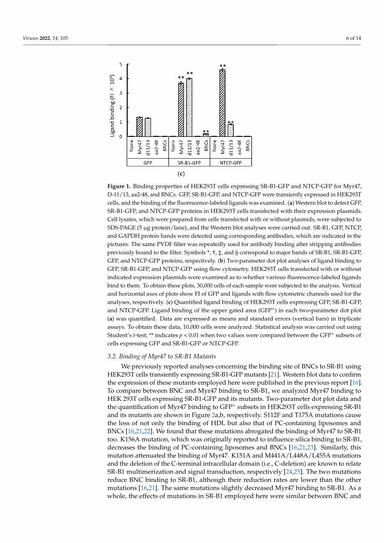
from GFP-expressing HEK293T cells under the conditions employed here. In a sampleobtained from SR-B1-GFP-expressing HEK293T cells treated with Myr47 and BS3, threebands were mainly detected, as indicated with symbols in the left picture. A faint band (*)was detected by the anti-SR-B1 antibody near a 75-kD maker. However, a correspondingband was not detected by the anti-GFP antibody. Based on the molecular size and lack ofGFP antigenicity, this band might be derived from endogenous SR-B1. The other two bands(indicated with † and ‡) were detected at 100–150 kD and more than 250 kD, respectively,by both antibodies. Bands (†) detected at 100–150 kD appeared to be derived from anSR-B1-GFP monomer binding Myr47, based on molecular size. As these were not a singleband, the number of Myr47 molecules binding to SR-B1-GFP would be heterogeneous.Unexpectedly, the strongest signal was detected in the bands of more than 250 kDa. Basedon molecular size, these bands were presumed to be a complex of SR-B1 multimer andMyr47. These results indicate that Myr47 binds SR-B1. Furthermore, it is suggested thatMyr47 preferentially binds SR-B1 multimer rather than monomer in HEK293T cells.
As HEK293T cells are a cell line established from human embryonic kidney cells,we next compared Myr47 binding to endogenous SR-B1 in HEK293T and HepG2 cells, ahepatic cell line. Myr47-linked endogenous SR-B1 was detected in the pull-down assays asfaint bands at 75–100 kD and more than 250 kD in HEK293T cells (indicated with * and †in Figure 5c, respectively); it should be noted that a very faint band at more than 250 kDwas observed in the lane of GFP-expressing HEK293T cells treated with Myr47 and BS3 inthe left Western blot of Figure 5b. In Figure 5c, bands with stronger signals were found inHepG2 cells at the same positions in HEK293T cells. The reason that strong signals wereobtained in the assays using HepG2 would be due to the more abundant expression ofendogenous SR-B1 in HepG2 cells than in HEK293T cells. In addition, a band at morethan 250 kD exhibited a greater signal than that at 75–100 kD in HepG2 cells. These resultsindicate that Myr47 binds both SR-B1 monomer and multimer not only in HEK293T cellsbut also in HepG2 cells.
Viruses 2022, 13, x FOR PEER REVIEW 11 of 15
three bands were mainly detected, as indicated with symbols in the left picture. A faint
band (*) was detected by the anti-SR-B1 antibody near a 75-kD maker. However, a corre-
sponding band was not detected by the anti-GFP antibody. Based on the molecular size
and lack of GFP antigenicity, this band might be derived from endogenous SR-B1. The
other two bands (indicated with † and ‡) were detected at 100–150 kD and more than 250
kD, respectively, by both antibodies. Bands (†) detected at 100–150 kD appeared to be
derived from an SR-B1-GFP monomer binding Myr47, based on molecular size. As these
were not a single band, the number of Myr47 molecules binding to SR-B1-GFP would be
heterogeneous. Unexpectedly, the strongest signal was detected in the bands of more than
250 kDa. Based on molecular size, these bands were presumed to be a complex of SR-B1
multimer and Myr47. These results indicate that Myr47 binds SR-B1. Furthermore, it is
suggested that Myr47 preferentially binds SR-B1 multimer rather than monomer in
HEK293T cells.
As HEK293T cells are a cell line established from human embryonic kidney cells, we
next compared Myr47 binding to endogenous SR-B1 in HEK293T and HepG2 cells, a he-
patic cell line. Myr47-linked endogenous SR-B1 was detected in the pull-down assays as
faint bands at 75–100 kD and more than 250 kD in HEK293T cells (indicated with * and †
in Figure 5c, respectively); it should be noted that a very faint band at more than 250 kD
was observed in the lane of GFP-expressing HEK293T cells treated with Myr47 and BS3
in the left Western blot of Figure 5b. In Figure 5c, bands with stronger signals were found
in HepG2 cells at the same positions in HEK293T cells. The reason that strong signals were
obtained in the assays using HepG2 would be due to the more abundant expression of
endogenous SR-B1 in HepG2 cells than in HEK293T cells. In addition, a band at more than
250 kD exhibited a greater signal than that at 75–100 kD in HepG2 cells. These results
indicate that Myr47 binds both SR-B1 monomer and multimer not only in HEK293T cells
but also in HepG2 cells.
(a)
Figure 5. Cont.
Viruses 2022, 14, 105 11 of 14Viruses 2022, 13, x FOR PEER REVIEW 12 of 15
(b)
(c)
Figure 5. Crosslink of Myr47 and SR-B1-GFP or endogenous SR-B1. (a) Illustrated image of pull-
down assays to detect SR-B1-GFP or endogenous SR-B1 crosslinked with biotinylated Myr47 using
BS3. (b) Western blot analyses of pull-down samples prepared from HEK293T cells expressing SR-
B1-GFP or GFP after sequential treatment with Myr47 and BS3. HEK293T cells transfected with in-
dicated expression plasmids were incubated with or without biotinylated Myr47. Subsequently,
they were treated with or without BS3. Cell lysates were prepared, and then they were applied for
pull-down assays using streptavidin-coated beads and a magnet. The same volume (10 μ) of sam-
ples was applied for SDS-PAGE and Western blot analyses. Antibodies used for the detection of
protein bands are indicated under the picture. Symbols, *, †, ‡, indicate Myr47-binding endogenous
SR-B1 multimer, SR-B1-GFP-monomer, and SR-B1-GFP multimer, respectively. (c) Western blot
analyses of pull-down samples prepared from HEK293T or HepG2 cells expressing endogenous SR-
B1 after sequential treatment with Myr47 and BS3. HEK293T or HepG2 cells were treated with bio-
tinylated Myr47 and BS3. Then, pull-down assays and Western blot analyses were carried out as
described in (b). Symbols, * and †, indicate Myr47-binding endogeous SR-B1 monomer and multi-
mer, respectively.
Figure 5. Crosslink of Myr47 and SR-B1-GFP or endogenous SR-B1. (a) Illustrated image of pull-down assays to detect SR-B1-GFP or endogenous SR-B1 crosslinked with biotinylated Myr47 usingBS3. (b) Western blot analyses of pull-down samples prepared from HEK293T cells expressing SR-B1-GFP or GFP after sequential treatment with Myr47 and BS3. HEK293T cells transfected with indicatedexpression plasmids were incubated with or without biotinylated Myr47. Subsequently, they weretreated with or without BS3. Cell lysates were prepared, and then they were applied for pull-downassays using streptavidin-coated beads and a magnet. The same volume (10 µ) of samples wasapplied for SDS-PAGE and Western blot analyses. Antibodies used for the detection of protein bandsare indicated under the picture. Symbols, *, †, ‡, indicate Myr47-binding endogenous SR-B1 multimer,SR-B1-GFP-monomer, and SR-B1-GFP multimer, respectively. (c) Western blot analyses of pull-downsamples prepared from HEK293T or HepG2 cells expressing endogenous SR-B1 after sequentialtreatment with Myr47 and BS3. HEK293T or HepG2 cells were treated with biotinylated Myr47 andBS3. Then, pull-down assays and Western blot analyses were carried out as described in (b). Symbols,* and †, indicate Myr47-binding endogeous SR-B1 monomer and multimer, respectively.

Viruses 2022, 14, 105 12 of 14
4. Discussion
To clarify the interaction between a ligand and a cell membrane receptor, flow cy-tometric analysis using a fluorescence-labeled ligand and cells expressing receptor-GFPfusion protein is a potent approach. By applying this method, we demonstrated that SR-B1plays a pivotal role in the binding and endocytosis of liposomes containing PC and PE inHEK293T cells [12,13]. Based on these results, we presumed that SR-B1 would play a rolein the cellular recognition of a variety of PNPs. We investigated whether BNCs can bindSR-B1 and then found that SR-B1 functions as a receptor for BNCs [21]. On the other hand,an N-terminal myristoylated pre-S1 peptide (i.e., Myr47) was demonstrated to inhibit bothHBV and HDV infection to hepatocytes [4–8]. As we considered that SR-B1 would be ableto bind not only BNCs but also Myr47, we examined the binding of Myr47 to SR-B1. In thisstudy, we demonstrated that Myr47 binds SR-B1, based on the results of (i) flow cytomet-ric analyses using fluorescence-labeled Myr47 and HEK293T cells expressing SR-B1-GFP,(ii) the colocalization of fluorescence-labeled Myr47 and SR-B1-GFP in SR-B1-GFP-HEKcells, and (iii) the crosslinking of Myr47 and SR-B1-GFP using BS3. We used HEK293T cellsto demonstrate the interaction of Myr47 and SR-B1-GFP. This cell line is widely used invirology and cellular biology experiments because it has simian virus 40 (SV40) large Tantigen, which can promote the replication of transfected DNA plasmids containing SV40Ori [26]. Therefore, we mainly used HEK293T cells in transient expression experiments inthis study.
We compared ligand-binding properties between SR-B1 and NTCP. Fluorescence-labeled Myr47 bound both SR-B1-GFP and NTCP-GFP expressed on HEK293T cells. How-ever, the Myr47-binding patterns of the two-parameter dot plots were different betweenthe two receptors. Roughly, Myr47 binding to SR-B1-GFP seemed to correlate betweenthe amount of Myr47 bound and the expression level of SR-B1-GFP in HEK293T cells (i.e.,cells expressing SR-B1-GFP at a high level bind Myr47 more than those at a low level).On the other hand, Mry47 binding to NTCP-GFP appeared to be limited at a certain level(i.e., cells expressing NTCP-GFP at a high level do not always bind Myr47 more thanthose at a low level), although it remains unclear which factor caused the difference inMyr47 binding between the two receptors. Myr47 has been shown to have the inhibitoryactivity of HBV infection to hepatic cells, whereas D-11/13 and aa2-48 do not have suchactivity [7,8]. The ligand-binding properties of NTCP-GFP matched well with the reportedHBV-infection-inhibitory activity of these peptides. However, SR-B1-GFP bound D-11/13as well as Myr47. In NTCP-GFP-expressing HEK293T cells, D-11/13 binding seemed tobe rather reduced, although the reason for this was unclear. In addition, BNCs boundSR-B1-GFP, but they did not NTCP-GFP. These results demonstrated that SR-B1 and NTCPhave differential binding properties regarding HBV-related ligands.
Here, we showed that SR-B1-GFP-HEK cells endocytosed Myr47 more efficiently thanGFP-HEK and HEK293T cells. As we demonstrated that Myr47 can bind SR-B1 by variousapproaches in this study, our results strongly suggest that the binding of Myr47 to SR-B1induces its endocytosis.
In the previous report, we demonstrated that SR-B1 can bind BNCs [21]. In this study,we showed that SR-B1 binds Myr47. In addition, analyses of Myr47 binding sites usingSR-B1 mutants suggested that both binding sites of BNCs and Myr47 are similar or close.Considering these results, it is strongly suggested that SR-B1 is involved in the cellularbinding of HBV. SR-B1 mRNA is abundantly expressed in the liver [27]. Using pull-downassays, we showed that Myr47 bound endogenous SR-B1 in HepG2 cells. NTCP-expressingHepG2 cells are frequently used for HBV infection experiments [10]. Therefore, there is apossibility that SR-B1 plays a role in the binding of HBV in hepatic cells. In some virusesincluding hepatitis C virus, SR-B1 is known to participate in viral binding to cells andinfectious processes with various other receptors [28]. However, no evidence has beenreported so far that SR-B1 is related to HBV infectivity. Therefore, even if SR-B1 is involvedin HBV cellular binding, it might have little influence on the establishment of HBV infection.Anyway, a precise evaluation of SR-B1 will be necessary to determine whether it is related

Viruses 2022, 14, 105 13 of 14
to the process of HBV infection. If SR-B1 does not have any effects on HBV infectivity,examining the binding of Mry47 derivatives to SR-B1 might be helpful to design and selectnew inhibitors, which can bind NTCP more specifically than Myr47.
By the present study using a crosslinker (BS3) and pull-down assay, it was suggestedthat Myr47 bound SR-B1 multimer rather than monomer. SR-B1 has been reported to form amultimer on plasma membranes [24]. In addition, it has been reported that SR-B1 multimerfunctions in ApoA-I binding and cholesterol efflux, whereas its monomer does not [29].Therefore, our findings, the preferential binding of Myr47 to SR-B1 multimer, suggest thatMyr47 can bind functional SR-B1 multimer. Our approach employed here will be useful inrevealing not only the binding nature of SR-B1 but also that of NTCP to Myr47.
Author Contributions: Conceptualization, S.H.; methodology, S.H.; validation, S.H.; formal analysis,S.H.; resources, S.K.; data curation, S.H.; writing—original draft preparation, S.H.; writing—reviewand editing, S.H. and S.K.; funding acquisition, S.K. All authors have read and agreed to the publishedversion of the manuscript.
Funding: This work was supported by the Japan Society for the Promotion of Science (KAKENHI,Grant-in-Aid for Scientific Research (S) (16H06314, to S.K.)), the Japan Agency for Medical Researchand Development (17cm0106214h0002, 17fk0310105h0001, to S.K.), and the Dynamic Alliance forOpen Innovation Bridging Human, Environment and Materials, of the Ministry of Education, Culture,Sports, Science and Technology of Japan (MEXT, to S.K.).
Data Availability Statement: All data generated and analyzed during this study are includedin this article.
Conflicts of Interest: The authors declare no conflict of interest.
References1. Heermann, K.H.; Goldmann, U.; Schwartz, W.; Seyffarth, T.; Baumgarten, H.; Gerlich, W.H. Large surface proteins of hepatitis B
virus containing the pre-S sequence. J. Virol. 1984, 52, 396–402. [CrossRef] [PubMed]2. Neurath, A.R.; Kent, S.B.; Strick, N.; Parker, K. Identification and chemical synthesis of a host cell receptor binding site on
hepatitis B virus. Cell 1986, 46, 429–436. [CrossRef]3. Blanchet, M.; Sureau, C. Infectivity determinants of the hepatitis B virus pre-S domain are confined to the N-terminal 75 amino
acid residues. J. Virol. 2007, 81, 5841–5849. [CrossRef] [PubMed]4. Barrera, A.; Guerra, B.; Notvall, L.; Lanford, R.E. Mapping of the hepatitis B virus pre-S1 domain involved in receptor recognition.
J. Virol. 2005, 79, 9786–9798. [CrossRef]5. Glebe, D.; Urban, S.; Knoop, E.V.; Cag, N.; Krass, P.; Grün, S.; Bulavaite, A.; Sasnauskas, K.; Gerlich, W.H. Mapping of the hepatitis
B virus attachment site by use of infection-inhibiting preS1 lipopeptides and Tupaia hepatocytes. Gastroenterology 2005, 129,234–245. [CrossRef]
6. Gripon, P.; Cannie, I.; Urban, S. Efficient inhibition of hepatitis B virus infection by acylated peptides derived from the large viralsurface protein. J. Virol. 2005, 79, 1613–1622. [CrossRef]
7. Schulze, A.; Schieck, A.; Ni, Y.; Mier, W.; Urban, S. Fine mapping of pre-S sequence requirements for hepatitis B virus largeenvelope protein-mediated receptor interaction. J. Virol. 2010, 84, 1989–2000. [CrossRef]
8. Yan, H.; Zhong, G.; Xu, G.; He, W.; Jing, Z.; Gao, Z.; Huang, Y.; Qi, Y.; Peng, B.; Wang, H.; et al. Sodium taurocholate cotransportingpolypeptide is a functional receptor for human hepatitis B and D virus. Elife 2012, 1, e00049. [CrossRef]
9. Lamas Longarela, O.; Schmidt, T.T.; Schöneweis, K.; Romeo, R.; Wedemeyer, H.; Urban, S.; Schulze, A. Proteoglycans act ascellular hepatitis delta virus attachment receptors. PLoS ONE 2013, 8, e58340. [CrossRef]
10. Somiya, M.; Liu, Q.; Yoshimoto, N.; Iijima, M.; Tatematsu, K.; Nakai, T.; Okajima, T.; Kuroki, K.; Ueda, K.; Kuroda, S. Cellularuptake of hepatitis B virus envelope L particles is independent of sodium taurocholate cotransporting polypeptide, but dependenton heparan sulfate proteoglycan. Virology 2016, 497, 23–32. [CrossRef]
11. Kang, C.; Syed, Y.Y. Bulevirtide: First approval. Drugs 2020, 80, 1601–1605. [CrossRef]12. Koh, C.; Da, B.L.; Glenn, J.S. HBV/HDV coinfection: A challenge for therapeutics. Clin. Liver Dis. 2019, 23, 557–572. [CrossRef]13. Nanahara, M.; Chang, Y.-T.; Somiya, S.; Kuroda, S. HBV pre-S1-derived myristoylated peptide (Myr47): Identification of the
inhibitory activity on the cellular uptake of lipid nanoparticles. Viruses 2021, 13, 929. [CrossRef]14. Fujita, K.; Somiya, M.; Kuroda, S.; Hinuma, S. Induction of lipid droplets in non-macrophage cells as well as macrophages by
liposomes and exosomes. Biochem. Biophys. Res. Commun. 2019, 510, 184–190. [CrossRef]15. Fujita, K.; Koide, N.; Somiya, M.; Kuroda, S.; Hinuma, S. A regulatory role of scavenger receptor class B type 1 in en-
docytosis and lipid droplet formation induced by liposomes containing phosphatidylethanolamine in HEK293T cells.Biochim. Biophys. Acta Mol. Cell. Res. 2021, 1868, 118859. [CrossRef]

Viruses 2022, 14, 105 14 of 14
16. Koide, N.; Fujita, K.; Kuroda, S.; Hinuma, S. Binding of liposomes composed of phosphatidylcholine to scavenger receptor classB type 1 and its modulation by phosphatidic acid in HEK293T cells. Biochim. Biophys. Acta Mol. Cell. Res. 2021, 1868, 119043.[CrossRef]
17. Shen, W.J.; Asthana, S.; Kraemer, F.B.; Azhar, S. Scavenger receptor B type 1: Expression, molecular regulation, and cholesteroltransport function. J. Lipid Res. 2018, 59, 1114–1131. [CrossRef]
18. Yu, D.; Amano, C.; Fukuda, T.; Yamada, T.; Kuroda, S.; Tanizawa, K.; Kondo, A.; Ueda, M.; Yamada, H.; Tada, H.; et al. Thespecific delivery of proteins to human liver cells by engineered bio-nanocapsules. FEBS J. 2005, 272, 3651–3660. [CrossRef]
19. Yamada, T.; Iwasaki, Y.; Tada, H.; Iwabuki, H.; Chuah, M.K.L.; VandenDriessche, T.; Fukuda, H.; Kondo, A.; Ueda, M.; Seno, M.;et al. Nanoparticles for the delivery of genes and drugs to human hepatocytes. Nat. Biotechnol. 2003, 21, 885–890. [CrossRef]
20. Jung, J.; Somiya, M.; Jeong, S.Y.; Choi, E.K.; Kuroda, S. Low immunogenic bio-nanocapsule based on hepatitis B virus escapemutants. Nanomedicine 2018, 14, 595–600. [CrossRef] [PubMed]
21. Hinuma, S.; Fujita, K.; Kuroda, S. Binding of nanoparticles harboring recombinant large surface protein of hepatitis B virus toscavenger receptor class B type 1. Viruses 2021, 13, 1334. [CrossRef]
22. Chadwick, A.C.; Sahoo, D. Functional characterization of newly-discovered mutations in human SR-BI. PLoS ONE 2012, 7, e45660.[CrossRef]
23. Tsugita, M.; Morimoto, N.; Tashiro, M.; Kinoshita, K.; Nakayama, M. SR-B1 is a silica receptor that mediates canonical inflamma-some activation. Cell Rep. 2017, 18, 1298–1311. [CrossRef]
24. Marques, P.E.; Nyegaard, S.; Collins, R.F.; Troise, F.; Freeman, S.A.; Trimble, W.S.; Grinstein, S. Multimerization and retention ofthe scavenger receptor SR-B1 in the plasma membrane. Dev. Cell 2019, 50, 283–295.e5. [CrossRef]
25. Sahebi, R.; Hassanian, S.M.; Ghayour-Mobarhan, M.; Farrokhi, E.; Rezayi, M.; Samadi, S.; Bahramian, S.; Ferns, G.A.; Avan, A.Scavenger receptor class B type I as a potential risk stratification biomarker and therapeutic target in cardiovascular disease.J. Cell Physiol. 2019, 234, 16925–16932. [CrossRef] [PubMed]
26. Reus, J.B.; Trivino-Soto, G.S.; Wu, L.I.; Kokott, K.; Lim, E.S. SV40 large T antigen is not responsible for the loss of STING in 293Tcells but can inhibit cGAS-STING interferon induction. Viruses 2020, 12, 137. [CrossRef] [PubMed]
27. The Human Protein Atlas. Available online: https://www.proteinatlas.org/ENSG00000073060-SCARB1/tissue (accessed on 1December 2021).
28. Jeulin, H.; Velay, A.; Murray, J.; Schvoerer, E. Clinical impact of hepatitis B and C virus envelope glycoproteins. World J. Gastroenterol.2013, 19, 654–664. [CrossRef]
29. Han, C.H.; Lee, M.H. Topology of scavenger receptor class B type I (SR-BI) on brush border membrane. J. Vet. Sci. 2002, 3, 265–272.[CrossRef]
Copyright © 2022 FDOKUMEN