Bilateral deep brain stimulation of the pedunculopontine and subthalamic nuclei in severe...
12
Bilateral deep brain stimulation of the pedunculopontine and subthalamic nuclei in severe Parkinson’s disease Alessandro Stefani, 1,2 Andres M. Lozano, 6 Antonella Peppe, 2 Paolo Stanzione, 1,2 Salvatore Galati, 1 Domenicantonio Tropepi, 1 Mariangela Pierantozzi, 1 Livia Brusa, 4 Eugenio Scarnati 3 and Paolo Mazzone 5 1 IRCCS Fondazione St Lucia, 2 Clinica Neurologica, Dipartimento di Neuroscienze, Universita' di Roma Tor Vergata, 3 Unita' Operativa di Neurochirurgia Funzionale e Stereotassica, Ospedale CTO ‘A. Alesini’, ASL RMC, 4 Unita' Operativa di Neurologia, Ospedale S. Eugenio, Roma, 5 Department of Biomedical Technology, University of L’Aquila, L’Aquila, Italy and 6 Toronto Western Hospital, Research Institute, University of Toronto, Toronto, ON, Canada Correspondence to: Alessandro Stefani, MD, Department of Neuroscience, University of Rome Tor Vergata, Roma, Italy E-mail: [email protected] Gait disturbances and akinesia are extremely disabling in advanced Parkinson’s disease. It has been suggested that modulation of the activity of the pedunculopontine nucleus (PPN) may be beneficial in the treatment of these symptoms. We report the clinical affects of deep brain stimulation (DBS) in the PPN and subthalamic nucleus (STN). Six patients with unsatisfactory pharmacological control of axial signs such as gait and postural stability underwent bilateral implantation of DBS electrodes in the STN and PPN. Clinical effects were evalu- ated 2^6 months after surgery in the OFF- and ON-medication state, with both STN and PPN stimulation ON or OFF, or with only one target being stimulated. Bilateral PPN-DBS at 25 Hz in OFF-medication produced an immediate 45% amelioration of the motor Unified Parkinson’s Disease Rating Scale (UPDRS) subscale score, followed by a decline to give a final improvement of 32% in the score after 3^6 months. In contrast, bilateral STN-DBS at 130^185 Hz led to about 54% improvement. PPN-DBS was particularly effective on gait and pos- tural items. In ON-medication state, the association of STN and PPN-DBS provided a significant further improvement when compared to the specific benefit mediated by the activation of either single target. Moreover, the combined DBS of both targets promoted a substantial amelioration in the performance of daily living activities.These findings indicate that, in patients with advanced Parkinson’s disease, PPN-DBS associated with standard STN-DBS may be useful in improving gait and in optimizing the dopamine-mediated ON-state, particularly in those whose response to STN only DBS has deteriorated over time. This combination of targets may also prove useful in extra-pyramidal disorders, such as progressive supranuclear palsy, for which treatments are currently elusive. Keywords: Parkinson’s disease; stereotactic neurosurgery; gait; deep brain stimulation; PPN Abbreviations: PPN ¼ pedunculopontine nucleus; STN ¼ subthalamic nucleus; DBS ¼ deep brain stimulation; SN ¼ substantia nigra Received January 23, 2006. Revised October 30, 2006. Accepted November 7 , 2006. Advance Access publication January 24, 2007 Introduction The pedunculopontine nucleus (PPN) plays an important role in the initiation and maintenance of locomotion in experimental animals (Skinner et al., 1990; Garcia-Rill et al., 1987). In MPTP (1-methyl-4-phenyl-1,2,3,6-tetrahydropyr- idine)-treated parkinsonian non-human primates, lesions of the PPN may produce akinesia (Nandi et al., 2002b); on the contrary, driving PPN activity by direct pharmaco- logical activation or electrical stimulation increases motor activity (Jenkinson et al., 2005; Nandi et al., 2002a, b, 2004). Recently it has been suggested that PPN could be a therapeutic target to improve gait in certain parkinsonian patients (Mena-Segovia et al., 2004; doi:10.1093/brain/awl346 Brain (2007), 130, 1596 ^1607 ß 2007 The Author(s) This is an Open Access article distributed under the terms of the Creative Commons Attribution Non-Commercial License (http://creativecommons.org/licenses/by-nc/2.0/uk/) which permits unrestricted non-commercial use, distribution, and reproduction in any medium, provided the original work is properly cited. by guest on March 6, 2014 http://brain.oxfordjournals.org/ Downloaded from
Transcript of Bilateral deep brain stimulation of the pedunculopontine and subthalamic nuclei in severe...
Bilateral deep brain stimulation of thepedunculopontine and subthalamic nucleiin severe Parkinson’s diseaseAlessandro Stefani,1,2 Andres M. Lozano,6 Antonella Peppe,2 Paolo Stanzione,1,2 Salvatore Galati,1
DomenicantonioTropepi,1Mariangela Pierantozzi,1 Livia Brusa,4 Eugenio Scarnati3 and Paolo Mazzone5
1IRCCS Fondazione St Lucia, 2Clinica Neurologica, Dipartimento di Neuroscienze, Universita' di RomaTor Vergata, 3Unita'Operativa di Neurochirurgia Funzionale e Stereotassica, Ospedale CTO ‘A. Alesini’, ASL RMC, 4Unita' Operativa diNeurologia, Ospedale S. Eugenio, Roma, 5Department of Biomedical Technology, University of L’Aquila, L’Aquila, Italy and6Toronto Western Hospital, Research Institute, University of Toronto, Toronto, ON, Canada
Correspondence to: Alessandro Stefani, MD, Department of Neuroscience, University of RomeTor Vergata, Roma, ItalyE-mail: [email protected]
Gait disturbances and akinesia are extremely disabling in advanced Parkinson’s disease. It has been suggestedthat modulation of the activity of the pedunculopontine nucleus (PPN) may be beneficial in the treatment ofthese symptoms.We report the clinical affects of deep brain stimulation (DBS) in the PPN and subthalamicnucleus (STN). Six patients with unsatisfactory pharmacological control of axial signs such as gait and posturalstability underwent bilateral implantation of DBS electrodes in the STN and PPN. Clinical effects were evalu-ated 2^6 months after surgery in the OFF- and ON-medication state, with both STNand PPN stimulation ONor OFF, or with only one target being stimulated. Bilateral PPN-DBS at 25Hz in OFF-medication produced animmediate 45% amelioration of the motor Unified Parkinson’s Disease Rating Scale (UPDRS) subscale score,followed by a decline to give a final improvement of 32% in the score after 3^6 months. In contrast, bilateralSTN-DBS at 130^185Hz led to about 54% improvement. PPN-DBS was particularly effective on gait and pos-tural items. In ON-medication state, the association of STN and PPN-DBS provided a significant furtherimprovement when compared to the specific benefit mediated by the activation of either single target.Moreover, the combined DBS of both targets promoted a substantial amelioration in the performance of dailyliving activities.These findings indicate that, in patients with advanced Parkinson’s disease, PPN-DBS associatedwith standard STN-DBS may be useful in improving gait and in optimizing the dopamine-mediated ON-state,particularly in those whose response to STNonly DBS has deteriorated over time.This combination of targetsmay also prove useful in extra-pyramidal disorders, such as progressive supranuclear palsy, for which treatmentsare currently elusive.
Keywords: Parkinson’s disease; stereotactic neurosurgery; gait; deep brain stimulation; PPN
Abbreviations: PPN¼ pedunculopontine nucleus; STN¼ subthalamic nucleus; DBS¼ deep brain stimulation;SN¼ substantia nigra
Received January 23, 2006. Revised October 30, 2006. Accepted November 7, 2006. Advance Access publication January 24, 2007
IntroductionThe pedunculopontine nucleus (PPN) plays an importantrole in the initiation and maintenance of locomotion inexperimental animals (Skinner et al., 1990; Garcia-Rill et al.,1987). In MPTP (1-methyl-4-phenyl-1,2,3,6-tetrahydropyr-idine)-treated parkinsonian non-human primates, lesionsof the PPN may produce akinesia (Nandi et al., 2002b);
on the contrary, driving PPN activity by direct pharmaco-logical activation or electrical stimulation increasesmotor activity (Jenkinson et al., 2005; Nandi et al.,2002a, b, 2004). Recently it has been suggested thatPPN could be a therapeutic target to improve gait incertain parkinsonian patients (Mena-Segovia et al., 2004;
doi:10.1093/brain/awl346 Brain (2007), 130, 1596^1607
� 2007 The Author(s)This is an Open Access article distributed under the terms of the Creative Commons Attribution Non-Commercial License (http://creativecommons.org/licenses/by-nc/2.0/uk/)which permits unrestricted non-commercial use, distribution, and reproduction in any medium, provided the original work is properly cited.
by guest on March 6, 2014
http://brain.oxfordjournals.org/D
ownloaded from
Papahill and Lozano, 2000). Currently available therapiesprovide only variable degrees of control for axial signs ofParkinson’s disease, such as deficits in gait and posture.
Two recent reports demonstrated that deep brainstimulation (DBS) in the PPN may be beneficial to patientswith Parkinson’s disease (Mazzone et al., 2005a; Plaha andGill, 2005). The data to date suggest that PPN-DBS may beconsidered relatively safe and may improve motor function.While these preliminary reports are encouraging, longerterm observations are required. In particular, the questionarises as to whether PPN represents a fully alternative targetarea with respect to well-established implantation areas(e.g. the subthalamic nucleus, STN). This article reports ourfindings on six patients who underwent simultaneousbilateral PPN and STN-DBS implantation, followed up to6 months postoperatively. Particular care was given toassessing both motor and gait subscores as well as thespecific PPN versus STN mediated benefits.
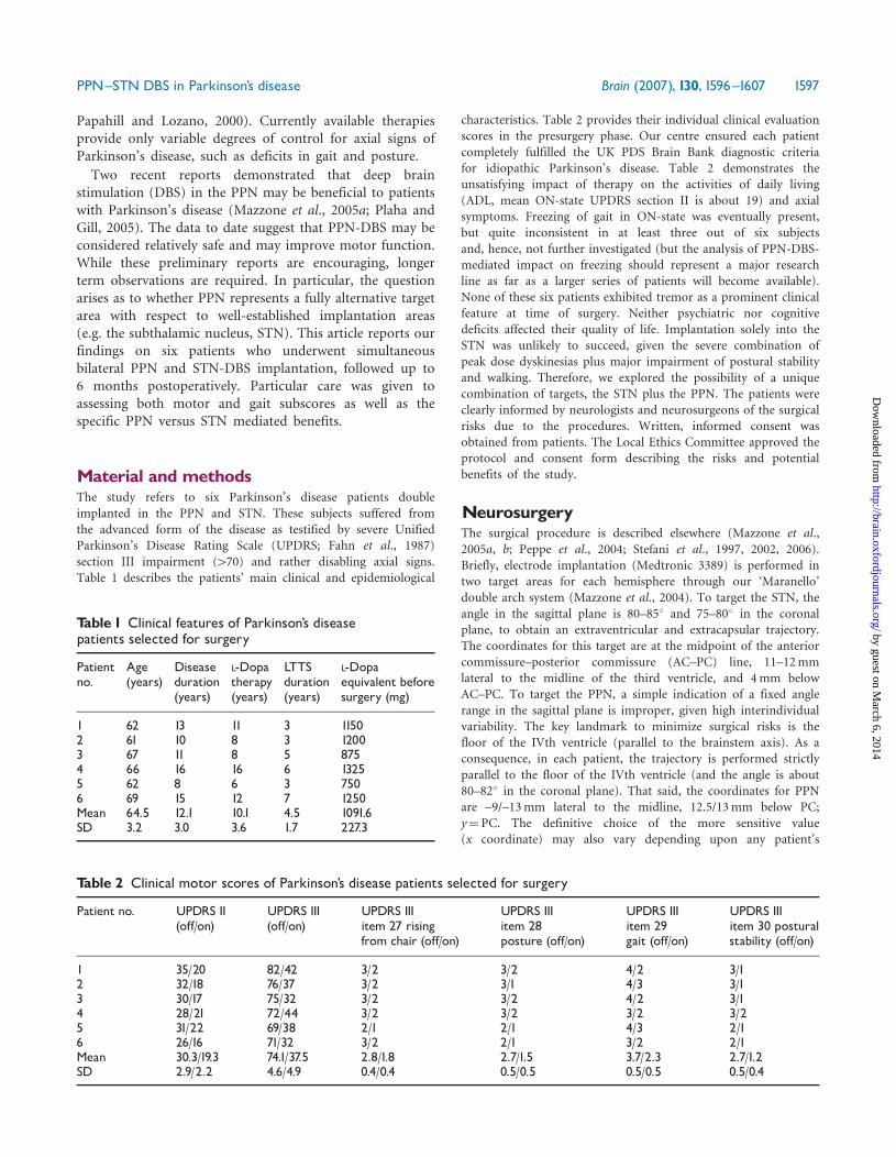
Material and methodsThe study refers to six Parkinson’s disease patients doubleimplanted in the PPN and STN. These subjects suffered fromthe advanced form of the disease as testified by severe UnifiedParkinson’s Disease Rating Scale (UPDRS; Fahn et al., 1987)section III impairment (470) and rather disabling axial signs.Table 1 describes the patients’ main clinical and epidemiological
characteristics. Table 2 provides their individual clinical evaluationscores in the presurgery phase. Our centre ensured each patientcompletely fulfilled the UK PDS Brain Bank diagnostic criteriafor idiopathic Parkinson’s disease. Table 2 demonstrates theunsatisfying impact of therapy on the activities of daily living(ADL, mean ON-state UPDRS section II is about 19) and axialsymptoms. Freezing of gait in ON-state was eventually present,but quite inconsistent in at least three out of six subjectsand, hence, not further investigated (but the analysis of PPN-DBS-mediated impact on freezing should represent a major researchline as far as a larger series of patients will become available).None of these six patients exhibited tremor as a prominent clinicalfeature at time of surgery. Neither psychiatric nor cognitivedeficits affected their quality of life. Implantation solely into theSTN was unlikely to succeed, given the severe combination ofpeak dose dyskinesias plus major impairment of postural stabilityand walking. Therefore, we explored the possibility of a uniquecombination of targets, the STN plus the PPN. The patients wereclearly informed by neurologists and neurosurgeons of the surgicalrisks due to the procedures. Written, informed consent wasobtained from patients. The Local Ethics Committee approved theprotocol and consent form describing the risks and potentialbenefits of the study.
NeurosurgeryThe surgical procedure is described elsewhere (Mazzone et al.,2005a, b; Peppe et al., 2004; Stefani et al., 1997, 2002, 2006).Briefly, electrode implantation (Medtronic 3389) is performed intwo target areas for each hemisphere through our ‘Maranello’double arch system (Mazzone et al., 2004). To target the STN, theangle in the sagittal plane is 80–85� and 75–80� in the coronalplane, to obtain an extraventricular and extracapsular trajectory.The coordinates for this target are at the midpoint of the anteriorcommissure–posterior commissure (AC–PC) line, 11–12mmlateral to the midline of the third ventricle, and 4mm belowAC–PC. To target the PPN, a simple indication of a fixed anglerange in the sagittal plane is improper, given high interindividualvariability. The key landmark to minimize surgical risks is thefloor of the IVth ventricle (parallel to the brainstem axis). As aconsequence, in each patient, the trajectory is performed strictlyparallel to the floor of the IVth ventricle (and the angle is about80–82� in the coronal plane). That said, the coordinates for PPNare –9/–13mm lateral to the midline, 12.5/13mm below PC;y¼PC. The definitive choice of the more sensitive value(x coordinate) may also vary depending upon any patient’s
Table 1 Clinical features of Parkinson’s diseasepatients selected for surgery
Patientno.
Age(years)
Diseaseduration(years)
L-Dopatherapy(years)
LTTSduration(years)
L-Dopaequivalent beforesurgery (mg)
1 62 13 11 3 11502 61 10 8 3 12003 67 11 8 5 8754 66 16 16 6 13255 62 8 6 3 7506 69 15 12 7 1250Mean 64.5 12.1 10.1 4.5 1091.6SD 3.2 3.0 3.6 1.7 227.3
Table 2 Clinical motor scores of Parkinson’s disease patients selected for surgery
Patient no. UPDRS II(off/on)
UPDRS III(off/on)
UPDRS IIIitem 27 risingfrom chair (off/on)
UPDRS IIIitem 28posture (off/on)
UPDRS IIIitem 29gait (off/on)
UPDRS IIIitem 30 posturalstability (off/on)
1 35/20 82/42 3/2 3/2 4/2 3/12 32/18 76/37 3/2 3/1 4/3 3/13 30/17 75/32 3/2 3/2 4/2 3/14 28/21 72/44 3/2 3/2 3/2 3/25 31/22 69/38 2/1 2/1 4/3 2/16 26/16 71/32 3/2 2/1 3/2 2/1Mean 30.3/19.3 74.1/37.5 2.8/1.8 2.7/1.5 3.7/2.3 2.7/1.2SD 2.9/2.2 4.6/4.9 0.4/0.4 0.5/0.5 0.5/0.5 0.5/0.4
PPN^STN DBS in Parkinson’s disease Brain (2007), 130, 1596^1607 1597
by guest on March 6, 2014
http://brain.oxfordjournals.org/D
ownloaded from
brainstem anatomy, the wideness of cisterna ambiens and the
location of cerebral posterior artery with respect to these
structures. Intra-operative microrecordings (MER) are performed
routinely with FHC tungsten microelectrodes (1M�) and are
described in detail elsewhere (Galati et al., 2006; Stefani et al.,
2006). By targeting the STN after PPN implantation (n¼ 4/6),
we had the opportunity to study STN firing activity before
and during PPN stimulation delivered at 10, 25 or 80Hz (data
not shown; Mazzone et al., 2006).Postsurgery, the definitive electrode locations were verified by
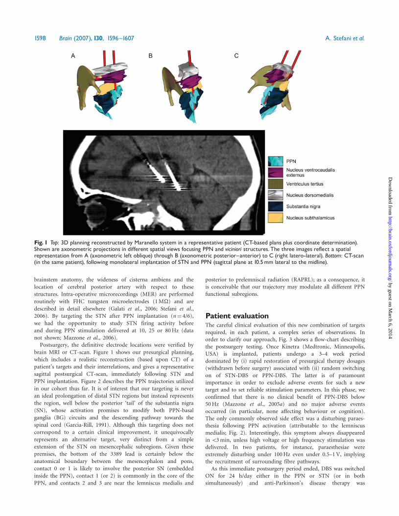
brain MRI or CT-scan. Figure 1 shows our presurgical planning,
which includes a realistic reconstruction (based upon CT) of a
patient’s targets and their interrelations, and gives a representative
sagittal postsurgical CT-scan, immediately following STN and
PPN implantation. Figure 2 describes the PPN trajectories utilized
in our cohort thus far. It is of interest that our targeting is never
an ideal prolongation of distal STN regions but instead represents
the region, well below the posterior ‘tail’ of the substantia nigra
(SN), whose activation promises to modify both PPN-basal
ganglia (BG) circuits and the descending pathway towards the
spinal cord (Garcia-Rill, 1991). Although this targeting does not
correspond to a certain clinical improvement, it unequivocally
represents an alternative target, very distinct from a simple
extension of the STN on mesencephalic subregions. Given these
premises, the bottom of the 3389 lead is certainly below the
anatomical boundary between the mesencephalon and pons,
contact 0 or 1 is likely to involve the posterior SN (embedded
inside the PPN), contact 1 (or 2) is commonly in the core of the
PPN, and contacts 2 and 3 are near the lemniscus medialis and
posterior to prelemniscal radiation (RAPRL); as a consequence, itis conceivable that our trajectory may modulate all different PPNfunctional subregions.
Patient evaluationThe careful clinical evaluation of this new combination of targetsrequired, in each patient, a complex series of observations. Inorder to clarify our approach, Fig. 3 shows a flow-chart describingthe postsurgery testing. Once Kinetra (Medtronic, Minneapolis,USA) is implanted, patients undergo a 3–4 week perioddominated by (i) rapid restoration of presurgical therapy dosages(withdrawn before surgery) associated with (ii) random switchingon of STN-DBS or PPN-DBS. The latter is of paramountimportance in order to exclude adverse events for such a newtarget and to set reliable stimulation parameters. In this phase, weconfirmed that there is no clinical benefit of PPN-DBS below50Hz (Mazzone et al., 2005a) and no major adverse eventsoccurred (in particular, none affecting behaviour or cognition).The only commonly observed side effect was a disturbing paraes-thesia following PPN activation (attributable to the lemniscusmedialis; Fig. 2). Interestingly, this symptom always disappearedin53min, unless high voltage or high frequency stimulation wasdelivered. In two patients, for instance, paraesthesiae wereextremely disturbing under 100Hz even under 0.5–1V, implyingthe recruitment of surrounding fibre pathways.As this immediate postsurgery period ended, DBS was switched
ON for 24 h/day either in the PPN or STN (or in bothsimultaneously) and anti-Parkinson’s disease therapy was
Fig. 1 Top: 3D planning reconstructed by Maranello system in a representative patient (CT-based plans plus coordinate determination).Shown are axonometric projections in different spatial views focusing PPN and viciniori structures. The three images reflect a spatialrepresentation from A (axonometric left oblique) through B (axonometric posterior^anterior) to C (right latero-lateral). Bottom: CT-scan(in the same patient), following monolateral implantation of STN and PPN (sagittal plane at 10.5mm lateral to the midline).
1598 Brain (2007), 130, 1596^1607 A. Stefani et al.
by guest on March 6, 2014
http://brain.oxfordjournals.org/D
ownloaded from
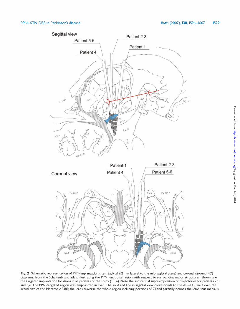
Fig. 2 Schematic representation of PPN-implantation sites. Sagittal (12mm lateral to the mid-sagittal plane) and coronal (around PC)diagrams, from the Schaltenbrand atlas, illustrating the PPN functional region with respect to surrounding major structures. Shown arethe targeted implantation locations in all patients of the study (n¼ 6).Note the substantial supra-imposition of trajectories for patients 2/3and 5/6. The PPN-targeted region was emphasized in cyan.The solid red line in sagittal view corresponds to the AC^PC line.Given theactual size of the Medtronic 3389, the leads traverse the whole region including portions of ZI and partially bounds the lemniscus medialis.
PPN^STN DBS in Parkinson’s disease Brain (2007), 130, 1596^1607 1599
by guest on March 6, 2014
http://brain.oxfordjournals.org/D
ownloaded from
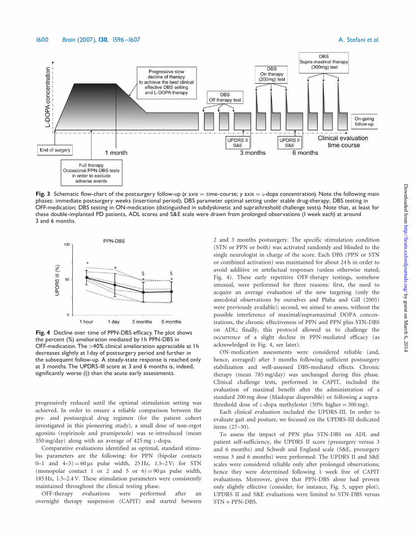
progressively reduced until the optimal stimulation setting was
achieved. In order to ensure a reliable comparison between the
pre- and postsurgical drug regimen (for the patient cohort
investigated in this pioneering study), a small dose of non-ergot
agonists (ropirinole and pramipexole) was re-introduced (mean
350mg/day) along with an average of 425mg L-dopa.Comparative evaluations identified as optimal, standard stimu-
lus parameters are the following: for PPN (bipolar contacts
0–1 and 4–5)¼ 60 ms pulse width, 25Hz, 1.5–2V; for STN
(monopolar contact 1 or 2 and 5 or 6)¼ 90 ms pulse width,
185Hz, 1.5–2.4 V. These stimulation parameters were consistently
maintained throughout the clinical testing phase.OFF-therapy evaluations were performed after an
overnight therapy suspension (CAPIT) and started between
2 and 3 months postsurgery. The specific stimulation condition(STN or PPN or both) was activated randomly and blinded to thesingle neurologist in charge of the score. Each DBS (PPN or STNor combined activation) was maintained for about 24 h in order toavoid additive or artefactual responses (unless otherwise stated;Fig. 4). These early repetitive OFF-therapy testings, somehowunusual, were performed for three reasons: first, the need toacquire an average evaluation of the new targeting (only theanecdotal observations by ourselves and Plaha and Gill (2005)were previously available); second, we aimed to assess, without thepossible interference of maximal/supramaximal DOPA concen-trations, the chronic effectiveness of PPN and PPN plus STN-DBSon ADL; finally, this protocol allowed us to challenge theoccurrence of a slight decline in PPN-mediated efficacy (asacknowledged in Fig. 4, see later).ON-medication assessments were considered reliable (and,
hence, averaged) after 3 months following sufficient postsurgerystabilization and well-assessed DBS-mediated effects. Chronictherapy (mean 785mg/day) was unchanged during this phase.Clinical challenge tests, performed in CAPIT, included theevaluation of maximal benefit after the administration of astandard 200mg dose (Madopar dispersible) or following a supra-threshold dose of L-dopa methylester (50% higher¼ 300mg).Each clinical evaluation included the UPDRS-III. In order to
evaluate gait and posture, we focused on the UPDRS-III dedicateditems (27–30).To assess the impact of PPN plus STN-DBS on ADL and
patient self-sufficiency, the UPDRS II score (presurgery versus 3and 6 months) and Schwab and England scale (S&E, presurgeryversus 3 and 6 months) were performed. The UPDRS II and S&Escales were considered reliable only after prolonged observations;hence they were determined following 1 week free of CAPITevaluations. Moreover, given that PPN-DBS alone had provenonly slightly effective (consider, for instance, Fig. 5, upper plot),UPDRS II and S&E evaluations were limited to STN-DBS versusSTNþPPN-DBS.
Fig. 3 Schematic flow-chart of the postsurgery follow-up (x axis ¼ time-course; y axis ¼ L-dopa concentration). Note the following mainphases: immediate postsurgery weeks (insertional period); DBS parameter optimal setting under stable drug-therapy; DBS testing inOFF-medication; DBS testing in ON-medication (distinguished in subdyskinetic and suprathreshold challenge tests). Note that, at least forthese double-implanted PD patients, ADL scores and S&E scale were drawn from prolonged observations (1week each) at around3 and 6 months.
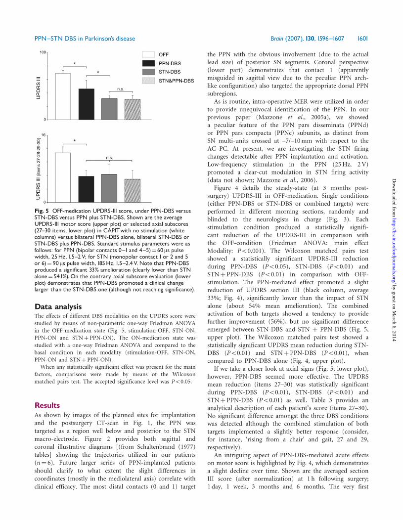
Fig. 4 Decline over time of PPN-DBS efficacy. The plot showsthe percent (%) amelioration mediated by 1h PPN-DBS inOFF-medication. The440% clinical amelioration appreciable at 1hdecreases slightly at 1day of postsurgery period and further inthe subsequent follow-up. A steady-state response is reached onlyat 3 months. The UPDRS-III score at 3 and 6 months is, indeed,significantly worse (x) than the acute early assessments.
1600 Brain (2007), 130, 1596^1607 A. Stefani et al.
by guest on March 6, 2014
http://brain.oxfordjournals.org/D
ownloaded from
Data analysisThe effects of different DBS modalities on the UPDRS score were
studied by means of non-parametric one-way Friedman ANOVA
in the OFF-medication state (Fig. 5, stimulation-OFF, STN-ON,
PPN-ON and STNþPPN-ON). The ON-medication state was
studied with a one-way Friedman ANOVA and compared to thebasal condition in each modality (stimulation-OFF, STN-ON,
PPN-ON and STNþPPN-ON).When any statistically significant effect was present for the main
factors, comparisons were made by means of the Wilcoxonmatched pairs test. The accepted significance level was P50.05.
ResultsAs shown by images of the planned sites for implantationand the postsurgery CT-scan in Fig. 1, the PPN wastargeted as a region well below and posterior to the STNmacro-electrode. Figure 2 provides both sagittal andcoronal illustrative diagrams [(from Schaltenbrand (1977)tables] showing the trajectories utilized in our patients(n¼ 6). Future larger series of PPN-implanted patientsshould clarify to what extent the slight differences incoordinates (mostly in the mediolateral axis) correlate withclinical efficacy. The most distal contacts (0 and 1) target
the PPN with the obvious involvement (due to the actuallead size) of posterior SN segments. Coronal perspective(lower part) demonstrates that contact 1 (apparentlymisguided in sagittal view due to the peculiar PPN arch-like configuration) also targeted the appropriate dorsal PPNsubregions.
As is routine, intra-operative MER were utilized in orderto provide unequivocal identification of the PPN. In ourprevious paper (Mazzone et al., 2005a), we showeda peculiar feature of the PPN pars disseminata (PPNd)or PPN pars compacta (PPNc) subunits, as distinct fromSN multi-units crossed at –7/–10mm with respect to theAC–PC. At present, we are investigating the STN firingchanges detectable after PPN implantation and activation.Low-frequency stimulation in the PPN (25Hz, 2 V)promoted a clear-cut modulation in STN firing activity(data not shown; Mazzone et al., 2006).
Figure 4 details the steady-state (at 3 months post-surgery) UPDRS-III in OFF-medication. Single conditions(either PPN-DBS or STN-DBS or combined targets) wereperformed in different morning sections, randomly andblinded to the neurologists in charge (Fig. 3). Eachstimulation condition produced a statistically signifi-cant reduction of the UPDRS-III in comparison withthe OFF-condition (Friedman ANOVA: main effectModality: P50.001). The Wilcoxon matched pairs testshowed a statistically significant UPDRS-III reductionduring PPN-DBS (P50.05), STN-DBS (P50.01) andSTNþ PPN-DBS (P50.01) in comparison with OFF-stimulation. The PPN-mediated effect promoted a slightreduction of UPDRS section III (black column, average33%; Fig. 4), significantly lower than the impact of STNalone (about 54% mean amelioration). The combinedactivation of both targets showed a tendency to providefurther improvement (56%), but no significant differenceemerged between STN-DBS and STN þ PPN-DBS (Fig. 5,upper plot). The Wilcoxon matched pairs test showed astatistically significant UPDRS mean reduction during STN-DBS (P50.01) and STNþ PPN-DBS (P50.01), whencompared to PPN-DBS alone (Fig. 4, upper plot).
If we take a closer look at axial signs (Fig. 5, lower plot),however, PPN-DBS seemed more effective. The UPDRSmean reduction (items 27–30) was statistically significantduring PPN-DBS (P50.01), STN-DBS (P50.01) andSTNþ PPN-DBS (P50.01) as well. Table 3 provides ananalytical description of each patient’s score (items 27–30).No significant difference amongst the three DBS conditionswas detected although the combined stimulation of bothtargets implemented a slightly better response (consider,for instance, ‘rising from a chair’ and gait, 27 and 29,respectively).
An intriguing aspect of PPN-DBS-mediated acute effectson motor score is highlighted by Fig. 4, which demonstratesa slight decline over time. Shown are the averaged sectionIII score (after normalization) at 1 h following surgery;1 day, 1 week, 3 months and 6 months. The very first
Fig. 5 OFF-medication UPDRS-III score, under PPN-DBS versusSTN-DBS versus PPN plus STN-DBS. Shown are the averageUPDRS-III motor score (upper plot) or selected axial subscores(27^30 items, lower plot) in CAPITwith no stimulation (whitecolumns) versus bilateral PPN-DBS alone, bilateral STN-DBS orSTN-DBS plus PPN-DBS. Standard stimulus parameters were asfollows: for PPN (bipolar contacts 0^1 and 4^5)¼ 60ms pulsewidth, 25Hz, 1.5^2V; for STN (monopolar contact 1 or 2 and 5or 6)¼ 90ms pulse width, 185Hz, 1.5^2.4V. Note that PPN-DBSproduced a significant 33% amelioration (clearly lower than STNalone¼ 54.1%).On the contrary, axial subscore evaluation (lowerplot) demonstrates that PPN-DBS promoted a clinical changelarger than the STN-DBS one (although not reaching significance).
PPN^STN DBS in Parkinson’s disease Brain (2007), 130, 1596^1607 1601
by guest on March 6, 2014
http://brain.oxfordjournals.org/D
ownloaded from
clinical assessment was surprising, with a peculiar 440%mean benefit associated with the subject’s enthusiasm(at least in five out of six patients). Yet, this ‘immediate’dramatic PPN-driven impact cannot be consistentlyreplicated with the following evaluations despite an ampleinterpatient variability. As any team familiar with post-surgery Parkinson’s disease patients knows, well balanced
and repeated clinical observations are necessary to avoidinterference from placebo-like effects (Benedetti et al.,2003). There were no major differences in observations at3 or 6 months and only then was the described benefitunder the condition ON-PPN considered reliable (Fig. 5).A similar time-related effect with an early reduction ofstimulation efficacy was not observed when STN-DBS waschallenged at different postsurgery times (data not shown).
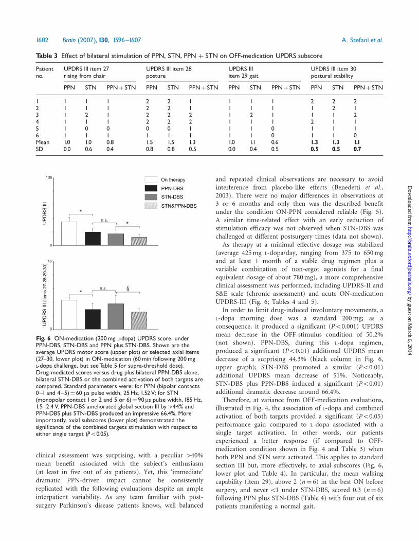
As therapy at a minimal effective dosage was stabilized(average 425mg L-dopa/day, ranging from 375 to 650mgand at least 1 month of a stable drug regimen plus avariable combination of non-ergot agonists for a finalequivalent dosage of about 780mg), a more comprehensiveclinical assessment was performed, including UPDRS-II andS&E scale (chronic assessment) and acute ON-medicationUPDRS-III (Fig. 6; Tables 4 and 5).
In order to limit drug-induced involuntary movements, aL-dopa morning dose was a standard 200mg; as aconsequence, it produced a significant (P50.001) UPDRSmean decrease in the OFF-stimulus condition of 50.2%(not shown). PPN-DBS, during this L-dopa regimen,produced a significant (P50.01) additional UPDRS meandecrease of a surprising 44.3% (black column in Fig. 6,upper graph); STN-DBS promoted a similar (P50.01)additional UPDRS mean decrease of 51%. Noticeably,STN-DBS plus PPN-DBS induced a significant (P50.01)additional dramatic decrease around 66.4%.
Therefore, at variance from OFF-medication evaluations,illustrated in Fig. 4, the association of L-dopa and combinedactivation of both targets provided a significant (P50.05)performance gain compared to L-dopa associated with asingle target activation. In other words, our patientsexperienced a better response (if compared to OFF-medication condition shown in Fig. 4 and Table 3) whenboth PPN and STN were activated. This applies to standardsection III but, more effectively, to axial subscores (Fig. 6,lower plot and Table 4). In particular, the mean walkingcapability (item 29), above 2 (n¼ 6) in the best ON beforesurgery, and never51 under STN-DBS, scored 0.3 (n¼ 6)following PPN plus STN-DBS (Table 4) with four out of sixpatients manifesting a normal gait.
Table 3 Effect of bilateral stimulation of PPN, STN, PPN þ STN on OFF-medication UPDRS subscore
Patientno.
UPDRS III item 27rising from chair
UPDRS III item 28posture
UPDRS IIIitem 29 gait
UPDRS III item 30postural stability
PPN STN PPNþSTN PPN STN PPNþSTN PPN STN PPNþSTN PPN STN PPNþSTN
1 1 1 1 2 2 1 1 1 1 2 2 22 1 1 1 2 2 1 1 1 1 1 2 13 1 2 1 2 2 2 1 2 1 1 1 24 1 1 1 2 2 2 1 1 1 2 1 15 1 0 0 0 0 1 1 1 0 1 1 16 1 1 1 1 1 1 1 1 0 1 1 0Mean 1.0 1.0 0.8 1.5 1.5 1.3 1.0 1.1 0.6 1.3 1.3 1.1SD 0.0 0.6 0.4 0.8 0.8 0.5 0.0 0.4 0.5 0.5 0.5 0.7
Fig. 6 ON-medication (200mg L-dopa) UPDRS score, underPPN-DBS, STN-DBS and PPN plus STN-DBS. Shown are theaverage UPDRS motor score (upper plot) or selected axial items(27^30, lower plot) in ON-medication (60min following 200mgL-dopa challenge, but seeTable 5 for supra-threshold dose).Drug-mediated scores versus drug plus bilateral PPN-DBS alone,bilateral STN-DBS or the combined activation of both targets arecompared. Standard parameters were: for PPN (bipolar contacts0^1 and 4^5)¼ 60 ms pulse width, 25Hz, 1.52V; for STN(monopolar contact 1 or 2 and 5 or 6)¼ 90ms pulse width, 185Hz,1.5^2.4V. PPN-DBS ameliorated global section III by444% andPPN-DBS plus STN-DBS produced an impressive 66.4%. Moreimportantly, axial subscores (lower plot) demonstrated thesignificance of the combined targets stimulation with respect toeither single target (P50.05).
1602 Brain (2007), 130, 1596^1607 A. Stefani et al.
by guest on March 6, 2014
http://brain.oxfordjournals.org/D
ownloaded from
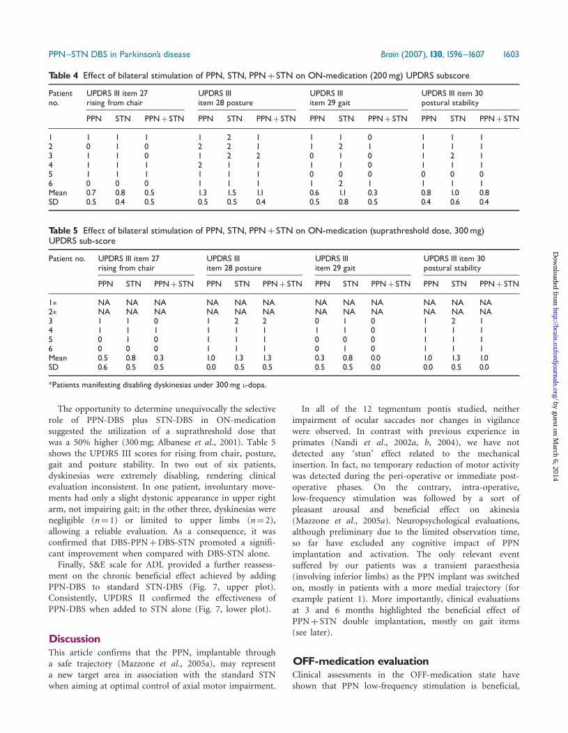
The opportunity to determine unequivocally the selectiverole of PPN-DBS plus STN-DBS in ON-medicationsuggested the utilization of a suprathreshold dose thatwas a 50% higher (300mg; Albanese et al., 2001). Table 5shows the UPDRS III scores for rising from chair, posture,gait and posture stability. In two out of six patients,dyskinesias were extremely disabling, rendering clinicalevaluation inconsistent. In one patient, involuntary move-ments had only a slight dystonic appearance in upper rightarm, not impairing gait; in the other three, dyskinesias werenegligible (n¼ 1) or limited to upper limbs (n¼ 2),allowing a reliable evaluation. As a consequence, it wasconfirmed that DBS-PPNþDBS-STN promoted a signifi-cant improvement when compared with DBS-STN alone.Finally, S&E scale for ADL provided a further reassess-
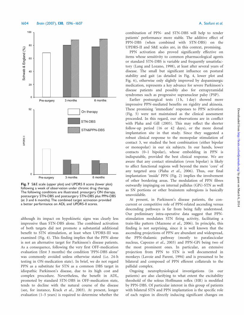
ment on the chronic beneficial effect achieved by addingPPN-DBS to standard STN-DBS (Fig. 7, upper plot).Consistently, UPDRS II confirmed the effectiveness ofPPN-DBS when added to STN alone (Fig. 7, lower plot).
DiscussionThis article confirms that the PPN, implantable througha safe trajectory (Mazzone et al., 2005a), may representa new target area in association with the standard STNwhen aiming at optimal control of axial motor impairment.
In all of the 12 tegmentum pontis studied, neitherimpairment of ocular saccades nor changes in vigilancewere observed. In contrast with previous experience inprimates (Nandi et al., 2002a, b, 2004), we have notdetected any ‘stun’ effect related to the mechanicalinsertion. In fact, no temporary reduction of motor activitywas detected during the peri-operative or immediate post-operative phases. On the contrary, intra-operative,low-frequency stimulation was followed by a sort ofpleasant arousal and beneficial effect on akinesia(Mazzone et al., 2005a). Neuropsychological evaluations,although preliminary due to the limited observation time,so far have excluded any cognitive impact of PPNimplantation and activation. The only relevant eventsuffered by our patients was a transient paraesthesia(involving inferior limbs) as the PPN implant was switchedon, mostly in patients with a more medial trajectory (forexample patient 1). More importantly, clinical evaluationsat 3 and 6 months highlighted the beneficial effect ofPPNþ STN double implantation, mostly on gait items(see later).
OFF-medication evaluationClinical assessments in the OFF-medication state haveshown that PPN low-frequency stimulation is beneficial,
Table 4 Effect of bilateral stimulation of PPN, STN, PPNþSTN on ON-medication (200mg) UPDRS subscore
Patientno.
UPDRS III item 27rising from chair
UPDRS IIIitem 28 posture
UPDRS IIIitem 29 gait
UPDRS III item 30postural stability
PPN STN PPNþSTN PPN STN PPNþSTN PPN STN PPNþSTN PPN STN PPNþSTN
1 1 1 1 1 2 1 1 1 0 1 1 12 0 1 0 2 2 1 1 2 1 1 1 13 1 1 0 1 2 2 0 1 0 1 2 14 1 1 1 2 1 1 1 1 0 1 1 15 1 1 1 1 1 1 0 0 0 0 0 06 0 0 0 1 1 1 1 2 1 1 1 1Mean 0.7 0.8 0.5 1.3 1.5 1.1 0.6 1.1 0.3 0.8 1.0 0.8SD 0.5 0.4 0.5 0.5 0.5 0.4 0.5 0.8 0.5 0.4 0.6 0.4
Table 5 Effect of bilateral stimulation of PPN, STN, PPNþSTN on ON-medication (suprathreshold dose, 300mg)UPDRS sub-score
Patient no. UPDRS III item 27rising from chair
UPDRS IIIitem 28 posture
UPDRS IIIitem 29 gait
UPDRS III item 30postural stability
PPN STN PPNþSTN PPN STN PPNþSTN PPN STN PPNþSTN PPN STN PPNþSTN
1� NA NA NA NA NA NA NA NA NA NA NA NA2� NA NA NA NA NA NA NA NA NA NA NA NA3 1 1 0 1 2 2 0 1 0 1 2 14 1 1 1 1 1 1 1 1 0 1 1 15 0 1 0 1 1 1 0 0 0 1 1 16 0 0 0 1 1 1 0 1 0 1 1 1Mean 0.5 0.8 0.3 1.0 1.3 1.3 0.3 0.8 0.0 1.0 1.3 1.0SD 0.6 0.5 0.5 0.0 0.5 0.5 0.5 0.5 0.0 0.0 0.5 0.0
*Patients manifesting disabling dyskinesias under 300mg L-dopa.
PPN^STN DBS in Parkinson’s disease Brain (2007), 130, 1596^1607 1603
by guest on March 6, 2014
http://brain.oxfordjournals.org/D
ownloaded from
although its impact on hypokinetic signs was clearly lessimpressive than STN-DBS alone. The combined activationof both targets did not promote a substantial additionalbenefit to STN stimulation, at least when UPDRS-III wasexamined (Fig. 4). This finding implies that the PPN aloneis not an alternative target for Parkinson’s disease patients.As a consequence, following the very first OFF-medicationevaluation (first 3 months) the condition ‘PPN-DBS alone’was commonly avoided unless otherwise stated (i.e. 24-htesting in ON-medication state). In brief, we do not regardPPN as a substitute for STN as a common DBS target inidiopathic Parkinson’s disease, due to its high cost andcomplex procedure. Nevertheless, the benefit in ADL,promoted by standard STN-DBS in OFF-medication state,tends to decline with the natural course of the disease(see, for instance, Krack et al., 2003). At present, longerevaluation (1–5 years) is required to determine whether the
combination of PPN- and STN-DBS will help to renderpatients’ performance more stable. The additive effect ofPPN-DBS (when combined with STN-DBS) on theUPDRS-II and S&E scales are, in this context, promising.
PPN activation also proved significantly effective onitems whose sensitivity to common pharmacological agentsor standard STN-DBS is variable and frequently unsatisfac-tory (Lang and Lozano, 1998), at least after several years ofdisease. The small but significant influence on posturalstability and gait (as detailed in Fig. 4, lower plot andFig. 6), otherwise only slightly improved by dopaminergicmedication, represents a key advance for severe Parkinson’sdisease patients and possibly also for extrapyramidalsyndromes such as progressive supranuclear palsy (PSP).
Earlier postsurgical tests (1 h, 1 day) showed moreimpressive PPN-mediated benefits on rigidity and akinesia.These promising ‘immediate’ responses to PPN activation(Fig. 5) were not maintained as the clinical assessmentproceeded. In this regard, our observations are in conflictwith Plaha and Gill (2005). This may reflect the shorterfollow-up period (16 or 42 days), or the more dorsalimplantation site in that study. Since they suggested arobust clinical response to the monopolar stimulation ofcontact 3, we studied the best combination (either bipolaror monopolar) in our six subjects. In our hands, lowercontacts (0–1 bipolar), whose embedding in PPN isindisputable, provided the best clinical response. We areaware that any contact stimulation (even bipolar) is likelyto affect functional regions well beyond the mere ‘core’ ofany targeted area (Plaha et al., 2006). Thus, our finalimplantation ‘inside’ PPN (Fig. 2) implies the involvementof other bordering areas. The modulation of PPN fibresoutwardly impinging on internal pallidus (GPi)-STN as wellas SN portions or other brainstem subregions is basicallyunavoidable.
At present, in Parkinson’s disease patients, the con-current or competitive role of PPN-related ascending versusdescending pathways is far from being fully understood.Our preliminary intra-operative data suggest that PPN-stimulation modulates STN firing activity, facilitating atrain-like pattern (Mazzone et al., 2006). In principle, thisfinding is not surprising, since it is well known that theascending projections of PPN are abundant and widespread,the PPN-thalamic pathway (mostly to parafascicularnucleus, Capozzo et al., 2003) and PPN-GPi being two ofthe most prominent ones. In particular, an extensiveprojection from PPN to STN is well documented inmonkeys (Lavoie and Parent, 1994) and is presumed to bebilateral and composed of PPN efferent collaterals to thepallidal complex.
Ongoing neurophysiological investigations (in ourpatients) are also clarifying to what extent the excitabilitythreshold of the soleus Hoffmann reflex (HR) is modifiedby PPN-DBS. Of particular interest in this group of patientswith bilateral STN and PPN implantation is the specific roleof each region in directly inducing significant changes on
Fig. 7 S&E scale (upper plot) and UPDRS II score (lower plot)following a week of observation under chronic drug therapy.The following conditions are illustrated: presurgery ON therapy,postsurgery STN-DBS and postsurgery STN-DBS plus PPN-DBS(at 3 and 6 months). The combined target activation provideda better performance on ADL and UPDRS-II scores.
1604 Brain (2007), 130, 1596^1607 A. Stefani et al.
by guest on March 6, 2014
http://brain.oxfordjournals.org/D
ownloaded from
spinal cord circuitries connected to lower limb muscles,whose activity is clearly involved in posture and locomotion(Spann and Grofova, 1989).
ON-medication evaluationThe key finding of our study is that PPN low-frequencyactivation potently ameliorates the ON-drug/ON-STNcondition (Fig. 6). This result is striking, in light of theassumption that the best drug-mediated ON-state should becomparably as effective as drug plus DBS (and notsignificantly less effective). On the other hand, evidence-based treatment recommendations, based on all availablepharmacological and surgical approaches, support theusefulness of STN-DBS for improving motor function andreducing off-time, albeit with a finite period of postsurgicalutility (Pahwa et al., 2006). We are aware that a clinicalimprovement of almost 50% under STN-DBS or STN- plusPPN-DBS (in ON-state and with respect to the L-doparesponse, Fig. 6) may sound unusual. Yet, 200mg L-dopamight not represent the maximal dose, implying a possibleunderestimation of drug-mediated peak effect, at least insome of our patients. As only four out of six patientstolerated supramaximal concentrations (Table 5), thepotential comparison was therefore not possible. Previousreports have described conflicting results on this issue.Anderson et al. (2003) found that DOPA alone and DOPAplus STN-DBS provided a similar degree of benefit (at leaston motor UPDRS). In contrast, Rodriguez-Oroz et al.(2005) estimated an additional effect of STN-DBS to thebest drug-induced ON-state (about 30% at 1 yearobservation). Intriguingly, STN-DBS provided an improve-ment to some axial signs implying a synergistic effectbetween L-dopa and STN stimulation (not found for limbsigns; Bejjani et al., 2000).The effect of PPN-DBS plus STN-DBS in ON-state
(versus OFF-state) may appear paradoxical, since axialsymptoms, if developing late in the natural course of thedisease, are generally ascribed to ‘non-dopaminergic’brainstem degeneration. In fact, the PPN-mediated bene-fits (exceeding those of L-dopa) support the possibilitythat PPN-mediated ascending projections—either direct tothe SN or indirect through the parafascicular nucleusand/or STN—modulate endogenous dopamine signallingpathways.Considering our protocol (bipolar stimulation of more
distal contacts), stimulus parameters (60 ms, 2 V, 25Hz) andthe size of the electrocatheter, it is likely that the electricaldipole does not act exclusively on small or specificsubpopulations of neurons. Although some degree ofclustering (mostly for cholinergic neurons) was describedin rodent PPN, our clinical observations are likely to derivefrom a combined stimulation of both glutamate andacetylcholine cell bodies in the densely cellular (PPNc)and diffuse (PPNd) areas (Bevan et al., 1995; Lavoie andParent, 1994; Mesulam et al., 1989; Takakusaki et al., 1996).
Future utilization of smaller devices, active on specificpathways, should be tested on MPTP-intoxicated non-human primates (and eventually in patients), in order toassess performance under the activation of more selectiveneuronal or fibre pathways. Since, however, double-labelledneurons (for glutamate and acetylcholine) are abundant inPPN (Lavoie and Parent, 1994b), a microdialysis study(Fedele et al., 2001; Stefani et al., 2005) seems well suited toaddressing the extracellular release of PPN projections ontarget areas.
Conclusions
(i) Parkinsonian axial deficits represent a challenging taskwhen disease progresses, although some degree ofL-dopa-sensitivity may persist even in severe patientswith motor fluctuations (Hughes et al., 1994).Traditional STN-DBS may provide some benefitsand a synergistic effect of DOPA and STN-DBS wasdocumented, as outlined by walking and posturalstability score (Bejjani et al., 2000). Yet, another seriesof STN-implanted patients showed substantial equiva-lence amongst L-dopa and STN-mediated benefit onposture subscores (Maurer et al., 2003). In a largecohort of severe Parkinson’s disease patients, the STN-DBS-mediated impact on limb signs seems morestriking than gait (Rodriguez-Oroz et al., 2005); andthe latter tends to decline in prolonged follow-up(5–10 years). Our patients manifested already anunsatisfactory control of axial signs even when thebest combination of dopamine-mimetic agents wasutilized (compare, for instance, 50% amelioration ofglobal UPDRS section III versus 35% for item 27 and37% for item 29).The postsurgery study demonstrates that PPN mayrepresent a strong candidate to provide additionalamelioration of items otherwise difficult to treat.More importantly, the two-target stimulation provedmore effective than the stimulation of each targetalone. In brief, the combination of targets adds valueto the control of axial signs. This evidence mightsupport the selection of previously STN-implantedpatients (with poor therapeutic response) for addi-tional PPN targeting. That said, the unimpressiveeffect of PPN-DBS, when stimulated alone, onUPDRS-III in OFF-therapy suggests that PPN is nota fully alternative target. In this respect, preliminarydata (Plaha and Gill, 2005) should be viewed withextreme caution. Nevertheless, extended follow-up [(asin the 5-year post-STN implantation prospective studyby Krack et al. (2003)] documented some degree ofdeterioration in the STN-mediated impact on axialsigns and akinesia. At present, it is unclear whetherSTNþ PPN combination will show an analogouspattern, with the same decline in efficacy.
PPN^STN DBS in Parkinson’s disease Brain (2007), 130, 1596^1607 1605
by guest on March 6, 2014
http://brain.oxfordjournals.org/D
ownloaded from

(ii) PPN-DBS may increase the efficacy of exogenouslyadministered therapy. Combined DBS (PPN þ STN)was significantly more efficacious when motor per-formance was assessed in ON-medication (under bothsubdyskinetic and suprathreshold concentrations).Moreover, despite the short follow-up, PPN-DBSplus STN-DBS significantly improved the ADL andpatient’s self-sufficiency. It is hard to determine thepossible mechanisms underlying this PPN-mediatedbenefit. Speculations involve an active role of PPN-DBS on the endogenous release of dopamine or onthe intrastriatal dopamine and acetylcholine balance.
(iii) Undesired involuntary movements occurred in twoout of six patients with L-dopa supramaximal dose(together with PPN-DBS and STN-DBS). This is notunexpected if PPN-DBS exceeds the L-dopa responseby recruiting endogenous amines. On one hand, thisclinical observation points out that PPN should neverbe considered a good candidate for treating dyskine-sias; on the other, it reinforces the need for prolongedfollow-up observations, comparing different drugregimens.
(iv) The surprising, although slight, decline of PPN-mediated efficacy on motor items, when comparingearly versus late OFF response in CAPIT raises amajor question, concerning the possible occurrence ofcomplex adaptive changes. Nevertheless, analogousresponse deterioration did not feature PPN-mediatedeffects on gait and posture. Given the relatively smallnumber of patients (and the difference in the timedependency of the response’s decline) it is difficult toidentify a conclusive explanation. It is possible thatunconventional (i.e. cyclic) protocol of stimulationwould be preferable for maintaining larger effective-ness of PPN activation.
(v) At present, we are testing the efficacy of PPN cyclicstimulation, together with continuous delivery ofSTN-DBS, in idiopathic Parkinson’s disease patients.In addition, we are also investigating to what extentPPN-DBS could be an effective option in otherwiseuntreatable advanced forms of extrapyramidaldisorders such as PSP.
AcknowledgementsWe express our gratitude to Dr Atticus H. Hainsworth(St George’s Hospital, London) for critical reading andediting of the manuscript. We are profoundly thankful toStefano Sposato MD and Giuseppe Gattoni for radiologicaland technical assistance. This manuscript has beensupported by grants from Ministero Della Salute, RegionePiemonte and Friuli Venezia Giulia to A.S. and P.S.
ReferencesAlbanese A, Bonuccelli U, Brefel C, Chaudhuri KR, Colosimo C,
Eichhorn T, et al. Consensus statement on the role of acute
dopaminergic challenge in Parkinson’s disease. Mov Disord 2001; 16:
197–201.
Anderson VC, Burchiel KJ, Hogarth P, Favre J. Hammerstad JP. Pallidal
vs. subthalamic nucleus deep brain stimulation in Parkinon disease.
Arch Neurol 2005; 62: 554–60.
Bejjani BP, Gervais D, Arnulf I, Papadopoulos S, Demerset S, Bonnet AM,
et al. Axial parkinsonian symptoms can be improved: the role of
levodopa and bilateral subthalamic stimulation. J Neurol Neurosurg
Psych 2000; 68: 595–600.
Benabid AL, Pollak P, Gross C, Hoffmann D, Benazzouz A, Gao DM, et al.
Acute and long-term effects of subthalamic nucleus stimulation in
Parkinson’s disease. Stereotact Funct Neurosurg 1994; 62: 76–84.
Benedetti F, Colloca L, Torre E, Lanotte M, Melcarne A, Pesare M, et al.
Placebo-responsive Parkinson patients show decreased activity in single
neurons of subthalamic nucleus. Nat Neurosci 2004; 7: 587–8.
Bevan MD, Bolam JP. Cholinergic GABAergic, and glutamate-enriched
inputs from the mesopontine tegmentum to the subthalamic nucleus in
the rat. J Neurosci 1995; 15: 7105–20.
Capozzo A, Florio T, Cellini R, Morioni U, Scarnati E. The pedunculo-
pontine nucleus projection to the parafascicular nucleus of the
thalamus: an electrophysiological investigation in the rat. J Neural
Transm 2003; 110: 733–47.
Fahn S, Elton RL. Members of the UPDRS Development Committee.
Unified Parkinson’s disease rating scale. In: Fahn S, Marsden CD,
Goldstein M, Calne DB, editors. Recent developments in Parkinson’s
disease. Vol. 2. Florham Park, New York: MacMillan; 1987. p. 153.
Fedele E, Mazzone P, Stefani A, Bassi A, Ansaldo MA, Raiteri M, et al.
Microdialysis in Parkinsonian patient basal ganglia: acute apomorphine-
induced clinical and electrophysiological effects not paralleled by
changes in the release of neuroactive amino acids. Exp Neurol 2001;
167: 356–65.
Galati S, Mazzone P, Fedele E, Peppe A, Pierantozzi M, Brusa L, et al.
Biochemical and electrophysiological nigral changes driven by sub-
thalamic stimulation in Parkinson’s disease patients. Eur J Neurosci
2006; 23: 2923–8.
Garcia-Rill E, Houser CR, Skinner RD, Smith W, Woodward DJ.
Locomotion-inducing sites in the vicinity of the pedunculopontine
nucleus. Brain Res Bull 1987; 18: 731–8.
Garcia-Rill E. The pedunculopontine nucleus. Prog Neurobiol 1991; 36:
363–89.
Hughes AJ, Frankel JP, Kempster PA, Stern GM, Lees AJ. Motor response
to levodopa in patients with parkinsonian motor fluctuations: a follow-
up study over three years. J Neurol Neurosurg Psych 1994; 57: 430–4.
Jenkinson N, Nandi D, Miall RC, Stein JF, Aziz TZ. Pedunculopontine
nucleus stimulation improves akinesia in a Parkinsonian monkey.
Neuroreport 2005; 15: 2621–4.
Krack P, Batir A, Van Blercom N, Chabardes S, Fraix V, Ardouin C, et al.
Five-year follow-up of bilateral stimulation of the subthalamic nucleus
in advanced Parkinson disease. New Engl J Med 2003; 349: 1925–34.
Lang AE, Lozano AM. Parkinson’s disease. First of two parts. N Engl
J Med 1998; 339: 1044–53.
Lavoie B, Parent A. Pedunculopontine nucleus in the squirrel monkey:
projections to the basal ganglia as revealed by anterograde tract-tracing
methods. J Comp Neurol 1994a; 334: 210–31.
Lavoie B, Parent A. Pedunculopontine nucleus in the squirrel monkey:
cholinergic and glutamatergic projections to the substantia nigra.
J Comp Neurol 1994b; 334: 232–41.
Maurer C, Mergner T, Xie J, Faist M, Pollak P, Lucking CH. Effect of
chronic bilateral subthalamic nucleus (STN) stimulation on postural
control in Parkinson’s disease. Brain 2003; 126: 1146–63.
Mazzone P, Insola A, Brown P, DiLazzaro V, Tonali P, Altibrandi MG.
Contemporary bilateral DBS on GPi and STN nuclei and preliminary
resultson contemporary bilateral DBS and CM-PF complex in PD. Acta
Neurochir (Wien) 2004; 146: 2A15.
Mazzone P, Lozano A, Stanzione P, Galati S, Peppe A, Scarnati E, et al.
Implantation of human pedunculopontine nucleus: a safe and clinically
relevant target in Parkinson’s disease. NeuroReport 2005a; 16: 1879–83.
1606 Brain (2007), 130, 1596^1607 A. Stefani et al.
by guest on March 6, 2014
http://brain.oxfordjournals.org/D
ownloaded from
Mazzone P, Brown P, DiLazzaro V, et al. Bilateral implantation in globus
pallidus internus and in subthalamic nucleus in Parkinson’s disease.
Neuromodulation 2005b; 8: 1–6.
Mazzone P. Multiple and unconventional targets in DBS for PD. In:
ASSFN, Boston, June 1–5, 2006.
Mena-Segovia J, Bolam JP, Magill PJ. Pedunculopontine nucleus and basal
ganglia: distant relatives or part of the same family? Trends Neurosci
2004; 27: 585–8.
Mesulam MM, Geula C, Bothwell MA, Hersh LB. Human reticular
formation cholinergic neurons of the pedunculopontine and laterodorsal
tegmental nuclei and some cytochemical comparison to forebrain
cholinergic neurons. J Comp Neurol 1989; 283: 611–33.
Moro E, Scerrati M, Romito LM, Roselli R, Tonali P, Albanese A. Chronic
subthalamic nucleus stimulation reduces medication requirements in
Parkinson’s disease. Neurology 1999; 53: 85–90.
Nandi D, Aziz TZ, Giladi N, Winter J, Stein JF. Reversal of akinesia in
experimental parkinsonism by GABA antagonist microinjections in the
pedunculopontine nucleus. Brain 2002a; 125: 2418–30.
Nandi D, Liu X, Winter JL, Aziz TZ, Stein JF. Deep brain stimulation of
the pedunculopontine region in the normal non-human primate. J Clin
Neurosci 2002b; 9: 170–4.
Nandi D, Miall RC, Stein JF, Aziz TZ. Pedunculopontine nucleus
stimulation improves akinesia in a Parkinsonian monkey. Neuroreport
2004; 15: 2621–4.
Pahapill PA, Lozano AM. The pedunculopontine nucleus and Parkinson’s
disease. Brain 2000; 123: 1767–83.
Pahwa R, Factor SA, Lyons KE, Ondo WG, Gronseth G, Bronte-Stewart H,
et al. Quality Standards Subcommittee of the American Academy of
Neurology. Practice parameter: treatment of Parkinson disease with
motor fluctuations and dyskinesia (an evidence-based review): report of
the Quality Standards Subcommittee of the American Academy of
Neurology. Neurology 2006; 66: 983–95.
Peppe A, Pierantozzi M, Bassi A, Altibrandi MG, Brusa L, Stefani A, et al.
Stimulation of the subthalamic nucleus compared with the globus pallidus
internus in patients with Parkinson disease. J Neurosurg 2004; 101: 195–200.
Plaha P, Gill SS. Bilateral deep brain stimulation of the pedunculopontine
nucleus for Parkinson’s disease. Neuroreport 2005; 16: 1883–7.
Plaha P, Ben-Shlomo Y, Patel NK, Gill SS. Stimulation of the caudal zona
incerta is superior to stimulation of the subthalamic nucleus in
improving contralateral parkinsonism. Brain 2006; 129: 1732–57.
Rodriguez-Oroz MC, Obeso JA, Lang AE, Houeto JL, Pollak P,
Rehncrona S, et al. Bilateral deep brain stimulation in Parkinson’s
disease: a multicentre study with 4 years follow-up. Brain 2005; 128:
2240–9.
Scarnati E, Proia A, Di Loreto S, Pacitti C. The reciprocal electro-
physiological influence between the nucleus tegmenti pedunculoponti-
nus and the substantia nigra in normal and decorticated rats. Brain Res
1987; 423: 116–24.
Schaltenbrand G, Wahren W. Atlas for stereotaxy of the human brain.
Stuttgart: George Thieme; 1977.
Skinner RD, Kinjo N, Ishikawa Y, Biedermann JA, Garcia-Rill E.
Locomotor projections from the pedunculopontine nucleus to the
spinal cord. Neuroreport 1990; 1: 183–6.
Spann BM, Grofova I. Origin of ascending and spinal pathways from the
nucleus tegmenti pedunculopontinus in the rat. J Comp Neurol 1989;
283: 13–27.
Stefani A, Stanzione P, Bassi A, Mazzone P, Vangelista T, Bernardi G.
Effects of increasing doses of apomorphine during stereotaxic neuro-
surgery in Parkinson’s disease: clinical score and internal globus pallidus
activity. J Neural Transm 1997; 104: 895–904.
Stefani A, Bassi A, Mazzone P, Pierantozzi M, Gattoni G, Altibrandi MG,
et al. Subdyskinetic apomorphine responses in globus pallidus and
subthalamus of parkinsonian patients: lack of clear evidence for the
‘indirect pathway’. Clin Neurophysiol 2002; 113: 91–100.
Stefani A, Fedele E, Galati S, Pepicelli O, Frasca S, Pierantozzi M, et al.
Subthalamic stimulation activates internal pallidus: evidence from cGMP
microdialysis in PD patients. Ann Neurol 2005; 57: 448–52.
Stefani A, Galati S, Peppe A, Bassi A, Pierantozzi M, Hainsworth AH, et al.
Spontaneous sleep modulates the firing pattern of parkinsonian
subthalamic nucleus. Exp Brain Res 2006; 168: 277–80.
Takakusaki K, Shiroyama T, Yamamoto T, Kitai ST. Cholinergic
and noncholinergic tegmental pedunculopontine projection neurons
in rats revealed by intracellular labeling. J Comp Neurol 1996; 371:
345–61.
PPN^STN DBS in Parkinson’s disease Brain (2007), 130, 1596^1607 1607
by guest on March 6, 2014
http://brain.oxfordjournals.org/D
ownloaded from