Beya wa Beya MSc Thesis Dec 2010 - CORE
92
i THE EFFECTS OF CRUDE AQUEOUS AND ALCOHOL EXTRACTS OF ALOE VERA ON THE GASTROINTESTINAL TRACT AND ACCESSORY ORGANS OF SUCKLING RATS Ben Beya wa Beya A dissertation submitted to the Faculty of Health Sciences, University of the Witwatersrand, School of Physiology in fulfilment of the requirements for the degree of Master in Medicine. Johannesburg, South Africa. 2010 brought to you by CORE View metadata, citation and similar papers at core.ac.uk provided by Wits Institutional Repository on DSPACE
-
Upload
khangminh22 -
Category
Documents
-
view
3 -
download
0
Transcript of Beya wa Beya MSc Thesis Dec 2010 - CORE

i
THE EFFECTS OF CRUDE AQUEOUS AND
ALCOHOL EXTRACTS OF ALOE VERA ON THE
GASTROINTESTINAL TRACT AND ACCESSORY
ORGANS OF SUCKLING RATS
Ben Beya wa Beya
A dissertation submitted to the Faculty of Health Sciences, University of the
Witwatersrand, School of Physiology in fulfilment of the requirements for the degree
of Master in Medicine.
Johannesburg, South Africa.
2010
brought to you by COREView metadata, citation and similar papers at core.ac.uk
provided by Wits Institutional Repository on DSPACE

ii
The copy right of the above mentioned described thesis rests with the
author or the university to which it was submitted. No portion of the text
derived from it may be published without the prior written consent of the
author or University (as may be appropriate). Short quotations may be
included in the text of thesis or dissertation for purposes of illustration,
comment or criticism, provided full acknowledgement is made of the
source, author and University.

i
DECLARATION
I declare that this dissertation is my own work, except where others have helped as
indicated in the acknowledgements and the reference list. This dissertation is being
submitted for the degree of Master in Medicine in the Faculty of Health Sciences at
the University of the Witwatersrand, Johannesburg, South Africa. It has not been
submitted before for any degree or examination in this or any other University.
………………………….
Ben Beya wa Beya
........ day of …………….....2010
Furthermore, I certify that the procedures used in this dissertation were approved by
the Animal’s Ethics Screening Committee of the University of the Witwatersrand
(AESC number 2008/37/03).

ii
DEDICATION
This research is dedicated to my major supervisor Associate Professor Kennedy
Erlwanger and the Head of School of Physiology Professor David Gray for their
unlimited guidance, support and motivation without which none of this would have
been possible. In everybody’s life there comes a time when someone influences the
path of one’s life.
To my parents, Beya Mwepu Shambuyi and Buambavi wa Ngindu for lovingly and
patiently giving me direction in my life, I would not have come this far if it weren’t for
you, thank you for your unconditional love and steadfast support in all of my
endeavours.

iii
ACKNOWLEDGMENTS
I would like to express my sincere gratitude to my co-supervisor Prof. Bruce
Davidson for his valuable assistance and guidance in both the execution and
preparation of the laboratory work.
Many thanks to Eliton Chivandi, I am indebted to you for your invaluable help and
guidance in the preparation of this manuscript, acknowledged authority in this field of
biochemistry and nutrition by giving me an intrinsic understanding of the subject.
I would like to extend my sincere and heartfelt thanks to Phiko Mgandela for his
encouragement and patience in the face of impetuosity and for his invaluable advice
so freely given.
Thanks to Margaret Badenhorst for her technical assistance with the processing of
tissues for histology, and for running the ELISAs for IGF-1 hormonal assays as well
as for technical assistance with the preparation of some of the reagents I used in the
study.
I would like to express my gratitude to Dr. Oluwafolajini and Trevor Nyakudya for
their assistance with the Statistics done during this research.
I would like to express my heartfelt gratitude to Thapelo Rammabi for her constant
encouragement and support.
I would like to take this opportunity to express my gratitude to Dr. Mulunda Mwanza
for the orientation and support throughout the preparation of this dissertation.

iv
This material is based upon work supported financially by the National Research
Foundation and the University of the Witwatersrand, Faculty of Health Sciences
Research Committee grants and the School of Physiology, South Africa.
Last but not least, to my friends and colleagues in the School of Physiology who
aided this study in many ways by providing both stimulating discussion and advice.

v
ABSTRACT
For centuries Aloe vera has been exploited for several verified and unverified medicinal
uses such as wound healing, treatment of gastrointestinal ulcers and for its many
biological effects including anti-microbial, laxative, anti-inflammatory and immuno-
stimulatory activities. Studies have generally focused on its effects in vitro and in adults.
When nursing mothers use Aloe vera extracts, their suckling infants are at risk of indirect
exposure to Aloe vera via breast feeding or directly as dietary/health supplements. The
gastrointestinal tract (GIT) of the neonate is sensitive to dietary manipulations during the
suckling period with long lasting effects that can be irreversible. Thus babies may be at
risk if administered Aloe vera extracts directly as dietary supplements or indirectly via
breast milk.
The main objectives of this study were to evaluate the effects of orally administered
aqueous and alcohol extracts of Aloe vera on growth performance, the morphometry and
morphology of the gastrointestinal tract and accessory organs, and liver function of
suckling rats. Suckling Sprague-Dawley rats (77), males (n=38) and females (n=39) of 6
days old were randomly assigned to one of five treatment groups and given once daily by
oral gavage a suspension of lyophilized crude alcohol or aqueous extracts of Aloe vera
suspended in distilled water. Group I (control) was gavaged with distilled water (vehicle).
Group II received a low dose of the aqueous extract (AqL) at 50mg. kg-1; Group III
received a high dose of the aqueous extract (AqH) at 500mg. kg-1; Group IV received a
low dose of the alcohol extract (AlcL) at 50mg. kg-1 whilst Group V received a high dose
of the alcohol extract (AlcH) at 500mg. kg-1. The extracts and distilled water were
administered at a volume of 10ml.kg-1. The pups remained with their dams for the

vi
duration of the study and after 8 days on the treatments, the pups were humanely killed to
harvest their tissues for measurements and physiological analysis. All data were
expressed as mean ± SD and analyzed by one way ANOVA, the values were considered
statistically significant when p < 0.05 and then a Bonferroni Post hoc test was applied.
The suckling rats fed respectively with high doses of AlcH and AqH had a significantly
higher body mass gain than the other groups (p < 0.05, one way ANOVA). Linear growth
as measured by tibial length was significantly increased in the AqH group compared to
the other groups. There was no significant difference in the mass and relative density of
the tibia bones of the rats from the different treatment groups. The differences in growth
could not be attributed to circulating concentrations of the somatotrophic hormone,
Insulin-like growth factor-1 (IGF-1) which was not significantly different between the
groups.
The treatments did not result in any significant differences in lengths, and mass of the
small and large intestine, however the caecum was significantly enlarged (hypertrophy of
muscularis, submucosa and mucosa) in the rats that received the Aloe vera extracts.
Although, there was no significant difference in the mass of the rats’ livers, the lipid and
glycogen content were significantly higher (p < 0.001) for the AqH group compared to
the other groups. Histologically, the hepatocytes showed enlarged nuclei, granular
cytoplasm and dilated sinusoids for AqH and AlcH as compared to the control group. An
indirect assessment of liver function by measurement of blood concentrations of alkaline
phosphatase (ALP) and alanine amino transaminase (ALT) did not reveal a significant

vii
difference between the groups. The non fasting concentration of metabolic substrates
(glucose and triglycerides) was also not significantly different between the groups.
The pups given high doses of the extracts had a significantly greater (p < 0.05) thymus
mass (hyperplastic) than the other groups.
The short term administration of Aloe vera extracts has shown a growth promoting effect,
enhanced hepatic storage of metabolic substrates and hypertrophy of the caecum and
thymus of neonatal rats. These effects need to be explored further to enhance animal
production and health.

viii
TABLE OF CONTENTS Page
DECLARATION……........................................................................................... i
DEDICATION…………………………………………………………………... ii
ACKNOWLEDGEMENTS……………………………………………………... iii
ABSTRACT …………………………………………………………………….. v
TABLE OF CONTENT ………………………………………………………… viii
LIST OF FIGURES …………………………………………………………….. xi
LIST OF TABLES …………………………………………………………….... xii
LIST OF ABBREVIATIONS …………………………………………………... xiii
Chapter 1- Introduction ………………………………………………………..... 1
1.1 Introduction ………………………………………………………………..... 2
1.1.1 Aloe vera ….................................................................................................. 2
1.1.2 Biochemistry ……………………………………………………………....
3
a) Vitamins …………………………………………………………………. 3
b) Enzymes ………………………………………………………….............
4
c) Minerals …………………………………………………………..............
4
d) Sugars ......................................................................................................... 4
e) Anthraquinones .......................................................................................... 5
1.1.3 Pharmacology and Therapeutic effects of Aloe vera .................................. 6
a) Skin and wound healing .............................................................................. 6
b) Diabetes mellitus and metabolism .............................................................. 7

ix
c) Anticancer .................................................................................................. 7
d) Cardiovascular system ................................................................................ 7
e) Gastrointestinal system................................................................................ 9
f) Musculoskeletal System ............................................................................ 10
g) Immune system ........................................................................................... 10
1.1.4 The gastrointestinal tract ………………………………………………...... 11
1.1.5 Growth and nutrition in neonates .................................................................
15
1.1.6 Justification of study.....................................................................................
17
1.1.7 Aims and objectives ..................................................................................... 18
a) The morphometry and morphology of the GIT and accessory organs of suckling rats..............................................................................................
18
b) Growth performance ……………………………………………………...
19
c) Liver function (enzymes and storage of glycogen and lipids)…………..... 19
d) Metabolic substrates (blood glucose and triglyceride concentrations)…....
20
Chapter 2 – Materials and methods ……………………………………………...
21
2.1 Study setting ………………………………………………………................
22
2.1.1 Study design ……………………………………………………….............
22
2.2 Materials………………………………………………………….…..............
23
2.2.1 Plant collection …………………………………………………....……….
23
2.3 Methods ………………………………………………………….....………..
23
2.3.1 Preparation of Aloe vera extracts ………………………………..………... 23

x
2.3.2 Dry matter determination ………………………………………..………...
24
2.3.3 Housing of animals ……………………………………….………............. 24
2.3.4 Experimental animals ………………………………………..……............. 24
2.3.5 Blood sampling and plasma processing ……………………....................... 25
2.3.6 Blood measurement for glucose and triglyceride concentration ….............. 26
2.3.7 Determination of liver enzymes (total ALT and ALP)……………………. 26
2.3.8 Measurements of insulin-like Growth Factor (IGF-1)……………….......... 27
2.3.9 Morphometry and morphology of abdominal viscera …………………...... 28
2.3.10 Determination of liver lipid content………………………….................... 29
2.3.11 Determination of liver glycogen storage …………………………............ 29
2.3.12 Femur, tibia lengths and mass …………………………………................
30
2.3.13 Statistical analysis ...................................................................................... 30
Chapter 3 – Results ……………………………………………………………...
31
3.1 Mortality and morbidity of the animals……………………………………... 32
3.2 The effects of Aloe vera extracts on the body mass .……………………….. 31
3.3 Visceral of organ mass and histology……………………….......……...........
31
3.4 The effects of Aloe vera extracts on the tibia and femur length and mass..... 33
3.5 The effects of Aloe vera extracts on liver lipid content and glycogen............. 33
Chapter 4 – Discussion ………………………………………………………….
48
Chapter 5 – Conclusion and recommendations .................................................... 56
Chapter 6 – References ......................................................................................... 59

xi
LIST OF FIGURES
Figure 1: Effect of Aloe vera extracts on the body mass gain (%) of suckling rats
after 8 days of treatment.............................................................................................34
Figure 2: Effect of Aloe vera extracts on the relative mass of the thymus
(% body mass) after 8 days of treatment.....................................................................36
Figure 3: Liver lipid yield (%)……………………………………………….............38
Figure 4: Effect of 8 days of feeding suckling rats with Aloe vera extracts on the liver
glycogen storage expressed as glucose equivalents of liver homogenate...................39
Figure 5a: Digital photograph showing the histology of the caecum 40X .................43
Figure 5b: Digital photograph showing the histology of the caecum 400X ...............44
Figure 6a: Digital photograph showing the histology of the thymus 40 X..................44
Figure 6b: Digital photograph showing the histology of the thymus 400X …...…... 45
Figure 7: Digital photograph showing the histology of the liver 400 X ….................42

xii
LIST OF TABLES
Table 1: Effect of Aloe vera extracts on the body mass, absolute (g) and relative
mass (%) and lengths (cm) of viscera of suckling rats...............................................35
Table 2: Effect of Aloe vera on the absolute and relative length and mass of tibia and
femurs of rats………………………………………………………………………. 37
Table 3: Effect of Aloe vera extracts on non fasting blood concentrations of glucose,
Triglycerides ALT, ALP and IGF-1............................................................................40
Table 4: Effect of 2 different doses of Aqueous and Alcohol extracts of Aloe vera
on the microscopic measurements of intestines of suckling rats.................................41
Table 5: Effect of Aloe vera extracts on the morphometry of the ceacum of suckling
rats............................................................................................................................... 42

xiii
LIST OF ABBREVIATIONS
Alanine-amino Transferase ALT
Alcohol high dose AlcH
Alcohol low dose AlcL
Alkaline Phosphatase ALP
Aqueous high dose AqH
Aqueous low dose AqL
Body mass BM
Enzyme-Linked Immunosorbent assay ELISA
Gastrointestinal tract GIT
Insulin-like growth hormone IGF-1
Tumour Necrosis Factor alpha TNF-α

1
Chapter 1 – Introduction

2
1.1 Introduction
For centuries, numerous plants have been used for medicinal properties and as key
ingredients for dietary supplements (Massimo, 2003). Although many have been
superseded by conventional pharmaceutical approaches, there is currently a resurgence in
interest in the use of natural products by the general public world-wide. As a result of
scientific efforts aimed at improving the understanding of their effect on human
physiology, some plants have been thoroughly characterized and their mechanism of
action is now well understood. A myriad of plants although used as pharmacological
agents, are yet to be characterised and undergo scientific testing on their proposed
efficacy against disease. Aloe vera is a plant with many reputed health benefits and has
many references in many cultures: Ancient Egyptians, Greeks, Romans, Indians and
Chinese (Cheney, 1970).
1.1.1 Aloe vera
The plant Aloe vera with origins in the eastern and southern regions of Africa is also
known as Aloe barbadensis and Aloe vera ferox (Cheney, 1970). It was originally
classified in the family Liliaceae, but according to Reynolds, it has now been designated
its own family, known as Aloaceae (Wallander et al., 2000). It is well adapted to growth
in arid and semi arid regions (Paeza et al., 2009). Aloe vera can reach heights of 10 to 20
meters with stems ranging up to 3 meters in circumference. The flowers appear annually,
are bright red-orange and, are arranged in an erect position with terminal spikes in
candle-shaped clusters, whilst the capsules contain numerous angular seeds.

3
When grown in pots, Aloe vera does not like crowded roots and the use of standard
potting soil mixed with sharp sand will help the plant growth (Johnson, 1999; Leffers et
al., 2003). When grown under low light intensity, the leaves lose their grayish cast and
become just plain green. Aloe vera is tolerant of dry air and prefers temperatures of
21degrees Celsius in the day time and night ( Paeza et al., 2009).
1.1.2 Biochemistry
Most of the biologically active constituents of Aloe vera are found in the leaves which are
composed of the rind, juice and a gel like substance (Ramachandra and Srinivasa, 2008).
The plant Aloe vera, being a cactus contains up to 80 per cent water with an average pH
of 4.5 (Bryant,1970).
It has been shown that some constituents of Aloe vera have similar biological activities to
amino acids, vitamin C, polysaccharides, anthraquinones, barbaloins (glucosides) and
growth factors (Davis et al., 1994). Aloe vera also contains several potentially bioactive
compounds such as salicylates, magnesium lactate, acemannan, lupeol, campestrol, β-
sitosterol, γ-linolenic acid, aloctin A, anthraquinones, Aloe-emodin, resins, and chromone
derivatives (Wichtl and Bisset, 1994; Bruneton, 1995; Esua and Rauwald, 2006).
The remaining solid material contains over 75 different ingredients which include:
a) Vitamins
Aloe vera contains most vitamins including Vitamin D, A (the antioxidant beta-carotene),
C, E, B1, B2, B6 and even traces of B12 and is one of the very few plant sources of this

4
vitamin (Yaron, 1991). It also has beta carotene, niacinimide, folic acid and choline
(Matsuda et al., 2008).
b) Enzymes
Aloe vera contains lipases and proteases which aid in digestion (Fermenia et al., 1999). It
also contains bradykinase which is thought to help reduce excessive inflammation and
pain when applied to the skin and also stimulate the immune system (Reuter et al., 2008).
c) Minerals
Aloe vera contains several minerals including calcium, sodium, potassium, manganese,
magnesium, copper, zinc, chromium and the anti-oxidant selenium (Zang, 2006).
Nutritionally, these minerals are trace elements and only needed in very small quantities
in the diet. They are essential for the proper functioning of various enzyme systems in
different metabolic pathways.
d) Sugars
These are derived from the mucilage layer of the plant which surrounds the inner gel. The
sugars are mainly mucopolysaccharides. Aloe vera contains both mono and
polysaccharides including the long chain sugars involving glucose and mannose, the
gluco-mannans (Hart et al., 2009). Some of these sugars can be absorbed whole from the
gastrointestinal tract and appear in the bloodstream in exactly the same form as when
ingested (Lairon et al., 2007). Once in the blood stream they are able to exert their
biological effects such as immunoregulation. These polysaccharides can also adhere to
epithelial cells in the gut and form a barrier preventing absorption of unwanted material

5
(Huggins et al., 2003). In topical preparations the sugars are thought to be the main
moisturisers (Chandan et al., 2007).
e) Anthraquinones
These are phenolic compounds that are found exclusively in the plant sap. In small
quantities, when they do not exert their purgative effect, they aid in absorptive processes
in the GIT and have anti-microbial and analgesic effects (Panda, 2000). Aloin and
emodin found in Aloe vera can act as analgesics and have been shown to have
antimicrobial effects (Chung et al., 1997).
The rich chemical composition of Aloe vera has resulted in it being exploited as a
medicinal plant with a wide range of uses in the different body systems.

6
1.1.3 Pharmacology and therapeutic effects of Aloe vera
Many therapeutic properties of Aloe vera have been described (Grindlay et al., 1986):
a) Skin and wound healing
Aloe vera has been shown to aid in the healing of various kinds of skin wounds and burns
including the speeding of incision wound repair after surgery (Yates et al., 1992, Heggers
et al., 1996). It is also used for the treatment of blisters, insect bites, rashes and sores
(Dold and Cocks 2000). Aloe vera usage helps to protect the outer layers of the skin
following cuts by reorganizing the skin cells (Crouch and Symmonds 2006). Studies have
also shown improved wound healing in diabetic individuals (Robert et al., 1979). Several
in vitro studies have shown that Aloe vera extracts can stimulate fibroblast and epithelial
cell growth and induce lectin-like responses in immune cells involved in skin wound
repair processes (healing and sealing) of the cells (Arnold et al., 2002). Aloe vera also
stimulates collagen synthesis and fibroblast activity in vitro and in vivo, as part of its
wound healing properties and it also has anti-inflammatory properties (Robert et al.,
1979, Heggers et al., 1996, Moon et al., 1999). The large polysaccharides, unique to fresh
raw Aloe vera, have been found to stimulate the release of Tumor Necrosis Factor alpha
(TNF-α) by the immune cells, which stimulates the production of new tissue.
In the cosmetic and toiletry industry, Aloe vera is used as part of the base material for the
production of creams, lotions, soaps, shampoos and cleansers (Stepanova et al., 2007).
Pharmaceutically, it has been used for the manufacturing of topical products such as
ointments and gels.

7
b) Diabetes mellitus and metabolism
Studies have suggested that extracts of Aloe vera have a significant anti-hyperglycaemic
effect and may be useful in treating diabetes mellitus by reducing the levels of fasting
blood sugar and glycated hemoglobin (Boudreau, 2006; Panda, 2000; Tanaka et al.,
2006). In streptozotocin-induced type –I diabetic rats, the oral administration of Aloe vera
significantly reduced the fasting blood glucose, hepatic transminases, plasma and tissue
cholesterol, triglycerides, free fatty acids and phospholids and in addition, it also
significantly increased plasma insulin levels (Ghannam et al., 1986). Similar positive
effects have been noted in type-II diabetic models (Can et al., 2004). The antidiabetic
effect of Aloe vera is thought to be mediated in part by compounds such as mannans and
anthraquinones (Reynolds and Dweck, 1999; Can et al., 2004).
c) Anti Cancer
The chemicals contained in Aloe vera extracts such as lectins, arginine, germanium and
beta carotene are powerful anti-tumor agents and immune modulators. They were shown
to retard the growth of tumor cells and increase the number of T-4 and T-8 lymphocytes
which destroy microbes and cancer cells (particularly leukaemia) (Amusan et al., 2002).
Aloe-emodin has shown anti-neoplastic activity against some human cancer cell lines
(Yu et al., 2006). Aloe vera is also rich in antioxidants which remove free radicals thus
preventing damage to the cells in body (Steenkamp and Stewart 2007).

8
However, recent studies have demonstrated the ability of Aloe vera products to cause a
proliferative effect on some cells, and thus could be associated with a greater risk for
carcinogenicity (Susana, 2010).
d) Cardiovascular system
The cardiovascular system transports food, hormones, metabolic wastes and gases to and
from cells. Oral administration of Aloe vera extracts improves cardiovascular system
functioning.
Aloe vera has been shown to have angiogenic activity which is mediated via beta-
sitosterol (Choi et al., 2002). Beta-sitosterol derived from Aloe vera also enhanced the
expression of proteins related to angiogenesis eg vascular endothelial growth factor
(VEGF) and blood vessel matrix laminin (Choi et al., 2002). Aloe vera (gel applied
topically) dilates capillaries, increasing blood circulation to the skin and alleviates
frostbite (Ajabnoor, 1990).
Aloe-emodin, aloin A, elgonica dimer A and bisbenzopyran derived from Aloe vera have
been shown to have hypotensive effects in rats (Saleem et al., 2001). Of the four
chemical constituents of Aloe vera tested, aloemodin was found to be the most potent
hypotensive (Saleem et al., 2001).
It has also been demonstrated that Aloe vera extracts extended the survival time of rats
subjected to lethal haemorrhage (Sakai et al., 2006).

9
e) Gastrointestinal system
Aloe vera extracts have been documented by Leffers et al., 2003 for the treatment of
ailments of the digestive system. In rat experimental models, Aloe vera has been shown
to decrease gastric acid secretion and confer gastro-protection from mucosal injury due to
hydrochloric acid (Yusuf et al., 2004). Aloe vera has also been shown to protect against
and/or cure gastric ulcers in humans (Kandil and Gobran, 1982) and rats (Mahattanadul,
1996 and Suvitayavat et al., 2004) via mechanisms which may include antioxidant and
immunosuppressive effects. The glycoprotein, Aloctin A is thought to be one of the
constituents of Aloes which inhibit the formation of indomethacin-induced gastric ulcers
when administered to rats (Saito et al., 1989). Aloe vera may also have a therapeutic
effect on inflammatory bowel disease (Langmead et al., 2004). The extracts from Aloe
vera reportedly have laxative and purgative properties when used for medicinal purposes
and relieve digestive disorders in animals (Hutchings et al., 1996). Aloe vera is
hydrolyzed in the gut to form Aloe-emodin anthrone which auto-oxidizes to a quinone,
Aloe-emodin. The anthraquinones which are poorly absorbed from GIT are cleared by gut
bacteria to produce Aloe emodin, which is more readily absorbed and responsible for the
purgative properties of Aloe vera preparations (Blumenthal et al., 1998). Aloe vera also
contains 1,8-dihydroxyanthracene derivatives which have a laxative effect.
Studies on rats have shown that Aloe vera can significantly extend the life span of rats
(Davis, 1993). Rats fed with Aloe vera extracts as a dietary supplement showed lesser
damage of the vital organs at an older age than control rats (Kallaya et al., 2006).

10
f) Musculoskeletal System
The use of Aloe has been recorded for the therapeutic management of depressed fontanel
in infants and for arthritis in the elderly (Gelfand et al., 1985). Literature also reports that
leaves of Aloe vera have been placed on broken limbs of animals to treat fractures
(Reynolds, 1950; Watt and Brandwijk 1962).
g) Immune system
Aloe vera contains biological response modifiers. These biological response modifiers
have also been shown to stimulate peritoneal macrophages, splenic T- and B- cell
proliferation, and to activate these cells to secrete TNF-alpha, IL-1, INF-, IL-2 and IL-6
(Leung et al., 2004; Sa et al., 2005). Studies on Aloe-emodin derived from Aloe vera
have shown that it increased the levels of tumor necrosis factor (TNF) alpha and
interleukin beta from leucocytes in vitro (Yu et al., 2006). A mannose rich purified
polysaccharide from Aloe vera has also been shown to have potent tumoricidal activity
against murine cells (Leung et al., 2004). Acemannan, (a complex carbohydrate) isolated
from Aloe vera, has been used effectively as an adjuvant in vaccination against some
avian viral diseases (Djeraba and Quere, 2000). In vitro, macrophages exposed to
acemannan, have shown an upregulated respiratory burst, phagocytosis and increased
candidicidal activity (Stuart et al., 1997). Acemannan also has tumoricidal activity (Peng
et al., 1991). The antifungal activity of Aloe vera has been further confirmed by Ali et al
(1999).

11
Early studies on Aloe vera showed some potential as a therapeutic tool in the fight against
Human Immunodeficiency Virus (HIV) & Acquired Immune Deficiency Syndrome
(AIDS), with reports that the virus became undetectable in some patients who used it on a
regular basis (Maliehe, 1997). However there has not been much further investigation on
this aspect of Aloe vera.
1.1.4. The gastrointestinal tract
A review of the literature on the use of Aloe vera and its extracts reveals that it is
generally administered per os the implications of which are that its first point of contact
with an organism exposed to it would be the gastrointestinal tract (GIT). The
Gastrointestinal system is the portal through which nutrients and fluids enter the body.
Physical digestion of ingesta commences in the mouth by action of the teeth and some
chemical digestion can also occur in the mouth as a result of the enzymes contained in
saliva (eg amylase) (Marini, 2004). However there generally is not enough time for
chemical digestion to occur in the mouth as the ingesta is rapidly swallowed and passed
on to the stomach. The stomach functions in the early stages of digestion and prepares the
ingesta for further processing in the small intestine. The stomach is also involved in the
physical and chemical digestion of food. Gastric juice with its acidic pH (due to
hydrochloric acid) and rich content of proteases (pepsin) and gastric lipase effect the
chemical digestion of the ingesta which then passes on to the small intestine. The small
intestine is an important anatomical structure in the digestion of food and its size varies
from one animal species to another (Lahey, 2009). Three anatomical sites are recognised
in the small intestine, namely the duodenum, jejunum and ileum. The GIT is directly

12
exposed to environmental factors as result of daily dietary intake and adapts its structure
and function in response to variations in diet (Sorensen, 2009). Food processed in the
GIT induces a series of physiologic responses including the release of regulatory and
trophic hormones, stimulation of the enteric nervous system and the activation of
motility, digestive and absorptive functions of the GIT (Heneghan, 1984).
The GIT is a major source of peptides that regulate local and whole body metabolism and
function (Sorensen, 2009). The GIT supplies the hypothalamus with various satiety and
adiposity signals which convey information concerning the body’s energy stores. The
information is then used to determine the type and amount of food which should be eaten
in order to restore the body’s energy balance to an optimal level (Wilding et al. 1997;
Woods et al. 2004; Wynne et al. 2005). Hormonal and paracrine substances which serve
as peripheral signals released from the gastrointestinal tract include, among others
ghrelin, leptin, PYY, oxyntomodulin and cholecystokinin, and these are released by
enteroendocrine cells in the GIT in response to the different physical and chemical
properties of the ingested food passing along the GIT (Strader and Woods, 2005; Wynne
et al. 2005).
The exocrine pancreas also contributes to the digestive process in the small intestines.
The exocrine pancreas makes up about 95% of the total pancreatic cell mass and consists
of two major functional parts namely, the ductal cells (5%) and acinar cells (90%).
Together these exocrine cells are responsible for the secretion of the pancreatic juice
which is released in response to the presence of acidic chyme and digestive products
within the duodenum, as well as a variety of cephalic and gastric signals. The rate of

13
secretion and the composition of pancreatic juice can be adapted to the composition of
different diets (Buddington and Lepine, 1999).
Although internal programming mechanisms remain in control of the development of
GIT function during early developmental stages, external factors, including diet,
hormones and growth factors, play a role in the development of normal GIT function
through transcriptional pathways (Strasburger, 2004). Studies on suckling and weanling
rodents have shown that shifts in the activities of different intestinal enzymes and
transporters are genetically programmed and relatively less influenced by dietary factors
or hormones (Wieringa et al., 2004). Diets rich in digestible carbohydrates result in lower
levels of lipases secreted by both the gastric mucosa and the pancreas, whereas diets rich
in indigestible carbohydrates or dietary fibre have been shown to cause increased lipase
levels and output into the duodenum (Lairon et al., 2007). Thus a number of factors
affect the digestive process in as far as the dietary components are concerned. In
particular the amount of nutrients ingested within the diet (i.e. the amount of
carbohydrates, lipids, proteins etc.) as well as the form in which they are ingested play an
important role.
Significant changes of diet occur during early developmental stages of individuals, with
two key phases being immediately postpartum and the weaning period. The
developmental patterns of the GIT at these stages are controlled by both genetic and
dietary factors (Baumann, 1991). Dietary manipulations during the suckling period
might have long lasting and possibly irreversible effects on some transport mechanisms
in the GIT (Pacha, 2000). The ingestion of Aloe vera extracts by suckling babies either

14
directly through supplements or indirectly via their mothers milk might have effects on
the GIT.

15
1.1.5. Growth and nutrition in neonates
Human growth, viewed as a long term process, is reasonably regular and is characterised
by a pattern of changing height velocity from infancy to adulthood (Ranke, 2003). In
order for growth to take place within the lifespan of the individual, the millions of cells
making up the individual’s body must be supplied with sufficient nutrient material to
enable them to multiply and reproduce new cells. Growth is not only characterised by an
increase in cell number (hyperplasia) it also involves an increase in cell size
(hypertrophy) (Gore et al., 1994). The chemicals required by the cells to accomplish their
various activities are referred to as “essential foods” and must be ingested in the diet of
the individual to ensure that the growth process does not fail or become distorted. If
however, for some reason the individual experiences a period of malnutrition, the growth
of that specific individual will be transiently inhibited (Washburn, 1950). With respect to
the “essential foods” required in order for growth to take place, some studies have
demonstrated the importance of specific nutrients for infant growth such as protein, fat
and carbohydrate from maternal milk to solid diet. The breast milk from a well nourished
mother can supply adequate amounts of most vitamins and minerals during the suckling
period.
Growth patterns differ from species to species such that rats continue to grow although at
a declining rate throughout life whereas in humans two phases of rapid growth occur (Tisi
et al., 2005).
The body is regulated by many internal factors, such as hormones and enzymes. The key
hormones involved in the regulation of growth are the androgens and oestrogens, thyroid
hormones, insulin, glucocorticoids and growth hormone which interact with the

16
somatomedins namely, insulin-like growth factor I (IGF-I) and insulin-like growth factor
II (IGF-II) (Tisi et al.,2005).
These hormones are all regulated by the hypothalamus (Ranke, 2003). The hypothalamus
produces the releasing hormones which in turn stimulate the anterior pituitary to release
one of six hormones (thyroid stimulating hormone (TSH), adrenocorticotropic hormone
(ACTH), growth hormone (GH), luteinizing hormone (LH), follicle stimulating hormone
(FSH) and prolactin (PRL). Other than prolactin, the other five hormones are tropic and
stimulate other endocrine glands to release hormonally active substances.
TSH from the pituitary stimulates the thyroid gland to secrete two hormones of primary
importance in growth, thyroxine (T4) and triiodothyronine (T3). T3 is frequently
considered the physiologically active hormone (Dunn, 2002).
Growth hormone mediates its growth promoting effects directly by stimulating protein
deposition in the body and stimulating bone and visceral growth (Tisi et al., 2005). It also
works indirectly via interaction with the somatomedins (IGF-I and IGF-II) which are
closely related to insulin (Mills et al., 1986). The indirect action of growth hormone is
thought to be its major mechanism for its effects (Millis et al., 1986). These hormones
promote long bone and tissue growth, cardiovascular maturation and function, and work
with other hormonal systems to maintain homeostasis in the body (Guyton, 2006 and
Sonntag et al., 2005).
Glucose is the major energy source for the foetus and neonate. The newborn brain
depends upon glucose almost exclusively. Up to 90% of total glucose used is consumed
by the brain (Butler, 1995). Ketones and lactate are produced in very low quantities
(Butler, 1995). Glucose regulatory mechanisms are sluggish at birth. Thus the infant is

17
susceptible to hypoglycaemia when glucose demands are increased or when exogenous or
endogenous glucose supply is limited (Butler, 1995).
Nutritional support for the neonate presents unique challenges because of high metabolic
demands for growth and body development. Early and appropriate nutritional support is
warranted to meet nutrient requirements without expending body reserves, especially for
preterm neonates in whom energy stores are considerably less when compared with term
neonates.
1.1.6. Justification of study
The expense of conventional medications, especially in developing countries, has pushed
researchers to focus on the healing potential of plants and plant extracts which are readily
available in the communities (Blumenthal et al., 1998). One plant that has received a lot
of attention for centuries and in different cultures is Aloe vera. As reviewed earlier, Aloe
vera has many tested and untested medicinal attributes affecting all the body systems.
Generally the route of administration of the plant extracts is per os. Thus their first point
of contact is with the gastrointestinal tract (GIT). The GIT is a major source of peptides
that regulate metabolism and function. Dietary manipulations during the suckling period
might have long lasting and possibly irreversible effects on some transport mechanisms
in the GIT (Gama & Alvares, 2000; Pacha, 2000). Given its many biologically active
compounds, Aloe vera and its metabolites may have trophic effects and modulate tissue
function and structure (Githens, 1979). Some plants/extracts have been shown to cause

18
precocious maturation of the GIT (Pari and Venkateswaran, 2004; Linderoth et al., 2005,
2006).
Parents may expose their babies to Aloe vera either by giving Aloe vera extracts to their
suckling babies or the biologically active metabolites of Aloe vera may pass to the infant
via the mother’s milk if the mother uses Aloe vera prenatally during pregnancy and
postnatally during the nursing period. There is thus need to further study the effects of
Aloe vera on the GIT which would be its first point of contact with the body. The
suckling rat is an ideal model for neonatal studies (Pari and Venkateswaran, 2004).
1.1.7 Aims and objectives
The aims and objectives of this study were to evaluate the effects of Aqueous and
Alcoholic extracts of Aloe vera on:
a) The morphometry and morphology of the GIT and accessory organs of suckling
rats
The GIT is the first point of contact with dietary substances and is sensitive to dietary
manipulations (which can change morphology) (Vazquez et al., 1999). Aloe vera has
been shown to contain growth factors, whose effects on the GIT in vivo have not been
fully investigated.
b) Growth performance
Given its many constituents, some of which could have trophic effects, Aloe vera
extracts may cause precocious maturation of the GIT which would result in improved
digestion and absorption of nutrients resulting in improved growth rates of the rats as

19
reflected in body mass changes and linearly as measured by the change in length of the
extremities (tibia and femur). In an attempt to explain any growth differences between the
different treatment groups, it would be essential to assay the plasma concentrations of
IGF-I a key hormone with growth promoting effects.
c) Liver function (enzymes and storage of glycogen and lipids)
The liver plays an important role in the metabolism of fat (synthesis, storage and
transport) and is a major organ involved in the synthesis and storage of glycogen. Liver
tissue also participates in the uptake, oxidation and metabolism of lipids, the synthesis of
cholesterol and phospholipids and the secretion of specific classes of serum lipoproteins.
The quantity of glycogen stored is significantly affected by dietary nutritional intake, thus
any changes in GIT function could result in changes in glycogen stored.
By nature of its numerous constituents, hepatotoxicity is a potential complication of the
use of plant extracts. It is thus important to evaluate the potential toxicity of the Aloe vera
in the neonate. Alkaline phosphatase and alanine aminotransferase are important markers
of liver damage hence their concentration in plasma can be used to assess possible
hepatotoxicity of the Aloe vera extracts (Bain et al., 2003). This would complement the
morphological studies (histopathology).
d) Metabolic substrates (blood glucose and triglyceride concentrations)
Dietary nutrients and water are absorbed from the GIT, metabolised and either
catabolised to provide energy or used anabolically in growth. The tissues of the vertebrate

20
brain can use glucose as their source of energy. The concentration of metabolic substrates
is an indicator of homeostasis in the body. The concentration of glucose in the blood
remains nearly constant and this is achieved by a complex set of interactions which
include regulation by a number of hormones including insulin, glucagon and cortisol.
Determination of triglycerides level in the blood could help to determine the disorders of
lipid metabolism (hyperlipidemia or hypolipidemia).

21
Chapter 2 - Materials and Methods

22
2.1 Study setting
The study was conducted in the Central Animal Services animal facility and the School of
Physiology at the Faculty of Heath Sciences, University of Witwatersrand, Johannesburg,
South Africa from 2008-2009.
The study was approved by the Animal Ethics Screening Committee of the University of
Witwatersrand and awarded an Ethical clearance certificate number 2008/37/03.
2.1.1 Study design
The study was undertaken with a total of 77 suckling, 6 day old Sprague Dawley rats (38
males and 39 females received from different batches with different dams during the
research period and according to their availability in the Central Animal Service). The
suckling rats were randomly allocated to five treatment groups (group I: served as a control
and was administered normal saline (0.9%), group II + III which were given low (50 mg.kg-
1) and high dose (500 mg.kg-1) respectively of Alcoholic extracts of Aloe vera and group IV
+ V which received low (50 mg.kg-1) and high doses (500 mg.kg-1) of Aqueous extracts of
Aloe vera daily respectively. All of the solutions were administered at a volume of 10
ml.kg-1 body mass by gavage with an orogastric tube attached to a 1 ml syringe containing
the requisite amounts of the different solutions.

23
2.2 Materials
2.2.1 Plant collection
Fresh Aloe vera plants, from which test extracts were made, were sourced from a
commercial plant nursery, Flora farm in Boksburg, Gauteng Province, South Africa. The
farm is on longitude of 28 º 24’ 15 and latitude 26 º 82’ 10 with a mean annual minimum
temperature of 14 ºC, mean annual maximum temperature of 30 ºC, mean annual
minimum rainfall of 450mm and mean annual maximum rainfall of 500mm. The
University of the Witwatersrand Herbarium research manager Mr Donald McCallum
verified the identity of the Aloe vera plants using established procedures for plant
identification and taxonomy. A sample of Aloe vera plant was dried using a wooden press
and stored in the University Herbarium with voucher number: Ben Beya 1.
2.3 Methods
2.3.1 Preparation of Aloe vera extracts
The Aqueous and Alcohol (ethanol) extracts were prepared from fresh Aloe vera leaves
as described by Brian and Turner (1975). Briefly, 370g of freshly cut leaves of Aloe vera
were either mixed with 100 ml of 70% ethanol (Univ AR®, Gauteng, South Africa) or
100 ml of distilled water and crushed in a blender (Waring®, lasec SA Company, USA)
for one minute. The crushed mixtures were then agitated on a shaker incubator (Chaker
RPM®, Lab design company, Vervaarding, South Africa), for 12 hours. The resulting
suspension was then filtered through filter paper (Albert®, Pore 7-11, size185mm,
England).The filtrate was frozen at -70 ºC and then lyophilized in a lyophiliser (Vacuum

24
Gauge, USA). The resultant powder was weighed using a balance (Precisa®310M,
Switzerland) to determine the yield of extracts. The yield for alcohol extracts was 1.5%
and for the aqueous extracts 1.2% powder from 370g fresh Aloe vera. The extracts were
stored in tightly sealed dark containers in a freezer at -20ºC for later use.
2.3.2 Dry matter determination
Dry matter of the Aloe vera was determined by weighing fresh leaves and placing them in
an oven at 40ºC for 5 days until a consistent mass was reached. The difference in fresh
mass and dry mass represented the moisture content. The moisture content of the Aloe
vera was 92%.
2.3.3 Housing of animals
Each dam and its respective pups were housed in separate cages with beddings of hard
wood shavings mixed with pieces of paper for environmental enrichment. The rats were
kept for 9 days in the Central Animal Service multipurpose animal rooms during
experiments. They were housed in a controlled environment with 12 hour light-dark
cycles (lights on 07.00-19.00) and constant room temperature of 22-24 ºC.
2.3.4 Experimental animals
Seven adult Sprague –Dawley female rats (Central Animal Services, University of the
Witwatersrand, Johannesburg, South Africa) that had given birth to a total of 77 suckling
pups (males and females) were used in the study. The 7 dams were kept with their own

25
pups and supplied with mice cubes (Epol®, Johannesburg, South Africa) and tap water
ad libitum (provided in 500ml water bottles with nipple tips). The dams did not receive
any treatment during the study, they were just weighed every third day as part of routine
husbandry to monitor their general health and growth performance.
When the pups in each litter were received at 6 days of age, they were randomly assigned
to each treatment group and identified by colour codes marked on their tails with a non
invasive, non toxic superficial permanent ink marker.
The pups in each treatment group received Aloe vera extracts at the two different doses
every morning between 09h00-10h00 over a period of 8 days pups were then
anaesthetised and subsequently killed with Sodium Pentobarbitone (150mg.kg-1)
(Euthanase®, Centaur labs, Johannesburg, South Africa) injected intraperitoneally. The
dams were returned to stock.
2.3.5 Blood sampling and plasma processing
Following administration of anaesthesia to the pups, blood samples were obtained by
cardiac puncture using 21G needles on 1ml syringes flushed with heparin (Heparin
Novo®, Novo Nordisk Company, Johannesburg, South Africa). The blood samples were
used for determination of glucose, triglycerides and liver enzymes. The samples were
transferred to plain blood tubes (Greiner Bio-one GnebH, Austria) and then centrifuged at
3000G at 4°C (SorvallRT®6000B) for 15min and the plasma was collected and stored at
-70°C until hormonal assays were performed.

26
2.3.6 Determination of glucose and triglyceride concentration in blood
The blood glucose and blood triglyceride concentrations were determined by colorimetric
enzyme reactions using a glucometer (Glucometer Elite® 3947, Bayer Company, Japan) and
triglyceride meter (Accutrend triglycerides, Roche Company, Mannheim, Germany)
respectively. The instruments were calibrated and used according to the manufacturer’s
instructions. During tissue harvesting, a drop of blood collected from cardiac puncture was
drawn on the test strip into the reagent chambers of each of the Glucometer and the
triglyceride meter and results for the metabolic substrates were then read off the meters’
display unit.
2.3.7 Determination of liver enzymes (total alkaline phosphatase and alanine
aminotransaminase)
The enzyme activity (U.L-1) was determined using the Reflotron machine (Reflotron®, Roche
diagnostics LTD, Burgess Hill west Sussex, RH159RY, United Kingdom). After calibration of
the machine according to the manufacturer’s instructions, drops of blood were placed on test
strips which were then placed into the Reflotron machine which performed the assays and gave
a print out of results. The test strips for the alanine aminotransaminase were Reflotron® GPT
(ALT) (Roche Diagnostics GmbH, Mannheim, Germany). The test strips for the alkaline
phosphatase were Reflotron® Alkal. Phosphatase (Roche Diagnostics GmbH, Mannheim,
Germany).

27
2.3.8 Measurement of insulin–like growth factor (IGF-1)
The hormone IGF-1 was determined by enzyme linked immunosorbent assay (ELISA)
using a Mouse IGF-I kit (Quantikine®, Mouse IGF-I, R&D SystemsEurope) according
to manufacturer instructions. The kit also recognises rat IGF-1 which shares 99% amino
acid sequence identity with mouse IGF-1. According to the manufacturers, the kit has
been validated for the determination of rat IGF-I. All the reagents and plates were
supplied in the kit. The assay employed the quantitative sandwich enzyme immunoassay
technique. Fundamentally the method utilised a monoclonal antibody specific for rat IGF-
1 which was pre-coated into 96 well polystyrene microplates. Standards, controls and
samples were pipetted into the 8 wells coated with monoclonal antibody specific for
mouse IGF-1 present that could bind by the immobilization of the antibody. After
washing away any unbound substances with buffer, an enzyme-linked polyclonal
antibody against rat IGF-1, conjugated to horseradish peroxidase, with preservatives, was
added to the wells. Following a wash with buffer concentrate to remove any unbound
antibody-enzyme reagent, a substrate solution was added to the wells. The intensity of the
colour measured was in proportion to the amount of rat IGF-1 bound in the initial step.
The sample values were then read on a plate reader machine (Multiskan Ascent, Lab
system, model no 354, Helsinki, Finland) at 450nm with a second correction wavelength
measurement at 540nm. A standard curve was constructed and the concentrations of the
IGF-I in the samples was determined with reference to the standard curve. According to
the manufacturers, the kit has a mean minimum detectable dose of 3.5 pg. ml-1.

28
2.3.9 Morphometry and morphology of abdominal viscera
After anaesthesia, the abdomen and thorax were opened carefully and all viscera removed
and weighed. The intestines were gently stretched out on a board for gross and
macroscopic measurements. Lengths of emptied large and small intestines were
determined and the mass of the accessory and abdominal organs were evaluated. After
weighing the livers, they were frozen at -20°C for later analysis of lipid content and
glycogen.
During dissection, samples of the intestine, liver, caecum and thymus were collected for
histological evaluation. Sections of the small intestine (from the proximal and distal
halves), 1cm long were collected together with sections of the liver and thymus and
preserved in 10% phosphate buffered formalin and then embedded in paraffin. They were
then sectioned and stained with haematoxylin and eosin. The stained histological
specimen was covered with a glass cover slip.
Morphometric parameters, in both the proximal and distal small intestine, in well-oriented
cross-sections, villus heights and crypt depth for the small intestine, were measured via a
light microscope using an eye piece micrometer (Reichert®, Austria). Similarly,
measurements of the caecum were made. An assessment of morphometry of the thymus
was also done under light microscopy. The microscope had a camera mounted linked to a
computer which ran a program for image capturing and assessment (motic images plus
2.0ML, Software, China group Co. Ltd, China).

29
2.3.10 Determination of liver lipid content
Standard procedures were used for lipid extraction (Bligh and Dyer, 1959). In summary,
1-2g of liver samples was mixed in 100 ml of chloroform – methanol mixture (2:1) and
left to extract overnight at 4°C. The samples were then filtered through filter paper
(Albert®, Pore 7-11 , Size 185 mm) and 30 ml 0.9% saline added, mixed, and allowed to
stand overnight at 4°C to allow separation into two phases.
The bottom (chloroform) phase was collected and reduced to dryness under vacuum at
37°C using a water bath (Labex®, vervaardigdeur, Krugersdorp, Transvaal, South Africa)
and then made up to 20 ml with chloroform. An aliquot of 2ml of the extracts were
placed in dried, pre-weighed vials, and re-dried at 50°C for 30 minutes, cooled and then
reweighed to determine the lipid content. The livers were also dried and their dry matter
content determined.
2.3.11 Determination of liver glycogen storage
The glycogen content of the liver was determined indirectly by acid hydrolysis to glucose
as described by Passoneau and Lauderdale (1974). Briefly, 0.1g of the liver was placed in
1ml of 0.03M hydrochloric acid and homogenised with an ultra turrex homogeniser for
20 seconds. To hydrolyse the glycogen, 1ml of 1M hydrochloric acid was added, liver
samples of suckling rats were sealed and placed in a boiling water bath for 2 hours. One
ml of 1M sodium hydroxide was then added to neutralise the samples before glucose
determination. Glucose concentration of the hydrosylate was determined with a glucose

30
(glucose oxidase) assay kit (Sigma catalogue, GAGO-20) and a spectrophotometer (LKB
Ultrospec II, LKB Biochrom Ltd., England). The glycogen was expressed as glucose
equivalents.
2.3.12 Femur and tibial lengths and masses
The right femoral head was removed gently from the acetabulum at the hip joint, the
muscles and soft tissues were removed from the bones (tibia and femur) and the length of
the bones was measured with a ruler. The bones were then dried in an oven (Salvis ®) at
40°C for 7 days (until constant mass) and then weighed to determine their dry mass.
2.3.13 Statistical analysis
All data are expressed as mean ± SD. A one way analysis of variance (ANOVA) was
used to assess the effects of Aloe vera extracts on the parameters measured between
groups followed by a Bonferroni test. The level of significance was set at p < 0.05. All
the statistical analysis were performed using Graph Pad Prism version 5.0 (Graph Pad
software, San Diego, C.A).

31
Chapter 3 - Results

32
3.1 Mortality and morbidity of the animals
During the study all of the animals remained healthy and consequently no incidental or
iatrogenic rat mortalities were recorded.
3.2 The effect of Aloe vera extracts on body mass
Figure 1 shows the body mass gain of the suckling rats after administration of the Aloe
vera extracts. There was no significant difference in the initial mass of the suckling rats
after allocation to different groups (Table1). After 8 days on the various treatments, all
groups body mass increased significantly (Table 1) with the greatest percentage body
mass gain noted in the groups given the high doses of the aqueous (AqL) and alcohol
(AlcL) Aloe vera extracts (Figure 1).
3.3 Visceral organ mass and histology
A significant increase was observed ( p < 0.05, ANOVA) in the absolute mass of the
caecum and thymus of suckling rats fed with high doses of the alcohol and the aqueous
extracts of Aloe vera (Table 1) and (Figure 2) compared to the control group which
received normal saline. When considering individually all of the other organs as a
percentage of body mass (relative to body mass), there were no significant differences
observed (p > 0.05, ANOVA) between the groups, however the relative mass of the
caecum was increased. The increase in caecal mass could be attributed to an increased
thickness of muscularis, submucosa and mucosa layers (Table 5) and (Figure 6).
Supplementation with Aloe vera extracts had no significant effect on the small intestinal
villus height and crypt depth (p > 0.05, ANOVA) (Table 4).
Microscopic examination did not reveal any severe histopathology of the liver (Figure 9).

33
3.4 The effect of Aloe vera extracts on the tibia and femur length and mass
There was no significant difference in linear growth of the femur amongst the different
treatment groups (p > 0.05). Although the rats fed with a high dose of the aqueous
extracts had significantly (p < 0.05) longer tibias than the controls, there was no
difference observed in the tibial masses or densities of the bones from the different
treatment groups. The bone density was calculated using the following formula:
Bone density (mg.mm-1) = Mass of the bone (mg) / length of the bone (mm)
3.5 The effects of Aloe vera extracts on liver lipid content and glycogen
Hepatic content of lipids (Figure 3) and glycogen (Figure 4) was significantly increased
in the suckling rats given the high dose of the aqueous extracts of Aloe vera (p < 0.001).

34
Figure 1. Key: Ctl=Control group of rats; AqL= low dose (50mg. kg-1) aqueous extract
group; AqH= high dose (500mg. kg-1) aqueous extract group; AlcL= low dose (50mg. kg-1)
alcohol extract group; AlcH= high dose(500mg. kg-1) alcohol extract group. The results
from figure 1 show that the high doses of the Aloe vera extracts significantly increased
the body mass gain (expressed as a percentage of the initial mass) of suckling rats after 8
days of treatment (mean ±SD). *p<0.05 for aqueous high (AqH =151±9%) vs control
(Ctl = 89±6%); aqueous low (AqL= 87±7%) and alcohol low (AlcL=); ***p<0.001
alcohol high (AlcH=151±9) vs all groups.
0.0
20.0
40.0
60.0
80.0
100.0
120.0
140.0
160.0
Ctl AqL AqH AlcL AlcH
Bod
y m
ass
gain
(%
)
Treatment Group
Effect of Aloe vera extracts on body mass gain
***
*

35
Table 1: Effect of Aloe vera extracts on the body mass, the absolute (g) and relative mass (% body mass) of viscera and, length (cm) of viscera of suckling rats.
KEY : BM= body mass; LI= large intestine; SI= small intestine; cm= centimetre; g= gram; Ctl= Control group of rats; AqL= low dose (50mg. kg-1) aqueous extract group; AqH= high dose (500mg. kg-1) aqueous extract group; AlcL= low dose (50mg. kg-1) alcohol extract group; AlcH= high dose (500mg. kg-1) alcohol extract group. Data in the same row indicate significant difference (*p<0.05 and ***p<0.001). The results in this table indicate that the rats fed the high doses of the extracts had a significant body mass gain and heavier caeca than the rats in the other groups.
Ctl AqL AqH AlcL AlcH
(n=11) (n=12) (n=12) ( n=12) (n=12)
Initial BM(g) 16 ± 1.50 16.33 ± 1.19 16.46 ± 1.51 14.63 ± 1.77 16.33 ± 1.9
End BM(g) 30.09 ± 2.30 30.75 ± 2.67 37.75 ± 3.71* 31.21 ± 3.11 40.25 ±1.97*
S.I (cm) 502.50 ± 57.01 526.67 ± 60.55 542.00 ± 128.82 513.33 ±23.38 517.50 ± 36.96
Absolute S.I (g) 0.03 ± 0.00 0.03 ± 0.00 0.03 ± 0.00 0.03 ± 0.00 0.03 ± 0.00
Relative S.I (%) 0.09 ± 0.00 0.09 ± 0.00 0.07 ± 0.00 0.09 ± 0.00 0.07 ± 0.00
L.I ( cm) 7 4.67 ± 6.87 73.57 ± 8.52 76.67 ± 13.29 75.71± 11.70 75.00 ± 10.22
Absolute L.I (g) 0.14 ± 0.06 0.12 ± 0.02 0.13 ± 0.03 0.11 ± 0.04 0.12 ± 0.03
Relative L.I (%) 0.46 ± 0.09 0.39 ± 0.05 0.34 ± 0.06 0.35 ± 0.07 0.29 ± 0.05
Caecum (g) 0.07 ± 0.01 0.08 ± 0.03 0.10 ± 0.06*** 0.08 ± 0.03 0.96 ± 0.08***
Liver (g) 0.87 ± 0.13 0.87 ± 0.11 1.20 ± 0.30 0.99 ± 0.10 1.27 ± 0.22
Liver (%) 2.91 ± 0.52 2.83 ± 0.33 3.25 ± 0.63 3.16 ± 0.24 3.15 ± 0.51
Stomach (g) 0.21 ± 0.06 0.22 ± 0.06 0.22 ± 0.44 0.19 ± 0.03 0.21 ± 0.04
Stomach (%) 0.69 ± 0.17 0.70 ± 0.18 0.59 ± 0.12 0.61 ± 0.12 0.53 ± 0.09
Spleen (g) 0.15 ± 0.03 0.17 ± 0.03 0.18 ± 0.03 0.16 ± 0.02 0.16 ± 0.03
Spleen % BM 0.51 ± 0.12 0.54 ± 0.12 0.48 ± 0.09 0.51 ± 0.08 0.40 ± 0.08

36
Figure 2. Key: Ctl=Control group of rats; AqL= low dose (50mg. kg-1) aqueous
extract group; AqH= high dose (500mg. kg-1) aqueous extract group; AlcL= low dose
(50mg. kg-1) alcohol extract group; AlcH= high dose(500mg. kg-1) alcohol extract
group. The results from figure 2 show that after 8 days of treatment the relative mass
(mean ± SD) of the thymus (expressed as a percentage body mass) of the rats given
the high doses of the Aloe vera extracts was significantly greater than that of rats in
the other groups (***p < 0.001 AqH (2.51±0.95), AlcH( 2.48±1.07) vs.
Ctl(0.49±0.62±), AqL(0.55±0.80) and AlcL(0.48±0.71)). n= 12 per group except Ctl
where n= 11.
0
0.5
1
1.5
2
2.5
3
3.5
Ctl AqL AqH AlcL AlcH
Rel
ativ
e m
ass
(% b
ody
mas
s)
Treatment Group
*** ***
Effect of Aloe vera extracts on the relative mass of thymus

37
Table 2: Effect of Aloe vera on the absolute and relative length and mass of the tibias
and femurs of the rats
T
Key: Ctl=Control group of rats; AqL= low dose (50mg. kg-1) aqueous extract group;
AqH= high dose (500mg. kg-1) aqueous extract group; AlcL= low dose (50mg. kg-1)
alcohol extract group; AlcH= high dose (500mg. kg-1) alcohol extract group. The
results in this table indicate that the rats administered with the high dose of aqueous
extracts for 8 days had significantly longer tibias (*** p < 0.001) compared to the
control group. Data are expressed as mean ± SD.
Ctl
(n = 11)
AqL
(n=12)
AqH
(n=12)
AlcL
(n=12)
AlcH
(n=12)
Tibia
Length (mm) 15.00 ± 0.89 15.66 ± 1.07 18.91 ± 1.31*** 16.75 ± 0.96 16.75 ± 1.05
Mass (mg) 4.10 ± 0.07 4.20 ± 0.07 4.60 ± 0.06 4.20 ± 0.09 4.20 ± 0.06
Density (mg.mm-1) 0.27 ± 0.78 0.26 ± 0.06 0.20 ± 0.04 0.25 ± 0.09 0.25 ± 0.05
Femur
Length (mm) 12.09 ± 0.94 12.25 ± 0.86 13.5 ± 0.52 12.33 ± 0.65 12.91 ± 0.79
Mass (mg) 4.30 ± 0.10 4.20 ± 0.50 4.00 ± 0.10 4.20 ± 0.70 4.00 ± 0.60
Density (mg.mm-1) 0.35 ± 0.10 0.34 ± 0.05 0.20 ± 0.19 0.34 ± 0.10 0.30 ± 0.07

38
Figure 3. Key: Ctl=Control group of rats; AqL= low dose (50mg. kg-1) aqueous
extract group; AqH= high dose (500mg. kg-1) aqueous extract group; AlcL= low dose
(50mg. kg-1) alcohol extract group; AlcH= high dose (500mg. kg-1) alcohol extract
group. The results in this figure show that the rats administered the high doses of the
extracts had significantly greater hepatic lipid yield (% liver mass) on a dry matter
basis (mean ± SD). ***p < 0.001 AqH (7.8 ±1.2%) vs all th other groups, ie AlcL
(3.7±1.0), AqL (2.8±0.9), Ctl (4.0±1.2). n= 12 per group except Ctl where n= 11.
0
1
2
3
4
5
6
7
8
9
10
Ctl AqL AqH AlcL AlcH
Lipi
d yi
eld
(%)
Treatment Group
***
Effect of Aloe vera extracts on the hepatic lipid content of the of the suckling rats

39
Figure 4. Key: Ctl=Control group of rats; AqL= low dose (50mg. kg-1) aqueous
extract group; AqH= high dose (500mg. kg-1) aqueous extract group; AlcL= low dose
(50mg. kg-1) alcohol extract group; AlcH= high dose(500mg. kg-1) alcohol extract
group. The results in figure 4 show that after 8 days of feeding suckling rats with
Aloe vera extracts the liver glycogen storage expressed as glucose equivalents in
mmol.l-1of liver homogenate (mean ± SD) was significantly increased ( *p < 0.05) in
the AqH (3.3±0.5) vs Ctl (1.8±0.4), AqL (2.4±0.6), AlcL(1.8±0.4) and AlcH(2.20.5).
all groups. n= 12 per group except Ctl where n= 11.
0
0.5
1
1.5
2
2.5
3
3.5
4
Ctl AqL AqH AlcL AlcH
Gly
coge
n (in
glu
cose
equ
ival
ents
) m
mol
.l-1
Treatment Group
*
Effect of Aloe vera extracts on the glycogen content of livers from suckling rats

40
Table 3: Effect of Aloe vera extracts on non fasting blood concentrations of
glucose, Triglycerides, ALT, ALP and IGF-1.
Ctl
(n = 11)
AqL
(n = 12)
AqH
(n = 12)
AlcL
(n = 12)
AlcH
(n = 12)
NF Glucose (mmol.l-1) 4.27 ± 1.16 5.93 ± 0.79 5.98 ± 0.49 5.28 ± 1.50 5.98 ± 0.50
NF TG (mmol.l-1) 4.27 ± 1.16 3.68 ± 0.52 3.79 ± 1.27 4.32 ± 1.03 4.36 ± 1.65
Mouse IGF-I
equivalent (Pg.ml-1)
140.5 ± 40 141.4 ± 25 118.6 ± 34 103.3 ± 13 151.8 ± 44
ALT (U. l -1.) 9.5 ± 5.8 11.9 ± 6.9 10.7 ± 8.1 11.9 ± 7.9 10.3 ± 5.5
ALP (U. l-1) 357 ± 16 355 ±64 432 ±13 413 ±12 474 ± 14***
Key: Ctl=Control group of rats; AqL= low dose (50mg. kg-1) aqueous extract group;
AqH= high dose (500mg. kg-1) aqueous extract group; AlcL= low dose (50mg. kg-1)
alcohol extract group; AlcH= high dose (500mg. kg-1) alcohol extract group, NF TG=
Non fasting triglycerides; IGF-1= insulin- like growth factor-1; ALT= alanine-amino-
transaminase; ALP= alkaline phosphatase. Data are expressed as mean ± SD.
The results in table 3 indicate that the after 8 days of feeding suckling rats aloe vera
extracts, the rats exposed to the high dose had significantly higher (*** p < 0.001) ALP
levels than the other groups. The metabolic substrates, liver enzymes and the hormone
IGF-I were not significantly different between the different groups of rats.

41
Table 4: Effect of 2 different doses of Aqueous and Alcohol extracts of Aloe vera
on the microscopic measurements of small intestines of suckling rats (mean ±
SD)
Group Villus Height (µm ) Crypt Depth (µm) CD
VH ratio
Ctl 40.06 ± 2.25 9.38 ± 0.83 4.27 ± 2.71
AqL 37.62 ± 6.36 8.43 ± 0.90 4.46 ± 7.06
AqH 39.69 ± 1.34 9.88 ± 0.66 4.01 ± 2.03
AlcL 40.00 ± 0.54 8.75 ± 0.74 4.57 ± 0.72
AlcH 38.81 ± 3.10 9.13 ± 0.75 4.25 ± 4.13
Key: Ctl=Control group of rats; AqL= low dose (50mg. kg-1) aqueous extract group;
AqH= high dose (500mg. kg-1) aqueous extract group; AlcL= low dose (50mg. kg-1)
alcohol extract group; AlcH= high dose (500mg. kg-1) alcohol extract group; VH=
villus height; CD= crypt depth VH/CD. n= 8 per group. The results in table 4 indicate
that the mophometry of the small intestinal villi and crypts was not significantly
affected after 8 days of feeding Aloe vera extracts.

42
Table 5: Effect of Aloe vera extracts on the morphometry of the caecum of
suckling rats (mean ± SD)
Ctl AqL AqH
AlcL AlcH
Serosa 1.4 ± 0.21 2.04 ±0.43 5.0 ± 0.83* 1.58± 0.23 2.06 ± 0.20
Muscularis 8.26 ± 0.65 8.26 ± 0.81 35.6 ± 7.19** 8.06 ± 1.07 20.76 ± 0.81*
Submucosa
+Mucosa
8.48 ± 0.87 13.5 ± 4.02 9.8 ± 6.61 12.82 ± 3.6 12.12 ± 3.23
Epithelium 2.80 ± 0.52 3.62 ± 0.48 3.76 ± 0.51 3.96 ± 0.40 3.59 ± 0.25
Total thickness 20.94 ± 2.25 30.72 ± 5.74 67.16 ±15.14*** 30.42 ±5.30 41.93 ±4.49***
Key: Ctl=Control group of rats; AqL= low dose (50mg. kg-1) aqueous extract group; AqH= high
dose (500mg. kg-1) aqueous extract group; AlcL= low dose (50mg. kg-1) alcohol extract group;
AlcH= high dose (500mg. kg-1) alcohol extract group. Data in the same row with different
superscripts indicate significant difference (***p < 0.001, **p< 0.01, *p< 0.05) between control
group and treatments groups. n= 8 per group. The results in table 5 indicate that the rats exposed
to high doses of the Aloe vera extracts had an increased thickness of the caecum wall with the
significant increases being in the Muscularis (in both the AlcH and AqH groups) and serosal
layer (only AlcH group).

Histology of the caecum
Figure 5a. Key: A= Control
group; C= high dose (50
alcohol extract group; E= AlcH=
figure is a digital photograph
magnification) of the caecum
treatments. The thickened muscularis and serosal layers are notable in the r
high doses of the extracts. The
A
D
of the caecum (low magnification)
Control group of rats; B = low dose (50mg. kg-1
high dose (500mg. kg-1) aqueous extract group; D= low dose (50mg. kg
group; E= AlcH= high dose (500mg. kg-1) alcohol extract
igital photograph of Hematoxylin and eosin stained sections
of the caecum of suckling rats (14 days old) following the
The thickened muscularis and serosal layers are notable in the r
high doses of the extracts. The Bar = 20 µm.
B C
E
Epithelium
Muscularis interna & externa
Serosa
43
1) aqueous extract
low dose (50mg. kg-1)
extract group. This
of Hematoxylin and eosin stained sections (40X
following the various
The thickened muscularis and serosal layers are notable in the rats on the
Epithelium
Muscularis interna & externa
Serosa

Histology of the caecum
Figure 5b. Key: A= Control
group; C= high dose (50
alcohol extract group; E= AlcH=
figure is a high magnification d
sections (40X magnification) of the caecum of suckling rats (14 days old) following the
various treatments. The thickened muscularis and serosal layers are notable in the rats
on the high doses of the extr
A
D
aecum (high magnification)
Control group of rats; B = low dose (50mg. kg-1)
high dose (500mg. kg-1) aqueous extract group; D= low dose (50mg. kg
group; E= AlcH= high dose (500mg. kg-1) alcohol extract
figure is a high magnification digital photograph of Hematoxylin and eosin stained
sections (40X magnification) of the caecum of suckling rats (14 days old) following the
various treatments. The thickened muscularis and serosal layers are notable in the rats
on the high doses of the extracts.
B C
E Serosa
Muscularis
Epithelium
44
) aqueous extract
low dose (50mg. kg-1)
hol extract group. This
of Hematoxylin and eosin stained
sections (40X magnification) of the caecum of suckling rats (14 days old) following the
various treatments. The thickened muscularis and serosal layers are notable in the rats
Muscularis
Epithelium

Histology of the thymus: low magnification
Figure 6a. Key: Key:
extract group; C= high dose (50
(50mg. kg-1) alcohol extract
group. This figure is a d
(40X magnification) of the thymus of suckling rats (14 days old) following the
various treatments. The reduction in the size of the cortex is notable in the rats
administered the Aloe vera
Bar =100 µm
A
D
of the thymus: low magnification
Key: A= Control group of rats; B = low dose (50mg. kg
high dose (500mg. kg-1) aqueous extract group; D=
alcohol extract group; E= AlcH= high dose (500mg. kg-
group. This figure is a digital photograph of Hematoxylin and eosin stained sections
(40X magnification) of the thymus of suckling rats (14 days old) following the
The reduction in the size of the cortex is notable in the rats
Aloe vera extracts (B-E) compared to the control (A).
B C
E
Cortex
Medulla
45
(50mg. kg-1) aqueous
group; D= low dose
-1) alcohol extract
of Hematoxylin and eosin stained sections
(40X magnification) of the thymus of suckling rats (14 days old) following the
The reduction in the size of the cortex is notable in the rats
l (A).

Histology of the thymus: high magnification
Figure 6b. Key: A= Control
group; C= high dose (50
alcohol extract group; E= AlcH=
figure is a digital photograph
magnification) of the thymus suckling rats (14 days old) following the various
treatments. The arrows
administered Aloe vera
in the rats administered high dose of the extracts (dashed arrow)
A
D
of the thymus: high magnification
Control group of rats; B = low dose (50mg. kg-1
high dose (500mg. kg-1) aqueous extract group; D= low dose (50mg. kg
group; E= AlcH= high dose (500mg. kg-1) alcohol extract
igital photograph of Hematoxylin and eosin stained sections (400X
magnification) of the thymus suckling rats (14 days old) following the various
The arrows (solid ) show the significant increase in cell size
Aloe vera compared to control, and the presence of Hassall’s corpuscle
the rats administered high dose of the extracts (dashed arrow).
B C
E
46
1) aqueous extract
low dose (50mg. kg-1)
hol extract group. This
of Hematoxylin and eosin stained sections (400X
magnification) of the thymus suckling rats (14 days old) following the various
show the significant increase in cell size in the rats
Hassall’s corpuscles

Histology of the liver
Figure 7. Key: Key:
extract group; C= high dose (50
(50mg. kg-1) alcohol extract
group. This figure is a d
(400X magnification) of liver sections from suckling rats (14 days old) following 8
days of the various treatments. T
membranes and enlarged nuclei,
(→DS) is notable in the rats exposed to the
A
D
of the liver
Key: A= Control group of rats; B = low dose (50mg. kg
high dose (500mg. kg-1) aqueous extract group; D=
alcohol extract group; E= AlcH= high dose (500mg. kg-
group. This figure is a digital photograph of Hematoxylin and eosin stained sections
(400X magnification) of liver sections from suckling rats (14 days old) following 8
days of the various treatments. The presence of hepatocytes with clear cells
larged nuclei, granular cytoplasm (→GC) and dilated
is notable in the rats exposed to the aloe vera.
B C
E
GC
47
(50mg. kg-1) aqueous
group; D= low dose -1) alcohol extract
of Hematoxylin and eosin stained sections
(400X magnification) of liver sections from suckling rats (14 days old) following 8
hepatocytes with clear cells
dilated sinusoids
DS

48
Chapter 4 – Discussion

49
This study investigated the effects of Aloe vera extracts administered for 8 days orally as
a dietary supplement to suckling rats. It was found that Aloe vera extracts had an effect
on the growth performance (as measured by body mass gain and tibial length), the
caecum and the thymus of the rats.
These results showed that orogastrically administered Aloe vera extracts for a period of 8
days, had a strong growth promoting effect in suckling rats. Heidi et al., 1984 found that
the rate of growth and development of rat pups can be greatly altered by varying
supplements administered to them during the suckling period.
The tibial length has been used in a number of studies as a more accurate indicator of
linear growth instead of the body mass which may be influenced by various factors
including food intake and hydration status thus making body mass a less reliable measure
(Eshet et al., 2004).
The increased tibial length could be as a result of an increase in metaphyseal bone tissue
and mineralization (Cho et al., 2001). An increase in bone mineralisation should have
resulted in an increased bone density of the tibias from the rats exposed to the high doses
of Aloe vera extracts; however I found that the tibial mass was not significantly affected
by the exposure to Aloe vera extracts.
To further explore the growth effects the plasma was assayed for the hormone IGF-1
which plays an important role in the regulation of bone homeostasis throughout life and
mediates most of the effects of growth hormone on skeletal metabolism (Baroncelli et al.,
2003). The results did not reveal any significant difference between the groups. Thus the
mechanisms underlying the increased growth performance of the rats exposed to the Aloe

50
vera need to be explored further, by microscopic examination of the bone growth plates
and assay of other growth promoting hormones.
The GIT of neonates is relatively immature and still requires morphological and
functional changes. In the neonatal period, pups have to adapt to the new stressful
environments and subsequently have to face the transition from a milk diet to solid feed
diet at weaning. During this time, the GIT and skeletal systems among others, undergo
changes that are induced by a number of factors (Baintner et al., 2007). Among them,
nutrients play a crucial role in the development and adaptation of the GIT which
determines its proper functioning (Luckey et al., 1954). At weaning, the GIT function is
often disturbed and diarrhoea develops thus causing body weight losses and in some
cases the pups die hence the intestinal requirements and cellular demands for metabolic
energy supply increases during this period. The intestine undergoes an array of
adaptative changes during post natal development which is similar to other organs. In the
intestinal mucosa, these changes occur in structures related to the exchange and
absorption processes such as villi, crypts, enterocytes and microvilli. Assessment of
morphological changes can be used in the diagnosis and assessment of intestinal
pathology (Weser and Tawil 1976, Isukura et al., 1992).
In this study, it has also been found that no significant changes occurred in the gross
morphometry of the small and large intestines other than the caecum. Histologically, the
intestinal morphology appeared to be normal with no evidence of pathophysiological
changes. The villus length was not different between rats which received the Aloe vera
treatment, compared to the control group. Due to the rapid turnover of epithelial cells in
the GIT, dietary manipulations rapidly manifest in morphological changes (Pacha,2000),

51
thus the effects of Aloe vera on the small intestine if any should have been visible. I did
not however test the function of the intestine in terms of its absorptive and secretory
capacity. So although there was no effect on the morphology, the function of the GIT
could have been affected. Future studies should also investigate the effects of Aloe vera
on apoptosis in the GIT. Intestinal maturation has also been related to either bacterial
flora or the presence of food antigens and it may be followed by a period of immune-
suppression leading to a final stabilization of cell production at adult levels (Pacha,
2000).
The caecum mass was significantly increased in the rats exposed to Aloe vera, the
increase in mass was as a result of hypertrophic and hyperplastic changes in the various
layers of the caecal wall. Erlwanger and Cooper (2008), in a study in which suckling rats
were administered extracts from the African potato (Hypoxis hemerocallidea) also found
an increased caecal mass as the only morphological change in the GIT of the suckling
pups. Dietary manipulation as well as the presence of osmotically active or fermentable
substances that are not absorbed from the small intestine may affect the GIT flora and
influence caecum weight (Baltrop and Brueton, 1990). An increase in caecal volume
could be also have been due to the presence of complex carbohydrates in the Aloe
extracts resulting in the development of the flora which affects the production of volatile
fatty acids and pH and improved divalent cation digestibility in rats (Reddy, 1971). The
role of growth factors in cell proliferation and in cell migration (Podolsky, 1993; Dignass
and Lynch-Devaney, 1995) should not be excluded as an explanation for the increased
caecal mass.

52
The liver is the primary site of detoxification of many substances in the body and the
major site of intense metabolism, and is therefore prone to various disorders as a
consequence of exposure to toxins of extrinsic as well as intrinsic origin (Gaskill et al.,
2004). In the liver, ALT and ALP are enzymes that help to metabolize proteins. When the
liver is damaged, ALT and ALP are increased and released into the bloodstream. There
was no significant difference in the circulating concentrations of ALT in the different
group of rats, suggesting an absence of liver damage. Previous studies have suggested
that Aloe-emodin could be responsible for the hepato-protection in rats as shown by
reduction in the elevation of ALT in rats exposed to emodin (Woo et al., 2002).
However, the ALP levels were significantly elevated in the rats exposed to the high dose
of alcohol extracts of Aloe vera. ALP elevations are usually associated with bile duct
damage/bile stasis. The decreased sinusoids in the rats exposed to the high dose of Aloe
vera extracts could have resulted in bile stasis.
ALP is a non hepatic specific enzyme also released from bone following pathology or
excessive remodelling as during rapid growth. I used growing rats, and the rats given the
high dose of alcohol extracted extracts showed the greatest bone growth, which could
help explain the high ALP levels in their blood. The ALP activity in rat serum is well
known to be higher than other mammalian species, and to be further increased after fat
feeding (Iwao et al., 1987). Most of the serum ALP in adult rats originates from the
duodenal mucosa which appears to contain two isoforms of the enzyme (Koyama et al.,
1987). The ALP in the plasma can also be elevated by the introduction of milk feeding
which has been shown to induce activity of a specific isoenzyme (Dziedziejko et al.,
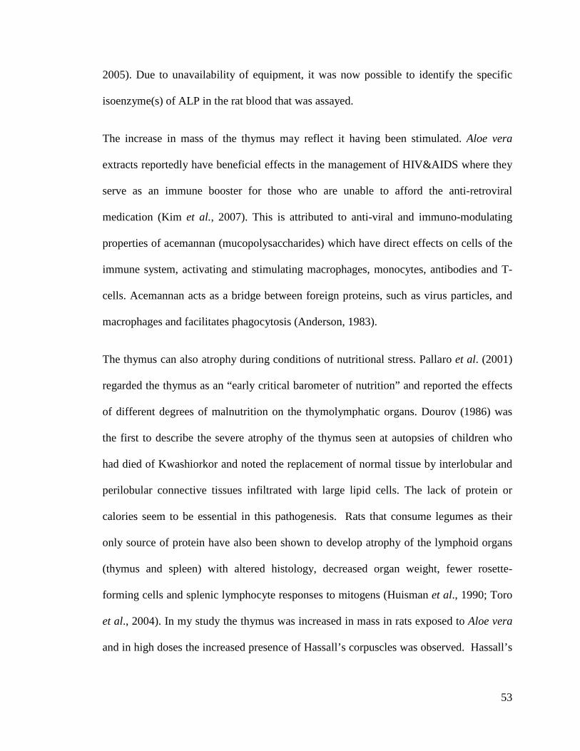
53
2005). Due to unavailability of equipment, it was now possible to identify the specific
isoenzyme(s) of ALP in the rat blood that was assayed.
The increase in mass of the thymus may reflect it having been stimulated. Aloe vera
extracts reportedly have beneficial effects in the management of HIV&AIDS where they
serve as an immune booster for those who are unable to afford the anti-retroviral
medication (Kim et al., 2007). This is attributed to anti-viral and immuno-modulating
properties of acemannan (mucopolysaccharides) which have direct effects on cells of the
immune system, activating and stimulating macrophages, monocytes, antibodies and T-
cells. Acemannan acts as a bridge between foreign proteins, such as virus particles, and
macrophages and facilitates phagocytosis (Anderson, 1983).
The thymus can also atrophy during conditions of nutritional stress. Pallaro et al. (2001)
regarded the thymus as an “early critical barometer of nutrition” and reported the effects
of different degrees of malnutrition on the thymolymphatic organs. Dourov (1986) was
the first to describe the severe atrophy of the thymus seen at autopsies of children who
had died of Kwashiorkor and noted the replacement of normal tissue by interlobular and
perilobular connective tissues infiltrated with large lipid cells. The lack of protein or
calories seem to be essential in this pathogenesis. Rats that consume legumes as their
only source of protein have also been shown to develop atrophy of the lymphoid organs
(thymus and spleen) with altered histology, decreased organ weight, fewer rosette-
forming cells and splenic lymphocyte responses to mitogens (Huisman et al., 1990; Toro
et al., 2004). In my study the thymus was increased in mass in rats exposed to Aloe vera
and in high doses the increased presence of Hassall’s corpuscles was observed. Hassall’s

54
corpuscles are lymphocytes that may be interpreted as an expression of a changing
thymic growth pattern in confined spaces and seem to have little immunological function.
The liver also plays an important role in metabolism to maintain energy levels (Aitken et
al., 2001). The blood concentration of glucose in young rats is maintained fairly at a
fairly constant level from 2 to 20 days after birth. This is probably due to the difference in
floral colonization of the gut between the 18-day-old suckling rat, whose diet is
predominatly milk and different to the adults whose diet contains both milk and solid
high-carbohydrate rat diet (Innis, 1985). The glucose level in non fasting blood after 8
days of treatment with Aloe vera extracts did not show any significant differences
between the groups. This may be the evidence that the Aloe vera extracts did not alter the
endocrine pancreatic function, which has an indirect influence on the blood glucose
levels.
It has been shown that the amount of free fatty acids taken up by the liver of rats (Poli et
al., 1987) is directly proportional to the concentration of free fatty acids in the plasma.
Fatty acids will accumulate unless compensatory increases in rates of outputs of fatty
acids in plasma lipoprotein or rates of oxidation to CO2 and ketone bodies occur (Erol et
al., 2003). Fatty liver is a metabolic disorder that occurs when the rate of fatty acid
uptake and esterification exceeds the rate of fatty acid depletion either through oxidation
or export as triglyceride within very low density lipoproteins. The rats exposed to the
high dose of alcohol Aloe vera extracts showed the greatest accumulation of lipids in the

55
liver, however the lipids were not profiled so the metabolic processes underlying the
increased accumulation could not be fully ascertained.
The results of this study on suckling rats concur with findings from other studies which
revealed the beneficial effects of Aloe vera extracts administration over a short period of
time. There is however a need to explore further the mechanisms of improved growth
performance, the significance of the increased thymus mass and the changes in caecal
morphology noted as a result of exposure of the suckling rats to the Aloe vera extracts.

56
Chapter 5 – Conclusion and
recommendations

57
Conclusion and recommendations
Herbal medicines are widely used in almost all sectors of the population. This study
has shown that Aloe vera extracts administered orally to suckling rats had a positive
effect on the body mass, caecum and bones. These positive effects could be useful in
preventing weight loss during the weaning period of pups and bone disturbances
during the development of fast growing animals. The present study’s findings confirm
that Aloe vera extracts could have a beneficial effect at least in the short term.
Further studies should be undertaken for a longer period of time in order to determine
the reversibility and functional significance of the significant findings observed in the
rats.
The increased growth of the animals exposed to the Aloe vera extracts should be
explored further, by assaying for other growth promoting hormones and microscopic
examination of the growth plates. The degree of bone mineralization and density
should also be evaluated using DEXA-scans.
The increased body mass gain observed in the rats given Aloe vera could be exploited
commercially in animal production to improve growth in farm animals. This study
involved the use of crude extracts, future studies should be undertaken to identify the
specific chemical components of Aloe vera which produced the significant
differences such as caecal and thymic enlargement. Future studies should also make
use of sugar solutions with mineral and vitamin supplements as a control solution to
match the nutritional composition of plant extracts so that any differences can be
attributed to the phytochemicals in the extracts. Herbal medicines although being

58
widely used, should be used cautiously and after thorough scientific interrogation of
their active constituents as they can contain harmful compounds and metabolites.

59
Chapter 6 – References

60
References
Aitken, M.M., Hall, E., Scott, L., Davot, J.L., Allen, W.M. (2001) Liver-related
biochemical changes in the serum of dogs being treated with
phenobarbitone. Veterinary Record, 153: 13-16.
Ajabnoor, M.A. (1990) Effect of Aloes on the blood glucose levels in normal and alloxan
diabetic mice. Journal of Ethnopharmacology, 28: 215-220.
Ali, M.I., Shalaby, N.M., Elgamal, M.H., Mousa, A.S. (1999) Antifungal effects of
different plant extracts and their major components of selected Aloe
species. Phytotherapy Research, 13: 401-7.
Anderson, B.C. (1983). Aloe vera juice: A veterinary medicament? The compendium on
continuing Education for the practicing Veterinarian, 5: 364-368.
Amusan, P.S., Dlamini, J.D., Msonthi, Makhubu, L.P. (2002) Some herbal remedies from
Manzini region, Swaziland. Journal of Ethnopharmacology 79, 109-112.
Arnold, Koldosky, O., Palanch, A.C. (2002) Medicinal and magical plants of Southern
Africa: An annotated checklist, National Botanical Institute, Pretoria.
Bain, P.J., Latimer, K.S., Mahaffey, E.A., Prasse, K.W., Duncan and Prasse’s, (2003)
Liver. Veterinary Laboratory Medicine: Clinical Pathology, fourth edition.
Ames, Iowa State Press 193-214.
Baintner, K., Zabielski, R., Gregory, P.C., Westrom, B. ( 2007) Vacuolation in the young
and the intestine in Growing Animals, first edition, Amsterdam: Elsevier
Science, 70, 55-110.

61
Baltrop and Brueton, T. M. (1990) The gastrointestinal tract and short term toxicity tests.
In: P. J., Bordeau, Editor, Short-term Toxicity Tests for non-genotoxic
Effects, John Wiley and Sons Ltd 99-110.
Baroncelli, G.I., Bertelloni, S.,Sodini, F., Saggese ,G. (2003) Acquisition of bone mass
in normal individuals and in patients with growth hormone deficiency.
Journal of Pediatry and Endocrinology Metabolism 16: 327-335.
Baumann, G., (1991) Growth Hormone heterogeneity. Genes, Isohormones, variants and
binding protein. Endocrine Reviews, 12: 424-449.
Beppu, H., Shimpo, K., Kutchuno, M. (2006) Antidiabetic effects of dietary
administration of Aloe arborescens Miller components on multiple low-
dose streptozotocin- induced diabetes in mice: investigation on
hypoglycaemic action and systemic absorption dynamics of Aloe
components. Journal of Ethnopharmacology, 103: 468-477.
Bligh, E.G. and Dyer, W.J. (1959) A rapid method of total lipid extraction and
purification. Canadian Journal of Biochemistry and Physiology 37: 911-
917.
Blumenthal, M., Gruenwald, J., Hall, T., Rister, R.S. (1998) The complete German
Commission E Monographs. Austin, Texas: American Botanical Council
111: 556-569.
Blumenthal, M., Busse, W.R., Goldberg, A. (1998) The complete commission
monograph: therapeutic guide to herbal medicine. Integrative Medical
Commission, Boston 80-87.

62
Boudreau, M.D. (2006) An evaluation of biological and toxicological proprieties of Aloe
Barbandesis (Miller), Aloe vera. Journal of Environmental Sciences Health
24: 103-154.
Brian, K.R., Turner, T.D. (1975) The practical evaluation of phytopharmaceuticals.
Wright-Scentechica, Bristol, first edition, 90.
Bruneton, J. (1995) Pharmacognosy, Phytochemistry, Medical Plants; 434-436. England,
Intercept, Hampshire.
Bryant, A.T. (1970) Zulu medicine and medicine men. Annals of the Natal Museum, 2: 1-
103.
Buddington, R.K., Lepine, A.J. (1999) Exocrine Pancreatic Functions of Carnivores. In:
Biology of Pancreas in Growing Animals. Pierzynowski, S.G. and
R.Zabielski (Eds.). Elsevier Science BV 409-422.
Butler, L.K. (1995) Regulation of blood glucose levels in normal and diabetic rats, in
tested studies for the laboratory teaching volume 16. Proceedings
of the 16th Workshop/Conference of the Association for Biology
Laboratory Education (ABLE), Austin, Texas. 181-202.
Can, A., Akev, N.,Ozsoy, N., Bolkent, S., Arda, B.P., Yanardag, R., Okyar, A. (2004)
Effect of Aloe vera leaf gel and pulp extracts on the liver in type-II diabetic
rat models. Biological and Pharmaceutical Bulletin, 27: 694-8.
Chandan, B.K., Saxena, A.K., Shukla, S., Sharma, N., Gupta, (2007) Hepatoprotective
potential of Aloe Barbadensis Mill, against carbon tetrachloride induced
hepatotoxicity. Journal of Ethnopharmacology, 111: 560-566.

63
Cheney, R.H. (1970) Aloe vera Drug in Human therapy. Quarterly. Journal Crude Drug
Resources, 10: 1523-15.
Choi,S., Kim, K.W., Choi, J.S, Han,S.T., Park,Y.I., Lee,S.K.., Kim, J.S. and Chung,
M.H.(2002) Angiogenic activity of beta-sitosterol in the ischaemia/
reperfusion-damaged brain of Mongolian gerbil.Department of
pharmacology, Seoul National University college of medicine, Seoul,
Korea. Planta Medica, 68: 330-5.
Cho, I .H, Ahn, J.H., Chung, C.Y., Yoo, W.J., Sung, H.Y. (2001) Expression of
vasculoendothelial growth factor in distraction osteogenesis of rat tibia.
Journal of Korean Orthopeutic Resources, 4: 114-20.
Chung, J.H., Cheong J.C., Lee, J.Y., Roh, H.K., Cha, Y.N. (1997) Acceleration of the
alcohol oxidation rate in the rats with aloin, a quinine derivative of Aloe.
Bio-Pharmacology 52: 1461-1468.
Crouch, N., Symmonds, R., Spring, W., Diederichs, N. (2006) Fact sheets for growing
popular medicinal plant species. In: N. Diederichs, Editor,
Commercialising Medicinal Plants—A Southern African Guide, Sun Press,
Stellenbosch. 97–142.
Cummins, A .G., Steele, T.W., Labrooy, J.T., Shearman, D.J.C. (1988) Maturation of the
rat small intestine at weaning: Changes in epithelial cell kinetics, bacterial
flora and mucosal immune activity. Gut, 29: 1672-9.
Davis, H.R. (1993) Biological activity of Aloe vera. The SOFW-Journal, 119: 647-649.

64
Davis, R.H., Donate, J.J., Hartman, G.M. (1994) Anti-inflammatory and wound healing
activity of growth substance in Aloe vera. Journal of the American
Podiatric Medical Association 84: 77-81.
Dignass, A.V., Lynch-Devaney, K. (1995) Hepatocyte growth factor/Scatter factor
modulates intestinal epithelial cell proliferation and migration. Biochemical
and Biophysical Research Communication, 202: 701-709.
Djeraba, A., Quere, P. (2000) In vivo macrophage activation in chickens with
Acemannan, a complex carbohydrate extracted from Aloe vera.
International Journal Immunopharmacology, 22: 365-72.
Dold, T., Cocks, M. (2000) The iNtelezi plants of the Eastern Cape: traditional and
contemporary medicines. Aloe, 37: 10-13.
Dourov, N. (1986) Thymic atrophy and immune deficiency in malnutrition In: Muller
H.K, first edition. The human thymus. Histopathology and pathology.
Berlin; Springer-verlag: 127.
Dunn, J.T. (2002): Editorial: guarding our nation’s thyroid health. Journal of Clinical Endocrinology and Metabolism 87: 486-488.
Dziedziejko, V., Safranow, K., Slowik-Zylka, D., Machoy-Mokrzynska, Millo, B.,
Machoy, Z., Chlubek, D.(2005) Comparison of rat and human alkaline
phosphatise isoenzymes and isoforms using HPLC and electrophoresis.
Biochimica et Biophysica Acta (BBA) - Proteins& Proteomics. 1752: 26-
33.
Erol, E., Kumar, L.S., Cline G.W., Shulman, G.L., Kelly, D.P., Binas, B. (2004) Liver
fatty acid-binding protein is required for high rates of hepatic fatty acid

65
oxidation but not for the action of PPAR-α in fasting mice. The FASEB
Journal, 18: 347-349.
Erlwanger,K.H., Cooper, R.G. (2008) The effects of orally administered crude alcohol
and aqueous extracts of African potato (Hypoxis hemerocallidea) corm on
the morphometry of viscera of suckling rats. Food and Chemical
Toxicology, 46: 136-139.
Eshet, R., Maor, G., Ari, B.T., Eliezer, B.M., Gatyablonski, G., Phillip, M. (2004) The
aromatase inhibitor letrozole increases epiphyseal growth plate height and
tibial length in peripubertal male mice. Journal of Endocrinology 182: 165-
172.
Esua, M.F., Rauwald, J.W. (2006) Novel bioactive maloyl glucans from Aloe vera gel:
isolation, structure elucidation and in vitro bioassays. Carbon Resource 27:
355-364.
Fermenia, A, Sanchez, E.S., Simal, S., Rosselo, C. (1999) Compositional features of
polyssacharides from Aloe vera (Aloe vera barbadensis Miller) plant
tissues. Carbohydrates Polymerases, 39: 109-17.
Fulton, J.E.J.R., (1990) The stimulation of post dermabration wound healing with
stabilized Aloe vera Gel-poly ethylene oxide dressing. Journal of
Dermatology and Surgical Oncology. 16: 460-467.
Gaskill,C.L., Hoffmann, W.E., Cribb, A.E. (2004) Serum alkaline phosphatase
isoenzyme profiles in Phenobarbital-treated epileptic dogs. Veterinary
Clinical Pathology. 33: 215-222.

66
Gama, P., Alvares, E.P. (2000) Early weaning and prolonged nursing induce changes in
cell proliferation in the gastric epithelium of developing rats. American
Society for Nutrition Sciences. 130: 2594-2598.
Gelfand, R.B., Drummond, S., Madera, B. (1885) The traditional medical practinioner in
Zimbabwe: Principles of practice and pharmacopolia, Mambo Press,
Gweru.
Ghannam, N., Kingston, M., Al-Mechaal, I.A. (1986) The antidiabetic activity of Aloe
vera L, Juice.II.Clinical trail in diabetes mellitus patients in combinaison
with glibenclamide. Phytomedicine. 3: 245-248.
Githens, T.S. (1979) Drug plants of Africa, University of Pennsyvania Press,
Philadelphia.
Gore,M.T, Young, R. B., Claeys, M. C., Chromiak, J. A., Rahe, C. H., Marple, D. N.,
J.D., Hough, Griffin, J. L., Mulvaney, D. R. (1994) Growth and
development of bovine fetuses and neonates representing three genotypes
Journal of Animal Sciences. 72: 2307-2318.
Grindlay, D., Reynolds, T. (1986) The Aloe vera Phenomeno: A review of the proprieties
and modern uses of the leaf parenchyma gel. Journal of
Ethnopharmacology, 16: 117-151.
Guyton, A, C., J, E., Hall and Jackson, (2006) Text book of Medical Physiology, 11th
edition, Elsevier, USA.

67
Hart, L.A., van Enckevort,P.H., van Dijk ,H., Zaat, R.,de Silva, K.T., Labadie, R.P.
(2009) Two functionally and chemically distinct immunomodulatory
compounds in gel of Aloe vera. Journal of Ethnopharmacology. 23: 61-71.
Heggers, J.P., Robson, M.C., Manavalen, K. (1996) Experimental and clinical
observations on frostbite. Annals of Emergency Medicine, 16: 1056-1062.
Heidi, H., Swamson, Bolwerk, E., Bremmer, E. (1884) Effects of couling in infant rats on
growth, maturation, sleep patterns and responses to food deprivation.
British Journal of Nutrition, 52: 139-148.
Heneghan, J.B. (1984) Physiology of the alimentary tract. In: The germ-free Animal in
Biomedical Research (Coates ME, Gustafsson BE,eds). London.
Laboratory Animals Ltd 169-92.
Hochberg, Z., Bick, T., Harel, Z. (1990) Alterations of human growth hormone binding
by rat liver membranes during hypo-and hyperthyroidism. Journal of
Endocrinology, 126: 325-9.
Huisman, J,vander Pod, A., Mouwen. J.,Van weerden, E. (1990) Effect of variable
protein contents in diets containing Phaseolus vulgaris beans on
performance, organ weights and blood variables in piglets, rats and
chickens. British Journal of Nutrition, 64:755.
Huggins, K.W., Camarota, L.M., Howles, P.N. and Hui, D.Y. (2003) Pancreatic
triglyceride lipase deficiency minimally affects dietary fat absorption but
dramatically decreases cholesterol absorption in mice. The Journal of
Biological Chemistry, 278: 42899-42905.
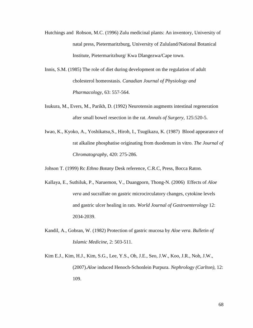
68
Hutchings and Robson, M.C. (1996) Zulu medicinal plants: An inventory, University of
natal press, Pietermaritzburg, University of Zululand/National Botanical
Institute, Pietermaritzburg/ Kwa Dlangezwa/Cape town.
Innis, S.M. (1985) The role of diet during development on the regulation of adult
cholesterol homeostasis. Canadian Journal of Physiology and
Pharmacology, 63: 557-564.
Isukura, M., Evers, M., Parikh, D. (1992) Neurotensin augments intestinal regeneration
after small bowel resection in the rat. Annals of Surgery, 125:520-5.
Iwao, K., Kyoko, A., Yoshikatsu,S., Hiroh, I., Tsugikazu, K. (1987) Blood appearance of
rat alkaline phosphatise originating from duodenum in vitro. The Journal of
Chromatography, 420: 275-286.
Johson T. (1999) Rc Ethno Botany Desk reference, C.R.C, Press, Bocca Raton.
Kallaya, E., Suthiluk, P., Naruemon, V., Duangporn, Thong-N. (2006) Effects of Aloe
vera and sucralfate on gastric microcirculatory changes, cytokine levels
and gastric ulcer healing in rats. World Journal of Gastroenterology 12:
2034-2039.
Kandil, A., Gobran, W. (1982) Protection of gastric mucosa by Aloe vera. Bulletin of
Islamic Medicine, 2: 503-511.
Kim E.J., Kim, H.J., Kim, S.G., Lee, Y.S., Oh, J.E., Seo, J.W., Koo, J.R., Noh, J.W.,
(2007).Aloe induced Henoch-Schonlein Purpura. Nephrology (Carlton), 12:
109.

69
Koyama, I.K., Arai, Y., Sakagishi, H., Ikezawa, T., Komoda, (1987) Blood appearance
of rat Alkaline phosphatise originating from the duodenum in vitro, Journal
of Chromatography, 420: 275-286.
Lahey, J. (2009) “Digestion- how the small intestine digests food.”16 Aug. Ezine
Articles.com.
Langmead, L., Makins, R.J., Rampton, D.S. ( 2004) Anti-inflammatory effects of Aloe
vera gel in human colorectal mucosa in vitro. Aliment of Pharmacology
Therapy, 19: 521-527.
Lairon, D., Play, B., Jourdheuil-Rahmanni, D. (2007) Digestible and indigestible
carbohydrates: interactions with postpradial lipid metabolism. Journal of
Nutritional Biochemistry, 18: 217-227.
Leffers, A., Gemsbok, B., Kalahari, T. (2003) Traditional plant use by the jul’hoansi in
Northern-Eastern Namibia, Gamsberg Mac millan, Windhoek.
Leung, M.Y., Liu, C., Koon, J.C., Zhu, L.F., Hui, Y.Z, Yu, B., Fung, K.P. (2004)
Macrophage activation by polysaccharide biological response modifier
isolated from Aloe vera L. Var. Chinensis (Haw.) Berg. International
Journal of Immunopharmacology, 6: 1634-41.
Linderoth, A., Prykhod'ko, O., Pierzynowski, S.G., Weström, B.R. (2006) Enterally but
not Parenterally Administered Phaseolus vulgaris Lectin Induces Growth
and Precocious Maturation of the Gut in Suckling Rats. Biology of the
Neonate. 89:60-68.
Luckey, T.D., Mende, T.J., Pleasants, J. (1954)The physical and chemical

70
characterisation of rat milk Journal of Nutrition 54, 345-359.
Lynch, C.D., Lyons, D., Khan, A., Bennett, S.A., Sonntag ,W.E. (2001) Insulin-like
growth factor-1 selectively increases glucose utilization in brains of aged
animals. Endocrinology. 142: 506-9.
Mahattanadul, (1996) Antigastric ulcer properties of Aloe vera. Songklanakarin Journal
of Sciences and Technology 18, 49-57.
Maliehe, E.B. (1997) Medicinal plants and herbs of Lesotho. Mafeteng Development
Project, Maseru.
Marini, I. (2004) Discovering an Accessible Enzyme: Salivary α-Amylase. Biochemistry
and Molecular Biology Education, 33: 112-116.
Massimo, M. (2003) Dietary supplements of plant origin: a nutrition and health approach.
Taylor Francis, London, 9-12.
Matsuda,Y., Yokohira, M., Suzuki, S. (2008) One –year chronic toxicity study of Aloe
arborenscens Miller var. natalensis Berger in Wistar Hannover rats. A
pilot study. Food Chemistry and Toxicology, 46: 733-739.
Matsuo, K., Yamashita, S., Niwa, M., Kurihara, M., Harakawa, S.,Izumi, M., Nagataki,
S.,Melmed,S. (1990) Thyroid hormone regulates rat pituitary insulin-like
growth factor-1 receptors. Journal of Endocrinology, 126: 550-4.
Mills, N.C., D’ercole, A.J., Underwood, L.E and Ilan., J. (1986) Synthesis of
somatomedin C/insulin-like growth factor I by human placenta. Molecular
Biology Reports, 11: 231-236.

71
Moon, E.J., Lee, Y.M., Lee, O.H. (1999) A novel angiogenic factor derived from Aloe
vera gel: beta- sitosterol, a plant sterol. Angiogenesis 3, 117-123.
Pacha,J. (2000) Development of intestinal transport function in mammals. Physiology
Reviews, 80: 1633-1667.
Paeza, A., Michael Gebreb, G., Gonzaleza, M.E., Tschaplinski, T.J. (2009) Growth,
soluble carbohydrate and aloin concentration of Aloe vera plants exposed
to three irradiance levels. Environmental and Experimental Biology, 44:
133-139.
Pallaro, A.N., Roux, M.E., Slobodianik, N.H. (2001) Nutrition disorders and
immunologic parameters: study of thymus in growing rats. Nutrition, 17:
972-973.
Panda, H., 2000 Aloe vera hand book cultivation, research findings, products,
formulation, extraction and processing. Asia Pacific Business Press, India. .
Pari, L., Venkateswaran, S. (2004) Protective Role of Phaseolus vulgaris on Changes in
the Fatty Acid Composition in Experimental Diabetes. Journal of
Medicinal Food 7, 204-209.
Passonneau, J.V., Lauderdale, V.R. (1974) A comparison of three methods of glycogen
measurement in tissue. Analytical Biochemistry, 60: 405-412.
Peng, S.Y., Norman, J., Curtin, G., Corrier, D., Mc Daniel, H.R., Busbee, D. (1991)
Decreased mortality of norman murine sarcoma in mice treated with the
immunomodulator, Acemannan. Molecular Biotherapy, 3:79-87.

72
Pierzynowski, S.G., Botermans, J.A.M. (1999) Relations between body live weight, feed
intake, daily weight gain and pancreatic exocrine secretion in growing pigs.
Journal of Animal Sciences, 77: 450-456.
Poli, G., Albano, E., Dianzani, M.U. (1987) The role of lipid peroxidation in liver damage.
Chemistry and Physics of Lipids, 45: 117-142.
Podolsky, D.K. (1993) Regulation of intestinal epithelial proliferation: a few answers,
many question. American Journal of Physiology, 264: G179-G186.
Ramachandra, C.T., Srinivasa, R. P. (2008) Processing of Aloe vera leaf Gel: A review
American Journal of Agricultural and Biological Sciences, 3: 502-510.
Ranke, M.B. (2003) Diagnosis of growth hormone deficiency and growth hormone
stimulation tests. In; Diagnostics of endocrine function in children and
adolescents (third edition) Basel, Karger , pp107-128.
Reddy, B.S. (1971) Calcium and magnesium absorption: role of intestinal microflora-
feeding. Federation Proceedings, 30: 1815-1821.
Reuter, J., Jocher, A., Stump, J., Grossjohann, B., Franke, G., Schempp, C.M. (2008)
Investigation of the anti-inflammatory potential of Aloe vera Gel (97.5%)
in the ultraviolet erythema test. Skin Pharmacology and Physiology 21:
106-110.
Reynolds,T., Dweck, A.C. (1999) Aloe vera leaf gel: a review up date. Journal of
Ethnopharmacology, 15: 3-37.
Reynolds, J.W. (1950) The Aloes of South Africa.The Trustees of Aloes of South Africa
book fund, Johannesburg, South Africa.
73
Robert, A., Nezamis, J.E, Lancaster, C, Hamchar, A.J. (1979) Cytoprotection by
prostaglandins in rats. Gastroenterology, 77: 433-443.
Sa, I.M., Oh, S.T., Song, S., Kim, M.R., Kim,D.S., Woo, S.S., Jo, T.H., Park, Y.I., Lee,
C.K. (2005) Identification of optimal molecular size of modified Aloe
polysaccharides with maximum immunomodulatory activity. International
Immunopharmacology, 5 : 271-9.
Saito, H., Imanishi, K., Okabe, S. (1989). Effects of Aloe extracts, Aloctin A, on gastric
secretion and on experimental gastric lesions in rats. Yakugaku Zasshi, 109:
335-9.
Sakai, T., Repko, B.M., Griffith, B.P., Waters, Kameneva, M.V. (2006) Infusion of a
drag-reducing polymer extracted from Aloe vera prolonged survival time in
a rat model of acute myocardial ischaemia, British Journal of Anaesthesia,
98 : 23-8.
Saleem,R., Faizi, S., Siddiqui, B.S., Ahmed, M.,Hussain, S.A., Qazi, A., Dar, A.,Ahmad,
S.I., Qazi, M.H., Akhtar, S., Hasnain, S.N. (2001) Hypotensive effect of
chemical constituents from Aloe barbadensis. Planta Medica, 67: 757-60.
Shah,A.H., Qureshi, S., Tariq, M., Angeel, A.M. (1989) Toxicity studies on six plants
used in the tarditional arab system of medicine. Phytotherapy Reactions, 3:
25-29.
Sorensen, A., Mayntz, D., Simpson, S.J., Raubenheimer, D. (2009) Dietary ration of
protein to carbohydrate induces plastic responses in the gastrointestinal
tract of mice. Journal of Physiology, 180: 259-266.

74
Steenkamp, V., Stewart, M.J. (2007) Medicinal applications and toxicological activities of
Aloe Products. Pharmaceutical Biology, 45: 411-420.
Stepanova, O.S., Prudnick, N.Z., Soloviera V.P., Golovchenko G.A., Svischchuk, A.A.,
Dubkova, O.M. (2007) Chemical composition and biological activity of
dry Aloe leaves. Fiziologicheski Aktivnye Veshchestva, 9: 94-97.
Strasburger, C.J. (2004) Commentary: Taking one step at a time. Clinical Endocrinology,
60: 540-.
Strader, A.D., Woods, S.C. (2005) Gastrointestinal Hormones and food intake . Journal
of Gastroenterology, 128: 175-91.
Stuart, R.W., Lefkowitz, D.L., Lincoln, J.A., Howard, K., Gelderman, M.P., Lefkowitz,
S.S. (1997) Up regulation of phagocytosis and Candidicidal activity of
macrophages exposed to the immunostimulant acemannan. International
Journal of Immunopharmacology, 19: 75-82.
Susana, A. (2010) Aloe vera juice: A Natural source of vitamins and minirals. Ezine
Articles.com (Http// ezine articles.com/ Aloe vera juice –a natural source of
vitamins and minerals &id=3691- 620).
Suvitayavat, W., Sumrongkit, C., Thirawarapan, S.S., Bunyapraphatsara, N. (2004)
Effects of Aloe preparation on the histamine-induced gastric secretion in
rats. Journal of Ethnopharmacology, 90: 239-247.
Tanaka, M., Matsuda, M.A. (2006) Identification of five phytosterols from Aloe vera gel
as antidiabetic compounds. Biological and Pharmaceutical Bulletin, 29:
1418-22.

75
Tisi, D.K., Liu, X.J., Wykes, L.J., Skinner, C.D., Koski, K.G. (2005) Insulin-Like
Growth Factor II and Binding Proteins 1 and 3 from Second Trimester
Human Amniotic Fluid Are Associated with Infant Birth Weight. Journal
of Nutrition, 135: 1667-1672.
Toro, F., Benshimol, A., Soyano, A. (2004) Spleen and thymus histology and
proliferative response of splenic cells in rats fed raw and cooked phaseolus
vulgaris beans. Archivos Latinamericanos de Nutricion, 42: 395-402.
Vazquez, I.A., Gomez –de-Segura, A.G., Grade, (1999) Protective effect of enriched diet
plus growth hormone administration on radiation-induced intestinal injury
and on its evolutionary pattern in the rat. Digestive Diseases and Sciences,
44: 2350-2358.
Wallander, E., Albert, V.A. (2000) Phylogeny and classification of Oleaceae based on
rps 16 and trnL-F sequence data. American Journal of Botany, 87: 1827-
1841.
Washburn, A.H. (1950) Growth: its significance in medicine viewed as human biology.
Pediatrics 5, 765-770.
Watt, J.M., Breyer-Brandwijk, M.G. (1962) Medicinal and poisonous plants of Southern
and Eastern Africa. Second edition, Edinburgh.
Weser, E., Tawil, T. (1976) Epithelial cell loss in remaining intestine after small bowel
resection in the rat. Gastroenterology 71: 412-15.

76
Woo, S.W., Nan, J.X., Lee, S.H., Park, E.J., Zhao, Y.Z., Sohn, D.H. (2002) Aloe-emodin
suppresses myofibroblastic differentiation of rat hepatic stellate cells in
primary culture. Pharmacology and Toxicology, 90: 193-198.
Woods, S.C, Benoit, C.S., Clegg, D.J., Seeley, R.J. (2004) Blocking carbohydrate
absorption and weight loss: A clinical trial using phase 2TM brand
proprietary fractioned white bean extract. Alternative Medicine Review 9:
63-69.
Wichtl, M., Bisset, N.G. (1994) Herbal Drugs and Phytopharmaceuticals. Third edition.
MedPharm Scientific Publishers, Stuttgart. Pp 25- 29.
Wilding, J., Wowson, P.W., Williams, G. (1997) Memory of specific foods to control and
restoration of body energy. British Medical Bulletin, 2: 286-306.
Sonntag, W.E., Ramsey, M., Carter, C.S. (2005) Growth hormone and insulin-like growth
factor-1 (IGF-1) and their influence on cognitive aging. Ageing Research
Reviews, 4: 195-212.
Wieringa, G.E., Barth, J.H., Trainer, P.J. (2004) Growth Hormone assay standardization: a
biased view? Clinical Endocrinology, 60: 538-539.
Wynne, K., Stanley, S., Bloom, S.,( 2005) The gut and regulation of body weight. Journal
of Clinical Endocrinology Metabolism 89:2576–2582.
Yaron A., (1991) Aloe vera: Chemical and physical properties and stabilization. Journal
of Botany 40:270.
Yates, K.M, Rosenberg, L.J., Harris, C.K., Bronstand, D.C., King,G.K., Bichle, G.A.,
Walker, B., Ford, C.R., Hall, J.E., Tizard,I.R. (1992) Pilot study of effect of

77
acemannan in cats infected with feline immunodeficiency virus. Veterinary
Immunology and Immunopathology, 35: 177-189.
Yeh, G.Y., Eisenberg, D.M., Kaptchuk, T.J, Phillips, R.S. (2003) Systematic review of
herbs and dietary supplements for glycemic control in diabetes. Diabetes
Care, 26: 1277-1294.
Yu, C.S., Yu, F.S., Chan, J.K., Li, T.M., Lin, S.S., Chen, S.C., Hsia, T.C., Chang, Y.H.,
Chung, J.G. (2006) Aloe-emodin affects the levels of cytokines and
functions of leucokytes from Sprague-Dawley rats. In vivo, 20: 505-9.
Yusuf, S., Agunu, A., Diana, M. (2004) The effect of Aloe vera A. Berger (Liliaceae) on
gastric acid secretion and acute gastric mucosal injury in rats. Journal of
Ethnopharmacology, 93: 33-7.
Zang, X.F., Wang, H.M., Song, Y.L. (2006) Isolation, structure elucidation, antioxidative
and immunomodulatory proprieties of two novel dihydrocoumarins from
Aloe vera. Bioorganic and Medicinal Chemistry Letters, 16: 949-953.