beta-Catenin directly regulates Islet1 expression in cardiovascular progenitors and is required for...
6
-Catenin directly regulates Islet1 expression in cardiovascular progenitors and is required for multiple aspects of cardiogenesis Lizhu Lin*, Li Cui*, Wenlai Zhou † , Daniel Dufort ‡ , Xiaoxue Zhang*, Chen-Leng Cai*, Lei Bu*, Lei Yang*, Jody Martin*, Rolf Kemler § , Michael G. Rosenfeld † , Ju Chen ¶ , and Sylvia M. Evans* *Skaggs School of Pharmacy and Pharmaceutical Sciences, † Howard Hughes Medical Institute and Department of Medicine, and ¶ School of Medicine, University of California at San Diego, 9500 Gilman Drive, La Jolla, CA 92093; ‡ Department of Obstetrics and Gynecology, Royal Victoria Hospital, 687 Pine Avenue West, Montreal, QC, Canada H3A 1A1; and § Max-Planck-Institute of Immunobiology, Stubeweg 51, 79108 Freiburg, Germany Edited by Eric N. Olson, University of Texas Southwestern Medical Center, Dallas, TX, and approved April 13, 2007 (received for review February 1, 2007) Recent studies have demonstrated that the LIM homeodomain transcription factor Islet1 (Isl1) marks pluripotent cardiovascular progenitor cells and is required for proliferation, survival, and migration of recently defined second heart field progenitors. Factors that are upstream of Isl1 in cardiovascular progenitors have not yet been defined. Here we demonstrate that -catenin is required for Isl1 expression in cardiac progenitors, directly regu- lating the Isl1 promoter. Ablation of -catenin in Isl1-expressing progenitors disrupts multiple aspects of cardiogenesis, resulting in embryonic lethality at E13. -Catenin is also required upstream of a number of genes required for pharyngeal arch, outflow tract, and/or atrial septal morphogenesis, including Tbx2, Tbx3, Wnt11, Shh, and Pitx2. Our findings demonstrate that -catenin signaling regulates proliferation and survival of cardiac progenitors. cardiac morphogenesis cardiac progenitors direct target Isl1 P revious studies have demonstrated a key role for the LIM homeodomain transcription factor Islet1 (Isl1) in cardiac de- velopment and as a marker for pluripotent cardiovascular progen- itors, which give rise to cardiomyocyte, endothelial, and smooth muscle lineages in vitro (1–5). Isl1 marks proliferating, undifferen- tiated progenitors of the second heart field, which are located dorsal/medially to the heart (4, 5). Isl1 is required for proliferation, survival, and migration of these progenitors into the forming heart (4). The second heart field migrates in and differentiates later than progenitors of the first heart field. Isl1 expression is down-regulated in second heart lineages as differentiation occurs. The second heart lineage includes cells of the secondary or anterior heart field, which gives rise to the outflow tract or outflow tract and right ventricle, respectively (6–8). Isl1-null mice die embryonically at embryonic day 10 (E10) with hearts missing segments derived from the second heart field, including the outflow tract and right ventricle, and exhibiting severely reduced atrial tissue. Isl1 also marks cardiac progenitors found within postnatal hearts of rodents and humans (9). These progenitors can be isolated, propagated, and readily differentiated into functional cardiomyocytes when cocultured with neonatal cardiac myo- cytes. More recent studies have demonstrated that Isl1 marks a pluripotential cardiovascular cell that coexpresses flk1 and Nkx2.5, which can be cloned and amplified from either embry- onic stem cells or embryos (1, 2). Amplified clones can give rise to endothelial, smooth muscle, and cardiomyocyte cell types. The pivotal role for Isl1 within cardiovascular progenitors makes it critical to understanding factors that regulate Isl1 expression in this context. A number of studies have demonstrated a key role for canonical Wnt signaling through -catenin in stem cells from a variety of cell types (10). The role of -catenin signaling in cardiovascular stem or progenitor cells, however, has been seemingly contradictory. Several studies have demonstrated the induction of cardiogenic mesoderm in response to the inhibition of Wnt signaling in chick, Xenopus, and mouse embryos. Wnt antagonists Dickkopf1 and Crescent produced by anterior endoderm in chick embryos stim- ulate differentiation of a cardiogenic mesoderm (11). In frog embryos, Dickkopf1 and Crescent secreted by Spemann’s organizer are also initiators of cardiac differentiation, acting indirectly on anterior mesendoderm to provoke secretion of an as-yet- unidentified cardiogenic induction factor (12, 13). In mouse em- bryos, ablation of -catenin using a Cytokeratin19 promoter-driven Cre (K19-Cre) recombinase resulted in ectopic heart formation, which was attributed to ablation of -catenin in endodermal tissues (14). In contrast to the foregoing, activation of Wnt signaling is required for cardiogenesis in Drosophila (15) and cell culture systems, including embryonic stem cells and embryonal carcinoma P19 cells (16 –18). In these cell culture systems, however, the spatial requirement for Wnt signaling has not been addressed. To address the temporal and spatial requirements for canonical Wnt signaling through -catenin in Isl1-expressing cardiac progen- itors, we have used a Cre recombinase expressed under the control of the endogenous Isl1 locus. The early expression of Isl1-Cre in cardiogenic progenitors, and in early pharyngeal endoderm (19, 20), a cardiac-inducing tissue, provided us with an opportunity to examine the requirement for -catenin in these tissues during early cardiogenesis. Results Activation of -Catenin During Early Cardiogenesis. To investigate where and when -catenin signaling was occurring in the early embryo, we used a T cell factor (TCF)/Lef-lacZ reporter line (21) (Fig. 1). Results of this analysis demonstrated -catenin activation in early cardiogenic mesoderm and adjacent endoderm at E7.5 (Fig. 1 A and B). At E8.5 (Fig. 1 C–E), expression of the reporter was observed in pharyngeal mesoderm, outf low tract myocardium, and at low levels in pharyngeal endoderm. At E9.5 (Fig. 1 F–H), expression in pharyngeal mesoderm and outf low tract myocardium was striking, with expression also observed in lateral and ventral pharyngeal endoderm and in proepicardium. At E12.5 (Fig. 1 I–L), TCF/Lef-lacZ reporter expression was observed in epicardium as well as subsets of cells within the outflow tract and ventricles. Author contributions: L.C. and W.Z. contributed equally to this work; L.L. and S.M.E. designed research; L.L., L.C., W.Z., X.Z., L.B., L.Y., and J.M. performed research; D.D., C.-L.C., and R.K. contributed new reagents/analytic tools; L.L., M.G.R., J.C., and S.M.E. analyzed data; and L.L. and S.M.E. wrote the paper. The authors declare no conflict of interest. This article is a PNAS Direct Submission. Abbreviations: En, embryonic day; PAA, pharyngeal arch artery; TCF, T cell factor. To whom correspondence should be addressed. E-mail: [email protected]. This article contains supporting information online at www.pnas.org/cgi/content/full/ 0700923104/DC1. © 2007 by The National Academy of Sciences of the USA www.pnas.orgcgidoi10.1073pnas.0700923104 PNAS May 29, 2007 vol. 104 no. 22 9313–9318 DEVELOPMENTAL BIOLOGY
-
Upload
independent -
Category
Documents
-
view
1 -
download
0
Transcript of beta-Catenin directly regulates Islet1 expression in cardiovascular progenitors and is required for...

�-Catenin directly regulates Islet1 expressionin cardiovascular progenitors and is requiredfor multiple aspects of cardiogenesisLizhu Lin*, Li Cui*, Wenlai Zhou†, Daniel Dufort‡, Xiaoxue Zhang*, Chen-Leng Cai*, Lei Bu*, Lei Yang*,Jody Martin*, Rolf Kemler§, Michael G. Rosenfeld†, Ju Chen¶, and Sylvia M. Evans*�
*Skaggs School of Pharmacy and Pharmaceutical Sciences, †Howard Hughes Medical Institute and Department of Medicine, and ¶School of Medicine,University of California at San Diego, 9500 Gilman Drive, La Jolla, CA 92093; ‡Department of Obstetrics and Gynecology, Royal Victoria Hospital,687 Pine Avenue West, Montreal, QC, Canada H3A 1A1; and §Max-Planck-Institute of Immunobiology, Stubeweg 51, 79108 Freiburg, Germany
Edited by Eric N. Olson, University of Texas Southwestern Medical Center, Dallas, TX, and approved April 13, 2007 (received for review February 1, 2007)
Recent studies have demonstrated that the LIM homeodomaintranscription factor Islet1 (Isl1) marks pluripotent cardiovascularprogenitor cells and is required for proliferation, survival, andmigration of recently defined second heart field progenitors.Factors that are upstream of Isl1 in cardiovascular progenitors havenot yet been defined. Here we demonstrate that �-catenin isrequired for Isl1 expression in cardiac progenitors, directly regu-lating the Isl1 promoter. Ablation of �-catenin in Isl1-expressingprogenitors disrupts multiple aspects of cardiogenesis, resulting inembryonic lethality at E13. �-Catenin is also required upstream ofa number of genes required for pharyngeal arch, outflow tract,and/or atrial septal morphogenesis, including Tbx2, Tbx3, Wnt11,Shh, and Pitx2. Our findings demonstrate that �-catenin signalingregulates proliferation and survival of cardiac progenitors.
cardiac morphogenesis � cardiac progenitors � direct target � Isl1
Previous studies have demonstrated a key role for the LIMhomeodomain transcription factor Islet1 (Isl1) in cardiac de-
velopment and as a marker for pluripotent cardiovascular progen-itors, which give rise to cardiomyocyte, endothelial, and smoothmuscle lineages in vitro (1–5). Isl1 marks proliferating, undifferen-tiated progenitors of the second heart field, which are locateddorsal/medially to the heart (4, 5). Isl1 is required for proliferation,survival, and migration of these progenitors into the forming heart(4). The second heart field migrates in and differentiates later thanprogenitors of the first heart field. Isl1 expression is down-regulatedin second heart lineages as differentiation occurs. The second heartlineage includes cells of the secondary or anterior heart field, whichgives rise to the outflow tract or outflow tract and right ventricle,respectively (6–8). Isl1-null mice die embryonically at embryonicday 10 (E10) with hearts missing segments derived from the secondheart field, including the outflow tract and right ventricle, andexhibiting severely reduced atrial tissue.
Isl1 also marks cardiac progenitors found within postnatalhearts of rodents and humans (9). These progenitors can beisolated, propagated, and readily differentiated into functionalcardiomyocytes when cocultured with neonatal cardiac myo-cytes. More recent studies have demonstrated that Isl1 marks apluripotential cardiovascular cell that coexpresses flk1 andNkx2.5, which can be cloned and amplified from either embry-onic stem cells or embryos (1, 2). Amplified clones can give riseto endothelial, smooth muscle, and cardiomyocyte cell types.The pivotal role for Isl1 within cardiovascular progenitors makesit critical to understanding factors that regulate Isl1 expressionin this context.
A number of studies have demonstrated a key role for canonicalWnt signaling through �-catenin in stem cells from a variety of celltypes (10). The role of �-catenin signaling in cardiovascular stem orprogenitor cells, however, has been seemingly contradictory.
Several studies have demonstrated the induction of cardiogenicmesoderm in response to the inhibition of Wnt signaling in chick,
Xenopus, and mouse embryos. Wnt antagonists Dickkopf1 andCrescent produced by anterior endoderm in chick embryos stim-ulate differentiation of a cardiogenic mesoderm (11). In frogembryos, Dickkopf1 and Crescent secreted by Spemann’s organizerare also initiators of cardiac differentiation, acting indirectly onanterior mesendoderm to provoke secretion of an as-yet-unidentified cardiogenic induction factor (12, 13). In mouse em-bryos, ablation of �-catenin using a Cytokeratin19 promoter-drivenCre (K19-Cre) recombinase resulted in ectopic heart formation,which was attributed to ablation of �-catenin in endodermaltissues (14).
In contrast to the foregoing, activation of Wnt signaling isrequired for cardiogenesis in Drosophila (15) and cell culturesystems, including embryonic stem cells and embryonal carcinomaP19 cells (16–18). In these cell culture systems, however, the spatialrequirement for Wnt signaling has not been addressed.
To address the temporal and spatial requirements for canonicalWnt signaling through �-catenin in Isl1-expressing cardiac progen-itors, we have used a Cre recombinase expressed under the controlof the endogenous Isl1 locus. The early expression of Isl1-Cre incardiogenic progenitors, and in early pharyngeal endoderm (19,20), a cardiac-inducing tissue, provided us with an opportunity toexamine the requirement for �-catenin in these tissues during earlycardiogenesis.
ResultsActivation of �-Catenin During Early Cardiogenesis. To investigatewhere and when �-catenin signaling was occurring in the earlyembryo, we used a T cell factor (TCF)/Lef-lacZ reporter line (21)(Fig. 1). Results of this analysis demonstrated �-catenin activationin early cardiogenic mesoderm and adjacent endoderm at E7.5 (Fig.1 A and B). At E8.5 (Fig. 1 C–E), expression of the reporter wasobserved in pharyngeal mesoderm, outflow tract myocardium, andat low levels in pharyngeal endoderm. At E9.5 (Fig. 1 F–H),expression in pharyngeal mesoderm and outflow tract myocardiumwas striking, with expression also observed in lateral and ventralpharyngeal endoderm and in proepicardium. At E12.5 (Fig. 1 I–L),TCF/Lef-lacZ reporter expression was observed in epicardium aswell as subsets of cells within the outflow tract and ventricles.
Author contributions: L.C. and W.Z. contributed equally to this work; L.L. and S.M.E.designed research; L.L., L.C., W.Z., X.Z., L.B., L.Y., and J.M. performed research; D.D., C.-L.C.,and R.K. contributed new reagents/analytic tools; L.L., M.G.R., J.C., and S.M.E. analyzeddata; and L.L. and S.M.E. wrote the paper.
The authors declare no conflict of interest.
This article is a PNAS Direct Submission.
Abbreviations: En, embryonic day; PAA, pharyngeal arch artery; TCF, T cell factor.
�To whom correspondence should be addressed. E-mail: [email protected].
This article contains supporting information online at www.pnas.org/cgi/content/full/0700923104/DC1.
© 2007 by The National Academy of Sciences of the USA
www.pnas.org�cgi�doi�10.1073�pnas.0700923104 PNAS � May 29, 2007 � vol. 104 � no. 22 � 9313–9318
DEV
ELO
PMEN
TAL
BIO
LOG
Y

Expression was also observed in venous valves within the rightatrium and in the region of the sinoatrial node.
Ablation of �-Catenin with Isl1-Cre Results in Early Embryonic Lethal-ity and Cardiac and Pharyngeal Arch Artery Defects. With the excep-tion of the epicardium, regions where �-catenin signaling was activeduring cardiogenesis were overlapping with Isl1 expression orIsl1-derived lineages (4). We therefore ablated �-catenin by using anIsl1-Cre generated in our laboratory (19, 20). Isl1-Cre;�-cateninmutants were embryonic-lethal. Examination of 12 litters with 89embryos demonstrated that 100% of Isl1-Cre;�-catenin mutantsdied at �E13.0. In contrast, control littermates of all other geno-types were completely normal and survived postnatally with noapparent phenotypic abnormalities.
Morphological analysis of mutants and control littermates dem-onstrated abnormal cardiac morphogenesis of mutants at E10.5,with mutant hearts exhibiting dilated outflow tracts, smaller rightventricles, and thin-walled myocardium (Fig. 2 A–F). Measure-ments performed by using ImageJ software on sections from threemutants and three control littermates at E10.5 demonstrated thatoutflow tracts in mutants were dilated compared with controllittermates. Isl1-Cre;�-catenin mutants also had smaller right ven-tricles. Hypomorphic mandibles, derived from the first pharyngealarch where Isl1 is expressed, were also observed in mutants (Fig. 2A–F). At E12.5, persistent truncus arteriosus, a single undividedoutflow tract, was observed in mutants. Formation of atrial septalstructures was aberrant, with thickened septum secundum andseptum primum primordia, and no apparent septum primum (Fig.2 G–N). In mutants, the superior atrioventricular cushion wasmissing, and the inferior atrioventricular cushion was smaller, with�30% fewer mesenchymal cells relative to wild-type littermates(Fig. 2 I, J, M, and N). Affected structures arise at least in part fromIsl1-expressing progenitors, as indicated by E10.5 cardiac sectionsfrom Isl1-Cre;R26R-LacZ lineage-traced animals, demonstratingthat �50% of mesenchymal cells within the atrioventricular cush-ions derive from Isl1-expressing cells (Fig. 2O). The latter obser-vation was confirmed by immunostaining for �-galactosidase pro-tein (data not shown).
Ink injections were performed to examine pharyngeal arch artery(PAA) structure and demonstrated left-sided PAA defects in
Isl1-Cre;�-catenin mutants, with smaller third and sixth PAAs andno apparent fourth PAA (Fig. 2 P and Q).
Potential Downstream Effector Targets of �-Catenin During Cardio-genesis and Branchial Arch Formation. To identify potential down-stream effector targets of �-catenin, we examined a number ofgenes known to be required for branchial arch, outflow tract, andatrial septal morphogenesis by whole-mount RNA in situ hybrid-ization analysis and by real-time quantitative PCR analysis of RNAextracted from cardiogenic regions at comparable stages to thoseexamined by in situ. Genes examined included Isl1, Wnt11, Tbx2,Tbx3, Shh, Pitx2, and Fgf8 (refs. 19, 22–25, 27, and 28; L.L. andS.M.E., unpublished data). Results demonstrated that expression ofIsl1, Wnt11, Pitx2, and Tbx3 was selectively down-regulated inIsl1-Cre;�-catenin mutants relative to expression observed in litter-mate controls (Fig. 3). Tbx2 expression in outflow tract, but not inthe region of the aortic arches, was decreased in mutants, whereasFgf8 was still expressed (Fig. 3 M, O–R).
Isl1 Is a Direct Downstream Target of �-Catenin. In Isl1-Cre;�-cateninmutants, expression of Isl1 was strongly down-regulated. Ablationof Isl1 in germ-line knockouts results in embryonic lethality at E10and severely abnormal heart formation, with mutant hearts missingthe outflow tract and right ventricle and having severely reducedatrial tissue (4, 29). We have found that a hypomorphic mutant ofIsl1 exhibits embryonic lethality at E12.5, with outflow tract defectscomparable to those observed with Isl1-Cre;�-catenin mutants (Y.
A B C D
F G H I J K L
E
Fig. 1. Expression of a TCF/Lef-LacZ transgene in mouse embryos. (A and B)Expression of TCF/Lef-LacZ was detected in cardiogenic mesoderm (arrow) andadjacent endoderm (arrowhead) at E7.5. (C–E) At E8.5, TCF/Lef-LacZ wasexpressed in pharyngeal mesoderm (D, arrowhead), outflow tract (D, arrows),and myocardium (E, arrow). (F–H) At E9.5, TCF/Lef-LacZ was expressed inforegut endoderm (G and H, arrowheads), outflow tract (G, bigger arrows),pharyngeal mesoderm (G, smaller arrow), and proepicardium (H, arrow). (I–L)At E12.5, TCF/Lef-LacZ was strongly expressed in cells surrounding the outflowtract region, right atria, and sinoatrial node region. (K and L) Section analysesdemonstrated that TCF/Lef-LacZ was expressed in the right ventricle, epicar-dium, pulmonary artery, aorta, and SA node region. END, endoderm; CM,cardiac mesoderm; PM, pharyngeal mesoderm; FE, foregut endoderm; OFT,outflow tract; RA, right atrium; LA, left atrium; RV, right ventricle; LV, leftventricle; PE, proepicardium; EP, epicardium; PA, pulmonary artery; SAN, SAnode; LSVC, left superior vena cava.
A B C
D E F
G
K
H I
M
J O
L N
P Q
Fig. 2. Ablation of �-catenin with Isl1-Cre results in embryonic lethality,abnormal cardiac morphology, and pharyngeal arch defects. (A–F) Isl1-Cre;�-catenin mutants exhibited a 12.4% increase in diameter of the outflowtract, and right ventricles in mutants were, on average, 9.5% smaller com-pared to control littermates. Size differences between control and mutantoutflow tract and right ventricle were found to be significant with P values�0.05. Hypomorphic mandibles were also observed in mutants (E). (G–N)Whole-mount and section analyses show that, at E12.5, Isl1-Cre;�-catenin mutants exhibited persistent truncus arteriosus. Section analysisdemonstrated that Isl1-Cre;�-catenin mutants exhibited thinner ventricularwalls, atrial septal defects, a hypomorphic right ventricle, and smaller inferioratrioventricular cushions, and were missing superior atrioventricular cushions.J and N show atrioventricular cushions highlighted in I and J. (O) Lineagesstudies for the Isl1-expressing progenitors by X-Gal staining counterstainedwith Eosin on E10.5 cardiac sections. Isl1 cells were observed within endocar-dium and contributed extensively to atrioventricular cushion mesenchyme atE10.5. (P and Q) Ink injection shows left-sided PAA defects in Isl1-Cre;�-cateninmutants, with smaller third and sixth PAAs and no apparent fourth PAA(arrowhead) at E10.5 embryos. Ao, aorta; PA, pulmonary artery; AS, atriaseptum; ASD, atrial septal defect; VV, venous valve; AVC, atrioventricularcushion; SAVC, superior atrioventricular cushion; IAVC, inferior atrioventric-ular cushion; PTA, persistent truncus arteriosus; SP, septum primum; SS, sep-tum secundum; PAA, pharyngeal arch artery.
9314 � www.pnas.org�cgi�doi�10.1073�pnas.0700923104 Lin et al.

Sun and S.M.E., unpublished data). Because these observationssuggested that Isl1 is a key effector target of �-catenin for cardiacmorphogenesis, we further investigated whether Isl1 was a directdownstream target of �-catenin.
The observed decreased expression of Isl1 might be reflective ofa requirement for �-catenin for Isl1 expression or reflect a selectiveloss of cells expressing Isl1 in which �-catenin has been deleted. Toinvestigate this issue, we performed coimmunostaining for Isl1 and�-catenin in Isl1-Cre;�-catenin mutants and control littermates[supporting information (SI) Fig. 8]. Results of this analysis dem-onstrated that �-catenin was efficiently ablated in regions overlap-ping with Isl1 expression and in descendents of Isl1-expressing cells,the latter including a majority of cells within the outflow tract andright ventricle (4). In cells lacking �-catenin protein, Isl1 expressionwas still evident, demonstrating their survival, but levels of Isl1protein were reduced within each cell. No decreases in Isl1 proteinlevels were observed in littermate controls of all other genotypes,including embryos that were heterozygous-null for Isl1. These datasuggested that decreased Isl1 expression in Isl1-Cre;�-catenin mu-tants is consequent to regulation of Isl1 expression by �-catenin ina direct or an indirect manner.
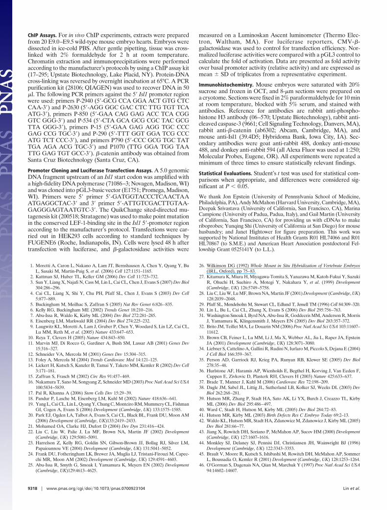
Bioinformatics analysis using standard parameters in rVISTA toexamine potential Isl1 promoter regions revealed two evolutionarilyconserved LEF1 sites 5� of the ATG start site or within intron 1 ofIsl1 genomic sequences. ChIP analysis performed by using extractsfrom E9.5 heart and antibodies to �-catenin demonstrated specificbinding to the 5�-conserved LEF1 response element (Fig. 4A).
To investigate the functional significance of binding, cotransfec-tion assays were performed with a 5-kb Isl1-promoter-luciferasereporter and expression vectors for LEF-1 and activated �-catenin.Results of these assays demonstrated activation of the Isl1-promoterby recruitment of LEF-1 and activated �-catenin (Fig. 4B). Acti-vation was disrupted by mutation of the two conserved LEF-1-binding sites (Fig. 4B), demonstrating that activation by LEF/�-catenin was dependent on the LEF-1 consensus site and that theLEF/�-catenin pathway directly regulates the Isl1 promoter.
Isl1 Protein Is Decreased by E8.0 in Isl1-Cre;�-Catenin Mutants.Ablation of �-catenin by Isl1-Cre dictates that �-catenin ablationoccurs after the expression of Isl1. To investigate whether Isl1expression was affected at early stages in Isl1-Cre;�-catenin mu-tants, we performed immunostaining with Isl1 antibody on sectionsfrom E8.0 embryos (Fig. 4C). Results of this analysis demonstratedsignificantly less Isl1 signal both in pharyngeal mesoderm andpharyngeal endoderm in Isl1-Cre;�-catenin mutants relative to con-trol littermates, demonstrating an early requirement for �-catenin.Isl1/Nkx2.5/flk1 multipotent progenitors reside in the pharyngealmesoderm domain at this stage (1), and Isl1 is reduced in thisdomain in Isl1-Cre;�-catenin mutants.
Proliferation and Apoptosis in Isl1-Cre;�-Catenin Mutants. Smallerbranchial arch derivatives, outflow tract, and right ventricle inIsl1-Cre;�-catenin mutants suggested that proliferation and/or apo-ptosis were altered in mutants. To investigate this issue, we per-formed immunostaining for phosphorylated histone H3 andcleaved activated caspase-3 to investigate proliferation and apopto-sis, respectively.
Results of this analysis demonstrated both a reduction in prolif-eration rate and an increase in apoptosis (Fig. 5). At E9.5 (Fig. 5 Aand B), the proliferation rate in outflow tract myocardium wasreduced from 4.6% in wild type to 2.1% in mutants and in foregutendoderm from 3.9% in wild type to 1.6% in mutants. At E10.5(Fig. 5 C and D), the proliferation rate in outflow tract myocardiumwas reduced from 3.8% in wild type to 1.0% in mutants and inforegut from 3.1% in wild type to 1.5% in mutants. At E10.5 (Fig.5 E–H), significant increases in apoptotic cells were observed inpharyngeal mesoderm, foregut endoderm, and outflow tract, re-gions consistent with those similarly affected in Isl1 or hedgehogsignaling mutants (4, 30–32).
Analysis of Cardiac Neural Crest Cells in Isl1-Cre;�-Catenin Mutants.Because cardiac neural crest cells are required for outflow tractseptation, we examined Isl1-Cre;�-catenin mutants and littermatecontrols for expression of the cardiac neural crest marker, PlexinA2(33). Results demonstrated that PlexinA2-expressing cells wereobserved in the outflow tract of Isl1-Cre;�-catenin mutants, butwere less abundant relative to littermate controls (Fig. 6 A and B).
DiscussionIsl1 plays a pivotal role in the development of cardiac progen-itors of the second heart field, marks postnatal progenitors inpostnatal heart, and marks cardiovascular progenitor cells in theearly embryo, which are pluripotent in vitro (4, 9). Therefore,understanding factors that regulate Isl1 expression is critical tounderstanding factors that drive cardiac progenitor prolifera-tion, survival, and migration, both in the context of normaldevelopment and for potential application to cell therapies usingcardiac progenitors.
Our results have demonstrated that �-catenin directly targets andactivates Isl1 expression, and we have observed a striking corre-
a
b
A B G H K M
I J L N
O
Q R U V Y Z
P S T W X
C
D E F
Fig. 3. Analysis of potential downstream effector targets in Isl1-Cre;�-catenin mutants and control littermates. (a) Results from whole-mount RNAin situ hybridization assays. Expression of Isl1 (A–F), Wnt11 (G–J), Pitx2 (K andL), Fgf8 (M and N), Tbx2 (O–R), Tbx3 (S–V), and Shh (W-Z) was examined inIsl1-Cre;�-catenin mutants and littermate (A–F) controls. Isl1 was down-regulated in the pharyngeal region posterior to the heart (arrows). (C and F)Section analysis demonstrated decreased Isl1 expression in the foregutendoderm (arrows) and in splanchnic mesoderm (arrow heads) in Isl1-Cre;�-catenin mutants relative to control littlemates. (G–J) In Isl1-Cre;�-cateninmutants, Wnt11 was down-regulated in outflow tract (arrows). (K and L) Pitx2was down-regulated in outflow tract (arrows) and pharyngeal arches (smallerarrows). (M and N) Expression of Fgf8 was unaffected. (O–R) Expression ofTbx2 was reduced in the outflow tract (smaller arrows) of Isl1-Cre;�-cateninmutants, but was not down-regulated in AV canal (arrowheads) or pharyngealarches (bigger arrows). (S–V) Tbx3 was down-regulated in pharyngeal arches(smaller arrows) and foregut endoderm (arrows). (W–Z) Shh was down-regulated in anterior foregut endoderm (arrows) and posterior foregutendoderm (smaller arrows). (b) Results from real-time quantitative PCR anal-yses. Total RNA was prepared from hearts and pharyngeal arches of E9.5embryos of either control or Isl1-Cre;�-catenin mutants and analyzed byreal-time RT-PCR. The mRNA level of each gene was normalized against themRNA level of hypoxanthine phosphoribosyltransferase. Data obtained fromthree independent experiments are shown as means � SD. Con, Isl1-Cre/�;�-catenin�/f; Mu, Isl1-Cre/�;�-catenin�/f.
Lin et al. PNAS � May 29, 2007 � vol. 104 � no. 22 � 9315
DEV
ELO
PMEN
TAL
BIO
LOG
Y

spondence between active �-catenin signaling and regions of Isl1expression; in progenitors of the second heart field, includingregions harboring Isl1/flk1/Nkx2.5 progenitors (1); in pharyngealendoderm; and in regions within the heart where persistent Isl1expression is observed (3, 9). Isl1-expressing cells isolated frompostnatal hearts are capable of expansion in culture and candifferentiate to functional cardiomyocytes. Isl1/f lk1/Nkx2.5-expressing cells clonally isolated from embryonic stem cells orembryos can be amplified and give rise to endothelial, smoothmuscle, or cardiac lineages. Factors that are required upstream of
Isl1 for proliferation, however, have not been defined. Our resultsdemonstrate a critical role for �-catenin in proliferation of Isl1-expressing progenitors.
Our results demonstrate a key role for �-catenin in outflow tractmorphogenesis, in formation of the atrial septum, and in atrioven-tricular cushion formation. With regard to the latter, our findingsare consistent with the expression of Isl1 in endocardial cells thatcontribute to cushion formation and previous data which demon-strated that ablation of �-catenin in endothelial cells disruptsepithelial-mesenchymal transformation and cushion formation(34–36). Requirements for �-catenin in cushion formation andseptation have also been demonstrated in chick and zebrafish (37).The observed requirement for �-catenin in atrial septation isconsistent with down-regulation of Isl1, Shh, and Pitx2 becausemutants affecting expression of these genes also have defects inatrial septation (30, 31, 38). The atrial septal defects in theIsl1-Cre;�-catenin mutant, however, appear to be quite distinctive,with two truncated septal primordia observed in the dorsal aspectof the atrial wall.
Decreased proliferation within the secondary/anterior heart fieldhas been demonstrated to result in outflow tract defects (39, 40).Because Isl1 expression is down-regulated in �-catenin mutants andbecause Isl1 is required for proliferation within the secondary/
A
B C a
b
Fig. 4. �-catenin acts at early stages of cardiogenesis to directly regulate Isl1 expression. (A) ChIP assay on extracts from E8.5–E9.0 hearts showing in vivorecruitment of �-catenin to the Isl1 5� promoter with conserved LEF-1-binding sites (lane 1, primers P-2940 and P-2630). ChIP analysis with control primers againstdistinct promoter regions revealed no recruitment of �-catenin (��-Cat, lanes 2, 3, and 4) (see Material and Methods for primer sequences). No recruitment wasobserved with IgG control. (B) Results of luciferase reporter assays. HEK293 cells were transiently transfected with Isl1 promoter-luciferase reporter (Isl1-Luc)constructs in combination with either control expression vector, LEF-1, or constitutively active �-catenin (a�-Cat) expression constructs. Isl1p-Luc, wild-type isl1promoter-luciferase construct; Isl1p (Mu)-Luc, Isl1 promoter-luciferase construct with mutated LEF-1 consensus sites. Data obtained from three independentexperiments, each performed in triplicate. Data are presented as fold activity over basal promoter activity (relative activity) and are expressed as mean � SD oftriplicates from a representative experiment. (C) Immunohistochemical analysis with anti-Isl1 antibody on sections from E8.0 embryos demonstrated reduced Isl1protein levels within Isl1� cardiovascular progenitors (arrows) and endoderm (arrowheads) in Isl1-Cre;�-catenin mutants when compared with controllittermates. Immunostaining analysis was performed in parallel on multiple experimental and control samples, and exposure times were the same for all samples.
A
B
C
D
E
F
G
H
Fig. 5. �-catenin is required for proliferation and survival of cardiac pro-genitors. (A–D) Proliferation was assessed by antibody staining for phosphor-ylated histone H3 (PHH3). Proliferation rate was significantly decreased in theforegut endoderm and myocardium of outflow tract in Isl1-Cre;�-cateninmutants. (E–H) Cleaved caspase-3 antibody staining showed increased apo-ptosis in the pharyngeal mesoderm, foregut endoderm, and outflow tract atE10.5 in Isl1-Cre;�-catenin mutants relative to control littermates. Arrows inA–D indicate proliferating cells. Arrows in E–H indicate apoptotic cells.
Fig. 6. �-catenin is not required for the migration of cardiac neural crestcells. (A and B) Whole-mount RNA in situ analysis was performed for E10.5Isl1-Cre;�-catenin mutants and littermate controls using a marker specific forcardiac neural crest cells, PlexinA2. Results demonstrated that PlexinA2-expressing cells were observed in the outflow tract of Isl1-Cre;�-cateninmutants, but were less abundant relative to littermate controls (arrows).
9316 � www.pnas.org�cgi�doi�10.1073�pnas.0700923104 Lin et al.

anterior heart field (4), it is likely that decreased Isl1 expression inthe secondary/anterior heart field and its derivatives, as observedhere, are contributing to observed outflow tract defects. Indeed, wehave observed similar cardiac phenotypes to those of the Isl1-Cre;�-catenin mutants in hypomorphic mutants of Isl1 (Y. Sun andS.M.E., unpublished data).
Cardiac neural crest cells are also required for outflow tractmorphogenesis and can affect proliferation of the secondary heartfield (41, 42). We have not observed expression of Isl1 in Wnt1-Cre;R26R-lacZ lineage-traced cardiac neural crest cells within theoutflow tract, although Isl1 is expressed within intrinsic cardiacganglia, which derive from the cardiac neural crest (3, 39–41, 43).From this observation, and a comparison of results with Wnt1-Creand Isl1-Cre (Y. Sun and S.M.E., unpublished data), it is unlikelythat Isl1 lineages contribute to the cardiac neural crest populationthat will contribute smooth muscle cells to the outflow tract, aorta,or pulmonary artery.
From the foregoing, we consider that the outflow tract pheno-type of Isl1-Cre;�-catenin mutants is unlikely to be owing to ablationof �-catenin within cardiac neural crest that will directly contributecells to the aorta and pulmonary artery. However, it is possible thatablation of �-catenin within Isl1-expressing domains secondarilyaffects this population of cardiac neural crest cells and in thismanner contributes to observed outflow tract phenotypes. Consis-tent with the latter possibility, we observed decreased levels ofPlexinA2-expressing cells (33) within the outflow tract of Isl1-Cre;�-catenin mutants relative to littermate controls.
In addition to decreased expression of Isl1 in Isl1-Cre;�-catenin mutants, we also observed down-regulation of a num-ber of other genes required for outflow tract morphogenesis,including Shh, Wnt11, Pitx2, Tbx2, and Tbx3 (refs. 19, 22–25, 27,and 28; L.L. and S.M.E., unpublished data). Selective decreaseswere observed in Tbx3 expression within the mandibular archand aortic arch regions, and Tbx2 expression in the outflow tractof Isl1-Cre;�-catenin mutants. Both Tbx2 and Tbx3 are requiredfor outflow tract formation (23) (V. Papaioannou and R. Kelly,personal communication).
Our data do not address whether �-catenin is required within thepharyngeal endoderm, cardiac mesoderm, or both, for cardiogen-esis. We observe active �-catenin signaling in both compartments,suggesting a potential role in both. Previous ablation of �-cateninwith a Cytokeratin-19 promoter-driven Cre resulted in ectopic heartformation, which was attributed to ablation of �-catenin in thepharyngeal endoderm (14). Despite ablation of �-catenin signalingin early ventral pharyngeal endoderm by Isl1-Cre, we did notobserve any ectopic heart formation. The Cytokeratin-19 promotermay be expressed earlier than Isl1 in ventral endoderm or anexpression domain of the Cytokeratin-19 promoter, which is distinctfrom that of Isl1 and is responsible for the observed phenotype.
We found that �-catenin was required within the Isl1 domain forbranchial and aortic arch artery formation. In Isl1-Cre;�-cateninmutants, the mandibular arch was severely hypomorphic, as wereleft aortic arch arteries, and the fourth PAA was absent. A similarphenotype is observed in Isl1 hypomorphs and mutants of the Shhpathway (30, 31). We have previously demonstrated that expressionof Shh in pharyngeal endoderm is downstream of Isl1. Similarapoptotic profiles are observed in Shh and Isl1-Cre;smoothenedmutants (30, 31), and they are described here in Isl1-Cre;�-cateninmutants. Together these data suggest a genetic cascade with�-catenin upstream of Isl1 and Isl1 upstream of Shh for pharyngealarch development. �-catenin may also regulate Shh independentlyof Isl1. Whether these regulatory interactions are direct or indirectremains to be examined.
We have previously shown that Shh is downstream of Isl1 ina signaling pathway required for outflow tract morphogenesis(30). Results of this study demonstrate that �-catenin is a keyupstream factor that drives expression of Isl1 in cardiac progen-itors and their proliferation and survival, and it is also upstream
in genetic cascades that regulate diverse aspects of pharyngealarch and cardiac morphogenesis (Fig. 7). Wnt ligands, whichregulate this pathway, are currently unknown. Wnt2 is expressedin the cardiogenic crescent (44) and may act redundantly withother Wnts upstream of �-catenin in this context.
Materials and MethodsMice. Floxed �-catenin mice were obtained from the laboratory ofRolf Kemler (45). Isl1-Cre mice were created in our laboratory bya Cre knockin into the endogenous Isl1 locus, replacing theendogenous Isl1 ATG (19, 20). Homozygous floxed �-catenin micewere crossed with Protamine-Cre mice (46) to generate �-catenin�/� mice, which were then crossed with Isl1-Cre mice to producedoubly heterozygous Isl1-Cre;�-catenin �/� mice. These mice werethen crossed to �-catenin floxed/floxed homozygous mice to obtainIsl1-Cre;�-catenin�/f mutants for analysis.
Whole-Mount RNA in Situ Hybridization and Histological Analyses.Whole-mount RNA in situ hybridization was carried out as previ-ously described (26). References for specific RNA in situ probes areas follow: Isl1 (EST; GenBank accession no. AA198791), Tbx2,Tbx3, and Pitx2 were from Marina Campione; Wnt11 was fromAndy McMahon; PlexinA2 was from Jon Epstein; Fgf8 was fromGail Martin; and Shh was from Deepak Srivastava. For histologicalanalyses, embryos were fixed in 4% paraformaldehyde, dehydratedin ethanol, embedded in paraffin, 8-�m sections prepared on amicrotome, and stained with H&E according to standard protocols.For ink injection, embryos were collected and injected intracardi-ally with India ink. All experiments were repeated a minimum ofthree times to ensure statistically relevant findings.
RNA Isolation and Real-Time Quantitative PCR Analyses. Total RNAwas isolated from hearts and pharyngeal arches of E9.5 embryos byan RNeasy kit (QIAGEN, Valencia, CA). Semiquantitative PCRwas carried out according to the Mx3000P Real-Time PCR Systemsmanual and the Brilliant qPCR reagent (Stratagene, La Jolla, CA).The mRNA levels of Pitx2, Wnt11, Shh, Isl1, Tbx3, and Tbx2 werenormalized to the mRNA levels of hypoxanthine phosphoribosyl-transferase to allow comparisons among different experimentalgroups. Primer sequences are available on request.
X-Galactosidase Staining of Mouse Embryos. Mouse embryos werefixed in 4% paraformaldehyde for 30 min on ice, permeabilized inPBS containing 0.02% Nadeoxycholate and 0.01% Nonidet P-40for 4 h at room temperature, and then subjected to X-gal stainingfor �1–2 h.
Fig. 7. �-catenin is required for Isl1 expression in cardiovascular progenitors.�-catenin directly regulates Isl1, which is required for survival and prolifera-tion of Isl1� cardiovascular progenitors. �-catenin is upstream of multiplegenes, which are required for pharyngeal arch and cardiac morphogenesis.Isl1 is upstream of Shh, but �-catenin may also regulate Shh independently ofits regulation of Isl1. Direct regulation is indicated by solid arrow, and director indirect regulation is indicated by dotted arrows.
Lin et al. PNAS � May 29, 2007 � vol. 104 � no. 22 � 9317
DEV
ELO
PMEN
TAL
BIO
LOG
Y
ChIP Assays. For in vivo ChIP experiments, extracts were preparedfrom 20 E9.0–E9.5 wild-type mouse embryo hearts. Embryos weredissected in ice-cold PBS. After gentle pipetting, tissue was cross-linked with 2% formaldehyde for 2 h at room temperature.Chromatin extraction and immunoprecipitations were performedaccording to the manufacturer’s protocols by using a ChIP assay kit(17–295; Upstate Biotechnology, Lake Placid, NY). Protein-DNAcross-linking was reversed by overnight incubation at 65°C. A PCRpurification kit (28106; QIAGEN) was used to recover DNA in 50�l. The following PCR primers against the 5� Isl1 promoter regionwere used: primers P-2940 (5�-GCG CCA GGA ACT GTG CTCCAA-3�) and P-2630 (5�-AGG GGC GAC CTC TTG TGT TCAATG-3�), primers P-850 (5�-GAA CAG GAG ACC TCA CGGGTC GGG-3�) and P-534 (5�-CTA GCA GCG CGC TAC GCGTTA GGG-3�), primers P-15 (5�-GAA GAG AGG TGC CCCGAG CCG TGC-3�) and P-290 (5�-TTT GGT GGA TCG CCCATG TCT CCC-3�), and primers P790 (5�-CCC GCG TGC TATTGA AGA ACG TGC-3�) and P1070 (TTG GGA TGG TAATTG GAG TGT GCC-3�). �-catenin antibody was obtained fromSanta Cruz Biotechnology (Santa Cruz, CA).
Promoter Cloning and Luciferase Transfection Assays. A 5.0 genomicDNA fragment upstream of an Isl1 start codon was amplified witha high-fidelity DNA polymerase (71086–3; Novagen, Madison, WI)and was cloned into pGL3-basic vector (E1751; Promega, Madison,WI). Primers were 5� primer 5�-GATGGTACCCTCAACTAAATGAGGCTAC-3� and 3� primer 5�-ATTGTCGACTTGTAA-GAGGGAGTAATGTC-3�. The QuikChange sited-directed mu-tagenesis kit (200518; Stratagene) was used to make point mutationin the conserved LEF-1-binding site in the Isl1 5�-promoter regionaccording to the manufacturer’s protocol. Transfections were car-ried out in HEK293 cells according to standard techniques byFUGENE6 (Roche, Indianapolis, IN). Cells were lysed 48 h aftertransfection with luciferase, and �-galactosidase activities were
measured on a Luminoskan Ascent luminometer (Thermo Elec-tron, Waltham, MA). For luciferase reporters, CMV-�-galactosidase was used to control for transfection efficiency. Nor-malized luciferase activities were compared with a pGL3 control tocalculate the fold of activation. Data are presented as fold activityover basal promoter activity (relative activity) and are expressed asmean � SD of triplicates from a representative experiment.
Immunohistochemistry. Mouse embryos were saturated with 20%sucrose and frozen in OCT, and 8-�m sections were prepared ona cryotome. Sections were fixed in 2% paraformaldehyde for 10 minat room temperature, blocked with 5% serum, and stained withantibodies. Reference for antibodies are rabbit anti-phospho-histone H3 antibody (06–570; Upstate Biotechnology), rabbit anti-cleaved caspase-3 (9661; Cell Signaling Technology, Danvers, MA),rabbit anti-�-catenin (ab6302; Abcam, Cambridge, MA), andmouse anti-Isl1 (39.4D5; Hybridoma Bank, Iowa City, IA). Sec-ondary antibodies were goat anti-rabbit 488, donkey anti-mouse488, and donkey anti-rabbit 594 (all Alexa Fluor was used at 1:250;Molecular Probes, Eugene, OR). All experiments were repeated aminimum of three times to ensure statistically relevant findings.
Statistical Evaluations. Student’s t test was used for statistical com-parisons when appropriate, and differences were considered sig-nificant at P � 0.05.
We thank Jon Epstein (University of Pennsylvania School of Medicine,Philadelphia, PA), Andy McMahon (Harvard University, Cambridge, MA),Deepak Srivastava (University of California, San Francisco, CA), MarinaCampione (University of Padua, Padua, Italy), and Gail Martin (Universityof California, San Francisco, CA) for providing us with cDNAs to makeriboprobes; Yunqing Shi (University of California at San Diego) for mousehusbandry; and Janet Hightower for figure preparation. This work wassupported by National Institutes of Health Grants R01 HL74066 and R01HL70867 (to S.M.E.) and American Heart Association postdoctoral Fel-lowship Grant 0525141Y (to L.L.).
1. Moretti A, Caron L, Nakano A, Lam JT, Bernshausen A, Chen Y, Qyang Y, BuL, Sasaki M, Martin-Puig S, et al. (2006) Cell 127:1151–1165.
2. Kattman SJ, Huber TL, Keller GM (2006) Dev Cell 11:723–732.3. Sun Y, Liang X, Najafi N, Cass M, Lin L, Cai CL, Chen J, Evans S (2007) Dev Biol
304:286–296.4. Cai CL, Liang X, Shi Y, Chu PH, Pfaff SL, Chen J, Evans S (2003) Dev Cell
5:877–889.5. Buckingham M, Meilhac S, Zaffran S (2005) Nat Rev Genet 6:826–835.6. Kelly RG, Buckingham ME (2002) Trends Genet 18:210–216.7. Abu-Issa R, Waldo K, Kirby ML (2004) Dev Biol 272:281–285.8. Eisenberg LM, Markwald RR (2004) Dev Biol 274:225–232.9. Laugwitz KL, Moretti A, Lam J, Gruber P, Chen Y, Woodard S, Lin LZ, Cai CL,
Lu MM, Reth M, et al. (2005) Nature 433:647–653.10. Reya T, Clevers H (2005) Nature 434:843–850.11. Marvin MJ, Di Rocco G, Gardiner A, Bush SM, Lassar AB (2001) Genes Dev
15:316–327.12. Schneider VA, Mercola M (2001) Genes Dev 15:304–315.13. Foley A, Mercola M (2004) Trends Cardiovasc Med 14:121–125.14. Lickert H, Kutsch S, Kanzler B, Tamai Y, Taketo MM, Kemler R (2002) Dev Cell
3:171–181.15. Zaffran S, Frasch M (2002) Circ Res 91:457–469.16. Nakamura T, Sano M, Songyang Z, Schneider MD (2003) Proc Natl Acad Sci USA
100:5834–5839.17. Pal R, Khanna A (2006) Stem Cells Dev 15:29–39.18. Pandur P, Lasche M, Eisenberg LM, Kuhl M (2002) Nature 418:636–641.19. Yang L, Cai CL, Lin L, Qyang Y, Chung C, Monteiro RM, Mummery CL, Fishman
GI, Cogen A, Evans S (2006) Development (Cambridge, UK) 133:1575–1585.20. Park EJ, Ogden LA, Talbot A, Evans S, Cai CL, Black BL, Frank DU, Moon AM
(2006) Development (Cambridge, UK)133:2419–2433.21. Mohamed OA, Clarke HJ, Dufort D (2004) Dev Dyn 231:416–424.22. Liu C, Liu W, Palie J, Lu MF, Brown NA, Martin JF (2002) Development
(Cambridge, UK) 129:5081–5091.23. Harrelson Z, Kelly RG, Goldin SN, Gibson-Brown JJ, Bollag RJ, Silver LM,
Papaioannou VE (2004) Development (Cambridge, UK) 131:5041–5052.24. Frank DU, Fotheringham LK, Brewer JA, Muglia LJ, Tristani-Firouzi M, Capec-
chi MR, Moon AM (2002) Development (Cambridge, UK) 129:4591–4603.25. Abu-Issa R, Smyth G, Smoak I, Yamamura K, Meyers EN (2002) Development
(Cambridge, UK)129:4613–4625.
26. Wilkinson DG (1992) Whole Mount in Situ Hybridization of Vertebrate Embryos(IRL, Oxford), pp 75–83.
27. Kitamura K, Miura H, Miyagawa-Tomita S, Yanazawa M, Katoh-Fukui Y, SuzukiR, Ohuchi H, Suehiro A, Motegi Y, Nakahara Y, et al. (1999) Development(Cambridge, UK) 126:5749–5758.
28. Liu C, Liu W, Lu MF, Brown NA, Martin JF (2001) Development (Cambridge, UK)128:2039–2048.
29. Pfaff SL, Mendelsohn M, Stewart CL, Edlund T, Jessell TM (1996) Cell 84:309–320.30. Lin L, Bu L, Cai CL, Zhang X, Evans S (2006) Dev Biol 295:756–763.31. Washington Smoak I, Byrd NA, Abu-Issa R, Goddeeris MM, Anderson R, Morris
J, Yamamura K, Klingensmith J, Meyers EN (2005) Dev Biol 283:357–372.32. Brito JM, Teillet MA, Le Douarin NM (2006) Proc Natl Acad Sci USA 103:11607–
11612.33. Brown CB, Feiner L, Lu MM, Li J, Ma X, Webber AL, Jia L, Raper JA, Epstein
JA (2001) Development (Cambridge, UK) 128:3071–3080.34. Liebner S, Cattelino A, Gallini R, Rudini N, Iurlaro M, Piccolo S, Dejana E (2004)
J Cell Biol 166:359–367.35. Person AD, Garriock RJ, Krieg PA, Runyan RB, Klewer SE (2005) Dev Biol
278:35–48.36. Hurlstone AF, Haramis AP, Wienholds E, Begthel H, Korving J, Van Eeden F,
Cuppen E, Zivkovic D, Plasterk RH, Clevers H (2003) Nature 425:633–637.37. Brade T, Manner J, Kuhl M (2006) Cardiovasc Res 72:198–209.38. Dagle JM, Sabel JL, Littig JL, Sutherland LB, Kolker SJ, Weeks DL (2003) Dev
Biol 262:268–281.39. Hutson MR, Zhang P, Stadt HA, Sato AK, Li YX, Burch J, Creazzo TL, Kirby
ML (2006) Dev Biol 295:486–497.40. Ward C, Stadt H, Hutson M, Kirby ML (2005) Dev Biol 284:72–83.41. Hutson MR, Kirby ML (2003) Birth Defects Res C Embryo Today 69:2–13.42. Waldo KL, Hutson MR, Stadt HA, Zdanowicz M, Zdanowicz J, Kirby ML (2005)
Dev Biol 281:66–77.43. Jiang X, Rowitch DH, Soriano P, McMahon AP, Sucov HM (2000) Development
(Cambridge, UK) 127:1607–1616.44. Monkley SJ, Delaney SJ, Pennisi DJ, Christiansen JH, Wainwright BJ (1996)
Development (Cambridge, UK) 122:3343–3353.45. Brault V, Moore R, Kutsch S, Ishibashi M, Rowitch DH, McMahon AP, Sommer
L, Boussadia O, Kemler R (2001) Development (Cambridge, UK) 128:1253–1264.46. O’Gorman S, Dagenais NA, Qian M, Marchuk Y (1997) Proc Natl Acad Sci USA
94:14602–14607.
9318 � www.pnas.org�cgi�doi�10.1073�pnas.0700923104 Lin et al.