Beneficial effects of high dose of L-arginine on airway hyperresponsiveness and airway inflammation...
10
Beneficial effects of high dose of L-arginine on airway hyperresponsiveness and airway inflammation in a murine model of asthma Ulaganathan Mabalirajan, MBBS, a Tanveer Ahmad, MSc, a Geeta Devi Leishangthem, MVSc, b Duraisamy Arul Joseph, MSc, a Amit Kumar Dinda, MD, PhD, b Anurag Agrawal, MD, PhD, a and Balaram Ghosh, PhD a Delhi and New Delhi, India Background: Disturbance in the delicate balance between L-arginine–metabolizing enzymes such as nitric oxide synthase (NOS) and arginase may lead to decreased L-arginine availability to constitutive forms of NOS (endothelial NOS), thereby increasing the nitro-oxidative stress and airway hyperresponsiveness (AHR). Objective: In this study, we investigated the effects of high doses of L-arginine on L-arginine–metabolizing enzymes and subsequent biological effects such as cyclic guanosine monophosphate production, lipid peroxidation, peroxynitrite, AHR, and airway inflammation in a murine model of asthma. Methods: Different doses of L-arginine were administered to ovalbumin–sensitized and challenged mice. Exhaled nitric oxide, AHR, airway inflammation, T H 2 cytokines, goblet cell metaplasia, nitro-oxidative stress, and expressions of arginase 1, endothelial NOS, and inducible NOS in lung were determined. Results: L-arginine significantly reduced AHR and airway inflammation including bronchoalveolar lavage fluid eosinophilia, T H 2 cytokines, TGF-b1, goblet cell metaplasia, and subepithelial fibrosis. Further, L-arginine increased ENO levels and cyclic guanosine monophosphate in lung and reduced the markers of nitro-oxidative stress such as nitrotyrosine, 8- isoprostane, and 8-hydroxy-29-deoxyguanosine. This was associated with reduced activity and expression of arginase 1, increased expression of endothelial NOS, and reduction of inducible NOS in bronchial epithelia. Conclusion: We conclude that L-arginine administration may improve disordered nitric oxide metabolism associated with allergic airway inflammation, and alleviates some features of asthma. (J Allergy Clin Immunol 2010;125:626-35.) Key words: Allergic airway inflammation, arginase, L-arginine, endothelial nitric oxide synthase, exhaled nitric oxide The incidence of allergic airway diseases such as asthma is rising in both developed and developing countries. Allergic airway inflammation (AAI) is a central feature of these diseases, and many of the long-term effects have been suggested to be a result of recruited inflammatory cells in the airway. 1 These in- flammatory cells affect the structural cells of the airway such as bronchial epithelia, which orchestrate the airway inflammation by interacting with various foreign proteins. 2 Recently, we have shown that oxidative stress and mitochondrial dysfunction are as- sociated with allergic airway inflammation. 3,4 AAI is character- ized by the infiltration of various inflammatory cells including eosinophils, and increased T H 2 response. In animal models of AAI, allergic sensitization to foreign proteins followed by re- peated allergen exposures also leads to structural changes such as subepithelial airway fibrosis and goblet cell metaplasia. 5 In hu- man AAI, increased exhaled nitric oxide (ENO) has been noted. 6 Although the role of nitric oxide (NO) in allergic diseases includ- ing asthma remains controversial, successful inhibition of airway NO synthesis does not improve asthma. 7 Although various studies have explored the complex interrelationships between the compo- nents of the L-arginine (L-ARG)–NO pathway in the pathogene- sis of allergic inflammatory diseases, it remains poorly understood. The multifunctional properties of NO depend on its enzyme source, substrate availability, microenvironment, and its final products after combination with other biomolecules. 8 NO is produced by NO synthase (NOS), which is of 2 principal types: constitutive (neuronal NOS, endothelial NOS [eNOS]) and induc- ible NOS (iNOS). One of the crucial limiting factors in NO pro- duction is substrate availability. 6 In normal airways, constitutive NOS consumes L-ARG to maintain airway smooth muscle tone through activation of the cyclic guanosine monophosphate From a the Molecular Immunogenetics Laboratory and Centre of Excellence for Transla- tional Research in Asthma and Lung Disease, Institute of Genomics and Integrative Biology, Delhi; and b the Division of Renal Pathology, Department of Pathology, All India Institute of Medical Sciences, New Delhi. Supported by projects NWP0033 and MLP 5501 at the Institute of Genomics and Integrative Biology, Council of Scientific and Industrial Research, Government of India. U.M. and G.D.L. are the recipients of fellowships from the Indian Council of Medical Research. Disclosure of potential conflict of interest: The authors have declared that they have no conflict of interest. Received for publication June 11, 2009; revised October 22, 2009; accepted for publica- tion October 23, 2009. Available online February 12, 2010. Reprint requests: Balaram Ghosh, PhD, Molecular Immunogenetics Laboratory, Institute of Genomics and Integrative Biology, Mall Road, Delhi-110007, India. E-mail: [email protected]. 0091-6749/$36.00 Ó 2010 American Academy of Allergy, Asthma & Immunology doi:10.1016/j.jaci.2009.10.065 Abbreviations used AAI: Allergic airway inflammation ADMA: Asymmetric dimethyl arginine AHR: Airway hyperresponsiveness BAL: Bronchoalveolar lavage cGMP: Cyclic guanosine monophosphate ENO: Exhaled nitric oxide eNOS: Endothelial nitric oxide synthase iNOS: Inducible nitric oxide synthase L-ARG: L-arginine NO: Nitric oxide NOS: Nitric oxide synthase OVA: Ovalbumin Penh: Enhanced pause sGAW: Specific airway conductance VEH: Vehicle 626
-
Upload
independent -
Category
Documents
-
view
2 -
download
0
Transcript of Beneficial effects of high dose of L-arginine on airway hyperresponsiveness and airway inflammation...
Beneficial effects of high dose of L-arginine on airwayhyperresponsiveness and airway inflammation in a murinemodel of asthma
Ulaganathan Mabalirajan, MBBS,a Tanveer Ahmad, MSc,a Geeta Devi Leishangthem, MVSc,b Duraisamy Arul Joseph,
MSc,a Amit Kumar Dinda, MD, PhD,b Anurag Agrawal, MD, PhD,a and Balaram Ghosh, PhDa Delhi and New Delhi, India
Abbreviations used
AAI: Allergic airway inflammation
ADMA: Asymmetric dimethyl arginine
AHR: Airway hyperresponsiveness
BAL: Bronchoalveolar lavage
cGMP: Cyclic guanosine monophosphate
ENO: Exhaled nitric oxide
eNOS: Endothelial nitric oxide synthase
iNOS: Inducible nitric oxide synthase
L-ARG: L-arginine
NO: Nitric oxide
NOS: Nitric oxide synthase
OVA: Ovalbumin
Penh: Enhanced pause
sGAW: Specific airway conductance
VEH: Vehicle
Background: Disturbance in the delicate balance betweenL-arginine–metabolizing enzymes such as nitric oxide synthase(NOS) and arginase may lead to decreased L-arginineavailability to constitutive forms of NOS (endothelial NOS),thereby increasing the nitro-oxidative stress and airwayhyperresponsiveness (AHR).Objective: In this study, we investigated the effects of high dosesof L-arginine on L-arginine–metabolizing enzymes andsubsequent biological effects such as cyclic guanosinemonophosphate production, lipid peroxidation, peroxynitrite,AHR, and airway inflammation in a murine model of asthma.Methods: Different doses of L-arginine were administered toovalbumin–sensitized and challenged mice. Exhaled nitricoxide, AHR, airway inflammation, TH2 cytokines, goblet cellmetaplasia, nitro-oxidative stress, and expressions of arginase 1,endothelial NOS, and inducible NOS in lung were determined.Results: L-arginine significantly reduced AHR and airwayinflammation including bronchoalveolar lavage fluideosinophilia, TH2 cytokines, TGF-b1, goblet cell metaplasia,and subepithelial fibrosis. Further, L-arginine increased ENOlevels and cyclic guanosine monophosphate in lung and reducedthe markers of nitro-oxidative stress such as nitrotyrosine, 8-isoprostane, and 8-hydroxy-29-deoxyguanosine. This wasassociated with reduced activity and expression of arginase 1,increased expression of endothelial NOS, and reduction ofinducible NOS in bronchial epithelia.Conclusion: We conclude that L-arginine administration mayimprove disordered nitric oxide metabolism associated withallergic airway inflammation, and alleviates some features ofasthma. (J Allergy Clin Immunol 2010;125:626-35.)
Key words: Allergic airway inflammation, arginase, L-arginine,endothelial nitric oxide synthase, exhaled nitric oxide
From athe Molecular Immunogenetics Laboratory and Centre of Excellence for Transla-
tional Research in Asthma and Lung Disease, Institute of Genomics and Integrative
Biology, Delhi; and bthe Division of Renal Pathology, Department of Pathology, All
India Institute of Medical Sciences, New Delhi.
Supported by projects NWP0033 and MLP 5501 at the Institute of Genomics and
Integrative Biology, Council of Scientific and Industrial Research, Government of
India. U.M. and G.D.L. are the recipients of fellowships from the Indian Council of
Medical Research.
Disclosure of potential conflict of interest: The authors have declared that they have no
conflict of interest.
Received for publication June 11, 2009; revised October 22, 2009; accepted for publica-
tion October 23, 2009.
Available online February 12, 2010.
Reprint requests: Balaram Ghosh, PhD, Molecular Immunogenetics Laboratory, Institute
of Genomics and Integrative Biology, Mall Road, Delhi-110007, India. E-mail:
0091-6749/$36.00
� 2010 American Academy of Allergy, Asthma & Immunology
doi:10.1016/j.jaci.2009.10.065
626
The incidence of allergic airway diseases such as asthma isrising in both developed and developing countries. Allergicairway inflammation (AAI) is a central feature of these diseases,and many of the long-term effects have been suggested to be aresult of recruited inflammatory cells in the airway.1 These in-flammatory cells affect the structural cells of the airway such asbronchial epithelia, which orchestrate the airway inflammationby interacting with various foreign proteins.2 Recently, we haveshown that oxidative stress and mitochondrial dysfunction are as-sociated with allergic airway inflammation.3,4 AAI is character-ized by the infiltration of various inflammatory cells includingeosinophils, and increased TH2 response. In animal models ofAAI, allergic sensitization to foreign proteins followed by re-peated allergen exposures also leads to structural changes suchas subepithelial airway fibrosis and goblet cell metaplasia.5 In hu-man AAI, increased exhaled nitric oxide (ENO) has been noted.6
Although the role of nitric oxide (NO) in allergic diseases includ-ing asthma remains controversial, successful inhibition of airwayNO synthesis does not improve asthma.7 Although various studieshave explored the complex interrelationships between the compo-nents of the L-arginine (L-ARG)–NO pathway in the pathogene-sis of allergic inflammatory diseases, it remains poorlyunderstood. The multifunctional properties of NO depend on itsenzyme source, substrate availability, microenvironment, and itsfinal products after combination with other biomolecules.8 NOis produced by NO synthase (NOS), which is of 2 principal types:constitutive (neuronal NOS, endothelial NOS [eNOS]) and induc-ible NOS (iNOS). One of the crucial limiting factors in NO pro-duction is substrate availability.6 In normal airways, constitutiveNOS consumes L-ARG to maintain airway smooth muscle tonethrough activation of the cyclic guanosine monophosphate
J ALLERGY CLIN IMMUNOL
VOLUME 125, NUMBER 3
MABALIRAJAN ET AL 627
(cGMP) pathway. It has been hypothesized earlier that exogenousadministration of L-ARG may increase the bronchodilating effectby constitutive forms of NOS.9 Unfortunately, this strategy failedto have a significant effect in human asthma10 and potentiated theairway inflammation in animal models.11 However, it remainspossible that substrate limitations of L-ARG exist in asthmaticairways because of competition by other L-ARG–metabolizingenzymes such as iNOS and arginase, and that the dosage usedby previous studies (50 mg/kg) was insufficient, possibly becausecompetition by other enzymes was not considered. Hence, the po-tentiation of airway inflammation could be a result of consump-tion of both endogenous and exogenous L-ARG by arginaseand iNOS. L-ARG has been used in various clinical conditionsup to 30 g/d without any adverse effects.12 Importantly, in cardi-ovascular diseases, higher doses were required to obtain sufficientvasodilation and other effects.12 Also, it was demonstrated thatdepletion of L-ARG in cytosol triggers superoxide generationin macrophages. This leads to increased formation of peroxyni-trite because of balanced synthesis of both superoxide and NOby iNOS with L-ARG depletion.13 Peroxynitrite is known forits properties of causing bronchoconstriction and airway inflam-mation.14 In this study, we have addressed the possibility thatthe state of allergic airway inflammation may be associatedwith relative L-ARG depletion causing increased nitro-oxidativestress and whether it can be overcome by high doses of L-ARGcomparable to those used in cardiovascular diseases.
METHODS
AnimalsMale BALB/c mice (8-10 weeks old; National Institute of Nutrition,
Hyderabad, India) were acclimatized for a week before starting the experi-
ments. All animals were maintained according to Committee for the Purpose
of Control and Supervision of Experiments on Animals guidelines, and
protocols were approved by the Institutional Animal Ethics Committee.
Grouping of miceIn pilot experiments, mice were divided into 5 groups, and each (n 5 6) was
named according to sensitization/challenge/treatment: SHAM/PBS/vehicle
(VEH; sham controls), ovalbumin (OVA)/OVA/VEH (allergic controls, OVA,
chicken egg OVA, Grade V; Sigma, St Louis, Mo), OVA/OVA/L-ARG 25
(low-dose L-ARG, 25 mg/kg; Sigma, St Louis, Mo), OVA/OVA/L-ARG
250, and OVA/OVA/L-ARG 500 (250 mg/kg and 500 mg/kg, respectively).
L-ARG was soluble in water (vehicle). From these pilot experiments, 250
mg/kg twice daily dose was selected as the most effective dose for further stud-
ies involving NO metabolism, in which there were 3 groups of mice (n 5 6 in
each group): SHAM/PBS/VEH (sham controls), OVA/OVA/VEH (allergic
controls), and OVA/OVA/L-ARG (250 mg/kg L-ARG treated).
Sensitization, challenge, and treatment of miceMice were sensitized and challenged as described previously.3,4 As shown
in Fig 1, mice were injected with 50 mg OVA in 4 mg aluminum hydroxide or
only 4 mg aluminum hydroxide by the intraperitoneal route on days 0, 7, and
14. Mice were challenged with 3% OVA in PBS or PBS from day 21 to day 32
(30 minutes per day). Vehicle or L-ARG was given orally from day 19 to 32
once a day in a 30-mL volume per dose.
Airway hyperresponsiveness measurementAirway hyperresponsiveness (AHR) to methacholine (Sigma) was deter-
mined in unrestrained and restrained conscious mice by single-chamber and
double-chamber whole-body plethysmography, respectively (Models PLY
3211 and PLY 3351; Buxco Electronics) as described previously.3,4 Also, air-
way resistance was estimated by invasive measurements on anesthetized mice
using the flexiVent system (Scireq), which integrates the computer-controlled
mouse ventilator with the measurements of respiratory mechanics as described
previously.15 Because methacholine aerosols have been nebulized directly to
the trachea in airway resistance measurements, the methacholine dose-
response curve for airway resistance might vary from specific airway conduc-
tance (sGAW) obtained from double-chamber plethysmography. Final results
were expressed in enhanced pause (Penh) or sGAW or airway resistance with
increasing concentrations of methacholine.
ENO measurementExhaled NO as a gas was measured as described previously16 by using a
standard clinical ENO analyzer (CLD88sp; Ecomedics, Durnten, Switzerland)
based on photometric determination of chemiluminescence.
Bronchoalveolar lavage and sera separationOn day 33, each mouse was killed bronchoalveolar lavage (BAL) was
performed, BAL fluids were processed,17,18 and absolute cell count for each
cell type was calculated after determining total cell account and differential
cell count. Blood was withdrawn by cardiac puncture, and serum was sepa-
rated as described previously.17,18
Lung histopathologyFormalin-fixed, paraffin-embedded lung tissue sections were stained with
hematoxylin and eosin, periodic acid-Schiff, and Masson Trichrome stainings
to assess the airway inflammation, goblet cell metaplasia, and subepithelial
fibrosis, respectively.3,4 Stained sections were observed, and microphoto-
graphs were taken with a Nikon microscope with a camera (model YS-100).
Inflammation scoring with hematoxylin and eosin–stained slides and quanti-
tative morphometry with periodic acid-Schiff and Masson Trichrome–stained
sections were performed as described previously.3,4
Measurements of IL-4, IL-5, TGF-b1, IL-13, eotaxin
levels in the lung, and OVA-specific
immunoglobulins in seraLung tissue homogenates in duplicate were used for ELISA of IL-4, IL-5,
and TGF-b1 (BD Pharmingen, San Diego, Calif; the lower detection ranges
are 7.5 pg/mL, 15.6 pg/mL, and 125 pg/mL, respectively) and IL-13 and eo-
taxin ELISA (R&D Systems, Minneapolis, Minn; the sensitivity for IL-13 is
1.5 pg/mL, and the lower detection range for eotaxin is 7.5 pg/mL). Results
were expressed in picograms and normalized by protein concentrations.
OVA-specific IgE, IgG1, and IgG2a were measured as described previously.3
Cytosolic separationAfter mice were killed, the lung portion below the trachea was removed and
processed to separate cytosolic fractions as described previously,3 and protein
estimation was done in those fractions by bicinchoninic acid (Sigma) assay.
Arginase activity and Western blotArginase activity was measured in lung cytosolic fractions as per the
manufacturer’s instructions by an indirect method of urea measurement
(Bioassay Systems, Hayward, Calif). Briefly, 25 mg cytosolic fraction protein
in 40 mL volume was mixed with 10 mL substrate buffer containing arginine,
and MnCl2, and the total mixture was incubated at 378C for 2 hours. The re-
action was terminated by addition of urea reagent and further incubated at
room temperature for 15 minutes and read at 430 nm. Negative control wells
were made for every sample in which substrate buffer was not added. Water
and 1 mmol/L urea have been taken as a background and standard,
respectively.
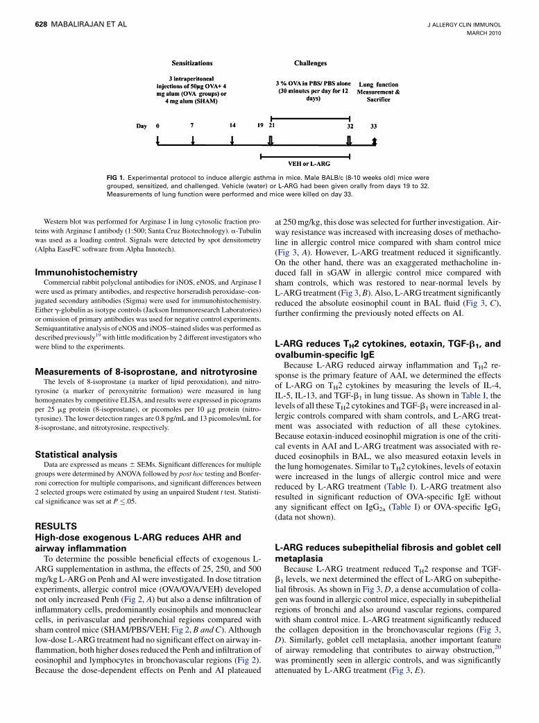
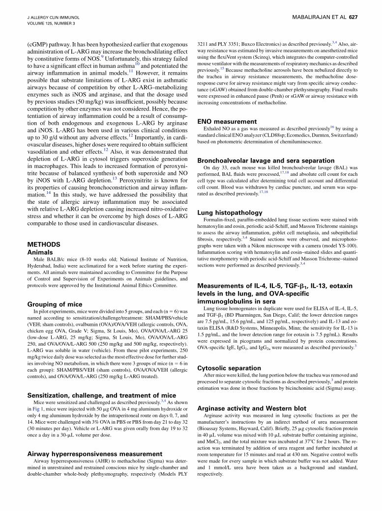
FIG 1. Experimental protocol to induce allergic asthma in mice. Male BALB/c (8-10 weeks old) mice were
grouped, sensitized, and challenged. Vehicle (water) or L-ARG had been given orally from days 19 to 32.
Measurements of lung function were performed and mice were killed on day 33.
J ALLERGY CLIN IMMUNOL
MARCH 2010
628 MABALIRAJAN ET AL
Western blot was performed for Arginase I in lung cytosolic fraction pro-
teins with Arginase I antibody (1:500; Santa Cruz Biotechnology). a-Tubulin
was used as a loading control. Signals were detected by spot densitometry
(Alpha EaseFC software from Alpha Innotech).
ImmunohistochemistryCommercial rabbit polyclonal antibodies for iNOS, eNOS, and Arginase I
were used as primary antibodies, and respective horseradish peroxidase–con-
jugated secondary antibodies (Sigma) were used for immunohistochemistry.
Either g-globulin as isotype controls (Jackson Immunoresearch Laboratories)
or omission of primary antibodies was used for negative control experiments.
Semiquantitative analysis of eNOS and iNOS–stained slides was performed as
described previously19 with little modification by 2 different investigators who
were blind to the experiments.
Measurements of 8-isoprostane, and nitrotyrosineThe levels of 8-isoprostane (a marker of lipid peroxidation), and nitro-
tyrosine (a marker of peroxynitrite formation) were measured in lung
homogenates by competitive ELISA, and results were expressed in picograms
per 25 mg protein (8-isoprostane), or picomoles per 10 mg protein (nitro-
tyrosine). The lower detection ranges are 0.8 pg/mL and 13 picomoles/mL for
8-isoprostane, and nitrotyrosine, respectively.
Statistical analysisData are expressed as means 6 SEMs. Significant differences for multiple
groups were determined by ANOVA followed by post hoc testing and Bonfer-
roni correction for multiple comparisons, and significant differences between
2 selected groups were estimated by using an unpaired Student t test. Statisti-
cal significance was set at P �.05.
RESULTS
High-dose exogenous L-ARG reduces AHR and
airway inflammationTo determine the possible beneficial effects of exogenous L-
ARG supplementation in asthma, the effects of 25, 250, and 500mg/kg L-ARG on Penh and AI were investigated. In dose titrationexperiments, allergic control mice (OVA/OVA/VEH) developednot only increased Penh (Fig 2, A) but also a dense infiltration ofinflammatory cells, predominantly eosinophils and mononuclearcells, in perivascular and peribronchial regions compared withsham control mice (SHAM/PBS/VEH; Fig 2, B and C). Althoughlow-dose L-ARG treatment had no significant effect on airway in-flammation, both higher doses reduced the Penh and infiltration ofeosinophil and lymphocytes in bronchovascular regions (Fig 2).Because the dose-dependent effects on Penh and AI plateaued
at 250 mg/kg, this dose was selected for further investigation. Air-way resistance was increased with increasing doses of methacho-line in allergic control mice compared with sham control mice(Fig 3, A). However, L-ARG treatment reduced it significantly.On the other hand, there was an exaggerated methacholine in-duced fall in sGAW in allergic control mice compared withsham controls, which was restored to near-normal levels byL-ARG treatment (Fig 3, B). Also, L-ARG treatment significantlyreduced the absolute eosinophil count in BAL fluid (Fig 3, C),further confirming the previously noted effects on AI.
L-ARG reduces TH2 cytokines, eotaxin, TGF-b1, and
ovalbumin-specific IgEBecause L-ARG reduced airway inflammation and TH2 re-
sponse is the primary feature of AAI, we determined the effectsof L-ARG on TH2 cytokines by measuring the levels of IL-4,IL-5, IL-13, and TGF-b1 in lung tissue. As shown in Table I, thelevels of all these TH2 cytokines and TGF-b1 were increased in al-lergic controls compared with sham controls, and L-ARG treat-ment was associated with reduction of all these cytokines.Because eotaxin-induced eosinophil migration is one of the criti-cal events in AAI and L-ARG treatment was associated with re-duced eosinophils in BAL, we also measured eotaxin levels inthe lung homogenates. Similar to TH2 cytokines, levels of eotaxinwere increased in the lungs of allergic control mice and werereduced by L-ARG treatment (Table I). L-ARG treatment alsoresulted in significant reduction of OVA-specific IgE withoutany significant effect on IgG2a (Table I) or OVA-specific IgG1
(data not shown).
L-ARG reduces subepithelial fibrosis and goblet cell
metaplasiaBecause L-ARG treatment reduced TH2 response and TGF-
b1 levels, we next determined the effect of L-ARG on subepithe-lial fibrosis. As shown in Fig 3, D, a dense accumulation of colla-gen was found in allergic control mice, especially in subepithelialregions of bronchi and also around vascular regions, comparedwith sham control mice. L-ARG treatment significantly reducedthe collagen deposition in the bronchovascular regions (Fig 3,D). Similarly, goblet cell metaplasia, another important featureof airway remodeling that contributes to airway obstruction,20
was prominently seen in allergic controls, and was significantlyattenuated by L-ARG treatment (Fig 3, E).
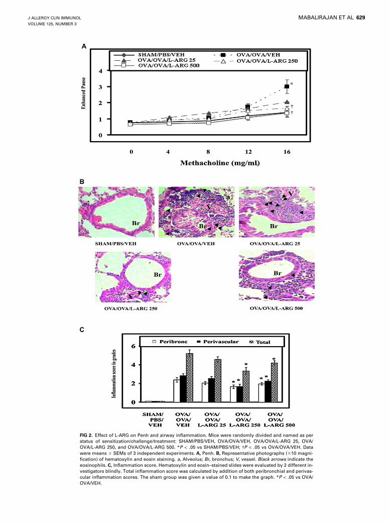
FIG 2. Effect of L-ARG on Penh and airway inflammation. Mice were randomly divided and named as per
status of sensitization/challenge/treatment: SHAM/PBS/VEH, OVA/OVA/VEH, OVA/OVA/L-ARG 25, OVA/
OVA/L-ARG 250, and OVA/OVA/L-ARG 500. *P < .05 vs SHAM/PBS/VEH; �P < .05 vs OVA/OVA/VEH. Data
were means 6 SEMs of 3 independent experiments. A, Penh. B, Representative photographs (310 magni-
fication) of hematoxylin and eosin staining. a, Alveolus; Br, bronchus; V, vessel. Black arrows indicate the
eosinophils. C, Inflammation score. Hematoxylin and eosin–stained slides were evaluated by 2 different in-
vestigators blindly. Total inflammation score was calculated by addition of both peribronchial and perivas-
cular inflammation scores. The sham group was given a value of 0.1 to make the graph. *P < .05 vs OVA/
OVA/VEH.
J ALLERGY CLIN IMMUNOL
VOLUME 125, NUMBER 3
MABALIRAJAN ET AL 629
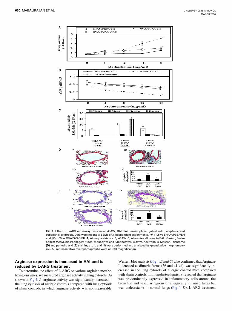
FIG 3. Effect of L-ARG on airway resistance, sGAW, BAL fluid eosinophilia, goblet cell metaplasia, and
subepithelial fibrosis. Data were means 6 SEMs of 3 independent experiments. *P < .05 vs SHAM/PBS/VEH
and �P < .05 vs OVA/OVA/VEH. A, Airway resistance. B, sGAW. C, Absolute cell types in BAL. Eosino, Eosin-
ophils; Macro, macrophages; Mono, monocytes and lymphocytes; Neutro, neutrophils. Masson Trichrome
(D) and periodic acid (E) stainings (i, ii, and iii) were performed and analyzed by quantitative morphometry
(iv). All representative microphotographs were at 310 magnification.
J ALLERGY CLIN IMMUNOL
MARCH 2010
630 MABALIRAJAN ET AL
Arginase expression is increased in AAI and is
reduced by L-ARG treatmentTo determine the effect of L-ARG on various arginine metabo-
lizing enzymes, we measured arginase activity in lung cytosols. Asshown in Fig 4, A, arginase activity was significantly increased inthe lung cytosols of allergic controls compared with lung cytosolsof sham controls, in which arginase activity was not measurable.
Western blot analysis (Fig 4, B and C) also confirmed that ArginaseI, detected as dimeric forms (36 and 41 kd), was significantly in-creased in the lung cytosols of allergic control mice comparedwith sham controls. Immunohistochemistry revealed that arginasewas predominantly expressed in inflammatory cells around thebronchial and vascular regions of allergically inflamed lungs butwas undetectable in normal lungs (Fig 4, D). L-ARG treatment

TABLE I. Effects of L-ARG on IL-4, IL-5, IL-13, OVA-specific IgE, IgG2a, eotaxin, and TGF-b1 levels
Groups
IL-4 (pg/100
mg protein)
IL-5 (pg/100
mg protein)
IL-13 (pg/100
mg protein)
OVA Specific
IgE (A.U.)
OVA Specific
IgG2a (A.U.)
Eotaxin
(pg/25 mg protein)
TGF-b1
(pg/50 mg
protein)
SHAM/PBS/VEH 63.3 6 7.1 27.9 6 10.4 5.2 6 1.1 0.8 6 0.1 1.3 6 0.1 13.7 6 0.7 318.5 6 57.0
OVA/OVA/VEH 97.0 6 3.3* 63.2 6 7.0* 35.5 6 12.8* 1.9 6 0.1* 2.6 6 0.7* 38.9 6 2.4* 521.8 6 63.4*
OVA/OVA/L-ARG 75.2 6 7.9� 30.7 6 10.8� 5.3 6 4.0� 1.3 6 0.1� 2.4 6 0.4 24.0 6 3.7� 356.4 6 33.9�
A.U., Arbitrary Units.
*P < .05 vs SHAM/PBS/VEH.
�P < .05 vs OVA/OVA/VEH.
J ALLERGY CLIN IMMUNOL
VOLUME 125, NUMBER 3
MABALIRAJAN ET AL 631
significantly reduced the activity and expression of Arginase I com-pared with allergic control mice (Fig 4, A-D). As mentioned, L-ARG treated mice had significantly less inflammation (Fig 2).
L-ARG enhances ENO and cGMP but reduces
peroxynitriteTo determine the effect of L-ARG treatment on NO metabo-
lism, we measured ENO as a marker of NO synthesis, cGMP as amarker of guanylyl cyclase activation by NO, and nitrotyrosine asa marker of peroxynitrite generation. Mice with AAI hadincreased ENO but lower cGMP than sham controls (Fig 5, Aand B). This was further associated with increased nitro-oxidativestress as shown by significantly increased 8-isoprostane and nitro-tyrosine, which represent lipid peroxidation and peroxynitrite, re-spectively (Fig 5, C and D). L-ARG treatment was associatedwith significantly greater ENO than either sham control mice orallergically inflamed mice (Fig 5, A). As shown in Fig 5, B,cGMP levels were significantly increased in L-ARG–treatedmice compared with mice with allergic inflammation but similarto sham controls. Importantly, despite even greater levels of ENOin L-ARG–treated mice, oxidative stress markers were lower thanallergic control mice, and similar (8-isoprostane) or reduced(nitrotyrosine) compared with the sham controls.
L-ARG enhances eNOS expression and reduces
iNOSTo investigate whether different sources of NO could be
responsible for the differences in cellular reactive nitrogenspecies, we studied the NOS expression patterns by immunohis-tochemistry in lung tissue sections. Sham control mice showedmild expression of eNOS in bronchial epithelial cells (Fig 6, A)and no expression of iNOS (Fig 6, B). Semiquantitative analysisscores correlating to stain intensity for sham control mice were4.1 6 0.85 for eNOS and 0.23 6 0.07 for iNOS (means 6
SEMs, arbitrary units). In contrast, mice with AAI showed a sig-nificant high expression of iNOS in inflammatory cells such asmacrophages and eosinophils and bronchial epithelia (Fig 6, B).Semiquantitative analysis scores were 4.4 6 0.4 for eNOS and3.1 6 0.5 for iNOS. However, L-ARG–treated allergic miceshowed a significantly greater expression of eNOS in bronchialepithelia compared with either sham controls or mice with AAIand reduced expression of iNOS compared with mice with AAI.Semiquantitative analysis scores were 7.2 6 0.3 for eNOS and1.5 6 0.3 for iNOS (P < .05 compared with allergic controlsfor both eNOS and iNOS). Thus, the major source for NO synthe-sis was iNOS in allergically inflamed lungs and eNOS in normaland L-ARG–treated lungs.
DISCUSSIONL-arginine is a conditionally essential basic amino acid, and
human beings normally consume 3.5 to 5 g daily.21 It has multiplepharmacologic properties (antioxidant, anti-inflammatory, anti-hypercholesterolemic, muscle relaxant, immunomodulatory,and so forth).12 These widespread activities could be a result ofits effect on various metabolizing enzymes and their final metab-olites.22 The use of L-ARG has been reported for various diseaseconditions (angina pectoris, congestive heart failure, hyperten-sion, intermittent claudication, pre-eclampsia, AIDS, burns, can-cer, diabetes, gastritis, male and female infertility, seniledementia, and so forth).12 Because most of the L-ARG–metabo-lizing enzymes are also present in lung, various studies have beenperformed to understand pulmonary arginine metabolism.23 A re-cent study reported that L-ARG bioavailability is strongly associ-ated with airflow obstructions in human beings.24 Because dietaryintake is the primary determinant of plasma arginine levels, die-tary supplementation or oral administration has been tried as a po-tential therapy for asthma at doses up to 50 mg/kg. Although thesestudies have found L-ARG to be ineffective in the treatment ofasthma, there have been no studies at higher doses that are shownto be safe and effective in other diseases.25 In this study, we reportfor the first time the beneficial effects of a 250 mg/kg dose of L-ARG in a murine model of allergic airway inflammation andAHR. Consistent with previous studies, these effects were notseen at a lower dose (25 mg/kg). At this dose (25 mg/kg), therewere no significant effects on the expressions of eNOS, iNOS,and arginase and ENO levels (data not shown). These results in-dicate that exogenous arginine at low doses may not be sufficientto increase the substrate availability of eNOS.
Importantly, we found that L-ARG administration profoundlyenhances eNOS expression in bronchial epithelia (Fig 6, A) andincreases the levels of ENO (Fig 5, A). It is known that L-ARGsupplementation increases the transcription of eNOS.26 A simul-taneous downregulation of iNOS and arginase noted in our studyindicates that the administered L-ARG was predominantly usedby eNOS, as evidenced by the increased ENO levels, increasedguanylyl cyclase activation, and low reactive nitrogen species.In contrast, mice with allergic airway inflammation that alsohad high levels of ENO probably had iNOS as the source, becauseeNOS expression was low. Further, the high arginase levels insuch mice may lead to generation of L-ornithine, which is a pre-cursor for polyamines and proline that may induce structuralchanges in the airway such as subepithelial fibrosis.27 Taken to-gether, downregulations of both iNOS and arginase and upregula-tion of eNOS by L-ARG treatment could be responsible for thereduction of airway inflammation and AHR (Figs 2 and 3). Tosupport this view, eNOS overexpression or arginase inhibition al-leviated the features of asthma in similar models.28,29 Most of the
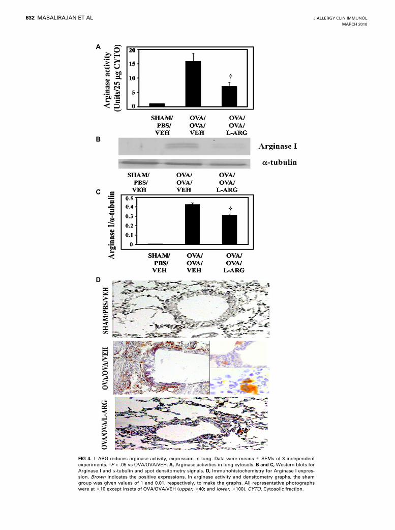
FIG 4. L-ARG reduces arginase activity, expression in lung. Data were means 6 SEMs of 3 independent
experiments. �P < .05 vs OVA/OVA/VEH. A, Arginase activities in lung cytosols. B and C, Western blots for
Arginase I and a-tubulin and spot densitometry signals. D, Immunohistochemistry for Arginase I expres-
sion. Brown indicates the positive expressions. In arginase activity and densitometry graphs, the sham
group was given values of 1 and 0.01, respectively, to make the graphs. All representative photographs
were at 310 except insets of OVA/OVA/VEH (upper, 340; and lower, 3100). CYTO, Cytosolic fraction.
J ALLERGY CLIN IMMUNOL
MARCH 2010
632 MABALIRAJAN ET AL
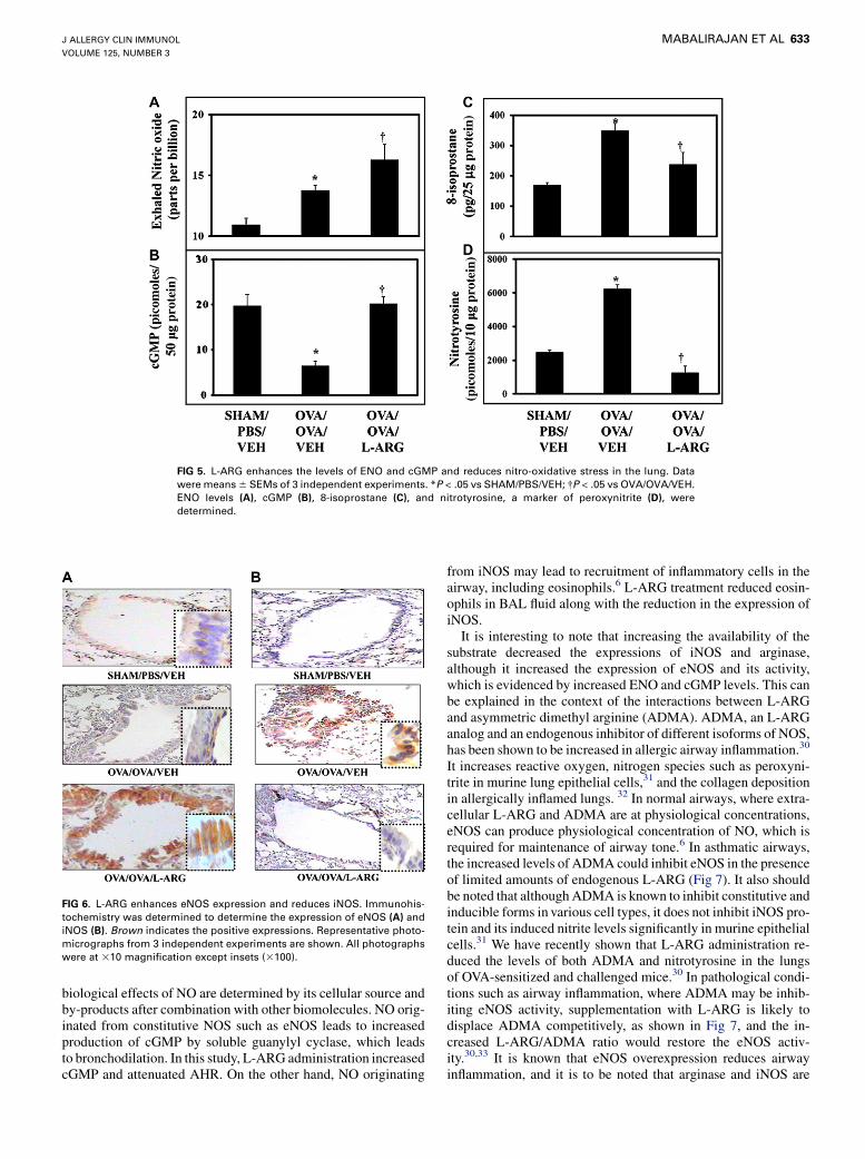
FIG 5. L-ARG enhances the levels of ENO and cGMP and reduces nitro-oxidative stress in the lung. Data
were means 6 SEMs of 3 independent experiments. *P < .05 vs SHAM/PBS/VEH; �P < .05 vs OVA/OVA/VEH.
ENO levels (A), cGMP (B), 8-isoprostane (C), and nitrotyrosine, a marker of peroxynitrite (D), were
determined.
FIG 6. L-ARG enhances eNOS expression and reduces iNOS. Immunohis-
tochemistry was determined to determine the expression of eNOS (A) and
iNOS (B). Brown indicates the positive expressions. Representative photo-
micrographs from 3 independent experiments are shown. All photographs
were at 310 magnification except insets (3100).
J ALLERGY CLIN IMMUNOL
VOLUME 125, NUMBER 3
MABALIRAJAN ET AL 633
biological effects of NO are determined by its cellular source andby-products after combination with other biomolecules. NO orig-inated from constitutive NOS such as eNOS leads to increasedproduction of cGMP by soluble guanylyl cyclase, which leadsto bronchodilation. In this study, L-ARG administration increasedcGMP and attenuated AHR. On the other hand, NO originating
from iNOS may lead to recruitment of inflammatory cells in theairway, including eosinophils.6 L-ARG treatment reduced eosin-ophils in BAL fluid along with the reduction in the expression ofiNOS.
It is interesting to note that increasing the availability of thesubstrate decreased the expressions of iNOS and arginase,although it increased the expression of eNOS and its activity,which is evidenced by increased ENO and cGMP levels. This canbe explained in the context of the interactions between L-ARGand asymmetric dimethyl arginine (ADMA). ADMA, an L-ARGanalog and an endogenous inhibitor of different isoforms of NOS,has been shown to be increased in allergic airway inflammation.30
It increases reactive oxygen, nitrogen species such as peroxyni-trite in murine lung epithelial cells,31 and the collagen depositionin allergically inflamed lungs. 32 In normal airways, where extra-cellular L-ARG and ADMA are at physiological concentrations,eNOS can produce physiological concentration of NO, which isrequired for maintenance of airway tone.6 In asthmatic airways,the increased levels of ADMA could inhibit eNOS in the presenceof limited amounts of endogenous L-ARG (Fig 7). It also shouldbe noted that although ADMA is known to inhibit constitutive andinducible forms in various cell types, it does not inhibit iNOS pro-tein and its induced nitrite levels significantly in murine epithelialcells.31 We have recently shown that L-ARG administration re-duced the levels of both ADMA and nitrotyrosine in the lungsof OVA-sensitized and challenged mice.30 In pathological condi-tions such as airway inflammation, where ADMA may be inhib-iting eNOS activity, supplementation with L-ARG is likely todisplace ADMA competitively, as shown in Fig 7, and the in-creased L-ARG/ADMA ratio would restore the eNOS activ-ity.30,33 It is known that eNOS overexpression reduces airwayinflammation, and it is to be noted that arginase and iNOS are
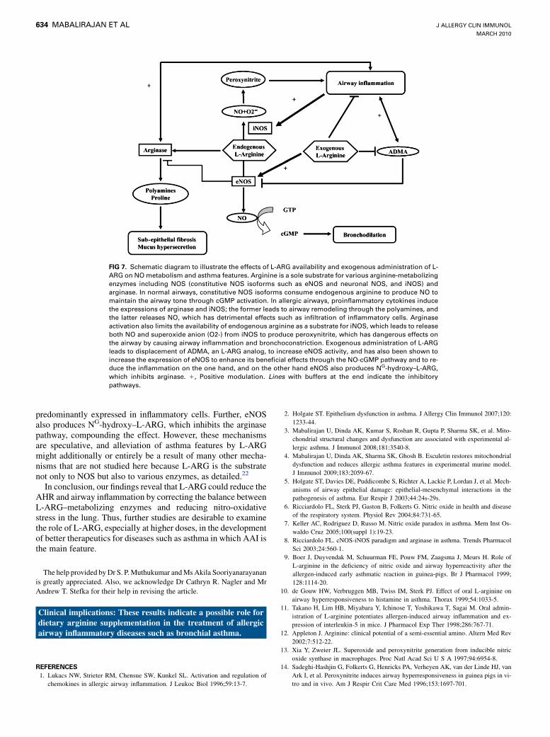
FIG 7. Schematic diagram to illustrate the effects of L-ARG availability and exogenous administration of L-
ARG on NO metabolism and asthma features. Arginine is a sole substrate for various arginine-metabolizing
enzymes including NOS (constitutive NOS isoforms such as eNOS and neuronal NOS, and iNOS) and
arginase. In normal airways, constitutive NOS isoforms consume endogenous arginine to produce NO to
maintain the airway tone through cGMP activation. In allergic airways, proinflammatory cytokines induce
the expressions of arginase and iNOS; the former leads to airway remodeling through the polyamines, and
the latter releases NO, which has detrimental effects such as infiltration of inflammatory cells. Arginase
activation also limits the availability of endogenous arginine as a substrate for iNOS, which leads to release
both NO and superoxide anion (O2-) from iNOS to produce peroxynitrite, which has dangerous effects on
the airway by causing airway inflammation and bronchoconstriction. Exogenous administration of L-ARG
leads to displacement of ADMA, an L-ARG analog, to increase eNOS activity, and has also been shown to
increase the expression of eNOS to enhance its beneficial effects through the NO-cGMP pathway and to re-
duce the inflammation on the one hand, and on the other hand eNOS also produces NG-hydroxy–L-ARG,
which inhibits arginase. 1, Positive modulation. Lines with buffers at the end indicate the inhibitory
pathways.
J ALLERGY CLIN IMMUNOL
MARCH 2010
634 MABALIRAJAN ET AL
predominantly expressed in inflammatory cells. Further, eNOSalso produces NG-hydroxy–L-ARG, which inhibits the arginasepathway, compounding the effect. However, these mechanismsare speculative, and alleviation of asthma features by L-ARGmight additionally or entirely be a result of many other mecha-nisms that are not studied here because L-ARG is the substratenot only to NOS but also to various enzymes, as detailed.22
In conclusion, our findings reveal that L-ARG could reduce theAHR and airway inflammation by correcting the balance betweenL-ARG–metabolizing enzymes and reducing nitro-oxidativestress in the lung. Thus, further studies are desirable to examinethe role of L-ARG, especially at higher doses, in the developmentof better therapeutics for diseases such as asthma in which AAI isthe main feature.
The help provided by Dr S. P. Muthukumar and Ms Akila Sooriyanarayanan
is greatly appreciated. Also, we acknowledge Dr Cathryn R. Nagler and Mr
Andrew T. Stefka for their help in revising the article.
Clinical implications: These results indicate a possible role fordietary arginine supplementation in the treatment of allergicairway inflammatory diseases such as bronchial asthma.
REFERENCES
1. Lukacs NW, Strieter RM, Chensue SW, Kunkel SL. Activation and regulation of
chemokines in allergic airway inflammation. J Leukoc Biol 1996;59:13-7.
2. Holgate ST. Epithelium dysfunction in asthma. J Allergy Clin Immunol 2007;120:
1233-44.
3. Mabalirajan U, Dinda AK, Kumar S, Roshan R, Gupta P, Sharma SK, et al. Mito-
chondrial structural changes and dysfunction are associated with experimental al-
lergic asthma. J Immunol 2008;181:3540-8.
4. Mabalirajan U, Dinda AK, Sharma SK, Ghosh B. Esculetin restores mitochondrial
dysfunction and reduces allergic asthma features in experimental murine model.
J Immunol 2009;183:2059-67.
5. Holgate ST, Davies DE, Puddicombe S, Richter A, Lackie P, Lordan J, et al. Mech-
anisms of airway epithelial damage: epithelial-mesenchymal interactions in the
pathogenesis of asthma. Eur Respir J 2003;44:24s-29s.
6. Ricciardolo FL, Sterk PJ, Gaston B, Folkerts G. Nitric oxide in health and disease
of the respiratory system. Physiol Rev 2004;84:731-65.
7. Keller AC, Rodriguez D, Russo M. Nitric oxide paradox in asthma. Mem Inst Os-
waldo Cruz 2005;100(suppl 1):19-23.
8. Ricciardolo FL. cNOS-iNOS paradigm and arginase in asthma. Trends Pharmacol
Sci 2003;24:560-1.
9. Boer J, Duyvendak M, Schuurman FE, Pouw FM, Zaagsma J, Meurs H. Role of
L-arginine in the deficiency of nitric oxide and airway hyperreactivity after the
allergen-induced early asthmatic reaction in guinea-pigs. Br J Pharmacol 1999;
128:1114-20.
10. de Gouw HW, Verbruggen MB, Twiss IM, Sterk PJ. Effect of oral L-arginine on
airway hyperresponsiveness to histamine in asthma. Thorax 1999;54:1033-5.
11. Takano H, Lim HB, Miyabara Y, Ichinose T, Yoshikawa T, Sagai M. Oral admin-
istration of L-arginine potentiates allergen-induced airway inflammation and ex-
pression of interleukin-5 in mice. J Pharmacol Exp Ther 1998;286:767-71.
12. Appleton J. Arginine: clinical potential of a semi-essential amino. Altern Med Rev
2002;7:512-22.
13. Xia Y, Zweier JL. Superoxide and peroxynitrite generation from inducible nitric
oxide synthase in macrophages. Proc Natl Acad Sci U S A 1997;94:6954-8.
14. Sadeghi-Hashjin G, Folkerts G, Henricks PA, Verheyen AK, van der Linde HJ, van
Ark I, et al. Peroxynitrite induces airway hyperresponsiveness in guinea pigs in vi-
tro and in vivo. Am J Respir Crit Care Med 1996;153:1697-701.

J ALLERGY CLIN IMMUNOL
VOLUME 125, NUMBER 3
MABALIRAJAN ET AL 635
15. Lee KS, Kim SR, Park SJ, Min KH, Lee KY, Jin SM, et al. Antioxidant
down-regulates interleukin-18 expression in asthma. Mol Pharmacol 2006;70:
1184-93.
16. Ahmad T, Mabalirajan U, Joseph DA, Makhija L, Singh VP, Ghosh B, et al. Ex-
haled nitric oxide estimation by a simple and efficient non-invasive technique
and its utility as a marker of airway inflammation in mice. J Appl Physiol 2009;
107:295-301.
17. Ram A, Mabalirajan U, Singh SK, Singh VP, Ghosh B. Mepacrine alleviates air-
way hyperresponsiveness and airway inflammation in a mouse model of asthma.
Int Immunopharmacol 2008;8:893-9.
18. Ram A, Mabalirajan U, Das M, Bhattacharya I, Dinda AK, Gangal SV, et al. Gly-
cyrrhizin alleviates experimental allergic asthma in mice. Int Immunopharmacol
2006;6:1468-77.
19. Kaczmarek E, Gorna A, Majewski P. Techniques of image analysis for quantitative
immunohistochemistry. Rocz Akad Med Bialymst 2004;49(suppl 1):155-8.
20. Agrawal A, Rengarajan S, Adler KB, Ram A, Ghosh B, Fahim M, et al. Inhibition
of mucin secretion with MARCKS-related peptide improves airway obstruction in
a mouse model of asthma. J Appl Physiol 2007;102:399-405.
21. Campbell BI, La Bounty PM, Roberts M. The ergogenic potential of arginine. J Int
Soc Sports Nutr 2004;1:35-8.
22. Wu G, Morris SM Jr. Arginine metabolism: nitric oxide and beyond. Biochem J
1998;336:1-17.
23. Maarsingh H, Zaagsma J, Meurs H. Arginine homeostasis in allergic asthma. Eur J
Pharmacol 2008;585:375-84.
24. Lara A, Khatri SB, Wang Z, Comhair SA, Xu W, Dweik RA, et al. Alterations of
the arginine metabolome in asthma. Am J Respir Crit Care Med 2008;178:
673-81.
25. West SG, Likos-Krick A, Brown P, Mariotti F. Oral L-arginine improves hemody-
namic responses to stress and reduces plasma homocysteine in hypercholesterol-
emic men. J Nutr 2005;135:212-7.
26. de Nigris F, Lerman LO, Ignarro SW, Sica G, Lerman A, Palinski W, et al. Bene-
ficial effects of antioxidants and L-arginine on oxidation-sensitive gene expression
and endothelial NO synthase activity at sites of disturbed shear stress. Proc Natl
Acad Sci U S A 2003;100:1420-5.
27. Zimmermann N, King NE, Laporte J, Yang M, Mishra A, Pope SM, et al. Dissec-
tion of experimental asthma with DNA microarray analysis identifies arginase in
asthma pathogenesis. J Clin Invest 2003;111:1863-74.
28. Ten Broeke R, De Crom R, Van Haperen R, Verweij V, Leusink-Muis T, Van Ark I,
et al. Overexpression of endothelial nitric oxide synthase suppresses features of al-
lergic asthma in mice. Respir Res 2006;7:58.
29. Maarsingh H, Zuidhof AB, Bos IS, van Duin M, Boucher JL, Zaagsma J, et al.
Arginase inhibition protects against allergen-induced airway obstruction, hyperres-
ponsiveness, and inflammation. Am J Respir Crit Care Med 2008;178:565-73.
30. Ahmad T, Mabalirajan U, Ghosh B, Agrawal A. Altered assymetric dimethyl
arginine metabolism in bronchial epithelium of allergically inflamed lungs. Am J
Respir Cell Mol Biol 2010;42:3-8.
31. Wells SM, Holian A. Asymmetric dimethylarginine induces oxidative and nitrosa-
tive stress in murine lung epithelial cells. Am J Respir Cell Mol Biol 2007;36:520-8.
32. Wells SM, Buford MC, Migliaccio CT, Holian A. Elevated asymmetric dimethylar-
ginine alters lung function and induces collagen deposition in mice. Am J Respir
Cell Mol Biol 2009;40:179-88.
33. Bode-B€oger SM, Scalera F, Ignarro LJ. The L-arginine paradox: importance of
the L-arginine/asymmetrical dimethylarginine ratio. Pharmacol Ther 2007;114:
295-306.