Behavioral and brain pattern differences between acting and observing in an auditory task
11
BioMed Central Page 1 of 11 (page number not for citation purposes) Behavioral and Brain Functions Open Access Research Behavioral and brain pattern differences between acting and observing in an auditory task Irene S Karanasiou* 1 , Charalabos Papageorgiou 2 , Eleni I Tsianaka 1 , George K Matsopoulos 1 , Errikos M Ventouras 3 and Nikolaos K Uzunoglu 1 Address: 1 Institute of Communications and Computer Systems, National Technical University of Athens, 9, Iroon Polytechneiou str., 157 73 Zografou Campus, Athens, Greece, 2 School of Medicine, National and Kapodistrian University of Athens, Greece and 3 Technological Education Institution of Athens, Greece Email: Irene S Karanasiou* - [email protected]; Charalabos Papageorgiou - [email protected]; Eleni I Tsianaka - [email protected]; George K Matsopoulos - [email protected]; Errikos M Ventouras - [email protected]; Nikolaos K Uzunoglu - [email protected] * Corresponding author Abstract Background: Recent research has shown that errors seem to influence the patterns of brain activity. Additionally current notions support the idea that similar brain mechanisms are activated during acting and observing. The aim of the present study was to examine the patterns of brain activity of actors and observers elicited upon receiving feedback information of the actor's response. Methods: The task used in the present research was an auditory identification task that included both acting and observing settings, ensuring concurrent ERP measurements of both participants. The performance of the participants was investigated in conditions of varying complexity. ERP data were analyzed with regards to the conditions of acting and observing in conjunction to correct and erroneous responses. Results: The obtained results showed that the complexity induced by cue dissimilarity between trials was a demodulating factor leading to poorer performance. The electrophysiological results suggest that feedback information results in different intensities of the ERP patterns of observers and actors depending on whether the actor had made an error or not. The LORETA source localization method yielded significantly larger electrical activity in the supplementary motor area (Brodmann area 6), the posterior cingulate gyrus (Brodmann area 31/23) and the parietal lobe (Precuneus/Brodmann area 7/5). Conclusion: These findings suggest that feedback information has a different effect on the intensities of the ERP patterns of actors and observers depending on whether the actor committed an error. Certain neural systems, including medial frontal area, posterior cingulate gyrus and precuneus may mediate these modulating effects. Further research is needed to elucidate in more detail the neuroanatomical and neuropsychological substrates of these systems. Published: 20 January 2009 Behavioral and Brain Functions 2009, 5:5 doi:10.1186/1744-9081-5-5 Received: 26 June 2008 Accepted: 20 January 2009 This article is available from: http://www.behavioralandbrainfunctions.com/content/5/1/5 © 2009 Karanasiou et al; licensee BioMed Central Ltd. This is an Open Access article distributed under the terms of the Creative Commons Attribution License (http://creativecommons.org/licenses/by/2.0 ), which permits unrestricted use, distribution, and reproduction in any medium, provided the original work is properly cited.
Transcript of Behavioral and brain pattern differences between acting and observing in an auditory task
BioMed CentralBehavioral and Brain Functions
ss
Open AcceResearchBehavioral and brain pattern differences between acting and observing in an auditory taskIrene S Karanasiou*1, Charalabos Papageorgiou2, Eleni I Tsianaka1, George K Matsopoulos1, Errikos M Ventouras3 and Nikolaos K Uzunoglu1Address: 1Institute of Communications and Computer Systems, National Technical University of Athens, 9, Iroon Polytechneiou str., 157 73 Zografou Campus, Athens, Greece, 2School of Medicine, National and Kapodistrian University of Athens, Greece and 3Technological Education Institution of Athens, Greece
Email: Irene S Karanasiou* - [email protected]; Charalabos Papageorgiou - [email protected]; Eleni I Tsianaka - [email protected]; George K Matsopoulos - [email protected]; Errikos M Ventouras - [email protected]; Nikolaos K Uzunoglu - [email protected]
* Corresponding author
AbstractBackground: Recent research has shown that errors seem to influence the patterns of brainactivity. Additionally current notions support the idea that similar brain mechanisms are activatedduring acting and observing. The aim of the present study was to examine the patterns of brainactivity of actors and observers elicited upon receiving feedback information of the actor'sresponse.
Methods: The task used in the present research was an auditory identification task that includedboth acting and observing settings, ensuring concurrent ERP measurements of both participants.The performance of the participants was investigated in conditions of varying complexity. ERP datawere analyzed with regards to the conditions of acting and observing in conjunction to correct anderroneous responses.
Results: The obtained results showed that the complexity induced by cue dissimilarity betweentrials was a demodulating factor leading to poorer performance. The electrophysiological resultssuggest that feedback information results in different intensities of the ERP patterns of observersand actors depending on whether the actor had made an error or not. The LORETA sourcelocalization method yielded significantly larger electrical activity in the supplementary motor area(Brodmann area 6), the posterior cingulate gyrus (Brodmann area 31/23) and the parietal lobe(Precuneus/Brodmann area 7/5).
Conclusion: These findings suggest that feedback information has a different effect on theintensities of the ERP patterns of actors and observers depending on whether the actor committedan error. Certain neural systems, including medial frontal area, posterior cingulate gyrus andprecuneus may mediate these modulating effects. Further research is needed to elucidate in moredetail the neuroanatomical and neuropsychological substrates of these systems.
Published: 20 January 2009
Behavioral and Brain Functions 2009, 5:5 doi:10.1186/1744-9081-5-5
Received: 26 June 2008Accepted: 20 January 2009
This article is available from: http://www.behavioralandbrainfunctions.com/content/5/1/5
© 2009 Karanasiou et al; licensee BioMed Central Ltd. This is an Open Access article distributed under the terms of the Creative Commons Attribution License (http://creativecommons.org/licenses/by/2.0), which permits unrestricted use, distribution, and reproduction in any medium, provided the original work is properly cited.
Page 1 of 11(page number not for citation purposes)
Behavioral and Brain Functions 2009, 5:5 http://www.behavioralandbrainfunctions.com/content/5/1/5
BackgroundThe ability to monitor ongoing performance is critical tobehavioral adaptation in changing environmental set-tings. Especially, the monitoring of performance errorsserves to optimize future response behavior.
The neural basis of error processing has attracted greatinterest, because its elucidation promises a better under-standing of the mechanisms underlying adaptive andnon-adaptive behavior. The strongest evidence for theexistence of the neural system that implements errorprocessing has come from the detection of the error-related negativity (ERN), a component of the event-related potentials (ERPs) generated within the medial-frontal cortex that is sensitive to error in performance[1,2].
This class of ERP components comprises a growing list ofpotentials, including the ERN/Ne elicited by errorresponses in reaction time tasks [1,3], the N2-like poten-tials elicited by error feedback stimuli [4-6], the variety ofN2-like potentials elicited in situations of response con-flict or response inhibition [7-9]. Importantly, accordingto the conflict hypothesis [10,11], which states that the Nedoes not reflect the mismatch, but rather the conflictbetween response representations, knowledge about thecorrect response is not necessary and the error relatedpotential is not specific for errors, but depends only onthe amount of conflict.
In this context it should be noted that based on the Mir-ror Neuron System MNS hypothesis [12], which sug-gests that acting and observing are associated withsimilar neural mechanisms, the feedback ERN has beenstudied in two participant conditions [13-16]. Thesestudies focused on the way this type of ERN reflects theevaluation of outcomes induced by others [14,16] eventhose of a simulated Brain Computer Interface BCI [15].The feedback-related negativity-like effects wereobtained in both single and two-participant condition,suggesting that similar neural mechanisms are involvedin evaluating the outcomes of one's own and the other'sactions.
Up to date the exact mechanisms underlying the error sys-tem continue to be a subject of ongoing debate, since theirpatterns and specificity still remain poorly understood. Inthis sense it is interesting to study potential differences inthe pattern of activity elicited by observed errors thanfrom self-made errors in conditions where the roles of act-ing and observing are alternating.
Considering the aforementioned viewpoint as well asfindings of recent experimental studies that there is an
ongoing debate about the characterization of brain activ-ity correlated with errors, a simple task design was used tocompare the electrophysiological effects related toobserved and self-made errors during performance moni-toring in two-participant conditions of varying complex-ity. The electrophysiological recordings weresimultaneously performed for both subjects as they alter-natively exchanged roles between acting and observing.With synchronous two-subject recordings, comparisonsof the elicited ERP conventional parameters (amplitudes,latencies), could be made both between actors andobservers as well as between errors and non-errors. Ourresearch hypothesis was to study the ERP patterns ofobservers and actors in a two-participant experiment elic-ited upon receiving feedback information of the actor'sresponse.
The task used in the present research was an auditoryidentification task comprising one single-actor and twotwo-actor conditions. In the latter the participants per-formed the task in an alternating fashion, exchangingroles between acting and observing in each trial. Theaim for the actor is to correctly map a horizontal sliderposition onto an active tone-frequency range and ineach trial he/she selects a slider position that matches atone that is initially presented to him/her. Both partic-ipants receive feedback information corresponding tothe slider position selection. The frequency range inwhich the auditory cues/stimuli are presented varies inthe experimental conditions in order to explore the par-ticipants' competency. In one of the two-actor condi-tions, auditory stimuli from the same frequency rangeare presented to both participants while in the othercondition each participant is presented with tones fromhis\her individual frequency range that is differentfrom that of the partner. The hypothesis is that cue fre-quency dissimilarity may hinder the pitch identifica-tion process by increasing the complexity of the task. Inthe case where the actors received different tones fromthe observers, this would impact performance and howaccuracy is judged. The subsequent neural responses tofeedback are investigated in all conditions and in bothactors and observers.
The recordings of the scalp ERPs both for the actor and theobserver were simultaneous. We investigated the errorrelated component of the ERPs related to feedback infor-mation from the actor response recorded during actingand observing both with the conventional constituents(amplitudes and latencies) as well as with the LORETAsource localization method which differentiates betweenstructural and energetic processes related to informationprocessing as revealed by the associated ERP waveform[17-19].
Page 2 of 11(page number not for citation purposes)
Behavioral and Brain Functions 2009, 5:5 http://www.behavioralandbrainfunctions.com/content/5/1/5
MethodsParticipantsFourteen healthy individuals (eight men and six women),with mean age of 26.6 ± 2.9 years and high level education(education years 17.7± 2.3), all with normal hearing asmeasured by pure-tone audiograms (thresholds <15 dBHL), participated in the experiment. The male and femalesubgroups were homogeneous with regards to age andeducational level. All the participants were right-handedand had no history of any hearing problem. Informedconsent was obtained from all subjects.
Stimuli and proceduresIn the present research an auditory identification task hasbeen used in one single-actor and two two-actor condi-tions. In the two-actor or "Joint" conditions the partici-pants performed the task in an alternating fashion,meaning that when one subject performed the task (actor)the other observed (observer) whereas in the Single con-dition performed the task alone. From one trial to thenext, the roles of actor and observer were switchedbetween the two dyad members.
The frequency range in which the auditory cues were pre-sented varied in the experimental conditions. In one ofthe joint conditions, auditory stimuli from the same fre-quency range were presented to both participants (Joint-1) while in the other condition each participant was pre-sented with tones from different frequency ranges (Joint-2). All participants were examined in all three conditions(single, Joint-1, Joint-2) in varying order. The dyads in theJoint-1 and Joint-2 condition were the same.
The actor and observer sat opposite but were screenedfrom each other. They both had computer screens in frontof them. At each trial they both heard the stimulus tonewith duration of 1 sec presented through the headphones.The stimulus tone was randomly selected for each trialwithin the fixed frequency range. The actor's task was toposition a slider presented on the computer screen with agamepad such that the slider position would match thefrequency of the stimulus tone. The mapping of the fre-quency range was fixed within a block of trials; however,at the start of the trial blocks, the participants did notknow the scaling of the frequency range within which theslider position should be mapped. After the positioning ofthe slider by the actor, the frequency corresponding to theactor's selected slider position (Feedback tone) was pre-sented to both the actor and the observer.
The single condition consisted of 40 trials. The joint con-ditions consisted of 80 trials for both subjects. The trialsin the joint conditions were accomplished alternately bythe two participants, 40 trials for each participant. Thestimuli were presented from four frequency ranges with a
bandwidth 400-Hz namely Range 1: 200–600 Hz, Range2: 620–1020 Hz, Range 3: 1040–1440 Hz, Range 4: 1460–1860 Hz.
Before the experiment, the subjects were submitted to anacoustic pre-test in order to examine their hearing abilityin the four frequency ranges that were used in the experi-ment. During this test two tones from each range werepresented to the participants. Then, the participants hadto identify which of these tones was higher than the other.The frequencies of the two tones selected from each rangefor the acoustic test were determined as the 25% and the75% of each range of 400 Hz bandwidth. The subjectsheard the tones with their headphones and respondedorally to the experimenter. All participants were capableto discriminate between the tones presented in the pre-test.
EEG recordings and experimental setupThe experimental setup included a Faraday room, whichscreened any electromagnetic interference that couldaffect the measurements. The EEG was recorded continu-ously using a 32-channel electrode cap (Biosemi, Active-two System) according to the International 10–20 EEGsystem [20]. The electrodes used were Fp1, Fp2, Pz, Fz,O1, O2, P3, P4, P7, P8, C3, C4, T7, T8, F3, F4, F7, F8, Cz,Oz, CP5, CP6, CP1, CP2, FC1, FC2, FC5, FC6, AF3, AF4,PO3 and PO4.
The bioelectrical brain activity was simultaneouslyrecorded from both participants using two differentrecording systems, daisy chained in a master-slave rela-tionship. Galvanic isolation of both participants wasensured by using optical receiver for trigger inputs, whilein parallel, interference pickup was also eliminated. Theelectrode cables were also bundled to eliminate potentialmagnetic interference. The vertical electro-oculogram(EOG) was recorded bipolarly from electrodes placedabove and below the eyes and the horizontal EOG wasmonitored from electrodes at the outer canthi of the eyes.The data were filtered off-line, high-pass at 0.05 Hz andlow-pass at 35 Hz. All signals were digitized with a sam-pling rate of 256 Hz. All scalp signals were referencedonline to both mastoids, but were later offline re-refer-enced to the average of all scalp electrodes. Trials wereaveraged to ERPs separately for each condition and eachsubject, relative to a 100 ms pre-stimulus (Feedback tone)baseline.
To eliminate EOG artifact, trials with EEG voltages exceed-ing 80 μV were rejected from the average. Artifact rejectionand averaging were performed offline. Due to artifact con-taminated epochs the measurements of one participantdyad were excluded. Consequently, the data were ana-lyzed for 6 dyads.
Page 3 of 11(page number not for citation purposes)
Behavioral and Brain Functions 2009, 5:5 http://www.behavioralandbrainfunctions.com/content/5/1/5
Categorization of correct and erroneous responsesThe ability of each participant to differentiate tones andhis/her auditory frequency perception resolution can bedescribed in terms of an Equivalent Rectangular Band-width (ERB) as a function of a centre frequency, whichbetter represents auditory frequency selectivity accordingto psychoacoustics. In the present context the concept ofERB can be conceived as the acceptable bandwidtharound the stimulus frequency within which the responsefrequency can be considered as a correct response value.According to psychoacoustics theory [21-24], the Equiva-lent Rectangular Bandwidth (in Hz) can be approximatedaccording to the following formula: Be = 6.23 10-6 f 2 +9.339 10-2 f + 28.52. The discrimination between theactor's erroneous and non-erroneous responses was per-formed with the use of the above formula.
LORETA source localization methodThe low resolution brain electromagnetic tomography(LORETA) differentiates between structural and energeticprocesses related to information processing as revealed bythe associated EEG/ERP waveform [17,18]. The structurallevel, revealed by the location of the local maxima of thecurrent source density distribution, describes the timedependent network of activated brain areas. The magni-tude of the source strength, a measure of the energeticcomponent, describes the allocation of processingresources [19]. The utilized LORETA version was regis-tered to the Talairach brain atlas [25]. The solution spaceconsisted of 2394 voxels with a spatial resolution of 7mm. Average LORETA images were constructed across allsubjects in all 4 cases (both for actors and observersdepending on whether the actor committed an error ornot). The voxel-by-voxel pairwise t-test differences werecarried out for the observers depending whether the actormade an error or not. The structure-Probability Maps atlas[26] was used to identify which brain regions wereinvolved in the ERP waveforms as well as in differencesbetween the compared groups. Brodmann area(s) andbrain regions close to the observed locations identified bythe Talairach coordinates are reported [25].
Statistical analysisThe primary behavioral outcome variable was the Abso-lute Frequency Error (AFE), defined as the absolute differ-ence between the stimulus and response frequency. Aunivariate general linear model (GLM) was used with theabsolute frequency error as the dependent variable andTrial (n = 40), Condition (Single, Joint-1, Joint-2), Fre-quency Range (n = 4) and Order (n = 3) as fixed factors.
The time window within which ERP analysis was per-formed was from -200 msec to +500 msec around theFeedback tone, which included 180 time points. In orderto take into account neurophysiological processes that
took place between Stimulus and Feedback tone thatcould affect the elicited ERP waveforms, the analysis wasalso performed by selecting a100 ms baseline before theStimulus tone yielding the same results. The mean ampli-tude values for every subject at each time point were sub-jected to three-way ANOVA. The first factor was whetherthe subject was the actor or the observer. The second factorwas whether the actor had committed an error or not,while the third factor was the condition (Joint-1, Joint-2).Factor or interaction effects were considered significantonly if they occurred over a time window of at least 20msec. When significant factor or interaction effects werefound, post hoc analyses with Bonferroni corrections werecarried out. Statistical significance was set at 0.05.
The LORETA mappings were performed at the time win-dow 144–171 msec where significant differences wereobserved resulting from the ANOVA procedures.
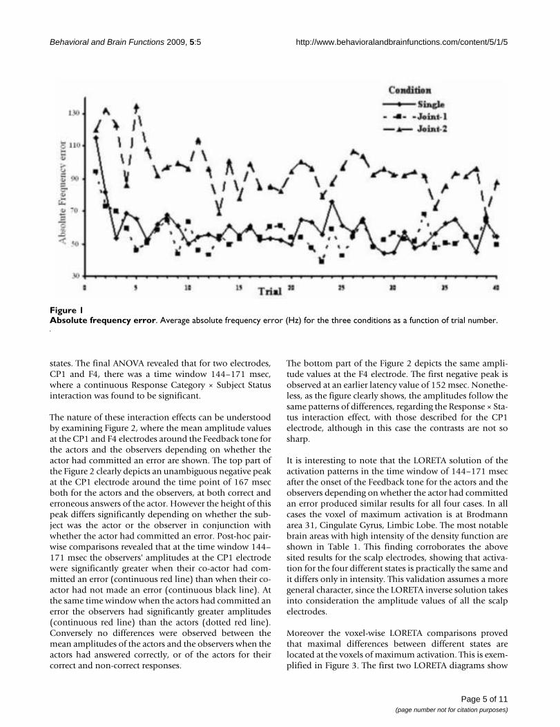
ResultsBehavioral dataThe ANOVA procedure with AFE as the dependent varia-ble and Condition, Trial, Frequency Range and Order asthe independent factors revealed that only Condition (F =23.6, p < 0.01) and Trial (F = 3.1, p < 0.01) had a signifi-cant effect. However the significance of the Trial effect isonly partial. Indeed, as post-hoc contrasts confirm, onlythe first trial (and partly the second one) had a signifi-cantly larger AFE than the other trials. Accordingly the sig-nificance of the Trial effect, after having excluded the dataof the first two trials, disappeared. Consequently, the fac-tor responsible for most of the variability of AFE remainedthe Condition (F = 28.5, p < 0.01). The average AFE abso-lute frequency error (Hz) for the three conditions as afunction of trial number is depicted in Figure 1, whichclearly depicts that the subjects' performance is signifi-cantly worse in the Joint-2 condition throughout the fortytrials, with the exception of the first one, while the Joint-1condition was practically the same as the Single condi-tion. It is interesting to note that the overall mean AFE val-ues in the Joint-2 condition do not differ from the meanAFE values of the first trial of the Single and Joint-1 condi-tions.
Event related potentials dataThe ANOVA procedures with the amplitudes at each leadand each time point with Condition, Response Category(Erroneous, Correct) and Subject Status (Actor, Observer)revealed that condition did not have any significant effect,either by itself or through its interactions with the othertwo factors. Therefore subsequent analyses of the ampli-tudes were performed disregarding the condition. Conse-quently, it should be noted that in the final ERP analysisthe data from both Joint conditions were used, resultingin the employment of 80 trials per acting and observing
Page 4 of 11(page number not for citation purposes)
Behavioral and Brain Functions 2009, 5:5 http://www.behavioralandbrainfunctions.com/content/5/1/5
states. The final ANOVA revealed that for two electrodes,CP1 and F4, there was a time window 144–171 msec,where a continuous Response Category × Subject Statusinteraction was found to be significant.
The nature of these interaction effects can be understoodby examining Figure 2, where the mean amplitude valuesat the CP1 and F4 electrodes around the Feedback tone forthe actors and the observers depending on whether theactor had committed an error are shown. The top part ofthe Figure 2 clearly depicts an unambiguous negative peakat the CP1 electrode around the time point of 167 msecboth for the actors and the observers, at both correct anderroneous answers of the actor. However the height of thispeak differs significantly depending on whether the sub-ject was the actor or the observer in conjunction withwhether the actor had committed an error. Post-hoc pair-wise comparisons revealed that at the time window 144–171 msec the observers' amplitudes at the CP1 electrodewere significantly greater when their co-actor had com-mitted an error (continuous red line) than when their co-actor had not made an error (continuous black line). Atthe same time window when the actors had committed anerror the observers had significantly greater amplitudes(continuous red line) than the actors (dotted red line).Conversely no differences were observed between themean amplitudes of the actors and the observers when theactors had answered correctly, or of the actors for theircorrect and non-correct responses.
The bottom part of the Figure 2 depicts the same ampli-tude values at the F4 electrode. The first negative peak isobserved at an earlier latency value of 152 msec. Nonethe-less, as the figure clearly shows, the amplitudes follow thesame patterns of differences, regarding the Response × Sta-tus interaction effect, with those described for the CP1electrode, although in this case the contrasts are not sosharp.
It is interesting to note that the LORETA solution of theactivation patterns in the time window of 144–171 msecafter the onset of the Feedback tone for the actors and theobservers depending on whether the actor had committedan error produced similar results for all four cases. In allcases the voxel of maximum activation is at Brodmannarea 31, Cingulate Gyrus, Limbic Lobe. The most notablebrain areas with high intensity of the density function areshown in Table 1. This finding corroborates the abovesited results for the scalp electrodes, showing that activa-tion for the four different states is practically the same andit differs only in intensity. This validation assumes a moregeneral character, since the LORETA inverse solution takesinto consideration the amplitude values of all the scalpelectrodes.
Moreover the voxel-wise LORETA comparisons provedthat maximal differences between different states arelocated at the voxels of maximum activation. This is exem-plified in Figure 3. The first two LORETA diagrams show
Absolute frequency errorFigure 1Absolute frequency error. Average absolute frequency error (Hz) for the three conditions as a function of trial number.
Page 5 of 11(page number not for citation purposes)

Behavioral and Brain Functions 2009, 5:5 http://www.behavioralandbrainfunctions.com/content/5/1/5
Page 6 of 11(page number not for citation purposes)
Mean amplitude values at CP1 and F4 electrodes around the Feedback toneFigure 2Mean amplitude values at CP1 and F4 electrodes around the Feedback tone. Mean amplitude values at the CP1 (top) and F4 (bottom) electrode around the Feedback tone for the actors and the observers depending on whether the actor had committed an error.

Behavioral and Brain Functions 2009, 5:5 http://www.behavioralandbrainfunctions.com/content/5/1/5
the observers' activation maps at their co-actors' errorsand correct answers. Clearly, the activation maps are sim-ilar but different in intensity. Specifically at the voxel ofmaximum activation (Brodmann area 31 CingulateGyrus, Limbic Lobe), the intensity of the density functionfor the observer's response at his co-actor's error was 3.65× 10⁻³, while for the observer's response at his co-actorscorrect answer the intensity was 2.25 × 10⁻³. The intensi-ties for the actor (not shown in the figure) at his own errorand at his correct answer were 2.65 × 10⁻³ and 2.55 × 10⁻³
respectively, i.e. they lay intermediately between theobserver's values, as was found for the electrodes. The bot-tom figure shows the result of the voxel, pairwise LORETAcomparisons between the two states for the observers:when the actors were correct and when the actors werewrong. More intense blue color denotes larger differencesbetween the two states. A comparison of this figure withthe activation maps leads to the conclusion that the differ-ence map practically coincide with the activation maps.Specifically the voxel of maximal differences is again atBrodmann area 31 Cingulate Gyrus, Limbic Lobe, atwhich the significance of the differences is <0.01.
In conclusion the activation patterns of the actors and theobservers at the actor's errors and correct answers are tem-porally and spatially congruent, varying only in the inten-sity of the density function and maximal differences in theintensity between states are observed at the areas of maxi-mum activation.
DiscussionThe present study was performed to examine brain activityin a two-participant condition task following actor-response related feedback correlated with errors. Thebehavioral results showed that the participants had signif-icantly poorer performance in conditions with increasedcomplexity. Comparisons of the ERP measurementsrevealed that the subjects during the observing conditionhad a significantly greater negative deflection located atthe medial frontal area and superior parietal regions whentheir co-actors committed an error than when they had
correct responses. The LORETA source localizationmethod yielded significantly larger electrical activity inthe supplementary motor area (Brodmann area 6), theposterior cingulate gyrus (Brodmann area 31/23) and theparietal lobe (Precuneus/Brodmann area 7/5). Neverthe-less, the condition factor did not have any significanteffect on the ERP patterns of actors and observers.
Regarding the behavioural data, results showed that par-ticipants' performance was strongly influenced by cue dis-similarity yielding significantly poorer scores in the morecomplex Joint-2 condition. The Joint-1 condition yieldedsimilar results with the Single condition. In the first trial,in all conditions, the actor is faced with the unknown. Theparticipant has no measure of inference with regards tothe minimum and maximum and hence to the range ofhis/her scale, so the first positioning of the slider is inessence arbitrary.
From the second trial onwards the actor works compara-tively with the previous trial. The mapping of the tone fre-quency range is better achieved in the case when soundidentification is assisted by sound discrimination, result-ing from the comparison of the present with the previoustone. Such is the case of the Joint-1 and Single conditions,where the participants are provided stimuli from the samefrequency range. As a result their task performance wassignificantly better than in the Joint-2 condition, wherethe participants were provided stimuli from different fre-quency ranges. Each participant, by observing the actionsof his/her partner, who performs in a different frequencyrange than him/her, seems to demodulate his/her percep-tion of the scale of his/her own frequency range, andresults in being disoriented when trying to place the slidercorrectly as actor in the subsequent trial. By receiving dis-tracting information from the previous cue not belongingto the mapped frequency range resulted in poor perform-ance, which is practically the same as his/her first trial. Inthe Single and Joint-1 conditions the participants' answersare fine-tuned as the trials in a block evolved but eachactor seemed to start right from the beginning during theJoint-2 condition throughout the trial blocks
The participants of each dyad were simultaneouslyrecorded and the relevant ERP analysis was performedusing both its conventional constituents (amplitude,latency) and the LORETA patterns. Regarding the analysisof amplitude data at the Feedback tone comparisons ofmeasurements of conventional constituents of the rele-vant event related potential, revealed that at the time win-dow 144–171 msec the observers' amplitudes at the F4and CP1 electrodes were significantly greater when theirco-actor had committed an error than when their co-actorhad not made an erroneous response. These findingscould be better understood if we take into account the
Table 1: Brain areas of maximal activation
Brodmann area 31, Cingulate Gyrus, Limbic Lobe
Brodmann area 7, Precuneus, Parietal Lobe
Brodmann area 5, Paracentral Lobule, Frontal Lobe
Brodmann area 23, Cingulate Gyrus, Limbic Lobe
Brodmann area 6, Paracentral Lobule, Frontal Lobe
Brain areas of maximal activation in the time window of 144–171 msec after the onset of the Feedback tone
Page 7 of 11(page number not for citation purposes)

Behavioral and Brain Functions 2009, 5:5 http://www.behavioralandbrainfunctions.com/content/5/1/5
Page 8 of 11(page number not for citation purposes)
LORETA maps of the activation patterns and differencesFigure 3LORETA maps of the activation patterns and differences. LORETA solution of the activation patterns in the time win-dow of 144–171 msec after the onset of the Feedback tone for the observers at their co-actor's errors and correct answers and voxel-wise comparison between the two states.
Behavioral and Brain Functions 2009, 5:5 http://www.behavioralandbrainfunctions.com/content/5/1/5
postulates of the Attribution theory [27,28] which claimsthat when someone observes another person he/she isinclined to regard the other more responsible for the neg-ative results than the positive ones or tends to assume thatthe other is more responsible for the lack of effort thaninferiority or tends to consider the other more responsiblefor the negative outcomes than his/her own self.
The neural sources with the LORETA method yielded sig-nificant electrical activity in the supplementary motorarea (Brodmann area 6), the posterior cingulate gyrus(Brodmann area 31/23) and the parietal lobe (Precuneus/Brodmann area 7/5).
A large number of electrophysiological and neuroimagingstudies have shown that the fronto-parietal mirror neuronsystem is engaged during the observation of actions ofothers and of our environment [29-33]. The obtainedresults with regard the ERP patterns of the observer at theF4 lead and its source localization at the supplementarymotor area (Brodmann area 6) are in accordance with pre-vious studies, suggesting that ERPs related to error of themedial frontal area and its associated activation of theBrodmann area 6 is a part of an evaluative function signi-fying 'worse than expected events' [29,30,34].
Interestingly, Gehring and Willoughby [35], with the viewto add to the knowledge whether all medial frontal nega-tivities are created equal, observed that the feedback-related medial frontal negativities are somewhat moreright lateralized and anterior in the scalp distribution thanis the classic ERN. Topographic analysis can show the dis-parity between two ERP components: a difference in scalptopographies implies that the underlying configuration ofneural generators must be different [36,37]. It is quiteplausible they share some activity but that there is alsosome distinct activity contributing to one or both compo-nents. Although these results indicate that the ERN andthe feedback-related negativities are not identical, it is stillan open question whether the conflict-detection of theERN must also accommodate findings from studies of thefeedback negativities. Some or all of the generators of theERN may be active when the feedback-related medialfrontal negativity is recorded, and vice versa [35].
The finding, concerning the ERP located at the superiorparietal lobe and the associated activation of regions con-sisting of the posterior cingulate gyrus (Brodmann area31/23) and the parietal lobe (Precuneus/Brodmann area7/5), seems to be in congruence with studies [29,38] pro-posing a distributed error processing system in the humanbrain including the medial prefrontal area, the anteriorcingulated cortex, and the posterior cingulated/precuneus(Brodmann area 31//29). However, an alternative expla-
nation concerning this finding- which does not excludethe above mentioned but possibly complements it- pro-vides the hypothesis that holds that human error process-ing is hierarchically organized [39]. According to thisapproach, it seems likely that the error related negativedeflection located at the superior parietal area in thepresent study may reflect the low-level error information[39].
Within this framework electrophysiological studies basedon No/Go events [40] have suggested that activity of theparietal cortex reflects stored potential motor response toexternal inputs, while activity in the prefrontal cortexreflects the intended response. Furthermore, the pre-cuneus is also transiently activated when external feed-back shifts from correct to incorrect during tasks, wheresubjects are required to alter stimulus-response judgments[41]. Moreover, lesions in the parietal cortex are known toinduce apraxia, an inability to manipulate commonobjects [42,43].
Finally, this finding seems to be consistent with studiesshowing activation of posterior cingulate and parietalareas during action observation [29,44]. These results sup-port the notion that these brain areas form a networkassociated with spatial attention and motor intention [44-46].
In our hypothesis we expected that if the actors receiveddifferent tones from the observers, this would impact theway accuracy is judged. The results show that indeed theparticipants showed significantly poorer performance inconditions with increased complexity. We also investi-gated the subsequent neural responses to feedback in allconditions for both actors and observers. The resultsrevealed that the condition factor did not have any signif-icant effect on the ERP amplitudes of actors and observerseven when it was separately studied in single factor ANO-VAs, not taking into consideration the error and non-errortrials. This finding, indicates that the error and non-errorelicited ERPs are independent of the condition and maybe due to the fact that psychophysiological indices such asERPs recorded in the current study, represent aspects ofthe 'endophenotype', while behavioral performanceexpresses the 'phenotype' part of the behavior. It is sug-gested that 'the endophenotype provides the manifesta-tion of a disorder via anomalies not observable bydiagnostic interviews or traditional psychological meas-ures' [47].
LimitationsCertain limitations of this investigation warrant consider-ation. Firstly, sample sizes were relatively small and themain findings need to be replicated in independent sam-
Page 9 of 11(page number not for citation purposes)
Behavioral and Brain Functions 2009, 5:5 http://www.behavioralandbrainfunctions.com/content/5/1/5
ples and it is to be determined, whether there is an associ-ation in a task-specific manner or across tasks.
Secondly, the age spectrum of the participants is referredto young adults; hence future studies controlling age, traitand state parameters in conjunction with more experi-ments that combine the time resolution of event-relatedpotentials with the spatial resolution of brain imagingtechniques may lead to clearer definitions of the brainfunctions in relation to the current findings.
ConclusionThe obtained results showed that in an auditory identifi-cation task that included both acting and observing set-tings, cue dissimilarity between trials was a demodulatingfactor leading to poorer performance. Even though thisincreased complexity considerably impaired behavioralperformance, it did not have any significant effect on theERP patterns of actors and observers.
Additionally, the electrophysiological results suggest thatfeedback information results in different ERP patterns ofobservers and actors depending on whether the actor hadmade an error or not. Certain neural systems, includingmedial frontal area, posterior cingulate gyrus and pre-cuneus may mediate these modulating effects. Furtherresearch is needed to elucidate in more detail the neuro-anatomical and neuropsychological substrates of thesesystems. The finding of consistent activity in specific brainregions during the processing and evaluation of one's ownand others' actions may have significant implications inclinical research.
Competing interestsThe authors declare that they have no competing interests.
Authors' contributionsISK and EIT performed the acquisition and analysis of theEEG data. CP, ISK and EIT participated to the interpreta-tion of the results and composed the manuscript. GKMand EMV participated to the interpretation of the results.NKU conceived the core of the study design. NKU, GKMand EMV also revised the manuscript critically. All authorsread and approved the final manuscript.
AcknowledgementsThis work has been funded by the project PENED 2003(03ED/134). The project is co-financed 80% of public expenditure through EC – European Social Fund, 20% of public expenditure through Ministry of Development – General Secretariat of Research and Technology and through private sec-tor, under measure 8.3 of operational programme "COMPETITIVENESS" in the 3rd Community Support Programme. The present study was also partly supported by the EU Project "Joint Action Science and Technology" (IST-FP6-003747). We thank Miltiades Kyprianou for his generous contri-bution in analysing the results. We also thank those who kindly volunteered to participate in the study.
References1. Gehring WJ, Goss B, Coles MGH, Meyer DE, Donchin E: A neural
system for error detection and compensation. Psychol Sci1993, 4:385-390.
2. Falkenstein M, Hohnsbein J, Hoormann J: Event-related potentialcorrelates of errors in reaction tasks. Electroencephalogr ClinNeurophysiol 1995, 44:287-296.
3. Falkenstein M, Hohnsbein J, Hoormann J, Clanke L: Effects of cross-modal divided attention on late ERP components II. Errorprocessing in choice reaction time. Electroencephalogr Clin Neu-rophysiol 1991, 78:447-455.
4. Miltner WHR, Braun CH, Coles MGH: Event-related brain poten-tials following incorrect feedback in a time estimation task:evidence for a "Generic" neural system for error detection.J Cogn Neurosci 1997, 9:788-798.
5. Holroyd CB, Coles MG: The neural basis of human errorprocessing: reinforcement learning, dopamine, and theerror-related negativity. Psychol Rev 2002, 109:679-709.
6. Ruchsow M, Grothe J, Spitzer M, Keifer M: Human anterior cingu-late cortex is activated by negative feedback: evidence fromevent-related potentials in a guessing task. Neurosci Lett 2002,325(3):203-206.
7. Gehring WJ, Gratton G, Coles MGH, Donchin E: Probabilityeffects on stimulus evaluation and response processes. J ExpPsychol Hum Percept Perform 1992, 18(1):198-216.
8. Kopp B, Mattler U, Goertz R, Rist F: N2, P3 and the lateralizedreadiness potential in a nogo task involving selectiveresponse priming. Electroencephalogr Clin Neurophysiol 1996,99(1):19-27.
9. Falkenstein M, Hoormann J, Hohnsbein J: ERP components in go/nogo tasks and their relation to inhibition. Acta Psychol 1999,101(2–3):267-291.
10. Carter C, Braver T, Barch D, Botvinick M, Noll , Cohen J: Anteriorcingulate cortex, error detection, and the online monitoringof performance. Science 1998, 280:747-9.
11. Van Veen V, Carter C: The timing of action-monitoring proc-esses in the anterior cingulate cortex. J Cogn Neurosci 2002,14:593-602.
12. Rizzolatti G, Fogassi L, Gallese V: Neurophysiological mecha-nisms underlying the understanding and imitation of action.Nat Rev Neurosci 2001, 2:661-670.
13. Bates AlT, Patel TP, Liddle PF: External behavior monitoringmirrors internal behavior monitoring. J Psychophysiol 2005,19(4):281-288.
14. Yu R, Zhou X: Brain responses to outcomes of one's own andother's performance in a gambling task. Neuroreport 2006,17(16):1747-1751.
15. Ferrez PW, Millán JR: Error-related EEG potentials generatedduring simulated brain-computer interaction. IEEE TransBiomed Eng 2008, 55(3):923.
16. Itagaki S, Katayama J: Self-relevant criteria determine the eval-uation of outcomes induced by others. Neuroreport 2008,19(3):383-387.
17. Pascual-Marqui RD, Michel CM, Lehmann D: Low resolution elec-tromagnetic tomography: a new method for localizing elec-trical activity in the brain. Int J Psychophysiol 1994, 18:49-65.
18. Michel CM, Thut G, Morand S, Khateb A, Pegna AJ, Grave de PeraltaR, Gonzalez S, Seeck M, Landis T: Electric source imaging ofhuman brain functions. Brain Res Brain Res Rev 2001, 36:108-118.
19. Anderer P, Saletu B, Semlitsch HV, Pascual-Marqui RD: Structuraland energetic processes related to P300: LORETA findingsin depression and effects of antidepressant drugs. MethodsFind Exp Clin Pharmacol 2002, 24:85-91.
20. Jasper H: The ten-twenty electrode system of the interna-tional federation. Electroencephalogr Clin Neurophysiol 1958,10:371-375.
21. Patterson RD: Auditory filter shapes derived with noise stim-uli. J Acoust Soc Am 1976, 59:640-654.
22. Moore BCJ, Glasberg BR: Suggested formulae for calculatingauditory-filter bandwidths and excitation patterns. J AcoustSoc Am 1983, 74:750-753.
23. Glasberg BR, Moore BCJ: Derivation of auditory filter shapesfrom notched-noise data. Hear Res 1990, 47:103-138.
24. Patterson R, Robinson K, Holdsworth J, Mc Keown D, Zhang C,Allerhand M: Complex sounds and auditory images. In Proceed-ings of the 9 h International Symposium on Hearing, Auditory Physiology
Page 10 of 11(page number not for citation purposes)
http://www.ncbi.nlm.nih.gov/entrez/query.fcgi?cmd=Retrieve&db=PubMed&dopt=Abstract&list_uids=7649035
http://www.ncbi.nlm.nih.gov/entrez/query.fcgi?cmd=Retrieve&db=PubMed&dopt=Abstract&list_uids=7649035
http://www.ncbi.nlm.nih.gov/entrez/query.fcgi?cmd=Retrieve&db=PubMed&dopt=Abstract&list_uids=1712280
http://www.ncbi.nlm.nih.gov/entrez/query.fcgi?cmd=Retrieve&db=PubMed&dopt=Abstract&list_uids=1712280
http://www.ncbi.nlm.nih.gov/entrez/query.fcgi?cmd=Retrieve&db=PubMed&dopt=Abstract&list_uids=1712280
http://www.ncbi.nlm.nih.gov/entrez/query.fcgi?cmd=Retrieve&db=PubMed&dopt=Abstract&list_uids=1532188
http://www.ncbi.nlm.nih.gov/entrez/query.fcgi?cmd=Retrieve&db=PubMed&dopt=Abstract&list_uids=1532188
http://www.ncbi.nlm.nih.gov/entrez/query.fcgi?cmd=Retrieve&db=PubMed&dopt=Abstract&list_uids=8758967
http://www.ncbi.nlm.nih.gov/entrez/query.fcgi?cmd=Retrieve&db=PubMed&dopt=Abstract&list_uids=8758967
http://www.ncbi.nlm.nih.gov/entrez/query.fcgi?cmd=Retrieve&db=PubMed&dopt=Abstract&list_uids=8758967
http://www.ncbi.nlm.nih.gov/entrez/query.fcgi?cmd=Retrieve&db=PubMed&dopt=Abstract&list_uids=9563953
http://www.ncbi.nlm.nih.gov/entrez/query.fcgi?cmd=Retrieve&db=PubMed&dopt=Abstract&list_uids=9563953
http://www.ncbi.nlm.nih.gov/entrez/query.fcgi?cmd=Retrieve&db=PubMed&dopt=Abstract&list_uids=9563953
http://www.ncbi.nlm.nih.gov/entrez/query.fcgi?cmd=Retrieve&db=PubMed&dopt=Abstract&list_uids=7876038
http://www.ncbi.nlm.nih.gov/entrez/query.fcgi?cmd=Retrieve&db=PubMed&dopt=Abstract&list_uids=7876038
http://www.ncbi.nlm.nih.gov/entrez/query.fcgi?cmd=Retrieve&db=PubMed&dopt=Abstract&list_uids=7876038
http://www.ncbi.nlm.nih.gov/entrez/query.fcgi?cmd=Retrieve&db=PubMed&dopt=Abstract&list_uids=1254791
http://www.ncbi.nlm.nih.gov/entrez/query.fcgi?cmd=Retrieve&db=PubMed&dopt=Abstract&list_uids=1254791
http://www.ncbi.nlm.nih.gov/entrez/query.fcgi?cmd=Retrieve&db=PubMed&dopt=Abstract&list_uids=6630731
http://www.ncbi.nlm.nih.gov/entrez/query.fcgi?cmd=Retrieve&db=PubMed&dopt=Abstract&list_uids=6630731
Behavioral and Brain Functions 2009, 5:5 http://www.behavioralandbrainfunctions.com/content/5/1/5
Publish with BioMed Central and every scientist can read your work free of charge
"BioMed Central will be the most significant development for disseminating the results of biomedical research in our lifetime."
Sir Paul Nurse, Cancer Research UK
Your research papers will be:
available free of charge to the entire biomedical community
peer reviewed and published immediately upon acceptance
cited in PubMed and archived on PubMed Central
yours — you keep the copyright
Submit your manuscript here:http://www.biomedcentral.com/info/publishing_adv.asp
BioMedcentral
and Perception: 1991; Carcans, France Edited by: Cazals Y, Demany L,Horner K. Pergamon, Oxford; 1992:429-446.
25. Talairach P, Tournoux J: A Stereotactic Coplanar Atlas of the HumanBrain Stuttgart: Thieme; 1988.
26. Lancaster JL, Woldorff MG, Parsons LM, Liotti M, Freitas CS, RaineyL, Kochunov PV, Nickerson D, Mikiten SA, Fox PT: AutomatedTalairach Atlas labels for functional brain mapping. Hum BrainMapp 2000, 10:120-131.
27. Rusbult CE, Van Lange PA: Interdependence, interaction, andrelationships. Annu Rev Psychol 2003, 54:351-75.
28. Johnson DW, Johnson RT: New developments in social interde-pendence theory. Genet Soc Gen Psychol Monogr 2005,131(4):285-358.
29. Herrmann MJ, Roemmler J, Ehlis A-C, Heidrich A, Fallgatter AJ:Source localization of the error-related-negativity and posi-tivity. Brain Res Cogn Brain Res 2004, 20:294-299.
30. Cunnington R, Windischberger C, Robinson S, Moser E: The selec-tion of intended actions and the observation of the others'actions: a time-resolved fMRI study. Neuroimage 2006,29:1294-1302.
31. Molnar-Szakacs I, Kaplan J, Greefield P, Iacobini M: Observing com-plex action sequences: the role of the fronto-parietal mirrorneuron system. Neuroimage 2006, 33:923-935.
32. Taylor S, Stern E, Gehring W: Neural systems for error monitor-ing: recent findings and theoretical perspectives. Neuroscien-tist 2007, 13:160-172.
33. Zaehle T, Jordan K, Wuestenberg T, Baudewig J, Dechent P, MastFW: The neural basis of the egocentric and allocentric spatialframe of reference. Brain Res 2007, 1137:92-103.
34. Holroyd CB, Larsen JT, Cohen JD: Context dependence of theevent-related brain potential associated with reward andpunishment. Psychophysiology 2004, 41:245-253.
35. Gehring WJ, Willoughby AR: Are all medial frontal negativitiescreated equal? toward a richer empirical basis for theories ofaction monitoring. In Errors, Conflicts, and the Brain. Current Opinionson Performance Monitoring Edited by: Ullsperger M, Falkenstein M.Leipzig, Germany: Max Planck Institute of Cognitive Neuroscience;2004:14-20.
36. McCarthy G, Wood CC: Scalp distributions of event-relatedpotentials: An ambiguity associated with analysis of variancemodels. Electroencephalogr Clin Neurophysiol 1985, 62:203-208.
37. Ruchkin DS, Johnson JR, Friedman D: Scaling is necessary whenmaking comparisons between shapes of event related poten-tial topographies: a reply to Haig et al. Psychophysiology 1999,36:832-834.
38. Menon V, Adleman NE, White CD, Glover GH, Reis AL: Error-related brain activation during a Go/No Go response inhibi-tion task. Hum Brain Mapp 2001, 12:131-143.
39. Krigolson OE, Holroyd CB: Hierarchical error processing: dif-ferent errors, different systems. Brain Res 2007, 1155:70-80.
40. Kalaska JF, Crammond DJ: Deciding not to go: neuronal corre-lates of response selection in a go/nogo task in primate pre-motor and parietal cortex. Cereb Cortex 1995, 5(5):410-428.
41. Nagahama Y, Okada T, Katsumi Y, Hayashi T, Yamauchi H, SawamotoN, Toma K, Nakamura K, Hanakawa T, Konishi J, Fukuyama H, Shiba-saki H: Transient neural activity in the medial superior frontalgyrus and precuneus time locked with attention shiftbetween object features. Neuroimage 1999, 10(2):193-199.
42. Freund HJ: The parietal lobe as a sensorimotor interface: aperspective from clinical and neuroimaging data. Neuroimage2001, 14:142-146.
43. Leiguarda RC: Apraxias and the lateralization of motor func-tions in the human parietal lobe. Adv Neurol 2003, 93:235-248.
44. Decety J, Grezes J: Neural mechanisms subserving the percep-tion of human actions. Trends Cogn Sci 1999, 3(5):172-178.
45. Andersen RA, Buneo CA: Intentional maps in posterior parietalcortex. Annu Rev Neurosci 2002, 25:189-220.
46. Shannon BJ, Buckner RL: Functional-anatomic correlates ofmemory retrieval that suggest nontraditional processingroles for multiple distinct regions within posterior parietalcortex. J Neurosci 2004, 24(45):10084-10092.
47. Klein DN, Anderson RL: The behavioral high-risk paradigm inthe mood disorders. In The behavioral high-risk paradigm in psycho-pathology Edited by: Miller GA. New York: Springer Verlag;1995:199-221.
Page 11 of 11(page number not for citation purposes)
http://www.ncbi.nlm.nih.gov/entrez/query.fcgi?cmd=Retrieve&db=PubMed&dopt=Abstract&list_uids=2581760
http://www.ncbi.nlm.nih.gov/entrez/query.fcgi?cmd=Retrieve&db=PubMed&dopt=Abstract&list_uids=2581760
http://www.ncbi.nlm.nih.gov/entrez/query.fcgi?cmd=Retrieve&db=PubMed&dopt=Abstract&list_uids=2581760
http://www.ncbi.nlm.nih.gov/entrez/query.fcgi?cmd=Retrieve&db=PubMed&dopt=Abstract&list_uids=8547788
http://www.ncbi.nlm.nih.gov/entrez/query.fcgi?cmd=Retrieve&db=PubMed&dopt=Abstract&list_uids=8547788